
Innate immunity in the deep sea hydrothermal vent mussel Bathymodiolus azoricus
12
Innate immunity in the deep sea hydrothermal vent mussel Bathymodiolus azoricus Raul Bettencourt a, ⁎, Paul Dando b , Patrick Collins c , Valentina Costa a , Bassem Allam d , Ricardo Serrão Santos a a IMAR/Department of Oceanography and Fisheries. Genetics and Molecular Biology Laboratory, University of the Azores, Rua Comendador Fernando da Costa, 9901-862 Horta, Portugal b Marine Biological Association of the UK, Citadel Hill, Plymouth PL1 2PB, UK c Duke University, Marine Laboratory, Beaufort, NC, USA d School of Marine and Atmospheric Sciences, Stony Brook University, Stony Brook, NY 11794-5000, USA abstract article info Article history: Received 19 September 2008 Received in revised form 27 October 2008 Accepted 29 October 2008 Available online 13 November 2008 Keywords: Hydrothermal Bivalve Bathymodiolus azoricus Cellular immunity Hemocyte Intracellular signaling MAPKs Mytilin Calcium Phagocytosis Vibrio sp. Bacillus subtilis The interaction between microorganisms and host defense mechanisms is a decisive factor for the survival of marine bivalves. They rely on cell-mediated and humoral reactions to overcome the pathogens that naturally occur in the marine environment. In order to understand host defense reactions in animals inhabiting extreme environments we investigated some of the components from the immune system of the deep sea hydrothermal vent mussel Bathymodiolus azoricus. Cellular constituents in the hemolymph and extrapallial fluid were examined and led to the identification of three types of hemocytes revealing the granulocytes as the most abundant type of cell. To further characterize hemocyte types, the presence of cell surface carbohydrate epitopes was demonstrated with fluorescent WGA lectin, which was mostly ascribed to the granulocytes. Cellular reactions were then investigated by means of phagocytosis and by the activation of putative MAPKs using the microbial compounds zymosan, glucan, peptidoglycan and lipopolysaccharide. Two bacterial agents, Bacillus subtilis and Vibrio parahaemolyticus, were also used to stimulate hemocytes. The results showed that granulocytes were the main phagocytic cells in both hemolymph and extrapallial fluid of B. azoricus. Western blotting analyses using commercially available antibodies against ERK, p38 and JNK, suggested that these putative kinases are involved in signal transduction pathways during experimental stimulation of B. azoricus hemocytes. The fluorescent Ca 2+ indicator Fura-2 AM was also insightful in demonstrating hemocyte stimulation in the presence of laminarin or live V. parahaemolyticus. Finally, the expression of the antibacterial gene mytilin was analyzed in gill tissues by means of RT-PCR and whole- mount in situ hybridization. Mytilin transcripts were localized in hemocytes underlying gill epithelium. Moreover, mytilin was induced by exposure of live animals to V. parahaemolyticus. These findings support the premise of a conserved innate immune system in B. azoricus. Such system is comparable to other Bivalves and involves the participation of cellular and humoral components. © 2008 Elsevier Inc. All rights reserved. 1. Introduction Oysters of the genus Crassostrea and mussels of the genus Mytilus are among the most intensively studied bivalves clearly for reasons due to their economical importance, environmental monitoring of pollutants and prevention of the spread of invertebrate diseases and human pathogens. Understanding how their biological systems respond to microbiological challenges is also an opportunity to investigate and elucidate the basic physiological principles that rule the defense mechanisms in Molluscs. The bivalve innate immune system is based on cellular constituents and soluble hemolymph (blood) factors, which play a prominent role in protecting the animals against invading microorganisms (Labreuche et al., 2006). The circulating hemocytes or blood cells are mostly found in the hemolymph and extrapallial fluid (Allam and Paillard, 1998). They are responsible for cell-mediated defense reactions such as phagocytosis and the activation of a variety of cytotoxic reactions such as the release of lysozomal enzymes and antimicrobial peptides (Mitta et al., 2000a). Moreover, the generation of highly reactive oxygen intermediates (ROIs) and nitric oxide also plays an important defense role against pathogens (Arumugam et al., 2000; Gourdon et al., 2001 . Besides their decisive role in protecting the host from microbial assaults, bivalve hemocytes have also been implicated in other important physiological functions, including nutrient transport, digestion, wound healing and shell regeneration and/or mineralization and excretion (Franchini and Ottaviani, 2000; Mount et al., 2004). In addition, the hemolymph serum contains humoral defense factors such as lectins that are directly and indirectly involved in the killing of pathogens (Pipe, 1990). They are important mediators of cellular reactions and exhibit opsonin properties, which facilitate the phagocytosis. The hemolymph also contains antibacterial factors and lysozomal components that ensure, along with hemocyte phagocytic and cytotoxic processes, the clearance of pathogenic bacteria (Mitta et al., 2000b; Labreuche et al., 2006). Over the recent years it has become evident that mitogen-activated protein kinase (MAPK)-signaling pathways play an essential role in the Comparative Biochemistry and Physiology, Part A 152 (2009) 278–289 ⁎ Corresponding author. Tel.: +351 292200400; fax: +351292200411. E-mail address: [email protected] (R. Bettencourt). 1095-6433/$ – see front matter © 2008 Elsevier Inc. All rights reserved. doi:10.1016/j.cbpa.2008.10.022 Contents lists available at ScienceDirect Comparative Biochemistry and Physiology, Part A journal homepage: www.elsevier.com/locate/cbpa
Transcript of Innate immunity in the deep sea hydrothermal vent mussel Bathymodiolus azoricus

Comparative Biochemistry and Physiology, Part A 152 (2009) 278–289
Contents lists available at ScienceDirect
Comparative Biochemistry and Physiology, Part A
j ourna l homepage: www.e lsev ie r.com/ locate /cbpa
Innate immunity in the deep sea hydrothermal vent mussel Bathymodiolus azoricus
Raul Bettencourt a,⁎, Paul Dando b, Patrick Collins c, Valentina Costa a, Bassem Allam d, Ricardo Serrão Santos a
a IMAR/Department of Oceanography and Fisheries. Genetics and Molecular Biology Laboratory, University of the Azores, Rua Comendador Fernando da Costa, 9901-862 Horta, Portugalb Marine Biological Association of the UK, Citadel Hill, Plymouth PL1 2PB, UKc Duke University, Marine Laboratory, Beaufort, NC, USAd School of Marine and Atmospheric Sciences, Stony Brook University, Stony Brook, NY 11794-5000, USA
⁎ Corresponding author. Tel.: +351 292200400; fax: +E-mail address: [email protected] (R. Bettencourt).
1095-6433/$ – see front matter © 2008 Elsevier Inc. Aldoi:10.1016/j.cbpa.2008.10.022
a b s t r a c t
a r t i c l e i n f oArticle history:
The interaction between mi Received 19 September 2008Received in revised form 27 October 2008Accepted 29 October 2008Available online 13 November 2008Keywords:HydrothermalBivalveBathymodiolus azoricusCellular immunityHemocyteIntracellular signalingMAPKsMytilinCalciumPhagocytosisVibrio sp.Bacillus subtilis
croorganisms and host defense mechanisms is a decisive factor for the survival ofmarine bivalves. They rely on cell-mediated and humoral reactions to overcome the pathogens that naturallyoccur in the marine environment. In order to understand host defense reactions in animals inhabitingextreme environments we investigated some of the components from the immune system of the deep seahydrothermal vent mussel Bathymodiolus azoricus. Cellular constituents in the hemolymph and extrapallialfluid were examined and led to the identification of three types of hemocytes revealing the granulocytes asthe most abundant type of cell. To further characterize hemocyte types, the presence of cell surfacecarbohydrate epitopes was demonstrated with fluorescent WGA lectin, which was mostly ascribed to thegranulocytes. Cellular reactions were then investigated by means of phagocytosis and by the activation ofputative MAPKs using the microbial compounds zymosan, glucan, peptidoglycan and lipopolysaccharide.Two bacterial agents, Bacillus subtilis and Vibrio parahaemolyticus, were also used to stimulate hemocytes.The results showed that granulocytes were the main phagocytic cells in both hemolymph and extrapallialfluid of B. azoricus. Western blotting analyses using commercially available antibodies against ERK, p38 andJNK, suggested that these putative kinases are involved in signal transduction pathways during experimentalstimulation of B. azoricus hemocytes. The fluorescent Ca2+ indicator Fura-2 AM was also insightful indemonstrating hemocyte stimulation in the presence of laminarin or live V. parahaemolyticus. Finally, theexpression of the antibacterial gene mytilin was analyzed in gill tissues by means of RT-PCR and whole-mount in situ hybridization. Mytilin transcripts were localized in hemocytes underlying gill epithelium.Moreover, mytilin was induced by exposure of live animals to V. parahaemolyticus. These findings support thepremise of a conserved innate immune system in B. azoricus. Such system is comparable to other Bivalvesand involves the participation of cellular and humoral components.
© 2008 Elsevier Inc. All rights reserved.
1. Introduction
Oysters of the genus Crassostrea andmussels of the genusMytilus areamong the most intensively studied bivalves clearly for reasons due totheir economical importance, environmental monitoring of pollutantsand prevention of the spread of invertebrate diseases and humanpathogens. Understanding how their biological systems respond tomicrobiological challenges is also an opportunity to investigate andelucidate the basic physiological principles that rule the defensemechanisms in Molluscs. The bivalve innate immune system is basedon cellular constituents and soluble hemolymph (blood) factors, whichplay a prominent role in protecting the animals against invadingmicroorganisms (Labreuche et al., 2006). The circulating hemocytes orblood cells are mostly found in the hemolymph and extrapallial fluid(Allam and Paillard, 1998). They are responsible for cell-mediateddefense reactions such as phagocytosis and the activation of a variety of
351 292200411.
l rights reserved.
cytotoxic reactions such as the release of lysozomal enzymes andantimicrobial peptides (Mitta et al., 2000a). Moreover, the generation ofhighly reactive oxygen intermediates (ROIs) and nitric oxide also playsan important defense role against pathogens (Arumugam et al., 2000;Gourdon et al., 2001. Besides their decisive role in protecting the hostfrommicrobial assaults, bivalve hemocytes have also been implicated inother important physiological functions, including nutrient transport,digestion, wound healing and shell regeneration and/or mineralizationand excretion (Franchini and Ottaviani, 2000; Mount et al., 2004).
In addition, the hemolymph serum contains humoral defensefactors such as lectins that are directly and indirectly involved in thekilling of pathogens (Pipe, 1990). They are important mediators ofcellular reactions and exhibit opsonin properties, which facilitate thephagocytosis. The hemolymph also contains antibacterial factors andlysozomal components that ensure, along with hemocyte phagocyticand cytotoxic processes, the clearance of pathogenic bacteria (Mittaet al., 2000b; Labreuche et al., 2006).
Over the recent years it has become evident thatmitogen-activatedprotein kinase (MAPK)-signaling pathways play an essential role in the

279R. Bettencourt et al. / Comparative Biochemistry and Physiology, Part A 152 (2009) 278–289
invertebrate immune responses. Members of the highly conservedMAPK superfamily have been implicated in the activation ofmolluscanhemocytes, particularly the bivalve hemocytes, and in mediating thearray of responses to mitogens and growth factors, stress-relatedsignals and infectiousmicroorganisms (Humphries and Yoshino, 2003;Pruzzo et al., 2005). Canesi and co-workers have established theinvolvement of MAPKs in bivalve hemocyte immune signalingprocesses using commercially available anti-phospho-antibodiesdirected against mammalian MAPK, p38 and JNK. It was thendemonstrated that when challenged with bacteria, mussel hemocytesexhibit distinct patterns of MAPK phosphorylation inWestern blottingexperiments (Canesi et al., 2002). Furthermore, challenges with E. coliresulted in a progressive induction of the stress activated p38MAPK inMytilus hemocyte extracts. The relative contribution of the differentkinases to the hemocyte immune responsewasmore clearly shown bythe results obtainedwith kinase inhibitors and their effect on bacterialclearing. Canesi et al. (2002) concluded that the activation of p38MAPK plays a key role in determining the overall defense response inthe mussel hemocytes and their capacity to kill bacteria.
The toxic chemical nature of deep sea hydrothermal vent surround-ings is seemingly deleterious to the animals dwelling around the vents.Yet, amid fluids rich in sulfur compounds, methane and high concen-trations of tracemetals, ventmussels seem to have developed successfulstrategies to cope with adverse physical and chemical environmentalconditions, as well as, high temperatures and hydrostatic pressure.
The originality of our model consisted of using vent mussels thatsurvived decompression from deep sea retrieval and were thensubjected to a laboratory set up which was regarded as suitable andamenable to study physiological adaptations (Bettencourt et al., 2008).Even in the absence of the high hydrostatic pressure, found at deep seavent sites, and without methane and/or sulfide supplementation, thevent mussels seemed to endure well aquarium conditions, allowing usto identify and characterize the components of the immune system inBathymodiolus azoricus. The present study is, thus centered on cellulardefensemechanisms in animals which have acclimatized to “sea level”conditions. Moreover, the loss of endosymbiont bacteria from gilltissue, within a fewweeks of aquarium acclimatization at atmosphericpressure and in plain sea water, is not apparently a hindrance factor tolong term maintenance of live vent mussels in our laboratory(Bettencourt et al., 2008). Actually, the maintenance of live ventmussels in our laboratory has been a key factor in giving us insightsinto the physiology of ventmussels including the study of evolutionaryconserved immune factors commonly found in other marine bivalves.
The fact that we had already demonstrated the generation ofsuperoxide by using chemicals of microbial origin, the expressionof Rel/NF-KB and the antimicrobial gene mytilin in gill tissues ofB. azoricus (Bettencourt et al., 2007), promptedus to further investigatethe immune system in the ventmussels and the role of its constituentsin host defense. The current study reports the nature and performanceof hemocytes as keyplayers in cellular reactions in B. azoricus. Based onthe carbohydrate binding specificity of wheat germ agglutinin, the useof fluorescent lectins proved to be valuable in identifying granulocytesFurthermore, the current investigation takes into account the activa-tion of mitogen-activated protein kinases (MAPKs), the intracellularCa2+ levels and phagocytosis as immune-related parameters afterexposure to microbial compounds, fluorescent yeast particles and livebacteria. Moreover, we further characterized the defense response inanimals exposed to live bacteria by investigating the expression of theantibacterial gene mytilin in gill tissues.
2. Methods and materials
2.1. Animal collection and tissue preparation
B. azoricusmussels (Von Cosel et al., 1999) were collected from thehydrothermal vent field Menez Gwen (37°50,8–37°51,6N; 31°30–
31°31,8W), on the Mid-Atlantic Ridge (MAR), with the French R/VPourquoi pas? using the ROV Victor 6000 (MoMARETO cruise (August6th–September 6th 2006). Somemusselswere placed in cages over thevents and recovered by acoustic release at intervals between October2006 and May 2007. In the LabHorta aquarium, the mussels were keptat 7–8 °C in plastic containers filled with 20 L of seawater (1 mussel/L)and aerated to give an oxygen saturation of 50% or bellow. Seawaterwas changed every other day to maintain a pH of 7–8. Between seawater replenishments, each container was supplied with 5 mL of afood mixture consisting of freeze dried ocean plankton enriched withvitamins (Ocean Plankton, Hikari BIO-PURE® FD). The mixture wasprepared by suspending 6 g of freeze dried product in 40 mL of sterilesea water and homogenized with a sample preparation homogenizer(Heidolph Instruments). Gill tissueswere immediately dissected eitherfrom freshly collected animals or from animals kept in aquaria andpreserved for histological processing according to standard protocolsusing 10% buffered formalin and 70% ethanol solutions. Gills fromfreshly collected animals were also used and sectioned through theventral end of gill filaments, exposing their frontal surface.
2.2. Scanning electron microscopy
Ctenidia were dissected out and washed in sterile seawater toremove debris and mucus and fixed in 3% glutaraldehyde preparedwith a buffer containing 0.15 M Na-cacodylate, 0.3 M sucrose, 0.2 MNaCl and 0.008 M CaCl2, pH 7.8. Subsequently, specimens werewashed in the same buffer and dehydrated in a graded dilution seriesof absolute ethanol and then further bathed for 30 min in acetone andtransferred to a critical point dryer. Specimens were then placed onaluminum stubs in Agar Auto Sputter Coater and coated with gold.Specimens were held in place on the stubs by means of double-sidedsticky tape. That was especially important for higher resolutionpictures when re-sputtering was needed. Specimens were observedusing a Cambridge S-120 scanning electron microscope. Images wereprocessed using the software package Orion V 5.25. Further dry-fracture procedure was assayed using sticky-tape to disrupt epithelialmembranes allowing, thus internal structures such as the lumen of gillfilaments to be viewed.
2.3. Hemocyte preparations and cellular reactions
Animals were kept on ice while hemocytes were collected from theextrapallalial fluid (EP) and hemolymph. For EP extraction, the shellcavity was accessed according to Hattan et al. (2001) using a sterileneedle attached to a syringe and placed underneath the mantle and thepallial line into the extrapallial space. Hemolymph samples werewithdrawn from the posterior adductor muscle according to standardprocedures (Wootton and Pipe, 2003). For visualization of live hemo-cytes, 200µLof EPandhemolymphsamplesweredirectly depositedontomicroscopic slides and allowed to settle during 15min at 4 °C afterwhichexcess fluid was removed. The excess of fluid was microscopicallyexamined and considered negligible for hemocyte counts.
The slides were examined using fluorescent light and differentialinterference contrast (DIC) microscopy using a Leica DM6000 digitalmicroscope (Leica Microsystems CMS GmbH, Germany). For differ-ential counts of granulocytes, hyalinocytes and hemoblast-likehemocytes, aliquots of 200 µL of hemolymph or extrapallial fluidwere used in microscopic slides preparations and examined underbright field microscopy at 100× magnification. One microscopy slidewas prepared for each animal and a total of 4 animals were used fordifferential counts of hemocytes. For each slide (1400 mm2), hemo-cyteswere counted overfive fields of view (eachfield corresponding to0.25 mm2) drawn with the Leica Application Suite image capturesoftware. For total cell counts an improved Neubauer haemocytometerwas used with samples extracted into formalin-sterile sea watersolution (5% formalin final concentration).
280 R. Bettencourt et al. / Comparative Biochemistry and Physiology, Part A 152 (2009) 278–289
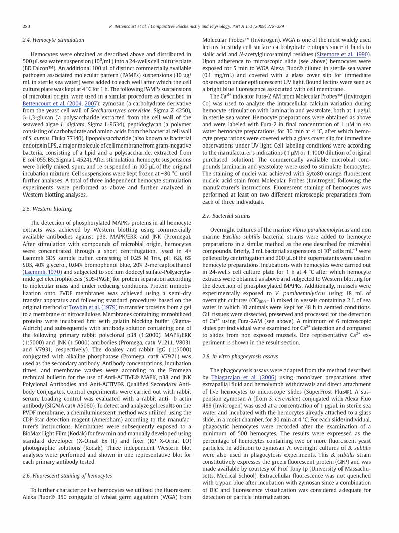
2.4. Hemocyte stimulation
Hemocytes were obtained as described above and distributed in500 µL seawater suspension (106/mL) into a 24-wells cell culture plate(BD Falcon™). An additional 100 µL of distinct commercially availablepathogen associated molecular pattern (PAMPs) suspensions (10 µg/mL in sterile sea water) were added to each well after which the cellculture platewas kept at 4 °C for 1 h. The following PAMPs suspensionsof microbial origin, were used in a similar procedure as described inBettencourt et al. (2004, 2007): zymosan (a carbohydrate derivativefrom the yeast cell wall of Saccharomyces cerevisiae, Sigma Z 4250),β-1,3-glucan (a polysaccharide extracted from the cell wall of theseaweed algae L. digitata, Sigma L-9634), peptidoglycan (a polymerconsisting of carbohydrate and amino acids from the bacterial cell wallof S. aureus, Fluka 77140), lipopolysaccharide (also known as bacterialendotoxin LPS, amajormolecule of cellmembrane fromgram-negativebacteria, consisting of a lipid and a polysaccharide, extracted fromE. coli 055:B5, Sigma L-4524). After stimulation, hemocyte suspensionswere briefly mixed, spun, and re-suspended in 100 µL of the originalincubation mixture. Cell suspensions were kept frozen at −80 °C, untilfurther analyses. A total of three independent hemocyte stimulationexperiments were performed as above and further analyzed inWestern blotting analyses.
2.5. Western blotting
The detection of phosphorylated MAPKs proteins in all hemocyteextracts was achieved by Western blotting using commerciallyavailable antibodies against p38, MAPK/ERK and JNK (Promega).After stimulation with compounds of microbial origin, hemocyteswere concentrated through a short centrifugation, lysed in 4×Laemmli SDS sample buffer, consisting of 0.25 M Tris, pH 6.8, 6%SDS, 40% glycerol, 0.04% bromophenol blue, 20% 2-mercaptoethanol(Laemmli, 1970) and subjected to sodium dodecyl sulfate-Polyacryla-mide gel electrophoresis (SDS-PAGE) for protein separation accordingto molecular mass and under reducing conditions. Protein immobi-lization onto PVDF membranes was achieved using a semi-drytransfer apparatus and following standard procedures based on theoriginal method of Towbin et al. (1979) to transfer proteins from a gelto a membrane of nitrocellulose. Membranes containing immobilizedproteins were incubated first with gelatin blocking buffer (Sigma-Aldrich) and subsequently with antibody solution containing one ofthe following primary rabbit polyclonal p38 (1:2000), MAPK/ERK(1:5000) and JNK (1:5000) antibodies (Promega, cat# V1211, V8031and V7931, respectively). The donkey anti-rabbit IgG (1:5000)conjugated with alkaline phosphatase (Promega, cat# V7971) wasused as the secondary antibody. Antibody concentrations, incubationtimes, and membrane washes were according to the Promegatechnical bulletin for the use of Anti-ACTIVE® MAPK, p38 and JNKPolyclonal Antibodies and Anti-ACTIVE® Qualified Secondary Anti-body Conjugates. Control experiments were carried out with rabbitserum. Loading control was evaluated with a rabbit anti- b actinantibody (SIGMA cat# A5060). To detect and analyze gel results on thePVDF membrane, a chemiluminescent method was utilized using theCDP-Star detection reagent (Amersham) according to the manufac-turer's instructions. Membranes were subsequently exposed to aBioMax Light Film (Kodak) for fewmin and manually developed usingstandard developer (X-Omat Ex II) and fixer (RP X-Omat LO)photographic solutions (Kodak). Three independent Western blotanalyses were performed and shown in one representative blot foreach primary antibody tested.
2.6. Fluorescent staining of hemocytes
To further characterize live hemocytes we utilized the fluorescentAlexa Fluor® 350 conjugate of wheat germ agglutinin (WGA) from
Molecular Probes™ (Invitrogen). WGA is one of the most widely usedlectins to study cell surface carbohydrate epitopes since it binds tosialic acid and N-acetylglucosaminyl residues (Sizemore et al., 1990).Upon adherence to microscopic slide (see above) hemocytes wereexposed for 5 min to WGA Alexa Fluor® diluted in sterile sea water(0.1 mg/mL) and covered with a glass cover slip for immediateobservation under epifluorescent UV light. Bound lectins were seen asa bright blue fluorescence associated with cell membrane.
The Ca2+ indicator Fura-2 AM fromMolecular Probes™ (InvitrogenCo) was used to analyze the intracellular calcium variation duringhemocyte stimulation with laminarin and yeastolate, both at 1 µg/µLin sterile sea water. Hemocyte preparations were obtained as aboveand were labeled with Fura-2 in final concentration of 1 µM in seawater hemocyte preparations, for 30 min at 4 °C, after which hemo-cyte preparations were covered with a glass cover slip for immediateobservations under UV light. Cell labeling conditions were accordingto the manufacturer's indications (1 µM or 1:1000 dilution of originalpurchased solution). The commercially available microbial com-pounds laminarin and yeastolate were used to stimulate hemocytes.The staining of nuclei was achieved with Syto80 orange-fluorescentnucleic acid stain from Molecular Probes (Invitrogen) following themanufacturer's instructions. Fluorescent staining of hemocytes wasperformed at least on two different microscopic preparations fromeach of three individuals.
2.7. Bacterial strains
Overnight cultures of the marine Vibrio parahaemolyticus and nonmarine Bacillus subtilis bacterial strains were added to hemocytepreparations in a similar method as the one described for microbialcompounds. Briefly, 3 mL bacterial suspensions of 108 cells mL−1 werepelleted by centrifugation and 200 μL of the supernatants were used inhemocyte preparations. Incubations with hemocytes were carried outin 24-wells cell culture plate for 1 h at 4 °C after which hemocyteextracts were obtained as above and subjected to Western blotting forthe detection of phosphorylated MAPKs. Additionally, mussels wereexperimentally exposed to V. parahaemolyticus using 18 mL ofovernight cultures (OD600=1) mixed in vessels containing 2 L of seawater in which 10 animals were kept for 48 h in aerated conditions.Gill tissues were dissected, preserved and processed for the detectionof Ca2+ using Fura-2AM (see above). A minimum of 6 microscopicslides per individual were examined for Ca2+ detection and comparedto slides from non exposed mussels. One representative Ca2+ ex-periment is shown in the result section.
2.8. In vitro phagocytosis assays
The phagocytosis assays were adapted from the method describedby Thiagarajan et al. (2006) using monolayer preparations afterextrapallial fluid and hemolymph withdrawals and direct attachmentof live hemocytes to microscope slides (SuperFrost Plus®). A sus-pension zymosan A (from S. cerevisiae) conjugated with Alexa Fluo488 (Invitrogen) was used at a concentration of 1 µg/µL in sterile seawater and incubated with the hemocytes already attached to a glassslide, in a moist chamber, for 30 min at 4 °C. For each slide/individual,phagocytic hemocytes were recorded after the examination of aminimum of 500 hemocytes. The results were expressed as thepercentage of hemocytes containing two or more fluorescent yeastparticles. In addition to zymosan A, overnight cultures of B. subtiliswere also used in phagocytosis experiments. This B. subtilis strainconstitutively expresses the green fluorescent protein (GFP) and wasmade available by courtesy of Prof Tony Ip (University of Massachu-setts, Medical School). Extracellular fluorescence was not quenchedwith trypan blue after incubation with zymosan since a combinationof DIC and fluorescence visualization was considered adequate fordetection of particle internalization.

Fig. 2. Differential interference contrast micrographs of live hemocytes within gillfilaments. Motile granulocytes (gr) are visualized through epithelia within minutes ofdissection and attachment of single filament to glass surface.
281R. Bettencourt et al. / Comparative Biochemistry and Physiology, Part A 152 (2009) 278–289
2.9. Whole mount in-situ hybridization
In order to demonstrate the presence of antibacterial gene tran-scripts in hemocytes present in B. azoricus tissues, single gill filamentswere separated from formalin-70% ethanol preserved gill tissues andanalyzed for the presence of mytilin transcripts according to Tautz(1996) and Braissant and Wahli (1998). Treatment of gills prior to andafter hybridization procedures was made according to Bettencourtet al. (2007). Briefly, pre-hybridization was carried out as describedin Braissant and Wahli (1998) for 3 h at 55 °C after which the hy-bridization reaction was performed at 55 °C for 16 h. A cDNA probecorresponding to mytilin (Bettencourt et al., 2007) was generated bypolymerase chain reaction (PCR), biotinylated using the Biotin-HighPrime labeling kit (Roche Diagnostics) and used to target the anti-bacterial gene mytilin or mytilin messengers (mRNA). Post hybridiza-tion andmytilin transcript visualization procedures were as describedin Bettencourt et al. (2007). Whole mount tissue preparations werethen visualized in a Leica Digital microscope (DM6000B, LeicaMicrosystems) using differential contrast interference (DIC) micro-scopy. Images were captured with a digital color camera (DFC 420,Leica Microsystems) using the freeware Irfanview graphic viewer.
2.10. Statistical analyses
Given the small sample size, a non parametric test (Kruskal–WallisANOVA, pb0.05) was applied to compare differential hemocyte counts
Fig. 1. Scanning electron microphotographs of Bathymodiolus azoricus gills. A. Ctenidiashowing stack of gill filaments (double arrow head). Gill filaments are held togetherthrough ciliated knobs (ck). Some filaments (fil) are shown with broken epithelia andexposing the central lumen (lum). B. Dry-fracture of one gill filament exposing thecentral lumen and itinerant hemocytes (brokenwhite square, arrow within inset). Scalebar in B, 100 µm.
between hemolymph and extrapallial fluid and the percentage ofphagocytosis among hemocyte types in the hemolymph. The resultsare shown as mean and standard deviation unless otherwise stated.
3. Results
3.1. Hemocyte characterization
In the current study, the presence of hemocytes was soughtprimarily in the hemolymph and in the extrapallial fluid foundbetween the mantle and the newly formed nacre. Since thehemolymph circulates around the body of mussels, bathing the gillsand mantle, where it is oxygenated, we first examined the structuralmorphology of the ctenidia under scanning electron and light micro-scopy. Similarly to other mussels, B. azoricus possesses fillibranchgills exhibiting numerous separate filaments (Fig. 1A, double arrow-head) connected through the ciliary junctions or ciliary knobs (Fig. 1,ck). Each filament is organized in two coalescent epithelial cell sheetsoverlaying a central lumen where hemocytes can be found (Fig. 1A,lum). This was evident in dry-fracture of gill filaments where putativehemocytes are exposed in the central lumen (Fig. 1B, arrow withininset). Furthermore, the thinness of gill filaments allowed usto visualize live hemocytes through the epithelium, and to monitorhemocyte motility directly under light microscopy (Fig. 2). Threemajor hemocyte types were discernible in the extrapallial fluidand hemolymph using a combination of light microscopy and stain-ing procedures. The most abundant hemocyte type was identifiedas granulocyte readily recognizable by their cytoplasmic granules(Fig. 3A–B). They appear fairly homogeneous in size and showing acharacteristic crescent, or half-moon shape morphology upon adher-ence to glass slides and before migratory movements. Granulocytesspread well onto the glass surface averaging 30–40 µm in length. Cellmembrane extension and to some degree the presence of philopodiacan be clearly observed in granulocytes visualized under phasecontrast microscopy (Fig. 3A′, B′ and C′). In the attached granulocytes,the nucleus occupies a small cell volume comparatively to the vol-ume of cytoplasm; hence the low nucleus to cytoplasm ratio found inB. azoricus hemocytes. In contrast, hyalinocytes, presented smoothercytoplasm, i.e. a non-granular appearance due to a lower amount ofcytoplasmic granules noticeable under phase contrast (Fig. 3C′ and D′)and DIC visualizations (Fig. 3C and D). Upon adherence to glass slide,hyalinocytes were readily observed within minutes and oftenassuming “dentritic” appearances (two hemocytes on right hand

Fig. 3. Micrographs of B. azoricus live hemocytes attached to glass surface showing: differential interference contrast and phase contrast images of granulocytes (gr; A, B and A′, B′respectively) and hyalinocytes (hy; C, D and C′, D′ respectively). E and F, exemplify bright fieldmicroscopy images of hemoblast-like cells. Scale bar=20 µm. G, percentage of hemocytetypes in the hemolymph and extrapallial fluid. Vertical bars represent means±SD (N=4).
282 R. Bettencourt et al. / Comparative Biochemistry and Physiology, Part A 152 (2009) 278–289
283R. Bettencourt et al. / Comparative Biochemistry and Physiology, Part A 152 (2009) 278–289
side of Fig. 3C, D). They were characterized by long and pronouncedphilopodia extensions denoting a strong adhesion and spreadingbehavior on glass surfaces. A third less common hemocyte type wasalso observed. They correspond to hemoblast-like cells and presenteda spherical shape appearance with higher nucleus to cytoplasm ratiowhen compared to granulocytes and hyalinocytes. Their size variedbetween 8 and 12 µm and did not seem to adhere well to glass slidesurface during light microscopic observations. Their visualization wasbest achieved under bright field microscopy (Fig. 3E and F). Dif-ferential hemocyte counts revealed that granulocytes accounted onaverage for 66% and 74%; hyalinocytes for 31% and 22% and hemoblastfor 4% and 5% of the total hemocyte population collected from thehemolymph and the extrapallial fluid, respectively. Significant differ-ence was only detected between hyalinocytes from the hemolymph
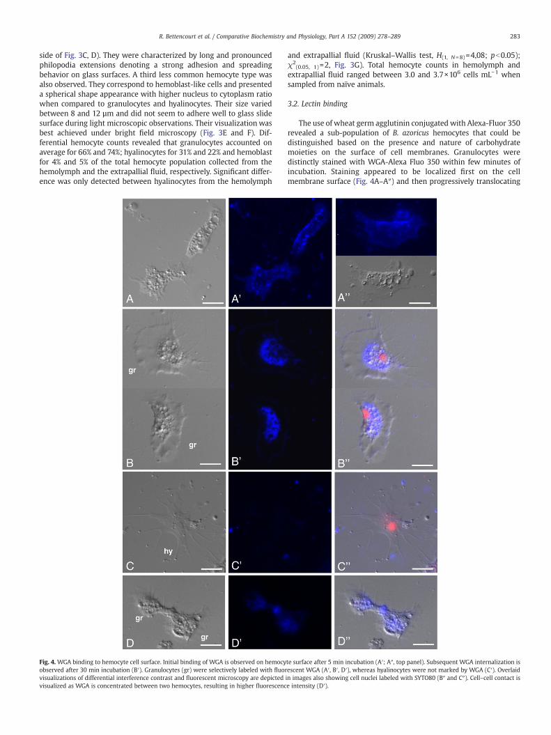
Fig. 4. WGA binding to hemocyte cell surface. Initial binding of WGA is observed on hemocyobserved after 30 min incubation (B′). Granulocytes (gr) were selectively labeled with fluorvisualizations of differential interference contrast and fluorescent microscopy are depictedvisualized as WGA is concentrated between two hemocytes, resulting in higher fluorescenc
and extrapallial fluid (Kruskal–Wallis test, H(1, N=8)=4,08; pb0.05);χ2
(0.05, 1)=2, Fig. 3G). Total hemocyte counts in hemolymph andextrapallial fluid ranged between 3.0 and 3.7×10⁶ cells mL−1 whensampled from naïve animals.
3.2. Lectin binding
The use of wheat germ agglutinin conjugated with Alexa-Fluor 350revealed a sub-population of B. azoricus hemocytes that could bedistinguished based on the presence and nature of carbohydratemoieties on the surface of cell membranes. Granulocytes weredistinctly stained with WGA-Alexa Fluo 350 within few minutes ofincubation. Staining appeared to be localized first on the cellmembrane surface (Fig. 4A–A″) and then progressively translocating
te surface after 5 min incubation (A′; A″, top panel). Subsequent WGA internalization isescent WGA (A′, B′, D′), whereas hyalinocytes were not marked by WGA (C′). Overlaidin images also showing cell nuclei labeled with SYTO80 (B″ and C″). Cell–cell contact ise intensity (D′).

284 R. Bettencourt et al. / Comparative Biochemistry and Physiology, Part A 152 (2009) 278–289
to intracellular compartments after 30 min (Fig. 4B–B″). All granulo-cytes were positively stained with WGA whereas staining of hyalino-cytes was not so evident and in most cases no staining was evidenced(Fig. 4C–C″). Increased lectin fluorescencewas also noted between twoadjacent granulocytes suggesting that carbohydrate moieties may beconcentrated at cell–cell points of contact (Fig. 4D–D″).
3.3. In vitro hemocyte stimulation
Microbial stimulants successfully induced transduction signals inhemocyte preparations as differences were seen in the detection of
Fig. 5. Detection of MAPK-like proteins in hemocyte extracts. Hemocytes were stimulated wanti-MAPK/ERK and anti-JNK polyclonal antibodies. A. Zymosan and lipopolysaccharide indu5, respectively), whereas glucan induced a phosphorylated protein detected by anti-JNK (wmicrobial compounds tested; however, Peptidoglycan induced a slight increase of a protein din one representativeWestern blot. B. Hemocytes were exposed to Vibrio parahaemolyticus anblotting. Vibrio induced the phosphorylation of distinct protein recognized by the p38 antibincrease of phosphorylation is seen with anti-JNK antibody when hemocytes are exposed tothe control experiment when anti-MAPK/ERK was used (B, right panel).
putative ERK, JNK and p38 proteins using commercially available anti-MAPK/ERK, JNK and p38 polyclonal antibodies directed againstmammalian kinases. Unstimulated hemocytes also showed evidenceof phosphorylated MAPKs which could be attributed to stress-relatedfactors while keeping hemocytes in cultures (Fig. 5, red arrow). Den-sitometric analyses (data not shown) of blots indicated that the twowell known immune stimulants, zymosan and LPS were better in-ducers of a p38-like phosphorylated protein (Fig. 5A, lanes 2 and 5,within white circle) whereas the use of glucan in hemocyte culturesincreased the phosphorylation of a 42–45 kDa kinase active form of aputative JNK (Fig. 5A, lane 3, within white circle). The activation of a
ith microbial compounds and cell extracts analyzed by Western blotting using anti-p38,ced a 42–45 kDa phosphorylated protein detected by anti-p38 (white circles, lanes 2 andhite circle, lane 3). Induction of MAPK (ERK) phosphorylation was achieved with alletected by anti-MAPK/ERK (white circle, lane 4). Immuno-detection of β-actin is shownd Bacillus subtilis bacterial supernatants and their cellular content analyzed byWestern
ody (B, dashed circle; left panel), whereas lower induction is seen with Bacillus. A slightVibrio (B, central panel). Levels of phosphorylated proteins were comparable to those in

285R. Bettencourt et al. / Comparative Biochemistry and Physiology, Part A 152 (2009) 278–289
protein recognized by anti-MAPK (ERK) was attained to similar levelsamong all the immune stimulants tested, apart from a slight increaseof phosphorylation obtained with peptidoglycan. In addition tomicrobial compounds, the effect of live bacteria on the phosphoryla-tion of presumed MAPKs was also tested with hemocyte preparations.Qualitative differences were found regardless of the use of nonmarineBacillus sp. or the marine Vibrio bacteria in in vitro hemocytestimulations. The effect of Vibrio sp. was most noticeable on theactivation of a phosphorylated protein that specifically cross reactedwith anti-p38 (Fig. 5B, white dashed circle). In contrast, the effect ofBacillus sp. was rather modest on the activation of putative JNK andERK compared to hemocytes stimulated with Vibrio (Fig. 5B, center
Fig. 6. Intracellular Ca2+ variation. The intracellular Ca2+ variation of stimulated hemocytes wwith laminarin induced the occurrence of distinct punctate structures visualized as blue fluwith yeastolate did not induce punctate fluorescence (B′). Tissue sections of gill filaments frowithin hemocytes (C′). Tissue sections of gills from control animals maintained only in plain sof punctate fluorescence within gill hemocytes is visualized in E′. Overlaid digital images ofstaining with the fluorescent SYTO80 marker is visualized in B″, C″, D″ and E″. Scale bar=20
and right panels). No major cross-reactivity was seen in controlexperiments carried out with rabbit serum (data not shown). Proteinloading control was monitored by densitometric analysis of constitu-tive proteins cross-reacting with polyclonal antibodies and present inthe same protein extracts loaded into different lanes (Fig. 5A, whitearrows). Additional loading control was assessed with anti-b actin (A,bottom panel) for all three MAPK immuno-blots.
3.4. Intracellular Ca2+ variation
The effect of microbial compounds was examined in hemocytesusing the fluorescent Ca2+ indicator Fura-2 AM. Laminarin induced a
as analyzed by fluorescence microscopy using the Ca2+ indicator Fura-2 AM. Stimulationorescence concentrated in numerous spots throughout the hemocyte (A′). Stimulationm B. azoricus exposed to Vibrio parahaemolyticus show punctate fluorescent structuresea water did not show hemocyte punctate fluorescence (D′). Higher magnification (63×)DIC and fluorescence microscopy images are visualized in A″, B″, C″, D″ and E″. Nuclearµm.

286 R. Bettencourt et al. / Comparative Biochemistry and Physiology, Part A 152 (2009) 278–289
strong intracellular Ca2+ variation in B. azoricus hemocytes (Fig. 6A′)whereas the use of the yeast extract, yeastolate did not induce anoticeable cytoplasmic Ca2+ variation (Fig. 6B′). These results suggestthat the variation of intracellular Ca2+ levels in B. azoricus hemocytescould be directly modulated by certain class of immune stimulants ofmicrobial origin. To confirm whether live bacteria could or could nothave the same effect on intracellular Ca2+ levels in live tissues, wesubjected vent mussels to V. parahaemolyticus cultures and processedthe gills for histological analyses and for Intracellular Ca2+ levels assess-ment. The hemocyteswithin gill tissues dissected fromanimals exposedto Vibrio revealed higher intracellular Ca2+ levels (Fig. 6C′and E′,punctate structures) than in the gills from control animals maintainedin sea water (Fig. 6D′).
3.5. Phagocytosis of fluorescent yeast particles
The in vitro phagocytic assays revealed that 70% of the hemocytescontaining more than 2 zymosan particles were granulocytes (Fig. 7).
Fig. 7. In vitro phagocytosis assay. The phagocytosis assays were performed with fluorescentparticles was recorded from aminimum of 500 hemocytes per slide. Digital images of granuloA′) and on fluorescent microscopy (A″). Panel A′ is a combined image from DIC and fluorescsubtilis. The percentages of hemocytes containing at least two or more phagocytized yeast pdifferent individuals. Vertical bars represent means±SD (N=4). Scale bar=20 µm.
However, the number of ingested particles was variable among hemo-cytes (data not shown). To a lesser extent, the percentage of phagocyticcells corresponding to hyalinocytes was 23%. In contrast, the percentageof hemoblasts containing ingested zymosan particles was 5–7%, thelowest revealed in this study. The phagocytosis of B. subtilis was alsoobserved in granulocytes although less apparent as for the assays withfluorescent zymosan (Fig. 7). There was statistically significant differ-ence observed between the percentages of phagocytosis within eachhemocyte type (Kruskal–Wallis, H(2, N=12)=9.85; pb0.01).
3.6. Mytilin gene expression in gill tissues
The presence of the antibacterial gene mytilin in gill tissues wasconfirmed by means of whole-mount in situ hybridization. This resultillustrates, as demonstrated in our earlier study, that mytilin mRNAtranscripts are found in hemocytes within single gill filaments (Betten-court et al., 2007). In addition, the expression of mytilin gene wasanalyzed in semi-quantitative reverse transcription-PCR experiments.
zymosan A as the phagocytic particle. The number of hemocytes containing fluorescentcytes (gr), showing internalization of yeast particles, were captured on DIC (panels A andent microscopy. Panels B–B″ show granulocytes with phagocytized fluorescent Bacillusarticles are shown in C. Results are from 3 different preparations of hemocytes from 4

Fig. 8.Mytilin transcript analysis. A. Qualitativemytilin gene expressionwas analyzed by reverse transcription-polymerase chain reaction using total RNA gill extractions from controland Vibrio exposed animals. Detectable mytilin amplification products were initially seen after 15 PCR cycles. As demonstrated by band intensity, after 20 cycles, higher levels ofmytilin amplification products were generated in animals exposed to Vibrio (exp) in comparison to control animals (cont) kept only in sea water samples (broken circles). Nodifferences were seen between the amplification of the internal standard 18S rRNA from control and Vibrio exposed animals, i.e. both control (cont) and experimental (exp) RNAcontained the same level of 18S transcript at the beginning of RT-PCR and throughout PCR amplification. B. Mytilin transcripts were analyzed in in-situ hybridization experiments.PCR-based cDNA products corresponding to mytilin gene were labeled with biotin and used as probes to target respective transcripts. Transcripts were identified in hemocyteslocated in gill filament epithelium or in between two coalescent unicellular layers of endothelial cells. cDNA probes corresponding to an irrelevant PCR product was used in controlexperiments. Scale bar=20 µm.
287R. Bettencourt et al. / Comparative Biochemistry and Physiology, Part A 152 (2009) 278–289
Our results indicated that gill RNA extracted from mussels exposed to V.parahaemolyticus contained higher levels of mytilin transcripts than gillRNA extracted from control animals (Fig. 8A).
4. Discussion
Thehydrothermal ventmusselB. azoricus is commonly found indensepopulations at the Mid Atlantic Ridge and appears to be well adapted totheir peculiar environment. Other bathymodiolid species are also foundin dense communities associated with deep sea hydrothermal ventsand cold-water sulfide/hydrocarbon seepsdistributed in both theAtlanticand Pacific Oceans. This corroborates their functional adaptability toextreme environments (Gustafson et al.,1998; Von Cosel et al.,1999). Thepresence of thiotrophic and methanotrophic bacterial symbiont types inspecialized epithelial cells of the vent mussel gill tissues is believed toprovide substantial nutritional advantage to the mussel, thus enhancingits aptitude to oxidize sulfide and methane at the vent sites (Distel et al.,1995; Fiala-Medioni et al., 2002; Duperron et al., 2006).
In spite of the toxic nature of deep sea hydrothermal vent en-vironments, the problem of microbial threat and the need forimmunity exist in both deep sea and shallow water bivalves however,differences in the genes of marine organisms living in so distincthabitats are likely to occur, particularly, at the transcription/regulationlevel. To understand the humoral and cellular defense mechanisms in
animals living in such extreme physical and chemical conditions weset out to investigate, in the deep-sea vent mussel B. azoricus, immuneparameters that are commonly found in other marine bivalves.
Withdrawal of hemolymph and fluid from the extrapallial spaceshowed large number of hemocytes (3.0–3.7×10⁶ cells mL−1). Thepredominant hemocyte type in B. azoricus was identified as being thegranulocyte. The abundance of intracellular granules among granulo-cytes and their glycoprotein nature had been confirmed by AB-PASstaining in our previous studies (Bettencourt et al., 2008). Theyaccounted for 60–75% of the total hemocyte population. This is inaccordance with many bivalve species, from which granulocytes arethe most numerous cell type and the main phagocytic cells, varying inthe number and nature of granules, whereas vent mussel hyalinocyteswere significantly less phagocytic when compared to data from othershallow water bivalves (Allam et al., 2002a,b; Wootton et al., 2003).Furthermore, total hemocyte counts revealed variability in the num-ber of circulating granulocytes between individuals; however, theextrapallial fluid seems to hold higher numbers of circulating granu-locytes, which suggests a role in defense processes that could takeplace between the mantle and the inner face of the shell againstinvading microorganisms (Allam et al., 2000). The nature of the cellsurface molecules was further analyzed in B. azoricus hemocytes usingthe differential binding property of the fluorescent lectin wheat germagglutinin (WGA). The granulocytes were distinctly labeled withWGA
288 R. Bettencourt et al. / Comparative Biochemistry and Physiology, Part A 152 (2009) 278–289
whereas hyalinocytes seemed less disposed to WGA binding (Fig. 4B′and C′). Thus, it is reasonable to assume that the two main residuesrecognized by WGA, N-acetyl-glucosamine and sialic acid moieties,are present on the cell surface of granulocytes. Moreover, cell–cellinteractions could also be visualized using fluorescent WGA, whichsuggests that carbohydrate epitopes may be involved in cell–cellinteractions between hemocytes.
In mammalian systems, the activation of signaling pathways isessential in innate immunity and includes protein-kinase and lipid-kinase driven intracellular phosphorylation cascades (Canesi et al.,2006). Following procedures similar to those of Canesi and co-workers,we demonstrated that compounds of microbial origin can triggerdetectable phosphorylation events in B. azoricus hemocyte extractsand are likely to involve the activation of different classes of Mito-gen Activated Protein Kinases. When challenged with either a marine,V. parahaemolyticus or a non-marine bacterium, B. subtilis, to stimulatehemocytes, cellular proteins were differently phosphorylated asdemonstrated in Western blotting experiments using the MAPK/ERK,p38 and JNK rabbit polyclonal antibodies. Moreover, the differencesseen in phosphorylation patterns could possibly be attributed toinherent properties of the bacterial strain used, differences in themechanisms of binding to hemocytes or differential activation of cell-membrane receptors and signaling pathways, resulting in differentpatterns in Western blots (Canesi et al., 2006). Western blottinganalyses, suggest that B. azoricus hemocytes display receptors withbinding affinities toward microbial molecules or to live bacteria.Moreover, the potential marine pathogen, V. parahaemolyticus, has adistinct effect on B. azoricus hemocytes as demonstrated in MAPKsphosphorylation patterns. This implies some degree of immunerecognition specificity by the vent mussel hemocytes, which clearlyrespond differently to distinctmicrobial molecules and to, at least, twodifferent live bacteria. Taken as a whole, our results suggest thatsignaling pathways involving protein kinases could contribute to theactivation of hemocyte responses in B. azoricus. A putative p38 kinaseorthologue is detected in B. azoricus, inasmuch as in shallow watermussels, by amammalian anti-p38polyclonal antibody. It is likely to beinvolved in signal transduction mechanisms in response to immune-related challenges either bymicrobial compounds or live bacteria. Thiscorroborates the findings by Canesi et al. (2002) on Mytilusgalloprovincialis hemocytes challenged with bacteria.
The presence of extracellular products deriving from Vibrio mayinduce alterations of the hemocyte host signal transduction pathwaysthat could lead to changes in hemocyte adherence. The toxicity ofbacteria towards bivalve hemocytes has been demonstrated in themussel Mytilus edulis. It was then shown that the rounding up ofhemocytes could be induced by the pathogenic Vibrio alginolyticusand Vibrio (now Listonella) anguillarum bacteria and this activity wasenhanced by a hemolymph factor (Lane and Birkbeck, 1999). AlthoughV. parahaemolyticus is not pathogenic to marine bivalves, it was usedin our experiments to demonstrate that bacterial products mayinteract with hemocytes in the way that intracellular phosphorylationevents could be visualized by Western blotting and variation inintracellular Ca2+ could also be observed (see bellow).
An early key feature in the immune cell activation is an increase ofthe intracellular Ca2+ concentration, which acts as a secondmessengerof signal transduction (Grafton and Thwaite, 2001; Lewis, 2001;Kimura et al., 2006). Thus, the variation in intracellular Ca2+ levels is,yet another immune-related parameter contributing to intracellularsignaling processes involved in hemocyte immune activation (Bauset al., 1996). Subsequently, we have induced an initial rise in intra-cellular Ca2+, distinctively visualized as fluorescent blue punctatestructures, following a stimulation caused by the microbial compoundlaminarin or by live V. parahaemolyticus. Yeastolate was used todemonstrate the differences in Ca2+ levels with regard to the size andpurity of the microbial compound used. While yeastolate is a solubleyeast extract from S. cerevisiae containing also peptides, amino acids
and carbohydrates, it may constitute larger macromolecules thatcannot be properly recognized by hemocyte membrane surfaces. Thepunctate staining was more evident inside the granulocytes than, inthe hyalinocytes. Furthermore, our findings suggest that the activationsignal caused by V. parahaemolyticus could be conveyed through gillepithelia and subsequently to hemocytes within the lumen of gillfilaments. For this reason, gill epithelia may be insightful whenstudying signal transduction mechanisms across the epithelial cellsleading to a hemocyte immune responsiveness. The role of calciumwasrecently evoked during hemocyte activation in a study involving flowcytometry and the use of the calciumprobe Fluo-3/AM. The differencesin calcium activity were measured while oyster hemocytes wereincubated in hemolymph or in artificial sea water (Aton et al., 2006).
Invading microorganisms elicit significant responses in themolluscan immune system including phagocytic reactions, variationin the number of circulating hemocytes, production of highly reactiveoxygen species, and the release of antimicrobial agents to eliminate orneutralize infectious microorganisms (Anderson, 1994; Lopez-Corteset al., 1999). Phagocytosis is, thus considered the predominantmechanism of cellular defense in bivalves and depends on circulatinghemocytes present in the blood which are able to migrate throughoutthe tissues in response to foreign objects including harmful pathogens(Lopez-Cortes et al., 1999). In the present study, the phagocytic activityof the vent mussel hemocytes was assessed with adherent cells inthe presence of fluorescent yeast particles. The adherent cells wereeasier to visualize under lightmicroscopy and also allowed convenientremoval of excess sea water containing yeast particles. Our resultsindicated that granulocytes are the main contributors to phagocytosisin B. azoricus. This agrees with phagocytic studies in other bivalvespecies (Wootton and Pipe, 2003; Wootton et al., 2003; Aladailehet al., 2007). We also performed phagocytosis assays with B. subtilisconstitutively expressing GFP (Fig. 7). As demonstrated for yeast par-ticles, the granulocytes also revealed their propensity to engulf fluo-rescent Bacillus sp. although the phagocytic percentages were ratherlow compared to those obtained with fluorescent yeast (data notshown). Taken together, the great phagocytic ability of B. azoricusgranulocytes seems to confirm their role in cellular defense in thehemolymph and the extrapallial fluid.
The expression of genes encoding antibacterial peptides is anessential component of host defense reactions against microbialinfections. In the mussel M. galloprovincialis, the synthesis of theantibacterial peptide mytilin occurs in hemocytes where the matureforms of the antibacterial peptide are stored (Mitta et al., 2000a). Therelease of the active mytilin form may also take place in systemicresponses during which its concentration is increased in the plasma.However, its co-localization with engulfed bacteria suggests anintracellular antibacterial activity (Mitta et al., 2000b).
In the present study, we further investigated the presence of mytilinmRNA in gill tissues by means of whole-mount in situ hybridization.Mytilin messengers were identified in hemocytes lying under the gillfilament epithelia (Fig. 8B). This finding corroborates studies carried outby Roch and co-authors using M. galloprovincialis as a model to studyinnate immunity inmarine bivalves and the biological activity ofmusselantibacterial peptides (Mitta et al., 2000a). Furthermore, the relativeabundance of mytilin transcripts was also analyzed in semi-quantitativeRT-PCR experiments by comparing the mRNA abundance of 18S rRNAgene, which is assumed to be expressed at equal levels in any cell typeregardless the experimental conditions. Therefore, we could verifyhigher levels of mytilin mRNA in gill tissues from animals exposed to V.parahaemolyticus in comparison to the gene expression levels of controlanimals (Fig. 8A).
Our findings support the strategic importance that gills assume atthe interface between the external milieu and the internal bodycavities of the animalwhere contactwithmicroorganisms is inevitableduring feeding processes inasmuch as host defense responses mayincur from interactions with infective pathogens during normal

289R. Bettencourt et al. / Comparative Biochemistry and Physiology, Part A 152 (2009) 278–289
filtration (Allam et al., 2002a; Wootton et al., 2003). For this reason,antibacterial responses against invading bacterial pathogens are likelyto take place in gill tissues (Mitta et al., 2000a,b).
In conclusion, supporting evidence presented here leads us topropose that the mussel B. azoricus is an amenable model to studycellular immune reactions in a deep sea vent organism. The un-precedented experimental acclimatization of B. azoricus may haveconferred its hemocytes with similar cellular properties as thosereported in shallow water mussels from coastal regions (Hattan et al.,2001; Aladaileh et al., 2007). The description of three main types ofhemocytes in B. azoricus was consistent with the general knowledgeon bivalve hemocytes. In addition, experimental tools such as the useof commercially available antibodies directed against MAPKs, fluor-escent lectins and Ca2+ indicators, RT-PCR and in-situ hybridization,contributed to further expand our knowledge on innate immunesystems in hydrothermal vent animals. The evidence so far gatheredseems to indicate that B. azoricus innate immune system is seeminglyhomologous to that of other bivalves. However, the mechanisms thatregulate gene expression might reveal unique cis-regulatory signa-tures or specific DNA sequences in the promoter region of theirimmune genes, irrespectively of the conditions to which vent musselsare acclimatized. Experiments are currently being developed in ourlaboratory to demonstrate the usefulness of hyperbaric chambers inthe expression of immune and stress-related genes in B. azoricus.
Acknowledgements
We thank the shipboard nautical, technical and scientific parties ofthe French R/V Pourquois Pas?/ROV VICTOR 6000 during theMoMareto cruise, as well as, the captain and crew members of theAzorean R/V Arquipélago for assistance in the collection of samples.We are most grateful to Mario Laranjo and Luis Pires for skillfullaboratory assistance and maintenance of acclimatized aquaria. Wealso thank Ana Colaço and Humberto Lopes for their support andMaria C. Magalhães for statistical analysis interpretations. This workwas supported by funds provided by the pluri-annual and program-matic funding scheme to IMAR/DOP research unit #531 and associatelaboratory #9; the network of Excellence MarBEF (Marine Biodiversityand Ecosystem Functioning-contract No. GOCE-CT-2003-505446); theLuso-American Foundation FLAD (Project L-V-173/2006); the AzoreanRegional Directorate for Science and Technology, DRCT. The Portu-guese FCT post-doctoral fellowship SFRH/BPD/14896/2004 (R.B.) isgratefully acknowledged. We are also indebted to Natalia Correia andIlda Bettencourt for proofreading of the manuscript and the anon-ymous reviewers for their helpful comments.
References
Aladaileh, S., Nair, S.V., Birch, D., Raftos, D.A., 2007. Sidney rock oyster (Saccostreaglomerata) hemocytes: morphology and function. J. Invertebr. Pathol. 96, 48–63.
Allam, B., Paillard, C., 1998. Defense factors in clam extrapallial fluids. Dis. Aquat. Organ.33, 123–128.
Allam, B., Paillard, C., Auffret, M., 2000. Alterations in hemolymph and extrapallial fluidparameters in the Manila clam, Ruditapes philippinarum, challenged with thepathogen Vibrio tapetis. J. Invertebr. Pathol 76, 63–69.
Allam, B., Ashton-Alcox, K.A., Ford, S.E., 2002a. Flowcytometric comparison of haemocytesfrom three species of bivalve molluscs. Fish Shellfish Immunol. 13, 141–158.
Allam, B., Ashton-Alcox, K.A., Ford, S.E., 2002b. Flow cytometric measurements ofhemocytes viability and phagocytic activity in the clam Ruditapes philippinarum.J. Shellfish Res. 21, 13–19.
Anderson, R., 1994. Hemocyte-derived reactive oxygen intermediate production in fourbivalve mollusks. Dev. Comp. Immunol. 18, 89–96.
Arumugam, M., Romestand, B., Torreilles, J., Roch, P., 2000. In vitro production ofsuperoxide and nitric oxide (as nitrite and nitrate) by Mytilus galloprovincialishaemocytes upon incubation with PMA or laminarin or during yeast phagocytosis.Eur. J. Cell Biol. 79, 513–519.
Aton, E., Renault, R., Gagnaire, B., Thomas-Guyon, H., Cognard, C., Imbert, N.A., 2006.Flow cytometric approach to study intracellular-free Ca in Crassostrea gigashaemocytes. J. Shellfish Res. 20, 493–502.
Baus, E., Andris, P.M., Dubois, J., Urbain, Leo, O.,1996. Dexamethasone inhibits early steps ofantigen receptor signaling in activated T lymphocytes. J. Immunol. 156, 4555–4561.
Bettencourt, R., Asha, H., Dearolf, C., Ip, Y.T., 2004. Hemolymph-dependent and-independent responses in Drosophila immune tissues. J. Cell. Biochem. 92, 84-49.
Bettencourt, R., Roch, P., Stefanni, S., Rosa, D., Colaço, A., Serrão Santos, R., 2007. Deep seaimmunity: unveiling immune constituents from the hydrothermal vent musselBathymodiolus azoricus. Mar. Environ. Res. 64, 108–127.
Bettencourt, R., Dando, P., Rosa, D., Riou, V., Colaço, A., Sarrazin, J., Sarradin, P.M., SerrãoSantos, R., 2008. Changes of gill and hemocyte-related bio-indicators during longterm maintenance of the vent mussel Bathymodiolus azoricus held in aquaria atatmospheric pressure. Comp. Biochem. Physiol. A 150, 1–7.
Braissant, O., Wahli, W., 1998. A simplified in situ hybridization protocol using non-radioactively labelled probes to detect abundant and raremRNAs on tissue sections.Biochemica 1, 10–16.
Canesi, L., Betti, M., Ciacci, C., Scarpato, A., Citterio, B., Pruzzo, C., Gallo, G., 2002. Signalingpathways involved in the physiological response of mussel hemocytes to bacterial chal-lenge: the role of stress-activated p38 MAP kinases. Dev. Comp. Immunol. 26, 325–334.
Canesi, L., Betti, M., Ciacci, C., Lorusso, L.C., Pruzzo, C., Gallo, G., 2006. Cell signaling inthe immune response of mussel hemocytes. Invertebr. Survival. J. 3, 40–49.
Distel, D.L., Lee, H.K.W., Cavanaugh, C.M., 1995. Intracellular coexistence of methano-trophic and Thioautotrophic bacteria in a hydrothermal vent mussel. Proc. Natl.Acad. Sci. U. S. A. 92, 9598–9602.
Duperron, S., Bergin, C., Zielinski, F., Blazejak, A., Pernthaler, A., McKiness, Z.P., DeChaine,E., Cavanaugh, C.M., Dubilier, N., 2006. A dual symbiosis shared by two musselspecies, Bathymodiolus azoricus and Bathymodiolus puteoserpentis (Bivalvia:Mytilidae), from hydrothermal vents along the northern Mid-Atlantic Ridge.Environ. Microbiol. 8, 1441–1447.
Fiala-Medioni, A., McKiness, Z.P., Dando, P., Boulegue, J., Mariotti, A., Alayse-Danet, A.M.,Robinson, J.J., Cavanaugh, C.M., 2002. Ultrastructural, biochemical, and immunologicalcharacterization of twopopulations of themytilidmusselBathymodiolus azoricus fromthe Mid-Atlantic Ridge: evidence for a dual symbiosis. Mar. Biol. 141, 1035–1043.
Franchini, A., Ottaviani, E., 2000. Repair of molluscan tissue injury: role of PDGF andTGF-β. Tissue Cell. 32, 312–321.
Gourdon, I., Guerin, M.C., Torreilles, J., Roch, P., 2001. Nitric oxide generation byhemocytes of the mussel Mytilus galloprovincialis. Nitric Oxide 5, 1–6.
Grafton, G., Thwaite, L., 2001. Calciumchannels in lymphocytes. Immunology 104,119–126.Gustafson, R.G., Turner, R.D., Lutz, R.A., Vrijenhoek, R.C., 1998. A new genus and five new
species of mussels (Bivalvia, Mytilidae) from deep-sea sulphide/hydrocarbon seepsin the Gulf of Mexico. Malacologia 40, 63–112.
Hattan, S.J., Laue, T., Chasteen, N.D., 2001. Purification and characterization of a novelcalcium-binding protein from the extracellular fluid of the mollusc, Mytilus edulis.J. Biol. Chem. 276, 4461–4468.
Humphries, J.E., Yoshino, T.P., 2003. Cellular receptors and signal transduction inmolluscan hemocytes: connections with the innate immune system of vertebrates.Integr. Comp. Biol. 43, 305–312.
Kimura, K., Reinhardt, A., Goff, J.P., 2006. Parturition and hypocalcemia blunts calciumsignals in immune cells of dairy cattle. J. Dairy Sci. 89, 2588–2595.
Labreuche, Y., Lambert, C., Soudant, P., Boulo, V., Huvet, A., Nicolas, J.-L., 2006. Cellular andmolecular hemocyte responses of the Pacific oyster, Crassostrea gigas, followingbacterial infectionwithVibrio aestuarianus strain01/32.Microbes Infect. 8, 2715–2724.
Laemmli, U.K., 1970. Cleavage of structural proteins during the assembly of the head ofbacteriophage T4. Nature 227, 680–685.
Lane, E., Birkbeck, T.H., 1999. Toxicity of bacteria towards haemocytes of Mytilus edulis.Aquat. Living Resour. 12, 343–350.
Lewis, R.S., 2001. Calcium signaling mechanisms in T lymphocytes. Annu. Rev. Immunol.19, 497–521.
Lopez-Cortes, L., Castro, D., Navas, J.I., Borrego, J.J., 1999. Phagocytic and chemotacticresponses of manila, and carpet shell clam haemocytes against Vibrio tapetis, thecausative agent of brown ring disease. Fish Shellfish Immunol. 9, 543–555.
Mitta, G., Vandenbulcke, F., Roch, P., 2000a. Original involvement of antimicrobialpeptides in mussel innate immunity. FEBS Lett. 486, 185–190.
Mitta, G., Vandenbulcke, F., Hubert, F., Salzet, M., Roch, P., 2000b. Involvement ofmytilins in mussel antibacterial defense. J. Biol. Chem. 275, 12954–12962.
Mount, A., Wheeler, R., Paradkar, P., Snider, D., 2004. Hemocyte-mediated shellmineralization in the eastern oyster. Science 304, 297–300.
Pipe, R.K., 1990. Differential binding of lectins to haemocytes of the mussel Mytilusedulis. Cell Tissue Res. 261, 261–268.
Pruzzo, C., Gallo, G., Canesi, L., 2005. Persistence of vibrios in marine bivalves: the role ofinteractions with haemolymph components. Environ. Microbiol. 7, 761–772.
Sizemore, R.K., Caldwell, J.J., Kendrick, A.S., 1990. Alternate gram staining techniqueusing a fluorescent lectin. Appl. Environ. Microbiol. 56, 2245–2247.
Tautz, D., 1996. Localization of the segmentation gene hunchback in Drosophila embryoswith DIG-labeled DNA probes, Non reactive in situ Hybridization ApplicationManual, 2nd ed. Boehringer Mannheim GmbH, Biochemica.
Thiagarajan, R., Gopalakrishnan, S., Thilagam, H., 2006. Immunomodulation in themarine green mussel Perna viridis exposed to sub-lethal concentrations of Cu andHg. Arch. Environ. Contam. Toxicol. 51, 392–399.
Towbin, H., Staehelin, T., Gordon, J., 1979. Electrophoretic transfer of proteins frompolyacrylamide gels to nitrocellulose sheets: procedure and some applications.Proc. Natl. Acad. Sci. U. S. A. 76, 4350–4354.
Von Cosel, R., Comtet, T., Krylova, E.M., 1999. Bathymodiolus (Bivalvia: Mytilidae) fromhydrothermal vents on the Azores triple junction and the Logatchev hydrothermalfield, Mid Atlantic Ridge. Veliger 42, 218–248.
Wootton, E.C., Pipe, R.K., 2003. Structural and functional characterization of the bloodcells of the bivalvemollusc, Scrobicularia plana. Fish Shellfish Immunol.15, 249–262.
Wootton, E.C., Dyrynda, E.A., Ratcliffe, N.A., 2003. Bivalve immunity: comparisonsbetween the marine mussel (Mytilus edulis), the edible cockle (Cerastoderma edule)and the razor-shell (Ensis siliqua). Fish Shellfish Immunol. 15, 195–210.