Injection of a recombinant AAV serotype 2 into canine skeletal muscles evokes strong immune...
12
ORIGINAL ARTICLE Injection of a recombinant AAV serotype 2 into canine skeletal muscles evokes strong immune responses against transgene products K Yuasa 1,2 , M Yoshimura 1 , N Urasawa 1 , S Ohshima 1 , JM Howell 3 , A Nakamura 1 , T Hijikata 2 , Y Miyagoe-Suzuki 1 and S Takeda 1 1 Department of Molecular Therapy, National Institute of Neuroscience, National Center of Neurology and Psychiatry, Kodaira, Tokyo, Japan; 2 Research Institute of Pharmaceutical Sciences, Faculty of Pharmacy, Musashino University, Nishi-tokyo, Tokyo, Japan and 3 Division of Veterinary and Biomedical Sciences, Murdoch University, Perth, Western Australia, Australia Using murine models, we have previously demonstrated that recombinant adeno-associated virus (rAAV)-mediated micro- dystrophin gene transfer is a promising approach to treatment of Duchenne muscular dystrophy (DMD). To examine further therapeutic effects and the safety issue of rAAV-mediated microdystrophin gene transfer using larger animal models, such as dystrophic dog models, we first investigated transduction efficiency of rAAV in wild-type canine muscle cells, and found that rAAV2 encoding b-galactosidase effectively transduces canine primary myotubes in vitro. Subsequent rAAV2 transfer into skeletal muscles of normal dogs, however, resulted in low and transient expression of b-galactosidase together with intense cellular infiltrations in vivo, where cellular and humoral immune responses were remarkably activated. In contrast, rAAV2 expressing no transgene elicited no cellular infiltrations. Co-administration of immunosup- pressants, cyclosporine and mycophenolate mofetil could partially improve rAAV2 transduction. Collectively, these results suggest that immune responses against the trans- gene product caused cellular infiltration and eliminated transduced myofibers in dogs. Furthermore, in vitro interferon-g release assay showed that canine splenocytes respond to immunogens or mitogens more susceptibly than murine ones. Our results emphasize the importance to scrutinize the immune responses to AAV vectors in larger animal models before applying rAAV-mediated gene therapy to DMD patients. Gene Therapy advance online publication, 21 June 2007; doi:10.1038/sj.gt.3302984 Keywords: AAV; gene transfer; skeletal muscle; dog; immune response; Duchenne muscular dystrophy Introduction Duchenne muscular dystrophy (DMD) is an X-linked, lethal disorder of skeletal muscle caused by mutations in the dystrophin gene, which encodes a large subsarco- lemmal cytoskeletal protein, dystrophin. DMD is characterized by a high incidence (one among 3500 boys) and a high frequency of de novo mutation. 1 The absence of dystrophin accompanies the loss of dystrophin- associated glycoprotein complex from the sarcolemma and results in progressive muscle weakness, cardiomyopathy and early death. Although several treatment modalities, such as gene, cell and pharmacological therapies, have been researched to aim at correcting the dystrophic phenotypes, DMD currently has no effective treatment. An adeno-associated virus (AAV) vector is a potential tool for gene therapy of inherited neuromuscular disorders. It is a nonpathogenic, low immunogenic and replication-defective viral vector that effectively infect nondividing cells, such as skeletal muscle fibers. 2 The size of exogenous DNA fragment which can be inserted into recombinant AAV vectors (rAAVs), however, is limited to up to 4.9 kb. Therefore, full-length dystrophin (14 kb) and mini-dystrophin (6.4 kb) cDNAs are too large to be incorporated into a rAAV. We and others have tried to design a short but functional microdystrophin gene that could be utilized as the therapeutic tool for DMD. 3–6 We constructed a series of rod-truncated microdystro- phin cDNAs, 3 and generated transgenic mdx mice expressing each microdystrophin, and demonstrated that microdystrophin CS1 with four rod repeats and three hinges was a good candidate for therapeutic molecule. 7 We have also showed that the muscle-specific muscle creatine kinase (MCK) promoter in a rAAV drives longer transgene expression than the ubiquitous cyto- megalovirus (CMV) promoter in mdx muscle. 8 Therefore, we generated the rAAV2 expressing microdystrophin DCS1 (3.8 kb cDNA) driven by the MCK promoter and introduced it into mdx muscles, and showed that sustained expression of microdystrophin from rAAV significantly ameliorates dystrophic phenotypes of Received 8 October 2006; revised 3 April 2007; accepted 14 May 2007 Correspondence: Dr S Takeda, Department of Molecular Therapy, National Institute of Neuroscience, National Center of Neurology and Psychiatry, 4-1-1 Ogawa-higashi, Kodaira, Tokyo 187-8502, Japan. E-mail: [email protected] Gene Therapy (2007), 1–12 & 2007 Nature Publishing Group All rights reserved 0969-7128/07 $30.00 www.nature.com/gt
-
Upload
independent -
Category
Documents
-
view
2 -
download
0
Transcript of Injection of a recombinant AAV serotype 2 into canine skeletal muscles evokes strong immune...
ORIGINAL ARTICLE
Injection of a recombinant AAV serotype 2 into canineskeletal muscles evokes strong immune responsesagainst transgene products
K Yuasa1,2, M Yoshimura1, N Urasawa1, S Ohshima1, JM Howell3, A Nakamura1, T Hijikata2,
Y Miyagoe-Suzuki1 and S Takeda1
1Department of Molecular Therapy, National Institute of Neuroscience, National Center of Neurology and Psychiatry, Kodaira, Tokyo,Japan; 2Research Institute of Pharmaceutical Sciences, Faculty of Pharmacy, Musashino University, Nishi-tokyo, Tokyo, Japan and3Division of Veterinary and Biomedical Sciences, Murdoch University, Perth, Western Australia, Australia
Using murine models, we have previously demonstrated thatrecombinant adeno-associated virus (rAAV)-mediated micro-dystrophin gene transfer is a promising approach totreatment of Duchenne muscular dystrophy (DMD). Toexamine further therapeutic effects and the safety issue ofrAAV-mediated microdystrophin gene transfer using largeranimal models, such as dystrophic dog models, we firstinvestigated transduction efficiency of rAAV in wild-typecanine muscle cells, and found that rAAV2 encodingb-galactosidase effectively transduces canine primarymyotubes in vitro. Subsequent rAAV2 transfer intoskeletal muscles of normal dogs, however, resulted inlow and transient expression of b-galactosidase togetherwith intense cellular infiltrations in vivo, where cellularand humoral immune responses were remarkably activated.
In contrast, rAAV2 expressing no transgene elicitedno cellular infiltrations. Co-administration of immunosup-pressants, cyclosporine and mycophenolate mofetil couldpartially improve rAAV2 transduction. Collectively, theseresults suggest that immune responses against the trans-gene product caused cellular infiltration and eliminatedtransduced myofibers in dogs. Furthermore, in vitro interferon-grelease assay showed that canine splenocytes respondto immunogens or mitogens more susceptibly than murineones. Our results emphasize the importance to scrutinizethe immune responses to AAV vectors in larger animalmodels before applying rAAV-mediated gene therapy toDMD patients.Gene Therapy advance online publication, 21 June 2007;doi:10.1038/sj.gt.3302984
Keywords: AAV; gene transfer; skeletal muscle; dog; immune response; Duchenne muscular dystrophy
Introduction
Duchenne muscular dystrophy (DMD) is an X-linked,lethal disorder of skeletal muscle caused by mutations inthe dystrophin gene, which encodes a large subsarco-lemmal cytoskeletal protein, dystrophin. DMD ischaracterized by a high incidence (one among 3500 boys)and a high frequency of de novo mutation.1 The absenceof dystrophin accompanies the loss of dystrophin-associated glycoprotein complex from the sarcolemmaand results in progressive muscle weakness, cardiomyopathyand early death. Although several treatment modalities,such as gene, cell and pharmacological therapies, havebeen researched to aim at correcting the dystrophicphenotypes, DMD currently has no effective treatment.
An adeno-associated virus (AAV) vector is a potentialtool for gene therapy of inherited neuromuscular
disorders. It is a nonpathogenic, low immunogenic andreplication-defective viral vector that effectively infectnondividing cells, such as skeletal muscle fibers.2 Thesize of exogenous DNA fragment which can be insertedinto recombinant AAV vectors (rAAVs), however, islimited to up to 4.9 kb. Therefore, full-length dystrophin(14 kb) and mini-dystrophin (6.4 kb) cDNAs are too largeto be incorporated into a rAAV. We and others have triedto design a short but functional microdystrophin genethat could be utilized as the therapeutic tool for DMD.3–6
We constructed a series of rod-truncated microdystro-phin cDNAs,3 and generated transgenic mdx miceexpressing each microdystrophin, and demonstratedthat microdystrophin CS1 with four rod repeats andthree hinges was a good candidate for therapeuticmolecule.7 We have also showed that the muscle-specificmuscle creatine kinase (MCK) promoter in a rAAV driveslonger transgene expression than the ubiquitous cyto-megalovirus (CMV) promoter in mdx muscle.8 Therefore,we generated the rAAV2 expressing microdystrophinDCS1 (3.8 kb cDNA) driven by the MCK promoter andintroduced it into mdx muscles, and showed thatsustained expression of microdystrophin from rAAVsignificantly ameliorates dystrophic phenotypes of
Received 8 October 2006; revised 3 April 2007; accepted 14 May2007
Correspondence: Dr S Takeda, Department of Molecular Therapy,National Institute of Neuroscience, National Center of Neurologyand Psychiatry, 4-1-1 Ogawa-higashi, Kodaira, Tokyo 187-8502,Japan.E-mail: [email protected]
Gene Therapy (2007), 1–12& 2007 Nature Publishing Group All rights reserved 0969-7128/07 $30.00
www.nature.com/gt
treated mdx mice.9 These results indicate that rAAV-mediated microdystrophin transfer is a good therapeuticstrategy for dystrophin deficiency.
For the application of our strategy to DMD patients, itis necessary to examine the therapeutic effects and thesafety issue in larger animal models. To this end, we haverecently established a colony of beagle-based canineX-linked muscular dystrophy in Japan (CXMDJ).10,11
In contrast to moderate dystrophic changes of mdx mice,CXMDJ show similar dystrophic phenotypes to those ofhuman DMD: increased serum creatine kinase level,gross muscle atrophy with joint contractures, cardiomyo-pathy, prominent muscle necrosis, degeneration withmineralization and concurrent regeneration, and endo-mysial and perimysial fibrosis.11,12 Therefore, affecteddogs are useful for preclinical trials to predict the clinicaleffectiveness in DMD application. In addition, sideeffects of treatment modalities should be investigatedin detail in the dog model to avoid unexpected,detrimental effects on DMD patients. For instance,human trial in hemophilia B was ineffective due toT-cell-mediated immunity to AAV capsid antigens, andrepresented the matter that further studies for immuno-modulation in preclinical and human trials were re-quired to achieve successful transduction.13
In this report, we demonstrate that rAAV2 efficientlyinfect canine myotubes and express the lacZ gene in vitro.In contrast, rAAV-mediated gene transfer into canine
muscle in vivo elicits severe immune responses againstthe gene product, due to susceptive immune responsesin the dog. These results suggest that it is important toknow molecular backgrounds of immune responseagainst AAV particles and its gene product in the hostand consider immunosuppression in preclinical andclinical settings.
Results
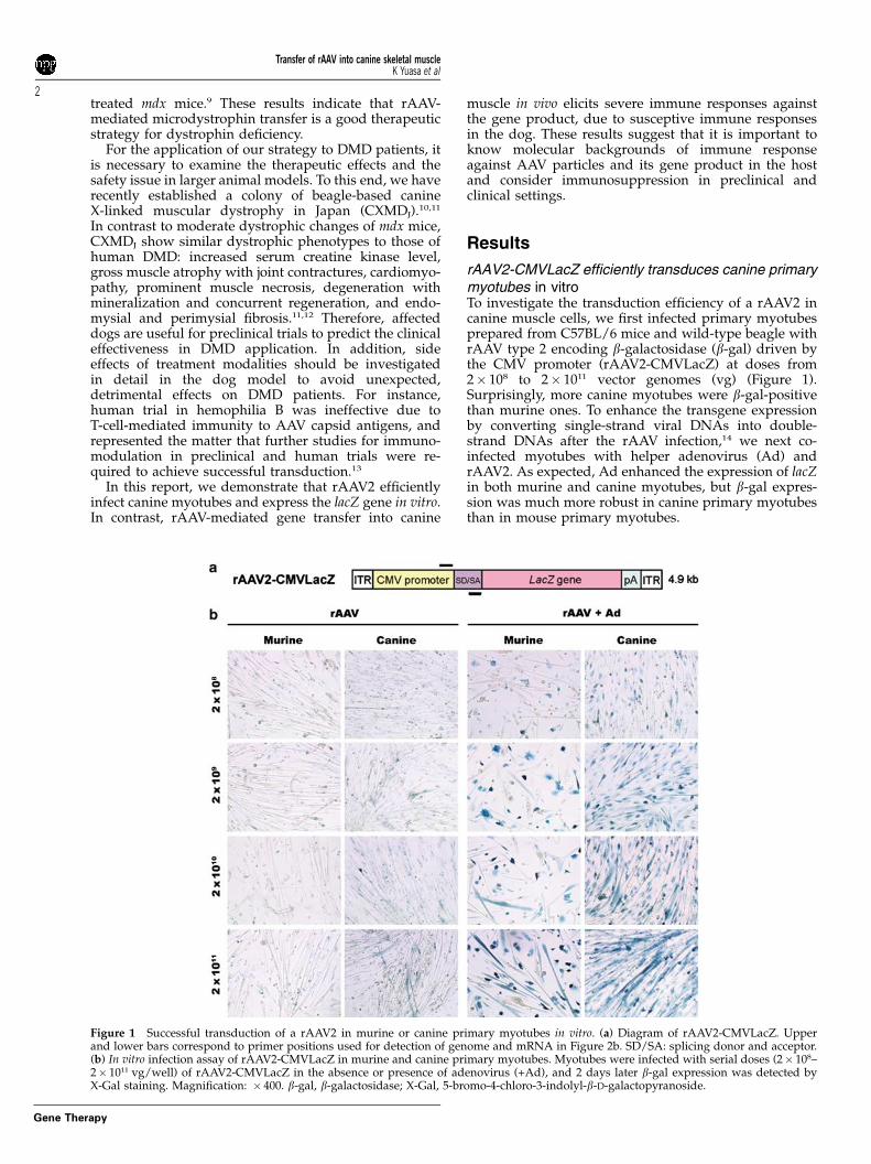
rAAV2-CMVLacZ efficiently transduces canine primarymyotubes in vitroTo investigate the transduction efficiency of a rAAV2 incanine muscle cells, we first infected primary myotubesprepared from C57BL/6 mice and wild-type beagle withrAAV type 2 encoding b-galactosidase (b-gal) driven bythe CMV promoter (rAAV2-CMVLacZ) at doses from2� 108 to 2� 1011 vector genomes (vg) (Figure 1).Surprisingly, more canine myotubes were b-gal-positivethan murine ones. To enhance the transgene expressionby converting single-strand viral DNAs into double-strand DNAs after the rAAV infection,14 we next co-infected myotubes with helper adenovirus (Ad) andrAAV2. As expected, Ad enhanced the expression of lacZin both murine and canine myotubes, but b-gal expres-sion was much more robust in canine primary myotubesthan in mouse primary myotubes.
Figure 1 Successful transduction of a rAAV2 in murine or canine primary myotubes in vitro. (a) Diagram of rAAV2-CMVLacZ. Upperand lower bars correspond to primer positions used for detection of genome and mRNA in Figure 2b. SD/SA: splicing donor and acceptor.(b) In vitro infection assay of rAAV2-CMVLacZ in murine and canine primary myotubes. Myotubes were infected with serial doses (2� 108–2� 1011 vg/well) of rAAV2-CMVLacZ in the absence or presence of adenovirus (+Ad), and 2 days later b-gal expression was detected byX-Gal staining. Magnification: � 400. b-gal, b-galactosidase; X-Gal, 5-bromo-4-chloro-3-indolyl-b-D-galactopyranoside.
Transfer of rAAV into canine skeletal muscleK Yuasa et al
2
Gene Therapy
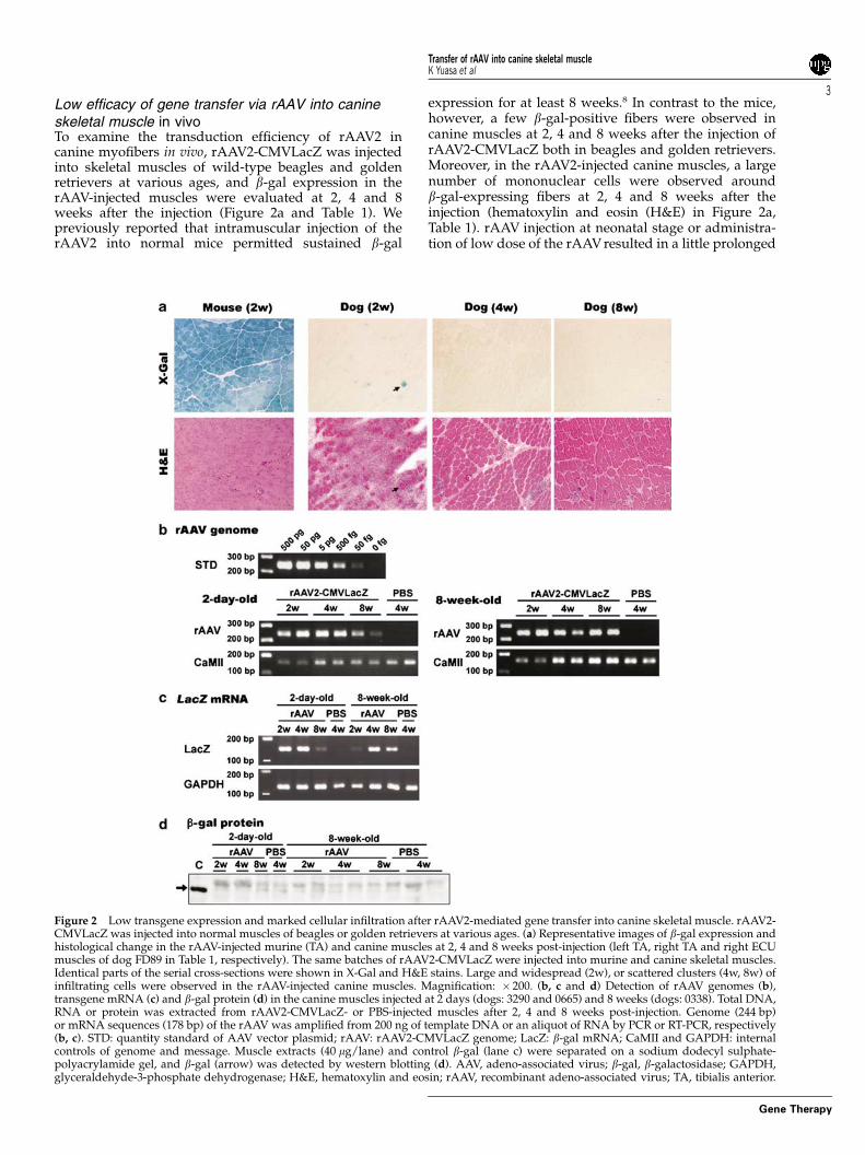
Low efficacy of gene transfer via rAAV into canineskeletal muscle in vivoTo examine the transduction efficiency of rAAV2 incanine myofibers in vivo, rAAV2-CMVLacZ was injectedinto skeletal muscles of wild-type beagles and goldenretrievers at various ages, and b-gal expression in therAAV-injected muscles were evaluated at 2, 4 and 8weeks after the injection (Figure 2a and Table 1). Wepreviously reported that intramuscular injection of therAAV2 into normal mice permitted sustained b-gal
expression for at least 8 weeks.8 In contrast to the mice,however, a few b-gal-positive fibers were observed incanine muscles at 2, 4 and 8 weeks after the injection ofrAAV2-CMVLacZ both in beagles and golden retrievers.Moreover, in the rAAV2-injected canine muscles, a largenumber of mononuclear cells were observed aroundb-gal-expressing fibers at 2, 4 and 8 weeks after theinjection (hematoxylin and eosin (H&E) in Figure 2a,Table 1). rAAV injection at neonatal stage or administra-tion of low dose of the rAAV resulted in a little prolonged
Figure 2 Low transgene expression and marked cellular infiltration after rAAV2-mediated gene transfer into canine skeletal muscle. rAAV2-CMVLacZ was injected into normal muscles of beagles or golden retrievers at various ages. (a) Representative images of b-gal expression andhistological change in the rAAV-injected murine (TA) and canine muscles at 2, 4 and 8 weeks post-injection (left TA, right TA and right ECUmuscles of dog FD89 in Table 1, respectively). The same batches of rAAV2-CMVLacZ were injected into murine and canine skeletal muscles.Identical parts of the serial cross-sections were shown in X-Gal and H&E stains. Large and widespread (2w), or scattered clusters (4w, 8w) ofinfiltrating cells were observed in the rAAV-injected canine muscles. Magnification: � 200. (b, c and d) Detection of rAAV genomes (b),transgene mRNA (c) and b-gal protein (d) in the canine muscles injected at 2 days (dogs: 3290 and 0665) and 8 weeks (dogs: 0338). Total DNA,RNA or protein was extracted from rAAV2-CMVLacZ- or PBS-injected muscles after 2, 4 and 8 weeks post-injection. Genome (244 bp)or mRNA sequences (178 bp) of the rAAV was amplified from 200 ng of template DNA or an aliquot of RNA by PCR or RT-PCR, respectively(b, c). STD: quantity standard of AAV vector plasmid; rAAV: rAAV2-CMVLacZ genome; LacZ: b-gal mRNA; CaMII and GAPDH: internalcontrols of genome and message. Muscle extracts (40 mg/lane) and control b-gal (lane c) were separated on a sodium dodecyl sulphate-polyacrylamide gel, and b-gal (arrow) was detected by western blotting (d). AAV, adeno-associated virus; b-gal, b-galactosidase; GAPDH,glyceraldehyde-3-phosphate dehydrogenase; H&E, hematoxylin and eosin; rAAV, recombinant adeno-associated virus; TA, tibialis anterior.
Transfer of rAAV into canine skeletal muscleK Yuasa et al
3
Gene Therapy
expression of the transferred gene (Table 1, dogs 3290,7690 and 901). To determine whether contamination ofcellular proteins in the stocks of AAV vectors lowerstransduction efficiency, we tried three different AAVpreparation protocols: (i) two-cycle CsCl density gradi-ent ultracentrifugation, (ii) heparin column chromato-graphy, or (iii) combination of them, and found thatpreparation using heparin column chromatographyshowed high levels of contamination of transgeneproducts and cellular proteins (Supplementary Figure 1).CsCl ultracentrifugation efficiently eliminated con-taminated empty viral particles. Combination of thesetwo methods almost completely eliminated contami-nated proteins. Nevertheless, all rAAV stocks showedhigh levels of b-gal expression in murine muscles (datanot shown), but evoked cellular infiltration in normalcanine muscles (Supplementary Figure 1).
To quantify the levels of infection and transduction ofrAAV2-CMVLacZ in canine muscles, we isolated DNAand RNA from the injected muscles, and semiquantita-tively evaluated rAAV genome copy numbers and thelevel of b-gal mRNA by PCR and reverse transcription
(RT)-PCR, respectively. AAV vector genomes and b-galmRNA were detected at 2, 4 and 8 weeks after theinjection (Figures 2b and c). b-Gal protein, however,could not be detected by western blot at all stages afterthe injection (Figure 2d). These results suggest thatcanine myofibers were transduced by a rAAV2 in vivo,but the transduced cells were eliminated by the host’sdefense mechanisms.
rAAV-mediated gene transfer into canine musclesevoke both cellular and humoral immune responseWe next analyzed cell markers on infiltrating cells in therAAV2-CMVLacZ-injected muscles (Figure 3a) at2 weeks after the injection at 12 weeks. Numerous CD4+or CD8+ T lymphocytes were detected in the interstitialspaces of the injected muscle. CD11b+ cells and B cellswere also detected in the cluster of infiltrating cells.Furthermore, the expression of the major histocompati-bility complex (MHC) class I and -II molecules werehighly upregulated on both mononuclear cells andmuscle fibers. IgG deposits were found in both the
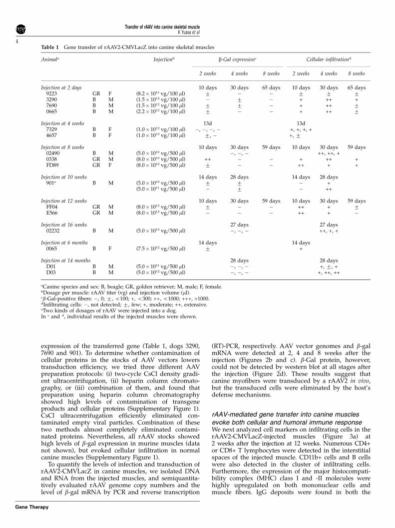
Table 1 Gene transfer of rAAV2-CMVLacZ into canine skeletal muscles
Animala Injectionb b-Gal expressionc Cellular infiltrationd
2 weeks 4 weeks 8 weeks 2 weeks 4 weeks 8 weeks
Injection at 2 days 10 days 30 days 65 days 10 days 30 days 65 days9223 GR F (8.2� 1011 vg/100 ml) 7 � � 7 7 73290 B M (1.5� 1012 vg/100 ml) � 7 � + ++ +7690 B M (1.5� 1012 vg/100 ml) 7 7 � + ++ 70665 B M (2.2� 1012 vg/100 ml) 7 � � + ++ 7
Injection at 4 weeks 13d 13d7329 B F (1.0� 1012 vg/100 ml) �, �, �, � +, +, +, +4657 B F (1.0� 1012 vg/100 ml) 7, � +, 7
Injection at 8 weeks 10 days 30 days 59 days 10 days 30 days 59 days02490 B M (5.0� 1012 vg/500 ml) �, �, � ++, ++, +0338 GR M (8.0� 1012 vg/500 ml) ++ � � + ++ +FD89 GR F (8.0� 1012 vg/500 ml) 7 � � ++ + +
Injection at 10 weeks 14 days 28 days 14 days 28 days901e B M (5.0� 1010 vg/500 ml) 7 7 � +
(5.0� 1011 vg/500 ml) � 7 � ++
Injection at 12 weeks 10 days 30 days 59 days 10 days 30 days 59 daysFF04 GR M (8.0� 1012 vg/500 ml) 7 � � ++ + 7E566 GR M (8.0� 1012 vg/500 ml) � � � ++ + �
Injection at 16 weeks 27 days 27 days02232 B M (5.0� 1012 vg/500 ml) �, �, � ++, +, +
Injection at 6 months 14 days 14 days0065 B F (7.5� 1012 vg/500 ml) 7 +
Injection at 14 months 28 days 28 daysD01 B M (5.0� 1011 vg/500 ml) �, �, � +, 7, +D03 B M (5.0� 1012 vg/500 ml) �, �, � +, ++, ++
aCanine species and sex: B, beagle; GR, golden retriever; M, male; F, female.bDosage per muscle: rAAV titer (vg) and injection volume (ml).cb-Gal-positive fibers: �, 0; 7, o100; +, o300; ++, o1000; +++, >1000.dInfiltrating cells: �, not detected; 7, few; +, moderate; ++, extensive.eTwo kinds of dosages of rAAV were injected into a dog.In c and d, individual results of the injected muscles were shown.
Transfer of rAAV into canine skeletal muscleK Yuasa et al
4
Gene Therapy

cytoplasm of myofibers and the extracellular space in therAAV-injected muscle. We next examined the antibodiesagainst the transgene product or rAAV particles in thesera of rAAV-injected dogs (Figure 3b). The levels of serumIgGs that react b-gal protein or rAAV2 particle weregradually increased with time in both 2-day-old and8-week-old injections. When injected at 8 weeks, the levelsof IgGs against b-gal or rAAV2 increased from 2 weeksafter the injection, and reached the peak at 4 weeks. Wheninjected at 2 days, anti-b-gal or anti-AAV antibodies werenot detected at 2 weeks after the injection, but had begun toincrease at 4 weeks. The results would explain why thelacZ was expressed for a longer time, when injected atneonatal age (Table 1). These results suggest that cellularand humoral immune responses are elicited after thetransfer of a rAAV2 into canine muscles.
Administration of a rAAV expressing no transgeneinto canine musclesTransduction of skeletal muscle by rAAV2-CMVLacZpresents two main foreign antigens, namely b-gal proteinand AAV capsid to the host’s immune system. To testwhich antigen is responsible for rapid elimination oftransduced myofibers after rAAV-mediated gene transferinto canine muscle, we constructed a rAAV2 expressingno transgene, named rAAV2-LacZ-P(�), by removing theMCK promoter from the parental rAAV2-MCKLacZ
(Figure 4a). We confirmed that the promoter-deletedrAAV2 expressed no b-gal in muscle after injection intoskeletal muscles of normal mice (5� 1011 vg/50 ml/site)(data not shown). Then, we injected the same titers ofrAAV2-LacZ-P(�) and rAAV2-CMVLacZ, into skeletalmuscles of normal adult beagles, and evaluated trans-duction efficiency by quantitative PCR of AAV genomesor histopathologically at 2 and 4 weeks post-injection. In5� 5� 10 mm tissues of rAAV2-LacZ-P(�)-injected mus-cles, 1–10 pg of rAAV genomes were detected by PCR,whereas 500 fg to 5 pg of rAAV sequences were amplifiedin the injected muscles of rAAV2-CMVLacZ (Figures 2band 4b). These results indicate that the promoter-deletedrAAV2 could successfully infect canine muscles and theviral genomes were retained stably in the fibers.
H&E staining showed that deletion of the MCKpromoter greatly reduced the cellular infiltration intothe rAAV2-LacZ-P(�)-injected muscles at 2 and 4 weeksafter the injection, in contrast to the rAAV2-CMVLacZ-injected muscles (Figure 4). Even in a muscle sample inwhich vector genome was detected at the highest levelby PCR, infiltrating cells were rarely found (sample 1c inFigures 4b and c). In addition, CD4+ or CD8+ cells werenot detected in the rAAV2-LacZ-P(�)-injected muscles(data not shown). These results suggest that thetransgene product but not AAV particle stronglyelicits immune responses that subsequently eliminatedtransduced myofibers.
Figure 3 Immune responses were remarkably activated after rAAV2-mediated gene transfer into canine muscles. (a) Infiltrating cells in therAAV2-CMVLacZ-injected canine TA muscles at 2 weeks after the injection. Serial cross-sections were immunostained with antibodies againstcanine CD4, CD8a, CD11b, B cell, MHC class I and -II, and IgGs. Insets show the noninjected muscles. Dog: FF04. Magnification: � 400.(b) Humoral immune responses against the transgene product and rAAV particle in the rAAV-treated dogs. Sera from dogs injected with rAAV2-CMVLacZ at 2 days and 8 weeks were analyzed for the presence of IgG antibodies against b-gal or AAV particle at 0, 2, 4 and 8 weeks afterthe injection, using ELISA. Dogs: 9223 and 0338. AAV, adeno-associated virus; b-gal, b-galactosidase; ELISA, enzyme-linked immunosorbentassay; MHC, major histocompatibility complex; rAAV, recombinant adeno-associated virus; TA, tibialis anterior.
Transfer of rAAV into canine skeletal muscleK Yuasa et al
5
Gene Therapy
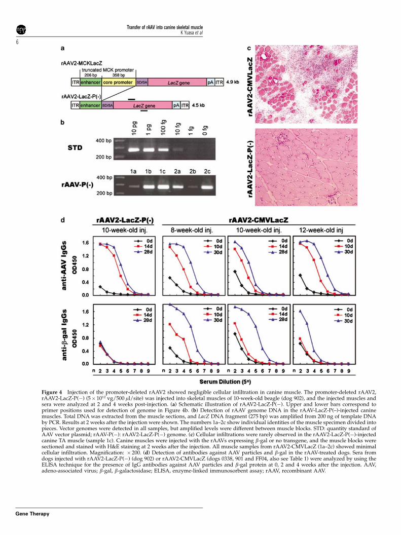
Figure 4 Injection of the promoter-deleted rAAV2 showed negligible cellular infiltration in canine muscle. The promoter-deleted rAAV2,rAAV2-LacZ-P(�) (5� 1012 vg/500 ml/site) was injected into skeletal muscles of 10-week-old beagle (dog 902), and the injected muscles andsera were analyzed at 2 and 4 weeks post-injection. (a) Schematic illustration of rAAV2-LacZ-P(�). Upper and lower bars correspond toprimer positions used for detection of genome in Figure 4b. (b) Detection of rAAV genome DNA in the rAAV-LacZ-P(–)-injected caninemuscles. Total DNA was extracted from the muscle sections, and LacZ DNA fragment (275 bp) was amplified from 200 ng of template DNAby PCR. Results at 2 weeks after the injection were shown. The numbers 1a–2c show individual identities of the muscle specimen divided intopieces. Vector genomes were detected in all samples, but amplified levels were different between muscle blocks. STD: quantity standard ofAAV vector plasmid; rAAV-P(�): rAAV2-LacZ-P(�) genome. (c) Cellular infiltrations were rarely observed in the rAAV2-LacZ-P(�)-injectedcanine TA muscle (sample 1c). Canine muscles were injected with the rAAVs expressing b-gal or no transgene, and the muscle blocks weresectioned and stained with H&E staining at 2 weeks after the injection. All muscle samples from rAAV2-CMVLacZ (1a–2c) showed minimalcellular infiltration. Magnification: � 200. (d) Detection of antibodies against AAV particles and b-gal in the rAAV-treated dogs. Sera fromdogs injected with rAAV2-LacZ-P(�) (dog 902) or rAAV2-CMVLacZ (dogs 0338, 901 and FF04, also see Table 1) were analyzed by using theELISA technique for the presence of IgG antibodies against AAV particles and b-gal protein at 0, 2 and 4 weeks after the injection. AAV,adeno-associated virus; b-gal, b-galactosidase; ELISA, enzyme-linked immunosorbent assay; rAAV, recombinant AAV.
Transfer of rAAV into canine skeletal muscleK Yuasa et al
6
Gene Therapy
Next, we measured the anti-AAV IgG type antibodiesin the serum of the treated dogs (Figure 4d). In therAAV2-LacZ-P(�)-injected dog at 2 and 4 weeks after theinjection, anti-AAV2 antibodies were detected at highlevels similar to those in rAAV2-CMVLacZ-injecteddogs, but antibodies against b-gal were undetectable.This indicates that antibodies against AAV particles weredeveloped in rAAV2-LacZ-P(�)-injected dogs but didnot lead to elimination of transduced cells.
Immunosuppression of the rAAV-injected dogs slightlyimproved the expression of the transgeneTo test whether immunosuppression could improvetransduction by rAAV-2 in canine muscle, we dailyadministered cyclosporine to the rAAV2-CMVLacZ-injected dogs from �5 day of the injection until thesampling day of muscle specimens (Table 2A). Unex-pectedly, b-gal expression in the injected muscles ofcyclosporine-treated dogs was as low as that in theuntreated dogs. Only the extensor digitorum longus(EDL) muscle of dog 403 expressed b-gal at a high levelat 2 weeks after the injection, although a large number ofinfiltrating cells with CD4+ or CD8+ cells and upregula-tion of MHC class I and -II were detected (data notshown). In contrast, in the rAAV2-CMVLacZ-injectedextensor carpi ulnaris (ECU) muscle of dog 403 at 2weeks post-injection, b-gal expression was much lowerthan that in the EDL muscle. Thus, as a whole,cyclosporine alone could not effectively improve trans-duction of rAAV2-CMVLacZ.
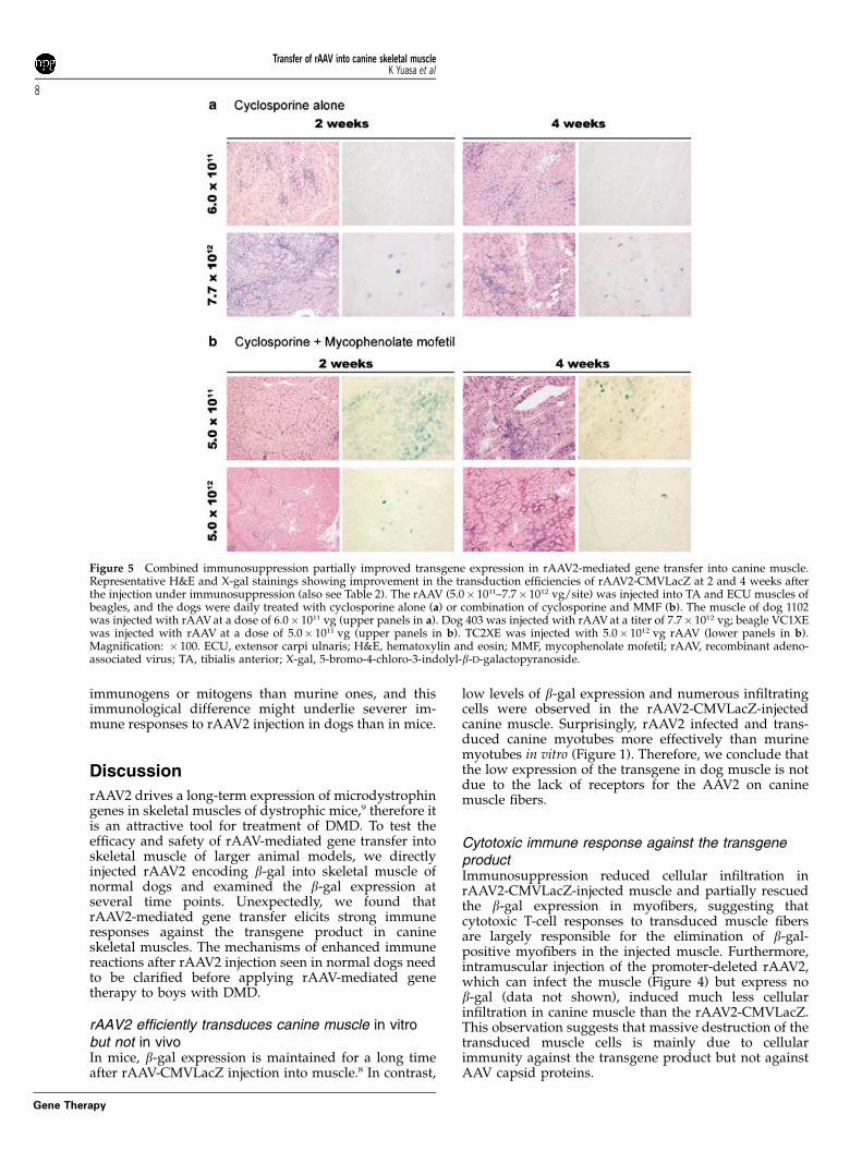
Mycophenolate mofetil (MMF) suppresses the functionsof T lymphocytes in a different way with cyclosporine.Therefore, to suppress immune responses more effectively,we treated the rAAV2-CMVLacZ-injected dogs both with
MMF and cyclosporine. MMF was daily administratedfrom 0 day to the day of sampling (Figure 5 andTable 2B). This combined immunosuppression signifi-cantly increased the numbers of b-gal-expressing fibers,compared with those in the untreated muscles whenexamined at 2 weeks after the injection, but cellularinfiltration was still observed. Some CD4+ or CD8+ cellsand upregulation of MHC class I and -II in infiltratingcells and myofibers were detected in rAAV2-CMVLacZ-injected muscle (data not shown). After 4 weeks, b-galexpression significantly decreased together with increas-ing infiltrating cells. These results indicated that combi-nation of cyclosporine and MMF could partially improvethe transduction efficiency of a rAAV2 in canine muscles.
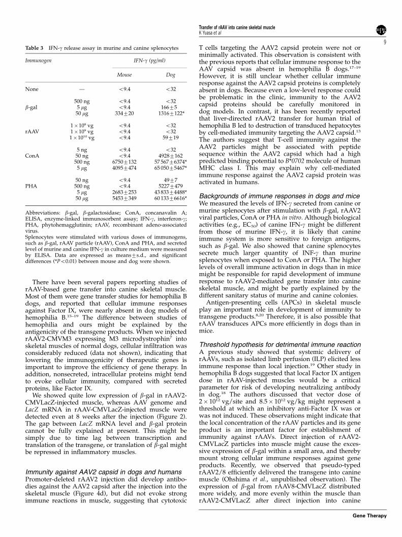
Immunological background of enhanced immuneresponses in dogsWe next investigated possible differences in immuneresponses between mice and dogs, which might explainwhy rAAV2 injection evoked strong immune responsesin canine muscles. To this end, we prepared single-cellsuspension from spleens of untreated dogs or mice,stimulated them with AAV capsids and purified b-galprotein in vitro, and assayed the secretion of interferon-g(IFN-g) by enzyme-linked immunosorbent assay (ELISA;Table 3). We also stimulated the splenocytes with twomajor mitogens for T lymphocytes, concanavalin A (ConA)and phytohemagglutinin (PHA). When stimulated withConA or PHA, canine splenocytes secreted much largeramount of IFN-g into the culture medium than murinecells. Furthermore, canine cells secreted slightly higherlevels of IFN-g in response to b-gal or rAAV2 capsidproteins than murine cells. These results suggest thatcanine splenocytes are innately more susceptive to
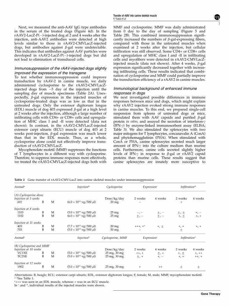
Table 2 Gene transfer of rAAV2-CMV-LacZ into canine skeletal muscles under immunosuppression
Animala Injectionb Cyclosporine Expressionc Infiltrationd
(A) Cyclosporine aloneInjection at 5 weeks Dose/kg/day 2 weeks 4 weeks 2 weeks 4 weeks
601 B M (6.0� 1012 vg/500 ml) 20 mg � +
Injection at 8 weeks1103 B M (5.0� 1010 vg/500 ml) 25 mg �, � 7, � 7, 7 +, �1102 B M (6.0� 1011 vg/500 ml) 25 mg 7, � 7, � +, 7 +, +
Injection at 11 weeks403 B M (7.7� 1012 vg/500 ml) 20 mg +++, +e +, 7 +, + +, +703 B M (5.0� 1012 vg/500 ml) 50 mg � 7
Animala Injectionb Cyclosporine, MMF Expressionc Infiltrationd
(B) Cyclosporine and MMFInjection at 10 weeks Dose/kg/day 2 weeks 4 weeks 2 weeks 4 weeks
VC1XE B M (5.0� 1011 vg/500 ml) 25 mg, 30 mg ++, + 7, + +, 7 +, ++TC2XE B M (5.0� 1012 vg/500 ml) 25 mg, 30 mg 7, + +, � +, + ++, +
Injection at 12 weeks1902 B M (5.0� 1011 vg/500 ml) 25 mg, 30 mg + ++ 7 7
Abreviations: B, beagle; ECU, extensor carpi ulnaris; EDL, extensor digitorum longus; F, female; M, male; MMF, mycophenolate mofetil.a–dSee Table 1.e+++ was seen in an EDL muscle, whereas + was in an ECU muscle.In c and d, individual results of the injected muscles were shown.
Transfer of rAAV into canine skeletal muscleK Yuasa et al
7
Gene Therapy
immunogens or mitogens than murine ones, and thisimmunological difference might underlie severer im-mune responses to rAAV2 injection in dogs than in mice.
Discussion
rAAV2 drives a long-term expression of microdystrophingenes in skeletal muscles of dystrophic mice,9 therefore itis an attractive tool for treatment of DMD. To test theefficacy and safety of rAAV-mediated gene transfer intoskeletal muscle of larger animal models, we directlyinjected rAAV2 encoding b-gal into skeletal muscle ofnormal dogs and examined the b-gal expression atseveral time points. Unexpectedly, we found thatrAAV2-mediated gene transfer elicits strong immuneresponses against the transgene product in canineskeletal muscles. The mechanisms of enhanced immunereactions after rAAV2 injection seen in normal dogs needto be clarified before applying rAAV-mediated genetherapy to boys with DMD.
rAAV2 efficiently transduces canine muscle in vitrobut not in vivoIn mice, b-gal expression is maintained for a long timeafter rAAV-CMVLacZ injection into muscle.8 In contrast,
low levels of b-gal expression and numerous infiltratingcells were observed in the rAAV2-CMVLacZ-injectedcanine muscle. Surprisingly, rAAV2 infected and trans-duced canine myotubes more effectively than murinemyotubes in vitro (Figure 1). Therefore, we conclude thatthe low expression of the transgene in dog muscle is notdue to the lack of receptors for the AAV2 on caninemuscle fibers.
Cytotoxic immune response against the transgeneproductImmunosuppression reduced cellular infiltration inrAAV2-CMVLacZ-injected muscle and partially rescuedthe b-gal expression in myofibers, suggesting thatcytotoxic T-cell responses to transduced muscle fibersare largely responsible for the elimination of b-gal-positive myofibers in the injected muscle. Furthermore,intramuscular injection of the promoter-deleted rAAV2,which can infect the muscle (Figure 4) but express nob-gal (data not shown), induced much less cellularinfiltration in canine muscle than the rAAV2-CMVLacZ.This observation suggests that massive destruction of thetransduced muscle cells is mainly due to cellularimmunity against the transgene product but not againstAAV capsid proteins.
Figure 5 Combined immunosuppression partially improved transgene expression in rAAV2-mediated gene transfer into canine muscle.Representative H&E and X-gal stainings showing improvement in the transduction efficiencies of rAAV2-CMVLacZ at 2 and 4 weeks afterthe injection under immunosuppression (also see Table 2). The rAAV (5.0� 1011–7.7� 1012 vg/site) was injected into TA and ECU muscles ofbeagles, and the dogs were daily treated with cyclosporine alone (a) or combination of cyclosporine and MMF (b). The muscle of dog 1102was injected with rAAV at a dose of 6.0� 1011 vg (upper panels in a). Dog 403 was injected with rAAV at a titer of 7.7� 1012 vg; beagle VC1XEwas injected with rAAV at a dose of 5.0� 1011 vg (upper panels in b). TC2XE was injected with 5.0� 1012 vg rAAV (lower panels in b).Magnification: � 100. ECU, extensor carpi ulnaris; H&E, hematoxylin and eosin; MMF, mycophenolate mofetil; rAAV, recombinant adeno-associated virus; TA, tibialis anterior; X-gal, 5-bromo-4-chloro-3-indolyl-b-D-galactopyranoside.
Transfer of rAAV into canine skeletal muscleK Yuasa et al
8
Gene Therapy
There have been several papers reporting studies ofrAAV-based gene transfer into canine skeletal muscle.Most of them were gene transfer studies for hemophilia Bdogs, and reported that cellular immune responsesagainst Factor IX, were nearly absent in dog models ofhemophilia B.15–19 The difference between studies ofhemophilia and ours might be explained by theantigenicity of the transgene products. When we injectedrAAV2-CMVM3 expressing M3 microdystrophin7 intoskeletal muscles of normal dogs, cellular infiltration wasconsiderably reduced (data not shown), indicating thatlowering the immunogenicity of therapeutic genes isimportant to improve the efficiency of gene therapy. Inaddition, nonsecreted, intracellular proteins might tendto evoke cellular immunity, compared with secretedproteins, like Factor IX.
We showed quite low expression of b-gal in rAAV2-CMVLacZ-injected muscle, whereas AAV genome andLacZ mRNA in rAAV-CMVLacZ-injected muscle weredetected even at 8 weeks after the injection (Figure 2).The gap between LacZ mRNA level and b-gal proteincannot be fully explained at present. This might besimply due to time lag between transcription andtranslation of the transgene, or translation of b-gal mightbe repressed in inflammatory muscles.
Immunity against AAV2 capsid in dogs and humansPromoter-deleted rAAV2 injection did develop antibo-dies against the AAV2 capsid after the injection into theskeletal muscle (Figure 4d), but did not evoke strongimmune reactions in muscle, suggesting that cytotoxic
T cells targeting the AAV2 capsid protein were not orminimally activated. This observation is consistent withthe previous reports that cellular immune response to theAAV capsid was absent in hemophilia B dogs.17–19
However, it is still unclear whether cellular immuneresponse against the AAV2 capsid proteins is completelyabsent in dogs. Because even a low-level response couldbe problematic in the clinic, immunity to the AAV2capsid proteins should be carefully monitored indog models. In contrast, it has been recently reportedthat liver-directed rAAV2 transfer for human trial ofhemophilia B led to destruction of transduced hepatocytesby cell-mediated immunity targeting the AAV2 capsid.13
The authors suggest that T-cell immunity against theAAV2 particles might be associated with peptidesequence within the AAV2 capsid which had a highpredicted binding potential to B*0702 molecule of humanMHC class I. This may explain why cell-mediatedimmune response against the AAV2 capsid protein wasactivated in humans.
Backgrounds of immune responses in dogs and miceWe measured the levels of IFN-g secreted from canine ormurine splenocytes after stimulation with b-gal, rAAV2viral particles, ConA or PHA in vitro. Although biologicalactivities (e.g., EC50) of canine IFN-g might be differentfrom those of murine IFN-g, it is likely that canineimmune system is more sensitive to foreign antigens,such as b-gal. We also showed that canine splenocytessecrete much larger quantity of INF-g than murinesplenocytes when exposed to ConA or PHA. The higherlevels of overall immune activation in dogs than in micemight be responsible for rapid development of immuneresponse to rAAV2-mediated gene transfer into canineskeletal muscle, and might be partly explained by thedifferent sanitary status of murine and canine colonies.
Antigen-presenting cells (APCs) in skeletal muscleplay an important role in development of immunity totransgene products.8,20 Therefore, it is also possible thatrAAV transduces APCs more efficiently in dogs than inmice.
Threshold hypothesis for detrimental immune reactionA previous study showed that systemic delivery ofrAAVs, such as isolated limb perfusion (ILP) elicited lessimmune response than local injection.19 Other study inhemophilia B dogs suggested that local Factor IX antigendose in rAAV-injected muscles would be a criticalparameter for risk of developing neutralizing antibodyin dog.18 The authors discussed that vector dose of2� 1012 vg/site and 8.5� 1012 vg/kg might represent athreshold at which an inhibitory anti-Factor IX was orwas not induced. These observations might indicate thatthe local concentration of the rAAV particles and its geneproduct is an important factor for establishment ofimmunity against rAAVs. Direct injection of rAAV2-CMVLacZ particles into muscle might cause the exces-sive expression of b-gal within a small area, and therebymount strong cellular immune responses against geneproducts. Recently, we observed that pseudo-typedrAAV2/8 efficiently delivered the transgene into caninemuscle (Ohshima et al., unpublished observation). Theexpression of b-gal from rAAV8-CMVLacZ distributedmore widely, and more evenly within the muscle thanrAAV2-CMVLacZ after direct injection into canine
Table 3 IFN-g release assay in murine and canine splenocytes
Immunogen IFN-g (pg/ml)
Mouse Dog
None — o9.4 o32
500 ng o9.4 o32b-gal 5 mg o9.4 16675
50 mg 334720 13167122*
1�108 vg o9.4 o32rAAV 1�109 vg o9.4 o32
1�1010 vg o9.4 59719
5 ng o9.4 o32ConA 50 ng o9.4 49287162
500 ng 67507132 57 56776374*5 mg 40957474 65 05075467*
50 ng o9.4 4977PHA 500 ng o9.4 52277479
5 mg 26837253 43 83374488*50 mg 54537349 60 13376616*
Abbreviations: b-gal, b-galactosidase; ConA, concanavalin A;ELISA, enzyme-linked immunosorbent assay; IFN-g, interferon-g;PHA, phytohemagglutinin; rAAV, recombinant adeno-associatedvirus.Splenocytes were stimulated with various doses of immunogens,such as b-gal, rAAV particle (rAAV), ConA and PHA, and secretedlevel of murine and canine IFN-g in culture medium were measuredby ELISA. Data are expressed as means7s.d., and significantdifferences (*Po0.01) between mouse and dog were shown.
Transfer of rAAV into canine skeletal muscleK Yuasa et al
9
Gene Therapy
muscle. We hypothesize that widespread transduction ofmyofibers at a low dose of a rAAV2/8 could escape theestablishment of immunity against the transgene pro-ducts. New serotypes of AAVs, such as AAV8 or AAV9can deliver the transgene into musculature of the wholebody via circulation.21,22 These vectors might overcomethe immunological problems that we encountered inrAAV2-mdiated gene transfer into canine muscles.
In conclusion, we found that intramuscular adminis-tration of rAAV2-CMVLacZ elicited rapid and severeimmune responses against the transgene product inwild-type dogs. This has not been observed in mice. It isimportant to understand well the mechanisms ofimmune reactions observed in dogs to circumventimmunological limitations, which might stand in ourway in preclinical and clinical trials for DMD.
Materials and methods
Construction and production of rAAVsWe used rAAV2-CMVLacZ and rAAV2-CMVM3, whichharbors the LacZ gene or microdystrophin M3 driven bythe CMV promoter, respectively.3,7,8 To improve theexpression efficiency of the LacZ gene in rAAV2-CMVLacZ, a chimeric intron (human b-globin splicingdonor and immunoglobulin splicing acceptor, Promega,Madison, WI, USA) is inserted between CMV promoterand the LacZ gene.8 rAAV2-LacZ-P(�) was generated bydeleting the truncated MCK promoter (358 bp) fromrAAV2-MCKLacZ, leaving the MCK enhancer intact.8 Weconfirmed that rAAV2-LacZ-P(�) could not express b-galin murine muscle cells both in vitro and in vivo (data notshown). The rAAV was produced by a plasmid triple-cotransfection method,23 purified by two CsCl densitygradient centrifugations24 and/or heparin column chro-matography,25 and titrated by a quantitative DNA dot-blot assay.
Administration of rAAVs into canine or murine skeletalmuscles and isolation of the injected musclesExperimental dogs were wild-type littermates of beagle-based CXMDJ breeding colony at National Center ofNeurology and Psychiatry (NCNP; Tokyo, Japan),10,11 orgolden retriever muscular dystrophy colony at MurdochUniversity (Perth, Australia).26 All animals were caredand treated in accordance with the guidelines approvedby Ethics Committee for Treatment of LaboratoryAnimals at NCNP or Animal Ethics Committee atMurdoch University, where three fundamental principles(replacement, reduction and refinement) were alsoconsidered. When a rAAV was injected into dogs, theanimals were not vaccinated to relive influences ofimmune response by vaccination. rAAVs were injectedintramuscularly at ages from 2 days to 14 months, andthe muscles were isolated after 2, 4 and 8 weeks.Surgeries were done under anesthesia with isoflurane,as described previously.27 Briefly, the ECU, tibialisanterior (TA) or EDL muscles were surgically exposedand two marker sutures were placed on the mid-belly ofthe muscle at proximal and distal positions apartapproximately 20 mm. rAAV (5� 1010–8� 1012 vg in 100or 500 ml) or phosphate-buffered saline (PBS) wasinjected slowly into the muscles between two markersalong the length. In biopsy and necropsy, the entire
muscle blocks with two sutures were removed, dividedinto some pieces, and immediately frozen in isopentaneprecooled with liquid nitrogen. rAAV (5� 1011 vg/50 ml)was also injected into TA muscles of C57BL/10 mice at 5weeks, and the muscles were isolated at 2 or 4 weeksafter the injection.
ImmunosuppressionTo suppress immune reaction, we treated rAAV-injecteddogs with cyclosporine alone or cyclosporine combinedwith MMF for immunosuppression. Cyclosporine(NEORAL capsules; Novartis, Basel, Switzerland) wasadministrated orally at doses of 20–50 mg/kg/day anddaily from �5 day of the injection. MMF (CellCeptcapsules, Roche Pharmaceuticals, Nutley, NJ, USA) wasadministrated orally at a dose of 30 mg/kg/day anddaily from 0 day of the injection.
Histological and immunohistochemical analysisTransverse cryosections from the rAAV-injected muscleswere stained with H&E or 5-bromo-4-chloro-3-indolyl-b-D-galactopyranoside (X-Gal).8
Immunohistochemistry was performed as describedpreviously.8 Cryosections were incubated with thefollowing antibodies: mouse monoclonal antibodiesagainst canine CD4 (CA13.1E4, Serotec, Oxford, UK),canine CD8a (CA9.JD3, Serotec), canine CD11b(CA16.3E10, Serotec), canine B cells (CA2.1D6, Serotec),class I major histocompatibility antigen (H58A, VMRD,Pullman, WA, USA) and canine MHC class II (CA2.1C12,Serotec), and fluorescein-conjugated goat affinity-puri-fied antibody to dog IgG (whole molecule) (Cappel,Solon, OH, USA). The primary antibodies were detectedusing VECTASTAIN ABC kit (Vector Laboratories,Burlingame, CA, USA), then visualized with diamino-benzidine, and counterstained with methyl green.
In vitro infection assay in primary myotubesCanine or murine primary myoblasts were isolated anddifferentiated into myotubes, according to the previouslypublished protocol28 with some modifications. Briefly,muscle mass was removed from dogs or mice at 0 day to2 weeks, weighted and minced. Cells were enzymaticallydissociated in PBS (4 ml per g of tissue) containingdispase II (2.4 IU/ml, Godo Shusei, Tokyo, Japan) andCollagenase type XI (0.2%, Sigma, St Louis, USA), and2.5 mM CaCl2. The slurry was incubated at 371C for 90–120 min with trituration every 15 min, and then passedthrough 100 mm pore mesh. The filtrate was spun at350 g, and the pellet was resuspended in growth medium(F-10 medium with 20% fetal bovine serum (FBS) and2.5 ng/ml human basic fibroblast growth factor (Sigma)).The cell suspension was then plated on non-coateddishes for 90 min, and non-adherent cells were collectedand plated onto new dishes. After several repeats of thispre-plating procedure, myoblasts were enriched. Toassess transduction efficiency of a rAAV in primarymyotubes, the same numbers of myoblasts (2� 104 cells/well) were plated onto 24-well plates and maintained fora few days in differentiation medium (Dulbecco’smodified Eagle’s medium with 5% FBS and 10 mg/mlhuman insulin (Sigma)), and then cultured in thepresence of rAAV2-CMVLacZ (2� 108–2� 1011 vg/well),or co-infected with the rAAV2 and adenovirus Ad5-dlX3
Transfer of rAAV into canine skeletal muscleK Yuasa et al
10
Gene Therapy
(1�107 plaque-forming unit (PFU)/ml). After 2 days,cells were stained with X-Gal.
Quantification of rAAV genome in canine muscleTo quantify rAAV genomes, muscle sections weredigested with proteinase K, followed by phenol/chloroformextraction and isopropanol precipitation. The DNA pelletwas resuspended in TE buffer, and amplified by PCRwith following primer sets: 50-ccacgctgttttgacctccatag-30
(downstream of transcription start site in the CMVpromoter) and 50-gtacaattccgcagcttttagagc-30 (upstreamof the LacZ gene) for rAAV2-CMVLacZ (the PCR productis 244 bp, illustrated in Figure 1a); 50-gcagttatctggaagatcagg-30 and 50-cataaccaccacgctcatcg-30 (within the LacZgene) for rAAV2-LacZ-P(�) (the PCR product is 275 bp,illustrated in Figure 4a). As an internal control, thecanine calmodulin gene II (CaMII; GenBank accessionno. D12622) was amplified with following primers:50-gagaggactcatccaaggtcacac-30 and 50-tcagaaaccacggcatcagg-30. AAV vector plasmids for rAAV2-CMVLacZ orrAAV2-LacZ-P(�) were served as a quantity control forstandard amplification.
RT-PCR for transgene mRNAsTotal RNA was isolated from muscle sections usingRNAqueous-Micro kit (Ambion, TX, USA). Then first-strand cDNA was synthesized using First-Strand cDNASynthesis Kit (GE Healthcare UK Ltd, Buckinghamshire,UK). mRNA of rAAV2-CMVLacZ was detected using thesame primers used for genomic DNA (Figure 1a). Theamplified product is 178 bp. As an internal controlglyceraldehyde-3-phosphate dehydrogenase mRNA(GenBank accession no. AB038240) was amplified usingthe following primer set: 50-tcatctctgctccttctgctgat-30 and50-ggctagaggagccaagcagtt-30.
Western blot analysisMuscle extracts were prepared from rAAV-injectedcanine muscles, separated on 7.5% sodium dodecylsulfate-polyacrylamide gels, and then transferred ontopolyvinylidene fluoride membranes, as described pre-viously.8 b-Gal protein was detected by rabbit polyclonalanti-b-gal antibody (Cappel).
ELISAELISA were performed as described previously.8 Themicrotiter plate was precoated with b-gal, rAAV2-CMVLacZ or rAAV2-LacZ-P(�), incubated with the serafrom dogs, and then reacted with a 1/5000 dilution ofperoxidase-conjugated rabbit anti-dog IgG (whole mole-cule) (Sigma). Reactivity was determined by the colorreaction of 3,30,5,50,-tetramethylbenzidine (TMBZ).
IFN-g release assaySpleen cells were isolated from C57BL/6 mice or beagledogs, which were untreated with rAAVs. Splenetic cellswere dissociated physically and passed through 100 mmfilters. After removal of red blood cells using 0.83%ammonium chloride buffer (Sigma), splenocytes werecultured in RPMI medium containing 10% FBS at adensity of 2� 106 cells/well in 24-well plates for 2 daysin the presence of various antigens: b-gal (500 ng to50 mg), rAAV2-CMVLacZ (1�108–1�1010 vg), ConA(5 ng to 5 mg, GE Healthcare Bio-Science) and phytohem-agglutinin (50 ng to 50 mg, Sigma). The supernatant of the
culture was assayed for the secretion of IFN-g usingMouse or Canine IFN-g Quantikine ELISA Kit (R&DSystems, Minneapolis, MN, USA). The amounts ofcanine or murine IFN-g were calibrated using recombi-nant canine or murine IFN-g proteins as standards,respectively, according to manufacturer’s instructions.Results are given as means with standard deviation (s.d.)of three experiments. Data were first evaluated by F-test.When equality of variances was shown, t-test was usedto evaluate the statistical significance. When rejected,Aspin–Welch-test was used.
Acknowledgements
We are grateful to Dr Katsujiro Sato, Dr YasushiMochizuki, Dr Naoko Yugeta, Dr Akiyo Nishiyama,Dr Madoka Ikemoto, Dr Michiko Wada, Ms KazueKinoshita, Ms Ryoko Nakagawa, Mr Satoru Masuda,Dr Masayuki Tomohiro and Dr Yoshiki Shimatsufor technical assistance. We also thank Mr Hideki Kita andMr Shinichi Ichikawa and other staff of JAC Co. for careand management of experimental animals. This workis supported by Grants-in-Aid for Scientific Researchfor Research on Nervous and Mental Disorders (10B-1,13B-1, 16B-3) and Health Sciences Research Grants forResearch on Human Genome and Gene Therapy (H10-genome-015, H13-genome-001, H16-genome-003) fromthe Ministry of Heath, Labor and Welfare of Japan, andGrant-in-Aid for Scientific Research (B) from the Minis-try of Education, Culture, Sports, Science and Technol-ogy (MEXT). A part of this work in MusashinoUniversity is supported by High-Tech Research CenterProject for Private Universities: matching fund subsidyfrom MEXT, 2004.
References
1 Emery AEH. Duchenne Muscular Dystrophy, 2nd edn. OxfordUniversity Press: Oxford, 1993.
2 Fisher KJ, Jooss K, Alston J, Yang Y, Haecker SE, High K et al.Recombinant adeno-associated virus for muscle directed genetherapy. Nat Med 1997; 3: 306–312.
3 Yuasa K, Miyagoe Y, Yamamoto K, Nabeshima Y, Dickson G,Takeda S. Effective restoration of dystrophin-associated proteinsin vivo by adenovirus-mediated transfer of truncated dystrophincDNAs. FEBS Lett 1998; 425: 329–336.
4 Harper SQ, Hauser MA, Dellorusso C, Duan D, Crawford RW,Phelps SF et al. Modular flexibility of dystrophin: implicationsfor gene therapy of Duchenne muscular dystrophy. Nat Med2002; 8: 253–261.
5 Watchko J, O’day T, Wang B, Zhou L, Tang Y, Li J et al. Adeno-associated virus vector-mediated minidystrophin gene therapyimproves dystrophic muscle contractile function in mdx mice.Hum Gene Ther 2002; 13: 1451–1460.
6 Fabb SA, Wells DJ, Serpente P, Dickson G. Adeno-associatedvirus vector gene transfer and sarcolemmal expression of a 144kDa micro-dystrophin effectively restores the dystrophin-asso-ciated protein complex and inhibits myofibre degeneration innude/mdx mice. Hum Mol Genet 2002; 11: 733–741.
7 Sakamoto M, Yuasa K, Yoshimura M, Yokota T, Ikemoto T,Suzuki M et al. Micro-dystrophin cDNA ameliorates dystrophicphenotypes when introduced into mdx mice as a transgene.Biochem Biophys Res Commun 2002; 293: 1265–1272.
Transfer of rAAV into canine skeletal muscleK Yuasa et al
11
Gene Therapy
8 Yuasa K, Sakamoto M, Miyagoe-Suzuki Y, Tanouchi A, Yama-moto H, Li J et al. Adeno-associated virus vector-mediated genetransfer into dystrophin-deficient skeletal muscles evokesenhanced immune response against the transgene product.Gene Therapy 2002; 9: 1576–1588.
9 Yoshimura M, Sakamoto M, Ikemoto M, Mochizuki Y, Yuasa K,Miyagoe-Suzuki Y et al. AAV vector-mediated microdystrophinexpression in a relatively small percentage of mdx myofibersimproved the mdx phenotype. Mol Ther 2004; 10: 821–828.
10 Shimatsu Y, Katagiri K, Furuta T, Nakura M, Tanioka Y, Yuasa Ket al. Canine X-linked muscular dystrophy in Japan (CXMDJ).Exp Animals 2003; 52: 93–97.
11 Shimatsu Y, Yoshimura M, Yuasa K, Urasawa N, Tomohiro M,Nakura M et al. Major clinical and histopathological character-isties of canine X-linked muscular dystrophy in Japan, CXMDJ.Acta Myologica 2005; 24: 145–154.
12 Valentine BA, Cooper BJ, Cummings JF, De Lahunta A. CanineX-linked muscular dystrophy: morphologic lesions. J Neurol Sci1990; 97: 1–23.
13 Manno CS, Arruda VR, Pierce GF, Glader B, Ragni M, Rasko J etal. Successful transduction of liver in hemophilia by AAV-FactorIX and limitations imposed by the host immune response. NatMed 2006; 12: 342–347.
14 Fisher KJ, Gao GP, Weitzman MD, DeMatteo R, Burda JF, WilsonJM. Transduction with recombinant adeno-associated virus forgene therapy is limited by leading-strand synthesis. J Virol 1996;70: 520–532.
15 Herzog RW, Yang EY, Couto LB, Hagstrom JN, Elwell D, FieldsPA et al. Long-term correction of canine hemophilia B by genetransfer of blood coagulation factor IX mediated by adeno-associated viral vector. Nat Med 1999; 5: 56–63.
16 Fields PA, Kowalczyk DW, Arruda VR, Armstrong E, McclelandML, Hagstrom JN et al. Role of vector in activation of T cellsubsets in immune responses against the secreted transgeneproduct factor IX. Mol Ther 2000; 1: 225–235.
17 Herzog RW, Mount JD, Arruda VR, High KA, Lothrop Jr CD.Muscle-directed gene transfer and transient immune suppres-sion result in sustained partial correction of canine hemophilia Bcaused by a null mutation. Mol Ther 2001; 4: 192–200.
18 Herzog RW, Fields PA, Arruda VR, Brubaker JO, Armstrong E,Mcclintock D et al. Influence of vector dose on factor IX-specific
T and B cell responses in muscle-directed gene therapy. HumGene Ther 2002; 13: 1281–1291.
19 Arruda VR, Stedman HH, Nichols TC, Haskins ME, NicholsonM, Herzog RW et al. Regional intravascular delivery of AAV-2-F.IXto skeletal muscle achieves long-term correction of hemophilia B ina large animal model. Blood 2005; 105: 3458–3464.
20 Zhang Y, chirmule N, Gao G, Wilson J. CD40 ligand-dependentactivation of cytotoxic T lymphocytes by adeno-associated virusvectors in vivo: role of immature dendritic cells. J. Virol 2000; 74:8003–8010.
21 Wang Z, Zhu T, Qiao C, Zhou L, Wang B, Zhang J et al. Adeno-associated virus serotype 8 efficiently delivers genes to muscleand heart. Nat Biotechnol 2005; 23: 321–328.
22 Inagaki K, Fuess S, Storm TA, Gibson GA, Mctiernan CF, KayMA et al. Robust systemic transduction with AAV9 vectors inmice: efficient global cardiac gene transfer superior to that ofAAV8. Mol Ther 2006; 14: 45–53.
23 Xiao X, Li J, Samulski RJ. Production of high-titer recombinantadeno-associated virus vectors in the absence of helperadenovirus. J Virol 1998; 72: 2224–2232.
24 Snyder R, Xiao X, Samulski RJ. Production of recombinantadeno-associated viral vectors. In: Dracopoli N, Haines J, Korf B,Morton C, Seidman C, Seidman JG, Smith D (eds). CurrentProtocols in Human Genetics. John Wiley & Sons Ltd: New York,1996, pp 12.1.1–12.2.23.
25 Auricchio A, Hildinger M, O’connor E, Gao GP, Wilson JM.Isolation of highly infectious and pure adeno-associated virustype 2 vectors with a single-step gravity-flow column. Hum GeneTher 2001; 12: 71–76.
26 Howell JM, Fletcher S, Kakulas BA, O’hara M, Lochmuller H,Karpati G. Use of the dog model for Duchenne musculardystrophy in gene therapy trials. Neuromuscul Disord 1997; 7:325–328.
27 Howell JM, Lochmuller H, O’hara A, Fletcher S, Kakulas BA,Massie B et al. High-level dystrophin expression after adeno-virus-mediated dystrophin minigene transfer to skeletal muscleof dystrophic dogs: prolongation of expression with immuno-suppression. Hum Gene Ther 1998; 9: 629–634.
28 Rando TA, Blau HM. Primary mouse myoblast purification,characterization, and transplantation for cell-mediated genetherapy. J Cell Biol 1994; 125: 1275–1287.
Supplementary Information accompanies the paper on Gene Therapy website (http://www.nature.com/gt)
Transfer of rAAV into canine skeletal muscleK Yuasa et al
12
Gene Therapy