three dimensional linear-elastic - Spiral – Imperial College ...
PHYSICAL REVIEW E 88, 062703 (2013)
Initiation and dynamics of a spiral wave around an ionic heterogeneityin a model for human cardiac tissue
Arne Defauw,1,* Peter Dawyndt,2 and Alexander V. Panfilov1
1Department of Physics and Astronomy, Ghent University, Ghent, Belgium2Department of Applied Mathematics, Computer Science and Statistics, Ghent University, Ghent, Belgium
(Received 19 August 2013; revised manuscript received 18 October 2013; published 3 December 2013)
In relation to cardiac arrhythmias, heterogeneity of cardiac tissue is one of the most important factors underlyingthe onset of spiral waves and determining their type. In this paper, we numerically model heterogeneity of realisticsize and value and study formation and dynamics of spiral waves around such heterogeneity. We find that the onlysustained pattern obtained is a single spiral wave anchored around the heterogeneity. Dynamics of an anchoredspiral wave depend on the extent of heterogeneity, and for certain heterogeneity size, we find abrupt regionalincrease in the period of excitation occurring as a bifurcation. We study factors determining spatial distribution ofexcitation periods of anchored spiral waves and discuss consequences of such dynamics for cardiac arrhythmiasand possibilities for experimental testings of our predictions.
DOI: 10.1103/PhysRevE.88.062703 PACS number(s): 87.19.Hh, 05.45.−a
I. INTRODUCTION
Contraction of the heart is initiated by the propagation ofelectrical waves of excitation. Electrical waves propagatingthrough the heart belong to a large class of nonlinear waves thatare widely studied theoretically in reaction-diffusion systems.One of the most important phenomena in such systems is theexistence of vortices in the form of spiral waves of excitation.Spiral waves were found in a variety of nonlinear excitablemedia. In physicochemical systems, they have been observedin oscillating reactions [1,2] and heterogeneous catalysis[3,4]. Biological examples of such media include spiral wavesof cAMP during morphogenesis of Dictyostelium discoideumamoebae [5,6], spiral waves of spreading depression in retinaand in cortical tissue [7], calcium waves in Xenophus oocytes[8,9], and spiral waves in cardiac tissue [10–12]. In the heart,spiral waves underlie life threatening cardiac arrhytmias. Oneof the most important scientific questions for applications isto understand the mechanisms of initiation of spiral waves,i.e., of cardiac arrhythmias. Another important question is tofind factors underlying their dynamics, as they are directlyrelated to the type of cardiac arrhythmia [13,14]. It turns outthat heterogeneity of cardiac tissue is important in the answerto both questions.
It was shown that heterogeneity substantially affects thedynamics of spiral waves. For example, spiral waves candrift because of heterogeneity [13–17]. Such drift can explainthe onset of arrhythmia with periodically varying electro-cardiogram (ECG), called torsades de pointes [16,18], or anarrhythmia with nonperiodic ECG: a polymorphic ventriculartachycardia [13,14].
Regarding the onset of spiral waves, it was shown that wavepropagation at heterogeneities can be temporarily blocked[19,20], and that such a pattern can evolve into spiral waves.The process of wave blocks and spiral wave formation in thepresence of heterogeneity was studied in various modelingstudies [21–26]. These studies showed that in order to be ableto generate a 2D spiral wave, the heterogeneity should have
*Corresponding author: [email protected]
a substantial size, comparable to the wavelength of the spiralwave [21].
Most of the listed studies were performed using genericmodels of cardiac tissue and by using generic types ofheterogeneity, as data on real heterogeneities, for example, inthe human heart, were not available. Recently, measurementsof heterogeneity in the human heart were performed [27].Interestingly, in many cases the size of the heterogeneity wassmall. The possibility of formation of spirals, and the dynamicsof spiral waves around such heterogeneities was not addressed,even at the generic level.
The aim of this paper is to study effects of heterogeneityof realistic size and value on the onset of spiral waves usingthe TP06 model [28] for human cardiac cells. We also studydynamics of spirals waves around such heterogeneities. Inparticular, we model heterogeneity similar to that measuredby Glukhov in Ref. [27]. We apply high-frequency forcingand study if spiral waves can be formed in such a situation.We find that formation of spiral waves is possible. However,in all cases the created spiral wave will be anchored aroundthe heterogeneity. Further, we study dynamics of such an-chored spiral waves and factors determining its dynamics byvarying the size and value of the heterogeneity. We discusspossible mechanisms of such dynamics and its importance forapplications.
II. MODELS AND METHODS
Model. In this paper we consider a monodomain descriptionof cardiac tissue [29], which has the following form:
Cm
∂Vm
∂t=
(∂
∂xi
Dij
∂Vm
∂xj
)− Iion, (1)
where Dij is a diffusion matrix accounting for anisotropy ofcardiac tissue, i,j = 1...n, where n = 1 in 1D, 2 in 2D..., Cm
is membrane capacitance, Vm is transmembrane voltage, t istime, and Iion is the sum of ionic transmembrane currentsdescribing the excitable behavior of individual ventricularcells. To represent human ventricular electrophysiologicalproperties, we used the ionic TP06 model [28,30]. This modelprovides a detailed description of voltage, ionic currents, and
062703-11539-3755/2013/88(6)/062703(9) ©2013 American Physical Society
ARNE DEFAUW, PETER DAWYNDT, AND ALEXANDER V. PANFILOV PHYSICAL REVIEW E 88, 062703 (2013)
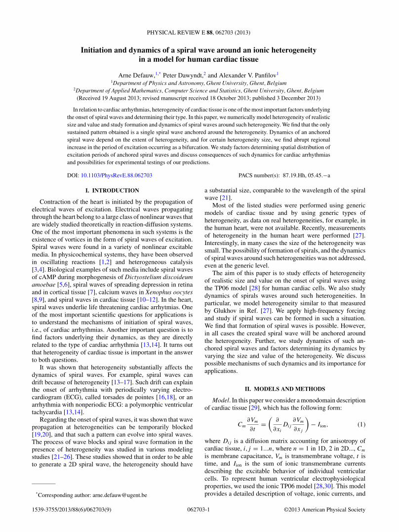
FIG. 1. (Color online) APD distribution in cardiac tissue simu-lated numerically in TP06 model [28,30]. Total size of the mediumis 60 mm × 20 mm. Colormap shows APD in ms. Max. APD =359.5 ms, min. APD = 290 ms. Size at 50% heterogeneity is 11.2 mmon 5.6 mm. Which is comparable to heterogeneity measured in thehuman heart [27]. In black, we show the size of the heterogeneity.
intracellular ion concentrations for human ventricular cells. Acomplete list of all equations can be found in Refs. [28,30].We used the default parameter settings from Ref. [28] forepicardial cells. All parameter changes made to obtain tissueheterogeneity are detailed in the text.
Numerical methods. For 1D and 2D computations, theforward Euler method was applied to integrate Eq. (1). Aspace step of �x = 0.2 mm and a time step of �t = 0.02 mswere used. To integrate the Hodgkin-Huxley-type equationsfor the gating variables of the various time-dependent currents(m, h, and j for INa; r and s for Ito; xr1 and xr2 for IKr; xs
for IKs; d, f , f2, and fCass for ICaL), the Rush and Larsenscheme [31] was used.
Anisotropy. In most of our simulations, the fibers aredirected along the x axis. In few simulations we study effectof rotational anisotropy, in that case the diffusion matrix isgiven by
Dxx = DL cos2 θ + DT sin2 θ,
Dxy = Dyx = 0, (2)
Dyy = DT ,
with θ (y) = y
d(θ2 − θ1) + θ1. Here d is the distance between
epicardium and endocardium, θ1 = −60 ◦, θ2 = 60 ◦, DL =0.128 mm2
ms , and DT = DL/4.Heterogeneity. To study heterogeneity, we change the
parameters GKs, GKr, and GCaL from their default values 0.392nS/pF, 0.153 nS/pF, and 3.980 × 10⁻⁵ cm
msμF for epicardial
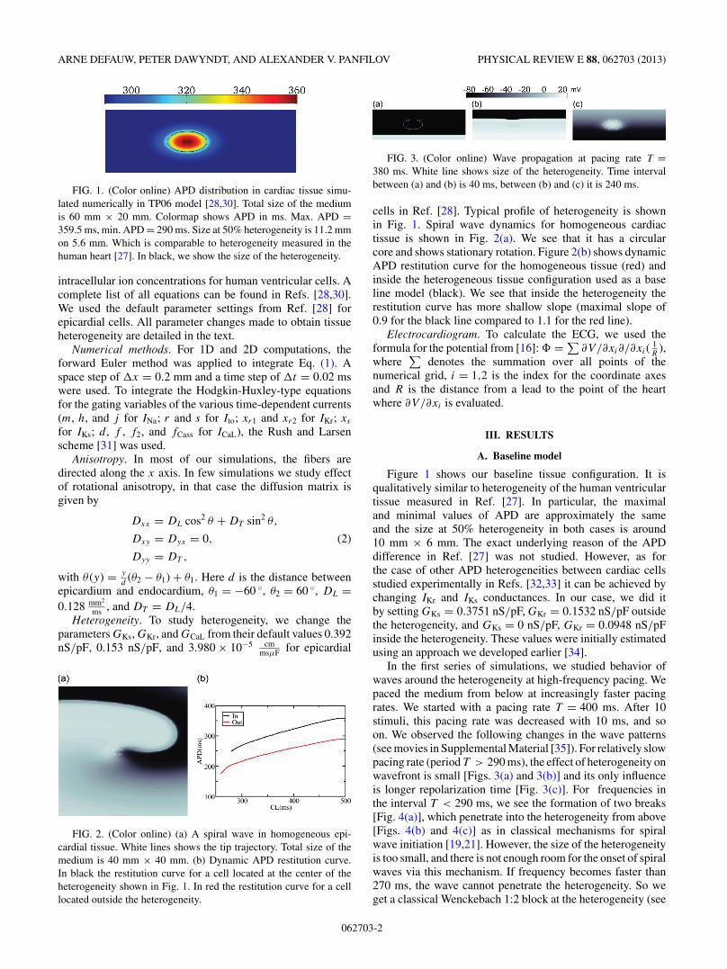
FIG. 2. (Color online) (a) A spiral wave in homogeneous epi-cardial tissue. White lines shows the tip trajectory. Total size of themedium is 40 mm × 40 mm. (b) Dynamic APD restitution curve.In black the restitution curve for a cell located at the center of theheterogeneity shown in Fig. 1. In red the restitution curve for a celllocated outside the heterogeneity.
FIG. 3. (Color online) Wave propagation at pacing rate T =380 ms. White line shows size of the heterogeneity. Time intervalbetween (a) and (b) is 40 ms, between (b) and (c) it is 240 ms.
cells in Ref. [28]. Typical profile of heterogeneity is shownin Fig. 1. Spiral wave dynamics for homogeneous cardiactissue is shown in Fig. 2(a). We see that it has a circularcore and shows stationary rotation. Figure 2(b) shows dynamicAPD restitution curve for the homogeneous tissue (red) andinside the heterogeneous tissue configuration used as a baseline model (black). We see that inside the heterogeneity therestitution curve has more shallow slope (maximal slope of0.9 for the black line compared to 1.1 for the red line).
Electrocardiogram. To calculate the ECG, we used theformula for the potential from [16]: � = ∑
∂V/∂xi∂/∂xi( 1R
),where
∑denotes the summation over all points of the
numerical grid, i = 1,2 is the index for the coordinate axesand R is the distance from a lead to the point of the heartwhere ∂V/∂xi is evaluated.
III. RESULTS
A. Baseline model
Figure 1 shows our baseline tissue configuration. It isqualitatively similar to heterogeneity of the human ventriculartissue measured in Ref. [27]. In particular, the maximaland minimal values of APD are approximately the sameand the size at 50% heterogeneity in both cases is around10 mm × 6 mm. The exact underlying reason of the APDdifference in Ref. [27] was not studied. However, as forthe case of other APD heterogeneities between cardiac cellsstudied experimentally in Refs. [32,33] it can be achieved bychanging IKr and IKs conductances. In our case, we did itby setting GKs = 0.3751 nS/pF, GKr = 0.1532 nS/pF outsidethe heterogeneity, and GKs = 0 nS/pF, GKr = 0.0948 nS/pFinside the heterogeneity. These values were initially estimatedusing an approach we developed earlier [34].
In the first series of simulations, we studied behavior ofwaves around the heterogeneity at high-frequency pacing. Wepaced the medium from below at increasingly faster pacingrates. We started with a pacing rate T = 400 ms. After 10stimuli, this pacing rate was decreased with 10 ms, and soon. We observed the following changes in the wave patterns(see movies in Supplemental Material [35]). For relatively slowpacing rate (period T > 290 ms), the effect of heterogeneity onwavefront is small [Figs. 3(a) and 3(b)] and its only influenceis longer repolarization time [Fig. 3(c)]. For frequencies inthe interval T < 290 ms, we see the formation of two breaks[Fig. 4(a)], which penetrate into the heterogeneity from above[Figs. 4(b) and 4(c)] as in classical mechanisms for spiralwave initiation [19,21]. However, the size of the heterogeneityis too small, and there is not enough room for the onset of spiralwaves via this mechanism. If frequency becomes faster than270 ms, the wave cannot penetrate the heterogeneity. So weget a classical Wenckebach 1:2 block at the heterogeneity (see
062703-2
INITIATION AND DYNAMICS OF A SPIRAL WAVE . . . PHYSICAL REVIEW E 88, 062703 (2013)
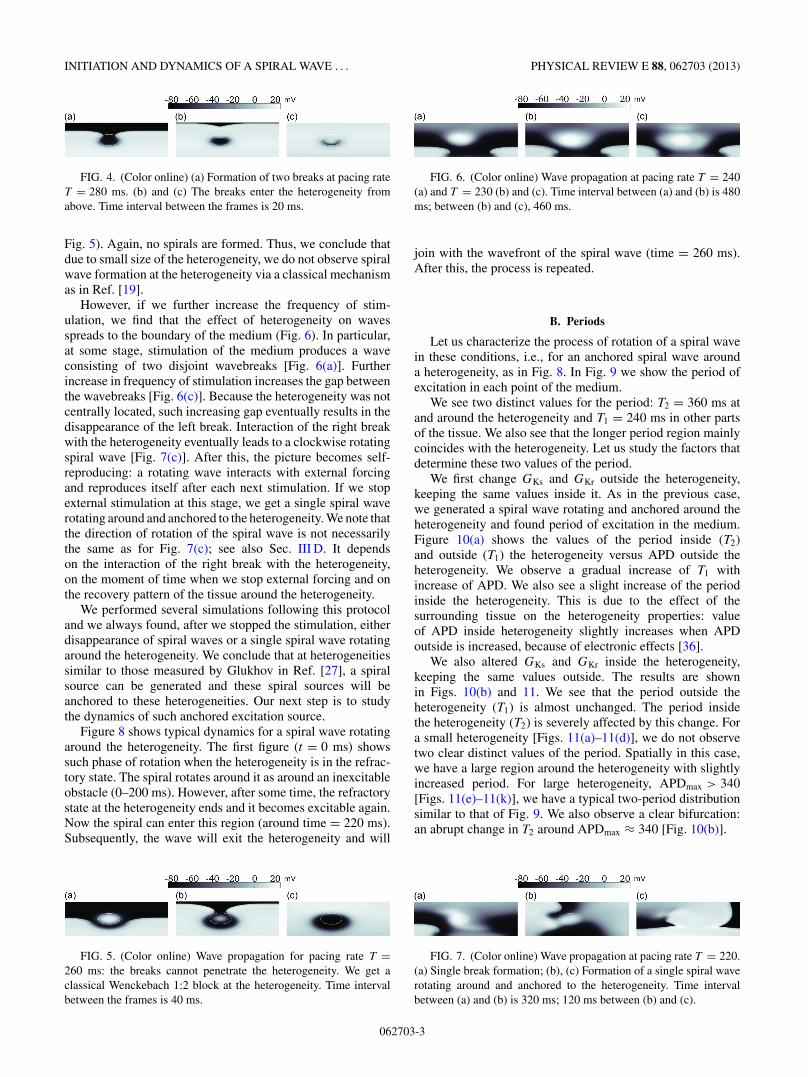
FIG. 4. (Color online) (a) Formation of two breaks at pacing rateT = 280 ms. (b) and (c) The breaks enter the heterogeneity fromabove. Time interval between the frames is 20 ms.
Fig. 5). Again, no spirals are formed. Thus, we conclude thatdue to small size of the heterogeneity, we do not observe spiralwave formation at the heterogeneity via a classical mechanismas in Ref. [19].
However, if we further increase the frequency of stim-ulation, we find that the effect of heterogeneity on wavesspreads to the boundary of the medium (Fig. 6). In particular,at some stage, stimulation of the medium produces a waveconsisting of two disjoint wavebreaks [Fig. 6(a)]. Furtherincrease in frequency of stimulation increases the gap betweenthe wavebreaks [Fig. 6(c)]. Because the heterogeneity was notcentrally located, such increasing gap eventually results in thedisappearance of the left break. Interaction of the right breakwith the heterogeneity eventually leads to a clockwise rotatingspiral wave [Fig. 7(c)]. After this, the picture becomes self-reproducing: a rotating wave interacts with external forcingand reproduces itself after each next stimulation. If we stopexternal stimulation at this stage, we get a single spiral waverotating around and anchored to the heterogeneity. We note thatthe direction of rotation of the spiral wave is not necessarilythe same as for Fig. 7(c); see also Sec. III D. It dependson the interaction of the right break with the heterogeneity,on the moment of time when we stop external forcing and onthe recovery pattern of the tissue around the heterogeneity.
We performed several simulations following this protocoland we always found, after we stopped the stimulation, eitherdisappearance of spiral waves or a single spiral wave rotatingaround the heterogeneity. We conclude that at heterogeneitiessimilar to those measured by Glukhov in Ref. [27], a spiralsource can be generated and these spiral sources will beanchored to these heterogeneities. Our next step is to studythe dynamics of such anchored excitation source.
Figure 8 shows typical dynamics for a spiral wave rotatingaround the heterogeneity. The first figure (t = 0 ms) showssuch phase of rotation when the heterogeneity is in the refrac-tory state. The spiral rotates around it as around an inexcitableobstacle (0–200 ms). However, after some time, the refractorystate at the heterogeneity ends and it becomes excitable again.Now the spiral can enter this region (around time = 220 ms).Subsequently, the wave will exit the heterogeneity and will
FIG. 5. (Color online) Wave propagation for pacing rate T =260 ms: the breaks cannot penetrate the heterogeneity. We get aclassical Wenckebach 1:2 block at the heterogeneity. Time intervalbetween the frames is 40 ms.
FIG. 6. (Color online) Wave propagation at pacing rate T = 240(a) and T = 230 (b) and (c). Time interval between (a) and (b) is 480ms; between (b) and (c), 460 ms.
join with the wavefront of the spiral wave (time = 260 ms).After this, the process is repeated.
B. Periods
Let us characterize the process of rotation of a spiral wavein these conditions, i.e., for an anchored spiral wave arounda heterogeneity, as in Fig. 8. In Fig. 9 we show the period ofexcitation in each point of the medium.
We see two distinct values for the period: T2 = 360 ms atand around the heterogeneity and T1 = 240 ms in other partsof the tissue. We also see that the longer period region mainlycoincides with the heterogeneity. Let us study the factors thatdetermine these two values of the period.
We first change GKs and GKr outside the heterogeneity,keeping the same values inside it. As in the previous case,we generated a spiral wave rotating and anchored around theheterogeneity and found period of excitation in the medium.Figure 10(a) shows the values of the period inside (T2)and outside (T1) the heterogeneity versus APD outside theheterogeneity. We observe a gradual increase of T1 withincrease of APD. We also see a slight increase of the periodinside the heterogeneity. This is due to the effect of thesurrounding tissue on the heterogeneity properties: valueof APD inside heterogeneity slightly increases when APDoutside is increased, because of electronic effects [36].
We also altered GKs and GKr inside the heterogeneity,keeping the same values outside. The results are shownin Figs. 10(b) and 11. We see that the period outside theheterogeneity (T1) is almost unchanged. The period insidethe heterogeneity (T2) is severely affected by this change. Fora small heterogeneity [Figs. 11(a)–11(d)], we do not observetwo clear distinct values of the period. Spatially in this case,we have a large region around the heterogeneity with slightlyincreased period. For large heterogeneity, APDmax > 340[Figs. 11(e)–11(k)], we have a typical two-period distributionsimilar to that of Fig. 9. We also observe a clear bifurcation:an abrupt change in T2 around APDmax ≈ 340 [Fig. 10(b)].
FIG. 7. (Color online) Wave propagation at pacing rate T = 220.(a) Single break formation; (b), (c) Formation of a single spiral waverotating around and anchored to the heterogeneity. Time intervalbetween (a) and (b) is 320 ms; 120 ms between (b) and (c).
062703-3

ARNE DEFAUW, PETER DAWYNDT, AND ALEXANDER V. PANFILOV PHYSICAL REVIEW E 88, 062703 (2013)
FIG. 8. (Color online) Rotation of spiral wave anchored around heterogeneity of Fig. 1. Figures show wave pattern at 20-ms intervals.White line shows size of the heterogeneity.
C. Period increase bifurcation
Bifurcation in the period of excitation obviously resultsin a change of the wave propagation pattern. If we considertwo successive points where the spiral wave tip enters theheterogeneity, we observe the following dynamics before andafter the period jump. For smaller heterogeneity [Fig. 11(a)],the wave tip makes a rotation of about 380 degrees beforeentering the heterogeneity again. To show it in Figs. 11(a)–11(d), we marked by black and red arrows the entry points ofthe wave into the heterogeneity for two successive rotations(first black, then red). For larger heterogeneity the rotation is
FIG. 9. (Color online) Period of excitation of the medium forspiral wave dynamics shown in Fig. 8. Figure shows average valueof period in each point over 15 excitations. Period inside theheterogeneity (T2) is approximately 360 ms. Period in the other partof the medium (T1) is around 240 ms.
about 390 degrees [Fig. 11(b)]. Thus, rotation angle increaseswhen we increase heterogeneity. However, at the bifurcationpoint, it approaches 400 degrees and then it jumps to about540 degrees, which results in an abrupt period increase.
We also found that this bifurcation only occurs in a limitedrange of size of the heterogeneity. Figure 12 shows resultssimilar to Fig. 10(b), but now with different sizes of theheterogeneity: we increased the size, respectively, 1.2, 1.5,1.7, and 2 times. We see that in Figs. 12(a)–12(c), we havequalitatively the same bifurcation as in Fig. 8(b).However, the
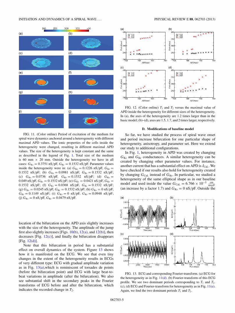
FIG. 10. (Color online) (a) T1 and T2 versus APD outside theheterogeneity: we alter GKs and GKr outside the heterogeneity. (b) T1
and T2 versus the maximal value of APD inside the heterogeneity:we alter GKs and GKr inside the heterogeneity.
062703-4
INITIATION AND DYNAMICS OF A SPIRAL WAVE . . . PHYSICAL REVIEW E 88, 062703 (2013)
FIG. 11. (Color online) Period of excitation of the medium forspiral wave dynamics anchored around a heterogeneity with differentmaximal APD values. The ionic properties of the cells inside theheterogeneity were changed, resulting in different maximal APDvalues. The size of the heterogeneity is kept constant and the sameas described in the legend of Fig. 1. Total size of the mediumis 60 mm × 20 mm. Outside the heterogeneity we have in allcases: GKs = 0.3751 nS/pF, GKr = 0.1532 nS/pF. Parameter valuesinside the heterogeneity were in: (a) GKs = 0.1226 nS/pF, GKr =0.1532 nS/pF; (b) GKs = 0.0981 nS/pF, GKr = 0.1532 nS/pF;(c) GKs = 0.0736 nS/pF, GKr = 0.1532 nS/pF; (d) GKs =0.049 nS/pF, GKr = 0.1532 nS/pF; (e) GKs = 0.0421 nS/pF, GKr =0.1532 nS/pF; (f) GKs = 0.0368 nS/pF, GKr = 0.1532 nS/pF;(g) GKs = 0.0245 nS/pF, GKr = 0.1532 nS/pF; (h) GKs = 0 nS/pF,GKr = 0.1149 nS/pF; (i) GKs = 0 nS/pF, GKr = 0.0948 nS/pF;(j) GKs = 0 nS/pF, GKr = 0.0479 nS/pF.
location of the bifurcation on the APD axis slightly increaseswith the size of the heterogeneity. The amplitude of the jumpfirst also slightly increases [Figs. 10(b), 12(a), and 12(b)], thendecreases [Fig. 12(c)], and finally the bifurcation disappears[Fig. 12(d)].
Note that this bifurcation in period has a substantialeffect on overall dynamics of the system. Figure 13 showshow it is manifested on the ECG. We see that even tinychanges in the extent of the heterogeneity results in ECGsof very different type: ECG with gradual amplitude variationas in Fig. 13(a),which is reminiscent of torsades de points(before the bifurcation point) and ECG with large beat-to-beat variations in amplitude (after the bifurcation). We alsosee substantial shift in the secondary peaks in the Fouriertransforms of ECG before and after the bifurcation, whichindicates the recorded change in T2.
FIG. 12. (Color online) T1 and T2 versus the maximal value ofAPD inside the heterogeneity for different sizes of the heterogeneity.In (a), the axes of the heterogeneity are 1.2 times larger than in thebasic model; (b)–(d), axes are 1.5, 1.7, and 2 times larger, respectively.
D. Modifications of baseline model
So far, we have studied the process of spiral wave onsetand period increase bifurcation for one particular shape ofheterogeneity, anisotropy, and parameter set. Here we extendour study to additional configurations.
In Fig. 1, heterogeneity in APD was created by changingGKs and GKr conductances. A similar heterogeneity can becreated by changing other parameter values. For instance,another current that has a substantial effect on APD is ICaL. Wehave checked if our results also hold for heterogeneity createdby changing GCaL instead of GKr. In particular, we studied aheterogeneity of the same elliptical shape as in our baselinemodel and used inside the value GCaL = 6.766 × 10⁻⁵ cm
msμF(an increase by a factor 1.7) and GKs = 0 nS/pF. Outside the
FIG. 13. ECG and corresponding Fourier-transform. (a) ECG forthe heterogeneity as in Fig. 11(d). (b) Fourier-transform of this ECGprofile. We see two dominant periods corresponding to T1 and T2.(c), (d) ECG and Fourier-transform for heterogeneity as in Fig. 11(e).Again, we find the two dominant periods T1 and T2.
062703-5
ARNE DEFAUW, PETER DAWYNDT, AND ALEXANDER V. PANFILOV PHYSICAL REVIEW E 88, 062703 (2013)
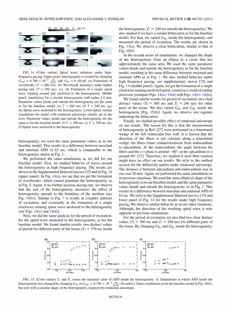
FIG. 14. (Color online) Spiral wave initiation under high-frequency pacing. Upper panel: heterogeneity is created by changingGCaL = 6.766 × 10⁻⁵ cm
msμF and GKs = 0 nS/pF. (a) Formation ofwavebreaks (T = 260 ms). (b) Wavebreak dynamics under higherpacing rate (T = 250 ms). (c), (d) Formation of a single spiralwave rotating around and anchored to the heterogeneity. Middlepanel: simulations for a circular heterogeneity with radius 4.5 mm.Parameter values inside and outside the heterogeneity are the sameas for the baseline model. (e) T = 260 ms; (f) T = 240 ms. (g),(h) Spiral wave anchored to the heterogeneity. Lower panel: similarsimulations for model with rotational anisotropy (details are in thetext). Parameter values inside and outside the heterogeneity are thesame as for the baseline model. (i) T = 260 ms; (j) T = 240 ms. (k),(l) Spiral wave anchored to the heterogeneity.
heterogeneity, we used the same parameter values as in ourbaseline model. This results in a difference between maximaland minimal APD of 62 ms, which is comparable to theheterogeneity shown in Fig. 1.
We performed the same simulations as we did for ourbaseline model. First, we studied behavior of waves aroundthe heterogeneity at high frequency pacing. The results areshown in the Supplemental Material movies [35] and in Fig. 14(upper panel). In Fig. 14(a), we see that we get the formationof wavebreaks, which cannot penetrate the heterogeneity, asin Fig. 5. Again, if we further increase pacing rate, we observethat the size of the heterogeneity increases: the effect ofheterogeneity spreads to the boundary of the medium [seeFig. 14(b)]. Similar to Fig. 7 it results in complex patternsof excitation, and eventually in the formation of a singleclockwise rotating spiral wave anchored to the heterogeneity(see Figs. 14(c) and 14(d)].
Next, we did the same analysis for the period of excitation,for the spiral wave anchored to the heterogeneity, as for thebaseline model. We found similar results: two distinct valuesof period for different parts of the tissue (T2 ≈ 370 ms inside
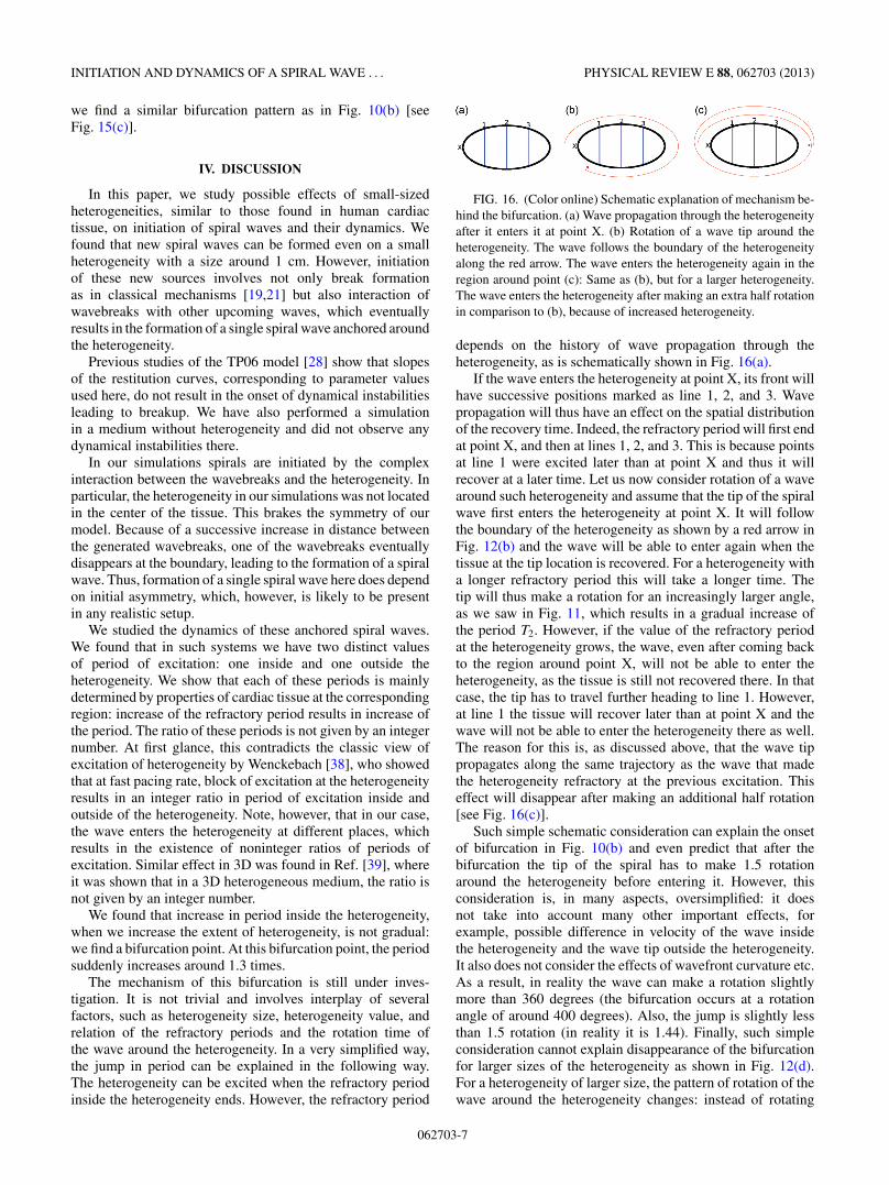
the heterogeneity, T1 ≈ 240 ms outside the heterogeneity). Wealso studied if we have a similar bifurcation as for the baselinemodel. For that, we varied GKs inside the heterogeneity, andmeasured the period of excitation. The results are shown inFig. 15(a). We observe a clear bifurcation, similar to that ofFig. 10(b).
In the second series of simulations, we changed the shapeof the heterogeneity from an ellipse to a circle that hasapproximately the same area. We used the same parametervalues inside and outside the heterogeneity as for the baselinemodel, resulting in the same difference between maximal andminimal APD as in Fig. 1. We also studied behavior underhigh frequency pacing: see supplementary movie [35] andFig. 14 (middle panel). Again, we get the formation of a singleclockwise rotating anchored spiral, created as a result of similarprocesses [compare Figs. 14(a)–14(d) with Figs. 14(e)–14(h)].
We found similar results for period of excitation: two cleardistinct values (T2 ≈ 360 ms and T1 ≈ 240 ms) for otherparts of the tissue. We also varied GKs and GKr inside theheterogeneity [Fig. 15(b)]. Again, we observe two regimesindicating the bifurcation.
Finally, we studied possible effect of rotational anisotropyon our results. The reason for this is that the measurementof heterogeneity in Ref. [27] were performed in a transmuralwedge of the left ventricular free wall. It is known that thedirection of the fibers is not constant along a transmuralwedge: the fibers rotate counterclockwise from endocardiumto epicardium. At the endocardium, the angle between thefibers and the x-y plane is around −60◦; at the epicardium it isaround 60◦ [37]. Therefore, we studied if such fiber rotationmight have an effect on our results. We refer to the methodsection for the diffusivity matrix under rotational anisotropy.The distance d between epicardium and endocardium was inour case 20 mm. Again, we performed the same simulations asin previous situations. We used the same elliptical shape of theheterogeneity as in our baseline model, and the same parametervalues inside and outside the heterogeneity, as in Fig. 1. Thisresults in a difference between maximal and minimal APD of62 ms. We refer to the Supplemental Material movies [35] andlower panel of Fig. 14 for the results under high frequencypacing. We observe similar behavior as in our other situations.Although, the direction of the resulting spiral wave is nowopposite to previous simulations.
For the period of excitation we also find two clear distinctvalues (T2 ≈ 360 ms and T1 ≈ 240 ms) for different parts ofthe tissue. By changing GKs and GKr inside the heterogeneity,
FIG. 15. (Color online) T1 and T2 versus the maximal value of APD inside the heterogeneity. A: Simulations in which APD inside theheterogeneity was changed by changing GKs at GCaL = 6.766 × 10⁻⁵ cm
msμF ; (b) and (c): Same simulations as for the baseline model in Fig. 10(b),but now with a circular shape of the heterogeneity, respectively rotational anisotropy.
062703-6
INITIATION AND DYNAMICS OF A SPIRAL WAVE . . . PHYSICAL REVIEW E 88, 062703 (2013)
we find a similar bifurcation pattern as in Fig. 10(b) [seeFig. 15(c)].
IV. DISCUSSION
In this paper, we study possible effects of small-sizedheterogeneities, similar to those found in human cardiactissue, on initiation of spiral waves and their dynamics. Wefound that new spiral waves can be formed even on a smallheterogeneity with a size around 1 cm. However, initiationof these new sources involves not only break formationas in classical mechanisms [19,21] but also interaction ofwavebreaks with other upcoming waves, which eventuallyresults in the formation of a single spiral wave anchored aroundthe heterogeneity.
Previous studies of the TP06 model [28] show that slopesof the restitution curves, corresponding to parameter valuesused here, do not result in the onset of dynamical instabilitiesleading to breakup. We have also performed a simulationin a medium without heterogeneity and did not observe anydynamical instabilities there.
In our simulations spirals are initiated by the complexinteraction between the wavebreaks and the heterogeneity. Inparticular, the heterogeneity in our simulations was not locatedin the center of the tissue. This brakes the symmetry of ourmodel. Because of a successive increase in distance betweenthe generated wavebreaks, one of the wavebreaks eventuallydisappears at the boundary, leading to the formation of a spiralwave. Thus, formation of a single spiral wave here does dependon initial asymmetry, which, however, is likely to be presentin any realistic setup.
We studied the dynamics of these anchored spiral waves.We found that in such systems we have two distinct valuesof period of excitation: one inside and one outside theheterogeneity. We show that each of these periods is mainlydetermined by properties of cardiac tissue at the correspondingregion: increase of the refractory period results in increase ofthe period. The ratio of these periods is not given by an integernumber. At first glance, this contradicts the classic view ofexcitation of heterogeneity by Wenckebach [38], who showedthat at fast pacing rate, block of excitation at the heterogeneityresults in an integer ratio in period of excitation inside andoutside of the heterogeneity. Note, however, that in our case,the wave enters the heterogeneity at different places, whichresults in the existence of noninteger ratios of periods ofexcitation. Similar effect in 3D was found in Ref. [39], whereit was shown that in a 3D heterogeneous medium, the ratio isnot given by an integer number.
We found that increase in period inside the heterogeneity,when we increase the extent of heterogeneity, is not gradual:we find a bifurcation point. At this bifurcation point, the periodsuddenly increases around 1.3 times.
The mechanism of this bifurcation is still under inves-tigation. It is not trivial and involves interplay of severalfactors, such as heterogeneity size, heterogeneity value, andrelation of the refractory periods and the rotation time ofthe wave around the heterogeneity. In a very simplified way,the jump in period can be explained in the following way.The heterogeneity can be excited when the refractory periodinside the heterogeneity ends. However, the refractory period
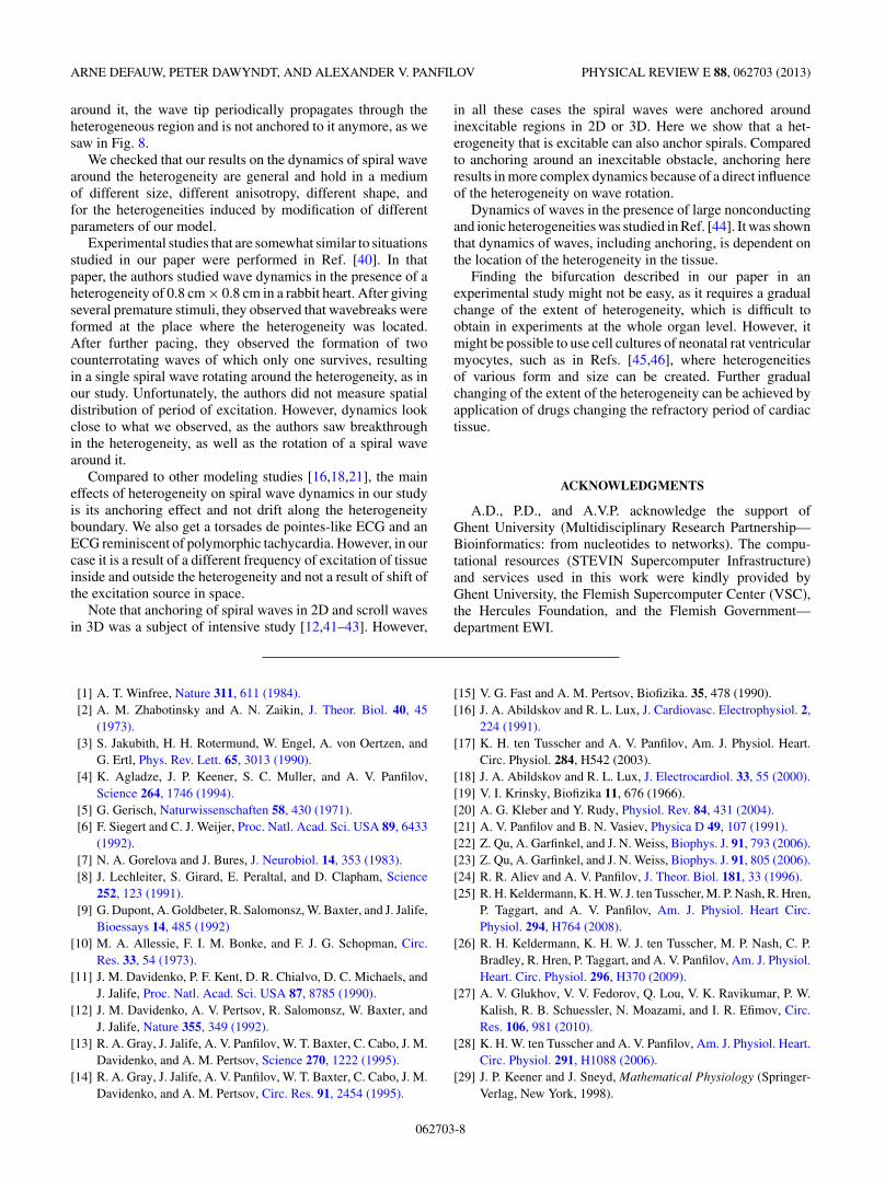
FIG. 16. (Color online) Schematic explanation of mechanism be-hind the bifurcation. (a) Wave propagation through the heterogeneityafter it enters it at point X. (b) Rotation of a wave tip around theheterogeneity. The wave follows the boundary of the heterogeneityalong the red arrow. The wave enters the heterogeneity again in theregion around point (c): Same as (b), but for a larger heterogeneity.The wave enters the heterogeneity after making an extra half rotationin comparison to (b), because of increased heterogeneity.
depends on the history of wave propagation through theheterogeneity, as is schematically shown in Fig. 16(a).
If the wave enters the heterogeneity at point X, its front willhave successive positions marked as line 1, 2, and 3. Wavepropagation will thus have an effect on the spatial distributionof the recovery time. Indeed, the refractory period will first endat point X, and then at lines 1, 2, and 3. This is because pointsat line 1 were excited later than at point X and thus it willrecover at a later time. Let us now consider rotation of a wavearound such heterogeneity and assume that the tip of the spiralwave first enters the heterogeneity at point X. It will followthe boundary of the heterogeneity as shown by a red arrow inFig. 12(b) and the wave will be able to enter again when thetissue at the tip location is recovered. For a heterogeneity witha longer refractory period this will take a longer time. Thetip will thus make a rotation for an increasingly larger angle,as we saw in Fig. 11, which results in a gradual increase ofthe period T2. However, if the value of the refractory periodat the heterogeneity grows, the wave, even after coming backto the region around point X, will not be able to enter theheterogeneity, as the tissue is still not recovered there. In thatcase, the tip has to travel further heading to line 1. However,at line 1 the tissue will recover later than at point X and thewave will not be able to enter the heterogeneity there as well.The reason for this is, as discussed above, that the wave tippropagates along the same trajectory as the wave that madethe heterogeneity refractory at the previous excitation. Thiseffect will disappear after making an additional half rotation[see Fig. 16(c)].
Such simple schematic consideration can explain the onsetof bifurcation in Fig. 10(b) and even predict that after thebifurcation the tip of the spiral has to make 1.5 rotationaround the heterogeneity before entering it. However, thisconsideration is, in many aspects, oversimplified: it doesnot take into account many other important effects, forexample, possible difference in velocity of the wave insidethe heterogeneity and the wave tip outside the heterogeneity.It also does not consider the effects of wavefront curvature etc.As a result, in reality the wave can make a rotation slightlymore than 360 degrees (the bifurcation occurs at a rotationangle of around 400 degrees). Also, the jump is slightly lessthan 1.5 rotation (in reality it is 1.44). Finally, such simpleconsideration cannot explain disappearance of the bifurcationfor larger sizes of the heterogeneity as shown in Fig. 12(d).For a heterogeneity of larger size, the pattern of rotation of thewave around the heterogeneity changes: instead of rotating
062703-7
ARNE DEFAUW, PETER DAWYNDT, AND ALEXANDER V. PANFILOV PHYSICAL REVIEW E 88, 062703 (2013)
around it, the wave tip periodically propagates through theheterogeneous region and is not anchored to it anymore, as wesaw in Fig. 8.
We checked that our results on the dynamics of spiral wavearound the heterogeneity are general and hold in a mediumof different size, different anisotropy, different shape, andfor the heterogeneities induced by modification of differentparameters of our model.
Experimental studies that are somewhat similar to situationsstudied in our paper were performed in Ref. [40]. In thatpaper, the authors studied wave dynamics in the presence of aheterogeneity of 0.8 cm × 0.8 cm in a rabbit heart. After givingseveral premature stimuli, they observed that wavebreaks wereformed at the place where the heterogeneity was located.After further pacing, they observed the formation of twocounterrotating waves of which only one survives, resultingin a single spiral wave rotating around the heterogeneity, as inour study. Unfortunately, the authors did not measure spatialdistribution of period of excitation. However, dynamics lookclose to what we observed, as the authors saw breakthroughin the heterogeneity, as well as the rotation of a spiral wavearound it.
Compared to other modeling studies [16,18,21], the maineffects of heterogeneity on spiral wave dynamics in our studyis its anchoring effect and not drift along the heterogeneityboundary. We also get a torsades de pointes-like ECG and anECG reminiscent of polymorphic tachycardia. However, in ourcase it is a result of a different frequency of excitation of tissueinside and outside the heterogeneity and not a result of shift ofthe excitation source in space.
Note that anchoring of spiral waves in 2D and scroll wavesin 3D was a subject of intensive study [12,41–43]. However,
in all these cases the spiral waves were anchored aroundinexcitable regions in 2D or 3D. Here we show that a het-erogeneity that is excitable can also anchor spirals. Comparedto anchoring around an inexcitable obstacle, anchoring hereresults in more complex dynamics because of a direct influenceof the heterogeneity on wave rotation.
Dynamics of waves in the presence of large nonconductingand ionic heterogeneities was studied in Ref. [44]. It was shownthat dynamics of waves, including anchoring, is dependent onthe location of the heterogeneity in the tissue.
Finding the bifurcation described in our paper in anexperimental study might not be easy, as it requires a gradualchange of the extent of heterogeneity, which is difficult toobtain in experiments at the whole organ level. However, itmight be possible to use cell cultures of neonatal rat ventricularmyocytes, such as in Refs. [45,46], where heterogeneitiesof various form and size can be created. Further gradualchanging of the extent of the heterogeneity can be achieved byapplication of drugs changing the refractory period of cardiactissue.
ACKNOWLEDGMENTS
A.D., P.D., and A.V.P. acknowledge the support ofGhent University (Multidisciplinary Research Partnership—Bioinformatics: from nucleotides to networks). The compu-tational resources (STEVIN Supercomputer Infrastructure)and services used in this work were kindly provided byGhent University, the Flemish Supercomputer Center (VSC),the Hercules Foundation, and the Flemish Government—department EWI.
[1] A. T. Winfree, Nature 311, 611 (1984).[2] A. M. Zhabotinsky and A. N. Zaikin, J. Theor. Biol. 40, 45
(1973).[3] S. Jakubith, H. H. Rotermund, W. Engel, A. von Oertzen, and
G. Ertl, Phys. Rev. Lett. 65, 3013 (1990).[4] K. Agladze, J. P. Keener, S. C. Muller, and A. V. Panfilov,
Science 264, 1746 (1994).[5] G. Gerisch, Naturwissenschaften 58, 430 (1971).[6] F. Siegert and C. J. Weijer, Proc. Natl. Acad. Sci. USA 89, 6433
(1992).[7] N. A. Gorelova and J. Bures, J. Neurobiol. 14, 353 (1983).[8] J. Lechleiter, S. Girard, E. Peraltal, and D. Clapham, Science
252, 123 (1991).[9] G. Dupont, A. Goldbeter, R. Salomonsz, W. Baxter, and J. Jalife,
Bioessays 14, 485 (1992)[10] M. A. Allessie, F. I. M. Bonke, and F. J. G. Schopman, Circ.
Res. 33, 54 (1973).[11] J. M. Davidenko, P. F. Kent, D. R. Chialvo, D. C. Michaels, and
J. Jalife, Proc. Natl. Acad. Sci. USA 87, 8785 (1990).[12] J. M. Davidenko, A. V. Pertsov, R. Salomonsz, W. Baxter, and
J. Jalife, Nature 355, 349 (1992).[13] R. A. Gray, J. Jalife, A. V. Panfilov, W. T. Baxter, C. Cabo, J. M.
Davidenko, and A. M. Pertsov, Science 270, 1222 (1995).[14] R. A. Gray, J. Jalife, A. V. Panfilov, W. T. Baxter, C. Cabo, J. M.
Davidenko, and A. M. Pertsov, Circ. Res. 91, 2454 (1995).
[15] V. G. Fast and A. M. Pertsov, Biofizika. 35, 478 (1990).[16] J. A. Abildskov and R. L. Lux, J. Cardiovasc. Electrophysiol. 2,
224 (1991).[17] K. H. ten Tusscher and A. V. Panfilov, Am. J. Physiol. Heart.
Circ. Physiol. 284, H542 (2003).[18] J. A. Abildskov and R. L. Lux, J. Electrocardiol. 33, 55 (2000).[19] V. I. Krinsky, Biofizika 11, 676 (1966).[20] A. G. Kleber and Y. Rudy, Physiol. Rev. 84, 431 (2004).[21] A. V. Panfilov and B. N. Vasiev, Physica D 49, 107 (1991).[22] Z. Qu, A. Garfinkel, and J. N. Weiss, Biophys. J. 91, 793 (2006).[23] Z. Qu, A. Garfinkel, and J. N. Weiss, Biophys. J. 91, 805 (2006).[24] R. R. Aliev and A. V. Panfilov, J. Theor. Biol. 181, 33 (1996).[25] R. H. Keldermann, K. H. W. J. ten Tusscher, M. P. Nash, R. Hren,
P. Taggart, and A. V. Panfilov, Am. J. Physiol. Heart Circ.Physiol. 294, H764 (2008).
[26] R. H. Keldermann, K. H. W. J. ten Tusscher, M. P. Nash, C. P.Bradley, R. Hren, P. Taggart, and A. V. Panfilov, Am. J. Physiol.Heart. Circ. Physiol. 296, H370 (2009).
[27] A. V. Glukhov, V. V. Fedorov, Q. Lou, V. K. Ravikumar, P. W.Kalish, R. B. Schuessler, N. Moazami, and I. R. Efimov, Circ.Res. 106, 981 (2010).
[28] K. H. W. ten Tusscher and A. V. Panfilov, Am. J. Physiol. Heart.Circ. Physiol. 291, H1088 (2006).
[29] J. P. Keener and J. Sneyd, Mathematical Physiology (Springer-Verlag, New York, 1998).
062703-8
INITIATION AND DYNAMICS OF A SPIRAL WAVE . . . PHYSICAL REVIEW E 88, 062703 (2013)
[30] K. H. W. ten Tusscher, D. Noble, P. J. Noble, and A. V. Panfilov,Am. J. Physiol. Heart Circ. Physiol. 286, H1573 (2004).
[31] S. Rush and H. Larsen, IEEE Trans. Biomed. Eng. 25, 389(1978).
[32] W. Shimizu and C. Antzelevitch, Circulation 98, 2314(1998).
[33] W. Shimizu and C. Antzelevitch, J. Am. Coll. Cardiol. 35, 778(2000).
[34] A. Defauw, I. V. Kazbanov, H. Dierckx, P. Dawyndt, and A. V.Panfilov, PLoS one 8, e79607 (2013).
[35] See Supplemental Material at http://link.aps.org/supplemental/10.1103/PhysRevE.88.062703 for movies.
[36] K. J. Sampson and C. S. Henriquez, Am. J. Physiol. Heart Circ.Physiol. 289, H350 (2005).
[37] D. Streeter, Handbook of Physiology, Am Physiol Soc. Chap.Gross Morphology and Fiber Geometry of the Heart (The Heart.Bethesda, Maryland, 1979), Sec. 2. Vol. I, pp. 61–112.
[38] K. F. Wenckebach, Z. Klin. Med. 37, 475 (1899).[39] A. V. Panfilov and J. P. Keener, Int. J. Bif. Chaos. 3, 445 (1993).[40] L. Boersma, Z. Zetelaki, J. Brugada, and M. Allessie, Circ. Res.
105, 3053 (2002).[41] M. Vinson, A. Pertsov, and J. Jalife, Physica D 72, 119 (1994).[42] T. Wu, M. Yashima, F. Xie, C. A. Athill, Y. Kim, M. C. Fishbein,
Z. Qu, A. Garfinkel, J. N. Weiss, H. S. Karagueuzian, andP. Chen, Circ. Res. 83, 448 (1998).
[43] C. W. Zemlin and A. M. Pertsov, Phys. Rev. Lett. 109, 038303(2012).
[44] T. K. Shajahan, S. Sinha, and R. Pandit, Phys. Rev. E 75, 011929(2007).
[45] B. O. Bingen, S. F. Askar, M. J. Schalij, I. V. Kazbanov, D. L.Ypey, A. V. Panfilov, and D. A. Pijnappels, Cardiovasc. Res. 97,161 (2013).
[46] K. Campbell, C. J. Calvo, S. Mironov, T. Herron, O. Berenfeld,and J. Jalife, J. Physiol. 590, 6363 (2012).
062703-9
Copyright © 2022 FDOKUMEN