Inhibition of in vivo breast cancer growth by antisense oligodeoxynucleotides to type I insulin-like...
14
Inhibition of in vivo breast cancer growth by antisense oligodeoxynucleotides to type I insulin-like growth factor receptor mRNA involves inactivation of ErbBs, PI-3K/Akt and p42/p44 MAPK signaling pathways but not modulation of progesterone receptor activity Mariana Salatino 1 , Roxana Schillaci 1 , Cecilia J Proietti 1 , Romina Carnevale 1 , Isabel Frahm 2 , Alfredo A Molinolo 1 , Adolfo Iribarren 3 , Eduardo H Charreau 1 and Patricia V Elizalde* ,1 1 Laboratory of Molecular Mechanisms of Carcinogenesis, Instituto de Biologı´a y Medicina Experimental (IBYME), CONICET, Obligado 2490, Buenos Aires 1428, Argentina; 2 Servicio de Patologı´a, Sanatorio Mater Dei, Buenos Aires, Argentina; 3 Instituto de Investigaciones en Ingenierı´a Gene´tica y Biologı´a Molecular (INGEBI), Argentina The present study addresses the effect of targeting type I insulin-like growth factor receptor (IGF-IR) with anti- sense strategies in in vivo growth of breast cancer cells. Our research was carried out on C4HD tumors from an experimental model of hormonal carcinogenesis in which the synthetic progestin medroxyprogesterone acetate (MPA) induced mammary adenocarcinomas in Balb/c mice. We employed two different experimental strategies. With the first one we demonstrated that direct intratumor injection of phosphorothioate antisense oligodeoxynucleo- tides (AS[S]ODNs) to IGF-IR mRNA resulted in a significant inhibition of C4HD tumor growth. In the second experimental strategy, we assessed the effect of intravenous (i.v.) injection of AS [S]ODN on C4HD tumor growth. This systemic treatment also resulted in significant reduction in tumor growth. The antitumor effect of IGF-IR AS[S]ODNs in both experimental protocols was due to a specific antisense mechanism, since growth inhibition was dose-dependent and no abrogation of tumor proliferation was observed in mice treated with phosphorothioate sense ODNs (S[S]ODNs). In addition, IGF-IR expression was inhibited in tumors from mice receiving AS[S]ODNs, as compared to tumors from control groups. We then investigated signal trans- duction pathways modulated in vivo by AS[S]ODNs treatment. Tumors from AS[S]ODN-treated mice of both intratumoral and intravenous protocols showed a signifi- cant decrease in the degree of insulin receptor substrate-1 (IRS-1) tyrosine phosphorylation. Activation of two of the main IGF-IR signaling pathways, phosphatidylinositol 3- kinase (PI-3K)/Akt and p42/p44 mitogen-activated pro- tein kinases (MAPK) was abolished in tumors growing in AS[S]ODN-treated animals. Moreover, ErbB-2 tyrosine phosphorylation was blocked by in vivo administration of AS[S]ODNs. On the other hand, we found no regulation of either progesterone receptor expression or activity by in vivo AS[S]ODNs administration. Our results for the first time demonstrated that breast cancer growth can be inhibited by direct in vivo administration of IGF-IR AS[S]ODNs. Oncogene (2004) 23, 5161–5174. doi:10.1038/sj.onc.1207659 Published online 3 May 2004 Keywords: breast cancer; IGF-IR; antisense strategies Introduction Type I insulin-like growth factor receptor (IGF-IR) plays a pivotal role in the regulation of cell growth in different ways. It is not only mitogenic for several cell types but also plays a crucial role in the establishment and maintenance of the transformed phenotype, pro- tects cells from a variety of apoptotic signals and finally also induces differentiation of certain cell types (for review see Werner and Le Roith, 2000). Evidence of a crucial role of IGF-IR in malignant transformation was first provided by the findings of Sell et al. (1994), showing that mouse embryo cells with a targeted disruption of IGF-IR (R-cells) cannot be transformed by SV40T antigen and/or activated Ha-ras oncogene (Sell et al., 1994) both of which easily transform embryo cells obtained from wild-type littermates. This finding has since then been confirmed with several viral and cellular oncogenes that also fail to transform R-cells (reviewed in Baserga, 1999). A growing body of evidence indicates that IGF-IR plays a key role in breast cancer development and that it is involved in a complex cross-talk with steroid hormones that regulates breast tumor growth (reviewed in Surmacz et al., 1998). High levels of IGF-IR expression have been found in breast tumors and breast cancer cell lines (reviewed in Surmacz et al., 1998). However, accumulating evidence supports the hypoth- esis that an obligatory requirement to establish and maintain the transformed phenotype is the presence of Received 17 November 2003; revised 13 February 2004; accepted 13 February 2004; published online 3 May 2004 *Correspondence: PV Elizalde; E-mail: [email protected] Oncogene (2004) 23, 5161–5174 & 2004 Nature Publishing Group All rights reserved 0950-9232/04 $30.00 www.nature.com/onc
-
Upload
independent -
Category
Documents
-
view
4 -
download
0
Transcript of Inhibition of in vivo breast cancer growth by antisense oligodeoxynucleotides to type I insulin-like...
Inhibition of in vivo breast cancer growth by antisenseoligodeoxynucleotides to type I insulin-like growth factor receptor mRNA
involves inactivation of ErbBs, PI-3K/Akt and p42/p44 MAPK signaling
pathways but not modulation of progesterone receptor activity
Mariana Salatino1, Roxana Schillaci1, Cecilia J Proietti1, Romina Carnevale1, Isabel Frahm2,Alfredo A Molinolo1, Adolfo Iribarren3, Eduardo H Charreau1 and Patricia V Elizalde*,1
1Laboratory of Molecular Mechanisms of Carcinogenesis, Instituto de Biologıa y Medicina Experimental (IBYME), CONICET,Obligado 2490, Buenos Aires 1428, Argentina; 2Servicio de Patologıa, Sanatorio Mater Dei, Buenos Aires, Argentina; 3Instituto deInvestigaciones en Ingenierıa Genetica y Biologıa Molecular (INGEBI), Argentina
The present study addresses the effect of targeting type Iinsulin-like growth factor receptor (IGF-IR) with anti-sense strategies in in vivo growth of breast cancer cells.Our research was carried out on C4HD tumors from anexperimental model of hormonal carcinogenesis in whichthe synthetic progestin medroxyprogesterone acetate(MPA) induced mammary adenocarcinomas in Balb/cmice. We employed two different experimental strategies.With the first one we demonstrated that direct intratumorinjection of phosphorothioate antisense oligodeoxynucleo-tides (AS[S]ODNs) to IGF-IR mRNA resulted in asignificant inhibition of C4HD tumor growth. In thesecond experimental strategy, we assessed the effect ofintravenous (i.v.) injection of AS [S]ODN on C4HDtumor growth. This systemic treatment also resulted insignificant reduction in tumor growth. The antitumoreffect of IGF-IR AS[S]ODNs in both experimentalprotocols was due to a specific antisense mechanism,since growth inhibition was dose-dependent and noabrogation of tumor proliferation was observed in micetreated with phosphorothioate sense ODNs (S[S]ODNs).In addition, IGF-IR expression was inhibited in tumorsfrom mice receiving AS[S]ODNs, as compared to tumorsfrom control groups. We then investigated signal trans-duction pathways modulated in vivo by AS[S]ODNstreatment. Tumors from AS[S]ODN-treated mice of bothintratumoral and intravenous protocols showed a signifi-cant decrease in the degree of insulin receptor substrate-1(IRS-1) tyrosine phosphorylation. Activation of two of themain IGF-IR signaling pathways, phosphatidylinositol 3-kinase (PI-3K)/Akt and p42/p44 mitogen-activated pro-tein kinases (MAPK) was abolished in tumors growing inAS[S]ODN-treated animals. Moreover, ErbB-2 tyrosinephosphorylation was blocked by in vivo administration ofAS[S]ODNs. On the other hand, we found no regulationof either progesterone receptor expression or activity by
in vivo AS[S]ODNs administration. Our results for thefirst time demonstrated that breast cancer growth can beinhibited by direct in vivo administration of IGF-IRAS[S]ODNs.Oncogene (2004) 23, 5161–5174. doi:10.1038/sj.onc.1207659Published online 3 May 2004
Keywords: breast cancer; IGF-IR; antisense strategies
Introduction
Type I insulin-like growth factor receptor (IGF-IR)plays a pivotal role in the regulation of cell growth indifferent ways. It is not only mitogenic for several celltypes but also plays a crucial role in the establishmentand maintenance of the transformed phenotype, pro-tects cells from a variety of apoptotic signals and finallyalso induces differentiation of certain cell types (forreview see Werner and Le Roith, 2000). Evidence of acrucial role of IGF-IR in malignant transformation wasfirst provided by the findings of Sell et al. (1994),showing that mouse embryo cells with a targeteddisruption of IGF-IR (R-cells) cannot be transformedby SV40T antigen and/or activated Ha-ras oncogene(Sell et al., 1994) both of which easily transform embryocells obtained from wild-type littermates. This findinghas since then been confirmed with several viral andcellular oncogenes that also fail to transform R-cells(reviewed in Baserga, 1999).A growing body of evidence indicates that IGF-IR
plays a key role in breast cancer development and that itis involved in a complex cross-talk with steroidhormones that regulates breast tumor growth (reviewedin Surmacz et al., 1998). High levels of IGF-IRexpression have been found in breast tumors and breastcancer cell lines (reviewed in Surmacz et al., 1998).However, accumulating evidence supports the hypoth-esis that an obligatory requirement to establish andmaintain the transformed phenotype is the presence of
Received 17 November 2003; revised 13 February 2004; accepted 13February 2004; published online 3 May 2004
*Correspondence: PV Elizalde;E-mail: [email protected]
Oncogene (2004) 23, 5161–5174& 2004 Nature Publishing Group All rights reserved 0950-9232/04 $30.00
www.nature.com/onc

physiological levels of IGF-IR instead of overexpressionof this receptor (reviewed in Yu and Rohan, 2000).We have already demonstrated that IGF-IR plays a
key role in the proliferation of C4HD tumors from anexperimental model of hormonal carcinogenesis inwhich the synthetic progestin medroxyprogesteroneacetate (MPA) induced mammary adenocarcinomas infemale Balb/c mice. As we have previously described indetail (Elizalde et al., 1998; Balana et al., 1999, 2001),C4HD tumors are of ductal origin, require MPAadministration to proliferate both in vivo and in in vitro,and express high levels of estrogen (ER) and progester-one receptors (PR) (Elizalde et al., 1998; Balana et al.,1999, 2001). C4HD tumors also express IGF-IR andsynthesize IGF-I (Elizalde et al., 1998). In previousworks, we found that blockage of IGF-IR expression byusing antisense oligodeoxynucleotides (ASODNs) toIGF-IR mRNA, inhibited MPA-stimulated growth ofprimary cultures of epithelial cells from C4HD tumor(Elizalde et al., 1998), indicating that a functionalautocrine loop involving IGF-I and IGF-IR participatesin MPA-induced proliferation of C4HD cells. On theother hand, our studies of the expression and function oftype I receptor tyrosine kinases (RTKs: EGF-R/ErbB-1,ErbB-2, ErbB-3 and ErbB-4), and their ligand, heregulin(HRG), revealed the existence of bi-directional cross-talks between progestins and HRG/Erbs signalingpathways that control C4HD cell growth (Balana et al.,1999, 2001). Finally, we found interaction between IGF-IR and HRG/ErbBs signaling, evidenced first by the factthat HRG induced a potent proliferative response inC4HD cells, which was completely abolished by theblockage of IGF-IR expression using ASODNs (Balanaet al., 1999); second, by the presence of a hierarchicalinteraction between IGF-IR and ErbB-2, by which IGF-IR directs ErbB-2 phosphorylation (Balana et al., 2001).As a whole, our findings consistently identify IGF-IR asplaying a key role in C4HD cell proliferation driven bythree different but related pathways, that is, by the PR,by HRG/ErbBs and lastly by its own IGF-I/IGF-IRautocrine loop.In the present study, we have assessed the effect of
targeting IGF-IR with antisense strategies in in vivogrowth of C4HD tumors. Our findings for the first timedemonstrated that either direct intratumor injection orsystemic administration of phosphorothioate antisenseODNs (AS[S]ODNs) to IGF-IR mRNA resulted in asignificant growth inhibition of C4HD tumors. Inaddition, we dissected a network of interactions amongIGF-IR, type I RTKs, and PR signaling pathways thatwere differentially affected by blockage of IGF-IRfunction.
Results
Effects of IGF-IR AS[S]ODNs on C4HD cell in vitroproliferation
The aim of the present study was to explore the effect ofthe abolishment of IGF-IR expression, by using
ASODNs, in in vivo growth of C4HD tumors. Toovercome the rapid degradation of the phosphodiesterODNs by nucleases, we used phosphorothioate ODNs([S]ODNs) in the experiments carried out in vivo. Here,we first compared the ability of the AS[S]ODNs to IGF-IR to inhibit in vitro proliferation of C4HD cells withthe effect of their unmodified counterparts, the phos-phodiester ASODNs, which we had already used in ourprevious studies (Elizalde et al., 1998; Balana et al.,1999, 2001). As shown in Figure 1a, phosphorothioateAS[S]ODNs are able to exert a dose-dependent inhibi-tion of MPA-induced C4HD cell growth, comparable tothe one exerted by the phosphodiester ASODNs. Sense[S]ODNs had no effect on proliferation at any of thetested doses. The effect of IGF-IR AS[S]ODNs onprotein expression was assessed by immunoblotting
Figure 1 Inhibition of MPA-induced in vitro proliferation ofC4HD cells by AS[S]ODNs to IGF-IR mRNA. (a) Primarycultures of C4HD cells were incubated for 48 h in medium with2.5% ChFCS supplemented with MPA 10 nM, MPAþphospho-phosphodiester ASODNS or SODNs, and MPAþphosphorothio-osphorothioate AS[S]ODNs or S[S]ODNs to IGF-IR mRNA.Incorporation of (3H)-thymidine was used as a measure of DNAsynthesis. Data are presented as mean7s.d. b vs a, and c vs b:Po0.001. The experiment shown is representative of a total offour. (b) Effect of AS[S]ODNs on IGF-IR protein synthesis. Atotal of 100mg protein from C4HD cell lysates was electrophoresedand immunoblotted with an IGF-IR a chain antibody. Densito-metric analysis of IGF-IR band, expressed as a percentage of thecontrol value (i.e. C4HD cells growing in MPA10nM) is 37% forcells treated with 2mM AS[S]ODNs. No significant differences werefound in densitometric values of IGF-IR band between controlcells and cells treated with 2mM S[S]ODNs. Western blot using anantiactin antibody was carried out using identical protein lysates ascontrol for the specificity of the IGF-IR AS[S]ODNs. This is arepresentative experiment of a total of four in which s.e. was within10%
Antisense IGF-IR therapy of breast cancerM Salatino et al
5162
Oncogene
C4HD cell lysates (Figure 1b). Densitometric evaluationdemonstrated that IGF-IR levels were reduced by 60–70% after 2 mM AS[S]ODN treatment, while S[S]ODNsdid not reduce IGF-IR levels (Figure 1b), confirmingthe specificity of the IGF-IR AS[S]ODNs. As we havepreviously reported using ligand-binding assays (Eli-zalde et al., 1998), MPA treatment did not regulate IGF-IR protein levels, since comparable levels of IGF-IRexpression were found in C4HD cells growing in 2.5%ChFCS or treated with MPA (Figure 1b, first andsecond lanes).
In vivo effects of IGF-IR AS[S]ODNs treatment onC4HD tumor growth
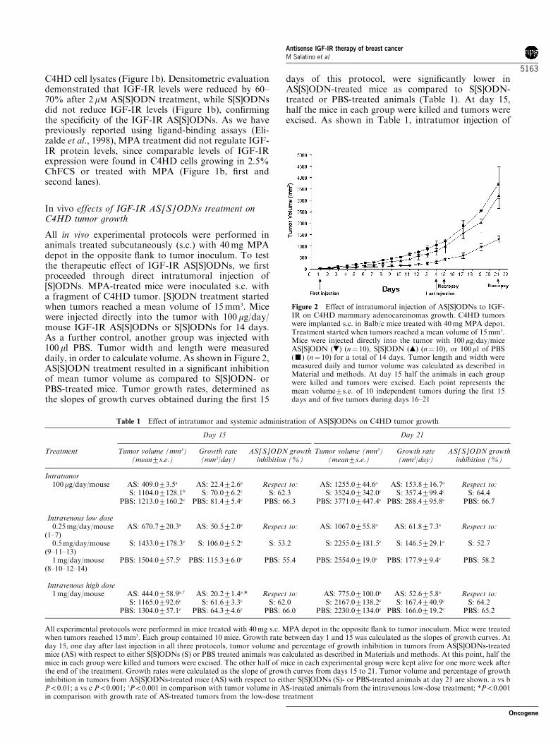
All in vivo experimental protocols were performed inanimals treated subcutaneously (s.c.) with 40mg MPAdepot in the opposite flank to tumor inoculum. To testthe therapeutic effect of IGF-IR AS[S]ODNs, we firstproceeded through direct intratumoral injection of[S]ODNs. MPA-treated mice were inoculated s.c. witha fragment of C4HD tumor. [S]ODN treatment startedwhen tumors reached a mean volume of 15mm3. Micewere injected directly into the tumor with 100 mg/day/mouse IGF-IR AS[S]ODNs or S[S]ODNs for 14 days.As a further control, another group was injected with100 ml PBS. Tumor width and length were measureddaily, in order to calculate volume. As shown in Figure 2,AS[S]ODN treatment resulted in a significant inhibitionof mean tumor volume as compared to S[S]ODN- orPBS-treated mice. Tumor growth rates, determined asthe slopes of growth curves obtained during the first 15
days of this protocol, were significantly lower inAS[S]ODN-treated mice as compared to S[S]ODN-treated or PBS-treated animals (Table 1). At day 15,half the mice in each group were killed and tumors wereexcised. As shown in Table 1, intratumor injection of
Figure 2 Effect of intratumoral injection of AS[S]ODNs to IGF-IR on C4HD mammary adenocarcinomas growth. C4HD tumorswere implanted s.c. in Balb/c mice treated with 40mg MPA depot.Treatment started when tumors reached a mean volume of 15mm3.Mice were injected directly into the tumor with 100mg/day/miceAS[S]ODN (.) (n¼ 10), S[S]ODN (m) (n¼ 10), or 100ml of PBS(’) (n¼ 10) for a total of 14 days. Tumor length and width weremeasured daily and tumor volume was calculated as described inMaterial and methods. At day 15 half the animals in each groupwere killed and tumors were excised. Each point represents themean volume7s.e. of 10 independent tumors during the first 15days and of five tumors during days 16–21
Table 1 Effect of intratumor and systemic administration of AS[S]ODNs on C4HD tumor growth
Day 15 Day 21
Treatment Tumor volume (mm3)(mean7s.e.)
Growth rate(mm3/day)
AS[S]ODN growthinhibition (%)
Tumor volume (mm3)(mean7s.e.)
Growth rate(mm3/day)
AS[S]ODN growthinhibition (%)
Intratumor100mg/day/mouse AS: 409.073.5a AS: 22.472.6a Respect to: AS: 1255.0744.6a AS: 153.8716.7a Respect to:
S: 1104.07128.1b S: 70.076.2c S: 62.3 S: 3524.07342.0c S: 357.4799.4c S: 64.4PBS: 1213.07160.2c PBS: 81.475.4c PBS: 66.3 PBS: 3771.07447.4c PBS: 288.4795.8c PBS: 66.7
Intravenous low dose0.25mg/day/mouse
(1–7)AS: 670.7720.3a AS: 50.572.0a Respect to: AS: 1067.0755.8a AS: 61.877.3a Respect to:
0.5mg/day/mouse(9–11–13)
S: 1433.07178.3c S: 106.075.2c S: 53.2 S: 2255.07181.5c S: 146.5729.1c S: 52.7
1mg/day/mouse(8–10–12–14)
PBS: 1504.0757.5c PBS: 115.376.0c PBS: 55.4 PBS: 2554.0719.0c PBS: 177.979.4c PBS: 58.2
Intravenous high dose1mg/day/mouse AS: 444.0758.9a,w AS: 20.271.4a,* Respect to: AS: 775.07100.0a AS: 52.675.8a Respect to:
S: 1165.0792.6c S: 61.673.3c S: 62.0 S: 2167.07138.2c S: 167.4740.9c S: 64.2PBS: 1304.0757.1c PBS: 64.374.6c PBS: 66.0 PBS: 2230.07134.0c PBS: 166.0719.2c PBS: 65.2
All experimental protocols were performed in mice treated with 40mg s.c. MPA depot in the opposite flank to tumor inoculum. Mice were treatedwhen tumors reached 15mm3. Each group contained 10 mice. Growth rate between day 1 and 15 was calculated as the slopes of growth curves. Atday 15, one day after last injection in all three protocols, tumor volume and percentage of growth inhibition in tumors from AS[S]ODNs-treatedmice (AS) with respect to either S[S]ODNs (S) or PBS treated animals was calculated as described in Materials and methods. At this point, half themice in each group were killed and tumors were excised. The other half of mice in each experimental group were kept alive for one more week afterthe end of the treatment. Growth rates were calculated as the slope of growth curves from days 15 to 21. Tumor volume and percentage of growthinhibition in tumors from AS[S]ODNs-treated mice (AS) with respect to either S[S]ODNs (S)- or PBS-treated animals at day 21 are shown. a vs bPo0.01; a vs c Po0.001; wPo0.001 in comparison with tumor volume in AS-treated animals from the intravenous low-dose treatment; *Po0.001in comparison with growth rate of AS-treated tumors from the low-dose treatment
Antisense IGF-IR therapy of breast cancerM Salatino et al
5163
Oncogene
AS[S]ODNs significantly inhibited C4HD tumorgrowth, determined by comparing mean tumor volumeof AS[S]ODN-treated mice with tumor volume of eitherS[S]ODN- or PBS-treated mice. Tumor growth delay atday 15 was of 5 days with respect to S[S]ODN-treatedmice and of 7 days with respect to PBS-treated animals.The other half of mice in each experimental group waskept alive for one more week after the end of thetreatment. Differences in tumor growth and growthrates between AS[S]ODN-treated mice and S[S]ODN-or PBS-treated control groups were still significant atday 21 (Table 1).Systemic treatment of solid tumors with antisense
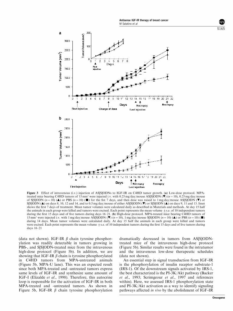
oligonucleotides is currently under investigation inseveral trials (Tamm et al., 2001). Therefore, positiveresults obtained in the intratumoral protocol encour-aged us to assess the effect of intravenous (i.v.) injectionof AS[S]ODNs on C4HD tumor growth. In the firstseries of experiments, mice were injected i.v. via theretroorbital venous sinus, with 0.25mg/day/mouseAS[S]ODNs or S[S]ODNs for 7 days. A third groupwas inoculated with the vehicle (100 ml PBS). From nowon, we will be referring to this experimental approach aslow-dose protocol. As shown in Figure 3a (inset),AS[S]ODN treatment for 7 days induced a detectablebut not significant decrease in tumor growth. Therefore,we decided to raise the dose to 1mg/day/mouse ofAS[S]ODNs or S[S]ODNs on days 8, 10, 12 and 14 andto 0.5mg/day/mouse of oligonucleotides on days 9, 11and 13. This treatment modification resulted in amarked reduction in tumor volume in AS[S]ODN-treated mice, as compared with either S[S]ODN orPBS control groups (Figure 3a). At day 15, half theanimals in each group were killed and tumors wereexcised. Results are summarized in Table 1. AS[S]ODNtreatment induced significant decrease both in tumorgrowth and in growth rates. At day 15, a delay of 6 daysin tumor growth was observed in mice treated withAS[S]ODNs with respect to tumors growing in eitherS[S]ODN- or PBS-treated animals. In an effort toimprove treatment with systemic AS[S]ODNs in the lastexperimental design, which we will be referring to ashigh-dose protocol, we rose [S]ODNs dose to 1mg/day/mouse from day 1 to day 14 of treatment. As seen inFigure 3b, the inhibitory effect of AS[S]ODNs wasevident from the beginning of the treatment. Thissystemic treatment proved to be highly effective toinhibit tumor growth (Table 1). A delay of 6 days intumor growth with respect to S[S]ODN and PBSadministration was observed at day 15 when, asdescribed for the other two therapeutic schedules,tumors were excised from half the mice in each group.As already seen in the intratumoral protocol, in bothintravenous protocols, differences observed in tumorgrowth and in growth rates were evident and significant1 week after the end of the treatment on day 21 (Table 1).No statistically significant differences either in tumorgrowth, in growth rates or in growth delay betweenS[S]ODN and PBS control groups were observed in anyof the experimental schedules (Table 1).
There were no signs of overt toxicity in mice treatedwith AS[S]ODNs either in intratumoral or in intrave-nous experiments and no significant changes in serumlevels of aspartate aminotransferase (AST), alanineaminotransferase (ALT), glucose and creatinin werefound between AS[S]ODN-treated and control groups(data not shown). In addition, we did not observe weightloss in mice receiving AS[S]ODNs in any of the threeexperimental protocols which evidences good toleranceby animals of the three experimental regimens.
Histhopathogical analysis
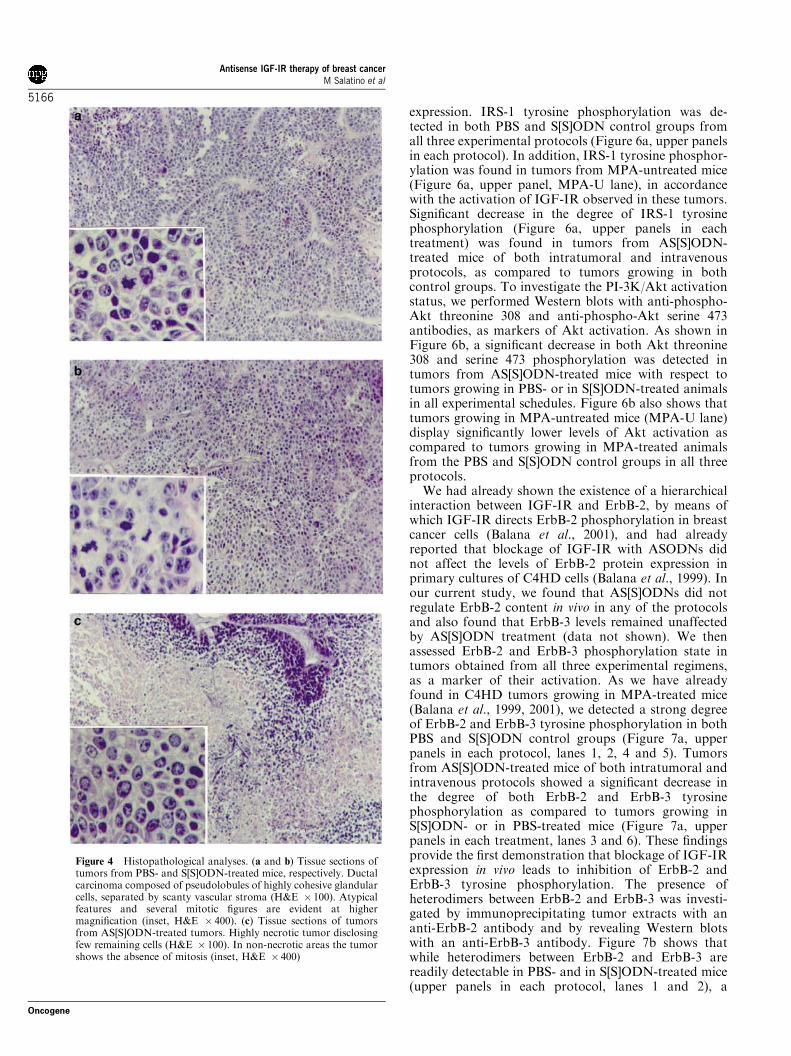
Morphology of tumors from AS[S]ODN-, S[S]ODN- orPBS-treated mice was evaluated by haematoxylin–eosin(H&E) staining of histological sections. Tumors wereexcised at day 15 of each protocol. Figure 4 showsrepresentative sections of tumors from AS[S]ODNs,S[S]ODNs and PBS-treated mice in the high-doseintravenous experimental protocol. A large percentage,about 70–90%, of tumor mass from AS[S]ODN-treatedmice was necrotic as compared to S[S]ODN- or PBS-treated mice, in which only 10–20% necrosis wasobserved. Tumors from mice receiving AS[S]ODNsshowed a significantly lower number of mitosis (0–5mitosis per 10 HPF) as compared to S[S]ODN- or PBS-treated animals, both of which showed over 10 mitosesper 10 HPF. Histopathological images of tumors fromthe intratumoral or low-dose intravenous protocols weresimilar to those observed in the high-dose treatmentgroup (data not shown). In addition, histologicalexamination of liver, lung, heart and pancreas did notreveal any pathological changes (data not shown).
Molecular mechanisms involved in the antitumoral effectof IGF-IR AS[S]ODNs
Since we achieved a significant though incompleteinhibition of C4HD tumor growth by IGF-IRAS[S]ODN administration, we had tumor samples avail-able to explore signaling pathways disrupted in vivoby targeting IGF-IR.To establish first whether the antitumor effect in vivo
was due to a specific antisense effect, IGF-IR expressionin tumors from AS[S]ODN-, S[S]ODN- and PBS-treatedmice was evaluated in protein extracts from tumorsexcised at day 15. As control, we included C4HDtumors growing in MPA-untreated animals (Figure 5a,MPA-U). As shown in Figure 5a, Western blot analysisshowed a marked inhibition of IGF-IRa chain expres-sions in tumors from mice receiving AS[S]ODNs, ascompared with tumors from S[S]ODN- or PBS-treatedanimals, in the intratumor and in both low-dose andhigh-dose i.v. therapeutic schedules. Densitometricanalysis of IGF-IR a chain band in tumor samplesfrom PBS-, S[S]ODN- and AS[S]ODN-treated miceshowed that AS[S]ODN treatment resulted in 65–90%inhibition of IGF-IR a chain expression with respect totumors growing in control groups in all three therapeu-tic protocols. Similar results on AS[S]ODN treatmentinhibition of IGF-IRb chain expression were found
Antisense IGF-IR therapy of breast cancerM Salatino et al
5164
Oncogene
(data not shown). IGF-IR b chain tyrosine phosphor-ylation was readily detectable in tumors growing inPBS-, and S[S]ODN-treated mice from the intravenoushigh-dose protocol (Figure 5b). In addition, we areshowing that IGF-IR b chain is tyrosine phosphorylatedin C4HD tumors from MPA-untreated animals(Figure 5b, MPA-U lane). This was an expected resultsince both MPA-treated and -untreated tumors expresssame levels of IGF-IR and synthesize same amount ofIGF-I (Elizalde et al., 1998). Therefore, this autocrineloop is responsible for the activation of IGF-IR in bothMPA-treated and -untreated tumors. As shown inFigure 5b, IGF-IR b chain tyrosine phosphorylation
dramatically decreased in tumors from AS[S]ODN-treated mice of the intravenous high-dose protocol(Figure 5b). Similar results were found in the intratumorand the intravenous low-dose therapeutic schedules(data not shown).An essential step in signal transduction from IGF-IR
is the phosphorylation of insulin receptor substrate-1(IRS-1). Of the downstream signals activated by IRS-1,the best characterized is the PI-3K/Akt pathway (Backeret al., 1993; Scrimgeour et al., 1997 and referenceswithin). Here, we assessed IRS-1 phosphorylation stateand PI-3K/Akt activation as a way to identify signalingpathways affected in vivo by the abolishment of IGF-IR
Figure 3 Effect of intravenous (i.v.) injection of AS[S]ODNs to IGF-IR on C4HD tumor growth. (a) Low-dose protocol. MPA-treated mice bearing C4HD tumors of 15mm3 were injected i.v. with 0.25mg/day/mouse AS[S]ODN (.) (n¼ 10), 0.25mg/day/mouseof S[S]ODN (n¼ 10) (m) or PBS (n¼ 10) (’) for the fist 7 days, and then dose was raised to 1mg/day/mouse AS[S]ODN (.) orS[S]ODN (m) on days 8, 10, 12 and 14, and to 0.5mg/day/mouse of either AS[S]ODN (.) or S[S]ODN (m) on days 9, 11 and 13. Insetshows the first 7 days of treatment. Mean tumor volumes were calculated daily as described in Materials and methods. At day 15 halfthe animals in each group were killed and tumors were excised. Each point represents the mean volume7s.e. of 10 independent tumorsduring the first 15 days and of five tumors during days 16–21. (b) High-dose protocol. MPA-treated mice bearing C4HD tumors of15mm3 were injected i.v. with 1mg/day/mouse AS[S]ODN (.) (n¼ 10), 1mg/day/mouse S[S]ODN (n¼ 10) (m) or PBS (n¼ 10) (’)during 14 days. Mean tumor volumes were calculated daily. At day 15 half the animals in each group were killed and tumorswere excised. Each point represents the mean volume7s.e. of 10 independent tumors during the first 15 days and of five tumors duringdays 16–21
Antisense IGF-IR therapy of breast cancerM Salatino et al
5165
Oncogene
expression. IRS-1 tyrosine phosphorylation was de-tected in both PBS and S[S]ODN control groups fromall three experimental protocols (Figure 6a, upper panelsin each protocol). In addition, IRS-1 tyrosine phosphor-ylation was found in tumors from MPA-untreated mice(Figure 6a, upper panel, MPA-U lane), in accordancewith the activation of IGF-IR observed in these tumors.Significant decrease in the degree of IRS-1 tyrosinephosphorylation (Figure 6a, upper panels in eachtreatment) was found in tumors from AS[S]ODN-treated mice of both intratumoral and intravenousprotocols, as compared to tumors growing in bothcontrol groups. To investigate the PI-3K/Akt activationstatus, we performed Western blots with anti-phospho-Akt threonine 308 and anti-phospho-Akt serine 473antibodies, as markers of Akt activation. As shown inFigure 6b, a significant decrease in both Akt threonine308 and serine 473 phosphorylation was detected intumors from AS[S]ODN-treated mice with respect totumors growing in PBS- or in S[S]ODN-treated animalsin all experimental schedules. Figure 6b also shows thattumors growing in MPA-untreated mice (MPA-U lane)display significantly lower levels of Akt activation ascompared to tumors growing in MPA-treated animalsfrom the PBS and S[S]ODN control groups in all threeprotocols.We had already shown the existence of a hierarchical
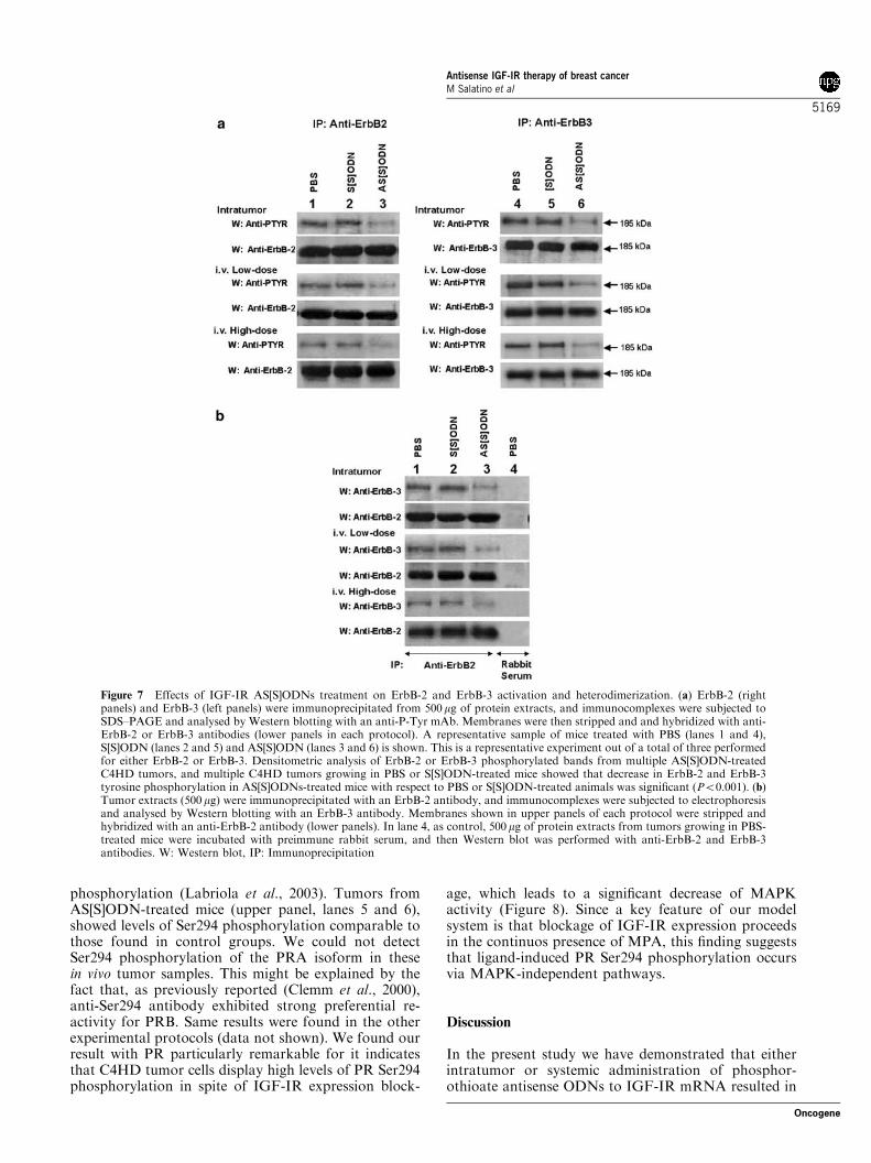
interaction between IGF-IR and ErbB-2, by means ofwhich IGF-IR directs ErbB-2 phosphorylation in breastcancer cells (Balana et al., 2001), and had alreadyreported that blockage of IGF-IR with ASODNs didnot affect the levels of ErbB-2 protein expression inprimary cultures of C4HD cells (Balana et al., 1999). Inour current study, we found that AS[S]ODNs did notregulate ErbB-2 content in vivo in any of the protocolsand also found that ErbB-3 levels remained unaffectedby AS[S]ODN treatment (data not shown). We thenassessed ErbB-2 and ErbB-3 phosphorylation state intumors obtained from all three experimental regimens,as a marker of their activation. As we have alreadyfound in C4HD tumors growing in MPA-treated mice(Balana et al., 1999, 2001), we detected a strong degreeof ErbB-2 and ErbB-3 tyrosine phosphorylation in bothPBS and S[S]ODN control groups (Figure 7a, upperpanels in each protocol, lanes 1, 2, 4 and 5). Tumorsfrom AS[S]ODN-treated mice of both intratumoral andintravenous protocols showed a significant decrease inthe degree of both ErbB-2 and ErbB-3 tyrosinephosphorylation as compared to tumors growing inS[S]ODN- or in PBS-treated mice (Figure 7a, upperpanels in each treatment, lanes 3 and 6). These findingsprovide the first demonstration that blockage of IGF-IRexpression in vivo leads to inhibition of ErbB-2 andErbB-3 tyrosine phosphorylation. The presence ofheterodimers between ErbB-2 and ErbB-3 was investi-gated by immunoprecipitating tumor extracts with ananti-ErbB-2 antibody and by revealing Western blotswith an anti-ErbB-3 antibody. Figure 7b shows thatwhile heterodimers between ErbB-2 and ErbB-3 arereadily detectable in PBS- and in S[S]ODN-treated mice(upper panels in each protocol, lanes 1 and 2), a
Figure 4 Histopathological analyses. (a and b) Tissue sections oftumors from PBS- and S[S]ODN-treated mice, respectively. Ductalcarcinoma composed of pseudolobules of highly cohesive glandularcells, separated by scanty vascular stroma (H&E � 100). Atypicalfeatures and several mitotic figures are evident at highermagnification (inset, H&E � 400). (c) Tissue sections of tumorsfrom AS[S]ODN-treated tumors. Highly necrotic tumor disclosingfew remaining cells (H&E � 100). In non-necrotic areas the tumorshows the absence of mitosis (inset, H&E � 400)
Antisense IGF-IR therapy of breast cancerM Salatino et al
5166
Oncogene

significant decrease in the abundance of these complexesis observed in tumors growing in AS[S]ODN-treatedmice (upper panels in each treatment, lane 3). Similarresults were found when tumor extracts were immuno-precipitated with an anti-ErbB-3 antibody, and Westernblotting was performed with an anti-ErbB-2 antibody(data not shown).We were then for several reasons interested in
assessing the effect of IGF-IR AS[S]ODNs on mito-gen-activated protein kinases (MAPK) activity. First, itis well acknowledged that IGF-IR activates the MAPK/Erk signaling pathway, mainly through Shc proteins,another IGF-IR major substrate (Scrimgeour et al.,1997 and references within, Sasaoka et al., 1994).Second, the key proliferative stimulus in our tumormodel is driven by MPA acting through the PR.Multiple lines of evidence, including our own work(Labriola et al., 2003), suggest that MAPK-induced
phosphorylation plays a role in PR function (Langeet al., 2000; Shen et al., 2001; Qiu et al., 2003). Finally,breast cancer often shows elevated MAPK activity thathas been associated to loss of estrogen response anddevelopment of metastasis (Adeyinka et al., 2002).Figure 8 shows results of the intravenous high-doseprotocol. All tumors expressed p42/p44 MAPK (lowerpanel). MAPKs activation was readily detectable intumors from control groups by performing Western blotusing an antibody specific for the dually phosphory-lated, active form of this kinase (Figure 8, upper panel).Tumors from AS[S]ODN-treated mice showed a sig-nificant decrease in the degree of MAPK activation ascompared to tumors growing in control groups(Figure 8, upper panel). Similar results were found inthe other experimental regimens (data not shown).Figure 8 also shows that tumors growing in MPA-untreated mice (MPA-U lane) display significantly lower
Figure 5 Effect of IGF-IR AS[S]ODNs treatment on IGF-IR expression. (a) A total of 100mg protein from tumor lysates waselectrophoresed and immunoblotted with an anti-IGF-IR a chain antibody. Two representative samples of mice treated with PBS(lanes 1 and 2), S[S]ODN (lanes 3 and 4) and AS[S]ODN (lanes 5 and 6) are shown. MPA-U, a representative C4HD tumor growing inMPA-untreated mouse. Western blots using an antiactin antibody were carried out using identical protein lysates as control for thespecificity of the IGF-IR AS[S]ODNs (lower panels in each protocol). Densitometric analysis of IGF-IRa band from multipleAS[S]ODN-treated C4HD tumors, expressed as a percentage of the control values (i.e. C4HD tumors growing in PBS or S[S]ODN-treated mice) varied between 10 and 35% for tumors growing in AS[S]ODNs-treated animals. Inhibition of IGF-IRa expression inAS[S]ODNs-treated mice with respect to PBS or S[S]ODN-treated animals was significant (Po0.001). (b) IGF-IR b chain wasimmunoprecipitated from 500mg of protein extracts and immunocomplexes were subjected to SDS–PAGE and analysed by Westernblotting with an anti-P-Tyr mAb. Lower panel: Identical aliquots of each immunoprecipitate were subjected to immunoblot analysiswith anti-IGF-IR b chain antibody to verify that nearly equal amount of immunoprecipitated proteins were loaded. Tworepresentative samples of mice treated with PBS (lanes 1 and 2), S[S]ODN (lanes 3 and 4) and AS[S]ODN (lanes 5 and 6) are shown.MPA-U, a sample from C4HD tumor growing in MPA-untreated mouse. This is a representative experiment out of a total of three.Densitometric analysis of IGF-IR b chain tyrosine phosphorylation from multiple AS[S]ODN-treated C4HD tumors, and frommultiple C4HD tumors growing in PBS or S[S]ODN-treated mice showed that decrease in IGF-IR b tyrosine phosphorylation inAS[S]ODNs-treated mice with respect to PBS or S[S]ODN-treated animals was significant (Po0.001). W: Western blot, IP:Immunoprecipitation
Antisense IGF-IR therapy of breast cancerM Salatino et al
5167
Oncogene

levels of MAPK activation as compared to tumorsgrowing in MPA-treated animals from the PBS (lanes 1and 2) and S[S]ODN control groups (lanes 3 and 4).This is in accordance with our previous resultsdemonstrating that MPA induces MAPK activationin vitro (Labriola et al., 2003).Recent evidence has clearly shown the presence of bi-
directional cross-talks between steroid hormones andgrowth factors (GFs) signaling pathways (Ignar-Trow-bridge et al., 1992; Pietras et al., 1995; Lange et al., 1998;Migliaccio et al., 1998; Balana et al., 1999, 2001;Boonyaratanakornkit et al., 2001; Shen et al., 2001;Labriola et al., 2003). Accumulating evidence hasdemonstrated interactions between ER and IGF-I/IGF-IR signaling pathways (Stewart et al., 1990; Leeet al., 1999; Oesterreich et al., 2001). However, lessinformation is available about cross-talks between IGF-IR and PR (Katzenellenbogen and Norman, 1990; Choet al., 1994; Cui et al., 2003). Therefore, we nextinvestigated whether blockage of IGF-IR expression
might affect PR expression and/or activity. First, weevaluated PR expression levels. The results obtained inthe intravenous high-dose protocol are shownin Figure 9. Similar PR protein levels were detected intumors from mice treated with AS[S]ODNs (lowerpanel, lanes 5 and 6) as compared to those found intumors from control groups (lower panel, lanes 1–4).Same results were found in the other therapeuticschedules (data not shown). Recent findings by Langeand co-workers (Lange et al., 2000; Shen et al., 2001)demonstrated that phosphorylation of PR on Ser294directly correlates with PR transcriptional activity.Therefore, we herein assessed the state of PR Ser294phosphorylation in an attempt to evaluate whetherabrogation of IGF-I/IGF-IR signaling could interferewith the activation of PR. As shown in Figure 9, PRphosphorylation on Ser294 was detected in PBS andS[S]ODN control groups (upper panel, lanes 1–4), inaccordance with our previous in vitro findings, demon-strating that MPA was able to induce PR Ser294
Figure 6 Effects of IGF-IR AS[S]ODNs treatment on IRS-1 and Akt phosphorylation. (a) Upper panel: IRS-1 wasimmunoprecipitated from 500mg of protein extracts and immunocomplexes were subjected to SDS–PAGE and analysed by Westernblotting with an anti-P-Tyr mAb. Lower panel: Identical aliquots of each immunoprecipitate were subjected to immunoblot analysiswith anti-IRS-1 antibody to verify that nearly equal amount of immunoprecipitated proteins were loaded. A representative sample oftumors growing in mice treated with PBS, S[S]ODN and AS[S]ODN is shown. MPA-U, a sample from C4HD tumor growing in MPA-untreated mouse. This is a representative experiment out of a total of three. Densitometric analysis of IRS-1 phosphorylated bandsfrom multiple AS[S]ODN-treated C4HD tumors, and from multiple C4HD tumors growing in PBS or S[S]ODN-treated mice showedthat decrease in IRS-1 tyrosine phosphorylation in AS[S]ODNs-treated mice with respect to PBS or S[S]ODN-treated animals wassignificant (Po0.001).W: Western blot, IP: Immunoprecipitation. (b) A total of 100mg protein from tumor lysates was electrophoresedand immunoblotted either with an anti-phospho-Akt threonine 308 (first panel) or with serine 473 antibody (third panel). Membraneswere then stripped and hybridized with an antibody anti-total Akt (second and fourth panels). A representative sample of tumorsgrowing in mice treated with PBS, S[S]ODN and AS[S]ODN in each protocol is shown. MPA-U, a sample from C4HD tumor growingin MPA-untreated mouse. This experiment was repeated three times with similar results. Densitometric analysis of phospho-threonine308 or -serine 473 Akt bands from multiple AS[S]ODN-treated C4HD tumors, and from multiple C4HD tumors growing in PBS orS[S]ODN-treated mice showed that decrease in Akt tyrosine phosphorylation in AS[S]ODNs-treated mice with respect to PBS orS[S]ODN-treated animals was significant (Po0.001)
Antisense IGF-IR therapy of breast cancerM Salatino et al
5168
Oncogene
phosphorylation (Labriola et al., 2003). Tumors fromAS[S]ODN-treated mice (upper panel, lanes 5 and 6),showed levels of Ser294 phosphorylation comparable tothose found in control groups. We could not detectSer294 phosphorylation of the PRA isoform in thesein vivo tumor samples. This might be explained by thefact that, as previously reported (Clemm et al., 2000),anti-Ser294 antibody exhibited strong preferential re-activity for PRB. Same results were found in the otherexperimental protocols (data not shown). We found ourresult with PR particularly remarkable for it indicatesthat C4HD tumor cells display high levels of PR Ser294phosphorylation in spite of IGF-IR expression block-
age, which leads to a significant decrease of MAPKactivity (Figure 8). Since a key feature of our modelsystem is that blockage of IGF-IR expression proceedsin the continuos presence of MPA, this finding suggeststhat ligand-induced PR Ser294 phosphorylation occursvia MAPK-independent pathways.
Discussion
In the present study we have demonstrated that eitherintratumor or systemic administration of phosphor-othioate antisense ODNs to IGF-IR mRNA resulted in
Figure 7 Effects of IGF-IR AS[S]ODNs treatment on ErbB-2 and ErbB-3 activation and heterodimerization. (a) ErbB-2 (rightpanels) and ErbB-3 (left panels) were immunoprecipitated from 500mg of protein extracts, and immunocomplexes were subjected toSDS–PAGE and analysed by Western blotting with an anti-P-Tyr mAb. Membranes were then stripped and and hybridized with anti-ErbB-2 or ErbB-3 antibodies (lower panels in each protocol). A representative sample of mice treated with PBS (lanes 1 and 4),S[S]ODN (lanes 2 and 5) and AS[S]ODN (lanes 3 and 6) is shown. This is a representative experiment out of a total of three performedfor either ErbB-2 or ErbB-3. Densitometric analysis of ErbB-2 or ErbB-3 phosphorylated bands from multiple AS[S]ODN-treatedC4HD tumors, and multiple C4HD tumors growing in PBS or S[S]ODN-treated mice showed that decrease in ErbB-2 and ErbB-3tyrosine phosphorylation in AS[S]ODNs-treated mice with respect to PBS or S[S]ODN-treated animals was significant (Po0.001). (b)Tumor extracts (500 mg) were immunoprecipitated with an ErbB-2 antibody, and immunocomplexes were subjected to electrophoresisand analysed by Western blotting with an ErbB-3 antibody. Membranes shown in upper panels of each protocol were stripped andhybridized with an anti-ErbB-2 antibody (lower panels). In lane 4, as control, 500 mg of protein extracts from tumors growing in PBS-treated mice were incubated with preimmune rabbit serum, and then Western blot was performed with anti-ErbB-2 and ErbB-3antibodies. W: Western blot, IP: Immunoprecipitation
Antisense IGF-IR therapy of breast cancerM Salatino et al
5169
Oncogene

a significant growth inhibition of the C4HD mousemammary tumor line, our experimental model ofmammary carcinogenesis in which progestins are themajor proliferative stimulus. We had previously foundthat progestins-induced proliferation of C4HD tumorsis driven by a complex bi-directional interaction amongactivated PR and type I and II RTKs (Balana et al.,1999, 2001; Labriola et al., 2003). Here, we dissected anetwork of interactions among IGF-IR, type I RTKsand PR signaling pathways that were differentiallyaffected by blockage of IGF-IR function Therefore, ourmodel system has provided a unique tool to explore themolecular bases of IGF-IR contribution to breast cancergrowth.A series of experimental strategies have been
employed to block either IGF-IR function or expres-sion, both in vitro and in vivo, in several tumor celllines derived from a number of species. These includedthe use of neutralizing antibodies (Rohlik et al., 1987;Arteaga and Osborne, 1989), dominant-negative recep-tors (Prager et al., 1994; Dunn et al., 1998; Li et al.,2000; Lee et al., 2003), antisense and triple-helix-formingoligonucleotides (Resnicoff et al., 1994a, b) and anti-sense expression vectors delivered by transfection oradenovirus (Resnicoff et al., 1994a, b; Burfeind et al.,1996; Lee et al., 1996; Nakamura et al., 2000). Startlingreports from Baserga and co-workers (Resnicoffet al., 1994a, b) demonstrated that either transfectionof cells with plasmids that express antisense RNA toIGF-IR or addition of ASODNs to IGF-IR mRNAwere efficient at inhibiting in vitro growth of ratglioblastoma C6 cells. In addition, C6 cells stablytransfected with a plasmid expressing antisense IGF-
IR RNA were nontumorigenic when injected s.c. intosyngeneic immunocompetent rats (Resnicoff et al.,1994a, b). Similarly, growth of human melanoma FO-1cells in nude mice was strongly inhibited when cellsstably expressed antisense IGF-IR RNA (Resnicoffet al., 1994a). Inhibition of tumorigenesis was alsofound when FO-1 cells were treated with ASODNs toIGF-IR mRNA prior to injection into nude mice(Resnicoff et al., 1994a).The effect of blockage of IGF-IR function by
antisense techniques in breast cancer cell has beenassessed in several reports. Thus, stable expression oftype I IGF-IR antisense RNA by using a plasmid vectorresulted in a significant decrease in IGF-I and serum-stimulated growth in MCF-7 human breast cancer cells(Neuenschwander et al., 1995). In addition, recentfindings demonstrated that transfection of the highlymetastatic, ER negative, MDA-MB-435s breast cancercell line with a construct carrying antisense IGF-IRRNA resulted in a significant reduction of cell pro-liferation and loss of soft agar growth (Chernicky et al.,2000). Moreover, there was suppression of tumorigen-esis, reduction in the metastatic potential and prolongedsurvival after injection of MDA-MB-435 s cells expres-sing antisense IGF-IR into the mammary fat pads ofimmune compromised mice (Chernicky et al., 2000).Recently, it was found that the murine mammarycarcinoma cell line EMT6 transfected with an antisenseIGF-IR plasmid, displayed a significant decrease inproliferation (Chernicky et al., 2002). Tumor sizedecreased when cells carrying the antisense IGF-IRwere injected into syngeneic mice (Chernicky et al.,2002).
Figure 8 Effects of IGF-IR AS[S]ODNs treatment on MAPK activation. In total, 100 mg protein from tumor lysates waselectrophoresed on 12% SDS gels and immunoblotted with an antiphospho p42/p44 MAPK antibody. The membrane was thenstripped and hybridized with an anti-total p42/p44 MAPK antibody (lower panel). Two representative samples from mice treated withPBS (lanes 1 and 2), S[S]ODN (lanes 3 and 4) and AS[S]ODN (lanes 5 and 6) are shown. MPA-U, a sample from C4HD tumor growingin MPA-untreated mouse. This is a representative experiment out of a total of three. Densitometric analysis of phospho-MAPKshowed that decrease in MAPK tyrosine phosphorylation in AS[S]ODNs-treated mice with respect to PBS or S[S]ODN-treatedanimals was significant (Po0.001)
Figure 9 Effects of IGF-IR AS[S]ODNs treatment on PR expression and PR Ser294 phosphorylation. A total of 100 mg protein fromtumor lysates was immunoblotted with an anti-phospho Ser 294hPR antibody (upper panel). The membrane was stripped andhybridized with an anti-hPR antibody (lower panel). Two representative samples of mice treated with PBS (lanes 1 and 2), S[S]ODN(lanes 3 and 4) and AS[S]ODN (lanes 5 and 6) are shown. This is a representative experiment out of a total of three
Antisense IGF-IR therapy of breast cancerM Salatino et al
5170
Oncogene

It is worth pointing out that in the aforementionedstudies, antisense RNA was produced intracellularly byan expression vector, in contrast to our present study inwhich we either injected ASODNs directly into tumorsgrowing in mice or delivered the oligodeoxynucleotidesby systemic administration through the retroorbitalvenous plexus. Another important difference betweenthese studies and our present work is that most previousreports have relied on modification of cells ex vivobefore inoculation to mice. Direct transmission of DNAinto established tumors has proved to be feasible andsafe at reducing tumor growth in experimental proce-dures in mice (Plautz et al., 1993) and in human clinicalstudies (Nabel et al., 1993). Though to our knowledgedirect injection of ASODNs to IGF-IR in mousemammary tumors has not been previously performed,direct injection of antisense IGF-IR plasmids intoestablished N2A tumors growing in syngeneic A/J miceresulted in marked inhibition of tumor growth (Liu et al.,1998). As we have reported here, this effect persistedafter the period of active DNA administration.We have herein demonstrated that the antitumor
effect of IGF-IR AS[S]ODNs was due to a specificantisense effect. First, inhibition of tumor growth byAS[S]ODNs was dose-dependent, and no abrogation oftumor proliferation was observed in any of the controlgroups. Thus, we found a significantly higher tumorgrowth inhibition with the high dose i.v. AS[S]ODNprotocol, as compared to the low-dose treatment.Second, IGF-IR expression was inhibited in tumorsfrom mice receiving AS[S]ODNs as compared to tumorsfrom control groups. Our findings have provided thefirst evidence that two major signal transduction path-ways known to be activated by IGF-IR, the PI-3K/Aktand the classical p42/p44 MAPK pathways (Scrimgeouret al., 1997 and references within, Sasaoka et al., 1994),were abolished by in vivo treatment of mammary tumorswith IGF-IR AS[S]ODNs. The PI-3K/Akt pathwayoriginates from the interaction of IGF-IR with one of itsmajor substrates, IRS-1 (Backer et al., 1993), whosetyrosine phosphorylation we found significantly lower inC4HD tumors from AS[S]ODN-treated mice as com-pared to tumors growing in control groups. On the otherhand, IGF-IR activates the MAPK/Erk signaling path-way, mainly through the Shc proteins, another IGF-IRkey substrate (Sasaoka et al., 1994; Scrimgeour et al.,1997). Our results are in accordance with recent findings(Peruzzi et al., 1999; Navarro and Baserga, 2001)showing that IGF-IR has at least three survival signalsthat are able to protect 32D murine hemopoietic cellsfrom apoptosis, the PI-3K/Akt, the MAPK/Erk signal-ing pathways and a third one that results in themitocondrial translocation of Raf 1, refered to as the14.3.3 pathway. Simultaneous inactivation of two ofthese pathways is required to inhibit IGF-IR capacity toprotect cells from apoptotic injuries. Therefore, we canhypothezise that blockage of two survival pathwaysachieved by in vivo administration of IGF-IR AS[-S]ODNs is directly involved in the successful inhibitionof C4HD breast tumor growth. Histological evaluationof tumors from AS[S]ODN-treated mice revealed a great
percentage of necrosis. We were not able to define thepresence of apoptosis induced by AS[S]ODN treatmentusing the TdT-mediated dUTP-biotin nick end labeling(TUNEL) method (data not shown). To some extentthis was an unexpected result since in many strategiesinvolving downregulation or functional impairment ofthe IGF-IR, massive apoptosis of tumor cells was found(Resnicoff et al., 1995a, b). Several explanations may beattempted. First, the rapidity of the death process makesit difficult to visualize and to quantify apoptosis. Oursamples were tumors that developed in mice treated for14 days with AS[S]ODNs. Withesell and co-workers(Liu et al., 1998) have reported results in the N2A mouseneuroblastoma cells that can provide an explanation toour findings. Transfection of N2A cells with IGF-IRantisense plasmids resulted in increase in apoptotic cellsdetectable at 36 h, followed by marked accumulation ofnecrotic cells at 48 h (Liu et al., 1998). These results areconsistent with a mechanism of cell death in which IGF-IR AS[S]ODNs induced apoptotic cell death followedby necrotic degradation of the apoptotic cells. There-fore, this latter phenomenon might apply to what weobserve in our tumor samples. Nevertheless, thepossibility that the prolonged and sustained abrogationof IGF-IR expression and signaling through PI-3K/Aktand p42/p44 MAPK, and the simultaneous blockage ofErbBs activation might both have led directly to celldeath by necrosis.One of the most exciting findings of our present work
is that we were able to demonstrate in vivo, what we hadalready found in vitro: the hierarchical interactionbetween IGF-IR and ErbB-2, by means of which IGF-IR directs ErbB-2 activation (Balana et al., 2001). Infact, we extended our results to show that IGF-IRAS[S]ODN-treatment also resulted in significant de-crease in the degree of ErbB-3 activation and in theamount of ErbB-2/ErbB-3 heterodimers. Our results arein line with a series of findings that have shown theinvolvement of IGF-IR in ErbBs signaling pathways(Coppola et al., 1994; Ram et al., 1996; Swantek andBaserga, 1999). Our findings indicating that in vivo IGF-IR blockage in breast cancer cells results in theinactivation of ErbBs has significant therapeuticalimplications since it indicates that by targeting IGF-IRin breast cancer one might also achieve inhibition ofErbBs activity even in cells overexpressing ErbB-2, suchas C4HD. The importance of the functional interactionbetween IGF-IR and ErbBs in the design of molecularstrategies to block tumor growth has also been high-lighted by recent findings showing that IGF-IR is able tomediate resistance to anti-EGF-R therapy in humanglioblastoma cells through activation of PI-3K/Aktpathway (Chakravarti et al., 2002).We achieved a significant but not complete inhibition
of C4HD tumor growth by IGF-IR AS[S]ODN admin-istration, indicating that certain signaling pathwaysinvolved in C4HD cell proliferation remained unaf-fected by blockage of IGF-IR. Since progestins are themain proliferative stimulus in C4HD cells, the firsttarget we explored was the PR. As measure of PRactivation, we studied PR Ser294 phosphorylation state,
Antisense IGF-IR therapy of breast cancerM Salatino et al
5171
Oncogene
since a series of recent findings showed that phosphor-ylation of PR on Ser294 directly correlated with PRtranscriptional activity (Lange et al., 2000; Shen et al.,2001). PR phosphorylation on Ser294 was detected inC4HD tumors from control groups, extending ourprevious in vitro findings which demonstrated thatMPA was able to induce PR Ser294 phosphorylation(Labriola et al., 2003). Tumors from AS[S]ODN-treatedmice of both intratumoral and intravenous protocolsshowed levels of Ser294 phosphorylation comparable tothose found in control groups. Several interestingconclusions on interaction between PR and IGF-IRsignaling, and on the role of MAPK on PR Ser294phosphorylation can be drawn from this result. First,in vivo abrogation of IGF-IR expression in thecontinuous presence of progestins has no effect on PRactivity, thereby indicating that the slow proliferation ofC4HD tumors after AS[S]ODN administration proceedsthrough a pathway driven by PR, which is IGF-IR andErbBs-independent. Second, since abrogation of IGF-IR expression resulted in inhibition of MAPKs activity(Figure 8), our finding provides further support to themost recent findings by Lange and co-workers suggest-ing that ligand-induced PR Ser294 phosphorylationoccurs via MAPK-independent pathways (Qiu et al.,2003). In line with these observations, we have recentlyfound that MAPK activity is not required for ligand-induced PR activity (Labriola et al., 2003).In summary, we have for the first time demonstrated
that breast cancer growth can be inhibited by eitherdirect intratumor injection or systemic administration ofAS[S]ODNs to IGF-IR mRNA. The fact that significantthough incomplete inhibition of breast tumor growthwas achieved by targeting IGF-IR adds further supportto increasing evidence, indicating that more than onetargeted therapy is required to achieve full blockage ofthe multiple signaling pathways used by breast tumorsto proliferate. Our finding that PR function remainsunaffected by targeting IGF-IR, highlights the impor-tance that a better understanding of the network ofpathways involved in breast cancer growth will necessa-rily have to be developed in the design of therapeuticstrategies.
Materials and methods
Animals and tumors
Experiments were carried out in virgin female Balb/c miceraised at the Institute of Biology and Experimental Medicine(IBYME) of Buenos Aires. All animal studies were conductedin accordance with the highest standards of animal care asoutlined in the NIH guide for the Care and Use of LaboratoryAnimals and were approved by the IBYME Animal ResearchCommittee. Hormone-dependent ductal tumor line C4HDoriginated in mice treated with 40mg MPA every 3 months for1 year, and has been maintained by serial transplantation inanimals treated with 40mg s.c. MPA depot in the oppositeflank to tumor inoculum (Elizalde et al., 1998; Balana et al.,1999, 2001). C4HD tumor line is of ductal origin and expressesPR and ER (Elizalde et al., 1998; Balana et al., 1999, 2001).
ODNs and [S]ODNs
Type I IGF-IR antisense oligodeoxynucleotide (50 TCC TCCGGA GCC AGA CTT), either phosphodiester (ASODN) orphosphorothioate (AS[S]ODN), comprises a sequence com-plementary to codons �29 to �24 of the signal sequence ofhuman IGF-IR precursor (Resnicoff et al., 1994a). Sensephosphodiester (SODN) or phosphorothioate (S[S]ODN)oligodeoxynucleotides (50 AAG TCT GGC TCC GGAGGA) were used as controls. ODN and [S]ODN werepurchased from Biognostik (Goettingen, Germany).
Cell cultures and proliferation assays
Primary cultures of epithelial cells from C4HD tumors,growing in MPA-treated mice, were performed as previouslydescribed (Elizalde et al., 1998; Balana et al., 1999, 2001).Epithelial cells were plated in culture flasks with DMEM/F12þ 5% steroid-stripped fetal calf serum (ChFCS, Gen S.A.,Buenos Aires), and allowed to attach for 24–48 h. Purity ofepithelial cultures was evaluated by cytokeratin staining. Cellswere incubated in DMEM/F12 (without phenol red, with100U/ml penicillin and with 100 m/ml streptomycin), with2.5% ChFCS in the presence of MPA 10nM and the indicatedconcentrations of either phosphodiester or phosphorothioateODNs. After a 24 h incubation, 50% of media was replaced byfresh media, and cells were incubated for another 24 h in thepresence of 0.8 mCi (3H)-thymidine (NEN, Dupont, Boston,MA, USA; specific activity: 70–90Ci/mmol). Cells were thentrypsinized and harvested. Assays were performed in octupli-cate. The differences between control and experimental groupswere analysed by ANOVA followed by Tukey t-test betweengroups. In former experiments we had demonstrated thatthymidine uptake correlates with the number of cells/well(Elizalde et al., 1998; Balana et al., 1999, 2001).
Intratumor and systemic administration of [S]ODNs
Mice were inoculated s.c. into the left flank with a fragment ofC4HD tumor (1mm3) and with the MPA pellet into the otherflank. Tumor growth was measured daily with a Vernier caliper.Tumor volume (mm3) was calculated as (L�W2)/2, where L isthe length (mm) and W the width (mm). [S]ODNs treatmentwas initiated when the tumor reached a mean of volume of15mm3 (approximately 1 week after inoculum). In theintratumor protocol, mice were injected directly into the tumorwith 100mg/day/mouse IGF-IR AS[S]ODNs, S[S]ODNs (dis-solved in 100ml of PBS) or PBS for 14 days. To test the effect ofsystemic administration of AS[S]ODNs, in the first protocol(low-dose), mice were injected i.v. via the retroorbital venoussinus with 0.25mg/day/mouse AS[S]ODN, [S]ODN or 100mlPBS for 7 days. Then, dose was raised to 1mg/day/mouse ofAS[S]ODN or S[S]ODN on days 8, 10, 12 and 14 and to 0.5mg/day/mouse of oligonucleotides on days 9, 11 and 13. In thesecond systemic protocol (high-dose), mice were injected with1mg/day/mouse AS[S]ODN, [S]ODN or 100ml PBS from day 1to day 14 of treatment. Tumor width and length were measureddaily, in order to calculate volume. Tumor growth rates weredetermined as the slopes of growth curves. The percentage oftumor growth inhibition was calculated by dividing the meantumor volume of the AS[S]ODN-treated group by the meantumor volume of control groups, subtracting the resulting valuefrom 1, and multiplying it by 100. Tumor growth delay wasevaluated as T�C, where T and C are the median times fortreated and control tumors respectively, to reach the samevolume. At day 15, half the animals from each group in all threeexperimental protocols were killed and tumors were removed.Tissues to be used for molecular studies were stored at �801C
Antisense IGF-IR therapy of breast cancerM Salatino et al
5172
Oncogene

and tissues for histophatological analysis were fixed in 10%buffered formalin. The other half of mice in each experimentalgroup was kept alive for one more week after the end of thetreatment. Samples of liver, lung, heart and pancreas were alsofixed for histological examination. Comparison of tumorvolumes between the different groups during specific timeswas made by ANOVA followed by Tukey t-test betweengroups. Linear regression analysis was performed on tumorgrowth curves, and the slopes were compared using ANOVAfollowed by parallelism test to evaluate the statistical signifi-cance of the differences.
Histophatological analysis
Tumors were excised and fixed in 10% buffered formalin.Representative fragments were embedded in parafin, 5 mmsections were obtained and stained with H&E for microscopicobservations.
Serum levels of glucose, creatinine, ALT and AST
The levels of glucose, creatinine, alanine aminotrasferase(ALT) and AST were measured using and Auto-BiochemicalAnalyzer (Autolab, Roche, Basel Switzerland) in serumsamples collected at day 15 in each experimental protocol.Blood samples were taken by retroorbital bleeding.
IGF-IR, IRS-1, AKT, ErbB-2, ErbB-3, MAPK, and PRexpression and activation
Protein lysates from C4HD tumors growing in mice subjectedto different treatments were prepared as previously described(Elizalde et al., 1998; Balana et al., 1999, 2001). Proteins weresolubilized in sample buffer (60mM Tris-HCl, pH 6.8, 2%SDS, 10% glycerol and 0.01% bromophenol blue) andsubjected to SDS–PAGE. Proteins were electroblotted ontonitrocellulose. Membranes were immunoblotted with the
following antibodies: IGF-IRa (sc-712), ErbB-2 (Neu C-18),ErbB-3 (C-17), anti-total p42/p44 MAPK (C-14), anti-phospho p42/p44 MAPK (E-4) and actin (C-2), all fromSanta Cruz Biotechnology (Santa Cruz, CA, USA), IGF-Ib(Ab-5), phospho-294-PR Ab-12 (clone 608) and hPR Ab-7(clone 7) from Neomarkers (Freemont, CA, USA), total Akt,phospho Akt (Thr308) and phospho Akt (Ser 473) from CellSignaling (Beverly, MA, USA). After washing, membraneswere incubated with HRP-conjugated secondary antibody(Amersham International, UK). Enhanced chemiluminescence(ECL) was performed according to the manufacturer’sinstructions (Amersham). To perform IRS-1, ErbB-2 andErbB-3 tyrosine phosphorylation analysis, cell lysates (500 mgprotein) were precleared with Protein A-Agarose (Santa CruzBiotechnology). In total, 2–5 mg of each primary antibody wasused in each immunoprecipitation, which was rocked for 2 h at41C. Thereafter, the immunocomplexes were captured byadding Protein A-Agarose and rocked for an additional 2 h.Beads were washed three times with lysis buffer, then boiledfor 10min in sample buffer and subjected to SDS–PAGE on a6% gel. Proteins were electroblotted onto nitrocellulose andfilters were probed with mouse monoclonal Anti-P-Tyr PY-99(Santa Cruz Biotechnology). Differences in protein expressionand activation between control and experimental groups wereanalysed by paired t-test.
Acknowledgements
This work was supported by grants from Lilly Centre forWomen’s Health, Eli Lilly and Company and from the CentroArgentino Brasilero de Biotecnologıa (CABBIO), bothawarded to PV Elizalde, and by grant IDB 802/OC-AR PICT0503402 from the National Agency of Scientific Promotion ofArgentina. We thank N Lope for her expert assistance withanimal care and C Lanari for providing the MPA-inducedmammary tumor model.
References
Adeyinka A, Nui Y, Cherlet T, Snell L, Watson PH andMurphy LC. (2002). Clin. Cancer Res., 8, 1747–1753.
Arteaga CL and Osborne CK. (1989). Cancer Res., 49,6237–6241.
Backer JM, Myers Jr MG, Sun XJ, Chin DJ, Shoelson SE,Miralpeix M and White MF. (1993). J. Biol. Chem., 268,8204–8212.
Balana ME, Labriola L, Salatino M, Movsichoff F, Peters G,Charreau EH and Elizalde PV. (2001). Oncogene, 20, 34–47.
Balana ME, Lupu R, Labriola L, Charreau EH and ElizaldePV. (1999). Oncogene, 18, 6370–6379.
Baserga R. (1999). Exp. Cell Res., 253, 1–6.Boonyaratanakornkit V, Scott MP, Ribon V, Sherman L,Anderson SM, Maller JL, Miller WT and Edwards DP.(2001). Mol. Cell, 8, 269–280.
Burfeind P, Chernicky CL, Rininsland F, Ilan J and Ilan J.(1996). Proc. Natl. Acad. Sci. USA, 93, 7263–7268.
Chakravarti A, Loeffler JS and Dyson NJ. (2002). Cancer Res.,62, 200–207.
Chernicky CL, Tan H, Yi L, Loret de Mola JR and Ilan J.(2002). Mol. Pathol., 55, 102–109.
Chernicky CL, Yi L, Tan H, Gan SU and Ilan J. (2000).Cancer Gene Ther., 7, 384–395.
Cho H, Aronica SM and Katzenellenbogen BS. (1994).Endocrinology, 134, 658–664.
Clemm DL, Sherman L, Boonyaratanakornkit V, SchraderWT, Weigel NL and Edwards DP. (2000). Mol. Endocrinol.,14, 52–65.
Coppola D, Ferber A, Miura M, Sell C, D’Ambrosio C, RubinR and Baserga R. (1994). Mol. Cell. Biol., 14, 4588–4595.
Cui X, Zhang P, Deng W, Oesterreich S, Lu Y, Mills GB andLee AV. (2003). Mol. Endocrinol., 17, 575–588.
Dunn SE, Ehrlich M, Sharp NJ, Reiss K, Solomon G,Hawkins R, Baserga R and Barrett JC. (1998). Cancer Res.,58, 3353–3361.
Elizalde PV, Lanari C, Molinolo AA, Guerra FK, Balana ME,Simian M, Iribarren AM and Charreau EH. (1998).J. Steroid Biochem. Mol. Biol., 67, 305–317.
Ignar-Trowbridge DM, Nelson KG, Bidwell MC, Curtis SW,Washburn TF, McLachlan JA and Korach KS. (1992). Proc.Natl. Acad. Sci. USA, 89, 4658–4662.
Katzenellenbogen BS and Norman MJ. (1990). Endocrinology,126, 891–898.
Labriola L, Salatino M, Proietti CJ, Pecci A, Coso OA,Kornblihtt AR, Charreau EH and Elizalde PV. (2003). Mol.Cell. Biol., 23, 1095–1111.
Lange CA, Richer JK, Shen T and Horwitz KB. (1998).J. Biol. Chem., 273, 31308–31316.
Lange CA, Shen T and Horwitz KB. (2000). Proc. Natl. Acad.Sci. USA, 97, 1032–1037.
Antisense IGF-IR therapy of breast cancerM Salatino et al
5173
Oncogene
Lee AV, Jackson JG, Gooch JL, Hilsenbeck SG, Coronado-Heinsohn E, Osborne CK and Yee D. (1999). Mol.Endocrinol., 13, 787–796.
Lee CT, Park KH, Adachi Y, Seol JY, Yoo CG, Kim YW,Han SK, Shim YS, Coffee K, Dikov MM and Carbone DP.(2003). Cancer Gene Ther., 10, 57–63.
Lee CT, Wu S, Gabrilovich D, Chen H, Nadaf-Rahrov S, CiernikIF and Carbone DP. (1996). Cancer Res., 56, 3038–3041.
Li W, Hyun T, Heller M, Yam A, Flechner L, Pierce JH andRudikoff S. (2000). Cancer Res., 60, 3909–3915.
Liu X, Turbyville T, Fritz A and Whitesell L. (1998). CancerRes., 58, 5432–5438.
Migliaccio A, Piccolo D, Castoria G, Di Domenico M,Bilancio A, Lombardi M, Gong W, Beato M and AuricchioF. (1998). EMBO J., 17, 2008–2018.
Nabel GJ, Nabel EG, Yang ZY, Fox BA, Plautz GE, Gao X,Huang L, Shu S, Gordon D and Chang AE. (1993). Proc.Natl. Acad. Sci. USA, 90, 11307–11311.
Nakamura K, Hongo A, Kodama J, Miyagi Y, YoshinouchiM and Kudo T. (2000). Cancer Res., 60, 760–765.
Navarro M and Baserga R. (2001). Endocrinology, 142, 1073–1081.
Neuenschwander S, Roberts Jr CT and LeRoith D. (1995).Endocrinology, 136, 4298–4303.
Oesterreich S, Zhang P, Guler RL, Sun X, Curran EM,Welshons WV, Osborne CK and Lee AV. (2001). CancerRes., 61, 5771–5777.
Peruzzi F, Prisco M, Dews M, Salomoni P, Grassilli E,Romano G, Calabretta B and Baserga R. (1999). Mol. Cell.Biol., 19, 7203–7215.
Pietras RJ, Arboleda J, Reese DM, Wongvipat N, PegramMD, Ramos L, Gorman CM, Parker MG, Sliwkowski MXand Slamon DJ. (1995). Oncogene, 10, 2435–2446.
Plautz GE, Yang ZY, Wu BY, Gao X, Huang L and NabelGJ. (1993). Proc. Natl. Acad. Sci. USA, 90, 4645–4649.
Prager D, Li HL, Asa S and Melmed S. (1994). Proc. Natl.Acad. Sci. USA, 91, 2181–2185.
Qiu M, Olsen A, Faivre E, Horwitz KB and Lange CA. (2003).Mol. Endocrinol., 17, 628–642.
Ram TG, Dilts CA, Dziubinski ML, Pierce LJ and Ethier SP.(1996). Mol. Carcinog., 15, 227–238.
Resnicoff M, Abraham D, Yutanawiboonchai W, RotmanHL, Kajstura J, Rubin R, Zoltick P and Baserga R. (1995a).Cancer Res., 55, 2463–2469.
Resnicoff M, Burgaud JL, Rotman HL, Abraham D andBaserga R. (1995b). Cancer Res., 55, 3739–3741.
Resnicoff M, Coppola D, Sell C, Rubin R, Ferrone S andBaserga R. (1994a). Cancer Res., 54, 4848–4850.
Resnicoff M, Sell C, Rubini M, Coppola D, Ambrose D,Baserga R and Rubin R. (1994b). Cancer Res., 54,2218–2222.
Rohlik QT, Adams D, Kull Jr FC and Jacobs S. (1987).Biochem. Biophys. Res. Commun., 149, 276–281.
Sasaoka T, Rose DW, Jhun BH, Saltiel AR, Draznin B andOlefsky JM. (1994). J. Biol. Chem., 269, 13689–13694.
Scrimgeour AG, Blakesley VA, Stannard BS and LeRoith D.(1997). Endocrinology, 138, 2552–2558.
Sell C, Dumenil G, Deveaud C, Miura M, Coppola D,DeAngelis T, Rubin R, Efstratiadis A and Baserga R.(1994). Mol. Cell. Biol., 14, 3604–3612.
Shen T, Horwitz KB and Lange CA. (2001). Mol. Cell. Biol.,21, 6122–6131.
Stewart AJ, Johnson MD, May FE and Westley BR. (1990). J.Biol. Chem., 265, 21172–21178.
Surmacz E, Guvakova MA, Nolan MK, Nicosia RF andSciacca L. (1998). Breast Cancer Res. Treat., 47, 255–267.
Swantek JL and Baserga R. (1999). Endocrinology, 140,3163–3169.
Tamm I, Dorken B and Hartmann G. (2001). Lancet, 358,489–497.
Werner H and Le Roith D. (2000). Cell Mol. Life Sci., 57,932–942.
Yu H and Rohan T. (2000). J. Natl. Cancer Inst., 92,1472–1489.
Antisense IGF-IR therapy of breast cancerM Salatino et al
5174
Oncogene


![Toán Học [3K]-Kiến Thức -Kỹ Năng -Kinh Nghiệm](https://static.fdokumen.com/doc/165x107/6322224464690856e109053a/toan-hoc-3k-kien-thuc-ky-nang-kinh-nghiem.jpg)