Inhibition Effects of Silver Nanoparticles against Rice Blast Disease caused by Magnaporthe grisea
13
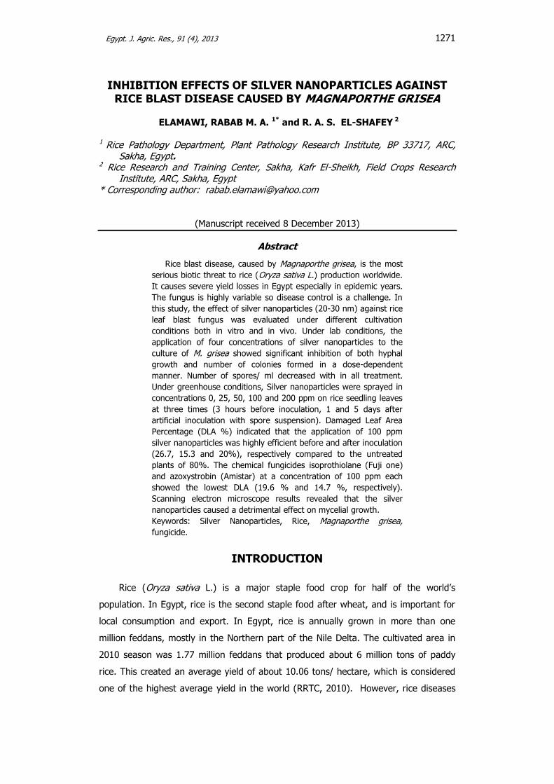
Egypt. J. Agric. Res., 91 (4), 2013 1271 INHIBITION EFFECTS OF SILVER NANOPARTICLES AGAINST RICE BLAST DISEASE CAUSED BY MAGNAPORTHE GRISEA ELAMAWI, RABAB M. A. 1* and R. A. S. EL-SHAFEY 2 1 Rice Pathology Department, Plant Pathology Research Institute, BP 33717, ARC, Sakha, Egypt. 2 Rice Research and Training Center, Sakha, Kafr El-Sheikh, Field Crops Research Institute, ARC, Sakha, Egypt * Corresponding author: [email protected] (Manuscript received 8 December 2013) Abstract Rice blast disease, caused by Magnaporthe grisea, is the most serious biotic threat to rice (Oryza sativa L.) production worldwide. It causes severe yield losses in Egypt especially in epidemic years. The fungus is highly variable so disease control is a challenge. In this study, the effect of silver nanoparticles (20-30 nm) against rice leaf blast fungus was evaluated under different cultivation conditions both in vitro and in vivo. Under lab conditions, the application of four concentrations of silver nanoparticles to the culture of M. grisea showed significant inhibition of both hyphal growth and number of colonies formed in a dose-dependent manner. Number of spores/ ml decreased with in all treatment. Under greenhouse conditions, Silver nanoparticles were sprayed in concentrations 0, 25, 50, 100 and 200 ppm on rice seedling leaves at three times (3 hours before inoculation, 1 and 5 days after artificial inoculation with spore suspension). Damaged Leaf Area Percentage (DLA %) indicated that the application of 100 ppm silver nanoparticles was highly efficient before and after inoculation (26.7, 15.3 and 20%), respectively compared to the untreated plants of 80%. The chemical fungicides isoprothiolane (Fuji one) and azoxystrobin (Amistar) at a concentration of 100 ppm each showed the lowest DLA (19.6 % and 14.7 %, respectively). Scanning electron microscope results revealed that the silver nanoparticles caused a detrimental effect on mycelial growth. Keywords: Silver Nanoparticles, Rice, Magnaporthe grisea, fungicide. INTRODUCTION Rice (Oryza sativa L.) is a major staple food crop for half of the world’s population. In Egypt, rice is the second staple food after wheat, and is important for local consumption and export. In Egypt, rice is annually grown in more than one million feddans, mostly in the Northern part of the Nile Delta. The cultivated area in 2010 season was 1.77 million feddans that produced about 6 million tons of paddy rice. This created an average yield of about 10.06 tons/ hectare, which is considered one of the highest average yield in the world (RRTC, 2010). However, rice diseases
-
Upload
independent -
Category
Documents
-
view
0 -
download
0
Transcript of Inhibition Effects of Silver Nanoparticles against Rice Blast Disease caused by Magnaporthe grisea
Egypt. J. Agric. Res., 91 (4), 2013
1271
INHIBITION EFFECTS OF SILVER NANOPARTICLES AGAINST RICE BLAST DISEASE CAUSED BY MAGNAPORTHE GRISEA
ELAMAWI, RABAB M. A. 1* and R. A. S. EL-SHAFEY 2
1 Rice Pathology Department, Plant Pathology Research Institute, BP 33717, ARC, Sakha, Egypt.
2 Rice Research and Training Center, Sakha, Kafr El-Sheikh, Field Crops Research Institute, ARC, Sakha, Egypt
* Corresponding author: [email protected]
(Manuscript received 8 December 2013)
Abstract
Rice blast disease, caused by Magnaporthe grisea, is the most
serious biotic threat to rice (Oryza sativa L.) production worldwide.
It causes severe yield losses in Egypt especially in epidemic years.
The fungus is highly variable so disease control is a challenge. In
this study, the effect of silver nanoparticles (20-30 nm) against rice
leaf blast fungus was evaluated under different cultivation
conditions both in vitro and in vivo. Under lab conditions, the
application of four concentrations of silver nanoparticles to the
culture of M. grisea showed significant inhibition of both hyphal
growth and number of colonies formed in a dose-dependent
manner. Number of spores/ ml decreased with in all treatment.
Under greenhouse conditions, Silver nanoparticles were sprayed in
concentrations 0, 25, 50, 100 and 200 ppm on rice seedling leaves
at three times (3 hours before inoculation, 1 and 5 days after
artificial inoculation with spore suspension). Damaged Leaf Area
Percentage (DLA %) indicated that the application of 100 ppm
silver nanoparticles was highly efficient before and after inoculation
(26.7, 15.3 and 20%), respectively compared to the untreated
plants of 80%. The chemical fungicides isoprothiolane (Fuji one)
and azoxystrobin (Amistar) at a concentration of 100 ppm each
showed the lowest DLA (19.6 % and 14.7 %, respectively).
Scanning electron microscope results revealed that the silver
nanoparticles caused a detrimental effect on mycelial growth.
Keywords: Silver Nanoparticles, Rice, Magnaporthe grisea,
fungicide.
INTRODUCTION
Rice (Oryza sativa L.) is a major staple food crop for half of the world’s
population. In Egypt, rice is the second staple food after wheat, and is important for
local consumption and export. In Egypt, rice is annually grown in more than one
million feddans, mostly in the Northern part of the Nile Delta. The cultivated area in
2010 season was 1.77 million feddans that produced about 6 million tons of paddy
rice. This created an average yield of about 10.06 tons/ hectare, which is considered
one of the highest average yield in the world (RRTC, 2010). However, rice diseases

INHIBITION EFFECTS OF SILVER NANOPARTICLES AGAINST
RICE BLAST DISEASE CAUSED BY MAGNAPORTHE GRISEA
1272
(especially rice blast) can reduce yield production by about 5 % in normal or mild
disease outbreaks, but during epidemics seasons the yield losses may reach as high
as 30 -50 % (Sehly et al., 2002).
Rice blast, caused by the fungus Pyricularia grisea (Cooke) Sacc. [anamorph of
Magnaporthe grisea (Hebert) Barr], is a devastating diseases of rice (Oryza sativa L.)
worldwide (Ou, 1985). Around 50% of the production may be lost in a field moderately
affected by infection (Zeigler et al., 1994). The disease is currently managed using
resistant cultivars, fungicides and cultural practices. Most of the rice cultivars are
susceptible to different fungus races. The pathogen is also highly variable so, breeding
for durable resistance to blast remains a major challenge (Roy-Barman and Chattoo,
2005). Fungicides are commonly used to control blast; however, these are becoming
less acceptable as they increase the potential for build-up of resistance in M. grisea to
fungicides and also conflict with the public concern for fungicide residues on human
health and environment (Coca et al., 2006).
Silver ions are very reactive, they inhibit microbial respiration and metabolism
and they cause physical damage (Bragg and Rannie1974; Thurman et al., 1989).
Silver has been used to treat medical ailments for over 100 years due to its natural
antibacterial and antifungal properties (Morones et al., 2005). Recently,
nanotechnology practices have amplified the effectiveness of silver particles as
antimicrobial agents (Elchiguerra et al., 2005; Yeo et al., 2003). Silver nanoparticles
have extremely large relative surface areas which increases their contact with bacteria
and fungi, vastly improving its bactericidal and fungicidal effectiveness. The larger
surface area-to-volume ratio of silver nanoparticles increases their contact with
microbes and their ability to permeate cells. When in contact with bacteria and
fungus, they will adversely affect cellular metabolism and inhibit cell growth. Silver
suppresses respiration, basal metabolism of electron transfer systems, and transport
of substrates in the microbial cell membrane. Nanoparticle development has restored
interest in the antimicrobial effects of metals, which declined following the widespread
application of modern synthetic antibiotics (Richards, 1981).
The use of nano-sized silver particles as antimicrobial agents has become more
common as technological advances made their production more economical. There
have been relatively few studies on the applicability of silver to control plant diseases;
especially for sclerotia-forming species of Rhizoctonia solani, Sclerotinia sclerotiorum
and S. minor (Min et al., 2009) and powdery mildew in cucurbits (Lamsal et al., 2011).
Antifungal activity of ionic or nanoparticle silver has a great potential for use in
controlling spore-producing fungal plant pathogens. Various forms of silver ions and

ELAMAWI, RABAB M. A. and R. A. S. EL-SHAFEY
1273
nanoparticles were tested to examine the antifungal activity on two plant-pathogenic
fungi, Bipolaris sorokiniana and Magnaporthe grisea (Young et al., 2009).
The objectives of this study were to determine the inhibitory property of silver
nanoparticles on fungal growth and colony formation of Magnaporthe grisea, and to
evaluate their efficacy for rice blast disease control.
MATERIALS AND METHODS
Silver nanoparticles and fungicide: Silver nanoparticles were obtained from King
Abd Alla Institute for nanotechnology, College of Science, King Saud University, Saudi
Arabia. According to the source, the particles size ranged from 20 to 30 nm and were
spherical shape. Size and morphology of silver nanoparticles particles were confirmed
by UV spectral analysis and Transmission Electron Microscopy (TEM). Different
concentrations of silver nanoparticles (25, 50, 100, and 200 ug m-1) were prepared by
diluting the original stock solution using sterile deionized water. All solutions were
stored at 4o C until use. The chemical fungicides isoprothiolane (Fuji one 40% EC) and
azoxystrobin (Amistar 25% SC) were used as controls.
Fungus preparation: Rice blast fungus was isolated from infected leaves of
Sakha 101 rice cultivar during 2009 season from Gharbia governorate and
identified as M. grisea race IG-1 according to disease reaction pattern on the
international differential varieties (Atkins et al., 1967).
Inhibition of both hyphal growth and colony formation by silver
nanoparticles: The antifungal activity of nanoparticles was examined based on
hyphal growth and new colony formation in vitro. For measurement of hyphal growth:
agar plugs (6 mm in diameter) were obtained from the actively-growing edge of a
pure culture of M. grisea, inoculated in the center of Banana dextrose agar (BDA)
medium (g/L: 200 Banana,15 glucose and 20 agar) supplemented with
different concentrations of silver nanoparticles with four replicates. The
inoculated plates were incubated at 28oC for 10 days. Colony diameter was measured
every 48 hr till the control reached its maximum. For new colony formation, conidia
were collected from M. grisea cultures, grown on BDA medium, and incubated at 25°C
for 10 days. Conidial suspension was diluted with sterile deionized water to a
concentration of 106 spores-1ml. 500 µl of the conidial suspension were mixed with
serial concentrations of silver particles to a final volume of 1 ml. Conidial suspension
was also prepared with sterile deionized water as control or mixed with the fungicides
in concern, Fuji-One and Amistar, at a concentration of 100 ug ml-1 each. All
treatments were incubated at 28˚C for 24 h. aliquots of 25 μl of each dilution was
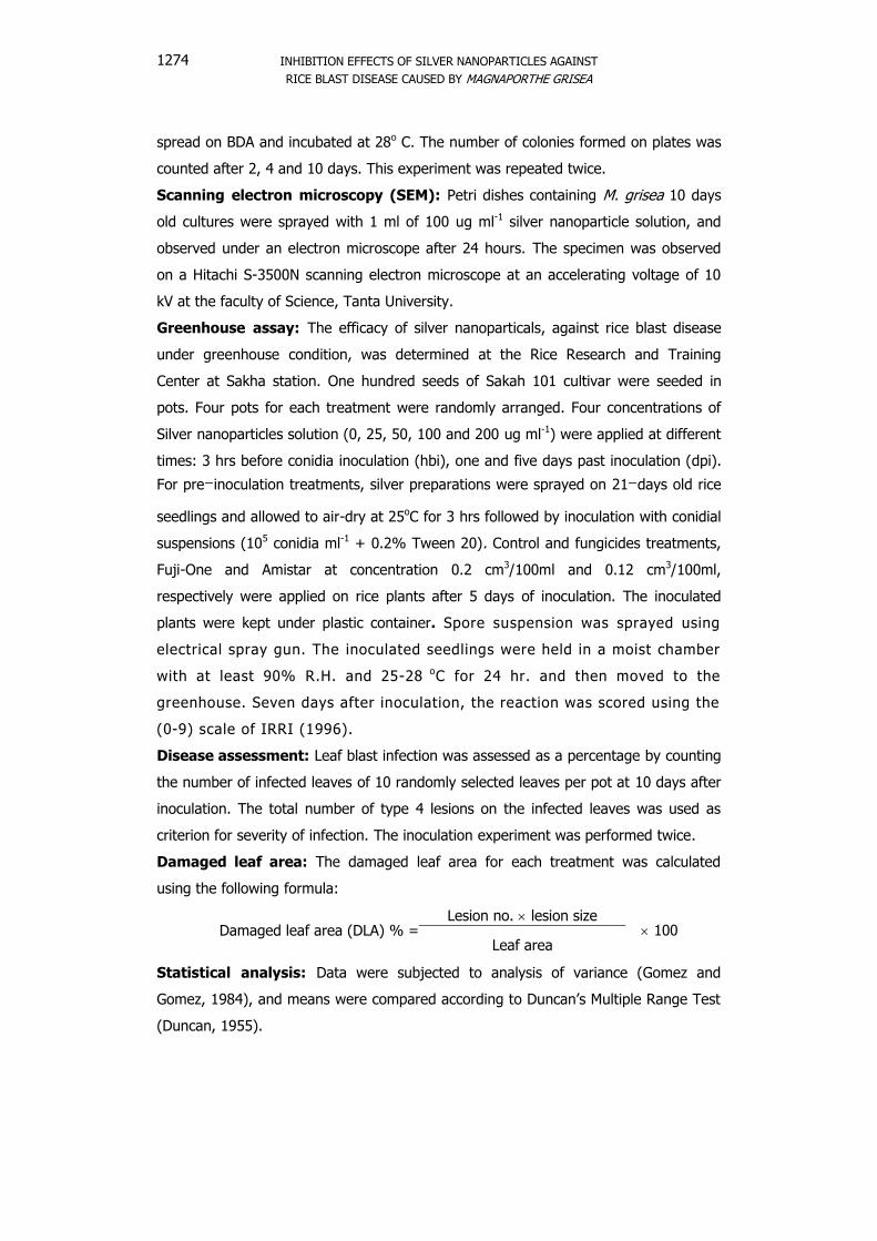
INHIBITION EFFECTS OF SILVER NANOPARTICLES AGAINST
RICE BLAST DISEASE CAUSED BY MAGNAPORTHE GRISEA
1274
spread on BDA and incubated at 28o C. The number of colonies formed on plates was
counted after 2, 4 and 10 days. This experiment was repeated twice.
Scanning electron microscopy (SEM): Petri dishes containing M. grisea 10 days
old cultures were sprayed with 1 ml of 100 ug ml-1 silver nanoparticle solution, and
observed under an electron microscope after 24 hours. The specimen was observed
on a Hitachi S-3500N scanning electron microscope at an accelerating voltage of 10
kV at the faculty of Science, Tanta University.
Greenhouse assay: The efficacy of silver nanoparticals, against rice blast disease
under greenhouse condition, was determined at the Rice Research and Training
Center at Sakha station. One hundred seeds of Sakah 101 cultivar were seeded in
pots. Four pots for each treatment were randomly arranged. Four concentrations of
Silver nanoparticles solution (0, 25, 50, 100 and 200 ug ml-1) were applied at different
times: 3 hrs before conidia inoculation (hbi), one and five days past inoculation (dpi).
For pre inoculation treatments, silver preparations were sprayed on 21 days old rice
seedlings and allowed to air-dry at 25oC for 3 hrs followed by inoculation with conidial
suspensions (105 conidia ml-1 + 0.2% Tween 20). Control and fungicides treatments,
Fuji-One and Amistar at concentration 0.2 cm3/100ml and 0.12 cm3/100ml,
respectively were applied on rice plants after 5 days of inoculation. The inoculated
plants were kept under plastic container. Spore suspension was sprayed using
electrical spray gun. The inoculated seedlings were held in a moist chamber
with at least 90% R.H. and 25-28 oC for 24 hr. and then moved to the
greenhouse. Seven days after inoculation, the reaction was scored using the
(0-9) scale of IRRI (1996).
Disease assessment: Leaf blast infection was assessed as a percentage by counting
the number of infected leaves of 10 randomly selected leaves per pot at 10 days after
inoculation. The total number of type 4 lesions on the infected leaves was used as
criterion for severity of infection. The inoculation experiment was performed twice.
Damaged leaf area: The damaged leaf area for each treatment was calculated
using the following formula:
Damaged leaf area (DLA) % =Lesion no. lesion size
100Leaf area
Statistical analysis: Data were subjected to analysis of variance (Gomez and
Gomez, 1984), and means were compared according to Duncan’s Multiple Range Test
(Duncan, 1955).
ELAMAWI, RABAB M. A. and R. A. S. EL-SHAFEY
1275
RESULTS
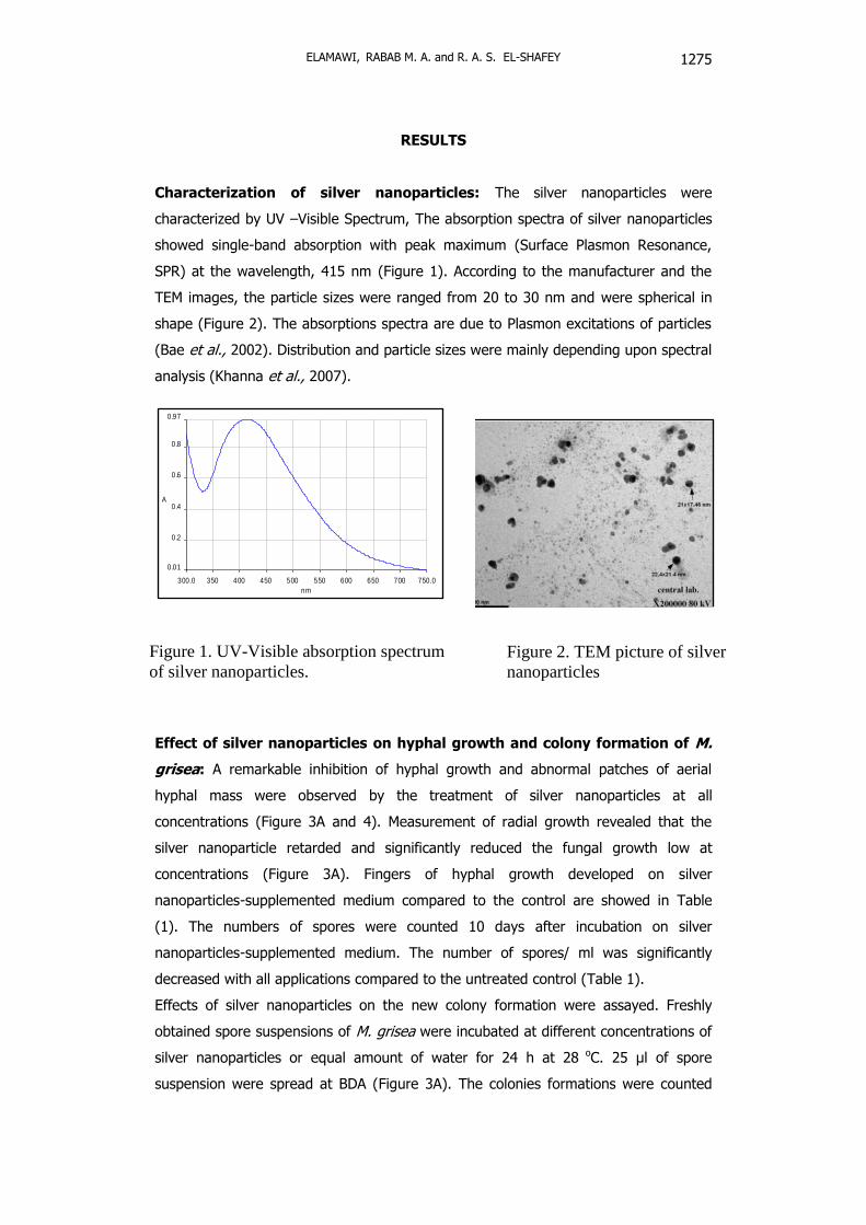
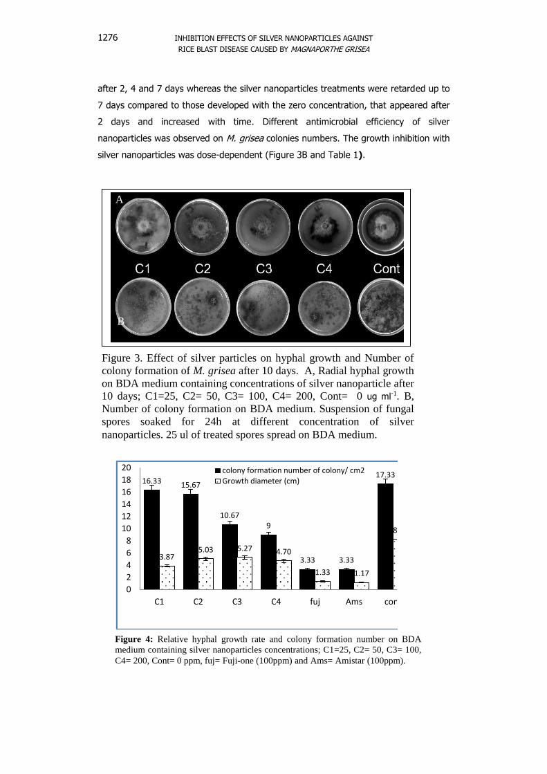
Characterization of silver nanoparticles: The silver nanoparticles were
characterized by UV –Visible Spectrum, The absorption spectra of silver nanoparticles
showed single-band absorption with peak maximum (Surface Plasmon Resonance,
SPR) at the wavelength, 415 nm (Figure 1). According to the manufacturer and the
TEM images, the particle sizes were ranged from 20 to 30 nm and were spherical in
shape (Figure 2). The absorptions spectra are due to Plasmon excitations of particles
(Bae et al., 2002). Distribution and particle sizes were mainly depending upon spectral
analysis (Khanna et al., 2007).
Effect of silver nanoparticles on hyphal growth and colony formation of M.
grisea: A remarkable inhibition of hyphal growth and abnormal patches of aerial
hyphal mass were observed by the treatment of silver nanoparticles at all
concentrations (Figure 3A and 4). Measurement of radial growth revealed that the
silver nanoparticle retarded and significantly reduced the fungal growth low at
concentrations (Figure 3A). Fingers of hyphal growth developed on silver
nanoparticles-supplemented medium compared to the control are showed in Table
(1). The numbers of spores were counted 10 days after incubation on silver
nanoparticles-supplemented medium. The number of spores/ ml was significantly
decreased with all applications compared to the untreated control (Table 1).
Effects of silver nanoparticles on the new colony formation were assayed. Freshly
obtained spore suspensions of M. grisea were incubated at different concentrations of
silver nanoparticles or equal amount of water for 24 h at 28 oC. 25 µl of spore
suspension were spread at BDA (Figure 3A). The colonies formations were counted
300.0 350 400 450 500 550 600 650 700 750.0
0.01
0.2
0.4
0.6
0.8
0.97
nm
A
Figure 1. UV-Visible absorption spectrum
of silver nanoparticles.
Figure 2. TEM picture of silver
nanoparticles
INHIBITION EFFECTS OF SILVER NANOPARTICLES AGAINST
RICE BLAST DISEASE CAUSED BY MAGNAPORTHE GRISEA
1276
after 2, 4 and 7 days whereas the silver nanoparticles treatments were retarded up to
7 days compared to those developed with the zero concentration, that appeared after
2 days and increased with time. Different antimicrobial efficiency of silver
nanoparticles was observed on M. grisea colonies numbers. The growth inhibition with
silver nanoparticles was dose-dependent (Figure 3B and Table 1).
A
B
Figure 3. Effect of silver particles on hyphal growth and Number of
colony formation of M. grisea after 10 days. A, Radial hyphal growth
on BDA medium containing concentrations of silver nanoparticle after
10 days; C1=25, C2= 50, C3= 100, C4= 200, Cont= 0 ug ml-1. B,
Number of colony formation on BDA medium. Suspension of fungal
spores soaked for 24h at different concentration of silver
nanoparticles. 25 ul of treated spores spread on BDA medium.
16.33 15.67
10.67 9
3.33 3.33
17.33
3.87 5.03 5.27 4.70
1.33 1.17
8.23
0
2
4
6
8
10
12
14
16
18
20
C1 C2 C3 C4 fuj Ams cont
colony formation number of colony/ cm2
Growth diameter (cm)
Figure 4: Relative hyphal growth rate and colony formation number on BDA
medium containing silver nanoparticles concentrations; C1=25, C2= 50, C3= 100,
C4= 200, Cont= 0 ppm, fuj= Fuji-one (100ppm) and Ams= Amistar (100ppm).
ELAMAWI, RABAB M. A. and R. A. S. EL-SHAFEY
1277
Table 1. Effect of silver nanoparticles on hyphal growth and colony formation
compared to fungicides.
Concentration
(ug ml-1)
Growth
diameter (cm)
*No. of
spores /ml
Number of formed-
Colonies (cm2)
Control (water) 8.23 91.67 17.33
25 3.87 13.19 16.33
50 5.03 40.97 15.67
100 5.27 10.42 10.67
200 4.70 15.97 9
Fuji-one (100) 1.33 4.167 3.33
Amistar (100) 1.17 4.861 3.33
LSD 5% 0.473 25.7 3.984
F ** ** **
*No. of spores×104
Effect of silver nanoparticles on blast disease parameters under greenhouse
conditions: the antifungal activity of the silver nanoparticles against M. grisea
causing the rice blast disease at different concentrations was presented in Table (2)
and illustrated in figure (5). Under greenhouse conditions, silver nanoparticles were
applied 3 hours before spore inoculation, one and five days post spore inoculation.
Silver nanoparticles effectively reduced blast lesion on Sakha 101 rice cultivar without
noticeable phytotoxicity. All treated plants showed lower disease reaction either
before or after inoculation compared to the untreated control. The average damasod
leaf area DLA% observed in the control plants was 80.0 %. Generally, the diseases
differed significantly at 3 h before inoculation and one day after inoculation. The DLA
% was significantly lower in rice plants treated with silver nanoparticle at 100 ppm at
one day after inoculation showing 15.3 %. The two fungicides showed lower disease
reaction. The Amistar fungicide showed the lowest DLA% (14.7) While, Fuji one
fungicide showed 19.6 %. Results that the application of silver side cated
nanoparticles at the concentration of 100 ug ml-1 showed DLA% as 26.7 %, 15.3 and
20.0 % as the most effective treatments at 3h, 24h and 5 days post-inoculation
compared to 80.0 % of the untreated control Table (2).
INHIBITION EFFECTS OF SILVER NANOPARTICLES AGAINST
RICE BLAST DISEASE CAUSED BY MAGNAPORTHE GRISEA
1278
Table 2. Effect of silver nanoparticles on some blast disease parameters.
Treatments
Concentration (ppm) Severity DLA %
No. of spores/ml
Control (water) 0 86.8a 80.0a 287.5a
3 hbi 25 39.7b 61.2c 176.25ab
50 48.9ab 69.0b 168.75ab
100 46.7ab 26.7g 150abcd
200 40.7b 32.8f 100bcde
1 dpi 25 42.6b 51.3d 25de
50 42.4b 62.0c 25de
100 30.3b 15.3i 31.25cde
200 41.2b 48.0d 225ab
5 dpi 25 45.6b 40.3e 56.25cde
50 53.8ab 43.7e 195ab
100 35.9b 20.0h 100cde
200 49.3ab 43.7e 56.25cde
Fuji-one 100 27.5b 19.6h 6.25e
Amstar 100 22.9b 14.7i 6.25e
LSD 5% 27.3 2.5 13.095
LSD 1% 36.8 3.4
DLA; Damaged Leaf Area, hbi; hrs before inoculation, dpi; days post-inoculation
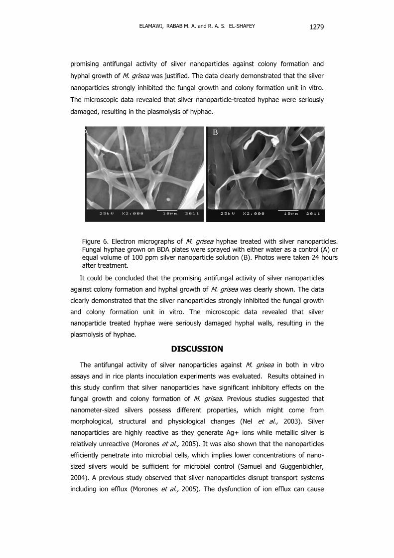
Effect of silver nanoparticles on hyphal growth. As mentioned before, silver
nanoparticles inhibited the hyphal growth and spores germination. Microscopic
observation revealed that silver nanoparticles clearly damaged hyphae (Figure 6B),
while hyphae treated with water appeared to remain intact (Figure 6A). In the
treatment of silver nanoparticles, the shape of hyphal walls turned abnormal, many
hyphae were collapsed at 24 hours after treatment. It could be concluded that the
C1
C2
C3
C4
C1
C2
C3
C4
C1
C2
C3
C
4
Fu
ij-o
ne
Am
esta
r
con
tro
l 3 hbi 1 dpi 5 dpi
Figure 5. Disease symptoms
on leaves of Sakha 101
cultivar post-inoculated with
M. grisea. Three hours before
conidia inoculation, 24 hrs
before inoculation (hbi) and 5
days post-inoculation (dpi).
The plants were treated with
different concentrations
(ppm) of silver nanoparticles
C1=25, C2= 50, C3= 100,
C4= 200, Control (water) = 0
ppm in addition the
fungicides (100 ppm).
ELAMAWI, RABAB M. A. and R. A. S. EL-SHAFEY
1279
Figure 6. Electron micrographs of M. grisea hyphae treated with silver nanoparticles.
Fungal hyphae grown on BDA plates were sprayed with either water as a control (A) or equal volume of 100 ppm silver nanoparticle solution (B). Photos were taken 24 hours
after treatment.
promising antifungal activity of silver nanoparticles against colony formation and
hyphal growth of M. grisea was justified. The data clearly demonstrated that the silver
nanoparticles strongly inhibited the fungal growth and colony formation unit in vitro.
The microscopic data revealed that silver nanoparticle-treated hyphae were seriously
damaged, resulting in the plasmolysis of hyphae.
It could be concluded that the promising antifungal activity of silver nanoparticles
against colony formation and hyphal growth of M. grisea was clearly shown. The data
clearly demonstrated that the silver nanoparticles strongly inhibited the fungal growth
and colony formation unit in vitro. The microscopic data revealed that silver
nanoparticle treated hyphae were seriously damaged hyphal walls, resulting in the
plasmolysis of hyphae.
DISCUSSION
The antifungal activity of silver nanoparticles against M. grisea in both in vitro
assays and in rice plants inoculation experiments was evaluated. Results obtained in
this study confirm that silver nanoparticles have significant inhibitory effects on the
fungal growth and colony formation of M. grisea. Previous studies suggested that
nanometer-sized silvers possess different properties, which might come from
morphological, structural and physiological changes (Nel et al., 2003). Silver
nanoparticles are highly reactive as they generate Ag+ ions while metallic silver is
relatively unreactive (Morones et al., 2005). It was also shown that the nanoparticles
efficiently penetrate into microbial cells, which implies lower concentrations of nano-
sized silvers would be sufficient for microbial control (Samuel and Guggenbichler,
2004). A previous study observed that silver nanoparticles disrupt transport systems
including ion efflux (Morones et al., 2005). The dysfunction of ion efflux can cause
A B

INHIBITION EFFECTS OF SILVER NANOPARTICLES AGAINST
RICE BLAST DISEASE CAUSED BY MAGNAPORTHE GRISEA
1280
rapid accumulation of silver ions, interrupting cellular processes at their lower
concentrations such as metabolism and respiration by reacting with molecules. Also,
silver ions are known to produce reactive oxygen species (ROS) via their reaction with
oxygen, which are detrimental to cells, causing damage to proteins, lipids, and nucleic
acids (Storz and Imlay, 1999; Hwang et al., 2008).
The present study results of microscopic data revealed that silver nanoparticle
treated hyphae severely damaged hyphal walls, resulting in the plasmolysis of hyphae.
Considering many cellular effects of silver ions, silver nanoparticle-mediated collapse
in M. grisea hyphae is probably not only by damaging hyphal walls, but also other
cellular effects, which need to be characterized.
The preventative and post-inoculation application of the silver nanoparticles
effectively reduced disease severity on plants at all concentrations. A mechanism of
this antifungal activity is suggested by the direct effect on germination and infection
process in the fungi. M. grisea can cause foliar disease and reproduce as asexual
conidia. Disease infection is initiated by the attachment of spores to the plant surface
and formation of germ tubes (Tucker and Talbot 2001). Under favorable conditions of
high humidity (~100% relative humidity) and warm temperature (25°C), conidia
germinate, and the resulting germ tubes penetrate plant surfaces within 24 hrs
(Howard and Ferrari 1989). Antifungal efficiency of silver nanoparticles was observed
at 24 h after inoculation, suggesting that direct contact of silver with spores or germ
tubes is critical in inhibiting disease development (Young et al., 2009). Moreover,
antifungal efficiency of silver was also observed at 5 days after inoculation, suggesting
that silver nanoparticles could have penetrated the plant cell wall and inhibited the
disease development.
It could be concluded that, silver nanoparticles can be used effectively in the
control of rice blast disease and the prevention of deleterious infections, even though
there are no phytotoxicity appeared on rice. Silver may be less toxic to humans and
animals than synthetic fungicides. Our results support the hypothesis that silver
nanoparticles are suitable for formulating new types of fungicidal materials. Our
follow-up research focuses on extended applicability of silver for control of M. grisea
in the field, and evaluation of the efficacy of silver on different types of pathogens
causing a problem for rice production. Further research should focus on the
development of silver compounds and mixing with fungicides. At the same time, the
environmental tracking of silver when applied in the field is important to assess the
impact on environmental and human health. This information is imperative for future
registration and labeling of the silver nanoparticles as fungicides for crop protection.
However, further investigation on the effect of copper, widely used in control of plant
diseases in the form of nanoparticles must be tried.
ELAMAWI, RABAB M. A. and R. A. S. EL-SHAFEY
1281
Acknowledgment
The author would like to thank Prof. Awatif Hendi Physics Department, College of
Science, King Saud University, Saudi Arabia for providing silver nanoparticle used in
this study.
REFERENCES
1. Atkins, J.G., A. L. Robert, C. R. Adair, K. Goto, T. Kozako, R. Yanagida, Y.
Yamada and S. Matsumoto. 1967. An international set of rice varieties for
differentiating races of Pyricularia oryzae. Phytopathology, 57: 298-301.
2. Bae C.H., S.M. Nam, S.M. Park. 2002. Formation of silver nanoparticles by laser
ablation of a silver target in NaCl solution. Applied Surface Science, 197: 628 –
634.
3. Bragg, P. D. and D. J. Rannie. 1974. The effect of silver ions on the respiratory
chain of Escherichia coli. Can J Microbiol; 20:883-9.
4. Coca, M. Peñas, G. Gómez, J. Campo, S. Bortolotti, C. Messeguer, J. San Segundo
B. 2006. Enhanced resistance to the rice blast fungus Magnaporthe grisea
conferred by expression of a cecropin A gene in transgenic rice, Planta 223,392-
406.
5. Duncan, D.B. 1955. Multiple ranges and multiple F test. Biometrics, 11:1-42.
6. Gomez, K.A. and A.A. Gomez. 1984. Statistical procedures for Agricultural
Research. Second Edition. John Wiley & Sons, New York.
7. Elchiguerra, J. L., J. L. Burt, J. R. Morones, A. Camacho-Bragado, X. Gao, H. H.
Lara, and M. J. Yacaman. 2005. Interaction of silver nanoparticles with hiv-1. J.
Nanobiotechnol. 3:6.
8. Howard, R. and M. Ferrari. 1989. Role of melanin in appressorium function. Exp.
Mycol. 13:403-418.
9. Hwang, E.T., J.H. Lee, Y. J. Chae, Y.S. Kim, B.C. Kim, B.I. Sang, MB. Gu. Analysis
of the toxic mode of action of silver nanoparticles using stress- pecific
bioluminescent bacteria. Small 2008;4:746-50.
10. IRRI (International Rice Research Institute) 1996. Standard Evaluation System for
Rice (IRRI) P.O. Box 933. 1099 Manila Philippines.
11. Khanna, P.K., N. Singh, D. Kulkarni, S. Deshmukh, S. Charan, P.V. Adhyapak.
2007. Water based simple synthesis of redispersable silver nano-particles.
Materials Letters, 61: 3366 - 3370
12. Lamsal, K., S. W. Kim, J. H. Jin Hee Jung, Y. S. Kim, K.S. Kim and Y. S. Lee.
2011. Inhibition effects of silver nanoparticles against powdery mildews on
Cucumber and Pumpkin. Mycobiology 39(1) : 26-32.
INHIBITION EFFECTS OF SILVER NANOPARTICLES AGAINST
RICE BLAST DISEASE CAUSED BY MAGNAPORTHE GRISEA
1282
13. Morones, J. R., Elechiguerra, J. L., Camacho, A., Holt, K., Kouri,J. B., Ramirez, J.
T. and Acaman, M. J. 2005. The bactericidal effect of silver nanoparticles.
Nanobiotechnology 16:2346- 2353.
14. Min, J. S., Kim, K. S., Kim, S. W., Jin Hee Jung, J. H., Lamsal, K. and Kim, S. B.
2009. Effects of colloidal silver nanoparticles on Sclerotium-forming
phytopathogenic fungi. Plant Pathol. J. 25(4): 376-380.
15. Nel, A., Xia, T., L. Mdler and N. Li. 2003. Toxic potential of materials at the
nanolevel. Science 311:622-627.
16. Ou, S.H. 1985. Rice Diseases II edition. CMI, Kew, England. pp. 337–364.
17. Richards, R.M. 1981. Antimicrobial action of silver nitrate. Microbios; 31:83-91.
18. RRTC (Rice Research and Training Center) 2010. Annual rice national campaign
report of rice program. Field Crops Research, Agric. Research Center, Ministry of
Agriculture, Egypt.
19. Samuel, U. and J. P. Guggenbichler. 2004. Prevention of catheter related
infections: the potential of a new nano-silver impregnated catheter. Intl. J.
Antimicrobial Agents 23S1: S75-S78.
20. Sehly, M.R., Z.H. Osman and E.A. Salem. 2002. Rice diseases. In: Rice in Egypt,
pp 301.
21. Subhankar Roy-Barman and Bharat B. Chattoo. 2005. Rice blast fungus
sequenced. Current science, (89) 6. 930-931.
22. Storz, G., Imlay, J.A. 1999. Oxidative stress. Curr Opin Microbiol; 2:188-94.
23. Thurman, R.B., C.P. Gerba, G. Bitton. 1989. The molecular mechanisms of copper
and silver ion disinfection of bacteria and viruses. Crit. Rev Environ Sci. technol.
18:295-315.
24. Tucker, S. L., and N. J. Talbot. 2001. Surface attachment and pre-penetration
stage development by plant pathogenic fungi. Annu. Rev. Phytopathol. 39:385-
417.
25. Young, K. J., Byung H. Kim and Geunhwa Jung 2009. Antifungal activity of silver
ions and nanoparticles on Phytopathogenic Fungi. Plant Disease, 1037-1043.
26. Yeo, S. Y., H. J. Lee and S. H. Jeong. 2003. Preparation of nanocomposite fibers
for permanent antibacterial effect. J. Mater. Sci. 38:2143-2147.
27. Zeigler, R.S., S.A. Leong , P.S. Teng, editors. 1994. Rice blast disease. In: Zeigler
RS, Leong SA, Teng PS, editors. Rice blast disease. Wallingford, Oxon (United
Kingdom): CAB International, Los Baños (Philippines):IRRI. 626 p.
ELAMAWI, RABAB M. A. and R. A. S. EL-SHAFEY
1283
Magnaporthe grisea
Magnaporthe grisea
Oryza sativa