Infrequent MODS TB culture cross-contamination in a high-burden resource-poor setting
17
Infrequent MODS TB culture cross-contamination in a high- burden resource-poor setting David A.J. Moore a,b,c,d,* , Luz Caviedes c , Robert H. Gilman a,b,c,d , Jorge Coronel c , Fanny Arenas c , Doris LaChira e , Cayo Salazar e , Juan Carlos Saravia f , Richard A. Oberhelman c , Maria-Graciela Hollm-Delgado b , A. Roderick Escombe a,b,c , Carlton A.W. Evans a,b,c,d , and Jon S. Friedland a,b,c,d a Department of Infectious Diseases and Immunity, Imperial College London Wellcome Centre for Clinical Tropical Medicine, Faculty of Medicine (Hammersmith Campus), W12 0NN London, UK b Asociación Benéfica PRISMA, San Miguel, Lima 32, Peru c Laboratorio de Investigación de Enfermedades Infecciosas, Universidad Peruana Cayetano Heredia, San Martín de Porras, Lima 31, Peru d Department of International Health, Johns Hopkins Bloomberg School of Public Health, Baltimore, MD 21205, USA e Dirección de Salud IV Lima Este (Ministerio de Salud), El Agustino, Lima 10, Peru f Dirección de Salud III Lima Norte (Ministerio de Salud), Pasaje San German 270, Rimac, Lima 25, Peru Abstract One obstacle to wider use of rapid liquid culture-based tuberculosis diagnostics such as the microscopic observation drug susceptibility (MODS) assay is concern about cross-contamination. We investigated the rate of laboratory cross-contamination in MODS, automated MBBacT, and Lowenstein–Jensen (LJ) cultures performed in parallel, through triangulation of microbiologic (reculturing stored samples), molecular (spoligotype/RFLP), and clinical epidemiologic data. At least 1 culture was positive for Mycobacterium tuberculosis for 362 (11%) of 3416 samples; 53 were regarded as potential cross-contamination suspects. Cross-contamination accounted for 17 false-positive cultures from 14 samples representing 0.41% (14/3416) and 0.17% (17/10 248) of samples and cultures, respectively. Positive predictive values for MODS, MBBacT (bioMérieux, Durham, NC), and LJ were 99.1%, 98.7%, and 99.7%, and specificity was 99.9% for all 3. Low rates of cross-contamination are achievable in mycobacterial laboratories in resource-poor settings even when a large proportion of samples are infectious and highly sensitive liquid culture-based diagnostics such as MODS are used. Keywords Tubercolosis; Multidrug resistance; Cross-contamination; Microscopic observation drug susceptability assay; MODS; Microbacterial culture © 2006 Published by Elsevier Inc. * Corresponding author. Laboratorio de Investigación de Enfermedades Infecciosas, Universidad Peruana Cayetano Heredia, San Martín de Porras, Lima 31, Peru. Tel.: +51-1382-3398; fax: +51-1464-0781. [email protected] (D.A.J. Moore).. Europe PMC Funders Group Author Manuscript Diagn Microbiol Infect Dis. Author manuscript; available in PMC 2010 July 30. Published in final edited form as: Diagn Microbiol Infect Dis. 2006 September ; 56(1): 35–43. doi:10.1016/j.diagmicrobio.2006.03.009. Europe PMC Funders Author Manuscripts Europe PMC Funders Author Manuscripts
-
Upload
independent -
Category
Documents
-
view
0 -
download
0
Transcript of Infrequent MODS TB culture cross-contamination in a high-burden resource-poor setting

Infrequent MODS TB culture cross-contamination in a high-burden resource-poor setting
David A.J. Moorea,b,c,d,*, Luz Caviedesc, Robert H. Gilmana,b,c,d, Jorge Coronelc, FannyArenasc, Doris LaChirae, Cayo Salazare, Juan Carlos Saraviaf, Richard A. Oberhelmanc,Maria-Graciela Hollm-Delgadob, A. Roderick Escombea,b,c, Carlton A.W. Evansa,b,c,d, andJon S. Friedlanda,b,c,d
aDepartment of Infectious Diseases and Immunity, Imperial College London Wellcome Centre forClinical Tropical Medicine, Faculty of Medicine (Hammersmith Campus), W12 0NN London, UKbAsociación Benéfica PRISMA, San Miguel, Lima 32, PerucLaboratorio de Investigación de Enfermedades Infecciosas, Universidad Peruana CayetanoHeredia, San Martín de Porras, Lima 31, PerudDepartment of International Health, Johns Hopkins Bloomberg School of Public Health,Baltimore, MD 21205, USAeDirección de Salud IV Lima Este (Ministerio de Salud), El Agustino, Lima 10, PerufDirección de Salud III Lima Norte (Ministerio de Salud), Pasaje San German 270, Rimac, Lima25, Peru
AbstractOne obstacle to wider use of rapid liquid culture-based tuberculosis diagnostics such as themicroscopic observation drug susceptibility (MODS) assay is concern about cross-contamination.We investigated the rate of laboratory cross-contamination in MODS, automated MBBacT, andLowenstein–Jensen (LJ) cultures performed in parallel, through triangulation of microbiologic(reculturing stored samples), molecular (spoligotype/RFLP), and clinical epidemiologic data. Atleast 1 culture was positive for Mycobacterium tuberculosis for 362 (11%) of 3416 samples; 53were regarded as potential cross-contamination suspects. Cross-contamination accounted for 17false-positive cultures from 14 samples representing 0.41% (14/3416) and 0.17% (17/10 248) ofsamples and cultures, respectively. Positive predictive values for MODS, MBBacT (bioMérieux,Durham, NC), and LJ were 99.1%, 98.7%, and 99.7%, and specificity was 99.9% for all 3. Lowrates of cross-contamination are achievable in mycobacterial laboratories in resource-poor settingseven when a large proportion of samples are infectious and highly sensitive liquid culture-baseddiagnostics such as MODS are used.
KeywordsTubercolosis; Multidrug resistance; Cross-contamination; Microscopic observation drugsusceptability assay; MODS; Microbacterial culture
© 2006 Published by Elsevier Inc.* Corresponding author. Laboratorio de Investigación de Enfermedades Infecciosas, Universidad Peruana Cayetano Heredia, SanMartín de Porras, Lima 31, Peru. Tel.: +51-1382-3398; fax: +51-1464-0781. [email protected] (D.A.J. Moore)..
Europe PMC Funders GroupAuthor ManuscriptDiagn Microbiol Infect Dis. Author manuscript; available in PMC 2010 July 30.
Published in final edited form as:Diagn Microbiol Infect Dis. 2006 September ; 56(1): 35–43. doi:10.1016/j.diagmicrobio.2006.03.009.
Europe PM
C Funders A
uthor Manuscripts
Europe PM
C Funders A
uthor Manuscripts
1. BackgroundIn mycobacteriology reference-level laboratories in the industrialized world, cross-contamination is estimated to account for between 0.5% and 6% of positive results (Bauer etal., 1997; Nivin et al., 1998; Burman and Reves 2000; de Boer et al., 2002; Jasmer et al.,2002; Ruddy et al., 2002) with significant associated cost implications (Northrup et al.,2002), and higher levels might be anticipated in high tuberculosis (TB)-burden resource-limited settings where laboratory facilities are less sophisticated and a greater proportion ofsamples contain Mycobacterium tuberculosis. The microscopic observation drugsusceptibility (MODS) assay in which 2 sputum samples are cultured on the same 24-welltissue-culture plate in Middlebrook 7H9 medium (Caviedes et al., 2000; Moore et al., 2004)has been proposed as a potential tool to bring low-tech sensitive TB diagnosis to thedeveloping world where the need is most urgent; however, the risk of cross-contamination inthis simple low-tech method using liquid culture medium and multiple samples in a single24-well plate has not been previously determined.
Examining samples obtained in a large community-based study in urban Lima, Peru, wedetermined the rate of M. tuberculosis cross-contamination in sputum cultures performed inparallel in MODS, MBBacT-automated mycobacterial culture system (bioMérieux, Durham,NC), and Lowenstein–Jensen (LJ) solid media by first defining and then investigatingcontamination suspect positive cultures.
2. Patients, materials, and methods2.1. Sample collection
After written informed consent, 1923 patients undergoing investigation for TB at healthcenters in Lima, Peru, were recruited over an 18-month period into an operational evaluationof the MODS assay. Two sputum samples were requested from each participant, and 3416samples were obtained for auramine stain microscopy and parallel culture by all of LJ,automated MBBacT, and MODS. The study protocol and informed consent forms wereapproved by all of the following: Ethics Committees of Universidad Peruana CayetanoHeredia, Lima, Peru; Asociación Benéfica PRISMA, Lima, Peru; Johns Hopkins BloombergSchool of Public Health, Baltimore, MD; Imperial College London, UK; and Dirección deSalud III Lima Norte and Dirección de Salud II Lima Este (Regional Ministry of Health),Peru.
2.2. LaboratoryAfter decontamination by the N-acetyl-L-cysteine (NALC)-NaOH method (WHO, 1998)and auramine smear microscopy, all samples were divided into 3 aliquots for culture i) on anLJ slant, ii) in the MBBacT colorimetric, automated mycobacterial culture system, and iii) in12 wells of a 24-well tissue-culture plate in the MODS assay (Fig. 1).
Lowenstein–Jensen cultures were examined twice weekly from day 7 to 60; after which, theabsence of typical colonies was regarded as a negative result. Ziehl-Neelson (ZN) smearswere made from characteristic colonies appearing before day 60 to confirm the presence ofacid fast bacilli. MBBacT cultures were automatically monitored continuously for 42 daysand determined as positive or negative in accordance with manufacturer's recommendations.ZN staining of an aliquot of culture media from any MBBacT bottle reported as positive wasperformed to confirm the presence of acid fast bacilli.
Microscopic observation drug susceptibility cultures were examined every weekday fromday 4 to 15, on alternate days from day 16 to 25, and twice weekly from day 26 to 40 underan inverted light microscope. Positive cultures were identified by the characteristic cording
Moore et al. Page 2
Diagn Microbiol Infect Dis. Author manuscript; available in PMC 2010 July 30.
Europe PM
C Funders A
uthor Manuscripts
Europe PM
C Funders A
uthor Manuscripts

morphology of M. tuberculosis growth in liquid media (in drug-free control wells) (Fig. 2)as described previously (Caviedes et al., 2000; Park et al., 2002; Moore et al., 2004).Nontuberculous mycobacteria (NTM) were recognized by their lack of cording or (in thecase of Mycobacterium chelonae that uniquely among NTM does form cords) rapidovergrowth of wells by day 5.
In the event of bacterial or fungal overgrowth in any of the 3 cultures, the original storeddecontaminated sample was decontaminated a second time by the same NALC–NaOHmethod, and the affected culture method was repeated. In the event of repeated bacterial/fungal over-growth, the culture was abandoned.
Additional molecular confirmation of the presence of M. tuberculosis was performed for allisolates. Fingerprinting of every isolate was performed by spoligotyping (Goyal et al.,1997), with subsequent selective restriction fragment length polymorphism (RFLP) typing(van Embden et al., 1993) where further discriminatory data were required.
2.3. Defining contamination suspectsThe process for determining whether a positive M. tuberculosis culture was the result ofcross-contamination involved identifying and then thoroughly investigating contaminationsuspects. Contamination suspect cultures were identified by a process of exclusion: positivecultures regarded as unlikely to be due to cross-contamination were those in which a) all 3culture methods for a sample were positive, or if there was fungal/bacterial overgrowth of 1method, the remaining 2 were positive; b) all 3 methods were culture positive for thepatient's other sputum sample (the protocol required submission of 2 sputum samples perpatient); or c) the auramine smear was positive.
2.4. Investigating contamination suspectsThe approach to distinguishing between true and false positives from among the remainingsmear-negative samples, which were culture-positive in only 1 or 2 of the 3 methods(contamination suspects), involved the triangulation of investigations using conventionalmicrobiology, molecular epidemiology, and conventional clinical epidemiology.
The procedures undertaken are described in detail in the Results section below, but briefly:
1) All the stored decontaminated sputum samples for this group of contaminationsuspects were recultured by all 3 methods to determine whether M. tuberculosis couldbe isolated again. In this rule-out step, any positive culture with a spoligotype matchingthe original was regarded as sufficient evidence that the original culture had been a truepositive. However, because most samples had been stored for more than 12 months at−70 °C, negative cultures were not regarded as necessarily indicative that the originalculture had been a false positive;
2) The molecular fingerprints of all isolates from cultures setup contemporaneouslywith the contamination suspect culture were compared. Initial molecular evaluation ofstrain diversity was performed for all isolates by spoligotyping—thus, for a patient with2 samples, which were culture positive in 2 of the 3 methods, there were 4 availablespoligotypes. For strains from contamination suspects with non-unique spoligotypes andfor which the date of sample processing of an identical strain overlapped within 2 days,subsequent IS6110 RFLP typing was performed to enhance discrimination;
3) All patients contributing contamination suspect samples were followed up todetermine clinical outcome. Smear-negative, culture-positive TB is managed on a caseby case basis in the National Tuberculosis Program (NTP), and all such study patients(which included all those with contamination suspect samples) were managed by one of
Moore et al. Page 3
Diagn Microbiol Infect Dis. Author manuscript; available in PMC 2010 July 30.
Europe PM
C Funders A
uthor Manuscripts
Europe PM
C Funders A
uthor Manuscripts

us (JCS, Ministry of Health TB physician) in a dedicated clinic. Thus, clinical outcomedata and information on any subsequent TB diagnostic testing or treatment elsewherewas collected, and follow-up samples were sought for repeat culture, again in triplicate.
3. Results3.1. Identification of contamination suspects
At least 1 culture was positive for M. tuberculosis in 362 (11%) of the 3416 samples (Fig.3). For 79% (n = 285) of these samples, all 3 culture methods were positive, whereas for afurther 7% (n = 24), the sample was i) culture positive by 2 of the 3 methods with bacterialor fungal overgrowth of the 3rd method (n = 4), ii) one of a pair of samples from the samepatient, the other of which was culture positive in all 3 methods (n = 16), or iii) none of theabove but auramine smear positive (n = 4). Thus, 53 smear-negative samples (from 47patients) yielded only 1 (n = 38) or 2 (n = 15) positive cultures (68 cultures in total) andlacked a 2nd corresponding patient sample, which was either smear positive or culturepositive in all 3 methodologies.
3.2. Investigation of contamination suspects3.2.1. Recovery of M. tuberculosis from stored decontaminated sputumsamples—Aliquots of frozen decontaminated sputum were available for reculture for 51 ofthe 53 suspect samples. Three samples yielded a positive culture in MODS alone, and theremainder were culture negative in all 3 methods (Table 1). This 2nd retrieval of M.tuberculosis from the same sample was regarded as definitive evidence against cross-contamination.
3.2.2. Molecular evaluation—Of the 68 original isolates (from all 53 contaminationsuspect samples), it was possible to obtain spoligotypes for 64 (from 49 cultured patientsamples). Reasons for lack of a spoligotype included loss or absence of stored isolate (n = 3)or technical failure (n = 1). The spoligotypes of the 3 isolates recovered from stored frozensputum (Table 1) matched those of the strain from the original culture in all 3 cases.Compared with all other positive isolates from the same 5-day period (2 days either side ofthe date of processing of the contamination suspect), 31 samples were found to have uniquespoligotypes, indicative of a very low probability of cross-contamination. Of the remaining18, which shared a spoligotype with a contemporary isolate, subsequent RFLP fingerprintingconfirmed identical strain identity in 10, which were thus deemed, on molecular grounds,highly suspicious of episodes of cross-contamination. The other 8 had RFLP fingerprintsthat differed from their contemporary spoligotype match. Of the 15 samples culture positiveonly in MODS and MBBacT (n = 13) or MODS and LJ (n = 2), both isolates showedidentical spoligotypes (evidence against cross-contamination) in all except 2 samples (Fig.4).
Forty-one patients contributed a single contamination suspect sample, but 6 patients eachcontributed 2 samples for which all but 1 pair shared identical spoligotypes. Overall, onmolecular grounds, cross-contamination was deemed unlikely for 39 samples and stillplausible for the other 14 (Fig. 4).
3.2.3. Clinical evaluation—Follow-up information was available for 39 of the 47 patients(accounting for 43 of the 53 samples), 10 of whom had microbiologic confirmation of TB(either positive sputum smear elsewhere or repeat culture in our laboratory) and 19 of whomhad been commenced on TB treatment—all 19 reported symptom improvement and hadobjective increases in weight on therapy (range, 2–8 kg), thus the 9 without microbiologicconfirmation were regarded as having probable TB. A further four samples deemed true
Moore et al. Page 4
Diagn Microbiol Infect Dis. Author manuscript; available in PMC 2010 July 30.
Europe PM
C Funders A
uthor Manuscripts
Europe PM
C Funders A
uthor Manuscripts

positives came from 3 patients who refused treatment or further investigation despitecontinuing constitutional and respiratory symptoms, ongoing weight loss (4–13 kg), and ahistory of prior TB treatment and/or contact (Table 1).
Although the phenomenon of self-cure is well recognized, we regarded reported recoverywithout TB treatment as suspicious that the original result had been falsely positive. Thestudy was conducted as an operational evaluation within the NTP in health centers; thus, allNTP-received samples from consenting adults had also been processed in the study—however, a proportion did not fulfill the NTP criteria for a TB suspect and did not receivetreatment (e.g., paucisymptomatic index-case contacts, insurance screening examinations),and in such instances, a lack of symptoms or reported recovery from symptoms at thefollow-up visit was also deemed highly suggestive, that the original culture had been falselypositive.
Overall, on clinical grounds, cross-contamination was deemed likely for 14 samples, highlyunlikely for 23 samples and indeterminate (consistent but not probable) for 16 samples (Fig.4).
3.2.4. Resolving true/false-positive allocation—Combining the evidence from the 3distinct approaches, the final assessment inferred that cross-contamination probablyaccounted for 17 false-positive cultures from 14 samples (Table 1), representing overallfalse-positive percentages of 0.41% (14/3416) and 0.17% (17/10 248) for samples andcultures, respectively. The number (percentage) of cultures that were deemed false positivebecause of cross-contamination was 12 (3.3%), 4 (1.1%), and 1 (0.3%) for MODS,MBBacT, and LJ representing specificities of 99.6%, 99.9%, and 99.9%, respectively (datanot shown). Positive predictive values for MODS, MBBacT, and LJ were 96.2%, 98.7%,and 99.7%, respectively. The contributions to “cross-contamination rule-out” of eachelement of the triangulating approach are shown in Fig. 5.
3.2.5. The effect of more rigorous definition of culture positivity in MODS—AMODS plate has thus far been defined as positive in this study if there was a characteristicand confirmed growth noted in any of the 4 control wells (Fig. 1), regardless of theappearance in the other wells. Of the 12 “probable MODS cross-contamination”, growthwas almost universally noted in only 1 (n =10) or 2 (n = 1) wells. By contrast, of the 285samples culture positive in all 3 methods (Fig. 3), mycobacterial growth was detected in 1,2, 3, and 4 wells for 1, 2, 1, and 281 samples, respectively. A receiver-operator characteristiccurve using these data confirmed the modest but detectable effect of the 4 potential casedefinitions—growth required in 1, 2, 3, or 4 wells—for a positive culture (not shown). Byrevising the case definition for a positive MODS culture to detection of mycobacterialgrowth in at least 2 of the 4 control wells, the number of false-positive cultures is reduced to3. This equates to overall study and MODS-specific false-positive culture rates of 1.4% and0.75% with a MODS positive predictive value of 99.1% and specificity of 99.9%.
4. DiscussionThe key finding of this head-to-head study of MODS with MBBacT and LJ culture was thesimilarly low proportion of positive cultures determined to be false positive because ofcross-contamination and high specificity (99.9%) of all 3 methods. In contrast to acceptedwisdom and some data (Small et al., 1993; Gascoyne-Binzi et al., 2001), we found nosignificant difference between the cross-contamination rates in liquid (MBBacT and MODS)and solid (LJ) media, despite the greater detection sensitivity of the former.
Moore et al. Page 5
Diagn Microbiol Infect Dis. Author manuscript; available in PMC 2010 July 30.
Europe PM
C Funders A
uthor Manuscripts
Europe PM
C Funders A
uthor Manuscripts

There were 2 major strengths of this study: first, we were able to evaluate 2 samples frommost patients by all 3 culture methods, and second we were then able to investigate thelikelihood that a positive culture represented cross-contamination by a triangulatingapproach of microbiologic, molecular, and clinical (conventional “shoe-leather”)epidemiology.
We applied strict criteria in requiring smear-negative samples (or the patient's other sample)to be culture positive in all 3 methods to be above suspicion of cross-contamination.Multiple contamination episodes affecting a patient's cultures have rarely been reported(Bauer et al., 1997), and we cannot exclude the possibility that occasional samples regardedas meeting this criterion resulted from cross-contamination. Patient follow-up wasincomplete, which prevented “rule-out” of cross-contamination on clinical grounds in 11untraceable individuals. All had provided false addresses, a phenomenon we have describedpreviously in TB suspects (Ouyang et al., 2005) and a potentially important obstacle toefficient TB case management.
Reported mycobacterial cross-contamination rates vary widely, partly reflecting reportingbias and nonstandardized ascertainment methodologies—pseudooutbreak reports havereached rates of 65% (de Ramos et al., 1999), whereas comprehensive molecularepidemiologic studies elsewhere have demonstrated underlying rates of 0.1% to 4% (Baueret al., 1997; Burman et al., 1997; de Boer et al., 2002; Jasmer et al., 2002; Ruddy et al.,2002). Our findings, the 1st reported from a resource-limited, high TB-burden setting, fallwithin this range for all 3 methodologies. The potential for cross-contamination is greater inlaboratories handling larger numbers of culture-positive samples and manipulating isolates(Carroll et al., 2003). That we were able to maintain such low-level cross-contaminationprobably reflects both the unusually bio-secure nature of our developing world laboratoryfacility (P3 level containment) and the experience of our mycobacterial laboratory staff.
Because the cross-contaminating inoculum is usually small (droplet), most false-positivecultures have low colony counts (MacGregor et al., 1975) and/or require prolongedincubation for detection. The differential cross-contamination rates determined by thenumber of MODS wells with confirmed growth is a useful check—the presence ofcharacteristic tangles in at least 2 wells is highly unlikely to be a false-positive or cross-contamination event.
Cross-contamination can occur at every stage from specimen collection to strain handlingfor indirect drug susceptibility testing (DST) or species determination (in which theconcentration of M. tuberculosis has been greatly amplified from the source sample) (VanDuin et al., 1998) and is further favored by inadequate environmental controls (Segal-Maurer et al., 1998). The contribution of external soiling of the much handled sputum potmay also be underestimated (Allen and Darrell 1983). Cross-contamination may occurdirectly (sample-sample, sample-culture media, isolate-sample, isolate-culture media) orindirectly (e.g., contaminated stock solution) (Van Duin et al., 1998), though modification oflaboratory practice (e.g., avoiding common flasks for reagent dispensing, waiting 5 min aftercentrifuging to allow settlement of potential tube aerosols) can reduce the incidence of false-positive cultures (Breese et al., 2001; Carroll et al., 2002). The MODS methodologyinherently protects against cross-contamination from the point of sample inoculation onwardbecause the culture is then sealed within a transparent plastic bag. Direct DST obviates theneed for further culture manipulation, reducing the potential for cross-contamination andbiohazard for laboratory staff. Cross-contamination between adjacent samples cultured onthe same plate has not been observed, concordant with the observation that in > 1100 MODScultures over a 3-year period, contaminating growth was never seen in a row of adjacentsample-free control wells containing media alone (Luz Caviedes, unpublished data).
Moore et al. Page 6
Diagn Microbiol Infect Dis. Author manuscript; available in PMC 2010 July 30.
Europe PM
C Funders A
uthor Manuscripts
Europe PM
C Funders A
uthor Manuscripts

New molecular techniques for strain differentiation (Burman et al., 1997; Poynten et al.,2002; Small et al., 1993; Bauer et al., 1997; de Ramos et al., 1999; Nivin et al., 2000; Filhoet al., 2002; Gascoyne-Binzi et al., 2001) with superior sensitivity and discriminatory powerhave rendered conventional methods such as phage testing (Sula and Sulova 1979; Maurer etal., 1984; Jones 1988) obsolete. Spoligotyping is a useful 1st-line tool (de Ramos et al.,1999; Nivin et al., 2000) in a 2-stage algorithm (as used in this study) in which subsequentRFLP examination may enhance discrimination of suspicious strains sharing the samespoligotype.
Methodologies with low innate cross-contamination risk are crucial in resource-limitedhigh-burden settings, where fluctuations in case detection or emergence of unusualphenotypes (Smith and Vance 1991; Nitta et al., 1996; Wurtz et al., 1996; Bearman et al.,2002) or genotypes (Nivin et al., 1998; Anonymous 2000) are much less likely to raise the“cross-contamination alarm”. Designing result readouts so that positive results forsequentially processed samples are easily seen (Bauer et al., 1997) is one potential strategyto aid detection.
In summary, through a vigorous 3-pronged approach to investigation of potential false-positive cultures, we have clearly defined that the cross-contamination rates in MODS, LJ,and MBBacT were all equally low. The finding that, even in this resource-poor setting, theuse of the highly sensitive but technically simple liquid culture assay MODS is associatedwith a specificity of 99.9% addresses a key concern and removes an important obstacle tothe wider implementation of this inexpensive methodology.
AcknowledgmentsThe following are thanked for their contributions of ensuring the success of this study: Adolfo Orellana, MilciadesReátegui Sanchez, Yuri Garcia, Maria Burga, Felipe Castillo, Fanny Garcia, Eleana Sanchez, Yrma Chuquiruna,Sonia Lopez, Rosmery Gutierrez, Raul Miranda Arrostigue, Betty Cajaleón Ascencios, José Calderón Yberico, LuisRivera Pérez, Walter Ramos Maguiña, Luz Vásquez Chávez, Félix Pari Loayza, Ruth Flores Escobar, Jesús CastilloDiaz, Alicia Vigo Alegria.
ReferencesAllen BW, Darrell JH. Contamination of specimen container surfaces during sputum collection. J Clin
Pathol. 1983; 36:479–481. [PubMed: 6403598]
Anonymous. Misdiagnoses of tuberculosis resulting from laboratory cross-contamination ofMycobacterium tuberculosis cultures—New Jersey, 1998. MMWR Morb Mortal Wkly Rep. 2000;49:413–416. [PubMed: 10905819]
Bauer J, Thomsen VO, et al. False-positive results from cultures of Mycobacterium tuberculosis due tolaboratory cross-contamination confirmed by restriction fragment length polymorphism. J ClinMicrobiol. 1997; 35:988–991. [PubMed: 9157170]
Bearman G, Vaamonde C, et al. Pseudo-outbreak of multidrug-resistant Mycobacterium tuberculosisassociated with presumed laboratory processing contamination. Infect Control Hosp Epidemiol.2002; 23:620–622. [PubMed: 12400894]
Breese PE, Burman WJ, et al. The effect of changes in laboratory practices on the rate of false-positivecultures for Mycobacterium tuberculosis. Arch Pathol Lab Med. 2001; 125:1213–1216. [PubMed:11520275]
Burman WJ, Reves RR. Review of false-positive cultures for Mycobacterium tuberculosis andrecommendations for avoiding unnecessary treatment. Clin Infect Dis. 2000; 31:1390–1395.[PubMed: 11096008]
Burman WJ, Stone BL, et al. The incidence of false-positive cultures for Mycobacterium tuberculosis.Am J Respir Crit Care Med. 1997; 155:321–326. [PubMed: 9001331]
Moore et al. Page 7
Diagn Microbiol Infect Dis. Author manuscript; available in PMC 2010 July 30.
Europe PM
C Funders A
uthor Manuscripts
Europe PM
C Funders A
uthor Manuscripts

Carroll NM, Richardson M, et al. Reduction of the rate of false-positive cultures of Mycobacteriumtuberculosis in a laboratory with a high culture positivity rate. Clin Chem Lab Med. 2002; 40:888–892. [PubMed: 12435105]
Carroll NM, Richardson M, et al. Criteria for identification of cross-contamination of cultures ofMycobacterium tuberculosis in routine microbiology laboratories. J Clin Microbiol. 2003; 41:2269.[PubMed: 12734301]
Caviedes L, Lee TS, et al. Rapid, efficient detection and drug susceptibility testing of Mycobacteriumtuberculosis in sputum by microscopic observation of broth cultures. The Tuberculosis WorkingGroup in Peru. J Clin Microbiol. 2000; 38:1203–1208. [PubMed: 10699023]
de Boer AS, Blommerde B, et al. False-positive Mycobacterium tuberculosis cultures in 44laboratories in The Netherlands (1993 to 2000): incidence, risk factors, and consequences. J ClinMicrobiol. 2002; 40:4004–4009. [PubMed: 12409366]
de Ramos CM, Soini H, et al. Extensive cross-contamination of specimens with Mycobacteriumtuberculosis in a reference laboratory. J Clin Microbiol. 1999; 37:916–919. [PubMed: 10074501]
Filho LA, Kritski AL, et al. Mycobacterium tuberculosis typing: usefulness of DRE-PCR to confirmcross-contamination in the mycobacteriology laboratory of a general reference hospital for AIDS.Int J Tuberc Lung Dis. 2002; 6:150–154. [PubMed: 11931414]
Gascoyne-Binzi DM, Barlow RE, et al. Rapid identification of laboratory contamination withMycobacterium tuberculosis using variable number tandem repeat analysis. J Clin Microbiol.2001; 39:69–74. [PubMed: 11136751]
Goyal M, Saunders NA, et al. Differentiation of Mycobacterium tuberculosis isolates by spoligotypingand IS6110 restriction fragment length polymorphism. J Clin Microbiol. 1997; 35:647–651.[PubMed: 9041405]
Jasmer RM, Roemer M, et al. A prospective, multicenter study of laboratory cross-contamination ofMycobacterium tuberculosis cultures. Emerg Infect Dis. 2002; 8:1260–1263. [PubMed: 12453353]
Jones WD Jr. Bacteriophage typing of Mycobacterium tuberculosis cultures from incidents ofsuspected laboratory cross-contamination. Tubercle. 1988; 69:43–46. [PubMed: 3140459]
MacGregor RR, Clark LW, et al. The significance of isolating low numbers of Mycobacteriumtuberculosis in culture of sputum specimens. Chest. 1975; 68:518–523. [PubMed: 809251]
Maurer JR, Desmond EP, et al. False-positive cultures of Mycobacterium tuberculosis. Chest. 1984;86:439–443. [PubMed: 6432457]
Moore DA, Mendoza D, et al. Microscopic observation drug susceptibility assay, a rapid, reliablediagnostic test for multidrug-resistant tuberculosis suitable for use in resource-poor settings. J ClinMicrobiol. 2004; 42:4432–4437. [PubMed: 15472289]
Nitta AT, Davidson PT, et al. Misdiagnosis of multidrug-resistant tuberculosis possibly due tolaboratory-related errors. JAMA. 1996; 276:1980–1983. [PubMed: 8971069]
Nivin B, Fujiwara PI, et al. Cross-contamination with Mycobacterium tuberculosis: an epidemiologicaland laboratory investigation. Infect Control Hosp Epidemiol. 1998; 19:500–503. [PubMed:9702572]
Nivin B, Driscoll J, et al. Use of spoligotype analysis to detect laboratory cross-contamination. InfectControl Hosp Epidemiol. 2000; 21:525–527. [PubMed: 10968719]
Northrup JM, Miller AC, et al. Estimated costs of false laboratory diagnoses of tuberculosis in threepatients. Emerg Infect Dis. 2002; 8:1264–1270. [PubMed: 12453354]
Ouyang H, Chepote F, et al. Failure to complete the TB diagnostic algorithm in urban Perú: a study ofcontributing factors. Trop Doc. 2005; 35:120–121.
Park WG, Bishai WR, et al. Performance of the microscopic observation drug susceptibility assay indrug susceptibility testing for Mycobacterium tuberculosis. J Clin Microbiol. 2002; 40:4750–4752.[PubMed: 12454186]
Poynten M, Andresen DN, et al. Laboratory cross-contamination of Mycobacterium tuberculosis: aninvestigation and analysis of causes and consequences. Intern Med J. 2002; 32:512–519. [PubMed:12412933]
Ruddy M, McHugh TD, et al. Estimation of the rate of unrecognized cross-contamination withMycobacterium tuberculosis in London microbiology laboratories. J Clin Microbiol. 2002;40:4100–4104. [PubMed: 12409381]
Moore et al. Page 8
Diagn Microbiol Infect Dis. Author manuscript; available in PMC 2010 July 30.
Europe PM
C Funders A
uthor Manuscripts
Europe PM
C Funders A
uthor Manuscripts

Segal-Maurer S, Kreiswirth BN, et al. Mycobacterium tuberculosis specimen contamination revisited:the role of laboratory environmental control in a pseudo-outbreak. Infect Control Hosp Epidemiol.1998; 19:101–105. [PubMed: 9510107]
Small PM, McClenny NB, et al. Molecular strain typing of Mycobacterium tuberculosis to confirmcross-contamination in the mycobacteriology laboratory and modification of procedures tominimize occurrence of false-positive cultures. J Clin Microbiol. 1993; 31:1677–1682. [PubMed:8102372]
Smith WB, Vance DW Jr. Specimen cross-contamination by a strain of Mycobacterium tuberculosislacking nitrate reductase activity. Diagn Microbiol Infect Dis. 1991; 14:523–526. [PubMed:1802543]
Sula L, Sulova J. Accidental contamination of diagnostic cultures of mycobacteria and phageidentification of the contaminating strain. Tubercle. 1979; 60:159–162. [PubMed: 117577]
Van Duin JM, Pijnenburg JE, et al. Investigation of cross contamination in a Mycobacteriumtuberculosis laboratory using IS6110 DNA fingerprinting. Int J Tuberc Lung Dis. 1998; 2:425–429. [PubMed: 9613640]
van Embden JD, Cave MD, et al. Strain identification of Mycobacterium tuberculosis by DNAfingerprinting: recommendations for a standardized methodology. J Clin Microbiol. 1993; 31:406–409. [PubMed: 8381814]
WHO. Laboratory Services in Tuberculosis Control. Part III—Culture, 41. World Health Organization;Geneva: 1998.
Wurtz R, Demarais P, et al. Specimen contamination in mycobacteriology laboratory detected bypseudo-outbreak of multidrug-resistant tuberculosis: analysis by routine epidemiology andconfirmation by molecular technique. J Clin Microbiol. 1996; 34:1017–1019. [PubMed: 8815074]
Moore et al. Page 9
Diagn Microbiol Infect Dis. Author manuscript; available in PMC 2010 July 30.
Europe PM
C Funders A
uthor Manuscripts
Europe PM
C Funders A
uthor Manuscripts

Fig. 1.Schematic of sample layout on MODS plate (2 samples per plate—no plate contained 2samples from the same patient).
Moore et al. Page 10
Diagn Microbiol Infect Dis. Author manuscript; available in PMC 2010 July 30.
Europe PM
C Funders A
uthor Manuscripts
Europe PM
C Funders A
uthor Manuscripts
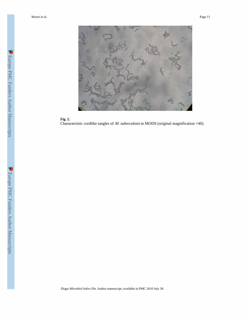
Fig. 2.Characteristic cordlike tangles of M. tuberculosis in MODS (original magnification ×40).
Moore et al. Page 11
Diagn Microbiol Infect Dis. Author manuscript; available in PMC 2010 July 30.
Europe PM
C Funders A
uthor Manuscripts
Europe PM
C Funders A
uthor Manuscripts

Fig. 3.Rule-out sequence to define contamination suspect positive cultures (n = 53) from 362culture-positive samples. *Includes 55 samples in which 1(n = 52), 2 (n = 2), or all 3 (n = 1)methods were irretrievably overgrown with fungi or bacteria; # 3 of the 4 were auraminepositive; $either positive or negative culture.
Moore et al. Page 12
Diagn Microbiol Infect Dis. Author manuscript; available in PMC 2010 July 30.
Europe PM
C Funders A
uthor Manuscripts
Europe PM
C Funders A
uthor Manuscripts

Fig. 4.Clinical, microbiologic, and molecular approach to determining cross-contamination (bysample).
Moore et al. Page 13
Diagn Microbiol Infect Dis. Author manuscript; available in PMC 2010 July 30.
Europe PM
C Funders A
uthor Manuscripts
Europe PM
C Funders A
uthor Manuscripts

Fig. 5.Contribution by method to “rule-out” of cross-contamination for 39 samples deemed true-positive cultures.
Moore et al. Page 14
Diagn Microbiol Infect Dis. Author manuscript; available in PMC 2010 July 30.
Europe PM
C Funders A
uthor Manuscripts
Europe PM
C Funders A
uthor Manuscripts

Europe PM
C Funders A
uthor Manuscripts
Europe PM
C Funders A
uthor Manuscripts
Moore et al. Page 15
Tabl
e 1
Indi
vidu
al d
ata
for
53 c
onta
min
atio
n su
spec
t sam
ples
Sam
ple
no.
Ori
gina
l cul
ture
pro
file
Rep
eat
cult
ure
prof
ilea
Uni
que
spol
igot
ype?
bIf
non
uniq
uesp
olig
otyp
e,un
ique
RF
LP
?c
Clin
ical
follo
w-u
pac
hiev
ed
Fol
low
-up
cult
ure
resu
ltd
Mic
robi
olog
icas
sess
men
t(r
ecul
ture
)
Mol
ecul
aras
sess
men
tC
linic
al a
sses
smen
t(i
nclu
ding
fol
low
-up
inve
stig
atio
ns)
Ove
rall
asse
ssm
ent
MO
DS
MB
Bac
TL
JM
OD
SM
BB
acT
LJ
MO
DS
MB
Bac
TL
J1
= C
onsi
sten
t w
ith
cros
s-co
ntam
inat
ion,
0 =
rega
rded
as
excl
udin
g cr
oss-
cont
amin
atio
n
1+
+−
−−
−N
N e
NN
AN
AN
A1
01
Not
cro
ss-c
onta
min
atio
n
2+
+−
+−
−Y
−N
NA
NA
NA
00
1N
ot c
ross
-con
tam
inat
ion
3+
+−
−−
−Y
−Y
−−
−1
01
Not
cro
ss-c
onta
min
atio
n
4+
+−
−−
−Y
−N
NA
NA
NA
10
1N
ot c
ross
-con
tam
inat
ion
5+
+−
−−
−Y
−N
NA
NA
NA
10
1N
ot c
ross
-con
tam
inat
ion
6+
−−
−−
−N
YN
NA
NA
NA
10
1N
ot c
ross
-con
tam
inat
ion
7+
−−
+−
−N
NY
NA
NA
NA
01
1N
ot c
ross
-con
tam
inat
ion
8+
−−
−−
−Y
−N
NA
NA
NA
10
1N
ot c
ross
-con
tam
inat
ion
9+
−−
−−
−Y
−Y
NA
NA
NA
10
1N
ot c
ross
-con
tam
inat
ion
10+
−−
−−
−Y
−Y
NA
NA
NA
10
1N
ot c
ross
-con
tam
inat
ion
11+
−−
−−
−Y
−N
NA
NA
NA
10
1N
ot c
ross
-con
tam
inat
ion
12+
−−
−−
−Y
−N
NA
NA
NA
10
1N
ot c
ross
-con
tam
inat
ion
13+
−−
−−
−Y
−Y
NA
NA
NA
10
1N
ot c
ross
-con
tam
inat
ion
14+
−−
−−
−Y
−N
NA
NA
NA
10
1N
ot c
ross
-con
tam
inat
ion
15+
−−
−−
−Y
−Y
NA
NA
NA
10
1N
ot c
ross
-con
tam
inat
ion
16+
−−
−−
−Y
−Y
NA
NA
NA
10
1N
ot c
ross
-con
tam
inat
ion
17−
+−
−−
−Y
−Y
−−
−1
01
Not
cro
ss-c
onta
min
atio
n
18−
−+
−−
−Y
−Y
NA
NA
NA
10
1N
ot c
ross
-con
tam
inat
ion
19+
+−
−−
−N
YY
OT
−O
T−
OT
−1
00
Not
cro
ss-c
onta
min
atio
n
20+
+−
−−
−N
YY
OT
−O
T−
OT
−1
00
Not
cro
ss-c
onta
min
atio
n
21+
+−
−−
−Y
−Y
−−
−1
00
Not
cro
ss-c
onta
min
atio
n
22+
+−
−−
−Y
−Y
NA
NA
NA
10
0N
ot c
ross
-con
tam
inat
ion
23+
+−
−−
−Y
−Y
NA
NA
NA
10
0N
ot c
ross
-con
tam
inat
ion
24+
−+
+−
−N
YY
NA
NA
NA
00
0N
ot c
ross
-con
tam
inat
ion
25+
−−
−−
−N
YY
OT
−O
T−
OT
−1
00
Not
cro
ss-c
onta
min
atio
n
Diagn Microbiol Infect Dis. Author manuscript; available in PMC 2010 July 30.

Europe PM
C Funders A
uthor Manuscripts
Europe PM
C Funders A
uthor Manuscripts
Moore et al. Page 16
Sam
ple
no.
Ori
gina
l cul
ture
pro
file
Rep
eat
cult
ure
prof
ilea
Uni
que
spol
igot
ype?
bIf
non
uniq
uesp
olig
otyp
e,un
ique
RF
LP
?c
Clin
ical
follo
w-u
pac
hiev
ed
Fol
low
-up
cult
ure
resu
ltd
Mic
robi
olog
icas
sess
men
t(r
ecul
ture
)
Mol
ecul
aras
sess
men
tC
linic
al a
sses
smen
t(i
nclu
ding
fol
low
-up
inve
stig
atio
ns)
Ove
rall
asse
ssm
ent
MO
DS
MB
Bac
TL
JM
OD
SM
BB
acT
LJ
MO
DS
MB
Bac
TL
J1
= C
onsi
sten
t w
ith
cros
s-co
ntam
inat
ion,
0 =
rega
rded
as
excl
udin
g cr
oss-
cont
amin
atio
n
26+
−−
−−
−N
YY
OT
+N
AO
T−
10
0N
ot c
ross
-con
tam
inat
ion
27+
−−
−−
−N
NY
−−
−1
10
Not
cro
ss-c
onta
min
atio
n
28+
−−
−−
−Y
−Y
NA
NA
NA
10
0N
ot c
ross
-con
tam
inat
ion
29+
−−
−−
−Y
−Y
NA
NA
NA
10
0N
ot c
ross
-con
tam
inat
ion
30+
−−
−−
−Y
−Y
NA
NA
NA
10
0N
ot c
ross
-con
tam
inat
ion
31+
−+
−−
−Y
−Y
++
+1
00
Not
cro
ss-c
onta
min
atio
n
32+
−−
−−
−Y
−Y
−−
−1
00
Not
cro
ss-c
onta
min
atio
n
33+
−−
−−
−Y
−Y
−−
−1
00
Not
cro
ss-c
onta
min
atio
n
34+
−−
−−
−Y
−Y
+−
+1
00
Not
cro
ss-c
onta
min
atio
n
35+
−−
−−
−Y
−Y
++
+1
00
Not
cro
ss-c
onta
min
atio
n
36+
−−
−−
−N
A−
YN
AN
AN
A1
NA
0N
ot c
ross
-con
tam
inat
ion
37−
+−
−−
−N
YY
−−
−1
00
Not
cro
ss-c
onta
min
atio
n
38−
−+
−−
−N
YY
−−
−1
00
Not
cro
ss-c
onta
min
atio
n
39−
−+
−−
−Y
−Y
−−
−1
00
Not
cro
ss-c
onta
min
atio
n
40+
+−
−−
−N
NY
−−
−1
11
Prob
able
cros
s-co
ntam
inat
ion
41+
+−
−−
−Y
−Y
OT
−O
T−
OT
−1
1f1
Prob
able
cros
s-co
ntam
inat
ion
42+
+−
−−
−Y
−N
NA
NA
NA
11f
1Pr
obab
lecr
oss-
cont
amin
atio
n
43+
−−
−−
−Y
YY
NA
NA
NA
11g
1Pr
obab
lecr
oss-
cont
amin
atio
n
44+
−−
−−
−Y
YY
NA
NA
NA
11g
1Pr
obab
lecr
oss-
cont
amin
atio
n
45+
−−
−−
−N
NY
NA
NA
NA
11
1Pr
obab
lecr
oss-
cont
amin
atio
n
46+
−−
−−
−N
NY
NA
NA
NA
11
1Pr
obab
lecr
oss-
cont
amin
atio
n
47+
−−
−−
−N
NY
−−
−1
11
Prob
able
cros
s-co
ntam
inat
ion
48+
−−
−−
−N
NY
NA
NA
NA
11
1Pr
obab
lecr
oss-
cont
amin
atio
n
Diagn Microbiol Infect Dis. Author manuscript; available in PMC 2010 July 30.

Europe PM
C Funders A
uthor Manuscripts
Europe PM
C Funders A
uthor Manuscripts
Moore et al. Page 17
Sam
ple
no.
Ori
gina
l cul
ture
pro
file
Rep
eat
cult
ure
prof
ilea
Uni
que
spol
igot
ype?
bIf
non
uniq
uesp
olig
otyp
e,un
ique
RF
LP
?c
Clin
ical
follo
w-u
pac
hiev
ed
Fol
low
-up
cult
ure
resu
ltd
Mic
robi
olog
icas
sess
men
t(r
ecul
ture
)
Mol
ecul
aras
sess
men
tC
linic
al a
sses
smen
t(i
nclu
ding
fol
low
-up
inve
stig
atio
ns)
Ove
rall
asse
ssm
ent
MO
DS
MB
Bac
TL
JM
OD
SM
BB
acT
LJ
MO
DS
MB
Bac
TL
J1
= C
onsi
sten
t w
ith
cros
s-co
ntam
inat
ion,
0 =
rega
rded
as
excl
udin
g cr
oss-
cont
amin
atio
n
49+
−−
−−
−N
NY
−−
−1
11
Prob
able
cros
s-co
ntam
inat
ion
50+
−−
−−
−N
NY
−−
−1
11
Prob
able
cros
s-co
ntam
inat
ion
51+
−−
−−
−N
A−
YN
AN
AN
A1
NA
1Pr
obab
lecr
oss-
cont
amin
atio
n
52−
+−
−−
−N
A−
YO
T−
OT
−O
T−
1N
A1
Prob
able
cros
s-co
ntam
inat
ion
53−
−+
−−
−N
A−
Y−
−−
1N
A1
Prob
able
cros
s-co
ntam
inat
ion
Tho
ugh
evid
ence
fro
m 1
app
roac
h m
ay h
ave
impl
ied
that
cro
ss-c
onta
min
atio
n w
as u
nlik
ely,
in s
ome
inst
ance
s, e
vide
nce
to th
e co
ntra
ry w
as r
egar
ded
as s
uper
ior
(e.g
., un
ique
and
dif
fere
nt m
olec
ular
fin
gerp
rint
s w
ere
obse
rved
for
2 c
ultu
res
from
the
sam
e pa
rtic
ipan
t in
who
mth
ere
was
no
clin
ical
sus
pici
on o
f di
seas
e—sh
e ha
d at
tend
ed th
e N
TP
asym
ptom
atic
ally
for
an
insu
ranc
e sc
reen
). N
A =
not
ava
ilabl
e.
a + =
rec
ultu
re o
f or
igin
al s
ampl
e po
sitiv
e; −
= r
ecul
ture
of
orig
inal
sam
ple
nega
tive.
b Uni
que
spol
igot
ype
for
date
of
sam
ple
proc
essi
ng a
nd 2
day
s at
eith
er s
ide
(5-d
ay w
indo
w);
4 p
atie
nts
had
2 sa
mpl
es w
ith id
entic
al s
polig
otyp
es (
11,1
2; 1
0,18
; 32,
33; 4
3,44
) un
ique
to e
ach
patie
nt.
c Stra
ins
with
con
tem
pora
ry m
atch
ing
spol
igot
ypes
wer
e al
so ty
ped
by R
FLP
to f
urth
er a
id d
iscr
imin
atio
n; th
us, “
uniq
ue R
FLP”
ref
ers
to d
iffe
rent
iatio
n fr
om s
trai
ns w
ith s
hare
d sp
olig
otyp
e.
d All
resu
lts a
re in
dup
licat
e (i
.e.,
2 sa
mpl
es p
roce
ssed
); O
T =
fol
low
-up
sam
ple
take
n on
TB
ther
apy.
e Cul
ture
d st
rain
s fr
om s
ampl
es 4
5 an
d 1,
but
no
othe
r co
ntem
pora
rily
pro
cess
ed s
ampl
es s
hare
d id
entic
al s
polig
otyp
es a
nd R
FLP
patte
rns;
45
prob
ably
cro
ss-c
onta
min
ated
fro
m 1
.
f Spol
igot
ypes
for
2 is
olat
es f
rom
the
sam
e sa
mpl
e di
ffer
.
g Alth
ough
spo
ligot
ypes
for
thes
e 2
sam
ples
fro
m th
e sa
me
patie
nt a
re te
mpo
rally
uni
que,
they
dif
fer
from
eac
h ot
her,
as
do th
eir
RFL
P pa
ttern
s.
Diagn Microbiol Infect Dis. Author manuscript; available in PMC 2010 July 30.