Influence of human dentine on the antibacterial activity of self-etching adhesive systems against...
8
UNCORRECTED PROOF Influence of human dentine on the antibacterial activity of self-etching adhesive systems against cariogenic bacteria Juliana O. Gondim a , Cristiane Duque b , Josimeri Hebling a , Elisa M.A. Giro a, * a Department of Paediatric Dentistry, School of Dentistry of Araraquara, Sa ˜o Paulo State University, UNESP, Rua Humaita ´, 1680 Araraquara, SP, Brazil b Department of Oral Diagnosis, School of Dentistry of Piracicaba, Sa ˜o Paulo State University, UNICAMP, Av. Limeira, 907 Piracicaba, SP, Brazil 1. Introduction Although the etiological factors of caries disease and the methods for its prevention have been widely investigated, secondary caries is still the main cause for replacement of restorations. 1,2 During removal of carious tissue, bacteria invariably remain entrapped in the dentinal substrate because neither the clinical parameters of dentine hardness and colour journal of dentistry xxx (2008) xxx–xxx 1 2 3 4 5 6 7 8 9 10 11 12 13 14 15 16 17 18 19 20 21 article info Article history: Received 27 September 2007 Received in revised form 7 December 2007 Accepted 22 December 2007 Keywords: Dentine bonding agents Bacteria Antibacterial agents Dentine Dentine permeability abstract Objectives: The incorporation of antibacterial agents into adhesive systems has been pro- posed to eliminate residual bacteria from dentine. This study used the agar diffusion method to evaluate the antibacterial activity of clearfil protect bond (CPB), clearfil SE bond (CSEB), clearfil Tri-S bond (C3SB) and Xeno-III (XIII) self-etching adhesive systems, with or without light-activation, against cariogenic bacteria, and to assess the influence of human dentine on the antibacterial activity of these materials. Methods: An aliquot of 10 ml per material (and individual components) were pipetted onto paper and dentine discs distributed in Petri dishes containing bacterial culture in BHI agar. Positive control was 0.2% chlorhexidine digluconate (CHX). Results: After incubation, the adhesive components of CPB and CSEB, liquid A of XIII and C3SB did not present antibacterial activity when applied to paper discs. The non-light- activated CPB primer + adhesive promoted the greatest inhibition of Streptococcus mutans ( p < 0.05), whereas with light-activation, there was no significant difference between primer + adhesive and primer alone. For Lactobacillus acidophilus, CPB primer presented the greatest antibacterial activity in both light-activation conditions ( p < 0.05). Regarding the dentine discs, only CHX promoted an inhibitory effect, though less intense than on paper discs ( p < 0.05). CHX presented greater antibacterial activity against S. mutans than against L. acidophilus ( p < 0.05). Conclusions: Light-activation significantly reduced the antibacterial activity of the self- etching adhesive systems; MDPB incorporation contributed to the effect of adhesive systems against cariogenic bacteria; the components eluted from the adhesive systems were not capable to diffuse through 400 mm-thick dentine disc to exert their antibacterial activity against cariogenic bacteria. # 2008 Elsevier Ltd. All rights reserved. * Corresponding author at: Faculdade de Odontologia de Araraquara, Departamento de Clı´nica Infantil Rua Humaita ´ , 1680 Centro Araraquara, SP, CEP 14801-903, Brazil. Tel.: +55 16 3301 6336. E-mail address: [email protected] (Elisa M.A. Giro). JJOD 1183 1–8 available at www.sciencedirect.com journal homepage: www.intl.elsevierhealth.com/journals/jden 0300-5712/$ – see front matter # 2008 Elsevier Ltd. All rights reserved. doi:10.1016/j.jdent.2007.12.007 Please cite this article in press as: Gondim JO, et al., Influence of human dentine on the antibacterial activity of self-etching adhesive systems against cariogenic bacteria, Journal of Dentistry (2008), doi:10.1016/j.jdent.2007.12.007
-
Upload
independent -
Category
Documents
-
view
3 -
download
0
Transcript of Influence of human dentine on the antibacterial activity of self-etching adhesive systems against...

123
4
5
6
7
8
9
101112
13
14
15
16
17
JJOD 1183 1–8
NC
OR
RE
CTE
D P
RO
OFInfluence of human dentine on the antibacterial activity of
self-etching adhesive systems against cariogenic bacteria
Juliana O. Gondim a, Cristiane Duque b, Josimeri Hebling a, Elisa M.A. Giro a,*aDepartment of Paediatric Dentistry, School of Dentistry of Araraquara, Sao Paulo State University, UNESP, Rua Humaita,
1680 Araraquara, SP, BrazilbDepartment of Oral Diagnosis, School of Dentistry of Piracicaba, Sao Paulo State University, UNICAMP, Av. Limeira,
907 Piracicaba, SP, Brazil
j o u r n a l o f d e n t i s t r y x x x ( 2 0 0 8 ) x x x – x x x
a r t i c l e i n f o
Article history:
Received 27 September 2007
Received in revised form
7 December 2007
Accepted 22 December 2007
Keywords:
Dentine bonding agents
Bacteria
Antibacterial agents
Dentine
Dentine permeability
a b s t r a c t
Objectives: The incorporation of antibacterial agents into adhesive systems has been pro-
posed to eliminate residual bacteria from dentine. This study used the agar diffusion
method to evaluate the antibacterial activity of clearfil protect bond (CPB), clearfil SE bond
(CSEB), clearfil Tri-S bond (C3SB) and Xeno-III (XIII) self-etching adhesive systems, with or
without light-activation, against cariogenic bacteria, and to assess the influence of human
dentine on the antibacterial activity of these materials.
Methods: An aliquot of 10 ml per material (and individual components) were pipetted onto
paper and dentine discs distributed in Petri dishes containing bacterial culture in BHI agar.
Positive control was 0.2% chlorhexidine digluconate (CHX).
Results: After incubation, the adhesive components of CPB and CSEB, liquid A of XIII and
C3SB did not present antibacterial activity when applied to paper discs. The non-light-
activated CPB primer + adhesive promoted the greatest inhibition of Streptococcus mutans
( p < 0.05), whereas with light-activation, there was no significant difference between
primer + adhesive and primer alone. For Lactobacillus acidophilus, CPB primer presented
the greatest antibacterial activity in both light-activation conditions (p < 0.05). Regarding
the dentine discs, only CHX promoted an inhibitory effect, though less intense than on paper
discs ( p < 0.05). CHX presented greater antibacterial activity against S.mutans than against L.
acidophilus ( p < 0.05).
Conclusions: Light-activation significantly reduced the antibacterial activity of the self-
etching adhesive systems; MDPB incorporation contributed to the effect of adhesive systems
against cariogenic bacteria; the components eluted from the adhesive systems were not
capable to diffuse through 400 mm-thick dentine disc to exert their antibacterial activity
against cariogenic bacteria.
# 2008 Elsevier Ltd. All rights reserved.
avai lab le at www.sc iencedi rec t .com
journal homepage: www. int l .e lsev ierhea l th .com/ journa ls / jden
18
19
20
21
U1. Introduction
Although the etiological factors of caries disease and the
methods for its prevention have been widely investigated,
* Corresponding author at: Faculdade de Odontologia de AraraquarAraraquara, SP, CEP 14801-903, Brazil. Tel.: +55 16 3301 6336.
E-mail address: [email protected] (Elisa M.A. Giro).
0300-5712/$ – see front matter # 2008 Elsevier Ltd. All rights reserveddoi:10.1016/j.jdent.2007.12.007
Please cite this article in press as: Gondim JO, et al., Influence of hu
systems against cariogenic bacteria, Journal of Dentistry (2008), do
secondary caries is still the main cause for replacement of
restorations.1,2 During removal of carious tissue, bacteria
invariably remain entrapped in the dentinal substrate because
neither the clinical parameters of dentine hardness and colour
a, Departamento de Clınica Infantil Rua Humaita, 1680 Centro
.
man dentine on the antibacterial activity of self-etching adhesive
i:10.1016/j.jdent.2007.12.007

22
23
24
25
26
27
28
29
30
31
32
33
34
35
36
37
38
39
40
41
42
43
44
45
46
47
48
49
50
51
52
53
54
55
56
57
58
59
60
61
62
63
64
65
66
67
68
69
70
71
72
73
74
75
76
j o u r n a l o f d e n t i s t r y x x x ( 2 0 0 8 ) x x x – x x x2
JJOD 1183 1–8
nor the caries-detector dyes are able to ensure complete
elimination of microorganisms.3,4 As the contemporary
dentine adhesive systems have not proved completely
effective in eliminating microleakage at tooth/restoration
interface, the incorporation of antibacterial agents into these
materials has been proposed in an attempt to prevent caries
recurrence.5–15
Among the antibacterial agents incorporated into adhesive
materials, the resin monomer 12-methacryloyloxydodecyl-
pyridinium bromide, known as MDPB, stands out.8,9,13 The
main advantage of MDPB is its capacity to copolymerize with
other resin monomers being immobilised within the polymer
matrix, which confers safety and prolonged antibacterial
action to this agent, as it does not leach to the medium. This
characteristic also ensures a good survival rate for the
restoration, as MDPB, unlike soluble antibacterial agents, is
not deleterious to the physical and mechanical proprieties of
the materials to which it is incorporated.8,9,16
The incorporation of MDPB into self-etching adhesive
systems has been advocated as extremely advantageous
because, in the clinical practice, these materials are directly
applied to the dentine with some level of contamination and,
unlike the two-step or three-step total-etching adhesive
systems, they do not have a separate acid etching step in
their protocol, which could reduce significantly the number of
residual microorganisms.14,17,18 In spite of the acidic nature of
self-etching adhesive systems, this characteristic does not
seem sufficient to completely eliminate residual bacteria from
dentine because this tissue acts as a solid buffering medium to
acidic monomers.15,19 Therefore, for the self-etching adhesive
UN
CO
RR
E
Table 1 – Specifications, main components and manufacturer
Material (manufacturer) Type
Clearfill protect bond (CPB)
(Kuraray Medical Inc.
Okayama, Japan)
Two-step self-etching
adhesive system
Prim
dimet
Adhes
hydro
N,N-d
CQ, si
BisGM
Clearfil SE bond (CSEB),
(Kuraray Medical Inc.,
Okayama, Japan)
Two-step self-etching
adhesive system
Prim
dimet
p-tolu
HEMA
dimet
N,N-d
CQ, si
silica,
Clearfil Tri-S bond (C3SB)
(Kuraray Medical Inc.
Okayama, Japan)
One-step self-etching
adhesive system
HEM
dimet
silica,
Xeno-III (XIII) (Dentsply,
Konstanz, Germany)
One-step self-etching
adhesive system
Liqui
THB, a
B: Piro
Ethyl
0.2% chlorhexidine digluconate
(CHX) (University Pharmacy,
UNESP, Araraquara, SP, Brazil)
Antibacterial solution 0.2%
aqueo
BisGMA: bisphenol A glycidyl dimethacrylate (BisGMA); THB: toluene
methacrylate; MDP: 10-methacryloxydecyl dihydrogen phosphate; MD
fluoro phosphazene modified methacrylate; Piro-EMA: phosphoric acid m
Please cite this article in press as: Gondim JO, et al., Influence of hu
systems against cariogenic bacteria, Journal of Dentistry (2008), do
ED
PR
OO
F
systems to inhibit the microorganisms that remain lodged
inside the dentinal tubules, they should present antibacterial
activity and capacity to diffuse to some extent through the
dentin.19
However, there is little published information on the
influence of dentine on the antibacterial properties of self-
etching adhesive systems.13,19,20 Therefore, the aim of this
study was to evaluate the antibacterial activity of self-etching
adhesive systems, with or without light-activation, against
cariogenic bacteria, and to assess the influence of the
interposition of 400 mm-thick human dentine discs on the
antibacterial activity of these materials using the agar
diffusion method.
2. Materials and methods
The specifications, main components and manufacturers of
the materials used in this study are presented in Table 1. The
antibacterial activity of the materials was evaluated against
the following bacterial strains: Streptococcus mutans (UA-159)
and Lactobacillus acidophilus (ATCC #IAL-523).
2.1. Agar diffusion method: application on paper discs
CPB, CSEB, C3SB and XIII self-etching adhesive systems, as
well as their individual components, were submitted to the
agar diffusion method. The positive and negative controls
were, respectively, 0.2% chlorhexidine digluconate (CHX) and
paper discs not impregnated with any material (PD).
CT
s of the tested materials
Composition pH Batch#
er: HEMA, MDP, hydrophilic
hacrylate, MDPB, water.
ive: HEMA, MDP,
phobic dimethacrylate,
iethanol-p-toluidine,
lanized colloidal silica,
A, sodium fluoride
P: 1.9. A: 2.8 41,137
er: HEMA, MDP, hydrophilic
hacrylate, N,N-diethanol-
idine, CQ, water. Adhesive:
, MDP, hydrophobic
hacrylate,
iethanol-p-toluidine,
lanized colloidal
BisGMA
P: 1.9. A: 2.8 51,308
A, BisGMA, MDP, hydrophobic
hacrylate, silanized colloidal
CQ, ethanol, water
2.4 61,113
d A: HEMA, water, ethanol,
morphous silica. Liquid
-EMA, PEM-F, UDMA, THB, CQ,
4-dimethylaminobenzoate
1.0 (mixed) 0,504,002,710
chlorhexidine digluconate
us solution
5.9 –
hydroxybutyrate; CQ: D,1-camphorquinone; HEMA: 2-hydroxyethyl
PB: 12-methacryloyloxydodecyl pyridinium bromide; PEM-F: mono
odified methacrylacte; UDMA: urethane dimethacrylate.
man dentine on the antibacterial activity of self-etching adhesive
i:10.1016/j.jdent.2007.12.007

C
77
78
79
80
81
82
83
84
85
86
87
88
89
90
91
92
93
94
95
96
97
98
99
100
101
102
103
104
105
106
107
108
109
110
111
112
113
114
115
116
117
118
119
120
121
122
123
124
125
126
127
128
129
130
131
132
133
134
135
136
137
138
139
140
141
142
143
144
145
146
147
148
149
150
151
152
153
154
155
156
157
158
159
160
161
162
163
164
165
166
167
168
169
170
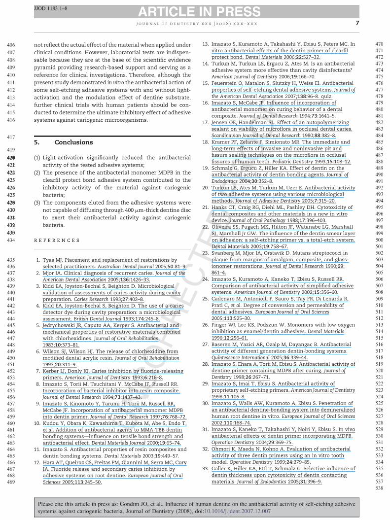
Fig. 1 – (A) 400 mm-thick dentine disc with a diameter of
8 mm. (B) Dentine disc with a protective barrier delimiting
a 6 mm diameter area. (C) Distribution of the specimens in
agar plate.
j o u r n a l o f d e n t i s t r y x x x ( 2 0 0 8 ) x x x – x x x 3
JJOD 1183 1–8
UN
CO
RR
E
For the agar diffusion method, the standard bacterial strains
were first reactivated in 5 ml of BHI broth (Brain Heart Infusion,
BHITM; Difco Laboratories, Detroit, MI, USA) for 24 h at 37 8C
under conditions of microaerophilia. From this culture, another
replication was performed in 5 ml of BHI broth and maintained
for 24 h at 37 8C in microaerophilia, which allowed obtaining an
inoculum containing 109 CFU/ml. To verify and standardize the
number of microorganisms present in the inoculum, serial
dilutions were performed up to 10�6.
An aliquot of 250 ml of one of the bacterial inocula was
spread onto the surface of Petri dishes containing 15 ml of BHI
agar. Because of the number of groups (n = 12), two Petri dishes
were necessary to distribute the specimens in such way that
on each dish seven paper discs (6 mm diameter; 1.5 mm thick)
were used. Five discs represented five different adhesive
groups, and two discs the control groups, which were repeated
for each Petri dish. A total of 8 specimens were used per group,
except for the positive and negative controls (n = 16). In the
group of specimens that were light activated, all materials
(including the positive and negative controls) were irradiated
for 10 s with a halogen light curing unit (Optilux 500; Kerr
Company, Orange, CA, USA) with a light intensity not lower
than 400 mW/cm2.
The dishes were maintained at room temperature for 2 h
for pre-diffusion of the materials and were incubated at 37 8C
in microaerophilia during 24 h for S. mutans and 48 h for L.
acidophilus. The diameter of the bacterial growth inhibition
zones produced around the paper discs impregnated with the
tested materials was measured at two regions in millimetres
using a digital calipter (Mitutoyo, Kawasaki, Japan). The largest
diameters measured horizontally and vertically were recorded
and an average was calculated for each inhibition zone. The
diameter of the paper disc (6 mm) was subtracted from this
value and the result was divided by two to obtain the width of
the inhibition zone of each specimen.
For positive and negative control groups eight paper discs
were randomly selected for analysis.
2.2. Agar diffusion method: application on dentine discs
In this assay, only the materials that presented antibacterial
activity by the agar diffusion method on paper discs were used
with the same bacterial strains. CHX was used as a positive
control applied onto dentine discs (CHXd) and paper discs
(CHXp).
Eight-four sound human third molars obtained from the
tooth bank of the School of Dentistry of Araraquara (UNESP,
Araraquara, SP, Brazil) were used after approval of the
research project by the institutional Ethics in Human Research
Committee (Process #54/05). The teeth were used within 6
months after extraction. For each tooth, a dentine disc of
approximately 500 mm thickness was obtained by transversal
section of the tooth at the cervical third using a water-cooled
diamond saw (Series 15 LC Diamond; Buehler, Lake Buff, IL,
USA) in a low-speed cutting machine (Isomet 1000; Buehler,
Lake Buff, IL, USA). The dentine discs were manually ground
with 320-grit silicon carbide paper until reaching a thickness
of 400 mm and were stored in phosphate buffer saline (PBS), pH
7.2, at �48C until the moment of dentine permeability
determination.
Please cite this article in press as: Gondim JO, et al., Influence of hu
systems against cariogenic bacteria, Journal of Dentistry (2008), do
TED
PR
OO
F
2.3. Determination of dentine permeability
The dentine discs were bilaterally etched with 0.5 M EDTA
solution, pH 7.2, for 30 s and thoroughly washed with deionised
water to remove the smear layer created during grounding.
Thereafter, the discs were individually held in a device known
as in vitro pulp chamber, modified from the original apparatus
developed by Hanks et al.,21 which allowed standardisation of a
dentine area of 0.28 cm2 for assessment of permeability. The
artificial pulp chamber was connected to an appliance (Flodec;
De Marco Engineering, Geneva, Switzerland) by means of a
0.5 mm-diameter polyethylene tube for dentine permeability
reading. A small air bubble was introduced into the system and
15 cm H2O hydrostatic pressure was applied. The air bubble
movement was recorded by an appliance’s infrared sensor and
transformed in dentine permeability value using proprietary
software (De Marco Engineering, Geneva, Switzerland). Based
onthe results of these readings, the dentinediscswereallocated
in 14 experimental groups with six discs each in such a way that
the mean dentine permeability of the groups did not differ
statisticaly (ANOVA; p > 0.05).
2.4. Determination of antibacterial activity
The 400 mm-thick dentine discs were reduced using #1090
diamond burs (KG Sorensen, Barueri, SP, Brazil) and 320-grit
SiC paper to a standardized diameter of 8 mm (Fig. 1A). The
occlusal surface of the discs was wet ground with 320-grit SiC
paper for 5 s to produce a new smear layer.22 On that surface a
dam was built up surrounding the periphery of the disc with
polyether impression material (Impregum Penta Soft; 3M
ESPE, Seefeld, Germany) using a PentaTM Elastomer syringe
(3M ESPE, Seefeld, Germany), in such a way to delimit a 6 mm
diameter area for application of the tested materials and to
avoid extravasation of the adhesives (Fig. 1B). The discs were
stored for 24 h in saline for complete setting of the impression
material and were thereafter autoclaved at 120 8C for 20 min.
The bacterial suspensions of S. mutans and L. acidophilus
were seeded onto six Petri dishes each, containing 15 ml of BHI
man dentine on the antibacterial activity of self-etching adhesive
i:10.1016/j.jdent.2007.12.007

E
OO
F
171
172
173
174
175
176
177
178
179
180
181
182
183
184
185
186
187
188
189
190
191
192
193
194
195
196
197
198
199
200
201
202
203
204
205
206
207
208
Table 2 – Width of the inhibition zones (mm) obtained for S. mutans (median, minimum and maximum) according to thematerial and light-activation condition by the agar diffusion test in paper discs
Material Without light-activation With light-activation
Clorexidina (CHX) (n = 8) 4.65 (3.88–5.00) Bb 5.09 (4.40–5.88) Aa
Paper disc (PD) (n = 8) 0 0
Clearfil protect bond-primer (CPB-P) (n = 8) 5.05 (3.65–6.56) Ab 4.75 (3.84–5.54) Aa
Clearfil protect bond-adhesive (CPB-A) (n = 8) 0 0
Clearfil protect bond-primer + adhesive (CPB-P + A) (n = 8) 6.92 (6.20–7.74) Aa 5.04 (4.08–6.13) Ba
Clearfil SE bond-primer (CSEB-P) (n = 8) 4.06 (3.40–4.56) Ac 1.96 (1.68–2.23) Bc
Clearfil SE bond-adhesive (CSEB-A) (n = 8) 0 0
Clearfil SE bond-primer + adhesive (CSEB-P + A) (n = 8) 3.39 (3.01–3.68) Ad 2.43 (1.73–3.19) Bb
Clearfil Tri S bond (C3SB) (n = 8) 0 0
Xeno-III liquid A (XIII-A) (n = 8) 0 0
Xeno-III liquid B (XIII-B) (n = 7) 3.04 (2.38–3,33) Ae 2.06 (1.79–3.21) Bbc
Xeno-III liquid A + liquid B (XIII-A + B) (n = 7) 2.19 (1.06–2.81) Af 1.46 (1.16–1.51) Bd
Different uppercase letters in lines and lowercase letters in rows indicate statistically significant difference at 5%.
j o u r n a l o f d e n t i s t r y x x x ( 2 0 0 8 ) x x x – x x x4
JJOD 1183 1–8
RR
agar. One paper and seven dentine discs were distributed in
each dish. After that 10 ml of the positive control (CHX) and the
other tested materials were pipetted on their surface. The
adhesive systems were not light-activated. After incubation of
the Petri dishes at 37 8C under conditions of microaerophilia
during 24 h (S. mutans) and 48 h (L. acidophilus), the width of the
inhibition zones produced around each specimen was
calculated in the same way as described for the agar diffusion
method with paper discs (Fig. 1C).
2.5. Statistical analysis
The Mann–Whitney non-parametric test was used to evaluate
the influence of light-activation on the antibacterial activity of
each adhesive system when using the agar diffusion method
with paper discs, and Kruskal–Wallis and Mann–Whitney tests
were used to compare the materials under each light-
activation condition. The evaluation of the influence of human
dentine on the materials’ antibacterial activity was performed
by comparing the widths of the bacterial growth inhibition
zones of the groups, using the Mann–Whitney test. Spear-
man’s correlation was also used to verify the existence of a
correlation between dentine permeability and inhibition zone
UN
CO
Table 3 – Width of the inhibition zones (mm) obtained for L. athe material and light-activation condition by the agar diffusi
Material
Chlorhexidine (CHX) (n = 8)
Paper disc (PD) (n = 8)
Clearfil protect bond-primer (CPB-P) (n = 8)
Clearfil protect bond-adhesive (CPB-A) (n = 8)
Clearfil protect bond-primer + adhesive (CPB-P + A) (n = 8)
Clearfil SE bond-primer (CSEB-P) (n = 8)
Clearfil SE bond-adhesive (CSEB-A) (n = 8)
Clearfil SE bond-primer + adhesive (CSEB-P + A) (n = 8)
Clearfil Tri S bond (C3SB) (n = 8)
Xeno-III liquid A (XIII-A) (n = 8)
Xeno-III liquid B (XIII-B) (n = 6)
Xeno-III liquid A + liquid B (XIII-A + B) (n = 6)
Different uppercase letters in lines and lowercase letters in rows indicat
Please cite this article in press as: Gondim JO, et al., Influence of hu
systems against cariogenic bacteria, Journal of Dentistry (2008), do
CTE
D P
Rwidths. A significance level of 5% (a = 0.05) was set for all
statistical tests.
3. Results
3.1. Agar diffusion method: application to paper discs
The medians of inhibition zone widths and the minimum and
maximum values according to the bacterial strain, material
and light-activation condition are presented in Tables 2 and 3.
Regardless of the type of microorganism and presence/
absence of light-activation, no inhibition zone was produced
with the adhesive components of clearfil protect bond (CPB-A)
and Clearfil SE bond (CSEB-A), liquid A of Xeno-III (XIII-A) and
clearfil Tri-S bond adhesive system (C3SB).
3.2. S. mutans
Clearfil protect bond (primer + adhesive) without light-activa-
tion produced the largest inhibition zone (p < 0.05), followed
by CPB primer (CPB-P) and CHX, which did not differ to each
other (p > 0.05). With light-activation, there was no statisti-
cidophilus (median, minimum and maximum) according toon test in paper discs
Without light-activation With light-activation
5.06 (4.03–5.44) Ac 4.47 (4.03–5.93) Ad
0 0
7.15 (6.99–8.37) Aa 6.93 (6.34–7.64) Aa
0 0
6.52 (6.21–7.00) Ab 6.12 (5.62–6.59) Bb
6.37 (5.84–7.46) Ab 5.59 (3.65–7.48) Abcd
0 0
6.52 (6.19–7.05) Ab 5.62 (5.19–6.13) Bc
0 0
0 0
0.68 (0.40–0.78) Ad 0.80 (0.42–0.96) Ae
0.66 (0.58–1.10) Ad 0.58 (0.49–0.73) Ae
e statistically significant difference at 5%.
man dentine on the antibacterial activity of self-etching adhesive
i:10.1016/j.jdent.2007.12.007

C
209
210
211
212
213
214
215
216
217
218
219
220
221
222
223
224
225
226
227
228
229
230
231
232
233
234
235
236
237
238
239
240
241
242
243
244
245
246
247
248
249
250
251
252
253
254
255
256
257
258
259
260
261
262
263
264
265
266
267
268
269
270
271
272
273
274
275
276
277
278
279
280
281
282
283
284
285
j o u r n a l o f d e n t i s t r y x x x ( 2 0 0 8 ) x x x – x x x 5
JJOD 1183 1–8
RR
E
cally significant difference between CPB components together
(CPB-P + A) and the primer alone (CPB-P), which presented
similar inhibition to that of CHX (p > 0.05). Regarding the
effect of light-activation on the inhibitory potential, the
materials, in general, exhibited lower antibacterial activity
after light-activation (p < 0.05), except for CPB-P, which
results with and without halogen light curing did not differ
significantly to each other (Table 2).
3.3. L. acidophilus
CPB-P presented the greatest antibacterial activity under both
conditions of curing (p < 0.05). Regarding the effect of light-
activation, there was no statistically significant difference
(p > 0.05) among the tested materials, except for CPB-P + A
and CSEB-P + A, which exhibited greater inhibitory potential
without light-activation (p < 0.05) (Table 3).
3.4. Agar diffusion method: application to dentine discs
When the dentine disc was interposed, none of the tested self-
etching adhesive systems presented antibacterial activity.
Although CHX presented an inhibitory effect against S. mutans
and L. acidophilus, it was less intense than that observed on the
paper disc (p < 0.05). In addition, CHX showed greater
antibacterial activity against S. mutans than against L.
acidophilus (p < 0.05) (Table 4). There was no positive correla-
tion between the degree of permeability of the dentine discs
and the produced antibacterial effect (r = �0.119; p > 0.05).
4. Discussion
The antibacterial activity of the tested self-etching adhesive
systems and their individual components with and without
light-activation was evaluated against S. mutans and L.
acidophilus, the main pathogens directly related to the
development of dental caries.23 The agar diffusion method
was used for being a widely employed methodology for
determination of the inhibitory activity of liquid substances, in
addition to being a simple and easy-to-handle technique and
facilitate the comparison to other studies.9,13,15,19,20,24
UN
CO
Table 4 – Widths of the inhibition zones (mm) obtained for S. maccording to the group by the agar diffusion test in dentin dis
Group
Chlorhexidine in paper disc (CLXp)
Chlorhexidine in dentin disc (CLXd)
Clearfil protect bond-primer (CPB-P)a
Clearfil protect bond-primer + adhesive (CPB-P + A)a
Clearfil SE bond-primer (CSEB-P)a
Clearfil SE bond-primer + adhesive (CSEB-P + A)a
Xeno-III liquid B (XIII-B)a
Xeno-III liquid A + liquid B (XIII-A + B)a
Different uppercase letters in lines and lowercase letters in rows indicata Uncured dentine bonding agents.
Please cite this article in press as: Gondim JO, et al., Influence of hu
systems against cariogenic bacteria, Journal of Dentistry (2008), do
TED
PR
OO
F
Corroborating the findings of Imazato et al.,24 it was
observed that the non-light-activated materials presented
significantly greater antibacterial activity in the groups CPB-
P + A, CSEB-P, CSEB-P + A, XIII-B and XIII-A + B when the
bacterial strain was S. mutans, and for the groups CPB-P + A
and CSEB-P + A, when the bacterial strain was L. acidophilus
(p < 0.05). Nevertheless, even with light-activation, there was
some antibacterial activity for these materials when applied to
paper discs. The polymerization of adhesive materials causes
entrapment in the polymeric matrix and decreases the release
of polymerizable antibacterial components, as well as adhe-
sion-promoting acidic monomers.24 Therefore, the formation
of inhibition zones of microbial growth when the adhesive
systems were light-activated may be explained by the fact that
a complete conversion of monomers into polymers does not
occur and hence residual monomers can be released to the
medium.25 Furthermore, the halogen light is not capable of
passing through the paper disc impregnated with the adhesive
components adequately, what added to the deficient blown off
water present in the primer composition when it is applied
onto paper discs, prevents great part of the monomers from
forming polymeric chains.24 The antibacterial effect might
also be due to the release of residual monomers present in the
oxygen-inhibited layer.26
The antibacterial action of adhesive systems may be
influenced by factors inherent to the material itself (pH,
viscosity, diffusion capacity and presence of antibacterial
agents) and factors related to dentine substrate (thickness and
permeability).14,15,19,24,27–31 The acidic nature of the primer of
self-etching adhesive systems has been considered as one of
the key factors related to bacterial inhbition.24,25,27,29 Phenyl-P,
4-META and MDP are the main acidic monomers contained in
the formulation of self-etching adhesive systems and are
capable of demineralizing and infiltrating simultaneously the
dentine substrate. According to Ohmori et al.,32 MDP has a
higher inhibitory action against microorganisms than Phenyl-
P. MDP is present both in the primer and in the adhesive
components of CPB and CSEB, and in C3SB (single-bottle).
Nevertheless, in the present study, as previously pub-
lished,19,20,27 only the primer alone and the primer + adhesive
association of CPB and CSEB exhibited antibacterial effect
against the tested bacterial strains when applied to paper
utans and L. acidophilus (median, minimum and maximum)cs
Microorganism
S. mutans (n = 6) L. acidophilus (n = 6)
6.34 (5.68–7.17) Aa 5.45 (4.50–6.31) Ba
2.80 (1.12–3.70) Ab 1.02 (0.78–1.61) Bb
0 0
0 0
0 0
0 0
0 0
0 0
e statistically significant difference at 5%.
man dentine on the antibacterial activity of self-etching adhesive
i:10.1016/j.jdent.2007.12.007

E
286
287
288
289
290
291
292
293
294
295
296
297
298
299
300
301
302
303
304
305
306
307
308
309
310
311
312
313
314
315
316
317
318
319
320
321
322
323
324
325
326
327
328
329
330
331
332
333
334
335
336
337
338
339
340
341
342
343
344
345
346
347
348
349
350
351
352
353
354
355
356
357
358
359
360
361
362
363
364
365
366
367
368
369
370
371
372
373
374
375
376
377
378
379
380
381
382
383
384
385
386
387
388
389
390
391
392
393
394
395
396
397
398
399
400
401
402
403
404
405
j o u r n a l o f d e n t i s t r y x x x ( 2 0 0 8 ) x x x – x x x6
JJOD 1183 1–8
UN
CO
RR
discs. In spite of containing MDP, the lack of antibacterial
activity of C3SB and the adhesive component alone of CPB and
CSEB might be attributed to the presence of hydrophobic
molecules in their formulation, which may impair their
diffusion in the agar medium.24
When applied to 400 mm-thick dentine discs, neither of the
tested materials presented antibacterial activity. This lack of
antibacterial activity may be attributed to the protective effect
produced by the buffer capacity of dentine against acidic
agents, that is, the dissolution of dentine apatite by the action
of acids may be neutralised by the bonds between dentine
phosphate and carboxylic ions with H+ protons. In addition,
the thicker the remaining dentine, the greater the buffering
effect.33 Therefore, the deeper the microorganisms are lodged
into the dentinal tubules, the lower the effectiveness of these
materials to successfully eliminate them.
Fluoride has been incorporated into adhesive systems and
restorative materials, but its activity is more related to the
inhibition of the demineralisation process and optimisation of
remineralisation than to its antibacterial activity.12 In accor-
dance with the findings of previous studies,19,20 in the present
investigation Xeno-III showed low inhibitory activity when
applied to paper discs, mainly against L. acidophilus. This
antibacterial effect has been attributed to the liquid B of this
adhesive system that contains fluoride among other compo-
nents. Schmalz et al.19 have stated that the removal of fluoride
annulled the antibacterial effect of Prime&Bond NT. Never-
theless, in spite of containing fluoride and having the lowest
pH (pH 1) among the materials evaluated in the present study,
Xeno-III has a greater viscosity due to the presence of UDMA in
its formulation, which is a hydrophobic resin monomer with
high molecular weight. Therefore, it is speculated that the
viscosity may modulate the antibacterial activity of this
material20 and hinder its diffusion through the dentinal
tubules, which could explain the lack of inhibition of bacterial
growth observed when applied to dentine discs.
Among the self-etching adhesive systems evaluated in this
study, CPB presented the greatest antibacterial activity when
applied onto paper discs. Similar effect has been reported in
previous studies.9,11,14,15,17,19,20 In addition to MDP, the primer
of this adhesive system contains another resin monomer,
known as MDPB, which is an antibacterial agent composed of a
quaternary ammonium with a methacryloyl group.8 The
incorporation of MDPB into adhesive systems does not
interfere with their acidic potential and viscosity.30 An
advantage of MDPB in relation to other antibacterial agents
is the fact that it is copolymerized together with other
monomers and does not leach from the material after light-
activation. In addition, this monomer maintains its antibac-
terial activity, though to a less extent, when the microorgan-
ism gets in contact with the material surface.8,11,13,15,28
In an attempt to elucidate the real contribution of MDPB,
Imazato et al.13 compared the antibacterial potential of the
primer of clearfil protect bond before and after removal of this
resin monomer from the primer’s composition. The authors
observed that, in the absence of MDPB, the primer did not exert
antibacterial effect against S. mutans and Lactobacillus. This
findings demonstrates that the antibacterial effect of CPB
depends not only on the acidity of the material, but also (and
mainly) on the presence of MDPB.31,34
Please cite this article in press as: Gondim JO, et al., Influence of hu
systems against cariogenic bacteria, Journal of Dentistry (2008), do
CTE
D P
RO
OF
Schmalz et al.19 evaluated the effect of dentine on the
antibacterial activity of adhesive systems, TEGDMA and HEMA
against S. mutans, S. sobrinus and L. acidophilus using different
agar diffusion methods. Unlike the results of the present
study, those authors found that the tested adhesive systems
produced bacterial growth inhibition zones around bovine
dentine discs. However, their results also demonstrated that
the dentine influences the antibacterial effect of the adhesive
systems reducing or annulling it. These findings are consistent
with those of the present study and prior investigations,18
which have also shown the modulation effect exerted by the
dentine substrate. It should be mentioned, however, that
some methodological differences existing between the pre-
sent study and the study conducted by Schmalz et al.19 may
possibly explain the distinct results obtained in both studies,
among which, dentine disc type and thickness and the
amount of adhesive system applied to disc surface. In the
present study, 400 mm-thick human dentine discs were used
and 10 ml of material (adhesive system or individual compo-
nents) were pipetted on the surface of each specimen, while
Schmalz et al.19 used 200–500 mm-thick bovine dentine speci-
mens that were impregnated with 20 ml of the tested
materials. In addition, in the present study, a protective
barrier was built to avoid overflow of the material applied to
the surface of the dentine discs.
Dentine has a recognized protective action, however this
effect is limited.33 The dentinal substrate may impede or
difficult the diffusion of large-sized molecules, while allows
the diffusion of toxins resulting from the bacterial metabolism
and small-sized hydrophilic molecules, such as TEGDMA and
HEMA, which, in spite of not presenting bactericidal effect are
cytotoxic.35,36 The lack of production of inhibition zones
observed in this study indicates that the components eluted
from the adhesives were not able to diffuse through 400 mm-
thick dentine or did not exert inhibitory effect against
cariogenic bacteria on the agar.
Dentine permeability varies from tooth to tooth and from
one region to another in the same tooth.37 Some factors, such
as the anatomic arrangement, diameter, length and density of
the dentinal tubules, may be related to this variation.37 Thus,
the permeability of the dentine discs used in this study was
measured in order to obtain groups with statistically similar
dentine permeability means and hence avoid the influence of
this confounder in group comparisons.
Material’s viscosity may also interfere with its diffusion
through the dentinal tubules.37,38 Therefore, the difference of
viscosity of the materials evaluated in this study might have
influenced the obtained results, as only CHX (pH 5.9), which is
a low-viscosity material, was able to permeate the smear layer
and penetrate the dentinal tubules, exerting an antibacterial
effect. Although this effect was lower than that produced
when the material was applied to paper discs (p < 0.05),
demonstrating the influence of dentine substrate on the
material’s antibacterial activity.
The methodology employed in the present experiment has
some limitations in reproducing the physiological conditions
of the pulpodentinal complex, such as the presence of
intratubular fluid and intra-pulpal pressure. Due to these
limitations, the results obtained in in vitro studies cannot be
directly extrapolated to the in vivo situation because they may
man dentine on the antibacterial activity of self-etching adhesive
i:10.1016/j.jdent.2007.12.007
C
406
407
408
409
410
411
412
413
414
415
416
417
418
419
420
421
422
423
424
425
426
427
428
429
430
420421
422
423424
425
426
427
428429
430
431
432
433
434
435
436
437
438
439
440
441
442
443
444
445
446
447
448
449
450
451
452
453
454
455
456
457
458
459
460
461
462
463
464
465
466
467
468
469
470
471
472
473
474
475
476
477
478
479
480
481
482
483
484
485
486
487
488
489
490
491
492
493
494
495
496
497
498
499
500
501
502
503
504
505
506
507
508
509
510
511
512
513
514
515
516
517
518
519
520
521
522
523
524
525
526
527
528
529
530
531
532
533
534
535
536
537
j o u r n a l o f d e n t i s t r y x x x ( 2 0 0 8 ) x x x – x x x 7
JJOD 1183 1–8
UN
CO
RR
E
not reflect the actual effect of the material when applied under
clinical conditions. However, laboratorial tests are indispen-
sable because they are at the base of the scientific evidence
pyramid providing research-based support and serving as a
reference for clinical investigations. Therefore, although the
present study demonstrated in vitro the antibacterial action of
some self-etching adhesive systems with and without light-
activation and the modulation effect of dentine substrate,
further clinical trials with human patients should be con-
ducted to determine the ultimate inhibitory effect of adhesive
systems against cariogenic microorganisms.
5. Conclusions
(1) Light-activation significantly reduced the antibacterial
activity of the tested adhesive systems;
(2) The presence of the antibacterial monomer MDPB in the
clearfil protect bond adhesive system contributed to the
inhibitory activity of the material against cariogenic
bacteria;
(3) The components eluted from the adhesive systems were
not capable of diffusing through 400 mm-thick dentine disc
to exert their antibacterial activity against cariogenic
bacteria.
r e f e r e n c e s
1. Tyas MJ. Placement and replacement of restorations byselected practitioners. Australian Dental Journal 2005;50:81–9.
2. Mjor IA. Clinical diagnosis of recurrent caries. Journal of theAmerican Dental Association 2005;136:1426–33.
3. Kidd EA, Joyston-Bechal S, Beighton D. Microbiologicalvalidation of assessments of caries activity during cavitypreparation. Caries Research 1993;27:402–8.
4. Kidd EA, Joyston-Bechal S, Beighton D. The use of a cariesdetector dye during cavity preparation: a microbiologicalassessment. British Dental Journal 1993;174:245–8.
5. Jedrychowski JR, Caputo AA, Kerper S. Antibacterial andmechanical properties of restorative materials combinedwith chlorhexidines. Journal of Oral Rehabilitation1983;10:373–81.
6. Wilson SJ, Wilson HJ. The release of chlorhexidine frommodified dental acrylic resin. Journal of Oral Rehabilitation1993;20:311–9.
7. Kerber LJ, Donly KJ. Caries inhibition by fluoride-releasingprimers. American Journal of Dentistry 1993;6:216–8.
8. Imazato S, Torii M, Tsuchitani Y, McCabe JF, Russell RR.Incorporation of bacterial inhibitor into resin composite.Journal of Dental Research 1994;73:1437–43.
9. Imazato S, Kinomoto Y, Tarumi H, Torii M, Russell RR,McCabe JF. Incorporation of antibacterial monomer MDPBinto dentin primer. Journal of Dental Research 1997;76:768–72.
10. Kudou Y, Obara K, Kawashima T, Kubota M, Abe S, Endo T,et al. Addition of antibacterial agents to MMA-TBB dentinbonding systems—influence on tensile bond strength andantibacterial effect. Dental Materials Journal 2000;19:65–74.
11. Imazato S. Antibacterial properties of resin composites anddentin bonding systems. Dental Materials 2003;19:449–57.
12. Hara AT, Queiroz CS, Freitas PM, Giannini M, Serra MC, CuryJA. Fluoride release and secondary caries inhibition byadhesive systems on root dentine. European Journal of OralSciences 2005;113:245–50.
Please cite this article in press as: Gondim JO, et al., Influence of hu
systems against cariogenic bacteria, Journal of Dentistry (2008), do
TED
PR
OO
F
13. Imazato S, Kuramoto A, Takahashi Y, Ebisu S, Peters MC. Invitro antibacterial effects of the dentin primer of clearfilprotect bond. Dental Materials 2006;22:527–32.
14. Turkun M, Turkun LS, Ergucu Z, Ates M. Is an antibacterialadhesive system more effective than cavity disinfectants?American Journal of Dentistry 2006;19:166–70.
15. Feuerstein O, Matalon S, Slutzky H, Weiss EI. Antibacterialproperties of self-etching dental adhesive systems. Journal ofthe American Dental Association 2007;138:96–8. quiz.
16. Imazato S, McCabe JF. Influence of incorporation ofantibacterial monomer on curing behavior of a dentalcomposite. Journal of Dental Research 1994;73:1641–5.
17. Jensen OE, Handelman SL. Effect of an autopolymerizingsealant on viability of microflora in occlusal dental caries.Scandinavian Journal of Dental Research 1980;88:382–8.
18. Kramer PF, Zelante F, Simionato MR. The immediate andlong-term effects of invasive and noninvasive pit andfissure sealing techniques on the microflora in occlusalfissures of human teeth. Pediatric Dentistry 1993;15:108–12.
19. Schmalz G, Ergucu Z, Hiller KA. Effect of dentin on theantibacterial activity of dentin bonding agents. Journal ofEndodontics 2004;30:352–8.
20. Turkun LS, Ates M, Turkun M, Uzer E. Antibacterial activityof two adhesive systems using various microbiologicalmethods. Journal of Adhesive Dentistry 2005;7:315–20.
21. Hanks CT, Craig RG, Diehl ML, Pashley DH. Cytotoxicity ofdental composites and other materials in a new in vitrodevice. Journal of Oral Pathology 1988;17:396–403.
22. Oliveira SS, Pugach MK, Hilton JF, Watanabe LG, MarshallSJ, Marshall Jr GW. The influence of the dentin smear layeron adhesion: a self-etching primer vs. a total-etch system.Dental Materials 2003;19:758–67.
23. Svanberg M, Mjor IA, Orstavik D. Mutans streptococci inplaque from margins of amalgam, composite, and glass-ionomer restorations. Journal of Dental Research 1990;69:861–4.
24. Imazato S, Kuramoto A, Kaneko T, Ebisu S, Russell RR.Comparison of antibacterial activity of simplified adhesivesystems. American Journal of Dentistry 2002;15:356–60.
25. Cadenaro M, Antoniolli F, Sauro S, Tay FR, Di Lenarda R,Prati C, et al. Degree of conversion and permeability ofdental adhesives. European Journal of Oral Sciences2005;113:525–30.
26. Finger WJ, Lee KS, Podszun W. Monomers with low oxygeninhibition as enamel/dentin adhesives. Dental Materials1996;12:256–61.
27. Baseren M, Yazici AR, Ozalp M, Dayangac B. Antibacterialactivity of different generation dentin-bonding systems.Quintessence International 2005;36:339–44.
28. Imazato S, Ehara A, Torii M, Ebisu S. Antibacterial activity ofdentine primer containing MDPB after curing. Journal ofDentistry 1998;26:267–71.
29. Imazato S, Imai T, Ebisu S. Antibacterial activity ofproprietary self-etching primers. American Journal of Dentistry1998;11:106–8.
30. Imazato S, Walls AW, Kuramoto A, Ebisu S. Penetration ofan antibacterial dentine-bonding system into demineralizedhuman root dentine in vitro. European Journal of Oral Sciences2002;110:168–74.
31. Imazato S, Kaneko T, Takahashi Y, Noiri Y, Ebisu S. In vivoantibacterial effects of dentin primer incorporating MDPB.Operative Dentistry 2004;29:369–75.
32. Ohmori K, Maeda N, Kohno A. Evaluation of antibacterialactivity of three dentin primers using an in vitro toothmodel. Operative Dentistry 1999;24:279–85.
33. Galler K, Hiller KA, Ettl T, Schmalz G. Selective influence ofdentin thickness upon cytotoxicity of dentin contactingmaterials. Journal of Endodontics 2005;31:396–9.
538
man dentine on the antibacterial activity of self-etching adhesive
i:10.1016/j.jdent.2007.12.007

538
539
540
541
542
543
544
545
546
547
548
549
550
551
552
553
554
555
556
557
558
j o u r n a l o f d e n t i s t r y x x x ( 2 0 0 8 ) x x x – x x x8
JJOD 1183 1–8
34. Imazato S, Torii Y, Takatsuka T, Inoue K, Ebi N, Ebisu S.Bactericidal effect of dentin primer containing antibacterialmonomer methacryloyloxydodecylpyridinium bromide(MDPB) against bacteria in human carious dentin. Journal ofOral Rehabilitation 2001;28:314–9.
35. Hamid A, Hume WR. The effect of dentine thickness ondiffusion of resin monomers in vitro. Journal of OralRehabilitation 1997;24:20–5.
36. Reichl FX, Walther UI, Durner J, Kehe K, Hickel R,Kunzelmann KH, et al. Cytotoxicity of dental composite
UN
CO
RR
E
Please cite this article in press as: Gondim JO, et al., Influence of hu
systems against cariogenic bacteria, Journal of Dentistry (2008), do
components and mercury compounds in lung cells. DentalMaterials 2001;17:95–101.
37. Ozok AR, Wu MK, Wesselink PR. Comparison of the invitro permeability of human dentine according to thedentinal region and the composition of thesimulated dentinal fluid. Journal of Dentistry 2002;30:107–11.
38. Garberoglio R, Brannstrom M. Scanning electronmicroscopic investigation of human dentinal tubules.Archives of Oral Biology 1976;21:355–62.
CTE
D P
RO
OF
man dentine on the antibacterial activity of self-etching adhesive
i:10.1016/j.jdent.2007.12.007