Primers for a PCR-Based Approach to Mitochondrial Genome Sequencing in Birds and Other Vertebrates
Adhesive permeability affects coupling of resincements that utilise self-etching primers to dentine
R.M. Carvalhoa, T.A. Pegoraroa, F.R. Tayb,*, L.F. Pegoraroc,N.R.F.A. Silvac, D.H. Pashleyd
aDepartment of Operative Dentistry, Bauru School of Dentistry, University of Sao Paulo, Sao Paulo, BrazilbPediatric Dentistry and Orthodontics, Faculty of Dentistry, The University of Hong Kong, Prince PhilipDental Hospital, 34 Hospital Road, Hong Kong, SAR, ChinacDepartment of Oral Rehabilitation, Bauru School of Dentistry, University of Sao Paulo, Sao Paulo, BrazildDepartment of Oral Biology and Maxillofacial Pathology, School of Dentistry, Medical College of Georgia,Augusta, GA 30912-1129, USA
Received 3 June 2003; revised 4 August 2003; accepted 12 August 2003
KEYWORDS(3–5): Resin cement;
Self-etching primer;
Water permeable; Resin
coating
Summary Objectives. To examine the effects of an experimental bonding technique thatreducesthepermeabilityof theadhesive layeronthecouplingof resincements todentine.
Methods. Extracted human third molars had their mid to deep dentin surface exposedflat by transversally sectioning the crowns. Resin composite overlays were constructedand cemented to the surfaces using either Panavia F (Kuraray) or Bistite II DC (Tokuyama)resin cements mediated by their respective one-step or two-step self-etch adhesives.Experimental groups were prepared in the same way, except that the additional layer of alow-viscositybonding resin (LVBR, ScotchbondMulti-Purpose Plus, 3M ESPE) was placedonthe bonded dentine surface before luting the overlays with the respective resin cements.The bonded assemblies were stored for 24 h in water at 37 8C and subsequently preparedfor microtensile bond strength testing. Beams of approximately 0.8 mm2 were tested intension at 0.5 mm/min in a universal tester. Fractured surfaces were examined underscanning electron microscopy (SEM). Additional specimens were prepared and examinedwith TEM using a silver nitrate-staining technique.
Results. Two-way ANOVA showed significant interactions between materials and bond-ing protocols ðp , 0:05Þ: When bonded according to manufacturer’s directions, Panavia Fproduced bond strengths that were significantly lower than Bistite II DC ðp , 0:05Þ: Theplacement of an additional layer of a LVBR improved significantly the bond strengths ofPanavia F ðp , 0:05Þ; but not of Bistite II DC ðp . 0:05Þ: SEM observation of the fracturedsurfaces in Panavia F showed rosette-like features that were exclusive for specimensbonded according to manufacturer’s directions. Such features corresponded well with theultrastructure of the interfaces that showed more nanoleakage associated with the morepermeable adhesive interface.The applicationof the additional layer of the LVBR reducedtheamountof silver impregnation forbothadhesives suggesting that reducedpermeabilityof the adhesives resulted in improved coupling of the resin cements to dentin.
Conclusions. Placement of an intermediate layer of a LVBR between thebonded dentinesurface and the resin cements resulted in improved coupling of Panavia F to dentine.q 2003 Elsevier Ltd. All rights reserved.
0300-5712/$ - see front matter q 2003 Elsevier Ltd. All rights reserved.doi:10.1016/j.jdent.2003.08.003
Journal of Dentistry (2004) 32, 55–65
www.elsevier.com/locate/jdent
*Corresponding author. Tel.: þ852-28590251; fax: þ852-23933201.E-mail address: [email protected]
Introduction
The advent of adhesive luting cements hasconsiderably expanded the scope of fixed prostho-dontics.1 Bonding of all-ceramic, metal or compo-site indirect restorations, including fibre posts toroot canals are now routine procedures in clinicalpractice. Clinicians can now select from a widespectrum of adhesive cements that includewater-based materials such as zinc phosphate,zinc polycarboxylate and glass-ionomer cements,the hybrid resin-modified glass-ionomer cements,as well as resin-based compomer and resin compo-site luting cements.2,3
Except for a self-adhesive resin cement (RelyXUnicem, 3M ESPE, St Paul, MN, USA) that does notrequire pre-treatment of tooth structures, couplingof resin-based cements traditionally requires theadjunctive use of dentin adhesives that are eithertotal-etch or self-etch in nature. The techniquesensitivity4 and the difficulty in achieving a hermeticseal5 associated with the use of total-etch adhesivesprobably accounted for the higher incidence of post-operative sensitivity reported with their use in thecementation of indirect restorations.6 Conversely,resin composite cements that utilise self-etchadhesive components are generally less techniquesensitive,7 and less post-operative cold sensitivityhas been reported.8 It is known that self- or dual-curable resin composites that employ basic aminesas part of the redox catalyst are incompatible withthe increased concentration of acidic resin mono-mers utilised in simplified-step dentine adhesives.9,
10 To circumvent this problem, the self-etchingprimers that are recommended for use with resincements contain ternary redox initiators such as arylsulphinate salts, ascorbic acid or barbituric acidsalts.11 This ensures that optimal polymerization ofthe resin cements occurs when they are used in anauto- or dual-cured mode.
Similar to self-etch adhesives that are marketedfor direct restorative procedures, self-etching pri-mers that are manufactured exclusively for use withresin cements may be classified into one-step or two-step systems. Panavia F (Kuraray Medical Inc.,Tokyo, Japan) is an example of a one-step self-etch system in which the resin cement is coupled toprimed enamel and dentine without an additionalresin coating. Bistite II DC (Tokuyama Corp., Tokyo,Japan) is an example of a two-step self-etch system,in which an additional resin coating is placed on topof the primed tooth substrates prior to the appli-cation of the dual-cured resin cement. It has beenpreviously reported that one-step self-etchadhesives, because of their higher concentrations
of hydrophilic and ionic resin monomers and the lackof the subsequent application of a more hydrophobicresin coating, behave as permeable membranesafter polymerization.12 The increase in permeabilityin one-step self-etch adhesives allows water todiffuse from dentine across the polymerisedadhesive, and form water droplets along theadhesive–composite interface. In the presence of aslow-setting composite, this diffusion process tendsto be exacerbated. Increase in adhesive per-meability thus provides a second cause ofadhesive–composite incompatibility, and is likelyto be the major reason for the premature decouplingof dual-cured composites even when ternary redoxcatalysts are present in the adhesives and/orcomposites.10 This may be the reason for therelatively low bond strengths observed when Pana-via F was used for luting indirect restorations tohydrated dentine.13 To overcome this problem, a‘resin coating’ technique has been recommendedfor Panavia F.14 In this technique, the dentine wassealed with a two-step self-etch adhesive and alight-cured, low viscosity microfilled resin prior toimpression taking. Indirect restorations were sub-sequently luted to this resin-coated tooth surfaceusing Panavia F.
This study examined the effect of adhesivepermeability on the coupling of resin cements thatemploy self-etching primers for bonding to dentine.The rationale behind our study was that if theconcept of adhesive permeability is equally appli-cable to resin cements, the application of anadditional coat of more hydrophobic resin to dentinethat is treated with the one-step self-etching primerin Panavia F should improve the coupling of this resincement system to hydrated dentin. Conversely, theuse of a similar resin coating would confer lessbenefit to a resin cement system such as Bistite II DCthat already incorporates a two-step self-etchingprimer. Thus, the null hypothesis tested was that theuse of a comparatively more hydrophobic resincoating has no effect on the coupling of both resincements to hydrated dentine.
Materials and methods
Bonding was performed on non-carious human thirdmolars that were extracted after informed consenthad been obtained under a protocol reviewed andapproved by the institutional review board fromBauru School of Dentistry USP, Brazil. They werestored in a 1% chloramine T solution at 4 8C and usedwithin one month after extraction. Prior to thebonding experiments, the teeth were retrieved
R.M. Carvalho et al.56
from the disinfectant solution and stored in distilledwater, with four changes of the latter within 48 h toremove the disinfectant.
Tooth preparation
Bonding was performed on the occlusal surfaces ofmid to deep coronal dentine. The occlusal enameland the superficial dentine of each tooth wereremoved using a slow-speed saw (Isomet, BuehlerLtd, Lake Bluff, IL, USA) under water cooling. Thetooth surfaces were polished with wet 320-gritsilicon carbide abrasive papers to create standardsmear layers. The teeth were divided into fourexperimental groups of seven teeth each. For eachgroup, two teeth were used for transmission electronmicroscopy (TEM), and the other five teeth formicrotensile bond strength (mTBS) evaluation andfractographic analysis using scanning electronmicroscopy (SEM). The compositions of the tworesin cement systems investigated are shown inTable 1. The four experimental groups were:
(a) Panavia F, with dentine treated with the one-step self-etching ED primer, according to themanufacturer’s instructions (Table 2).
(b) Panavia F, with the primed dentine covered by athin layer of a low-viscosity bonding resin (LVBR;Scotchbond Multi-Purpose Plus adhesive, 3MESPE, St Paul, MN, USA). This adhesive containsBis-GMA,HEMA and a blend of amine initiators torender it compatible with auto- or dual-curedcomposites. The resin coating was air thinnedand light-cured prior to the coupling of the resincement.
(c) Bistite II DC, with dentine treated with the two-step self-etching primer, according to themanufacturer’s instructions (Table 2).
(d) Bistite II DC, with the primed dentine covered bya layer of LVBR. The bonding resin was similarlyair thinned and light-cured prior to the couplingof the resin cement.
Coupling of processed composites
Void-free composite blocks were first producedusing a heat- and light-activated hybrid resincomposite (Tescera, Bisco Inc., Schaumburg, IL,USA). 5-mm thick layers of composite were dis-pensed into 2 £ 2 cm2 flat Teflon moulds (ElectronMicroscopy Sciences, Fort Washington, PA, USA).The moulds containing the uncured composite were
Table 1 Compositions of the two resin cements investigated in this study.
Resin cement Components Composition Lot number
Panavia F (Kuraray Medical Inc.,Tokyo, Japan)
One-step self-etching primer
ED primer A HEMA, MDP, 5-NMSA, water, accelerator 00139BED primer B 5-NMSA, accelerator, water, sodium
benzene sulphinate00025B
Dual-cured resin cementUniversal paste Hydrophobic aromatic dimethacrylate,
hydrophobic aliphatic dimethacrylate,hydrophilic dimethacrylate, sodiumaromatic sulfinate (TPBSS), N,N-diethanol-p-toluidine, surface-treated (functionalized)sodium fluoride, silanized barium glass
00102A
Catalyst paste MDP, hydrophobic aromatic dimethacrylate,hydrophobic aliphatic dimethacrylate,hydrophilic dimethacrylate, silanized silica,photoinitiator, dibenzoyl peroxide
00035B
Two-step self-etching primerBistite II DC (Tokuyama Corp.,Tokyo, Japan)
Primer 1A Phosphoric acid monomer, acetone, water 118
Primer 1B Initiator, alcohol, water 212Primer 2 HEMA, acetone, water 3133
Dual-cured resin cementPaste A and B MAC-10, methacrylic monomers, initiators,
silica-zirconia filler (77 wt%), Bis-MPEPP,NPGDMA, camphorquinone, initiator
84B-08R
5-NMSA: N-methacryloxyl-5-aminosalicyclic acid; Bis-MPEPP: 2,2-bis(4-(methacryloxypolyethoxy)phenyl)propane; HEMA: 2-hydro-xyethyl methacylate; MAC-10: methacryloxyundecane dicarboxylic acid; MDP: 10-methacryloyloxydecyl dihydrogen phosphate;NPGDMA: noepentyl glycol dimethacrylate.
Adhesive permeability affects coupling of resin cements that utilise self-etching primers to dentine 57
placed inside a composite inlay processing chamber(Nitro-Therma-Lite, Bisco Inc.) and light-activatedunder pressurized nitrogen maintained at 551.6 kPa(i.e. 80 psi) for one complete cycle at 125 8C for10 min.
After processing, the composite blocks werereduced with the Isomet saw under water coolingto produce smaller blocks that approximate thedimensions of the teeth to be bonded. Each reducedblock was then sectioned with the Isomet saw toproduce 3 mm thick, parallel-sided composite over-lays. The intaglio surface of each composite overlaywas sandblasted with 50 mm alumina, cleaned witha phosphoric acid gel (Uni-Etch, Bisco, Inc.), air-dried and silane-treated using RelyX ceramic primer(3M ESPE).
The resin cements were mixed according torespective manufacturer’s directions and placedon the treated surface of the composite overlays.The resin blocks were then luted on theirrespective bonded tooth surface under a 5 kgload that was maintained for 30 s during whichthe excess cement was carefully removed with abrush. The load was then removed and the dual-cured resin cement (Bistite II DC) was light-curedalong the bonded interface (XL3000, 3M ESPE, StPaul, MN, USA, operating at 550 mW/cm2) at fourantagonistic sites for 40 s each. Liquid glycerinegel (Air Barrier for Bistite II DC) was liberallyapplied around the resin cement margin. ForPanavia F, the bonded interfaces were entirelycovered with the liquid glycerine gel (Oxyguard IIfor Panavia F) immediately after the load was
removed to enable optimal anaerobic polymeriz-ation. Both bonded assemblies were left in thisposition until complete setting of the resincement (4 min). The bonded teeth were storedin distilled water at 37 8C for 24 h before furtherlaboratory processing.
TEM examination
A 2 mm thick slab was sectioned from the widestpart of each bonded tooth that was designated forTEM examination. These slabs were coated withfast-setting nail varnish applied 1 mm from thebonded interfaces. Without allowing these slabs tobe dehydrated, they were immersed immediately ina 50 wt% ammoniacal silver nitrate solution for24 h, following the protocol for nanoleakage exam-ination described by Tay et al.10 The silver-stainedslabs were rinsed with distilled water and placed inphoto-developing solution for 8 h under a fluor-escent light to facilitate reduction of the diaminesilver ion complexes into metallic silver particleswithin potential voids along the bonded interfaces.The slabs were dehydrated and embedded in epoxyresin, according to the TEM embedding protocoldescribed by Tay et al.10 90–120 nm thick, unde-mineralised TEM sections were prepared, collectedon single slot, carbon- and formvar-coated coppergrids (Electron Microscopy Sciences, Fort Washing-ton, PA, USA) and examined without furtherstaining using a transmission electron microscope(Philips 208S, Philips, Eindhoven, The Netherlands)operating at 80 kV.
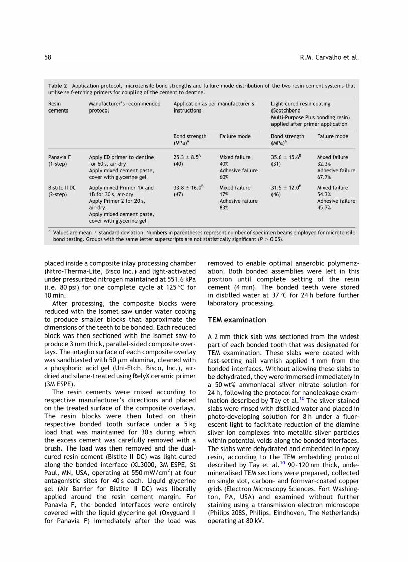
Table 2 Application protocol, microtensile bond strengths and failure mode distribution of the two resin cement systems thatutilise self-etching primers for coupling of the cement to dentine.
Resincements
Manufacturer’s recommendedprotocol
Application as per manufacturer’sinstructions
Light-cured resin coating(ScotchbondMulti-Purpose Plus bonding resin)applied after primer application
Bond strength(MPa)a
Failure mode Bond strength(MPa)a
Failure mode
Panavia F(1-step)
Apply ED primer to dentinefor 60 s, air-dry
25.3 ^ 8.5A
(40)Mixed failure40%
35.6 ^ 15.6B
(31)Mixed failure32.3%
Apply mixed cement paste,cover with glycerine gel
Adhesive failure60%
Adhesive failure67.7%
Bistite II DC(2-step)
Apply mixed Primer 1A and1B for 30 s, air-dry
33.8 ^ 16.0B
(47)Mixed failure17%
31.5 ^ 12.0B
(46)Mixed failure54.3%
Apply Primer 2 for 20 s,air-dry.
Adhesive failure83%
Adhesive failure45.7%
Apply mixed cement paste,cover with glycerine gel
a Values are mean ^ standard deviation. Numbers in parentheses represent number of specimen beams employed for microtensilebond testing. Groups with the same letter superscripts are not statistically significant ðP . 0:05Þ:
R.M. Carvalho et al.58
mTBS evaluation and SEM fractographicanalysis
Each tooth was sectioned occluso-gingivally into0.9 mm thick serial slabs using an Isomet saw underwater cooling. Two of these slabs from each toothwere further sectioned into 0.9 £ 0.9 mm2
composite overlay-dentine beams, according tothe technique for the ‘non-trimming’ version ofthe microtensile test.15 The exact dimensions of thebeams were measured using a pair of digital calipers.The five teeth from each group yielded 31–47 beamsfor bond strength evaluation. The specimens werestressed to failure under tension using a universaltesting machine (Model EMIC DL500, Emic Ltd, S. J.dos Pinhais, PR, Brazil) at a crosshead speed of0.5 mm/min. The data were analyzed using two-wayANOVA, to examine the effect of materials (i.e.Panavia F vs. Bistite II DC) and application methods(i.e. manufacturer’s instructions vs. additionalSBMP Plus resin coating), and the interaction ofthese two factors on bond strength. The totalnumber of tested beams in each group was used inthe statistical analysis, with each individual beamconsidered as an independent specimen. Post hoccomparisons were performed using Student–New-man–Keuls multiple comparison tests at a ¼ 0:05:
After the beams were fractured, they wereexamined using an endodontic microscope (OPMIpico, Carl Zeiss, Oberkochen, Germany) to deter-mine the failure mode. As no gross cohesive failureoccurred within the composite overlay or dentinesubstrate, failure was classified as adhesive failure,along the cement–dentine interface, or mixedfailure that occurred both along the interface andwithin the resin cement. The composite overlayside and the dentine side of representative frac-tured beams from the four experimental groupswere air-dried and sputter-coated with gold/palla-dium for examination with a scanning electronmicroscope (Cambridge Stereoscan 360, Cam-bridge, United Kingdom) operating at 20 kV.
Results
The results of the mTBS tests are shown in Table 2.None of the specimens failed prematurely duringsectioning. Two-way ANOVA revealed that neitherthe factor ‘materials’ ðP ¼ 0:292Þ nor the factor‘application methods’ ðP ¼ 0:06Þ significantlyaffected the bond strength results. However, theinteraction of these two factors was statisticallysignificant ðP ¼ 0:003Þ: Student Newman Keul’smultiple comparison tests further showed that for
Panavia F, there was a statistically significantdifference between manufacturer’s recommendedbonding protocol (i.e. ED primer only) and theadditional use of a coating of LVBR ðP , 0:05Þ:Conversely, there was no difference between themanufacturer’s recommended protocol and the useof an additional resin coating for Bistite II DC ðP .
0:05Þ: When bonding was performed according tothe manufacturer’s recommended protocol, themean mTBS of Panavia F was significantly lower thanthat exhibited by Bistite II DC ðP , 0:05Þ: Bycontrast, there was no difference between PanaviaF and Bistite II DC when these two resin cementswere coupled to primed dentine that were coveredwith an additional resin coating ðP . 0:05Þ:
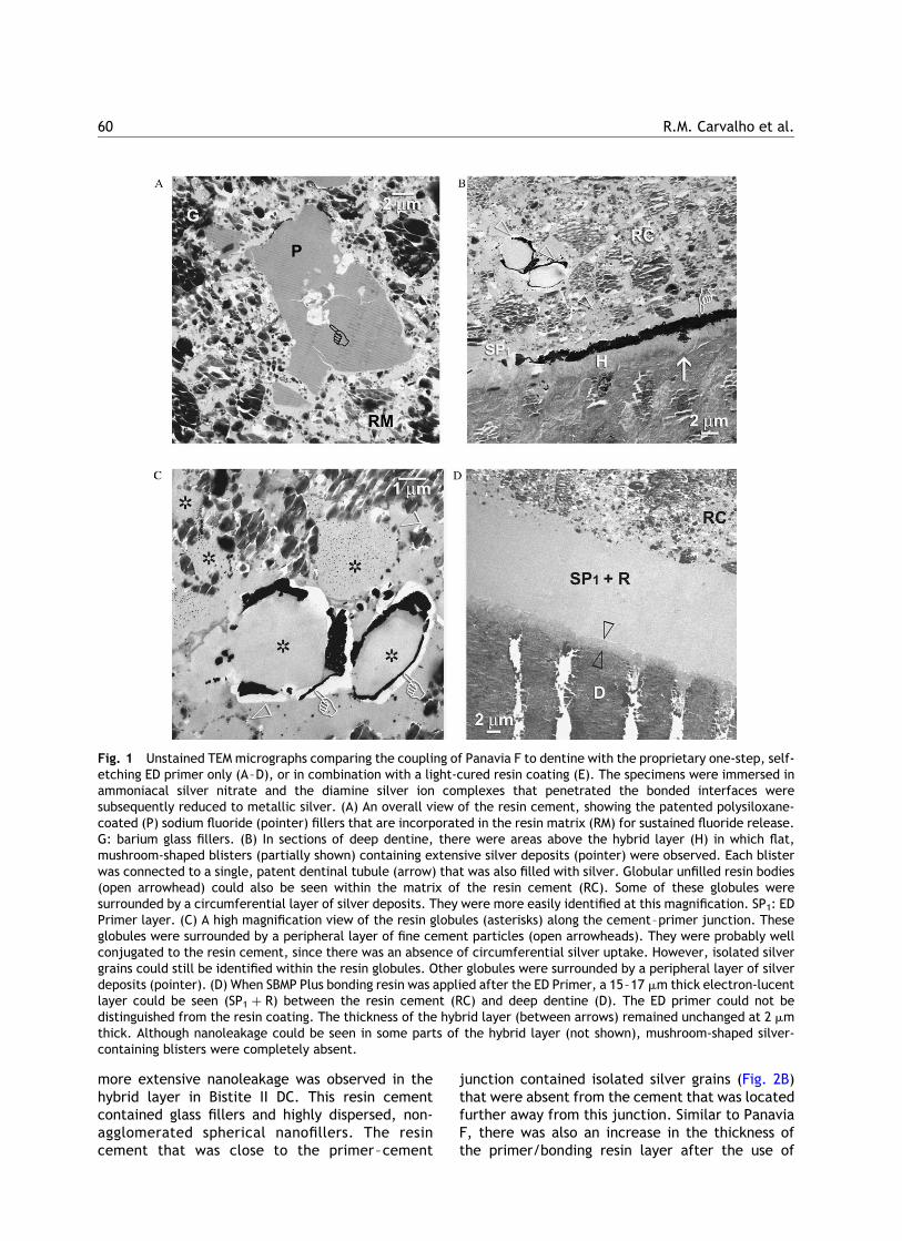
TEM micrographs of dentine coupled with Pana-via F are shown in Fig. 1. An overall view of the resincement revealed the presence of electron-lucentfillers surrounded by a slightly electron-densecoating that were randomly dispersed among theglass fillers within the resin matrix (Fig. 1A). Whendeep dentine was treated with the ED primer only,the primer could be identified as a 8–9 mm thick,slightly electron-dense layer between the hybridlayer and the resin cement (not shown). In the teethexposed to silver nitrate, many examined sectionsrevealed large, flat, mushroom-shaped blisterswere present within this primer layer that con-tained extensive silver deposits. Each blister wasconnected to a patent tubular orifice that alsocontained silver deposits (Fig. 1B). In addition, resinglobules that were devoid of glass filler particlescould be identified within the adjacent resincement matrices (Fig. 1C). These unfilled resinglobules were lined with small glass filler particlesalong their periphery. Some of them were segre-gated from the rest of the resin matrix and weresurrounded by a peripheral rim of silver deposits.However, the majority were well conjugated to theresin cement matrix and could only be identified bythe filler particles along their peripheral border,and the presence of isolated silver grains within theresin bodies (Fig. 1C). Both the mushroom-shapedblisters and the resin globules were absent when anadditional coat of LVBR was applied, air thinned andlight-cured prior to the coupling of the resin cement(Fig. 1D). While the thickness of the hybrid layerremained unchanged, there was a slight increase inthe overall thickness of the unfilled resin layer (ca.15–17 mm thick). The nanoleakage within thehybrid layer was also substantially reduced.
The mushroom-shaped blisters and resin globuleswere also absent from the interfaces of Bistite II DCwhen this resin cement was coupled to dentin thatwas treated with the two-step self-etching primeronly (Fig. 2A). However, compared with Panavia F,
Adhesive permeability affects coupling of resin cements that utilise self-etching primers to dentine 59
more extensive nanoleakage was observed in thehybrid layer in Bistite II DC. This resin cementcontained glass fillers and highly dispersed, non-agglomerated spherical nanofillers. The resincement that was close to the primer–cement
junction contained isolated silver grains (Fig. 2B)that were absent from the cement that was locatedfurther away from this junction. Similar to PanaviaF, there was also an increase in the thickness ofthe primer/bonding resin layer after the use of
Fig. 1 Unstained TEM micrographs comparing the coupling of Panavia F to dentine with the proprietary one-step, self-etching ED primer only (A–D), or in combination with a light-cured resin coating (E). The specimens were immersed inammoniacal silver nitrate and the diamine silver ion complexes that penetrated the bonded interfaces weresubsequently reduced to metallic silver. (A) An overall view of the resin cement, showing the patented polysiloxane-coated (P) sodium fluoride (pointer) fillers that are incorporated in the resin matrix (RM) for sustained fluoride release.G: barium glass fillers. (B) In sections of deep dentine, there were areas above the hybrid layer (H) in which flat,mushroom-shaped blisters (partially shown) containing extensive silver deposits (pointer) were observed. Each blisterwas connected to a single, patent dentinal tubule (arrow) that was also filled with silver. Globular unfilled resin bodies(open arrowhead) could also be seen within the matrix of the resin cement (RC). Some of these globules weresurrounded by a circumferential layer of silver deposits. They were more easily identified at this magnification. SP1: EDPrimer layer. (C) A high magnification view of the resin globules (asterisks) along the cement–primer junction. Theseglobules were surrounded by a peripheral layer of fine cement particles (open arrowheads). They were probably wellconjugated to the resin cement, since there was an absence of circumferential silver uptake. However, isolated silvergrains could still be identified within the resin globules. Other globules were surrounded by a peripheral layer of silverdeposits (pointer). (D) When SBMP Plus bonding resin was applied after the ED Primer, a 15–17 mm thick electron-lucentlayer could be seen (SP1 þ R) between the resin cement (RC) and deep dentine (D). The ED primer could not bedistinguished from the resin coating. The thickness of the hybrid layer (between arrows) remained unchanged at 2 mmthick. Although nanoleakage could be seen in some parts of the hybrid layer (not shown), mushroom-shaped silver-containing blisters were completely absent.
R.M. Carvalho et al.60
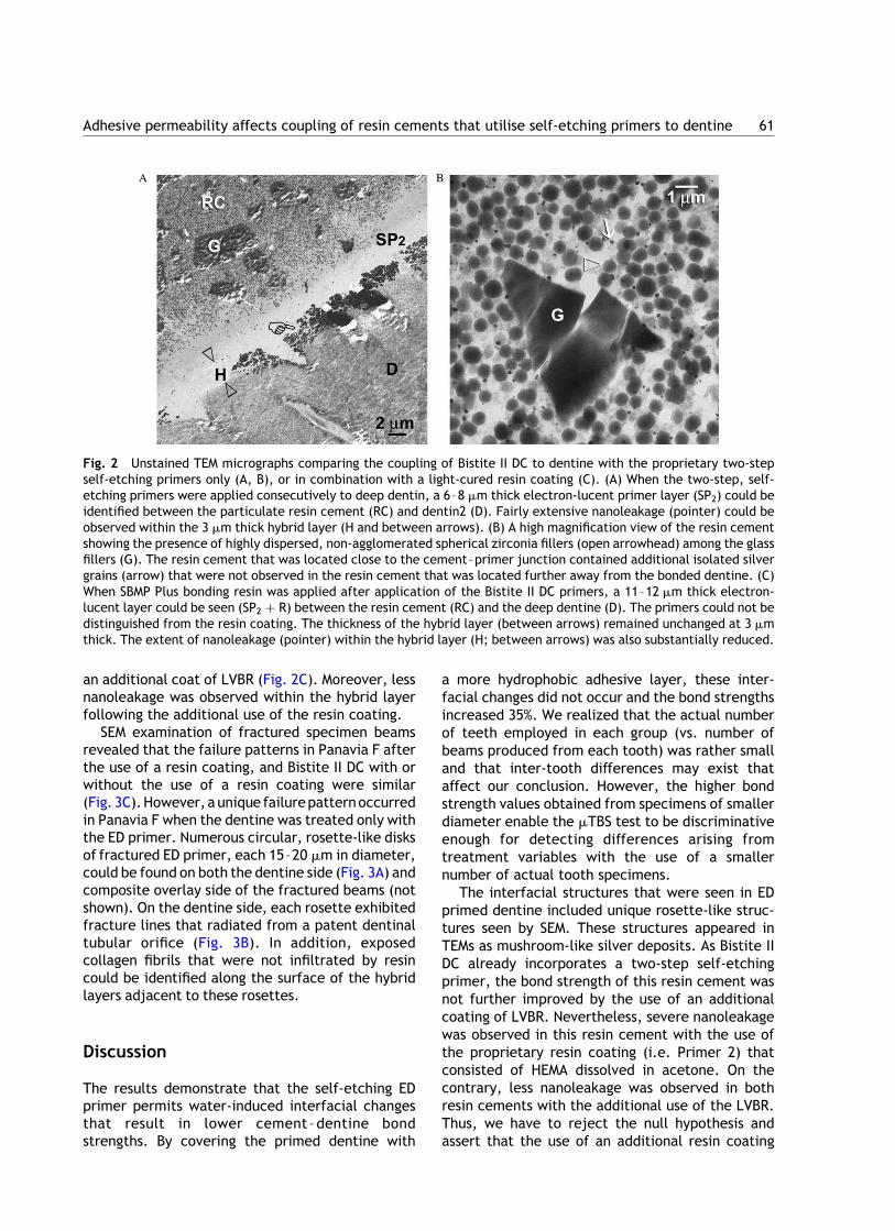
an additional coat of LVBR (Fig. 2C). Moreover, lessnanoleakage was observed within the hybrid layerfollowing the additional use of the resin coating.
SEM examination of fractured specimen beamsrevealed that the failure patterns in Panavia F afterthe use of a resin coating, and Bistite II DC with orwithout the use of a resin coating were similar(Fig. 3C).However, a unique failurepatternoccurredin Panavia F when the dentine was treated only withthe ED primer. Numerous circular, rosette-like disksof fractured ED primer, each 15–20 mm in diameter,could be found on both the dentine side (Fig. 3A) andcomposite overlay side of the fractured beams (notshown). On the dentine side, each rosette exhibitedfracture lines that radiated from a patent dentinaltubular orifice (Fig. 3B). In addition, exposedcollagen fibrils that were not infiltrated by resincould be identified along the surface of the hybridlayers adjacent to these rosettes.
Discussion
The results demonstrate that the self-etching EDprimer permits water-induced interfacial changesthat result in lower cement –dentine bondstrengths. By covering the primed dentine with
a more hydrophobic adhesive layer, these inter-facial changes did not occur and the bond strengthsincreased 35%. We realized that the actual numberof teeth employed in each group (vs. number ofbeams produced from each tooth) was rather smalland that inter-tooth differences may exist thataffect our conclusion. However, the higher bondstrength values obtained from specimens of smallerdiameter enable the mTBS test to be discriminativeenough for detecting differences arising fromtreatment variables with the use of a smallernumber of actual tooth specimens.
The interfacial structures that were seen in EDprimed dentine included unique rosette-like struc-tures seen by SEM. These structures appeared inTEMs as mushroom-like silver deposits. As Bistite IIDC already incorporates a two-step self-etchingprimer, the bond strength of this resin cement wasnot further improved by the use of an additionalcoating of LVBR. Nevertheless, severe nanoleakagewas observed in this resin cement with the use ofthe proprietary resin coating (i.e. Primer 2) thatconsisted of HEMA dissolved in acetone. On thecontrary, less nanoleakage was observed in bothresin cements with the additional use of the LVBR.Thus, we have to reject the null hypothesis andassert that the use of an additional resin coating
Fig. 2 Unstained TEM micrographs comparing the coupling of Bistite II DC to dentine with the proprietary two-stepself-etching primers only (A, B), or in combination with a light-cured resin coating (C). (A) When the two-step, self-etching primers were applied consecutively to deep dentin, a 6–8 mm thick electron-lucent primer layer (SP2) could beidentified between the particulate resin cement (RC) and dentin2 (D). Fairly extensive nanoleakage (pointer) could beobserved within the 3 mm thick hybrid layer (H and between arrows). (B) A high magnification view of the resin cementshowing the presence of highly dispersed, non-agglomerated spherical zirconia fillers (open arrowhead) among the glassfillers (G). The resin cement that was located close to the cement–primer junction contained additional isolated silvergrains (arrow) that were not observed in the resin cement that was located further away from the bonded dentine. (C)When SBMP Plus bonding resin was applied after application of the Bistite II DC primers, a 11–12 mm thick electron-lucent layer could be seen (SP2 þ R) between the resin cement (RC) and the deep dentine (D). The primers could not bedistinguished from the resin coating. The thickness of the hybrid layer (between arrows) remained unchanged at 3 mmthick. The extent of nanoleakage (pointer) within the hybrid layer (H; between arrows) was also substantially reduced.
Adhesive permeability affects coupling of resin cements that utilise self-etching primers to dentine 61
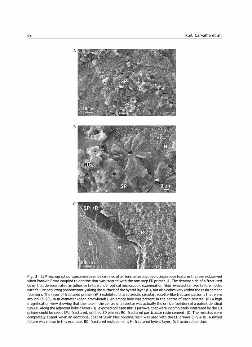
Fig. 3 SEM micrographs of specimen beams examined after tensile testing, depicting unique features that were observedwhen Panavia F was coupled to dentine that was treated with the one-step ED primer. A. The dentine side of a fracturedbeam that demonstrated an adhesive failure under optical microscopic examination. SEM revealed a mixed failure mode,with failure occurring predominantly along the surface of the hybrid layer (H), but also cohesively within the resin cement(pointer). The layer of fractured primer (SP1) exhibited characteristic circular, rosette-like fracture patterns that werearound 15–20 mm in diameter (open arrowheads). An empty hole was present in the centre of each rosette. (B) A highmagnification view showing that the hole in the centre of a rosette was actually the orifice (pointer) of a patent dentinaltubule. Along the adjacent hybrid layer (H), exposed collagen fibrils (arrows) that were incompletely infiltrated by the EDprimer could be seen. SP1: fractured, unfilled ED primer; RC: fractured particulate resin cement. (C) The rosettes werecompletely absent when an additional coat of SBMP Plus bonding resin was used with the ED primer (SP1 þ R). A mixedfailure was shown in this example. RC: fractured resin cement; H: fractured hybrid layer; D: fractured dentine.
R.M. Carvalho et al.62
improves the coupling of Panavia F to hydrateddentine.
Improving the adhesion of resin cements to toothsubstrates is paramount for increasing the fractureresistance of brittle indirect restorations.16 PanaviaF contains sodium benzene sulphinate in the PrimerB component, and a proprietary sodium aromaticsulphinate in the Universal paste of the resincement to ensure that optimal polymerization ofthe cement occurs under an acidic environment. Italso contains patented polysiloxane-coated sodiumfluoride fillers for sustained fluoride release17 thatare probably represented by the coated fillersdepicted in Fig. 1A. In the absence of adverseacid–base reactions that resulted in adhesive–composite incompatibility, the low bond strengththat was observed when this resin cement wascoupled to dentine without a ‘resin coating’technique18 may be explained by the increase inpermeability10 associated with the one-step self-etching ED primer. This increase in adhesivepermeability was manifested ultrastructurally bythe presence of mushroom-like blisters that werecontinuous with the lumen of patent dentinaltubules. Presumably, these blisters were eitherfilled with water that permeated from the dentinaltubules, or represented incompletely polymerisedregions within the primer layer that resulted fromthe entrapment of water. When stressed to failure,this extension of the dentinal tubules within thecement may function as sites of stress concen-tration, resulting in cracks that propagated in acentripetal orientation, and producing the charac-teristic rosette-like fractures. In addition, thewater that permeated through the primer layermay also result in emulsion polymerization of themore hydrophobic resin components within theresin cement, as previously demonstrated byanother study.13 Formation of similar resin globulesalong the composite–adhesive interface has alsobeen observed when one-step adhesives wereemployed for the bonding of resin composites.12
The TEM observation of fairly extensive nano-leakage within the hybrid layers when both resincements were used as per the manufacturers’recommendations was also confirmed by the identi-fication of exposed collagen fibrils on the surface ofhybrid layers when specimen beams were examinedwith SEM after tensile testing. These results werecontrary to those from a recent micro-Ramanspectroscopical study.19 In that study, the authorsclaimed that no exposed dentine matrix waspresent along the dentine/resin cement interfacewhen a self-etching primer was used for condition-ing dentine. Conversely, nanoleakage was consist-ently observed in hybrid layers created by self-etch
adhesives.20,21 In the present study, the applicationof an additional coating of LVBR appeared to haveresulted in, at least qualitatively, a reduction of thenanoleakage within the hybrid layers. As the LVBR isnon-solvented and fairly viscous, it is unlikely thatnanoleakage could be reduced by the infiltration ofthis resin into residual spaces within the hybridlayer that were not completely infiltrated bythe self-etching primers. It is known that theinclusion of acidic monomers reduces the rate andextent of polymerization of both light- and auto-cured resin blends, with the reduction being morepronounced in the latter.22 The reduction in the rateof polymerization of self-etching primers mayaccount for the low early bond strengths observedwith auto-cured resin cement systems that bond todentine via a self-etching mechanism.23 Apparently,early water exposure of self-etching, slow-curingprimers or adhesives does compromise their mech-anical properties due to plasticisation of the polymermolecules.24 This renders the adhesive layer weakerand lowers the bond strength. When self-etchadhesives were applied to hydrated dentine andstored for 24 h in water, bond strengths weresignificantly lower than when similar specimenswere stored dry (unpublished observations). Theuse of an additional light-cured resin coating mayhaveprovided additional free radicals toenhance therate and extent of polymerization of the self-etchingprimers. This probably reduced the permeability ofthe adhesive layer to water from the substrate andfrom the storage media before testing, as shown bythe reduced amount of silver impregnation. Redu-cing the amount of nanoleakage in hybrid layersformed by self-etching primers between resincements and dentine may improve the long-termdurability of these adhesive joints.25 Further studiesshould be done to compare the longevity of bondscreated by the self-etching type resin cements, withor without the use of a resin coating.
Both the self-etching primers that are used withthe resin cements investigated in this study havevery low film thickness when they were usedaccording to the manufacturers’ instructions (seeFigs. 1B, C and 2A). It has also been shown that thinlayers of dentine adhesives were less capable ofpreventing the formation of interfacial gaps.26 Forrigid, non-compliant cast restorations, the contrac-tion stress induced by a resin cement was reportedto be inversely proportional to the resin layerthickness.27,28 Under these situations, even thelower contraction stress of the cements in the auto-cure mode had enough magnitude to disrupt thebonding to dentine.29 Thus, an additional coat ofunfilled resin may also have contributed to therelief of shrinkage stresses in non-compliant
Adhesive permeability affects coupling of resin cements that utilise self-etching primers to dentine 63
adhesive joints.30 The severe nanoleakage and lowfilm thickness of the primer layer observed whenBistite II DC was used according to the manufac-turer’s instructions may account for the occasionalpost-operative sensitivity reported with the use ofthis resin cement (Junichi Miyata, personal com-munication). Thus, it would be of clinical interest tosee if post-operative sensitivity may be reducedwith the use of an additional resin coating prior tothe coupling of the resin cement.
Within the limits of this study, it maybe concludedthat the use of a LVBR is beneficial for the coupling ofPanavia F, the resin cement that utilises a one-stepself-etching primer to dentine. One additional pointof concern is whether the use of such a resin coatingwill adversely affect the fit of the indirect restor-ations. Based on the TEM micrographs, it could beseen that provided that the adhesive resin wassufficiently air thinned, the film thickness of theprimer layer was increased by no more than 10 mm inboth resin cements. The increase in thickness of theprimer layer may also be partially compensated by areduction in the thickness of the resin cement layer.Considering that the cement spaces in indirectceramic or composite restorations are in the rangeof 50–100 mm,31 – 33 the slight increase in filmthickness may not adversely affect the fit of theserestorations. Moreover, if impressions are takenafter coating the teeth with the additional layer,fitting problems may be eliminated.14 As the presentstudy was performed on flat dentine surfaces,further studies should be performed to examine theeffect of a light-cured unfilled resin coating on theinternal adaptation of indirect restorations.
Acknowledgements
We thank W.S. Lee of the Electron Microscopy Unit,the University of Hong Kong for technical assist-ance. The resin cements investigated in this studywere generous gifts from Kuraray Medical Inc. andTokuyama Corp. This study was supported in part,by grants from the Faculty of Dentistry, theUniversity of Hong Kong, grant 300481/95-0 fromCNPq, grants 02/02179-3 and 02/06682-1 fromFAPESP, Brazil and by grant DE014911 from theNIDCR, USA. The authors are grateful to Zinnia Pangand Michelle Barnes for secretarial support.
References
1. Kramer N, Lohbauer U, Frankenberger R. Adhesive luting ofindirect restorations. American Journal of Dentistry 2000;13(Spec No):60D—76D.
2. Rosenstiel SF, Land MF, Crispin BJ. Dental luting agents: areview of the current literature. Journal of ProstheticDentistry 1998;80:280—301.
3. Diaz-Arnold AM, Vargas MA, Haselton DR. Current status ofluting agents for fixed prosthodontics. Journal of ProstheticDentistry 1999;81:135—41.
4. Frankenberger R, Kramer N, Petschelt A. Technique sensi-tivity of dentin bonding: effect of application mistakes onbond strength and marginal adaptation. Operative Dentistry2000;25:324—30.
5. Bouillaguet S, Duroux B, Ciucchi B, Sano H. Ability ofadhesive systems to seal dentin surfaces: an in vitro study.Journal of Adhesive Dentistry 2000;2:201—8.
6. Christensen GJ. Solving the frustrations of crown cementa-tion. Journal of the American Dental Association 2002;133:1121—2.
7. Van Meerbeek B, De Munck J, Yoshida Y, Inoue S, Vargas M,Vijav P, et al. Buonocore memorial lecture. Adhesion toenamel and dentin: current status and future challenges.Operative Dentistry 2003;28:215—35.
8. Sensat ML, Brackett WW, Meinberg TA, Beatty MW. Clinicalevaluation of two adhesive composite cements for thesuppression of dentinal cold sensitivity. Journal of Prosthe-tic Dentistry 2002;88:50—3.
9. Sanares AME, King NM, Itthagarun A, Tay FR, Pashley DH.Adverse surface interactions between one-bottle light-curedadhesives and chemical-cured composites. Dental Materials2001;17:542—56.
10. Tay FR, Pashley DH, Yiu CKY, Sanares AM, Wei SW. Factorscontributing to the incompatibility between simplified-stepadhesives and self-cured or dual-cured composites. PartI. Single-step self-etch adhesive. Journal of AdhesiveDentistry 2003;5:27—40.
11. Ikemura K, Endo T. Effect on adhesion of new polymerizationinitiator systems comprising 5-monosubstituted barbituricacids, aromatic sulphonate amides, and tert-butyl perox-ymaleic acid in dental adhesive resin. Journal of AppliedPolymer Science 1999;72:1655—68.
12. Tay FR, Pashley DH, Suh BI, Carvalho RM, Itthagarun A.Single-step adhesives are permeable membranes. Journal ofDentistry 2002;30:371—82.
13. Mak YF, Lai SCN, Cheung GSP, Chan AW, Tay FR, PashleyDH. Micro-tensile bond testing of resin cements to dentinand an indirect resin composite. Dental Materials 2002;18:609—21.
14. Jayasooriya PR, Pereira PN, Nikaido T, Barrow MF, TagamiJ. The effect of a ‘resin coating’ on the interfacialadaptation of composite inlays. Operative Dentistry 2003;28:28—35.
15. Shono Y, Ogawa T, Terashita M, Carvalho RM, Pashley EL,Pashley DH. Regional measurement of resin—dentin bondingas an array. Journal of Dental Research 1999;78:699—705.
16. Furukawa K, Inai N, Tagami J. The effects of luting resinbond to dentin on the strength of dentin supported byindirect resin composite. Dental Materials 2002;18:136—42.
17. Kawashima M. Kenichi H. Coated metal fluoride particles anda dental composition containing coated metal fluorideparticles. United States Patent number 5,908,879; 1999.
18. Jayasooriya PR, Pereira PN, Nikaido T, Tagami J. Efficacy ofa resin coating on bond strengths of resin cement to dentin.Journal of Esthetic and Restorative Dentistry 2003;15:105—13.
19. Walker MP, Wang Y, Spencer P. Morphological and chemicalcharacterization of the dentin/resin cement interfaceproduced with a self-etching primer. Journal of AdhesiveDentistry 2002;4:181—9.
R.M. Carvalho et al.64
20. Li H, Burrow MF, Tyas MJ. The effect of concentration and pHof silver nitrate solution on nanoleakage. Journal ofAdhesive Dentistry 2003;5:19—25.
21. Tay FR, King NM, Chan KM, Pashley DH. How can nanoleakageoccur in self-etching adhesive systems that demineralize andinfiltrate simultaneously? Journal of Adhesive Dentistry2002;4:255—69.
22. Tay FR, Feng L, Suh BI, Pashley DH. Factors contributing tothe incompatibility between simplified-step adhesives andchemical-cured or dual-cured composites. Part III. Effect ofacidic resin monomers. Journal of Adhesive Dentistry 2003;in press.
23. Burrow MF, Nikaido T, Satoh M, Tagami J. Early bonding ofresin cements to dentin—effect of bonding environment.Operative Dentistry 1996;21:196—202.
24. Carrilho MRO, Carvalho RM, Tay FR, Pashley DH. Effects ofstorage medium on the mechanical properties of adhesivesystems. American Journal of Dentistry 2003; in press.
25. Kitasako Y, Burrow MF, Katahira N, Nikaido T, Tagami J.Shear bond strengths of three resin cements to dentine over3 years in vitro. Journal of Dentistry 2001;29:139—44.
26. Koike T, Hasegawa T, Itoh K, Yukitani W, Yamashita T,Wakumoto S, et al. Effect of multiple application of a dentinadhesive on contraction gap width of a resin-based compo-site. American Journal of Dentistry 2002;15:159—63.
27. Alster D, Feilzer AJ, De Gee AJ, Davidson CL. Tensilestrength of thin resin composite layers as a function oflayer thickness. Journal of Dental Research 1995;74:1745—8.
28. Alster D, Feilzer AJ, De Gee AJ, Davidson CL. Polymeriz-ation contraction stress in thin resin composite layers asa function of layer thickness. Dental Materials 1997;13:146—50.
29. Braga RR, Ferracane JL, Condon JR. Polymerization con-traction stress in dual-cure cements and its effect oninterfacial integrity of bonded inlays. Journal of Dentistry2002;30:333—40.
30. Choi KK, Condon JR, Ferracane JL. The effects of adhesivethickness on polymerization contraction stress of composite.Journal of Dental Research 2000;79:812—7.
31. Audenino G, Bresciano ME, Bassi F, Carossa S. In vitroevaluation of fit of adhesively luted ceramic inlays.International Journal of Prosthodontics 1999;12:342—7.
32. Molin MK, Karlsson SL, Kristiansen MS. Influence of filmthickness on joint bend strength of a ceramic/resincomposite joint. Dental Materials 1996;12:245—9.
33. Wassell RW, Gagliano G. Effects of adhesive fixed prosthesisretainer design on resultant resin luting agent thickness.Journal of Prosthetic Dentistry 1998;80:479—84.
Adhesive permeability affects coupling of resin cements that utilise self-etching primers to dentine 65
Copyright © 2022 FDOKUMEN