Induction of human T cell leukemia virus type I receptors on quiescent naive T lymphocytes by...
10
of June 5, 2016. This information is current as β Lymphocytes by TGF- Type I Receptors on Quiescent Naive T Induction of Human T Cell Leukemia Virus Huang, Daniel C. Bertolette and Francis W. Ruscetti Kathryn S. Jones, Salem Akel, Cari Petrow-Sadowski, Ying http://www.jimmunol.org/content/174/7/4262 doi: 10.4049/jimmunol.174.7.4262 2005; 174:4262-4270; ; J Immunol References http://www.jimmunol.org/content/174/7/4262.full#ref-list-1 , 44 of which you can access for free at: cites 71 articles This article Subscriptions http://jimmunol.org/subscriptions is online at: The Journal of Immunology Information about subscribing to Permissions http://www.aai.org/ji/copyright.html Submit copyright permission requests at: Email Alerts http://jimmunol.org/cgi/alerts/etoc Receive free email-alerts when new articles cite this article. Sign up at: Print ISSN: 0022-1767 Online ISSN: 1550-6606. Immunologists All rights reserved. Copyright © 2005 by The American Association of 9650 Rockville Pike, Bethesda, MD 20814-3994. The American Association of Immunologists, Inc., is published twice each month by The Journal of Immunology by guest on June 5, 2016 http://www.jimmunol.org/ Downloaded from by guest on June 5, 2016 http://www.jimmunol.org/ Downloaded from
Transcript of Induction of human T cell leukemia virus type I receptors on quiescent naive T lymphocytes by...

of June 5, 2016.This information is current as
βLymphocytes by TGF-Type I Receptors on Quiescent Naive T Induction of Human T Cell Leukemia Virus
Huang, Daniel C. Bertolette and Francis W. RuscettiKathryn S. Jones, Salem Akel, Cari Petrow-Sadowski, Ying
http://www.jimmunol.org/content/174/7/4262doi: 10.4049/jimmunol.174.7.4262
2005; 174:4262-4270; ;J Immunol
Referenceshttp://www.jimmunol.org/content/174/7/4262.full#ref-list-1
, 44 of which you can access for free at: cites 71 articlesThis article
Subscriptionshttp://jimmunol.org/subscriptions
is online at: The Journal of ImmunologyInformation about subscribing to
Permissionshttp://www.aai.org/ji/copyright.htmlSubmit copyright permission requests at:
Email Alertshttp://jimmunol.org/cgi/alerts/etocReceive free email-alerts when new articles cite this article. Sign up at:
Print ISSN: 0022-1767 Online ISSN: 1550-6606. Immunologists All rights reserved.Copyright © 2005 by The American Association of9650 Rockville Pike, Bethesda, MD 20814-3994.The American Association of Immunologists, Inc.,
is published twice each month byThe Journal of Immunology
by guest on June 5, 2016http://w
ww
.jimm
unol.org/D
ownloaded from
by guest on June 5, 2016
http://ww
w.jim
munol.org/
Dow
nloaded from

Induction of Human T Cell Leukemia Virus Type I Receptorson Quiescent Naive T Lymphocytes by TGF-�1,2
Kathryn S. Jones,3* Salem Akel,†‡ Cari Petrow-Sadowski,* Ying Huang,† Daniel C. Bertolette,†
and Francis W. Ruscetti†
The retrovirus human T cell leukemia virus (HTLV) type I (HTLV-I) is primarily transmitted by breast-feeding or sexual contact,by cell-to-cell contact between T cells. TGF-�, which has been shown to enhance transmission of HTLV-I in vitro, is found at highlevels in breast milk and semen. In this study, the ability of TGF-� to regulate expression of molecules involved in HTLV-I bindingand entry was examined. Previous studies using a soluble form of the HTLV-I envelope protein SU have shown that quiescenthuman T cells do not express cell surface molecules that specifically bind SU. After T cell activation, HTLV SU binding proteinsare rapidly induced. In this study, we report that TGF-� induces expression of proteins that bind soluble HTLV SU and HTLVvirions on naive CD4� T lymphocytes. The induction of these proteins occurred without cell cycle entry or expression of activationmarkers, involved TGF-�-induced intracellular signaling, and required de novo transcription and translation. Treatment of naiveCD4� T lymphocytes with TGF-� induced expression of GLUT-1, which has recently been reported to function as a receptor forHTLV. Treatment of a TGF-�-sensitive human myeloid cell line increased the titer of both HTLV-I- and HTLV-II-pseudotypedviruses. Although earlier studies suggested that HTLV SU binding proteins might be an early marker of T cell activation and/orcell proliferation, we report in this study that TGF-� induces binding of HTLV virions and expression of glucose transporter type1 in primary CD4� T lymphocytes that remain quiescent. The Journal of Immunology, 2005, 174: 4262–4270.
T he human T cell leukemia virus (HTLV)4 type I(HTLV-I) retrovirus, the first disease-causing human ret-rovirus isolated (1), is endemic in certain geographic ar-
eas, including southwestern Japan, the Caribbean basin, and partsof West Africa, South America, and Melanesia (2). Although themajority of 10 million infected individuals remain healthy lifelongasymptomatic carriers, HTLV-I is the etiologic agent for severaldiseases. The two most common are a severe lymphocyte neopla-sia called adult T cell leukemia (ATL) (1, 3) and an inflammatoryneurological disease known as HTLV-I-associated myelopathy/tropical spastic paraparesis (HAM/TSP) (4, 5).
ATL is a malignancy of CD4� T cells, and HTLV-I has a pref-erential tropism for CD4� T cells in asymptomatic patients. In-fection of both memory (CD45RO�) (6) and effector/memory(CD27�CD45RA�) (7) CD4� T cells has been observed. Recent
studies have indicated that, in HAM/TSP patients, both CD4� andCD8� T cells serve as viral reservoirs (8). The closely relatedretrovirus HTLV type II (HTLV-II), which is believed to share acommon receptor with HTLV-I (9, 10), also infects both CD4�
and CD8� T cells in vivo (11, 12). Both HTLV-I and HTLV-II caninfect all human lymphocytic subsets, causing polyclonal andmonoclonal T cell lymphocytoses (reviewed in Ref. 13).
Entry of retroviruses into target cells involves the interaction oftwo envelope (Env) glycoproteins, a surface glycoprotein (SU),and a transmembrane glycoprotein (TM), with at least one specifichost cell receptor (14). In order for infection to occur, cell surfacemolecules must specifically bind the viral envelope, and this bind-ing must lead to subsequent fusion of the viral and cellular mem-branes. For some retroviruses such as HIV, the binding and fusionmolecules are separate; for others, such as ecotropic murine leu-kemia viruses, they appear to be a single molecule.
Because primary T cells are difficult to infect with HTLV-I invitro, the majority of the work over the past 20 years has examinedrequirements for HTLV Env-mediated binding and fusion on es-tablished (often non-T) cell lines. These studies revealed that, incontrast to the limited in vivo tropism of HTLV, molecules capableof functioning as HTLV receptors are found on a wide variety ofcells (15–18). Cell surface molecules capable of specifically bind-ing HTLV SU are even more widely expressed; they have beenfound on every vertebrate cell line tested to date (19–21).
Recently, the first identification of a specific molecule involvedin HTLV binding and fusion was reported (22). The glucose trans-porter type 1 (GLUT-1) specifically binds a soluble form of boththe HTLV-I and HTLV-II SU proteins in T cell and non-T celllines, and was shown to be critical for efficient entry of HTLV-II-pseudotyped virions.
For some retroviruses, it is now clear that cell surface moleculesother than the primary entry receptors can bind virions (recentlyreviewed in Ref. 23). These molecules, referred to as attachmentfactors, can play a critical role in the efficiency of retroviral entry.
*Basic Research Program, Science Applications International Corporation-Frederick,Inc., and †Laboratory of Experimental Immunology, National Cancer Institute-Fred-erick, Frederick, MD 21702; and ‡Department of Medical Laboratory Sciences,Hashemite University, Zarka, Jordan
Received for publication August 6, 2004. Accepted for publication January 11, 2005.
The costs of publication of this article were defrayed in part by the payment of pagecharges. This article must therefore be hereby marked advertisement in accordancewith 18 U.S.C. Section 1734 solely to indicate this fact.1 This publication has been funded in whole or in part with federal funds from theNational Cancer Institute, National Institutes of Health, under Contract No.NO1-CO-12400.2 The content of this publication does not reflect the views or policies of the Depart-ment of Health and Human Services, nor does mention of trade names, commercialproducts, or organizations imply endorsement by the U.S. Government.3 Address correspondence and reprint requests to Dr. Kathryn S. Jones, Building 567,Room 253, Basic Research Program, Science Applications International Corporation-Frederick, Inc., National Cancer Institute-Frederick, Frederick, MD 21702-1201. E-mail address: [email protected] Abbreviations used in this paper: HTLV, human T cell leukemia virus; ATL, adultT cell leukemia; HAM/TSP, HTLV-I-associated myelopathy/tropical spastic parapa-resis; GLUT-1, glucose transporter type 1; HSPG, heparan sulfate proteoglycan; LTR,long terminal repeat; EGFP, enhanced GFP; VSV, vesicular stomatitis virus; Treg,regulatory T.
The Journal of Immunology
Copyright © 2005 by The American Association of Immunologists, Inc. 0022-1767/05/$02.00
by guest on June 5, 2016http://w
ww
.jimm
unol.org/D
ownloaded from

Heparan sulfates, which are widely expressed on the surface ofcells, have been shown to be used by a number of viruses includingthe retroviruses HIV-1 and murine leukemia virus (24, 25). Morerecent studies have revealed that heparan sulfate proteoglycans(HSPG) can also specifically bind soluble HTLV SU and enhanceentry of HTLV-I pseudotypes (26, 27). This indicates that theamount of binding of soluble SU and the titer of HTLV-pseudotyped viruses, at least on certain cell lines, reflects interac-tions with HSPG, as well as interactions with specific receptors.Because HSPGs are expressed at low or undetectable levels onprimary CD4� T cells (28–30), they may not play a role in theability of HTLV-I to infect its primary target cell. Thus, it is notclear whether the requirements for HTLV-I Env-mediated fusionare identical in established cell lines and in primary T cells or howmuch of the binding of the soluble SU is to attachment factorsrather than binding/fusion receptors.
The cell surface molecules required for efficient entry ofHTLV-I virions into primary T lymphocytes have not been rigor-ously defined. Recent work by our group and another laboratoryfound that HTLV SU binding molecules are expressed on mostprimary cells of the immune system (21, 31), with the highestlevels of binding observed on CD4� and CD8� T cells undergoingan immune response. In contrast to both stimulated T lymphocytesand established cell lines, quiescent T cells derived from adult andcord blood cells do not bind detectable levels of HTLV-I SU.However, SU binding proteins were rapidly induced after stimu-lation. Thus, it has been suggested that these HTLV SU bindingproteins are an early marker of T cell activation (21, 31).
HTLV-I is primarily transmitted in vivo by breast-feeding orsexual contact. HTLV transmission appears to require passage ofcells between individuals, and infection is believed to require con-tact between T cells. Circulating T cells are almost entirely in theG0 phase of the cell cycle; these cells do not express the HTLV SUbinding protein. This suggests that viral transmission occurs inareas of extensive immune reactivity such as lymph nodes or sitesof inflammation. Alternatively, humoral factors could stimulateexpression of HTLV-I receptors on quiescent T cells that thencould serve as targets for viral entry. For HIV-1, it has been re-ported that the virus can enter quiescent T cells, which can harborthe virus in an inactive state until subsequent mitogenic stimula-tion allows completion of the early stages of infection (32).
TGF-� is a member of a superfamily of growth factors thatregulate numerous physiological processes including cell cyclecontrol, differentiation, and the immune response (33). TGF-� caninhibit the growth of most epithelial and lymphoid cells by arrest-ing cells in mid-G1 phase (34). Intracellular signaling is initiatedupon the binding of TGF-� to the cell surface type II TGF-� re-ceptor (T�RII), which recruits type I TGF-� receptor (T�RI) to amultimeric complex (reviewed in Ref. 35). The propagation of theintracellular signal to the nucleus then occurs through phosphor-ylation of Smads, which regulate gene expression in cooperationwith other transcription factors (23, 36, 37).
TGF-�, which can enhance transmission of HTLV-I in vitro, isfound at high levels in breast milk and semen. TGF-� can induceexpression of HTLV-I viral proteins in PBMCs from HAM/TSP(38) and asymptomatic carriers (39). In the latter study, it wasshown that TGF-� transactivates the long terminal repeat (LTR)promoter and can accelerate transmission of the virus from in-fected to uninfected cells. For HIV-1, TGF-� has also been shownto both increase transcription from the LTR (40) and to up-regulatecell surface expression of coreceptors in target cells in the absenceof T cell activation and proliferation (41, 42).
In this study, we observed that TGF-� induces expression ofHTLV SU binding proteins without activation of naive CD4� T
lymphocytes and can increase the titer of HTLV-I- and HTLV-II-pseudotyped viruses. Treatment of naive CD4� T lymphocyteswith TGF-� induces expression of GLUT-1, a receptor for HTLVentry (22). Thus, TGF-� can stimulate cell surface expression ofproteins involved in HTLV binding and fusion in the absence of Tcell activation and cell cycle entry.
Materials and MethodsCell culture and isolation of lymphocytes
Cord blood samples obtained from healthy volunteer donors during vaginalbirths from Frederick Memorial Hospital (Frederick, MD) and leukopaksof peripheral blood from healthy donors were collected according to theNational Institutes of Health approved institutional review board protocols.Mononuclear leukocytes were isolated by Ficoll-Hypaque gradient centrif-ugation. The light density fraction (buffy coat) was collected, washed twicewith PBS, and then enriched for CD4� T cells by removal of CD8-, CD19-,CD45RO-, CD11b-, and glycophorin-A-positive cells using MACs meth-ods according to manufacturer’s instructions (Miltenyi Biotec). After iso-lation, the cells were cultured in either RPMI 1640 medium supplementedeither with 20% BIT (BSA, insulin, and transferrin; StemCell Technolo-gies) or with 10% human Ab, as indicated, along with 2 mM L-glutamine,sodium pyruvate (1 mM), and antibiotics. For TGF-� treatment, cells wereexposed to TGF-�2 (Genzyme) or TGF-�1 (BioSource) at the concentra-tions and times indicated. T cells were cultured in 20 U/ml IL-2 (Zeptome-trix) and activated with PHA (1 �g/ml; Abbott Diagnostics) or culturedwith 10 ng/ml IL-7 (PeproTech). In some studies, SB-431542 (TocrisCookson) an inhibitor of TGF-�RI signaling (43, 44) was added along withTGF-�. In one study, the cells were treated with actinomycin D (Sigma-Aldrich) or cyclohexamide (Sigma-Aldrich). KPC/1 is a clone of the hu-man myeloid cell line K562. It was generated by transfecting K562 with aplasmid encoding a neo selectable marker (pcDNA), selecting for trans-fected cells plated at 100 cells/well in a 96-well plate with 500 �g/mlG418, and identifying the cells most sensitive to TGF-� (as judged bygrowth inhibition and level of Smad phosphorylation). All cells werecultured in a humidified atmosphere at 37°C in an incubator containing5% CO2.
Expression of the recombinant HTLV-I SU envelope-Fc fusionproteins
Soluble SU proteins were generated essentially as previously described,with the following modifications. 293-T cells were transfected with eitherthe plasmid vector encoding HTSU-IgG (HTSU-IgG/pSK100; Ref. 20) ora plasmid encoding a similar fusion protein (SUA-rIgG) containing the SUprotein from the avian retrovirus ALSV-A (45). The latter was used as anegative control in all studies. Two days after transfection, the cells werewashed with PBS, and then resuspended in ice-cold PBS to which pro-teinase inhibitor mixture (Sigma-Aldrich) had been added. The cells werethen sonicated twice, and the lysates were centrifuged at 800 � g for 5 min.The clarified lysate was then aliquoted and stored at �80°C, and the con-centration of immunoadhesins was determined by ELISA, as previouslydescribed (20).
Flow cytometric analysis
Specific binding of HTSU-IgG to target cells was examined essentially aspreviously described (20) with the following modifications. Briefly, thelysates containing immunoadhesin (either HTSU-IgG or SUA-IgG) werecentrifuged at 12,000 � g for 1 min. Because HTSU-IgG can be rapidlyinternalized following binding to cells, and fixation does not significantlyreduce the level of HTSU-IgG binding, target cells were fixed in 4% para-formaldehyde for 30 min on ice, washed with PBS, and then resuspendedin PBS/2% FCS/0.02% sodium azide. The target cells (1 � 106) were thenincubated on ice with lysates containing immunoadhesins to the final vol-ume of 0.3 ml for 30 min. The cells were washed, incubated for 30 min onice with FITC-conjugated Ab specific for rabbit Igs (BioSource Interna-tional), washed again, and resuspended in PBS.
Studies for analysis of activation markers were performed as follows.The cells were washed once with PBS/2% FCS/0.02% sodium azide, andincubated with anti- CD4, CD25, CD45RA, CD69, and Ki67 Abs at 1/20dilution in PBS/2% FCS/0.02% sodium azide to the final volume of 200 �l.For the experiments involving two-color flow cytometry, PE-labeled anti-CD25 and PerCP-labeled anti-CD69 (BD Biosciences) were used. Afterincubating at 4°C for 30 min, cells were washed and fixed with 4%paraformaldehyde.
Because there are no commercially available Abs that recognize externalepitopes on all the glycosylated forms of GLUT-1 (22), the expression of
4263The Journal of Immunology
by guest on June 5, 2016http://w
ww
.jimm
unol.org/D
ownloaded from

GLUT-1 was monitored by intracellular staining. The cells were fixed andpermeabilized using Cytofix/Cytoperm kit (BD Biosciences) as directed bythe manufacturer. Cells were blocked with 10% human AB serum for 15min, and then 4 �l of a 1 �g/�l stock of anti-GLUT-1 Ab (Alpha Diag-nostic International) was added to a final volume of 100 �l. The cells wereincubated at 4°C for 30 min, washed with the perm/wash buffer as directed,and resuspended 50 �l of perm/wash buffer/1% skim milk containing 4 �lof FITC-conjugated goat anti-rabbit Ab (BioSource International). Thecells were incubated at 4°C for 30 min and washed twice in cold perm/wash buffer, and then resuspended in 400 �l of PBS. To verify the extentof permeabilization, a portion of cells (100 �l) were stained with 2 �l ofFITC-labeled anti-actin Ab (Sigma-Aldrich) following fixation/permeabi-lization. Twenty to fifty thousand live cell events were measured on aFACScan (BD Pharmingen) and analyzed using FlowJo software (TreeStar).
Cell cycle analysis was performed by fixing cells in 75% ethanol andstaining the DNA with 50 �g/ml propidium iodide (Sigma-Aldrich) for 1 h.The DNA was analyzed by flow cytometry on a FACScan. For detection ofthe propidium iodide, the cells were excited at 488 nm, and their emissionswere collected at 600 nm.
RNA extraction and RT-PCR analysis
Total RNA was isolated from cells using TRIzol reagent (Invitrogen LifeTechnologies), according to the manufacturer’s recommendation. The con-centration and purity of RNA samples were measured, and 5 �g of totalRNA was used for reverse transcription using oligo(dT) as reverse primer(SuperScript First-Strand Synthesis System for RT-PCR; Invitrogen LifeTechnologies). Five percent of the cDNA product was used for the PCRamplification of GLUT-1 or GAPDH. The PCR conditions were as follows:incubation for 3 min at 94°C followed by 25 cycles of 30 s at 94°C, 30 sat 55°C, and 1 min at 72°C, and a final extension for 7 min at 72°C in aPerkinElmer 9700 Thermal Cycler. The primers used for GLUT-1 were5�-TCCACGAGCATCTTCGAGA-3� (sense) and 5�-AACATGTTTGCTGGTGTCTGC-3� (antisense) (46); for actin were 5�-TGACGGGGTCACCCACACTGTGCCCATCTA-3� (sense) and 5�-CTAGAAGCATTTGCGGTGGACGATGGAGGG-3� (antisense). PCR products were identified on1.5% agarose gel stained with ethidium bromide.
Western blot analysis of GLUT-1
Exponentially growing cells were pelleted, washed twice with PBS, andlysed for 1 h on ice in Digitonin lysis buffer (PBS with 0.25% deoxy-cholate, 1% digitonin, and protease inhibitor mixture (Sigma-Aldrich)).Debris was removed by centrifugation at 4°C for 10 min at 16,000 � g.Protein concentration was determined (Protein Assay reagent; Bio-Rad),and equal amounts of protein (200–300 �g) were separated by SDS-PAGEelectrophoresis on a 10% Tris-glycine gel (Invitrogen Life Technologies)and transferred to Immobilon-P membrane (Millipore). The membraneswere blocked with 10% nonfat dry milk/1� TBST (TBS with 0.1% TritonX-100) for 30 min at room temperature, and then incubated overnight at4°C with a polyclonal antiserum directed against GLUT-1 (Abcam) diluted1/1000 in 5% nonfat dry milk/1� TBST. The membranes were thenwashed twice with 1� TBST, hybridized with HRP-conjugated anti-rabbitIgG for 1 h at room temperature, and washed three times with 1� TBST.The bands were visualized using ECL reagent (Amersham Biosciences)and exposed to film (Kodak).
Generation of pseudotyped retroviral vectors
HIV-based retroviral vectors were generated by cotransfecting 293-T cellswith an HIV-based retroviral vector encoding the enhanced GFP (EGFP)(pHIV-eGFP; gift of V. KewalRamani, National Cancer Institute-Freder-ick) (47) and a plasmid encoding the appropriate Env protein. The plasmidsencoding HTLV-I Env (CMV-ENV-LTR) and the G protein of vesicularstomatitis virus (VSV)-G have been previously described (20). The plas-mid encoding HTLV-II Env (CMV-ENV-LTR-II) was constructed as fol-lows: a full-length clone of HTLV-II (pH6neo; gift of P. Green, Ohio StateUniversity, Columbus, OH) (48, 49) was digested with SphI and ClaI(5124–7384) to generate a fragment that included the entire HTLV-II Envcoding sequences. The plasmid CMV-ENV-LTR was digested with SphIand ClaI to remove the homologous fragment in the HTLV-I provirus(5145–7495 of the sequence with accession no. J02029), and the fragmentreplaced with the SphI-ClaI fragment from pH6neo.
To generate virus, 293-T cells were transfected with a total of 8 �g ofDNA using FuGene 6 (Roche), at a 0.1:1 ratio of Env-expressing vector:pHIV-eGFP. Twenty hours after transfection, the cells were refed. Approx-imately 16 h later, viral supernatants were harvested, subjected to low-speed centrifugation, filtered through a 0.45-�m pore size filter, and usedimmediately.
Viral transduction
KPC/1 cells were resuspended in RPMI 1640 supplemented with 10% FCSat a concentration of 106 cells/ml. For each well in a six-well plate, 0.5 ml(5 � 105 cells) were added along with 1 ml of viral supernatant, and cellswere transduced using a modification of the spin infection method as de-scribed previously (20). The amount of viral particles present in each sam-ple were quantified using a gag p24 ELISA (Zeptometrix). Following thespin, the cells were incubated at 37°C for 2 h, and then washed twice inmedium and incubated at 37°C. For determination of relative viral titer,target cells were incubated with 5-fold dilutions of supernatant containingthe pseudotyped viruses. Negative control cultures (cells transduced withHIV vectors with no Env) were included in each experiment. Four dayslater, 50,000 live cells were analyzed by flow cytometry for the expressionof EGFP to determine the percentage of transduced cells. Relative titers ofthe pseudotyped viruses were determined from the dilution with the lowestpercentage that was between 5 and 40% positive for EGFP. The relativetiters were determined using the following formula: Dilution factor � (%positive � % positive in negative control) � (5 � 105) (number of cells inwell on at time of transduction). Titers were corrected for the amount ofviral p24 protein present in the inoculum, and adjusted to 1 ml. Eachsample was performed in duplicate, and the SEM was calculated.
Virion binding assay
Specific binding of HTLV virions to target cells was examined using amodification of a previously described method (50). Virus was concen-trated from the supernatant of an HTLV-I-producing cell line (MT-2) bygrowing the cell line at a high density (3–5 � 106/ml) in the inner chamberof an Integra CL1000 flask (Integra Biosciences). After 2 days, the cellswere removed by centrifugation at 800 � g for 5 min and filtering thesupernatant through a 0.22-�m filter (Millipore), and the viral preparationwas stored at �80°C. The amount of HTLV-1p19 in the sample was de-termined using an ELISA (Zeptometrix). For the viral binding assay, targetcells (1 � 106) were centrifuged at 800 � g for 5 min, washed withice-cold PBS, centrifuged again, and resuspended in 0.2 ml of PBS. Cellswere then incubated with 20 �g of p19 from the viral preparation at 22°Cfor 30 min. As a control, cells containing no virus were incubated in par-allel. After incubation, both sets of cells were washed in PBS, resuspendedin 200 �l of PBS, and incubated for 30 min on ice with 1.0 �g of mAbdirected against HTLV SU (anti-gp46 mAb clone 65/6C2.2.34; Zeptome-trix). Cells were then washed and resuspended in 100 �l of PBS with a goatanti-mouse FITC-conjugated Ab for 30 min on ice. After the incubation,the cells were washed and immediately analyzed by flow cytometry.
ResultsTGF-� stimulates cell surface expression of HTLV-I Envbinding proteins on CD4� T lymphocytes
Recently, we and others have reported that naive resting CD4� Tcells do not bind detectable levels of HTLV-I SU. However, mol-ecules that specifically bind HTLV SU proteins are rapidly in-duced (4–6 h) after different modes of T cell stimulation. Wewanted to examine whether expression of these proteins could beinduced by TGF-�.
Freshly isolated lymphocytes from cord blood, enriched forCD4� T cells, were left untreated, treated with TGF-�2, or stim-ulated with PHA and IL-2. After 48 h of culture in serum-freemedium, the cell surface expression of the HTLV-I SU bindingproteins was determined from the level of soluble SU (HTSU-IgG)bound to the cells by flow cytometry analysis. As a control fornonspecific binding, the amount of binding to a similar immuno-adhesin SUA-rIgG, which contains the SU protein from an unre-lated avian retrovirus, was determined in parallel. A significantincrease in binding with HTSU-IgG was observed when the cellswere incubated with TGF-�, in comparison with the cells culturedin medium alone (Fig. 1A, compare left and middle panels). Asexpected from previous work (21, 31), a dramatic increase in bind-ing with HTSU-IgG was observed when the cells were stimulatedfollowing exposure to PHA and IL-2 (Fig. 1A, right panel).
These observations indicated that treatment of naive CD4� Tlymphocytes with TGF-� induced cell surface expression of mol-ecules that specifically bound HTLV SU. TGF-� signals through a
4264 TGF-� INDUCES HTLV-I RECEPTOR EXPRESSION
by guest on June 5, 2016http://w
ww
.jimm
unol.org/D
ownloaded from

receptor complex of type I (TGF-�RI) and type II (TGF-�RII)receptors. To verify that the effect we observed involved TGF-�-induced intracellular signaling via the Smad pathway, the effect ofan inhibitor of Smad signaling on HTSU binding was examined.Unstimulated CD4� T cells were treated with TGF-� either aloneor in the presence of a specific small molecule inhibitor of TGF-�type I receptor signaling (SB-431542; Refs. 43 and 44). Two dayslater, the level of binding of HTSU-IgG to these cells was deter-mined (Fig. 1B). As reported above, TGF-�2 dramatically in-creased cell surface levels of HTLV-I SU binding proteins. In con-trast, treatment of the cord blood lymphocytes with both TGF-�2and the SB-431524 inhibitor resulted in a level of HTLV SU bind-ing proteins similar to the untreated controls. These results indicatethat TGF-� induces cell surface expression of molecules that spe-cifically bind to HTLV-I SU by intracellular signaling throughTGF-�RI. Similar results were seen when cells were treated withthe same concentrations of TGF-�1 in place of TGF-�2 (data notshown).
Kinetics of expression of the HTLV SU binding proteinsfollowing TGF-� treatment
Previously, it has been reported that the HTLV SU binding pro-teins are rapidly expressed following immune activation of naive Tcells (21, 31). The level of binding then diminished as the T lym-phocytes returned to their resting state. We next examined thekinetics of expression of the HTLV-I SU binding protein(s) fol-lowing exposure to TGF-�. Unstimulated CD4� cord blood Tlymphocytes were exposed to either TGF-� or PHA/IL-2. Cellsbound significant levels of soluble SU 18 h after exposure toTGF-� (Fig. 1C). The level of binding was increased at 48 h after
exposure; by 5 days after exposure, the binding was decreased withno detectable binding at day 10 (Fig. 1C and data not shown). Thispattern of expression had similar kinetics to that observed follow-ing stimulation with PHA and IL-2 (21, 31). However, the level ofbinding was somewhat lower for the cells exposed to TGF-� thanthose exposed to PHA/IL-2. This was a consistent finding in �12cord blood samples. However, the kinetics of cell surface expres-sion of the HTLV SU binding proteins was similar in cells exposedto TGF-� and PHA/IL-2. As previously described for PHA/IL-2-stimulated cells (21, 31), detectable levels of HTSU-IgG bindingwas observed 6 h after TGF-� treatments (data not shown).
TGF-�-induced expression of HTLV SU binding proteins onquiescent T cells requires de novo protein synthesis
It has been reported previously that the activation of HTLV SUbinding molecules on CD4� lymphocytes required de novo proteinsynthesis (21). We wanted to determine whether induction ofHTLV binding proteins by TGF-� also required de novo proteinsynthesis. Naive cord blood lymphocytes, enriched for CD4�
cells, were exposed to TGF-� or PHA and IL-2, either alone or inthe presence of cycloheximide. Three days later, binding ofHTSU-IgG to these cells was determined. When protein synthesiswas blocked, neither TGF-� nor PHA/IL-2 induced the expressionof HTLV SU binding proteins (Fig. 2, right panels). Treatmentwith actinomycin D, an inhibitor of transcription, also blockedexpression of these proteins, suggesting that de novo transcriptionas well as translation was required (Fig. 2, middle panels).
Specific binding of HTSU-IgG following TGF-� treatmentoccurs without activation of quiescent CD4� T lymphocytes
Previously, it has been reported that several different modes of Tcell activation all lead to the rapid induction of HTLV-I-SU bind-ing on naive CD4� T lymphocytes (21, 31). At early times afteractivation, T cells that were still phenotypically naive could bindsoluble HTLV SU. This led to the suggestion that this protein is avery early marker of T cell activation.
We next examined whether induction of HTLV-I-SU bindingproteins by TGF-� required T cell activation. The percentage of
FIGURE 1. TGF-� induces molecules that specifically bind HTLV SUon quiescent cord blood T lymphocytes. A, Cord blood lymphocytes wereenriched for naive CD4� T cells (�95% purity) as described in Materialsand Methods. Cells were then cultured in RPMI 1640 medium supple-mented with BIT at a concentration at 1 � 106/ml either alone, withTGF-�2 (10 ng/ml), or with PHA (1 �g/ml) and IL-2 (20 U/ml). Two dayslater, the cells were assayed for their ability to specifically bind HTSU-IgGperformed as described in Materials and Methods. Light line, Binding toHTSU-IgG; dark line, binding to negative control SUA-IgG. Data are rep-resentative of results obtained in eight separate experiments. B, Cells werecultured as in A with an additional TGF-�2 culture that contained a specificsmall molecule inhibitor of TGF-� type I receptor signaling (SB-431542) at 20�M added 15 min earlier. Two days later, binding assays to HTSU-IgG wereperformed using 500 ng/ml HTSU-IgG and SUA-IgG. Data are representativeof results obtained in three separate experiments. C, CD4� enriched naive cordblood T lymphocytes were cultured as described in A. Cells were collected at1, 2, and 5 days after culture, HTSU-IgG binding assays were performed asabove, and mean fluorescence intensity (MFI) was determined. Data are rep-resentative of results obtained in three separate experiments.
FIGURE 2. TGF-�-induced expression of HTLV SU binding proteinsrequires de novo transcription and translation. Naive CD4� enriched cordblood lymphocytes were resuspended at 1 � 106 in RPMI 1640/10% hu-man AB sera. The cells were then either left untreated, or treated with 10�g/ml actinomycin D, or 5 �g/ml cyclohexamide. For each condition, the cellswere divided into two flasks and additionally treated with either TGF-�2 (10ng/ml), or with PHA and IL-2. Three days later, the cells were harvested, andthe amount of binding of HTSU-IgG was determined. Light line, Binding toHTSU-IgG; dark line, binding to negative control SUA-IgG. Data are repre-sentative of results obtained in two separate experiments.
4265The Journal of Immunology
by guest on June 5, 2016http://w
ww
.jimm
unol.org/D
ownloaded from

cells positive for cell surface expression of two early activationmarkers, CD25 (IL-2R �-chain) and CD69, was determined, alongwith percentage of cells binding HTSU-IgG (Fig. 3A). As ex-pected, naive cord blood lymphocytes treated with PHA and IL-2expressed high levels of CD25 and CD69, as well as HTLV SUbinding proteins, on their cell surface. In contrast, TGF-� inducedcell surface expression of the HTLV SU binding proteins but notthe activation markers. Further evidence that TGF-� inducedHTSU-IgG binding in the absence of activation came from theobservations that there was no expression of Ki-67, a marker ofproliferation, the cells remained CD45RA positive, and no in-creases in forward- and side-angle light scattering of the TGF-�-treated T cells was observed (data not shown). Furthermore, cellcycle analysis revealed that 40 h after treatment, the percentage ofthe T cells in S/G2M in the TGF-�-treated cells and the untreatedcells was similar. In contrast, for the same cells treated in parallelwith PHA and IL-2, the percentage of cells in S/G2M had in-creased to 27% over the control. Thus, HTLV SU binding proteinscan be induced in the absence of T cell activation by TGF-�.
It has been reported that IL-7 differentially regulates cell cycleprogression in neonatal and adult CD4� T cells (51) and that IL-7-treated quiescent adult T cells, unlike quiescent neonatal T cells,do not enter S phase and do not express molecules that bindHTLV-I SU (21). We also examined the effect of TGF-� on adultquiescent CD4� T cells to determine whether it differed from thatobserved in neonatal cells. We observed that TGF-�, unlike IL-7,induced the expression of HTLV-I SU binding proteins on bothquiescent adult and cord blood T lymphocytes (data not shown).
Treatment of quiescent T cells with TGF-� increases binding ofHTLV-I virions
To complement the studies of the binding of soluble HTLV SUprotein, we also examined the effect of TGF-� on the binding ofHTLV-I virions. CD4� naive T lymphocytes were cultured for 3days in either medium alone, or with medium containing eitherTGF-� or PHA/IL-2. The cells were then incubated with concen-trated cell-free HTLV-I virions, and the relative amount of virionbinding was determined using a recently described flow-based as-say (50). The binding of HTLV-I virions was similar to that ob-served for binding of the soluble HTLV SU alone. Treatment withTGF-� induced the binding of HTLV-I virions to these cells. Ac-tivation of the cells by PHA and IL-2 also induced virion binding,at a higher level than that observed for TGF-� (Fig. 3B). Thus,TGF-� treatment of quiescent T lymphocytes induces cell surfaceexpression of molecules that specifically bind both HTLV-I virionsand soluble HTLV SU, in the absence of markers of activation.
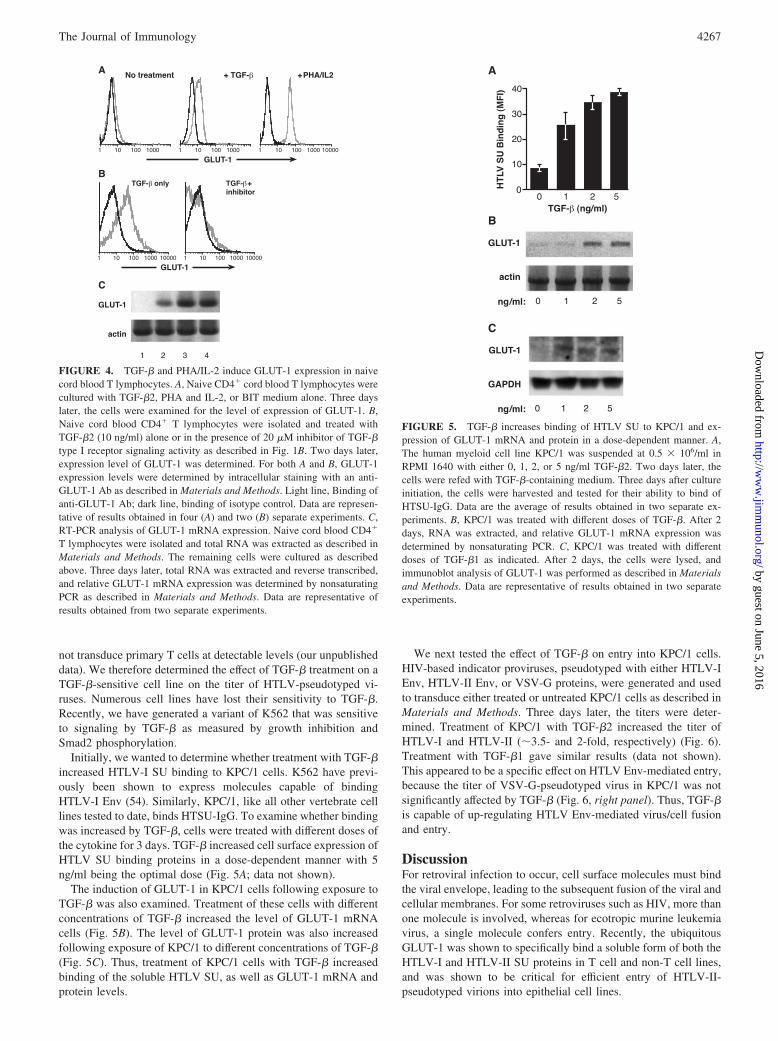
TGF-� induces expression of GLUT-1 in primary T lymphocytes
While this study was in progress, it was reported that the glucosetransporter protein GLUT-1 could specifically bind soluble HTLVSU and was critical for efficient HTLV-II Env-mediated entry (22).Because those studies were performed in established cell lines, weexamined the level of GLUT-1 in primary CD4� T cells afterexposure to TGF-�.
As a first step, GLUT-1 expression was examined on treated anduntreated cord blood lymphocytes. The level of GLUT-1 protein inthe cells was determined by flow cytometry, as described in Ma-terials and Methods. Cells cultured without any additional supple-ments except BIT medium did not express significant levels ofGLUT-1 (Fig. 4A, left panel). Similar results were obtained withfreshly isolated CD4� cord blood lymphocytes (data not shown).Two days after activation with PHA/IL-2, the lymphocytes ex-pressed high levels of GLUT-1 (Fig. 4A, right panel). TGF-� alsoinduced expression of GLUT-1, but at a lower level than PHA/IL-2-activated T cells (Fig. 4A, middle panel). Levels of HTLV SUbinding, determined in parallel, were also higher for the lympho-cytes activated by PHA and IL-2 (data not shown).
To verify that GLUT-1 expression was induced by intracellularsignaling following TGF-� stimulation, we examined the effect ofa TGF-� inhibitor. GLUT-1 expression was determined 4 daysafter exposure to TGF-�, either with or without the inhibitor ofTGF-� type I receptor kinase described above. As shown in Fig.4B, the induction of GLUT-1 by TGF-� was completely blockedby the inhibitor.
TGF-� and PHA/IL-2 treatment of naive CD4� T cells alsoinduced GLUT-1 mRNA expression (Fig. 4C). GLUT-1 mRNAwas not detected in freshly isolated CD4� T cells (Fig. 4C, lane 1).Following 2 days of treatment with either TGF-� (Fig. 4C, lane 3)or PHA/IL-2 (lane 4), a dramatic increase in the level of GLUT-1mRNA was observed. In the cells cultured in medium alone, a lowbut significant level of GLUT-1 mRNA was present (lane 2). Thisis consistent with the low levels of HTSU-IgG binding observedfollowing several days of culture in medium (see Figs. 1, B and C,and 3), and may reflect the activation of GLUT-1 transcription bythe insulin present in the BIT supplement.
Treatment of target cells with TGF-� increases the titer ofHTLV Env-pseudotyped viruses
Because requirements for retroviral Env-mediated fusion can differfrom those of binding alone, we wanted to determine the effect ofTGF-� on the titer of HTLV-pseudotyped viruses. HTLV virionsare poorly infectious (52, 53), and HTLV-pseudotyped viruses do
FIGURE 3. Specific binding of soluble SU and HTLV virions followingTGF-� treatment occurs without activation of quiescent cord blood T lym-phocytes. A, Naive cord blood CD4� T cells were either left untreated inRPMI 1640/BIT medium, or treated with TGF-�2 (10 ng/ml), or with PHAand IL-2. After 2 days in culture, the cells were harvested, and the amountof binding of HTSU-IgG, anti-CD25, and anti-CD69 Abs were determined.The percent positive for HTLV SU binding was calculated from the fol-lowing formula: (% positive for HTSU-IgG) � (% positive for SUA-IgG);for the Abs, the formula used was as follows: (% positive for Ab) � (%positive for isotype control). Data are representative of results obtained inthree separate experiments. B, Naive CD4� cord blood T lymphocyteswere treated as described above. Two days later, with no detectable cellactivation, a virion binding assay was performed as described in Materialsand Methods. Detection of the amount of virions bound was determinedwith an Ab directed against the HTLV SU. Black, Binding of HTLV-Ivirions; white, negative control (binding to cells incubated with mediumwithout virus).
4266 TGF-� INDUCES HTLV-I RECEPTOR EXPRESSION
by guest on June 5, 2016http://w
ww
.jimm
unol.org/D
ownloaded from
not transduce primary T cells at detectable levels (our unpublisheddata). We therefore determined the effect of TGF-� treatment on aTGF-�-sensitive cell line on the titer of HTLV-pseudotyped vi-ruses. Numerous cell lines have lost their sensitivity to TGF-�.Recently, we have generated a variant of K562 that was sensitiveto signaling by TGF-� as measured by growth inhibition andSmad2 phosphorylation.
Initially, we wanted to determine whether treatment with TGF-�increased HTLV-I SU binding to KPC/1 cells. K562 have previ-ously been shown to express molecules capable of bindingHTLV-I Env (54). Similarly, KPC/1, like all other vertebrate celllines tested to date, binds HTSU-IgG. To examine whether bindingwas increased by TGF-�, cells were treated with different doses ofthe cytokine for 3 days. TGF-� increased cell surface expression ofHTLV SU binding proteins in a dose-dependent manner with 5ng/ml being the optimal dose (Fig. 5A; data not shown).
The induction of GLUT-1 in KPC/1 cells following exposure toTGF-� was also examined. Treatment of these cells with differentconcentrations of TGF-� increased the level of GLUT-1 mRNAcells (Fig. 5B). The level of GLUT-1 protein was also increasedfollowing exposure of KPC/1 to different concentrations of TGF-�(Fig. 5C). Thus, treatment of KPC/1 cells with TGF-� increasedbinding of the soluble HTLV SU, as well as GLUT-1 mRNA andprotein levels.
We next tested the effect of TGF-� on entry into KPC/1 cells.HIV-based indicator proviruses, pseudotyped with either HTLV-IEnv, HTLV-II Env, or VSV-G proteins, were generated and usedto transduce either treated or untreated KPC/1 cells as described inMaterials and Methods. Three days later, the titers were deter-mined. Treatment of KPC/1 with TGF-�2 increased the titer ofHTLV-I and HTLV-II (�3.5- and 2-fold, respectively) (Fig. 6).Treatment with TGF-�1 gave similar results (data not shown).This appeared to be a specific effect on HTLV Env-mediated entry,because the titer of VSV-G-pseudotyped virus in KPC/1 was notsignificantly affected by TGF-� (Fig. 6, right panel). Thus, TGF-�is capable of up-regulating HTLV Env-mediated virus/cell fusionand entry.
DiscussionFor retroviral infection to occur, cell surface molecules must bindthe viral envelope, leading to the subsequent fusion of the viral andcellular membranes. For some retroviruses such as HIV, more thanone molecule is involved, whereas for ecotropic murine leukemiavirus, a single molecule confers entry. Recently, the ubiquitousGLUT-1 was shown to specifically bind a soluble form of both theHTLV-I and HTLV-II SU proteins in T cell and non-T cell lines,and was shown to be critical for efficient entry of HTLV-II-pseudotyped virions into epithelial cell lines.
FIGURE 4. TGF-� and PHA/IL-2 induce GLUT-1 expression in naivecord blood T lymphocytes. A, Naive CD4� cord blood T lymphocytes werecultured with TGF-�2, PHA and IL-2, or BIT medium alone. Three dayslater, the cells were examined for the level of expression of GLUT-1. B,Naive cord blood CD4� T lymphocytes were isolated and treated withTGF-�2 (10 ng/ml) alone or in the presence of 20 �M inhibitor of TGF-�type I receptor signaling activity as described in Fig. 1B. Two days later,expression level of GLUT-1 was determined. For both A and B, GLUT-1expression levels were determined by intracellular staining with an anti-GLUT-1 Ab as described in Materials and Methods. Light line, Binding ofanti-GLUT-1 Ab; dark line, binding of isotype control. Data are represen-tative of results obtained in four (A) and two (B) separate experiments. C,RT-PCR analysis of GLUT-1 mRNA expression. Naive cord blood CD4�
T lymphocytes were isolated and total RNA was extracted as described inMaterials and Methods. The remaining cells were cultured as describedabove. Three days later, total RNA was extracted and reverse transcribed,and relative GLUT-1 mRNA expression was determined by nonsaturatingPCR as described in Materials and Methods. Data are representative ofresults obtained from two separate experiments.
FIGURE 5. TGF-� increases binding of HTLV SU to KPC/1 and ex-pression of GLUT-1 mRNA and protein in a dose-dependent manner. A,The human myeloid cell line KPC/1 was suspended at 0.5 � 106/ml inRPMI 1640 with either 0, 1, 2, or 5 ng/ml TGF-�2. Two days later, thecells were refed with TGF-�-containing medium. Three days after cultureinitiation, the cells were harvested and tested for their ability to bind ofHTSU-IgG. Data are the average of results obtained in two separate ex-periments. B, KPC/1 was treated with different doses of TGF-�. After 2days, RNA was extracted, and relative GLUT-1 mRNA expression wasdetermined by nonsaturating PCR. C, KPC/1 was treated with differentdoses of TGF-�1 as indicated. After 2 days, the cells were lysed, andimmunoblot analysis of GLUT-1 was performed as described in Materialsand Methods. Data are representative of results obtained in two separateexperiments.
4267The Journal of Immunology
by guest on June 5, 2016http://w
ww
.jimm
unol.org/D
ownloaded from

Recent studies have shown that initial binding of a retrovirus tothe surface of target cells can involve molecules other than thebinding/fusion receptors. For HIV-1, a number of molecules thatpromote initial attachment of virions to the cell surface have beenidentified; these include HSPGs, LFA-1, and nucleolin (23, 55).Recently, it has been reported that HSPGs can specifically bindsoluble HTLV SU and enhance entry of HTLV-I pseudotypes (26,27). Although the role of HSPGs in the transmission of HTLV invivo is not yet clear, these observations indicate that HSPG, aswell as HTLV binding/fusion receptors, contribute to the level ofbinding of soluble SU and the titer of HTLV-pseudotyped viruses,at least on certain cell lines.
The cell surface molecules required for efficient entry of HTLV-Ivirions into primary T lymphocytes have not been rigorously defined.The highest levels of HTLV SU binding on primary cells are observedon CD4� and CD8� T cells undergoing an immune response (21, 31).In contrast, quiescent naive and memory T cells derived from adultand cord blood cells do not bind detectable levels of HTLV-I SU. Inthis study, we show that TGF-� can induce expression of the HTLVSU binding proteins on quiescent T cells.
HTLV-I is primarily transmitted vertically from mother to in-fant, primarily by breast milk, and horizontally by sexual contactor by blood products. The virus is believed to spread primarily byT cell-to-T cell contact. Contact between T cells has been shownto result in polarization of the cytoskeleton, leading to transfer ofviral cores to the uninfected cell (56). Circulating T cells are al-most entirely in the G0 phase of the cell cycle and are not express-ing the HTLV SU binding proteins. The observations that HTLVbinding proteins are induced by immune activation, and that viruscan be transferred during interactions between T cells, suggeststhat viral transmission may occur primarily in areas of extensiveimmune reactivity such as lymph nodes or sites of inflammation.This study suggests that, alternatively, TGF-�-induced expressionof HTLV receptors could allow binding of HTLV-I virions to Tcells in the absence of immune activation. For HIV-1, it has beenpreviously shown that the virus could interact with quiescent Tcells, which led to productive infection when those cells were sub-sequently stimulated (32).
TGF-�, which has been shown to enhance transmission ofHTLV-I in vitro (39), is found at high levels in breast milk andsemen. TGF-� has previously been shown to increase virus pro-
duction in PMBCs isolated from infected individuals and tran-scription from the HTLV-I LTR (38, 39). In the current study, weobserved that treatment of these cells with TGF-� induces expres-sion of molecules which specifically bind both a soluble form ofHTLV SU (Fig. 1A) and HTLV-I virions (Fig. 3B). The effect ofTGF-� on these cells involves TGF-�-induced intracellular sig-naling via the Smad pathway, because the effect was blocked by aninhibitor of TGF-� type I receptor signaling (43, 44) (Fig. 1B). Thecell surface expression of HTLV-I SU binding proteins required denovo transcription and translation (Fig. 2).
We also examined the level of GLUT-1 proteins in treated anduntreated naive CD4� T lymphocytes. As previously reported(57), GLUT-1 was not expressed at detectable levels in unstimu-lated CD4� T lymphocytes (Fig. 4). Following exposure of thecells to TGF-�, expression of both GLUT-1 protein (Fig. 4A) andmRNA (C) was induced. The GLUT-1 expression was blocked bya specific inhibitor of TGF-� type I receptor signaling (Fig. 4B).This observation is consistent with previous reports that TGF-�up-regulates GLUT-1 in mesenchymal cells (58, 59) and in mousefibroblast cells (60).
GLUT-1 expression was also observed following activation ofthe cells by PHA and IL-2. The kinetics of the expression ofGLUT-1 (Fig. 4) was similar to what has been previously reportedfor expression of HTLV SU binding proteins following treatmentof naive T cells by either PHA and IL-2 or TGF-� (21, 31). Al-though indirect, these observations in primary CD4� T cells areconsistent with the results from established cell lines showing thatGLUT-1 can directly bind HTLV SU proteins.
Previous observations that HTLV-I SU binding proteins arerapidly induced on quiescent CD4� T lymphocytes followingdifferent modes of activation led to the suggestion that this pro-tein is a very early marker of T cell activation (21, 31). Fur-thermore, IL-7, which had been shown to induce proliferation inneonatal but not adult T cells (51), was later shown to induceexpression of HTLV-I SU binding proteins on neonatal but notadult cells. These observations led to the suggestion that the SUbinding proteins are distinct activation markers in neonatal andadult CD4� lymphocytes (21).
In the current study, we found that TGF-� can induce binding ofsoluble HTLV SU and expression of GLUT-1 in these cells with-out activation or proliferation of the T lymphocytes. Induction ofHTLV SU binding was observed in the absence of expression oftwo early markers of T cell activation, CD25 and CD69 (Fig. 3A).The cells also retained their CD45RAhigh, Ki67� phenotype, anddid not form blasts. Cell cycle analysis confirmed that, as expectedfrom previous work (34), the TGF-�-treated cells and the untreatedcontrols had little or no cells in S-G2M. Previously, it has beenreported that IL-7 (51) and TGF-� (34) treatments of adult cellsresult in a block in the G1b stage of the cell cycle. In contrast toIL-7, which does not induce HTLV-I SU binding on quiescentadult T cells (21, 51), TGF-� can stimulate HTLV-I SU binding onthe same quiescent cells.
It has been reported recently that stimulation of the TCR onnaive CD4�CD25� T cells in the presence of TGF-� can result indifferentiation of the cells to CD4�CD25� regulatory T (Treg)cells (61). Although induction of CD25 was not observed follow-ing exposure to TGF-� alone, we further examined whether dif-ferentiation toward Treg cells occurred in these cultures by lookingat expression of the FoxP3-specific marker for Treg cells. As pre-viously reported (61), real-time RT-PCR analysis revealed that ex-posure of naive CD4� T cells to TGF-� and anti-CD3/CD28 in-duced FoxP3 mRNA expression, whereas exposure to TGF-�alone did not induce FoxP3 expression (data not shown). Thus, itappears unlikely that the binding of HTLV SU observed following
FIGURE 6. TGF-� increases titer of HTLV-I- and HTLV-II-pseudotyped viruses in KPC/1 cells. KPC/1 cells were suspended at 1 �106/ml in RPMI 1640, and either left untreated or treated with 5 ng/mlTGF-�2. Two days later, the cells were transduced with HTLV-I-, HTLV-II-, or VSV-G-pseudotyped virus particles, generated as described in Ma-terials and Methods. The relative amount of viral particles present in thesupernatants, quantified using a p24 ELISA, were determined to be asfollows: HTLV-I, 24 ng/ml; HTLV-II, 33 ng/ml; VSV-G, 29 ng/ml. Fourdays later, the cells were harvested, and the titer was determined as de-scribed in Materials and Methods, and normalized for the amount of p24present in the inoculum. Each sample was performed in duplicate, and theSEM was determined. White, No treatment; black, treated with TGF-�.Data are representative of results obtained in three separate experiments.
4268 TGF-� INDUCES HTLV-I RECEPTOR EXPRESSION
by guest on June 5, 2016http://w
ww
.jimm
unol.org/D
ownloaded from

exposure of naive CD4� T cells to TGF-� reflects differentiationtoward Treg cells.
The observation that TGF-� can induce expression of HTLVreceptors without activating T cells parallels what has previouslybeen reported for the coreceptors of HIV-1. TGF-� increases ex-pression of CXCR4 but not CD4 on resting T cells without induc-ing expression of CD25, CD69, or Ki67 (62), even after 10 days inculture. The induction of GLUT-1 expression in the absence ofproliferation is consistent with another study demonstrating thatTGF-� induces GLUT-1 expression without stimulating DNA syn-thesis in quiescent murine fibroblasts (60). Because quiescent Tcells have a very low metabolic rate, it is not surprising thatGLUT-1 is induced very rapidly. In contrast, because TGF-� isknown to be a potent immunosuppressive molecule (33), we areinvestigating what other rapidly induced molecules might be in-volved in this immunosuppression.
The effect of TGF-� on HTLV Env-mediated entry was alsoexamined. TGF-� treatment increased the titer of both HTLV-I-and HTLV-II-pseudotyped viruses, but not the titer of VSV-Gpseudotypes, in a TGF-�-sensitive K562 cell line (Fig. 6). Onedrawback of these studies, as discussed previously for the studiesidentifying GLUT-1 as an HTLV receptor (63), is that the effect ofTGF-� on the titer and the level of HTLV SU binding is beingmeasured on cells that can be transduced by HTLV pseudotypesand bind HTLV SU before treatment. Indeed, recent work showingthat HTLV-I SU binds HSPG and that blocking HSPG/SU inter-actions by dextran sulfate dramatically reduces the titer of HTLV-IEnv-pseudotyped viral particles (27) indicates that more than onemolecule can be required for efficient HTLV Env-mediated entry.Thus, it is unclear whether treatment with TGF-� is up-regulatingmolecules (GLUT-1 and/or other) that are necessary, or only en-hance, HTLV Env-mediated entry. Studies to directly examinewhether GLUT-1 mediates all of the effects of TGF-� on HTLVSU binding to quiescent T cells are beyond the scope of this report.Studies to define conditions for small interfering RNA entry intoprimary quiescent T cells without activation are in progress.
The observation that TGF-� up-regulates the expression ofbinding receptors on naive T lymphocytes and increases the titer ofHTLV-I- and HTLV-II-pseudotyped virions suggests that TGF-�plays a role in transmission of HTLV-I virus in vivo. In vitro studieshave shown that TGF-� increases production of virus in cultures ofHTLV-I-infected cells (38, 39, 64). This reflects at least in part acti-vation of the HTLV-I LTR by TGF-� (39). In that study, it appearedthat TGF-� enhanced transmission as well as virus production, be-cause TGF-� increased virus production more dramatically in cocul-tures of infected and uninfected nonactivated T cells. Our observa-tions suggest that TGF-� can induce HTLV-I receptors on the targetcells and are consistent with a previous report that TGF-� cannotprevent HTLV-I-mediated T cell activation (65).
Although HTLV-I infection leads to T cell activation, it, para-doxically, increases production of TGF-� (66, 67). Studies withTGF-�1-null mice have previously shown that TGF-� is the primein vivo block of T cell activation (68, 69). Both ATL and HAM/TSP cells are resistant to the growth-inhibitory effects of TGF-�(38, 64, 65). However, when spontaneously growing HTLV-I-in-fected T clones were further stimulated by cross-linking of theCD3/TCR complex, the superimposed proliferation was inhibitedby TGF-�. This indicated that the TGF-� signaling pathway inthese cells is intact but is bypassed by HTLV-I-induced T cellactivation (65).
It has recently been reported that independent expression of theHTLV-I transactivator protein Tax blocks growth suppression byTGF-� by perturbing Smad-dependent TGF-� signaling (64, 70,71). Observations from this study that TGF-� can regulate the
expression of proteins important for HTLV-I entry suggest anotherfunction for the blocking of TGF-� signaling by Tax. Many en-veloped viruses, including retroviruses, down-regulate cell surfaceexpression of their receptor on infected cells to prevent superin-fection (14). One way viruses accomplish this is by Env-receptorinteractions, either on the cell surface or in the endoplasmic retic-ulum. Alternatively, other virally encoded proteins can down-reg-ulate the receptor. In cells infected with HIV-1, the viral regulatoryVpu and Nef proteins participate along with Env in CD4 down-regulation (recently reviewed in Ref. 72). In the current study, weobserved that TGF-� induces cell surface expression of proteinsthat bind HTLV Env proteins. These observations, taken togetherwith previous reports that Tax can block TGF-� signaling, raisethe possibility that Tax could down-regulate expression of thebinding receptor during HTLV infection. Studies to directly ex-amine whether Tax can down-regulate the amount of binding ofHTLV SU and/or the level of GLUT-1 expression by interferingwith TGF-� signaling are in progress.
AcknowledgmentsWe thank Pat Green and Vineet KewalRamani for providing reagents, help-ful suggestions, and encouragement, and Chris Grant and Steve Jacobson fortheir help with PCR.
DisclosuresThe authors have no financial conflict of interest.
References1. Poiesz, B. J., F. W. Ruscetti, A. F. Gazdar, P. A. Bunn, J. D. Minna, and
R. C. Gallo. 1980. Detection and isolation of type C retrovirus particles fromfresh and cultured lymphocytes of a patient with cutaneous T-cell lymphoma.Proc. Natl. Acad. Sci. USA 77:7415.
2. Manns, A., M. Hisada, and L. La Grenade. 1999. Human T-lymphotropic virustype I infection. Lancet 353:1951.
3. Yoshida, M., I. Miyoshi, and Y. Hinuma. 1982. Isolation and characterization ofretrovirus from cell lines of human adult T-cell leukemia and its implication inthe disease. Proc. Natl. Acad. Sci. USA 79:2031.
4. Gessain, A., F. Barin, J. C. Vernant, O. Gout, L. Maurs, A. Calender, andG. de The. 1985. Antibodies to human T-lymphotropic virus type-I in patientswith tropical spastic paraparesis. Lancet 2:407.
5. Osame, M., S. Izumo, A. Igata, M. Matsumoto, T. Matsumoto, S. Sonoda,M. Tara, and Y. Shibata. 1986. Blood transfusion and HTLV-I associated my-elopathy. Lancet 2:104.
6. Richardson, J. H., A. J. Edwards, J. K. Cruickshank, P. Rudge, andA. G. Dalgleish. 1990. In vivo cellular tropism of human T-cell leukemia virustype 1. J. Virol. 64:5682.
7. Hanon, E., P. Goon, G. P. Taylor, H. Hasegawa, Y. Tanaka, J. N. Weber, andC. R. Bangham. 2001. High production of interferon-� but not interleukin-2 byhuman T-lymphotropic virus type I-infected peripheral blood mononuclear cells.Blood 98:721.
8. Nagai, M., M. B. Brennan, J. A. Sakai, C. A. Mora, and S. Jacobson. 2001. CD8�
T cells are an in vivo reservoir for human T-cell lymphotropic virus type I. Blood98:1858.
9. Sommerfelt, M. A., and R. A. Weiss. 1990. Receptor interference groups of 20retroviruses plating on human cells. Virology 176:58.
10. Sommerfelt, M. A., B. P. Williams, P. R. Clapham, E. Solomon,P. N. Goodfellow, and R. A. Weiss. 1988. Human T cell leukemia viruses use areceptor determined by human chromosome 17. Science 242:1557.
11. Ijichi, S., M. B. Ramundo, H. Takahashi, and W. W. Hall. 1992. In vivo cellulartropism of human T cell leukemia virus type II (HTLV-II). J. Exp. Med. 176:293.
12. Prince, H. E., D. M. Weber, and E. R. Jensen. 1991. Spontaneous lymphocyteproliferation in HTLV-I/II infection reflects preferential activation of CD8 andCD16/56 cell subsets. Clin. Immunol. Immunopathol. 58:419.
13. Green, P. L., and I. S. Y. Chen. 2001. Human T-cell leukemia virus types 1 and2. In Field’s Virology, 4th Ed. D. Knipe, P. Howley, D. Griffin, R. Lamb, M. Martin,and S. Straus, eds. Lippincott Williams & Wilkins, Philadelphia, p. 1941.
14. Overbaugh, J., A. D. Miller, and M. V. Eiden. 2001. Receptors and entry cofac-tors for retroviruses include single and multiple transmembrane-spanning pro-teins as well as newly described glycophosphatidylinositol-anchored and secretedproteins. Microbiol. Mol. Biol. Rev. 65:371.
15. Trejo, S. R., and L. Ratner. 2000. The HTLV receptor is a widely expressedprotein. Virology 268:41.
16. Sutton, R. E., and D. R. Littman. 1996. Broad host range of human T-cell leu-kemia virus type 1 demonstrated with an improved pseudotyping system. J. Virol.70:7322.
17. Okuma, K., M. Nakamura, S. Nakano, Y. Niho, and Y. Matsuura. 1999. Hostrange of human T-cell leukemia virus type I analyzed by a cell fusion-dependentreporter gene activation assay. Virology 254:235.
4269The Journal of Immunology
by guest on June 5, 2016http://w
ww
.jimm
unol.org/D
ownloaded from

18. Li, Q. X., D. Camerini, Y. Xie, M. Greenwald, D. R. Kuritzkes, and I. S. Chen.1996. Syncytium formation by recombinant HTLV-II envelope glycoprotein. Vi-rology 218:279.
19. Jassal, S. R., M. D. Lairmore, A. J. Leigh-Brown, and D. W. Brighty. 2001.Soluble recombinant HTLV-1 surface glycoprotein competitively inhibits syncy-tia formation and viral infection of cells. Virus Res. 78:17.
20. Jones, K. S., M. Nath, C. Petrow-Sadowski, A. C. Baines, M. Dambach,Y. Huang, and F. W. Ruscetti. 2002. Similar regulation of cell surface humanT-cell leukemia virus type 1 (HTLV-1) surface binding proteins in cells highlyand poorly transduced by HTLV-1-pseudotyped virions. J. Virol. 76:12723.
21. Manel, N., S. Kinet, J. L. Battini, F. J. Kim, N. Taylor, and M. Sitbon. 2003. TheHTLV receptor is an early T-cell activation marker whose expression requires denovo protein synthesis. Blood 101:1913.
22. Manel, N., F. J. Kim, S. Kinet, N. Taylor, M. Sitbon, and J. L. Battini. 2003. Theubiquitous glucose transporter GLUT-1 is a receptor for HTLV. Cell 115:449.
23. Nisole, S., and A. Saib. 2004. Early steps of retrovirus replicative cycle. Retro-virology 1:9.
24. Mondor, I., S. Ugolini, and Q. J. Sattentau. 1998. Human immunodeficiency virustype 1 attachment to HeLa CD4 cells is CD4 independent and gp120 dependentand requires cell surface heparans. J. Virol. 72:3623.
25. Walker, S. J., M. Pizzato, Y. Takeuchi, and S. Devereux. 2002. Heparin binds tomurine leukemia virus and inhibits Env-independent attachment and infection.J. Virol. 76:6909.
26. Okuma, K., K. P. Dalton, L. Buonocore, E. Ramsburg, and J. K. Rose. 2003.Development of a novel surrogate virus for human T-cell leukemia virus type 1:inhibition of infection by osteoprotegerin. J. Virol. 77:8562.
27. Pinon, J. D., P. J. Klasse, S. R. Jassal, S. Welson, J. Weber, D. W. Brighty, andQ. J. Sattentau. 2003. Human T-cell leukemia virus type 1 envelope glycoproteingp46 interacts with cell surface heparan sulfate proteoglycans. J. Virol. 77:9922.
28. Clasper, S., S. Vekemans, M. Fiore, M. Plebanski, P. Wordsworth, G. David, andD. G. Jackson. 1999. Inducible expression of the cell surface heparan sulfateproteoglycan syndecan-2 (fibroglycan) on human activated macrophages can reg-ulate fibroblast growth factor action. J. Biol. Chem. 274:24113.
29. Ibrahim, J., P. Griffin, D. R. Coombe, C. C. Rider, and W. James. 1999. Cell-surface heparan sulfate facilitates human immunodeficiency virus type 1 entryinto some cell lines but not primary lymphocytes. Virus Res. 60:159.
30. Manakil, J. F., P. B. Sugerman, H. Li, G. J. Seymour, and P. M. Bartold. 2001.Cell-surface proteoglycan expression by lymphocytes from peripheral blood andgingiva in health and periodontal disease. J. Dent. Res. 80:1704.
31. Nath, M. D., F. W. Ruscetti, C. Petrow-Sadowski, and K. S. Jones. 2003. Reg-ulation of the cell-surface expression of an HTLV-I binding protein in human Tcells during immune activation. Blood 101:3085.
32. Zack, J. A., S. J. Arrigo, S. R. Weitsman, A. S. Go, A. Haislip, and I. S. Chen.1990. HIV-1 entry into quiescent primary lymphocytes: molecular analysis re-veals a labile, latent viral structure. Cell 61:213.
33. Letterio, J. J., and A. B. Roberts. 1998. Regulation of immune responses byTGF-�. Annu. Rev. Immunol. 16:137.
34. Morris, D. R., C. A. Kuepfer, L. R. Ellingsworth, Y. Ogawa, andP. S. Rabinovitch. 1989. Transforming growth factor-� blocks proliferation butnot early mitogenic signaling events in T-lymphocytes. Exp. Cell Res. 185:529.
35. Shi, Y., and J. Massague. 2003. Mechanisms of TGF-� signaling from cell mem-brane to the nucleus. Cell 113:685.
36. Lagna, G., A. Hata, A. Hemmati-Brivanlou, and J. Massague. 1996. Partnershipbetween DPC4 and SMAD proteins in TGF-� signalling pathways. Nature 383:832.
37. Zhang, Y., X. Feng, R. We, and R. Derynck. 1996. Receptor-associated Madhomologues synergize as effectors of the TGF-� response. Nature 383:168.
38. Nagai, M., S. Ijichi, W. W. Hall, and M. Osame. 1995. Differential effect ofTGF-�1 on the in vitro activation of HTLV-I and the proliferative response ofCD8� T lymphocytes in patients with HTLV-I-associated myelopathy (HAM/TSP). Clin. Immunol. Immunopathol. 77:324.
39. Moriuchi, M., and H. Moriuchi. 2002. Transforming growth factor-� enhanceshuman T-cell leukemia virus type I infection. J. Med. Virol. 67:427.
40. Li, J. M., X. Shen, P. P. Hu, and X. F. Wang. 1998. Transforming growth factor-�stimulates the human immunodeficiency virus 1 enhancer and requires NF-�Bactivity. Mol. Cell. Biol. 18:110.
41. Zoeteweij, J. P., H. Golding, H. Mostowski, and A. Blauvelt. 1998. Cytokinesregulate expression and function of the HIV coreceptor CXCR4 on human maturedendritic cells. J. Immunol. 161:3219.
42. Sato, K., H. Kawasaki, H. Nagayama, M. Enomoto, C. Morimoto, K. Tadokoro,T. Juji, and T. A. Takahashi. 2000. TGF-�1 reciprocally controls chemotaxis ofhuman peripheral blood monocyte-derived dendritic cells via chemokine recep-tors. J. Immunol. 164:2285.
43. Inman, G. J., F. J. Nicolas, J. F. Callahan, J. D. Harling, L. M. Gaster, A. D. Reith,N. J. Laping, and C. S. Hill. 2002. SB-431542 is a potent and specific inhibitorof transforming growth factor-� superfamily type I activin receptor-like kinase(ALK) receptors ALK4, ALK5, and ALK7. Mol. Pharmacol. 62:65.
44. Laping, N. J., E. Grygielko, A. Mathur, S. Butter, J. Bomberger, C. Tweed,W. Martin, J. Fornwald, R. Lehr, J. Harling, et al. 2002. Inhibition of transform-ing growth factor (TGF)-�1-induced extracellular matrix with a novel inhibitor ofthe TGF-� type I receptor kinase activity: SB-431542. Mol. Pharmacol. 62:58.
45. Zingler, K., and J. A. Young. 1996. Residue Trp-48 of Tva is critical for viralentry but not for high-affinity binding to the SU glycoprotein of subgroup A avianleukosis and sarcoma viruses. J. Virol. 70:7510.
46. Schroppel, B., M. Fischereder, P. Wiese, S. Segerer, S. Huber, M. Kretzler,P. Heiss, T. Sitter, and D. Schlondorff. 1998. Expression of glucose transportersin human peritoneal mesothelial cells. Kidney Int. 53:1278.
47. Unutmaz, D., V. N. KewalRamani, S. Marmon, and D. R. Littman. 1999. Cyto-kine signals are sufficient for HIV-1 infection of resting human T lymphocytes.J. Exp. Med. 189:1735.
48. Shimotohno, K., D. W. Golde, M. Miwa, T. Sugimura, and I. S. Chen. 1984.Nucleotide sequence analysis of the long terminal repeat of human T-cell leuke-mia virus type II. Proc. Natl. Acad. Sci. USA 81:1079.
49. Chen, I. S., J. McLaughlin, J. C. Gasson, S. C. Clark, and D. W. Golde. 1983.Molecular characterization of genome of a novel human T-cell leukaemia virus.Nature 305:502.
50. Hague, B. F., T. M. Zhao, and T. J. Kindt. 2003. Binding of HTLV-1 virions toT cells occurs by a temperature and calcium-dependent process and is blocked bycertain type 2 adenosine receptor antagonists. Virus Res. 93:31.
51. Dardalhon, V., S. Jaleco, S. Kinet, B. Herpers, M. Steinberg, C. Ferrand,D. Froger, C. Leveau, P. Tiberghien, P. Charneau, et al. 2001. IL-7 differentiallyregulates cell cycle progression and HIV-1-based vector infection in neonatal andadult CD4� T cells. Proc. Natl. Acad. Sci. USA 98:9277.
52. Derse, D., S. A. Hill, P. A. Lloyd, H. Chung, and B. A. Morse. 2001. Examininghuman T-lymphotropic virus type 1 infection and replication by cell-free infec-tion with recombinant virus vectors. J. Virol. 75:8461.
53. Fan, N., J. Gavalchin, B. Paul, K. H. Wells, M. J. Lane, and B. J. Poiesz. 1992.Infection of peripheral blood mononuclear cells and cell lines by cell-free humanT-cell lymphoma/leukemia virus type I. J. Clin. Microbiol. 30:905.
54. Niyogi, K., and J. E. Hildreth. 2001. Characterization of new syncytium-inhib-iting monoclonal antibodies implicates lipid rafts in human T-cell leukemia virustype 1 syncytium formation. J. Virol. 75:7351.
55. Ugolini, S., I. Mondor, and Q. J. Sattentau. 1999. HIV-1 attachment: anotherlook. Trends Microbiol. 7:144.
56. Igakura, T., J. C. Stinchcombe, P. K. Goon, G. P. Taylor, J. N. Weber,G. M. Griffiths, Y. Tanaka, M. Osame, and C. R. Bangham. 2003. Spread ofHTLV-I between lymphocytes by virus-induced polarization of the cytoskeleton.Science 299:1713.
57. Chakrabarti, R., C. Y. Jung, T. P. Lee, H. Liu, and B. K. Mookerjee. 1994.Changes in glucose transport and transporter isoforms during the activation ofhuman peripheral blood lymphocytes by phytohemagglutinin. J. Immunol.152:2660.
58. Mogyorosi, A., and F. N. Ziyadeh. 1999. GLUT1 and TGF-�: the link betweenhyperglycaemia and diabetic nephropathy. Nephrol. Dial. Transplant. 14:2827.
59. Gnudi, L., G. Viberti, L. Raij, V. Rodriguez, D. Burt, P. Cortes, B. Hartley,S. Thomas, S. Maestrini, and G. Gruden. 2003. GLUT-1 overexpression: linkbetween hemodynamic and metabolic factors in glomerular injury? Hypertension42:19.
60. Kitagawa, T., A. Masumi, and Y. Akamatsu. 1991. Transforming growth fac-tor-�1 stimulates glucose uptake and the expression of glucose transportermRNA in quiescent Swiss mouse 3T3 cells. J. Biol. Chem. 266:18066.
61. Fantini, M. C., C. Becker, G. Monteleone, F. Pallone, P. R. Galle, andM. F. Neurath. 2004. Cutting edge: TGF-� induces a regulatory phenotype inCD4�CD25� T cells through Foxp3 induction and down-regulation of Smad7.J. Immunol. 172:5149.
62. Wang, J., E. Guan, G. Roderiquez, and M. A. Norcross. 2001. Synergistic in-duction of apoptosis in primary CD4� T cells by macrophage-tropic HIV-1 andTGF-�1. J. Immunol. 167:3360.
63. Overbaugh, J. 2004. HTLV-1 sweet-talks its way into cells. Nat. Med. 10:20.64. Arnulf, B., A. Villemain, C. Nicot, E. Mordelet, P. Charneau, J. Kersual,
Y. Zermati, A. Mauviel, A. Bazarbachi, and O. Hermine. 2002. Human T-celllymphotropic virus oncoprotein Tax represses TGF-�1 signaling in human T cellsvia c-Jun activation: a potential mechanism of HTLV-I leukemogenesis. Blood100:4129.
65. Hollsberg, P., L. J. Ausubel, and D. A. Hafler. 1994. Human T cell lymphotropicvirus type I-induced T cell activation: resistance to TGF-�1-induced suppression.J. Immunol. 153:566.
66. Niitsu, Y., Y. Urushizaki, Y. Koshida, K. Terui, K. Mahara, Y. Kohgo, andI. Urushizaki. 1988. Expression of TGF-� gene in adult T cell leukemia. Blood71:263.
67. Kim, S. J., J. H. Kehrl, J. Burton, C. L. Tendler, K. T. Jeang, D. Danielpour,C. Thevenin, K. Y. Kim, M. B. Sporn, and A. B. Roberts. 1990. Transactivationof the transforming growth factor �1 (TGF-�1) gene by human T lymphotropicvirus type 1 tax: a potential mechanism for the increased production of TGF-�1in adult T cell leukemia. J. Exp. Med. 172:121.
68. Christ, M., N. L. McCartney-Francis, A. B. Kulkarni, J. M. Ward, D. E. Mizel,C. L. Mackall, R. E. Gress, K. L. Hines, H. Tian, S. Karlsson, et al. 1994. Immunedysregulation in TGF-�1-deficient mice. J. Immunol. 153:1936.
69. Letterio, J. J., A. G. Geiser, A. B. Kulkarni, H. Dang, L. Kong, T. Nakabayashi,C. L. Mackall, R. E. Gress, and A. B. Roberts. 1996. Autoimmunity associatedwith TGF-�1-deficiency in mice is dependent on MHC class II antigen expres-sion. J. Clin. Invest. 98:2109.
70. Lee, D. K., B. C. Kim, J. N. Brady, K. T. Jeang, and S. J. Kim. 2002. HumanT-cell lymphotropic virus type 1 tax inhibits transforming growth factor-� sig-naling by blocking the association of Smad proteins with Smad-binding element.J. Biol. Chem. 277:33766.
71. Mori, N., M. Morishita, T. Tsukazaki, C. Z. Giam, A. Kumatori, Y. Tanaka, andN. Yamamoto. 2001. Human T-cell leukemia virus type I oncoprotein Tax re-presses Smad-dependent transforming growth factor � signaling through inter-action with CREB-binding protein/p300. Blood 97:2137.
72. Levesque, K., A. Finzi, J. Binette, and E. A. Cohen. 2004. Role of CD4 receptordown-regulation during HIV-1 infection. Curr. HIV Res. 2:51.
4270 TGF-� INDUCES HTLV-I RECEPTOR EXPRESSION
by guest on June 5, 2016http://w
ww
.jimm
unol.org/D
ownloaded from


![Free Indirect Discourse for the Naive [Edited transcript of talk, 2013]](https://static.fdokumen.com/doc/165x107/63128fbb3ed465f0570a4970/free-indirect-discourse-for-the-naive-edited-transcript-of-talk-2013.jpg)