Increased proliferative activity, loss of β-adrenergic receptor function and class I major...
10
Pergamon 0898-6568(94)00036-0 Cellular Signalling Vol. 6, No. 7, pp. 783-792, 1994. Copyright © 1994 Elsevier Science Ltd Printed in Great Britain. All rights reserved 0898-6568/94 $7.00 ÷ 0.00 INCREASED PROLIFERATIVE ACTIVITY, LOSS OF ~-ADRENERGIC RECEPTOR FUNCTION AND CLASS I MAJOR HISTOCOMPATIBILITY COMPLEX ANTIGEN SURFACE EXPRESSION IN A MODIFIED LYMPHOMA CELL LINE GRACIELA A. CREMASCHI,*t SILVIA MIGUEL,:~ CLAUDIA CAZAUX* and LEONOR STERIN-BORDA* *Centro de Estudios Farmacol6gicos y Bot~lnicos(CEFYBO), CONICET; and :~Institutode Estudios de la Inmunidad Humoral (IDEHU), UBA-CONICET, Buenos Aires, Argentina (Received 9 January 1994; and accepted 13 April 1994) Abstract--The molecular interaction of class I major histocompatibility complex (MHC) antigens (Ag) and of I~- adrenergic receptors was previously demonstrated on lymphocytes. By long-term culturing with high concentration of foetal calf serum, the murine S49 lymphoma cell line was modified ($49m) giving phenotypic alterations in 13- adrenergic receptors and class I Ag expression. $49m cells displayed a reduced number of 13-adrenergic sites that were uncoupled to the adenylate cyclase system. These were unable to respond to ~ agonist stimulation, despite the fact that direct activation of Gs could be achieved with aluminium tetrafluoride. Although Sa9m cells showed normal expression of the thy 1.2 Ag, they displayed no expression of class I Ag of the d haplotype. This was assessed by the evident lack of cytotoxic activity of specific monoclonal antibodies (Mo Ab) and of their binding. When performing IFI staining on permeabilized cells, we found positive staining with anti-class d Ab inside the cell. This loss of expression and activity of ~-adrenoceptors and the internalization of class I Ag were accompanied by a higher rate of proliferation in S49 m cells. The possibility that the loss of both molecules would modify the biology of the cell is also discussed. Key words: ~-adrenoceptor, class I MHC antigen, adenylate cyclase, proliferation, lymphoma cell line. INTRODUCTION The interaction between the neuroendocrine and the immune system implies a bidirectional circuit that involves shared usage of common signal mol- ecules and their receptors (R) [1, 2]. As an exam- ple of these interactions, class I molecules of the major histocompatibility complex (MHC), in addition to their immunological role as antigenic peptide-presenting and self restriction elements, may serve not only as important association mole- cules in the cell membrane for lymphocyte spe- cific R [3, 4], but as peptide hormone and neuro- transmitter R as well [5-9]. Despite the characterization of MHC class I products interaction with neuroendocrine R, little tAuthor to whom correspondence should be addressed. is known about their physiological significance. For class I histocompatibility (HC) antigen (Ag) and 13-adrenoceptor interaction, we have demon- strated that the specific recognition of class I mol- ecules by alloantibodies, triggers the intraceUular signals involved in adenylate cyclase (a.c.) cou- pled ~-adrenoceptor activation [10, 11], i.e. cAMP production, both in cardiac tissue and in normal lymphocytes. Moreover, in lymphocytes, G regu- latory proteins may be involved in class I Ag-13- adrenergic R interactions [10], and this seems to be also the case for class I HC interactions with cardiac muscarinic cholinergic R [9]. We have previously studied the distribution of ~-adrenoceptors on the murine lymphoma cell line, and we have demonstrated the lack of functional 9- adrenergic R in these tumour lymphoid cells [12]. Additionally, the well characterized $49 lymphosar- 783
-
Upload
independent -
Category
Documents
-
view
0 -
download
0
Transcript of Increased proliferative activity, loss of β-adrenergic receptor function and class I major...

Pergamon
0898-6568(94)00036-0
Cellular Signalling Vol. 6, No. 7, pp. 783-792, 1994. Copyright © 1994 Elsevier Science Ltd
Printed in Great Britain. All rights reserved 0898-6568/94 $7.00 ÷ 0.00
I N C R E A S E D P R O L I F E R A T I V E A C T I V I T Y , L O S S O F ~ - A D R E N E R G I C
R E C E P T O R F U N C T I O N A N D C L A S S I M A J O R H I S T O C O M P A T I B I L I T Y
C O M P L E X A N T I G E N S U R F A C E E X P R E S S I O N IN A M O D I F I E D L Y M P H O M A
C E L L L I N E
GRACIELA A. CREMASCHI,*t SILVIA MIGUEL,:~ CLAUDIA CAZAUX* and LEONOR STERIN-BORDA*
*Centro de Estudios Farmacol6gicos y Bot~lnicos (CEFYBO), CONICET; and :~Instituto de Estudios de la Inmunidad Humoral (IDEHU), UBA-CONICET, Buenos Aires, Argentina
(Received 9 January 1994; and accepted 13 April 1994)
Abstract--The molecular interaction of class I major histocompatibility complex (MHC) antigens (Ag) and of I~- adrenergic receptors was previously demonstrated on lymphocytes. By long-term culturing with high concentration of foetal calf serum, the murine S49 lymphoma cell line was modified ($49m) giving phenotypic alterations in 13- adrenergic receptors and class I Ag expression. $49m cells displayed a reduced number of 13-adrenergic sites that were uncoupled to the adenylate cyclase system. These were unable to respond to ~ agonist stimulation, despite the fact that direct activation of Gs could be achieved with aluminium tetrafluoride. Although Sa9m cells showed normal expression of the thy 1.2 Ag, they displayed no expression of class I Ag of the d haplotype. This was assessed by the evident lack of cytotoxic activity of specific monoclonal antibodies (Mo Ab) and of their binding. When performing IFI staining on permeabilized cells, we found positive staining with anti-class d Ab inside the cell. This loss of expression and activity of ~-adrenoceptors and the internalization of class I Ag were accompanied by a higher rate of proliferation in S49 m cells. The possibility that the loss of both molecules would modify the biology of the cell is also discussed.
Key words: ~-adrenoceptor, class I MHC antigen, adenylate cyclase, proliferation, lymphoma cell line.
INTRODUCTION
The interaction between the neuroendocrine and the immune system implies a bidirectional circuit that involves shared usage of common signal mol- ecules and their receptors (R) [1, 2]. As an exam- ple of these interactions, class I molecules of the major histocompatibility complex (MHC), in addition to their immunological role as antigenic peptide-presenting and self restriction elements, may serve not only as important association mole- cules in the cell membrane for lymphocyte spe- cific R [3, 4], but as peptide hormone and neuro- transmitter R as well [5-9].
Despite the characterization of MHC class I products interaction with neuroendocrine R, little
t A u t h o r to w h o m c o r r e s p o n d e n c e shou ld be add re s sed .
is known about their physiological significance. For class I histocompatibility (HC) antigen (Ag) and 13-adrenoceptor interaction, we have demon- strated that the specific recognition of class I mol- ecules by alloantibodies, triggers the intraceUular signals involved in adenylate cyclase (a.c.) cou- pled ~-adrenoceptor activation [10, 11], i.e. cAMP production, both in cardiac tissue and in normal lymphocytes. Moreover, in lymphocytes, G regu- latory proteins may be involved in class I Ag-13- adrenergic R interactions [10], and this seems to be also the case for class I HC interactions with cardiac muscarinic cholinergic R [9].
We have previously studied the distribution of ~-adrenoceptors on the murine lymphoma cell line, and we have demonstrated the lack of functional 9- adrenergic R in these tumour lymphoid cells [12]. Additionally, the well characterized $49 lymphosar-
783
784 G.A. Cremaschi et al.
coma T-cell line, widely used in the study of [32- adrenergic R, by long term culture in medium con- taining a high percentage of bovine foetal calf serum (FCS) derived in a modified variant with
decreased expression of 132-adrenoceptors [ 13]. As it is known that ~-adrenergic R are able to
modulate the activity of lymphoid cells [ 12], the aim of the present work was to analyse the func- tion of the remaining 13-adrenergic R and the localization of the class I Ag expressed in these
modified 849 (849m) cells and to study whether these events are accompanied by an alteration in
the proliferative activity of the cells. We have observed a related loss of class I HC
Ag surface expression, together with the decrease in [~-adrenoceptor number in 849m cells, concomi- tantly with a loss of 13-adrenergic R coupling to the a.c. system. These effects were accompanied by an increase in the proliferation index of $49 m cells with respect to $49 unmodified cells. These results demonstrate that the loss of both [3-adrenergic R and class I HC Ag expression would serve as a marker of the biological alteration of the cell.
MATERIALS AND METHODS
Tumour cell line and culture conditions
The tumour cell line was a kind gift from Dr A. Schimpl (Institute for Virologie und Immunobiologie der Universit~it W~irzburg, Germany). Its immunologi- cal characterization through the expression of the appropriate surface markers was checked by Dr A. Schimpl by fluorescent activated cell sorter analysis. 549 express the following markers: H-2 J, Thy 1.2 Ag, CD4 molecule and o~ T-cell receptor, in accordance with the American Type Culture Collection (ATCC) Catalogue of Cell Line & Hibridomas, 6 th, 1988.
Cells were grown in suspension in RPMI 1640 medium (Gibco Co.) containing 5% heat inactivated FCS, 105 U penicillin, 0.01% streptomycin and supple- mented with glutamine (culture medium). Cells were cultured at a concentration of 1-5 × 10⁵ cells/ml.
For the modified cell line, the culture medium con- tained 15% FCS and growing conditions were the same.
Membrane preparations
Cells were resuspended in 20 mM Hepes, 150 mM NaCI, 5 mM MgCI,, 1 mM EDTA, pH = 7.8, at a con-
centration of 107 cells/ml and lysed by nitrogen cavita- tion in a Parr bomb at 500 psi at 4°C for 20 min. Membranes were obtained by differential centrifugation as described before [12]. Membranes were resuspended in 50 mM Tris-HCl, pH = 7.4, 5 mM MgC12, 1 mM EDTA and 10% glycerol, rapidly frozen in liquid nitro- gen and stored at -70°C.
~25i_cyanopindolo I (i 25 I_ CYP) binding to intact cells and membranes
1-4 × 10⁶ cells/tube or 10-50 lag membranes/tube were added to t25I-CYP (Amersham Co., 2000 Ci/mmol) solutions of the radioligand, the concentra- tion of which varied from 1 to 300 pM in a final volume of 0.2 ml of 20 mM Hepes buffer, with 12 mM MgCI2, and made isotonic for intact cell binding assays. Binding was performed as described [12], and samples were filtered through Whatman GF/C filters and counted. Total binding curves from all such experi- ments were analysed by the computer program LIG- AND which fits parameters for B .... Kd and non-spe- cific binding [14]. The non-specific binding parameters fitted by LIGAND did not differ from those determined experimentally using 1 ~tM L-propranolol.
Adenylate cyclase (a.c.) assay
Enzyme activity was measured as described before [12]. Briefly, the assay was performed in 150 lal of 50 mM Tris-HC1, 2 mM MgC12, 1 mM EDTA, pH = 7.4, containing 0.1 mM ATP, 5 mM theophylline, 0.2 mg/ml creatine kinase, 10 mM creatine phosphate and 2-4 laCi (~-32p)-ATP. Direct activation of Gs protein was tested with aluminium tetrafluoride (A1F14) (10 mM NaF, 20/aM A1C13 and 6 mM MgCI2).
Hormone stimulation was tested in the presence of 10/aM (-)-isoproterenol (ISO) or (-)-epinephrine (EPI).
The assay was carried out at 30°C and the radiola- belled cAMP formed was measured according to the Salomon et al. [15] procedure.
Cyclic-AMP (cAMP) production in intact cells
l07 Cells/ml in RPMI 1640, were incubated at 30°C for 20 min with 3-isobutyl-L-methylxanthine (MIX, 1 mM) and were then left alone (basal value) or incubated for 5 min with 10/aM ISO. At the end of the incubation time, 2 ml of chilled ethanol were added. Cells were homogeneized, supernatants were evaporated at 55°C under nitrogen stream and cAMP in the residue was dis- solved in 0.5 ml of assay buffer (Tris-HCl 50 mM, pH = 7.4, theophylline 8 raM, 2-mercaptoethanol 6 mM, EDTA 1 raM). Aliquots of 50-100 ~1 samples were taken for nucleotide determination using a protein- kinase assay as described by Brown et al. [16].

Loss of 13-adrenoceptor and class I antigen expression 785
Cloning of cells by limiting dilution
Cell cloning by picking a single cell from a growing culture is the only cloning method that ensures that clones arise from a single cell. The cells were serially diluted in a 96 well tissue culture plate until an average of 1 cell/well was obtained, by using the culture medium with 15% FCS and half diluted with the super- natant of the established culture (previously centrifuged at 30,000 g for 15 min), as we supposed it contained factors inductive for cell line, and following standard protocols described elsewhere [ 17].
Determination of surface markers on cell line
The expression of Thy 1.2 Ag and class I Ag of the d haplotype were checked both in S49 cells and its modi- fied variant by a trypan blue exclusion complement- dependent microcytotoxic assay as described before [10], by using the following specific monoclonal anti- bodies (Mo Ab): anti-thy 1.2 Ab (Sigma Chemical Co.), anti-Dd(34-5-8 S, ATCC HB 102) and anti-L d (B22-249 R1) (anti-class I d Ab were a kind gift from Dr E. Pfaff, Federal Research Centre for Virus Disease of Animals, Germany).
The expression of both Ags was also studied using the same Mo Ab on cloned 849m cells by enzyme-linked immunosorbent assay (ELISA) over cells fixed, on 96 flat bottom wells (Falcon 3912, Becton Dickinson), with 0.5% gluteraldehyde in PBS. A rabbit anti-mouse IgG peroxidase-conjugated (Sigma Chemical Co.) and 2,2'-azino-bis (3-ethylbenzthiazoline-6-sulfonic acid) (ABTS) (Sigma Chemical Co.) as substrate were used for developing a green coloration that was read, after 10 min in NaF-stopped reactions, at 405 nm.
Indirect immunofluorescence (IFI) tests were also performed with the mentioned Mo Ab on cloned 549m cell suspensions and on smears of cells permeabilized by treatment with chilled acetone at -20°C for 5 min. A goat anti-mouse IgG FITC-conjugated (Sigma Chemical Co.) was used for staining.
It is worth noting that all the dilutions of Mo Ab were made in PBS containing 1 mM Na azide and with 10-15% of normal sera arising from the animal species
where the revealing second Ab was developed (i.e. rab- bit sera for ELISA assays and goat sera for IFI tech- niques) to manage the blockade of Fc receptors if pre- sent on lymphoma cells.
Proliferation assays
Lymphoma cells were washed twice, resuspended in fresh medium with 10% FCS at the indicated concentra- tions, and cultured for the times indicated in Results. Six hours prior to harvest, 1 ~tCi/well of [3H]-methyl- thymidine (3H-TdR) (ICN, 15 Ci/mmol) was added. The cells were harvested on filter paper with an auto- matic cell harvester (Scatron, Norway) and the radioac- tivity was counted by liquid scintillation. Stimulation indexes were calculated as the ratio between the experi- mental value for growing cells and the basal value obtained with non proliferative cells kept in a medium without FCS and pulsed for 6 h with 3H-TdR, previ- ously subtracting from both values the non-specific incorporation (pulsed cells picked immediately at time zero of culture).
RESULTS
Expression of functional ~-adrenergic receptors on $49 and S49rn cells
The expression of [3-adrenoceptors was studied
on both 549 and 549m cells and membranes by radioligand binding with the 13 antagonist ~25I-
CYP. Saturation assays and their Scatchard plot
demonstrate that the variant type of $49 cells,
549m, showed a significant decrease in the number of 13-adrenergic receptor sites on intact cells and
on membranes (Table 1) with no modification in
the affinity or dissociation constants. It is worth
noting that the expression of [3-adrenoceptors on
549 lymphoma cells and their membranes is in accordance (concentration and affinity for the radioligand) with published data [ 18].
Table 1. ~zsI-CYP binding to 13-adrenergic receptor in 549 and 549 M cells and membranes
549 549m
K d Bma x Ka Bm,x
Membranes 35.0 _+ 3.5 pM 204 _+ 17 fmol/mg 43.7 _+ 3.1 pM 22.7 ___ 1.9 fmol/mg* Cells 64.2 _+ 5.3 pM 3469 _+ 436 sites/cells 60.5 + 5.9 pM 49.0 _+ 1.4 sites/cells**
~25I--CYP binding to intact cells and membranes was performed as indicated in Methods. Kd and Bma x values were calculated by LIGAND analysis, fitting data corresponding to three independent experiments.
*P < 0.005 vs $49. **P < 0.001 vs 549.

786 G. A. Cremaschi et al.
Table 2. Adenylate cyclase activity on membranes and cAMP production on intact $49 and 549M, cells
Adenylate cyclase activity (pmol/mg/min)
Cell type Basal ISO EPI AIFI4
$4~ 3.02 _+ 0.31 30.75 _+ 1.02" 20.81 ± 1.95" 45.76 _+ 1.50" $49m 3.87 ± 0.33 3.41 _+ 0.10 3.16 _+ 0.04 23.91 ± 0.70*
cAMP (pmol/107 cell)
Cell type Basal ISO Prop. + ISO
$49 I 1.1 _+ 3.8 149.8 _+ 9.9* 9.5 ___ 2.6 $49m 4.1 ± 0.9 5.3 ± 2.4 3.8 ± 0.7
Upper panel: stimulation of a.c. activity by: (-)-isoproterenol (ISO) (10 I/M) and (-)-epinephrine (EPI) (10 p.M) and alu- minium tetrafluoride (AIFI4) on $4~ and $49 m membrane. Values are the mean of six and three separate experiments for $49 and $49m membranes, respectively. Lower panel: ISO-stimulation of cAMP in absence and in presence of 20 laM propranolol (prop) on 549 and 549m cells. Mean values of four independent experiments performed in triplicate are shown.
*Significantly different from basal value with P < 0.05 (according to Dunnett's test).
For analysing the functionality of these recep- tors, we studied hormone-sensit ive a.c. activity in
both cell types stimulating enzyme activity with the ~-agonists isoproterenol (ISO) and epineph- rine (EPI). As is shown in Table 2 and Fig. 1,549 cells but not their modified variant, responded as expected to both agonists with a significant increase in the enzyme activity. Furthermore, the response to ISO was higher than that obtained with the same dose of EPI (Fig. 1) as was expected for the 132 adrenergic receptor subtype present in these ceils and described before for lymphoma cell lines [ 12].
To study whether the negative response to 13- agonists in 549 cells was due to the lack of activa- tion of G~ regulatory protein, we determined enzyme activity in response to aluminium tetraflu- oride (A1FI4). We found that S49m cells were able to respond to the direct stimulator of Gs protein, AIF14, as $49 cells did (Fig. 1, Table 2) although their response was lower than that obtained with A1FI4 on $49 cells. To confirm that the lack of stimulation by 13-agonists of 549 m cells was not due to an artefact in membrane preparation, cAMP production was measured on intact cells. Only 549 cells showed a statistically significant increase in cAMP production upon stimulation with ISO, and their stimulation was specifically
blocked by the ~ antagonist propranolol (Table 2, Fig. 1). Therefore, the 13-adrenoceptors dis- played on 549 m cells were not coupled to the a.c.
system.
Stimulation (-fold)
20
S 49 S 49n'~
Fig. 1. Comparison of fold-stimulation of adenylate cyclase by (-)-isoproterenol (ISO) ( I ) , (-)-epinephrine (EPI) (k~) and aluminium tetrafluoride (A1FI4) ([]) in membranes of $49 and $49m cells and of ISO-induced stimulation of cAMP production (5~) in both cell types.

Loss of [3-adrenoceptor and class 1 antigen expression 787
Table 3. Cytotoxic titer of monoclonal antibodies specific for class I products of the d haplotype and for thy 1.2 antigen on
$49 and 549m cells
Antibody 849 $49 m
~D d (34-5-8S) 512 < 10
~L d (B22-249 R1) 512 < 10 ct Thy 1.2 1024 1024
The above mentioned monoclonal antibodies (Mo Ab) were assayed for cytotoxic activity on both $49 and S49m cells as described in Materials and Methods. Titers are reported as the reciprocal of the highest 2-fold dilution producing more than 50% of maximum lysis obtained for the Ab assayed above the percentage of dead cells (less than 10%) obtained in controls [cells + normal murine IgG without complement (C') or cells + C' alone].
Class I Ag expression on $49 and S49rn cells
To assess and conf i rm the expression of sur-
face markers on 549 and 549 m cells cytotoxic titer of monoc lona l ant ibodies (Mo Ab) directed
against class I M H C Ag of the d haplotype were studied. Ant i -D d and anti-L d Ab displayed com-
p lement (C ' ) dependent cytotoxic activity on S49 cells but not 549m cells (Table 3). Al though 549m cells seemed not to express in their surface class I
Ag of the d haplotype, they displayed thy 1.2 Ag,
a marker present on 549 l ymphoma cells, as a Mo Ab directed against thy 1.2 Ag showed a similar
cytotoxic titer on 549 and 549 m cells. To be sure that we were working with a single
cell type, we c loned 549m cells by l imit ing dilu-
t ion and performed ELISA assays upon the cloned cells with the same Mo Ab. As shown in Fig. 2,
only ant i- thy 1.2 M o A b was posit ive on 549 cloned cells, be ing ant i -D J and ant i -L d Ab nega-
tive on those cells. To conf i rm these results, and to check the pos-
sibili ty that class I d Ag were present inside the
cell, we performed IFI studies on 549m cell sus- pensions and permeabi l ized cells. As can be seen in Table 4, ant i -D d and anti-L d (also Fig. 3A) were
negat ive upon IFI s taining on 849m cells, while ant i- thy 1.2 was posit ive (also Fig. 3B). W h e n performing the assays on smears of permeabi l ized cells, all Mo Ab gave posit ive green colourat ion (Table 4), but while ant i -D d and anti-L d (Fig. 3C)
2.0
O.D. ( 4 0 6 nm)
1.0
0.6
0 , 0 i 0 i = = ~o 2 40 oo ~eo 320 a4o ~2eo
Im~ 'ee of =ntibody dilution
Fig. 2. Valoration through ELISA technique of Mo Ab binding to $49 cloned cells. Assays were performed on fixed cells as described in Materials and Methods with the following Mo Ab: anti-D d (©--©) , anti-LJ(O--O) and anti-thy 1.2 (R- - J3) and with normal mice IgG (A). All Ab were diluted with PBS containing 15% of rabbit normal sera as indicated before (see Materials and Methods). Data shown are mean values of two experiments + S.E.
Table 4. Anti-class I Ag binding to intact or permeabilized $49M cells
Ab
I F I staining a
Dilution b Intact cells Permeabilized cells
o~D d 1/50 - +++ 1/100 - ++
ctL d 1/50 _+ ++ 1 / 1 0 0 - ++
ctThy 1.2 1/50 +++ +++ 1/100 ++ +++
Normal mouse 1/50 - _+ IgG 1/100
*IFI: staining of 549m intact or permeabilized cells was performed as described in Materials and Methods. Strongly positive IFI colouration: +++; positive colouration: ++; slightly positive: _+; negative IFI colouration: -. In all cases, conjugate controls were negative.
tThe indicated Mo Ab used were diluted as indicated in the table, with PBS containing 10% or 15% normal goat serum for intact and permeabilized cells, respectively.
gave posit ive s taining inside the cell, anti- thy 1.2 was main ly posit ive on the cell membrane (Fig. 3D). These results indicate that cell surface expression on class I d Ag is lacking on 549m
cells.
788 G.A. Cremaschi et al.
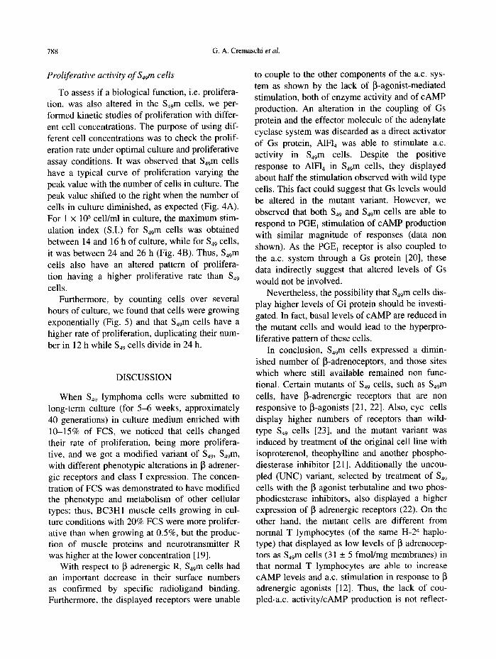
Proliferative activity of S49m cells
To assess if a biological function, i.e. prolifera- tion, was also altered in the 549m cells, we per- formed kinetic studies of proliferation with differ- ent cell concentrations. The purpose of using dif- ferent cell concentrations was to check the prolif- eration rate under optimal culture and proliferative assay conditions. It was observed that $49m cells have a typical curve of proliferation varying the peak value with the number of cells in culture. The peak value shifted to the right when the number of cells in culture diminished, as expected (Fig. 4A). For 1 x 10⁵ cell/ml in culture, the maximum stim- ulation index (S.I.) for S49m cells was obtained between 14 and 16 h of culture, while for 549 cells, it was between 24 and 26 h (Fig. 4B). Thus, S49m cells also have an altered pattern of prolifera- tion having a higher proliferative rate than 549 cells.
Furthermore, by counting cells over several hours of culture, we found that cells were growing exponentially (Fig. 5) and that $49m cells have a higher rate of proliferation, duplicating their num- ber in 12 h while 549 cells divide in 24 h.
DISCUSSION
When 549 lymphoma cells were submitted to long-term culture (for 5-6 weeks, approximately 40 generations) in culture medium enriched with 10-15% of FCS, we noticed that cells changed their rate of proliferation, being more prolifera- tive, and we got a modified variant of 549, 549m, with different phenotypic alterations in 13 adrener- gic receptors and class I expression. The concen- tration of FCS was demonstrated to have modified the phenotype and metabolism of other cellular types; thus, BC3HI muscle cells growing in cul- ture conditions with 20% FCS were more prolifer- ative than when growing at 0.5%, but the produc- tion of muscle proteins and neurotransmitter R was higher at the lower concentration [ 19].
With respect to ~ adrenergic R, S49 m cells had an important decrease in their surface numbers as confirmed by specific radioligand binding. Furthermore, the displayed receptors were unable
to couple to the other components of the a.c. sys- tem as shown by the lack of [~-agonist-mediated stimulation, both of enzyme activity and of cAMP production. An alteration in the coupling of Gs protein and the effector molecule of the adenylate cyclase system was discarded as a direct activator of Gs protein, A1FI 4 was able to stimulate a.c. activity in 549m cells. Despite the positive response to A1F14 in 549m cells, they displayed about half the stimulation observed with wild type cells. This fact could suggest that Gs levels would be altered in the mutant variant. However, we observed that both S49 and $49m cells are able to respond to PGE~ stimulation of cAMP production with similar magnitude of responses (data non shown). As the PGE~ receptor is also coupled to the a.c. system through a Gs protein [20], these data indirectly suggest that altered levels of Gs would not be involved.
Nevertheless, the possibility that 549m cells dis- play higher levels of Gi protein should be investi- gated. In fact, basal levels of cAMP are reduced in the mutant cells and would lead to the hyperpro- liferative pattern of these cells.
In conclusion, 549m cells expressed a dimin- ished number of ~-adrenoceptors, and those sites which where still available remained non func- tional. Certain mutants of 549 cells, such as 549m cells, have 13-adrenergic receptors that are non responsive to [~-agonists [21, 22]. Also, cyc- cells display higher numbers of receptors than wild- type $49 cells [23], and the mutant variant was induced by treatment of the original cell line with isoproterenol, theophylline and another phospho- diesterase inhibitor [21]. Additionally the uncou- pled (UNC) variant, selected by treatment of $49 cells with the 13 agonist terbutaline and two phos- phodiesterase inhibitors, also displayed a higher expression of [3 adrenergic receptors (22). On the other hand, themutant cells are different from normal T lymphocytes (of the same H-2 d haplo- type) that displayed as low levels of [~ adrenocep- tors as $49m cells (31 _+ 5 fmol/mg membranes) in that normal T lymphocytes are able to increase cAMP levels and a.c. stimulation in response to adrenergic agonists [12]. Thus, the lack of cou- pled-a.c, activity/cAMP production is not reflect-

Fig. 3. IFI staining of Mo Ab against class I Ag and against thy 1.2 Ag. A and B: 1 x 10⁶ cells of lymphoma S49m type were incubated with 1/100 dilution of Mo Ab anti-L a and anti-thy 1.2, for A and B, respectively, at 4°C with 1/32 dilution of a FlTC-goat anti-mouse IgG Ab. C and D: 949 m cells were previously permeabilized with chilled acetone as indicated in Materials and Methods, and the smears of cells were incubated with 1/100 dilutions of anti- L d or anti-thy 1.2, for C and D, respectively, and the IFI procedure was followed as indicated for cell suspensions, but incubations were performed at 37°C. In both cases, dilutions of Mo Ab were made with PBS containing 10% or 15% normal goat serum for cells and smears, respectively. 160 x images are shown.
789


Loss of I~-adrenoceptor and class I antigen expression 791
® ® C P M ( x 1 0 4 ) . . . .
TI l~ I " 60 ¢l~w-u) IWl
16 3~11 .+ Z I I1=1 * 12
26 64O :t 4:1 I ~ + 2 30 440 _+ I '~ t42_~ II
3O
2O
"" " 0 " - 13 . . . . . i ~ . . . .
10
omlC;--"L , , , , i l , I I I I J ' •
2 4 O 8 10 12 14 16 18 20 22 24 26 28 ~0
1 1 m ( h o u r i )
Fig. 4. Proliferation rate of 549 and 549m cells. A: kinet- ic curves of proliferation of 549 m cells were performed as indicated in Materials and Methods with different concentrations of cells: 3 x 10⁵ cell/ml (O--O); 1.5 x 10⁵ cell/ml (A--A); 1.0 × l0 s cell/ml (©--O) and 0.5 × 10⁵ cell/ml ([3--[3). Values shown are an example of three experiments. B: stimulation index (S.I.) obtained as described in Materials and Methods, for 1.0 × 10⁵ cell/ml 549 and S49 m cells _+ S.E. are shown.
log oel ls /ml (x lO - ° ) 5o
1o
1 ~ I1~--,,"~ @ , , , J ' o 12 24 3e 4s so 72
Time (houro)
Fig. 5. Measurement of cell proliferation by counting of viable cells. 1 x 10⁵ 549 (O--O) or S49m ( 0 - - 0 ) were cultured as indicated before, and duplication time for both cell types was calculated from the curves' slopes: 24 _+ 2 h for 549 , and 12 ± 1 h for 549m cells.
ing failure of detectable activity due to low levels of receptor expression.
Besides, associated with this loss of 13 adreno- ceptors, 549m cells were not able to display class I Ag of the d haplotype on their surface membranes.
This was demonstrated by a lack of C'-dependent cytotoxic activity with specific Mo Ab and a lack of binding of those Ab by the ELISA technique and IFI staining on cloned intact cells. Despite this, S49m still expressed a 549 marker, the thy 1.2 Ag. However, when performing IFI assays with class I Ab on smears of permeabilized cells, posi- tive staining was obtained inside the cells, differ- ently to anti-thy 1.2 staining that remained mainly on the cell membrane. These results, though sur- prising, are supported by the fact that the MHC phenotype for normal and malignant cells is not fixed, but changes in response to environmental factors. Thus, treatment of normal or malignant cells with interferons can drastically change the expression of cell surface MHC Ag [24] and pas- sage of the mouse lymphoma YAC-1 cells from in
vivo to in vitro culture is related to progressive loss of H-2 Ag [25] as occurred with 549m cells.
Moreover, this is not exclusive for MHC Ag on lymphoid cells, as epidermal langerhans cells exhibit modifications of surface expression of class II MHC Ag during short-term culture [26].
In summary, the loss of ~ adrenergic receptors would be accompanied by an internalization of class I molecules, or alternatively, a fault of class I Ag surface expression on 549 m cells. As it was demonstrated that MHC class I Ag molecularly interact with 13 adrenoceptors on lymphocytes [10] this related loss of both ~ adrenoceptors and class I molecules might suggest that their expression can be modulated mutually.
It was previously demonstrated that increases in the cellular level of cAMP down-regulates lym- phocyte proliferation as a consequence of cAMP- dependent protein kinase (PK) A activation, while PKC activation leads to proliferation [27]. It can be hypothesized that the down-regulation of func- tional I~ adrenoceptors related to a high prolifera- tive rate, would be one of the mechanisms to avoid high intracellular levels of the cyclic nucleotide that are related to cellular proliferation inhibition. The lower response of the 549m cells to the direct activation of AIF14 is probably an addi- tional factor implicated in the hyperproliferative response of the cell. Alternatively, it could be a consequence of a very active PKC which could

792 G.A. Cremaschi et al.
lead to phosphorylation and subsequent desensiti- zation and uncoupling of the receptor to the adenylate cyclase system [28]. Indeed, Tse and Pernis [29] showed that after T lymphocyte in
vitro stimulation with mitogen or in a mixed lym- phocyte reaction class I Ag are spontaneously internalized. Moreover, phosphorylation of MHC class I molecules has been implicated in determin- ing endocytic uptake from the cell surface and may function in down-regulating surface expres-
sion of MHC class I molecules [30], and PKC was demonstrated to be involved in MHC Ag hyper-
phosphorylation [31].
Acknowledgements--This work was supported by Grants 3025100/92-93 from CONICET. The authors wish to thank Dr E. S. Borda for his helpful discussion of the manuscript and also thanks MRS E. Vannucchi and Mr M. RodriGuez for their excellent technical assistance.
REFERENCES
1. Blalock J. E. (1989) Physiol. Rev. 69, 1. 2. Cavagnaro J. and Lewis R. M. (1989) The Year in
Immunology. Cellular, Molecular and Clinical Aspects (Cruce J. M. and Lewis R. E. Jr, Eds), Vol. 4, p. 241. Basel, Karger.
3. Bushkin Y., Demaria S., Le J. and Schwabs R. (1988) Proc. natn. Acad. Sci. U.S.A. 85, 3985.
4. Harel-Bellan A., Krief P., Rimsky L., Farrar W. L. and Mishal Z. (1990)Biochem. J. 268, 35.
5. Schreiber A. B., Schlessinger J. and Edidin M. (1984) J. Cell Biol. 98, 725.
6. Solano A. R., Cremachi G. A., Sanchez M. L., Borda E. S., Sterin-Borda L. and Podesta E. J. (1988) Proc. natn. Acad. Sci. U.S.A. 85, 5087.
7. Verland S., Simonsen M., Gammeltoft S., Allen H., Flavel R. A. and Olsson L. (1989) J. Immunol. 143, 945.
8. Cremaschi G. A. and Sterin-Borda L. (1989) FEBS Lett. 249, 302.
9. Cremaschi G. A., Gorelik G., Genaro A. M., Borda E. and Sterin-Borda L. (1992a) Biochem. Pharmac. 43, 2493.
10. Cremaschi G. A., Genaro A. M. and Sterin-Borda L. (1989) Molec. lmmunol. 26, 601.
11. Cremaschi G. A., Borda E. S., Sales M. E., Genaro A. M. and Sterin-Borda L. (1990) Biochem. Pharmac. 39, 1861.
12. Cremaschi G. A., Fisher P. and Boege F. (1991) Immunopharmacology 22, 195.
13. Cremaschi G. A., Miguel S., Cazaux C. and Sterin- Borda L. (1992b) Medicina 52, 404.
14. Munson P. J. and Rodbard D. (1980) Analyt. Biochem. 107, 220.
15. Salomon Y., Londos C. and Rodbell M. (1974) Analyt. Biochem. 58, 541.
16. Brown B. L., Albano J. D. M., Ekins R. P. and Sgherzi A. M. (1971) Biochem. J. 121, 561.
17. Oi V. T. and Herzenberg L. A. (1980) Selected Methods in Cellular Immunology (Mishell B. B. and Shigii S. M., Eds), p. 366. L. W. H. Freeman and Company, U.S.A.
18. Ross E. M., Maguire M. E., Sturgill T. W., Biltonen R. L. and Gilman A. G. (1977) J. biol. Chem. 252, 5761.
19. Kelvin D. J., Simard G., Tai H. H., Yamaguchi T. P. and Connolly J. A. (1989)J. Cell Biol. 108, 159.
20. Gilman A. G. (1984) Cell36, 577. 21. Bourne H., Coffino P., Melmon K. L., Tomkins G.
M. and Weinstein Y. (1975) Adv. Cyclic Nucleotide Res. 5, 771.
22. Haga T., Ross E. M., Anderson H. J. and Gilman A. G. (1977) Proc. natn. Acad. Sci. U.S.A. 74, 2016.
23. Abramson S. N. and Molinoff P. B. (1985) J. biol. Chem. 260, 14580.
24. Mandsley D. J. and Pound J. D. (1991) lmmunol. Today 12, 429.
25. Peterson M. G. E., Karre K., Cochet M., Kourilsky P. and Kiessling R. (1987) Cell Immunol. 108, 460.
26. Cohen P. J. and Katz S. I. (1992) J. Invest. Dermatol. 98, 331.
27. Roszman T. L. and Brooks W. H. (1988) Prog. Allergy 43, 140.
28. Sibley D. R., Benovis J. L., Caron M. G. and Lefkowitz R. J. (1988) Endocr. Rev. 9, 38.
29. Tse D. B. and Pernis B. (1984) J. exp. Med. 159, 193.
30. Lipp6 R., Luke E., Kuah Y. T., Lomas C. and Jefferies W. A. (1991) J. exp. Med. 174, 1159.
31. Capps G. G., van Kampen M., Ward C. L. and Zuniga M. C. (1989)J. Cell Biol. 108, 1317.