Increased formation of 8-iso-prostaglandin F 2? is associated with altered bone metabolism and lower...
10
doi: 10.1111/j.1365-2796.2007.01784.x Increased formation of 8-iso-prostaglandin F 2a is associated with altered bone metabolism and lower bone mass in hypercholesterolaemic subjects R. A. Mangiafico 1 , G. Malaponte 2 , P. Pennisi 1 , G. Li Volti 3,4 , G. Trovato 1 , M. Mangiafico 1 , Y. Bevelacqua 2 , F. Mazza 3 & C. E. Fiore 1 From the Departments of 1 Internal Medicine, 2 Biomedical Sciences and 3 Biological Chemistry, University of Catania, Catania; and 4 Department of Biomedical Sciences, University of Brescia, Brescia; Italy Abstract. Mangiafico RA, Malaponte G, Pennisi P, Li Volti G, Trovato G, Mangiafico M, Bevelacqua Y, Mazza F, Fiore CE (University of Catania, Catania; and University of Brescia, Brescia; Italy) Increased formation of 8-iso-prostaglandin F 2a is associated with altered bone metabolism and lower bone mass in hypercholesterolaemic subjects. J Intern Med 2007; 261: 587–596. Objectives. To investigate the relationship of 8-iso- prostaglandin (PG) F 2a levels, a reliable marker of in vivo oxidative stress and lipid peroxidation, with bone mineral density (BMD), bone turnover markers, osteoprotegerin (OPG) and receptor activator of nuclear factor-kappa B ligand (RANKL) in hypercho- lesterolaemia. Design. Cross-sectional study Setting. University hospital centre Methods. Serum 8-iso-PGF 2a levels were measured in 173 hypercholesterolaemic subjects and in 152 age- and sex-matched normocholesterolaemic controls. Femoral neck and lumbar spine BMD, serum bone- specific alkaline phosphatase (BAP), osteocalcin (OC), OPG and RANKL levels, as well as urinary levels of C-terminal telopeptides of type I collagen (CTX-I), were also assessed. Results. Hypercholesterolaemic subjects showed higher (P < 0.0001) serum 8-iso-PGF 2a levels than controls. They also had decreased (P < 0.0001) femoral neck and lumbar spine BMD, and lower (P < 0.0001) serum BAP and OC levels. No significant differences between hypercholesterolaemic and control subjects were found when comparing urinary CTX-I levels, or serum OPG and RANKL levels. In multivariate linear regression analysis, serum 8-iso-PGF 2a was the only negative predictor for femoral neck BMD and serum BAP and OC levels in hypercholesterolaemic subjects. No significant correlation (all P > 0.25) was present between serum 8-iso-PGF 2a levels and urinary CTX-I levels, or serum OPG and RANKL levels, in hyper- cholesterolaemic subjects. Conclusions. We found an association between increased serum 8-iso-PGF 2a levels and lower bone mass and reduced serum BAP and OC concentrations in hyper- cholesterolaemic subjects. These results would suggest a possible role for oxidative stress in the development of lower bone mass in hypercholesterolaemia. Keywords: bone density, hypercholesterolaemia, lipids, osteoporosis, oxidative stress. Introduction Increasing evidence suggests an age-independent association between osteoporosis and atherosclerosis [1–6], but the underlying mechanisms remain elusive. Recently, several lines of research have converged to indicate a possible link between hypercholesterolae- mia, a major risk factor for atherosclerosis [7] and ª 2007 Blackwell Publishing Ltd 587 Original Article |
Transcript of Increased formation of 8-iso-prostaglandin F 2? is associated with altered bone metabolism and lower...
doi: 10.1111/j.1365-2796.2007.01784.x
Increased formation of 8-iso-prostaglandin F2a isassociated with altered bone metabolism and lower bonemass in hypercholesterolaemic subjects
R. A. Mangiafico1, G. Malaponte2, P. Pennisi1, G. Li Volti3,4, G. Trovato1, M. Mangiafico1, Y. Bevelacqua2,F. Mazza3 & C. E. Fiore1
From the Departments of 1Internal Medicine, 2Biomedical Sciences and 3Biological Chemistry, University of Catania, Catania;and 4Department of Biomedical Sciences, University of Brescia, Brescia; Italy
Abstract. Mangiafico RA, Malaponte G, Pennisi P,
Li Volti G, Trovato G, Mangiafico M, Bevelacqua Y,
Mazza F, Fiore CE (University of Catania, Catania;
and University of Brescia, Brescia; Italy) Increased
formation of 8-iso-prostaglandin F2a is associated with
altered bone metabolism and lower bone mass in
hypercholesterolaemic subjects. J Intern Med 2007;
261: 587–596.
Objectives. To investigate the relationship of 8-iso-
prostaglandin (PG) F2a levels, a reliable marker of
in vivo oxidative stress and lipid peroxidation, with
bone mineral density (BMD), bone turnover markers,
osteoprotegerin (OPG) and receptor activator of
nuclear factor-kappa B ligand (RANKL) in hypercho-
lesterolaemia.
Design. Cross-sectional study
Setting. University hospital centre
Methods. Serum 8-iso-PGF2a levels were measured in
173 hypercholesterolaemic subjects and in 152 age-
and sex-matched normocholesterolaemic controls.
Femoral neck and lumbar spine BMD, serum bone-
specific alkaline phosphatase (BAP), osteocalcin
(OC), OPG and RANKL levels, as well as urinary
levels of C-terminal telopeptides of type I collagen
(CTX-I), were also assessed.
Results. Hypercholesterolaemic subjects showed higher
(P < 0.0001) serum 8-iso-PGF2a levels than controls.
They also had decreased (P < 0.0001) femoral neck
and lumbar spine BMD, and lower (P < 0.0001)
serum BAP and OC levels. No significant differences
between hypercholesterolaemic and control subjects
were found when comparing urinary CTX-I levels, or
serum OPG and RANKL levels. In multivariate linear
regression analysis, serum 8-iso-PGF2a was the only
negative predictor for femoral neck BMD and serum
BAP and OC levels in hypercholesterolaemic subjects.
No significant correlation (all P > 0.25) was present
between serum 8-iso-PGF2a levels and urinary CTX-I
levels, or serum OPG and RANKL levels, in hyper-
cholesterolaemic subjects.
Conclusions. We found an association between increased
serum 8-iso-PGF2a levels and lower bone mass and
reduced serum BAP and OC concentrations in hyper-
cholesterolaemic subjects. These results would suggest
a possible role for oxidative stress in the development
of lower bone mass in hypercholesterolaemia.
Keywords: bone density, hypercholesterolaemia, lipids,
osteoporosis, oxidative stress.
Introduction
Increasing evidence suggests an age-independent
association between osteoporosis and atherosclerosis
[1–6], but the underlying mechanisms remain elusive.
Recently, several lines of research have converged to
indicate a possible link between hypercholesterolae-
mia, a major risk factor for atherosclerosis [7] and
ª 2007 Blackwell Publishing Ltd 587
Original Article |
osteoporosis. Hypercholesterolaemia has been reported
to be associated with low bone mass in a number of
studies focused on postmenopausal or osteoporotic
women [8–12]. The relationship between bone mass
and lipid values has been shown to be negative for
low-density lipoprotein (LDL) cholesterol [8, 12] and
positive for high-density lipoprotein (HDL) choles-
terol [8]. However, other studies found an opposite
relationship between lipid profile and bone mineral
density (BMD) [13], or no relationship at all [14].
Data from various observational studies [15–17],
albeit not consistent [18, 19], suggested that the use
of the lipid-lowering drugs statins (3-hydroxy-3-meth-
ylglutaryl coenzyme A reductase inhibitors) is associ-
ated with a reduced risk of fractures and increased
bone mass. However, such association was not found
in randomized controlled clinical trials of statins for
cardiovascular disease [20, 21].
In vitro and animal studies showed that diet-induced
hyperlipidaemia reduced bone density [22] by inhibit-
ing osteoblastic [23, 24] and promoting osteoclastic
differentiation [25, 26] via increased formation of
lipid oxidation products. F2-isoprostanes are stable
prostaglandin (PG) F2-like end products of lipid
peroxidation and are formed in vivo from the nonen-
zymatic free radical-induced peroxidation of arachi-
donic acid, a ubiquitous polyunsaturated fatty acid
[27]. F2-isoprostanes, including 8-iso-PGF2a, are
widely regarded as the best available biomarkers of
in vivo oxidative stress and lipid peroxidation [28].
Other measures of oxidative stress, including malondi-
aldehyde and lipid hydroperoxides, have been demon-
strated to be of limited value in vivo because they
lack sensitivity and/or specificity [28].
It has been shown that enhanced formation of 8-iso-
PGF2a occurs in hypercholesterolaemic subjects [29,
30]. Moreover, urinary levels of 8-iso-PGF2a have
been reported to be negatively correlated with BMD
at various sites in a sample from the general popula-
tion [31]. However, no information is available from
clinical studies on the possible relationship between
F2-isoprostanes and bone mass in male and female
subjects with hypercholesterolaemia. It is also
unknown whether F2-isoprostanes are related to bone
turnover markers and osteoclast regulatory factors
osteoprotegerin (OPG) and receptor activator of nuc-
lear factor-kappa B ligand (RANKL) in hypercholes-
terolaemia. Therefore, we assessed serum 8-iso-PGF2alevels in relation to BMD in a group of untreated
hypercholesterolaemic subjects. We also investigated
the relationship of serum 8-iso-PGF2a levels to two
bone formation markers, serum bone-specific alkaline
phosphatase (BAP) and osteocalcin (OC), one bone
resorption marker, urinary C-terminal telopeptides of
type I collagen, and serum levels of OPG and
RANKL.
Materials and methods
Study population
A total of 173 ambulatory, well-functioning subjects
with primary hypercholesterolaemia (aged 32–
71 years; 83 males and 90 females) were consecu-
tively recruited from the Lipid Clinic at our hospital
between January and October 2005. Hypercholesterol-
aemia was defined as LDL cholesterol level
>4.13 mmol L)1. Exclusion criteria were secondary
hyperlipidaemia owing to diabetes mellitus, renal,
liver or thyroid diseases, and alcohol abuse. A total of
152 age- and sex-matched clinically healthy normo-
cholesterolaemic subjects (aged 31–72 years; 75
males and 77 females) were selected from individuals
receiving a medical check-up at our hospital and
served as controls. Hypercholesterolaemic subjects
were included in the study if they were not taking
lipid-lowering drugs and were not on lipid-lowering
diets. Patients with known hypertension or with car-
diovascular disease (as assessed by clinical history,
physical examination, and ECG) were excluded from
the study. Patients who were taking drugs with estab-
lished effect on bone turnover or were suffering from
clinical disorders related to bone metabolism were
also not enrolled in the study. None of the participants
was a current smoker, and none was receiving vitamin
or calcium supplements. All subjects completed a
questionnaire that included age at menarche, postmen-
opausal status, lifestyle factors, medical history, and
medication use. Physical activity was measured by the
R. A. Mangiafico et al. | 8-iso-PGF2a and BMD in hypercholesterolaemia
588 ª 2007 Blackwell Publishing Ltd Journal of Internal Medicine 261; 587–596

Paffenbarger Physical Activity Index [32]. The average
daily calcium intake was determined by a quantitative
food frequency questionnaire [33]. All participants
underwent clinical examination, ECG, and measure-
ments of BMD and biochemical parameters. The
study protocol was approved by our institutional
ethics committee in accordance to the Helsinki
Declaration, and all study participants gave informed
consent.
Measurement of bone mineral density
Areal BMD (g cm)2; bone mineral content relative to
projection area) was measured by dual-energy X-ray
absorptiometry (Lunar DPXL, Lunar, Madison, WI,
USA) at the lumbar spine (L2–L4) and the femoral
neck. At these measurement sites, the precision of the
method (coefficient of variation, CV) at our laboratory
was 0.7% for the lumbar spine and 0.5% for the
femoral neck.
Laboratory measurements
Venous blood samples and urine specimens were
obtained after a overnight fast, between 9.00 and
9.30 a.m. Serum and urine aliquots were stored at
)80 �C until analysis. The following measurements
were carried out:
Total serum 8-iso-PGF2a levels were measured by
using a commercially available enzyme immunoassay
(EIA) kit (Cayman Chemicals, Ann Arbor, MI, USA)
according to the manufacturer’s instructions. The
detection limit of this assay was 5 pg mL)1. The
intra-assay and interassay CVs were <10%.
BAP was measured by an enzyme-linked immunoab-
sorbent assay (ELISA) kit (Tandem-MP Ostase, Beck-
man Coulter, Fullerton, CA, USA). The sensitivity of
the method was 0.7 lg L)1. Within-run CV was 6.5%
for low values, and 4.5% for high values.
OC was measured with a commercially available
ELISA kit (N-MID Osteocalcin One Step) provided
by Nordic Bioscience Diagnostics A/S, Herlev,
Denmark. The sensitivity of the method was
0.5 ng mL)1. Intra-assay CV was 3.4% for low val-
ues and 2.4% for high values.
Urinary CTX-I levels were measured by ELISA using
reagents provided by Osteometer Bio Tech A/S (Cros-
sLaps). The detection limit of this assay was
50 lg L)1. Intra-assay and interassay CVs were,
respectively, 5.7% and 9.4% for low values and 5.4%
and 8.6% for very high values.
Serum concentration of OPG was determined by a
sandwich ELISA (Osteoprotegerin, Immun Diagnos-
tik, Bensheim, Germany). The detection limit of this
assay system was 0.14 pmol L)1. Intra-assay and
interassay CVs were <10% at both low and high
concentrations of OPG.
Serum levels of soluble RANKL were measured by
EIA (sRANKL, Biomedica, Vienna, Austria). The
detection limit of this assay system was 0.08 pmol L)1.
Intra-assay and interassay CVs were 5% and 9%,
respectively.
Serum total cholesterol, HDL cholesterol and triglyc-
erides were measured enzymatically using the Cobas
Integra Roche analyser (Roche Diagnostics, Milan,
Italy). LDL cholesterol was calculated using the Frie-
dewald formula [34]. Serum calcium, phosphorus, and
albumin were determined by colorimetric assays on
the Cobas Integra Roche analyser (Roche Diagnos-
tics). Serum calcium was adjusted for albumin
concentration.
Statistical analysis
Continuous data are reported as mean ± standard
deviation (SD) and categorical data as percentages.
Males and females were analysed separately. Compar-
isons between groups were made by means of
unpaired t-test or Mann–Whitney test (in the case of
non-normal distribution) for continuous variables and
by Fisher’s Exact Test for categorical variables.
Univariate relationships between 8-iso-PGF2a levels
and outcome variables amongst hypercholesterolaemic
subjects were tested by Spearman’s correlation coeffi-
cient. Outcome variables included femoral neck and
R. A. Mangiafico et al. | 8-iso-PGF2a and BMD in hypercholesterolaemia
ª 2007 Blackwell Publishing Ltd Journal of Internal Medicine 261; 587–596 589
lumbar spine BMD, serum levels of BAP, OC, OPG
and RANKL, and urinary CTX-I levels. Multivariate
linear regression analyses were performed to look for
independent associations between 8-iso-PGF2a levels
(independent variable) and the outcome variables
(dependent variables) that were found to have a statis-
tical association (P < 0.05) with 8-iso-PGF2a levels in
the univariate analysis. In the models of multivariate
analysis the following variables were also included as
independent variables: age, body mass index (BMI),
calcium intake, physical activity index, LDL choles-
terol, HDL cholesterol, triglycerides, and for females,
age at menarche and menopausal status. As total cho-
lesterol and LDL cholesterol were highly correlated in
both male and female hypercholesterolaemic subjects
(r ¼ 0.90, P < 0.0001), only LDL cholesterol was
included in the models of multivariate analysis. We
also tested the relationships between lipid parameters
(total cholesterol, LDL cholesterol, HDL cholesterol
and triglycerides) and each of the outcome variables,
as well as those between lipid parameters and 8-iso-
PGF2a levels. Univariate and multivariate analyses
were also carried out in the whole hypercholesterolae-
mic population. In addition, correlation analyses were
used to investigate the relationships between 8-iso-
PGF2a levels and BMD and the other outcome varia-
bles in the controls, both in the male and female
subgroups and in the whole group. In multivariate
analyses for the study groups as a whole, we also cor-
rected for sex. In all analyses, a two-tailed P value
<0.05 was considered significant. Statistical analysis
was performed using graphpad instat version 3.00
for Windows software (GraphPad Software, San
Diego, CA, USA).
Results
The characteristics of the study population are shown
in Table 1. Mean age, BMI, systolic and diastolic
blood pressure values, daily calcium intake, estimated
physical activity levels, as well as serum calcium and
phosphorus were not significantly different comparing
all the groups. By definition, serum total and LDL
cholesterol levels were significantly (P < 0.001)
higher in the hypercholesterolaemia group than in the
control group, whereas HDL cholesterol and triglycer-
ide concentrations were similar between all groups.
There were no sex differences.
Serum 8-iso-PGF2a levels were significantly higher in
the hypercholesterolaemic subjects than in the respect-
ive controls, with no differences between sexes
(178.0 ± 126.0 and 28.5 ± 12.2 pg mL)1 in male
hypercholesterolaemic and control subjects, respect-
ively, P < 0.0001; 181.5 ± 122.0 and 30.0 ±
13.0 pg mL)1 in female hypercholesterolaemic and
control subjects, respectively, P < 0.0001). Mean
BMD at the femoral neck (0.78 ± 0.14 and
1.00 ± 0.04 g cm)2 in male hypercholesterolaemic
and control subjects, respectively, P < 0.0001;
0.77 ± 0.14 and 0.99 ± 0.07 g cm)2 in female hyper-
cholesterolaemic and control subjects, respectively,
P < 0.0001) and lumbar spine (1.02 ± 0.13 and
1.16 ± 0.02 g cm)2 in male hypercholesterolaemic
and control subjects, respectively, P < 0.0001;
1.05 ± 0.17 and 1.16 ± 0.04 g cm)2 in female hyper-
cholesterolaemic and control subjects, respectively,
P < 0.0001) were similarly reduced in both male and
female subjects with hypercholesterolaemia when
compared with the respective controls (Table 1).
There were no significant differences in serum levels
of OPG and RANKL, and in urinary levels of CTX-I
between all groups (Table 2). Serum BAP and OC
levels were similarly lower (P < 0.0001) in male
(8.2 ± 2.0 lg L)1 and 19.9 ± 3.2 ng mL)1, respect-
ively) and female (8.0 ± 1.8 lg L)1 and 19.8 ±
5.3 ng mL)1, respectively) subjects with hypercholes-
terolaemia when compared with male (13.4 ±
1.6 lg L)1 and 27.0 ± 3.7 ng mL)1, respectively) and
female (13.6 ± 0.7 lg L)1 and 28 ± 3.1 ng mL)1,
respectively) controls (Table 2).
In univariate analysis for male and female hypercho-
lesterolaemic subjects, serum 8-iso-PGF2a levels were
significantly negatively correlated with femoral neck
BMD in both males (r ¼ )0.27, P ¼ 0.01) and
females (r ¼ )0.29, P ¼ 0.004). A significant neg-
ative correlation between serum iso-PGF2a levels and
serum BAP (r ¼ )0.35, P ¼ 0.001, in males;
r ¼ )0.32, P ¼ 0.002, in females) and OC
(r ¼ )0.33, P ¼ 0.001, in males; r ¼ )0.34,
R. A. Mangiafico et al. | 8-iso-PGF2a and BMD in hypercholesterolaemia
590 ª 2007 Blackwell Publishing Ltd Journal of Internal Medicine 261; 587–596

P ¼ 0.0008, in females) concentrations was also
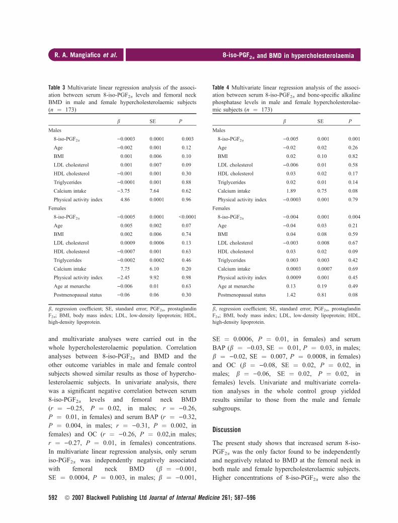
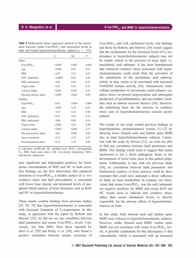
found. In multivariate linear regression analysis for
male and female hypercholesterolaemic subjects,
serum 8-iso-PGF2a was the only significant negative
predictor for femoral neck BMD and serum BAP and
OC levels in males and females (Tables 3–5). In both
male and female hypercholesterolaemic subjects, there
were no correlations (all P > 0.25) between serum
8-iso-PGF2a levels and lumbar spine BMD, as well as
between serum 8-iso-PGF2a levels and serum OPG
and RANKL concentrations, or urinary CTX-I levels.
No correlation (all P > 0.20) was found between lipid
values and femoral neck or lumbar spine BMD, as
well as between lipid values and serum BAP, OC,
OPG and RANKL concentrations, or urinary CTX-I
levels. There was also no significant correlation (all
P > 0.10) between lipid parameters and 8-iso-PGF2alevels. Similar results were obtained when univariate
Table 1 Characteristics of hypercholesterolaemic and control subjects
Characteristic
Hypercholesterolaemic subjects (n ¼ 173) Controls (n ¼ 152)
Male Female Male Female
Number (%) 83 (47.9) 90 (52.0) 75 (49.3) 77 (50.6)
Age (years) 52.2 ± 10.0 52.3 ± 12.0 53.4 ± 10.7 52.7 ± 11.5
Body mass index (kg m)2) 26.6 ± 2.2 26.5 ± 2.3 26.1 ± 1.2 26.6 ± 1.6
Age at menarche (years) – 13.1 ± 1.0 – 12.9 ± 0.8
Postmenopausal status, n (%) – 43 (47.7) – 36 (46.7)
Systolic blood pressure (mmHg) 122.6 ± 4.7 121 ± 6.5 121.8 ± 6.0 122 ± 6.6
Diastolic blood pressure (mmHg) 72.8 ± 4.4 73.6 ± 5.0 74.0 ± 3.6 74.5 ± 3.1
Daily calcium intake (mg) 852.2 ± 194.8 857.7 ± 239.2 834.9 ± 291.9 831.4 ± 288.8
Physical activity index (kcal week)1) 825.9 ± 142.1 821.1 ± 144.5 846.9 ± 208.5 836.1 ± 166.9
Total cholesterol (mmol L)1) 7.22 ± 0.71* 7.20 ± 0.74* 4.93 ± 0.24 5.01 ± 0.28
LDL cholesterol (mmol L)1) 5.36 ± 0.58* 5.34 ± 0.61* 3.07 ± 0.18 3.12 ± 0.19
HDL cholesterol (mmol L)1) 1.31 ± 0.23 1.29 ± 0.25 1.32 ± 0.14 1.35.3 ± 0.17
Triglycerides (mmol L)1) 1.14 ± 0.01 1.26 ± 0.55 1.15 ± 0.16 1.15 ± 0.35
Calcium (mmol L)1) 2.32 ± 0.07 2.34 ± 0.08 2.31 ± 0.02 2.33 ± 0.06
Phosphorus (mmol L)1) 1.07 ± 0.08 1.08 ± 0.09 1.08 ± 0.02 1.07 ± 0.07
8-iso-PGF2a (pg mL)1) 178.0 ± 126.0* 181.5 ± 122.0* 28.5 ± 12.2 30.0 ± 13.0
Femoral neck BMD (g cm)2) 0.78 ± 0.14* 0.77 ± 0.14* 1.00 ± 0.04 0.99 ± 0.07
Lumbar spine BMD (g cm)2) 1.02 ± 0.13* 1.05 ± 0.17* 1.16 ± 0.02 1.16 ± 0.04
Values are reported as mean ± standard deviation unless otherwise indicated. LDL, low-density lipoprotein; HDL, high-density lipoprotein;PGF2a, prostaglandin F2a; BMD, bone mineral density. *P < 0.0001 vs. controls.
Table 2 Serum levels of OPG, RANKL, BAP, OC and urinary CTX-I levels in hypercholesterolaemic (n ¼ 173) and control(n ¼ 152) subjects
Variable
Hypercholesterolaemic subjects Controls
Male Female Male Female
OPG (pmol L)1) 3.6 ± 0.9 3.7 ± 1.1 3.7 ± 1.0 3.6 ± 0.9
RANKL (pmol L)1) 0.6 ± 0.2 0.5 ± 0.3 0.5 ± 0.2 0.5 ± 0.2
BAP (lg L)1) 8.2 ± 2.0* 8.0 ± 1.8* 13.4 ± 1.6 13.6 ± 0.7
OC (ng mL)1) 19.9 ± 3.2* 19.8 ± 5.3* 27.0 ± 3.7 28 ± 3.1
Urinary CTX-I (lg mmol)1 creatinine) 259.8 ± 31.6 270.2 ± 46.9 262.9 ± 42.5 257.2 ± 31.3
Values are reported as mean ± standard deviation. OPG, osteoprotegerin; RANKL, receptor activator of nuclear factor-kappa B ligand; BAP,bone-specific alkaline phosphatase; OC, osteocalcin; CTX-I, C-terminal telopeptides of type I collagen. *P < 0.0001 vs. controls.
R. A. Mangiafico et al. | 8-iso-PGF2a and BMD in hypercholesterolaemia
ª 2007 Blackwell Publishing Ltd Journal of Internal Medicine 261; 587–596 591
and multivariate analyses were carried out in the
whole hypercholesterolaemic population. Correlation
analyses between 8-iso-PGF2a and BMD and the
other outcome variables in male and female control
subjects showed similar results as those of hypercho-
lesterolaemic subjects. In univariate analysis, there
was a significant negative correlation between serum
8-iso-PGF2a levels and femoral neck BMD
(r ¼ )0.25, P ¼ 0.02, in males; r ¼ )0.26,P ¼ 0.01, in females) and serum BAP (r ¼ )0.32,P ¼ 0.004, in males; r ¼ )0.31, P ¼ 0.002, in
females) and OC (r ¼ )0.26, P ¼ 0.02,in males;
r ¼ )0.27, P ¼ 0.01, in females) concentrations.
In multivariate linear regression analysis, only serum
iso-PGF2a was independently negatively associated
with femoral neck BMD (b ¼ )0.001,SE ¼ 0.0004, P ¼ 0.003, in males; b ¼ )0.001,
SE ¼ 0.0006, P ¼ 0.01, in females) and serum
BAP (b ¼ )0.03, SE ¼ 0.01, P ¼ 0.03, in males;
b ¼ )0.02, SE ¼ 0.007, P ¼ 0.0008, in females)
and OC (b ¼ )0.08, SE ¼ 0.02, P ¼ 0.02, in
males; b ¼ )0.06, SE ¼ 0.02, P ¼ 0.02, in
females) levels. Univariate and multivariate correla-
tion analyses in the whole control group yielded
results similar to those from the male and female
subgroups.
Discussion
The present study shows that increased serum 8-iso-
PGF2a was the only factor found to be independently
and negatively related to BMD at the femoral neck in
both male and female hypercholesterolaemic subjects.
Higher concentrations of 8-iso-PGF2a were also the
Table 3 Multivariate linear regression analysis of the associ-ation between serum 8-iso-PGF2a levels and femoral neckBMD in male and female hypercholesterolaemic subjects(n ¼ 173)
b SE P
Males
8-iso-PGF2a )0.0003 0.0001 0.003
Age )0.002 0.001 0.12
BMI 0.001 0.006 0.10
LDL cholesterol 0.001 0.007 0.09
HDL cholesterol )0.001 0.001 0.30
Triglycerides )0.0001 0.001 0.88
Calcium intake )3.75 7.64 0.62
Physical activity index 4.86 0.0001 0.96
Females
8-iso-PGF2a )0.0005 0.0001 <0.0001
Age 0.005 0.002 0.07
BMI 0.002 0.006 0.74
LDL cholesterol 0.0009 0.0006 0.13
HDL cholesterol )0.0007 0.001 0.63
Triglycerides )0.0002 0.0002 0.46
Calcium intake 7.75 6.10 0.20
Physical activity index )2.45 9.92 0.98
Age at menarche )0.006 0.01 0.63
Postmenopausal status )0.06 0.06 0.30
b, regression coefficient; SE, standard error; PGF2a, prostaglandinF2a; BMI, body mass index; LDL, low-density lipoprotein; HDL,high-density lipoprotein.
Table 4 Multivariate linear regression analysis of the associ-ation between serum 8-iso-PGF2a and bone-specific alkalinephosphatase levels in male and female hypercholesterolae-mic subjects (n ¼ 173)
b SE P
Males
8-iso-PGF2a )0.005 0.001 0.001
Age )0.02 0.02 0.26
BMI 0.02 0.10 0.82
LDL cholesterol )0.006 0.01 0.58
HDL cholesterol 0.03 0.02 0.17
Triglycerides 0.02 0.01 0.14
Calcium intake 1.89 0.75 0.08
Physical activity index )0.0003 0.001 0.79
Females
8-iso-PGF2a )0.004 0.001 0.004
Age )0.04 0.03 0.21
BMI 0.04 0.08 0.59
LDL cholesterol )0.003 0.008 0.67
HDL cholesterol 0.03 0.02 0.09
Triglycerides 0.003 0.003 0.42
Calcium intake 0.0003 0.0007 0.69
Physical activity index 0.0009 0.001 0.45
Age at menarche 0.13 0.19 0.49
Postmenopausal status 1.42 0.81 0.08
b, regression coefficient; SE, standard error; PGF2a, prostaglandinF2a; BMI, body mass index; LDL, low-density lipoprotein; HDL,high-density lipoprotein.
R. A. Mangiafico et al. | 8-iso-PGF2a and BMD in hypercholesterolaemia
592 ª 2007 Blackwell Publishing Ltd Journal of Internal Medicine 261; 587–596
only significant and independent predictor for lower
serum concentrations of BAP and OC in both sexes.
Our findings are the first observation that enhanced
formation of 8-iso-PGF2a, a reliable marker of in vivooxidative stress and lipid peroxidation, is associated
with lower bone density and decreased levels of per-
ipheral blood markers of bone formation such as BAP
and OC in hypercholesterolaemia.
These results confirm findings from previous studies
[29, 30, 35] that hypercholesterolaemia is associated
with increased formation of F2-isoprostanes. In this
study, in agreement with the report by Roberts and
Morrow [35], we did not see any correlation between
lipid parameters and serum 8-iso-PGF2a levels. Con-
versely, our data differ from those reported by
Davı et al. [29] and Reilly et al. [30], who found a
positive correlation between urinary excretion of
8-iso-PGF2a and LDL cholesterol levels. Our findings
and those by Roberts and Morrow [36] would suggest
that the mechanisms for the increased levels of F2-iso-
prostanes in hypercholesterolaemic subjects may not
be simply related to the presence of more lipid, i.e.
arachidonic acid substrate. It has been hypothesized
that enhanced oxidative stress associated with hyper-
cholesterolaemia could result from the activation of
the metabolism of the arachidonic acid pathway,
which, in turn, seems to be associated with increased
NAD(P)H oxidase activity [36]. Alternatively, intra-
cellular metabolism of mevalonate could enhance oxi-
dative stress via protein isoprenylation and subsequent
production of proinflammatory and pro-oxidant cytok-
ines such as tumour necrosis factor-a [36]. However,
the underlying basis for the increase in oxidative
stress seen in hypercholesterolaemia remains poorly
defined.
The results of our study extend previous findings in
hyperlipidaemic postmenopausal women [11,12] by
showing lower femoral neck and lumbar spine BMD
also in male hypercholesterolaemic subjects. Consis-
tent with the study by Orozco [11], we were not able
to find any correlation between lipid parameters and
BMD. This finding would seem to suggest that serum
cholesterol is not a direct pathogenic factor in the
development of lower bone mass in this patient popu-
lation. Additionally, in line with one previous study
[10], no correlations between lipid parameters and
biochemical markers of bone turnover could be dem-
onstrated that could have indicated a direct influence
of lipids on bone metabolism. In contrast, our obser-
vation that serum 8-iso-PGF2a was the only independ-
ent negative predictor for BMD and serum BAP and
OC would seem to indicate that oxidative stress,
rather than serum cholesterol levels, is directly
responsible for the adverse effects of hypercholester-
olaemia on bone.
In this study, both femoral neck and lumbar spine
BMD were reduced in hypercholesterolaemic subjects.
However, unlike femoral neck BMD, lumbar spine
BMD was not correlated with serum 8-iso-PGF2a lev-
els. A possible explanation for this discrepancy is that
osteoarthritis, which is associated with an increased
Table 5 Multivariate linear regression analysis of the associ-ation between serum 8-iso-PGF2a and osteocalcin levels inmale and female hypercholesterolaemic subjects (n ¼ 173)
b SE P
Males
8-iso-PGF2a )0.009 0.002 0.002
Age )0.008 0.03 0.81
BMI )0.07 0.15 0.62
LDL cholesterol 0.0009 0.01 0.95
HDL cholesterol 0.03 0.04 0.33
Triglycerides )0.01 0.03 0.55
Calcium intake )0.001 0.001 0.55
Physical activity index )0.005 0.002 0.09
Females
8-iso-PGF2a )0.01 0.004 0.006
Age 0.003 0.10 0.97
BMI 0.15 0.23 0.50
LDL cholesterol )0.01 0.02 0.53
HDL cholesterol 0.08 0.05 0.14
Triglycerides )0.001 0.01 0.90
Calcium intake )0.002 0.002 0.27
Physical activity index 0.01 0.003 0.08
Age at menarche )0.31 0.55 0.57
Postmenopausal status )0.52 2.36 0.82
b, regression coefficient; SE, standard error; PGF2a, prostaglandinF2a; BMI, body mass index; LDL, low-density lipoprotein; HDL,high-density lipoprotein.
R. A. Mangiafico et al. | 8-iso-PGF2a and BMD in hypercholesterolaemia
ª 2007 Blackwell Publishing Ltd Journal of Internal Medicine 261; 587–596 593
BMD, occurs quite often in the spine in older adults,
whereas the femoral neck is less often involved [37].
This may result in an overestimation of BMD meas-
ured at lumbar spine and may have concealed a
relation between lumbar spine BMD and serum
8-iso-PGF2a in hypercholesterolaemic subjects.
Correlation analyses between 8-iso-PGF2a and BMD
and the other outcome variables in the control group
showed results similar as those of hypercholesterolae-
mic group, thus confirming the independent negative
relationship between 8-iso-PGF2a and BMD and bone
formation markers, even in a population with a smal-
ler range in serum 8-iso-PGF2a levels.
In the present study, BAP and OC levels were
reduced in hypercholesterolaemic subjects, and this
finding may indicate that osteoblastic activity is inhib-
ited in hypercholesterolaemia. The observation of
a relationship between serum 8-iso-PGF2a and decrea-
ses in bone mass and serum BAP and OC concentra-
tions would suggest that enhanced oxidative stress, as
measured by serum 8-iso-PGF2a levels, may have a
negative impact on bone mass in hypercholesterolae-
mic subjects possibly via reduced bone formation.
This suggestion is supported by in vitro and animal
studies showing that lipid oxidation products may
inhibit osteoblastic differentiation of preosteoblasts,
alkaline phosphatase activity [23, 24] and OC expres-
sion [38], and increase adipogenic and reduce osteo-
genic differentiation of marrow stromal cells [24].
Oxidative stress has also been shown to inhibit in vitrothe differentiation of osteoblastic cells, with antioxi-
dants counteracting this effect [39]. Furthermore,
animal studies demonstrated that antioxidant vitamin
E protects against cellular lipid peroxidation and has
beneficial effects on bone mass [40]. Potential
involvement of oxidative stress in osteoporosis is also
consistent with previous evidence from some [41–43],
but not all [44], epidemiological studies that sugges-
ted that higher dietary antioxidant intake has a pro-
tective role on bone health.
Based on an assumption that serum and urine levels
of bone markers, and serum OPG and RANKL
concentrations track tissue levels, this study shows no
evidence that upregulation of bone resorption stimula-
ting mechanisms takes part in the pathophysiologic
processes mediating bone loss in hypercholesterolae-
mic subjects.
No statistically significant difference in urinary CTX-I
levels between hypercholesterolaemic and control sub-
jects was observed, thus suggesting that osteoclastic
activity is not enhanced in hypercholesterolaemia.
This finding and the lack of a correlation between
serum 8-iso-PGF2a and urinary CTX-I do not support
the hypothesis from experimental research that bio-
active products of lipid oxidation [25, 26] and oxida-
tive stress [45] may promote bone resorptive potential
in hypercholesterolaemic subjects.
No increased activation of the RANKL/RANK system
was found in hypercholesterolaemic subjects, a find-
ing indicating that this osteoclast regulatory system
[46] is not involved in the development of bone loss
associated with hypercholesterolaemia. Based on our
findings, where serum 8-iso-PGF2a was not associated
with OPG and RANKL levels, it is unlikely that the
relationship between oxidative stress and bone
in hypercholesterolaemia is mediated through the
OPG/RANKL signalling pathway.
Taken together, these findings would suggest that
increased oxidative stress associated with hypercholes-
terolaemia may adversely influence bone metabolism
by uncoupling bone formation (decreased) from bone
resorption (unchanged), which may result in a loss of
bone mass. However, the mechanisms underlying
selective effects of oxidative stress on bone formation
over bone resorption remain to be elucidated.
In conclusion, our findings show an association
between increased serum 8-iso-PGF2a levels and
reduced bone density in hypercholesterolaemic sub-
jects. Increased serum 8-iso-PGF2a also appears to be
associated with reduced serum concentrations of the
bone formation markers BAP and OC. These results
would suggest that enhanced oxidative stress might
play a role in the development of lower bone
mass associated with hypercholesterolaemia possibly
via reduced bone formation. Additional studies are
R. A. Mangiafico et al. | 8-iso-PGF2a and BMD in hypercholesterolaemia
594 ª 2007 Blackwell Publishing Ltd Journal of Internal Medicine 261; 587–596
however needed to investigate the association between
oxidative stress and lower bone mass in hypercholes-
terolaemia and to define precisely the underlying
mechanisms.
Conflict of interest statement
All authors have no conflicts of interest.
References
1 Tanko LB, Bagger YZ, Christiansen C. Low bone mineral den-
sity in the hip as a marker of advanced atherosclerosis in elderly
women. Calcif Tissue Int 2003; 73: 15–20.
2 van der Klift M, Pols HA, Hak AE, Witteman JC, Hofman A,
de Laet CE. Bone mineral density and the risk of peripheral
arterial disease: the Rotterdam Study. Calcif Tissue Int 2002;
70: 443–9.
3 Barengolts EI, Berman M, Kukreja SC, Kouznetsova T, Lin C,
Chomka EV. Osteoporosis and coronary atherosclerosis in
asymptomatic postmenopausal women. Calcif Tissue Int 1998;
62: 209–13.
4 Uyama O, Yoshimoto Y, Yamamoto Y, Kawai A. Bone changes
and carotid atherosclerosis in postmenopausal women. Stroke
1997; 28: 1730–2.
5 Pennisi P, Signorelli SS, Riccobene S et al. Low bone density
and abnormal bone turnover in patients with atherosclerosis of
peripheral vessels. Osteoporos Int 2004; 15: 389–95.
6 Mangiafico RA, Russo E, Riccobene S et al. Increased preval-
ence of peripheral arterial disease in osteoporotic postmenopau-
sal women. J Bone Miner Metab 2006; 24: 125–31.
7 Kannel WB, D’Agostino RB, Sullivan L, Wilson PW. Concept
and usefulness of cardiovascular risk profiles. Am Heart J 2004;
148: 16–26.
8 Yamaguchi T, Sugimoto T, Yano S et al. Plasma lipids and
osteoporosis in postmenopausal women. Endocr J 2002; 49:
211–7.
9 Broulik PD, Kapitola J. Interrelations between body weight,
cigarette smoking and spine mineral density in osteoporotic
Czech women. Endocr Regul 1993; 27: 57–60.
10 Tanko LB, Bagger YZ, Nielsen SB, Christiansen C. Does serum
cholesterol contribute to vertebral bone loss in postmenopausal
women? Bone 2003; 32: 8–14.
11 Orozco P. Atherogenic lipid profile and elevated lipoprotein (a)
are associated with lower bone mineral density in early post-
menopausal overweight women. Eur J Epidemiol 2004; 19:
1105–12.
12 Poli A, Bruschi F, Cesana B, Rossi M, Paoletti R, Crosignani
PG. Plasma low-density lipoprotein cholesterol and bone mass
densitometry in postmenopausal women. Obstet Gynecol 2003;
102: 922–6.
13 Adami S, Braga V, Zamboni M et al. Relationship between
lipids and bone mass in 2 cohorts of healthy women and men.
Calcif Tissue Int 2004; 74: 136–42.
14 Solomon DH, Avorn J, Canning CF, Wang PS. Lipid levels and
bone mineral density. Am J Med 2005; 118: 1414.
15 Wang PS, Solomon DH, Mogun H, Avorn J. HMG-CoA reduc-
tase inhibitors and the risk of hip fractures in elderly patients.
JAMA 2000; 283: 3211–6.
16 Edwards CJ, Hart DJ, Spector TD. Oral statins and increased
bone-mineral density in postmenopausal women. Lancet 2000;
355: 2218–9.
17 Schoofs MW, Sturkenboom MC, van der Klift M, Hofman A,
Pols HA, Stricker BH. HMG-CoA reductase inhibitors and the
risk of vertebral fracture. J Bone Miner Res 2004; 19: 1525–30.
18 LaCroix AZ, Cauley JA, Pettinger M et al. Statin use, clinical
fracture, and bone density in postmenopausal women: results
from the Women’s Health Initiative Observational Study. Ann
Intern Med 2003; 139: 97–104.
19 van Staa TP, Wegman S, de Vries F, Leufkens B, Cooper C.
Use of statins and risk of fractures. JAMA 2001; 285: 1850–5.
20 Reid IR, Hague W, Emberson J et al. Effect of pravastatin on
frequency of fracture in the LIPID study: secondary analysis of
a randomised controlled trial. Long-term Intervention with
Pravastatin in Ischaemic Disease. Lancet 2001; 357: 509–12.
21 Heart Protection Study Collaborative Group. MRC/BHF Heart
Protection Study of antioxidant vitamin supplementation in
20 536 high-risk individuals: a randomised placebo-controlled
trial. Lancet 2002; 360: 23–33.
22 Parhami F, Tintut Y, Beamer WG, Gharavi N, Goodman W,
Demer LL. Atherogenic high-fat diet reduces bone mineraliza-
tion in mice. J Bone Miner Res 2001; 16: 182–8.
23 Parhami F, Morrow AD, Balucan J et al. Lipid oxidation prod-
ucts have opposite effects on calcifying vascular cell and bone
cell differentiation. Arterioscler Thromb Vasc Biol 1997; 17:
680–7.
24 Parhami F, Jackson SM, Tintut Y et al. Atherogenic diet and
minimally oxidized low density lipoprotein inhibit osteogenic
and promote adipogenic differentiation of marrow stromal cells.
J Bone Miner Res 1999; 14: 2067–78.
25 Tintut Y, Parhami F, Tsingotjidou A, Tetradis S, Territo M,
Demer LL. 8-Isoprostaglandin E2 enhances receptor-activated
NFkappa B ligand (RANK L)-dependent osteoclastic potential
of marrow hematopoietic precursors via the cAMP pathway.
J Biol Chem 2002; 277: 14221–6.
26 Tintut S, Morony S, Demer LL. Hyperlipidemia promotes osteo-
clastic potential of bone marrow cells ex vivo. Arterioscler
Thromb Vasc Biol 2004; 24: e6–10.
27 Morrow JD, Hill KE, Burk RF, Nammour TM, Badr KF,
Roberts LJ, II A series of prostaglandin F2-like compounds are
produced in vivo in humans by a noncyclooxygenase, free
radical-catalyzed mechanism. Proc Natl Acad Sci U S A 1990;
87: 9383–7.
28 Roberts LJ, Morrow JD. Measurement of F(2)-isoprostanes as
an index of oxidative stress in vivo. Free Radic Biol Med 2000;
28: 505–13.
29 Davı G, Alessandrini P, Mezzetti A et al. In vivo formation of
8-epi-prostaglandin F2a is increased in hypercholesterolemia.
Arterioscler Thromb Vasc Biol 1997; 17: 3230–5.
R. A. Mangiafico et al. | 8-iso-PGF2a and BMD in hypercholesterolaemia
ª 2007 Blackwell Publishing Ltd Journal of Internal Medicine 261; 587–596 595
30 Reilly MP, Pratico D, Delanty N et al. Increased formation of
distinct F2 isoprostanes in hypercholesterolemia. Circulation
1998; 98: 2822–8.
31 Basu S, Michaelsson K, Olofsson H, Johansson S, Melhus H.
Association between oxidative stress and bone mineral density.
Biochem Biophys Res Commun 2001; 288: 275–9.
32 Paffenbarger RS, Jr, Wing AL, Hyde RT. Physical activity as an
index of heart attack risk in college alumni. Am J Epidemiol
1978; 108: 161–75.
33 Ireland P, Jolley D, Giles G et al. Development of the Mel-
bourne FFQ: a food frequency questionnaire for use in an Aus-
tralian prospective study involving an ethnically diverse cohort.
Asia Pac J Clin Nutr 1994; 3: 19–31.
34 Friedewald WT, Levy RI, Fredrickson DS. Estimation of the
concentration of low-density lipoprotein cholesterol in plasma,
without use of the preparative ultracentrifuge. Clin Chem 1972;
18: 499–502.
35 Roberts LJ, Morrow JD. Isoprostanes as markers of lipid perox-
idation in atherosclerosis. In: Serhan CN, Ward PA, eds.
Molecular and Cellular Basis of Inflammation. Totowa, NJ:
Humana Press, 1998; 141–63.
36 Violi F, Cangemi R, Sabatino G, Pignatelli P. Vitamin E for the
treatment of cardiovascular disease: is there a future? Ann N Y
Acad Sci 2004; 1031: 292–304.
37 Burger H, van Daele PL, Odding E et al. Association of radio-
graphically evident osteoarthritis with higher bone mineral den-
sity and increased bone loss with age. The Rotterdam Study.
Arthritis Rheum 1996; 39: 81–6.
38 Shouhed D, Kha HT, Richardson JA, Amantea CM, Hahn TJ,
Parhami F. Osteogenic oxysterols inhibit the adverse effects of
oxidative stress on osteogenic differentiation of marrow stromal
cells. J Cell Biochem 2005; 95: 1276–83.
39 Mody N, Parhami F, Sarafian TA, Demer LL. Oxidative stress
modulates osteoblastic differentiation of vascular and bone cells.
Free Radical Biol Med 2001; 31: 509–16.
40 Xu H, Watkins BA, Seifert MF. Vitamin E stimulates trabecular
bone formation and alters epiphyseal cartilage morphometry.
Calcif Tissue Int 1995; 57: 293–300.
41 Morton DJ, Barrett-Connor EL, Schneider DL. Vitamin C sup-
plement use and bone mineral density in postmenopausal
women. J Bone Miner Res 2001; 16: 135–40.
42 Hall SL, Greendale GA. The relation of dietary Vitamin C
intake to bone mineral density: results from the PEPI study.
Calcif Tissue Int 1998; 63: 183–9.
43 Melhus H, Michaelsson K, Holmberg L, Wolk A, Ljunghall S.
Smoking, antioxidant Vitamins, and the risk of hip fracture.
J Bone Miner Res 1999; 14: 129–35.
44 Wolf RL, Cauley JA, Pettinger M et al. Lack of a relation
between vitamin and mineral antioxidants and bone mineral
density: results from the Women’s Health Initiative. Am J Clin
Nutr 2005; 82: 581–8.
45 Garrett IR, Boyce BF, Oreffo RO, Bonewald L, Poser J, Mundy
GR. Oxygen-derived free radicals stimulate osteoclastic bone
resorption in rodent bone in vitro and in vivo. J Clin Invest
1990; 85: 632–9.
46 Hofbauer LC, Khosla S, Dunstan CR, Lacey DL, Boyle WJ,
Riggs BL. The roles of osteoprotegerin and osteoprotegerin
ligand in the paracrine regulation of bone resorption. J Bone
Miner Res 2000; 15: 2–12.
Correspondence: Dr Roberto Mangiafico, Clinica Medica ‘L. Con-
dorelli’, Ospedale Vittorio Emanuele, Via Plebiscito 628, 95124
Catania, Italy.
(fax: +39 095 7435363; e-mail: [email protected]).
R. A. Mangiafico et al. | 8-iso-PGF2a and BMD in hypercholesterolaemia
596 ª 2007 Blackwell Publishing Ltd Journal of Internal Medicine 261; 587–596