Incidence of Antimicrobial Residues in Meat Using a Broad Spectrum Screening Strategy
10
_____________________________________________________________________________________________________ *Corresponding author: Email: [email protected]; European Journal of Nutrition & Food Safety 5(3): 156-165, 2015, Article no.EJNFS.2015.015 ISSN: 2347-5641 SCIENCEDOMAIN international www.sciencedomain.org Incidence of Antimicrobial Residues in Meat Using a Broad Spectrum Screening Strategy David Sanz 1* , Pedro Razquin 1 , Santiago Condón 2 , Teresa Juan 3 , Benito Herraiz 4 and Luis Mata 1 1 Department of R&D, ZEULAB S.L., Polígono PLAZA, C/Bari, 25 dpdo, 50197, Zaragoza, Spain. 2 Faculty of Veterinary Medicine, University of Zaragoza, C/ Miguel Servet, 177, 50013 Zaragoza, Spain. 3 Agrifood Research and Technology Centre of Aragon (CITA), Montañana, 930, 50059 Zaragoza, Spain. 4 S.A.T. Num 42 Los Gonzalez, C/ Extramuros, s/N, 44300 Monreal del Campo, Spain. Authors’ contributions This work was carried out in collaboration between all authors. Author SC headed the project. Article Information DOI: 10.9734/EJNFS/2015/13795 Editor(s): (1) Prof. Hans Verhagen, Senior Scientific Advisor ‘Nutrition and Food Safety’, National Institute for Public Health and the Environment (RIVM), P.O.Box 1, 3720 BA, Bilthoven, The Netherlands. Received 4 th September 2014 Accepted 6 th March 2015 Published 6 th April 2015 ABSTRACT Aims: The aim of this paper was to assess the incidence of antimicrobial residues in market muscle samples from different animal species (bovine, ovine, poultry and porcine) using a new screening strategy. Methodology: 4849 samples were evaluated with a methodology that combines a broad spectrum microbial test (Explorer) and a specific test for quinolones detection (Equinox). Supplementary tests were performed to achieve additional information about the nature of antimicrobials in positive samples. Results: In a first step, 355 samples (7.3%) showed a positive result in Explorer and/or Equinox tests. The highest incidence of positive samples was obtained in poultry (9.7%) while the lowest rate was found in porcine samples (3.4%). Half of the positive screening samples (53%) showed also a positive result with supplementary tests indicating that tetracyclines, aminoglycosides sulphonamides and quinolones might be present in these samples. Aminoglycosides were the predominant residues in poultry while tetracyclines were more frequent in bovine and porcine samples. Sulphonamides were the main family of residues found in ovine. Conclusion: Our results suggest that the current strategies used for control of antimicrobial Original Research Article
Transcript of Incidence of Antimicrobial Residues in Meat Using a Broad Spectrum Screening Strategy
_____________________________________________________________________________________________________ *Corresponding author: Email: [email protected];
European Journal of Nutrition & Food Safety 5(3): 156-165, 2015, Article no.EJNFS.2015.015
ISSN: 2347-5641
SCIENCEDOMAIN international www.sciencedomain.org
Incidence of Antimicrobial Residues in Meat Using a Broad Spectrum Screening Strategy
David Sanz1*, Pedro Razquin1, Santiago Condón2, Teresa Juan3,
Benito Herraiz4 and Luis Mata1
1Department of R&D, ZEULAB S.L., Polígono PLAZA, C/Bari, 25 dpdo, 50197, Zaragoza, Spain. 2Faculty of Veterinary Medicine, University of Zaragoza, C/ Miguel Servet, 177, 50013 Zaragoza,
Spain. 3Agrifood Research and Technology Centre of Aragon (CITA), Montañana, 930, 50059 Zaragoza,
Spain. 4S.A.T. Num 42 Los Gonzalez, C/ Extramuros, s/N, 44300 Monreal del Campo, Spain.
Authors’ contributions
This work was carried out in collaboration between all authors. Author SC headed the project.
Article Information
DOI: 10.9734/EJNFS/2015/13795
Editor(s): (1) Prof. Hans Verhagen, Senior Scientific Advisor ‘Nutrition and Food Safety’, National Institute for Public Health and the
Environment (RIVM), P.O.Box 1, 3720 BA, Bilthoven, The Netherlands.
Received 4th
September 2014 Accepted 6
th March 2015
Published 6th April 2015
ABSTRACT
Aims: The aim of this paper was to assess the incidence of antimicrobial residues in market muscle samples from different animal species (bovine, ovine, poultry and porcine) using a new screening strategy. Methodology: 4849 samples were evaluated with a methodology that combines a broad spectrum microbial test (Explorer) and a specific test for quinolones detection (Equinox). Supplementary tests were performed to achieve additional information about the nature of antimicrobials in positive samples. Results: In a first step, 355 samples (7.3%) showed a positive result in Explorer and/or Equinox tests. The highest incidence of positive samples was obtained in poultry (9.7%) while the lowest rate was found in porcine samples (3.4%). Half of the positive screening samples (53%) showed also a positive result with supplementary tests indicating that tetracyclines, aminoglycosides sulphonamides and quinolones might be present in these samples. Aminoglycosides were the predominant residues in poultry while tetracyclines were more frequent in bovine and porcine samples. Sulphonamides were the main family of residues found in ovine. Conclusion: Our results suggest that the current strategies used for control of antimicrobial
Original Research Article
Sanz et al.; EJNFS, 5(3): 156-165, 2015; Article no.EJNFS.2015.015
157
residues in muscle could not be adequate enough. In order to protect consumers from antibiotic exposition, it should be advisable to implement more efficient methods for the screening of antibiotic residues in muscle.
Keywords: Screening; antibiotic; antimicrobial residues; muscle; microbiological test. 1. INTRODUCTION The use of pharmacological products in livestock –for treatment of animal diseases or prophylaxis purposes -might lead to the presence of antimicrobial residues in muscle and other organs from animals. As a consequence, several potential concerns may occur: Allergic reactions in sensitized individuals, technological problems in fermented products and toxicological effects [1-3]. Nevertheless, most experts consider that the selection of antimicrobial resistant pathogenic bacteria is the main risk derived from the use of antimicrobials in farm animals. Maximum Residue Limits (MRLs) for veterinary drug residues in different animal tissues and milk were set by the European legislation [4]. Therefore, food of animal origin must be analyzed to ensure that residues do not exceed MRLs. Since antimicrobials do not share a common chemical structure, it could be necessary to use different analytical procedures to detect every family or even each single compound. Thus, in a first step, efficient control of residues would require screening tests, which are expected to be cheap, easy to perform, allow simultaneous analysis of large numbers of samples and give rapid results [5]. Screening tests are qualitative tools that can differentiate inhibitory samples (samples with antimicrobial residues concentration above detection limits of the test) from non-inhibitory samples. Microbial tests are generally used at screening level and they are based on a bacterial growth inhibition produced by residues contained in the samples [6]. A post-screening step (usually microbial multi-plate tests or immunological methods) may be performed after screening test to identify a family of antibiotics. Finally, positive screening samples must be confirmed, generally with chromatographic methods coupled to mass spectrometric detection [7,8]. Microbial agar diffusion tests have been widely used for detection of antibiotic residues in foods from animal origin. Among them, the most traditional methods are multiplate screening tests involving several bacterial strains; these methods were extensively reviewed by Pikkemaat [8].
Although they are relatively easy to perform, they are usually time-consuming, display a poor sensitivity for sulphonamides and could exhibit wide variations in the performance between laboratories [9]. Microbial inhibition tube tests using an indicator of bacterial growth have been also developed for the screening of antimicrobial residues in muscle. Generally, these tests use a medium seeded with Geobacillus stearothermophilus and a pH or redox indicator. The interpretation of results is based on the colour change of the medium caused by microbial growth when antibiotics are not present at inhibitory concentrations in the sample. Tube tests have several advantages since they are generally ready to use, easy to perform and can detect a broad range of antimicrobial residues. Furthermore, results can be obtained in a shorter time (< 3 h) and a photometric reading may be applied, avoiding variations due to visual interpretation made by different technicians or performed over different days [10,5].
However, screening tests based on G. stearothermophilus are not able to detect quinolones at MRL levels [11,12]. Consequently, these tests should be combined with specific methods able to detect quinolones in order to cover a wider range of antimicrobials at screening step [13].
The objective of the present study was to assess the incidence of antimicrobial residues in market muscle samples from different animal species (bovine, ovine, poultry and porcine). To achieve this goal, a large number of muscle samples were evaluated with a screening strategy that combines a broad spectrum microbial test (Explorer) with a specific test for quinolones detection (Equinox).
2. MATERIALS AND METHODS 2.1 Screening Tests 2.1.1 Explorer test Explorer (ZEULAB, Zaragoza, Spain) is a qualitative test kit for the detection of inhibitory substances in raw meat and other matrices

Sanz et al.; EJNFS, 5(3): 156-165, 2015; Article no.EJNFS.2015.015
158
(kidney, liver, feed and eggs). The test is based on the inhibition of microbial growth of G. stearothermophilus. Each well contains an agar-based medium spread with the target bacteria and a pH indicator. When the test is incubated at 65°C, spores germinate and cells grow producing acid and changing the medium pH. Variations of pH will cause changes of the medium colour from blue to yellowish. When the sample contains inhibitors concentrations above detection limits (LOD) of the test, microorganisms will not grow and neither colour changes will be observed. Samples were extracted by heating a piece of lean meat (3±0.5 g) without adipose or conjunctive tissue in a microwave (“defrost” setting for 1–2 min) and clarified by centrifugation (2000 g for 3 min). Sample meat fluid was added (0.1 ml) to each well with a micropipette. The wells were pre-incubated at room temperature for 20 min to allow the sample to diffuse through the well. Afterwards, the sample was eliminated by washing the wells with distilled water. Finally, the wells were sealed with an adhesive film and incubated at 65°±1C. The endpoint of the assay was reached when the negative control sample (antibiotic-free meat fluid) turned yellow. An objective interpretation of the results was made by performing photometric measurements. The plate was ready to be read when the result for the negative control sample (difference of absorbance at 590 nm and 650 nm) was between 0.15 and 0.25 OD (optical density units). A sample was declared positive when:
SA590 nm - SA650 nm ≥ NA590 nm - NA650 nm + 0.15
where NA is the negative control absorbance and SA the sample absorbance. The performance characteristics of Explorer test for detection of antibiotic residues in muscle from different animal species have been described previously, including detection capabilities (CCβ), specificity, false-positive rate and robustness [10,14]. 2.1.2 Equinox Equinox (ZEULAB, Zaragoza, Spain) is a specific kit test for quinolones detection in several food matrices. The test is based on the inhibition of microbial growth of Escherichia coli ATCC
11303. The kit includes ampoules with a standardized number of freeze-dried bacteria and ampoules with a specific detection medium containing a redox indicator. During incubation time at 37°C bacterial cells will multiply and modify the redox potential of the medium. As a consequence, a colour change in the medium (from blue to brown/orange) will be observed. Samples containing concentrations of quinolone residues above the Equinox LODs will inhibit the growth of E. coli and will prevent the indicator colour change. The extraction procedure of samples was performed as described previously (2.1.1). Prior to the analysis, the ampoule with E. coli was resuspended with the specific detection medium. The assay was carried out in microtiter plates, mixing gently 50 uL of sample or control with 200 uL of reconstituted E. coli. The wells were sealed with an adhesive sheet and incubated at 37±1°C. The endpoint of the assay was reached when the negative control sample (antibiotic-free meat fluid) had turned brown-orange. Equinox results were evaluated by a photometric measurement for an objective interpretation. The assay ended when the result for the negative control sample (difference of absorbance at 590 nm and 650 nm) was between 0.2 and 0.5 OD (optical density units). A sample was declared positive when:
SA590 nm - SA650 nm ≥ NA590 nm - NA650 nm + 0.4
where NA is the negative control absorbance and SA the sample absorbance. An evaluation of the performance of Equinox as a test for detection of quinolone residues in muscle was carried out by Sanz et al. [13].
2.1.3 Supplementary screening tests
To achieve additional information about the chemical nature of antimicrobials contained in screening positive samples, the following supplementary screening tests were performed:
- Test for tetracyclines: Plate with Bacillus subtilis (BGA), pH 6 [15].
- Test for aminoglycosides: plate with B. subtilis (BGA), pH 8 [15].
Sanz et al.; EJNFS, 5(3): 156-165, 2015; Article no.EJNFS.2015.015
159
- Test for beta-lactams/macrolides: Plate with Kocuria rhizophila ATCC 9341, pH 8 [15].
- Test for sulphonamides: the plate seeded with B. subtilis (BGA) at pH 7.2 [15] was not suitable for our purpose since it cannot detect sulphonamides in muscle at MRLs [16]. As an alternative, an additional analysis was performed using 4-aminobenzoic acid (PABA, Sigma-Aldrich, Steinheim, Germany): a sample with PABA (50 µg/ml of muscle fluid) and without PABA were analyzed again with Explorer test at the same time. A loss of inhibitory effect was observed in Explorer test when PABA was added to samples spiked with sulphonamides (up to 2000 µg/Kg of sulfadiazine, sulfametoxipiridazine, sulfametazine or sulfathiazole) [17].
- Test for beta-lactams: since plate seeded with K. rhizophila ATCC 9341 at pH 8 was not able to discriminate if inhibitory samples contained beta-lactams or macrolides, an alternative was performed to detect specifically beta-lactams. Thus, an additional analysis with Explorer was performed using penicillinase (Sigma-Aldrich, Steinheim, Germany) in a similar way as described previously for sulphonamides: Explorer results were compared when analyzing at the same time a sample without and with penicillinase (100 µg/ml of muscle fluid) [18]. When penicillinase was added to a sample spiked with penicillins (up to 1000 µg/Kg of amoxicillin, bencilpenicillin, cephalexin or ampicillin) a loss of inhibition effect was observed [17].
2.2 Evaluation of LODs Antimicrobial standards of known purity with certificates of analysis were purchased from Sigma-Aldrich (Steinheim, Germany). Antibiotic and sulphonamide stock solutions (1 mg/ml) were prepared and aliquots were kept at -20°C for no more than 2 months. For evaluation of detection limits, intermediate dilutions were obtained in water immediately before every assay and final testing concentrations were prepared in negative bovine muscle fluid. LODs of Explorer and Equinox for different antimicrobials were published previously [10,13]. Sensitivity data from both studies are compiled in Table 1. Moreover, additional sensitivity data for
some antimicrobials were determined in the present study.
2.3 Samples
A total of 4849 muscle samples were analyzed, comprising different animal species: bovine (1302), ovine (1283), poultry (1280) and porcine (984). Bovine, ovine and porcine samples were taken from thoracic diaphragm at different slaughterhouses in the north of Spain while poultry ones (chicken thighs) were purchased from different supermarkets. Samples were individually identified with a code related to animal specie and origin. Muscle samples were transported under refrigeration and screening tests (Explorer and Equinox) were performed in the laboratory upon arrival. Then, samples were frozen and kept at -20°C.
2.4 Sample Extraction The extraction protocol recommended by the manufacturer of the tests was applied to the samples (see 2.1.1 and 2.1.2.). Briefly, meat fluid was extracted by heating in a microwave and was subsequently clarified by centrifugation [10,13].
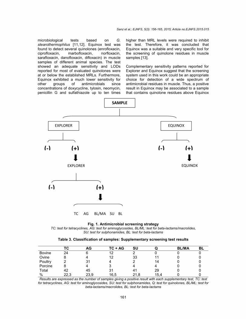
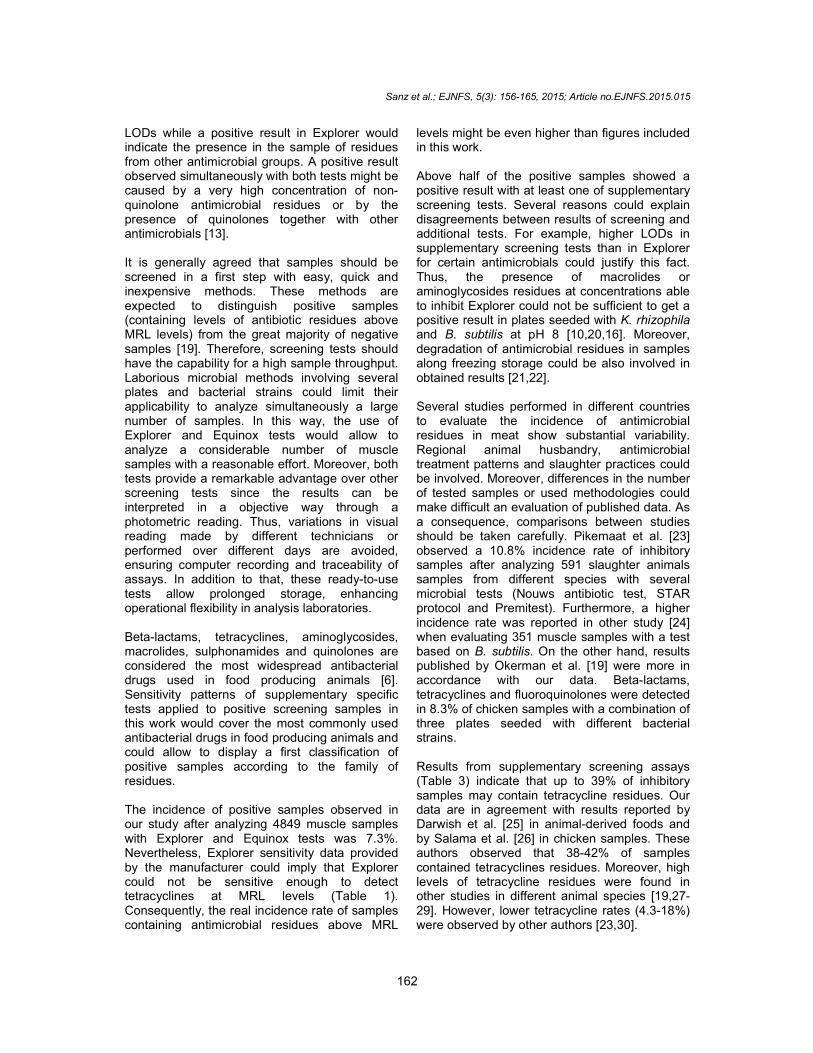
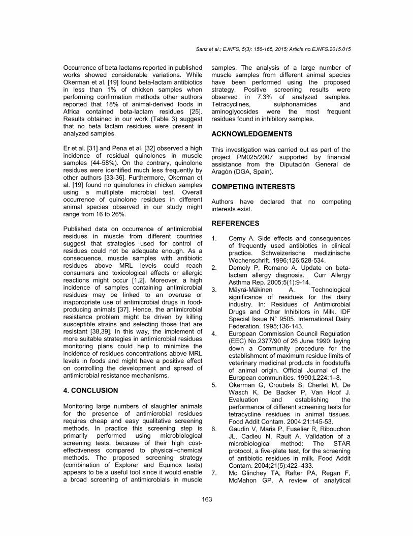
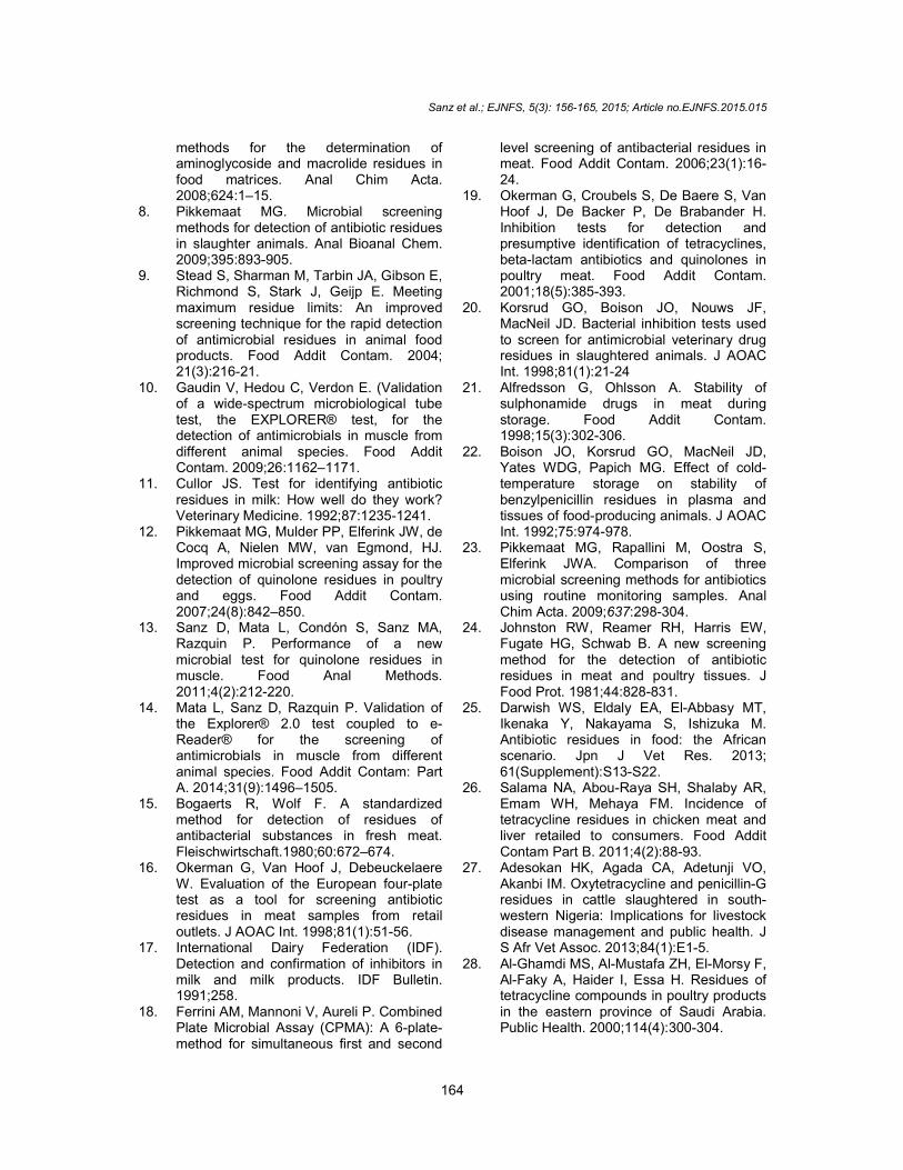
2.5 Analysis of Samples The antimicrobial screening strategy performed in this study is summarized in Fig. 1. Muscle samples were evaluated, in a first step, with Explorer and Equinox tests. Positive samples detected with Explorer were confirmed by a new assay with this test. If the inhibitory effect was observed also in the second analysis, the sample was further analyzed with specific screening tests for tetracyclines, aminoglycosides, beta-lactams, macrolides and sulphonamides. Besides, positive samples detected by Equinox were tested again with the same test to confirm the results. No more tests were performed to positive samples in first and second analysis, since Equinox is a highly specific test for quinolones.
3. RESULTS AND DISCUSSION
3.1 Results 3.1.1 Screening of meat samples In the first step of the study, 4849 samples were analyzed for antibiotic residues with Explorer and
Sanz et al.; EJNFS, 5(3): 156-165, 2015; Article no.EJNFS.2015.015
160
Equinox. As described previously, every positive result in Explorer or Equinox was confirmed with a second analysis in the same test. Only samples that showed inhibitory effect in both assays would be considered as positives. Every sample that showed a positive result at the first screening step remained as positive in a second analysis with the same test. As it is summarized in Table 2, 355 samples (7.3%) were identified as positive by Explorer and/or Equinox test. The highest incidence of positive samples was obtained in poultry (9.7%) while the lowest rate was found in porcine samples (3.4%).
Table 1. LODs (µg/Kg) of Equinox and Explorer for several antibiotics and sulphonamides in bovine muscle
(from [10, 13])
LOD (ug/kg) Equinox Explorer UE-MRL
Doxycycline ≤5000 200 100
Tetracycline n.e. 500* 100
Oxytetracycline n.e. 700* 100
Penicillin G >5000 5* 50
Amoxicillin n.e. 10 50
Cefalexin n.e. >500 200
Gentamycin n.e. 400* 50 Neomycin ≤10000 300* 500
Tylosin >10000 100 100 Erythromycin n.e. 200* 200
Enrofloxacin 100 n.e. 100a
Norfloxacin 200 n.e.
Sarafloxacin 100 n.e.
Marbofloxacin 100 n.e. 150
Ciprofloxacin 25 n.e. 100a
Danofloxacin 200 n.e. 200
Difloxacin 300 n.e. 400
Flumequine 2000 n.e. 200
Sulfathiazole >10000 200 100
Sulfamethoxypyridazine n.e. 300* 100
Sulfadiazine n.e. 200* 100
LODs: Limits of detection, UE-MRL: maximum residue limits EEC (2377/90),
a: sum of enrofloxacin and
ciprofloxacin, n.e.: not evaluated, *: data provided by
manufacturer
Table 2. Screening results of meat samples with Explorer and Equinox
Number of
samples Results
Negative (%)
Positive (%)
Bovine 1302 1210 (92.9) 92 (7.1)
Ovine 1283 1177 (91.7) 106 (8.3) Poultry 1280 1156 (90.3) 124 (9.7)
Porcine 984 951 (96.6) 33 (3.4)
Total (%) 4849 4494 (92.7) 355 (7.3)
3.1.2 Supplementary screening test (classification of samples)
Supplementary analyses were performed to Explorer positive samples to confirm results and obtain additional information about the nature of these samples. Results obtained with these tests are summarized in Table 3. Half of the positive screening samples (3.9% of total analyzed samples) showed also a positive result with at least one of the supplementary tests. According to Table 3, these samples might contain residues of tetracyclines (22.3%), aminoglycosides (23.9%), sulphonamides (21.8%), and quinolones (15.4%). Furthermore, results suggest that aminoglycosides and tetracyclines could be combined in 31 samples (16.5%) since an inhibition was observed in two different specific tests at the same time. None of the evaluated samples was found positive at plate seeded with K. rhizophila or Explorer with penicillinase. In such a way, a low probability of finding residues of macrolides and beta-lactams would be expected in positive screening samples.
A different pattern was observed when comparing the results from different animal species. Thus, aminoglycosides might be present in 66% of poultry samples but were less common in other species (23-41%). By contrast, data suggest that tetracyclines might be the most frequent residues in bovine and porcine samples while sulphonamide residues were the main family of residues expected in ovine. Quinolone residues might be found in every species (16-26%) except for bovine samples.
3.2 Discussion The screening strategy used in this work aimed to detect a large range of antimicrobials in muscle samples. The screening system combined 2 microbial methods: A broad spectrum test (Explorer) and a specific test for quinolones (Equinox). Gaudin et al. [10] reported that Explorer was able to detect compounds belonging to different antimicrobial families (penicillins, cephalosporins, tetracyclines, sulphonamides and macrolides) in muscle samples from different species (bovine, porcine, ovine and poultry). These authors observed that detection capabilities were around MRL levels for tested antimicrobials and concluded that Explorer might be used as a wide spectrum screening test for antimicrobials in muscle samples. However, a lack of sensitivity to quinolones is expected in
Sanz et al.; EJNFS, 5(3): 156-165, 2015; Article no.EJNFS.2015.015
161
microbiological tests based on G. stearothermophilus [11,12]. Equinox test was found to detect several quinolones (enrofloxacin, ciprofloxacin, marbofloxacin, norfloxacin, sarafloxacin, danofloxacin, difloxacin) in muscle samples of different animal species. The test showed an adequate sensitivity and LODs reported for most of evaluated quinolones were at or below the established MRLs. Furthermore, Equinox exhibited a much lower sensitivity for other groups of antimicrobials since concentrations of doxycycline, tylosin, neomycin, penicillin G and sulfathiazole up to ten times
higher than MRL levels were required to inhibit the test. Therefore, it was concluded that Equinox was a suitable and very specific tool for the screening of quinolone residues in muscle samples [13]. Complementary sensitivity patterns reported for Explorer and Equinox suggest that the screening system used in this work could be an appropriate choice for detection of a wide spectrum of antimicrobial residues in muscle. Thus, a positive result in Equinox may be associated to a sample that contains quinolone residues above Equinox
Fig. 1. Antimicrobial screening strategy TC: test for tetracyclines, AG: test for aminoglycosides, BL/ML: test for beta-lactams/macrolides,
SU: test for sulphonamides, BL: test for beta-lactams
Table 3. Classification of samples: Supplementary screening test results
TC AG TC + AG SU Q BL/MA BL Bovine 24 6 12 2 0 0 0 Ovine 8 4 12 33 11 0 0 Poultry 2 31 4 2 14 0 0 Porcine 8 4 3 4 4 0 0 Total 42 45 31 41 29 0 0 % 22,3 23,9 16,5 21,8 15,4 0 0 Results are expressed as the number of samples giving a positive result with each supplementary test, TC: test
for tetracyclines, AG: test for aminoglycosides, SU: test for sulphonamides, Q: test for quinolones, BL/ML: test for beta-lactams/macrolides, BL: test for beta-lactams
EXPLORER
EXPLORER
(-) (+)
TC AG BL/MA SU BL
(-) (+)
SAMPLE
EQUINOX
(-) (+)
EQUINOX
Sanz et al.; EJNFS, 5(3): 156-165, 2015; Article no.EJNFS.2015.015
162
LODs while a positive result in Explorer would indicate the presence in the sample of residues from other antimicrobial groups. A positive result observed simultaneously with both tests might be caused by a very high concentration of non-quinolone antimicrobial residues or by the presence of quinolones together with other antimicrobials [13]. It is generally agreed that samples should be screened in a first step with easy, quick and inexpensive methods. These methods are expected to distinguish positive samples (containing levels of antibiotic residues above MRL levels) from the great majority of negative samples [19]. Therefore, screening tests should have the capability for a high sample throughput. Laborious microbial methods involving several plates and bacterial strains could limit their applicability to analyze simultaneously a large number of samples. In this way, the use of Explorer and Equinox tests would allow to analyze a considerable number of muscle samples with a reasonable effort. Moreover, both tests provide a remarkable advantage over other screening tests since the results can be interpreted in a objective way through a photometric reading. Thus, variations in visual reading made by different technicians or performed over different days are avoided, ensuring computer recording and traceability of assays. In addition to that, these ready-to-use tests allow prolonged storage, enhancing operational flexibility in analysis laboratories. Beta-lactams, tetracyclines, aminoglycosides, macrolides, sulphonamides and quinolones are considered the most widespread antibacterial drugs used in food producing animals [6]. Sensitivity patterns of supplementary specific tests applied to positive screening samples in this work would cover the most commonly used antibacterial drugs in food producing animals and could allow to display a first classification of positive samples according to the family of residues. The incidence of positive samples observed in our study after analyzing 4849 muscle samples with Explorer and Equinox tests was 7.3%. Nevertheless, Explorer sensitivity data provided by the manufacturer could imply that Explorer could not be sensitive enough to detect tetracyclines at MRL levels (Table 1). Consequently, the real incidence rate of samples containing antimicrobial residues above MRL
levels might be even higher than figures included in this work. Above half of the positive samples showed a positive result with at least one of supplementary screening tests. Several reasons could explain disagreements between results of screening and additional tests. For example, higher LODs in supplementary screening tests than in Explorer for certain antimicrobials could justify this fact. Thus, the presence of macrolides or aminoglycosides residues at concentrations able to inhibit Explorer could not be sufficient to get a positive result in plates seeded with K. rhizophila and B. subtilis at pH 8 [10,20,16]. Moreover, degradation of antimicrobial residues in samples along freezing storage could be also involved in obtained results [21,22]. Several studies performed in different countries to evaluate the incidence of antimicrobial residues in meat show substantial variability. Regional animal husbandry, antimicrobial treatment patterns and slaughter practices could be involved. Moreover, differences in the number of tested samples or used methodologies could make difficult an evaluation of published data. As a consequence, comparisons between studies should be taken carefully. Pikemaat et al. [23] observed a 10.8% incidence rate of inhibitory samples after analyzing 591 slaughter animals samples from different species with several microbial tests (Nouws antibiotic test, STAR protocol and Premitest). Furthermore, a higher incidence rate was reported in other study [24] when evaluating 351 muscle samples with a test based on B. subtilis. On the other hand, results published by Okerman et al. [19] were more in accordance with our data. Beta-lactams, tetracyclines and fluoroquinolones were detected in 8.3% of chicken samples with a combination of three plates seeded with different bacterial strains. Results from supplementary screening assays (Table 3) indicate that up to 39% of inhibitory samples may contain tetracycline residues. Our data are in agreement with results reported by Darwish et al. [25] in animal-derived foods and by Salama et al. [26] in chicken samples. These authors observed that 38-42% of samples contained tetracyclines residues. Moreover, high levels of tetracycline residues were found in other studies in different animal species [19,27-29]. However, lower tetracycline rates (4.3-18%) were observed by other authors [23,30].
Sanz et al.; EJNFS, 5(3): 156-165, 2015; Article no.EJNFS.2015.015
163
Occurrence of beta lactams reported in published works showed considerable variations. While Okerman et al. [19] found beta-lactam antibiotics in less than 1% of chicken samples when performing confirmation methods other authors reported that 18% of animal-derived foods in Africa contained beta-lactam residues [25]. Results obtained in our work (Table 3) suggest that no beta lactam residues were present in analyzed samples. Er et al. [31] and Pena et al. [32] observed a high incidence of residual quinolones in muscle samples (44-58%). On the contrary, quinolone residues were identified much less frequently by other authors [33-36]. Furthermore, Okerman et al. [19] found no quinolones in chicken samples using a multiplate microbial test. Overall occurrence of quinolone residues in different animal species observed in our study might range from 16 to 26%. Published data on occurrence of antimicrobial residues in muscle from different countries suggest that strategies used for control of residues could not be adequate enough. As a consequence, muscle samples with antibiotic residues above MRL levels could reach consumers and toxicological effects or allergic reactions might occur [1,2]. Moreover, a high incidence of samples containing antimicrobial residues may be linked to an overuse or inappropriate use of antimicrobial drugs in food-producing animals [37]. Hence, the antimicrobial resistance problem might be driven by killing susceptible strains and selecting those that are resistant [38,39]. In this way, the implement of more suitable strategies in antimicrobial residues monitoring plans could help to minimize the incidence of residues concentrations above MRL levels in foods and might have a positive effect on controlling the development and spread of antimicrobial resistance mechanisms.
4. CONCLUSION Monitoring large numbers of slaughter animals for the presence of antimicrobial residues requires cheap and easy qualitative screening methods. In practice this screening step is primarily performed using microbiological screening tests, because of their high cost-effectiveness compared to physical–chemical methods. The proposed screening strategy (combination of Explorer and Equinox tests) appears to be a useful tool since it would enable a broad screening of antimicrobials in muscle
samples. The analysis of a large number of muscle samples from different animal species have been performed using the proposed strategy. Positive screening results were observed in 7.3% of analyzed samples. Tetracyclines, sulphonamides and aminoglycosides were the most frequent residues found in inhibitory samples.
ACKNOWLEDGEMENTS This investigation was carried out as part of the project PM025/2007 supported by financial assistance from the Diputación General de Aragón (DGA, Spain).
COMPETING INTERESTS Authors have declared that no competing interests exist.
REFERENCES 1. Cerny A. Side effects and consequences
of frequently used antibiotics in clinical practice. Schweizerische medizinische Wochenschrift. 1996;126:528-534.
2. Demoly P, Romano A. Update on beta-lactam allergy diagnosis. Curr Allergy Asthma Rep. 2005;5(1):9-14.
3. Mäyrä-Mäkinen A. Technological significance of residues for the dairy industry. In: Residues of Antimicrobial Drugs and Other Inhibitors in Milk. IDF Special Issue N° 9505. International Dairy Federation. 1995;136-143.
4. European Commission Council Regulation (EEC) No.2377/90 of 26 June 1990: laying down a Community procedure for the establishment of maximum residue limits of veterinary medicinal products in foodstuffs of animal origin. Official Journal of the European communities. 1990;L224:1–8.
5. Okerman G, Croubels S, Cherlet M, De Wasch K, De Backer P, Van Hoof J. Evaluation and establishing the performance of different screening tests for tetracycline residues in animal tissues. Food Addit Contam. 2004;21:145-53.
6. Gaudin V, Maris P, Fuselier R, Ribouchon JL, Cadieu N, Rault A. Validation of a microbiological method: The STAR protocol, a five-plate test, for the screening of antibiotic residues in milk. Food Addit Contam. 2004;21(5):422–433.
7. Mc Glinchey TA, Rafter PA, Regan F, McMahon GP. A review of analytical
Sanz et al.; EJNFS, 5(3): 156-165, 2015; Article no.EJNFS.2015.015
164
methods for the determination of aminoglycoside and macrolide residues in food matrices. Anal Chim Acta. 2008;624:1–15.
8. Pikkemaat MG. Microbial screening methods for detection of antibiotic residues in slaughter animals. Anal Bioanal Chem. 2009;395:893-905.
9. Stead S, Sharman M, Tarbin JA, Gibson E, Richmond S, Stark J, Geijp E. Meeting maximum residue limits: An improved screening technique for the rapid detection of antimicrobial residues in animal food products. Food Addit Contam. 2004; 21(3):216-21.
10. Gaudin V, Hedou C, Verdon E. (Validation of a wide-spectrum microbiological tube test, the EXPLORER® test, for the detection of antimicrobials in muscle from different animal species. Food Addit Contam. 2009;26:1162–1171.
11. Cullor JS. Test for identifying antibiotic residues in milk: How well do they work? Veterinary Medicine. 1992;87:1235-1241.
12. Pikkemaat MG, Mulder PP, Elferink JW, de Cocq A, Nielen MW, van Egmond, HJ. Improved microbial screening assay for the detection of quinolone residues in poultry and eggs. Food Addit Contam. 2007;24(8):842–850.
13. Sanz D, Mata L, Condón S, Sanz MA, Razquin P. Performance of a new microbial test for quinolone residues in muscle. Food Anal Methods. 2011;4(2):212-220.
14. Mata L, Sanz D, Razquin P. Validation of the Explorer® 2.0 test coupled to e-Reader® for the screening of antimicrobials in muscle from different animal species. Food Addit Contam: Part A. 2014;31(9):1496–1505.
15. Bogaerts R, Wolf F. A standardized method for detection of residues of antibacterial substances in fresh meat. Fleischwirtschaft.1980;60:672–674.
16. Okerman G, Van Hoof J, Debeuckelaere W. Evaluation of the European four-plate test as a tool for screening antibiotic residues in meat samples from retail outlets. J AOAC Int. 1998;81(1):51-56.
17. International Dairy Federation (IDF). Detection and confirmation of inhibitors in milk and milk products. IDF Bulletin. 1991;258.
18. Ferrini AM, Mannoni V, Aureli P. Combined Plate Microbial Assay (CPMA): A 6-plate-method for simultaneous first and second
level screening of antibacterial residues in meat. Food Addit Contam. 2006;23(1):16-24.
19. Okerman G, Croubels S, De Baere S, Van Hoof J, De Backer P, De Brabander H. Inhibition tests for detection and presumptive identification of tetracyclines, beta-lactam antibiotics and quinolones in poultry meat. Food Addit Contam. 2001;18(5):385-393.
20. Korsrud GO, Boison JO, Nouws JF, MacNeil JD. Bacterial inhibition tests used to screen for antimicrobial veterinary drug residues in slaughtered animals. J AOAC Int. 1998;81(1):21-24
21. Alfredsson G, Ohlsson A. Stability of sulphonamide drugs in meat during storage. Food Addit Contam. 1998;15(3):302-306.
22. Boison JO, Korsrud GO, MacNeil JD, Yates WDG, Papich MG. Effect of cold-temperature storage on stability of benzylpenicillin residues in plasma and tissues of food-producing animals. J AOAC Int. 1992;75:974-978.
23. Pikkemaat MG, Rapallini M, Oostra S, Elferink JWA. Comparison of three microbial screening methods for antibiotics using routine monitoring samples. Anal Chim Acta. 2009;637:298-304.
24. Johnston RW, Reamer RH, Harris EW, Fugate HG, Schwab B. A new screening method for the detection of antibiotic residues in meat and poultry tissues. J Food Prot. 1981;44:828-831.
25. Darwish WS, Eldaly EA, El-Abbasy MT, Ikenaka Y, Nakayama S, Ishizuka M. Antibiotic residues in food: the African scenario. Jpn J Vet Res. 2013; 61(Supplement):S13-S22.
26. Salama NA, Abou-Raya SH, Shalaby AR, Emam WH, Mehaya FM. Incidence of tetracycline residues in chicken meat and liver retailed to consumers. Food Addit Contam Part B. 2011;4(2):88-93.
27. Adesokan HK, Agada CA, Adetunji VO, Akanbi IM. Oxytetracycline and penicillin-G residues in cattle slaughtered in south-western Nigeria: Implications for livestock disease management and public health. J S Afr Vet Assoc. 2013;84(1):E1-5.
28. Al-Ghamdi MS, Al-Mustafa ZH, El-Morsy F, Al-Faky A, Haider I, Essa H. Residues of tetracycline compounds in poultry products in the eastern province of Saudi Arabia. Public Health. 2000;114(4):300-304.
Sanz et al.; EJNFS, 5(3): 156-165, 2015; Article no.EJNFS.2015.015
165
29. De Wasch K, Okerman L, De Brabander H, Van Hoof J, Croubels S, De Backer P. Detection of residues of tetracycline antibiotics in pork and chicken meat: correlation between results of screening and confirmatory tests. Analyst. 1998;123:2737-2741.
30. Cetinkaya F, Yibar A, Soyutemiz GE, Okutan B, Ozcan A, Karaca MY. Determination of tetracycline residues in chicken meat by liquid chromatography-tandem mass spectrometry. Food Addit Contam. 2012;5(1):45-49.
31. Er B, Onurdag FK, Demirhan B, Ozgacar SÖ, Oktem AB, Abbasoglu U. Screening of quinolone antibiotic residues in chicken meat and beef sold in the markets of Ankara, Turkey. Poult Sci. 2013;92(8):2212-2215.
32. Pena A, Silva LJG, Pereira A, Meisel L, Lino CM. Determination of fluoroquinolone residues in poultry muscle in Portugal. Anal Bioanal Chem. 2010;397:2615–2621.
33. Salehzadeh F, Salehzadeh A, Rokni N, Madani R, Golchinefar F. Enrofloxacin residue in chicken tissues from Tehran slaughterhouses in Iran. PJN. 2007;6:409–413.
34. Silfrany RO, Caba RE, Solís de Los Santos F, Hanning I. Detection of quinolones in poultry meat obtained from retail centers in Santiago Province, the Dominican Republic. J Food Prot. 2013;76(2):352-354.
35. Weiss C, Conte A, Milandri C, Scortichini G, Semprini P, Usberti R, Migliorati, G. Veterinary drugs residue monitoring in Italian poultry: Current strategies and possible developments. Food Control. 2007;18:1068–1076.
36. Zhao S, Li X, Ra Y, Li C, Jiang H, Li J, Qu Z, Zhang S, He F, Wan Y, Feng C, Zheng Z, Shen J. Developing and optimizing an immunoaffinity cleanup technique for determination of quinolones from chicken muscle. J Agric Food Chem. 2009;57:365–371.
37. Anonymous. Background Document for the Joint WHO/FAO/OIE Expert Workshop on non-human. antimicrobial usage and antimicrobial resistance: Scientific Assessment, Geneva, Switzerland; 2003. Available:http://www.who.int/gfn/links/en/background.pdf (Accessed 9 September 2014).
38. Levy SB, Marshall B. Antibacterial resistance worldwide: Causes, challenges and responses. Nat Med. 2004;10(12 Suppl):S122-9.
39. Chantziaras I, Boyen F, Callens B, Dewulf J. Correlation between veterinary antimicrobial use and antimicrobial resistance in food-producing animals: A report on seven countries. J Antimicrob Chemother. 2014;69(3):827-834.
© 2015 Sanz et al.; This is an Open Access article distributed under the terms of the Creative Commons Attribution License (http://creativecommons.org/licenses/by/4.0), which permits unrestricted use, distribution, and reproduction in any medium, provided the original work is properly cited.
Peer-review history: The peer review history for this paper can be accessed here:
http://www.sciencedomain.org/review-history.php?iid=855&id=30&aid=8701