In vitro evaluation of the antioxidant activities in fruit extracts from citron and blood orange
8
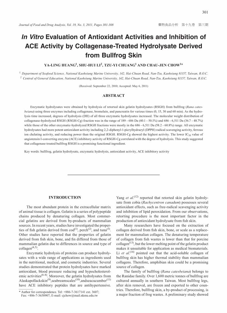
Journal of Food and Drug Analysis, Vol. 19, No. 3, 2011, Pages 301-308 301 藥物食品分析 第十九卷 第三期 In Vitro Evaluation of Antioxidant Activities and Inhibition of ACE Activity by Collagenase-Treated Hydrolysate Derived from Bullfrog Skin YA-LING HUANG 1 , SHU-HUI LI 2 , TZU-YI CHUANG 1 AND CHAU-JEN CHOW 1 * 1. Department of Seafood Science, National Kaohsiung Marine University, 142, Hai-Chuan Road, Nan-Tzu, Kaohsiung 81157, Taiwan, R.O.C. 2. Central of General Education, National Kaohsiung Marine University, 142, Hai-Chuan Road, Nan-Tzu, Kaohsiung 81157, Taiwan, R.O.C. (Received: September 22, 2010; Accepted: May 6, 2011) ABSTRACT Enzymatic hydrolysates were obtained by hydrolysis of retorted skin gelatin hydrolysates (RSGH) from bullfrog (Rana cates- beiana) using three enzymes including collagenase, bromelain, and pancreatin for various times (0, 15, 30 and 60 min). As the hydro- lysis time increased, degrees of hydrolysis (DH) of all three enzymatic hydrolysates increased. The molecular weight distribution of collagenase-hydrolyzed RSGH (RSGH-Cg) fraction was in the range of 189 - 686 Da (40.1 - 50.1%) and 686 - 6,511 Da (36.7 - 48.7%) while those of the other enzymatic-hydrolyzed RSGH fractions were mostly in the 686 - 6,511 Da (58.2 - 60.8%) range. All enzymatic hydrolysates had more potent antioxidant activity including 2,2-diphenyl-1-picrylhydrazyl (DPPH) radical scavenging activity, ferrous ion chelating activity, and reducing power than the original RSGH. RSGH-Cg showed the highest activity. The lower IC 50 value of angiotensin I-converting enzyme (ACE) inhibitory activity of RSGH-Cg correlated with the degree of hydrolysis. This study suggested that collagenase treated bullfrog RSGH is a promising functional ingredient. Key words: bullfrog, gelatin hydrolysate, enzymatic hydrolysis, antioxidant activity, ACE inhibitory activity INTRODUCTION The most abundant protein in the extracellular matrix of animal tissue is collagen. Gelatin is a series of polypeptide chains produced by denaturing collagen. Most commer- cial gelatins are derived from by-products of mammalian sources. In recent years, studies have investigated the proper- ties of fish gelatin derived from cod (1) , perch (2) , and tuna (3) . Other studies have reported that the properties of gelatin derived from fish skin, bone, and fin differed from those of mammalian gelatin due to differences in source and type of collagen (4,5) . Enzymatic hydrolysis of proteins can produce hydroly- sates with a wide range of applications as ingredients used in the nutritional, medical, and cosmetic industries. Several studies demonstrated that protein hydrolysates have marked antioxidant, blood pressure reducing and hypocholesterol- emic activities (6-8) . Moreover, the gelatin hydrolysates from Alaskapollackskin (9) ,seabreamscales (10) ,andseacucumber (11) have ACE inhibitory peptides that are antihypertensive. Yang et al. (12) reported that retorted skin gelatin hydroly- sate from cobia (Rachycentron canadum) possesses several antioxidant effects, such as free-radical scavenging activity and inhibition of lipid peroxidation. From our observations, retorting procedure is the most important factor in the production of antioxidant hydrolysate from fish skin. Many researchers have focused on the extraction of collagen derived from fish skin, bone, or scale as a replace- ment for mammalian collagen. The denaturing temperature of collagen from fish wastes is lower than that for porcine collagen (13) , but the lower melting point of the gelatin product makes it unsuitable for application as medical biomaterials. Li et al. (14) pointed out that the acid-soluble collagen of bullfrog skin has higher thermal stability than mammalian collagens. Therefore, amphibian skin could be a promising source of collagen. The family of bullfrog (Rana catesbeiana) belongs to the Ranidae family. Over 1,600 metric tonnes of bullfrog are cultured annually in southern Taiwan. Most bullfrog legs, after skin removal, are frozen and exported to other coun- tries. Therefore, bullfrog skin, a by-product of processing, is a major fraction of frog wastes. A preliminary study showed * Author for correspondence. Tel: +886-7-3617141 ext. 3607; Fax: +886-7-3658907; E-mail: [email protected]
Transcript of In vitro evaluation of the antioxidant activities in fruit extracts from citron and blood orange
Journal of Food and Drug Analysis, Vol. 19, No. 3, 2011, Pages 301-308
301
藥物食品分析 第十九卷 第三期
In Vitro Evaluation of Antioxidant Activities and Inhibition of ACE Activity by Collagenase-Treated Hydrolysate Derived
from Bullfrog SkinYA-LING HUANG1, SHU-HUI LI2, TZU-YI CHUANG1 AND CHAU-JEN CHOW1*
1. Department of Seafood Science, National Kaohsiung Marine University, 142, Hai-Chuan Road, Nan-Tzu, Kaohsiung 81157, Taiwan, R.O.C.2. Central of General Education, National Kaohsiung Marine University, 142, Hai-Chuan Road, Nan-Tzu, Kaohsiung 81157, Taiwan, R.O.C.
(Received: September 22, 2010; Accepted: May 6, 2011)
ABSTRACT
Enzymatic hydrolysates were obtained by hydrolysis of retorted skin gelatin hydrolysates (RSGH) from bullfrog (Rana cates-beiana) using three enzymes including collagenase, bromelain, and pancreatin for various times (0, 15, 30 and 60 min). As the hydro-lysis time increased, degrees of hydrolysis (DH) of all three enzymatic hydrolysates increased. The molecular weight distribution of collagenase-hydrolyzed RSGH (RSGH-Cg) fraction was in the range of 189 - 686 Da (40.1 - 50.1%) and 686 - 6,511 Da (36.7 - 48.7%) while those of the other enzymatic-hydrolyzed RSGH fractions were mostly in the 686 - 6,511 Da (58.2 - 60.8%) range. All enzymatic hydrolysates had more potent antioxidant activity including 2,2-diphenyl-1-picrylhydrazyl (DPPH) radical scavenging activity, ferrous ion chelating activity, and reducing power than the original RSGH. RSGH-Cg showed the highest activity. The lower IC50 value of angiotensin I-converting enzyme (ACE) inhibitory activity of RSGH-Cg correlated with the degree of hydrolysis. This study suggested that collagenase treated bullfrog RSGH is a promising functional ingredient.
Key words: bullfrog, gelatin hydrolysate, enzymatic hydrolysis, antioxidant activity, ACE inhibitory activity
INTRODUCTION
The most abundant protein in the extracellular matrix of animal tissue is collagen. Gelatin is a series of polypeptide chains produced by denaturing collagen. Most commer-cial gelatins are derived from by-products of mammalian sources. In recent years, studies have investigated the proper-ties of fish gelatin derived from cod(1), perch(2), and tuna(3). Other studies have reported that the properties of gelatin derived from fish skin, bone, and fin differed from those of mammalian gelatin due to differences in source and type of collagen(4,5).
Enzymatic hydrolysis of proteins can produce hydroly-sates with a wide range of applications as ingredients used in the nutritional, medical, and cosmetic industries. Several studies demonstrated that protein hydrolysates have marked antioxidant, blood pressure reducing and hypocholesterol-emic activities(6-8). Moreover, the gelatin hydrolysates from Alaska pollack skin(9), sea bream scales(10), and sea cucumber(11) have ACE inhibitory peptides that are antihypertensive.
Yang et al.(12) reported that retorted skin gelatin hydroly-sate from cobia (Rachycentron canadum) possesses several antioxidant effects, such as free-radical scavenging activity and inhibition of lipid peroxidation. From our observations, retorting procedure is the most important factor in the production of antioxidant hydrolysate from fish skin.
Many researchers have focused on the extraction of collagen derived from fish skin, bone, or scale as a replace-ment for mammalian collagen. The denaturing temperature of collagen from fish wastes is lower than that for porcine collagen(13), but the lower melting point of the gelatin product makes it unsuitable for application as medical biomaterials. Li et al.(14) pointed out that the acid-soluble collagen of bullfrog skin has higher thermal stability than mammalian collagens. Therefore, amphibian skin could be a promising source of collagen.
The family of bullfrog (Rana catesbeiana) belongs to the Ranidae family. Over 1,600 metric tonnes of bullfrog are cultured annually in southern Taiwan. Most bullfrog legs, after skin removal, are frozen and exported to other coun-tries. Therefore, bullfrog skin, a by-product of processing, is a major fraction of frog wastes. A preliminary study showed
* Author for correspondence. Tel: +886-7-3617141 ext. 3607; Fax: +886-7-3658907; E-mail: [email protected]
Journal of Food and Drug Analysis, Vol. 19, No. 3, 2011
302
that fresh bullfrog skin contains approximately 25.3% protein that is acid-soluble collagen (~80% of total nitrogen content). The recovery of gelatin and its hydrolysate from fish skin collagen by thermal hydrolysis with retorting has been investigated in recent years. Bullfrog skin gelatin hydrolysate obtained by retorting treatment has been further converted to value-added enzymatic protein hydrolysates, which can be widely used as a potential bioactive substance. However, little information regarding the characteristics of gelatin hydrolysates derived from bullfrog skin has been reported. Therefore, the objective of this study was to characterize the enzymatic hydrolysates obtained from retorted bullfrog skin using enzymatic hydrolysis, by determining degree of hydrolysis and molecular weight distribution, to evaluate their antioxidant properties and angiotensin I-converting enzyme (ACE) inhibitory activity.
MATERIALS AND METHODS
I. Materials
The skin of bullfrog (Rana catesbeiana) was obtained from Ho-Wei Fishery Co., LTD, Pingtung County, Taiwan. After manual removal of limb tissue residues, the skin sample (30 ± 1.5 g per bullfrog) was washed, cut into small pieces (1.5 × 1.5 cm), placed in polyethylene bags, and stored at -20°C until use. All reagents were analytical grade.
II. Preparation of Bullfrog Retorted Skin Gelatin Hydroly-sate (RSGH)
The frozen bullfrog skin was thawed to 4°C with tap water and soaked in 0.1 N NaOH with a skin: solution ratio of 1 : 10 (w/v) at 4°C for 3 h with gentle stirring. The skin sample was pooled and washed with tap water until neutral pH reached. The cleaned skin was further immersed in 6% phosphoric acid with a skin: solution ratio of 1 : 4 (w/v) for 3 h, drained and then washed under running tap water to remove excess acid. To obtain the gelatin hydrolysate, the swollen bullfrog skin was added with distilled water in 1 : 2 (w/v) ratio and hydrolyzed by retorting in an autoclave (HA-300M, Hirayama, Japan) at 121°C for 120 min. After filtration with metal sieve, the pH value of the retorted solu-tion was 1.8 - 2.0. The solution was then neutralized with saturated Ca(OH)2 to pH 7. The neutralized solution was filtered again through a Buchner funnel with Toyo No. 5A filter paper to remove insoluble precipitants. The filtrate was dried by lyophilization and the dry matter was referred as ‘RSGH’ and stored at -20°C until further analysis.
III. Enzymatic Hydrolysis of RSGH Treated with Proteases
For further digestion of RSGH, the enzymatic hydro-lysis was carried out using various enzymes including colla-genase (from Clostridium histolyticum, NO. C0130, Sigma, St. Louis, MO, USA), bromelain (No. 1.01651.0025, Merck,
Darmstadt, Germany), and pancreatin (No. P1500, Sigma) at optimal activity temperature (37°C for collagenase, 50°C for bromelain, and 50°C for pancreatin). Dry RSGH sample (1 g) was digested with one of each protease in 0.1 M sodium phosphate buffer (pH 7.0) at an enzyme: hydrolysate ratio of 1 : 100 (w/w) and then hydrolyzed at optimal temperature for 0, 15, 30, and 60 min in a shaker-water bath with continuous stirring. The reaction was terminated in a boiling water bath for 10 min to inactivate enzyme activity. After centrifu-gation at 5,000 ×g for 10 min at 4°C, the supernatant was lyophilized and then stored at -20°C until use. The resultant product obtained from enzyme-treated RSGH was named RSGH-Bm (bromelain-treated), RSGH-Pc (pancreatin-treated), and RSGH-Cg (collagenase-treated), respectively.
IV. Determination of Degree of Hydrolysis (DH)
Following the method of Church et al.(15), the degree of hydrolysis was determined with spectrophotometric assay using o-phthaldialdehyde (OPA). An aliquot (50 µL) was added to 1 mL OPA reagent. After mixing, the mixture was measured spectrophotometrically at 340 nm. Gelatin extracted at 50°C for 4 h from bullfrog skin was used as the control. The calibration curve is drawn using a series of concentrations of Leu-Gly as a reference, which is converted to the number of peptide bond. Degree of hydrolysis was defined as percentage ratio of the number of peptide bonds released after enzymatic hydrolysis of RSGH to the total number of peptide bonds of gelatin extracted at 50°C and was calculated as follows:
DH (%) = Ng
NgNh × 100−
where Nh was number of cleaved peptide bonds after enzymatic hydrolysis of RSGH and Ng was total number of peptide bonds of gelatin extracted at 50°C.
V. Molecular Weight Distribution
Molecular weight distributions of the hydrolysate samples were determined by the gel permeation chroma-tography using a Hitachi 2130 HPLC system equipped with Superdex peptide® 10/300 column (10 × 300 mm, Amersham Pharmacia Biotech, Uppsala, Sweden). Chromatography was carried out using 0.02 M phosphate buffer (pH 7.2) as mobile phase. An injection volume of 20 µL of each hydrolysate (2 mg protein/mL) and a flow rate of 0.5 mL/min and absor-bance at 214 nm was used in the analysis. The molecular weight standard distributions were determined using the following standards including trypsin inhibitor (20 kDa, No. 202-09221, Wako, Osaka, Japan), cytochrome C (12.5 kDa, No. C2506, Sigma), aprotinin (6,511 Da, No. 52682, Serva Electrophoresis GmbH, Heideberg, Germany), pepstatin A (686 Da, No. 616370, Calbiochem, La Jolla, CA), Gly-Gly-Gly (189 Da, No. G1377, Sigma), and Gly (75 Da, No. 17-1323-01, Amersham Pharmacia Biotech). For quantitative determinations of molecular weight distribution (%), peak
Journal of Food and Drug Analysis, Vol. 19, No. 3, 2011
303
area in the HPLC chromatograms was integrated in refer-ence to the peak area of the standard proteins, using Hitachi EZChrom Elite 3.1 chromatography data system software.
VI. Scavenging Effect on 2,2-Diphenyl-1-picrylhydrazyl (DPPH) Radical
DPPH radical scavenging activity was determined by the method of Shimada et al.(16) with slight modifications. Briefly, 0.1 mM solution of DPPH in 95% ethanol was prepared and 1 mL of this solution was added to 1 mL hydro-lysate sample (6 mg/mL). The mixture was shaken vigorously and allowed to stand at room temperature for 30 min. Then the absorbance of resulting solution was measured spectro-photometrically at 517 nm. Lower absorbance of the reaction mixture indicates higher free radical scavenging activity. The percentage of DPPH scavenging effect was calculated using the following equation:
DPPH scavenging effect (%) = 100 – 100A
AA0
10 ×
−
where A0 was the absorbance of the control reaction and A1 was the absorbance in the presence of the standard or hydrolysate sample.
VII. Determination of Reducing Power
The reducing power of hydrolysate sample was deter-mined according to the method of Wu et al (17). An aliquot (1 mL) of each hydrolysate sample was mixed with 1 mL of 0.2 M phosphate buffer (pH 6.6) and 1 mL of 1% (w/v) potassium ferricyanide. The mixture was incubated at 20°C for 20 min and cooled rapidly. Then 1 mL of 10% (w/v) trichloroacetic acid was added to the mixture, which was then centrifuged at 1,000 ×g for 10 min. The upper layer of solution (2 mL) was mixed with distilled water (2 mL) and 0.1% (w/v) ferric chloride (0.5 mL) and allowed to stand in a dark room for 10 min. The absorbance of the resulting solution was measured spectrophotometrically at 700 nm. Higher absorbance of the reaction mixture indicated greater reducing power. L-Ascorbic acid (1,000 ppm) was used as a positive control.
VIII. Determination of Metal Ion Chelating Activity
The chelating of ferrous ions by the hydrolysate was measured using the method described by Wu et al.(17) with slight modification. An aliquot (1 mL) of the hydrolysate sample was added with 2 mM FeCl2 (100 µL). Subsequently, 200 µL of 5 mM ferrozine was added to this mixture. The reaction mixture was shaken vigorously and left standing at room temperature (24°C) for 10 min. The absorbance at 562 nm was then measured spectrophotometrically. The absorbance of 1,000 ppm EDTA was used as a positive control. Percentage of decrease in absorbance of the reaction mixture indicates increased metal ion chelating activity.
IX. Determination of ACE Inhibitory Activity
The ACE inhibitory activity was measured using the method described by Chushman and Cheung(18) with slight modifications. The reaction mixture was prepared by adding 20 µL of 5 mM Hip-His-Leu into the 100 mM sodium borate buffer (pH 8.3) containing 300 mM NaCl as a substrate. An aliquot (80 µL) of hydrolysate treated with collagenase was mixed with the above reaction mixture and preincubated at 37°C for 5 min. Then, 20 µL of ACE (0.11 U/mL) was added and the mixture was incubated in a water bath at 37°C for 60 min and the reaction was stopped by adding 150 µL of 1.0 N HCl. Released hippuric acid was extracted by the addition of 1.7 µL ethyl acetate with continuous vortex mixing. After centrifuged at 1,600 ×g for 10 min, 1 mL of the upper layer was transferred into test tube and evaporated in vacuum. The resulting hippuric acid was redissolved in 1 mL distilled water and absorbance was measured spectrophotometrically at 280 nm. The concentration of hydrolysate caused a 50% reduction of ACE activity was expressed as the 50% inhibi-tory concentration (IC50).
X. ACE Activity of Hydrolysates Following Digestion by Gastrointestinal Enzymes In Vitro
According to the methods of Wu and Ding(6) and Chushman and Cheung(18) with slight modifications, 1% (w/v) hydrolysate sample in 0.1 M KCl-HCl buffer (pH 2.0) was digested with pepsin in a water bath at 37°C for 4 h and placed in a boiling water bath for 15 min in order to terminate the enzymatic reaction. A portion of part of the mixture was subjected to the assay of ACE inhibitory activity and part of the mixture was neutralized with 2 N NaOH, followed by incubating with 2% (w/v) pancreatin in a 37°C water bath for 4 h. The reaction was stopped by heating in a 100°C water bath for 10 min. After centrifugation at 1,000 ×g for 10 min, the supernatant was used for ACE inhibitory activity determination. Inhibitory activity was expressed as the concentration of hydrolysate required to inhibit 50% of the ACE activity (IC50).
XI. Statistical Analysis
All analyses were carried out in triplicate. Statistical analysis on the determinations was performed by one-way analysis of variance using the Statistical Analysis System (SAS). Values of p < 0.05 were considered statistically significant.
RESULTS AND DISCUSSION
In this study, the skin content of the whole bullfrog was about 6.0 g/100 g. After alkaline pretreatment and acid soaking, the gelatin hydrolysates obtained from the bullfrog skin were extracted with retorting in an autoclave, leading to a high nitrogen content of bullfrog RSGH. The nitrogen
Journal of Food and Drug Analysis, Vol. 19, No. 3, 2011
304
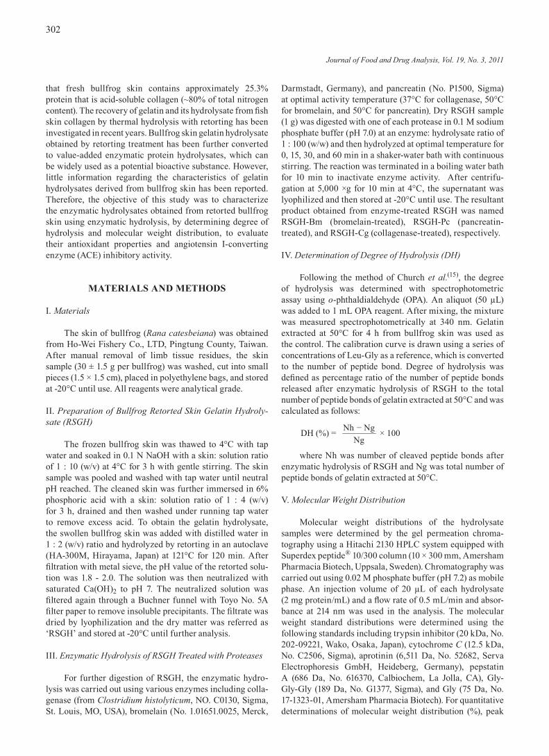
content of the retorted filtrate reached approximately 50.1% of bullfrog skin (data not shown). Therefore, bullfrog skin can serve as a source of proteins. Yang et al.(12) reported that the amount of nitrogen in cobia RSGH accounted for 46.9% of its nitrogen content in the skin, which is lower than that of bullfrog RSGH. Furthermore, degree of hydrolysis is used as a proteolysis monitoring parameter, and it is the most widely used indicator for comparison among different protein hydrolysates(19). As shown in Figure 1, the extent of further degradation of RSGH by proteolytic enzymes was estimated by assessing the degree of hydrolysis. It was observed to be 83.1%, 74.3%, and 67.4%, respectively, for collagenase (37°C), bromelain (50°C), and pancreatin (50°C) after 15 min hydrolysis. Between 30 and 60 min of hydrolysis, no significant change in degree of hydrolysis of bullfrog RSGH was observed with all three proteases used. Among these hydrolysates, the degree of hydrolysis of RSGH treated with collagenase was the highest after 60 min hydrolysis. It is possible that collagenase contributed to a higher degree of hydrolysis due to its specific enzymatic action. During the retorting process, gelatin hydrolysate resulting from thermal denaturation produced from cobia skin was possibly attrib-uted to the collagen solubility(12). Moreover, the difference of degree of hydrolysis between the gelatin hydrolysates obtained by different proteases may be due to the extent of specific peptide bonds available for enzyme action(19,20). Therefore, it was speculated that the degree of hydrolysis of gelatin hydrolysates was affected by the kind of raw material, as well as its pre-treatment (e.g. enzyme type) in the process.
The peak area of HPLC gel chromatography was evalu-ated for the molecular weight distribution of bullfrog RSGH during enzymatic hydrolysis. As shown in Table 1, bullfrog skin gelatin hydrolysates before any protease digestion gave rise to the dominant fraction of the smaller molecular weight, with 686 - 6,511 Da, which accounted for 54.0% of the total
hydrolysates. After 15 min of hydrolysis, all enzyme-treated RSGH had a size distribution of 686 - 6,511 Da. Moreover, the original RSGH fraction with a molecular weight larger than 6,511 Da was 26.5% of the hydrolysates. As hydro-lysis time was increased to 60 min, the percentage of the RSGH-Cg, RSGH-Bm, and RSGH-Pc having molecular weights of over 6,511 Da significantly (p < 0.05) decreased to 0.9, 15.8, and 19.7%, respectively. Hence, the collagenase hydrolysis of retorted gelatin hydrolysates yielded more medium and small peptides. On the other hand, those having molecular weights below 686 Da were increased by treatment of collagenase (46.0 - 62.4%), bromelain (23.6 - 25.3%), and pancreatin (18.5 - 19.7%) from 15 min to 60 min hydrolysis.
Figure 1. Degree of hydrolysis of bullfrog RSGH by adding various proteases. With the same hydrolysis time, values (means ± SD, n = 3) with different letters (a-a, g-i, p-r, w-y) are significantly different (Duncan, p < 0.05).
Hydrolysis time (min)
Deg
ree
of h
ydro
lysi
s (%
)
0
20
40
60
80
100CollagenaseBromelian Pancreatin
0 15 30 60
a a a
g
h
i
p
qr
w
xy
Table 1. The distribution of molecular weight fractions in the bullfrog RSGH treated with various proteases
Hydrolysate Hydrolysis time (min)
Molecular weighta distribution (%)b
> 20,000 6,511 - 20,000 686 - 6,511 189 - 686 < 189
RSGH 1.4 25.1 54.0 14.2 5.3
RSGH-Cgc 15 0.0 3.2 48.7 40.1 5.9
30 0.0 2.0 43.2 44.4 10.4
60 0.0 0.9 36.7 50.1 12.3
RSGH-Bmd 15 0.2 18.0 58.2 17.8 5.8
30 0.1 17.5 58.7 18.0 5.7
60 0.1 15.7 58.8 19.4 5.9
RSGH-Pce 15 0.3 20.8 60.4 13.1 5.4
30 0.3 20.2 60.6 13.5 5.4
60 0.3 19.4 60.8 14.1 5.6aThe molecular weight (Da) of all hydrolysates was evaluated using standard proteins as described in the Materials and Methods.b For determination of molecular weight distribution (%), peak area in the HPLC chromatograms was integrated in reference to the peak area of the standard proteins. Each value represents the mean of triplicate determinations.
cdeRSGH-Cg, RSGH-Bm, and RSGH-Pc was obtained from RSGH treated with collagenase, bromelain, and pancreatin, respectively.
Journal of Food and Drug Analysis, Vol. 19, No. 3, 2011
305
As a result, collagenase is capable of producing smaller peptides and peptides ranging from 189 to 686 Da account for almost half of the proteins in the collagenase-treated RSGH. A similar trend in molecular weight pattern was found in all enzyme-treated hydrolysate at hydrolysis times of 15 min to 60 min, but collagenase was the most effective protease of the enzymes tested. For all enzymes used in our study, the amount of small peptides obtained by enzymatic hydrolysis was apparently achievable within 60 min. Taken together, the molecular weights of each hydrolysate decreased distinc-tively as the peaks of high molecular weight fractions were reduced. Radha et al.(21) studied the most evident change observed in the hydrolysate of oilseed protein in comparison to the untreated protein showed a reduction in the molecular weight of hydrolysate due to the presence of protease action. The production of enzymatic protein hydrolysates has smaller molecular sizes and less secondary structures than those of the original protein.
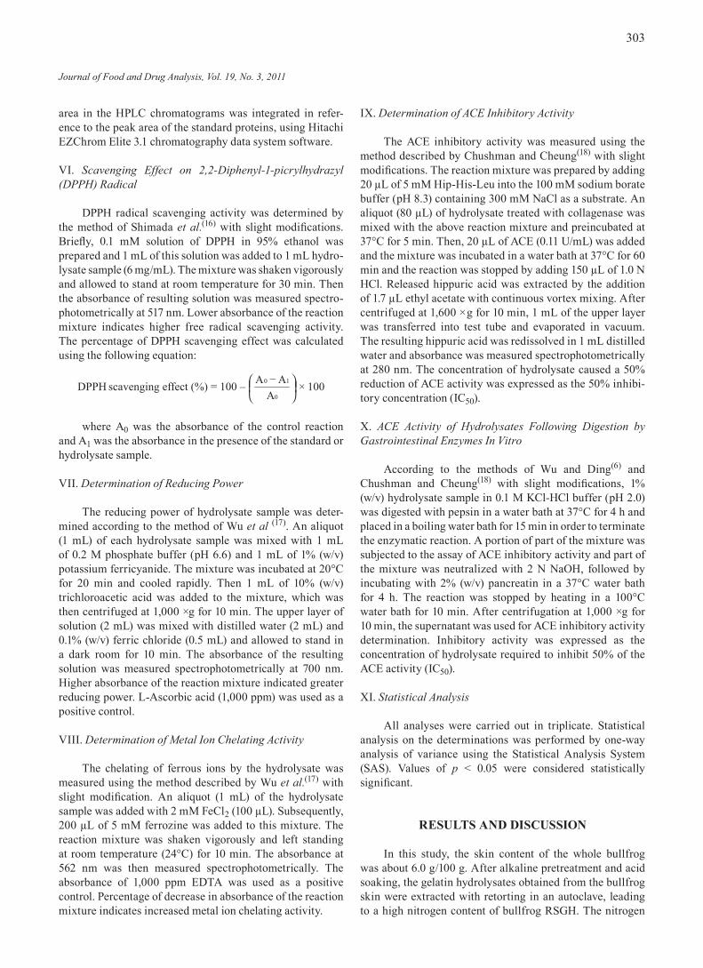
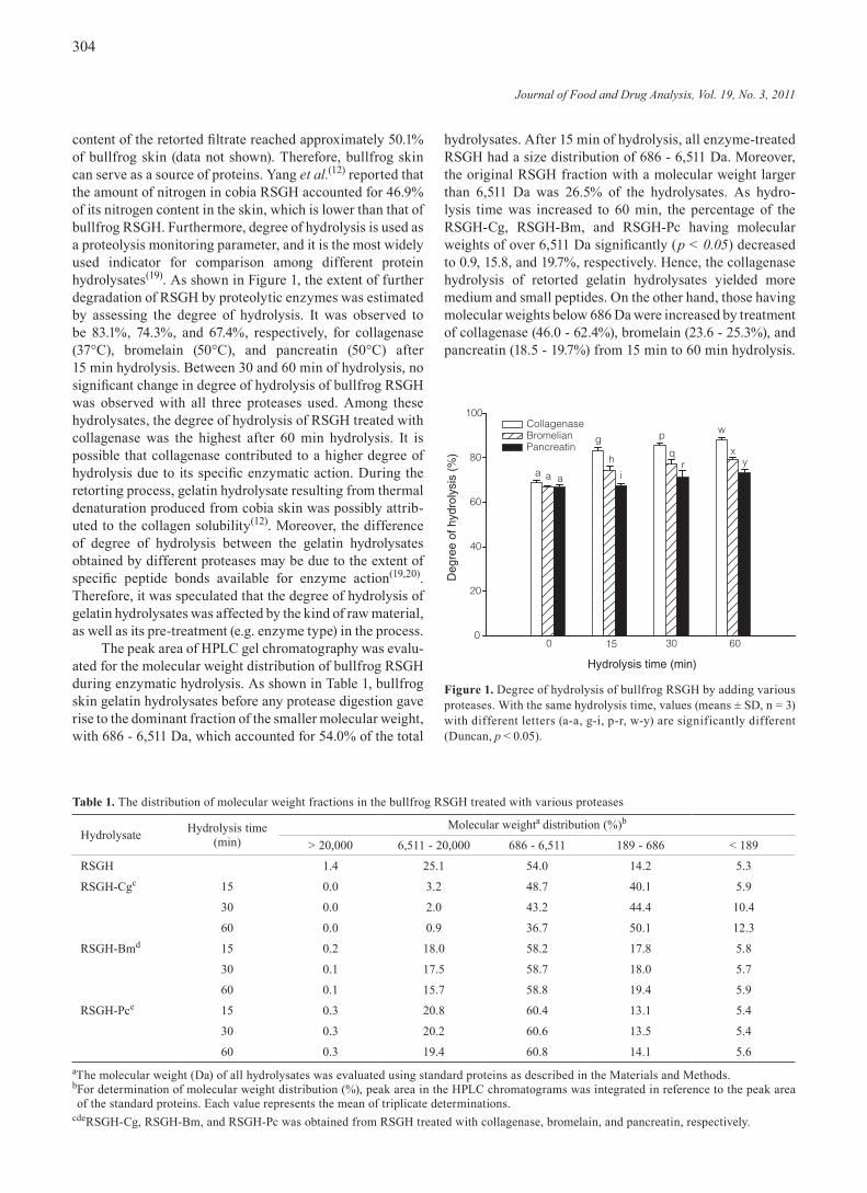
Enzymatic digestion has been extensively employed to improve the antioxidant properties of proteins. Gelatin hydrolysates produced by different proteolytic enzymes might have different molecular properties, leading to the varying characteristics of these resultant hydrolysates. Therefore, different methods (DPPH scavenging effect, metal ion chelating effect, and reducing power) need to be used to study their antioxidant properties. Generally, the effect of antioxidants on DPPH radical scavenging was due to their hydrogen donating ability. As depicted in Figure 2A, a significant (p < 0.05) decrease in the concentration of DPPH radical occurs with an increase concentration of the bullfrog RSGH and there is a good correlation between its concentration (2 - 8 mg/mL) and DPPH radical scavenging effects (20 - 72%). The DPPH scavenging effect of gelatin hydrolysate with further enzymatic digestion is shown in Figure 2B. The scavenging effect of the original hydroly-sate at 6 mg/mL on DPPH radical was about 80%. As the hydrolysis time increased, there were no significant changes in DPPH scavenging activity for RSGH-Bm and RSGH-Pc. Among all three enzymatic hydrolysates, the treatment of RSGH with collagenase increased the maximal DPPH scav-enging effect dependent on hydrolysis time (up to 87.0%). On the other hand, ascorbic acid exhibited 96.3% DPPH radical scavenging activity. RSGH-Cg may contain considerable amounts of small peptides that are electron donors and can react with free radicals to terminate the radical chain reac-tion. During hydrolysis, a wide variety of small peptides and free amino acids were generated which in turn might affect the resulting antioxidant activity(22,23). Compared with a previous report, the DPPH scavenging activity (80 - 99%) of marine protein hydrolysates at a concentration of 40 mg/mL varied with different degree of hydrolysis(20). Bullfrog skin gelatin hydrolysate had a higher scavenging activity of DPPH radicals than those marine protein hydrolysates. As shown in Figure 3, the antioxidant activities of gelatin hydroly-sates were evaluated using reducing power as an indicator. The result suggested that reducing power was markedly (p < 0.05) enhanced by ascorbic acid at the concentration of
Figure 2. (a) DPPH radical scavenging activity of different concen-trations of bullfrog RSGH. (b) Effect of enzymatic hydrolysate derived from bullfrog RSGH and commercial antioxidant on DPPH radical scavenging activity. The concentration of all enzymatic hydrolysate used in all assays was 6 mg/mL, while that of ascorbic acid was 1,000 ppm. Values (means ± SD, n = 3) with different letters (a-c) are statistically significant (Duncan, p < 0.05).
(A)
(B)
RSGH concentration (mg/mL)0 2 4 6 8 10 12 14
DP
PH
Sca
veng
ing
effe
ct (
%)
Ascorbic acid 0 min15 min30 min60 min
DP
PH
sca
veng
ing
effe
ct (
%)
a
bc c c
bbb b bb bb
Collag
enas
e
Bromela
in
Pancr
eatin
Ascor
bic ac
id
100
80
60
40
20
0
100
80
60
40
20
0
Figure 3. Reducing power of enzymatic hydrolysate derived from bullfrog RSGH and commercial antioxidant. The concentration of all enzymatic hydrolysates used in all assays was 6 mg/mL, while that of ascorbic acid was 1,000 ppm. Values (means ± SD, n = 3) with different letters (a-d) are statistically significant (Duncan, p < 0.05).
0.0
0.5
1.0
1.5
2.0
Ascorbic acid 0 min15 min 30 min 60 min
Abs
orba
nce
at 7
00 n
m
a
b bc
d
bb b b
b bb b
Ascor
bic ac
id
Collag
enas
e
Bromela
in
Pancr
eatin
Journal of Food and Drug Analysis, Vol. 19, No. 3, 2011
306
1,000 ppm. By comparison, in the original RSGH sample, the reducing power (absorbance at 700 nm) of various enzymatic hydrolysates increased only slightly with increasing time of hydrolysis. With 15 min of hydrolysis, there was no signifi-cant change in the reducing power among all enzymatic hydrolysates. With an increased hydrolysis time to 60 min, RSGH-Cg exhibited more effective reducing power than the other two enzymatic hydrolysates. It appears that the reducing power of all gelatin hydrolysates depended on the enzyme used. Je et al.(24) demonstrated that protein hydrolysates of tuna liver showed a good reducing power, resulting from high contents of hydrogen-donating peptides from various enzymatic hydrolysates. Therefore, it is speculated that the RSGH-Cg having an effective antioxidant activity might be the result of increased exposure to a functional group such as hydroxyl and carboxyl with hydrogen donating abilities during thermal and proteolytic processes.
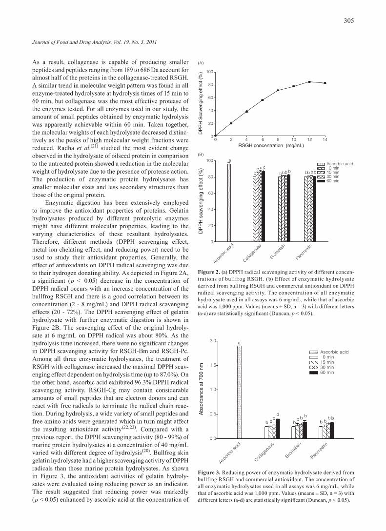
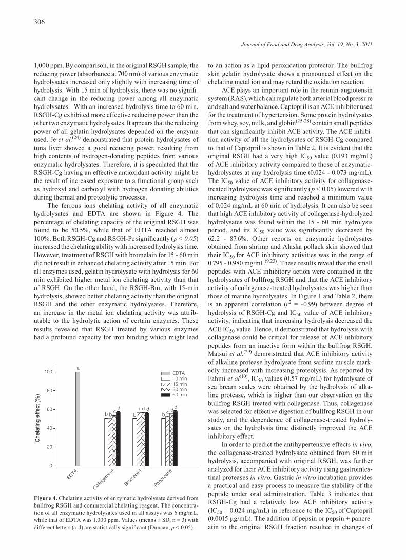
The ferrous ions chelating activity of all enzymatic hydrolysates and EDTA are shown in Figure 4. The percentage of chelating capacity of the original RSGH was found to be 50.5%, while that of EDTA reached almost 100%. Both RSGH-Cg and RSGH-Pc significantly (p < 0.05) increased the chelating ability with increased hydrolysis time. However, treatment of RSGH with bromelain for 15 - 60 min did not result in enhanced chelating activity after 15 min. For all enzymes used, gelatin hydrolysate with hydrolysis for 60 min exhibited higher metal ion chelating activity than that of RSGH. On the other hand, the RSGH-Bm, with 15-min hydrolysis, showed better chelating activity than the original RSGH and the other enzymatic hydrolysates. Therefore, an increase in the metal ion chelating activity was attrib-utable to the hydrolytic action of certain enzymes. These results revealed that RSGH treated by various enzymes had a profound capacity for iron binding which might lead
to an action as a lipid peroxidation protector. The bullfrog skin gelatin hydrolysate shows a pronounced effect on the chelating metal ion and may retard the oxidation reaction.
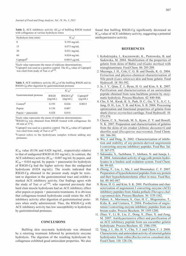
ACE plays an important role in the rennin-angiotensin system (RAS), which can regulate both arterial blood pressure and salt and water balance. Captopril is an ACE inhibitor used for the treatment of hypertension. Some protein hydrolysates from whey, soy, milk, and globin(25-28) contain small peptides that can significantly inhibit ACE activity. The ACE inhibi-tion activity of all the hydrolysates of RSGH-Cg compared to that of Captopril is shown in Table 2. It is evident that the original RSGH had a very high IC50 value (0.193 mg/mL) of ACE inhibitory activity compared to those of enzymatic-hydrolysates at any hydrolysis time (0.024 - 0.073 mg/mL). The IC50 value of ACE inhibitory activity for collagenase-treated hydrolysate was significantly (p < 0.05) lowered with increasing hydrolysis time and reached a minimum value of 0.024 mg/mL at 60 min of hydrolysis. It can also be seen that high ACE inhibitory activity of collagenase-hydrolyzed hydrolysates was found within the 15 - 60 min hydrolysis period, and its IC50 value was significantly decreased by 62.2 - 87.6%. Other reports on enzymatic hydrolysates obtained from shrimp and Alaska pollack skin showed that their IC50 for ACE inhibitory activities was in the range of 0.795 - 0.980 mg/mL(9,23). These results reveal that the small peptides with ACE inhibitory action were contained in the hydrolysates of bullfrog RSGH and that the ACE inhibitory activity of collagenase-treated hydrolysates was higher than those of marine hydrolysates. In Figure 1 and Table 2, there is an apparent correlation (r2 = -0.99) between degree of hydrolysis of RSGH-Cg and IC50 value of ACE inhibitory activity, indicating that increasing hydrolysis decreased the ACE IC50 value. Hence, it demonstrated that hydrolysis with collagenase could be critical for release of ACE inhibitory peptides from an inactive form within the bullfrog RSGH. Matsui et al.(29) demonstrated that ACE inhibitory activity of alkaline protease hydrolysate from sardine muscle mark-edly increased with increasing proteolysis. As reported by Fahmi et al(10), IC50 values (0.57 mg/mL) for hydrolysate of sea bream scales were obtained by the hydrolysis of alka-line protease, which is higher than our observation on the bullfrog RSGH treated with collagenase. Thus, collagenase was selected for effective digestion of bullfrog RSGH in our study, and the dependence of collagenase-treated hydroly-sates on the hydrolysis time distinctly improved the ACE inhibitory effect.
In order to predict the antihypertensive effects in vivo, the collagenase-treated hydrolysate obtained from 60 min hydrolysis, accompanied with original RSGH, was further analyzed for their ACE inhibitory activity using gastrointes-tinal proteases in vitro. Gastric in vitro incubation provides a practical and easy process to measure the stability of the peptide under oral administration. Table 3 indicates that RSGH-Cg had a relatively low ACE inhibitory activity (IC50 = 0.024 mg/mL) in reference to the IC50 of Captopril (0.0015 µg/mL). The addition of pepsin or pepsin + pancre-atin to the original RSGH fraction resulted in changes of
Figure 4. Chelating activity of enzymatic hydrolysate derived from bullfrog RSGH and commercial chelating reagent. The concentra-tion of all enzymatic hydrolysates used in all assays was 6 mg/mL, while that of EDTA was 1,000 ppm. Values (means ± SD, n = 3) with different letters (a-d) are statistically significant (Duncan, p < 0.05).
Che
latin
g ef
fect
(%
)
0
20
40
60
80
100 EDTA 0 min15 min 30 min 60 min
EDTA
Collag
enas
e
Bromela
in
Pancr
eatin
a
b bc
db
d d db c d
d
Journal of Food and Drug Analysis, Vol. 19, No. 3, 2011
307
IC50 value (0.156 and 0.428 mg/mL, respectively) relative to that of undigested RSGH (0.193 mg/mL). In contrast, the ACE inhibitory activity (IC50 = 0.007 mg/mL by pepsin, and IC50 = 0.014 mg/mL by pepsin + pancreatin) for hydrolysis of RSGH-Cg had the higher activity than the undigested hydrolysate (0.024 mg/mL). The results indicated that RSGH-Cg obtained in the present study might be resis-tant to digestion in the gastrointestinal tract and exhibit a marked ACE inhibitory activity. Our findings agree with the study of Tsai et al.(30), who reported previously that hard clam muscle hydrolysate had an ACE inhibitory effect after pepsin or pepsin + pancreatin treatments. It is obvious that collagenase-treated hydrolysate could retain the ACE inhibitory activity after digestion of gastrointestinal prote-ases when orally administered. Thus, the RSGH-Cg with ACE inhibitory activity has low susceptibility to hydrolysis by gastrointestinal proteases.
CONCLUSIONS
Bullfrog skin enzymatic hydrolysate was obtained by a retorting treatment followed by proteolytic enzyme hydrolysis. The digestion of the resulting hydrolysate by collagenase exhibited good antioxidant properties. We also
found that bullfrog RSGH-Cg significantly decreased an IC50 value of ACE inhibitory activity, suggesting a potential antihypertensive activity.
REFERENCES
1. Kołodziejska, I., Kaczorowski, K., Piotrowska, B. and Sadowska, M. 2004. Modification of the properties of gelatin from skins of Baltic cod (Gadus morhua) with transglutaminase. Food Chem. 86: 203-209.
2. Muyonga, J. H., Cole, C. G. B. and Duodu, K. G. 2004. Extraction and physico-chemical characterisation of Nile perch (Lates niloticus) skin and bone gelatin. Food Hydrocoll. 18: 581-592.
3. Je, J. Y., Qian, Z. J., Byun, H. G. and Kim, S. K. 2007. Purification and characterization of an antioxidant peptide obtained from tuna backbone protein by enzy-matic hydrolysis. Process Biochem. 42: 840-846.
4. Cho, S. M., Kwak, K. S., Park, D. C., Gu, Y. S., Ji, C. I., Jang, D. H., Lee, Y. B. and Kim, S. B. 2004. Processing optimization and functional properties of gelatin from shark (Isurus oxyrinchus) cartilage. Food Hydrocoll. 18: 573-579.
5. Cheow, C. S., Norizah, M. S., Kyaw, Z. Y. and Howell, N. K. 2007. Preparation and characterisation of gelatins from the skins of sin croaker (Johnius dussumieri) and shortfin scad (Decapterus macrosoma). Food Chem. 101: 386-391.
6. Wu, J. and Ding, X. 2002. Characterization of inhibi-tion and stability of soy-protein-derived angiotensin I-converting enzyme inhibitory peptides. Food Res. Int. 35: 367-375.
7. Sakanaka, S., Tachibana, Y., Ishihara, N. and Juneja, L. R. 2004. Antioxidant activity of egg-yolk protein hydro-lysates in a linoleic acid oxidation system. Food Chem. 86: 99-103.
8. Zhong, F., Liu, J., Ma, J. and Shoemaker, C. F. 2007. Preparation of hypocholesterol peptides from soy protein and their hypocholesterolemic effect in mice. Food Res. Int. 40: 661-667.
9. Byun, H. G. and Kim, S. K. 2001. Purification and char-acterization of angiotensin I converting enzyme (ACE) inhibitory peptides from Alaska pollack (Theragra chal-cogramma) skin. Process Biochem. 36: 1155-1162.
10. Fahmi, A., Morimura, S., Guo, H. C., Shigematsu, T., Kida, K. and Uemura, Y. 2004. Production of angio-tensin I converting enzyme inhibitory peptides from sea bream scales. Process Biochem. 39: 1195-1200.
11. Zhao, Y., Li, B., Liu, Z., Dong, S., Zhao, X. and Zeng, M. 2007. Antihypertensive effect and purification of an ACE inhibitory peptide from sea cucumber gelatin hydrolysate. Process Biochem. 42: 1586-1591.
12. Yang, J. I., Ho, H. Y., Chu, Y. J. and Chow, C. J. 2008. Characteristic and antioxidant activity of retorted gelatin hydrolysates from cobia (Rachycentron canadum) skin. Food Chem. 110: 128-136.
Table 2. ACE inhibitory activity (IC50) of bullfrog RSGH treated with collagenase at various hydrolysis times
Hydrolysis time (min) IC50a
0 0.193 mg/mL
15 0.073 mg/mL
30 0.031 mg/mL
60 0.024 mg/mL
Captoprilb 0.0015 µg/mLaEach value represents the mean of triplicate determinations.b Captopril was used as a positive control. The IC50 value of Captopril was cited from study of Tsai et al(30).
Table 3. ACE inhibitory activity (IC50) of the bullfrog RSGH and its RSGH-Cg after digestion by gastrointestinal protease
Gastrointestinal protease
IC50a
RSGH (mg/mL)
RSGH-Cgb
(mg/mL)Captoprilc(µg/mL)
Controld 0.193 0.024 0.0015
Pepsin 0.156 0.007
Pepsin + pancreatin 0.428 0.014aEach value represents the mean of triplicate determinations.b RSGH-Cg was obtained from RSGH treated with collagenase for 60 min at 37°C.
c Captopril was used as a positive control. The IC50 value of Captopril was cited from study of Tsai et al(30).
d Control refers to the hydrolysate samples without adding any proteases.
Journal of Food and Drug Analysis, Vol. 19, No. 3, 2011
308
13. Nagai, T. and Suzuki, N. 2000. Isolation of collagen from fish waste material–skin, bone and fins. Food Chem. 68: 277-281.
14. Li, H., Liu, B. L., Gao, L. Z. and Chen, H. L. 2004. Studies on bullfrog skin collagen. Food Chem. 84: 65-69.
15. Church, F. C., Swaisgood, H. E., Porter, D.H. and Catignani, G. L. 1983. Spectrophotometric assay using o-phthaldialdehyde for determination of proteolysis in milk and isolated milk proteins. J. Dairy Sci. 66: 1219-1227.
16. Shimada, K., Fujikawa, K., Yahara, K. and Nakamura, T. 1992. Antioxidative properties of xanthan on the antioxidation of soybean oil in cyclodextrin emulsion. J. Agric. Food Chem. 40: 945-948.
17. Wu, H. C., Pan, B. S., Chang, C. L. and Shiau, C. Y. 2005. Low molecular weight peptides as related to antioxidant properties of chicken essence. J. Food Drug Anal. 13: 176-183.
18. Chushman, D. W. and Cheung, H. S. 1971. Spectro-photometric assay and properties of the angiotensin-converting enzyme of rabbit lung. Biochem. Pharmacol. 20: 1637-1648.
19. Guerard, F., Dufosse, L., De La Broise, D. and Binet, A. 2001. Enzymatic hydrolysis of protein from yellowfin tuna (Thunnus albacares) wastes using alcalase. J. Mol. Catal. B: Enzym. 11: 1051-1059.
20. Klompong, V., Benjakul, S., Kantachote, D. and Shahidi, F. 2007. Antioxidative activity and functional properties of protein hydrolysate of yellow stripe trevally (Selar-oides leptolepis) as influenced by the degree of hydro-lysis and enzyme type. Food Chem. 102: 1317-1327.
21. Radha, C., Kumar, P. R. and Parakash, V. 2008. Prepara-tion and characterization of a protein hydrolysate from an oilseed flour mixture. Food Chem. 106: 1166-1174.
22. Wu, H. C., Chen, H. M. and Shiau, C. Y. 2003. Free amino acids and peptides as related to antioxidant prop-erties in protein hydrolysates of mackerel (Scomber austriasicus). Food Res. Int. 36: 949-957.
23. He, H., Chen, X., Sun, C., Zhang, Y. and Gao, P. 2006. Preparation and functional evaluation of oligopeptide-enriched hydrolysate from shrimp (Acetes chinensis) treated with crude protease from Bacillus sp. SM98011. Bioresour. Technol. 97: 385-390.
24. Je, J. Y., Lee, K. H., Lee, M. H. and Ahn, C. B. 2009. Antioxidant and antihypertensive protein hydrolysates produced from tuna liver by enzymatic hydrolysis. Food Res. Int. 42: 1266-1272.
25. Mullally, M. M., Meisel, H. and Fitzgerald, R. J. 1997. Angiotensin-I-converting enzyme inhibitory activities of gastric and pancreatic proteinase digests of whey proteins. Int. Dairy J. 7: 299-303.
26. Gibbs, B. F., Zougman, A., Masse, R. and Mulligan, C. 2004. Production and characterization of bioactive peptides from soy hydrolysate and soy-fermented food. Food Res. Int. 37: 123-131.
27. Yu, Y., Hu, J., Bai, X., Du, Y. and Lin, B. 2006. Prepara-tion and function of oligopeptide-enriched hydrolysate from globin by pepsin. Process Biochem. 41: 1589-1593.
28. Otte, J., Shalaby, S. M., Zakora, M., Pripp, A. H. and EI-Shabrawy, S. A. 2007. Angiotensin-converting enzyme inhibitory activity of milk protein hydrolysates: Effect of substrate, enzyme and time of hydrolysis. Int. Dairy J. 17: 488-503.
29. Matsui, T., Matsufuji, H., Seki, E., Osajima, K., Nakashima, M., Osajima, Y. 1993. Inhibition of angio-tensin I-converting enzyme by Bacillus licheniformis alkaline protease hydrolyzates derived from sardine muscle. Biosci Biotech Biochem 57: 922-925.
30. Tsai, J. S., Chen, J. L. and Pan, B. S. 2008. ACE-inhib-itory peptides identified from the muscle protein hydro-lysate of hard clam (Meretrix lusoria). Process Biochem. 43: 743-747.