Tea and Chicory Extract Characterization, Classification and ...
Upload
khangminh22Category
view
2download
0
IN VITRO EFFECTS OF AQUEOUS LEAF EXTRACT OF MORINGA OLEIFERA
ON HUMAN SPERM
by
FAITH TEBATSO MOICHELA
DISSERTATION
Submitted in fulfilment of the requirements for the degree of
MASTER OF SCIENCE
in
MEDICAL SCIENCES
in the
FACULTY OF HEALTH SCIENCES
(School of Health Care Sciences)
at the
UNIVERSITY OF LIMPOPO
SUPERVISOR: Dr C.S. Opuwari
CO-SUPERVISOR: Prof R. Henkel
CO-SUPERVISOR: Dr G.A. Adefolaju
2020

ii
DEDICATION
I dedicate this work to my family and Mr Jim Smurthwaite for their inestimable
sacrifices and support.

iii
DECLARATION
I, Faith Tebatso Moichela, declare that the dissertation titled: “In vitro effects of
aqueous leaf extracts of Moringa oleifera on human sperm” is my own work and that
all the sources I have used or quoted have been duly indicated and acknowledged by
means of complete references. Furthermore, I declare that this work has not been
submitted before for any degree at any other institution.
Faith Tebatso Moichela 16.02.2021
Full names Date
Signature

iv
ACKNOWLEDGEMENTS
I would like to before and beyond, thank God for continuously replenishing my strength
to move forward.
Research is an arduous process and therefore cannot be individually encountered.
Accordingly, the following individuals are thankfully acknowledged for their invaluable
contributions towards the completion of this project:
• My infinite indebtedness goes to my mentor, role model, and supervisor Dr
Chinyerum Sylvia Opuwari for your patience, dedication, guidance, and
timeous feedbacks that have seen the successful completion of this
dissertation.
• Prof Ralf Henkel for advising the content development of the report, providing
laboratory access and equipment, and statistical analysis basics.
• Dr Gbenga Adefolaju for continuously providing technical support of the project
and providing recommendations.
• Mr Aqeel Morris, Ms Shannen Keyser, Dr Michael Solomon and Mr Cleyson
Mupfiga for introducing me to basic sperm-handling techniques, and additional
computer-aided sperm analysis (CASA) training and mentorship.
• Donors, patients, and staff of the Tygerberg and Vincent Pallotti Hospitals for
making semen samples available on time.
• Dr Paulina Bopape-Mabapa for providing Moringa oleifera leaves and the
Biochemistry, Microbiology and Biotechnology Department (UL) for guidance
through the plant extraction process.
• To everyone in the University of the Western Cape Medical Bioscience, Free
State University Basic Medical Sciences, and the University of Limpopo
Physiology and Environmental Health Departments, whose contributions
influenced the success of the study in various capacities. Your efforts are
appreciated.
• To Dr Claudia Ntsapi, thank you for the constant encouragement and
confidantship.
• The National Research Foundation (NRF) for financial support.
• To my partner in crime, Beauty Ndivhuho Takalani, thank you for keeping up
with me, the friendship, and laboratory assistance.
• Particular thanks are accorded to my forbearing but otherwise fond family,
especially my grandmother, aunt Lillian, Nthabeleng and friends, whose
unfailing support and prayers propelled me to regroup and soldier on along the
tedious journey of this research.

v
ABSTRACT
Infertility affects nearly 186 million couples globally, with male factors contributing to
half of the cases. Oxidative stress is an established cause of declining semen quality.
Moringa oleifera has proven antioxidants. This study aimed to investigate in vitro
effects of aqueous leaf extract of M. oleifera on human sperm functions. Semen
samples from donors (n = 40) and patients (n = 30) were washed with HTF-bovine
serum albumin (BSA), and then incubated with various concentrations of M. oleifera
(0, 0.625, 6.25, 62.5, and 625 µg/ml) at 37°C for 1 hour. Sperm motility, vitality,
mitochondrial membrane potential (MMP), reactive oxygen species (ROS), DNA
fragmentation, capacitation, and acrosome reaction were assessed. Sperm motility,
vitality, MMP, and capacitation were enhanced, while ROS production, and DNA
fragmentation decreased after M. oleifera treatment. Uncapacitated spermatozoa
increased significantly with a reduction in acrosome reaction in donors. M. oleifera
antioxidant compounds suppressed excessive ROS, preserved mitochondrial
membrane, DNA and acrosome integrity, while enhancing sperm motility and viability.
vi
KEY CONCEPTS
Sperm function
Oxidative stress
Infertility
Antioxidants
Moringa oleifera

vii
TABLE OF CONTENTS
DEDICATION .............................................................................................................. ii
DECLARATION .......................................................................................................... iii
ACKNOWLEDGEMENTS .......................................................................................... iv
KEY CONCEPTS ....................................................................................................... vi
TABLE OF CONTENTS ............................................................................................ vii
RESEARCH OUTPUTS ............................................................................................ xii
LIST OF FIGURES ................................................................................................... xiii
LIST OF TABLES ........................................................................................................ ii
LIST OF ABBREVIATIONS ........................................................................................ iii
CHAPTER 1 ............................................................................................................... 1
INTRODUCTION ........................................................................................................ 1
1.1. Background ................................................................................................... 1
1.2. Research problem ......................................................................................... 5
1.3. Purpose of the study ..................................................................................... 6
1.3.1 Aim ............................................................................................................. 6
1.3.2 Objectives ................................................................................................... 6
The objectives of the study were to: .................................................................... 6
CHAPTER 2 ............................................................................................................... 7
LITERATURE REVIEW .............................................................................................. 7
2.1 Overview of the basic functioning of the male reproductive system .................. 7
2.2 The architecture of the male reproductive system ............................................ 8
2.2.1 Male accessory glands and male reproductive function ............................. 8
2.2.2 Male reproductive tract function .................................................................. 9
2.2.2.1 Epididymis ........................................................................................... 9
2.2.2.2 Vas deferens ..................................................................................... 10
2.2.2.3 Urethra ............................................................................................... 10
2.3 Testicular function ........................................................................................... 11
2.3.1 Hypothalamic-pituitary testicular axis (HPT-axis) ...................................... 12
2.3.1.1 Gonadotropin-releasing hormone (GnRH) ......................................... 12
2.3.1.2 Follicle stimulating hormone and luteinising hormone ....................... 13
2.3.2 Cellular composition of the testis .......................................................... 15
2.3.2.1 Sertoli cells function ........................................................................... 15
2.3.2.2 Leydig cells function ....................................................................... 16

viii
2.4 Spermatogenesis ............................................................................................ 16
2.4.1 Events at the basal compartment of the seminiferous tubules .................. 17
2.4.2 Events at the adluminal compartment of the seminiferous tubules ........... 17
2.4.3 Spermiogenesis and spermiation .............................................................. 18
2.5 Structure of human sperm ............................................................................... 19
2.5.1 Head ......................................................................................................... 20
2.5.2 Neck ......................................................................................................... 20
2.5.3 Flagellum .................................................................................................. 21
2.6 Evaluation of sperm functional parameters ..................................................... 22
2.6.1 Motility....................................................................................................... 24
2.6.1.1 Computer-aided sperm analysis (CASA) ........................................... 24
2.6.2 Vitality ....................................................................................................... 25
2.6.2.1 One-step eosin nigrosine staining technique ..................................... 25
2.6.3 Mitochondrial membrane potential (MMP) (∆ѱM) ...................................... 26
2.6.3.1 Lipophilic cationic dye ........................................................................ 27
2.6.4 Reactive oxygen species (ROS) ............................................................... 27
2.6.4.1 ROS generation ................................................................................. 27
2.6.4.1 Sources of ROS ................................................................................. 28
2.6.4.2 Antioxidants ....................................................................................... 29
2.6.4.3 ROS measurement using a fluorescent probe ................................... 30
2.6.5 DNA fragmentation ................................................................................... 30
2.6.5.1 Sperm DNA fragmentation quantification........................................... 31
2.6.6 Capacitation (CP) and acrosome reaction (AR) ........................................ 32
2.6.6.1 Quantification of CP and AR .............................................................. 35
2.7 Infertility ........................................................................................................... 35
2.8 Herbal medicine .............................................................................................. 39
2.8.1 Herbal medicine and male infertility .......................................................... 41
2.9 Moringa oleifera .............................................................................................. 43
2.9.1 Physical characterisation .......................................................................... 43
2.9.2 Distribution ................................................................................................ 44
2.9.3 Phytochemistry and pharmacological properties ...................................... 45
2.9.4 Previous studies of M. oleifera on mammalian reproductive function other
than sperm functioning ...................................................................................... 46
2.9.4.1 M. oleifera and hypothalamic regulation of mammalian reproduction .... 47
2.9.4.2 M. oleifera and pituitary regulation of mammalian reproduction ............. 47

ix
2.9.4.3 M. oleifera and testicular regulation of mammalian reproduction ........... 48
CHAPTER 3 ............................................................................................................. 49
RESEARCH METHODOLOGY ................................................................................ 49
3.1. Introduction .................................................................................................... 49
3.2 Research Methods .......................................................................................... 49
3.2.1 Methods and materials ............................................................................. 49
3.2.1.1 Chemicals and apparatus .................................................................. 49
3.3 Study design ................................................................................................... 49
3.4 Sampling ......................................................................................................... 49
3.4.1 Study sites ................................................................................................ 49
3.4.2 Study population ....................................................................................... 50
3.4.3 Sampling procedure .................................................................................. 51
3.4.3.1 Inclusion and exclusion criteria .......................................................... 51
3.5 Data collection ................................................................................................ 52
3.5.1 Preparation and extraction of M. oleifera leaves ....................................... 52
3.5.1.1 The rationale for the determination of therapeutic dose .................... 52
3.5.1 Experimental procedures .......................................................................... 53
3.5.2 Preparation of culture medium and semen .............................................. 53
3.6 Laboratory analysis ......................................................................................... 56
3.6.1 Determination of effects of M. oleifera aqueous leaf extracts on sperm
motility 56
3.6.2 Determination of effects of M. oleifera aqueous leaf extracts on sperm
vitality 58
3.6.3 Determination of effects of M. oleifera aqueous extracts on sperm
mitochondrial membrane potential (∆ψm) .......................................................... 59
3.6.4 Determination effects of M. oleifera aqueous extracts on sperm reactive
oxygen species (ROS) production ..................................................................... 61
3.6.5 Determination of effects of M. oleifera aqueous extracts on sperm DNA
fragmentation. .................................................................................................... 62
3.6.6 Determination of effects of M. oleifera aqueous extracts on sperm
capacitation and acrosome reaction .................................................................. 63
3.7 Ethical considerations ..................................................................................... 65
3.7.1 Ethical clearance and approval ................................................................. 65
3.7.2 Harm ......................................................................................................... 65
3.7.3 Disposal of waste ...................................................................................... 65
3.7.4 Anonymity and confidentiality ................................................................... 65

x
3.8 Data analyses ................................................................................................. 66
3.8.1 Reliability and validity ............................................................................... 66
3.8.2 Quality assurance ..................................................................................... 66
3.8.3 Bias ........................................................................................................... 66
3.8.4 Statistical analysis .................................................................................... 66
CHAPTER 4 ............................................................................................................. 68
RESULTS ................................................................................................................. 68
4.1. Introduction .................................................................................................... 68
4.2. Summary statistics of standard semen parameters according to the WHO
(2010) ................................................................................................................... 68
4.3. Effects of M. oleifera leaf extracts on sperm motility parameters in vitro........ 71
4.3.1. Correlations of sperm total motility and functional parameters. ............... 78
4.3.2. Correlation between progressive motility and functional parameters ....... 79
4.3.3 Correlation between sperm hyperactivation and functional parameters ... 80
4.4. Effects of M. oleifera leaf extracts on sperm vitality in vitro ............................ 81
4.4.1. Correlation of sperm vitality with other functional parameters.................. 83
4.5.1 Correlation of sperm-intact mitochondrial membrane potential and
functional parameters ........................................................................................ 86
4.6. Effects of M. oleifera on reactive oxygen species-positive (ROS) spermatozoa
in vitro ................................................................................................................... 86
4.6.1 Correlation of reactive oxygen species with functional parameters .......... 88
4.7. Effects of M. oleifera leaf extracts on sperm DNA fragmentation in vitro ....... 89
4.7.1 Correlation of sperm DNA fragmentation and functional parameters ........ 91
4.8 Effects of M. oleifera on sperm capacitation and acrosome reaction in vitro... 91
4.8.1 Correlation of sperm capacitation and acrosome reaction ........................ 95
4. 9. Effects of M. oleifera on asthenozoospermic sperm in vitro .......................... 96
4.9.1 Effects of M. oleifera on sperm motility from asthenozoospermic samples in
vitro 96
4.9.2. Effects of M. oleifera on asthenozoospermic vitality in vitro ..................... 98
4.9.3. Effects of M. oleifera extract on mitochondrial membrane potential (MMP-
intact) in asthenozoospermic samples in vitro ................................................... 99
4.9.4. Effects of M. oleifera on asthenozoospermic reactive oxygen species-
positive spermatozoa in vitro ........................................................................... 100
4.9.5. Effects of M. oleifera on DNA-fragmented sperm in asthenozoospermic
samples in vitro ................................................................................................ 101
4.9.6. Effects of M. oleifera on capacitation and acrosome reaction in
asthenozoospermic sperm in vitro ................................................................... 102
xi
4.9.7. Correlation of asthenozoospermic sperm parameters ........................... 104
CHAPTER 5 ........................................................................................................... 107
DISCUSSION ......................................................................................................... 107
5.1. Effects of M. oleifera leaf extracts on sperm motility in vitro ...................... 108
5.3 Effects of M. oleifera leaf extracts on sperm vitality in vitro ........................... 111
5.4 Effects of M. oleifera leaf extracts on sperm mitochondrial membrane potential
in vitro ................................................................................................................. 114
5.5 Effects of M. oleifera leaf extracts on reactive oxygen species (ROS) in vitro
............................................................................................................................ 117
5.6 Effects of M. oleifera leaf extracts on sperm DNA fragmentation in vitro ...... 119
5.8 Conclusion .................................................................................................... 124
5.9 Future outlooks and recommendations ......................................................... 125
CHAPTER 6 ........................................................................................................... 127
REFERENCES ....................................................................................................... 127
APPENDICES APPENDIX 1: University of Limpopo TREC approval .................. 181
APPENDIX 2: University of the Western Cape BMREC approval .......................... 182
APPENDIX 3: Permission for semen collection from Tygerberg Hospital .............. 183
APPENDIX 4: Permission for semen collection from Vincent Pallotti Hospital ....... 184
APPENDIX 5: Informed consent form (English) ..................................................... 185
APPENDIX 6: Informed consent form (Afrikaans) .................................................. 187
APPENDIX 7: Informed consent form (IsiXhosa) ................................................... 190

xii
RESEARCH OUTPUTS
Parts of the results were presented and submitted for publication as follows:
• F.T. Moichela, G.A. Adefolaju, R. Henkel, and C.S. Opuwari. In vitro effects of
Moringa oleifera leaf extract on human sperm functions. Anatomical Society of
Southern Africa 47th Annual Conference, 07-10 April 2019, Pilanesberg, North
West, South Africa. Abstract book, pg 75.
• F.T. Moichela, G.A. Adefolaju, R. Henkel, and C.S. Opuwari. Antioxidative effects
of Moringa oleifera (MO) on human sperm functions in vitro. University of Limpopo
and Venda Health Science 1st Biennial conference 2019. Won 2nd position.
• F.T. Moichela, G.A. Adefolaju, R. Henkel, and C.S. Opuwari. Aqueous leaf extract
of Moringa oleifera reduces sperm DNA fragmentation in fertile men: An in vitro
study. Fertility Africa 2020 Accepted (Meeting postponed).
• F.T. Moichela, G.A. Adefolaju, R. Henkel, and C.S. Opuwari. Effects of Moringa
oleifera leaf extracts on asthenozoospermic semen in vitro. Anatomical Society of
Southern Africa 48th Annual Conference, Accepted.
• FT. Moichela, G.A. Adefolaju, R. Henkel, and C.S. Opuwari, 2020. In vitro study of
antioxidant effect of aqueous Moringa oleifera leaf extract on human sperm.
(Published in Andrologia).

xiii
LIST OF FIGURES
Figure 1: Posterior view of the male reproductive system depicting the testis, ducts,
glands and the external organs.
Figure 2: Posterior view of male accessory glands; prostate, vesicular and
bulbourethral glands.
Figure 3: Male urethra depicting prostatic, membranous, and spongy segments.
Figure 4: Sagittal view of the human testis with connecting tubules and two tunica
membranes.
Figure 5: Schematic depiction of the neuroendocrine regulation of hypothalamic
pituitary testicular axis males.
Figure 6: A summary of mammalian spermatogenesis in the seminiferous epithelium
showing the Sertoli cell blood testes barrier (BTB) that separates two compartments.
Figure 7: Structure of mammalian spermatozoa.
Figure 8: Sperm motility kinematics as seen under the CASA system.
Figure 9: The physiological and pathophysiological roles of ROS in male infertility.
Figure 10: Molecular events involved in the regulation of mammalian sperm
capacitation.
Figure 11: Illustration of molecular events regulating acrosome reaction.
Figure 12: Demographic illustration of global male factor infertility.
Figure 13: Schematic illustration of global dependence on herbal medicine.
Figure 14: M. oleifera aerial parts.
Figure 15: Depiction of the geographical locations where semen samples were
obtained.
Figure 16: Experimental layout outlining functional assays and computational systems
utilised.
xiv
Figure 17: (a) Computer-aided sperm class analyser (CASA) and (b) Leja slides used
for determination of motility and concentrations.
Figure 18: One-step eosin-nigrosin (E&N) staining of human sperm specimen. The
dye excludes viable/live (white) from dead (pink) cells based on the membrane
intactness. Samples were viewed at 10X objective.
Figure 19: Spermatozoal mitochondrial membrane potential (MMP) integrity status as
observed using epifluorescence. Mid-piece fluoresces orange-red (red arrow) denotes
intact MMP, while green arrow depicts membrane disrupted spermatozoa. Specimens
were viewed at 100X magnification.
Figure 20: Detection of excessive superoxide anion by human spermatozoa as
detected by a DHE fluorescent probe. Green arrows indicate ROS-negative
spermatozoa and red arrows show ROS-positive spermatozoa. Specimens were
viewed at 100X magnification.
Figure 21: Detection of sperm DNA fragmentation using TUNEL assay. Specimens
were viewed at 100X magnification.
Figure 22: Three staining CTC patterns of human spermatozoa as seen under a
fluorescence microscope. Specimens were viewed at 100X magnification.
Figure 23: Total motility after 1 hr incubation with M. oleifera in vitro. Mean ± SEM.
Blue bars represent the mean values of donors (N = 40), purple bars are for patients
(N = 30), and grey bars are for combined group (patients and donors) (N = 70).
Figure 24: Progressive (a) and non-progressive motility (b) after 1 hr incubation with
M. oleifera in vitro. Blue bars represent the mean values of donors (N = 40), purple
bars are for patients (N = 30), and grey bars are for combined group (patients and
donors) (N = 70).
Figure 25: Percentage of live sperm after 1 hr incubation with M. oleifera in vitro. Blue
bars represent the mean values of donors (N = 40), purple bars are for patients (N =
30), and grey bars are for combined group (patients and donors) (N = 70).
Figure 26. Percentage intact mitochondrial membrane potential (MMP) spermatozoa
after 1 hr incubation with M. oleifera in vitro. Blue bars represent the mean values of

xv
donors (N = 40), purple bars are for patients (N = 30), and grey bars are for combined
group (patients and donors) (N = 70).
Figure 27: Percentage ROS-positive spermatozoa after 1 hr incubation with M.
oleifera in vitro. Blue bars represent the mean values of donors (N = 40), purple bars
are for patients (N = 30), and grey bars are for combined group (patients and donors)
(N = 70).
Figure 28. Percentage of sperm DNA fragmentation after 1 hr incubation with M.
oleifera in vitro. Blue bars represent the mean values of donors (N = 40), purple bars
are for patients (N = 30), and grey bars are for combined group (patients and donors)
(N = 70).
Figure 29. Percentage sperm uncapacitation and capacitation after 1 hr incubation
with M. oleifera in vitro. Blue bars represent the mean values of donors (N = 40), purple
bars are for patients (N = 30), and grey bars are for combined group (patients and
donors) (N = 70).
Figure 30. Percentage of sperm acrosome reaction after 1 hr incubation with M.
oleifera in vitro. Blue bars represent the mean values of donors (N = 40), purple bars
are for patients (N = 30), and grey bars are for combined group (patients and donors)
(N = 70).
Figure 31: Percentage of live asthenozoospermic sperm after 1 hr treatment with M.
oleifera extracts for 1 h in vitro.
Figure 32: Percentage of asthenozoospermic MMP-intact sperm after 1 hr incubation
with M. oleifera in vitro.
Figure 33: Percentage of ROS-positive asthenozoospermic sperm after 1 hr
incubation with M. oleifera in vitro.
Figure 34: Percentage of asthenozoospermic DNA fragmentation after 1 hr with M.
oleifera in vitro.
Figure 35: Percentage of asthenozoospermic sperm capacitation and acrosome
reaction after 1 hr incubation with M. oleifera in vitro.

ii
LIST OF TABLES
Table 1: Categories of sperm motility defined
Table 2: Sperm motility kinematic parameters
Table 3: Summary statistics of standard semen parameters after liquefaction before
treatment with M. oleifera aqueous leaf extracts
Table 4: Comparative results on donors, patients, and combined sperm motility
kinematic parameters after treatment with aqueous extract of M. oleifera
Table 5: Relationships between sperm total motility and functional parameters
Table 6: Correlation between progressive motility and functional parameters
Table 7: Correlation between sperm hyper-activation and functional parameters
Table 8: Correlation of sperm vitality (live) and functional parameters
Table 9: Correlation of MMP-intact sperm with functional parameters
Table 10: Correlation of reactive oxygen species’ production with functional
parameters
Table 11: Correlation of sperm DNA fragmentation and functional parameters
Table 12. Correlation of sperm capacitation and acrosome reaction
Table 13: Effects of M. oleifera on sperm motility parameters on asthenozoospermic
samples in vitro
Table 14: Effects of M. oleifera sperm on motility kinematics from asthenozoospermic
samples in vitro
Table 15: Correlation of asthenozoospermic sperm parameters
iii
LIST OF ABBREVIATIONS
ABP Androgen binding protein
ALH Amplitude of lateral head displacement
AMH Anti-Műllerian hormone
ANOVA Analysis of variance
ART Assisted reproductive technologies
AR Acrosome reaction
ASRM American Society for Reproductive Medicine
ATP Adenosine Triphosphate
BCF Beat cross frequency
BMREC Biomedical Research Ethics Committee
BTB Blood testes barrier
BSA Bovine serum albumin
Ca2+ Calcium
cAMP cyclic Adenosine monophosphate
CASA Computer-aided sperm analysis
CAT Catalase
CP Capacitation
CTC Chlortetracycline
DHE Dihydroethidium
DNA Deoxyribonucleic acid
DPX Dibutylphthalate polystyrene xylene
E and N Eosin and nigrosine
ETC Electron transport chain
ERK Extracellular signal kinase
FSH Follicle stimulating hormone
GABA Gamma amino butyric acid
GIFT Gamete intrafallopian transfer
GnRH Gonadotropin-releasing hormone

iv
GnIH Gonadotropin inhibiting hormone
GSH Glutathione
GPx Glutathione peroxidase
hCG human Chorionic gonadotropin
HTF Human tubular fluid
Hyp Hyper-activation
HPT Hypothalamic pituitary testicular axis
ICMART International Committee for Monitoring Assisted Reproductive
Techniques
ICSI Intracytoplasmic sperm injection
IFV In vitro fertilisation
LH Luteinising hormone
LIN Linearity index
MDA Malondialdehyde
MMP Mitochondrial membrane potential
NADPH Nicotinamide adenine dinucleotide phosphate
NO Nitric oxide
ODF Outer dense fibres
OSPHOX Oxidative phosphorylation
OS Oxidative stress
PBS Phosphate buffer saline
PKA Protein kinase A
PKC Protein kinase C
PMN Polymorphonuclear
PTK Phosphorylation of tyrosine kinase
PUFA Polyunsaturated fatty acids
PVP40 Polyvinylpyrrolidone
RM Repeated measure
ROS Reactive oxygen species
SCs Sertoli cells
SCA Sperm class analyser

v
SCD Sperm chromatin dispersion
SCSA Sperm chromatin structure assay
SOD Superoxide dismutase
SSC Spermatogonial stem cell
STI/(D) Sexually transmitted infection (disease)
STR Straightness index
TEM Transmission electron microscopy
TCM Traditional Chinese Medicine
TGF-β Tumour growth factor-beta
TM Traditional medicine
TNF-α Tumour necrosis factor-alpha
TREC Turfloop Research Ethics
TUNEL Terminal deoxynucleotidyl transferase-mediated nick ending labelling
VAP Average path velocity
VCL Curvilinear velocity
VSL Straight-line velocity
WOB Wobble/oscillation index
WHO World Health Organisation
ZP Zona pellucida

1
CHAPTER 1
INTRODUCTION
1.1. Background
Infertility and population growth are increasing parad oxical burdens of reproduction
and the health care systems. Infertility may not receive as much consideration and
remedial intervention as its counterpart, overpopulation, but it has and continues to
devastate those suffering from it (Ombelet and Goossens, 2017). It is defined as the
disease of the reproductive system, and characterised by failure of a non-
contracepting, sexually active couple to establish a pregnancy or carry to term after
one year of regular coitus in the absence of any known reproductive pathology
(Zegers-Hochschild et al., 2009; Chimbatata and Malimba, 2016). This disease affects
one in eight couples who attempt to conceive for the first time, and one in six couples
with preceding pregnancies, despite the outcome (Jungwirth et al., 2015). It is also
clear that infertility and its complications can no longer be confined to biological
function, but presents emotional, psychological, social, and financial distress (Wu et
al., 2013).
Although the absolute prevalence of infertility is impossible to estimate, the World
Health Organisation (WHO) in 2010 asserted that about 48.5 million (from 42 million
in 1992) couples were struggling to fall pregnant (Nagórska et al., 2019). Subsequent
estimates in 2017 revealed a steady but increasing trend towards the burden of
infertility affecting 186 million couples worldwide (Inhorn and Patrizio, 2015). However,
the known prevalence and incidence rates have likely gone up, especially in
developing countries where the incidence of genito-urinary tract infections have
increased (Chigbu et al., 2012; Solomon and Henkel, 2017). Pelvic inflammatory
diseases (Rametse et al., 2018) and metabolic diseases have progressively
contributed to infertility (Leisegang et al., 2014). Environmental chemical exposure
owing to industrialisation and technological advancements have been indicated to
augment the pathogenesis of infertility (Kim, 2011; Nazıroğlu et al., 2013). As a result,
there is an overall decline in gamete production and quality in both sexes (Buckett and
Tan, 2005; Silva et al., 2019).

2
At the same time, high rates of infertility have also been reported for developed
countries in Europe (Coale and Watkins, 2017), Asia (Sharma et al., 2009), the Middle
East (Eldib and Tashani, 2018), and America (Louis et al., 2013), despite assisted
reproductive technologies, facilities, specialists, and other contemporary therapies for
infertility (Mascarenhas et al., 2012) being available. Africa, on the other hand, is
enduring the burden of declining fertility with concomitantly high fertility rates (Ombelet
and Onofre, 2019). Reportedly, the prevalence of infertility in Africa was amongst the
highest in the world, with about 32% incidence in 2009 (Sharma et al., 2009). This
phenomenon came to represent what was termed the “infertility belt”, stretching across
from the western, to the central, and the eastern part of the continent (Larsen, 2003;
Okonofua and Obi, 2009).
Sub-Saharan Africa region is no exception to high infertility levels and subsequently
decreased fertility in both male and females, primarily due to the disproportionately
high burden of sexually transmitted diseases (STDs) and lack of access to well-
equipped fertility centres (Fledderjohann, 2012; Apari et al., 2014). Previous studies
in Nigeria and Uganda revealed that 21.7% and 66% of couples, respectively, could
not establish or produce a live birth following their several attempts (Mugisha et al.,
2013; Osaikhuomwomwan and Osenmwenka, 2015). The incidence of infertility in
South Africa was 15-20% in 2014 (Pedro and Andipatin, 2014).
Despite presumed as innately fertile, especially in Africa (Masuku, 2005), males are
accountable for almost half of the global cases of infertility (about 40-50%)
(Campagne, 2013; Kumar and Singh, 2015). The remaining 20-30% are shared
between men and women, as well as idiopathic causes (Agarwal et al., 2015). Male
infertility is multifactorial, with causes classified as non-obstructive testicular
spermatogenic failure (Tiseo et al., 2015). Obstructive infertility involves blocked ducts,
resulting in a defective transit of testicular secretions and spermatozoa (Cocuzza et
al., 2013) into the female reproductive tract. The last category is coital infertility, which
is less common, but contributes significantly to the cases of azoospermia (Raheem
and Ralph, 2011). Coital infertility occurs in patients whose ductal systems and sperm
production are standard, but the difficulty hinges on erectile and ejaculatory function
(Gudeloglu and Parekattil, 2013).

3
In the majority of childless cases where the female factor is ruled out, the male
diagnosis starts with basic semen analysis, which gives an overview of the fertilising
potential of a man (Andrade‐Rocha, 2003). Semen analysis is useful in detecting
subtle abnormalities in sperm morphology, motility, and counts (Agarwal and Sharma,
2007), which are negatively associated with successful fertilisation (naturally or using
assisted reproductive techniques) (Rogers et al., 1983; Franken and Henkel, 2012),
and embryonic development (Shi et al., 2016). Because of the limited predictive value
of semen analysis, its sensitivity towards detection of sperm with functional defects
creates an opportunity for exploration of advanced sperm functional testing (Aitken,
2006; Franken and Oehninger, 2012).
Comprehensive assays give insight into every segment of sperm that underlie
functional fertility (Talwar and Hayatnagarkar, 2015). Therefore, the biological
mechanisms governing reproductive processes ensure that morphologically mature
and genetically uncompromised is selected for fertilisation in natural conception, but
also in medically assisted techniques. Oxidative stress plays a clinical role in
differentiating an infertile man from a fertile man in unexplained causes (Agarwal et
al., 2006). The diverse aetiology of ROS-induced male infertility significantly impedes
the prognostic efficacy of conventional therapies. These reasons further increase the
search for holistic and more affordable alternatives such as herbal antioxidants.
Evidence can be found in complementary and alternative methods such as Chinese
traditional medicine (Zhou et al., 2019), acupuncture (Pei et al., 2005), Ayurveda
(Doddamani et al., 2019) and African medicine (Mahomoodally, 2013) to remedy
various aspects of male infertility (Kotta et al., 2013). Alternative and supplementary
therapies have been discussed in length in conjunction with male reproductive
dysfunctions (Rama Devi et al., 2004; Mahomoodally, 2013). Herbal therapies, an
alternative to Western allopathic medicine, have been proven to be the most widely
used, commercialised, and investigated forms of fertility enhancers in males (Lampiao
et al., 2008; Mohdmmad et al., 2015).
Moringa oleifera of the monogeneric family Moringaceae is one the most valuable
plants of the 21st century (Sujatha and Patel, 2017). M. oleifera is an indigenous
Himalayan plant that was naturalised in tropical and subtropical parts of India,
Pakistan, Afghanistan, and Bangladesh (Farooq et al., 2012; Abd-Rabou et al., 2017).

4
M .oleifera has proven relevance in applied fields of nutrition (Yang et al., 2006), the
economy (Ajayi et al., 2013), and most importantly, in medicine (Coppin, 2008). M.
oleifera leaves are endowed with various antioxidant compounds, minerals and
vitamins, phenolics, and other phytochemicals with potent free-radical scavenging
activity (Melo et al., 2013; Abd Rani, Husain, and Kumolosasi, 2018).
M. oleifera has anti-inflammatory (Mittal et al., 2017), anti-clastogenic (Promkum et
al., 2010), antibiotic (Maurya and Singh, 2014), antidiabetic, anti-carcinogenic (Abd-
Rabou et al., 2017), hypotensive (Acuram and Chichioco Hernandez, 2019; Goothy
and Sudhan, 2019), hypolipidemic (Sugunabai et al., 2014), and antioxidative (Ashok
Kumar and Pari, 2003) properties. All these pharmacological properties have led to
the commonly accepted notion that M. oleifera is the panacea of alternative medicine
(Koul and Chase, 2015).
Several studies have been dedicated to the understanding of how M. oleifera affects
male reproductive functions (Cajuday and Pcsidio, 2010; Afolabi et al., 2013).
Aphrodisiac properties of aqueous seed extract of M. oleifera was investigated in male
albino rats, and results revealed an improvement in sexual performance (Pare and
Zade, 2010). Serum follicle stimulating hormone (FSH) and cholesterol increased after
M. oleifera administration in male Wistar rats (Nwamarah et al., 2015). Androgenic and
antiperoxidative effects of M. oleifera were proven in male Wistar rats through an
increase in serum testosterone and testicular malondialdehyde (MDA) in cadmium-
chloride exposed rats (Chatterjee et al., 2017). Harmful effects of electromagnetic
radiation on rat testes were reversed by M. oleifera (Bin-Meferij and El-kott, 2015). In
the same study, sperm count, and morphology improved significantly after treatment
with M. oleifera. Activities of antioxidant enzymes catalase (CAT) and superoxide
dismutase (SOD) increased in M. oleifera treatment groups compared to controls.
Lastly, histological architecture, spermatogenic maturation and proliferation improved
after exposure to M. oleifera (Bin-Meferij and El-kott, 2015).
To further highlight the dose- and time-dependency effect of the M. oleifera extract on
mammalian spermatogenic function, semen of Friesian bulls was extended with crude
M. oleifera extracts. Afterwards, the percentage of sperm progressive motility,
morphology, and plasma membrane increased significantly at low doses (Sokunbi et
al., 2015). However, cytotoxic effects were observed with longer periods as evidenced

5
by a significant decrease in the percentage viability of the bull sperm (Sokunbi et al.,
2015). In the same study, moderate doses and durations retained acrosome integrity
when compared to untreated groups. According to the best my knowledge, no studies
exist to support the ethno-medicinal use of M. oleifera for the treatment of male
reproductive and sexual dysfunction in humans. This study reports for the first time,
the direct effects of aqueous leaf extracts of M. oleifera leaf on human sperm
functionality.
1.2. Research problem
Having a child is a responsibility that is culturally engraved in the societal norms of
many communities. The presence of children not only ensures perpetual preservation
and progression of clans or tribes, but affords parents a sense of fulfilment, belonging,
and happiness. The inability to fulfil these biological and cultural expectations brings
with it feelings of inadequacies and failure. Despite the sustained belief in other
societies that women are solely accountable for barrenness, it is firmly established
that the causes of infertility are distributed equally amongst both genders.
Demographic reports on deteriorating semen profiles across the globe are substantive
of the enormous contribution males have in infertility. By acknowledging the decline in
semen quality and quantity, it became imperative to study spermatogenesis, sperm
functions, and sperm factors that culminate into infertility. Understanding sperm factors
contributing to infertility led to the development of primary and state-of-the-art systems
of evaluating infertility.
Assisted reproductive techniques are amongst the advanced methods of evaluating
and managing male infertility. These methods allow conception to take place outside
the woman’s body. While the successes of assisted reproductive techniques (ARTs)
are embraced as medical breakthroughs in modern reproductive medicine, they do not
cater for most infertile couples. Availability, affordability, accessibility, and success
rates of these modern-day interventions in third world countries are still insufficient.
For example, only a small proportion of sub-Saharan countries practices ARTs, and
less than 10% of South Africans have access to ARTs. Western therapies such as
ARTs are confined to the middle- and upper-income classes, and people residing in
the metropolitan / suburban areas with adequate health care centres.

6
On the other hand, traditional herbal medicines are ubiquitously distributed in all areas;
they are affordable, holistic, and reportedly involve little to no adverse effects. M.
oleifera is an excellent source of phytochemical compounds offering a wide range of
pharmacological properties. Most of the medicinally active fractions are found in the
leaves; and they have demonstrated potent antioxidative activity in vivo and in vitro in
animal reproduction. Additionally, the few studies on genotoxic effects of M. oleifera
were mainly confined to basic sperm parameters and yielded contradicting results.
Study designs are needed to rationalise and standardise the safe use of M. oleifera
leaves for treating reproductive and sexual challenges in humans. Accordingly, as far
as we are aware, no study to date investigated the effects of M. oleifera on the
functional parameters of human spermatozoa in vitro. Therefore, it was against this
background that this study was conducted.
1.3. Purpose of the study
1.3.1 Aim
The aim of the study was to evaluate the in vitro effects of aqueous leaf extracts of
M. oleifera on human sperm parameters.
1.3.2 Objectives
The objectives of the study were to:
1. Determine the effects of aqueous M. oleifera leaf extracts on sperm motility;
2. Determine the effects of aqueous M. oleifera leaf extracts on sperm vitality;
3. Assess sperm mitochondrial membrane potential after treatment with aqueous
M. oleifera leaf extracts;
4. Measure the effect of aqueous M. oleifera leaf extracts on sperm (reactive
oxygen species) ROS production;
5. Determine the effects of aqueous M. oleifera leaf extracts on sperm DNA
fragmentation;
6. Assess capacitation and acrosome reaction after treatment with aqueous M.
oleifera leaf extracts;
7. Assess the effects of aqueous M. oleifera leaf extracts on asthenozoospermic
sperm parameters.
8. Compare the effects of aqueous M. oleifera leaf extracts on fertile and infertile
human sperm.

7
CHAPTER 2
LITERATURE REVIEW
2.1 Overview of the basic functioning of the male reproductive system
The reproductive system functions to ensure the perpetual existence of a species. The
system requires the interconnection of organs, glands, endocrine and paracrine
secretions to efficiently carry out this function (Cooke et al., 1991; Foley, 2016). Males
contribute a genetically intact spermatozoon, while females produce meiotically-
competent oocyte, and additionally provide a site for fertilisation (Bodnar, 1961; Desai
et al., 2017). Sperm cells are produced in the seminiferous tubules of the testis, and
are released into the epididymis for storage and maturation (Schoysman and Bedford,
1986; Bergmann, 2006). Afterwards, sperm cells are suspended in seminal secretions
that serve as the medium and vehicle from three accessory glands: prostate, Cowper’s
(bulbourethral) and vesicular (Mann and Lutwak-Mann, 1951; Flint et al., 2015). From
the testis, spermatozoa are emptied into the vas deferens, which transports them to
the ejaculatory duct. Semen (seminal fluid and spermatozoa) is then expelled into the
outside through the urethra of the penis during sexual intercourse into the vagina (van
der Horst et al., 1999a; Lohiya et al., 2001) (Figure 1).
Figure 1: Posterior view of the male reproductive system depicting the testis, ducts, glands, and the
external organs [reproduced from Da Silva, 2018].

8
2.2 The architecture of the male reproductive system
The male reproductive system exists exclusively for the production, development, and
maturation of spermatozoon, which in turn serves as a vehicular apparatus for paternal
genetic material (Rieth et al., 2000). It can accomplish this due to the anatomical and
histological architecture of the internal and external circuits functioning synchronously.
Internally, the system comprises accessory glands, the genital duct system, and testes
(Stan, 2015). The glandular system of male reproduction includes the prostate,
bulbourethral (Cowper’s) and seminal vesicles (vesicular glands) (Condorelli et al.,
2014).
2.2.1 Male accessory glands and male reproductive function
Secretions of the accessory glands provide a hospitable environment for viability,
maturation, and transport of spermatozoa, and contribute enormously to the ejaculate
volume (Juyena and Stelletta, 2012). Additionally, glandular products counteract the
post-copulatory immune activity of the female tract against spermatozoa.
The prostate produces alkaline mucus, prostasomes, zinc, citric acid, choline, lipids,
and coagulating and liquefying factors, which maintain pH and seminal consistency
vital for sperm motility, competition, and ultimately, successful fertilisation (Poiani,
2006). The bulbourethral gland (Cowper’s) is positioned inferiorly to the prostate, and
secretes thick mucus that neutralises acidic residual urine left in the spongy urethra
before ejaculation (Chughtai et al., 2005) (Figure 2). Vesicular glands, based
superiorly to the prostate (Figure 2) (Hammerich, Ayala, and Wheeler, 2008), secrete
fructose mainly for sperm energy requirements (Du Plessis et al., 2013),
prostaglandins, and other pro-inflammatory signalling molecules (Bromfield et al.,
2014), which facilitate the transit of spermatozoon into the female reproductive tract.
Additionally, seminal vesicles also contain vitamin C (Gonzales, 1989; McKay and
Sharma, 2020). The glands mentioned above release their secretions into the
reproductive tract.

9
Figure 2. Posterior view of male accessory glands; prostate, vesicular and bulbourethral glands.
Reproduced from Moore et al. (2013).
2.2.2 Male reproductive tract function
The entire reproductive elements follow the male development pattern; the Wolffian
ducts become virilised and stabilised as testosterone binds to their receptors
(MacLeod et al., 2010). The reproductive tract consists of the epididymis and ductus
deferens (Jacob et al., 2012).
2.2.2.1 Epididymis
Epididymis attaches superiorly to the testes as it continues from the vasa efferentia
(efferent ducts) and terminates at the vas deferens that spans 5-7 m of the entire duct
(Arrotéia et al., 2012). The tightly coiled tube divides into the caput (head), corpus
(body), and the cauda (tail) segments, each of which is surrounded by the smooth
muscle, and is cased within the tunica vaginalis membrane of the testis (Clement and
Giuliano, 2015) (Figure 3). The epididymal epithelium proteins play a protective role in
preventing premature capacitation of spermatozoa, which in turn ensures a large
number of competent sperm cells reaching the ampulla of the oviduct for fertilisation
(Bailey, 2010). Spermatozoa exit the epididymis at its distal caudal region into the vasa
deferential ducts.

10
2.2.2.2 Vas deferens
The vas deferens is a muscular continuation of the excurrent tube that conducts
spermatozoa and seminal fluids from the epididymal cauda at a length of 30-40 cm to
the ejaculatory duct, and ultimately to the urethra (Goldstein and Schlegel, 2013;
Kadioglu et al., 2014). Inside the pelvic cavity, each of the ducts runs down the bladder.
The terminal of each duct is called an ampulla (Figure 2), which is the reservoir of
sperm (Jones and Lopez, 2014). The fundamental physiological importance of the
ductus deferens is to capacitate, store, and ensure efficient transit of spermatozoa
down the reproductive tract (Mahmud et al., 2015). Additionally, these ducts provide
insights into the evaluation of obstructive azoospermia (De Boer et al., 2004; ASRM,
2008).
2.2.2.3 Urethra
The urethra forms part of both the urinary and reproductive tracts. From a clinical
perspective, the susceptibility of the urethra to developing retention cysts due to
occlusion of accessory glands, trauma, and genito-urinary tract infections are critical
in male infertility evaluation (Mittal et al., 2017). These cysts may obstruct ejaculation,
thereby leading to infertility.
Figure 3: Male urethra depicting prostatic, membranous, and spongy segments. Reproduced from
Moore et al. (2013).

11
2.3 Testicular function
In human adult males, testes appear as a pair of ellipsoidal-shaped testes enveloped
in 3 parenchymal layers; tunica vasculosa, tunica vaginalis, and a tough membranous
tunica albuginea with a diameter of 2,5 x 4 cm with a variable length ranging between
4.5–5.1 cm each (Holstein et al., 2003; MacLeod et al., 2010). Thin fibrous septum
stemming from the tunica albuginea penetrates testes parenchyma, thereby
separating the highly circuitous seminiferous tubules (tubular compartment) from the
testis interstitial compartment (Basu, 2011). The seminiferous tubules converge into
rete testes, which then enjoin to form larger efferent ducts that eventually terminate
into the head of the epididymis (Figure 4).
The lymphatic vessels, blood, macrophages, nerves, fibroblasts, lymphocytes,
extracellular matrix with collagen, and most interestingly, the Leydig cells constitute
the interstitial component. Leydig cells are the steroidogenic powerhouse of
testosterone (Weinbauer et al., 2010; Basu, 2011; Desai et al., 2017).
Figure 4: Sagittal view of the human testis with connecting tubules and two tunica membranes
(Sudakoff et al., 2002).

12
2.3.1 Hypothalamic-pituitary testicular axis (HPT-axis)
In contrast to the previously held notion that the hypophysis is the master gland, the
hypothalamus arguably qualifies as the master endocrine organ due to its pivotal role
in central innervation of the HPT-axis. The hypothalamus is the smallest of the brain;
yet, it has an organised circuitry of a specialised population of cells that produce
hormones and neurotransmitters (Trachtman, 2010; Saper and Lowell, 2014) (Figure
5). These chemical messengers regulate the reproductive function through innervating
pituitary gonadotropin synthesis and release, which exert its effects on the testis. The
system is termed the hypothalamic-pituitary-gonadal axis (Dagklis et al., 2015).
The assortment of peripheral, central, and environmental networks ensures a
proportionate hypothalamic reproductive activity with various external and internal
cues (Wahab et al., 2015) (Figure 5). The responsiveness of the hypothalamus to the
internal and external inputs becomes apparent during puberty.
2.3.1.1 Gonadotropin-releasing hormone (GnRH)
Structurally classified as a decapeptide, GnRH is produced by the GnRH neurons
found in the hypothalamic arcuate nucleus (infundibulum) and preoptic nucleus next
to the median eminence (Dees et al., 1981; Forni and Wray, 2015). Humans have two
isoforms of GnRH, which are GnRH-I and GnRH-II; and the first type plays a crucial
role in reproduction (Cheng and Leung, 2005). The neurons release the
neurohormone GnRH into the hypophyseal portal vasculature, which in turn stimulates
the anterior pituitary gland (Zimmer et al., 2010). The beginning of puberty is
characterised by increased secretion of another hypothalamic neuropeptide, called
kisspeptin, hence termed “gatekeeper of puberty” (Messager, 2005; Terasawa et al.,
2013). Kisspeptin triggers hypothalamic secretion and pulsatile release of GnRH into
the systemic circulation (Ramaswamy et al., 2008). These populations of cells are not
only the prime activators of puberty, but they are also indirectly involved in the pulsatile
release of gonadotropins. Also, kisspeptin and its associated neurons (neurokinin B
and dynorphin) indirectly facilitate the release of two gonadotropins, released by the
anterior pituitary; luteinising hormone (LH) and the follicle stimulating hormone (FSH)
(Moore et al., 2019) (Figure 5).

13
2.3.1.2 Follicle stimulating hormone and luteinising hormone
The anterior pituitary gland is found inferiorly to the hypothalamus (Yeung et al., 2006).
The anterior is the second component that is indispensable to the functional cascade
of the reproductive axis. The plasma membrane of gonadotropes expresses high-
affinity GnRH receptors, upon which the stimulatory effects of the follicle stimulating
hormone (FSH) and the LH biosynthesis, and secretion are exerted (Marques et al.,
2018). Once released, the GnRH differentially modulates secretory patterns of both
gonadotropins with a concomitant pulsatile LH (Mullen et al., 2013) and tonic FSH
release (Mullen, Cooke, and Crow, 2013). Following their exocytotic transport into the
systemic circulation, FSH and LH affect testicular events central to male fertility,
spermatogenesis in the Sertoli cells and testosterone synthesis by the Leydig cells,
respectively (Jin et al., 2013).
Gonadotropins secretion is subject to a carefully orchestrated negative feedback
system to adequately maintain optimal testicular function (Weinbauer et al., 2010).
Accordingly, cellular signalling molecules such as hypothalamic corticotropin-
releasing hormone (CRH) and gonadotropin-inhibiting hormone (GnIH) (Kirby et al.,
2009), and neurotransmitters (such as GABA and NO) (García-Galiano et al., 2012),
activin, inhibin, anti-Műllerian hormone (AMH) (Matuszczak et al., 2013). Contribute to
an interplay of all mechanism mentioned above, which contributes significantly to the
synthesis and release of gonadotropic hormones (Figure 5). Consequently, any
disruptions in either the synthesis, metabolism, or elimination of these intracellular
factors distort the reproductive capacity and amplify testicular pathology, which may
result in infertility.

14
Figure 5: Schematic depiction of neuroendocrine regulation of the hypothalamic pituitary testicular axis in males. The pituitary gland was reproduced from [https://www2.estrellamountain.edu/faculty/farabee/biobk/BioBookENDOCR.html], Brain was accessed from [https://www2.estrellamountain.edu/faculty/farabee/biobk/BioBookENDOCR.html]. Spermatogenesis was reproduced from Sharma and Agarwal (2018).
A
Environmental stimuli and internal cues
HYPOTHALAMUS
PVN
VN
PHN
DMN
PON
AHN
ARC
Kisspeptin
neurons Neurokinin
B neurons
Dynorphin
neurons
GnRH neurons KISSR1, NK3R & KOR
LH
FSH
Neurotransmitters
• NO, GABA,
dopamine,
serotonin.
TESTIS
Testosterone
PITUITARY
AB Protein
Inhibin
T
GnIH
GnRH
Peripheral organs
SPERMATOGENESIS
stimulation
inhibition
KEY

15
2.3.2 Cellular composition of the testis
The testis comprises of the seminiferous tubules and the interstitium house the
following cells: Between the lobules of the the seminiferous tubules are the diploid
cells, Sertoli cells providing nourishment for the developing germ cells (Basu, 2011).
Housed inside the interstitial component of the testis ids the Leydig cells (Desai et
al., 2017).
2.3.2.1 Sertoli cells function
Residing inside the seminiferous tubules are germ cells in their respective
developmental stages (Chaturvedi and Johnson, 1993; Suede et al., 2020). Sertoli
cells (SCs), also called “sustentacular or “nurse” provide a cytoarchitectural and
immunological environment favourable for spermatogenesis (Rebourcet et al., 2014).
The spatial arrangement of spermatozoa into various levels of development is
anchored by the testicular somatic cells; (Sertoli cells) interspersed between the
seminiferous epithelium (Johnson, Thompson, and Varner, 2008). Sertoli cells bind to
testosterone and FSH, and synthesise a glycoprotein androgen binding protein (ABP),
which regulates germ cell maturation to spermatozoa (Guitton et al., 2000).
Additionally, other SCs transport proteins such as transferrin and ceruloplasmin, which
deliver Fe3+ and Cu2+, to the germ cells for nutritive purposes (Stallard and Griswold,
1990; Fujisawa, 2006).
Adjacent SCs have tight junctions in between, creating a physical barrier called the
blood testes barrier (BTB) in between themselves, and near the basement membrane
of the seminiferous tubule (Cheng et al., 2011; Mital et al., 2011). BTB defines
functional permeability of the seminiferous epithelium by regulating the entry of
electrolytes, sugars, ions, and water into the adluminal compartment (transcellular) (Li
et al., 2016).
Also, the barrier restricts the movement of noxious substances such as pesticides,
biphenyls, and food additives into the adluminal compartment (Xiao et al., 2014).
Under normal conditions, SCs directly augment spontaneous apoptosis (abortive
apoptosis) through the Fas-fas ligand paracrine pathway, which is essential in
balancing germ cell population with SC supporting capacity (Murphy and Richburg,
2015). Moreover, SCs eliminate the accumulation of apoptotic, residual, and

16
degenerate cytoskeletal sperm elements through phagocytosis (Baradi and Rao,
1983; Wang and Han, 2019).
By compartmentalising the seminiferous epithelium into basal and adluminal
segments (Figure 7), SCs’ BTB prevents the autoimmune attack of meiotic and post-
meiotic sperm cells from systemic circulation (Cheng and Mruk, 2012). Disruption of
the SCs’ BTB would not only expose germ cells to xenobiotics but would expose their
antigens to the immunologic response from vasculature causing infertility.
2.3.2.2 Leydig cells function
The most fundamental functions of sex organs are the initiation and maintenance of
two physiologically and biochemically related processes, referring to gametogenesis
and steroidogenesis (McGuire and Bentley, 2010). Leydig cells, together with Sertoli
cells, are critical in the proper functions of the former processes. One of the first
population of cells to colonise the interstitium of the testes are the Leydig cells
(Griswold and Behringer, 2009; O’Shaughnessy and Fowler, 2011).
Leydig cells are self-regulating cells, wherein testosterone exerts inhibitory effects on
the upstream tissues of the hypothalamic-anterior pituitary testes axis (Peper et al.,
2010). Pulsatile GnRH is secreted into the hypophyseal vascular portal system to
stimulate LH biosynthesis and release by the adenohypophysis, which consequently
modulates testicular androgen synthesis (Corradi et al, 2016).
2.4 Spermatogenesis
Spermatogenesis encompasses a series of complex stages that halve the genetic
material and modify the structural make-up of a primitive germ cell into a fully functional
mature spermatozoon (Krausz and Sassone-Corsi, 2005). The purpose of this
transformative differentiation is to produce a genetically unique sperm (Sharma and
Agarwal, 2011). The cyclical events of this complicated process require highly specific
and timely gene expression, hormonal control, and specialised cells such as Leydig
and Sertoli cells (Wistuba et al, 2009; Xiao et al., 2014).
Seminiferous tubules are the sperm-manufacturing organelles in the testis (Xiao et al.,
2014). The convoluted tubule, making up 80 – 90% of testicular volume, is stacked
into lobules defined by the fibrous septa inside testes (Esfandiari and Dehghani, 2010;

17
Mbaeri et al., 2013). A pluripotent undifferentiated cell mitotically divides for the
regeneration of germ cell population, resulting in a spermatogonial stem cell (SSC)
that remains at the basement compartment (Desai et al., 2017). However, one of the
resulting clone cells is fated to become a mature spermatozoon through mitosis,
meiosis, and spermiogenesis (Dimitriadis et al., 2015). It takes approximately 74 days
for an undifferentiated sperm to be perfected into a mature spermatid (Stukenborg et
al., 2014).
2.4.1 Events at the basal compartment of the seminiferous tubules
During embryogenesis, gonocytes have the potential to either take the female (enter
meiosis) or male pathway (enter mitotic arrest); and in the latter pathway, they remain
in quiescence until puberty (Stukenborg et al., 2014; Nikolic et al., 2016). Once
migrated into the testis, these primordial germ cells become prespermatogonium
(prototype stem cells) in the basal membrane (Amann, 2008). At puberty,
differentiation of SSCs occurs in the SSC niche at the basement membrane, tunica
propria (Cheng and Mruk, 2012). Two types of spermatogonial sub-types have been
found in men; type A and type B, both of which are morphologically distinct in terms of
nuclear stainability (Holstein, Schulze, and Davidoff, 2003) (Figure 6). Type A further
divides into pale type A (Apale), and dark type A (Adark), which is a non-proliferative
reserve for the replenishment of the stem cell pool. Pale (Apale) type self-renews and
with time, it differentiates into type B spermatogonia, which marks the last mitotic
division (Durairajanayagam et al., 2015).
Type B spermatogonia are the precursors of meiotic differentiation (primary
spermatocytes), and dividing synchronously, they give rise to preleptotene
spermatocytes. The latter cells can migrate the tight junctions (Mays-Hoopes et al.,
1995; Mruk and Cheng, 2010; Griswold, 2016) (Figure 6). While in transit, preleptotene
would differentiate into leptotene, and then zygotene spermatocytes; and this stage is
signalled by chromatin condensation (Cheng and Mruk, 2009).
2.4.2 Events at the adluminal compartment of the seminiferous tubules
It is in this compartment that by meiotically halving of the primary spermatocytes, a
population of genetically different daughter cells is created. Behind the BTB, zygotene
spermatocytes are committed to the first meiotic division to yield pachytene

18
spermatocytes through chromosomal pairing (Lui et al., 2003; Cheng and Mruk, 2009;
Sharma and Agarwal, 2011b) (Figure 6). The last stage of the first meiotic division is
characterised by chromosomal crossing over (chiasma) that yield diplotene
spermatocytes (duplication) (Holm and Rasmussen, 1983; Paniagua et al., 2020).
Chromatids from the previous division enter meiosis II to yield four haploid gametes
with 23 chromosomes (22 autosomes and 1 X or Y sex chromosomes)
(Durairajanayagam et al., 2015).
2.4.3 Spermiogenesis and spermiation
These round spermatids are mitotically inert, and will transiently move to the
elongation and remodelling differentiation known at spermiogenesis (Weinbauer et al.,
2010). The hallmark step of spermiogenesis is the packaging of nuclear DNA, followed
by protamination of the histones (Tanaka and Baba, 2005). During spermiogenesis,
the round spermatids come into direct contact with the nuclear membrane and
flattening of the sperm head (Lehti and Sironen, 2016). Also, Golgi apparatus,
mitochondrial sheath as well as acrosomal vesicle appear (de Boer et al., 2015; Lebelo
and van der Horst, 2016). Microtubule axoneme of the flagellum appears as elongation
ensues (O’Donnell, 2014). Excess cytoplasmic residues are extruded and
phagocytosed by Sertoli cells to complete the apical basement maturation (Blanco-
Rodríguez and Martínez-García, 1999; O’Donnell, 2014). At approximately day 74
since the differentiation of the primitive spermatogonia, mature sperm cells detach
from the Sertoli cells, while exiting the seminiferous epithelium into the lumen through
spermiation (O’Donnell et al., 2011) (Figure 6).

19
Figure 6: A summary of mammalian spermatogenesis in the seminiferous epithelium showing the
Sertoli cell BTB that separates two compartments (Xiao et al., 2014).
2.5 Structure of human sperm
Although mammalian sperm looks deceptively small and simple, it is infrastructurally
complex and a terminally developed cell. The principal purpose of sperm is to deliver
haploid genomic material for fertilisation of an oocyte (Mortimer, 1997). Interestingly,
sperm delivers more than just nuclear DNA; some RNA molecules that are crucial for
embryogenesis are also delivered (Bukowska et al., 2013). In order to fulfil this role,
spermatozoa need to protect the DNA (in the head) from noxious clastogenic
substances (Zini and Libman, 2006), transport it to the oviduct (flagellum), and provide
energy for transport through mitochondrial oxidative phosphorylation (OSPHOX) (in
the mid-piece) (Pesch and Bergmann, 2006). All functional characteristics crucial for
fertility competence are enclosed in the plasma membrane (Ayad, 2018) (Figure 7).

20
This membrane consists of heterogeneous glycoproteins and lipid profiles known as
regional domains (Petrunkina et al., 2001). The domains play specific roles based on
the antigen distribution in the recognition and binding of sperm with the oocyte (Prag,
2017).
2.5.1 Head
Most of the sperm head volume is occupied by the nucleus, acrosome, and minimal
cytoplasmic elements. Histologically, the head is flattened and oval-shaped, spanning
4.0–5.5 µm and 2.5–3.5 µm of the total sperm length and width (Maree et al., 2010;
Desai et al., 2017). The nucleus is condensed into a much smaller structure, and the
compaction is thought to be significant to the kinematic dynamics of the sperm and
fertilisation (Buffone et al., 2012).
The spermatozoon head is divided into the acrosomal, post-acrosomal, posterior ring
and equatorial segments (Varner and Johnson, 2007) (Figure 7). The acrosomal
region is directly involved in acrosome reaction, while the post-acrosomal and
equatorial regions play a significant role in sperm-egg membrane recognition and
fusion (Toshimori, 2009; Ngcauzele, 2018). The acrosomal matrix separates the inner
membrane from the outer membrane, and both membranes contain acrosomal
contents involved in exocytosomal release and hydrolysis of the zona pellucida of the
oocyte for fertilisation (Abou-Haila and Tulsiani, 2000; Ngcauzele, 2018).
The posterior ring prevents head cytosolic contents from mixing with that of the
flagellum, and anchors the nuclear envelope (Varner and Johnson, 2007) (Figure 7).
2.5.2 Neck
The neck is the shortest component of the human spermatozoon, measuring only 1
µm (Prag, 2017). It is a junction between the sperm head and flagellum. The neck is
more than just an articular piece; it houses a specialised connecting piece consisting
of 9 columns of proteins closed by the capitulum, which is attached to the sperm head
(Yuan et al., 2015). The essential organelle in the sperm neck is the centriole (Avidor-
Reiss et al., 2019) (Figure 7). Centrioles are reportedly involved in early embryonic
development, and their impairment has been correlated with male infertility and
pregnancy failure (Chemes and Alvarez Sedo, 2012).

21
2.5.3 Flagellum
A mature mammalian spermatozoon is a characteristically elongated cell, and most of
the length is attributed to the flagellum. It is subdivided into the mid-piece, principal,
and end-pieces, making it approximately 40–50 µm (van der Horst and Maree, 2009;
Desai et al., 2017). The mid-piece is wound helically around the anterior flagellum,
which contains a variable number of mitochondria that produce ATP through OSPHOX
(Ramalho-Santos et al., 2007) (Figure 7). A mitochondrial sheath surrounds the mid-
piece, and it is separated from the principal piece by an annulus, which is a ring-like
structure that controls the diffusion of molecules between the two segments (Rawe et
al., 2000; Guan et al., 2009) (Figure 7).
The principal piece makes up the majority of the tail with an approximate length of 40
µm. A fibrous sheath covers it; a cytoskeletal structure overlying the plasma
membrane is made up of two longitudinal columns ending at the end-piece (Barone et
al., 1994; Rawe et al., 2000). The principal piece acts as an anchor to the mid-piece
by preventing dislocation of the mitochondria as the sperm bends (Barone et al., 1994)
The function of the sheath is mainly structural in that it is a scaffold protein and enables
recoiling during flagella beatings (Eddy, 2007; Lindemann and Lesich, 2016).
The axoneme human spermatozoa, just like any mammalian, is arranged in a 9+2
microtubular circle and about 250 accessory structures (Inaba, 2003). The nine
represents the outer dense fibres (ODFs) arranged in microtubules adjacent to two
large outer and inner dynein motor proteins (Figure 7). These proteins provide active
flagellar movement through microtubule sliding mechanism, increasing beat velocity
and frequency. The outer dynein arm contains ATPases, whose activity mostly
includes chemical energy production through the glycolytic pathway (Goodson et al.,
2012). Apart from being responsible for structural flexibility and the flagellar sliding
mechanics, the axonemal microtubule dense fibres and the sheath could be playing a
role in ATP production for hyperactivated, whiplash movement, capacitation, and
acrosome reaction (Eddy et al., 2003; de Krester et al., 2016; Fisher and Henkel,
2020). The end-piece is devoid of the protective sheath and other cytoskeletal
components, and consists of only the axoneme (van der Horst et al., 2011) (Figure 7).

22
Figure 7: Structure of mammalian spermatozoa (Elgeti et al., 2015).
2.6 Evaluation of sperm functional parameters
Sperm analysis is fundamentally based on the principle of competition and selection
of functionally and structurally intactness. Ideally, spermatozoa with normal
morphology, motility including swimming velocities, intact MMP, vitality, condensed
nuclear material, and controlled physiological ROS stand a better chance of
capacitation, acrosome reaction, and ultimately reaching fertilisation (Agarwal et al.,
2014; del Barco-Trillo et al., 2016; Sousa et al., 2011). Selection of spermatozoon with
fertilising capacity is a stringent and efficient process occurring at critical points along
the female genital tract, namely; the cervix, uterus, uterotubal junction, oviduct,
cumulus oophorus, and zona pellucida (Henkel, 2012; Sakkas et al., 2015). The
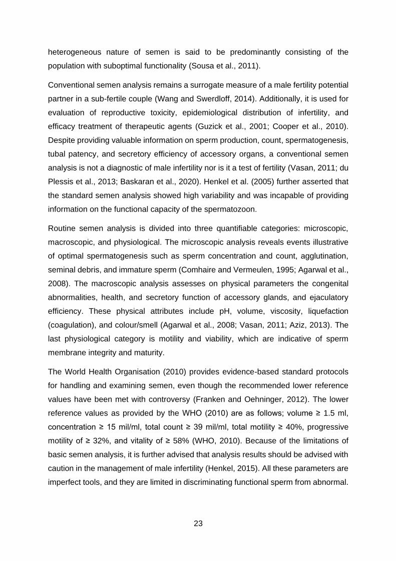
23
heterogeneous nature of semen is said to be predominantly consisting of the
population with suboptimal functionality (Sousa et al., 2011).
Conventional semen analysis remains a surrogate measure of a male fertility potential
partner in a sub-fertile couple (Wang and Swerdloff, 2014). Additionally, it is used for
evaluation of reproductive toxicity, epidemiological distribution of infertility, and
efficacy treatment of therapeutic agents (Guzick et al., 2001; Cooper et al., 2010).
Despite providing valuable information on sperm production, count, spermatogenesis,
tubal patency, and secretory efficiency of accessory organs, a conventional semen
analysis is not a diagnostic of male infertility nor is it a test of fertility (Vasan, 2011; du
Plessis et al., 2013; Baskaran et al., 2020). Henkel et al. (2005) further asserted that
the standard semen analysis showed high variability and was incapable of providing
information on the functional capacity of the spermatozoon.
Routine semen analysis is divided into three quantifiable categories: microscopic,
macroscopic, and physiological. The microscopic analysis reveals events illustrative
of optimal spermatogenesis such as sperm concentration and count, agglutination,
seminal debris, and immature sperm (Comhaire and Vermeulen, 1995; Agarwal et al.,
2008). The macroscopic analysis assesses on physical parameters the congenital
abnormalities, health, and secretory function of accessory glands, and ejaculatory
efficiency. These physical attributes include pH, volume, viscosity, liquefaction
(coagulation), and colour/smell (Agarwal et al., 2008; Vasan, 2011; Aziz, 2013). The
last physiological category is motility and viability, which are indicative of sperm
membrane integrity and maturity.
The World Health Organisation (2010) provides evidence-based standard protocols
for handling and examining semen, even though the recommended lower reference
values have been met with controversy (Franken and Oehninger, 2012). The lower
reference values as provided by the WHO (2010) are as follows; volume ≥ 1.5 ml,
concentration ≥ 15 mil/ml, total count ≥ 39 mil/ml, total motility ≥ 40%, progressive
motility of ≥ 32%, and vitality of ≥ 58% (WHO, 2010). Because of the limitations of
basic semen analysis, it is further advised that analysis results should be advised with
caution in the management of male infertility (Henkel, 2015). All these parameters are
imperfect tools, and they are limited in discriminating functional sperm from abnormal.

24
For this reason, it becomes imperative to include advanced functional tests that can
accurately predict men’s fertilising potential.
2.6.1 Motility
Motility is generally accepted as the definitive proxy of male infertility (Natali and Turek,
2011). However, the consideration of motion or motility as a sole index for sperm life
or fertility potential can be erroneous, since not all motile cells are competent for
fertilisation, and not all immotile cells are dead (Van Zyl and Alwelien, 2015). Manual
semen analysis has been widely applied in clinical male work-up settings and
seminology laboratories to determine motility. However, their inability to sufficiently
capture sperm head and flagellum kinematic patterns have rendered the introduction
of an automated, computational system (CASA) a useful necessity (Vasan, 2011).
2.6.1.1 Computer-aided sperm analysis (CASA)
CASA is an automated, high precision tool that measures sperm motility accurately
and objectively for various species (Maree and van der Horst, 2013; van der Horst,
Maree and du Plessis, 2018). As with the use of any new computational technology,
the reliability of CASA can be significantly diminished by technical and operational
faults of the system by the user, by biological variability of samples, poor internal
quality control such as inappropriate pipetting, inconsistent dilutions, or centrifugal
factors (Talarczyk-Desole et al., 2017; Patel et al., 2018). Other than measuring sperm
sub-populations’ motilities based on progression and speed (total, progressive, non-
progressive motility, and immotility), CASA measures percentage hyperactivation and
kinematic characteristics (Alipour et al., 2017). Sperm kinematics include velocities:
curvilinear (VCL), straight-line (VSL), and average path (VAP) velocities (µm. s-1),
head oscillatory/wobble index (WOB), straightness index (STR) and linearity index
(%). Flagellar attributes are: beat cross frequency (Hz), and amplitude of lateral head
displacement (µm) (Larsen et al., 2000; World Health Organization, 2010) (Figure 8).
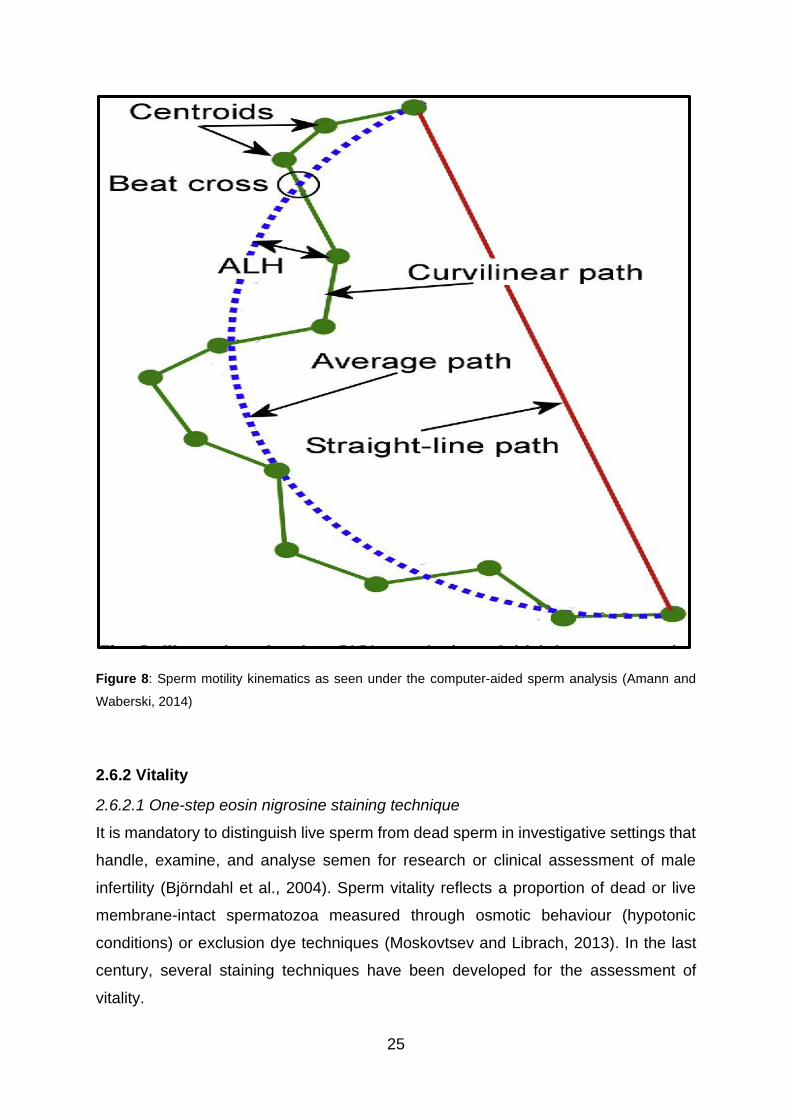
25
Figure 8: Sperm motility kinematics as seen under the computer-aided sperm analysis (Amann and
Waberski, 2014)
2.6.2 Vitality
2.6.2.1 One-step eosin nigrosine staining technique
It is mandatory to distinguish live sperm from dead sperm in investigative settings that
handle, examine, and analyse semen for research or clinical assessment of male
infertility (Björndahl et al., 2004). Sperm vitality reflects a proportion of dead or live
membrane-intact spermatozoa measured through osmotic behaviour (hypotonic
conditions) or exclusion dye techniques (Moskovtsev and Librach, 2013). In the last
century, several staining techniques have been developed for the assessment of
vitality.

26
However, few have been developed to assess incompatibility with human sperm until
the 1990s and the discovery of eosin-nigrosin (Mortimer, 1985; Björndahl et al., 2003).
One-step eosin-nigrosin has proven valuable for the prediction of a ratio of live / dead
cell in the semen sample in andrology.
It works on the principle that metabolically active sperm, with an intact membrane, is
impermeable to eosin stain (Björndahl et al., 2004), while nigrosin dye adjusts the
contrast-background ratio of the sperm heads, thereby increasing visibility under a
bright field microscope. Dead cells absorb the dye and stain deep pink/purple or red,
while live cells appear white (Agarwal et al., 2016). Hence, it had been recommended
by the WHO (2010) for viability evaluation in basic semen analysis.
2.6.3 Mitochondrial membrane potential (MMP) (∆ѱM)
The mitochondrion is usually referred to as the powerhouse of the cell (Opuwari,
2009). Nutrients are oxidised in the mitochondria to produce ATP through oxidative
phosphorylation, which is a series of redox reactions that establishes an
electrochemical gradient (Cottet-Rousselle et al., 2011). Generation of the proton
motive force establishes a mitochondrial membrane potential (Meyers, Bulkeley, and
Foutouhi, 2019). MMP regulates sperm motility and maintains viability (Agnihotri et al.,
2016). MMP is not only sensitive to detecting the electron transport chain (ETC)
efficiency, but it was indicated as a reliable marker of mitochondrial functionality and
as being predictive of sperm fertilising success (Marchetti et al., 2002; Amaral et al.,
2013; Henkel, 2015).
Under pathological conditions, the capacity of mitochondria to generate energy is
disturbed, resulting in a decline in the electrochemical gradient, which is signified by
mitochondrial permeability transition complexes (Shunmugam, 2016). The pores in the
outer membrane of the mitochondria release pro-apoptotic proteins (mostly
cytochrome c) to facilitate a cascade of events that ultimately lead to the demise of the
cell (Bernardi and Di Lisa, 2015). The fluorescent probes for quantifying functional
membrane integrity take advantage of the sensitivity of sperm plasma membrane to
minute changes in the chemiosmotic intramembrane gradients (Sakamuru et al.,
2012).
27
2.6.3.1 Lipophilic cationic dye
Lipophilic cationic fluorescent dyes can readily diffuse into hydrophobic membranes,
and hence, they have the highest binding affinity such that they can distribute equally
within the mitochondria and accumulate (Ehrenberg et al., 1988; Perry et al., 2011).
The DePsipher dye accumulates in membrane-intact mitochondria and fluoresces red,
while in apoptotic cells it cannot aggregate in the mitochondria; it just remains in the
cytoplasm and therefore fluoresces green (Sivandzade et al., 2019). One of the
lipophilic dyes proven to be working on the principle, as mentioned above, is (5’, 6, 6’-
tetrachloro-1, 1’, 3, 3’-tetraethylbenzimiddasolyl-carbocyanine iodide (JC-1) (Marchetti
et al., 2004).
2.6.4 Reactive oxygen species (ROS)
2.6.4.1 ROS generation
Diatomic oxygen is relatively inert, even though two of its valence electrons are
unpaired (Ford, 2004). However, when oxygen-containing molecules get reduced,
their by-products become highly unstable and reactive, as they will remain with the
unpaired electron, creating sequelae of oxidative reactions (Wagner, Cheng, and Ko,
2018). Some of the clinically relevant free radical in the human ejaculate are singlet
anion (1O2) and peroxyl radical (Ford, 2004).
Of the known ROS, O2–• is the most potent intracellularly produced ROS in nearly all
biological systems (Zorov et al., 2014). However, because of its size, superoxide has
limited permeability to sperm lipid membrane, and therefore becomes changed into
hydrogen peroxide (H2O2) either spontaneously or by enzymatic dismutation (Mitchell
et al., 1990; Dunand et al., 2007). Although H2O2 is not precisely a free radical, it is
involved in subsequent ROS synthesis (Aprioku, 2013). H2O2 and superoxide can be
converted to indiscriminately, highly reactive hydroxyl (OH•-) through the iron-
dependent Haber-Weiss reaction (Kehrer, 2000). Hydroxyl anion initiates membrane
damage to spermatozoa by removing electrons from lipid membranes of the sperm.
Spermatozoa, similar to any anaerobic cell, are often confronted with what is
commonly termed the ROS paradox (Agarwal et al., 2003; Tvrdá et al., 2017). To the
same extent that oxygen is indispensable to aerobic life, its intermediates can initiate
or/and facilitate cellular demise (Ogbuewu et al., 2010). Extrinsically, ROS generation
can be triggered or exacerbated by environmental, lifestyle factors (smoking and
28
drinking), where medication, and genetics contribute the “oxidative interface”
(Lavranos et al., 2012; Ray et al., 2012) (Figure 10).
2.6.4.1 Sources of ROS
Reactive oxygen species (ROS) leaks through mitochondrial OSPHOX and glycolytic
systems of ATP production, using nicotinamide adenine dinucleotide phosphate
(NADPH) oxidase (Nascimento et al., 2008). Additionally, immature or defective sperm
cells have excess cytoplasm (which would have been customarily shed during
spermiation) (O’Donnell, 2014) (Figure 10). This excess cytoplasm contains
membrane bound-glucose-6 phosphate dehydrogenase (G-6-DPH) involved in
additional ROS generation through the pentose monophosphate-shunt pathway
(Gomez et al., 1996; Tremellen, 2008). In this pentose phosphate shunt, the
biosynthesis of cytoplasmic 8-docosahexaenoic acid upregulates the availability of
NADPH (Said et al., 2005). The putative NADPH subsequently donates electrons for
the production of spermatozoal ROS at the sperm membrane level (O’Flaherty, 2015).
There is a positive association between abnormally high levels of ROS in
oligoasthenoteratozoospermia males with impaired spermatogenesis (Ollero et al.,
2001; Hosseinzadeh et al., 2013).
Also, sperm is enveloped in polyunsaturated fatty acids (PUFA) rich membrane, which
contains fewer antioxidant capacities, therefore rendering spermatozoa particularly
vulnerable to peroxidative and oxidative damage (Hsieh et al., 2006; Parekattil and
Agarwal, 2012). It is generally noted that ROS production by seminal
polymorphonuclear granulocyte exceeds physiological ROS levels a 1000-fold
(Agarwal, et al., 2014; Wagner et al., 2018). In the presence of genitourinary infections,
cryptorchidism, inflammation, iatrogenic factors, and varicocele, the activation of PMN
leukocytes is often pronounced, with corresponding excess ROS levels (Henkel and
Schill, 2003; Henkel, 2011; Lobascio et al., 2015; Cho et al., 2016; Solomon and
Henkel, 2017) (Figure 10). The balance between pro-oxidants and anti-oxidants is a
margin within which sperm ROS can mediate sperm signal transduction of
hyperactivation, motility, capacitation, acrosome reaction, and sperm-egg fusion
(Aitken et al., 1989; de Lamirande and Gagnon, 1995; Kothari et al., 2010) (Figure 10).
On the other hand, overproduction of sperm ROS results in an imbalance between the

29
oxidants and antioxidant defences in favour of the former; this phenomenon is termed
oxidative stress (OS) (Agarwal et al., 2019). OS is a leading proven cause of male
infertility (Agarwal et al., 2019). One of the hallmark clinical manifestations of the
testicular OS is sperm DNA damage (Iommiello et al., 2015) (Figure 9).
Figure 9: The physiological and pathophysiological roles of ROS in male infertility (Sharma and
Agarwal, 2020).
2.6.4.2 Antioxidants
Spermatozoa possess an intrinsic free radical scavenging activity, which is limited by
the reduced cytoplasmic volume, rendering them particularly vulnerable to oxidative
damage (Henkel et al., 2018). Additionally, sperm cells are bathed in a seminal milieu
endowed with enzymatic, non-enzymatic antioxidant systems to combat excessive
ROS (Pasqualotto et al., 2004; Papas et al., 2019). Nutritional antioxidants have been
prescribed for pathologies, in which oxidative stress was implicated as a cause or
modulator, including male infertility (de Young et al., 2004; Henkel et al., 2019).
Enzymatic antioxidants found in the seminal plasma are superoxide dismutase (SOD),

30
catalase (CAT), glutathione peroxidase (GPx), and glutathione (GSH) (Tavilani et al.,
2008; Tomar et al., 2017). Non-enzymatic antioxidants such as vitamins C, E, B12,
carotenoids, carnitine, bioflavonoids bilirubin, albumin, uric acid, N-cysteine, and folate
are exogenously obtained from the diet (Armstrong et al., 1998; Ferreira, 2012;
Aprioku, 2013; Adewoyin et al., 2017). Trace elements such as zinc, selenium, and
magnesium also have oxidative protectants (Palani and Alshatteri, 2017).
2.6.4.3 ROS measurement using a fluorescent probe
There exists a thin margin, upon which ROS are physiological and pathological for
sperm function, further making it necessary to quantify ROS levels essential for normal
function (Tomar et al., 2017). Therefore, a variety of methods of detection include
chemiluminescent or fluorescent assays that have been developed for direct
measurement of ROS production, and which rely on the extent to which superoxide
will oxidise the functional groups of reagents in the assay (Gosalvez et al., 2017).
Similarly, the measurement of sperm ROS in this study used dihydroethidium (DHE).
DHE is a fluorescing probe that fluoresces red when oxidised by O2-. The intercalation
of sperm nuclear DNA with the dye is marked by hydroxylation of DHE into a red or
orange 2-hydroxyethidium substance, which fluoresces at excitation and emission
wavelengths of 535 nm and 635 nm, respectively (Aitken et al., 2013; Held, 2015).
2.6.5 DNA fragmentation
Intrinsic sperm DNA damage results from incorrect chromatin packaging, post-
ejaculatory defective apoptosis, endogenous nuclease activity, or oxidative damage
(Agarwal et al., 2014; Mupfiga et al., 2013; Zini and Libman, 2006). These causes can
be further aggravated by environmental pollutants, radiation, and chemotherapy
(Henkel, 2017). Once membrane peroxidation (by excess ROS) has been initiated, its
intermediates such as malondialdehyde proceed to the sperm mid-piece to induce and
propagate mitochondrial dysfunction (Hsieh et al., 2006). The cascade, which further
exacerbates electron leakage, and one of the free radicals that actively participates in
the early DNA base attack is hydrogen peroxide (Aitken and Koppers, 2011).
Initiation of the DNA backbone attack through oxidising nucleotide bases, forming
adducts such as 8-hydroxy-2,’-deoxyguanosine (8OHdg), is close to or follows
apoptosis (Aitken et al., 2011). The formation of adducts will continue to form single
and double strands in sperm nuclear DNA backbone, and ultimately, chromatin cross-

31
linking (Iommiello et al., 2015). Higher levels of DNA damage have consistently been
found in men with abnormal seminology, when compared to healthy normozoospermic
men (Wang, 2003; Zhang et al., 2008). More concerning is the coexistence of “normal”
with pathology, as some of the normozoospermic men are still experiencing infertility,
and their unidentifiable factors are often termed idiopathic (Hamada et al., 2012;
Henkel, 2017).
Sperm DNA fragmentation has also been negatively correlated with the percentage of
basic semen parameters, viability, quality of the embryo, pregnancy loss, disrupted
early development, and therefore neonatal morbidities in both naturally conceived and
medically assisted reproduction offsprings (Marchetti et al., 2002; Henkel and
Franken, 2011; Sivanarayana et al., 2012; Samplaski et al., 2015; Henkel, 2017).
Quantifying DNA fragmentation is further necessitated by the extensive practice of
assisted reproductive techniques, in which the natural selection of genetically intact
spermatozoa is bypassed, leading to the inadvertent use of oxidatively damaged DNA
sperm (Aitken and Koppers, 2011).
2.6.5.1 Sperm DNA fragmentation quantification
DNA fragmentation traditionally does not form part of the routine semen analysis in
the andrological diagnostic setup (Cissen et al., 2016). Despite being used less
frequently as an indirect tool for quantifying male infertility, sperm DNA fragmentation
is gaining considerable attention (Lim et al., 2013). Discrepancies in fertilising
tendencies between normozoospermic and oligoasthenoteratozoospermia patients in
infertility treatment have led to the appreciation and use of sperm DNA and chromatin
integrity assessment techniques (Belloc et al., 2014).
Numerous techniques have been developed for the assessment of DNA fragmentation
such as sperm chromatin dispersion (SCD) and sperm chromatin structure assay
(SCSA), which detect strand breaks quantitatively (continuous basis)(Larson-Cook et
al., 2003; Henkel, 2017). Terminal deoxynucleotidyl transferase-mediated nick ending
labelling (TUNEL), on the other hand, is emerging as the golden tool in the diagnosis
of unexplained male infertility, as it qualitatively detects single and double breaks
(exact ratio of fragmented to non-fragmented sperm DNA), despite a lack of
standardisation for human sperm (Sharma and Agarwal, 2011a; Cho and Agarwal,

32
2017; Ribeiro et al., 2017). TUNEL is based on the enzymatic ability of terminal
transferase (TdT) to detect fluoresceinated dUTP single or double-stranded DNA nicks
by labelling the resultant free hydroxyl groups (3-OH) (McKenna et al., 1998; Sharma
et al., 2016). In these techniques, analysis of DNA fragmented sperm can be done by
flow cytometry and fluorescence microscopy (Henkel and Franken, 2011).
2.6.6 Capacitation (CP) and acrosome reaction (AR)
Capacitation (CP) and acrosomal reaction (AR) are indispensable steps in the
fertilisation of most mammalian species (de Lamirande, Leclerc, and Gagnon, 1997).
CP is a collective term for the physiological and biochemical modifications the sperm
undergo as they transit the female reproductive tract to render them competent for
fertilisation (O’Flaherty, de Lamirande, and Gagnon, 2006). The oviductal luminal
milieu primes sperm for cervical mucus penetration and fusion of sperm with zona
pellucida, which in turn activates sperm acrosome for penetration of oocyte vestments
(de Lamirande et al., 1997; Badawy et al., 2006). Signalling pathways regulating
sperm capacitation and hyperactivation have few similarities, such as all having Ca2+
dependency (Visconti et al., 2011; Beltrán et al., 2016). CP is a time-dependent event
that depends on lipid composition of the sperm plasma membrane, the redistribution
and reorganisation of membrane proteins, ion influxes, and the amount of ROS
present (de Lamirande et al., 1997).
An efflux of cholesterol initiates CP in the presence of albumin, which increases
membrane fluidity (Ickowicz et al., 2012) (Figure 10). Increase in membrane
permeability allows the opening of calcium ion channels (CatSper) and sodium
bicarbonate cotransporter protein, which facilitate the influx of Ca2+ and bicarbonate
(HCO3-) ions, leading to a hyperpolarised sperm membrane (Ickowicz et al., 2012;
Leemans et al., 2019) (Figure 10). The presence of HCO3- increases the activity of
soluble adenylyl cyclase, which in turn increases the biosynthesis and the activity of a
secondary messenger cyclic adenosine monophosphate (cAMP) (Rivlin et al., 2004;
Bailey, 2010) (Figure 10). The formation of cAMP is accompanied by the formation
and involvement of ROS, particularly superoxide (de Lamirande and Gagnon, 1995;
Jin and Yang, 2016) (Figure 10). Cyclic monophosphate activation induces activation
of a protein kinase A (PKA) subunit, which coincides with a whiplash vigorous flagella
movement called hyperactivation (Yanagimachi, 1994; Ickowicz et al., 2012). After
cAMP-PKA activation, this either inhibits phosphatases or the stimulation of

33
phosphorylation of kinases (Visconti et al., 1995; Tulsiani and Abou-Haila, 2012;
O’Flaherty, 2015) (Figure 10). It has been further demonstrated that ROS also plays
a signalling role in downstream phosphorylation of kinases, leading up to CP (Leclerc
et al., 1997; Rivlin et al., 2004; O’Flaherty et al., 2006) (Figure 10).
Figure 10: Molecular events involved in the regulation of mammalian sperm capacitation (Jin and Yang,
2016).
The only spermatozoon that underwent capacitation can recognise oocyte signals and
undergo AR. An acrosome reaction is an exocytotic event, in which the apical vesicle
in the sperm head fuses with the inner plasma membrane to empty acrosomal
contents that digest the zona pellucida of the oocyte to facilitate entry into egg

34
vestments (Yanagimachi, 1994; Fraser, 1998). Hyaluronidase, phospholipase A2 and
acrosin are hydrolytic acrosomal contents released first to penetrate the cumulus
oophorus, and secondly to disperse the zona pellucida (ZP) of the egg (Tulsiani et al.,
1998; C. Esteves and Verza Jr., 2011). Acrosin is a serine protease that not only
facilitates protein activation, but clears off steric charges that may hinder egg-sperm
recognition and binding at the oolemma (Takano et al., 1993; Cuasnicú et al., 2016).
One of the four ZP glycosylated proteins with sperm binding site is ZP3. It is accepted
that the binding of sperm to ZP3 glycoprotein through Gi-receptors triggers a series of
events leading up to AR (Fraser, 1998; Tulsiani and Abou-Haila, 2012). Similar to CP
and hyperactivation, AR is a ROS-regulated AMP-PKA-dependent signalling pathway
that leads to the opening of CatSper ion channels increasing calcium (Fraser, 1998;
Sun et al., 2017) (Figure 11). An increase in extracellular Ca2+ stimulates an increase
in intracellular Ca2+ concentration that is crucial for phosphorylation of protein kinase-
C (PKC) subunit (Tateno et al., 2013). The phosphorylation of PKC increases
membrane fluidity, which in turn mediates phosphorylation of tyrosine kinase (PTK)
and eventually AR (Ickowicz et al., 2012) (Figure 11).
Figure 11: Illustration of molecular events regulating acrosome reaction (Dutta et al., 2020).

35
2.6.6.1 Quantification of CP and AR
Molecular interactions regulating CP and AR suggest that they are highly coordinated
events with complex signalling pathways. Understanding mechanisms that augment
CP and AR precedes the development of fertilisation models in vivo and in vitro
(Esteves and Verza, 2011). Therefore, direct or indirect bio-assaying of the two
processes as biomarkers of male infertility is essential (Lessard et al., 2011).
Fluorescence labels commonly used in andrology settings are immune-monoclonal
localising probes such as antibodies (Grasa et al., 2008), glycan-binding lectins (Xin
et al., 2016), and membrane Ca2+-sensitive stains (Gillan et al., 2005). The unbiased
labelling using fluorescent dyes in both instances depends on permeability properties
of the spermatozoon surface characteristics (Fierro et al., 2013).
Chlortetracycline (CTC) antibiotic has shown to have the highest affinity for bivalent
cations such as Ca2+/Mg2+, giving an intense fluorescent emission in living, fixed, or
unfixed non-permeabilised cells (Green and Watson, 2001; Pietrobon et al., 2001;
Silva and Gadella, 2006). For adjustment of the contrast background ratio of live,
membrane-responsive, and acrosome-reacted segments, supravital stains such as
Hoechst 33258 or propidium iodide are recommended (Pintado et al., 2000).
Some of the widely accepted methods of quantifying CP and acrosomal patterns are
transmission electron microscopy (TEM) (Pesch and Bergmann, 2006), phase-
contrast, differential-inferential contrast dyes (Topper et al., 1999), and fluorescent
probes (Lee et al., 1987; Fierro et al., 2013). Limitations to analyse only small
populations, different culture media of sperm, and tiny acrosomal regions specified
against conventional phase-contrast dyes have shifted preference to fluorescence
microscopy for evaluation of CP and AR in reproductive biology (Cross and Meizel,
1989; Esteves et al., 2007).
2.7 Infertility
The World Health Organisation passed an operational definition of health as – “the
complete state of physical, mental, and social well-being, and not merely the absence
of infirmity” in 1948 (Huber et al., 2011). From a reproductive perspective, health
includes full utilisation of sexual, reproductive systems, processes, and functions
throughout life. In 1994, at an International Conference of Population and
Development, the WHO declared that reproductive health implies that people can have

36
a satisfying and safe sex life, and that they can reproduce and have the freedom to
decide if, when, and how often to do so (Jansen, 2007). For an involuntarily childless
couple, this state of health is not achievable.
For this reason, the International Committee for Monitoring Assisted Reproductive
Technology and the WHO define infertility as the disease of the reproductive system
that is characterised by the failure to achieve clinical pregnancy within one year or
more of regular unprotected sex in a non-contracepting couple (Zegers-Hochschild et
al., 2009). Infertility affects nearly 15 - 20% of couples globally (Agarwal et al., 2015).
Infertility − just as any other disease − causes its sufferers a great deal of emotional,
physical, social, and financial burden, and mental anguish (Dyer et al., 2004;
Chachamovich et al., 2010; Dyer and Patel, 2012). It is worth noting that the
consequences of infertility affect women more significantly than men, especially in
pronatalist and patriarchal communities (Greil et al., 2011).
Statistically, nearly 85% of couples will fall pregnant within one year of attempting to
conceive, and the probability increases to 95% within two years. For couples trying for
three or more years to conceive without success, their chances of a successful
conception fall below 25% (Eldib and Tashani, 2018). Depending on the length of
unprotected intercourse, the female’s age and parity, seminal profile, and frequency
of intercourse, the couple’s infertility status can be classified as either primary or
secondary (Anwar and Anwar, 2016).
Primary infertility is diagnosed in cases where nulliparous women have not conceived,
but have been in a non-contracepting union for at least five years, while secondary
infertility refers to the absence of live birth by women who have previously fallen
pregnant, despite the outcome of their previous pregnancy (Mascarenhas et al., 2012;
Anwar and Anwar, 2016). Data from Europe, Asia, the Middle East, and Africa suggest
that primary infertility is less common than secondary infertility (Mascarenhas et al.,
2012; Inhorn and Patrizio, 2015). Sub-Saharan African countries record the unusually
high prevalence of infertility of about 30% that coexists with high fertility rates, a
phenomenon that is termed “barren amid plenty” (Cui, 2010; Inhorn and Patrizio,
2015). Persistently high infertility rates observed in the region stretching from central
Africa to the southern parts of Africa were collectively referred to as the infertility belt
(Collet et al., 1988; Inhorn, 2003; Ombelet and Onofre, 2019).

37
Most of infertility inferential data derives from gynaecological investigations, mainly
because women are typically blamed for the inability to have children in marriage
(Mohammed-Durosinlorun et al., 2019). In many normative African countries, social
status, security, and identity of women is determined or strongly associated with the
parity (Dhont et al., 2010; Mumtaz et al., 2013). To apply further pressure and blame
on women, in the event of childlessness, men are often protected from the public
scrutiny either by remarriage or having a close family member impregnating the wife
(Mumtaz et al., 2013; Agarwal et al., 2015). Therefore, the socio-cultural values of
infertility have shaped the differences in infertility treatment-seeking behaviour.
The exclusion of men from infertility discourse in some regions does not rule out their
contribution. Males independently contribute 20-40% of global cases of infertility, a
percentage that is similar to that found among females, while some causes are shared
between the two partners (Agarwal et al., 2015; Kumar and Singh, 2015) (Figure 12).
About 90% of all male infertility cases are attributed to abnormal semen parameters,
specifically total count and concentration. Infertility can be complete or partial, while
the latter manifestation is termed subfertility (Zegers-Hochschild et al., 2009). The
causes of infertility can be extrinsic such as environmental pollutants, diet, smoking
and drinking, medication, and including but not limited to recreational drugs (Braga et
al., 2012; Xiao et al., 2014; Anwar and Anwar, 2016; Sharma, 2017). Intrinsic factors
can be congenital, medical or result from cryptorchidism, varicocele, genetic disorders,
endocrinopathy, infectious diseases, obstruction, metabolic diseases, and
immunological factors (Anwar and Anwar, 2016; Opuwari et al., 2017).
Subfertility or infertility can be caused by the absence of sperm (azoospermia), low
sperm concentration (oligozoospermia), low sperm motility (asthenozoospermia),
abnormal sperm morphology (teratozoospermia), and reduced sperm vitality
(necrozoospermia), or a combination of the first three abnormalities
(oligoasthenoteratozoospermia) (Guzick et al., 2001; Agarwal and Sekhon, 2011;
Aziz, 2013; Sharma, 2017). Deterioration of semen quality can originate or be
influenced by pre-testicular, testicular, and post testicular factors (Iwamoto, Nozawa
and Yoshiike, 2007).
Secretions from vesicles, prostate, and epididymis are indirectly but strongly linked to
unknown causes of infertility (Sharma, 2017). When these factors merge to complicate

38
the aetiology and pathogenesis of infertility, the type of infertility is termed idiopathic
(Hamada et al., 2012). Idiopathic infertility is diagnosed when the cause is
unidentifiable; this diagnosis has been reported to affect 30% of couples globally
(Sharma, 2017). At the core of idiopathic cases is an excess of ROS production.
Oxidative stress has been found to play an independent role in about 30–80% of
unexplained causes of infertility (Agarwal et al., 2019).
Idiopathy in male infertility evaluation makes therapy even more difficult. Depending
on the cause, treatment of infertility can be non-surgical or surgical (Hall, 2014).
Surgical methods may include orchidopexy, varicocelectomy, and testicular sperm
aspiration (Hanerhoff and Welliver, 2014; Barekat et al., 2016). These interventions
correct anatomical deformities impeding sperm production, transit, or functionality
(Lok, Ledger, and Li, 2003; Goldstein and Schlegel, 2013). Non-surgical interventions
include hormonal therapy such exogenous human chorionic gonadotropin (hCG),
FSH, testosterone, dopamine agonist, oral corticoids, phosphodiesterase inhibitors,
and antioxidants (Chehval and Mehan, 1979; Cocuzza and Agarwal, 2007; Henkel,
Sandhu, and Agarwal, 2019). Empiric therapies can be used in conjunction with
assisted reproductive technologies (ART) (Liu, 2003). The International Committee for
Monitoring Assisted Reproductive Techniques (ICMART) (2009) and the WHO defined
ARTs as all treatments or procedures that include in vitro handling of human oocyte
and sperm or embryo to establish a pregnancy. These techniques include in vitro
fertilisation (IVF), intracytoplasmic sperm injection (ICSI), gamete intrafallopian
transfer (GIFT), embryo transfer, zygote intrafallopian transfer, embryo
cryopreservation, oocyte and embryo donation, and gestational surrogacy (Zegers-
Hochschild et al., 2009). These techniques have made a remarkable contribution to
infertility management. However, they are not readily accessible, accepted, or
affordable for and by the majority of people who wish to conceive / have a child. For
this reason, alternatives have been sought.

39
Figure 12: Demographic illustration of global male factor infertility (Agarwal et al.,
2015)
2.8 Herbal medicine
Traditional medicine (TM) refers to the sum-total of knowledge, skills, and practices
based on beliefs, theories, and experiences indigenous to different cultures, whether
explicable or not, and used for the maintenance of health as well as in the prevention,
diagnosis, improvement, or treatment of mental or physical illnesses (Mothibe and
Sibanda, 2019). Traditional medicines are usually preferred by people believing in their
efficacy for being holistic in that they take a person’s environment, and mind-body
interactions into consideration as opposed to their orthodox medicinal counterparts
that are mainly palliative (Roberti di Sarsina, 2007). Central to the holistic methods of
healing are herbs (Wachtel-Galor and Benzie, 2011). Herbal medicines may be herbal
materials, herbal preparations, or finished herbal products (Mothibe and Sibanda,
2019).
The latter form of herbals provides the raw material for the mass synthesis of
pharmaceutical drugs. These conventional / Western medicines have revolutionised
the health care system in most parts of the world (Wachtel-Galor and Benzie, 2011).
Conventional medicines remain readily recognised as the official form of health care,
because they are scientifically proven (Hardy, 2008), while the preference for TM

40
among a growing number of people across the globe is mostly based on personal
positive experience, affordability and accessibility. The evidence-based efficacy of
allopathic medicine is most effective in alleviating symptoms and providing a response
in emergencies (More, 2016). Allopathic medicine has made breakthroughs in
infectious diseases, vaccines, surgery, imaging, organ transplanting, genetics, and
proteomics, and sophisticated disease detection technologies, to name a few
(Weatherall et al., 2006; Barry et al., 2009; van Rooyen et al., 2015). As the population
numbers grow, pathology progresses, and the disease burden overwhelms even the
most capacitated health care systems in the world (Singh, 2010). When conventional
medicine is insufficient in meeting the rising public health care needs, natural remedies
can compensate effectively. Integration of complementary medicine into day-to-day
health care provision has therefore gained considerable attention (Bodeker and
Kronenberg, 2002; Lampiao et al., 2019).
The inclusion of herbal therapies in everyday healthcare has seen an escalation in
trade and export, in a regulatory framework, and research activities geared towards
their use (Figure 13). As a result, the use of herbal medicines is now a global
phenomenon with nearly 4 billion of the world population (making 80%) relying on them
for their primary health care needs (Ekor, 2014). Even progressive nations such as
selected parts of Europe (van Andel and Carvalheiro, 2013), the US (Rashrash et al,
2017), and China (McQuade et al., 2012) use traditional natural medicine. The
dependence on ethnobotanical medicine increases proportionately with its
affordability, accessibility, proximity, sociocultural influences, availability, and lack of
possible alternatives in middle- and low-income countries (Abdullahi, 2011; Antwi-
Baffour, 2014; Semenya and Potgieter, 2014; Gyasi et al., 2016). In Africa, about 90%
of the Ethiopian population primarily uses herbal medicines, and traditional practices
to diagnose and treat ailments (Abay, 2009). South Africa reports a similarly high
support of herbal medicines, where 80% of South Africans consult a traditional
practitioner for the restoration of their well-being before resorting to biomedical
medicines (van Rooyen et al., 2015). In 2007, trade in traditional medicines yielded
nearly R2.9 billion a year, contributing to 5.6% of the National Health budget (Mander
et al., 2007). Additionally, in 2014, it was documented that nearly 35,000–70,000 of
plants were harvested for medicinal use (Xego et al., 2016). It has to be noted that the
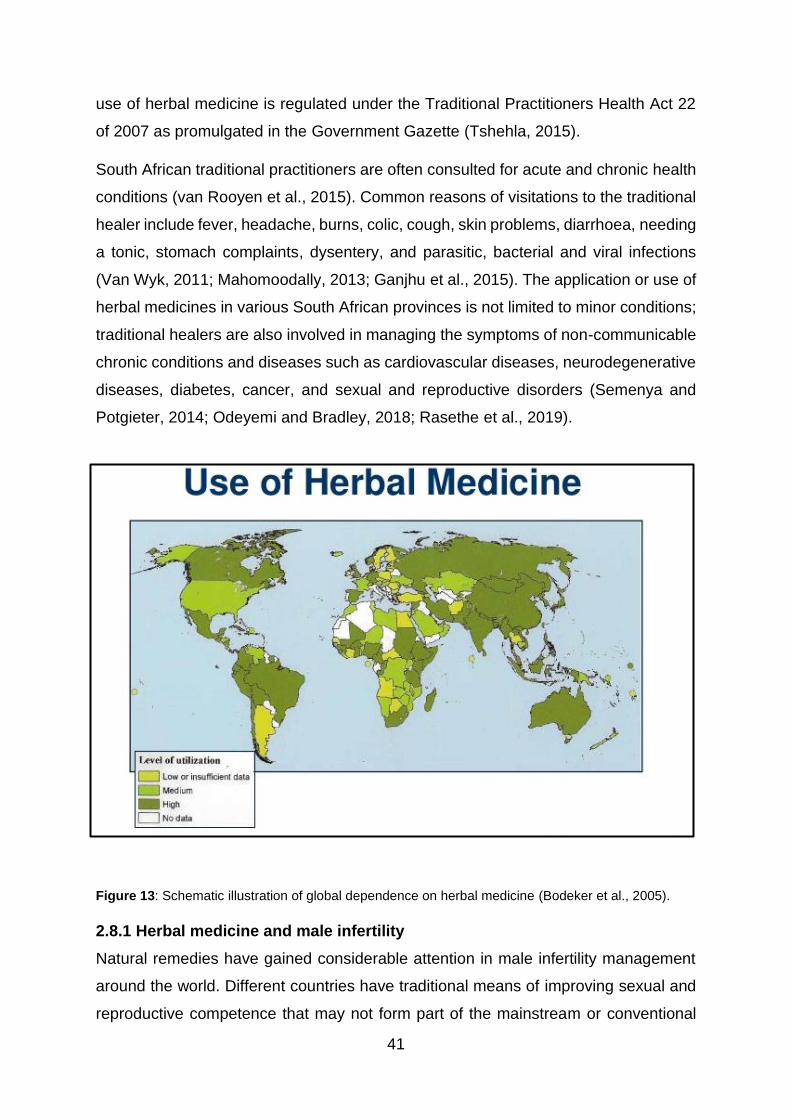
41
use of herbal medicine is regulated under the Traditional Practitioners Health Act 22
of 2007 as promulgated in the Government Gazette (Tshehla, 2015).
South African traditional practitioners are often consulted for acute and chronic health
conditions (van Rooyen et al., 2015). Common reasons of visitations to the traditional
healer include fever, headache, burns, colic, cough, skin problems, diarrhoea, needing
a tonic, stomach complaints, dysentery, and parasitic, bacterial and viral infections
(Van Wyk, 2011; Mahomoodally, 2013; Ganjhu et al., 2015). The application or use of
herbal medicines in various South African provinces is not limited to minor conditions;
traditional healers are also involved in managing the symptoms of non-communicable
chronic conditions and diseases such as cardiovascular diseases, neurodegenerative
diseases, diabetes, cancer, and sexual and reproductive disorders (Semenya and
Potgieter, 2014; Odeyemi and Bradley, 2018; Rasethe et al., 2019).
Figure 13: Schematic illustration of global dependence on herbal medicine (Bodeker et al., 2005).
2.8.1 Herbal medicine and male infertility
Natural remedies have gained considerable attention in male infertility management
around the world. Different countries have traditional means of improving sexual and
reproductive competence that may not form part of the mainstream or conventional
42
modern-day medical standards. These natural modalities are indicated for infertile
men presenting with impotence, hormonal deficiencies, impaired sexual drive,
behaviour, and performance, and poor semen (Yao and Mills, 2016). For instance, in
India, the Ayurvedic system is one of the oldest forms of using holistic medicine in the
world (Chauhan et al., 2014). In terms of male infertility, there exist Ayurvedic
Vajikarana (aphrodisiacs) herbs such as Withania somnifera (Ashwagandha or the
Indian ginseng), Asparagus racemosus (Shatavari), Cannabis sativa (marijuana), and
Mucuna pruriens (velvet bean), which have been used for the enhancement of
ejaculatory function, libido, spermatogenesis, and semen quality (Whan et al., 2006;
M. Ahmad et al., 2008; Thakuret al., 2009; Dutta and Sengupta, 2018). Traditional
Chinese Medicine (TCM) has also proven to be useful in augmenting hypothalamic
pituitary testicular function, testosterone production, boosting of seminal antioxidants,
and semen parameters in men with idiopathic infertility (Chen et al., 1999; Liu et al.,
2004; McQuade et al., 2012; Zhou et al., 2019). Most herbs are found in more than
one natural medicine system.
Extracts of Lycium barbarum (matrimony vine / wolfberry) has demonstrated pro-
fertility effects in both animals and humans (Luo et al., 2006; Wang et al., 2018). Some
of the TCM herbs proven to ameliorate male infertility are Cuscuta chinensis (dodder)
(Jo et al., 2015), Epimedium brevicornum Maxim (horny goat weed) (Abdelaziz et al.,
2020) and Panax ginseng (Asian ginseng) (Salvati et al., 1996).
The African natural medical system comprises approximately 50,000 plant species,
25% of which are readily accessible for ethno-medicinal use, especially in sub-
Saharan countries (Iwu, 2014). Because of the limited health care amenities in Africa
for the detection and treatment of male infertility, reproductive issues are the second
most common health care burden (Gerrits and Shaw, 2010; Idu et al., 2016. Common
sexual reproductive complaints reported to the traditional healers by males in African
countries include a low sex drive, STDs, erectile and ejaculatory dysfunction, and a
low sperm count (Abdillahi and Van Staden, 2012; Agarwal et al., 2015; Bechoua et
al., 2016).
An ethnomedicinal survey that was conducted in Nigeria revealed several species
such as Aloe vera (aloe), Sesacum indicum (sesame seed), Terminalia catapa (Indian
almond), Allium sativum (garlic), and Piper guineense (Guinea black pepper), but this

43
list was not limited to Afromomum melegeuta (Alligator pepper), where the species
were prescribed and prepared to be used as aphrodisiacs, fertility tonics, and sperm
enhancers (Yakubu and Afolayan, 2009; Singh et al., 2010; Lawal et al., 2013; Idu et
al., 2016). Most of the reports on the native medicines used in African traditional
practices, stated that although they were putatively therapeutic, they still remained to
be scientifically validated for practical use. Moringa oleifera is one of the most
extensively used plants with acclaimed healing properties, most of which had
unverified.
2.9 Moringa oleifera
2.9.1 Physical characterisation
Known colloquially as the horseradish (owing to the root taste), drumstick (due to the
shape of its pods) or Ben tree oil, M. oleifera is a medium-sized deciduous tree that
proliferates to reach a height average of 5-10 m (Fahey, 2005; Shih et al., 2011)
(Figure 15 d). It has a softwood perennial trunk with whitish-grey gummy and corky
barks (Fahey, 2005; Farooq, 2012; Leone et al., 2016). The branches appear delicate
and are widespread (Parrotta, 2014). Its green tripinnately-shaped leaf compounds
hold small leaflets that appear yellowish or white (Amabye and Tadesse, 2016) (Figure
15 a and 15 e). M. oleifera’s mature fruit (pods) is about 50 cm long, carrying 50 ovoid
or triangular seeds that are wrapped in a papery sheath (Vlahov, Chepkwony, and
Ndalut, 2002; Taher et al., 2017; Salama et al., 2020) (Figure 14 a, 14 b, 14 c and 14
f). All parts of the plant are used for nutritional and medicinal purposes.

44
Figure 14: M. oleifera aerial parts: leaves (A), pods or fruit (B and C), tree (D), flowers (E) and seeds
(F). (https://www.freshlymoms.com/blogs/news/moringa-benefits-for-new-mothers).
2.9.2 Distribution
Moringaceae is a monotypic family of order brassicales, comprising 33 species; and
14 of them have wide medicinal, nutritional, industrial, and economical applications,
earning its popularity as “the miracle tree of life” (Arora et al., 2013). M. oleifera is the
most versatile, widely accepted, available, and studied species of the genus Moringa.
M. oleifera is native to sub-Himalayan countries; India, Bangladesh, Pakistan, and
Afghanistan (Verdcourt, 1985). Because of its high adaptability to grow in various
weather conditions, M. oleifera is now distributed and naturalised in various parts of
the world (Farooq, 2012). It grows well in arid, tropical, and subtropical regions of
Central, North and South America, in Nepal, the Philippines, the Pacific Islands, the
Caribbean, and in Africa (Morton, 1991; Coote et al., 1997; Alegbeleye, 2018). In
Africa, Ghana, Malawi, and Ethiopia are the largest producers of M. oleifera, while the
South African farmers have also taken an interest in the plant (Pakade et al., 2013).
45
In South Africa, M. oleifera is grown mostly in Limpopo, Mpumalanga, KwaZulu-Natal,
the Free State, and Gauteng (Mabapa et al., 2017).
2.9.3 Phytochemistry and pharmacological properties
M. oleifera is enriched with essential nutrients, micro and macro elements, and
antioxidant phytochemical constituents. Elemental analyses of M. oleifera indicated
the presence of zinc, magnesium, potassium, calcium, sulphur, iron, selenium,
manganese, and copper, which are integral to the preventative and curative role in
pathogenesis (Kasolo et al., 2010; Valdez-Solana et al., 2015; Dilawar et al., 2018).
Mineral elements are mainly found in the diet, and their importance in cell-signalling,
functioning, and maintenance, while metabolism, detoxification, and oxidative stress-
related pathologies have been extensively demonstrated (Soetan et al., 2010; Siddiqui
et al., 2014). M. oleifera leaves exhibit antidiabetic (Sugunabai et al., 2014), anti-
carcinogenic (Tiloke et al., 2013), antibacterial (Amabye and Tadesse, 2016),
antihypertensive (Acuram et al., 2019), neuroprotective (Sutalangka et al., 2013), anti-
inflammatory (Mittal et al., 2017), and antioxidative properties (Tesfay et al., 2011;
Abdelazem, 2019).
M. oleifera leaves are a source of simple and complex carbohydrates such as glucose,
galactose, arabinose, mannose, raffinose, fructose, sucrose, and xylose, which can
serve as substrates for cellular fuel metabolism (Tesfay et al., 2011; Chen et al., 2017).
Except for methionine, M. oleifera leaves have been found to contain all essential and
non-essential amino acids that play a role in protein biosynthesis in biological systems
(Makkar and Becker, 1996; Sánchez-Machado et al., 2010). Furthermore, leaves of
M. oleifera have variable compositions of polyunsaturated, monounsaturated,
saturated and fatty acids such as oleic, arachidonic, stearic, linolenic and palmitic acid,
to name a few (Castillo-López et al., 2017). M. oleifera’s phytochemicals, therefore,
play a role in enzyme synthesis and metabolism (Leone et al., 2016; Saini, Sivanesan,
and Keum, 2016). The proximate nutritional analysis further confirmed the high fibre
content in M. oleifera leaves (Valdez-Solana et al., 2015).
Vitamins A, B, C, and E have been identified in M. oleifera leaves (Fahey, 2005).
Vitamin A (β-carotene) is crucial for cell proliferation, reproduction, and embryonic
development (Kidmose et al., 2007; Vergara-Jimenez et al., 2017). Vitamin B
complexes found in M. oleifera include biotin, folate, nicotinic acid, thiamine, riboflavin,

46
and pyridoxine (Mbikay, 2012). Vitamin C (ascorbate) in M. oleifera is involved in
antioxidative protection of cell membranes from peroxidative damage through radical
scavenging activity (Fahey, 2005; Sankhyan et al., 2013; Gopalakrishnan et al., 2016;
Vergara-Jimenez et al., 2017). Vitamin E (α-tocopherol) is a chain-breaking non-
enzymatic antioxidant that plays a stabilising role in the redox regulation status of
biological systems through the interference of ROS production (Ajantha et al., 2018).
Vitamin deficiencies are often associated with oxidative stress (Ryan et al., 2010).
Also, alkaloids, tannins, saponins, flavonoids, and phenolic compounds with proven
antioxidative potential have been isolated and characterised from M. oleifera leaves
(Valdez-Solana et al., 2015; Gopalakrishnan et al., 2016). Flavonoids identified in M.
oleifera include quercetin, myricetin, rutin, kaempferol, catechin, isothiocyanates,
(Saini et al., 2016). These metabolites boost M. oleifera’s biological activities such as
steroidogenesis, and antimicrobial, anticlastogenic, antitumor, and antiradical
activities (Leone et al., 2016). Caffeolic, ferulic, chlorogenic and gallic acids are some
of the versatile phenolic acids identified in the M. oleifera leaves, and possess free
radical scavenging ability or activity (Valdez-Solana et al., 2015; Castillo-López et al.,
2017; Lin et al., 2019). The bioavailability of M. oleifera’s bioactive compounds depend
on extraction methods, solvents used, and extract storage.
2.9.4 Previous studies of M. oleifera on mammalian reproductive function other
than sperm functioning
Reproductive development, function and competency, including fertility, are strictly
functions of hypothalamic-pituitary-gonadal (HPG) axis (Dwyer and Quinton, 2019).
Therefore, male infertility may occur at any of the three fertility regulating points;
neurohormonal imbalances, impairment in reproductive organs and glands,
compromised sexual behaviour / activity, and abnormal semen quality and quantity
(Dutta and Sengupta, 2018). As a result, medicinal plants or their extracts exert their
effects directly or indirectly at any of the sites of interaction mentioned above (Nudell
et al., 2002). Assessments of M. oleifera on mammalian male reproductive
enhancement and aphrodisiac activity have yielded affirmative results (Zade et al.,
2013).
47
2.9.4.1 M. oleifera and hypothalamic regulation of mammalian reproduction
The neuromodulatory effects of M. oleifera have been studied in rats (Ray, Hazra, and
Guha, 2003; Ray et al., 2004; Abdel-Rahman Mohamed et al., 2019). Concerning the
regulating effects of the plant on sexual activities, M. oleifera aqueous, alcohol, and
chloroform extracts at concentrations of 100, 200, and 500 mg/kg caused significant
increases in mounting and intromission frequencies, and ejaculation latency with
decreased mounting latency in male albino rats (Zade et al., 2013). In another study,
M. oleifera attenuated sexual dysfunction in male Wistar rats through inhibition of
cleaving enzymes monoamine oxidase type B (MOAB) and phosphodiesterase (PDE-
5), thereby increasing mounting and intromission numbers (Prabsattroo et al., 2012).
Similarly, libido and sperm parameters of Bali bulls increased significantly in the group
supplemented with M. oleifera leaves (Syarifuddin et al., 2017).
2.9.4.2 M. oleifera and pituitary regulation of mammalian reproduction
An in vivo study showed beneficial effects of M. oleifera supplementation on male
rabbits, where serum gonadotropin levels (FSH and LH) were dose-dependently
increased (Nwamarah et al., 2015; Ajuogu et al., 2019). To further indicate the
importance of the variabilities in the M. oleifera aerial parts and their toxicities on FSH,
LH, testosterone biosynthesis and availability, another in vivo study was conducted
(Prabsattroo et al., 2012). Similar increases in gonadotropin and testosterone, and a
seminal counts increase were noted after M. oleifera ingestion. In the same study, M.
oleifera seeds, flowers, and roots showed contradicting adverse effects on fertility
indices of adult male Wistar rats (Ogunsola et al., 2017). A similar study was carried
out to assess hyperglycemia-induced reproductive dysfunction in male Sprague
Dawley after M. oleifera administration, and the findings revealed a significant
decrease in glucose levels, accompanied by pronounced improvement in seminal
parameters and testicular histological appearance (Kamalrudin et al., 2018).
Still, on the pituitary regulation, another remarkable effect of M. oleifera in male fertility
was the significant reduction in serum prolactin levels with increased gonadotropin
levels in male albino rats (Harrison et al., 2016). The latter observation was indicative
that M. oleifera could potentially relieve hyperprolactinemia-induced male infertility.
48
2.9.4.3 M. oleifera and testicular regulation of mammalian reproduction
Studies on testicular effects of M. oleifera revealed that the plant affects
steroidogenesis (mainly testosterone), spermatogenesis, the architecture of
seminiferous tubules and Sertoli cells, and seminal antioxidants activities.
Serum testosterone synthesis and availability were significantly increased in male rats
and rabbits in these studies after treatment with M. oleifera (Ebong et al., 2014;
Prabsattroo et al., 2015; Harrison et al., 2016; Suarni et al., 2019). Testicular weights,
rate of spermatogenesis, density and diameter of seminiferous tubules, volume and
hyperactive Leydig cells were improved in bucks and rats administered with aqueous
leaf extracts of M. oleifera (Cajuday and Pocsidio, 2010; Walaa et al., 2015).
Chemically-induced histopathologies in rat testes were dose-dependently reversed
following M. oleifera administration (Bassey et al., 2013; Sadek, 2014; Bin-Meferij and
El-kott, 2015; Abarikwu et al., 2017). M. oleifera leaves significantly increased seminal
SOD, CAT, and glutathione peroxidase in rats’ testes (Afolabi et al., 2013; Saalu et
al., 2011; Zeng et al., 2019).

49
CHAPTER 3
RESEARCH METHODOLOGY
3.1. Introduction
This chapter outlines the components of the methodology, the study site (herbal
extraction, semen collection, and in vitro experiment), sampling, apparatus utilised,
testing protocols, statistical analysing tools, and ethical considerations observed to
investigate the aim of the study.
3.2 Research Methods
3.2.1 Methods and materials
3.2.1.1 Chemicals and apparatus
Unless otherwise stated, all chemicals and laboratory consumables were purchased
from Sigma Aldrich.
3.3 Study design
This study followed an experimental design, in which the effect of the independent or
causal variable (M. oleifera aqueous leaf extract concentration(s) was / were
manipulated to objectively measure dependent variable(s) or outcomes, referring to
sperm functional parameters). Semen samples were collected from fertile donors
(unproven fertile) and patients (unproven infertile), liquefied for 30 min at 37˚C,
washed off of the seminal plasma using Human Tubular Fluid (HTF) supplemented
with 1% bovine serum albumin (BSA) through centrifugation at 300 x g for 10 min.
3.4 Sampling
3.4.1 Study sites
Fresh M. oleifera leaves were harvested from the University’s experimental farm upon
receipt of permission. Leaves were dried and ground at the School of Agricultural and
Environmental Sciences, Department of Plant Production, Soil Sciences and the
Agricultural Engineering Unit at the University of Limpopo. Extraction of the leaves
was carried out in the Department of Biochemistry, Microbiology and Biotechnology’s
laboratory, University of Limpopo.
The Reproductive Biology unit, Department of Obstetrics and Gynaecology (Figure
15), of the Tygerberg Academic and Vincent Pallotti Hospitals were the site for sperm
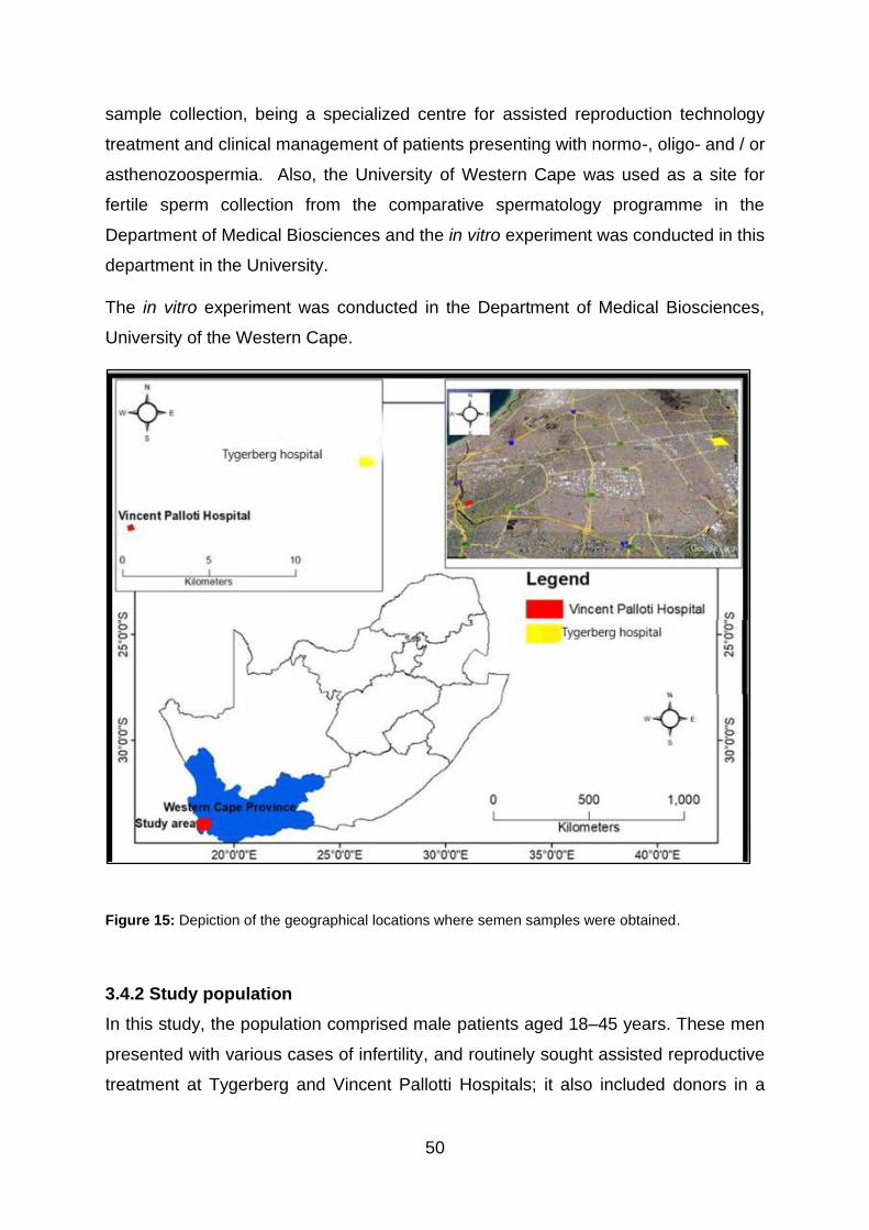
50
sample collection, being a specialized centre for assisted reproduction technology
treatment and clinical management of patients presenting with normo-, oligo- and / or
asthenozoospermia. Also, the University of Western Cape was used as a site for
fertile sperm collection from the comparative spermatology programme in the
Department of Medical Biosciences and the in vitro experiment was conducted in this
department in the University.
The in vitro experiment was conducted in the Department of Medical Biosciences,
University of the Western Cape.
Figure 15: Depiction of the geographical locations where semen samples were obtained.
3.4.2 Study population
In this study, the population comprised male patients aged 18–45 years. These men
presented with various cases of infertility, and routinely sought assisted reproductive
treatment at Tygerberg and Vincent Pallotti Hospitals; it also included donors in a
51
comparative spermatology group in the Medical Bioscience Department of the
University of the Western Cape.
3.4.3 Sampling procedure
Participants were sampled by convenience based on their convenient accessibility,
availability, willingness, and eligibility to the research objectives.
In this study, the population comprised male patients (unproven infertile) aged 18–45
years. These men presented with various cases of infertility, and routinely sought
assisted reproductive treatment at Tygerberg and Vincent Pallotti Hospitals; it also
included donors (unproven fertile) in a comparative spermatology group in the Medical
Bioscience Department of the University of the Western Cape. The donor and patients
groups were subsequently combined and analysed separately as a “combined group”.
The combined group was included and analysed to firstly, determine the cumulative
(overall) effect of the plant extracts on semen parameters. Secondly, the inclusion of
the combined group underpins the phenomenon that normal coexist with pathology or
idiopathy or unexplained infertility. In unexplained cases, normozoospermic men are
still incapable of achieving pregnancy while in idiopathic cases, seminal quality is
compromised without any detectable cause. For these reasons, the introduction of the
“combined group” was aimed at measuring the overall potency of the plant in defective
aspects of “normally functional sperm” such as as excess ROS.
3.4.3.1 Inclusion and exclusion criteria
Inclusion: Normozoospermic men whose seminograms conformed to the World Health
Organization’s cut-off values were included in the study. Men whose samples with
concentration ≤ 15 million sperm / ml (oligozoospermia) or progressive motility < 32%
and / or total motility < 40% (asthenozoospermia) (WHO, 2010) from the patient group
were included in the study but were analysed separately.
Exclusion: Participants with low semen volume (<1 ml) were excluded, irrespective of
the sperm concentration. Azoospermic samples were also not part of the study. Their
samples were discarded as biohazardous waste.

52
3.5 Data collection
3.5.1 Preparation and extraction of M. oleifera leaves
Following their harvest, fresh leaves were allowed to dry for seven days at room
temperature. The dried material was ground on the laboratory bench, using a typical
grinder at the University of Limpopo. For the preparation of extract, 100 g of M. oleifera
leaf powder was decocted in 1 L of distilled water for 20 min at a moderate heat. The
average extract yield recovered was 25% (w/w). The mixture was decanted and
filtered using cheesecloth, followed by centrifugation at 800 x g for 10 min at room
temperature (Shunmugam, 2016). A further filtration was done using Whatman filter
paper (No. 4 and 1), and the filtrate was stored overnight at 4˚C, followed by -80˚C,
before freeze-drying (lyophilisation). The dried extract yielded 24.52% (w/w) on
average and was stored at -20˚C for further experimental use.
3.5.1.1 The rationale for the determination of therapeutic dose
The calculations for the concentrations were based on the previous assumption that
an average healthy man weighs approximately 80 kg (Erasmus et al., 2012). Type 2
diabetes mellitus treatment-naïve patients in a prospective randomised placebo study
were administered 500 mg of M. oleifera, encapsulated into tablets, for two weeks
(Taweerutchana et al., 2017). The therapeutic dose from this study was inferred
because of lack of congruent reporting on the prescription of M. oleifera leaves to
males by practitioners of traditional medicine. Based on the preceding data, the
following calculation was used to determine the therapeutic dose:
Since 1 kg = 1 L = 1000 ml
Therefore, conversion of 80 kg (weight of an average man) to in vitro dilutions gives:
80 kg = 80,000 ml (in vivo)
𝑋 (therapeutic concentration) =(estimate traditional dosage)
(average weight of a man in ml)
𝑋 (therapeutic concentration) =500 mg
80 000 ml
X =1 mg
160 ml
X = 6.25 ×10⁻³mg
ml
53
X = 6.25 µg/ml
A stock solution containing 62.5 mg/ml of M. oleifera in HTF-BSA was prepared, and
serial dilutions were performed to obtain final working concentrations of 0, 0.625, 6.25,
62.5, and 625 µg/ml of M. oleifera.
3.5.1 Experimental procedures
The in vitro experiment was conducted in the Department of Medical Biosciences,
University of the Western Cape.
3.5.2 Preparation of culture medium and semen
3.5.2.1 Preparation of Human Tubular Fluid (HTF)
The HTF medium was formulated based on the WHO (2010) prescriptions.
Accordingly, HTF-BSA was prepared from 5.931 g NaCl, 0.35 g KCl, 0.05 g
MgSO4.7H2O, 0.05 g KH2PO4, 2.1 g NaHCO3, 0.5 g D-glucose, 0.036 g sodium
pyruvate, 0.3 g CaCl2.2H2O, 4.0 g sodium DL-lactate (60% (v/v), 10 µg phenol red, and
20 mmol/l HEPES in distilled water. The medium was prepared under aseptic
conditions. The human tubular fluid was supplemented with 1% of bovine serum
albumin (BSA).
3.5.2.2 Determination of HTF-BSA volume
The amount of HTF-BSA and used to wash the semen after liquefaction was
determined from baseline semen concentration and volume, the number of
parameters tested, and the following formula was used:
7.5 𝑚𝑖𝑙𝑙𝑖𝑜𝑛 /𝑚𝑙
𝑆𝑝𝑒𝑟𝑚 𝑐𝑜𝑛𝑐𝑒𝑛𝑡𝑟𝑎𝑡𝑖𝑜𝑛 (𝑚𝑖𝑙𝑙𝑖𝑜𝑛
𝑚𝑙)
𝑥 𝑁𝑜 𝑜𝑓 𝑝𝑎𝑟𝑎𝑚𝑒𝑡𝑒𝑟𝑠 𝑡𝑒𝑠𝑡𝑒𝑑
= 𝑆𝑒𝑚𝑒𝑛 𝑣𝑜𝑙𝑢𝑚𝑒 (𝑚𝑙) 𝑥 𝑁𝑜 𝑜𝑓 𝑝𝑎𝑟𝑎𝑚𝑒𝑡𝑒𝑟𝑠 𝑡𝑒𝑠𝑡𝑒𝑑
= 𝑉𝑜𝑙𝑢𝑚𝑒 𝑜𝑓 𝐻𝑇𝐹 − 𝐵𝑆𝐴 (𝑚𝑙)

54
3.5.2.3 Source and preparation of semen
Following the receipt of informed consent by participants (Appendix 4, 5, and 6),
semen samples were collected in sterile vials from 70 participants by masturbation
after 3-5 days of abstinence. Samples were incubated at 37°C for 30 min for
liquefaction upon 1 hr of acquisition. After that, 2 µL of liquefied semen was placed on
pre-warmed Leja slides® (Delfran Pharmaceuticals, Cape Town) and Sperm Class
Analyzer® (SCA, version 6.4: Microptic Barcelona, Spain) to determine sperm
concentration. Simultaneously, the time it too semen to fill the chamber was recorded
using a stopwatch as a measure of viscosity (sec).
The motile sperm was recovered and reconstituted in human tubular fluid,
supplemented with 1% of BSA (1:5), centrifuged at 300 x g for 10 min, and the pellet
was re-suspended in a fresh HTF-HSA. Subsequently, sperm suspension was
exposed to increasing concentrations of aqueous extract of M. oleifera (0, 0.625, 6.25,
62.5, and 625 µg/ml) for 1 hr at 37oC, and various sperm parameters were analysed
as indicated in Figure 16.
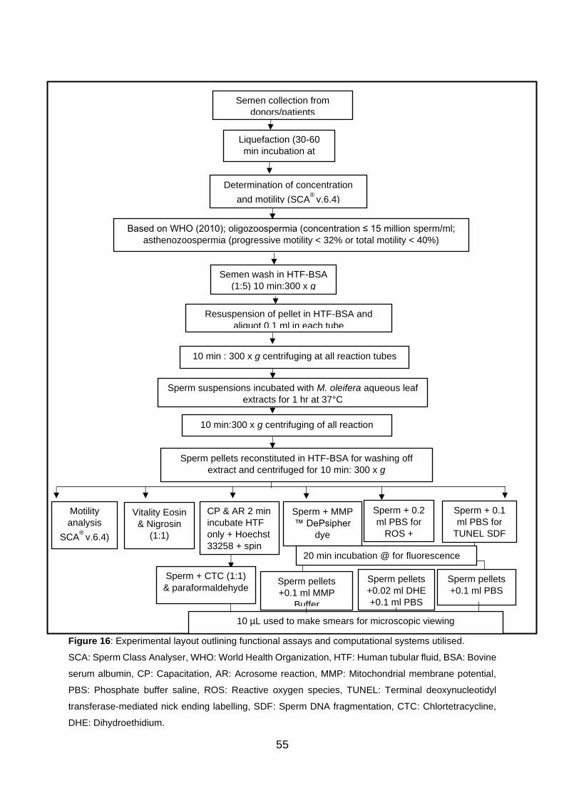
55
Figure 16: Experimental layout outlining functional assays and computational systems utilised.
SCA: Sperm Class Analyser, WHO: World Health Organization, HTF: Human tubular fluid, BSA: Bovine
serum albumin, CP: Capacitation, AR: Acrosome reaction, MMP: Mitochondrial membrane potential,
PBS: Phosphate buffer saline, ROS: Reactive oxygen species, TUNEL: Terminal deoxynucleotidyl
transferase-mediated nick ending labelling, SDF: Sperm DNA fragmentation, CTC: Chlortetracycline,
DHE: Dihydroethidium.
Semen collection from
donors/patients
Liquefaction (30-60
min incubation at
37°C)
Determination of concentration
and motility (SCA®
v.6.4)
Based on WHO (2010); oligozoospermia (concentration ≤ 15 million sperm/ml;
asthenozoospermia (progressive motility < 32% or total motility < 40%)
Semen wash in HTF-BSA
(1:5) 10 min:300 x g
Resuspension of pellet in HTF-BSA and
aliquot 0.1 ml in each tube
10 min : 300 x g centrifuging at all reaction tubes
Sperm suspensions incubated with M. oleifera aqueous leaf
extracts for 1 hr at 37°C
Sperm pellets reconstituted in HTF-BSA for washing off
extract and centrifuged for 10 min: 300 x g
Motility
analysis
SCA®
v.6.4)
10 min:300 x g centrifuging of all reaction
tubes
Vitality Eosin
& Nigrosin
(1:1)
CP & AR 2 min
incubate HTF
only + Hoechst
33258 + spin
for 5min with
Sperm + MMP
™ DePsipher
dye
Sperm + 0.2
ml PBS for
ROS +
Sperm + 0.1
ml PBS for
TUNEL SDF
Sperm pellets
+0.1 ml MMP
Buffer
Sperm pellets
+0.02 ml DHE
+0.1 ml PBS
Sperm pellets
+0.1 ml PBS
Sperm + CTC (1:1)
& paraformaldehyde
10 µL used to make smears for microscopic viewing
20 min incubation @ for fluorescence

56
3.6 Laboratory analysis
3.6.1 Determination of effects of M. oleifera aqueous leaf extracts on sperm
motility
To determine the effects of aqueous leaf extract of M. oleifera on sperm motility,
washed sperm suspensions were incubated at 37°C with the various concentrations
(0, 0.625, 6.25, 62.5, and 625 µg/ml) of the extract for 1 hr. Suspensions were
subsequently re-suspended in 100 µl HTF-BSA. Thereafter, 10µl of the sperm
suspension was loaded on a pre-warmed Leja slide, and at least 200 spermatozoa
were assessed according to the WHO criteria, using Sperm Class Analyzer® (Figure
17) (SCA, Microptic S.L., Barcelona, Spain version 6.4) with a Zeiss photomicroscope
using a 10 X objective. The following characteristics of the sperm motility were
analysed: Total motility (%), progressive motility (%), beat cross frequency (BCF; Hz),
linearity (LIN; %), straightness (STR; %), average path velocity (VAP; µm/s),
curvilinear velocity (VCL; µm/s), straight-line velocity (VSL; µm/s), Wobble (WOB; %),
and hyperactivation (%) (Tables 1 and 2).
Figure 17: (a) Computer-aided sperm class analyser (CASA) and (b) Leja slides used for
determination of sperm motility and concentration.

57
Table 1: Categories of sperm motility classes defined
Category Definition Unit
Total motility Includes all spermatozoa moving
progressively or not in a field.
This classification does not take
speed into consideration.
%
Non-progressive (grade C) Exhibited by a population of
spermatozoa that do not swim in
large circles or linearly, where
flagella beat does not change
head movements.
%
Progressive motility
(grades A + B)
Spermatozoa are moving linearly
forward or in large circles.
%
Immotility (grade D) No sperm movement. %
Hyperactivation Asymmetrical, vigorous and
random form of motility sperm
exhibits following the acquisition
of fertilising potential, and
evident at the site of fertilisation.
%
58
Table 2: Sperm motility kinematic parameters
Motility kinematic
parameters
Definition Units
Curvilinear velocity (VCL) Time average velocity of sperm head
along its actual curvilinear path.
µm/s
Straight-line velocity
(VSL)
Average time velocity of sperm head in a
straight line between two points of
detection.
µm/s
Average path velocity
(VAP)
The average velocity of the sperm head
along its average spatial trajectory.
µm/s
Amplitude of lateral head
displacement (ALH)
The maximum lateral displacement of
sperm head about its average path.
µm
Oscillatory index /
Wobble (WOB)
“Wiggling” or oscillatory movements of
sperm’s actual path about its average.
%
Linearity index (LIN) The linearity of the average path.
%
Straightness (STR) Straightness of the average path. %
Beat cross frequency
(BCF)
The time-averaged rate at which sperm
curvilinear tracks crosses its average
path trajectory.
Hz
3.6.2 Determination of effects of M. oleifera aqueous leaf extracts on sperm
vitality
After 1 hr incubation at 37°C with M. oleifera extracts, an aliquot of 50 µl of E & N dye
was transferred into an Eppendorf tube. Then, sperm suspension and E & N were
mixed (1:1). The tubes were incubated for 2 minutes. After incubation, 10 µl of the
mixture was smeared on slides (Lasec®, Delfran Pharmaceutical, Cape Town). The
slides were air-dried at room temperature, mounted with dibutyl phthalate polystyrene

59
xylene (DPX) medium, and viewed through a 10 X objective using a light microscope
(Zeiss, Oberkochen, Germany). Sperm cells stained pink represented dead sperm
cells, while those stained white were live cells (Figure 18). Two hundred (200)
spermatozoa were assessed for each sample. The percentage of live sperm was
calculated.
3.6.3 Determination of effects of M. oleifera aqueous extracts on sperm
mitochondrial membrane potential (∆ψm)
Sperm mitochondrial membrane potential (MMP) was assessed using the lipophilic
cationic dye (DePsipher™, Trevigen, Minneapolis, USA).
The protocol was applied with modifications from the manufacturer’s instructions as
previously described by Henkel et al. (2011). Briefly, a reaction buffer was diluted with
distilled water (1:10), and 20 µl of stabiliser was added per 1 millilitre of the buffer. A
volume of 1 µl of DePsipher was added per millilitre of the buffer. After that, the mixture
was thoroughly vortexed for 1 min at 10,000 x g for homogenisation. The supernatant
was transferred into an Eppendorf tube, and covered in foil for later use.
Sperm cells were exposed to various concentrations (0, 0.625, 6.25, 62.5, and 625
µg/ml) of M. oleifera aqueous extracts at 37°C for 1 hr, and then washed with HTF-
Figure 18: One-step Eosin-nigrosin (E & N) staining of human sperm specimen. The dye excludes
viable / live (white) from dead (pink) cells, based on the membrane intactness. Samples were viewed
through a 10 X objective.
10 µm
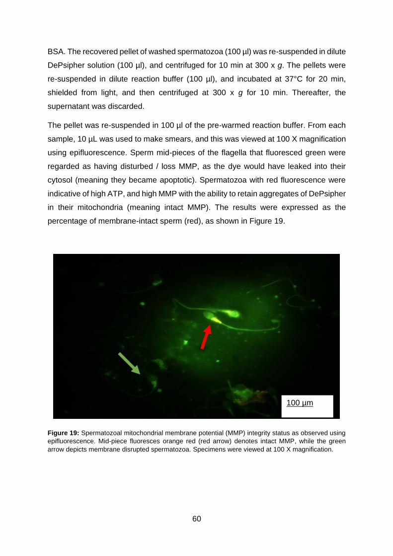
60
BSA. The recovered pellet of washed spermatozoa (100 µl) was re-suspended in dilute
DePsipher solution (100 µl), and centrifuged for 10 min at 300 x g. The pellets were
re-suspended in dilute reaction buffer (100 µl), and incubated at 37°C for 20 min,
shielded from light, and then centrifuged at 300 x g for 10 min. Thereafter, the
supernatant was discarded.
The pellet was re-suspended in 100 µl of the pre-warmed reaction buffer. From each
sample, 10 µL was used to make smears, and this was viewed at 100 X magnification
using epifluorescence. Sperm mid-pieces of the flagella that fluoresced green were
regarded as having disturbed / loss MMP, as the dye would have leaked into their
cytosol (meaning they became apoptotic). Spermatozoa with red fluorescence were
indicative of high ATP, and high MMP with the ability to retain aggregates of DePsipher
in their mitochondria (meaning intact MMP). The results were expressed as the
percentage of membrane-intact sperm (red), as shown in Figure 19.
Figure 19: Spermatozoal mitochondrial membrane potential (MMP) integrity status as observed using
epifluorescence. Mid-piece fluoresces orange red (red arrow) denotes intact MMP, while the green
arrow depicts membrane disrupted spermatozoa. Specimens were viewed at 100 X magnification.
100 µm
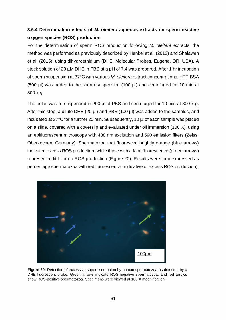
61
3.6.4 Determination effects of M. oleifera aqueous extracts on sperm reactive
oxygen species (ROS) production
For the determination of sperm ROS production following M. oleifera extracts, the
method was performed as previously described by Henkel et al. (2012) and Shalaweh
et al. (2015), using dihydroethidium (DHE; Molecular Probes, Eugene, OR, USA). A
stock solution of 20 µM DHE in PBS at a pH of 7.4 was prepared. After 1 hr incubation
of sperm suspension at 37°C with various M. oleifera extract concentrations, HTF-BSA
(500 µl) was added to the sperm suspension (100 µl) and centrifuged for 10 min at
300 x g.
The pellet was re-suspended in 200 µl of PBS and centrifuged for 10 min at 300 x g.
After this step, a dilute DHE (20 µl) and PBS (100 µl) was added to the samples, and
incubated at 37°C for a further 20 min. Subsequently, 10 µl of each sample was placed
on a slide, covered with a coverslip and evaluated under oil immersion (100 X), using
an epifluorescent microscope with 488 nm excitation and 590 emission filters (Zeiss,
Oberkochen, Germany). Spermatozoa that fluoresced brightly orange (blue arrows)
indicated excess ROS production, while those with a faint fluorescence (green arrows)
represented little or no ROS production (Figure 20). Results were then expressed as
percentage spermatozoa with red fluorescence (indicative of excess ROS production).
Figure 20: Detection of excessive superoxide anion by human spermatozoa as detected by a
DHE fluorescent probe. Green arrows indicate ROS-negative spermatozoa, and red arrows
show ROS-positive spermatozoa. Specimens were viewed at 100 X magnification.
100µm

62
3.6.5 Determination of effects of M. oleifera aqueous extracts on sperm DNA
fragmentation.
Effects of M. oleifera extract on sperm DNA integrity were determined using TUNEL
assay (terminal deoxynucleotidyl transferase-mediated dUTP-biotin nick-end
labelling) [Dead End]™ Fluoremetric, Promega, Madison, USA. The TUNEL system is
based on the enzymatic ability of terminal transferase (TdT) to detect fluoresceinated
dUTP single- or double-stranded DNA nicks by labelling the resultant free hydroxyl
groups (3-OH) (Sharma and Agarwal, 2011a). In the study, TUNEL assay was
performed for SDF, and at least 200 TUNEL-positive spermatozoa were assessed.
After 1 h incubation with different concentrations of MOL, the samples were
centrifuged for 10 min at 300 x g in HTF-BSA. Thereafter, sperm suspension (100 µl)
was re-suspended in PBS and centrifuged again, with this step repeated once. Then,
10 µl of the sample was smeared on Starfrost slides (Knittel Gläser, Braunschweig,
Germany).
Slides were left to air-dry, and subsequently fixed with fresh 4% methanol-free
formaldehyde (KIMIX chemicals, Eppingdust) in PBS for 25 min at 4˚C. Slides were
shortly washed with PBS for another 5 min and allowed to dry at room temperature.
The sperm plasma membrane was permeabilised with 0.2% Triton X-100 detergent in
PBS for 5 min and rinsed twice with PBS at room temperature. Excess liquid was
removed by tapping the slides on the paper towel-layered working counter, and 100
µl of equilibration buffer was added to the slides, and the samples were left to
equilibrate for roughly 10 min as per the manufacturer’s instructions. Then, 50 µl TdT
reagent buffer was added, and the coverslip was placed on slides, and the sample
was incubated for 1 h in a light-devoid humidified chamber.
Slides were submerged in 2 X SSC buffer (Promega) for 15 min, washed three times
with PBS for 5 min each, and placed back in the humidified chamber to avoid drying
while reading. At least 200 spermatozoa were randomly selected and analysed using
an epifluorescence microscope (Zeiss, Oberkochen, Germany). Excitation and
emission filters were 488 nm and 530 nm, respectively, and viewed at 100 X
magnification. DNA intact sperm exhibited a bit of a background staining (TUNEL-
negative), whereas DNA-fragmented sperm emitted a bright green fluorescence
(TUNEL-positive) as shown in Figure 22. Accordingly, the percentage of TUNEL-
positive spermatozoa were calculated from at least 200 cells.

63
Figure 21: Detection of sperm DNA fragmentation using TUNEL assay. Blue arrow indicates TUNEL-
positive sperm, and yellow arrows indicate TUNEL negative sperm. Specimens were viewed at 100 X
magnification.
3.6.6 Determination of effects of M. oleifera aqueous extracts on sperm
capacitation and acrosome reaction
This study adopted the dual staining of Hoechst 32258 / Chlortetracycline (CTC)
according to Green et al. (1996) for assessment of capacitation and acrosome status
of human spermatozoa, following M. oleifera extract exposure. Following 1 hr
incubation of sperm with the aqueous extracts, one microliter (1µl) of Hoechst 32258
was mixed with 100 µl sperm suspension in HTF-BSA. The samples were mixed with
4 ml of 2% polyvinylpyrrolidone (PVP40) in HTF only, then incubated at room
temperature for 2 min, and centrifuged at 900 x g for 5 min.
Chlortetracycline (CTC) buffer (pH 7.8) was prepared on the day of use from the
following reagents: 750 µm CTC in a buffer of 130 mM NaCl, 5 mM cysteine, and 20

64
mM Tris-HCl. The solution was wrapped in a foil and stored at 4˚C for subsequent use
for up to 4 weeks. Hoechst-stained sperm suspension (45 µl) was mixed with CTC
solution (45 µl) and 12.5% w/v paraformaldehyde in 0.5 Tris-HCl (pH 7.4) (8 µl). Then,
10 µl of the mixture was loaded on a slide, and one drop of 0.22 M 1, 4–diazabicyclo
(2.2.2) octane (DABCO) dissolved in glycerol: PBS (9:1) was carefully placed to delay
any loss of fluorescence.
Slides were viewed with a 100 X objective, using an epifluorescent Zeiss microscope.
From each slide, 200 live (Hoechst-negative) were assessed for CTC fluorescent
patterns, where uniform fluorescence over the entire head indicated an F pattern (non-
capacitated, acrosome intact). Fluorescence-free band of the post-acrosomal
segment, B pattern (capacitated, acrosome-intact cells) (represented by blue arrows),
and (indicated by yellow arrows) dull or absent fluorescent over the entire head with /
without a fluorescent post-acrosomal region characterised AR pattern (capacitated
and acrosome-reacted cells) and corresponded with white arrows as shown in Figure
22).
Figure 22: Three staining CTC patterns of human spermatozoa as seen under fluorescence
microscope. A) F pattern, B) B pattern and C) AR pattern. Specimens were viewed at 100 X
magnification.
A B C

65
3.7 Ethical considerations
3.7.1 Ethical clearance and approval
Ethical clearance was obtained from the Turfloop Research Ethics Committee (TREC)
of the University of Limpopo (TREC/45/2019: PG; Appendix 1) and the Medical
Biosciences Research Ethics Committee (BMREC) from the University of the Western
Cape (BM17/7/14; Appendix 2). Approval to collect samples from the Tygerberg
Hospital was granted by the Western Cape Department of Health (Appendix 3).
Permission for collection of semen samples from the Vincent Pallotti Hospital was
granted by the Aevitas Fertility clinic (Appendix 4).
3.7.2 Harm
The procedural approach during data collection involved no medical risk or harm,
because participants were thoroughly briefed before consenting to their participation.
After their briefing, they willingly provided semen for experimentation. Participants
(especially donors) were assured of their safety, and their choice to withdraw from the
study at any time. The University Counselling Services were at the participants’
disposal in case they were to experience any distress during the semen provision. As
stated previously, there were no risks incurred. In any case where a participant could
have misunderstood the handling of the samples, they were briefed.
3.7.3 Disposal of waste
At the end of the experiment, any residual semen samples were disposed of as
biohazardous waste in VT Medwaste bins. The waste was later incinerated by the
BCL-mediated medical waste company.
3.7.4 Anonymity and confidentiality
No actual participant names were used in the recording of data or disclosed to anyone
in the dissemination of results. Participant’s results were restricted only to researchers
directly involved with the data collection, for confidential reasons. A non-coercive
informed consent form was provided for participants to sign as an assurance of their
willingness to take part in the research, and that they had been informed that they
could withdraw whenever they decided to do so without penalty or risk to their well-
being. The informed consent form was available in English, Afrikaans, and Xhosa to
eliminate any potential language barriers (Appendices 5-7). Participants were
assigned sample numbers or codes as a way of maintaining their confidentiality.

66
3.8 Data analyses
3.8.1 Reliability and validity
In this study, donors were screened twice before recruitment into the programme to
ensure reliability of the study findings. In hospitals, the infertile status of patients was
diagnosed by doctors, andrologists or practised reproductive biologists.
All testing kits / protocols were performed strictly according to the manufacturer’s
instructions.
3.8.2 Quality assurance
It is well known that quality assurance in an andrology-testing laboratory is difficult due
to the lack of standardisation of the protocols, and biological variation of semen sample
either from an individual or between different samples. One sample was used to test
all parameters at a time, and experimental procedures were performed according to
the WHO (2010) laboratory manual for examination and processing semen to ensure
these limitations did not interfere with the quality of results in this study.
3.8.3 Bias
Convenience sampling was used. Sampling bias could not be avoided in this study,
because the sampling frame was poorly defined as it included any available, eligible
and willing donor. At the same time, infertile couples were people of any race or
nationality who sought fertility treatment at either of the two hospitals (Figure 16). The
availability of the researcher or participant, financial, and time constraints justified the
type of sampling chosen.
3.8.4 Statistical analysis
Data were analysed using GraphPad Prism 6.0.1 for Windows (GraphPad Software,
San Diego, California, USA). Descriptive data were generated using column statistic
function, and the D’Agostino-Pearson Omnibus test was used for normality testing.
Consequently, further analysis included parametric testing using one-way analysis of
variance (ANOVA), while non-parametric analyses were performed using the Kruskal-
Wallis test, in which Dunnets’ multiple tests were applied for comparisons of group
means. Repeated measures one-way ANOVA (RM 1-way ANOVA) were used in non-
Gaussian data for trend analyses. Spearman correlation was used to assess

67
relationships between the sperm parameters. Statistical significance was set at p <
0.05.

68
CHAPTER 4
RESULTS
4.1. Introduction
Findings in this study reported in this chapter consist of two parts. The first part reports
on the donor, patient, and combined (donor and patient) groups. The second part
entails results on the asthenozoospermic group. The study consisted of 70 samples.
Forty donors (40) were recruited based on the WHO (2010) guidelines from the
Andrology Research Programme in the comparative spermatology group of the
Medical Biosciences Department, University of the Western Cape. Using the same
inclusion criteria, semen samples from 30 patients attending the Tygerberg and
Vincent Pallotti Hospitals were included. Standard semen parameters of donor and
patient groups are summarised in the table below (Table 3).
In an effort to determine the effects of M. oleifera aqueous leaf extracts on human
sperm functions in vitro, sperm suspensions were incubated at 37 ̊C with various
concentrations of M. oleifera (0, 0.625, 6.25, 62.5 and 625 µg/ml) for one hour.
4.2. Summary statistics of standard semen parameters according to the WHO
(2010)
After 30 to 60 minutes of liquefaction at 37 ̊C before treatment, ejaculates were
assessed for inclusion. The overall descriptive data are summarised in Table 3. The
study included donors whose seminograms were above the WHO’s (2010) lower
reference points: concentration ≥ 15 million/ml, total motility ≥ 40% or progressive
motility ≥ 32%. The cut-off values defining male infertility (patients) in this study were
set as follows: oligozoospermia (concentration < 15 million/ml), asthenozoospermia
(total motility < 40% or progressive motility < 32%), and they formed part of the study.
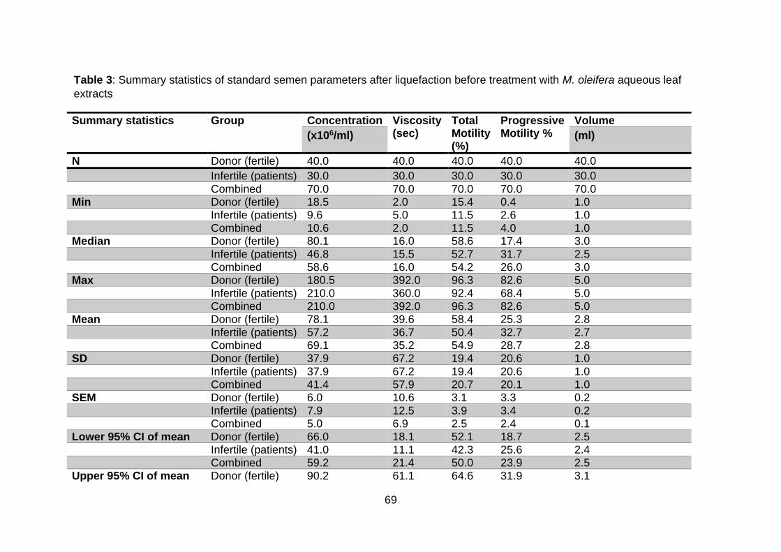
69
Summary statistics Group Concentration Viscosity (sec)
Total Motility (%)
Progressive Motility %
Volume
(x106/ml) (ml)
N Donor (fertile) 40.0 40.0 40.0 40.0 40.0 Infertile (patients) 30.0 30.0 30.0 30.0 30.0 Combined 70.0 70.0 70.0 70.0 70.0
Min Donor (fertile) 18.5 2.0 15.4 0.4 1.0 Infertile (patients) 9.6 5.0 11.5 2.6 1.0 Combined 10.6 2.0 11.5 4.0 1.0
Median Donor (fertile) 80.1 16.0 58.6 17.4 3.0 Infertile (patients) 46.8 15.5 52.7 31.7 2.5 Combined 58.6 16.0 54.2 26.0 3.0
Max Donor (fertile) 180.5 392.0 96.3 82.6 5.0 Infertile (patients) 210.0 360.0 92.4 68.4 5.0 Combined 210.0 392.0 96.3 82.6 5.0
Mean Donor (fertile) 78.1 39.6 58.4 25.3 2.8 Infertile (patients) 57.2 36.7 50.4 32.7 2.7 Combined 69.1 35.2 54.9 28.7 2.8
SD Donor (fertile) 37.9 67.2 19.4 20.6 1.0 Infertile (patients) 37.9 67.2 19.4 20.6 1.0 Combined 41.4 57.9 20.7 20.1 1.0
SEM Donor (fertile) 6.0 10.6 3.1 3.3 0.2 Infertile (patients) 7.9 12.5 3.9 3.4 0.2 Combined 5.0 6.9 2.5 2.4 0.1
Lower 95% CI of mean Donor (fertile) 66.0 18.1 52.1 18.7 2.5 Infertile (patients) 41.0 11.1 42.3 25.6 2.4 Combined 59.2 21.4 50.0 23.9 2.5
Upper 95% CI of mean Donor (fertile) 90.2 61.1 64.6 31.9 3.1
Table 3: Summary statistics of standard semen parameters after liquefaction before treatment with M. oleifera aqueous leaf
extracts

70
Descriptive statistics of the donor (N = 40), patient (N = 30) and combined (N = 70). N = Number of samples, SD = standard
deviation, SEM = Standard error of the mean, Min = minimum, Max = maximum, CI = confidence interval
.
Infertile (patients) 73.5 62.4 58.4 39.7 3.1 Combined 78.9 49.0 59.8 33.5 3.0
Coefficient of variation Donor (fertile) 48.5 169.6 33.3 81.6 34.8 Infertile (patients) 76.1 187.0 42.9 57.6 35.2 Combined 60.0 164.5 37.6 69.8 34.5

71
4.3. Effects of M. oleifera leaf extracts on sperm motility parameters in vitro
Summary statistics of sperm motility following 1-hr incubation with increasing
concentrations (0, 0.625, 6.25, 62.5, and 625 µg/ml) of aqueous leaf extracts of M.
oleifera are displayed in Table 4.
Following treatment of human sperm with M. oleifera leaf extracts for 1 hr, the plant
did not significantly affect overall sperm total motility (p = 0.1212) in the donor group.
The highest concentration (625 µg/ml) produced a significant increase in total motility
of the patient and combined groups in comparison with control groups, respectively (p
= 0.005 and p = 0.0427), with no change in the donor group (p = 0.9714) (Figure 23).
No differences in total motility were observed in the control and all M. oleifera
treatments (0.625, 6.25, 62.5, and 625 µg/ml) between donors, patients, and the
combined groups (p > 0.05) (Figure 23). Repeated measures one-way ANOVA
revealed a significant positive trend in sperm total motility (p < 0.0001) of patients and
combined groups, while no trends could be observed for donors after treatment with
increasing concentrations of M. oleifera.
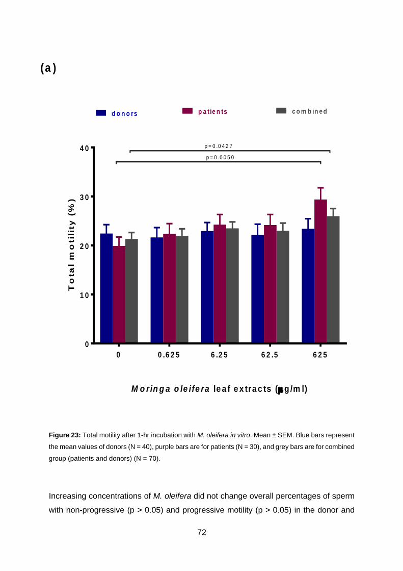
72
Figure 23: Total motility after 1-hr incubation with M. oleifera in vitro. Mean ± SEM. Blue bars represent
the mean values of donors (N = 40), purple bars are for patients (N = 30), and grey bars are for combined
group (patients and donors) (N = 70).
Increasing concentrations of M. oleifera did not change overall percentages of sperm
with non-progressive (p > 0.05) and progressive motility (p > 0.05) in the donor and
M o rin g a o le ife ra le a f e x tra c ts (g /m l)
To
ta
l m
otil
ity
(%
)
0 0 .6 2 5 6 .2 5 6 2 .5 6 2 5
0
1 0
2 0
3 0
4 0p = 0 .0 0 5 0
p = 0 .0 4 2 7
d o n o rs p a tie n ts c o m b in e d
(a )

73
combined groups, respectively. Despite insignificant results with other concentrations
of M. oleifera, the highest concentration (625 µg/ml) significantly increased the
progressive (p = 0.0022) and non-progressive sperm motility (p = 0.0215) in the patient
group when compared to the respective controls (Figures 24a and b). On the contrary,
no significant effects were observed in progressive and non-progressive motility when
the highest concentration of M. oleifera extract was compared to the control in donor
(p > 0.05).
The control of progressive motility showed no statistical difference between the
combined groups and donors (p > 0.05). Patients’ progressive motility was significantly
higher than that of the donors when the controls were compared (p = 0.0150).
However, non-progressive motility of patients was significantly higher than that of
donors (p = 0.0017) (Figure 24b). Controls between patients and the combined groups
showed no difference (p > 0.05)
M. oleifera improved progressive and non-progressive motility significantly more in
patients than in donors at doses 0.625 µg/ml (p = 0.0005 and p < 0.0001), 6.25 µg/ml
(p = 0.0002 and p = 0.0002), 62.5 µg/ml (p = 0.0001 and p = 0.0059) and 625 µg/ml
(p < 0.0001 and p = 0.0083) (Figures 25a and b). Similar observations were made in
progressive and non-progressive motility between patients and the combined groups
at concentrations 0.625 µg/ml (p = 0.0499 and p = 0.0246), 6.25 µg/ml (p = 0.0335
and p = 0.0314), 62.5 µg/ml (p = 0.0275) and 625 µg/ml (p = 0.0119) (Figures 24a and
b).
Positive linear trends were significant for progressive motility and non-progressive
motility (p = 0.0117 and p < 0.0001) of the patient group. In addition, significant
straight-line trends were shown for percentage of progressive (p = 0.0023) and non-
progressive motility (p = 0.0013) in the combined group. However, no significant trends
could be recorded with increasing doses of M. oleifera in the percentage of progressive
or non-progressive motility in the donor group.
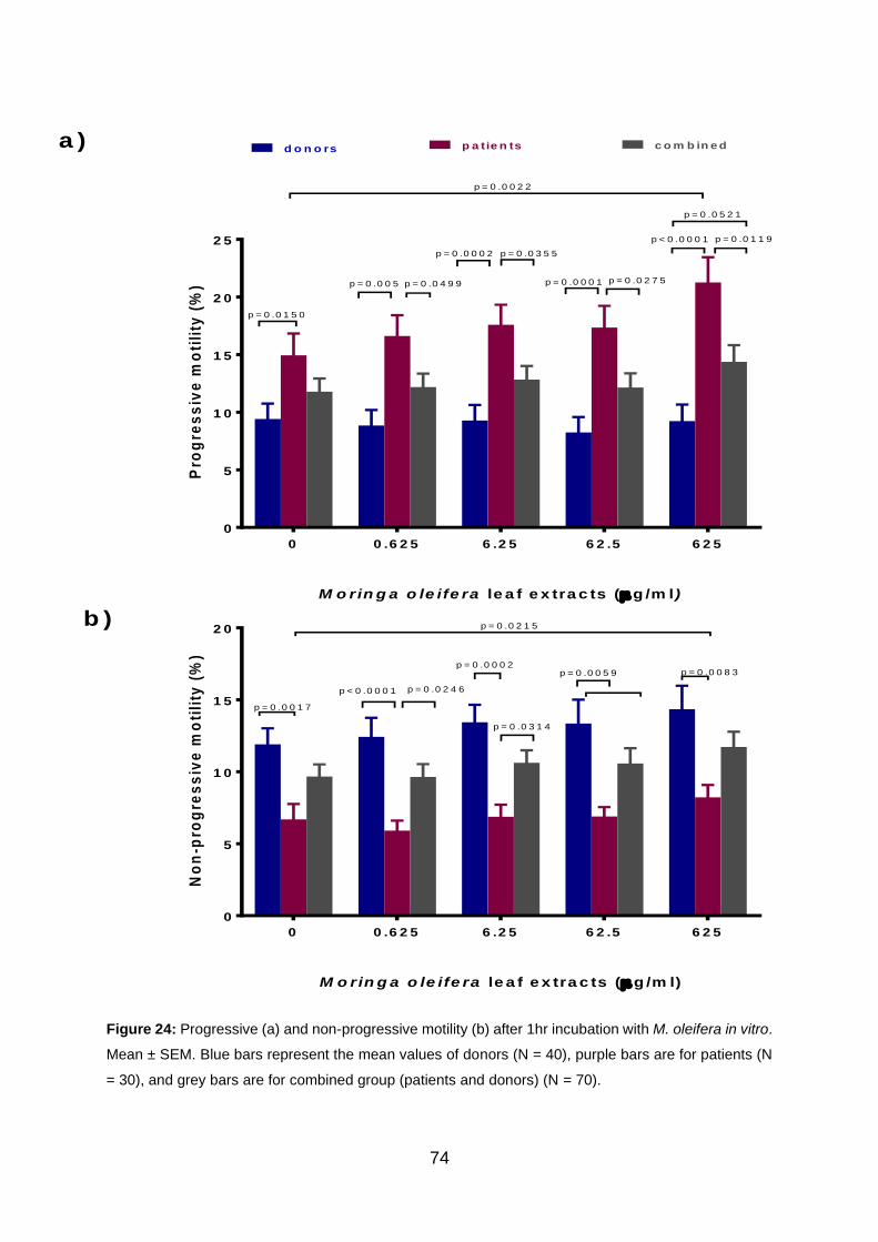
74
Figure 24: Progressive (a) and non-progressive motility (b) after 1hr incubation with M. oleifera in vitro.
Mean ± SEM. Blue bars represent the mean values of donors (N = 40), purple bars are for patients (N
= 30), and grey bars are for combined group (patients and donors) (N = 70).
a )
b )
M o rin g a o le ife ra le a f e x tra c ts (g /m l)
Pro
gre
ss
ive
mo
tili
ty (
%)
0 0 .6 2 5 6 .2 5 6 2 .5 6 2 5
0
5
1 0
1 5
2 0
2 5
p = 0 .0 0 2 2
d o n o rs p a tie n ts c o m b in e d
p = 0 .0 1 5 0
p < 0 .0 0 0 1
p = 0 .0 5 2 1
p = 0 .0 1 1 9
p = 0 .0 0 5 p = 0 .0 4 9 9
p = 0 .0 0 0 2 p = 0 .0 3 5 5
p = 0 .0 0 0 1 p = 0 .0 2 7 5
M o rin g a o le ife ra le a f e x tra c ts (g /m l)
No
n-p
rog
res
siv
e m
oti
lity
(%
)
0 0 .6 2 5 6 .2 5 6 2 .5 6 2 5
0
5
1 0
1 5
2 0 p = 0 .0 2 1 5
p = 0 .0 0 1 7
p = 0 .0 0 8 3
p < 0 .0 0 0 1 p = 0 .0 2 4 6
p = 0 .0 0 0 2
p = 0 .0 3 1 4
p = 0 .0 0 5 9

75
After 1-hr incubation of sperm with M. oleifera aqueous extracts, sperm kinematic
parameters were investigated. Overall, M. oleifera leaf extracts did not alter sperm
kinematic parameters in the three groups tested: donor, patient, and the combined
groups (p > 0.05) (Table 4).
Although no specific changes could be seen in sperm kinematics of motility when all
groups were exposed to various concentrations of M. oleifera, significant trends were
recorded in the straightness index (STR), straight-line velocity (VSL), and average
path velocity (VAP) in patients and combined groups. The one-way ANOVA repeated
measure revealed significant decreasing tendencies in the sperm percentage
straightness in patients (p = 0.0177), and in combined (p = 0.0012) groups. Similarly,
a decreasing trend was observed in sperm straight / rectilinear velocity (VSL) with
increasing dose of M. oleifera in patients (p = 0.0262), and in combined (p = 0.0040)
groups. The linearity of sperm demonstrated dose-dependent significant decreasing
trends in the patients' group, following a 1-hour incubation with the plant (p = 0.0236).
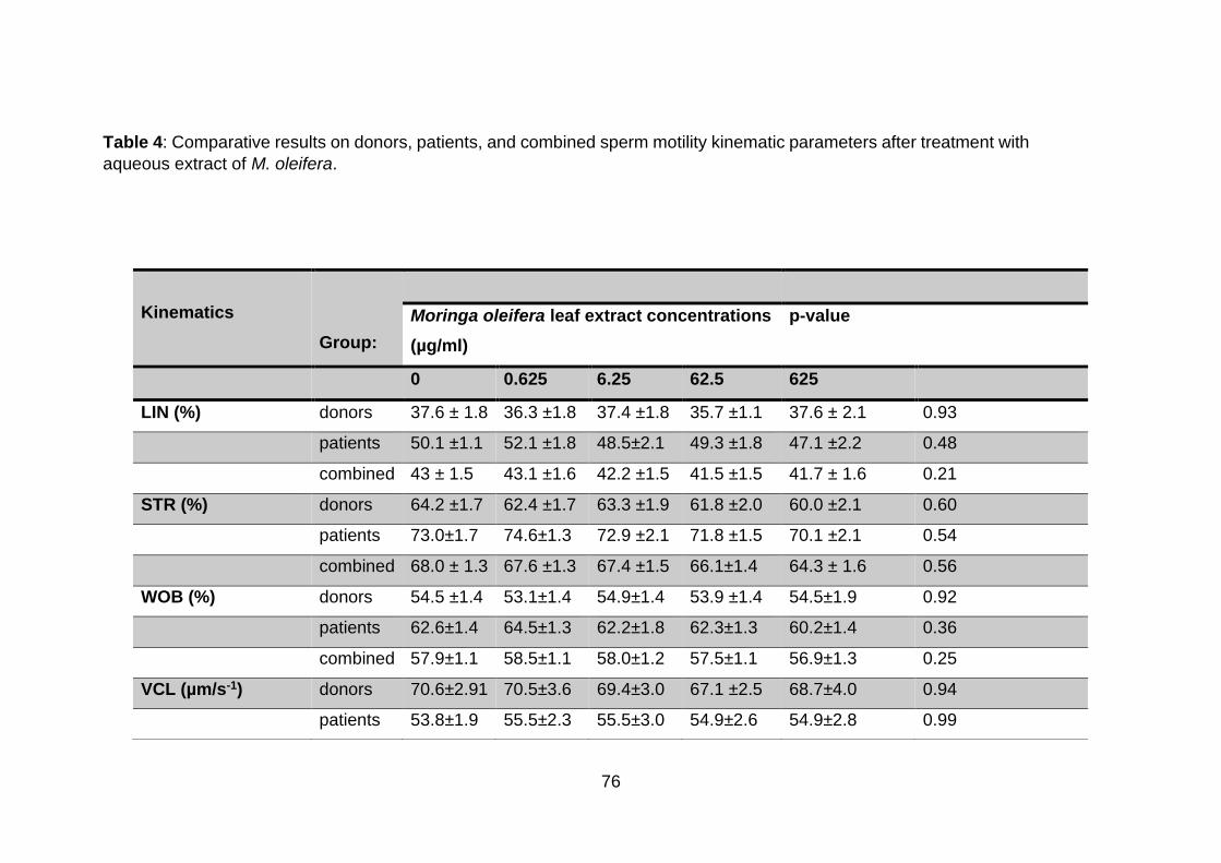
76
Kinematics
Group:
Moringa oleifera leaf extract concentrations
(µg/ml)
p-value
0 0.625 6.25 62.5 625
LIN (%) donors 37.6 ± 1.8 36.3 ±1.8 37.4 ±1.8 35.7 ±1.1 37.6 ± 2.1 0.93
patients 50.1 ±1.1 52.1 ±1.8 48.5±2.1 49.3 ±1.8 47.1 ±2.2 0.48
combined 43 ± 1.5 43.1 ±1.6 42.2 ±1.5 41.5 ±1.5 41.7 ± 1.6 0.21
STR (%) donors 64.2 ±1.7 62.4 ±1.7 63.3 ±1.9 61.8 ±2.0 60.0 ±2.1 0.60
patients 73.0±1.7 74.6±1.3 72.9 ±2.1 71.8 ±1.5 70.1 ±2.1 0.54
combined 68.0 ± 1.3 67.6 ±1.3 67.4 ±1.5 66.1±1.4 64.3 ± 1.6 0.56
WOB (%) donors 54.5 ±1.4 53.1±1.4 54.9±1.4 53.9 ±1.4 54.5±1.9 0.92
patients 62.6±1.4 64.5±1.3 62.2±1.8 62.3±1.3 60.2±1.4 0.36
combined 57.9±1.1 58.5±1.1 58.0±1.2 57.5±1.1 56.9±1.3 0.25
VCL (µm/s-1) donors 70.6±2.91 70.5±3.6 69.4±3.0 67.1 ±2.5 68.7±4.0 0.94
patients 53.8±1.9 55.5±2.3 55.5±3.0 54.9±2.6 54.9±2.8 0.99
Table 4: Comparative results on donors, patients, and combined sperm motility kinematic parameters after treatment with
aqueous extract of M. oleifera.

77
Data are expressed as means ± SEM of donors (N = 40), patients (N = 30), combined (N = 70) groups. NS = non-significant, LIN
(linearity), WOB (oscillation index), VCL (curvilinear velocity), VSL (straight-line velocity), BCF (beat cross frequency), ALH
(amplitude of lateral displacement).
combined 63.4 ± 2.1 64.1 ± 2.4 63.5 ± 2.3 61.8 ± 1.9 62.6 ± 2.7 0.92
VAP (µm/s-1) donors 37.2±1.4 36.5 ±1.5 36.5±1.3 35.1±1.2 35.5±1.5 0.58
patients 34.6±1.7 35.8±1.7 34.3±1.7 34.5±1.7 33.0±1.9 0.86
combined 36.1 ±1.1 36.2 ± 1.1 35.5 ± 1.1 34.8 ± 0.1 34.4 ± 1.2 0.51
VSL (µm/s-1) donor 25.7±1.3 24.3±1.2 25.1 ±1.1 23.5±1.2 22.7±1.2 0.39
patients 32.4±2.1 33.4±1.1 31.3±1.9 31.9±2.1 29.5±2.1 0.72
combined 28.6 ± 1.2 28.2 ±1.2 27.7 ± 1.1 27.1 ± 1.2 25.6 ± 1.2 0.28
ALH (µm) donors 2.4±0.1 2.4±0.1 2.4±0.1 2.3±0.1 2.3±0.1 0.98
patients 2.2±0.1 2.2±0.1 2.3±0.1 2.3±0.1 2.3±0.1 0.89
combined 2.3 ±0.1 2.3 ±0.1 2.3 ±0.1 2.3 ±0.1 2.3 ±0.1 1.00
BCF (Hz) donors 15.7±0.8 14.7±0.9 14.7±0.8 15.5±1.2 13.6±1.0 0.49
patients 8.3±0.4 8.8±0.1 8.2±0.5 8.4±0.6 8.0±1.0 0.72
combined 12.5 ± 0.7 12.1±0.7 11.9 ± 0.6 12.5 ± 0.9 11.2 ± 0.6 0.78
Hyperactivation (%) donors 3.1 ±1.0 4.9 ± 1.4 3.5 ±0.8 2.8 ± 0.8 5.3 ± 1.7 0.49
patients 0.9± 0.3 0.9 ± 0.3 1.4 ±0.8 1.0 ± 0.4 1.2 ± 0.1 0.90
combined 2.2 ±0.1 3.2 ±0.8 2.6 ± 0.6 2.0 ±0.5 3.5 ± 1.0 0.98
78
4.3.1. Correlations of sperm total motility and functional parameters.
Table 5 depicts various associations between total sperm motility and other functional
parameters following treatment of M. oleifera for 1-h. Total motility showed no
association with percentage of sperm progression in donors (r = 0.50, p = 0.4500), or
in combined groups (r = 0.90, p = 0.0800), while a strong positive correlation was
observed in patients (r = 1.00, p = 0.0167). Furthermore, no correlation was recorded
between total motility and hyperactivation in patients (r = 0.90, p = 0.0833), in
combined group (r = 0.50, p = 0.4500), and in the donors (r = 0.40, p = 0.5167). Viable
spermatozoa showed no relationship with total motility in the donors (r = 0.50, p =
0.4500). In contrast, the association between total sperm motility and live vitality was
significantly and positively correlated in patients and combined groups (r = 1.00, p =
0.0167).
In addition, total sperm motility showed no relationship with MMP-intact spermatozoa
in donors (r = 0.73, p = 0.1639), or in patients and the combined group (r = 0.90, p =
0.0833). Similarly, no correlations could be drawn with reactive oxygen species-
positive (ROS-positive) sperm in donors (r = -0.59, p = 0.2925), or in patients, and in
the combined groups (r = -0.80, p = 0.1333). Again, no relationship was established
between total motility with sperm capacitation in the donors (r = -0.56, p = 0.3275), in
patients (r = -0.90, p = 0.0833), and in the combined group (r = -0.50, p = 0.4500).
Acrosome reaction demonstrated no relationship in the distinctive groups: donors (r =
-0.64, p = 0.2423), and the combined (r = -0.10, p = 0.9500) group. However, contrary
results were apparent in patients’ total motility when correlated with percentage
acrosome reaction, and a significant, strong, and positive association was shown (r =
1.00, p = 0.0167). The degree of sperm DNA fragmentation did not show any
relationship with total motility in any of the groups; donors (r = -0.74, p = 0.1491),
patients (r = -0.84, p = 0.0761), and the combined group (r = -0.80, p = 0.1333).

79
Table 5: Relationships between total sperm motility and functional parameters
4.3.2. Correlation between progressive motility and functional parameters
Regarding the percentage of sperm progression to functional parameters investigated
in the donor, combined, and patient group (Table 6), no relationships could be
observed with sperm forward-movement in the donor group; hyperactivation, vitality (r
= 0.10, p = 0.9500), and capacitation, reactive oxygen species, acrosome reaction,
DNA fragmentation (r = 0.02, p = 0.7833), and MMP. (r = 0.70, p = 0.2333).
Similarly, no association with established with hyperactivation and MMP (r = 0.90, p =
0.0833) was present in patients. On the other hand, the relationships with vitality and
acrosome reaction were significantly strong (r = 1.00, p = 0.0167) in patients. The
percentage of ROS-positive (r = -0.80, p = 0.1333), capacitation, and DNA
fragmentation (r = -0.90, p = 0.0833) were not correlated to progressive motility in the
patients’ group. No relationship was seen between forward-moving sperm and
hyperactivation (r = 0.80, p = 0.1333), vitality (r = 0.90, p = 0.0833), and MMP integrity
(r = 0.70, p = 0.2333) in the combined group. No correlation was observed between
progression, sperm ROS, and DNA fragmentation (r = -0.50, p = 0.4500) and
Parameters Donors Patients Combined group
r p r p r p
Progressive motility (%) 0.50 0.4500 1.00 0.0167 0.90 0.0833
Hyperactivated motility
(%)
0.40 0.5167 0.90 0.0833 0.50 0.4500
Vitality (%) 0.50 0.4500 1.00 0.0167 1.00 0.0167
MMP-intact (%) 0.73 0.1639 0.90 0.0833 0.90 0.0833
ROS-positive (%) -0.59 0.2925 -0.80 0.1333 -0.80 0.1333
Capacitation (%) -0.56 0.3275 -0.90 0.0833 -0.50 0.4500
Acrosome reaction (%) -0.64 0.2423 1.00 0.0167 -0.10 0.9500
DNA fragmentation (%) -0.74 0.1491 -0.84 0.0761 -0.80 0.1333

80
capacitation (r = -0.20, p = 0.7833), and acrosome reaction (r = -0.30, p = 0.6833),
respectively, in the combined group.
Table 6: Correlation between progressive motility and functional parameters
4.3.3 Correlation between sperm hyperactivation and functional parameters
Sperm hyperactivated motility is correlated with other parameters as shown in Table
7. The Spearman correlation test revealed no associations between hyperactivated
motility and live sperm, MMP integrity (r = 0.60, p = 0.3500), and DNA fragmentation
(r = 0.90, p = 0.0833) in the donor group. In the same group, hyperactivation showed
no correlation with respect to ROS-positive, capacitated, and acrosome-reacted
sperm (r = -0.10, p = 0.9500). Similarly, no relationship was found between
hyperactivated sperm and sperm vitality, DNA fragmentation (r = 0.90, p = 0.0833),
and MMP integrity (r = 0.70, p = 0.2333). Contrarily, a significant positive correlation
was revealed between hyperactivation and acrosome reaction (r = 1.00, p = 0.0167)
in the patient group. When correlated with ROS production and capacitation,
hyperactivation showed no significant relationship (r = -0.60, p = 0.3500) and (r = -
0.70, p = 0.2333).
Parameters Donors Patient Combined group
r p r p r p
Hyperactivated motility (%) 0.10 0.95 0.90 0.0833 0.80 0.1333
Vitality live (%) 0.10 >0.9999 1.00 0.0167 0.90 0.0833
MMP-intact (%) 0.70 0.2333 0.90 0.0833 0.70 0.2333
ROS-positive (%) 0.20 0.7833 -0.80 0.1333 -0.50 0.4500
Capacitation (%) 0.30 0.6833 -0.90 0.0833 -0.20 0.7833
Acrosome reaction (%) 0.20 0.7833 1.00 0.0167 -0.30 0.6833
DNA fragmentation (%) 0.20 0.7833 -0.90 0.0833 -0.50 0.4500

81
When the two groups (patients and donors) were analysed, no correlation was
observed between hyperactivation and other parameters. Correlation with live sperm
(r = 0.50, p = 0.4500), and MMP-intact (r = 0.30, p = 0.6833) sperm yielded no
association. Acrosome reaction, ROS production and DNA fragmentation of the
combined group showed similar association as with the patient group (r = -0.10, p =
0.9500.). In addition, no relationship was observed with capacitation in the combined
group (r = 0.00, p > 0.9999).
Table 7: Correlation between sperm hyperactivation and functional parameters
4.4. Effects of M. oleifera leaf extracts on sperm vitality in vitro
Sperm suspensions were incubated with various concentrations (0, 0.625. 6.25, 62.5,
and 625 µg/ml) of M. oleifera leaf extracts for 1 hr. Individual concentrations of M.
oleifera also did not alter sperm vitality in the donor group when compared to the
control group.
Parameters Donors Patient Combined group
r p r p r p
Vitality live (%) 0.60 0.3500 0.90 0.0833 0.50 0.4500
MMP-intact (%) 0.60 0.3500 0.70 0.2333 0.30 -0.6833
ROS-positive (%) -0.10 0.9500 -0.60 0.3500 -0.10 0.9500
Capacitation (%) -0.10 0.9500 -0.70 0.2333 0.00 >0.9999
Acrosome reaction (%)
0.90 0.0833 1.00 0.0167 -0.70 0.2333
DNA fragmentation (%)
-0.10 0.9500 0.90 0.0833 -0.10 0.9500
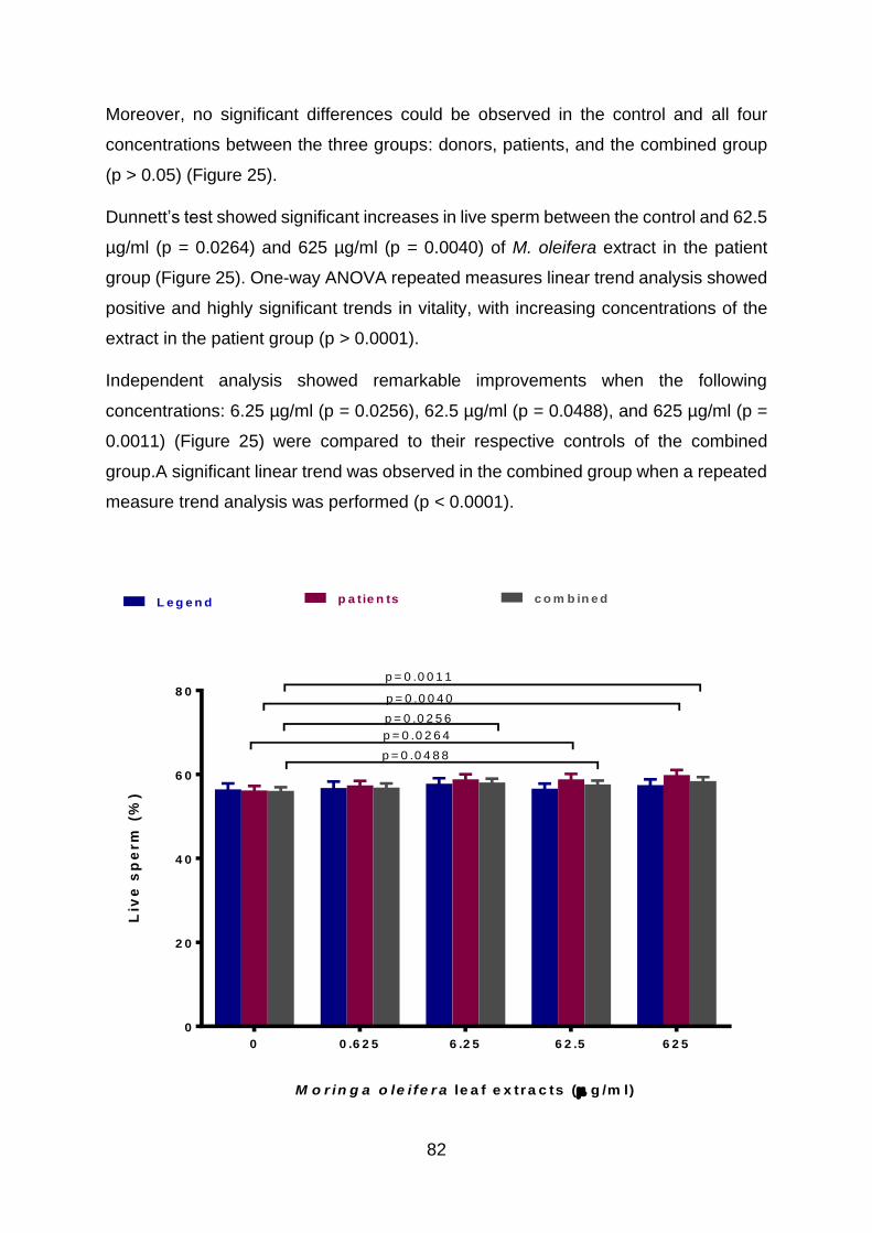
82
Moreover, no significant differences could be observed in the control and all four
concentrations between the three groups: donors, patients, and the combined group
(p > 0.05) (Figure 25).
Dunnett’s test showed significant increases in live sperm between the control and 62.5
µg/ml (p = 0.0264) and 625 µg/ml (p = 0.0040) of M. oleifera extract in the patient
group (Figure 25). One-way ANOVA repeated measures linear trend analysis showed
positive and highly significant trends in vitality, with increasing concentrations of the
extract in the patient group (p > 0.0001).
Independent analysis showed remarkable improvements when the following
concentrations: 6.25 µg/ml (p = 0.0256), 62.5 µg/ml (p = 0.0488), and 625 µg/ml (p =
0.0011) (Figure 25) were compared to their respective controls of the combined
group.A significant linear trend was observed in the combined group when a repeated
measure trend analysis was performed (p < 0.0001).
M o r in g a o le i fe r a le a f e x tra c ts ( g /m l)
Liv
e s
pe
rm
(%
)
0 0 .6 2 5 6 .2 5 6 2 .5 6 2 5
0
2 0
4 0
6 0
8 0
L e g e n d p a tie n ts c o m b in e d
p = 0 .0 4 8 8
p = 0 .0 2 6 4
p = 0 .0 2 5 6
p = 0 .0 0 4 0
p = 0 .0 0 1 1

83
Figure 25: Percentage of live sperm after 1hr incubation with M. oleifera in vitro. Mean ± SEM. Blue
bars represent the mean values of donors (N = 40), purple bars are for patients (N = 30), and grey bars
are for the combined group (patients and donors) (N = 70).
4.4.1. Correlation of sperm vitality with other functional parameters
The relationship between sperm vitality and functional parameters is summarised in
Table 8. Vitality and MMP integrity showed no relationship in the donor group (r = 0.20,
p = 0.7833). Also, no relationship was noted with percentages of sperm capacitation,
acrosome reaction (r = 0.30, p = 0.6833), and ROS and DNA fragmentation (r = -0.30,
p = 0.6833) of donors.
No correlation was observed between live sperm with intact MMP in patients and in
the combined group (r = 0.90, p = 0.0833). However, a strong, positive and significant
relationship was seen with the percentage of the acrosome reaction in the patient (r =
1.00, p = 0.0167). When the percentage of live sperm was correlated with ROS-
positive spermatozoa and DNA fragmentation in patients and the combined groups,
no link was established (r = -0.80, p = 0.1333). Moreover, correlation analysis of
capacitation and DNA-fragmented sperm in the patients showed no relationship (r = -
0.90, p = 0.0833). In addition, no association was shown with acrosome-reacted sperm
of the combined group (r = -0.10, p = 0.9500).

84
Table 8: Correlation of sperm vitality (live) and functional parameters
4.5. Effects of M. oleifera leaf extracts on sperm mitochondrial membrane
potential (∆ψm) in vitro
After incubation with increasing concentrations of M. oleifera (0, 0.625. 6.25, 62.5 and
625 µg/ml), no significant changes in the percentage of intact mitochondrial membrane
potential in sperm were observed when compared to the control (p > 0.05) (Figure 26)
in the donor group. However, a statistically significant increase in the percentage of
MMP-intact sperm of the patients after exposure to M. oleifera extracts was observed
(p = 0.0262). A significant increase in the percentage of intact MMP was observed at
625 µg/ml in the patient group in comparison with the untreated group (p = 0.0094)
(Figure 26). Similarly, increments in MMP-intact sperm at the highest concentration of
the plant were observed in the combined group (p = 0.0277) (Figure 26). Conversely,
as was the case with the percentage of live sperm, no improvements in MMP were
observed between the donor control and the therapeutic dose of 625 µg/ml (p > 0.05).
The control and low concentrations of M. oleifera did not produce statistically
significant differences between donors and patients: 0.625 µg/ml (p = 0.0904) and
6.25 µg/ml (p = 0.0887), and the combined sperm MMP (p > 0.05). In contrast, MMP
Parameters Donors Patient Combined
r p r r R r
MMP-intact (%) 0.20 0.7833 0.90 0.0833 0.90 0.0833
ROS-positive (%) -0.30 0.6833 -0.80 0.1333 -0.80 0.1333
Capacitation (%) 0.30 0.6833 -0.90 0.0833 -0.50 0.4500
Acrosome reaction (%)
0.30 0.6833 1.00 0.0167 -0.10 0.9500
DNA fragmentation (%)
-0.20 0.7833 -0.90 0.0833 -0.80 0.1333

85
in patients was significantly more improved than in donors at concentrations 62.5
µg/ml (p = 0.0052) and 625 µg/ml (p = 0.0077) (Figure 26).
The repeated measure showed a significant positive trend in MMP with increasing
concentrations of M. oleifera (p < 0.0001) in the patient sperm. No trend was seen in
the donor and combined groups.
M o r in g a o le i fe r a le a f e x tra c ts ( g /m l)
Inta
ct-
mit
oc
ho
nd
ria
l
me
mb
ra
ne
po
ten
tia
l (%
)
0 0 .6 2 5 6 .2 5 6 2 .5 6 2 5
0
2 0
4 0
6 0
8 0
d o n o rs p a tie n ts c o m b in e d
p = 0 .0 0 9 4
p = 0 .0 2 7 7
p = 0 .0 0 7 7p = 0 .0 0 5 2
Figure 26. Percentage-intact mitochondrial membrane potential (MMP) spermatozoa after 1 hr
incubation with M. oleifera in vitro. Means ± SEM. Blue bars represent the mean values of donors (N =
40), purple bars are for patients (N = 30), and grey bars are for the combined group (patients and
donors) (N = 70).

86
4.5.1 Correlation of sperm-intact mitochondrial membrane potential and
functional parameters
Mitochondrial membrane potential intactness of sperm was correlated with other
functional parameters in three groups, and results are displayed in Table 9. Intactness
of sperm MMP showed no relationship with ROS-positive and DNA-fragmented sperm
(r = -0.30, p = 0.6833), capacitation (r = 0.30, p = 0.6833), and acrosome reaction (r
=-0.70, p = 0.2333), respectively, in the donor’s group. In patients, acrosome reaction
displayed no significant correlation with MMP (r = 0.90, p = 0.0833). No relationship
was seen between MMP and ROS production (r = -0.90, p = 0.0833), while on the
contrary, negative strong correlations were observed with capacitation (r = -1.00, p =
0.0167) and DNA fragmentation (r = -1.00, p = 0.0167) in patients and the combined
groups. Despite relationship observed with other parameters in the combined group,
MMP showed no relationship with capacitation (r = -0.70, p = 0.2333).
Table 9: Correlation of MMP-intact sperm with functional parameters
Parameters Donors Patient Combined
r p r p r p
ROS-positive (%) -0.30 0.6833 -0.90 0.0833 -0.90 0.0833
Capacitation (%) 0.30 0.6833 -1.00 0.0167 -0.70 0.2333
Acrosome reaction (%) -0.70 0.2333 0.90 0.0833 0.00 >0.9999
DNA fragmentation
(%)
-0.30 0.6833 -1.00 0.0167 -0.90 0.0833
4.6. Effects of M. oleifera on reactive oxygen species-positive (ROS)
spermatozoa in vitro
M. oleifera leaf extracts showed remarkable changes in sperm superoxide free
radicals scavenging, following 1-hour incubation. Overall, M. oleifera aqueous extracts
induced a significant decrease in sperm intracellular ROS production (ROS-positive

87
spermatozoa) at higher concentrations of the extract in all groups. On the other hand,
M. oleifera leaf extracts significantly reduced spermatozoa intracellular reactive
oxygen species (ROS) production in the patient group, even at moderate
concentrations tested compared to the control as follows; 6.25 µg/ml (p = 0.0350) (not
displayed on the graph), and high concentration, 62.5 µg/ml (p < 0.0001). When the
control was compared to concentration 625 µg/ml in all groups, a highly significant
reduction was observed (p < 0.0001) (Figure 27).
ROS-positive spermatozoa were significantly higher in the control, 0.625 µg/ml, 6.25
µg/ml, 62.5 µg/ml, and 625 µg/ml of patients rather than donors, which were (p <
0.0001, p = 0.0033, and p = 0.0043) and the combined group (p = 0.0160, p = 0.0063,
and p = 0.0427) (Figure 27). Correspondingly, ROS-positive sperm in the combined
group was slightly higher than in the healthy donor control group (p = 0.0641) (Figure
27), 0.625 µg/ml (p = 0.0331), 6.25 µg/ml (p = 0.0033) and 62.5 µg/ml (p = 0.0027).
Furthermore, ROS positive spermatozoa in patients was significantly higher that of the
combined group treated with 0.625 µg/ml M. oleifera (p = 0.0068) (Figure 27). The
inhibitory effect of M. oleifera at the highest dose of 625 µg/ml did not differ between
the combined group and donors (p > 0.05) as well as patients (p > 0.05).
The one-way ANOVA repeated measures trend analysis revealed a highly significant
inverse tendency in ROS-positive spermatozoa in a dose-dependent manner, across
all three groups (p < 0.0001).

88
M o rin g a o le ife ra le a f e x tra c ts (g /m l)
Re
ac
tiv
e o
xy
ge
n s
pe
cie
s-p
os
itiv
e (
%)
0 0 .6 2 5 6 .2 5 6 2 .5 6 2 5
0
2 0
4 0
6 0
8 0
d o n o rs p a tie n ts c o m b in e d
p < 0 .0 0 0 1
p = 0 .0 0 4 3
p < 0 .0 0 0 1 p = 0 .0 1 6 0
p < 0 .0 0 0 1
p < 0 .0 0 0 1
p < 0 .0 0 0 1 p = 0 .0 0 6 8
p = 0 .0 3 3 1
p = 0 .0 0 3 3 p < 0 .0 0 0 1
p = 0 .0 0 6 8
p = 0 .0 4 2 0
p = 0 .0 0 2 7
Figure 27: Percentage ROS-positive spermatozoa after 1 hr incubation with M. oleifera in vitro. Means
± SEMs. Blue bars represent the mean values of donors (N = 40), purple bars are for patients (N = 30),
and grey bars are for combined group (patients and donors) (N = 70).
4.6.1 Correlation of reactive oxygen species with functional parameters
Sperm ROS production were correlated with other sperm functional characteristics as
shown in Table 10. The percentage of ROS-positive sperm with capacitated sperm
showed no relationship in donors (r = 0.70, p = 0.2333), patients, and the combined
group (r = 0.90, p = 0.0833). Similarly, no association could be established between
ROS-positive spermatozoa with acrosome reaction in donors (r = 0.00, p > 0.9999),
patients, and the combined group (r = 0.10, p = 0.9500), respectively. Interestingly,
sperm ROS production was strongly positively correlated with the percentage of sperm
with DNA fragmentation in all groups (r = 1.00, p = 0.0167). Patient ROS-positive

89
sperm displayed no significant correlation with DNA fragmentation (r = 0.90, p =
0.0833).
Table 10: Correlation of reactive oxygen species production with functional
parameters
4.7. Effects of M. oleifera leaf extracts on sperm DNA fragmentation in vitro
Aqueous M. oleifera extracts in higher concentrations improved DNA intactness in all
groups tested (Figure 28). However, low concentrations (0.625 µg/ml, and 6.25 µg/ml)
of M. oleifera did not significantly change the percentage of spermatozoa with DNA
fragmentation (p > 0.05) when compared to their controls (Figure 28).
On the other hand, incubation with higher M. oleifera concentrations of 62.5 µg/ml (p
= 0.0167), and 625 µg/ml) (p < 0.0001) of the extract resulted in a significant decrease
in the percentage of TUNEL-positive sperm (DNA-fragmented spermatozoa) in
comparison with the control (Figure 28).
Percentage of sperm DNA fragmentation in the patient group dropped significantly at
doses 62.5 µg/ml (p = 0.0082), and 625 µg/ml (p = 0.0006) in comparison with the
respective controls (Figure 28).
Parameters Donors Patient Combined group
r p r p r p
Capacitation (%) 0.70 0.2333 0.90 0.0833 0.90 0.0833
Acrosome reaction (%)
0.00 >0.9999 0.10 0.9500 0.10 0.9500
DNA fragmentation (%)
1.00 0.0167 0.90 0.0833 1.00 0.0167

90
No differences were measured between the control, 6.25 µg/ml, 62.5 µg/ml, and 625
µg/ml of the three groups; donors, patients, and the combined group (p > 0.05) (Figure
28). However, marginal significance in sperm with DNA fragmentation could be
observed between the donors and patients at the lowest dose of M. oleifera (0.625
µg/ml) (p = 0.0380). Repeated measures correspondingly showed significant negative
linear trends in the percentage of spermatozoa with DNA fragmentation with
increasing concentrations of M. oleifera extracts after 1-h incubation (p < 0.0001) in
the donors, patients, and the combined groups.
M o rin g a o le ife ra le a f e x tra c ts (g /m l)
Sp
erm
DN
A
fra
gm
en
tati
on
(%
)
0 0 .6 2 5 6 .2 5 6 2 .5 6 2 5
0
2 0
4 0
6 0
8 0
d o n o rs p a tie n ts c o m b in e d
p < 0 .0 0 0 1
p < 0 .0 0 0 1
p < 0 .0 0 0 1
p = 0 .0 0 8 2
p = 0 .0 0 0 1
p = 0 .0 3 8 0
Figure 28. Percentage of sperm DNA fragmentation after 1 hr incubation with M. oleifera in vitro. Means
± SEM. Blue bars represent the mean values of donors (N = 40), purple bars are for patients (N = 30),
and grey bars are for the combined group (patients and donors) (N = 70).

91
4.7.1 Correlation of sperm DNA fragmentation and functional parameters
Sperm DNA fragmentation was correlated with functional parameters, capacitation
and acrosome reaction and the Spearman analysis results are shown in Table 11.
DNA fragmentation showed no relationship with capacitation in donors (r = 0.70, p =
0.2333), patients, and the combined group (r = 0.90, p = 0.0833), respectively.
Similarly, no relationship was established between sperm with acrosome reaction in
the combined (r = 0.10, p=0.9500) and the donor groups (r = 0.00, p > 0.9999).
Contrarily, a significantly positive relationship was demonstrated between patients’
DNA fragmentation and the percentage of acrosome-reacted spermatozoa (r = 1.00,
p = 0.0167).
Table 11: Correlation of sperm DNA fragmentation and functional parameters
Parameters Donors Patient Combined group
r p r p r p
Capacitation (%) 0.70 0.2333 0.90 0.0833 0.90 0.0833
Acrosome reaction (%)
0.00 >0.9999 1.00 0.0167 0.10 0.9500
4.8 Effects of M. oleifera on sperm capacitation and acrosome reaction in vitro
Low concentrations, as observed with previous parameters, did not affect the sperm
capacitation-acrosomal status. Based on the Hoechst 33258 counterstain, three
statuses could be recorded for capacitation and acrosome reaction: F (uncapacitated-
acrosome-intact spermatozoa), B (capacitated acrosome-intact spermatozoa) and AR
(capacitated acrosome-reacted spermatozoa) patterns. Percentage of uncapacitated
and acrosome-intact spermatozoa (F pattern) significantly increased at 625 µg/ml
when compared to the control group (p = 0.0047) (Figure 29a) in the donor group. A
significant decrease in the lower treatment groups was observed when compared to
the highest concentration (625 µg/ml) as follows (0.625 µg/ml (p = 0.0022), 6.25 µg/ml
(p = 0.0009), and 62.5 µg/ml (p = 0.0023), respectively.
92
No changes in the percentage of uncapacitated (F pattern) spermatozoa were
observed in patients after treatment with M. oleifera (p > 0.05) (Figure 29a). The
analysis of the combined group revealed a marginally significant increase in the
percentage of F pattern sperm after M. oleifera exposure in the combined group (p =
0.0113) (Figure 29a).
The control and 62.5 µg/ml in F pattern of the patient group was slightly higher than
that of the donor group (p = 0.0136 and p = 0.0095) (Figure 29a).Furthermore,
uncapacitation was consistently higher in the patient group than in combined group in
all M. oleifera concentrations; control (p = 0.0356), 0.625 µg/ml (p = 0.0050), 6.25
µg/ml (p = 0.0067), 62.5 µg/ml, and 625 µg/ml (p < 0.0001) (Figure 29a). Trend
analysis showed a significant increase in uncapacitated (F pattern) sperm in the donor
(p = 0.0036), and the combined group (p = 0.0032), and no trends were observed in
patients when increasing concentrations were applied.
The extracts did not alter sperm capacitation (B pattern) in the donor, patient, and the
combined groups (p > 0.05) (Figure 29b). Dunnets test showed a marginally significant
decrease in capacitated acrosome-intact sperm at concentration 625 µg/ml in the
patient group (p = 0.0491) (Figure 29b). Trend analysis showed significant increases
in uncapacitated (F pattern) sperm in the donor group (p = 0.0036), and in the
combined group (p = 0.0032), while no trends in patients were observed as the dose
increased.
Capacitation in the donors appeared to be significantly higher than in patients in all
concentration tested; 0 µg/ml (p < 0.0001), 0.625 µg/ml, 6.25 µg/ml, 62.5 µg/ml, and
625 µg/ml (p < 0.0001) (Figure 29b). Similarly, percentage of capacitated spermatozoa
was significantly higher in the combined group than peatients at the following
concentrations: 0.625 µg/ml (p = 0.0032), 6.25 µg/ml (p = 0.0009) and 62.5 µg/ml (p =
0.0035). Furthermore, the same increases in donors’ B pattern spermatozoa in
comparison with the combined group were observed at concentrations; 0.625 µg/ml
(p = 0.0185), 6.25 µg/ml (p = 0.0077), 62.5 µg/ml (p = 0.0221), and 625 µg/ml (p =
0.0083) (Figure 29b). The combined group also showed significant differences in B
pattern sperm when compared to patients at doses: control (p = 0.0008) (Figure 29b),
0.625 µg/ml (p = 0.0032), 6.25 µg/ml (p = 0.0009), and (625 µg/ml) (p = 0.0001) (Figure
29b). However, linear trends were observed in the percentage of capacitated

93
acrosome-intact spermatozoa (B pattern) in the combined group (p = 0.0270) and
patients (p < 0.0001) as the concentrations of M. oleifera increased.
M o rin g a o le ife ra le a f e x tra c ts ( g /m l)
Un
ca
pa
cit
ati
on
(%
)
0 0 .6 2 5 6 .2 5 6 2 .5 6 2 5
0
1 0
2 0
3 0
4 0
5 0
d o n o rs p a tie n ts c o m b in e d
p = 0 .0 0 4 7
p = 0 .0 1 3 6 p = 0 .0 3 5 6
p = 0 .0 0 1 3
p < 0 .0 0 0 1
p = 0 .0 1 1 3
p = 0 .0 0 5 6
p = 0 .0 0 6 7p = 0 .0 0 9 5
M o rin g a o le ife ra le a f e x tra c ts ( g /m l)
Ca
pa
cit
ata
tio
n (
%)
0 0 .6 2 5 6 .2 5 6 2 .5 6 2 5
0
1 0
2 0
3 0
4 0
5 0
p = 0 .0 4 9 1
p < 0 .0 0 0 1
p = 0 .0 0 0 8p < 0 .0 0 0 1
p = 0 .0 0 1 0
p = 0 .0 0 8 3
p = 0 .0 1 0 1
p = 0 .0 0 3 2
p < 0 .0 0 0 1
p < 0 .0 0 0 1
p < 0 .0 0 0 1
p = 0 .0 0 7 7
p = 0 .0 0 0 9
p = 0 .0 0 2 2
p < 0 .0 0 0 1
p = 0 .0 0 3 5
a )
b )

94
Figure 29: Percentage of uncapacitated (F pattern) (a) and capacitated (B pattern) spermatozoa (b)
after 1 hr incubation with M. oleifera in vitro. Means ± SEM. Blue bars represent the mean values of
donors (N = 40), purple bars are for patients (N = 30), and grey bars are for the combined group (patients
and donors) (N = 70).
The percentage of capacitated acrosome-reacted (AR pattern) spermatozoa was
slightly but significantly decreased in the donor group compared to the control p =
0.0378) (Figure 30). Similarly, the decrease in acrosome-reacted sperm at M. oleifera
highest concentration 625 µg/ml was significantly different compared to the lower
concentrations tested of 0.625 µg/ml (p = 0.0035), 6.25 µg/ml (p = 0.0096), and 62.5
µg/ml (p = 0.0079) in the donor group. No changes in sperm acrosome reaction
occurred in either the patient or the combined groups (Figure 30) after treatment with
M. oleifera extracts.
No significant changes could be recorded in percentage acrosome reaction between
the donors, patients, and combined groups in the control, 0.625 µg/ml, 6.25 µg/ml, and
62.5 µg/ml (p > 0.05) (Figure 30). On the other hand, treatment with the highest
concentration of 625 µg/ml, patient’s acrosome reaction was significantly higher
compared to the donor (p = 0.0004), and the combined groups (p = 0.0449) (Figure
30).
Furthermore, increasing concentrations of M. oleifera extract caused a positive trend
in the percentage of acrosome reaction (AR pattern) in donors (p = 0.0208), while no
significant trends were observed in the patients and combined groups (p > 0.05)
respectively.

95
M o rin g a o le ife ra le a f e x tra c ts (g /m l)
Ac
ros
om
e r
ea
cti
on
(%
)
0
0.6
25
6.2
5
62.5
625
0
1 0
2 0
3 0
4 0
p = 0 .0 4 4 9p = 0 .0 0 0 4
p = 0 .0 3 7 8(c )
p = 0 .0 0 7 9
p = 0 .0 0 9 6
p = 0 .0 0 3 5
Figure 30. Percentage sperm of acrosome reaction after 1 hr incubation with M. oleifera in vitro. Mean
± SEM. Blue bars represent the mean values of donors (N = 40), purple bars are for patients (N = 30),
and grey bars are for combined group (patients and donors) (N = 70).
4.8.1 Correlation of sperm capacitation and acrosome reaction
Two interrelated sperm functional processes were correlated as shown in Table 12.
Capacitation and acrosome-reacted spermatozoa showed no relationship in the donor
and the combined group (r = 0.30, p = 0.6833) as well as (r = -0.90, p = 0.0833).
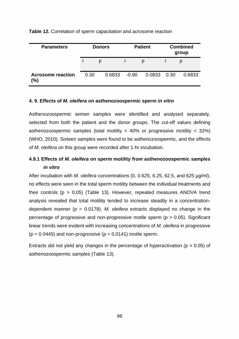
96
Table 12. Correlation of sperm capacitation and acrosome reaction
4. 9. Effects of M. oleifera on asthenozoospermic sperm in vitro
Asthenozoospermic semen samples were identified and analysed separately,
selected from both the patient and the donor groups. The cut-off values defining
asthenozoospermic samples (total motility < 40% or progressive motility < 32%)
(WHO, 2010). Sixteen samples were found to be asthenozoospermic, and the effects
of M. oleifera on this group were recorded after 1-hr incubation.
4.9.1 Effects of M. oleifera on sperm motility from asthenozoospermic samples
in vitro
After incubation with M. oleifera concentrations (0, 0.625, 6.25, 62.5, and 625 µg/ml),
no effects were seen in the total sperm motility between the individual treatments and
their controls (p > 0.05) (Table 13). However, repeated measures ANOVA trend
analysis revealed that total motility tended to increase steadily in a concentration-
dependent manner (p = 0.0178). M. oleifera extracts displayed no change in the
percentage of progressive and non-progressive motile sperm (p > 0.05). Significant
linear trends were evident with increasing concentrations of M. oleifera in progressive
(p = 0.0445) and non-progressive (p = 0.0141) motile sperm.
Extracts did not yield any changes in the percentage of hyperactivation (p > 0.05) of
asthenozoospermic samples (Table 13).
Parameters Donors Patient Combined group
r p r p r p
Acrosome reaction (%)
0.30 0.6833 -0.90 0.0833 0.30 0.6833

97
Table 13: Effects of M. oleifera on sperm motility parameters on asthenozoospermic
samples in vitro
Kinematic parameters investigated in asthenozoospermic samples were sperm
percentage linearity index (LIN) (%), straightness index (STR) (%), wobble / oscillation
index (WOB) (%), flagella speeds: curvilinear velocity (VCL) (µms-1), straight-line /
rectilinear velocity (VSL) (µms-1), and average path velocity (VAP) (µms-1) as well as
other essential spatial sperm kinematic characteristics, the amplitude of lateral head
displacement (ALH) (µm) and flagella beat cross frequency (BCF) (Hz). Results on the
effect of M. oleifera extracts on asthenozoospermic kinematics are displayed in Table
13.
Various concentrations of M. oleifera extract (0, 0.625, 6.25, 62.5, and 625 µg/ml) did
not cause any changes in all of the sperm kinematics in the asthenozoospermic group
(p > 0.05) (Table 14).
Sperm motility parameters (%)
Moringa oleifera leaf extract concentrations (µg/ml) p-value
0 0.625 6.25 62.5 625
Total motility (All grades)
16.0 ± 12.3
16.3 ± 2.0
17.5 ± 1.9
17.5 ± 1.9
22.1 ± 2.7
0.3414
Progressive (grade A+B)
7.1 ± 1.5 9.0 ± 1.8 10.0 ±
1.8 9.0 ± 2.0 13.5±1.4
0.2413
Non-progressive (Grade C)
6.2 ±1.0 6.6 ±1.1 6.9 ±1.1 7.3 ±0.7 8.4 ±1.1 0.418
6
Hyperactivation
1.0 ±0.5 2.3 ± 1.4 3.2 ±1.5 2.5 ± 1.2 5.8± 3.5 0.975
8
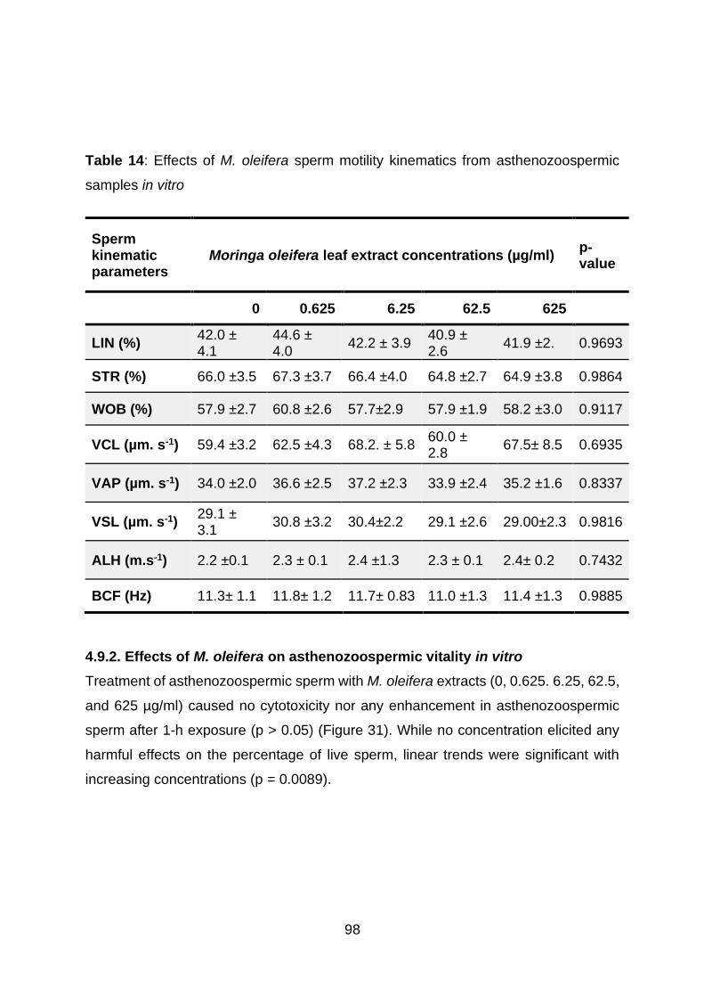
98
Table 14: Effects of M. oleifera sperm motility kinematics from asthenozoospermic
samples in vitro
4.9.2. Effects of M. oleifera on asthenozoospermic vitality in vitro
Treatment of asthenozoospermic sperm with M. oleifera extracts (0, 0.625. 6.25, 62.5,
and 625 µg/ml) caused no cytotoxicity nor any enhancement in asthenozoospermic
sperm after 1-h exposure (p > 0.05) (Figure 31). While no concentration elicited any
harmful effects on the percentage of live sperm, linear trends were significant with
increasing concentrations (p = 0.0089).
Sperm kinematic parameters
Moringa oleifera leaf extract concentrations (µg/ml) p-value
0 0.625 6.25 62.5 625
LIN (%) 42.0 ± 4.1
44.6 ± 4.0
42.2 ± 3.9 40.9 ± 2.6
41.9 ±2. 0.9693
STR (%) 66.0 ±3.5 67.3 ±3.7 66.4 ±4.0 64.8 ±2.7 64.9 ±3.8 0.9864
WOB (%) 57.9 ±2.7 60.8 ±2.6 57.7±2.9 57.9 ±1.9 58.2 ±3.0 0.9117
VCL (µm. s-1) 59.4 ±3.2 62.5 ±4.3 68.2. ± 5.8 60.0 ± 2.8
67.5± 8.5 0.6935
VAP (µm. s-1) 34.0 ±2.0 36.6 ±2.5 37.2 ±2.3 33.9 ±2.4 35.2 ±1.6 0.8337
VSL (µm. s-1) 29.1 ± 3.1
30.8 ±3.2 30.4±2.2 29.1 ±2.6 29.00±2.3 0.9816
ALH (m.s-1) 2.2 ±0.1 2.3 ± 0.1 2.4 ±1.3 2.3 ± 0.1 2.4± 0.2 0.7432
BCF (Hz) 11.3± 1.1 11.8± 1.2 11.7± 0.83 11.0 ±1.3 11.4 ±1.3 0.9885

99
M o rin g a o le ife ra le a f e x tra c ts (g /m l)
Liv
e s
pe
rm
(%
)
0 0 .6 2 5 6 .2 5 6 2 .5 6 2 5
0
2 0
4 0
6 0
8 0
Figure 31: Percentage of live asthenozoospermic sperm after treatment with M. oleifera extracts for 1
hr in vitro. Mean ± SEM.
4.9.3. Effects of M. oleifera extract on mitochondrial membrane potential (MMP-
intact) in asthenozoospermic samples in vitro
M. oleifera extracts (0, 0,625, 6.25, 62.5, and 625 µg/ml) did not cause any significant
change in the percentage of sperm with intact MMP of asthenozoospermic samples (p
> 0.05) (Figure 32). Repeated measures showed an increase of asthenozoospermic
MMP-intact in the post treatment linear trends (p = 0.0051).

100
M o rin g a o le ife ra le a f e x tra c ts (g /m l)
Inta
ct
mit
oc
ho
nd
ria
l
me
mb
ra
ne
po
ten
tia
l (%
)
0 0 .6 2 5 6 .2 5 6 2 .5 6 2 5
0
2 0
4 0
6 0
8 0
Figure 32: Percentage of asthenozoospermic MMP-intact sperm after 1hr incubation with M. oleifera in
vitro. Mean ± SEM.
4.9.4. Effects of M. oleifera on asthenozoospermic reactive oxygen species-
positive spermatozoa in vitro
M. oleifera significantly reduced the percentage of ROS-positive sperm in the
asthenozoospermic group (p < 0.0001) in a concentration-dependent manner (Figure
33). An insignificant decrease in ROS was shown at 62.5 µg/ml (p > 0.05), followed by
a pronounced significant decrease at 625 µg/ml (p = 0.0005) (Figure 33) compared to
the control group. Additionally, a significant decrease was observed when comparing
the highest (625 µg/ml) with lower concentrations (0.625 µg/ml; p < 0.0001) and (6.25
µg/ml; p = 0.0009), respectively (Figure 33). ANOVA linear trends also showed a highly
significant decrease in ROS-positive asthenozoospermic spermatozoa when the M.
oleifera dose was increased (p < 0.0001).

101
M o rin g a o le ife ra le a f e x tra c ts (g /m l)
Re
ac
tiv
e o
xy
ge
n
sp
ec
ies
-po
sit
ive
(%
)
0 0 .6 2 5 6 .2 5 6 2 .5 6 2 5
0
2 0
4 0
6 0
8 0
p < 0 .0 0 0 1
p = 0 .0 0 0 5
p = 0 .0 7 5 0
p = 0 .0 0 0 9
Figure 33: Percentage of ROS-positive asthenozoospermic sperm after 1 hr incubation with M. oleifera
in vitro. Mean ± SEM.
4.9.5. Effects of M. oleifera on DNA-fragmented sperm in asthenozoospermic
samples in vitro
A significant decrease in asthenozoospermic DNA fragmentation was observed when
the two highest concentrations, 62.5 µg/ml (p = 0.0439) and 625 µg/ml (p = 0.0094),
were compared to the control (Figure 33). The highest concentration of M. oleifera
(625 µg/ml) differed significantly when compared to the concentrations of 0.625 µg/ml
(p = 0.0065) and non-significantly to 6.25 µg/ml (p > 0.05).Consequently, a highly
significant negative linear trend in sperm DNA fragmentation was observed with
increasing concentrations of M. oleifera extract (p = 0.0007).
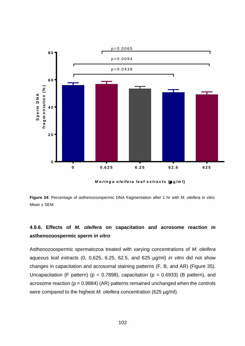
102
M o rin g a o le ife ra le a f e x tra c ts ( g /m l)
Sp
erm
DN
A
fra
gm
en
tati
on
(%
)
0 0 .6 2 5 6 .2 5 6 2 .5 6 2 5
0
2 0
4 0
6 0
8 0
p = 0 .0 4 3 9
p = 0 .0 0 9 4
p = 0 .0 0 6 5
Figure 34: Percentage of asthenozoospermic DNA fragmentation after 1 hr with M. oleifera in vitro.
Mean ± SEM.
4.9.6. Effects of M. oleifera on capacitation and acrosome reaction in
asthenozoospermic sperm in vitro
Asthenozoospermic spermatozoa treated with varying concentrations of M. oleifera
aqueous leaf extracts (0, 0,625, 6.25, 62.5, and 625 µg/ml) in vitro did not show
changes in capacitation and acrosomal staining patterns (F, B, and AR) (Figure 35).
Uncapacitation (F pattern) (p = 0.7898), capacitation (p = 0.6933) (B pattern), and
acrosome reaction (p = 0.9884) (AR) patterns remained unchanged when the controls
were compared to the highest M. oleifera concentration (625 µg/ml).
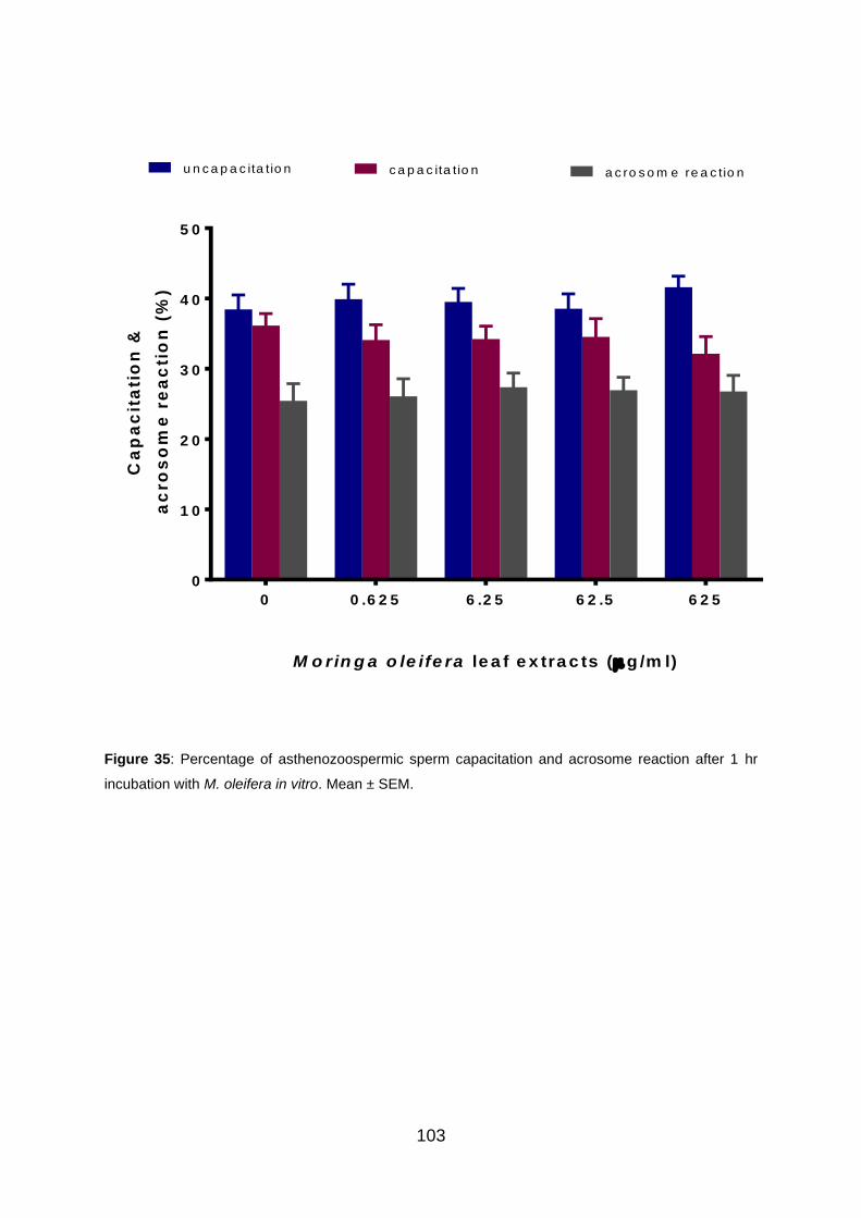
103
M o rin g a o le ife ra le a f e x tra c ts (g /m l)
Ca
pa
cit
ati
on
&
ac
ro
so
me
re
ac
tio
n (
%)
0 0 .6 2 5 6 .2 5 6 2 .5 6 2 5
0
1 0
2 0
3 0
4 0
5 0
u n c a p a c ita tio n c a p a c ita tio n a c ro s o m e re a c tio n
Figure 35: Percentage of asthenozoospermic sperm capacitation and acrosome reaction after 1 hr
incubation with M. oleifera in vitro. Mean ± SEM.

104
4.9.7. Correlation of asthenozoospermic sperm parameters
The results of the Spearman analysis for correlations of all parameters is displayed in
Table 15. The percentage of total, progressive, and hyperactivated motility grades of
asthenozoospermic sperm were significantly and positively correlated (r = 1.00, p =
0.0167). The three types of motility further showed no correlations with vitality (r =
0.90, p = 0.0833), MMP intactness (r = 0.50, p = 0.4500), and acrosome reaction (r =
0.70, p = 0.2333). Similarly, no correlation was observed between three types of
motility and sperm ROS production, and DNA fragmentation (r = -0.80, p = 0.1333)
and capacitation (r = -0.70, p = 0.2333).
The percentage live sperm and intact MMP in the asthenozoospermic group also did
not show any correlation (r = 0.70, p = 0.2333), nor did it show a correlation with
acrosome reaction (r = 0.60, p = 0.3500), sperm ROS production (r = -0.90, p =
0.0833), DNA fragmentation (r = -0.90, p = 0.0833), or capacitation (r = -0.60, p =
0.3500). In addition, asthenozoospermic sperm MMP integrity displayed no
relationship with acrosome reaction (r = 0.10, p = 0.9500) and capacitation (r = -0.10,
p = 0.9500).
Reactive oxygen species (ROS) production by asthenozoospermic sperm showed a
significantly strong positive relationship with the percentage DNA fragmentation (r =
1.00, p = 0.0167). No association could be established between ROS production and
acrosome reaction (r = -0.50, p = 0.4500) as well as capacitation (r = 0.30, p = 0.6833).
Similar to ROS, DNA fragmentation of asthenozoospermic men was not correlated to
capacitation (r = 0.30, p = 0.6833) and acrosome reaction (r = -0.50, p = 0.4500).
Capacitation also showed no relationship with asthenozoospermic acrosome reaction
(r = 0.30, p = 0.6833).

105
Table 15: Correlation of asthenozoospermic sperm parameters.
Sperm parameters
Total motility
(%)
Progressive
motility (%)
Hyperactivated motility
(%)
Vitality live (%)
MMP-intact
(%)
ROS-positive
(%)
DNA fragmenta
tion (%)
Capacitation (%)
Acrosome
reaction (%)
r p r p r p r p r p r p r p r p r p
Total motility (%)
- - 1.00
0.0167
1.00 0.0167
0.90
0.0833
0.50 0.4500
-0.8
0
0.1333
-0.8
0
0.1333
-0.7
0
0.2333
0.70
0.2333
Progressive motility (%)
1.00
0.0167
- - - - 0.90
0.0833
0.50 0.4500
-0.8
0
0.1333
-0.8
0
0.1333
-0.7
0
0.2333
0.70
0.2333
Hyperactivated motility (%)
1.00
0.0167
0.90
0.0833
0.90 0.0833
0.90
0.0833
0.50 0.4500
-0.8
0
0.1333
-0.8
0
0.1333
-0.7
0
0.2333
0.70
0.2333
Vitality live (%) 0.90
0.0833
0.90
0.0833
0.90 0.0833
- - 0.70 0.2333
-0.9
0
0.0833
-0.9
0
0.0833
-0.6
0
0.3500
0.60
0.3500
MMP-intact (%) 0.50
0.4500
0.50
0.4500
0.50 0.4500
0.70
0.2333
- - -0.9
0
0.0833
-0.9
0
0.0833
-0.1
0
0.9500
0.10
0.9500
ROS-positive (%)
-0.8
0
0.1333
-0.8
0
0.1333
-0.80
0.1333
-0.9
0
0.0833
-0.90 0.0833
- - 1.00
0.0167
0.30
0.6833
-0.5
0
0.4500
DNA fragmentation (%)
-0.8
0
0.1333
-0.8
0
0.1333
-0.80
0.1333
-0.9
0
0.0833
-0.90 0.0833
1.00
0.0167
- - 0.30
0.6833
-0.5
0
0.4500
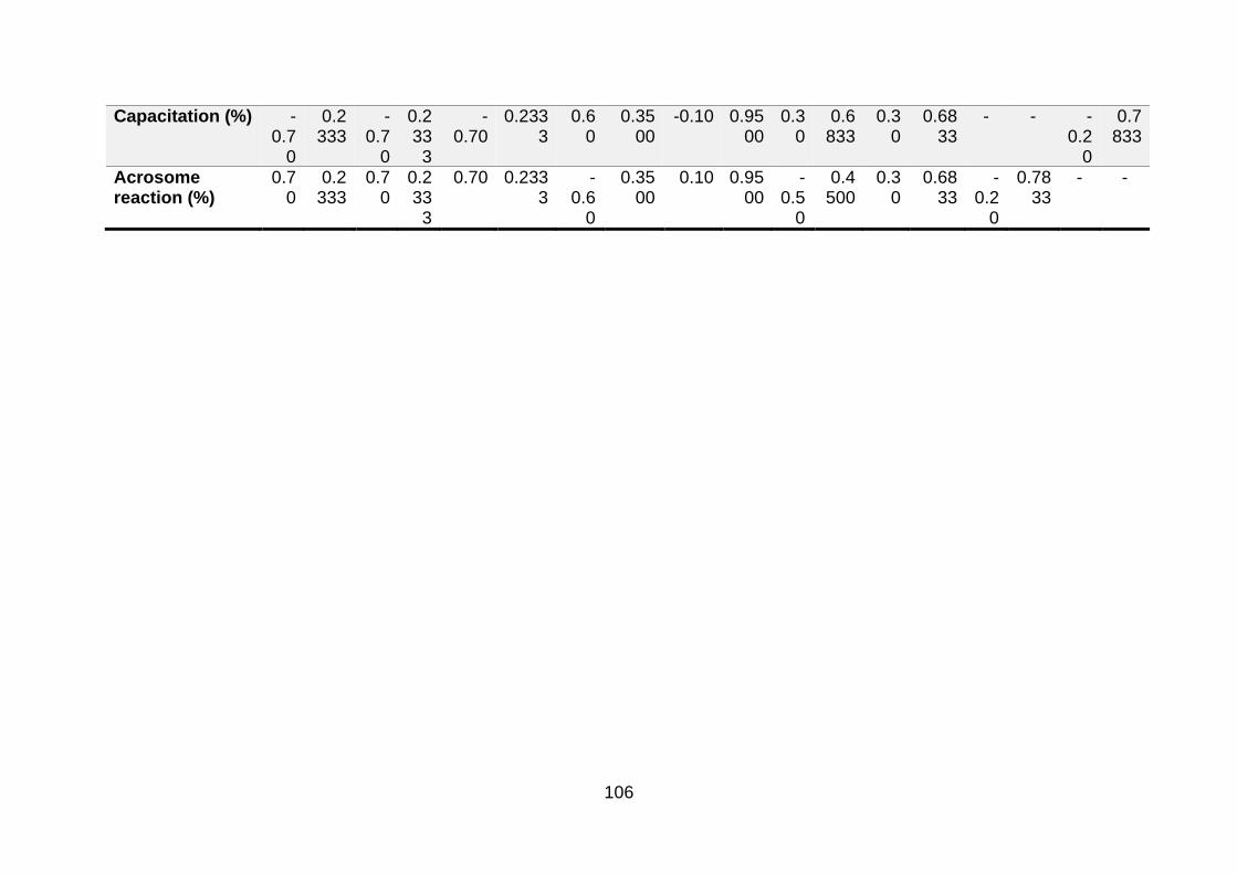
106
Capacitation (%) -0.7
0
0.2333
-0.7
0
0.2333
-0.70
0.2333
0.60
0.3500
-0.10 0.9500
0.30
0.6833
0.30
0.6833
- - -0.2
0
0.7833
Acrosome reaction (%)
0.70
0.2333
0.70
0.2333
0.70 0.2333
-0.6
0
0.3500
0.10 0.9500
-0.5
0
0.4500
0.30
0.6833
-0.2
0
0.7833
- -

107
CHAPTER 5
DISCUSSION
Plant remedies for sexual and reproductive dysfunctions and fertility improvement are
the oldest form of therapy to date., tThey are reliable and accessible. The fact that
nearly 80% of people rely chiefly on traditional herbal medicine (Yuan et al., 2016) is
an indication of the urgency to decipher the mechanisms responsible for the often
observed pharmacodynamics and pharmacokinetics of herbal medicines. Within the
context of male infertility and related reproductive issues, naturopathic systems such
as Ayurveda (Mahdi et al., 2011; Hussain et al., 2018); Siddha (Mohan et al., 2013);
Unani (Ahmad et al., 2008), Chinese (Dieterle et al., 2009); and African remedies
(Kolahdooz et al., 2014) have been benefitting men all over the world.
These plants have been prepared unempirically as unprocessed materials through
decoction, concoction, infusion, ashing, and taken as vegetables or condiments
(Abera, 2003; Ozioma and Chinwe, 2019). On the other hand, pharmaceutical
formulations have been semi-purified from plants for infertility (Nantia et al., 2009). A
review by Maridass and Britto (2008) reported that 50% of natural medicines and their
analogues in the pharmacopoeia were said to be botanical-based, while recent reports
indicated a proportion of 25% of pharmaceutical infertility therapies are derived from
plants (Jaradat and Zaid, 2019). Socio-economic reasons driving the use of traditional
herbs for sexual and reproductive restoration are related to the inherent limitations in
conventional therapies of fertility treatments such as their prohibitively high costs,
adverse side effects, and their limited availability to the majority of third world country
populations (Kaadaaga et al., 2014).
South Africans rely almost exclusively on plants for the provision of their health care
needs (Mbatha et al., 2012). The rising interest and demand for traditional medicine in
South Africa have been met with considerable concerns around legislative regulation
(Pefile, 2005.). Fortunately, the South African Government put forward a regulatory
framework in the Traditional Health Practitioners Act No 22 of 2007, according to which
traditional medical practices can be carried out safely and efficaciously in health care
delivery (Tshehla, 2015; Street, 2016) in South Africa.

108
For the safety of natural medicines to be ensured, thorough scientific validation of
claimed properties must be undertaken. Although its widespread use and
commercialisation fall under the regulation of a non-profit initiative, the Moringa
Development Association of South Africa (MDASA) (Mabapa et al., 2017), most of its
acclaimed activities still remain to be scientifically proven. However, M. oleifera’s
pharmacological properties have been studied at length, using in vivo and in vitro
models for efficacy assessments in nearly all body systems, and biological activities
were ascribed mainly to antioxidants (Gopalakrishnan et al., 2016; Vergara-Jimenez
et al., 2017). The effects of M. oleifera on LH and FSH, prolactin, testicular antioxidant
enzymes and testosterone synthesis had also been investigated (Bin-Meferij and El-
kott, 2015; Orji et al., 2016; Ododo et al., 2019). However, most studies that examined
how M. oleifera affects the male reproductive, sexual, and seminal functions, were
limited to animal models. Also, studies on the effects of M. oleifera on seminal
parameters were confined to basic semen characteristics such as motility,
morphology, and concentration (Afolabi et al., 2013), and did not examine advanced
sperm functional assays. To the best of my knowledge, no study to date has previously
evaluated the effects of M. oleifera leaves on the physiological functioning of human
spermatozoa in vitro. Given the abovementioned evidence, our study aimed to shed
light on in vitro effects of aqueous leaf extracts of M. oleifera on human spermatozoa
functioning.
5.1. Effects of M. oleifera leaf extracts on sperm motility in vitro
Sperm motility remains the lifeblood of male fertility evaluation as it best predicts the
fertilising capacity of a man (Shibahara et al., 1997; Turner, 2006). Essentially, sperm
requires all forms of motility to be able to fertilise an egg successfully. Complete lack
of motility is another form of infertility (Ortega et al., 2011). Immotile sperm cells, alive
or not, are incapable of first, penetrating cervical mucus and reaching the site of
fertilisation (Suarez and Pacey, 2006). Moreover, sperm needs to first move not in
tight circles, but in a straight line to ascent to the oocyte-cumulus complexes (Mansour
et al., 1995). Finally, sperm requires vigorous flagellar beating to penetrate the corona
radiata surrounding the oocyte, and fuse the oocyte for fertilisation (Wallach et al.,
1980; Skinner et al., 2019). Hence, several studies have emphasised the definitive

109
role of including sperm motility when evaluating the possible effects of medications in
the management of male infertility (Esteves, 2014).
The effects of M. oleifera leaf extract concentrations (0, 0.625, 6.25, 62.5, and 625
µg/ml) on various grades of motility, including flagellar and head kinematics, were
investigated for the donor, patient, and asthenozoospermic groups in vitro. In this
study, total, progressive, and non-progressive motility were investigated after
treatment with aqueous leaf extracts of M. oleifera in vitro. Donors and
asthenozoospermic groups did not show any alterations in sperm motion parameters
after treatment with M. oleifera. Similar results were recorded of no alteration to human
sperm motility with aqueous extracts of Cissampelos capensis rhizome (dawidjies)
(Shalaweh et al., 2015), roots of Mondia whitei (white ginger) (Tendwa, 2016),
ethanolic leaf extracts of Custanea sativa (sweet chestnut) (Biagi et al., 2019) in vitro.
The ineffectiveness of M. oleifera to restore sperm motility of asthenozoospermic to
normal in this study was similar to that recorded with Mucuna pruriens (velvet bean)
(Ahmad et al., 2008). Treatment with M. oleifera extracts showed significantly
increasing tendencies of forward progression of motile sperm in the
asthenozoospermic group.
In contrast, M. oleifera leaf extracts significantly improved sperm total, progressive,
and non-progressive motilities in the patient group and the combined groups in vitro.
Consequent to improvements in the enumerated motility grades by the aqueous leaf
extracts of M. oleifera, a highly significant reduction in non-motile spermatozoa in vitro
was observed. Although M. oleifera extract concentrations (0.625, 6.25, 62.5, and 625
µg/ml) showed no significant improvement in the donor and the combined groups, a
significant dose-dependent increasing trend was evident in all grades of motility in the
patient group.
Unfortunately, no group exhibited hyperactivated motility in all the treatments.
Kinematic motility parameters are useful as relatively objective and quantitative
indicators of sperm maturational status. Hence, their analysis assists in discriminating
sperm populations with fertilising capacity (van der Horst et al., 1999b; Spiropoulos,
2001). The effects of M. oleifera leaf extracts on hyperactivation in all groups was
further supported by no effects observed in sperm kinematics after treatment, that is,
ALH, VSL, VCL, VAP, and BCF. Tendwa (2016) also recorded no change in sperm

110
kinematics after treating human sperm with increasing aqueous extracts of M. whitei
in vitro.
Ideally, a substance that enhances hyperactivated motility would improve kinematics
related to sperm vigour such as increased VCL, VAP, ALH, and BCF, and reduce
parameters that are characteristic of linear progression such as LIN, STR, and VSL
(Mortimer and Mortimer, 1990; Cancel et al., 2000; Adamkovicova et al., 2016). To
support this, Shalaweh (2015) demonstrated that increasing concentrations of C.
capensis in vitro increased BCF and hyperactivation in human spermatozoa.
Interestingly, treatment with M. oleifera leaf extracts showed significant decreasing
trends in the percentage STR in patients and the combined groups.
Although being incomparable models, the effects of M. oleifera on sperm motility in
this study, were in agreement with previous findings in vivo. Increases in sperm motility
were observed when M. oleifera was administered concomitantly with
cyclophosphamide in Swiss albino rats (Nayak et al., 2016). Similarly, a coconut oil
and M. oleifera leaf extract mixture blocked spermicidal effects of mercury chloride in
male rats (Abarikwu et al., 2017). Mean sperm motility of bulls’ semen extended with
crude extracts of M. oleifera was significantly higher than the respective control
(Sokunbi et al., 2015). Total and progressive motility, velocities, and amplitude were
significantly increased with decreases in linearity in Bali bulls’ sperm after M. oleifera
supplementation (Syarifuddin et al., 2017). The differences in the in vivo and in vitro
effects of M. oleifera on sperm motility can in part be explained in terms of the inherent
methodological disparities in both models. In addition, in vivo models involve complex
systemic interactions that may affect pharmacokinetic and pharmacodynamics of the
plant or its bioactive compounds. In vitro models, on the other hand, provide insight
into the direct effect of the plant on a specific cell. These reasons could partially explain
why the results from in vivo studies and the current study differ.
Although the mechanism through which aqueous leaf extracts of M. oleifera in vitro
affected sperm motility is not known, the improvement can be surmised from the
reported phytochemistry of M oleifera. Sperm motility is heavily dependent on
oxidative phosphorylation in the mitochondria and glycolytic pathways for provision of
ATP in the flagellum (Nascimento et al., 2008). Reports indicated that both pathways
use a wide range of substrates, depending on the localisation of the enzymes

111
(Tourmente et al., 2015). It can be speculated that M. oleifera’s simple and complex
sugars (glucose, mannose, xylose, arabinose, and stachyose) (Teixeira et al., 2014;
Chen et al., 2017; Caicedo-Lopez et al., 2019), and its amino acids and fatty acids
(Teixeira et al., 2014) served as substrates for ATP production; hence the
improvement of sperm motility.
Other possible explanations may be attributed to the presence of the antioxidative
trace elements calcium, magnesium, manganese, zinc, iron available in M. oleifera
leaves (Valdez-Solana et al., 2015). The significance of bivalent cations in sperm
function and motility has been confirmed previously (Magnus et al., 1990; Sorensen,
1999). A combination of secondary bioactive compounds in M. oleifera might have
complemented or synergistically exerted their stimulatory effects on patient sperm
motility as found in this study.
Other medicinal plants with proven enhancing effect on human sperm motility are
Astralagus membranaceous (Mongolian milkvetch), which showed an increase in pure
semen and washed semen from 22.6% to 146.6% and 13.8% to 138.2%, respectively,
at a concentration of 10 mg/ml (Hong et al., 1992). Additionally, A. membranaceous
significantly increased the percentage of progressive and total motility in 30 infertile
men in vitro (Liu et al., 2004), similar to an observation made after 1-h incubation of
sperm with M. oleifera in this study. Withania somniferia (horse’s smell) is another herb
with proven efficacy in the improvement of sperm motility of oligozoospermic men
(Ambiye et al., 2013). Tribulis terrestris (puncture vine) significantly increased sperm
motility in 40 healthy men after 60 min of treatment (Khaleghi et al., 2017), and this
observation was contrastingly made in patient rather than in donor sperm after
treatment with M. oleifera in the current study. Conversely, Typha capensis (bulrush)
(Henkel et al., 2012), Zingiber officinale (ginger) (Jorsaraei et al., 2008), Carica papaya
(papaya) (Ghaffarilaleh et al., 2019), Ruta graveolens (common rue) (Harat et al.,
2008) have been shown to exert immobilising effects on human spermatozoa. The
differences in effects are mainly due to the presence of bioactive compounds.
5.3 Effects of M. oleifera leaf extracts on sperm vitality in vitro
Sperm vitality forms an integral part of the conventional semen analysis, and its
assessment is recommended by the WHO (Björndahl et al., 2003; World Health
Organization, 2010). Vitality distinguishes the proportion of membrane-viable, live

112
cells, despite the motility status, and is usually assessed by exclusion dyes and
membrane osmo-tolerance (Moskovtsev and Librach, 2013). Viable sperm may be
motile or immotile; and that implies that the percentage immotile cells (within a margin
of error in a given sample) must exceed that of dead cells (World Health Organization,
2010). Hence, it is advised that a vitality test should be performed when total and
progressive motilities are below 15 and 40%, respectively (Agarwal et al., 2016).
The understanding that a substance could affect the liveability of sperm, independent
of its motility, has strengthened the inclusion of sperm vitality assessment in the safety
testing of therapeutics with the potential to improve male fertility, including herbal
medicines. Plants or plant derivatives exert their biological effect on membrane
integrity through interacting with surface proteins and phospholipids (Qiu et al., 2011).
In this study, aqueous leaf extracts of M. oleifera in vitro did not cause any direct
cytotoxic effects on sperm vitality in any of the groups; instead, some improvements
were apparent. The increase in the percentage of live cells after treatment was evident
at 625 µg/ml of the extract. Our results of improvement in the percentage of live sperm
are in line with previous in vivo studies: Senduro goats semen, supplemented with M.
oleifera, was found to have significantly increased sperm viability (Wahjuningsih et al.,
2019). Also, semen of Friesian bulls, in which the extender was infused with M. oleifera
leaf extracts, showed an increased proportion of live sperm (El-Nagar, 2017).
Similarly, New Zealand rabbit bucks’ sperm viability together with motility and MMP
were significantly increased after administration of aqueous leaf extracts of M. oleifera
(El-makawy, 2016). Again, M. oleifera leaf extracts dose-dependently increased the
sperm viability without affecting the motility of cadmium-exposed Wista rats (Ododo et
al., 2019). On the other hand, increasing concentrations of M. oleifera-fed rabbit bucks
showed no significant effect in the liveability of sperm (Abu et al., 2013). The
differences may not only be due to modulatory effects of the chemical constituents of
M. oleifera, but the apparent disparities inherent to the models used (in vivo and in
vitro), the preparation of the extracts, and the type and the way the viability tests
differentiate live from dead sperm.
Sperm longevity and survival are dependent on the presence of seminal electrolytes,
which not only provide nutrition, but also serve as a cushion from oxidative injury
(Hamad, 2014; Vitku et al., 2017). These could in part speak to the results of M.
oleifera on spermatogenic function in vivo, but not in vitro. In the current study, sperm

113
underwent several wash steps before and after treatment that removed the protective
function that seminal plasma affords spermatozoa. However, M. oleifera leaves are
highly enriched in mineral elements such as calcium, magnesium, cobalt,
molybdenum, potassium, iron, zinc, copper, phosphorus, sulphur, and manganese
(Biel et al., 2017; Valdez-Solana et al., 2015). The mode of action by which M. oleifera
maintained and increased the percentage of live sperm in vitro in the present study is
not clear, since sperm is terminally differentiated. However, the elemental properties
may have contributed to the observed results.
Another possible mechanism can be explained in terms of the free radical scavenging
potential of M. oleifera, particularly of superoxide anion. Excessive ROS is known to
disrupt sperm plasma membrane integrity by increasing the oxidation of membrane
proteins and lipids (Sanocka and Kurpisz, 2004). These intermediates (peroxides)
from oxidation initiate a cascade of events that progress (if not attenuated) to induce
mitochondrial injury (Hamilton et al., 2016). The damage is then manifested through
apoptotic-like damage in sperm cells such as caspase activation (Mupfiga et al., 2013).
Uninterrupted, the culmination of these events will be diminished viability or death of
the sperm cell (Mahfouz et al., 2010). Inhibition or absence of such sperm death (non-
viability) could be attributed to the presence of antioxidants in M. oleifera.
Some of the antioxidant compounds isolated from M. oleifera leaves are polyphenols
such as rutin, kaempferol, quercetin, caffeine, and gallic acids (Valdez-Solana et al.,
2015). Additionally, M. oleifera leaves provide a rich source of vitamins C, E, and A
(Vergara-Jimenez, et al., 2017). Therefore, observed increases in vital sperm
percentages in vitro could have been the result of the mentioned interactions between
plant extracts and sperm cytoskeletal elements.
Similar increases in human sperm vitality were confirmed for the following medicinal
plants: T. terrestris (puncture vine) (Asadmobini et al., 2017), Z. officinale (ginger), and
Paullinia cupana (guaraná) (Werner et al., 2017). Interestingly, Aloe vera increased
sperm viability with decreased motility in asthenozoospermic men in vitro (Baqir et al.,
2014). There are more plants with proven spermicidal effects on human sperm, and
their mode of action involves decreasing sperm membrane integrity and diminished
vitality. Passiflora edulis (passion fruit) (Alvarez-Gómez et al., 2010), Stephania
hernandifolia (tape vine), and Achyranthes aspera (chaff flower) (Paul et al., 2006)
114
were found to diminish human sperm vitality. Moreover, Polygala tenuifolia (Yuan Zhi)
induced death in 100% of the sperm population within 20 minutes of exposure by
compromising sperm plasma membrane (Qiu et al., 2011).
5.4 Effects of M. oleifera leaf extracts on sperm mitochondrial membrane
potential in vitro
Mitochondrial membrane potential (MMP) is central in defining functional and fertilising
aspects of a sperm (Agnihotri et al., 2016). Mammalian sperm mitochondrion
participates in more than just ATP production, but also in ROS production (especially
superoxide anion), calcium metabolism, intrinsic apoptotic death, and steroidogenesis
(Koppers et al., 2008; Costello et al., 2009; Amaral et al., 2013).
Sperm thrives on the ability of the inner mitochondrial to synthesise ATP energy for
function through oxidative phosphorylation, while the electron transport chain (ETC)
maintains the flow of electrons across the membrane (Piomboni et al., 2012). By using
transmembrane protein complexes, the net movement of electrons creates an
electrochemical gradient, which is called to the MMP (Wang et al., 2003). The
multifaceted nature of the MMP function makes it a reliable indicator of sperm quality
(Espinoza et al., 2009). Because of this diverse functioning of sperm MMP, several
studies have shown that it correlates strongly with basic parameters such as motility,
viability, and morphology (Gallon et al., 2006; Zhang et al., 2016), and conversely
negative with excessive ROS (Wang et al., 2003; Li et al., 2012), and DNA
fragmentation (Donnelly et al., 2000). In light of the above, sperm DNA fragmentation
and capacitation of the patient group showed a strong inverse relationship with MMP
intactness in this study. By contrast, no associations could be observed between
vitality, motility, capacitation, and ROS production.
Aqueous leaf extracts of M. oleifera did not alter the percentage of sperm with intact
MMP in the healthy donors and the asthenozoospermic groups in all concentrations
included. These particular findings were in line with a recent study that evaluated the
post-thaw effect of M. oleifera seed extracts on ram semen, in which it was revealed
that seed extracts did not affect sperm MMP significantly (Carrera-Chávez et al.,
2020). Perhaps the leaf extracts did not have any deleterious effects on the

115
permeability of sperm mitochondrial membrane. On the other hand, the patient and
combined groups showed a marginal increase in sperm with intact MMP at the
concentration of 625 µg/ml. However, in a different cell type, methanolic pod extracts
of M. oleifera improved MMP of male Wistar rats’ liver cells in vitro. The researchers
attributed these results to the ability of M. oleifera extracts to inhibit the opening of
mitochondrial permeability transition pore and subsequent lipid peroxidation, which
would have led to impaired MMP (Bezi et al., 2018). These findings are in line with our
findings that M. oleifera has the potential to improve sperm cell MMP.
Also, human spermatozoa have been shown to sustain MMP, viability, and motility
when compared to other mammalian spermatozoa (Agnihotri et al., 2016). This may
be due to a high volume of mitochondria (50 - 75) (Barbagallo et al., 2020) enveloped
in the human spermatozoon mid-piece, compared to other species such as mice (20 -
70 in mice) (Cassina et al., 2015). Moreover, with no alteration on sperm MMP,
viability, and motility found when exposed to M. oleifera aqueous leaf extracts, this
could support the longest standing controversy on what pathway between
mitochondrial OXPHOS and flagella glycolysis is most efficient in providing functional
energy for sperm (Fisher and Henkel, 2020). In this study, sperm cells that fluoresced
green (disrupted MMP) exhibited motility under an epifluorescence microscope, which
suggested that sperm in the current study might have resorted to anaerobic glycolytic
ATP synthesis, using M. oleifera substrates.
M. oleifera elicits different activities in MMPs of various cell types. Most studies, in
which the plant affected in vitro cellular MMP, were limited to immortalised cancer cell
lines, with no study on mammalian spermatozoa. M. oleifera aqueous leaf extracts
were used to treat human epithelial immortalised liver (HepG2), colorectal (CaCo-2),
lymphocyte (Jurkat), and kidney (HEK293) cells in vitro. The results revealed a
significant time-dependent decrease in MMPs of all cells, followed by increased ROS
production, an increase in pro-apoptotic proteins, depletion of GSH, and decreased
viability (Madi et al., 2016). Similarly, M. oleifera leaf extract-derived gold nanoparticles
induced a significantly increased mitochondrial depolarisation, increased caspase
activity, and phosphatidylserine externalisation of A549 lung and SNO oesophageal
cancer cell lines in vitro (Tiloke et al., 2016). These results are in sharp contrast with
our findings, for obvious reasons. The mode of cell death induction in immortal and
normal cells differs markedly, although it is clear that the basis upon which M. oleifera

116
extracts affect cell MMP in vitro depends on OSPHOX enzymes, lipid oxidation and
regulation of apoptosis.
Other studies, in which plant extracts improved the percentage of human sperm with
intact MMP in vitro, were with M. whitei (Tendwa, 2016). In another study, human
spermatozoal dysfunction was induced by bisphenol-A. Incubation of damaged sperm
with Eruca sativa (garden rocket) aqueous leaf extracts, which contained polyphenols
and flavonoids, was shown to recover sperm MMP at low doses, but impaired both
plasma and mitochondrial membrane potentials at high doses (Grami et al., 2018).
The effects of polyphenols on MMP are in line with the findings in the current study
where M. oleifera bioactive compounds (flavonoids and polyphenols with proven
fertility properties) improved patient MMP at the highest concentration. In another in
vitro study, propolis (bee gel) maintained human sperm MMP, while increasing
activities of OSPHOX enzyme complexes II and IV in vitro (Cedikova et al., 2014).
In contrast, most male contraceptive formulations exert their inhibitory effects by
destabilising the integrity of sperm inner MMP. One of the main psychoactive
constituents of Cannabis sativa (marijuana) is cannabinoids, and they have been
shown to negatively impact male fertility (Whan et al., 2006). The addition of
anandamide to human sperm in vitro, dose-dependently diminished mitochondrial
activity when compared to the respective control (Rossato et al., 2005). C. papaya
induced similar effects of decreased MMP in vitro (Ghaffarilaleh, Fisher, and Henkel,
2019). Thymoquinone is the main active compound of Nigella sativa (black seed), and
its spermostatic activities are owed to its ability to mitigate MMP in human sperm in
vitro (Iranpour et al., 2017). However, thymoquinone significantly ameliorated nicotine-
induced MMP damage in male rats in vivo (Rosli et al., 2019). The observed
discrepancies regarding the effects of plant extracts on mammalian sperm MMP were
mostly linked to excessive ROS production. This warrants further critical assessment.
In normal cells such as human spermatozoa, M. oleifera decreases excessive
endogenous ROS, inhibits lipid peroxidation, and inhibits the opening of the
mitochondrial permeability pores. Once again, these activities were possibly directly
mediated through the inhibition or reduction of sperm excessive endogenous ROS by
antioxidants properties of M. oleifera leaves.

117
5.5 Effects of M. oleifera leaf extracts on reactive oxygen species (ROS) in vitro
Sperm cells as part of their normal respiration, intrinsically generate ROS in their
mitochondrial ETC (Aitken et al., 1997; Cassina et al., 2015). The principal ROS
produced by sperm is superoxide, and it precedes all free radical generation, both
intrinsically and extrinsically (Henkel et al., 2005; Gosalvez et al., 2017). In their basal
state, sperm ROS are non-detrimental; in fact, low regulated ROS play a central role
in physiological functions such as fertilisation (Henkel, 2011; Chen et al., 2013). The
ability of intracellular superoxide to mediate the functions, as mentioned earlier, is
dependent on its amount in the ejaculate or processed sample as well as the
concentrations of antioxidants. An imbalance between sperm ROS production and
antioxidants systems leads to oxidative stress, which is a well-established cause of
infertility (Agarwal et al., 2008).
Reportedly, excessive sperm ROS is present in about 25-88% in idiopathic causes of
male infertility (Moein et al., 2014). There have been significant differences in the ROS
levels of infertile men compared to their healthy counterparts (Pasqualotto et al., 2008;
Agarwal et al., 2014). The same phenomenon was observed in the current study,
where the mean of sperm ROS production in the patient group was significantly higher
than that from the donor group, the combined (donor and patient) group, and the
asthenozoospermic group. Similarly, non-normozoospermic samples were found to
have a low percentage of MitoSOX (sperm mitochondrial superoxide production
fluorescent probe with little or no staining) when compared to normozoospermic after
centrifugation (Marques et al., 2014). The MitoSOX probe uses the same principle as
the DHE probe used in the present study. The dihydroethidium probe was used to
detect levels of sperm superoxide anion with epifluorescence after treatment with
increasing M. oleifera leaf extracts in vitro.
In the present study, M. oleifera leaf extracts showed significant decreases in
superoxide radical production in a dose-dependent manner, specifically at
concentrations 6.25, 62.5, and 625 µg/ml in donors, patients, the combined group, and
the asthenozoospermic group. The first possible explanation for the observed
reduction or inhibition of intracellular ROS production after M. oleifera treatment is that
there were already little or no ROS present in the treatment samples before incubation.

118
The removal of ROS may in part be attributed to the wash process that removed
immature, dead, and abnormal ROS-producing spermatozoa and polymorphonuclear
leukocytes; hence, no excess ROS was detected with DHE (Henkel and Schill, 2003;
Ricci et al., 2009). The second possible explanation for the observed decrease in
intracellular ROS after M. oleifera treatment could be attributed to the extract free-
radical scavenging activities. Nutritional antioxidants have been prescribed for
pathologies, in which oxidative stress was implicated as a cause, including male
infertility (Balercia et al., 2005).
Under in vivo conditions, sperm cells are bathed in the seminal milieu, containing both
enzymatic (SOD, CAT, and GSH) and non-enzymatic antioxidants (vitamin C, E, and
glutathione) (Aitken and Roman, 2008; Micheli et al., 2016). M. oleifera has been
proven to boost enzymatic antioxidants in mammalian semen under oxidative stress
in vivo (Sadek, 2014; Abarikwu et al., 2017). Additionally, M. oleifera leaves are also
rich in vitamins C and E (Ajantha et al., 2018), which have been indicated as major
chain-breaking antioxidants with strong reducing potential that fine-tune the balance
between free radicals and antioxidation scavenging activities (Henkel et al., 2019).
Quercetin and kaempferol are amongst the abundant flavonoids in M. oleifera leaves
(Verma et al., 2009). Although reports on beneficial and detrimental effects in male
reproductive and fertility potential of quercetin are contradictory (Ranawat et al., 2013),
the reducing potential observed in this study could be attributed in part to the presence
of quercetin. An addition of quercetin to rat sperm suspension significantly improved
sperm viability, morphology, motility, and antioxidants, and successfully inhibited lipid
peroxidation, following hydrogen peroxide exposure (Abdallah et al., 2011).
Polyphenols donate electrons or hydrogen groups in nearly all biological cellular redox
reactions (Tsao, 2010). M. oleifera is rich in phenolic compounds such as chlorogenic,
ferulic, gallic and coumaric acids, which have a potent free-radical quenching ability
(Castillo-López et al., 2017). Seminal intracellular enzyme complexes and their
involvement in male fertility regulation are highly dependent on co-factors such as Zn,
Se, Mg, Mn, and Cu (Dobrakowski et al., 2018; Shquirat et al., 2013); and their
presence in M. oleifera leaves has been confirmed (Dilawar et al., 2018). It is possible
that the superoxide radical scavenging or inhibition we observed at the highest
concentrations in the current study could have involved an interplay of the
abovementioned mechanisms.

119
The findings of ROS inhibition or reduction by M .oleifera in this study are similar to
those found by Kedechi et al. (2017) after human sperm were incubated with
hydroxytyrosol extracted from Olea europaea (olive) oil in vitro. Again, increasing
concentrations of methylxanthine and polyphenol compound derived from P. cupana
(guaraná) protected human sperm from cryogenic oxidative stress by reducing ROS
and NO levels post-thaw (Werner et al., 2017).
Contradictory results were reported in similar studies that investigated in vitro effects
of Eurycoma longifolia (Tongkat ali) (Erasmus et al., 2012), and C. capensis and T.
capensis extracts (Henkel et al., 2012; Shalaweh et al., 2015) in human sperm, where
increasing concentrations enhanced the intrinsic ROS production by sperm. In similar
studies, extracts showed significant increases in endogenous sperm ROS production
in sperm with fragmented DNA after incubation.
5.6 Effects of M. oleifera leaf extracts on sperm DNA fragmentation in vitro
Sperm cells are charged with the responsibility of transporting paternal genetic
complement to the oocyte for fertilisation (Zini and Agarwal, 2018). Delivery of such
genomic material is dependent upon intact sperm nuclear DNA. The direct causal
relationship between oxidative stress and sperm DNA fragmentation is now firmly
established (Lopes et al., 1998; Agarwal et al., 2014; Dobrakowski et al., 2018). DNA
fragmentation was negatively correlated with motility (Huang et al., 2005), viability
(Samplaski et al., 2015), and MMP (Lobascio et al., 2015), and positively correlated
with ROS production (Iommiello et al., 2015). Similarly, in the current study, the
percentage of sperm with DNA fragmentation showed a strong positive correlation with
ROS in donors, patients, the combined group, and the asthenozoospermic group.
Excessive ROS augments the DNA instability through direct modification of bases
(Koh et al., 2016), disrupting the maturational stage(s) of sperm (Hamilton et al., 2018),
and the dysregulation of abortive apoptosis (Sakkas et al., 2003). Lipid peroxidation
and increased membrane fluidity due to PUFA oxidation ((Tvrdá et al., 2017),
phosphatidyl-serine translocation, and caspase and endonuclease activation (Wang
et al., 2003; Jeng et al., 2016) are hallmark steps indicative of sperm ROS-induced
apoptotic DNA damage (Aitken and Koppers, 2011).
120
Treatment with M. oleifera revealed a significant reduction in the percentage of DNA-
fragmented (TUNEL-positive) sperm in the donors, patients, the combined group, and
the asthenozoospermic group. The first reason for the observed reduction in TUNEL-
positive (DNA-fragmented) sperm or the lack of detection thereof in this study, might
be related more to the organisational arrangement of mammalian sperm than the
effect of M. oleifera. Mature sperm is transcriptionally inert (with the exclusion of
mitochondrial protein synthesis) (Grunewald et al., 2005; Baker, 2011). This was
substantiated by a study by Lachaud et al. (2004), in which the presence of apoptotic
markers that resulted from disturbed abortive apoptosis pre-ejaculation was found.
This indicates that any chemical substance such as plant extract (in this regard), in the
sperm’s immediate environment is incapable of directly exerting any significant effect
on sperm nuclear DNA. The second possibility that might not have been influenced by
M. oleifera extract involves the compact nature of sperm nuclear DNA. Unlike somatic
cells, the nucleus of mammalian spermatozoa is highly condensed due to chromatin
remodelling in spermiogenesis, in which histones are replaced by protamines (Wu et
al., 2015). This modification makes sperm DNA 6-7 times more compact than any
somatic cell (Bani-Hani, 2011; Champroux et al., 2016), and therefore makes it difficult
to detect changes in DNA fragmentation due to M. oleifera accurately applied extracts
in vitro. Despite this tight packaging of sperm DNA, it still is denatured, dispersed, and
fragmented.
Since superoxide availability precedes the production of free radicals in the sperm’s
microenvironment (Gosalves et al., 2017), it is possible that the absence or inhibition
of superoxide resulted in a subsequent inhibition effect on the production of
peroxidative derivatives. Intermediates such as H2O2 and nitric oxide that would initiate
and facilitate DNA damage (Verma et al., 2009); hence, a significant reduction.
The loss of mitochondrial membrane integrity and DNA damage are reliable early and
late markers of apoptosis, respectively (Aziz et al., 2007; Li et al., 2010). The
relationship between sperm mitochondrial function and sperm nuclear DNA integrity
has been studied extensively (Marchetti et al., 2002; Zhang et al., 2008), and has been
reflected in this study as sperm MMP integrity, correlated inversely to sperm with DNA
fragmentation. The correlation between sperm loss of MMP and DNA fragmentation
121
seems to revolve around the presence, origin, and amount of ROS (Mupfiga et al.,
2013; Lobascio et al., 2015). Therefore, it can safely be assumed that through
stabilising sperm MMP, M. oleifera indirectly conferred protection to sperm oxidative
DNA damage.
Vitamins E and C have been shown to mitigate oxidative DNA damage of human
sperm both in vivo and in vitro by neutralising lipid peroxides or interfering with lipid
peroxidation (Greco et al., 2005; Li et al., 2010). Besides, N-acetyl cysteine is another
potent antioxidant with proven ameliorative effects in DNA damage (Hughes et al.,
1998; Barekat et al., 2016; Baetas et al., 2019). Interestingly, M. oleifera leaves
contain essential amino acids such as cysteine (Okereke and Akaninwor, 2013).
Therefore, the presence of cysteine in the leaves might have been one of the ways
through which M. oleifera extracts decreased free radicals and subsequently TUNEL-
positive (DNA-fragmented) percentages after treatment. Zinc and selenium (present
in M .oleifera leaves) (Dilawar et al., 2018) have yielded positive results towards
human sperm DNA integrity in antioxidant therapy of infertile men (Ménézo et al.,
2007; Tunc et al., 2009; Pourmasumi et al., 2019). Another vitamin previously shown
to be antioxidative in terms of ROS-induced DNA is a vitamin (B9) known as folate or
folic acid (synthetic) when combined with other minerals (Agarwal and Sekhon, 2011;
Saint et al., 2019). A tetrahydrofolic acid is a form of folate that was confirmed in M.
oleifera leaves (Saini et al., 2015). The protective effects of M. oleifera may have
resulted from synergistic actions of any of the antioxidants mentioned above against
ROS-induced sperm DNA damage.
M. oleifera has been demonstrated to protect mammalian sperm against oxidative
DNA damage in vivo. M. oleifera leaf aqueous and ethanolic extracts reversed
cyclophosphamide-induced sperm DNA damage in male rats (Sathya et al., 2010;
Nayak et al., 2016). Similarly, M. oleifera significantly reduced testicular lipid
peroxidation dose-dependently (indicated by MDA levels) as well as sperm DNA
fragmentation in rats exposed to gamma radiation (Eshak and Osman, 2013). The
antioxidant properties of M. oleifera demonstrated in these studies prove useful in
mitigating DNA damage of mammalian sperm mainly through free-radical scavenging
mechanisms.
122
Plant-based antioxidants similarly protected human sperm DNA fragmentation in vitro
and in vivo: Terminalia arjuna (Arjun tree) (Parameswari et al., 2017), Ceratonia siliqua
(carlob) (Faramarzi et al., 2019), Opuntia ficus-indica (barbary fig) (Meamar et al.,
2012) and Morinda officialis (mulberry) (Chen et al., 2014). On the other hand,
moderate to high doses of Cestrum parqui (green crestrum) methanolic extracts
induced apoptosis and necrosis of human sperm in vitro (Chenni et al., 2011). Another
study aimed to investigate in vivo and in vitro the spermicidal effects of Platycodon
saponin derived from Platycodon grandiflorum (balloon flower), where the extract
exerted its activity through ultrastructural modification without DNA damage in human
and rat sperm (Lu et al., 2013). The dissimilarities on the effects of various phyto-
antioxidants on sperm DNA damage indices in vitro and in vivo are an indication that
antioxidants supplementation should always match the presence of pro-oxidants to
prevent antioxidative stress (Halliwell, 2000).
5.7 Effects of M. oleifera leaf extracts on sperm capacitation and acrosome
reaction in vitro
M. oleifera extracts neutralised the sperm oxidative damage and consequently,
provided protective activities towards sperm functional characteristics, while
capacitation and spontaneous acrosome were negatively affected.
Aqueous leaf extract of M. oleifera significantly increased the number of non-
capacitated (F pattern) and acrosome-intact spermatozoa and caused a
corresponding reduction in the number of acrosome-reacted spermatozoa at 625
µg/ml in the donor and patient groups. The extracts showed a decrease in capacitation
(B pattern) in the patient group. Low and regulated ROS are prerequisites for induction
of processes crucial for fertilisation. For instance, superoxide anion was proven to
exert modulatory effects on capacitation (de Lamirande, Lamothe, and Villemure,
2009), hyperactivation (de Lamirande and Gagnon, 1993; Oehninger et al., 1995),
acrosome reaction (Ichikawa et al., 1999), and sperm-oocyte fusion (Aitken, Irvine,
and Wu, 1991). ROS, especially superoxide, is involved in extracellular processes
such as cholesterol efflux, followed by an intracellular increase in Ca2+, an increase in
cyclic adenosine monophosphate (cAMP), and downstream phosphorylation of
kinases and tyrosines, and subsequent inhibition of phosphatases in protein kinase
activation (PKA) and extracellular signal-regulated kinase (ERK) pathways (O’Flaherty

123
et al., 2006; Jin and Yang, 2016). This may be due to the superoxide scavenging
activity elicited by M. oleifera at the highest concentrations, as the level of ROS
showed significant reductions at the highest concentration. Compounds such as
glucosinolates and isothiocyanates in M. oleifera (Dinkova-Kostova and Kostov, 2012;
Vergara-Jimenez et al., 2017) may inhibit capacitation through antagonising ROS-
dependent sulfhydryl proteins (Ford, 2004).
In other in vivo studies, where mammalian sperm capacitation and acrosome reaction
were studied, ethanolic M. oleifera extracts resulted in a significant increase in the
percentage of acrosome-intact uncapacitated rabbit sperm ((El-Desoky et al., 2017).
Along this line, Sokunbi et al. (2015) found M. oleifera crude extracts increased the
percentage of uncapacitated bull sperm with intact acrosome after 24 hours of
treatment.
Prostatic gland granules rich in vitamin E inhibited capacitation without affecting the
acrosome reaction of rabbit sperm (Mourvaki et al., 2010). Vitamins E and C
supplementation to a bull semen capacitating medium resulted in significant
decreases in the percentages of sperm capacitation (O’Flaherty et al., 1997).
Furthermore, progesterone-induced capacitation showed a dependence upon redox
regulation, as it was stimulated by ROS generation, but inhibited by the presence of
catalase and other membrane-permeant thiols (Aitken et al., 1996). The presence of
zinc has been shown to inhibit capacitation (Andrews et al., 1994). Quercetin, present
in M. oleifera leaves, prevented premature capacitation and acrosome reaction in
stored stallion sperm (McNiven and Richardson, 2006). It can be assumed that dose-
dependent inhibition of capacitation and ultimately acrosome reaction may have
followed the mode of actions involving ROS scavenging activity of M. oleifera.
Hyperactivated motility coincides with the onset of capacitation (Buzadzik et al., 2014);
accordingly, M. oleifera extracts did not affect sperm hyperactivation. A possible
explanation for the unaltered number of capacitated spermatozoa could be associated
with that finding. Only capacitated sperm can undergo an acrosome reaction.
Hyperactivation, capacitation, and acrosome reaction are interrelated processes that
ensure successful fertilisation. The strong relationship between these parameters was
also confirmed in the current study. Correspondingly, a significant concomitant
decrease in sperm acrosome reaction (AR pattern) was recorded at the highest

124
concentration of 625 µg/ml in the donors. Although most independent, capacitation
and AR are transduced by nearly the same biochemical pathways (Ickowicz et al.,
2012). It was, therefore, anticipated that an increase in uncapacitation (F pattern)
would yield a reduction in acrosome-reacted spermatozoa (AR pattern).
Again, M. oleifera could have led to these observations through inhibition or decrease
in superoxide anion and other ROS. In the absence of essential ROS that modulates
phosphorylation of tyrosine-like/protein kinase, cascades leading to capacitation and
acrosome reaction would not occur (Ickowicz et al., 2012; Jin and Yang, 2016).
Alternatively, it is probable that aqueous M. oleifera leaf extracts may have affected
acrosome reaction sperm through altering sperm membrane lipid profiles and fluidity
(Breitbart, 1997), Ca2+ ion channels (Benoff et al., 1994; Morakinyo et al., 2011),
transporter proteins that regulate acrosome reaction (Bastián et al., 2010).
An increase in sperm with intact acrosome in our study was consistent with those
reported by El-Harairy et al. (2016) and El-Nagar (2017) where M. oleifera leaves were
used as semen extenders. Similar observations were made when human spermatozoa
were exposed to Thymus munbyanus (thyme) essential oil in vitro (Chikhoune et al.,
2015). Our results were in contrast with reports from studies conducted by with C.
capensis (Shalaweh et al., 2015) and E. longofolia (Erasmus et al., 2012), in which
increased intracellular ROS production promoted the spontaneous acrosome reaction
of human sperm in vitro. Dissimilarities in the latter studies could be accounted for by
variabilities in sperm samples, intra-laboratory differences and the obvious
phytochemicals in respective plant parts. Although it would generally appear that
excess antioxidant activities of extracts mediate their actions through ROS “trapping”,
which eventually interfere with hyperphosphorylation of proteins involved in CP and
AR, exact mechanisms are not precise.
5.8 Conclusion
M. oleifera leaves are endowed with vast bio-constituents that span pharmacological
spectrum in the management of sexual and reproductive afflictions. Previous studies
have alluded to the biological activities of medicinal fractions extracted from the
leaves. Antioxidants reportedly play a significant role in ameliorating oxidative stress-
related reproductive pathologies, including sperm dysfunction. The existing
phytochemical analysis of M. oleifera leaves already reveals numerous polyphenols,

125
trace elements, vitamins, and flavonoids, to name a few that exert significant free-
radical scavenging activity in the intracellular and extracellular immediate environment
of the sperm. In this study, M. oleifera aqueous leaf extracts dose-dependently
inhibited excessive endogenous sperm ROS in donors, patients, and the
asthenozoospermic samples, possibly through boosting the antioxidative capacities.
The differences in effects of the plant on the donors and the patient group can in part
be attributed to the difference in biochemical phenomena underlying physiology and
pathology respectively. For instance, in healthy donor group the spermatozoal
microenvironment balanced redox, was driven towards a reductive state because of
the presence of excess antioxidants of M. oleifera. By the same token, spermatozoa
fom the infertile group are already functionally compromised or their immediate
enivrionment mainly due to oxidative stress, incubation with the extract contrastingly
reverses the state to “normal”. By neutralising or inhibiting superoxide radical
overproduction, M. oleifera leaf extracts successfully improved the percentage of live
sperm and total motility in patients. The enhancement in sperm vitality was
accompanied by a dose-dependent increase in sperm with intact mitochondrial
membrane potential, nuclear DNA, and acrosome with the prevention of precocious
capacitation and acrosome reaction.In conclusion, M. oleifera free-radical scavenging
properties successfully boosted sperm functional parameters in vitro in all groups by
conferring protection against excessive ROS production, nuclear DNA strand break,
and premature capacitation and acrosome reaction. Our findings concur with other
studies that maintain that M. oleifera could be beneficial in the management of male
infertility, either as raw remedies or through a pharmaceutically processed plant.
Collectively, the indications made from this study provide further justification for
pharmacological and toxicological study designs on how M. oleifera could be utilised
for fertility enhancement in males.
5.9 Future outlooks and recommendations
While the findings from this study reaffirm previous reports on the in vitro effects of the
plant on mammalian reproductive cells, we have identified experimental shortcomings,
which future studies could benefit from, and which can upgrade the validity, reliability,
and reproducibility of results. M. oleifera is widely distributed and utilised; geographical
differences can affect the phytochemicals and nutritional composition of the plant

126
(Mabapa et al., 2017). This implies that M. oleifera’s phytochemical characteristics, to
which biological activities in vitro are attributed, will vary considerably from region to
region, and consequently, from study to study. This fact will therefore bring into
question the assertions claimed of the effects of the plant on tested cells or tissue.
Future studies evaluating the effects of herbal extracts on any tissue in vitro may
benefit from conducting 2,2′-diphenyl-1-picrylhydrazyl (DDPH) assaying for
antioxidant activity, phenolic content, vitamin and mineral content, and simultaneously
preliminary phytochemical screening or characterisation of their extract. Inclusion of
such analyses will ensure reliable conclusions on the biological effects of extracts as
observed in the study.
In studies such as this, where oxidative stress was potentially a significant element
linking all sperm parameters, oxidative-reduction potential (ORP) testing would prove
useful. Oxidative-reductive potential measures the tendency with which one substance
oxidises (loses electrons) or reduces (gains electrons) another. This would provide a
baseline redox capacity of the extracts pre-treatment.
Finally, there exist overlaps in basic semen parameters between fertile and infertile
groups, as is the case with any biological samples. This brings in high levels of
variability between samples. Therefore, the classification of semen as infertile from
fertile was not based solely on the source or the WHO-based spermiogram cut-off
points, but also on other non-spermatozoidal seminal markers such as
bacteriospermia and the presence of polymorphonuclear (PMN) leukocytes (visible in
CASA system). Future similar studies would benefit from conducting functional testing
such as the Endtz test on raw semen to help properly categorise samples as healthy
or infertile before treatment.
127
CHAPTER 6
REFERENCES
Abarikwu, S. O., Benjamin, S., Ebah, S.G., Obilor. and Agbam, G. (2017) ‘Oral administration of Moringa oleifera oil but not coconut oil prevents mercury-induced testicular toxicity in rats’, Andrologia, 49: doi: 10.1111/and.12597.
Abay, S. (2009) ‘Ethiopian Herbal Medicine Practice and the Recognition with Modern Medicine’, Pharmacognosy Reviews, 3: 44–47.
Abd Rani, N. Z., Husain, K. and Kumolosasi, E. (2018) ‘Moringa Genus: A Review of Phytochemistry and Pharmacology’, Frontiers in Pharmacology, 9: doi: 10.3389/fphar.2018.00108.
Abdelazem, H. (2019) ‘Effect of Moringa oleifera on antioxidant enzymes and oxidative stress induced by aluminium exposure in male albino rat testes’, International Journal of Cancer and Biomedical Research. The Egyptian Association for Cancer Research (EACR), 4:34–41.
Abdelaziz, A. S., Kamel, M.A., Ahmed, A.I., Shalaby, S.I., El-darier, S.M.,Magdy Beshbishy, A., Batiha, G.S. Alomar, S.Y. and Khodeer, D.(2020) ‘Chemotherapeutic Potential of Epimedium brevicornum Extract: The cGMP-Specific PDE5 Inhibitor as Anti-Infertility Agent Following Long-Term Administration of Tramadol in Male Rats’, Antibiotics, 9:doi: 10.3390/antibiotics9060318.
Mohamed, A.A.R., Metwally, M.M., Khalil, S.R., Salem, G.A. and Ali, H.A 2019. Moringa oleifera extract attenuates the CoCl2 induced hypoxia of rat's brain: expression pattern of HIF-1α, NF-kB, MAO and EPO. Biomedicine & Pharmacotherapy, 109: 1688 –16997. doi: 10.1016/j.biopha.2018.11.019.
Abdillahi, H. S. and Van Staden, J. (2012) ‘South African plants and male reproductive healthcare: Conception and contraception’, Journal of Ethnopharmacology, 143: 475–480. doi: 10.1016/j.jep.2012.06.047.
Abdullahi, A. A. (2011) ‘Trends and Challenges of Traditional Medicine in Africa’, African Journal of Traditional, Complementary, and Alternative Medicines, 8: 115–123. doi: 10.4314/ajtcam.v8i5S.5.
Abera, B. (2003) ‘Medicinal plants used in traditional medicine in Jimma Zone, Oromia, Southwest Ethiopia’, Ethiopian Journal of Health Sciences,13: 85–94.
Abou-Haila, A. and Tulsiani, D. R. (2000) ‘Mammalian sperm acrosome: formation, contents, and function’, Archives of Biochemistry and Biophysics, 379:173–182, doi: 10.1006/abbi.2000.1880.

128
Abu, A. H., Ahemen, T. and Ikpechukwu, P. (2013) ‘Testicular Morphometry and Sperm Quality of Rabbit Bucks Fed Graded Levels of Moringa oleifera Leaf Meal (MOLM).’, Agrosearch, 13: 49–56. doi: 10.4314/agrosh.v13i1.5.
Acuram, L. K. and Chichioco Hernandez, C. L. (2019) ‘Anti-hypertensive effect of Moringa oleifera Lam’, Cogent Biology. Edited by D. Popovich, 5. doi: 10.1080/23312025.2019.1596526.
Adamkovicova, M., Toman, R., Martiniakova, M., Omelka, R., Babosova, R., Krajcovicova, V., Grosskopf, B. and Massanyi, P., (2016) ‘Sperm motility and morphology changes in rats exposed to cadmium and diazinon’, Reproductive Biology and Endocrinology, 14: doi: 10.1186/s12958-016-0177-6.
Adewoyin, M., Ibrahim, M., Roszaman, R., Isa, M.L.M., Alewi, N.A.M., Rafa, A.A.A. and Anuar, M.N.N. (2017) ‘Male Infertility: The Effect of Natural Antioxidants and Phytocompounds on Seminal Oxidative Stress’, Diseases. Multidisciplinary Digital Publishing Institute, 5, doi: 10.3390/diseases5010009.
Afolabi, A., Aderoju, H. and Alagbonsi, I. (2013) ‘Effects of methanolic extract of Moringa Oleifera leave on semen and biochemical parameters in cryptorchid rats’, African Journal of Traditional, Complementary and Alternative Medicines, 10: 230–235. doi: 10.4314/ajtcam.v10i5.3.
Agarwal, A., Virk, G., Ong, C. and Du Plessis, S.S. (2014) ‘Effect of Oxidative Stress on Male Reproduction’, The World Journal of Men’s Health, 32:1–17. doi: 10.5534/wjmh.2014.32.1.1.
Agarwal, A., Mulgund, A., Alshahrani, S., Assidi, M., Abuzenadah, A.M., Sharma, R. and Sabanegh, E., (2014) ‘Reactive oxygen species and sperm DNA damage in infertile men presenting with low level leukocytospermia’, Reproductive Biology and Endocrinology, 12. doi: 10.1186/1477-7827-12-126.
Agarwal, A., Mulgund, A., Hamada, A. and Chyatte, M.R. (2015) ‘A unique view on male infertility around the globe’, Reproductive biology and endocrinology. BioMed Central, 13. doi: 10.1186/s12958-015-0032-1.
Agarwal, A., Parekh, N., Panner Selvam, M.K., Henkel, R., Shah, R., Homa, S.T., Ramasamy, R., Ko, E., Tremellen, K., Esteves, S. and Majzoub, A. (2019) ‘Male Oxidative Stress Infertility (MOSI): Proposed Terminology and Clinical Practice Guidelines for Management of Idiopathic Male Infertility’, The World Journal of Men’s Health, 37: 296. doi: 10.5534/wjmh.190055.
Agarwal, A., Bragais, F. M. and Sabanegh, E. (2008) ‘Assessing Sperm Function’, Urologic Clinics of North America, 35: 157–171. doi: 10.1016/j.ucl.2008.01.012.
Agarwal, A., Gupta, S. and Sharma, R. (2016) ‘Eosin-Nigrosin Staining Procedure’, in Agarwal, A., Gupta, S., and Sharma, R. (eds) Andrological Evaluation of Male Infertility: A Laboratory Guide. Cham: Springer International Publishing, 73–77. doi: 10.1007/978-3-319-26797-5_8.

129
Agarwal, A., Makker, K. and Sharma, R. (2008) ‘Clinical relevance of oxidative stress in male factor infertility: an update’, American Journal of Reproductive Immunology (New York, N.Y.: 1989), 59: 2–11. doi: 10.1111/j.1600-0897.2007.00559.x.
Agarwal, A., Saleh, R. A. and Bedaiwy, M. A. (2003) ‘Role of reactive oxygen species in the pathophysiology of human reproduction’, Fertility and Sterility, 79: 829–843. doi: 10.1016/S0015-0282(02)04948-8.
Agarwal, A. and Sekhon, L. H. (2011) ‘Oxidative stress and antioxidants for idiopathic oligoasthenoteratospermia: Is it justified?’, Indian Journal of Urology, 27: doi: 10.4103/0970-1591.78437.
Agarwal, A. and Sharma, R. (2007) ‘Automation is the key to standardized semen analysis using the automated SQA-V sperm quality analyzer’, Fertility and Sterility, 87: 156–162. doi: 10.1016/j.fertnstert.2006.05.083.
Agnihotri, S.K., Agrawal, A.K., Hakim, B.A., Vishwakarma, A.L., Narender, T., Sachan, R. and Sachdev, M. (2016) ‘Mitochondrial membrane potential (MMP) regulates sperm motility’, In Vitro Cellular & Developmental Biology. Animal, 52: 953–960. doi: 10.1007/s11626-016-0061-x.
Ahmad, M.K., Mahdi, A.A., Shukla, K.K., Islam, N., Jaiswar, S.P. and Ahmad, S., (2008) ‘Effect of Mucuna pruriens on semen profile and biochemical parameters in seminal plasma of infertile men’, Fertility and Sterility, 90: 627–635. doi: 10.1016/j.fertnstert.2007.07.1314.
Aitken, R.J., Buckingham, D.W., Harkiss, D., Paterson, M., Fisher, H. and Irvine, D.S. (1996) ‘The extragenomic action of progesterone on human spermatozoa is influenced by redox regulated changes in tyrosine phosphorylation during capacitation’, Molecular and Cellular Endocrinology, 117: 83–93. doi: 10.1016/0303-7207(95)03733-0.
Aitken, R.J., Fisher, H.M., Fulton, N., Gomez, E., Knox, W., Lewis, B. and Irvine, S., (1997) ‘Reactive oxygen species generation by human spermatozoa is induced by exogenous NADPH and inhibited by the flavoprotein inhibitors diphenylene iodonium and quinacrine’, Molecular Reproduction and Development, 47: 468–482. doi: 10.1002/(SICI)1098-2795(199708)47:4<468::AID-MRD14>3.0.CO;2-S.
Aitken, R. J. (2006) ‘Sperm function tests and fertility’, International Journal of Andrology, 29: 69–75. doi: 10.1111/j.1365-2605.2005.00630.x.
Aitken, R.J., Findlay, J.K., Hutt, K.J. and Kerr, J.B., (2011) ‘Apoptosis in the germ line’, Reproduction. BioScientifica, 141:139–150. doi: 10.1530/REP-10-0232.
Aitken, R.J., Smith, T.B., Lord, T., Kuczera, L., Koppers, A.J., Naumovski, N., Connaughton, H., Baker, M.A. and De Iuliis, G.N. (2013) ‘On methods for the detection of reactive oxygen species generation by human spermatozoa: analysis of the cellular responses to catechol oestrogen, lipid aldehyde, menadione and arachidonic acid’, Andrologia: 192–205. doi: 10.1111/j.2047-2927.2012.00056.x.
130
Aitken, R. J., Clarkson, J. S. and Fishel, S. (1989) ‘Generation of reactive oxygen species, lipid peroxidation, and human sperm function’, Biology of Reproduction, 41: 183–197. doi: 10.1095/biolreprod41.1.183.
Aitken, R. J., Irvine, D. S. and Wu, F. C. (1991) ‘Prospective analysis of sperm-oocyte fusion and reactive oxygen species generation as criteria for the diagnosis of infertility’, American Journal of Obstetrics and Gynecology, 164: 542–551. doi: 10.1016/s0002-9378(11)80017-7.
Aitken, R. J. and Koppers, A. J. (2011) ‘Apoptosis and DNA damage in human spermatozoa’, Asian Journal of Andrology, 13:36–42. doi: 10.1038/aja.2010.68.
Aitken, R. J. and Roman, S. D. (2008) ‘Antioxidant Systems and Oxidative Stress in the Testes’, in Cheng, C. Y. (ed.) Molecular Mechanisms in Spermatogenesis. New York, NY: Springer (Advances in Experimental Medicine and Biology), 154–171. doi: 10.1007/978-0-387-09597-4_9.
Ajantha, A., Kathirvelan, C., Purushothaman, M.R., & Visha, P. (2018). "Study on Nutrients, Mineral and Vitamin Profile of Moringa oleifera Leaf Meal Meant for Livestock Feeding. International Journal of Current Microbiology and Applied Sciences, 7, 2478-2481‘. doi: 10.20546/ijcmas.2018.705.284.
Ajayi, C. A., Willilams, O.A., Famuyide, O.O. and Adebayo, O. (2013) ‘Economic Potential Of Moringa Oleifera As A Commercial Tree Species And Its Suitability For Forest Management Intervention In Taungya Farming System’, Agrosearch, 13: 242–255. doi: 10.4314/agrosh.v13i3.7S.
Ajuogu, P. K. Mgbere, O.O., Bila, D.S. and McFarlane, J.R. (2019) ‘Hormonal changes, semen quality and variance in reproductive activity outcomes of post pubertal rabbits fed Moringa oleifera Lam. leaf powder’, Journal of Ethnopharmacology, 233: 80–86. doi: 10.1016/j.jep.2018.12.036.
Alegbeleye, O. O. (2018) ‘How Functional Is Moringa oleifera? A Review of Its Nutritive, Medicinal, and Socioeconomic Potential’, Food and Nutrition Bulletin. SAGE Publications Inc, 39: 149–170. doi: 10.1177/0379572117749814.
Alipour, H. G. Van Der Horst, O.B. Christiansen, F. Dardmeh, N. Jørgensen, H.I. Nielsen, C. Hnida. (2017) ‘Improved sperm kinematics in semen samples collected after 2 h versus 4-7 days of ejaculation abstinence’, Human Reproduction (Oxford, England), 32: 1364–1372. doi: 10.1093/humrep/dex101.
Alvarez-Gómez, A. M. Cardona-Maya, W., Forero, J. and Cadavid, A.P.,. (2010) ‘Human Spermicidal Activity of Passiflora edulis Extract’, Journal of Reproduction and Contraception, 21: 95–100. doi: 10.1016/S1001-7844(10)60017-X.
Amabye, G. and Tadesse, M., Firehiwot (2016) ‘Phytochemical and Antibacterial Activity of Moringa Oleifera Available in the Market of Mekelle’, Journal of Analytical & Pharmaceutical Research, 2. doi: 10.15406/japlr.2016.02.00011.
Amann, R. P. (2008) ‘The cycle of the seminiferous epithelium in humans: a need to revisit?’, Journal of Andrology, 29: 469–487. doi: 10.2164/jandrol.107.004655.

131
Amann, R. P. and Waberski, D. (2014) ‘Computer-assisted sperm analysis (CASA): capabilities and potential developments.’, Theriogenology. doi: 10.1016/j.theriogenology.2013.09.004.
Amaral, A. Lourenço, B., Marques, M. and Ramalho-Santos, J. (2013) ‘Mitochondria functionality and sperm quality’, Reproduction (Cambridge, England), 146: R163-174. doi: 10.1530/REP-13-0178.
Ambiye, V. R. Langade, D., Dongre, S., Aptikar, P., Kulkarni, M. and Dongre, A. (2013) ‘Clinical Evaluation of the Spermatogenic Activity of the Root Extract of Ashwagandha (Withania somnifera) in Oligospermic Males: A Pilot Study’, Evidence-based Complementary and Alternative Medicine : eCAM, 2013. doi: 10.1155/2013/571420.
Antwi-Baffour, S.S., Bello, A.I., Adjei, D.N., Mahmood, S.A. and Ayeh-Kumi, P.F. (2014) ‘The Place of Traditional Medicine in the African Society: The Science, Acceptance and Support’, American Journal of Health Research, 2:49–54 doi: 10.11648/j.ajhr.20140202.13.
Van Andel, T. and Carvalheiro, L. G. (2013) Why Urban Citizens in Developing Countries Use Traditional Medicines: The Case of Suriname, Evidence-Based Complementary and Alternative Medicine. Hindawi. doi: https://doi.org/10.1155/2013/687197.
Andrade‐Rocha, F. T. (2003) ‘Semen analysis in laboratory practice: An overview of routine tests’, Journal of Clinical Laboratory Analysis, 17: 247–258. doi: 10.1002/jcla.10105.
Andrews, J. C. Nolan, J.P., Hammerstedt, R.H. and Bavister, B.D. (1994) ‘Role of Zinc during Hamster Sperm Capacitation1’, Biology of Reproduction, 51: 1238–1247. doi: 10.1095/biolreprod51.6.1238.
Anwar, S. and Anwar, A. (2016) Infertility : A Review on Causes , Treatment and Management. Available at: /paper/Infertility-%3A-A-Review-on-Causes-%2C-Treatment-and-Anwar-Anwar/a8a0eb03132d7731e026b4298d7d8b91a9b7cea6 (Accessed: 12 June 2020).
Apari, P., de Sousa, J. D. and Müller, V. (2014) ‘Why Sexually Transmitted Infections Tend to Cause Infertility: An Evolutionary Hypothesis’, PLoS Pathogens, 10. doi: 10.1371/journal.ppat.1004111.
Aprioku, J. S. (2013) ‘Pharmacology of Free Radicals and the Impact of Reactive Oxygen Species on the Testis’, Journal of Reproduction & Infertility, 14: 158–172.
Armstrong, J.S., Rajasekaran, M., Hellstrom, W.J. and Sikka, S.C., (1998) ‘Antioxidant potential of human serum albumin: role in the recovery of high quality human spermatozoa for assisted reproductive technology’, Journal of Andrology, 19: 412–419.
Arora, D. S., Onsare, J. and Kaur, H. (2013) ‘Bioprospecting of Moringa (Moringaceae): Microbiological Perspective’, Journal of Pharmacognosy and phytochemistry, 1, 193–215.
132
Arroteia, K.F., Garcia, P.K., Barbieri, M.F., Justino, M.L. and Pereira, L.A.V. (2012) ‘The Epididymis: Embryology, Structure, Function And Its Role In Fertilization And Infertility’, doi: 10.5772/35847.
Asadmobini, A. Bakhtiari, M., Khaleghi, S., Esmaeili, F. and Mostafaei, A. (2017) ‘The effect of Tribulus terrestris extract on motility and viability of human sperms after cryopreservation’, Cryobiology, 75: 154–159. doi: 10.1016/j.cryobiol.2017.02.005.
Ashok Kumar, N. and Pari, L. (2003) ‘Antioxidant Action of Moringa oleifera Lam. (Drumstick) Against Antitubercular Drugs Induced Lipid Peroxidation in Rats’, Journal of Medicinal Food. Mary Ann Liebert, Inc., publishers, 6: 255–259. doi: 10.1089/10966200360716670.
Avidor-Reiss, T. Mazur, M., Fishman, E.L. and Sindhwani, P. (2019) ‘The Role of Sperm Centrioles in Human Reproduction – The Known and the Unknown’, Frontiers in Cell and Developmental Biology. Frontiers, 7. doi: 10.3389/fcell.2019.00188.
Ayad, B. M. (2018) ‘Basic semen parameters assisted by Computer-Aided sperm analysis (CASA) and their correlations with advanced semen parameters in normozoospermic men with different abstinence periods’, Doctoral dissertation,Stellenbosch University,South Africa.
Aziz, N. Said, T., Paasch, U. and Agarwal, A. (2007) ‘The relationship between human sperm apoptosis, morphology and the sperm deformity index’, Human Reproduction, 22: 1413–1419. doi: 10.1093/humrep/dem016.
Aziz, N. (2013) ‘The importance of semen analysis in the context of azoospermia’, Clinics, 68(S1), pp. 35–38. doi: 10.6061/clinics/2013(Sup01)05.
Badawy, S. Z. A., Shue, F. and Chohan, K. R. (2006) ‘Sperm capacitation: effect of assisted reproductive technology’, 11: 77–81.
Baetas, J. et al. (2019) ‘Protective role of N-acetylcysteine (NAC) on human sperm exposed to etoposide’, Basic and Clinical Andrology, 29. doi: 10.1186/s12610-018-0082-2.
Bailey, J. L. (2010) ‘Factors Regulating Sperm Capacitation’, Systems Biology in Reproductive Medicine. Taylor & Francis, 56, 334–348. doi: 10.3109/19396368.2010.512377.
Baker, M. A. (2011) ‘The ’omics revolution and our understanding of sperm cell biology’, Asian Journal of Andrology, 13:6–10. doi: 10.1038/aja.2010.62.
Balercia, G. Regoli, F., Armeni, T., Koverech, A., Mantero, F. and Boscaro, M. (2005) ‘Placebo-controlled double-blind randomized trial on the use of l-carnitine, l-acetylcarnitine, or combined l-carnitine and l-acetylcarnitine in men with idiopathic asthenozoospermia’, Fertility and Sterility, 84: 662–671. doi: 10.1016/j.fertnstert.2005.03.064.
Bani-Hani, S. A. (2011) Semen quality and chemical oxidative stress; quantification and remediation (Doctoral dissertation, Cleveland State University. United States of America.

133
Baqir, M., Fakhrildin, M. and Sodani, I. (2014) ‘Effect of Aloe vera extracts on in vitro human sperm parameters for asthenozoospermic patients’, Journal of Thi-Qar Science, 5:8–13.
Baradi, A. F. and Rao, N. S. (1983) ‘Spermatid Residual Bodies in the Mouse as Viewed by Scanning Electron Microscopy’, Archives of Andrology, 10: 219–221. doi: 10.3109/01485018308987568.
Barbagallo, F. Vignera, S.L., Cannarella, R., Aversa, A., Calogero, A.E. and Condorelli, R.A. (2020) ‘Evaluation of Sperm Mitochondrial Function: A Key Organelle for Sperm Motility’, Journal of Clinical Medicine, 9: doi: 10.3390/jcm9020363.
Barekat, F. Tavalaee, M., Deemeh, M.R., Bahreinian, M., Azadi, L., Abbasi, H., Rozbahani, S. and Nasr-Esfahani, M. (2016) ‘A Preliminary Study: N-Acetyl-L-Cysteine Improves Semen Quality Following Varicocelectomy’, International Journal of Fertility and Sterility, 10: 120–126.
Barone, J. G. de LARA, J. Cummings, K.B. and Ward, W.S. (1994) ‘DNA organization in human spermatozoa’, Journal of Andrology, 15:139–144.
Baskaran, S. inelli, R., Agarwal, A. and Henkel, R. (2020) ‘Diagnostic value of routine semen analysis in clinical andrology’, Andrologia. doi: 10.1111/and.13614.
Bassey, R. B. Bala, D.N., Edagha, I.A. and Peter, A.I. (2013) ‘The effect of ethanolic extract of Moringa oleifera on alcohol-induced testicular histopathologies in pre-pubertal albino Wistar rats’, Biology and Medicine.
Bastián, Y., Roa-Espitia, A.L., Mújica, A. and Hernández-González, E.O., (2010) ‘Calpain modulates capacitation and acrosome reaction through cleavage of the spectrin cytoskeleton’, Reproduction. BioScientifica, 140: 673–684. doi: 10.1530/REP-09-0545.
Basu, S. (2011) Male Reproductive Dysfunction. Jaypee Brothers Medical Publishers Pvt. Ltd. New Delhi, India.
Bechoua, S., Hamamah, S. and Scalici, E. (2016) ‘Male infertility: an obstacle to sexuality?’, Andrology, 4:395–403. doi: 10.1111/andr.12160.
Belloc, S. Benkhalifa, M., Cohen-Bacrie, M., Dellac, A., Amar,E. and Zini, A. (2014) ‘Sperm deoxyribonucleic acid damage in normozoospermic men is related to age and sperm progressive motility’, Fertility and Sterility. Elsevier, 101: 1588–1593. doi: 10.1016/j.fertnstert.2014.02.006.
Beltrán, C. Trevino, C.L., Mata-Martinez, E., Chavez, J.C., Sanchez-Cardenas, C, Baker, M. and Darszon, A.(2016) ‘Role of Ion Channels in the Sperm Acrosome Reaction’, In Sperm Acrosome Biogenesis and Function During Fertilization. Cham: Springer International Publishing (Advances in Anatomy, Embryology and Cell Biology), 35–69. doi: 10.1007/978-3-319-30567-7_3.
Ben Abdallah, F., Zribi, N. and Ammar-Keskes, L. (2011) ‘Antioxidative potential of Quercetin against hydrogen peroxide induced oxidative stress in spermatozoa in vitro:

134
Antioxidative potential of Quercetin’, Andrologia, 43: 261–265. doi: 10.1111/j.1439-0272.2010.01063.x.
Benoff, S. Cooper, G.W.,Huerley, I., Mandel, F.S., Rosenfeld, D.L., Scholl, G.M., Gibert, A. and Hershlag, A. (1994) ‘The effect of calcium ion channel blockers on sperm fertilization potential. Annual Meeting of The American Fertility Society, Montreal, Quebec, Canada, October.’, Fertility and Sterility, 62: 606–617. doi: 10.1016/S0015-0282(16)56953-2.
Bergmann, M. (2006) ‘Physiology of Spermatogenesis’, in Schill, W.-B., Comhaire, F., and Hargreave, T. B. (eds) Andrology for the Clinician. Berlin, Heidelberg: Springer, 272–281. doi: 10.1007/3-540-33713-X_58.
Bernardi, P. and Di Lisa, F. (2015) ‘The mitochondrial permeability transition pore: molecular nature and role as a target in cardioprotection’, Journal of Molecular and Cellular Cardiology, 78: 100–106. doi: 10.1016/j.yjmcc.2014.09.023.
Bezi, E. C., Adetunji, O. A. and Talabi, O. T. (2018) ‘In vitro biochemical evaluation of methanol extract of Moringa oleifera pods on rat liver mitochondrial membrane permeability transition pore and lipid peroxidation’, Advancements in Life Sciences, 5: 179–184.
Biagi, M. Noto, D., Corsini, M., Baini, G., Cerretani, D., Cappellucci, G. and Moretti, E., (2019) ‘Antioxidant Effect of the Castanea sativa Mill. Leaf Extract on Oxidative Stress Induced upon Human Spermatozoa’, Oxidative Medicine and Cellular Longevity. Hindawi Limited. doi: 10.1155/2019/8926075.
Biel, W., Jaroszewska, A. and Łysoń, E. (2017) ‘Nutritional quality and safety of moringa (Moringa oleifera lam., 1785) leaves as an alternative source of protein and minerals’, Journal of Elementology, 22: 569–579.
Bin-Meferij, M. M. and El-kott, A. F. (2015) ‘The radioprotective effects of Moringa oleifera against mobile phone electromagnetic radiation-induced infertility in rats’, International Journal of Clinical and Experimental Medicine, 8:12487–12497.
Björndahl, L. Söderlund, I., Johansson, S., Mohammadieh, M., Pourian, M.R. and Kvist, U., (2004) ‘Why the WHO Recommendations for Eosin-Nigrosin Staining Techniques for Human Sperm Vitality Assessment Must Change’, Journal of Andrology, 25: 671–678. doi: 10.1002/j.1939-4640.2004.tb02839.x.
Björndahl, L., Söderlund, I. and Kvist, U. (2003) ‘Evaluation of the one‐step eosin‐nigrosin staining technique for human sperm vitality assessment’, Human Reproduction. Oxford Academic, 18: 813–816. doi: 10.1093/humrep/deg199.
Blanco-Rodríguez, J. and Martínez-García, C. (1999) ‘Apoptosis Is Physiologically Restricted to a Specialized Cytoplasmic Compartment in Rat Spermatids’, Biology of Reproduction, 61: 1541–1547. doi: 10.1095/biolreprod61.6.1541.
Bodeker, G. and Ong, C.K. (2005) WHO global atlas of traditional, complementary and alternative medicine. Kobe, Japan : WHO Centre for Health Development. Available at: https://apps.who.int/iris/handle/10665/43108 (Accessed: 16 June 2020).

135
Bodeker, G. and Kronenberg, F. (2002) ‘A Public Health Agenda for Traditional, Complementary, and Alternative Medicine’, American Journal of Public Health, 92: 1582–1591.
Bodnar, S. (1961) ‘Studies on the Morphology and Biology of Human Spermatozoa’, Fertility and Sterility, 12: 328–342. doi: 10.1016/S0015-0282(16)34204-2.
De Boer, P. De Boer, P., Giele, M., Lock, M.T.W.T., de Rooij, D.G., Giltay, J., Hochstenbach, R. and Te Velde, E.R., (2004) ‘Kinetics of meiosis in azoospermic males: a joint histological and cytological approach’, Cytogenetic and Genome Research. Karger Publishers, 105: 36–46. doi: 10.1159/000078007.
De Boer, P, Vries, M. de and Ramos, L. (2015) ‘A mutation study of sperm head shape and motility in the mouse: lessons for the clinic’, Andrology, 3: 174–202. doi: 10.1111/andr.300.
Braga, D. P. D. A.F., Halpern, G., Rita de Cássia, S.F., Setti, A.S., Iaconelli Jr, A. and Borges Jr, E. (2012) ‘Food intake and social habits in male patients and its relationship to intracytoplasmic sperm injection outcomes’, Fertility and Sterility, 97: 53–59. doi: 10.1016/j.fertnstert.2011.10.011.
Breitbart, H. (1997) ‘The biochemistry of the acrosome reaction’, Molecular Human Reproduction, 3: 195–202. doi: 10.1093/molehr/3.3.195.
Bromfield, J. J., Schjenken, J.E., Chin, P.Y., Care, A.S., Jasper, M.J. and Robertson, S.A. (2014) ‘Maternal tract factors contribute to paternal seminal fluid impact on metabolic phenotype in offspring’, Proceedings of the National Academy of Sciences. National Academy of Sciences, 111: 2200–2205. doi: 10.1073/pnas.1305609111.
Buckett, W. M. and Tan, S. L. (2005) ‘Congenital abnormalities in children born after assisted reproductive techniques: how much is associated with the presence of infertility and how much with its treatment?’, Fertility and Sterility, 84: 1318–1319. doi: 10.1016/j.fertnstert.2005.04.065.
Buffone, M. G. Buffone, M.G., Ijiri, T.W., Cao, W., Merdiushev, T., Aghajanian, H.K. and Gerton, G.L. (2012) ‘Heads or Tails? Structural Events and Molecular Mechanisms That Promote Mammalian Sperm Acrosomal Exocytosis and Motility’, Molecular reproduction and development, 79: 4–18. doi: 10.1002/mrd.21393.
Bukowska, D. Kempisty, B., Piotrowska, H., Sosinska, P., Wozna, M., Ciesiolka, S., Antosik, P., Jaskowski, J.M., Brüssow, K.P. and Nowicki, M. (2013) ‘The structure and role of mammalian sperm RNA: A review’, Veterinarni Medicina, 58: 57–64. doi: 10.17221/6696-VETMED.
Buzadzic, B., Vucetic, M., Jankovic, A., Stancic, A., Korac, A., Korac, B. and
Otasevic, V., 2015. New insights into male (in) fertility: the importance of NO. British
Journal ofpharmacology, 172:1455–1467.
Caicedo-Lopez, L. H. Luzardo-Ocampo, I., Cuellar-Nuñez, M.L., Campos-Vega, R., Mendoza, S. and Loarca-Pina, G. (2019) ‘Effect of the in vitro gastrointestinal digestion on free-phenolic compounds and mono/oligosaccharides from Moringa oleifera

136
leaves: Bioaccessibility, intestinal permeability and antioxidant capacity’, Food Research International, 120: 631–642. doi: 10.1016/j.foodres.2018.11.017.
Cajuday, L. A. and Pocsidio, G. L. (2010) ‘Effects of Moringa oleifera Lam. (Moringaceae) on the reproduction of male mice (Mus musculus)’, Journal of Medicinal Plants Research. Academic Journals, 4:1115–1121. doi: 10.5897/JMPR09.200.
Campagne, D. M. (2013) ‘Can Male Fertility Be Improved Prior to Assisted Reproduction through The Control of Uncommonly Considered Factors?’, International Journal of Fertility & Sterility, 6: 214–223.
Cancel, A. obdell, D., Mendola, P. and Perreault, S.D. (2000) ‘Objective evaluation of hyperactivated motility in rat spermatozoa using CASA’, Human reproduction (Oxford, England), 15: 1322–8. doi: 10.1093/humrep/15.6.1322.
Carrera-Chávez, J. M., Carrera-Chávez, J.M., Jiménez-Aguilar, E.E., Acosta-Pérez, T.P., Núñez-Gastélum, J.A., Quezada-Casasola, A., Escárcega-Ávila, A.M., Itza-Ortiz, M.F. and Orozco-Lucero, E. (2020) ‘Effect of Moringa oleifera seed extract on antioxidant activity and sperm characteristics in cryopreserved ram semen’, Journal of Applied Animal Research. Taylor & Francis, 48: 114–120. doi: 10.1080/09712119.2020.1741374.
Cassina, A. Cassina, A., Silveira, P., Cantu, L., Montes, J.M., Radi, R. and Sapiro, R.,. (2015) ‘Defective Human Sperm Cells Are Associated with Mitochondrial Dysfunction and Oxidant Production’, Biology of Reproduction. Oxford Academic, 9. doi: 10.1095/biolreprod.115.130989.
Castillo-López, R. I. Leon-Felix, J., Angulo-Escalante, M.A., Gutierrez-Dorado, R.O.B.E.R.T.O., Muy-Rangel, M.D. and Heredia, J.B.,. (2017) ‘Nutritional and phenolic characterization of Moringa oleifera leaves grown in sinaloa, méxico’, Pakistan Journal of Botany, 49:161-168.
Cedikova, M. Miklikova, M., Stachova, L., Grundmanova, M., Tuma, Z., Vetvicka, V., Zech, N., Kralickova, M. and Kuncova, J. (2014) 'Effects of the Czech Propolis on Sperm Mitochondrial Function, Evidence-Based Complementary and Alternative Medicine. Hindawi. doi: https://doi.org/10.1155/2014/248768.
Chachamovich, J. L. Chachamovich, J.L., Chachamovich, E., Ezer, H., Cordova, F.P., Fleck, M.M., Knauth, D.R. and Passos, E.P., (2010) ‘Psychological distress as predictor of quality of life in men experiencing infertility: a cross-sectional survey’, Reproductive Health, 7: doi: 10.1186/1742-4755-7-3.
Champroux, A.,Torres-Carreira, J., Gharagozloo, P., Drevet, J.R. and Kocer, A., (2016) ‘Mammalian sperm nuclear organization: resiliencies and vulnerabilities’, Basic and Clinical Andrology, 26. doi: 10.1186/s12610-016-0044-5.
Chatterjee, P. K. Chatterjee, P.K., Anantharaya, V.N.M., Shiva, R.K., Kumar, N.A., Shetty, S.B., Budihal, S.V. and Bhat, M.R., (2017) ‘Pre and Post-Treatment Effects: Estimation of Serum Testosterone and Lipid Peroxidation Levels on Moringa olifera Extract Induced Cadmium Exposed Rats’, Pharmacognosy Journal, 9: 846–849. doi: 10.5530/pj.2017.6.132.

137
Chaturvedi, P. K. and Johnson, L. (1993) ‘Architectural arrangement of stages of the spermatogenic cycle within human seminiferous tubules is related to efficiency of spermatogenesis’, Cell and Tissue Research, 273: 65–70. doi: 10.1007/BF00304612.
Chauhan, N. S. Sharma, V., Dixit, V.K. and Thakur, M. (2014) ‘A Review on Plants Used for Improvement of Sexual Performance and Virility’, BioMed Research International, 1–19. doi: 10.1155/2014/868062.
Chehval, M. J. and Mehan, D. J. (1979) ‘Chorionic Gonadotropins in the Treatment of the Subfertile Male*’, Fertility and Sterility, 31: 666–668. doi: 10.1016/S0015-0282(16)44058-6.
Chemes, H. E. and Alvarez Sedo, C. (2012) ‘Tales of the Tail and Sperm Head Aches Changing concepts on the prognostic significance of sperm pathologies affecting the head, neck and tail’, Asian Journal of Andrology, 14:14–23. doi: 10.1038/aja.2011.168.
Chen, C. Chen, C., Zhang, B., Huang, Q., Fu, X. and Liu, R.H. (2017) ‘Microwave-assisted extraction of polysaccharides from Moringa oleifera Lam. leaves: Characterization and hypoglycemic activity’, Industrial Crops and Products, 100: 1–11. doi: 10.1016/j.indcrop.2017.01.042.
Chen, D.-L. (2014) ‘Confocal mirco-Raman spectroscopic analysis of the antioxidant protection mechanism of the oligosaccharides extracted from Morinda officinalis on human sperm DNA’, Journal of Ethnopharmacology, 153:119–124. doi: 10.1016/j.jep.2014.01.021.
Chen, J.-C. Li, N., Lin, L., Long, H.M., Lin, H., Chen, J., Zhang, H.M., Zeng, C.C. and Liu, S.H. (1999) ‘Effect of Panax notoginseng Extracts on Inferior Sperm Motility in vitro’, The American Journal of Chinese Medicine. World Scientific Publishing Co., 27:123–128. doi: 10.1142/S0192415X9900015X.
Chen, S. Allam, J.P., Duan, Y.G. and Haidl, G. (2013) ‘Influence of reactive oxygen species on human sperm functions and fertilising capacity including therapeutical approaches’, Archives of Gynecology and Obstetrics, 288:191–199. doi: 10.1007/s00404-013-2801-4.
Cheng, C. K. and Leung, P. C. K. (2005) ‘Molecular biology of gonadotropin-releasing hormone (GnRH)-I, GnRH-II, and their receptors in humans’, Endocrine Reviews, 26:283–306. doi: 10.1210/er.2003-0039.
Cheng, C. Y. Wong, E.W., Lie, P.P., Li, M.W., Mruk, D.D., Yan, H.H., Mok, K.W., Mannu, J., Mathur, P.P., Lui, W.Y. and Lee, W.M. (2011) ‘Regulation of blood-testis barrier dynamics by desmosome, gap junction, hemidesmosome and polarity proteins: An unexpected turn of events’, Spermatogenesis. Taylor & Francis, 1: 105. doi: 10.4161/spmg.1.2.15745.
Cheng, C. Y. and Mruk, D. D. (2009) ‘An intracellular trafficking pathway in the seminiferous epithelium regulating spermatogenesis: A biochemical and molecular perspective’, Critical Reviews in Biochemistry and Molecular Biology, 44: 245–263. doi: 10.1080/10409230903061207.

138
Cheng, C. Y. and Mruk, D. D. (2012) ‘The blood-testis barrier and its implications for male contraception’, Pharmacological Reviews, 64: 16–64. doi: 10.1124/pr.110.002790.
Chenni, H. Chenni, H., Ghosh, D., Ali, K.M., Chatterjee, K. and Trabelsi, M.M. (2011) ‘Spermicidal Effects of Methanolic Extract of Cestrum parqui Leaves on Human Spermatozoa: A View through DNA Breakage and Disruption of Membrane Ultrastructure’, Journal of Reproduction and Contraception, 22: 223–232. doi: 10.1016/S1001-7844(12)60019-4.
Chigbu, L.N., Aluka, C., Kamanu, C. and Iroegbu, C.U. (2012) ‘Genito-urinary tract infections among males: implications for infertility problems in Aba, Abia State, Nigeria’, Journal of Medical Investigation and Practice, 8: 5–9. doi: 10.4314/jomip.v8i0.
Chikhoune, A. Stouvenel, L., Iguer-Ouada, M., Hazzit, M., Schmitt, A., Lorès, P., Wolf, J.P., Aissat, K., Auger, J., Vaiman, D. and Touré, A. (2015) ‘In-vitro effects of Thymus munbyanus essential oil and thymol on human sperm motility and function’, Reproductive BioMedicine Online, 31: 411–420. doi: 10.1016/j.rbmo.2015.06.011.
Chimbatata, N. B. W. and Malimba, C. (2016) ‘Infertility in Sub-Saharan Africa: A Woman’s Issue for How Long? A Qualitative Review of Literature’, in. doi: 10.4236/jss.2016.48012.
Cho, C.-L. and Agarwal, A. (2017) ‘Role of sperm DNA fragmentation in male factor infertility: A systematic review’, Arab Journal of Urology, 16: 21–34. doi: 10.1016/j.aju.2017.11.002.
Cho, C.-L., Esteves, S. C. and Agarwal, A. (2016) ‘Novel insights into the pathophysiology of varicocele and its association with reactive oxygen species and sperm DNA fragmentation’, Asian Journal of Andrology, 18:186–193. doi: 10.4103/1008-682X.170441.
Chughtai, B. Sawas, A., O'Malley, R.L., Naik, R.R., Ali Khan, S. and Pentyala, S., (2005) ‘A neglected gland: A review of Cowper’s gland’, International Journal of Andrology, 28: 74–77. doi: 10.1111/j.1365-2605.2005.00499.x.
Chul Kim, S., Kyoung Kwon, S. and Pyo Hong, Y. (2011) ‘Trends in the incidence of cryptorchidism and hypospadias of registry-based data in Korea: a comparison between industrialized areas of petrochemical estates and a non-industrialized area’, Asian Journal of Andrology, 13: 715–718. doi: 10.1038/aja.2010.53.
Cissen, M., Wely, M.V., Scholten, I., Mansell, S., Bruin, J.P.D., Mol, B.W., Braat, D., Repping, S. and Hamer, G. (2016) ‘Measuring Sperm DNA Fragmentation and Clinical Outcomes of Medically Assisted Reproduction: A Systematic Review and Meta-Analysis’, PloS One, 11: doi: 10.1371/journal.pone.0165125.
Clement, P. and Giuliano, F. (2015) ‘Anatomy and physiology of genital organs - men’, Handbook of Clinical Neurology, 130: 19–37. doi: 10.1016/B978-0-444-63247-0.00003-1.
Coale, A. J. (2017) The Decline of Fertility in Europe. Princeton University Press. New Jersey, United States of America.

139
Cocuzza, M. and Agarwal, A. (2007) ‘Nonsurgical treatment of male infertility: specific and empiric therapy’, Biologics : Targets & Therapy. Dove Press, 1: 259.
Cocuzza, M., Alvarenga, C. and Pagani, R. (2013) ‘The epidemiology and etiology of azoospermia’, Clinics, 68:15–26. doi: 10.6061/clinics/2013(Sup01)03.
Collet, M., Reniers, J., Frost, E., Gass, R., Yvert, F., Leclerc, A., Roth‐Meyer, C., Ivanoff, B. and Meheus, A. (1988) ‘Infertility in Central Africa: infection is the cause’, International Journal of Gynaecology and Obstetrics: The Official Organ of the International Federation of Gynaecology and Obstetrics, 26:423–428. doi: 10.1016/0020-7292(88)90340-2.
Comhaire, F. and Vermeulen, L. (1995) ‘Human semen analysis’, Human Reproduction Update. Oxford Academic, 1: 343–362. doi: 10.1093/humupd/1.4.343.
Condorelli, R. A. Calogero, A.E., Vicari, E., Favilla, V., Cimino, S., Russo, G.I., Morgia, G. and La Vignera, S. (2014) Male Accessory Gland Infection: Relevance of Serum Total Testosterone Levels, International Journal of Endocrinology. Hindawi. doi: https://doi.org/10.1155/2014/915752.
Cooke, P. S., Young, P. and Cunha, G. R. (1991) ‘Androgen Receptor Expression in Developing Male Reproductive Organs’, Endocrinology. Oxford Academic, 128: 2867–2873. doi: 10.1210/endo-128-6-2867.
Cooper, T. G. Noonan, E., Von Eckardstein, S., Auger, J., Baker, H.W., Behre, H.M., Haugen, T.B., Kruger, T., Wang, C., Mbizvo, M.T. and Vogelsong, K.M. (2010) ‘World Health Organization reference values for human semen characteristics’, Human Reproduction Update, 16:231–245. doi: 10.1093/humupd/dmp048.
Coote, C., Stewart, M. and Bonongwe, C. (1997) ‘The distribution, uses and potential for development of Moringa oleifera in Malawi.’, The distribution, uses and potential for development of Moringa oleifera in Malawi., (No. 67). Available at: https://www.cabdirect.org/cabdirect/abstract/19980607772 (Accessed: 17 June 2020).
Coppin, J. (2008) ‘A study of the nutritional and medicinal values of Moringa oleifera leaves from Sub-saharan Africa: Ghana, Rwanda Senegal and Zambia’A Masters dissertation. The State University of New Jersey, United States of America.
Corradi, P. F., Corradi, R. B. and Greene, L. W. (2016) ‘Physiology of the Hypothalamic Pituitary Gonadal Axis in the Male’, Urologic Clinics of North America, 43:151–162. doi: 10.1016/j.ucl.2016.01.001.
Costello, S. et al. (2009) ‘Ca2+-stores in sperm: their identities and functions’, Reproduction. BioScientifica, 138:425–437. doi: 10.1530/REP-09-0134.
Cottet-Rousselle, C. Ronot, X., Leverve, X. and Mayol, J.F., (2011) ‘Cytometric assessment of mitochondria using fluorescent probes’, Cytometry. Part A: The Journal of the International Society for Analytical Cytology, 79: 405–425. doi: 10.1002/cyto.a.21061.

140
Cross, N. L. and Meizel, S. (1989) ‘Methods for evaluating the acrosomal status of mammalian sperm’, Biology of Reproduction, 41: 635–641. doi: 10.1095/biolreprod41.4.635.
Cuasnicú, P. S. Cuasnicú, P.S., Da Ros, V.G., Munoz, M.W. and Cohen, D.J. (2016) ‘Acrosome Reaction as a Preparation for Gamete Fusion’, Advances in Anatomy, Embryology, and Cell Biology, 220:159–172. doi: 10.1007/978-3-319-30567-7_9.
Cui, W. (2010) ‘Mother of nothing: the agony of infertility’, Bulletin of the World Health Organization, 88:881–882. doi: 10.2471/BLT.10.011210.
Dagklis, T. Ravanos, K., Makedou, K., Kourtis, A. and Rousso, D.,. (2015) ‘Common features and differences of the hypothalamic-pituitary-gonadal axis in male and female’, Gynecological Endocrinology: The Official Journal of the International Society of Gynecological Endocrinology, 31: 14–17. doi: 10.3109/09513590.2014.959917.
Da Silva, L.R.C. Zika Virus Trafficking and Interactions in the Human Male
Reproductive Tract. Pathogens, 7: DOI: 10.3390/pathogens7020051.
Dees, W. L. Sorensen Jr, A.M., Kemp, W.M. and McArthur, N.H. (1981) ‘GnRH localization in the equine brain and infundibulum: an immunohistochemical study’, Brain Research, 208: 123–134. doi: 10.1016/0006-8993(81)90625-9.
Desai, N. Ludgin, J., Sharma, R., Anirudh, R.K. and Agarwal, A. (2017) ‘Female and Male Gametogenesis’, in Falcone, T. and Hurd, W. W. (eds) Clinical Reproductive Medicine and Surgery: A Practical Guide. Cham: Springer International Publishing, 19–45. doi: 10.1007/978-3-319-52210-4_2.
Devi, P.R., Laxmi, V., Charulata, C. and Rajyalakshmi, A. (2004) ‘“Alternative medicine”—a right choice for male infertility management’, International Congress Series. (Research Papers in Fertility and Reproductive Medicine. Proceedings of the 18th World Congress on Fertility and Sterillity (IFFS 2004)), 1271: 67–70. doi: 10.1016/j.ics.2004.05.115.
Dhont, N. Luchters, S., Ombelet, W., Vyankandondera, J., Gasarabwe, A., Van de Wijgert, J. and Temmerman, M. (2010) ‘Gender differences and factors associated with treatment-seeking behavior for infertility in Rwanda’, Human reproduction (Oxford, England), 25: 2024–2230. doi: 10.1093/humrep/deq161.
Dieterle, S. Li, C., Greb, R., Bartzsch, F., Hatzmann, W. and Huang, D.(2009) ‘A prospective randomized placebo-controlled study of the effect of acupuncture in infertile patients with severe oligoasthenozoospermia’, Fertility and Sterility, 92:1340–1343. doi: 10.1016/j.fertnstert.2009.02.041.
Dilawar, S., Shah, A., Khan, H.U., Imad, S. and Shirin, K., (2018) ‘Elemental analysis in the indigenous medicinal plant moringa oleifera and their association with ameliorative activity’, Journal of the Chemical Society of Pakistan, 40: 834–841.
Dimitriadis, F., Tsiampali, C., Chaliasos, N., Tsounapi, P., Takenaka, A. and Sofikitis, N. (2015) ‘The Sertoli cell as the orchestra conductor of spermatogenesis: spermatogenic cells dance to the tune of testosterone’, Hormones (Athens, Greece), 14: 479–503. doi: 10.14310/horm.2002.1633.

141
Dinkova-Kostova, A. T. and Kostov, R. V. (2012) ‘Glucosinolates and isothiocyanates in health and disease’, Trends in Molecular Medicine, 18: 337–347. doi: 10.1016/j.molmed.2012.04.003.
Nudell, D.M., Monoski, M.M. and Lipshultz, L.I.. (2002) ‘Common medications and drugs: how they affect male fertility.’, The Urologic Clinics of North America, 29:965–973. doi: 10.1016/s0094-0143(02)00079-4.
Dobrakowski, M. Dobrakowski, M., Kaletka, Z., Machoń-Grecka, A., Kasperczyk, S., Horak, S., Birkner, E., Zalejska-Fiolka, J. and Kasperczyk, A. (2018) ‘The Role of Oxidative Stress, Selected Metals, and Parameters of the Immune System in Male Fertility’, Oxidative Medicine and Cellular Longevity. doi: 10.1155/2018/6249536.
Doddamani, S. H. Shubhashree, M.N., Giri, S.K., Naik, R. and Bharali, B.K. (2019) ‘Ayurvedic management of necrozoospermia - A case report’, Ayu, 40:44–47. doi: 10.4103/ayu.AYU_120_15.
Donnelly, E. T. O'Connell, M., McClure, N. and Lewis, S.E.(2000) ‘Differences in nuclear DNA fragmentation and mitochondrial integrity of semen and prepared human spermatozoa’, Human Reproduction. Oxford Academic, 15:1552–1561. doi: 10.1093/humrep/15.7.1552.
Du Plessis, S. S., Gokul, S. and Agarwal, A. (2013) ‘Semen hyperviscosity: causes, consequences, and cures’, Frontiers in Bioscience (Elite Edition), 5: 224–231. doi: 10.2741/e610.
Dunand, C., Crèvecoeur, M. and Penel, C. (2007) ‘Distribution of superoxide and hydrogen peroxide in Arabidopsis root and their influence on root development: possible interaction with peroxidases’, New Phytologist, 174:332–341. doi: 10.1111/j.1469-8137.2007.01995.x.
Durairajanayagam, D. engan, A.K., Sharma, R.K. and Agarwal, A. (2015) ‘Sperm Biology from Production to Ejaculation’, 29–42. doi: 10.1007/978-1-4939-2140-9_5.
Dutta, S. Henkel, R., Sengupta, P. and Agarwal, A. (2020) ‘Physiological Role of ROS in Sperm Function’, in Parekattil, In Male Infertility: Contemporary Clinical Approaches, Andrology, ART and Antioxidants. Cham: Springer International Publishing,337–345. doi: 10.1007/978-3-030-32300-4_27.
Dutta, S. and Sengupta, P. (2018) ‘Medicinal herbs in the management of male infertility’, Journal of Pregnancy and Reproduction, 2: doi: 10.15761/JPR.1000128.
Dwyer, A. and Quinton, R. (2019) ‘Anatomy and Physiology of the Hypothalamic-Pituitary-Gonadal (HPG) Axis’, 839–852. doi: 10.1007/978-3-319-99817-6_43.
Dyer, S. J., Abrahams, N., Mokoena, N.E. and van der Spuy, Z.M. (2004) ‘“You are a man because you have children”: experiences, reproductive health knowledge and treatment‐seeking behaviour among men suffering from couple infertility in South Africa’, Human Reproduction. Oxford Academic, 19:960–967. doi: 10.1093/humrep/deh195.
142
Dyer, S. J. and Patel, M. (2012) ‘The economic impact of infertility on women in developing countries ‐ a systematic review’, Facts, Views & Vision in ObGyn, 4: 102–109.
Ebong, P. E. Efiong, E.E., Mgbeje, B.I., Igile, G.O. and Itam, E.H. (2014) ‘Combined Therapy of Moringa oleifera and Ocimum gratissimum Reversed Testicular Damage in Diabetic Rats’, Journal of Advances in Medicine and Medical Research, 2277–2290. doi: 10.9734/BJMMR/2014/7078.
Eddy, E. M. (2007) ‘The scaffold role of the fibrous sheath’, Society of Reproduction and Fertility Supplement, 65: 45–62.
Eddy, E. M., Toshimori, K. and O’Brien, D. A. (2003) ‘Fibrous sheath of mammalian spermatozoa’, Microscopy Research and Technique, 61: 103–115. doi: 10.1002/jemt.10320.
Ehrenberg, B., Montana, V., Wei, M.D., Wuskell, J.P. and Loew, L.M. (1988) ‘Membrane potential can be determined in individual cells from the nernstian distribution of cationic dyes’, Biophysical Journal, 53:785–794. doi: 10.1016/S0006-3495(88)83158-8.
Ekor, M. (2014) ‘The growing use of herbal medicines: issues relating to adverse reactions and challenges in monitoring safety’, Frontiers in Pharmacology, 4. doi: 10.3389/fphar.2013.00177.
El-Desoky, N.I., Hashem, N.M., Elkomy, A. and Abo-Elezz, Z.R. (2017) ‘Physiological response and semen quality of rabbit bucks supplemented with Moringa leaves ethanolic extract during summer season’, animal. Cambridge University Press, 11: 1549–1557. doi: 10.1017/S1751731117000088.
Eldib, A. and Tashani, O. (2018) ‘Infertility in the Middle East and North Africa Region: A systematic review with meta-Analysis of prevalence surveys’, Libyan Journal of Medical Sciences, 2: doi: 10.4103/LJMS.LJMS_24_18.
Elgeti, J., Winkler, R. G. and Gompper, G. (2015) ‘Physics of microswimmers—single particle motion and collective behavior: a review’, Reports on Progress in Physics. IOP Publishing, 78:. doi: 10.1088/0034-4885/78/5/056601.
El-Harairy, M. A., Shamiah, S. M. and Ghodaia, A. E. (2016) ‘Influence of Oral Whole Extract from Moringa Oleifera on Semen Characteristics of Rabbits’, Journal of Animal and Poultry Production. Mansoura University, Faculty of Agriculture, 7: 217–224. doi: 10.21608/jappmu.2016.48704.
Erasmus, N. et al. (2012) ‘Effect of Eurycoma longifolia Jack (Tongkat ali) extract on human spermatozoa in vitro’, Andrologia, 44:308–314. doi: 10.1111/j.1439-0272.2012.01282.x.
Esfandiari, A. and Dehghani, R. (2010) ‘Histomorphometrical study of seminiferous tubule in rats after used Tribulus terresteris’, Journal of Cell and Animal Biology, 4: 68–72.
143
Eshak, M. G. and Osman, H. F. (2013) Role of moringa oleifera leaves on biochemical and genetical alterations in irradiated male rats, ResearchGate. doi: http://dx.doi.org/10.5829/idosi.mejsr.2013.16.10.7659.
Espinoza, J. A., Paasch, U. and Villegas, J. V. (2009) ‘Mitochondrial membrane potential disruption pattern in human sperm’, Human Reproduction. Oxford Academic, 24: 2079–2085. doi: 10.1093/humrep/dep120.
Esteves, S. C. Sharma, R.K., Thomas Jr, A.J. and Agarwal, A. (2007) ‘Evaluation of acrosomal status and sperm viability in fresh and cryopreserved specimens by the use of fluorescent peanut agglutinin lectin in conjunction with hypo-osmotic swelling test’, International Braz j Urol, 33, 364–376. doi: 10.1590/S1677-55382007000300009.
Esteves, C.S. and Verza Jr., S. (2011) ‘Relationship of in Vitro Acrosome Reaction to
Sperm Function: An Update’, The Open Reproductive Science Journal, 3. Available
at: https://benthamopen.com/ABSTRACT/TORSJ-3-72 (Accessed: 11 June 2020)
Esteves, S. C. (2014) ‘Clinical relevance of routine semen analysis and controversies surrounding the 2010 World Health Organization criteria for semen examination’, International Braz j Urol, 4: 433–453. doi: 10.1590/S1677-5538.IBJU.2014.04.02.
Fahey, J. (2005) ‘Moringa oleifera: A Review of the Medical Evidence for Its Nutritional, Therapeutic, and Prophylactic Properties. Part 1.’, Trees Life J, 1. doi: 10.1201/9781420039078.ch12.
Faramarzi, A., Aghaz, F., Bakhtiari, M. and Khazaei, M. (2019) ‘In vitro application of Ceratonia siliqua improved sperm parameters and chromatin quality after vitrifacation in normozoospermic aged men’, Middle East Fertility Society Journal, 24: doi: 10.1186/s43043-019-0007-9.
Ferreira, A. (2012) ‘Exercise and Doxorubicin effects on Testes function’ Masters Dissertation, University of Aveiro, Portugal..
Fierro, R. Fierro, R., González-Márquez, H., Ortiz, R. and Foliguet, B. (2013) ‘Fluorometric Viability Assessment of Capacitated and Acrosome-Reacted Boar Spermatozoa by Flow Cytometry’. Scientific Research Publishing, 2013. doi: 10.4236/opj.2013.31007.
Fisher, D. and Henkel, R. (2020) ‘Mitochondrial Function and Male Infertility’, 137–153. doi: 10.1007/978-3-030-37972-8_8.
Fledderjohann, J. J. (2012) ‘“Zero is not good for me”: implications of infertility in Ghana’, Human Reproduction, 27:1383–1390. doi: 10.1093/humrep/des035.
Flint, M. Flint, M., McAlister, D.A., Agarwal, A. and du Plessis, S.S.. (2015) ‘Male Accessory Sex Glands: Structure and Function’, in Singh, S. (ed.) Mammalian Endocrinology and Male Reproductive Biology. CRC Press. 245–258. doi: 10.1201/b18900-10.
Foley, G. L. (2016) ‘Overview of Male Reproductive Pathology’:, Toxicologic Pathology. Sage PublicationsSage CA: Los Angeles, CA. doi: 10.1080/019262301301418856.
144
Ford, W. C. L. (2004) ‘Regulation of sperm function by reactive oxygen species’, Human Reproduction Update. Oxford Academic, 10: 387–399. doi: 10.1093/humupd/dmh034.
Forni, P. E. and Wray, S. (2015) ‘GnRH, anosmia and hypogonadotropic hypogonadism--where are we?’, Frontiers in Neuroendocrinology, 36: 165–177. doi: 10.1016/j.yfrne.2014.09.004.
Fozia Farooq, (2012) ‘Medicinal properties of Moringa oleifera: An overview of promising healer’, Journal of Medicinal Plants Research, 6:doi: 10.5897/JMPR012.279.
Franken, D. and Henkel, R. (2012) ‘Morphopathology of Sperm: It’s Impact on Fertilization’, Journal of Reproductive and Stem Cell Biotechnology. SAGE Publications, 3:1–8. doi: 10.1177/205891581200300102.
Franken, D. R. and Oehninger, S. (2012) ‘Semen analysis and sperm function testing’, Asian Journal of Andrology, 14:6–13. doi: 10.1038/aja.2011.58.
Fraser, L. R. (1998) ‘Sperm capacitation and the acrosome reaction’, Human Reproduction (Oxford, England), 13 : 9–19. doi: 10.1093/humrep/13.suppl_1.9.
Fujisawa, M. (2006) ‘Regulation of testicular function by cell‐to‐cell interaction’, Reproductive Medicine and Biology, 5:9–17. doi: 10.1111/j.1447-0578.2006.00118.x.
Gallon, F. Marchetti, C., Jouy, N. and Marchetti, P. (2006) ‘The functionality of mitochondria differentiates human spermatozoa with high low fertilizing capability’, Fertility and sterility, 86:1526–1530. doi: 10.1016/j.fertnstert.2006.03.055.
Ganjhu, R. K. Mudgal, P.P., Maity, H., Dowarha, D., Devadiga, S., Nag, S. and Arunkumar, G. (2015) ‘Herbal plants and plant preparations as remedial approach for viral diseases’, VirusDisease, 26: 225–236. doi: 10.1007/s13337-015-0276-6.
García-Galiano, D., van Ingen Schenau, D., Leon, S., Krajnc-Franken, M.A., Manfredi-Lozano, M., Romero-Ruiz, A., Navarro, V.M., Gaytan, F., van Noort, P.I., Pinilla, L. and Blomenröhr, M. (2012) ‘Kisspeptin Signaling Is Indispensable for Neurokinin B, but not Glutamate, Stimulation of Gonadotropin Secretion in Mice’, Endocrinology. Oxford Academic, 153:316–328. doi: 10.1210/en.2011-1260.
Gerrits, T. and Shaw, M. (2010) ‘Biomedical infertility care in sub-Saharan Africa: a social science review of current practices, experiences and view points’, Facts, Views & Vision in ObGyn, 2: 194–207.
Ghaffarilaleh, V., Fisher, D. and Henkel, R. (2019) ‘Carica papaya seed extract slows human sperm’, Journal of Ethnopharmacology, 241, doi: 10.1016/j.jep.2019.111972.
Gillan, L., Evans, G. and Maxwell, W. M. C. (2005) ‘Flow cytometric evaluation of sperm parameters in relation to fertility potential’, Theriogenology. (Proceedings of the V International Conference on Boar Semen Preservation), 63: 445–457. doi: 10.1016/j.theriogenology.2004.09.024.

145
Goldstein, M. and Schlegel, P. N. (2013) Surgical and Medical Management of Male Infertility. Cambridge University Press.
Golshan Iranpour, F. Fazelian, K. and Dashti, G.R. (2017) ‘Thymoquinone as a natural spermostatic substance in reproductive medicine: An experimental study’, International Journal of Reproductive BioMedicine, 15: 641–648. doi: 10.29252/ijrm.15.10.7.
Gomez, E., Buckingham, D.W., Brindle, J., Lanzafame, F., Irvine, D.S. and Aitken, R.J. (1996) ‘Development of an image analysis system to monitor the retention of residual cytoplasm by human spermatozoa: correlation with biochemical markers of the cytoplasmic space, oxidative stress, and sperm function’, Journal of Andrology, 17: 276–287.
Gonzales, G. F. (1989) ‘Functional Structure and Ultrastructure of Seminal Vesicles’, Archives of Andrology, 22:1–13. doi: 10.3109/01485018908986745.
Goodson, S.G., Qiu, Y., Sutton, K.A., Xie, G., Jia, W. and O'Brien, D.A., (2012) ‘Metabolic Substrates Exhibit Differential Effects on Functional Parameters of Mouse Sperm Capacitation1’, Biology of Reproduction, 87. doi: 10.1095/biolreprod.112.102673.
Goothy, S. S. and Sudhan, D. (2019) ‘Effect of Moringa oliefera leaves on blood pressure in hypertensive patients’, 350.
Gopalakrishnan, L., Doriya, K. and Kumar, D. S. (2016) ‘Moringa oleifera: A review on nutritive importance and its medicinal application’, Food Science and Human Wellness, 5: 49–56. doi: 10.1016/j.fshw.2016.04.001.
Gosalvez, J., Tvrda, E. and Agarwal, A. (2017) ‘Free radical and superoxide reactivity detection in semen quality assessment: past, present, and future’, Journal of Assisted Reproduction and Genetics, 34: 697–707. doi: 10.1007/s10815-017-0912-8.
Grami, D. Grami, D., Rtibi, K., Selmi, S., Jridi, M., Sebai, H., Marzouki, L., Sabovic, I., Foresta, C. and De Toni, L. (2018) ‘Aqueous extract of Eruca Sativa protects human spermatozoa from mitochondrial failure due to bisphenol A exposure’, Reproductive Toxicology, 82:103–110. doi: 10.1016/j.reprotox.2018.10.008.
Grasa, P. Grasa, P., Coward, K., Young, C. and Parrington, J. (2008) ‘The pattern of localization of the putative oocyte activation factor, phospholipase Czeta, in uncapacitated, capacitated, and ionophore-treated human spermatozoa’, Human Reproduction (Oxford, England), 23: 2513–2522. doi: 10.1093/humrep/den280.
Greco, E., Romano, S., Iacobelli, M., Ferrero, S., Baroni, E., Minasi, M.G., Ubaldi, F., Rienzi, L. and Tesarik, J. (2005) ‘ICSI in cases of sperm DNA damage: beneficial effect of oral antioxidant treatment’, Human Reproduction. Oxford Academic, 20: 2590–2594. doi: 10.1093/humrep/dei091.
Green, C. E. and Watson, P. F. (2001) ‘Comparison of the capacitation-like state of cooled boar spermatozoa with true capacitation’, Reproduction (Cambridge, England), 122: 889–898.
146
Green, C. M., Cockle, S. M., Watson P. F.and Fraser, L. R. (1996) ‘A possible mechanism of action for fertilization promoting peptide, a TRH-related tripeptide that promotes capacitation and fertilizing ability in mammalian spermatozoa’, Molecular Reproduction and Development, 45: 244–252. doi: 10.1002/(SICI)1098-2795(199610)45:2<244::AID-MRD19>3.0.CO;2-0.
Greil, A., McQuillan, J. and Slauson‐Blevins, K. (2011) ‘The Social Construction of Infertility’, Sociology Compass, 5: 736–746. doi: 10.1111/j.1751-9020.2011.00397.x.
Griswold, M. D. (2016) ‘Spermatogenesis: The Commitment to Meiosis’, Physiological Reviews, 96: 1–17. doi: 10.1152/physrev.00013.2015.
Griswold, S. L. and Behringer, R. R. (2009) ‘Fetal Leydig Cell Origin and Development’, Sexual development : genetics, molecular biology, evolution, endocrinology, embryology, and pathology of sex determination and differentiation, 3:1–15. doi: 10.1159/000200077.
Grunewald, S. Paasch, U., Glander, H.J. and Anderegg, U. (2005) ‘Mature human spermatozoa do not transcribe novel RNA’, Andrologia, 37:69–71. doi: 10.1111/j.1439-0272.2005.00656.x.
Guan, J., Kinoshita, M. and Yuan, L. (2009) ‘Spatiotemporal association of DNAJB13 with the annulus during mouse sperm flagellum development’, BMC Developmental Biology, 9: doi: 10.1186/1471-213X-9-23.
Gudeloglu, A. and Parekattil, S. J. (2013) ‘Update in the evaluation of the azoospermic male’, Clinics, 68: 27–34. doi: 10.6061/clinics/2013(Sup01)04.
Guitton, N., Touzalin, A.M., Sharpe, R.M., Cheng, C.Y., Pinon‐Lataillade, G., Meritte, H., Chenal, C. and Jegou, B. (2000) ‘Regulatory influence of germ cells on sertoli cell function in the pre-pubertal rat after acute irradiation of the testis’, International Journal of Andrology, 23: 332–339. doi: 10.1046/j.1365-2605.2000.00248.x.
Guzick, D.S., Overstreet, J.W., Factor-Litvak, P., Brazil, C.K., Nakajima, S.T., Coutifaris, C., Carson, S.A., Cisneros, P., Steinkampf, M.P., Hill, J.A. and Xu, D. (2001) ‘Sperm morphology, motility, and concentration in fertile and infertile men’, The New England Journal of Medicine, 345: 1388–1393. doi: 10.1056/NEJMoa003005.
Gyasi, R.M., Asante, F., Abass, K., Yeboah, J.Y., Adu-Gyamfi, S. and Amoah, P.A. (2016) ‘Do health beliefs explain traditional medical therapies utilisation? Evidence from Ghana’, Cogent Social Sciences. Edited by J. Halsall. Cogent OA, 2. doi: 10.1080/23311886.2016.1209995.
Hall, J. E. (2014) ‘Infertility and Contraception’, in Kasper, D. et al. (eds) Harrison’s Principles of Internal Medicine. 19th edn. New York, NY: McGraw-Hill Education. Available at: accessmedicine.mhmedical.com/content.aspx?aid=1120815894 (Accessed: 13 June 2020).
Halliwell, B. (2000) ‘The antioxidant paradox’, The Lancet. Elsevier, 355:1179–1180. doi: 10.1016/S0140-6736(00)02075-4.

147
Shquirat, W.D., Al-Daghistani, H.I., Hamad, A.R., Abdel-Dayem, M. and Al-Swaifi, M. (2013) ‘Zinc, Manganese and Magnesium in Seminal Fluid and Their Relationship to Male Infertility in Jordan’, Int J Pharm Med Sc 3:1–10. doi: 10.5829/idosi.ijpms.2013.3.1.81214.
Hamada, A. Hamada, A., Esteves, S.C., Nizza, M. and Agarwal, A. (2012) ‘Unexplained Male infertility: diagnosis and Management’, International braz j urol. Sociedade Brasileira de Urologia, 38: 576–594. doi: 10.1590/S1677-55382012000500002.
Dos Santos Hamilton, T.R., de Castro, L.S., de Carvalho Delgado, J., de Assis, P.M., Siqueira, A.F.P., Mendes, C.M., Goissis, M.D., Muino-Blanco, T., Cebrián-Pérez, J.Á., Nichi, M. and Visintin, J.A. (2016) ‘Induced lipid peroxidation in ram sperm: semen profile, DNA fragmentation and antioxidant status’, Reproduction. Bioscientifica Ltd, 151:379–390. doi: 10.1530/REP-15-0403.
Dos Santos Hamilton, T.R.D.S., Siqueira, A.F.P., Castro, L.S.D., Mendes, C.M., Delgado, J.D.C., de Assis, P.M., Mesquita, L.P., Maiorka, P.C., Nichi, M., Goissis, M.D. and Visintin, J.A. (2018) ‘Effect of Heat Stress on Sperm DNA: Protamine Assessment in Ram Spermatozoa and Testicle’, Oxidative Medicine and Cellular Longevity, 1–14. doi: 10.1155/2018/5413056.
Hammerich, K., Ayala, G. and Wheeler, T. (2008) ‘Anatomy of the prostate gland and surgical pathology of prostate cancer’, Prostate Cancer. doi: 10.1017/CBO9780511551994.003.
Hanerhoff, B. L. and Welliver, C. (2014) ‘Does early orchidopexy improve fertility?’, Translational Andrology and Urology, 3: 370–376. doi: 10.3978/j.issn.2223-4683.2014.11.09.
Harat, Z.N., Sadeghi, M.R., Sadeghipour, H.R., Kamalinejad, M. and Eshraghian, M.R. (2008) ‘Immobilization effect of Ruta graveolens L. on human sperm: a new hope for male contraception’, Journal of Ethnopharmacology, 115: 36–41. doi: 10.1016/j.jep.2007.09.004.
Hardy, A. (2008) ‘The Integration of Traditional and Western Medicine in Ngaoundere, Cameroon’, Masters Dissertation, Wesleyan University, Cameroon.
Orji, U.H., Ilodigwe, E.E., Akah, P.A., Okonta, M.J., Ajaghaku, D.L. and Ihekwereme, C.P.,. (2016) ‘Moringa Oleifera increases serum level of testosterone and gonadotropins in male albino rats’, Journal of International Research in Medical and Pharmaceutical Sciences, 9: 41–48.
Held, P. (2015) An Introduction to Reactive Oxygen Species Measurement of ROS in Cells. Available at: /paper/An-Introduction-to-Reactive-Oxygen-Species-of-ROS-Held/ed3385e1ac7f8d604e2d3e4fa91c668d925f51fe (Accessed: 9 June 2020).
Henkel, R., Kierspel, E., Stalf, T., Mehnert, C., Menkveld, R., Tinneberg, H.R., Schill, W.B. and Kruger, T.F. (2005) ‘Effect of reactive oxygen species produced by spermatozoa and leukocytes on sperm functions in non-leukocytospermic patients’, Fertility and Sterility, 83: 635–642. doi: 10.1016/j.fertnstert.2004.11.022.

148
Henkel, R., Maa, G., Bödeker, R.H., Scheibelhut, C., Stalf, T., Mehnert, C., Schuppe, H.C., Jung, A. and Schill, W.B. (2005) ‘Sperm function and assisted reproduction technology’, Reproductive Medicine and Biology, 4: 7–30. doi: 10.1111/j.1447-0578.2005.00087.x.
Henkel, R. (2012) ‘Sperm preparation: state-of-the-art—physiological aspects and application of advanced sperm preparation methods’, Asian Journal of Andrology, 14: 260–269. doi: 10.1038/aja.2011.133.
Henkel, R., Fransman, W., Hipler, U.C., Wiegand, C., Schreiber, G., Menkveld, R., Weitz, F. and Fisher, D (2012) ‘Typha capensis (Rohrb.)N.E.Br. (bulrush) extract scavenges free radicals, inhibits collagenase activity and affects human sperm motility and mitochondrial membrane potential in vitro: a pilot study’, Andrologia. John Wiley & Sons, Ltd, 44: 287–294. doi: 10.1111/j.1439-0272.2011.01179.x.
Henkel, R. (2015) ‘Novel Sperm Tests and Their Importance’, in Agarwal, A., Borges Jr., E., and Setti, A. S. (eds) Non-Invasive Sperm Selection for In Vitro Fertilization: Novel Concepts and Methods. New York, NY: Springer, doi: 10.1007/978-1-4939-1411-1_3.
Henkel, R. (2017) ‘Clinical utility of sperm DNA fragmentation testing: a commentary’, Translational Andrology and Urology, 6: S632–S635.
Henkel, R. R. (2011) ‘Leukocytes and oxidative stress: dilemma for sperm function and male fertility’, Asian Journal of Andrology, 13: 43–52. doi: 10.1038/aja.2010.76.
Henkel, R. R. and Franken, D. R. (2011) ‘Sperm DNA Fragmentation: Origin and Impact on Human Reproduction’, Journal of Reproductive and Stem Cell Biotechnology, 2:88–108. doi: 10.1177/205891581100200204.
Henkel, R. R. and Schill, W.-B. (2003) ‘Sperm preparation for ART’, Reproductive Biology and Endocrinology, 1: https://doi.org/10.1186/1477-7827-1-108.
Henkel, R., Samanta, L. and Agarwal, A. (2018) Oxidants, Antioxidants, and Impact of the Oxidative Status in Male Reproduction. Academic Press, Massachusetts, United States of America.
Henkel, R., Sandhu, I. S. and Agarwal, A. (2019) ‘The excessive use of antioxidant therapy: A possible cause of male infertility?’, Andrologia, 51: doi: 10.1111/and.13162.
Holm, P. B. and Rasmussen, S. W. (1983) ‘Human meiosis VII. Chiasma formation in human spermatocytes’, Carlsberg Research Communications, 48: doi: 10.1007/BF02911919.
Holstein, A.-F., Schulze, W. and Davidoff, M. (2003) ‘Understanding spermatogenesis is a prerequisite for treatment’, Reproductive biology and endocrinology : RB&E, 1: doi: 10.1186/1477-7827-1-107.
Hong, C. Y., Ku, J. and Wu, P. (1992) ‘Astragalus Membranaceus Stimulates Human Sperm Motility In Vitro’, The American Journal of Chinese Medicine. World Scientific Publishing Co., 20: 289–294. doi: 10.1142/S0192415X92000308.

149
Van der Horst, G., Seier, J.V., Spinks, A.C. and Hendricks, S. (1999) ‘The maturation of sperm motility in the epididymis and vas deferens of the vervet monkey, Cercopithecus aethiops’, International Journal of Andrology, 22:197–207. doi: 10.1046/j.1365-2605.1999.00171.x.
Van der Horst, G., Maree, L., Kotzé, S.H. and O'Riain, M.J. (2011) ‘Sperm structure and motility in the eusocial naked mole-rat, Heterocephalus glaber: a case of degenerative orthogenesis in the absence of sperm competition?’, BMC Evolutionary Biology, 11: doi: 10.1186/1471-2148-11-351.
Van der Horst, G. and Maree, L. (2009) ‘SpermBlue: a new universal stain for human and animal sperm which is also amenable to automated sperm morphology analysis’, Biotechnic & Histochemistry: Official Publication of the Biological Stain Commission, 84: 299–308. doi: 10.3109/10520290902984274.
Van der Horst, G., Maree, L. and du Plessis, S. S. (2018) ‘Current perspectives of CASA applications in diverse mammalian spermatozoa’, Reproduction, Fertility, and Development, 30:875–888. doi: 10.1071/RD17468.
Hosseinzadeh Colagar, A., Karimi, F. and Jorsaraei, S. G. A. (2013) ‘Correlation of sperm parameters with semen lipid peroxidation and total antioxidants levels in astheno- and oligoasheno- teratospermic men’, Iranian Red Crescent Medical Journal, 15: 780–785. doi: 10.5812/ircmj.6409.
Hsieh, Y.-Y., Chang, C.-C. and Lin, C.-S. (2006) ‘Seminal malondialdehyde concentration but not glutathione peroxidase activity is negatively correlated with seminal concentration and motility’, International Journal of Biological Sciences, 2: 23–29. doi: 10.7150/ijbs.2.23.
Huang, C.C., Lin, D.P.C., Tsao, H.M., Cheng, T.C., Liu, C.H. and Lee, M.S., 2005.
Sperm DNA fragmentation negatively correlates with velocity and fertilization rates
but might not affect pregnancy rates. Fertility and Sterility, 84: 130–140.
Huber, M., Knottnerus, J.A., Green, L., van der Horst, H., Jadad, A.R., Kromhout, D., Leonard, B., Lorig, K., Loureiro, M.I., van der Meer, J.W. and Schnabel, P. (2011) ‘How should we define health?’, BMJ (Clinical research ed.), 343: doi: 10.1136/bmj.d4163.
Hughes, C.M., Lewis, S.E., McKelvey-Martin, V.J. and Thompson, W. (1998) ‘The effects of antioxidant supplementation during Percoll preparation on human sperm DNA integrity.’, Human Reproduction. Oxford Academic, 13: 1240–1247. doi: 10.1093/humrep/13.5.1240.
Hussain, S.A., Hameed, A., Nasir, F., Wu, Y., Suleria, H.A.R. and Song, Y.(2018) Evaluation of the Spermatogenic Activity of Polyherbal Formulation in Oligospermic Males, BioMed Research International. Hindawi. doi: https://doi.org/10.1155/2018/2070895.
Ichikawa, T., Oeda, T., Ohmori, H. and Schill, W.B. (1999) ‘Reactive oxygen species influence the acrosome reaction but not acrosin activity in human spermatozoa’, International Journal of Andrology, 22: 37–42. doi: 10.1046/j.1365-2605.1999.00145.x.

150
Ickowicz, D., Finkelstein, M. and Breitbart, H. (2012) ‘Mechanism of sperm capacitation and the acrosome reaction: role of protein kinases’, Asian Journal of Andrology, 14:. 816–821. doi: 10.1038/aja.2012.81.
Idu, M., Oghale, O.-U. and Hauwa, G. J. (2016) ‘Ethnomedicinal survey of plants used in the treatment of female ınfertility in Chanchaga Local Government Area in Minna, Niger State, Nigeria.’, Marmara PharmaceutcaL Journal, 20: doi: 10.12991/mpj.20162064109.
Inaba, K. (2003) ‘Molecular architecture of the sperm flagella: molecules for motility and signaling’, Zoological Science, 20:1043–1056. doi: 10.2108/zsj.20.1043.
Inhorn, M. C. (2003) ‘“The Worms Are Weak”: Male Infertility and Patriarchal Paradoxes in Egypt’, Men and Masculinities. SAGE Publications Inc, 5: 236–256. doi: 10.1177/1097184X02238525.
Inhorn, M. C. and Patrizio, P. (2015) ‘Infertility around the globe: new thinking on gender, reproductive technologies and global movements in the 21st century’, Human Reproduction Update. Oxford Academic, 21(4), pp. 411–426. doi: 10.1093/humupd/dmv016.
Iommiello, V.M., Albani, E., Di Rosa, A., Marras, A., Menduni, F., Morreale, G., Levi, S.L., Pisano, B. and Levi-Setti, P.E.. (2015) ‘Ejaculate Oxidative Stress Is Related with Sperm DNA Fragmentation and Round Cells’, International Journal of Endocrinology, 1–6. doi: 10.1155/2015/321901.
Iwamoto, T., Nozawa, S. and Yoshiike, M. (2007) ‘Semen quality of Asian men’, Reproductive Medicine and Biology, 6:185–193. doi: 10.1111/j.1447-0578.2007.00184.x.
Iwu, M. M. (2014) Handbook of African medicinal plants, second edition, Handbook of African Medicinal Plants, Second Edition,doi: 10.1201/b16292.
Jacob, M., Yusuf, F. and Jacob, H. J. (2012) ‘Development, Differentiation and Derivatives of the Wolffian and Müllerian Ducts’, The Human Embryo. IntechOpen. doi: 10.5772/34351.
Jansen, Y.-O. (2007) ‘The Right to Freely Have Sex? Beyond Biology: Reproductive Rights and Sexual Self-Determination’, Akron Law Review, 40:311–317.,
Jaradat, N. and Zaid, A. N. (2019) ‘Herbal remedies used for the treatment of infertility in males and females by traditional healers in the rural areas of the West Bank/Palestine’, BMC Complementary and Alternative Medicine, 19: doi: 10.1186/s12906-019-2617-2.
Jeng, H.A., Pan, C.H., Chao, M.R., Chiu, C.C., Zhou, G., Chuo, C.K. and Lin, W.Y. (2016) ‘Sperm quality and DNA integrity of coke oven workers exposed to polycyclic aromatic hydrocarbons’, International Journal of Occupational Medicine and Environmental Health, 29: 915–926. doi: 10.13075/ijomeh.1896.00598.
Jin, S.-K. and Yang, W.-X. (2016) ‘Factors and pathways involved in capacitation: how are they regulated?’, Oncotarget, 8:3600–3627. doi: 10.18632/oncotarget.12274.

151
Jo, J., Lee, S.H., Lee, J.M. and Jerng, U.M. (2015) ‘Semen Quality Improvement in a Man with Idiopathic Infertility Treated with Traditional Korean Medicine: A Case Report’, Explore, 11: 320–323. doi: 10.1016/j.explore.2015.04.007.
Johnson, L., Thompson, D. and Varner, D. (2008) ‘Role of Sertoli cell number and function on regulation of spermatogenesis’, Animal reproduction science, 105: 23–51. doi: 10.1016/j.anireprosci.2007.11.029.
Jones, R. E. and Lopez, K. H. (2014) ‘Chapter 4 - The Male Reproductive System’, in Jones, R. E. and Lopez, K. H. (eds) Human Reproductive Biology (Fourth Edition). San Diego: Academic Press, 67–83. doi: 10.1016/B978-0-12-382184-3.00004-0.
Jorsaraei, S. G. A. Yousefnia, YR.P., Zainalzadeh,M.,Moghadamnia A.A., Beiky, A.A. and Damavandi, R.M. (2008) ‘The effects of methanolic extracts of ginger (Zingiber officinale) on human sperm parameters; an in vitro study’, Pakistan journal of biological sciences: PJBS, 11: 1723–1727. doi: 10.3923/pjbs.2008.1723.1727.
Jungwirth, A., Diemer, T., Dohle, G.R., Giwercman, A., Kopa, Z., Krausz, C. and Tournaye, H. (2015) ‘Guidelines on male infertility’, Eur Urol, 62: 324–332.
Juyena, N. S. and Stelletta, C. (2012) ‘Seminal plasma: an essential attribute to spermatozoa’, Journal of Andrology, 33: 536–551. doi: 10.2164/jandrol.110.012583.
Kaadaaga, H.F., Ajeani, J., Ononge, S., Alele, P.E., Nakasujja, N., Manabe, Y.C. and Kakaire, O. l. (2014) ‘Prevalence and factors associated with use of herbal medicine among women attending an infertility clinic in Uganda’, BMC Complementary and Alternative Medicine, 14: doi: 10.1186/1472-6882-14-27.
Kamalrudin, A., Jasamai, M. and Noor, M.M. (2018) ‘Ameliorative Effect of Moringa oleifera Fruit Extract on Reproductive Parameters in Diabetic-induced Male Rats’, Pharmacognosy Journal, 10: s54–s58. doi: 10.5530/pj.2018.6s.10.
Kasolo, J. N. Kasolo, J.N., Bimenya, G.S., Ojok, L., Ochieng, J. and Ogwal-Okeng, J.W. (2010) ‘Phytochemicals and uses of Moringa oleifera leaves in Ugandan rural communities’, Journal of Medicinal Plants Research, 4: 753–757.
Kedechi, S., Zribi, N., Louati, N., Menif, H., Sellami, A., Lassoued, S., Ben Mansour, R., Keskes, L., Rebai, T. and Chakroun, N. (2017) ‘Antioxidant effect of hydroxytyrosol on human sperm quality during in vitro incubation’, Andrologia, 49: doi: 10.1111/and.12595.
Kehrer, J. P. (2000) ‘The Haber-Weiss reaction and mechanisms of toxicity’, Toxicology, 149: 43–50. doi: 10.1016/s0300-483x(00)00231-6.
Khaleghi, S. et al. (2017) ‘Tribulus terrestris Extract Improves Human Sperm Parameters In Vitro’, Journal of Evidence-based Complementary & Alternative Medicine, 22: 407–412. doi: 10.1177/2156587216668110.
Walaa, H.K., Faten, M.I., Aida, I.E., Hafiza, A.S., Wagdy, K.B.K. and Nagwa, A.M., 2016. Safety and fertility enhancing role of Moringa oleifera leaves aqueous extracts in New Zealand rabbit bucks. International Journal of Pharmacy, 6: 156-168.

152
Kidmose, U. hristensen, L.P., Agili, S.M. and Thilsted, S.H. (2007) ‘Effect of home preparation practices on the content of provitamin A carotenoids in coloured sweet potato varieties (Ipomoea batatas Lam.) from Kenya’, Innovative Food Science & Emerging Technologies, 8: 399–406. doi: 10.1016/j.ifset.2007.03.025.
Kirby, E.D., Geraghty, A.C., Ubuka, T., Bentley, G.E. and Kaufer, D.. (2009) ‘Stress increases putative gonadotropin inhibitory hormone and decreases luteinizing hormone in male rats’, Proceedings of the National Academy of Sciences of the United States of America, 106: 11324–11329. doi: 10.1073/pnas.0901176106.
Koh, S.-A., Sanders, K. and Burton, P. (2016) ‘Effect of male age on oxidative stress markers in human semen’, Journal of Reproductive Biotechnology and Fertility, 5: doi: 10.1177/2058915816673242.
Kolahdooz, M., Nasri, S., Modarres, S.Z., Kianbakht, S. and Huseini, H.F. (2014) ‘Effects of Nigella sativa L. seed oil on abnormal semen quality in infertile men: A randomized, double-blind, placebo-controlled clinical trial’, Phytomedicine, 21: 901–905. doi: 10.1016/j.phymed.2014.02.006.
Koppers, A.J., De Iuliis, G.N., Finnie, J.M., McLaughlin, E.A. and Aitken, R.J.. (2008) ‘Significance of Mitochondrial Reactive Oxygen Species in the Generation of Oxidative Stress in Spermatozoa’, The Journal of Clinical Endocrinology & Metabolism. Oxford Academic, 93: 3199–3207. doi: 10.1210/jc.2007-2616.
Kotta, S., Ansari, S. H. and Ali, J. (2013) ‘Exploring scientifically proven herbal aphrodisiacs’, Pharmacognosy Reviews, 7:1–10. doi: 10.4103/0973-7847.112832.
Koul, B. and Chase, N., 2015. Moringa oleifera Lam.: Panacea to several maladies. Journal of Chemical and Pharmaceutical Research, 7:687–707.
Krausz, C. and Sassone-Corsi, P. (2005) ‘Genetic control of spermiogenesis: insights from the CREM gene and implications for human infertility’, Reproductive Biomedicine Online, 10: 64–71. doi: 10.1016/s1472-6483(10)60805-x.
De Kretser, D. M., Loveland, K. and O’Bryan, M. (2016) ‘Chapter 136 - Spermatogenesis’, In Endocrinology: Adult and Pediatric (Seventh Edition). Philadelphia: W.B. Saunders, doi: 10.1016/B978-0-323-18907-1.00136-0.
Kumar, N. and Singh, A. (2015) ‘Trends of male factor infertility, an important cause of infertility: A review of literature’, Journal of Human Reproductive Sciences, 8: doi: 10.4103/0974-1208.170370.
Lachaud, C., Tesarik, J., Cañadas, M.L. and Mendoza, C. (2004) ‘Apoptosis and necrosis in human ejaculated spermatozoa’, Human Reproduction. Oxford Academic, 19: 607–610. doi: 10.1093/humrep/deh130.
de Lamirande, E. and Gagnon, C. (1993) ‘A positive role for the superoxide anion in triggering hyperactivation and capacitation of human spermatozoa’, International Journal of Andrology, 16: 21–25. doi: 10.1111/j.1365-2605.1993.tb01148.x.

153
de Lamirande, E. and Gagnon, C. (1995) ‘Capacitation-associated production of superoxide anion by human spermatozoa’, Free Radical Biology & Medicine, 18:487–495. doi: 10.1016/0891-5849(94)00169-k.
de Lamirande, E., Lamothe, G. and Villemure, M. (2009) ‘Control of superoxide and nitric oxide formation during human sperm capacitation’, Free radical biology & medicine, 46: 1420–1427. doi: 10.1016/j.freeradbiomed.2009.02.022.
de Lamirande, E., Leclerc, P. and Gagnon, C. (1997) ‘Capacitation as a regulatory event that primes spermatozoa for the acrosome reaction and fertilization’, Molecular Human Reproduction, 3: 175–194. doi: 10.1093/molehr/3.3.175.
Lampiao, F., Chisaka, J. and Clements, C. (2019) ‘Communication Between Traditional Medical Practitioners and Western Medical Professionals’, Frontiers in Sociology. Frontiers, 4. doi: 10.3389/fsoc.2019.00037.
Lampiao, F., Krom, D. and Plessis, S. S. du (2008) ‘The in vitro effects of Mondia whitei on human sperm motility parameters: Mondia Whitei and sperm motility’, Phytotherapy Research, 22: 1272–1273. doi: 10.1002/ptr.2469.
Larsen, L., Scheike, T., Jensen, T.K., Bonde, J.P., Ernst, E., Hjollund, N.H., Zhou, Y., Skakkebæk, N.E., Giwercman, A. and Danish First Pregnancy Planner Study Team. (2000) ‘Computer-assisted semen analysis parameters as predictors for fertility of men from the general population’, Human Reproduction. Oxford Academic, 15: 1562–1567. doi: 10.1093/humrep/15.7.1562.
Larsen, U. (2003) ‘Infertility in Central Africa’, Tropical Medicine & International Health, 8:354–367. doi: 10.1046/j.1365-3156.2003.01039.x.
Larson, K.L., Brannian, J.D., Hansen, K.A., Jost, L.K. and Evenson, D.P.(2003) ‘Relationship between assisted reproductive techniques (ART) outcomes and DNA fragmentation (DFI) as measured by the sperm chromatin structure assay (SCSA®)’, Fertility and sterility, 80: 895–902. doi: 10.1016/S0015-0282(03)01116-6.
Lavranos, G., Balla, M., Tzortzopoulou, A., Syriou, V. and Angelopoulou, R., (2012) ‘Investigating ROS sources in male infertility: A common end for numerous pathways’, Reproductive Toxicology, 34: 298–307. doi: 10.1016/j.reprotox.2012.06.007.
Lawal, I.O., Uzokwe, N.E., Igboanugo, A.B.I., Adio, A.F., Awosan, E.A., Nwogwugwu, J.O., Faloye, B., Olatunji, B.P. and Adesoga, A.A., 2010. Ethno medicinal information on collation and identification of some medicinal plants in Research Institutes of South-west Nigeria. African Journal of Pharmacy and Pharmacology, 4: 001–007.
Lebelo, S. L. and Horst, G. V. D. (2016) ‘Ultrastructural Changes Occurring During Spermiogenesis of the Vervet Monkey, Chlorocebus aethiops’, Asian Journal of Animal Sciences, 10: 247–254. doi: 10.3923/ajas.2016.247.254.
Leclerc, P., De Lamirande, E. and Gagnon, C. (1997) ‘Regulation of Protein-Tyrosine Phosphorylation and Human Sperm Capacitation by Reactive Oxygen Derivatives’, Free Radical Biology and Medicine, 22: 643–656. doi: 10.1016/S0891-5849(96)00379-6.

154
Lee, M.A., Trucco, G.S., Bechtol, K.B., Wummer, N., Kopf, G.S., Blasco, L. and Storey, B.T. (1987) ‘Capacitation and acrosome reactions in human spermatozoa monitored by a chlortetracycline fluorescence assay’, Fertility and Sterility, 48: 649–658. doi: 10.1016/S0015-0282(16)59480-1.
Leemans, B., Stout, T.A., De Schauwer, C., Heras, S., Nelis, H., Hoogewijs, M., Van Soom, A. and Gadella, B.M. (2019) ‘Update on mammalian sperm capacitation: how much does the horse differ from other species?’, Reproduction. Bioscientifica Ltd, 157:R181–R197. doi: 10.1530/REP-18-0541.
Lehti, M. S. and Sironen, A. (2016) ‘Formation and function of the manchette and flagellum during spermatogenesis.’, Reproduction. doi: 10.1530/REP-15-0310.
Leisegang, K., Udodong, A., Bouic, P.J.D. and Henkel, R.R. (2014) ‘Effect of the metabolic syndrome on male reproductive function: a case-controlled pilot study’, Andrologia, 46: 167–176. doi: 10.1111/and.12060.
Leone, A., Spada, A., Battezzati, A., Schiraldi, A., Aristil, J. and Bertoli, S. (2016) ‘Moringa oleifera Seeds and Oil: Characteristics and Uses for Human Health’, International Journal of Molecular Sciences, 17: doi: 10.3390/ijms17122141.
Lessard, C., Siqueira, L.G., D'Amours, O., Sullivan, R., Leclerc, P. and Palmer, C. (2011) ‘Infertility in a beef bull due to a failure in the capacitation process’, Theriogenology, 76:891–899. doi: 10.1016/j.theriogenology.2011.04.020.
Li, N., Tang, E. I. and Cheng, C. Y. (2016) ‘Regulation of blood-testis barrier by actin binding proteins and protein kinases’, Reproduction (Cambridge, England). NIH Public Access, 151: doi: 10.1530/REP-15-0463.
Li, Z., Lin, Q., Liu, R., Xiao, W. and Liu, W. (2010) ‘Protective Effects of Ascorbate and Catalase on Human Spermatozoa During Cryopreservation’, Journal of Andrology, 31: 437–444. doi: 10.2164/jandrol.109.007849.
Li, Z., Zhou, Y., Liu, R., Lin, H., Liu, W., Xiao, W. and Lin, Q. (2012) ‘Effects of semen processing on the generation of reactive oxygen species and mitochondrial membrane potential of human spermatozoa’, Andrologia, 44: 157–163. doi: 10.1111/j.1439-0272.2010.01123.x.
Lim, J.J., Lee, J.I., Kim, D.H., Song, S.H., Kim, H.J., Lee, W.S. and Lee, D.R. (2013) ‘DNA fragmentation of human sperm can be detected by ligation-mediated real-time polymerase chain reaction’, Fertility and Sterility. Elsevier, 100:1564-1571.e5. doi: 10.1016/j.fertnstert.2013.08.017.
Lin, H., Zhu, H., Tan, J., Wang, H., Wang, Z., Li, P., Zhao, C. and Liu, J. (2019) ‘Comparative Analysis of Chemical Constituents of Moringa oleifera Leaves from China and India by Ultra-Performance Liquid Chromatography Coupled with Quadrupole-Time-Of-Flight Mass Spectrometry’, Molecules, 24: doi: 10.3390/molecules24050942.
Lindemann, C. B. and Lesich, K. A. (2016) ‘Functional anatomy of the mammalian sperm flagellum’, Cytoskeleton (Hoboken, N.J.), 73: 652–669. doi: 10.1002/cm.21338.

155
Liu, J., Liang, P., Yin, C., Wang, T., Li, H., Li, Y. and Ye, Z. l. (2004) ‘Effects of several Chinese herbal aqueous extracts on human sperm motility in vitro’, Andrologia, 36: 78–83. doi: 10.1111/j.1439-0272.2004.00607.x.
Liu, P. Y. (2003) ‘The present and future state of hormonal treatment for male infertility’, Human Reproduction Update, 9: 9–23. doi: 10.1093/humupd/dmg002.
Lobascio, A.M., De Felici, M., Anibaldi, M., Greco, P., Minasi, M.G. and Greco, E. (2015) ‘Involvement of seminal leukocytes, reactive oxygen species, and sperm mitochondrial membrane potential in the DNA damage of the human spermatozoa’, Andrology, 3: 265–270. doi: 10.1111/andr.302.
Lohiya, N.K., Manivannan, B., Mishra, P.K. and Pathak, N. (2001) ‘Vas deferens, a site of male contraception: An overview’, Asian journal of andrology, 3: 87–95.
Lok, F., Ledger, W. L. and Li, T. C. (2003) ‘Surgical intervention in infertility management’, Human Fertility. Taylor & Francis, 6: S52–S59. doi: 10.1080/1464770312331369273.
Lopes, S., Jurisicova, A., Sun, J.G. and Casper, R.F.,. (1998) ‘Reactive oxygen species: potential cause for DNA fragmentation in human spermatozoa’, Human Reproduction, 13: 896–900. doi: 10.1093/humrep/13.4.896.
Louis, J.F., Thoma, M.E., Sørensen, D.N., McLain, A.C., King, R.B., Sundaram, R., Keiding, N. and Buck Louis, G.M.. (2013) ‘The prevalence of couple infertility in the United States from a male perspective: evidence from a nationally representative sample’, Andrology, 1: 741–748. doi: 10.1111/j.2047-2927.2013.00110.x.
Lu, Z., Wang, L., Zhou, R., Qiu, Y., Yang, L., Zhang, C., Cai, M., Mi, M. and Xu, H. (2013) ‘Evaluation of the Spermicidal and Contraceptive Activity of Platycodin D, a Saponin from Platycodon grandiflorum’, PLoS ONE, 8: doi: 10.1371/journal.pone.0082068.
Lui, W.Y., Mruk, D., Lee, W.M. and Cheng, C.Y. (2003) ‘Sertoli Cell Tight Junction Dynamics: Their Regulation During Spermatogenesis’, Biology of Reproduction. Oxford Academic, 68: 1087–1097. doi: 10.1095/biolreprod.102.010371.
Luo, Q., Li, Z., Huang, X., Yan, J., Zhang, S. and Cai, Y.Z. (2006) ‘Lycium barbarum polysaccharides: Protective effects against heat-induced damage of rat testes and H2O2-induced DNA damage in mouse testicular cells and beneficial effect on sexual behavior and reproductive function of hemicastrated rats’, Life Sciences, 79: 613–621. doi: 10.1016/j.lfs.2006.02.012.
Mabapa, M.P., Ayisi, K.K., Mariga, I.K., Mohlabi, R.C. and Chuene, R.S. (2017) ‘Production and Utilization of Moringa by Farmers in Limpopo Province, South Africa’, International Journal of Agricultural Research, 12: 160–171. doi: 10.3923/ijar.2017.160.171.
Macleod, D.J., Sharpe, R.M., Welsh, M., Fisken, M., Scott, H.M., Hutchison, G.R., Drake, A.J. and Van Den Driesche, S. (2010) ‘Androgen action in the masculinization programming window and development of male reproductive organs’, International Journal of Andrology, 33: 279–287. doi: 10.1111/j.1365-2605.2009.01005.x.

156
Madi, N., Dany, M., Abdoun, S. and Usta, J. (2016) ‘Moringa oleifera’s Nutritious Aqueous Leaf Extract Has Anticancerous Effects by Compromising Mitochondrial Viability in an ROS-Dependent Manner’, Journal of the American College of Nutrition. Taylor & Francis, 35: 604–613. doi: 10.1080/07315724.2015.1080128.
Magnus, Ø., Brekke, I., Åbyholm, T. and Purvis, K (1990) ‘Effects of manganese and other divalent cations on progressive motility of human sperm’, Archives of Andrology, 24: 159–166. doi: 10.3109/01485019008986875.
Mahdi, A.A., Shukla, K.K., Ahmad, M.K., Rajender, S., Shankhwar, S.N., Singh, V. and Dalela, D. (2011) Withania somnifera Improves Semen Quality in Stress-Related Male Fertility, Evidence-Based Complementary and Alternative Medicine. Hindawi. doi: https://doi.org/10.1093/ecam/nep138.
Mahfouz, R.Z., du Plessis, S.S., Aziz, N., Sharma, R., Sabanegh, E. and Agarwal, A. (2010) ‘Sperm viability, apoptosis, and intracellular reactive oxygen species levels in human spermatozoa before and after induction of oxidative stress’, Fertility and Sterility, 93: 814–821. doi: 10.1016/j.fertnstert.2008.10.068.
Mahmud, M.A., Onu, J., Shehu, S.A., Umaru, A., Danmaigoro, A. and Atabo, M.S. (2015) ‘Morphological Studies on Epididymis and Vas Deferens of One-Humped Camel Bull (Camelus dromedarius), Uda Ram and Red Sokoto Buck’, Journal of Bioscience and Bioengineering.
Mahomoodally, M. F. (2013) ‘Traditional Medicines in Africa: An Appraisal of Ten Potent African Medicinal Plants’, Evidence-Based Complementary and Alternative Medicine, 1–14. doi: 10.1155/2013/617459.
Makkar, H. P. S. and Becker, K. (1996) ‘Nutrional value and antinutritional components of whole and ethanol extracted Moringa oleifera leaves’, Animal Feed Science and Technology, 63: 211–228. doi: 10.1016/S0377-8401(96)01023-1.
Mander, M., Ntuli, L., Diederichs, N. and Mavundla, K. (2007) ‘Economics of the traditional medicine trade in South Africa’, South African Health Review: 189–196.
Mann, T. and Lutwak-Mann, C. (1951) ‘Secretory Function of Male Accessory Organs of Reproduction in Mammals’, Physiological Reviews. American Physiological Society, 31: 27–55. doi: 10.1152/physrev.1951.31.1.27.
Mansour, R.T., Aboulghar, M.A., Serour, G.I., Abbas, A.M. and Elattar, I. (1995) ‘The life span of sperm motility and pattern in cumulus coculture’, Fertility and Sterility, 63: 660–662. doi: 10.1016/S0015-0282(16)57442-1.
Marchetti, C., Obert, G., Deffosez, A., Formstecher, P. and Marchetti, P. (2002) ‘Study of mitochondrial membrane potential, reactive oxygen species, DNA fragmentation and cell viability by flow cytometry in human sperm’, Human Reproduction. Oxford Academic, 17: 1257–1265. doi: 10.1093/humrep/17.5.1257.
Marchetti, C. et al. (2004) ‘Comparison of four fluorochromes for the detection of the inner mitochondrial membrane potential in human spermatozoa and their correlation with sperm motility’, Human Reproduction. Oxford Academic, 19: 2267–2276. doi: 10.1093/humrep/deh416.

157
Maree, L., Du Plessis, S.S., Menkveld, R. and Van der Horst, G. (2010) ‘Morphometric dimensions of the human sperm head depend on the staining method used’, Human Reproduction. Oxford Academic, 25: 1369–1382. doi: 10.1093/humrep/deq075.
Maree, L. and Van der Horst, G. (2013) ‘Quantification and identification of sperm subpopulations using computer-aided sperm analysis and species-specific cut-off values for swimming speed’, Biotechnic & Histochemistry: Official Publication of the Biological Stain Commission, 88: 181–193. doi: 10.3109/10520295.2012.757366.
Maridass, M. and Britto, A. J. D. (2008) ‘Origins of Plant Derived Medicines’, Ethnobotanical Leaflets,12: 373–387.
Marques, M., Sousa, A.P., Paiva, A., Almeida-Santos, T. and Ramalho-Santos, J. (2014) ‘Low amounts of mitochondrial reactive oxygen species define human sperm quality’, Reproduction. Bioscientifica Ltd, 147: 817–824. doi: 10.1530/REP-13-0644.
Marques, P., Skorupskaite, K., George, J.T. and Anderson, R.A. (2018) Physiology of GNRH and Gonadotropin Secretion, Endotext [Internet]. MDText.com, Inc. Available at: https://www.ncbi.nlm.nih.gov/sites/books/NBK279070/ (Accessed: 30 May 2020).
Mascarenhas, M.N., Flaxman, S.R., Boerma, T., Vanderpoel, S. and Stevens, G.A. (2012) ‘National, regional, and global trends in infertility prevalence since 1990: a systematic analysis of 277 health surveys’, PLoS medicine, 9: doi: 10.1371/journal.pmed.1001356.
Masuku, N., 2005. Perceived Oppression of Women in Zulu Folklore: Feminist Critique (Doctoral dissertation, University of South Africa).
Matuszczak, E., Hermanowicz, A., Komarowska, M. and Debek, W. (2013) Serum AMH in Physiology and Pathology of Male Gonads, International Journal of Endocrinology. Hindawi. doi: https://doi.org/10.1155/2013/128907.
Maurya, S. K. and Singh, A. K. (2014) Clinical Efficacy of Moringa oleifera Lam. Stems Bark in Urinary Tract Infections, International Scholarly Research Notices. Hindawi. doi: https://doi.org/10.1155/2014/906843.
Mays-Hoopes, L.L., Bolen, J., Riggs, A.D. and Singer-Sam, J (1995) ‘Preparation of Spermatogonia, Spermatocytes, and Round Spermatids for Analysis of Gene Expression Using Fluorescence-Activated Cell Sorting1’, Biology of Reproduction, 53: 1003–1011. doi: 10.1095/biolreprod53.5.1003.
Mbaeri, T.U., Orakwe, J.C., Nwofor, A.M., Oranusi, K.C. and Mbonu, O.O. (2013) ‘Accuracy of Prader orchidometer in measuring testicular volume’, Nigerian Journal of Clinical Practice. Medknow Publications and Media Pvt. Ltd., 16: 348–348.
Mbatha, N., Street, R.A., Ngcobo, M. and Gqaleni,N. (2012) ‘Sick certificates issued by South African traditional health practitioners: Current legislation, challenges and the way forward’, South African Medical Journal, 102: 129–131. doi: 10.7196/SAMJ.5295.

158
Mbikay, M. (2012) ‘Therapeutic Potential of Moringa oleifera Leaves in Chronic Hyperglycemia and Dyslipidemia: A Review’, Frontiers in Pharmacology. Frontiers, 3. doi: 10.3389/fphar.2012.00024.
McGuire, N. L. and Bentley, G. E. (2010) ‘A functional neuropeptide system in vertebrate gonads: Gonadotropin-inhibitory hormone and its receptor in testes of field-caught house sparrow (Passer domesticus)’, General and Comparative Endocrinology. (Symposium on Neuroendocrinology), 166: 565–572. doi: 10.1016/j.ygcen.2010.01.010.
McKay, A. C. and Sharma, S. (2020) ‘Anatomy, Abdomen and Pelvis, Seminal Vesicle’, in StatPearls. Treasure Island (FL): StatPearls Publishing. Available at: http://www.ncbi.nlm.nih.gov/books/NBK499854/ (Accessed: 21 May 2020).
McKenna, S.L., Hoy, T., Holmes, J.A., Whittaker, J.A., Jackson, H. and Padua, R.A. (1998) ‘Flow cytometric apoptosis assays indicate different types of endonuclease activity in haematopoietic cells and suggest a cautionary approach to their quantitative use’, Cytometry, 31: 130–136. doi: 10.1002/(SICI)1097-0320(19980201)31:2<130::AID-CYTO8>3.0.CO;2-Q.
McNiven, M. a. and Richardson, G. F (2006) ‘Effect of Quercetin on Capacitation Status and Lipid Peroxidation of Stallion Spermatozoa’, Cell Preservation Technology. Mary Ann Liebert, Inc., publishers, 4: 169–177. doi: 10.1089/cpt.2006.4.169.
McQuade, J.L., Meng, Z., Chen, Z., Wei, Q., Zhang, Y., Bei, W., Palmer, J.L. and Cohen, L. (2012) Utilization of and Attitudes towards Traditional Chinese Medicine Therapies in a Chinese Cancer Hospital: A Survey of Patients and Physicians, Evidence-Based Complementary and Alternative Medicine. Hindawi. doi: https://doi.org/10.1155/2012/504507.
Meamar, M., Zribi, N., Cambi, M., Tamburrino, L., Marchiani, S., Filimberti, E., Fino, M.G., Biggeri, A., Menezo, Y., Forti, G. and Baldi, E. (2012) ‘Sperm DNA fragmentation induced by cryopreservation: new insights and effect of a natural extract from Opuntia ficus-indica’, Fertility and Sterility, 98: 326–333. doi: 10.1016/j.fertnstert.2012.05.001.
Melo, V., Vargas, N., Quirino, T. and Calvo, C.M.C.(2013) ‘Moringa oleifera L. An underutilized tree with macronutrients for human health’, Emirates Journal of Food and Agriculture, 25: doi: 10.9755/ejfa.v25i10.17003.
Ménézo, Y.J., Hazout, A., Panteix, G., Robert, F., Rollet, J., Cohen-Bacrie, P., Chapuis, F., Clément, P. and Benkhalifa, M.. (2007) ‘Antioxidants to reduce sperm DNA fragmentation: an unexpected adverse effect’, Reproductive BioMedicine Online, 14: 418–421. doi: 10.1016/S1472-6483(10)60887-5.
Messager, S. (2005) ‘Kisspeptin and its receptor: new gatekeepers of puberty’, Journal of Neuroendocrinology, 17:687–688. doi: 10.1111/j.1365-2826.2005.01357.x.
Meyers, S., Bulkeley, E. and Foutouhi, A. (2019) ‘Sperm mitochondrial regulation in motility and fertility in horses’, Reproduction in Domestic Animals, 54: 22–28. doi: 10.1111/rda.13461.

159
Micheli, L., Cerretani, D., Collodel, G., Menchiari, A., Moltoni, L., Fiaschi, A.I. and Moretti, E. (2016) ‘Evaluation of enzymatic and non-enzymatic antioxidants in seminal plasma of men with genitourinary infections, varicocele and idiopathic infertility’, Andrology, 4: 456–464. doi: 10.1111/andr.12181.
Mital, P., Hinton, B. T. and Dufour, J. M. (2011) ‘The Blood-Testis and Blood-Epididymis Barriers Are More than Just Their Tight Junctions’, Biology of Reproduction. Oxford Academic, 84: 851–858. doi: 10.1095/biolreprod.110.087452.
Mitchell, J.B., Samuni, A., Krishna, M.C., DeGraff, W.G., Ahn, M.S., Samuni, U. and Russo, A. (1990) ‘Biologically active metal-independent superoxide dismutase mimics’, Biochemistry, 29: 2802–2807. doi: 10.1021/bi00463a024.
Mittal, A., Sharma, M., David, A., Vishwakarma, P., Saini, M., Goel, M. and Saxena, K.K. (2017) ‘An experimental study to evaluate the anti-inflammatory effect of moringa oleifera leaves in animal models’, International Journal of Basic & Clinical Pharmacology, 6. doi: 10.18203/2319-2003.ijbcp20170347.
Mittal, P.K., Little, B., Harri, P.A., Miller, F.H., Alexander, L.F., Kalb, B., Camacho, J.C., Master, V., Hartman, M. and Moreno, C.C. (2017) ‘Role of Imaging in the Evaluation of Male Infertility’, RadioGraphics. Radiological Society of North America, 37:837–854. doi: 10.1148/rg.2017160125.
Moein, M.R., Vahidi, S., Ghasemzadeh, J. and Tabibnejad, N. (2014) ‘Comparison of reactive oxygen species in neat and washed semen of infertile men’, Iranian Journal of Reproductive Medicine, 12: 301–306.
Mohan,S.,Viyajakumar, V., Sofia,H.N. and Vetha, M.K.H (2013) ‘Siddha Medicine challenging Male Infertility (Oligospermia) -An Observational case report’, First International Conference on Unani, Ayurveda, Siddha and Traditional Medicine 2013, At Institute of Indigenous Medicine, Uniiversity of Colombu, Rajagiriya, Sri Lanka, Volume: Souvenir&Abstract Book page 74.
Mohammed-Durosinlorun, A., Adze, J., Bature, S., Abubakar, A., Mohammed, C., Taingson, M. and Airede, L. (2019) ‘Use and pattern of previous care received by infertile Nigerian women’, Fertility Research and Practice, 5. doi: 10.1186/s40738-019-0068-6.
Mohdmmad, A. S., Halima, N. and Khan, U. (2015) ‘Idiopathic oligospermia treatment: An alternate approach’, International Journal of Medicine and Medical Sciences, 7: 74–79. doi: 10.5897/IJMMS2015.1149.
Moore, A. M., Coolen, L. M. and Lehman, M. N. (2019) ‘Kisspeptin/Neurokinin B/Dynorphin (KNDy) cells as integrators of diverse internal and external cues: evidence from viral-based monosynaptic tract-tracing in mice’, Scientific Reports. Nature Publishing Group, 9: doi: 10.1038/s41598-019-51201-0.
Morakinyo, A., Iranloye, B. and Adegoke, O. (2011) ‘Calcium antagonists modulate oxidative stress and acrosomal reaction in rat spermatozoa’, Archives of medical science: AMS, 7: 613–618. doi: 10.5114/aoms.2011.24130.

160
More, B. (2016) ‘Overview of Medicine- Its Importance and Impact’, DJ International Journal Medical Research, 1:1–8. doi: 10.18831/djmed.org/2016011001.
Mortimer, D. (1985) The male factor in infertility. Chicago: Year Book Medical Publishers.United States of America.
Mortimer, S. T. (1997) ‘A critical review of the physiological importance and analysis of sperm movement in mammals’, Human Reproduction Update. Oxford Academic, 3: 403–439. doi: 10.1093/humupd/3.5.403.
Mortimer, S. T. and Mortimer, D. (1990) ‘Kinematics of human spermatozoa incubated under capacitating conditions’, Journal of Andrology, 11: 195–203.
Morton, J. F. (1991) ‘The horseradish tree,Moringa pterygosperma (Moringaceae)—A boon to Arid Lands?’, Economic Botany, 45: 318–333. doi: 10.1007/BF02887070.
Moskovtsev, S. I. and Librach, C. L. (2013a) ‘Methods of sperm vitality assessment’, Methods in Molecular Biology (Clifton, N.J.), 927: 13–19. doi: 10.1007/978-1-62703-038-0_2.
Moskovtsev, S. I. and Librach, C. L. (2013b) ‘Methods of Sperm Vitality Assessment’, in Carrell, D. T. and Aston, K. I. (eds) Spermatogenesis: Methods and Protocols. Totowa, NJ: Humana Press (Methods in Molecular Biology), 13–19. doi: 10.1007/978-1-62703-038-0_2.
Mothibe, M. E. and Sibanda, M. (2019) ‘African Traditional Medicine: South African Perspective’, Traditional and Complementary Medicine. IntechOpen. doi: 10.5772/intechopen.83790.
Mourvaki, E., Cardinali, R., Dal Bosco, A. and Castellini, C. (2010) ‘In vitro antioxidant activity of the prostatic secretory granules in rabbit semen after exposure to organic peroxides’, Reproductive Biology and Endocrinology : RB&E, 8, doi: 10.1186/1477-7827-8-16.
Mruk, D. D. and Cheng, C. Y. (2010) ‘Tight junctions in the testis: new perspectives’, Philosophical Transactions of the Royal Society B: Biological Sciences, 365:1621–1635. doi: 10.1098/rstb.2010.0010.
Mugisha, J., Kudesia, R., Hickmon, C. and Klatsky, P.C. (2013) ‘Etiology of infertility in rural Africa’, Fertility and Sterility. Elsevier, 100: S142–S143. doi: 10.1016/j.fertnstert.2013.07.1566.
Mullen, M. P., Cooke, D. J. and Crow, M. A. (2013) ‘Structural and Functional Roles of FSH and LH as Glycoproteins Regulating Reproduction in Mammalian Species’, Gonadotropin. IntechOpen. doi: 10.5772/48681.
Mumtaz, Z., Shahid, U. and Levay, A. (2013) ‘Understanding the impact of gendered roles on the experiences of infertility amongst men and women in Punjab’, Reproductive Health, 10: doi: 10.1186/1742-4755-10-3.
Mupfiga, C., Fisher, D., Kruger, T. and Henkel, R. (2013) ‘The relationship between seminal leukocytes, oxidative status in the ejaculate, and apoptotic markers in human

161
spermatozoa’, Systems Biology in Reproductive Medicine, 59: 304–311. doi: 10.3109/19396368.2013.821540.
Murphy, C. J. and Richburg, J. H. (2015) ‘Implications of Sertoli cell induced germ cell apoptosis to testicular pathology’, Spermatogenesis, 4: doi: 10.4161/21565562.2014.979110.
El-Nagar, H. A. (2017) ‘Effect of some Semen Extenders as a Natural Source of Antioxidants on Quality of Frozen Friesian Semen’, in. doi: 10.21608/jappmu.2017.46014.
Nagórska, K., Bikowski, M. and Obuchowski, M. (2019) ‘Gender Differences in the Experience of Infertility Concerning Polish Couples: Preliminary Research’, International Journal of Environmental Research and Public Health, 16: doi: 10.3390/ijerph16132337.
Nantia, E.A., Moundipa, P.F., Monsees, T.K. and Carreau, S l. (2009) ‘Medicinal plants as potential male anti-infertility agents: a review’, Basic and Clinical Andrology, 19:148–158. doi: 10.1007/s12610-009-0030-2.
Nascimento, J.M., Shi, L.Z., Tam, J., Chandsawangbhuwana, C., Durrant, B., Botvinick, E.L. and Berns, M.W. (2008) ‘Comparison of glycolysis and oxidative phosphorylation as energy sources for mammalian sperm motility, using the combination of fluorescence imaging, laser tweezers, and real-time automated tracking and trapping’, Journal of Cellular Physiology, 217: 745–751. doi: 10.1002/jcp.21549.
Natali, A. and Turek, P. J. (2011) ‘An assessment of new sperm tests for male infertility’, Urology, 77: 1027–1034. doi: 10.1016/j.urology.2010.10.005.
Nayak, G., Vadinkar, A., Nair, S., Kalthur, S.G., D'Souza, A.S., Shetty, P.K., Mutalik, S., Shetty, M.M., Kalthur, G. and Adiga, S.K. (2016) ‘Sperm abnormalities induced by pre-pubertal exposure to cyclophosphamide are effectively mitigated by Moringa oleifera leaf extract’, Andrologia, 48: 125–136. doi: 10.1111/and.12422.
Nazıroğlu, M., Yüksel, M., Köse, S.A. and Özkaya, M... (2013) ‘Recent Reports of Wi-Fi and Mobile Phone-Induced Radiation on Oxidative Stress and Reproductive Signaling Pathways in Females and Males’, The Journal of Membrane Biology, 246: 869–875. doi: 10.1007/s00232-013-9597-9.
Ngcauzele, A. (2018) ‘Seasonal differences in semen characteristics and sperm functionality in Tankwa goats’,(Masters dissertation,University of the Western Cape),South Africa.
Nikolic, A., Volarevic, V., Armstrong, L., Lako, M. and Stojkovic, M. (2016) ‘Primordial Germ Cells: Current Knowledge and Perspectives’, Stem Cells International, 2016. doi: 10.1155/2016/1741072.
Nistal, M., Paniagua, R. and González-Peramato, P. (2020) ‘12 - Nonneoplastic Diseases of the Testis’, in Cheng, L., MacLennan, G. T., and Bostwick, D. G. (eds) Urologic Surgical Pathology (Fourth Edition). Philadelphia: Content Repository Only!, 549-730.e81. doi: 10.1016/B978-0-323-54941-7.00012-8.
162
Nwamarah, J. U., Otitoju, O. and Otitoju, G. T. O. (2015) ‘Effects of Moringa oleifera Lam. aqueous leaf extracts on follicle stimulating hormone and serum cholesterol in Wistar rats’, African Journal of Biotechnology, 14: 181–186. doi: 10.4314/ajb.v14i3.
Odeyemi, S. and Bradley, G. (2018) ‘Medicinal Plants Used for the Traditional Management of Diabetes in the Eastern Cape, South Africa: Pharmacology and Toxicology’, Molecules, 23: doi: 10.3390/molecules23112759.
Ododo, A., Ojeka, S. O. and Dapper, V. D. (2019a) ‘Ameliorative Effect of Aqueous Leaf Extract of Moringa oleifera on Reproductive Function Following Cadmium Chloride Induced Oxidative Stress in Male Wistar Rats’, Notulae Scientia Biologicae, 11: 352–357. doi: 10.15835/nsb11410485.
O'Donnell, L., Nicholls, P.K., O’Bryan, M.K., McLachlan, R.I. and Stanton, P.G. (2011) ‘Spermiation’, Spermatogenesis, 1: 14–35. doi: 10.4161/spmg.1.1.14525.
O’Donnell, L. (2014) ‘Mechanisms of spermiogenesis and spermiation and how they are disturbed’, Spermatogenesis, 4:doi: 10.4161/21565562.2014.979623.
Oehninger, S., Blackmore, P., Mahony, M. and Hodgen, G. (1995) ‘Effects of hydrogen peroxide on human spermatozoa’, Journal of Assisted Reproduction and Genetics, 12: 41–47. doi: 10.1007/BF02214128.
O’Flaherty, C. (2015) ‘Redox regulation of mammalian sperm capacitation’, Asian Journal of Andrology, 17: 583–590. doi: 10.4103/1008-682X.153303.
O’Flaherty, C., Beconi, M. and Beorlegui, N. (1997) ‘Effect of natural antioxidants, superoxide dismutase and hydrogen peroxide on capacitation of frozen-thawed bull spermatozoa’, Andrologia, 29: 269–275. doi: 10.1111/j.1439-0272.1997.tb00481.x.
O’Flaherty, C., de Lamirande, E. and Gagnon, C. (2006) ‘Reactive oxygen species modulate independent protein phosphorylation pathways during human sperm capacitation’, Free radical biology & medicine, 40: 1045–55. doi: 10.1016/j.freeradbiomed.2005.10.055.
Ogbuewu, I.P., Aladi, N.O., Etuk, I.F., Opara, M.N., Uchegbu, M.C., Okoli, I.C. and Iloeje, M.U (2010) ‘Relevance of Oxygen Free Radicals and Antioxidants in Sperm Production and Function’, Research Journal of Veterinary Sciences, 3: 138–164. doi: 10.3923/rjvs.2010.138.164.
Ogunsola, O.A., Owolabi, J.O., Fabiyi, O.S., Nwobi, N.L., Faluyi, B. and Akinbola, A.S., (2017). Moringa Plant Parts Consumption Had Effects on Reproductive Functions in Male And Female Rat Models. IOSR Jorunal of Dental and Medical Sciences, 16: 82–86 .
Okereke, C. J. and Akaninwor, J. O. (2013) ‘The protein quality of raw leaf, seed and root of Moringa oleifera grown in Rivers State, Nigeria’ Annals of biological research. 4:34–38.
Okonofua, F. E. and Obi, H. (2009) ‘Specialized Versus Conventional Treatment of infertility in Africa: Time for a Pragmatic Approach’, African Journal of Reproductive Health, 13: 9–15.
163
Ollero, M., Gil-Guzman, E., Lopez, M.C., Sharma, R.K., Agarwal, A., Larson, K., Evenson, D., Thomas Jr, A.J. and Alvarez, J.G. (2001) ‘Characterization of subsets of human spermatozoa at different stages of maturation: implications in the diagnosis and treatment of male infertility’, Human Reproduction. Oxford Academic, 16: 1912–1921. doi: 10.1093/humrep/16.9.1912.
Ombelet, W. and Goossens, J. (2017) ‘Global reproductive health – Why do we persist in neglect- ing the undeniable problem of childlessness in resource-poor countries?’, Facts, Views & Vision in ObGyn, 9:1–3.
Ombelet, W. and Onofre, J. (2019) ‘IVF in Africa: what is it all about?’, Facts, Views & Vision in ObGyn. Vlaamse Vereniging voor Obstetrie en Gynaecologie, 11: 65–76.
Opuwari, C. (2009) ‘Effect of Basella alba and Hibiscus macranthus on TM4 Sertoli cell functions’, (Masters dissertation, University of the Western Cape), South Africa.
Opuwari, C. S. and Henkel, R. R. (2016) 'An Update on Oxidative Damage to Spermatozoa and Oocytes, BioMed Research International. Hindawi. doi: https://doi.org/10.1155/2016/9540142.
World Health Organization, (2010) WHO laboratory manual for the examination and processing of human semen. World Health Organization. Available at: https://apps.who.int/iris/handle/10665/44261 (Accessed: 20 April 2020).
Ortega, C., Verheyen, G., Raick, D., Camus, M., Devroey, P. and Tournaye, H. (2011) ‘Absolute asthenozoospermia and ICSI: what are the options?’, Human Reproduction Update. Oxford Academic, 17: 684–692. doi: 10.1093/humupd/dmr018.
Osaikhuomwomwan, J. A. and Osenmwenka, A. P. (2015) ‘Aetiological pattern of infertility; an appraisal of contemporary trend in the Niger-Delta region of Nigeria’, International J medicine and health research, 1: 75–79.
O’Shaughnessy, P. J. and Fowler, P. A. (2011) ‘Endocrinology of the mammalian fetal testis’, Reproduction (Cambridge, England), 141: 37–46. doi: 10.1530/REP-10-0365.
Ozioma, E.-O. J. and NwamakaChinwe, O. A. (2019) ‘Herbal Medicines in African Traditional Medicine’, Herbal Medicine. IntechOpen. doi: 10.5772/intechopen.80348.
Pakade, V., Cukrowska, E. and Chimuka, L. (2013) ‘Metal and flavonol contents of Moringa oleifera grown in South Africa’, South African Journal of Science, 109: 1–7. doi: 10.1590/sajs.2013/835.
Palani, A. F. and Alshatteri, A. H. A. (2017) ‘Impact of Trace Elements in the Seminal Plasma on Sperm Quality in Infertile Men’, Zanco Journal of Pure and Applied Sciences, 29: 150–156. doi: 10.21271/ZJPAS.29.s4.18.
Papas, M., Catalán, J., Fernandez-Fuertes, B., Arroyo, L., Bassols, A., Miró, J. and Yeste, M. (2019) ‘Specific Activity of Superoxide Dismutase in Stallion Seminal Plasma Is Related to Sperm Cryotolerance’, Antioxidants. Multidisciplinary Digital Publishing Institute, 8. doi: 10.3390/antiox8110539.

164
Parameswari, R., Rao, K.A., Mano, K., Aruna, M., Vickram, A.S., Rameshpathy, M. and Sridharan, T.B. (2017) ‘Human sperm DNA damage inhibition and antioxidant activity of T. arjuna bark: an in vitro study’, 3 Biotech, 7: doi: 10.1007/s13205-017-0853-z.
Pare, S. and Zade, V., 2010. Aphrodisiac potential of the aqueous extract of moringa oleifera seed in male albino mice. International Journal of Pharmacology and Biological Sciences, 4: 45–51.
Parrotta, J. A. (2014) ‘Moringa oleifera’, in Enzyklopädie der Holzgewächse: Handbuch und Atlas der Dendrologie. American Cancer Society, doi: 10.1002/9783527678518.ehg2005015.
Pasqualotto, F.F., Allamaneni, S.S., Pasqualotto, E.B., Oliveira, C., Salvador, M. and Agarwal, Al. (2004) ‘Levels of antioxidant enzyme in infertile patients with normal and abnormal semen parameters and fertile men’, Fertility and Sterility. Elsevier, 82: doi: 10.1016/j.fertnstert.2004.07.769.
Pasqualotto, F.F., Sundaram, A., Sharma, R.K., Borges Jr, E., Pasqualotto, E.B. and Agarwal, A. (2008) ‘Semen quality and oxidative stress scores in fertile and infertile patients with varicocele’, Fertility and Sterility, 89: 602–607. doi: 10.1016/j.fertnstert.2007.03.057.
Patel, A. S., Leong, J. Y. and Ramasamy, R. (2018) ‘Prediction of male infertility by the World Health Organization laboratory manual for assessment of semen analysis: A systematic review’, Arab Journal of Urology, 16: 96–102. doi: 10.1016/j.aju.2017.10.005.
Paul, D., Bera, S., Jana, D., Maiti, R. and Ghosh, D. (2006) ‘In vitro determination of the contraceptive spermicidal activity of a composite extract of Achyranthes aspera and Stephania hernandifolia on human semen’, Contraception, 73: 284–288. doi: 10.1016/j.contraception.2005.07.014.
Sujatha, B.K. and Patel, P., 2017. Moringa Oleifera–Nature’s Gold. Imperial J Interdisciplinary Res, 3: 1175-1179.
Pedro, A. and Andipatin, M. (2014) ‘A Qualitative Exploration of South African Women’s Psychological and Emotional Experiences of Infertility’, Open Journal of Preventive Medicine. Scientific Research Publishing, doi: 10.4236/ojpm.2014.45040.
Pei, J., Strehler, E., Noss, U., Abt, M., Piomboni, P., Baccetti, B. and Sterzik, K (2005) ‘Quantitative evaluation of spermatozoa ultrastructure after acupuncture treatment for idiopathic male infertility’, Fertility and Sterility. Elsevier, 141–147. doi: 10.1016/j.fertnstert.2004.12.056.
Peper, J.S., Brouwer, R.M., van Leeuwen, M., Schnack, H.G., Boomsma, D.I., Kahn, R.S. and Pol, H.E.H. 2010. HPG-axis hormones during puberty: A study on the association with hypothalamic and pituitary volumes. Psychoneuroendocrinology, 35: 133-140s.

165
Perry, S.W., Norman, J.P., Barbieri, J., Brown, E.B. and Gelbard, H.A .(2011) ‘Mitochondrial membrane potential probes and the proton gradient: a practical usage guide’, BioTechniques, 50:98–115. doi: 10.2144/000113610.
Pesch, S. and Bergmann, M. (2006) ‘Structure of mammalian spermatozoa in respect to viability, fertility and cryopreservation’, Micron, 37: 597–612. doi: 10.1016/j.micron.2006.02.006.
Petrunkina, A.M., Gehlhaar, R., Drommer, W., Waberski, D. and Töpfer-Petersen, E. (2001) ‘Selective sperm binding to pig oviductal epithelium in vitro’, Reproduction (Cambridge, England), 121: 889–896.
Pietrobon, E.O., Domínguez, L.A., Vincenti, A.E., Burgos, M.H. and F., M.W (2001) ‘Detection of the Mouse Acrosome Reaction by Acid Phosphatase. Comparison With Chlortetracycline and Electron Microscopy’, Journal of Andrology, 22: 96–103. doi: 10.1002/j.1939-4640.2001.tb02158.x.
Pintado, B., de la Fuente, J. and Roldan, E. R. (2000) ‘Permeability of boar and bull spermatozoa to the nucleic acid stains propidium iodide or Hoechst 33258, or to eosin: accuracy in the assessment of cell viability’, Journal of Reproduction and Fertility, 118:145–152.
Piomboni, P., Focarelli, R., Stendardi, A., Ferramosca, A. and Zara, V. (2012) ‘The role of mitochondria in energy production for human sperm motility’, International Journal of Andrology, 35: 109–124. doi: 10.1111/j.1365-2605.2011.01218.x.
Poiani, A. (2006) ‘Complexity of seminal fluid: a review’, Behavioral Ecology and Sociobiology, 60: 289–310. doi: 10.1007/s00265-006-0178-0.
Pourmasumi, S., Ghasemi, N., Talebi, A., Ghasemzadeh, J. and Sabeti, P. (2019) ‘Protective Effects of Antioxidant Supplements on Sperm Parameters, Sperm DNA Damage and Level of Seminal ROS in RPL Patients: A Clinical Trial Study’, Iranian Red Crescent Medical Journal, 21: doi: 10.5812/ircmj.94197.
Prabsattroo, T., Wattanathorn, J., Iamsa-ard, S., Muchimapura, S. and Thukhammee, W. (2012) ‘Moringa Oleifera Leaves Extract Attenuates Male Sexual Dysfunction’, American Journal of Neuroscience, 3: 17–24. doi: 10.3844/amjnsp.2012.17.24.
Prabsattroo, T., Wattanathorn, J., Iamsaard, S., Somsapt, P., Sritragool, O., Thukhummee, W. and Muchimapura, S. (2015) ‘Moringa oleifera extract enhances sexual performance in stressed rats’, Journal of Zhejiang University. Science. B, 16: 179–190. doi: 10.1631/jzus.B1400197.
Prag, F. C. (2017) ‘Evaluation of standard and development of new sperm functional tests in selected primate species’, (Masters dissertation, University of the Western Cape), South Africa.
Promkum, C., Kupradinun, P., Tuntipopipat, S. and Butryee, C. (2010) ‘Nutritive evaluation and effect of Moringa oleifera pod on clastogenic potential in the mouse’, Asian Pacific journal of cancer prevention: APJCP, 11: 627–632.

166
Qiu, Y., Wang, L.G., Jia, Y.F., Yang, D.T., Zhang, M.H., Zhang, Y.P., Zhang, L.H. and Gai, L .(2011) ‘Effects of the crude extract of Polygala tenuifolia Willd on human sperm in vitro’, Journal of Zhejiang University Science B, 12: 448–454. doi: 10.1631/jzus.B1000347.
Hamad, A.W.R., Al-Daghistani, H.I., Shquirat, W.D., Abdel-Dayem, M. and Al-Swaifi, M. (2014) ‘Sodium, Potassium, Calcium and Copper Levels in Seminal Plasma are Associated with Sperm Quality in Fertile and Infertile Men’, Biochemistry & Pharmacology: Open Access, 03: doi: 10.4172/2167-0501.1000141.
Raheem, A. A. and Ralph, D. (2011) ‘Male infertility: causes and investigations’, Trends in Urology & Men’s Health, 2: 8–11. doi: 10.1002/tre.216.
Rakuambo, N.C., Meyer, J.J.M., Hussein, A., Huyser, C., Mdlalose, S.P. and Raidani, T.G. (2006) ‘In vitro effect of medicinal plants used to treat erectile dysfunction on smooth muscle relaxation and human sperm’, Journal of Ethnopharmacology, 105: 84–88. doi: 10.1016/j.jep.2005.10.008.
Ramalho-Santos, J., Amaral, A., Sousa, A.P., Rodrigues, A.S., Martins, L., Baptista, M., Mota, P.C., Tavares, R., Amaral, S. and Gamboa, S. (2007) ‘Probing the Structure and Function of Mammalian Sperm using Optical and Fluorescence Microscopy’, in Modern Research and Educational Topics in Microscopy, 397–402.
Ramaswamy, S., Guerriero, K.A., Gibbs, R.B. and Plant, T.M.(2008) ‘Structural Interactions between Kisspeptin and GnRH Neurons in the Mediobasal Hypothalamus of the Male Rhesus Monkey (Macaca mulatta) as Revealed by Double Immunofluorescence and Confocal Microscopy’, Endocrinology. Oxford Academic, 149: 4387–4395. doi: 10.1210/en.2008-0438.
Rametse, C.L., Adefuye, A.O., Olivier, A.J., Curry, L., Gamieldien, H., Burgers, W.A., Lewis, D.A., Williamson, A.L., Katz, A.A. and Passmore, J.A.S.. (2018) ‘Inflammatory Cytokine Profiles of Semen Influence Cytokine Responses of Cervicovaginal Epithelial Cells’, Frontiers in Immunology, 9. doi: 10.3389/fimmu.2018.02721.
Ranawat, P., Kaushik, G., Saikia, U.N., Pathak, C.M. and Khanduja, K.L. (2013) ‘Quercetin impairs the reproductive potential of male mice’, Andrologia, 45: 56–65. doi: 10.1111/j.1439-0272.2012.01311.x.
Rasethe, M. T., Semenya, S. S. and Maroyi, A. (2019) Medicinal Plants Traded in Informal Herbal Medicine Markets of the Limpopo Province, South Africa, Evidence-Based Complementary and Alternative Medicine. Hindawi. doi: https://doi.org/10.1155/2019/2609532.
Rashrash, M., Schommer, J. C. and Brown, L. M. (2017) ‘Prevalence and Predictors of Herbal Medicine Use Among Adults in the United States’, Journal of Patient Experience, 4: 108–113. doi: 10.1177/2374373517706612.
Rawe, V.Y., Galaverna, G.D., Olmedo, S.B., Acosta, A.A. and Chemes, H.E. (2000) ‘Mitochondrial Sheath Configuration in Abnormal Human Sperm’, Fertility and Sterility. Elsevier, 74: doi: 10.1016/S0015-0282(00)01092-X.

167
Ray, K., Hazra, R., Debnath, P.K. and Guha, D. (2004) ‘Role of 5-hydroxytryptamine in Moringa oleifera induced potentiation of pentobarbitone hypnosis in albino rats’, Indian Journal of Experimental Biology, 42: 632–635.
Ray, K., Hazra, R. and Guha, D. (2003) ‘Central inhibitory effect of Moringa oleifera root extract: possible role of neurotransmitters’, Indian Journal of Experimental Biology, 41: 1279–1284.
Ray, P. D., Huang, B.-W. and Tsuji, Y. (2012) ‘Reactive oxygen species (ROS) homeostasis and redox regulation in cellular signaling’, Cellular Signalling, 24: 981–990. doi: 10.1016/j.cellsig.2012.01.008.
Rebourcet, D., O’Shaughnessy, P.J., Monteiro, A., Milne, L., Cruickshanks, L., Jeffrey, N., Guillou, F., Freeman, T.C., Mitchell, R.T. and Smith, L.B. (2014) ‘Sertoli Cells Maintain Leydig Cell Number and Peritubular Myoid Cell Activity in the Adult Mouse Testis’, PLOS ONE. Public Library of Science, 9. doi: 10.1371/journal.pone.0105687.
Ribeiro, S., Sharma, R., Gupta, S., Cakar, Z., De Geyter, C. and Agarwal, A (2017) ‘Inter- and intra-laboratory standardization of TUNEL assay for assessment of sperm DNA fragmentation’, Andrology, 5: 477–485. doi: 10.1111/andr.12334.
Ricci, G., Perticarari, S., Boscolo, R., Montico, M., Guaschino, S. and Presani, G. (2009) ‘Semen preparation methods and sperm apoptosis: swim-up versus gradient-density centrifugation technique’, Fertility and Sterility, 91: 632–638. doi: 10.1016/j.fertnstert.2007.11.068.
Rieth, A., Pothier, F. and Sirard, M.-A. (2000) ‘Electroporation of bovine spermatozoa to carry DNA containing highly repetitive sequences into oocytes and detection of homologous recombination events’, Molecular Reproduction and Development, 57: 338–345. doi: 10.1002/1098-2795(200012)57:4<338::AID-MRD5>3.0.CO;2-K.
Rivlin, J., Mendel, J., Rubinstein, S., Etkovitz, N. and Breitbart, H. (2004) ‘Role of Hydrogen Peroxide in Sperm Capacitation and Acrosome Reaction’, Biology of Reproduction. Oxford Academic, 70: 518–522. doi: 10.1095/biolreprod.103.020487.
Roberti di Sarsina, P. (2007) ‘The Social Demand for a Medicine Focused on the Person: The Contribution of CAM to Healthcare and Healthgenesis’, Evidence-based Complementary and Alternative Medicine : eCAM, 4: 45–51. doi: 10.1093/ecam/nem094.
Rogers, B.J., Bentwood, B.J., Van Campen, H.A.N.A., Helmbrecht, G., Soderdahl, D. and HALE, R.Wl. (1983) ‘Sperm Morphology Assessment as an Indicator of Human Fertilizing Capacity’, Journal of Andrology, 4: 119–125. doi: 10.1002/j.1939-4640.1983.tb00735.x.
Van Rooyen, D., Pretorius, B., Tembani, N.M. and Ten Ham, W. (2015) ‘Allopathic and traditional health practitioners’ collaboration’, Curationis, 38: doi: 10.4102/curationis.v38i2.1495.
Rosli, F.D., Hashim, N.H., Osman, K., Ibrahim, S.F. and Kabir, N. (2019) ‘Ameliorative Effects of Thymoquinone on Sperm Parameters and Testosterone Level of Nicotine-

168
Treated Sprague Dawley Rats’, Brazilian Archives of Biology and Technology. Tecpar, 62. doi: 10.1590/1678-4324-2019180039.
Rossato, M., Ion Popa, F., Ferigo, M., Clari, G. and Foresta, C. (2005) ‘Human Sperm Express Cannabinoid Receptor Cb1, the Activation of Which Inhibits Motility, Acrosome Reaction, and Mitochondrial Function’, The Journal of Clinical Endocrinology & Metabolism. Oxford Academic, 90: 984–991. doi: 10.1210/jc.2004-1287.
Ryan, M.J., Dudash, H.J., Docherty, M., Geronilla, K.B., Baker, B.A., Haff, G.G., Cutlip, R.G. and Alway, S.E.. (2010) ‘Vitamin E and C supplementation reduces oxidative stress, improves antioxidant enzymes and positive muscle work in chronically loaded muscles of aged rats’, Experimental gerontology, 45: 882–895. doi: 10.1016/j.exger.2010.08.002.
Saalu, L.C., Osinubi, A.A., Akinbami, A.A., Yama, O.E., Oyewopo, A.O. and Enaibe, B.U. (2011) ‘Moringa oleifera Lamarck (drumstick) Leaf Extract Modulates the Evidences of Hydroxyurea -Induced Testicular Derangement’, International Journal of Applied Research in Natural Products.4: 32–45.
Sadek, K. M. (2014) ‘Chemotherapeutic efficacy of an ethanolic Moringa oleifera leaf extract against chromium-induced testicular toxicity in rats’, Andrologia, 46: 1047–1054. doi: 10.1111/and.12196.
Said, T.M., Agarwal, A., Sharma, R.K., Thomas Jr, A.J. and Sikka. (2005) ‘Impact of sperm morphology on DNA damage caused by oxidative stress induced by ?-nicotinamide adenine dinucleotide phosphate’, Fertility and Sterility, 83: 95–103. doi: 10.1016/j.fertnstert.2004.06.056.
Saini, R.K., Manoj, P., Shetty, N.P., Srinivasan, K. and Giridhar, P. (2015) ‘Relative bioavailability of folate from the traditional food plant Moringa oleifera L. as evaluated in a rat model’, Journal of Food Science and Technology, 53. doi: 10.1007/s13197-015-1828-x.
Saini, R. K., Sivanesan, I. and Keum, Y.-S. (2016) ‘Phytochemicals of Moringa oleifera: a review of their nutritional, therapeutic and industrial significance’, 3 Biotech, 6: doi: 10.1007/s13205-016-0526-3.
Saint, C.É, Kadoch, I.J., Bissonnette, F., Choi, J., Zini, J., San, M. and Gabriel, A.Z. (2019) ‘Beneficial effect of antioxidant therapy on sperm DNA integrity is not associated with a similar effect on sperm chromatin integrity’, AME Medical Journal, 4. doi: 10.21037/amj.2019.06.01.
Sakamuru, S., Li, X., Attene-Ramos, M.S., Huang, R., Lu, J., Shou, L., Shen, M., Tice, R.R., Austin, C.P. and Xia, M (2012) ‘Application of a homogenous membrane potential assay to assess mitochondrial function’, Physiological Genomics, 44: 495–503. doi: 10.1152/physiolgenomics.00161.2011.
Sakkas, D., Seli, E., Bizzaro, D., Tarozzi, N. and Manicardi, G.C (2003) ‘Abnormal spermatozoa in the ejaculate: abortive apoptosis and faulty nuclear remodelling during

169
spermatogenesis’, Reproductive BioMedicine Online, 7: 428–432. doi: 10.1016/S1472-6483(10)61886-X.
Sakkas, D., Ramalingam, M., Garrido, N. and Barratt, C.L. (2015) ‘Sperm selection in natural conception: what can we learn from Mother Nature to improve assisted reproduction outcomes?’, Human Reproduction Update. Oxford Academic, 21: 711–726. doi: 10.1093/humupd/dmv042.
Salama, M.A.E.B., Owon, M., Osman, M., Ibrahim, A. and Matthaeus, B. (2020) ‘Effect of germination and roasting on oil profile of Moringa oleifera and Moringa peregrina seeds’, Journal of Food Measurement and Characterisation. doi: 10.1007/s11694-020-00469-2.
Salvati, G., Genovesi, G., Marcellini, L., Paolini, P., De Nuccio, I., Pepe, M. and Re, M. (1996) ‘Effects of Panax Ginseng C.A. Meyer saponins on male fertility’, Panminerva Medica, 38: 249–254.
Samplaski, M.K., Dimitromanolakis, A., Lo, K.C., Grober, E.D., Mullen, B., Garbens, A. and Jarvi, K.A. (2015) ‘The relationship between sperm viability and DNA fragmentation rates’, Reproductive Biology and Endocrinology : RB&E, 13. doi: 10.1186/s12958-015-0035-y.
Sánchez-Machado, D.I., Núñez-Gastélum, J.A., Reyes-Moreno, C., Ramírez-Wong, B. and López-Cervantes, . (2010) ‘Nutritional Quality of Edible Parts of Moringa oleifera’, Food Analytical Methods, 3: 175–180. doi: 10.1007/s12161-009-9106-z.
Sankalp S, D. P. (2014) ‘Microdissection Testicular Sperm Extraction (Micro-TESE): Results of a Large Series from India’, Andrology-Open Access, 03: doi: 10.4172/2167-0250.1000113.
Sankhyan, N.I.D.H.I., Sharma, A.N.S.H.U.L., Seth, C.A., Chauhan, A.N.J.A.L.I. and Kulshrestha,S. (2013) ‘Determination and comparision of vitaminc content from moringa oleifera by different methods’, International Journal of Agricultural Science and Research (IJASR), 3: 67–70.
Sanocka, D. and Kurpisz, M. (2004) ‘Reactive oxygen species and sperm cells’, Reproductive biology and endocrinology : RB&E. BioMed Central, 2: doi: 10.1186/1477-7827-2-12.
Saper, C. B. and Lowell, B. B. (2014) ‘The hypothalamus’, Current Biology, 24: R1111–R1116. doi: 10.1016/j.cub.2014.10.023.
Sathya, T.N., Aadarsh, P., Deepa, V. and Murthy, P.B. (2010) ‘Moringa oleifera Lam. leaves prevent Cyclophosphamide-induced micronucleus and DNA damage in mice’, International Journal of Phytomedicine, 2. Available at: https://www.arjournals.org/index.php/ijpm/article/view/101 (Accessed: 30 April 2020).
Schoysman, R. J. and Bedford, J. M. (1986) ‘The role of the human epididymis in sperm maturation and sperm storage as reflected in the consequences of epididymovasostomy’, Fertility and Sterility, 46: 293–299. doi: 10.1016/s0015-0282(16)49528-2.

170
Semenya, S. S. and Potgieter, M. J. (2014) ‘Bapedi traditional healers in the Limpopo Province, South Africa: Their socio-cultural profile and traditional healing practice’, Journal of Ethnobiology and Ethnomedicine, 10:doi: 10.1186/1746-4269-10-4.
Shalaweh, S., Bouic, P., Weitz, F. and Henkel, R. (2015) ‘Cissampelos capensis Rhizome Extract Induces Intracellular ROS Production, Capacitation and DNA Fragmentation in Human Spermatozoa’, International Journal of Biotechnology and Bioengineering, 9: 576–583.
Sharma, A. (2017) ‘Male Infertility; Evidences, Risk Factors, Causes, Diagnosis and Management in Human’, Annals of Clinical and Laboratory Research, 5: doi: 10.21767/2386-5180.1000188.
Sharma, R., Ahmad, G., Esteves, S.C. and Agarwal, A. (2016) ‘Terminal deoxynucleotidyl transferase dUTP nick-end labelling (TUNEL) assay using bench top flow cytometer for evaluation of sperm DNA fragmentation in fertility laboratories: protocol, reference values, and quality control’, Journal of Assisted Reproduction and Genetics, 33: 291–300. doi: 10.1007/s10815-015-0635-7.
Sharma, R. and Agarwal, A. (2011a) ‘Laboratory Evaluation of Sperm Chromatin: TUNEL Assay’, in Zini, A. and Agarwal, A. (eds) Sperm Chromatin. New York, NY: Springer New York, 201–215. doi: 10.1007/978-1-4419-6857-9_14.
Sharma, R. and Agarwal, A. (2011b) ‘Spermatogenesis: An Overview’, in Zini, A. and Agarwal, A. (eds) Sperm Chromatin. New York, NY: Springer New York, 19–44. doi: 10.1007/978-1-4419-6857-9_2.
Sharma, R. and Agarwal, A. (2020) ‘Oxidative Stress Measurement in Semen and Seminal Plasma’, in Parekattil, S. J., Esteves, S. C., and Agarwal, A. (eds) Male Infertility: Contemporary Clinical Approaches, Andrology, ART and Antioxidants. Cham: Springer International Publishing, doi: 10.1007/978-3-030-32300-4_7.
Sharma, S., Mittal, S. and Aggarwal, P. (2009) ‘Management of infertility in low resource countries’, BJOG: An International Journal of Obstetrics & Gynaecology, 116: 77–83. doi: 10.1111/j.1471-0528.2009.02311.x.
Shi, S.L., Yao, G.D., Jin, H.X., Song, W.Y., Zhang, F.L., Yang, H.Y. and Sun, Y.P (2016) ‘Correlation between morphological abnormalities in the human oocyte zona pellucida, fertilisation failure and embryonic development’, Int J Clin Exp Med, 9:260–267.
Shibahara, H., Naito, S., Hasegawa, A., Mitsuo, M., Shigeta, M. and Koyama, K. (1997) ‘Evaluation of sperm fertilising ability using the Sperm Quality Analyzer’, International Journal of Andrology, 20: 112–117. doi: 10.1046/j.1365-2605.1997.00043.x.
Shih, M.C., Chang, C.M., Kang, S.M. and Tsai, M.L. (2011) ‘Effect of different parts (leaf, stem and stalk) and seasons (summer and winter) on the chemical compositions and antioxidant activity of Moringa oleifera’, International Journal of Molecular Sciences, 12: 6077–6088. doi: 10.3390/ijms12096077.

171
Shunmugam, L. (2016) ‘Moringa oleifera crude aqueous leaf extract induces apoptosis in human hepatocellular carcinoma cells via the upregulation of NF-kB and IL-6/STAT3 pathway.’(Masters disseration, University of Kwa-Zulu Natal), South Africa.
Siddiqui, K., Bawazeer, N. and Scaria Joy, S. (2014) Variation in Macro and Trace Elements in Progression of Type 2 Diabetes, The Scientific World Journal. Hindawi. doi: https://doi.org/10.1155/2014/461591.
Silva, P. F. N. and Gadella, B. M. (2006) ‘Detection of damage in mammalian sperm cells’, Theriogenology, 65: 958–978.doi:10.1016/j.theriogenology.2005.09.010.
Silva, S.G.D., Bertoldi, A.D., Silveira, M.F.D., Domingues, M.R., Evenson, K.R. and Santos, I.S.D (2019) ‘Assisted reproductive technology’, Revista de Saúde Pública, 53, doi: 10.11606/S1518-8787.2019053000737.
Singh, A. R. (2010) ‘Modern Medicine: Towards Prevention, Cure, Well-being and Longevity’, Mens Sana Monographs, 8: 17–29. doi: 10.4103/0973-1229.58817.
Singh, B., Gupta, V., Bansal, P., Singh, R. and Kumar, D. (2010) ‘Pharmacological potential of plant used as aphrodisiacs’, 5: 104–113.
Sivanarayana, T., Krishna, C.R., Prakash, G.J., Krishna, K.M., Madan, K., Rani, B.S., Sudhakar, G. and Raju, G.R (2012) ‘CASA derived human sperm abnormalities: correlation with chromatin packing and DNA fragmentation’, Journal of Assisted Reproduction and Genetics, 29: 1327–1334. doi: 10.1007/s10815-012-9885-9.
Sivandzade, F., Bhalerao, A. and Cucullo, L. (2019) ‘Analysis of the Mitochondrial Membrane Potential Using the Cationic JC-1 Dye as a Sensitive Fluorescent Probe’, Bio-protocol, 9. doi: 10.21769/BioProtoc.3128.
Skinner, W.M., Mannowetz, N., Lishko, P.V. and Roan, N.R. (2019) ‘Single-cell Motility Analysis of Tethered Human Spermatozoa’, Bio-protocol, 9: doi: 10.21769/BioProtoc.3182.
Soetan, K. O., Olaiya, C. O. and Oyewole, O. E. (2010) ‘The importance of mineral elements for humans, domestic animals and plants: A review’African Journal of Food Science, 4:200–222.
Sokunbi, O.A., Ajani, O.S., Lawanson, A.A. and Amao, E.A .(2015) ‘Antibiotic Potential of Moringa Leaf (Moringa oleifera Lam.) Crude Extract in Bull Semen Extender’, European Journal of Medicinal Plants, 9: 1–8. doi: 10.9734/EJMP/2015/18546.
Solomon, M. and Henkel, R. (2017) ‘Semen culture and the assessment of genito-urinary tract infections’, Indian Journal of Urology : IJU : Journal of the Urological Society of India, 33: 188–193. doi: 10.4103/iju.IJU_407_16.
Sorensen, M. B. (1999) ‘Zinc, magnesium and calcium in human seminal fluid: relations to other semen parameters and fertility’, Molecular Human Reproduction, 5: 331–337. doi: 10.1093/molehr/5.4.331.
Sousa, A.P., Amaral, A., Baptista, M., Tavares, R., Campo, P.C., Peregrín, P.C., Freitas, A., Paiva, A., Almeida-Santos, T. and Ramalho-Santos, J. (2011) ‘Not All

172
Sperm Are Equal: Functional Mitochondria Characterise a Subpopulation of Human Sperm with Better Fertilisation Potential’, PLoS ONE, 6 :doi: 10.1371/journal.pone.0018112.
Spiropoulos, J. (2001) ‘Computerised semen analysis (casa): effect of semen concentration and chamber depth on measurements’, Archives of Andrology, 46: 37–42. doi: 10.1080/01485010117848.
Stallard, B. J. and Griswold, M. D. (1990) ‘Germ Cell Regulation of Sertoli Cell Transferrin mRNA Levels’, Molecular Endocrinology, 4: 393–401. doi: 10.1210/mend-4-3-393.
Stan, F. G. (2015) ‘Anatomical Particularities of Male Reproductive System of Guinea Pigs (Cavia porcellus)’, Bulletin of University of Agricultural Sciences and Veterinary Medicine Cluj-Napoca. Veterinary Medicine, 72: 288–295. doi: 10.15835/buasvmcn-vm:11410.
Street, R. A. (2016) ‘Unpacking the new proposed regulations for South African traditional health practitioners’, South African Medical Journal, 106: doi: 10.7196/SAMJ.2016.v106i4.10623.
Stukenborg, J.B., Kjartansdóttir, K.R., Reda, A., Colon, E., Albersmeier, J.P. and Söder, O (2014) ‘Male Germ Cell Development in Humans’, Hormone Research in Paediatrics. Karger Publishers, 81: 2–12. doi: 10.1159/000355599.
Suarez, S. S. and Pacey, A. A. (2006) ‘Sperm transport in the female reproductive tract’, Human Reproduction Update. Oxford Academic, 12: 23–37. doi: 10.1093/humupd/dmi047.
Suarni, N.M.R., Oka, I.G.L., Mahardika, I.G. and Suyadnya, I.P. (2019) ‘Testosterone level and mating capability of male rabbit fed commercial feed substituted with moringa oleifera leaf meal’, IOP Conference Series: Earth and Environmental Science, 347. doi: 10.1088/1755-1315/347/1/012014.
Sudakoff, G.S., Quiroz, F., Karcaaltincaba, M. and Foley, W.D. (2002) ‘Scrotal ultrasonography with emphasis on the extratesticular space: anatomy, embryology, and pathology’, Ultrasound Quarterly, 18: 255–273. doi: 10.1097/00013644-200212000-00004.
Suede, S. H., Malik, A. and Sapra, A. (2020) ‘Histology, Spermatogenesis’, in StatPearls. Treasure Island (FL): StatPearls Publishing. Available at: http://www.ncbi.nlm.nih.gov/books/NBK553142/ (Accessed: 25 May 2020).
Sugunabai, J., Jayaraj, M., Karpagam, T. and Varalakshmi, B. (2014) ‘Antidiabetic efficiency of Moringa oleifera and Solanum nigrum’, International Journal of Pharmacy and Pharmaceutical Sciences, 6: 40–42.
Sun, X.H., Zhu, Y.Y., Wang, L., Liu, H.L., Ling, Y., Li, Z.L. and Sun, L.B. (2017) ‘The Catsper channel and its roles in male fertility: a systematic review’, Reproductive Biology and Endocrinology : RB&E. BioMed Central, 15. doi: 10.1186/s12958-017-0281-2.

173
Sutalangka, C., Wattanathorn, J., Muchimapura, S. and Thukham-mee, W. (2013) Moringa oleifera Mitigates Memory Impairment and Neurodegeneration in Animal Model of Age-Related Dementia, Oxidative Medicine and Cellular Longevity. Hindawi. doi: https://doi.org/10.1155/2013/695936.
Syarifuddin, N.A., Toleng, A.L., Rahardja, D.P., Ismartoyo, I. and Yusuf, M. (2017) ‘Improving Libido and Sperm Quality of Bali Bulls by Supplementation of Moringa oleifera Leaves’, Media Peternakan, 40: 88–93. doi: 10.5398/medpet.2017.40.2.88.
Taher, M.A., Nyeem, M.A.B., Ahammed, M.M., Hossain, M.M. and Islam, M.N. (2017) ‘Moringa oleifera (Shajna): the wonderful indigenous medicinal plant’, Asian Journal of Medical and Biological Research, 3: 20–30. doi: 10.3329/ajmbr.v3i1.32032.
Takano, H., Yanagimachi, R. and Urch, U. A. (1993) ‘Evidence that acrosin activity is important for the development of fusibility of mammalian spermatozoa with the oolemma: inhibitor studies using the golden hamster’, Zygote (Cambridge, England), 1(1), pp. 79–91. doi: 10.1017/s0967199400001325.
Talarczyk-Desole, J., Berger, A., Taszarek-Hauke, G., Hauke, J., Pawelczyk, L. and Jedrzejczak, P. (2017) ‘Manual vs. computer-assisted sperm analysis: can CASA replace manual assessment of human semen in clinical practice?’, Ginekologia Polska, 88: 56–60. doi: 10.5603/GP.a2017.0012.
Tanaka, H. and Baba, T. (2005) ‘Gene expression in spermiogenesis’, Cellular and molecular life sciences : CMLS, 62: 344–54. doi: 10.1007/s00018-004-4394-y.
Tateno, H., Krapf, D., Hino, T., Sánchez-Cárdenas, C., Darszon, A., Yanagimachi, R. and Visconti, P.E. (2013) ‘Ca2+ ionophore A23187 can make mouse spermatozoa capable of fertilising in vitro without activation of cAMP-dependent phosphorylation pathways’, Proceedings of the National Academy of Sciences. National Academy of Sciences, 110: 18543–18548. doi: 10.1073/pnas.1317113110.
Tavilani, H., Goodarzi, M.T., Vaisi-Raygani, A., Salimi, S. and Hassanzadeh, T. (2008) ‘Activity of antioxidant enzymes in seminal plasma and their relationship with lipid peroxidation of spermatozoa’, International braz j urol., 34: 485–491. doi: 10.1590/S1677-55382008000400011.
Taweerutchana, R., Lumlerdkij, N., Vannasaeng, S., Akarasereenont, P. and Sriwijitkamol, A.. (2017) Effect of Moringa oleifera Leaf Capsules on Glycemic Control in Therapy-Naïve Type 2 Diabetes Patients: A Randomised Placebo Controlled Study, Evidence-Based Complementary and Alternative Medicine. Hindawi. doi: https://doi.org/10.1155/2017/6581390.
Teixeira, E.M.B., Carvalho, M.R.B., Neves, V.A., Silva, M.A. and Arantes-Pereira, L (2014) ‘Chemical characteristics and fractionation of proteins from Moringa oleifera Lam. leaves’, Food Chemistry, 147: 51–54. doi: 10.1016/j.foodchem.2013.09.135.
Tendwa, M. B. (2016) ‘OF Mondia whitei on sperm functionality’, (Masters dissertation, University of the Western Cape), South Africa.

174
Terasawa, E., Guerriero, K. A. and Plant, T. M. (2013) ‘Kisspeptin and Puberty in Mammals’, Advances in experimental medicine and biology, 784: 253–273. doi: 10.1007/978-1-4614-6199-9_12.
Tesfay, S.Z., Bertling, I., Odindo, A.O., Workneh, T.S. and Mathaba, N. (2011) ‘Levels of antioxidants in different parts of moringa (Moringa oleifera) seedling’, African Journal of Agricultural Research, 6: 5123–5132.
Thakur, M., Bhargava, S. and Dixit, V. K. (2009) ‘Effect of Asparagus racemosus on sexual dysfunction in hyperglycemic male rats’, Pharmaceutical Biology. Taylor & Francis, 47: 390–395. doi: 10.1080/13880200902755234.
Male Infertility Best Practice Policy Committee of the American Urological Association and Practice Committee of the American Society for Reproductive Medicine ‘The management of infertility due to obstructive azoospermia’ (2008) Fertility and Sterility, 90: S121–S124. doi: 10.1016/j.fertnstert.2008.08.096.
Tiloke, C., Phulukdaree, A., Anand, K., Gengan, R.M. and Chuturgoon, A.A (2016) ‘Moringa oleifera Gold Nanoparticles Modulate Oncogenes, Tumour Suppressor Genes, and Caspase-9 Splice Variants in A549 Cells:, Journal of Cellular Biochemistry, 117: 2302–2314. doi: 10.1002/jcb.25528.
Tiloke, C., Phulukdaree, A. and Chuturgoon, A. A. (2013) ‘The antiproliferative effect of Moringa oleifera crude aqueous leaf extract on cancerous human alveolar epithelial cells’, BMC Complementary and Alternative Medicine, 13:. doi: 10.1186/1472-6882-13-226.
Tiseo, B. C., Hayden, R. P. and Tanrikut, C. (2015) ‘Surgical management of non-obstructive azoospermia’, Asian Journal of Urology, 2: 85–91. doi: 10.1016/j.ajur.2015.04.020.
Tomar, G., Joshi, T., Varghes, A., Sasidharan, S. and Kural, M.R. (2017) ‘Relationship of antioxidant system and reactive oxygen species with clinical semen parameters in infertile men’, Journal of Family Medicine and Primary Care, 6: doi: 10.4103/2249-4863.222051.
Topper, E.K., Killian, G.J., Way, A., Engel, B. and Woelders, H.. (1999) ‘Influence of capacitation and fluids from the male and female genital tract on the zona binding ability of bull spermatozoa’, Journal of Reproduction and Fertility, 115: 175–183. doi: 10.1530/jrf.0.1150175.
Toshimori, K. (2009) ‘Dynamics of the mammalian sperm head: modifications and maturation events from spermatogenesis to egg activation’, Advances in Anatomy, Embryology, and Cell Biology, 204: 5–94.
Tourmente, M., Villar-Moya, P., Rial, E. and Roldan, E.R. (2015) ‘Differences in ATP Generation Via Glycolysis and Oxidative Phosphorylation and Relationships with Sperm Motility in Mouse Species’, The Journal of Biological Chemistry, 290: 20613–20626. doi: 10.1074/jbc.M115.664813.
Trachtman, J. (2010) ‘Vision and the hypothalamus’, Optometry (St. Louis, Mo.), 81: 100–15. doi: 10.1016/j.optm.2009.07.016.

175
Tremellen, K. (2008) ‘Oxidative stress and male infertility—a clinical perspective’, Human Reproduction Update. Oxford Academic, 14: 243–258. doi: 10.1093/humupd/dmn004.
delBarco-Trillo, J., García-Álvarez, O., Soler, A.J., Tourmente, M., Garde, J.J. and Roldan, E.R (2016) ‘A cost for high levels of sperm competition in rodents: increased sperm DNA fragmentation’, Proceedings of the Royal Society B: Biological Sciences. Royal Society, 283: doi: 10.1098/rspb.2015.2708.
Tsao, R. (2010) ‘Chemistry and Biochemistry of Dietary Polyphenols’, Nutrients, 2: 1231–1246. doi: 10.3390/nu2121231.
Tshehla, B. (2015) ‘The Traditional Health Practitioners Act 22 of 2007 : a perspective on some of the statute’s strengths and weaknesses’, Indilinga African Journal of Indigenous Knowledge Systems. UZ Foundation, 14: 42–51.
Tulsiani, D.R., Abou-Haila, A., Loeser, C.R. and Pereira, B.M .(1998) ‘The biological and functional significance of the sperm acrosome and acrosomal enzymes in mammalian fertilisation’, Experimental Cell Research, 240: 151–164. doi: 10.1006/excr.1998.3943.
Tulsiani, D. R. P. and Abou-Haila, A. (2012) Biological Processes that Prepare Mammalian Spermatozoa to Interact with an Egg and Fertilise It, Scientifica. Hindawi. doi: https://doi.org/10.6064/2012/607427.
Tunc, O., Thompson, J. and Tremellen, K. (2009) ‘Improvement in sperm DNA quality using an oral antioxidant therapy’, Reproductive BioMedicine Online, 18: 761–768. doi: 10.1016/S1472-6483(10)60024-7.
Turner, R. M. (2006) ‘Moving to the beat: a review of mammalian sperm motility regulation’, Reproduction, Fertility and Development, 18: doi: 10.1071/RD05120.
Tvrdá, E., Massanyi, P. and Lukáč, N. (2017) ‘Physiological and Pathological Roles of Free Radicals in Male Reproduction’, Spermatozoa - Facts and Perspectives. IntechOpen. doi: 10.5772/intechopen.70793.
Valdez-Solana, M.A., Mejía-García, V.Y., Téllez-Valencia, A., García-Arenas, G., Salas-Pacheco, J., Alba-Romero, J.J. and Sierra-Campos, E. (2015) Nutritional Content and Elemental and Phytochemical Analyses of Moringa oleifera Grown in Mexico, Journal of Chemistry. Hindawi. doi: https://doi.org/10.1155/2015/860381.
Van Wyk, B.-E. (2011) ‘The potential of South African plants in the development of new medicinal products’, South African Journal of Botany. (Special issue on Economic Botany), 77: 812–829. doi: 10.1016/j.sajb.2011.08.011.
Van Zyl, V. M. and Alwelien, E. (2015) The effect of incubation time and temperature on sperm motility, human sperm DNA and assisted reproductive technologies (ART) outcome, undefined. Available at: /paper/The-effect-of-incubation-time-and-temperature-on-Zyl-Alwelien/8a02fa688764e3c2cbdad00db1bfb33a855e4cf9 (Accessed: 4 June 2020).
176
Varner, D.D. and Johnson, L., 2007, December. From a sperm's eye view-revisiting our perception of this intriguing cell. In AAEP Proceedings, 53: 104–177.
Vasan, S. (2011) ‘Semen analysis and sperm function tests: How much to test?’, Indian Journal of Urology, 27: doi: 10.4103/0970-1591.78424.
Verdcourt, B. (1985) ‘A Synopsis of the Moringaceae’, Kew Bulletin. [Springer, Royal Botanic Gardens, Kew], 40. doi: 10.2307/4108470.
Vergara-Jimenez, M., Almatrafi, M. and Fernandez, M. (2017) ‘Bioactive Components in Moringa Oleifera Leaves Protect against Chronic Disease’, Antioxidants, 6: doi: 10.3390/antiox6040091.
Verma, A.R., Vijayakumar, M., Mathela, C.S. and Rao, C.V.(2009) ‘In vitro and in vivo antioxidant properties of different fractions of Moringa oleifera leaves’, Food and Chemical Toxicology, 47: 2196–2201. doi: 10.1016/j.fct.2009.06.005.
Visconti, P.E., Moore, G.D., Bailey, J.L., Leclerc, P., Connors, S.A., Pan, D., Olds-Clarke, P. and Kopf, G.S. (1995) ‘Capacitation of mouse spermatozoa. II. Protein tyrosine phosphorylation and capacitation are regulated by a cAMP-dependent pathway’, Development. The Company of Biologists Ltd, 121: 1139–1150.
Visconti, P.E., Krapf, D., De La Vega-beltrán, J.L., Acevedo, J.J. and Darszon, A. (2011) ‘Ion channels, phosphorylation and mammalian sperm capacitation’, Asian Journal of Andrology, 13: 395–405. doi: 10.1038/aja.2010.69.
Vitku, J., Kolatorova, L. and Hampl, R. (2017) ‘Occurrence and reproductive roles of hormones in seminal plasma’, Basic and Clinical Andrology, 27: doi: 10.1186/s12610-017-0062-y.
Vlahov, G., Chepkwony, P. K. and Ndalut, P. K. (2002) ‘13C NMR Characterisation of Triacylglycerols of Moringa oleifera Seed Oil: An “Oleic-Vaccenic Acid” Oil’, Journal of Agricultural and Food Chemistry. American Chemical Society, 50: 970–975. doi: 10.1021/jf011054a.
Wachtel-Galor, S. and Benzie, I. F. F. (2011) ‘Herbal Medicine: An Introduction to Its History, Usage, Regulation, Current Trends, and Research Needs’, in Benzie, I. F. F. and Wachtel-Galor, S. (eds) Herbal Medicine: Biomolecular and Clinical Aspects. 2nd edn. Boca Raton (FL): CRC Press/Taylor & Francis. Available at: http://www.ncbi.nlm.nih.gov/books/NBK92773/ (Accessed: 13 June 2020).
Wagner, H., Cheng, J. W. and Ko, E. Y. (2018) ‘Role of reactive oxygen species in male infertility: An updated review of literature’, Arab Journal of Urology, 16: 35–43. doi: 10.1016/j.aju.2017.11.001.
Wahab, F., Shahab, M. and Behr, R. (2015) ‘The involvement of gonadotropin inhibitory hormone and kisspeptin in the metabolic regulation of reproduction’, The Journal of Endocrinology, 225: 49-66. doi: 10.1530/JOE-14-0688.
Wahjuningsih, S., Ihsan, M.N., Ciptadi, G., Isnaini, N., Rahayu, S. and Afifah, D.D (2019) ‘Effect of Moringa oleifera leaves extract on post–thawed semen quality of

177
Senduro Goat’, IOP Conference Series: Earth and Environmental Science, 247: doi: 10.1088/1755-1315/247/1/012055.
Wallach, E., Amelar, R.D., Dubin, L. and Schoenfeld, C.Y. (1980) ‘Sperm Motility’, Fertility and Sterility, 34: 197–215. doi: 10.1016/S0015-0282(16)44949-6.
Wang, C. and Swerdloff, R. S. (2014) ‘Limitations of semen analysis as a test of male fertility and anticipated needs from newer tests’, Fertility and Sterility, 102: 1502–1507. doi: 10.1016/j.fertnstert.2014.10.021.
Wang, F. and Han, D. (2019) ‘Sertoli Cell Phagocytosis: An Essential Event for Spermatogenesis’, Male Reproductive Health. IntechOpen. doi: 10.5772/intechopen.86808.
Wang, S.C., Wang, S.C., Li, C.J., Lin, C.H., Huang, H.L., Tsai, L.M. and Chang, C.H (2018) ‘The Therapeutic Effects of Traditional Chinese Medicine for Poor Semen Quality in Infertile Males’, Journal of Clinical Medicine, 7: doi: 10.3390/jcm7090239.
Wang, X., Sharma, R.K., Gupta, A., George, V., Thomas Jr, A.J., Falcone, T. and Agarwal, A. (2003) ‘Alterations in mitochondria membrane potential and oxidative stress in infertile men: a prospective observational study’, Fertility and Sterility, 80: 844–850. doi: 10.1016/S0015-0282(03)00983-X.
Wang, X. (2003) ‘Oxidative stress is associated with increased apoptosis leading to spermatozoa DNA damage in patients with male factor infertility’, Fertility and Sterility, 80: 531–535. doi: 10.1016/S0015-0282(03)00756-8.
Weatherall, D., Greenwood, B., Chee, H.L. and Wasi, P. (2006) ‘Science and Technology for Disease Control: Past, Present, and Future’, In Disease Control Priorities in Developing Countries. 2nd edn. Washington (DC): World Bank. Available at: http://www.ncbi.nlm.nih.gov/books/NBK11740/ (Accessed: 14 June 2020).
Weinbauer, G.F., Luetjens, C.M., Simoni, M. and Nieschlag, E. (2010) ‘Physiology of Testicular Function’, in Andrology: Male Reproductive Health and Dysfunction, 11–59. doi: 10.1007/978-3-540-78355-8_2.
Werner, C., Cadoná, F.C., da Cruz, I.B.M., da Silveira Flôres, E.R., Machado, A.K., Fantinel, M.R., Weis, G.C.C., Assmann, C.E., de Oliveira Alves, A., Bonadiman, B.D.S.R. and Ribeiro, E.E. (2017) ‘A chemical compound based on methylxanthine–polyphenols lowers nitric oxide levels and increases post-thaw human sperm viability’, Zygote. Cambridge University Press, 25: 719–730. doi: 10.1017/S0967199417000600.
Whan, L.B., West, M.C., McClure, N. and Lewis, S.E. (2006) ‘Effects of delta-9-tetrahydrocannabinol, the primary psychoactive cannabinoid in marijuana, on human sperm function in vitro’, Fertility and Sterility, 85: 653–660. doi: 10.1016/j.fertnstert.2005.08.027.
Wistuba, J., Stukenborg, J.-B. and Luetjens, C. M. (2009) Mammalian Spermatogenesis. Available at: /paper/Mammalian-Spermatogenesis-Wistuba-Stukenborg/f979a8aa7048539162d33efad69efed178dcc8d4 (Accessed: 28 May 2020).

178
World Health Organization (5th ed.) (2010) WHO laboratory manual for the examination and processing of human semen. 5th ed. Geneva: World Health Organization. https://apps.who.int/iris/handle/10665/44261 (Accessed: 20 April 2020).
Wu, A.K., Elliott, P., Katz, P.P. and Smith, J.F. (2013) ‘Time Costs of Fertility Care: The Hidden Hardship of Building a Family’, Fertility and sterility, 99: 2025–2030. doi: 10.1016/j.fertnstert.2013.01.145.
Wu, H., Hauser, R., Krawetz, S.A. and Pilsner, J.R (2015) ‘Environmental Susceptibility of the Sperm Epigenome During Windows of Male Germ Cell Development’, Current Environmental Health Reports, 2: 356–366. doi: 10.1007/s40572-015-0067-7.
Xego, S., Kambizi, L. and Nchu, F. (2016) ‘Threatened medicinal plants of South Africa: Case of the family hyacinthaceae’, 13: 169–180. doi: 10.4314/ajtcam.v13i3.20.
Xiao, X., Mruk, D.D., Tang, E.I., Wong, C.K., Lee, W.M., John, C.M., Turek, P.J., Silvestrini, B. and Cheng, C.Y (2014) ‘Environmental toxicants perturb human Sertoli cell adhesive function via changes in F-actin organisation mediated by actin regulatory proteins’, Human Reproduction (Oxford, England), 29: 1279–1291. doi: 10.1093/humrep/deu011.
Xiao, X., Mruk, D.D., Wong, C.K. and Yan Cheng, C. (2014) ‘Germ Cell Transport Across the Seminiferous Epithelium During Spermatogenesis’, Physiology, 29: 286–298. doi: 10.1152/physiol.00001.2014.
Xin, A., Cheng, L., Diao, H., Wu, Y., Zhou, S., Shi, C., Sun, Y., Wang, P., Duan, S., Zheng, J. and Wu, B.. (2016) ‘Lectin binding of human sperm associates with DEFB126 mutation and serves as a potential biomarker for subfertility’, Scientific Reports, 6. doi: 10.1038/srep20249.
Yakubu, M. T. and Afolayan, A. J. (2009) ‘Effect of aqueous extract of Bulbine natalensis (Baker) stem on the sexual behaviour of male rats’, International Journal of Andrology, 32: 629–636. doi: 10.1111/j.1365-2605.2008.00910.x.
Yanagimachi, R. (1994) ‘Fertility of mammalian spermatozoa: its development and relativity’, Zygote (Cambridge, England), 2: 371–372. doi: 10.1017/s0967199400002240.
Yao, D. F. and Mills, J. N. (2016) ‘Male infertility: lifestyle factors and holistic, complementary, and alternative therapies’, Asian Journal of Andrology, 18: 410–418. doi: 10.4103/1008-682X.175779.
Yeung, C.M., Chan, C.B., Leung, P.S. and Cheng, C.H. (2006) ‘Cells of the anterior pituitary’, The International Journal of Biochemistry & Cell Biology, 38: 1441–1449. doi: 10.1016/j.biocel.2006.02.012.
De Young, L., Yu, D., Bateman, R.M. and Brock, G.B. (2004) ‘Oxidative Stress and Antioxidant Therapy: Their Impact in Diabetes-Associated Erectile Dysfunction’, Journal of Andrology, 25: 830–836. doi: 10.1002/j.1939-4640.2004.tb02862.x.

179
Yuan, H., Ma, Q., Ye, L. and Piao, G. (2016) ‘The Traditional Medicine and Modern Medicine from Natural Products’, Molecules, 21: doi: 10.3390/molecules21050559.
Yuan, S., Stratton, C.J., Bao, J., Zheng, H., Bhetwal, B.P., Yanagimachi, R. and Yan, W. (2015) ‘Spata6 is required for normal assembly of the sperm connecting piece and tight head–tail conjunction’, Proceedings of the National Academy of Sciences. National Academy of Sciences, 112: E430–E439. doi: 10.1073/pnas.1424648112.
Zade, V.S., Dabhadkar, D.K., Thakare, V.G. and Pare, S.R. (2013) ‘Effect of Aqueous Extract of Moringa oleifera Seed on Sexual Activity of Male Albino Rats’, Int J Bio Forum, 5: 129-40.
Zegers-Hochschild, F., Nygren, K.G., Adamson, G.D., de Mouzon, J., Lancaster, P., Mansour, R., Sullivan, E. and International Committee Monitoring Assisted Reproductive Technologies, (2009) ‘International Committee for Monitoring Assisted Reproductive Technology (ICMART) and the World Health Organization (WHO) revised glossary of ART terminology, 2009∗’, Fertility and Sterility. Elsevier, 92: 1520–1524. doi: 10.1016/j.fertnstert.2009.09.009.
Zeng, B., Luo, J., Wang, P., Yang, L., Chen, T., Sun, J., Xie, M., Li, M., Zhang, H., He, J. and Zhang, Y (2019) ‘The beneficial effects of Moringa oleifera leaf on reproductive performance in mice’, Food Science & Nutrition, 7: 738–746. doi: 10.1002/fsn3.918.
Zhang, G., Wang, Z., Ling, X., Zou, P., Yang, H., Chen, Q., Zhou, N., Sun, L., Gao, J., Zhou, Z. and Cao, (2016) ‘Mitochondrial Biomarkers Reflect Semen Quality: Results from the MARCHS Study in Chongqing, China’, PLOS ONE. 3. doi: 10.1371/journal.pone.0168823.
Zhang, H.B., Lu, S.M., Ma, C.Y., Wang, L., Li, X. and Chen, Z.J.. (2008) ‘Early apoptotic changes in human spermatozoa and their relationships with conventional semen parameters and sperm DNA fragmentation’, Asian Journal of Andrology, 10: 227–235. doi: 10.1111/j.1745-7262.2008.00295.x.
Zhou, S.H., Deng, Y.F., Weng, Z.W., Weng, H.W. and Liu, Z.D. (2019) ‘Traditional Chinese Medicine as a Remedy for Male Infertility: A Review’, The World Journal of Men’s Health, 37: doi: 10.5534/wjmh.180069.
Zimmer, C. A., Ehrmann, D. A. and Rosenfield, R. L. (2010) ‘Potential diagnostic
utility of intermittent administration of short-acting gonadotropin-releasing hormone
agonist in gonadotropin deficiency’, Fertility and Sterility, 94: 2697–2702. doi:
10.1016/j.fertnstert.2010.04.019.
Zini, A. and Agarwal, A. (2018) 'A Clinician’s Guide to Sperm DNA and Chromatin Damage. Springer.
Zini, A. and Libman, J. (2006) ‘Sperm DNA damage: clinical significance in the era of assisted reproduction’, CMAJ : Canadian Medical Association Journal, 175: 495–500. doi: 10.1503/cmaj.060218.

180
Zorov, D. B., Juhaszova, M. and Sollott, S. J. (2014) ‘Mitochondrial reactive oxygen species (ROS) and ROS-induced ROS release’, Physiological Reviews, 94: 909–950. doi: 10.1152/physrev.00026.2013.

181
APPENDICES
APPENDIX 1: University of Limpopo TREC approval
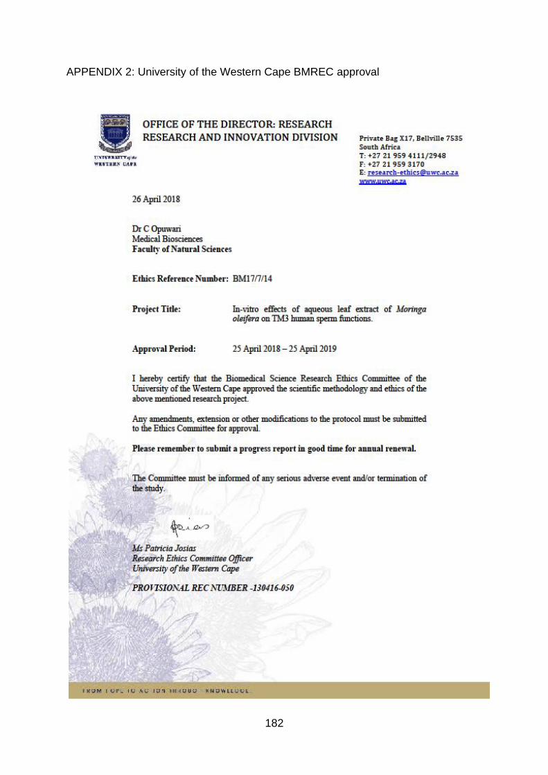
182
APPENDIX 2: University of the Western Cape BMREC approval

183
APPENDIX 3: Permission for semen collection from Tygerberg Hospital

184
APPENDIX 4: Permission for semen collection from Vincent Pallotti Hospital
11 September 2019
Ethics Reference: TREC/45/2019:PG
TITLE: In vitro effects of aqueous leaf extract of Moringa oleifera on human sperm functions
Dear Dr Moichela
PERMISSION TO CONDUCT YOUR RESEARCH AT AEVITAS CLINIC FROM 6.3.2019 TO 6.3.2020
1. In accordance with the Provisional Research Policy and Tygerberg Hospital
Notice No 40/2009, permission is hereby granted for you to conduct the
abovementioned research here at Aevitas Clinic.
Prof T I Siebert

185
APPENDIX 5: Informed consent form (English)
A Written and informed consent form will be given to participants prior to semen
collection.
PARTICIPATION INFORMED CONSENT FORM In vitro effects of aqueous leaf extract of Moringa oleifera on human sperm
functions
Information sheet and consent to participate in Research
To whom it may concern,
You are requested to consider participating in a scientific study on male sperm
functions and medicinal plants. The aim of this study is to investigate the in vitro effects
of Moringa oleifera on human sperm functions.
After collection of your semen through masturbation, it will be exposed to various
concentrations of the aqueous leaf extract of Moringa oleifera.
The objectives of the study are as follows:
• To determine effects of M. oleifera on sperm viability, motility and morphology.
• To measure of the effects of aqueous extracts of M. oleifera on ROS production
in sperm.
• To determine effects of M. oleifera on sperm DNA fragmentation.
• To determine spermatozoan mitochondrial membrane potential, capacitation
and acrosome reaction after extract exposure.
• To compare the effects of M. oleifera leaf extracts on fertile and infertile men.
For those participating in the assisted reproductive treatment, after your semen has
been processed for the treatment you are scheduled for, a small portion of the semen
sample (<100 µl) will be taken for these additional tests. This procedure will under no
circumstance affect the assisted reproduction treatment or outcome as only the
residual samples will be used. This will allow us to determine the possible effects of
Moringa oleifera on human sperm, that is whether it can improve male fertility or not.
The current study will not involve any additional medical risk or cost. Given the invasive
nature of the study it may include little discomfort. Participation in this scientific study
is voluntary and participants are allowed to withdraw at any point in time until the data
will be submitted for publication without any consequences. Withdrawal from the study

186
should be done in a formally addressed email. In order to protect confidentiality of the
participants, a unique number will be assigned to each sample and will contain no
personal information of the participants involved in the study. All data will be
confidential and available to the scientific researchers involved as well as to the
participants if requested. The samples will be disposed of in the correct medical
procedure by incineration once the data is captured.
The UWC Biomedical Research Ethics Committee has ethically received the study
(Approval number: 130416-050)
In the event of any problems or concerns, additional information can be obtained from
the doctor or scientific investigator directly by email.
Consent
I have been informed about the study titled “In vitro effects of aqueous leaf extract of
Moringa oleifera in human sperm functions” by the scientific investigator.
I understand the purpose and procedure of the study.
I have been given an opportunity to ask questions about the study and have had
answers to my satisfaction.
I declare that my participation in this study is entirely voluntary and uncompensated
and that I may withdraw at any point in time until the data will be submitted for
publication without any consequences and without affecting any treatment or care that
I am entitled to.
If I have any further questions or query related to the study, I understand that I may
contact the researchers.
If I have any questions or concerns about my rights as a study participant, or if I am
concerned about any aspect of the study or researcher, then I may contact:
________________________ __________________________
Signature of Participant Date
Signature of Investigator Date

187
APPENDIX 6: Informed consent form (Afrikaans)
DEELNAME INGELIGTE TOESTEMMING VORM
In vitro effekte van waterige blaar uittreksel van Moringa oleifera o n h Uman
sperm funksies
Inligtingsblad en toestemming om deel te neem aan Navorsing
Aan wie dit mag gaan,
Jy is versoek om te oorweeg om aan 'n wetenskaplike studie deel te neem oor manlike
spermfunksies en medisinale plante. Die doel van hierdie studie is om die in vitro te
ondersoek gevolge van moringa oleifera op menslike spermfunksies .
Na die versameling van y ons semen monsters masturbasie , Dit sal blootgestel word
aan verskeie konsentrasies van die waterige blaar uittreksel van Moringa oleifera .
Die doelstellings van die studie is soos volg:
• Om gevolge van M. oleifera op spermvatbaarheid, motiliteit en morfologie te
bepaal.
• Om spermatozoan te meet -ROS neutraliserende vermoë van waterige
uittreksels van M. oleifera.
• Om sperm DNA fragmentering te bepaal.
• Om spermatozoan mitochondriale membraanpotensiaal, kapasitasie en
akrosome reaksie na eksponeringsblootstelling na te gaan.
• Om die effekte van M. oleifera blaar uittreksels op vrugbare en onvrugbare
mans te vergelyk.
Vir diegene wat deelneem aan die assistente reproduktiewe behandeling, nadat jou
semen verwerk vir die behandeling wat jy is geskeduleer vir 'n klein gedeelte van die
semen monster (<100 μl) geneem sal word vir hierdie addisionele toets s. Hierdie
proses sal onder geen omstandighede beïnvloed die bygestaan voortplanting
behandeling of uitkoms as net die oorblywende monsters sal gebruik word. Dit sal
ons toelaat om die moontlike effek s van Moringa te bepaal oleifera op menslike
sperm, dit is of dit manlike vrugbaarheid kan verbeter of nie.
Die huidige studie sal geen verdere mediese risiko of koste inhou nie . Gegewe die
indringende aard van die studie kan dit min ongemak insluit. Deelname aan hierdie
wetenskaplike studie is heeltemal vrywillig en deelnemers mag op enige tydstip

188
terugtrek totdat die data sonder enige gevolge vir publikasie ingedien sal word.
Onttrekking uit die studie moet gedoen word in 'n formele aangespreek e-pos. Ten
einde die vertroulikheid van die deelnemers te beskerm, sal 'n unieke nommer aan
elke monster toegeken word en sal geen persoonlike inligting van die vrywilligers wat
by die studie betrokke is, bevat nie . Alle data sal vertroulik en beskikbaar wees vir
die betrokke wetenskaplike navorsers sowel as die deelnemers indien dit aangevra
word. Die monsters sal deur die verbranding in die korrekte mediese prosedure
weggedoen word sodra die data gevang is.
Die studie is eties ontvang deur die UWC Biomediese Navorsingsetiekkomitee
(Goedkeuringsnommer:130416-050)
In geval van enige probleme of probleme, kan addisionele inligting direk per e-pos by
die dokter of wetenskaplike ondersoeker verkry word.
toestemming
Ek is ingelig oor die studie getiteld " In vitro effekte van waterige blaar uittreksel van
Moringa oleifera in menslike spermfunksies "deur die wetenskaplike ondersoeker.
Ek verstaan die doel en prosedure van die studie.
Ek het die geleentheid gekry om vrae oor die studie te vra en antwoorde tot my
bevrediging gehad.
Ek verklaar dat my deelname aan hierdie studie heeltemal vrywillig en
ongekompenseer is en dat ek op enige tydstip kan terugtrek totdat die data sonder
enige gevolge vir publikasie voorgelê sal word sonder om enige behandeling of sorg
waarvoor ek geregtig is, te beïnvloed.
As ek verdere vrae of navrae het wat verband hou met die studie, verstaan ek dat ek
die navorsers kan kontak.
As ek enige vrae of kommer het oor my regte as studieleier, of as ek bekommerd is
oor enige aspek van die studie of navorser, kan ek kontak:
189
_________________________ __________________
Handtekening van Deelnemer datum
_________________________ __________________
Handtekening van Ondersoeker datum
190
APPENDIX 7: Informed consent form (IsiXhosa)
INKQUBO YOMSEBENZISWANO OQHUBILEYO
Iimpembelelo ze- in vitro ze-extract leaf of Moringa oleifera o n h uman
imisebenzi yesidoda
Iphepha leenkcukacha kunye nemvume yokuthatha inxaxheba kuPhando
Ngubani onokukhathalela,
Uceliwe ukuba uthathe inxaxheba ekutheni uthathe inxaxheba kwisifundo
sezenzululwazi kwimisebenzi yesilisa kunye nemithi yezityalo. Injongo yale sifundo
kukuphanda i- in vitro nemiphumo kaMoringa i-oleifera kwimisebenzi yesintu .
Emva kokuqokelelwa y yethu isampuli isampula, kuya kubonakala kwiindawo
ezahlukahlukeneyo ze-extract leaf of Moringa oleifera .
Iinjongo zesifundo zilandelayo:
• Ukumisela iziphumo zikaMnu oleifera kwi-sperm yokusebenza, i-motility kunye ne-morphology.
• Ukumisela ukuhlukana kwe-DNA yesininzi.
• Ukujonga i- spermatozoan mitochondrial potential membrane, ukuxhotyiswakunye ne-acrosome reaction emva kokukhutshwa kwe-extract.
• Ukuthelekisa nemiphumo ye- M. oleifera yamacwecwe amacwecwe kumadoda athile angenasiphelo.
Okanye ngabo abathatha inxaxheba unyango ekuzaleni, emva kokuba imbewu yakho
sele ukwenzela unyango ziyacwangciswa, inxalenye encinane kwamadlozi isampula
(<100 μl) ziya kuthathwa kula uvavanyo s olongezelelweyo. Le nkqubo iya phantsi
akukho iimeko kuchaphazela unyango ekuzaleni okanye isiphumo njengoko kuphela
iisampuli intsalela iza kusetyenziswa. Oku kuya kusenza sikwazi ukubona ukuba
kunokwenzeka isiphumo s ye Moringa oleifera kwisidoda somntu, oko kukuthi ingaba
kuphuculwe ukuzala komntu okanye cha.
Uphononongo lwangoku aluyi kubandakanyeka nayiphi na ingozi yonyango okanye
eyindleko. Ukubonelelwa ngendalo ebangelwayo isifundo singabandakanyeka
kakhulu. Ukuthatha inxaxheba kulolu cwaningo lwezenzululwazi ngokuzithandela
kwaye abathathi-nxaxheba bavunyelwe ukuhoxisa naliphi na ixesha ngexesha lokuba
idatha ingeniswe ukupapashwa ngaphandle kwemiphumo. Ukurhoxiswa kweso
sifundo kufuneka kwenziwe kwikhompyutha echongiweyo. Ukuze ukhusele imfihlo
yabathathi-nxaxheba, inombolo ekhethekileyo iya kubelwa isampula nganye kwaye
ayiyi kuqulethelwa ngolwazi oluqulethwe ngamavavolontiya abandakanyekayo
kwisifundo. Yonke idatha iya kuba yimfihlo kwaye ifumaneke kubaphandi bezesayensi
abachaphazelekayo kunye nabachaphazelekayo ukuba bayacelwa. Iisampuli ziya
kulahlwa kwinkqubo efanelekileyo yezocwangco ngokutshatyalaliswa xa idatha
ithathwa.

191
Uphononongo luye lwafunyanwa ngokomthetho yiKomidi yeeNkcazo zoBuchule be-
UWC
(Inombolo yemvume: 130416-050)
Xa kwenzeka nayiphi na ingxaki okanye ukukhathazeka, ulwazi olongezelelweyo
lunokufumaneka kwiregrha okanye uphando loosayensi ngokuchanekileyo nge-
imeyile.
Mvume
Ndixelelwe ngophando oluthi " In vitro iziphumo zecatshulwa lamagqabi e- Moringa
oleifera kwimisebenzi yobuninzi babantu "ngumphandi wenzululwazi.
Ndiyayiqonda injongo nenkqubo yesifundo.
Ndinike ithuba lokuba ndibuze imibuzo malunga nokufunda kwaye ndifumene
iimpendulo kwaneliseko lwam.
Ndiyaxela ukuba inxaxheba kweso sifundo iphela ngokuzithandela kwaye
ingenakuhlawulwa kwaye ndize ndirhoxise naliphi na ixesha ngexesha lokuba idatha
ingeniswe ukupapashwa ngaphandle kwemiphumo kwaye ngaphandle
kokuchaphazela nayiphi na inyango okanye ukunakekelwa kwam.
Ukuba ndinemibuzo eminye okanye umbuzo ophathelene nesifundo, ndiyaqonda
ukuba ndidibana nabaphandi.
Ukuba ngaba nayiphi na imibuzo okanye ukuxhalabisa ngamalungelo am nje
njengomfundi othabatha inxaxheba, okanye ukuba ndixhalabele nayiphi na into
yesifundo okanye umphandi, ngoko ndiyaqhagamshelana nayo:
_________________________ ________________________
U tyikityo lwaBathathi- nxaxheba Umhla
Isayinwe yoMphandi Umhla
Copyright © 2022 FDOKUMEN