In Vitro Activity of a Novel Antimycobacterial Compound, N-Octanesulfonylacetamide, and Its Effects...
8
ANTIMICROBIAL AGENTS AND CHEMOTHERAPY, 0066-4804/01/$04.0010 DOI: 10.1128/AAC.45.4.1143–1150.2001 Apr. 2001, p. 1143–1150 Vol. 45, No. 4 Copyright © 2001, American Society for Microbiology. All Rights Reserved. In Vitro Activity of a Novel Antimycobacterial Compound, N-Octanesulfonylacetamide, and Its Effects on Lipid and Mycolic Acid Synthesis NIKKI M. PARRISH, 1 TODD HOUSTON, 2 ² PAUL B. JONES, 2 ‡ CRAIG TOWNSEND, 2 AND JAMES D. DICK 1,3 * Department of Pathology, School of Medicine, 1 Department of Molecular Microbiology and Immunology, School of Public Health, 3 and Department of Chemistry, Johns Hopkins School of Arts and Sciences, 2 Johns Hopkins University, Baltimore, Maryland Received 29 September 2000/Returned for modification 8 November 2000/Accepted 22 January 2001 b-Sulfonyl carboxamides have been proposed to serve as transition-state analogues of the b-ketoacyl synthase reaction involved in fatty acid elongation. We tested the efficacy of N-octanesulfonylacetamide (OSA) as an inhibitor of fatty acid and mycolic acid biosynthesis in mycobacteria. Using the BACTEC radiometric growth system, we observed that OSA inhibits the growth of several species of slow-growing mycobacteria, including Mycobacterium tuberculosis (H37Rv and clinical isolates), the Mycobacterium avium complex (MAC), Mycobacterium bovis BCG, Mycobacterium kansasii, and others. Nearly all species and strains tested, including isoniazid and multidrug resistant isolates of M. tuberculosis, were susceptible to OSA, with MICs ranging from 6.25 to 12.5 mg/ml. Only three clinical isolates of M. tuberculosis (CSU93, OT2724, and 401296), MAC, and Mycobacterium paratuberculosis required an OSA MIC higher than 25.0 mg/ml. Rapid-growing mycobacterial species, such as Mycobacterium smegmatis, Mycobacterium fortuitum, and others, were not susceptible at con- centrations of up to 100 mg/ml. A 2-dimensional thin-layer chromatography system showed that OSA treatment resulted in a significant decrease in all species of mycolic acids present in BCG. In contrast, mycolic acids in M. smegmatis were relatively unaffected following exposure to OSA. Other lipids, including polar and nonpolar extractable classes, were unchanged following exposure to OSA in both BCG and M. smegmatis. Transmission electron microscopy of OSA-treated BCG cells revealed a disruption in cell wall synthesis and incomplete septum formation. Our results indicate that OSA inhibits the growth of several species of mycobacteria, including both isoniazid-resistant and multidrug resistant strains of M. tuberculosis. This inhibition may be the result of OSA-mediated effects on mycolic acid synthesis in slow-growing mycobacteria or inhibition via an undescribed mechanism. Our results indicate that OSA may serve as a promising lead compound for future antituberculous drug development. Tuberculosis continues to be the leading cause of death worldwide due to an infectious agent (8). Approximately 8 million new active cases arise each year, with about 3 million deaths (8). Of equivalent concern has been the emergence of multidrug-resistant Mycobacterium tuberculosis. As a result, newly infected individuals no longer have the assurance that prophylaxis with isoniazid (INH) will eliminate infection or that active disease will be treatable with our current arsenal of drugs. In addition, therapies for the treatment of atypical my- cobacterial infections in immunocompromised patients are limited (24). Thus, the development of new drugs is essential in combating both drug resistant M. tuberculosis and opportunis- tic infections with atypical mycobacteria, such as the Mycobac- terium avium complex (MAC). Potential new targets for antimycobacterial drug develop- ment may exist among the synthetic enzymes needed to make the unique lipids produced by mycobacteria, such as mycolic acids. These high-molecular-weight, a-alkyl, b-hydroxy fatty acids comprise the single largest component of the mycobac- terial cell envelope (3, 4, 9, 10, 29, 30, 37). They are found in free lipids as trehalose mono- and dimycolate and esterified to the arabinogalactan matrix of the mycobacterial cell wall (5, 10). They are vital for the growth and survival of mycobacteria, as evidenced by the bactericidal properties of mycolic acid inhibitory drugs, such as isoniazid and ethionamide (1, 2, 32, 33, 43, 44, 47, 48, 51, 53–58). Synthesis of mycolic acids and other mycobacterial lipids requires a variety of fatty acid synthase and elongation en- zymes (7, 10, 23). Although the synthesis of fatty acids is essentially the same at the primary chemical level, fatty acid synthases (FAS) are organized into two types. In Type I FAS (FAS I), most often found in eukaryotes, the individual enzy- matic reactions are contained in one multienzyme complex. In Type II FAS (FAS II), commonly found in prokaryotes, the enzyme functions are carried out by seven individual proteins. Mycobacteria are known to possess both FAS I and II (6, 7, 23). Thus, inhibition of these enzymes, especially those in- volved in chain elongation of unique mycobacterial fatty acids, may provide novel targets for drug design. In the past, characterization of FAS has been aided through the use of two natural product inhibitors of FAS components, * Corresponding author. Mailing address: Johns Hopkins Medical Institutions, Department of Pathology/Division of Medical Microbiol- ogy, 600 N. Wolfe St., Baltimore, MD 21287. Phone: (410) 955-5077. Fax: (410) 614-8087. E-mail: [email protected]. ² Present address: Department of Chemistry, Virginia Common- wealth University, Richmond, Va. ‡ Present address: Department of Chemistry, Wake Forest Univer- sity, Winston-Salem, N.C. 1143 on March 22, 2016 by guest http://aac.asm.org/ Downloaded from
-
Upload
independent -
Category
Documents
-
view
2 -
download
0
Transcript of In Vitro Activity of a Novel Antimycobacterial Compound, N-Octanesulfonylacetamide, and Its Effects...

ANTIMICROBIAL AGENTS AND CHEMOTHERAPY,0066-4804/01/$04.0010 DOI: 10.1128/AAC.45.4.1143–1150.2001
Apr. 2001, p. 1143–1150 Vol. 45, No. 4
Copyright © 2001, American Society for Microbiology. All Rights Reserved.
In Vitro Activity of a Novel Antimycobacterial Compound,N-Octanesulfonylacetamide, and Its Effects on
Lipid and Mycolic Acid SynthesisNIKKI M. PARRISH,1 TODD HOUSTON,2† PAUL B. JONES,2‡ CRAIG TOWNSEND,2
AND JAMES D. DICK1,3*
Department of Pathology, School of Medicine,1 Department of Molecular Microbiology and Immunology,School of Public Health,3 and Department of Chemistry, Johns Hopkins School of
Arts and Sciences,2 Johns Hopkins University, Baltimore, Maryland
Received 29 September 2000/Returned for modification 8 November 2000/Accepted 22 January 2001
b-Sulfonyl carboxamides have been proposed to serve as transition-state analogues of the b-ketoacylsynthase reaction involved in fatty acid elongation. We tested the efficacy of N-octanesulfonylacetamide (OSA)as an inhibitor of fatty acid and mycolic acid biosynthesis in mycobacteria. Using the BACTEC radiometricgrowth system, we observed that OSA inhibits the growth of several species of slow-growing mycobacteria,including Mycobacterium tuberculosis (H37Rv and clinical isolates), the Mycobacterium avium complex (MAC),Mycobacterium bovis BCG, Mycobacterium kansasii, and others. Nearly all species and strains tested, includingisoniazid and multidrug resistant isolates of M. tuberculosis, were susceptible to OSA, with MICs ranging from6.25 to 12.5 mg/ml. Only three clinical isolates of M. tuberculosis (CSU93, OT2724, and 401296), MAC, andMycobacterium paratuberculosis required an OSA MIC higher than 25.0 mg/ml. Rapid-growing mycobacterialspecies, such as Mycobacterium smegmatis, Mycobacterium fortuitum, and others, were not susceptible at con-centrations of up to 100 mg/ml. A 2-dimensional thin-layer chromatography system showed that OSA treatmentresulted in a significant decrease in all species of mycolic acids present in BCG. In contrast, mycolic acids inM. smegmatis were relatively unaffected following exposure to OSA. Other lipids, including polar and nonpolarextractable classes, were unchanged following exposure to OSA in both BCG and M. smegmatis. Transmissionelectron microscopy of OSA-treated BCG cells revealed a disruption in cell wall synthesis and incompleteseptum formation. Our results indicate that OSA inhibits the growth of several species of mycobacteria,including both isoniazid-resistant and multidrug resistant strains of M. tuberculosis. This inhibition may be theresult of OSA-mediated effects on mycolic acid synthesis in slow-growing mycobacteria or inhibition via anundescribed mechanism. Our results indicate that OSA may serve as a promising lead compound for futureantituberculous drug development.
Tuberculosis continues to be the leading cause of deathworldwide due to an infectious agent (8). Approximately 8million new active cases arise each year, with about 3 milliondeaths (8). Of equivalent concern has been the emergence ofmultidrug-resistant Mycobacterium tuberculosis. As a result,newly infected individuals no longer have the assurance thatprophylaxis with isoniazid (INH) will eliminate infection orthat active disease will be treatable with our current arsenal ofdrugs. In addition, therapies for the treatment of atypical my-cobacterial infections in immunocompromised patients arelimited (24). Thus, the development of new drugs is essential incombating both drug resistant M. tuberculosis and opportunis-tic infections with atypical mycobacteria, such as the Mycobac-terium avium complex (MAC).
Potential new targets for antimycobacterial drug develop-ment may exist among the synthetic enzymes needed to make
the unique lipids produced by mycobacteria, such as mycolicacids. These high-molecular-weight, a-alkyl, b-hydroxy fattyacids comprise the single largest component of the mycobac-terial cell envelope (3, 4, 9, 10, 29, 30, 37). They are found infree lipids as trehalose mono- and dimycolate and esterified tothe arabinogalactan matrix of the mycobacterial cell wall (5,10). They are vital for the growth and survival of mycobacteria,as evidenced by the bactericidal properties of mycolic acidinhibitory drugs, such as isoniazid and ethionamide (1, 2, 32,33, 43, 44, 47, 48, 51, 53–58).
Synthesis of mycolic acids and other mycobacterial lipidsrequires a variety of fatty acid synthase and elongation en-zymes (7, 10, 23). Although the synthesis of fatty acids isessentially the same at the primary chemical level, fatty acidsynthases (FAS) are organized into two types. In Type I FAS(FAS I), most often found in eukaryotes, the individual enzy-matic reactions are contained in one multienzyme complex. InType II FAS (FAS II), commonly found in prokaryotes, theenzyme functions are carried out by seven individual proteins.Mycobacteria are known to possess both FAS I and II (6, 7,23). Thus, inhibition of these enzymes, especially those in-volved in chain elongation of unique mycobacterial fatty acids,may provide novel targets for drug design.
In the past, characterization of FAS has been aided throughthe use of two natural product inhibitors of FAS components,
* Corresponding author. Mailing address: Johns Hopkins MedicalInstitutions, Department of Pathology/Division of Medical Microbiol-ogy, 600 N. Wolfe St., Baltimore, MD 21287. Phone: (410) 955-5077.Fax: (410) 614-8087. E-mail: [email protected].
† Present address: Department of Chemistry, Virginia Common-wealth University, Richmond, Va.
‡ Present address: Department of Chemistry, Wake Forest Univer-sity, Winston-Salem, N.C.
1143
on March 22, 2016 by guest
http://aac.asm.org/
Dow
nloaded from
cerulenin and thiolactomycin (15, 16, 36, 39–42, 46). Ceruleninis a potent inhibitor of both FAS I and FAS II systems whilethiolactomycin inhibits only synthases of the FAS II variety.Activity of both of these inhibitors on the mycolic acids ofmycobacteria has recently been described (25, 42, 50). Al-though cerulenin and thiolactomycin are structurally different,both compounds inhibit the two-carbon homologation cata-lyzed by the b-ketoacyl synthase, the condensing enzyme re-quired for fatty acid biosynthesis. Specifically, cerulenin irre-versibly inhibits the b-ketoacyl synthase (20, 39, 40), whilethiolactomycin inhibits both the b-ketoacyl–acyl carrier pro-tein (ACP) synthase and acetyl coenzyme A:ACP transacylase(15). b-Sulfonyl carboxamides were designed to mimic thetransition state of the reaction catalyzed by the b-ketoacylsynthase. In the following study we evaluated the in vitro ac-tivity of one of these compounds, N-octanesulfonylacetamide(OSA), on a variety of mycobacteria and specifically evaluatedits effects on lipid and mycolic acid synthesis in Mycobacteriumbovis BCG and Mycobacterium smegmatis.
MATERIALS AND METHODS
Synthesis of OSA. The synthesis of alkyl sulfoxides and sulfones has beendescribed previously (22). Briefly, OSA was synthesized in three steps fromcommercially available materials. Octyl bromide and methyl thioglycolate werereacted together to yield methyl 3-thioundecanoate. This sulfide was then oxi-dized to the sulfoxide by using 3-chloroperoxybenzoic acid. OSA was obtainedfrom the ammonylysis of the methyl ester. Overall yield was 70% followingcrystallization of the final product (Fig. 1).
Mycobacteria. M. tuberculosis strains H37Rv and CSU93 (52), M. bovis (ATCC35734), M. bovis BCG (Pasteur strain, ATCC 35734), Mycobacterium kansasii(ATCC 12478), Mycobacterium paratuberculosis (ATCC 19698), and M. smegma-tis (mc2 6 1-2c) (53) were utilized as reference strains. Clinical and other isolateswere speciated using standard methods (38) and included MAC, Mycobacteriumfortuitum, Mycobacterium chelonei, Mycobacterium abscessus, and both INH- andmultidrug-resistant clinical isolates of M. tuberculosis.
Susceptibility testing. Susceptibility testing and determination of MICs for M.tuberculosis, M. bovis, M. kansasii, and M. bovis BCG were done using theBACTEC radiometric growth system (Becton Dickinson, Sparks, Md.) and astandardized method (42, 49). Initial stock solutions (1 mg/ml) and subsequentdilutions of OSA, cerulenin (Sigma, St. Louis, Mo.), and thiolactomycin (gener-ously provided by T. Yoshida) were prepared in dimethyl sulfoxide (Sigma). Amodification of this procedure adopted by The National Jewish Center forImmunology and Respiratory Medicine was used to determine MICs for MAC(17). Susceptibility testing of M. paratuberculosis was accomplished by varying thestandard BACTEC protocol to include the addition of mycobactin J (AlliedMonitor, Fayette, Mo.) to commercially prepared 12B media (Becton Dickin-son). Initial mycobactin J solutions (2 mg/ml) were brought up in 95% ethanoland diluted in sterile distilled water to a concentration of 40 mg/ml. MycobactinJ was then added to each BACTEC vial (final concentration 5 1.0 mg/ml) alongwith OSA. All primary drugs were purchased from Becton Dickinson. Suscepti-bilities and MIC determinations of specific inhibitors for M. smegmatis, M.fortuitum, M. chelonei, and M. abscessus were established by broth dilution usingMiddlebrook 7H9-ADC incubated at 37°C for 4 days.
Treatment of cultures with OSA and lipid pulse labeling. BCG and MAC cellswere grown in M7H9-ADC-Tween (Difco, Detroit, Mich.) to early log phase.From this, a 1.0 McFarland suspension was prepared and diluted to yield a finalconcentration of 3 3 107 cells/ml in a total volume of 50 ml in M7H9-ADC-Tween. Cultures were aerated and incubated at 37°C for 24 h (approximately 1generation time). Each inhibitor was added at its MIC (final concentrations:
thiolactomycin, 25.0 mg/ml [BCG] and 75.0 mg/ml [M. smegmatis]; OSA, 6.25mg/ml [BCG], 25.0 mg/ml [MAC], and 100 mg/ml [M. smegmatis]), and cultureswere incubated under the same conditions for approximately 1 generation time(BCG and MAC, approximately 24 h; M. smegmatis, approximately 5 h). Subse-quently, 1 mCi of [1,2-14C]acetic acid (Amersham, Arlington Heights, Ill.)/ml wasadded and the cultures were incubated as before for an additional 24 h. In orderto demonstrate a concentration-dependent effect of OSA on mycolic acid syn-thesis in BCG, lipid pulse labeling was also performed at OSA concentrations of12.5 and 25.0 mg/ml. A slight variation of this protocol was used for labeling inM. smegmatis cells. Since this species of mycobacteria was not susceptible toOSA, the highest concentration tested (100 mg/ml) in this study was used forlabeling purposes. Additionally, a 0.5 McFarland suspension was done with aninitial incubation time of 10 h prior to the addition of compound and subsequentincubations of 5 h each (based on a doubling time of 3 to 5 h) following theaddition of drug and label, respectively. All assays were performed in duplicate.
Preparation of extractable mycobacterial lipids. Extractions were performedas previously described (13, 34, 42). Briefly, 100 to 150 mg (wet weight) of cellswas suspended in methanolic saline (methanol–0.3% aqueous NaCl [100:10,vol/vol] [2 ml]) and extracted three times with petroleum ether, yielding nonpolarextractable lipids. The remaining cells and residual aqueous phase were boiledfor 5 min, cooled for 5 min at 37°C, and extracted with monophasic chloroform–methanol–0.3% NaCl (90:100:30, vol/vol; used once) and chloroform–methanol–0.3% NaCl (50:100:40, vol/vol; used twice). All extracts were subsequently driedunder N2 at room temperature. The defatted cells containing saponifiable lipidswere saved.
Mycolic acid extraction and preparation of MAMES and FAMES. Extractionsof mycobacterial mycolic acids were performed as previously described (13, 35,42). Briefly, 50-ml cultures of M. smegmatis, BCG, or MAC cells were harvestedby centrifugation at 3,000 3 g for 10 min. Equal volumes of cells (100 to 150 mg[wet weight]) were extracted to remove polar and nonpolar extractable lipids (13,42). The resulting defatted cells containing bound mycolic acids and other sa-ponifiable lipids were subjected to alkaline hydrolysis in methanol (1 ml), 30%KOH (1 ml), and toluene (0.1 ml) at 75°C overnight and subsequently cooled toroom temperature (13, 42). The mixture was then acidified to pH 1 with 3.6%HCl and extracted three times with diethyl ether. Combined extracts were driedunder N2. Mycolic acid methyl esters (MAMES) and other long-chain fatty acids(fatty acid methyl esters [FAMES]) were prepared by mixing dichloromethane (1ml), a catalyst solution (1 ml) (14), and iodomethane (25 ml) for 30 min;centrifuging; and discarding the upper phase. The lower phase was dried underN2.
[1,2-14C]acetate incorporation into mycobacterial lipids. Incorporation of[1,2-14C]acetate into polar and nonpolar extractable and saponifiable lipid frac-tions was determined by scintillation counting and expressed in counts perminute (cpm) (Beckman LS6500 multi-purpose scintillation counter).
Analysis of MAMES and FAMES. Mycobacterial saponifiable extracts con-taining MAMES and FAMES were dissolved in chloroform and equal counts (incounts per minute) of each sample were loaded onto thin-layer chromatography(TLC) plates (20- by 20-cm silica gel G, 250-mm-diamter analytical plates; An-altech, Newark, Del.). Samples were subsequently subjected to a 2-dimensionalsolvent system (petroleum ether [bp 60 to 80°C]–acetone [95:5, vol/vol]) in thefirst dimension [three times] and toluene-acetone [97:3, vol/vol] in the seconddimension [one time]).
Data analysis. Mycolic acids of each species of mycobacteria were identifiedaccording to methods described by Dobson et al. (13, 42). Visualization andcomparison of thin-layer chromatograms were done using a Fuji Systems (FujixBAS 1000) phosphorimager. Spots were quantified using NIH Image (version1.57; National Institutes of Health, Bethesda, Md.) software programs. Due tothe nonequivalency of the counts per minute as determined by scintillationcounting and phosphor counts as determined by phosphorimaging, relative in-tensities of chromatographed compounds were calculated for each TLC plate onthe basis of total number of phosphor counts per plate. Phosphor counts forMAMES and FAMES were normalized for each TLC plate pair (control andinhibitor treated). The difference in normalized phosphor counts between eachcontrol and inhibitor treated pair represents the percent change.
Electron microscopy. All chemicals and reagents for electron microscopy wereobtained from Electron Microscopy Sciences, Ft. Washington, Pa. Cultures (50ml) of BCG were grown to early log phase (optical density at 600 nm, ;0.2), atwhich time OSA (100 mg/ml) or diluent (dimethyl sulfoxide) was added totreated and control cultures, followed by additional aeration and incubation at37°C for 24 h. Cells were harvested by low-speed centrifugation and washed in0.1 M cacodylate (CACO) buffer (pH 7.3). Washed cells were then fixed in 0.1M CACO buffer (pH 7.3) containing 2.0% glutaraldehyde–osmium tetroxide(1:1, vol/vol) for 45 min at 4°C (11, 28, 45). Secondary fixation was done at 4°C
FIG. 1. Structure of OSA.
1144 PARRISH ET AL. ANTIMICROB. AGENTS CHEMOTHER.
on March 22, 2016 by guest
http://aac.asm.org/
Dow
nloaded from

overnight in 4.0% formaldehyde–1.0% glutaraldehyde. Samples were post-fixedat room temperature for 1 h in 0.1 M CACO buffer containing 1% tannic acidand dehydrated through a graded ethanol series of 50, 70, 95 (twice), and 100%(three times). Subsequently, samples were infiltrated at room temperature witha series of Spurrs resins (Spurrs-ethanol [1:1, vol/vol; 2 h], Spurrs-ethanol [2:1,vol/vol; 2 h], and pure Spurrs [overnight]), and blocks were polymerized at 60°Cfor 48 h. Sections were cut on a Sorvall MT2B microtome, and 80-nm-thicksections were picked up on 200-mesh copper grids and stained with uranylacetate and lead citrate. Prepared samples were then analyzed on a HitachiHU12A electron microscope.
RESULTS
In vitro susceptibilities. Tables 1 and 2 show the suscepti-bility of various mycobacterial species and strains to OSA usingthe BACTEC radiometric growth system. Nearly all strains ofM. tuberculosis and other slow-growing mycobacterial species,such as M. bovis, M. bovis BCG, and M. kansasii, were suscep-tible to OSA, with MICs ranging from 6.25 to 12.5 mg/ml. Onlythree clinical isolates of M. tuberculosis (CSU 93, 42-1C9383,and 10129-3), the MAC, and M. paratuberculosis required ahigher OSA MIC of 25 mg/ml. None of the rapid-growingmycobacterial species tested, M. smegmatis, M. fortuitum, M.chelonei, and M. abscessus, were susceptible to OSA at con-centrations up to 100 mg/ml. Resistance to known antimyco-bacterial agents did not give cross-resistance to OSA. Threeclinical isolates of M. tuberculosis, resistant to either INH aloneor INH plus rifampin, ethambutol, streptomycin, and pyrazin-
amide (19), were found to be susceptible to OSA, with MICs of12.5 and 6.25 mg/ml, respectively. Similar results were obtainedfor cerulenin as previously reported (42). BCG and M. smeg-matis were susceptible to thiolactomycin at a MIC of 25 and 75mg/ml, respectively.
Overall effects of OSA on mycobacterial lipids. Labelingassays were conducted at the calculated MIC (6.25 mg/ml) ofOSA for BCG and at the highest concentration of compoundtested for M. smegmatis. This particular concentration of OSAis not equivalent to the lethal concentration of compound inmycobacteria, as evidenced by continued 14CO2 production inthe radiometric susceptibility test system, which indicated con-tinued metabolism, albeit at a reduced level relative to those ofcontrols. OSA had no significant effect on [14C]acetate incor-poration into nonpolar extractable or polar extractable lipids ata concentration of 6.25 mg/ml, the calculated MIC for M. bovisBCG, or 100 mg/ml for M. smegmatis (Fig. 2). A moderatedecrease in the incorporation of label was observed in sapon-ifiable lipids in OSA-treated BCG. However, this change wasnot statistically significant. Label incorporation into the samelipid fraction in M. smegmatis was unaltered by exposure toOSA (Fig. 2). In order to demonstrate a concentration-depen-dent effect of OSA on lipid metabolism, studies were run at12.5 mg/ml (two times the MIC) and 25.0 mg/ml (four times theMIC). At these higher concentrations, there was a dose-de-pendent decrease of label incorporation in the saponifiablelipid fraction with a concomitant increase of label in the non-polar extractable fraction (data not shown).
Effect of OSA on mycolic acid synthesis as compared withthiolactomycin. Qualitative and quantitative analysis of myco-bacterial saponifiable lipids containing MAMES was per-formed using [14C]acetate pulse-labeling with 2-dimensionalTLC and phosphorimaging. Differences in the effects of OSAand thiolactomycin were found between BCG and M. smegma-tis. OSA treatment in BCG resulted in inhibition of all mycolicacids commonly found in this mycobacterial species (Fig. 3;Table 3). This decrease or percent change in both a- andketo-mycolates reached greater than 90% with an OSA con-centration of four times the MIC. Similar results were demon-
TABLE 1. Susceptibility of different species of mycobacteriato OSA
Organism MIC (mg/ml) of OSA
M. bovis ................................................................................. 6.25M. bovis BCG....................................................................... 6.25M. kansasii ............................................................................ 12.5M. avium complex ................................................................ 25.0M. paratuberculosis............................................................... 25.0M. smegmatis .........................................................................100M. fortuitum ...........................................................................100M. chelonei .............................................................................100M. abscessus ...........................................................................100
TABLE 2. Activities of OSA and first-line antimycobacterial drugs against various strains of M. tuberculosisa
M. tuberculosis strain
INHRIF
(2.0 mg/ml)EMB
(2.0 mg/ml)STR
(2.0 mg/ml)PZA
(100 mg/ml)
OSA
0.1mg/ml
0.4mg/ml
6.25mg/ml
12.5mg/ml
25.0mg/ml
H37Rv S S S S S S S S S5D5178 S S S S S S S S S1D4924 S S S S S S S S S1H1337 S S S S S S S S S42401315 S S S S S S S S S6T2709 S S S S S S S S S1T2768 S S S S S S S S STBL54 EP066 R R R R R R S S S7D5245 S S S S S S S S SOT2769 S S S S S S R S S42-1C9383 R R S S S S R S S10129-3 R R S S S S R S SCSU93 S S S S S S R R SOT2724 S S S S S S R R S401296 S S S S S S R R S
a S, susceptible; R, resistant; RIF, rifampin; EMB, ethambutol; STR, streptomycin; PZA, pyrazinamide.
VOL. 45, 2001 EFFECT OF OSA ON LIPID AND MYCOLIC ACID SYNTHESIS 1145
on March 22, 2016 by guest
http://aac.asm.org/
Dow
nloaded from

strated for MAC (data not shown). However, in M. smegmatis,individual mycolate classes were only slightly inhibited follow-ing exposure to OSA (Fig. 4; Table 3). Other lipids present inthe saponifiable fraction included FAMES. No appreciablechange was observed in this lipid class following OSA treat-ment in either of the two mycobacterial species characterizedin this study (Fig. 3 and 4; Table 3).
Thiolactomycin caused a decrease in all mycolate species inBCG (Fig. 3; Table 3) and M. smegmatis. However, whilethiolactomycin uniformly inhibited all mycolate species inBCG, in M. smegmatis, a differential effect was observed be-tween individual mycolic acid classes. Both a-mycolates andepoxymycolates were nearly completely diminished (95 and87%, respectively), whereas a9-mycolates were less affected(57%) (Fig. 4; Table 3). In contrast to OSA, FAMES accumu-lated in both BCG and M. smegmatis following treatment withthiolactomycin.
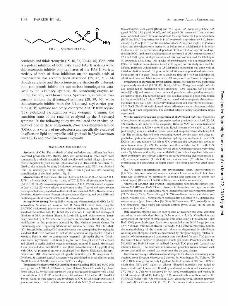
Transmission electron microscopy of OSA-treated BCG. In-hibition of mycolic acid synthesis is known to disrupt the my-cobacterial cell wall. Figure 5 shows transmission electron mi-crographs of OSA-treated BCG cells during cell division.
Control organisms exhibited an intact cell wall and clearlydefined septum, whereas in the presence of OSA, cell wallsynthesis was disrupted with incomplete septum formation. Inaddition, outer-wall-associated lipids appeared to be dispersedfrom the electron transparent zone of the mycolic acids.
DISCUSSION
In this study, we demonstrate that OSA, a compound de-signed to inhibit fatty acid synthesis by mimicking the transi-tion state of the b-ketoacyl synthase, is inhibitory for a broadrange of slow-growing mycobacterial species, including multi-drug-resistant M. tuberculosis. OSA treatment reduced mycolicacid accumulation in BCG and MAC cells, presumably by itseffect on FAS systems in these mycobacteria. Cross-resistancewas not observed for isolates resistant to isoniazid, rifampin,ethambutol, streptomycin, and pyrazinamide. A comparison ofisoniazid, a known inhibitor of mycolic acid synthesis, and OSArevealed pertinent information. Isoniazid has been shown toinhibit both InhA, an enoyl-ACP reductase involved in fattyacid elongation, and KasA, a b-ketoacyl–ACP synthase (2, 12,
FIG. 2. Effects of OSA on the synthesis of various lipid fractions in M. bovis BCG and M. smegmatis. Incorporation of [1,2-14C]acetate intoextractable polar, nonpolar, and saponifiable lipids in the presence and absence of OSA. Abbreviations: NPF, nonpolar extractable lipids; PLF,polar extractable lipids; SF, saponifiable lipids, including mycolic acids. Concentrations of OSA used: for M. smegmatis, 100 mg/ml; for M. bovisBCG, 12.5 mg/ml. Differences were not statistically significant.
1146 PARRISH ET AL. ANTIMICROB. AGENTS CHEMOTHER.
on March 22, 2016 by guest
http://aac.asm.org/
Dow
nloaded from

28, 31, 32). INH resistance has been associated with mutationsin both of these genes, as well as the KatG gene, which encodesthe mycobacterial catalase-peroxidase (54, 55, 57, 58). In thisstudy, the mechanism of INH resistance for the M. tuberculosisisolates used was not determined. Of relevance is the findingthat OSA inhibited the growth of INH-resistant M. tuberculosis(.0.04 mg/ml) as well as MAC, typically resistant to INH (.2.5mg/ml) (19). This observation suggests that the inhibition ofmycolic acid synthesis by OSA may be due to interaction withan enzymatic target different from that of INH, indicating anovel and possibly unexploited mechanism of action of OSA inM. tuberculosis.
Although OSA was designed to inhibit b-ketoacyl synthases,
it has not been tested as an inhibitor of isolated FAS. In anearlier, related study conducted in our laboratory, structurallyrelated sulfones and sulfoxides were found to inhibit FAS Iisolated from M. smegmatis (41). Several of these compoundswere both FAS I inhibitors and active against M. tuberculosisH37Rv in the radiometric growth assay. However, the corre-lation was not exact and issues of cell wall permeability andsolubility prevented direct comparison of the data. OSA wasselected for further study based on its solubility and its perfor-mance in whole-cell assays.
The differential susceptibility to OSA observed betweenslow- and rapid-growing mycobacterial species argues for thepresence of unique targets in BCG and MAC. M. smegmatismay contain the same OSA target as BCG and MAC butpossesses alternate cell wall compounds that permit survival inspite of OSA inhibition. Another potential possibility is therequirement for alteration, i.e., activation, of OSA prior to itsinteraction with the target protein(s) which may occur in slow-growing mycobacteria but not in rapid-growing species. How-ever, the most probable interpretation is that differences in themycolic acid biosynthetic pathway may exist between mycobac-terial species. This possibility is further strengthened when thework of other investigators is considered in conjunction withour own. For example, InhA, a long chain, enoyl-ACP-depen-dent reductase involved in fatty acid elongation, is present inboth M. smegmatis and M. tuberculosis (31). Several studieshave revealed compelling evidence that InhA is the target forKatG-activated INH in M. smegmatis (12, 44). However, addi-tional investigations suggest that this particular enzyme is notthe principal target for KatG-activated INH in M. tuberculosis
FIG. 3. Two-dimensional TLC showing the comparative effects ofOSA and thiolactomycin on the mycolic acids of M. bovis BCG. Firstdimension, petroleum ether (bp 60 to 80°C)–acetone (95:5, vol/vol;three times); second dimension, toluene-acetone (97:3, vol/vol; onetime). Abbreviations: ori, origin; a, a-mycolate; k, keto-mycolate.Equivalent counts per minute of the saponifiable lipid fraction werespotted at each origin.
TABLE 3. Effects of OSA and thiolactomycin on the mycolic acidsof M. bovis BCG and M. smegmatis
Organism Mycolic acid species
Inhibition(% change)
OSA TLM
M. bovis BCG a 256 269keto 252 279FAMES —a 1147
M. smegmatis a 219 295a9 218 257epoxy 28 287FAMES — 1134
a —, no appreciable change was observed.
FIG. 4. Two-dimensional TLC showing the comparative effects ofOSA and thiolactomycin on the mycolic acids of M. smegmatis. Firstdimension, petroleum ether (bp 60 to 80°C)-acetone (95:5, vol/vol;three times); second dimension, toluene-acetone (97:3, vol/vol; onetime). Abbreviations: ori, origin; a, a-mycolate; a9, a9-mycolate; e,epoxymycolate. Equivalent counts per minute of the saponifiable lipidfraction were spotted at each origin.
VOL. 45, 2001 EFFECT OF OSA ON LIPID AND MYCOLIC ACID SYNTHESIS 1147
on March 22, 2016 by guest
http://aac.asm.org/
Dow
nloaded from
(31, 32). This discrepancy may reflect inherent differences inmycolate biosynthesis between the two organisms (31, 32, 42).Additionally, previous studies in our laboratory examining theeffect of cerulenin on mycolate synthesis in M. smegmatis andBCG revealed clear differences in mycolic acid profiles be-tween these two mycobacterial species following inhibitor ex-posure. Not only did the changes in mycolate synthesis differbetween BCG and M. smegmatis, they were in direct opposi-tion. For instance, completed mycolates decreased in BCGfollowing exposure to cerulenin, whereas in M. smegmatis, allmycolate species increased, again suggesting that inherent dif-ferences in the mycolate biosynthetic pathway between the twospecies are responsible for these disparate responses (42).
This possibility is further strengthened by the differential
effect of thiolactomycin on individual mycolic acid classes in M.smegmatis. In both the present study and that of other inves-tigators (50), exposure to thiolactomycin resulted in substan-tially decreased amounts of a-mycolates and epoxymycolates,with a minor decrease in a9-mycolates (49). The authors of theprevious study suggested that potential targets for thiolacto-mycin in this mycobacterial species include an elongation en-zyme leading from either the C24:1D5 intermediate or from theshorter-chain a9-mycolates to the longer a-mycolates and ox-ygenated mycolates (50). In view of the current experimentalevidence, the latter seems to be the more likely possibility. Itshould be noted that a9-mycolates, commonly found in rapid-growing mycobacteria, are not present in BCG and other slow-growing mycobacteria characterized in this study (18). Thus,
FIG. 5. Transmission electron microscopy of OSA-treated M. bovis BCG (6.25 mg/ml). (Top) Intact cell wall ultrastructure (left) (magnification,3117,000) and completed septum (right) (magnification, 3108,000). (Bottom) Disrupted cell wall ultrastructure (left) (magnification, 3130,000)and incomplete septum formation (right) (magnification, 378,000).
1148 PARRISH ET AL. ANTIMICROB. AGENTS CHEMOTHER.
on March 22, 2016 by guest
http://aac.asm.org/
Dow
nloaded from

while thiolactomycin treatment of BCG and M. smegmatis re-sulted in inhibition of both a-mycolates and oxygenated my-colates, the presence of a9-mycolic acids in the latter case maypartially explain the differences observed in MICs between thetwo mycobacterial species (for BCG, 25.0 mg/ml; for M. smeg-matis, 75 mg/ml) and indicate that disparities in mycolate bio-synthesis may exist between BCG and M. smegmatis. One couldspeculate that such differences may extend to other slow- ver-sus rapid-growing mycobacterial species.
Differences in mycolic acid profiles following OSA treat-ment in BCG and M. smegmatis were also noted. In the presentstudy, all mycolic acids were significantly and uniformly inhib-ited in OSA-treated BCG (a- and keto-mycolates). This inhi-bition increased with an increasing concentration of OSA. Incontrast, the effect of OSA on the mycolates of M. smegmatis(a9, a, and epoxy) was negligible and not inhibitory to growth.Previous studies have suggested the existence of multiple ACP-dependent FAS II systems in mycobacteria, responsible for notonly fatty acid biosynthesis but also mycolic acid biosynthesis(2, 32, 42, 44). Such systems could be envisioned to interactwith separate and distinct b-ketoacyl–ACP synthases as well asother enzymes involved in biosynthetic reactions of this type,including b-ketoacyl–ACP reductases, b-hydroxyacyl–ACP de-hydratases, and b-enoyl–ACP reductases. A biological prece-dent for the existence of such enzymes has been described forEsherichia coli, in which multiple b-ketoacyl–ACP synthaseshave been found (21, 26, 27). Although thiolactomycin wasoriginally thought to inhibit all three b-ketoacyl–ACP syn-thases in E. coli, recent evidence suggests that the principaltarget of thiolactomycin in this particular organism may beonly b-ketoacyl–ACP I (25). Since OSA was designed to inhibitthe b-ketoacyl synthase by mimicking the transition state of thereaction catalyzed by this enzyme, this compound could intheory inhibit both the multifunctional FAS I and monofunc-tional FAS II mycobacterial systems, as in the case of cerule-nin. However, in this study, additional assays were performedwhich were designed to indirectly determine FAS I activity inthe presence of each inhibitor by measuring phospholipid pro-duction. Only cerulenin, known to inhibit both FAS I and FASII systems, interfered with phospholipid synthesis in eitherBCG or M. smegmatis (42). Neither thiolactomycin, active onlyagainst FAS II systems, nor OSA inhibited phospholipid syn-thesis in either of the two mycobacterial species tested (datanot shown), suggesting that the principal target of OSA may liein an ACP-dependent FAS II system. In addition, previouswork in our laboratory and others has demonstrated that ceru-lenin and thiolactomycin strongly inhibited [14C]acetate incor-poration into other extractable mycobacterial lipids, a findingconsistent with the known mechanism of action of both inhib-itors. In contrast, OSA inhibited only mycolic acids, with noappreciable change in label incorporation in any of the othermycobacterial lipid classes tested. This distinction suggests thepresence of a unique and highly specific target for this com-pound in slow-growing mycobacteria. Such a target may in-volve an as-yet-unidentified enzyme or enzyme system presentin slow-growing mycobacteria which is not present or inactivein rapid-growing species.
Additional information was obtained by careful analysis ofFAMES in OSA-treated BCG and M. smegmatis. Other inves-tigators have determined that these lipids most likely represent
saturated alkyl intermediate(s) in mycolic acid synthesis (31).While 2-dimensional TLC of OSA-treated BCG revealed thatmycolic acid synthesis was inhibited, no apparent effect wasseen in the FAMES present in this fraction when the com-pound was used at the MIC (6.25 mg/ml). A similar observationwas noted in OSA-treated M. smegmatis. However, at fourtimes the MIC, OSA treatment of BCG resulted in an increasein label incorporation into extractable nonpolar lipids. Thismay suggest that a noncovalently bound, extractable interme-diate in mycolate synthesis accumulates following OSA treat-ment, an effect intensified with higher concentrations (fourtimes the MIC) of compound. In contrast, FAMES increasedin thiolactomycin-treated BCG, while mycolic acids decreased,a finding consistent with that of earlier studies using cerulenin(42). Thus, in BCG, while completed mycolates decreased withOSA, thiolactomycin, and cerulenin (42), the changes inFAMES were clearly not the same, suggesting that inhibitionof mycolic acid synthesis in BCG may occur prior to synthesisof the saturated alkyl intermediate with OSA, but between thisintermediate and completed mycolates with cerulenin andthiolactomycin. Alternatively, OSA-mediated inhibition of my-colate synthesis in BCG and MAC may involve an as-yet-unidentified enzyme or enzyme system. In summary, the effectsof OSA, cerulenin, and thiolactomycin are mycobacterial spe-cies specific and compound specific and inherent differences inthe mycolic acid biosynthetic pathway may exist between rapid-and slow-growing mycobacteria.
ACKNOWLEDGMENTS
We thank Janet Folmer for performance of electron microscopy andWilliam Bishai for providing equipment and laboratory space for someportions of this study.
This study was financially supported in part by NIH grant AI43846,the Raynam Research Fund, and the Becton Dickinson CentennialFellowship in Clinical Microbiology (N.M.P.).
REFERENCES
1. Altamirano, M., J. Marostenmaki, A. Wong, M. Fitzgerald, W. Black, and J.Smith. 1994. Mutations in the catalase-peroxidase gene from isoniazid-re-sistant Mycobacterium tuberculosis isolates. J. Infect. Dis. 169:1162–1165.
2. Bannerjee, A., E. Dubnau, A. Quemard, V. Balasubramanian, K. S. Um, T.Wilson, D. Collins, G. DeLisle, and W. R. Jacobs, Jr. 1994. inhA, a geneencoding a target for isoniazid and ethionamide in Mycobacterium tubercu-losis. Science 263:227–230.
3. Barry, C., III, and K. Mdluli. 1996. Drug sensitivity and environmentaladaptation of mycobacterial cell wall components. Trends Microbiol. 4:275–281.
4. Besra, G., and D. Chatterjee. 1994. Lipids and carbohydrates of Mycobacte-rium tuberculosis, p. 285–306. In B. Bloom (ed.), Tuberculosis: pathogenesis,protection, and control. ASM Press, Washington, D.C.
5. Besra, G., K. Khoo, M. McNeil, A. Dell, H. Morris, and P. Brennan. 1995. Anew interpretation of the structure of the mycolyl-arabinogalactan complexof Mycobacterium tuberculosis as revealed through the characterization ofoligoglycosyl alditol fragments by fast-atom bombardment mass spectrome-try and 1H-nuclear magnetic resonance spectroscopy. Biochemistry 24:84–90.
6. Bloch, K. 1975. Fatty acid synthases from Mycobacterium phlei. MethodsEnzymol. 35:84–90.
7. Bloch, K. 1977. Control mechanisms for fatty acid synthesis in Mycobacte-rium smegmatis, p. 1–84. In A. Meister (ed.), Advances in enzymology. JohnWiley and Sons, New York, N.Y.
8. Bloom, B., and C. Murray. 1992. Tuberculosis: commentary on a reemergentkiller. Science 257:1055–1064.
9. Brennan, P., and P. Draper. 1994. Ultrastructure of Mycobacterium tubercu-losis, p. 271–284. In B. Bloom (ed.), Tuberculosis: pathogenesis, protection,and control. ASM Press, Washington, D.C.
10. Brennan, P., and H. Nikaido. 1995. The envelope of mycobacteria. Annu.Rev. Biochem. 64:29–63.
11. Cunningham, A., and C. Spreadbury. 1998. Mycobacterial stationary phase
VOL. 45, 2001 EFFECT OF OSA ON LIPID AND MYCOLIC ACID SYNTHESIS 1149
on March 22, 2016 by guest
http://aac.asm.org/
Dow
nloaded from
induced by low oxygen tension: cell wall thickening and localization of the16-kilodalton a-crystallin homolog. J. Bacteriol. 180:801–808.
12. Dessen, A., A. Quemard, J. S. Blanchard, W. R. Jacobs, Jr., and J. C.Sachettini. 1995. Crystal structure and function of the isoniazid target ofMycobacterium tuberculosis. Science 267:1638–1641.
13. Dobson, G., D. Minnikin, S. Minnikin, J. Parlett, and M. Goodfellow. 1985.Systematic analysis of complex mycobacterial lipids, p. 237–265. In M. Good-fellow and D. Minnikin (ed.), Chemical methods in bacterial systematics.Academic Press, London, United Kingdom.
14. Gyllenhaal, O., and H. Ehrsson. 1975. Determination of sulphonamides byelectron-capture gas chromatography. Preparation and properties of perflu-oroacyl and pentaflurobenzyl derivatives. Chromatography 107:327–333.
15. Hayashi, T., O. Yamamoto, H. Sasaki, H. Kawaguchi, and H. Okazaki. 1983.Mechanism of action of the antibiotic thiolactomycin: inhibition of fatty acidsynthesis in Escherichia coli. Biochim. Biophys. Res. Commun. 115:1108–1113.
16. Hayashi, T., O. Yamamoto, H. Sasaki, and H. Okazaki. 1984. Inhibition offatty acid synthesis by the antibiotic thiolactomycin. J. Antibiot. 37:1456–1461.
17. Heifets, H., P. Lindholm-Levy, J. Libonati, et al. 1993. Radiometric brothmacrodilution method for determination of minimal inhibitory concentra-tions (MIC’s) with Mycobacterium avium complex isolates. National JewishCenter for Immunology and Respiratory Medicine, Denver, Colo.
18. Hinrikson, H. P., and G. E. Pfyffer. 1994. Mycobacterial mycolic acids. Med.Microbiol. Lett. 3:49–57.
19. Inderlied, C., and M. Salfinger. 1995. Antimicrobial agents and susceptibilitytests: mycobacteria, p. 1385–1404. In P. Murray (ed.), Manual of clinicalmicrobiology. ASM Press, Washington, D.C.
20. Inokoshi, J., H. Tomoda, H. Hashimoto, A. Watanabe, H. Takeshima, and S.Omura. 1994. Cerulenin-resistant mutants of Saccharomyces cerevesiae withan altered fatty acid synthase gene. Mol. Gen. Genet. 244:90–96.
21. Jackowski, S., J. E. Cronan, Jr., and C. O. Rock. 1991. Lipid metabolism inprokaryotes, p. 43–85. In D. E. Vance and J. Vance (ed.), Biochemistry oflipids, lipoproteins, and membranes. Elsevier Publishers B. V., Amsterdam,The Netherlands.
22. Jones, P. B., N. M. Parrish, T. A. Houston, A. S. Stapon, N. P. Bansal, J. D.Dick, and C. A. Townsend. 2000. A new class of anti-tuberculosis agents.J. Med. Chem. 43:3304–3314.
23. Kolattukudy, P., N. Fernandes, A. Azad, A. Fitzmaurice, and T. Sirakova.1997. Biochemistry and molecular genetics of cell wall lipid biosynthesis inmycobacteria. Mol. Microbiol. 24:263–270.
24. Korvick, J., and C. Benson. 1996. Advances in the treatment and prophylaxisof Mycobacterium avium complex in individuals infected with human immu-nodeficiency virus, p. 241–262. In J. Korvick and C. Benson (ed.), Mycobac-terium avium complex infection: progress in research and treatment, vol. 87.Lung biology in Health and Disease. Marcel Dekker, Incorporated, NewYork, N.Y.
25. Kremer, L., J. D. Douglas, A. R. Baulard, C. Morehouse, M. R. Guy, D.Alland, L. G. Dover, H. Lakey, W. R. Jacobs, P. J. Brennan, D. E. Minnikin,and G. S. Besra. 2000. Thiolactomycin and related-analogues as novel anti-mycobacterial agents targeting KasA and KasB condensing enzymes in My-cobacterium tuberculosis. J. Biol. Chem. 275:16857–16864.
26. Lowe, P. N., and S. Rhodes. 1988. Purification and characterization of [acyl-carrier-protein]acetyltransferase from Escherichia coli. Biochem. J. 250:789–796.
27. Magnuson, K. S., S. Jackowski, C. O. Rock, and J. E. Cronan, Jr. 1993.Regulation of fatty acid biosynthesis in Escherichia coli. Microbiol. Rev.57:522–542.
28. McDonough, K., V. Kress, and B. Bloom. 1993. Pathogenesis of tuberculosis:interaction of Mycobacterium tuberculosis with macrophages. Infect. Immun.61:2763–2773.
29. McNeil, M., and P. Brennan. 1991. Structure, function, and biogenesis of thecell envelope of mycobacteria in relation to bacterial physiology, pathogen-esis and drug resistance; some thoughts and possibilities arising from recentstructural information. Res. Microbiol. 142:451–463.
30. McNeil, M., M. Daffe, and P. Brennan. 1991. Location of the mycolyl estersubstituents in the cell walls of mycobacteria. J. Biol. Chem. 266:6934–6943.
31. Mdluli, K., D. R. Sherman, M. J. Hickey, B. N. Kreiswirth, S. Morris, C. K.Stover, and C. E. Barry III. 1996. Biochemical and genetic data suggest thatInhA is not the primary target for activated isoniazid in Mycobacteriumtuberculosis. J. Infect. Dis. 174:1085–1090.
32. Mdluli, K., R. Slayden, Y. Zhu, S. Ramaswamy, X. Pan, D. Mead, D. Crane,J. Musser, and C. Barry III. 1998. Inhibition of Mycobacterium tuberculosisb-ketoacyl ACP synthase by isoniazid. Science 280:1607–1610.
33. Middlebrook, G. 1954. Isoniazid-resistance and catalase activity of tuberclebacilli. Am. Rev. Tuberc. 69:471–472.
34. Minnikin, D., A. O’Donnell, M. Goodfellow, G. Alderson, M. Athalye, A.Schaal, et al. 1984. An integrated procedure for the extraction of bacterialisoprenoid quinones and polar lipids. J. Microbiol. Methods 2:233–241.
35. Minnikin, D., S. Minnikin, A. O’Donnell, and M. Goodfellow. 1984. Extrac-tion of mycobacterial mycolic acids and other long-chain compounds by analkaline methanolysis procedure. J. Microbiol. Methods 2:243–249.
36. Morisaki, N., H. Funabashi, R. Shimazawa, J. Furukawa, A. Kawaguchi, S.Okuda, and S. Iwasaki. 1993. Effect of side-chain structure on inhibition ofyeast fatty-acid synthase by cerulenin analogues. Eur. J. Biochem. 211:111–115.
37. Nikaido, H., S. Kim, and E. Rosenberg. 1993. Physical organization of lipidsin the cell wall of Mycobacterium chelonae. Mol. Microbiol. 8:1025–1030.
38. Nolte, F., and B. Metchock. 1995. Mycobacterium, p. 400–437. In P. Murray,E. Baron, M. Pfaller, F. Tenover, and R. Yolken (ed.), Manual of clinicalmicrobiology. American Society for Microbiology, Washington, D.C.
39. Omura, S. 1976. The antibiotic cerulenin, a novel tool for biochemistry as aninhibitor of fatty acid synthesis. Bacteriol. Rev. 40:681–697.
40. Omura, S. 1981. Cerulenin. Methods Enzymol. 72:520–535.41. Parrish, N. 1999. Ph.D. thesis. Johns Hopkins University, Baltimore, Md.42. Parrish, N., F. Kuhajda, H. Heine, W. Bishai, and J. Dick. 1999. Antimyco-
bacterial activity of cerulenin and its effects on lipid synthesis. J. Antimicrob.Chemother. 43:219–226.
43. Quemard, A., C. Lacave, and G. Laneelle. 1991. Isoniazid inhibition ofmycolic acid synthesis by cell extracts of sensitive and resistant strains ofMycobacterium aurum. Antimicrob. Agents Chemother. 35:1035–1039.
44. Quemard, A., J. C. Sacchettini, A. Dessen, C. Vilcheze, R. Bittman, W. R.Jacobs, Jr., and J. S. Blanchard. 1995. Enzymatic characterization of thetarget for isoniazid in Mycobacterium tuberculosis. Biochemistry 34:8235–8241.
45. Rastogi, N., C. Frehel, and H. David. 1986. Triple-layered structure of themycobacterial cell wall: evidence of a polysaccharide-rich outer layer in 18mycobacterial species. Curr. Microbiol. 13:237–242.
46. Sasaki, H., H. Oishi, T. Hayashi, I. Matsura, K. Ando, and M. Sawada. 1982.Thiolactomycin, a new antibiotic: structure elucidation. J. Antibiot. 35:396–400.
47. Shoeb, H., B. Bowman, A. Ottolenghi, and A. Merola. 1985. Evidence for thegeneration of active oxygen by isoniazid treatment of extracts of Mycobac-terium tuberculosis H37Ra. Antimicrob. Agents Chemother. 27:404–407.
48. Shoeb, H., B. Bowman, A. Ottolenghi, and A. Merola. 1985. Peroxidase-mediated oxidation of isoniazid. Antimicrob. Agents Chemother. 27:399–403.
49. Siddiqi, S. 1992. Radiometric (BACTEC) tests for slowly growing mycobac-teria, p. 5.14.1–5.14.25. In H. Isenberg (ed.), Clinical microbiology proce-dures handbook, vol. 1. American Society for Microbiology, Washington,D.C.
50. Slayden, R., A., R. E. Lee, J. W. Armour, A. M. Cooper, I. M. Orme, P. J.Brennan, and G. S. Besra. 1996. Antimycobacterial activity of thiolactomy-cin: an inhibitor of fatty acid and mycolic acid synthesis. Antimicrob. AgentsChemother. 40:2813–2819.
51. Takayama, K., H. K. Schnoes, E. L. Armstrong, and R. W. Boyle. 1975. Siteof inhibitory action of isoniazid in the synthesis of mycolic acids in Myco-bacterium tuberculosis. J. Lipid Res. 16:308–317.
52. Valway, S., M. Sanchez, T. Shinnick, I. Orme, T. Agerton, D. Hoy, J. Jones,H. Westmoreland, and I. Onorato. 1998. An outbreak involving extensivetransmission of a virulent strain of Mycobacterium tuberculosis. N. Engl. J.Med. 338:633–639.
53. Youtt, J. 1969. A review of the action of isoniazid. Am. Rev. Respir. Dis.99:729–749.
54. Zhang, Y., T. Garbe, and D. Young. 1993. Transformation with katG restoresisoniazid-sensitivity in Mycobacterium tuberculosis isolates resistant to arange of drug concentrations. Mol. Microbiol. 8:521–524.
55. Zhang, Y., B. Heym, B. Allen, D. Young, and S. Cole. 1992. The catalase-peroxidase gene and isoniazid resistance of Mycobacterium tuberculosis. Na-ture 358:591–593.
56. Zhang, Y., R. Lathigra, T. Garbe, D. Catty, and D. Young. 1991. Geneticanalysis of superoxide dismutase, the 23 kilodalton antigen of Mycobacteriumtuberculosis. Mol. Microbiol. 5:381–391.
57. Zhang, Y., and D. Young. 1993. Molecular mechanisms of isoniazid: a drugat the frontline of tuberculosis control. Trends Microbiol. 1:109–113.
58. Zhang, Y., and D. Young. 1994. Strain variation of the katG region ofMycobacterium tuberculosis. Mol. Microbiol. 14:301–308.
1150 PARRISH ET AL. ANTIMICROB. AGENTS CHEMOTHER.
on March 22, 2016 by guest
http://aac.asm.org/
Dow
nloaded from