In Silico Mutagenesis Reveals Specific Binding Residues that Regulate KSRP – microRNA Precursor...
11
____________________________________________________________________________________________ *Corresponding author: Email: [email protected]; Annual Research & Review in Biology 4(1): 143-153, 2014 SCIENCEDOMAIN international www.sciencedomain.org In Silico Mutagenesis Reveals Specific Binding Residues that Regulate KSRP – microRNA Precursor Interactions in Human Sayak Ganguli 1* and Abhijit Datta 1 1 DBT Centre for Bioinformatics, Presidency University, Kolkata, India. Authors’ contributions Both the authors contributed equally towards the design of the study. Author SG performed the analyses and both authors approve of the manuscript. Received 1 st July 2013 Accepted 24 th August 2013 Published 4 th October 2013 ABSTRACT Aims: The main aim of the study is to identify the key residues involved in interactions of KSRP and microRNAs of human. Study Design: The KH type splicing regulatory protein acronymed as KSRP is a member of the FUSE binding protein family. The FUSE is an AT rich DNA element which is present 1.7kb upstream of the c-Myc oncogene promoter. KSRP also known as FUSE binding protein 2(FBP2). Various activities of KSRP has been reported by many workers over the years, however recent report suggest that KSRP is involved in a crucial step of the microRNA biogenesis pathway – the generation of precursor microRNAs from primary microRNAs. The premise of this work stems from the observation that much micro RNA has been implicated in numerous diseases and though antisense constructs have been projected as possible therapeutic agents against such microRNAs, protein inhibitors or Site directed mutagenesis (SDM) approaches should open up new intervention strategies. Place and Duration: The work was done entirely at the DBT Centre for Bioinformatics, Presidency university, Kolkata for a period from June 2012 – May 2013. Methodology: Comparative modeling followed by molecular dynamic simulation strategies based on flexible and rigid docking approaches were combined with in silico mutagenesis to analyze the interactions of KSRP and human “microRNAs” Results: Results indicate that specific residues of the K type single stranded RNA binding domain play important roles in RNA binding and in their absence the binding affinities are affected. Research Article
Transcript of In Silico Mutagenesis Reveals Specific Binding Residues that Regulate KSRP – microRNA Precursor...
____________________________________________________________________________________________
*Corresponding author: Email: [email protected];
Annual Research & Review in Biology4(1): 143-153, 2014
SCIENCEDOMAIN internationalwww.sciencedomain.org
In Silico Mutagenesis Reveals Specific BindingResidues that Regulate KSRP – microRNA
Precursor Interactions in Human
Sayak Ganguli1* and Abhijit Datta1
1DBT Centre for Bioinformatics, Presidency University, Kolkata, India.
Authors’ contributions
Both the authors contributed equally towards the design of the study. Author SG performedthe analyses and both authors approve of the manuscript.
Received 1st July 2013Accepted 24th August 2013Published 4th October 2013
ABSTRACT
Aims: The main aim of the study is to identify the key residues involved in interactions ofKSRP and microRNAs of human.Study Design: The KH type splicing regulatory protein acronymed as KSRP is a memberof the FUSE binding protein family. The FUSE is an AT rich DNA element which is present1.7kb upstream of the c-Myc oncogene promoter. KSRP also known as FUSE bindingprotein 2(FBP2). Various activities of KSRP has been reported by many workers over theyears, however recent report suggest that KSRP is involved in a crucial step of themicroRNA biogenesis pathway – the generation of precursor microRNAs from primarymicroRNAs. The premise of this work stems from the observation that much micro RNAhas been implicated in numerous diseases and though antisense constructs have beenprojected as possible therapeutic agents against such microRNAs, protein inhibitors or Sitedirected mutagenesis (SDM) approaches should open up new intervention strategies.Place and Duration: The work was done entirely at the DBT Centre for Bioinformatics,Presidency university, Kolkata for a period from June 2012 – May 2013.Methodology: Comparative modeling followed by molecular dynamic simulation strategiesbased on flexible and rigid docking approaches were combined with in silico mutagenesisto analyze the interactions of KSRP and human “microRNAs”Results: Results indicate that specific residues of the K type single stranded RNA bindingdomain play important roles in RNA binding and in their absence the binding affinities areaffected.
Research Article
Annual Research & Review in Biology, 4(1): 143-153, 2014
144
Conclusion: In silico mutagenesis in protein structures can serve as important initial stepsto understand the interactions of proteins and their substrates. In context of KSRP theprotein active site residues were identified and these could serve as important targets forfuture experiments.
Keywords: Micro-RNA; KSRP; in silico mutation; interacting residues.
1. INTRODUCTION
The generation of mature microRNAs is an evolutionarily conserved phenomenon in whichprimary miRNAs (pri – miRNAs) are initially processed to precursor microRNAs and finally tothe mature micro RNAs. Ribonucleases such as Drosha and Dicer are involved in regulatedendonucleolytic cleavages [1,2]. Over the years numerous experimental data haveaccumulated a large body of evidence towards the understanding of microRNA biogenesis[3,4]. As our understandings have increased it has become clear that numerous activatorsand coactivators are required to successfully accomplish the mature silent assassinproduction [5]. The identification of the microprocessor complex has helped us to understandthe roles of dead box helicases such as p68 and p78, Drosha cofactors that have RNAbinding activity – hnRNPA1 and KSRP [6–8]. Heo et.al. (2008) [9,10] have demonstratedthat KSRP promotes processing of let7g precursors. Trabucchi et al. (2009) [11] have shownthat KSRP is an essential component of the DICER complex in HELA cells. They havefurther indicated that this protein recognizes short G rich stretches (KH3) with high specificaffinity. Knockout studies have also revealed that let7a mediated silencing pathway wasaffected when KSRP action was inhibited. Recombinant KSRP was also shown to increasethe processivity of DICER.
The terminal loop (TL) acts as a pivotal structure as it is the very site where miRNAprocessing activators interact. They are also of the opinion that repressors for example lin 28are regulated by the same mechanism [12]. It has been proposed that KSRP – RNArecognition depends both on the availability of single stranded RNA sequences and on theselectivity of sequence of the KH domains. The KH3 domain displays a strong affinitytowards G containing sequences. It has also been implied that the recognition of the REsequences by KSRP is dependent on the binding modes.
2. MATERIAL AND METHODS / EXPERIMENTAL DETAILS / METHODOLOGY
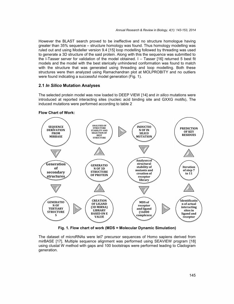
The sequences of micro RNA precursors were derived from miRbase and initially thesecondary structures were obtained using the MFold server [13]. This server utilizes amodified Zuker’s algorithm for the prediction of RNA secondary structures and sorts the beststructure according to the free energy. Once the secondary structures were obtained asymbolic programming approach was used at the MC SYM pipeline and the 3D structures ofthe precursors were obtained. These structures were then sorted according to their least freeenergies and a library of 3D structures belonging to the precursors was created. Thecreation of such a library was essential so as to sort the precursor structures according totheir length and class. Once the library creation was completed the KSRP protein sequencewas derived from the NCBI Genpept database and secondary structural analyses of thesequence were performed (Fig. 2). Following this a BLAST search was performed againstthe PDB database to identify suitable homologous structure.
Annual Research & Review in Biology, 4(1): 143-153, 2014
145
However the BLAST search proved to be ineffective and no structure homologue havinggreater than 35% sequence – structure homology was found. Thus homology modelling wasruled out and using Modeller version 9.4 [15] loop modelling followed by threading was usedto generate a 3D structure of the said protein. Along with this the sequence was submitted tothe I-Tasser server for validation of the model obtained. I – Tasser [16] returned 5 best fitmodels and the model with the best sterically unhindered conformation was found to matchwith the structure that was generated using threading and loop modelling. Both thesestructures were then analyzed using Ramachandran plot at MOLPROBITY and no outlierswere found indicating a successful model generation (Fig. 1).
2.1 In Silico Mutation Analyses
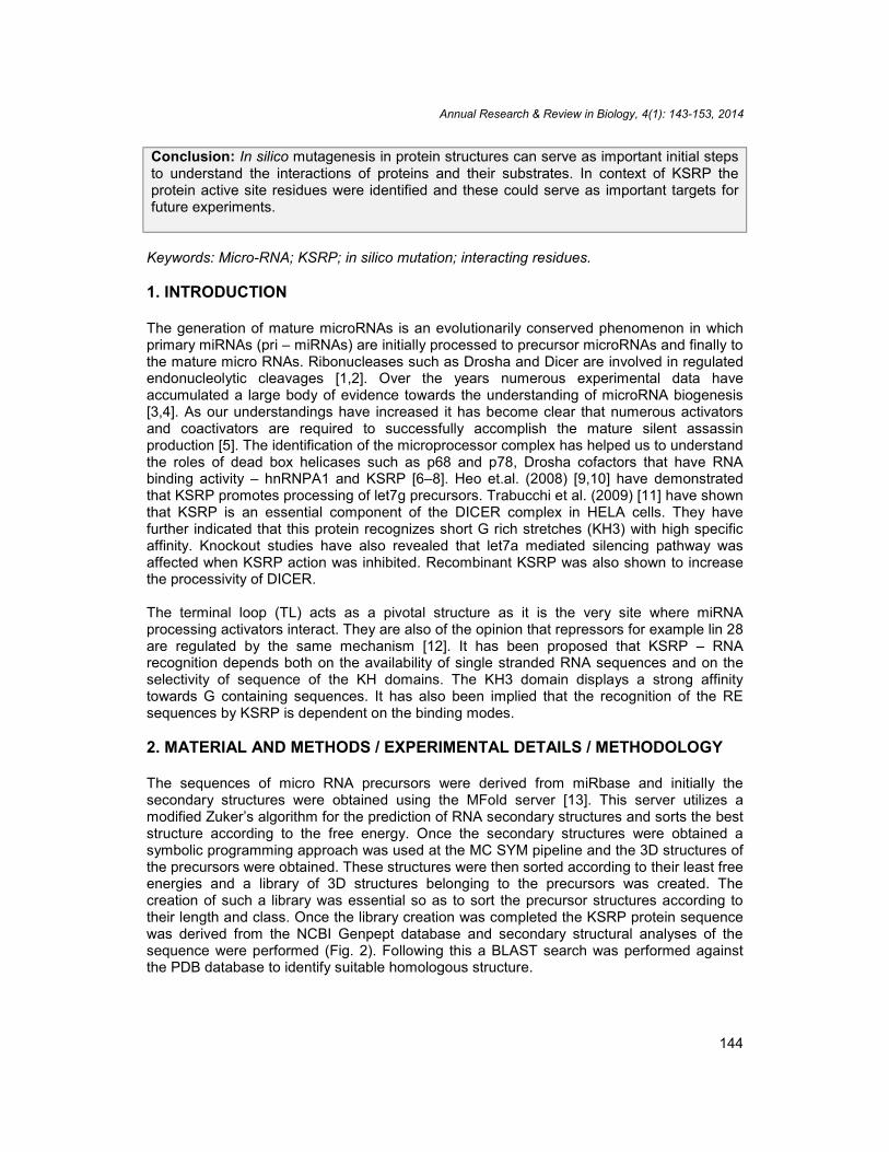
The selected protein model was now loaded to DEEP VIEW [14] and in silico mutations wereintroduced at reported interacting sites (nucleic acid binding site and GXXG motifs), Theinduced mutations were performed according to table 2
Flow Chart of Work:
Fig. 1. Flow chart of work (MDS = Molecular Dynamic Simulation)
The dataset of microRNAs were let7 precursor sequences of Homo sapiens derived frommirBASE [17]. Multiple sequence alignment was performed using SEAVIEW program [18]using clustal W method with gaps and 100 bootstraps were performed leading to Cladogramgeneration.
SEQUENCEDERIVATION
FROMMIRBASE
Generationof
secondarystructures
GENERATION OF
TERTIARYSTRUCTURE
S
Annual Research & Review in Biology, 4(1): 143-153, 2014
145
However the BLAST search proved to be ineffective and no structure homologue havinggreater than 35% sequence – structure homology was found. Thus homology modelling wasruled out and using Modeller version 9.4 [15] loop modelling followed by threading was usedto generate a 3D structure of the said protein. Along with this the sequence was submitted tothe I-Tasser server for validation of the model obtained. I – Tasser [16] returned 5 best fitmodels and the model with the best sterically unhindered conformation was found to matchwith the structure that was generated using threading and loop modelling. Both thesestructures were then analyzed using Ramachandran plot at MOLPROBITY and no outlierswere found indicating a successful model generation (Fig. 1).
2.1 In Silico Mutation Analyses
The selected protein model was now loaded to DEEP VIEW [14] and in silico mutations wereintroduced at reported interacting sites (nucleic acid binding site and GXXG motifs), Theinduced mutations were performed according to table 2
Flow Chart of Work:
Fig. 1. Flow chart of work (MDS = Molecular Dynamic Simulation)
The dataset of microRNAs were let7 precursor sequences of Homo sapiens derived frommirBASE [17]. Multiple sequence alignment was performed using SEAVIEW program [18]using clustal W method with gaps and 100 bootstraps were performed leading to Cladogramgeneration.
CREATIONOF LIGAND
(3D MIRNA)LIBRARY
BASED ON EVALUE
GENERATION OF 3D
STRUCTUREOF PROTEIN
ANALYSES OFSTRUCTURE
STABILITY ANDSELECTION OF
BESTSTRUCTURE
INDUCTION OF INSILICO
MUTATION
Analyses ofstructuralstability of
mutants andcreation of
receptorlibrary
MDS ofreceptor
and ligand(16000
complexes)
Annual Research & Review in Biology, 4(1): 143-153, 2014
145
However the BLAST search proved to be ineffective and no structure homologue havinggreater than 35% sequence – structure homology was found. Thus homology modelling wasruled out and using Modeller version 9.4 [15] loop modelling followed by threading was usedto generate a 3D structure of the said protein. Along with this the sequence was submitted tothe I-Tasser server for validation of the model obtained. I – Tasser [16] returned 5 best fitmodels and the model with the best sterically unhindered conformation was found to matchwith the structure that was generated using threading and loop modelling. Both thesestructures were then analyzed using Ramachandran plot at MOLPROBITY and no outlierswere found indicating a successful model generation (Fig. 1).
2.1 In Silico Mutation Analyses
The selected protein model was now loaded to DEEP VIEW [14] and in silico mutations wereintroduced at reported interacting sites (nucleic acid binding site and GXXG motifs), Theinduced mutations were performed according to table 2
Flow Chart of Work:
Fig. 1. Flow chart of work (MDS = Molecular Dynamic Simulation)
The dataset of microRNAs were let7 precursor sequences of Homo sapiens derived frommirBASE [17]. Multiple sequence alignment was performed using SEAVIEW program [18]using clustal W method with gaps and 100 bootstraps were performed leading to Cladogramgeneration.
Identification of actualinteracting
sites inligand andreceptor
Iterationof step 7
to 11
PREDICTIONOF KEY
RESIDUES
Annual Research & Review in Biology, 4(1): 143-153, 2014
146
3. RESULTS AND DISCUSSION
Over the years there has been much debate as to how the amino acid sequence of theprotein actually influences the biological property. An accepted standard measure of theimportance of protein residues has been sequence conservation [19-22]. Halabi et al. [23]working on S1A serine proteases have indicated that “decomposition of the protein into threequasi-independent groups of correlated amino acids” which they have termed as proteinsectors. However these sectors are actually basically structural evolutionary units and areinstrumental in maintenance of protein structures. The question however still remains thatconserved protein residues actually have influence on function or is the function of theprotein dependent on the spatial arrangement of a group of amino acids.
Fig. 2. Secondary Structure details of KSRP
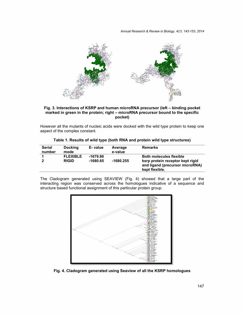
Through this work with KSRP and its interactions with precursor miRNA structures we havebeen able to locate key interaction sites in both the protein and RNA structures (Fig. 3). As isindicated in the methodology the identification involved an exhaustive pipeline of in silicomutation analyses. All mutants except six (406 MET/CYS; 424 GLY/PRO; 461 SER/THR;473 LYS/ARG; 477 ILE/LEU; 619 THR /TYR;) exhibited differences in the binding energieswhich were on the higher side (Table 1&2). The six mentioned complexes exhibited lowerbinding energy when compared with the wild type.
The second set of docking runs which involved the mutant analyses of nucleotides showed asimilar trend to the protein mutants.
Annual Research & Review in Biology, 4(1): 143-153, 2014
146
3. RESULTS AND DISCUSSION
Over the years there has been much debate as to how the amino acid sequence of theprotein actually influences the biological property. An accepted standard measure of theimportance of protein residues has been sequence conservation [19-22]. Halabi et al. [23]working on S1A serine proteases have indicated that “decomposition of the protein into threequasi-independent groups of correlated amino acids” which they have termed as proteinsectors. However these sectors are actually basically structural evolutionary units and areinstrumental in maintenance of protein structures. The question however still remains thatconserved protein residues actually have influence on function or is the function of theprotein dependent on the spatial arrangement of a group of amino acids.
Fig. 2. Secondary Structure details of KSRP
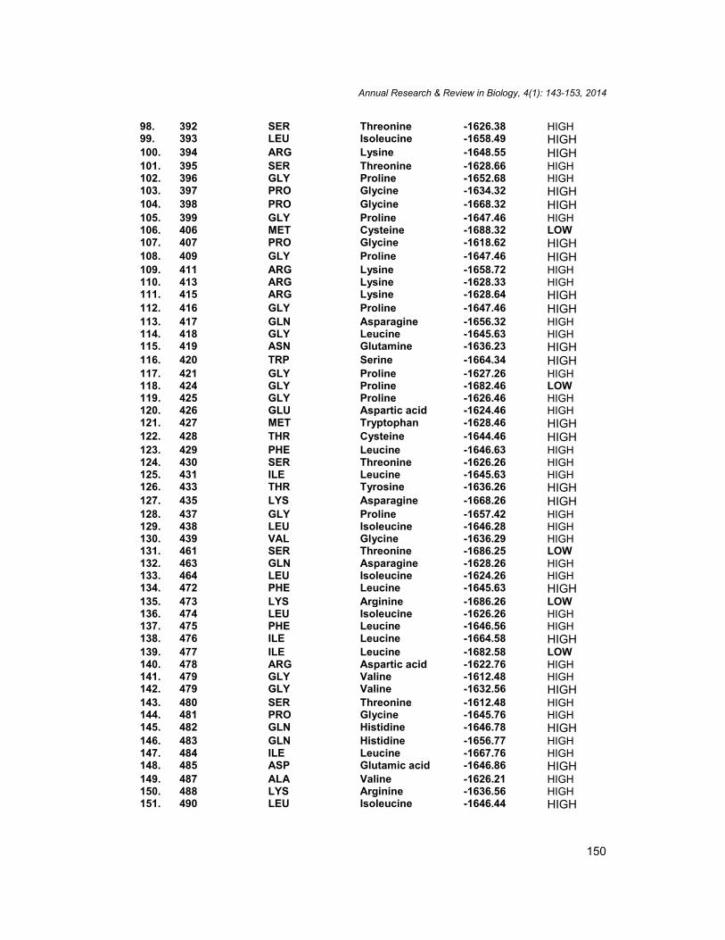
Through this work with KSRP and its interactions with precursor miRNA structures we havebeen able to locate key interaction sites in both the protein and RNA structures (Fig. 3). As isindicated in the methodology the identification involved an exhaustive pipeline of in silicomutation analyses. All mutants except six (406 MET/CYS; 424 GLY/PRO; 461 SER/THR;473 LYS/ARG; 477 ILE/LEU; 619 THR /TYR;) exhibited differences in the binding energieswhich were on the higher side (Table 1&2). The six mentioned complexes exhibited lowerbinding energy when compared with the wild type.
The second set of docking runs which involved the mutant analyses of nucleotides showed asimilar trend to the protein mutants.
Annual Research & Review in Biology, 4(1): 143-153, 2014
146
3. RESULTS AND DISCUSSION
Over the years there has been much debate as to how the amino acid sequence of theprotein actually influences the biological property. An accepted standard measure of theimportance of protein residues has been sequence conservation [19-22]. Halabi et al. [23]working on S1A serine proteases have indicated that “decomposition of the protein into threequasi-independent groups of correlated amino acids” which they have termed as proteinsectors. However these sectors are actually basically structural evolutionary units and areinstrumental in maintenance of protein structures. The question however still remains thatconserved protein residues actually have influence on function or is the function of theprotein dependent on the spatial arrangement of a group of amino acids.
Fig. 2. Secondary Structure details of KSRP
Through this work with KSRP and its interactions with precursor miRNA structures we havebeen able to locate key interaction sites in both the protein and RNA structures (Fig. 3). As isindicated in the methodology the identification involved an exhaustive pipeline of in silicomutation analyses. All mutants except six (406 MET/CYS; 424 GLY/PRO; 461 SER/THR;473 LYS/ARG; 477 ILE/LEU; 619 THR /TYR;) exhibited differences in the binding energieswhich were on the higher side (Table 1&2). The six mentioned complexes exhibited lowerbinding energy when compared with the wild type.
The second set of docking runs which involved the mutant analyses of nucleotides showed asimilar trend to the protein mutants.
Annual Research & Review in Biology, 4(1): 143-153, 2014
147
Fig. 3. Interactions of KSRP and human microRNA precursor (left – binding pocketmarked in green in the protein; right – microRNA precursor bound to the specific
pocket)
However all the mutants of nucleic acids were docked with the wild type protein to keep oneaspect of the complex constant.
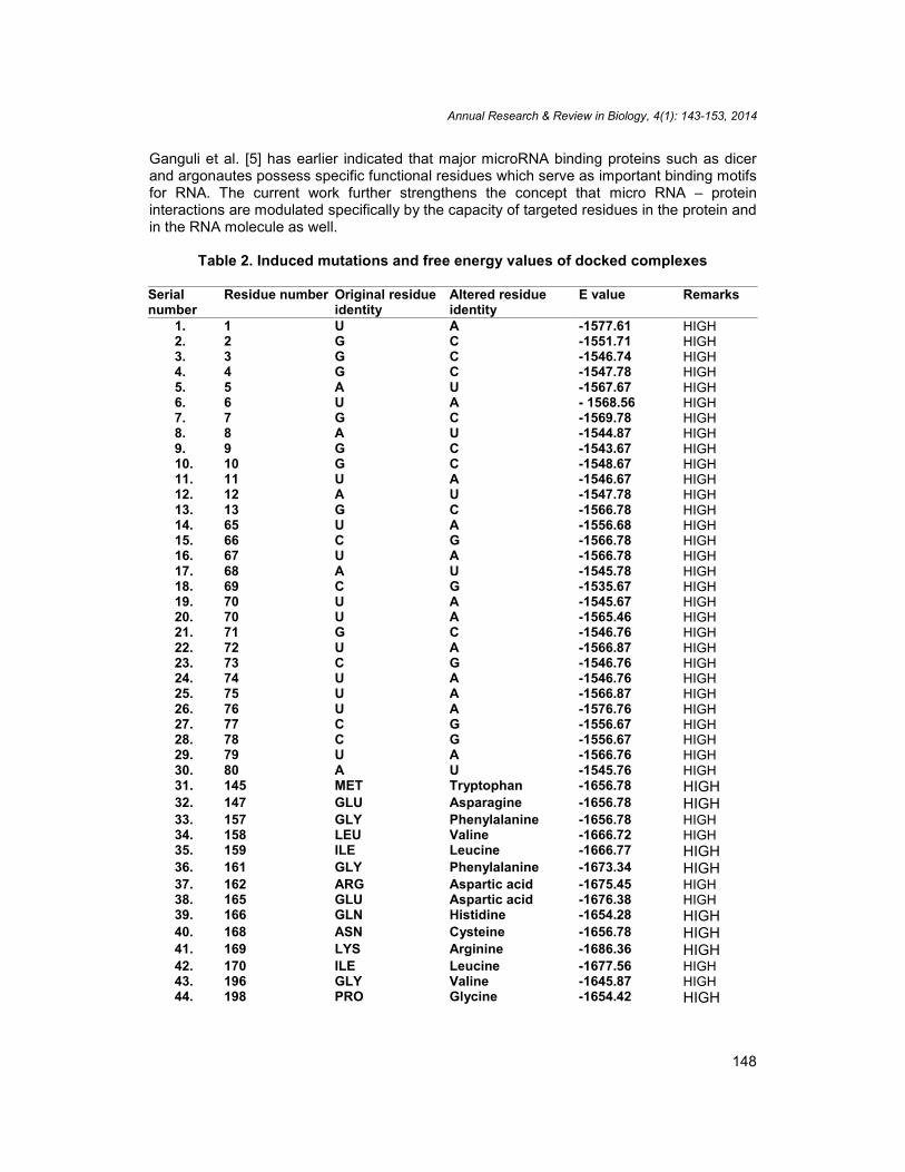
Table 1. Results of wild type (both RNA and protein wild type structures)
Serialnumber
Dockingmode
E- value Averagee-value
Remarks
1 FLEXIBLE -1679.86-1680.255
Both molecules flexible2 RIGID -1680.65 ksrp protein receptor kept rigid
and ligand (precursor microRNA)kept flexible.
The Cladogram generated using SEAVIEW (Fig. 4) showed that a large part of theinteracting region was conserved across the homologues indicative of a sequence andstructure based functional assignment of this particular protein group.
Fig. 4. Cladogram generated using Seaview of all the KSRP homologues
Annual Research & Review in Biology, 4(1): 143-153, 2014
147
Fig. 3. Interactions of KSRP and human microRNA precursor (left – binding pocketmarked in green in the protein; right – microRNA precursor bound to the specific
pocket)
However all the mutants of nucleic acids were docked with the wild type protein to keep oneaspect of the complex constant.
Table 1. Results of wild type (both RNA and protein wild type structures)
Serialnumber
Dockingmode
E- value Averagee-value
Remarks
1 FLEXIBLE -1679.86-1680.255
Both molecules flexible2 RIGID -1680.65 ksrp protein receptor kept rigid
and ligand (precursor microRNA)kept flexible.
The Cladogram generated using SEAVIEW (Fig. 4) showed that a large part of theinteracting region was conserved across the homologues indicative of a sequence andstructure based functional assignment of this particular protein group.
Fig. 4. Cladogram generated using Seaview of all the KSRP homologues
Annual Research & Review in Biology, 4(1): 143-153, 2014
148
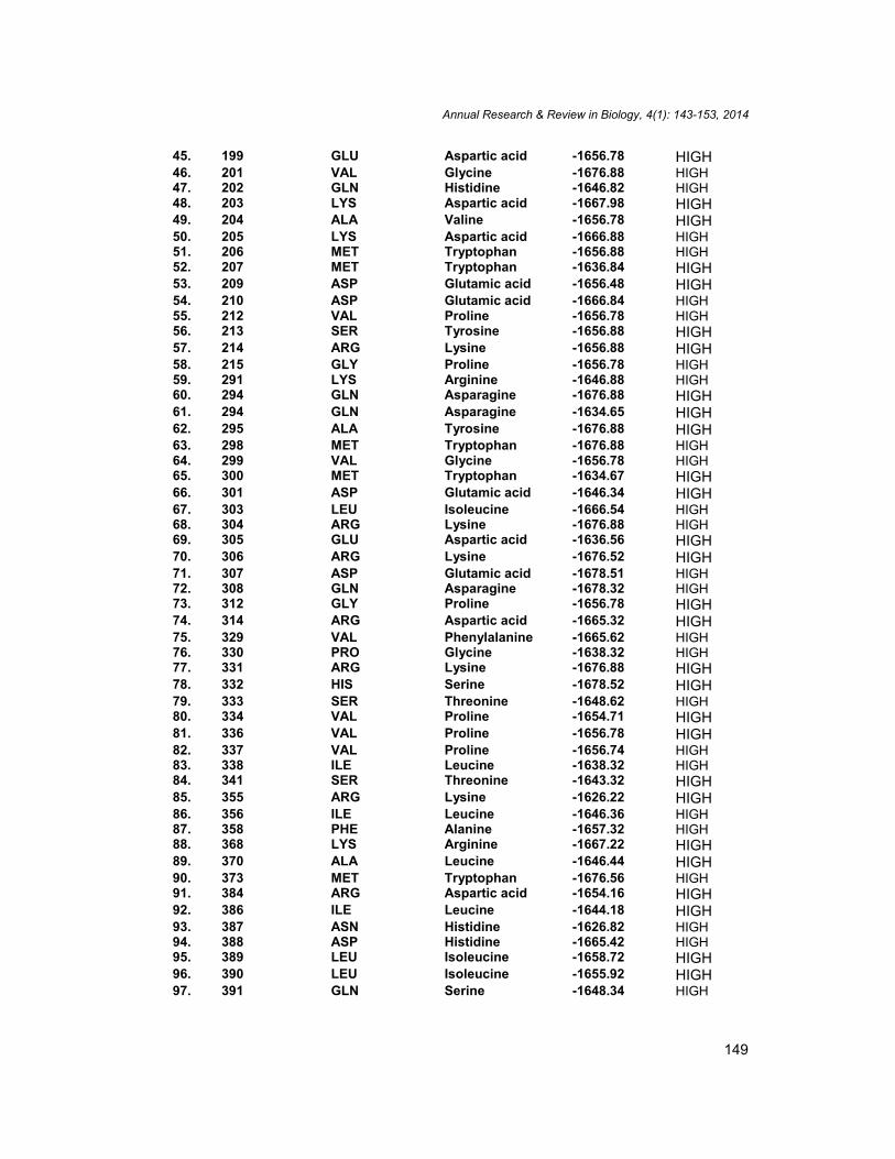
Ganguli et al. [5] has earlier indicated that major microRNA binding proteins such as dicerand argonautes possess specific functional residues which serve as important binding motifsfor RNA. The current work further strengthens the concept that micro RNA – proteininteractions are modulated specifically by the capacity of targeted residues in the protein andin the RNA molecule as well.
Table 2. Induced mutations and free energy values of docked complexes
Serialnumber
Residue number Original residueidentity
Altered residueidentity
E value Remarks
1. 1 U A -1577.61 HIGH2. 2 G C -1551.71 HIGH3. 3 G C -1546.74 HIGH4. 4 G C -1547.78 HIGH5. 5 A U -1567.67 HIGH6. 6 U A - 1568.56 HIGH7. 7 G C -1569.78 HIGH8. 8 A U -1544.87 HIGH9. 9 G C -1543.67 HIGH10. 10 G C -1548.67 HIGH11. 11 U A -1546.67 HIGH12. 12 A U -1547.78 HIGH13. 13 G C -1566.78 HIGH14. 65 U A -1556.68 HIGH15. 66 C G -1566.78 HIGH16. 67 U A -1566.78 HIGH17. 68 A U -1545.78 HIGH18. 69 C G -1535.67 HIGH19. 70 U A -1545.67 HIGH20. 70 U A -1565.46 HIGH21. 71 G C -1546.76 HIGH22. 72 U A -1566.87 HIGH23. 73 C G -1546.76 HIGH24. 74 U A -1546.76 HIGH25. 75 U A -1566.87 HIGH26. 76 U A -1576.76 HIGH27. 77 C G -1556.67 HIGH28. 78 C G -1556.67 HIGH29. 79 U A -1566.76 HIGH30. 80 A U -1545.76 HIGH31. 145 MET Tryptophan -1656.78 HIGH32. 147 GLU Asparagine -1656.78 HIGH33. 157 GLY Phenylalanine -1656.78 HIGH34. 158 LEU Valine -1666.72 HIGH35. 159 ILE Leucine -1666.77 HIGH36. 161 GLY Phenylalanine -1673.34 HIGH37. 162 ARG Aspartic acid -1675.45 HIGH38. 165 GLU Aspartic acid -1676.38 HIGH39. 166 GLN Histidine -1654.28 HIGH40. 168 ASN Cysteine -1656.78 HIGH41. 169 LYS Arginine -1686.36 HIGH42. 170 ILE Leucine -1677.56 HIGH43. 196 GLY Valine -1645.87 HIGH44. 198 PRO Glycine -1654.42 HIGH
Annual Research & Review in Biology, 4(1): 143-153, 2014
149
45. 199 GLU Aspartic acid -1656.78 HIGH46. 201 VAL Glycine -1676.88 HIGH47. 202 GLN Histidine -1646.82 HIGH48. 203 LYS Aspartic acid -1667.98 HIGH49. 204 ALA Valine -1656.78 HIGH50. 205 LYS Aspartic acid -1666.88 HIGH51. 206 MET Tryptophan -1656.88 HIGH52. 207 MET Tryptophan -1636.84 HIGH53. 209 ASP Glutamic acid -1656.48 HIGH54. 210 ASP Glutamic acid -1666.84 HIGH55. 212 VAL Proline -1656.78 HIGH56. 213 SER Tyrosine -1656.88 HIGH57. 214 ARG Lysine -1656.88 HIGH58. 215 GLY Proline -1656.78 HIGH59. 291 LYS Arginine -1646.88 HIGH60. 294 GLN Asparagine -1676.88 HIGH61. 294 GLN Asparagine -1634.65 HIGH62. 295 ALA Tyrosine -1676.88 HIGH63. 298 MET Tryptophan -1676.88 HIGH64. 299 VAL Glycine -1656.78 HIGH65. 300 MET Tryptophan -1634.67 HIGH66. 301 ASP Glutamic acid -1646.34 HIGH67. 303 LEU Isoleucine -1666.54 HIGH68. 304 ARG Lysine -1676.88 HIGH69. 305 GLU Aspartic acid -1636.56 HIGH70. 306 ARG Lysine -1676.52 HIGH71. 307 ASP Glutamic acid -1678.51 HIGH72. 308 GLN Asparagine -1678.32 HIGH73. 312 GLY Proline -1656.78 HIGH74. 314 ARG Aspartic acid -1665.32 HIGH75. 329 VAL Phenylalanine -1665.62 HIGH76. 330 PRO Glycine -1638.32 HIGH77. 331 ARG Lysine -1676.88 HIGH78. 332 HIS Serine -1678.52 HIGH79. 333 SER Threonine -1648.62 HIGH80. 334 VAL Proline -1654.71 HIGH81. 336 VAL Proline -1656.78 HIGH82. 337 VAL Proline -1656.74 HIGH83. 338 ILE Leucine -1638.32 HIGH84. 341 SER Threonine -1643.32 HIGH85. 355 ARG Lysine -1626.22 HIGH86. 356 ILE Leucine -1646.36 HIGH87. 358 PHE Alanine -1657.32 HIGH88. 368 LYS Arginine -1667.22 HIGH89. 370 ALA Leucine -1646.44 HIGH90. 373 MET Tryptophan -1676.56 HIGH91. 384 ARG Aspartic acid -1654.16 HIGH92. 386 ILE Leucine -1644.18 HIGH93. 387 ASN Histidine -1626.82 HIGH94. 388 ASP Histidine -1665.42 HIGH95. 389 LEU Isoleucine -1658.72 HIGH96. 390 LEU Isoleucine -1655.92 HIGH97. 391 GLN Serine -1648.34 HIGH
Annual Research & Review in Biology, 4(1): 143-153, 2014
150
98. 392 SER Threonine -1626.38 HIGH99. 393 LEU Isoleucine -1658.49 HIGH100. 394 ARG Lysine -1648.55 HIGH101. 395 SER Threonine -1628.66 HIGH102. 396 GLY Proline -1652.68 HIGH103. 397 PRO Glycine -1634.32 HIGH104. 398 PRO Glycine -1668.32 HIGH105. 399 GLY Proline -1647.46 HIGH106. 406 MET Cysteine -1688.32 LOW107. 407 PRO Glycine -1618.62 HIGH108. 409 GLY Proline -1647.46 HIGH109. 411 ARG Lysine -1658.72 HIGH110. 413 ARG Lysine -1628.33 HIGH111. 415 ARG Lysine -1628.64 HIGH112. 416 GLY Proline -1647.46 HIGH113. 417 GLN Asparagine -1656.32 HIGH114. 418 GLY Leucine -1645.63 HIGH115. 419 ASN Glutamine -1636.23 HIGH116. 420 TRP Serine -1664.34 HIGH117. 421 GLY Proline -1627.26 HIGH118. 424 GLY Proline -1682.46 LOW119. 425 GLY Proline -1626.46 HIGH120. 426 GLU Aspartic acid -1624.46 HIGH121. 427 MET Tryptophan -1628.46 HIGH122. 428 THR Cysteine -1644.46 HIGH123. 429 PHE Leucine -1646.63 HIGH124. 430 SER Threonine -1626.26 HIGH125. 431 ILE Leucine -1645.63 HIGH126. 433 THR Tyrosine -1636.26 HIGH127. 435 LYS Asparagine -1668.26 HIGH128. 437 GLY Proline -1657.42 HIGH129. 438 LEU Isoleucine -1646.28 HIGH130. 439 VAL Glycine -1636.29 HIGH131. 461 SER Threonine -1686.25 LOW132. 463 GLN Asparagine -1628.26 HIGH133. 464 LEU Isoleucine -1624.26 HIGH134. 472 PHE Leucine -1645.63 HIGH135. 473 LYS Arginine -1686.26 LOW136. 474 LEU Isoleucine -1626.26 HIGH137. 475 PHE Leucine -1646.56 HIGH138. 476 ILE Leucine -1664.58 HIGH139. 477 ILE Leucine -1682.58 LOW140. 478 ARG Aspartic acid -1622.76 HIGH141. 479 GLY Valine -1612.48 HIGH142. 479 GLY Valine -1632.56 HIGH143. 480 SER Threonine -1612.48 HIGH144. 481 PRO Glycine -1645.76 HIGH145. 482 GLN Histidine -1646.78 HIGH146. 483 GLN Histidine -1656.77 HIGH147. 484 ILE Leucine -1667.76 HIGH148. 485 ASP Glutamic acid -1646.86 HIGH149. 487 ALA Valine -1626.21 HIGH150. 488 LYS Arginine -1636.56 HIGH151. 490 LEU Isoleucine -1646.44 HIGH
Annual Research & Review in Biology, 4(1): 143-153, 2014
151
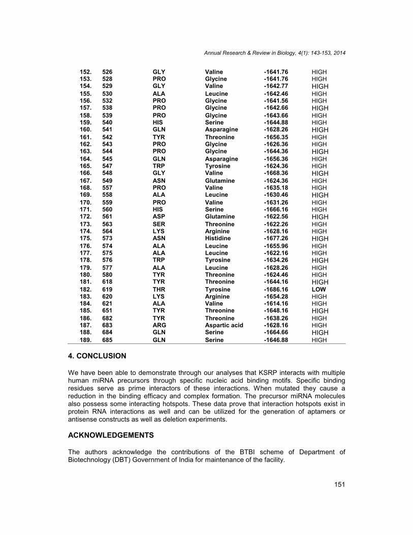
152. 526 GLY Valine -1641.76 HIGH153. 528 PRO Glycine -1641.76 HIGH154. 529 GLY Valine -1642.77 HIGH155. 530 ALA Leucine -1642.46 HIGH156. 532 PRO Glycine -1641.56 HIGH157. 538 PRO Glycine -1642.66 HIGH158. 539 PRO Glycine -1643.66 HIGH159. 540 HIS Serine -1644.88 HIGH160. 541 GLN Asparagine -1628.26 HIGH161. 542 TYR Threonine -1656.35 HIGH162. 543 PRO Glycine -1626.36 HIGH163. 544 PRO Glycine -1644.36 HIGH164. 545 GLN Asparagine -1656.36 HIGH165. 547 TRP Tyrosine -1624.36 HIGH166. 548 GLY Valine -1668.36 HIGH167. 549 ASN Glutamine -1624.36 HIGH168. 557 PRO Valine -1635.18 HIGH169. 558 ALA Leucine -1630.46 HIGH170. 559 PRO Valine -1631.26 HIGH171. 560 HIS Serine -1666.16 HIGH172. 561 ASP Glutamine -1622.56 HIGH173. 563 SER Threonine -1622.26 HIGH174. 564 LYS Arginine -1628.16 HIGH175. 573 ASN Histidine -1677.26 HIGH176. 574 ALA Leucine -1655.96 HIGH177. 575 ALA Leucine -1622.16 HIGH178. 576 TRP Tyrosine -1634.26 HIGH179. 577 ALA Leucine -1628.26 HIGH180. 580 TYR Threonine -1624.46 HIGH181. 618 TYR Threonine -1644.16 HIGH182. 619 THR Tyrosine -1686.16 LOW183. 620 LYS Arginine -1654.28 HIGH184. 621 ALA Valine -1614.16 HIGH185. 651 TYR Threonine -1648.16 HIGH186. 682 TYR Threonine -1638.26 HIGH187. 683 ARG Aspartic acid -1628.16 HIGH188. 684 GLN Serine -1664.66 HIGH189. 685 GLN Serine -1646.88 HIGH
4. CONCLUSION
We have been able to demonstrate through our analyses that KSRP interacts with multiplehuman miRNA precursors through specific nucleic acid binding motifs. Specific bindingresidues serve as prime interactors of these interactions. When mutated they cause areduction in the binding efficacy and complex formation. The precursor miRNA moleculesalso possess some interacting hotspots. These data prove that interaction hotspots exist inprotein RNA interactions as well and can be utilized for the generation of aptamers orantisense constructs as well as deletion experiments.
ACKNOWLEDGEMENTS
The authors acknowledge the contributions of the BTBI scheme of Department ofBiotechnology (DBT) Government of India for maintenance of the facility.
Annual Research & Review in Biology, 4(1): 143-153, 2014
152
CONSENT
Not applicable.
ETHICAL APPROVAL
Not applicable.
COMPETING INTERESTS
The authors declare that there are no competing interests.
REFERENCES
1. Das AK, Ganguli S, Gupta S and Datta A. Secondary structural analysis of MicroRNAand their precursors in plants Intl. Journal of Agr. Sci. 2011;3(1):62-64.
2. Davis BN, Hata A. Regulation of MicroRNA Biogenesis: A myriad of mechanisms. CellCommun Signal. 2009;7:18.
3. Davis BN, Hilyard AC, Nguyen PH, Lagna G, Hata A. Induction of microRNA-221 byplatelet-derived growth factor signaling is critical for modulation of vascular smoothmuscle phenotype. J Biol Chem. 2009;284:3728–3738.
4. Fukuda T, Yamagata K, Fujiyama S, Matsumoto T, Koshida I, et al. DEAD-box RNAhelicase subunits of the Drosha complex are required for processing of rRNA and asubset of microRNAs. Nat Cell Biol. 2007;9:604–611.
5. Ganguli S, De M, and Datta A. Analyses of argonaute– microRNA interactions in Zeamays, Intl. Jour.of.Comp. Biol. 2011;2(1):32-34
6. Garc´ıa-Mayoral MF, D´ıaz-Moreno I, Hollingworth D, Ramos A. The sequenceselectivity of KSRP explains its flexibility in the recognition of the RNA targets. NucleicAcids Res. 2008;36:5290–5296
7. Guil S, C´ aceres JF. The multifunctional RNA-binding protein hnRNP A1 is requiredfor processing of miR-18a. Nat Struct Mol Biol. 2007;14:591–596.
8. Hagan JP, Piskounova E, Gregory RI. Lin28 recruits the TUTase Zcchc11 to inhibit let-7 maturation in mouse embryonic stem cells. Nat Struct Mol Biol. 2009;16:1021–1025.
9. Heo I, Joo C, Cho J, HaM, Han J, et al. Lin28 mediates the terminal uridylation of let-7precursor MicroRNA. Mol Cell. 2008;32:276–284.
10. Heo I, Joo C, Kim YK, Ha M, Yoon MJ, et al. TUT4 in concert with Lin28 suppressesmicroRNA biogenesis through pre-microRNA uridylation. Cell. 2009;138:696–708.
11. Trabucchi M, Briata P, Garcia-Mayoral M, Haase AD, Filipowicz W, et al. The RNA-binding protein KSRP promotes the biogenesis of a subset of microRNAs. Nature.2009;459:1010–1014
12. Ruggiero T, Trabucchi M, De Santa F, Zupo S, Harfe BD, et al. LPS induces KH-typesplicing regulatory protein-dependent processing of microRNA- 155 precursors inmacrophages. FASEB J. 2009;23:2898–2908
13. Zuker M. Mfold web server for nucleic acid folding and hybridization prediction. Nucl.Acid. Res. 2003;31(13):3406-15,
14. Guex N, Peitsch MC. SWISS-MODEL and the Swiss-PdbViewer: An environment forcomparative protein modeling. Electrophoresis. 1997;18:2714-2723.
Annual Research & Review in Biology, 4(1): 143-153, 2014
153
15. Eswar N, Marti-Renom MA, Webb B, Madhusudhan MS, Eramian D, Shen M, PieperU, Sali A. Comparative Protein Structure Modeling With MODELLER. CurrentProtocols in Bioinformatics, John Wiley & Sons, Inc., Supplement. 2006;15:5.6.1-5.6.30.
16. Yang Zhang. I-TASSER server for protein 3D structure prediction. BMC Bioinfor.2008;9:40.
17. Kozomara A, Griffiths-Jones S. miRBase: integrating microRNA annotation and deep-sequencing data. Nucl. Acid. Res. 2011;39:D152-D157.
18. Gouy M, Guindon S, Gascuel O. SeaView version 4: a multiplatform graphical userinterface for sequence alignment and phylogenetic tree building. Molecular Biologyand Evolution. 2010;27(2):221-224.
19. Winter J, Jung S, K. Keller S, Gregory RI, Diederichs S. Many roads to maturity:microRNA biogenesis pathways and their regulation. Nat Cell Biol. 2009;11:228–234.
20. Wulczyn FG, Smirnova L, Rybak A, Brandt C, Kwidzinski E, et al. Post-transcriptionalregulation of the let-7 microRNA during neural cell specification. FASEB J.2007;21:415–426.
21. Capra JA, Singh M. Characterization and prediction of residues determining proteinfunctional specificity. Bioinfor. 2008;24(13):1473-80.
22. Zvelebil MJ JM, Barton GJ, Taylor WR, Sternberg MJE. Prediction of proteinsecondary structure and active sites using the alignment of homologous sequences. J.Mol. Biol. 1987;195:957-961.
23. Halabi N, Rivoire O, Leibler S, Ranganathan R. Protein sectors: evolutionary units ofthree-dimensional structure. Cell. 2009;138(4):774–786.
_________________________________________________________________________© 2014 Ganguli and Datta; This is an Open Access article distributed under the terms of the Creative CommonsAttribution License (http://creativecommons.org/licenses/by/3.0), which permits unrestricted use, distribution, andreproduction in any medium, provided the original work is properly cited.
Peer-review history:The peer review history for this paper can be accessed here:
http://www.sciencedomain.org/review-history.php?iid=287&id=32&aid=2136


![in silico (BFA thesis), 2002 [español]](https://static.fdokumen.com/doc/165x107/631f4913dbf756400702aca8/in-silico-bfa-thesis-2002-espanol.jpg)