In silico identification and expression analysis of MscS like gene family in rice
10
In silico identification and expression analysis of MscS like gene family in rice Ankush Ashok Saddhe, Kundan Kumar ⁎ Department of Biological Sciences, Birla Institute of Technology & Science Pilani, K. K. Birla Goa Campus, Goa 403726, India abstract article info Article history: Received 30 October 2014 Received in revised form 15 December 2014 Accepted 16 December 2014 Available online 4 January 2015 Keywords: MscS-like Mechanosensitive Oryza sativa Rice In silico Mechanosensitive channels are membrane proteins that open and shut in response to mechanical forces produced by osmotic pressure, sound, touch and gravity. These channels are involved in multiple physiological functions including hypoosmotic pressure, pain, hearing, blood pressure and cell volume regulation. In plants, these channels play a major role in proprioception, gravity sensing and maintenance of plastid shape and size. In the present study, we identified the mechanosensitive channel of small conductance like (MscS) homologue gene family in rice and analyzed their structure, phylogenetic relationship, localization and expression pattern. Five MscS like genes of rice (OsMSL) were found to be distributed on four chromosomes and clustered into two major groups. Subcellular localization predictions of the OsMSL family revealed their localization to plasma membrane, plastid envelope and mitochondria. The predicted gene structure, bonafide conserved signature motif, domain and the presence of transmembrane regions in each OsMSL strongly supported their identity as members of MscS-like gene family. Furthermore, in silico expression analysis of OsMSL genes revealed differential regulation patterns in tissue specific and abiotic stress libraries. These findings indicate that the in silico approach used here successfully identified in a genome-wide context MscS like gene family in rice, and further suggest the functional importance of MscS-like genes in rice. © 2015 Published by Elsevier B.V. This is an open access article under the CC BY-NC-ND license (http://creativecommons.org/licenses/by-nc-nd/4.0/). 1. Introduction Ion channels are multipass transmembrane proteins that form small aqueous pores across a membrane and allow ions to flow down their electrochemical gradient. They are ubiquitously present in all organisms and exhibit a significant role as sensor and effector molecules. Ion channels can be broadly classified into voltage gated, ligand gated and mechanical gated ion channels. A voltage gated ion channel is regulated by voltage difference across a membrane and a ligand gated ion channel responds to a ligand–receptor interaction and mechanosensitive (MS) ion channel sense and responds to mechanical stimuli. MS channels recognize a wide range of mechanical stimuli, and play important role in many physiological functions including osmotic pressure, touch and pain sensation, hearing, blood pressure, cell volume regulation, proprioception and gravity sense (Martinac, 1993; Sackin, 1995; Sachs and Morris, 1998; Hamill and Martinac, 2001). Prokaryotic MS channels have been extensively studied in Escherichia coli which harbors several MS channels (Martinac et al., 1987; Berrier et al., 1989; Delcour et al., 1989; Booth et al., 2007; Edwards et al., 2012; Reuter et al., 2014). Mechanosensitive channels have been classified based on their ion conductance as mechanosensitive channels of large conductance (MscL), mechanosensitive channels of small conductance (MscS), mechanosensitive channels of mini conductance (MscM) and mechano- sensitive channels of K + -selective (MscK) (Sukharev et al., 1994; Berrier et al., 1996). MS channels in bacteria also serve as emergency safety valves during hypoosmotic shock, which provide a graded series of response to osmotic stress in different environmental and developmental conditions (Martinac et al., 1987; Levina et al., 1999; Booth et al., 2005; Booth and Blount, 2012). Membrane topology of E. coli MscL channel predicted the formation of transmembrane helices TM1 and TM2 (Sukharev et al., 1994, 1999; Blount et al., 1996a,b). MscL channel is a transmembrane protein which exposed N-terminal towards cytoplasmic interface of plasma membrane and C-terminal faces the cytoplasm (Steinbacher et al., 2007; Iscla et al., 2008). Similarly, the structure of E. coli MscS channel revealed that this protein is a homoheptamer with each subunit containing a membrane domain and three transmembrane regions of TM1, TM2 and TM3 helices. TM3 helix is highly conserved throughout domains as compared with TM1 and TM2 of MscL and functionally equivalent to TM1 of MscL channels (Bass et al., 2002; Balleza and Gómez-Lagunas, 2009). Plant Gene 1 (2015) 8–17 Abbreviations: OsMSL, Oryza sativa MscS like; MscS, mechanosensitive channel small conductance; MscL, mechanosensitive channel large conductance; AtMSL, Arabidopsis thaliana MscS like. ⁎ Corresponding author. Tel.: +91 832 2580196; fax: +91 832 255 7033. E-mail address: [email protected] (K. Kumar). http://dx.doi.org/10.1016/j.plgene.2014.12.001 2352-4073/© 2015 Published by Elsevier B.V. This is an open access article under the CC BY-NC-ND license (http://creativecommons.org/licenses/by-nc-nd/4.0/). Contents lists available at ScienceDirect Plant Gene journal homepage: www.elsevier.com/locate/plant-gene
Transcript of In silico identification and expression analysis of MscS like gene family in rice

Plant Gene 1 (2015) 8–17
Contents lists available at ScienceDirect
Plant Gene
j ourna l homepage: www.e lsev ie r .com/ locate /p lant -gene
In silico identification and expression analysis of MscS like gene familyin rice
Ankush Ashok Saddhe, Kundan Kumar ⁎Department of Biological Sciences, Birla Institute of Technology & Science Pilani, K. K. Birla Goa Campus, Goa 403726, India
Abbreviations:OsMSL,Oryza sativaMscS like; MscS, mconductance; MscL, mechanosensitive channel large cothalianaMscS like.⁎ Corresponding author. Tel.: +91 832 2580196; fax: +
E-mail address: [email protected] (K. Kuma
http://dx.doi.org/10.1016/j.plgene.2014.12.0012352-4073/© 2015 Published by Elsevier B.V. This is an o
a b s t r a c t
a r t i c l e i n f oArticle history:Received 30 October 2014Received in revised form 15 December 2014Accepted 16 December 2014Available online 4 January 2015
Keywords:MscS-likeMechanosensitiveOryza sativaRiceIn silico
Mechanosensitive channels are membrane proteins that open and shut in response to mechanical forcesproduced by osmotic pressure, sound, touch and gravity. These channels are involved in multiple physiologicalfunctions including hypoosmotic pressure, pain, hearing, blood pressure and cell volume regulation. In plants,these channels play a major role in proprioception, gravity sensing and maintenance of plastid shape and size.In the present study, we identified the mechanosensitive channel of small conductance like (MscS) homologuegene family in rice and analyzed their structure, phylogenetic relationship, localization and expression pattern.Five MscS like genes of rice (OsMSL) were found to be distributed on four chromosomes and clustered intotwo major groups. Subcellular localization predictions of the OsMSL family revealed their localization to plasmamembrane, plastid envelope and mitochondria. The predicted gene structure, bonafide conserved signaturemotif, domain and the presence of transmembrane regions in each OsMSL strongly supported their identity asmembers ofMscS-like gene family. Furthermore, in silico expression analysis of OsMSL genes revealeddifferentialregulation patterns in tissue specific and abiotic stress libraries. These findings indicate that the in silico approachused here successfully identified in a genome-wide context MscS like gene family in rice, and further suggest thefunctional importance of MscS-like genes in rice.
© 2015 Published by Elsevier B.V. This is an open access article under the CC BY-NC-ND license(http://creativecommons.org/licenses/by-nc-nd/4.0/).
1. Introduction
Ion channels are multipass transmembrane proteins that form smallaqueous pores across a membrane and allow ions to flow down theirelectrochemical gradient. They are ubiquitously present in all organismsand exhibit a significant role as sensor and effector molecules. Ionchannels can be broadly classified into voltage gated, ligand gated andmechanical gated ion channels. A voltage gated ion channel is regulatedby voltage difference across amembrane and a ligand gated ion channelresponds to a ligand–receptor interaction and mechanosensitive (MS)ion channel sense and responds to mechanical stimuli. MS channelsrecognize a wide range of mechanical stimuli, and play important rolein many physiological functions including osmotic pressure, touchand pain sensation, hearing, blood pressure, cell volume regulation,proprioception and gravity sense (Martinac, 1993; Sackin, 1995; SachsandMorris, 1998; Hamill andMartinac, 2001). Prokaryotic MS channelshave been extensively studied in Escherichia coli which harbors several
echanosensitive channel smallnductance; AtMSL, Arabidopsis
91 832 255 7033.r).
pen access article under the CC BY-N
MS channels (Martinac et al., 1987; Berrier et al., 1989; Delcour et al.,1989; Booth et al., 2007; Edwards et al., 2012; Reuter et al., 2014).Mechanosensitive channels have been classified based on their ionconductance as mechanosensitive channels of large conductance(MscL), mechanosensitive channels of small conductance (MscS),mechanosensitive channels ofmini conductance (MscM) andmechano-sensitive channels of K+-selective (MscK) (Sukharev et al., 1994; Berrieret al., 1996). MS channels in bacteria also serve as emergency safetyvalves during hypoosmotic shock, which provide a graded seriesof response to osmotic stress in different environmental anddevelopmental conditions (Martinac et al., 1987; Levina et al., 1999;Booth et al., 2005; Booth and Blount, 2012).
Membrane topology of E. coliMscL channel predicted the formationof transmembrane helices TM1 and TM2 (Sukharev et al., 1994, 1999;Blount et al., 1996a,b). MscL channel is a transmembrane proteinwhich exposed N-terminal towards cytoplasmic interface of plasmamembrane and C-terminal faces the cytoplasm (Steinbacher et al.,2007; Iscla et al., 2008). Similarly, the structure of E. coli MscS channelrevealed that this protein is a homoheptamer with each subunitcontaining a membrane domain and three transmembrane regions ofTM1, TM2 and TM3 helices. TM3 helix is highly conserved throughoutdomains as compared with TM1 and TM2 of MscL and functionallyequivalent to TM1 of MscL channels (Bass et al., 2002; Balleza andGómez-Lagunas, 2009).
C-ND license (http://creativecommons.org/licenses/by-nc-nd/4.0/).
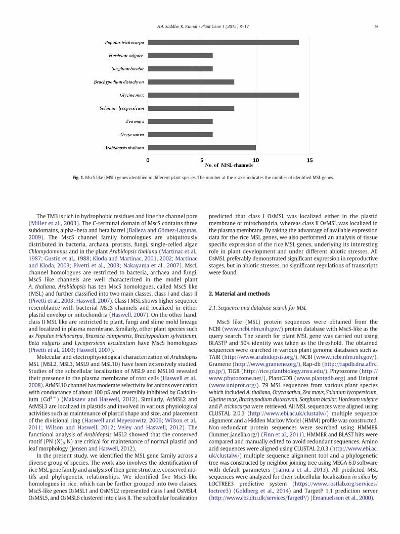
Fig. 1. MscS like (MSL) genes identified in different plant species. The number at the x-axis indicates the number of identified MSL genes.
9A.A. Saddhe, K. Kumar / Plant Gene 1 (2015) 8–17
The TM3 is rich in hydrophobic residues and line the channel pore(Miller et al., 2003). The C-terminal domain of MscS contains threesubdomains, alpha–beta and beta barrel (Balleza and Gómez-Lagunas,2009). The MscS channel family homologues are ubiquitouslydistributed in bacteria, archaea, protists, fungi, single-celled algaeChlamydomonas and in the plant Arabidopsis thaliana (Martinac et al.,1987; Gustin et al., 1988; Kloda and Martinac, 2001, 2002; Martinacand Kloda, 2003; Pivetti et al., 2003; Nakayama et al., 2007). MscLchannel homologues are restricted to bacteria, archaea and fungi.MscS like channels are well characterized in the model plantA. thaliana. Arabidopsis has ten MscS homologues, called MscS like(MSL) and further classified into two main classes, class I and class II(Pivetti et al., 2003; Haswell, 2007). Class I MSL shows higher sequenceresemblance with bacterial MscS channels and localized in eitherplastid envelop or mitochondria (Haswell, 2007). On the other hand,class II MSL like are restricted to plant, fungi and slime mold lineageand localized in plasma membrane. Similarly, other plant species suchas Populus trichocarpa, Brassica campestris, Brachypodium sylvaticum,Beta vulgaris and Lycopersicon esculentum have MscS homologues(Pivetti et al., 2003; Haswell, 2007).
Molecular and electrophysiological characterization of ArabidopsisMSL (MSL2, MSL3, MSL9 and MSL10) have been extensively studied.Studies of the subcellular localization of MSL9 and MSL10 revealedtheir presence in the plasma membrane of root cells (Haswell et al.,2008). AtMSL10 channel hasmoderate selectivity for anions over cationwith conductance of about 100 pS and reversibly inhibited by Gadolin-ium (Gd3+) (Maksaev and Haswell, 2012). Similarly, AtMSL2 andAtMSL3 are localized in plastids and involved in various physiologicalactivities such as maintenance of plastid shape and size, and placementof the divisional ring (Haswell and Meyerowitz, 2006; Wilson et al.,2011; Wilson and Haswell, 2012; Veley and Haswell, 2012). Thefunctional analysis of Arabidopsis MSL2 showed that the conservedmotif (PN (X)9 N) are critical for maintenance of normal plastid andleaf morphology (Jensen and Haswell, 2012).
In the present study, we identified the MSL gene family across adiverse group of species. The work also involves the identification ofriceMSL gene family and analysis of their gene structure, conservedmo-tifs and phylogenetic relationships. We identified five MscS-likehomologues in rice, which can be further grouped into two classes.MscS-like genes OsMSL1 and OsMSL2 represented class I and OsMSL4,OsMSL5, and OsMSL6 clustered into class II. The subcellular localization
predicted that class I OsMSL was localized either in the plastidmembrane or mitochondria, whereas class II OsMSL was localized inthe plasma membrane. By taking the advantage of available expressiondata for the rice MSL genes, we also performed an analysis of tissuespecific expression of the rice MSL genes, underlying its interestingrole in plant development and under different abiotic stresses. AllOsMSL preferably demonstrated significant expression in reproductivestages, but in abiotic stresses, no significant regulations of transcriptswere found.
2. Material and methods
2.1. Sequence and database search for MSL
MscS like (MSL) protein sequences were obtained from theNCBI (www.ncbi.nlm.nih.gov/) protein database with MscS-like as thequery search. The search for plant MSL gene was carried out usingBLASTP and 50% identity was taken as the threshold. The obtainedsequences were searched in various plant genome databases such asTAIR (http://www.arabidopsis.org/), NCBI (www.ncbi.nlm.nih.gov/),Gramene (http://www.gramene.org/), Rap-db (http://rapdb.dna.affrc.go.jp/), TIGR (http://rice.plantbiology.msu.edu/), Phytozome (http://www.phytozome.net/), PlantGDB (www.plantgdb.org) and Uniprot(www.uniprot.org/). 79 MSL sequences from various plant specieswhich included A. thaliana, Oryza sativa, Zeamays, Solanum lycopersicum,Glycinemax, Brachypodium distachyon, Sorghum bicolor,Hordeum vulgareand P. trichocarpawere retrieved. All MSL sequences were aligned usingCLUSTAL 2.0.3 (http://www.ebi.ac.uk/clustalw/) multiple sequencealignment and a HiddenMarkovModel (HMM) profile was constructed.Non-redundant protein sequences were searched using HMMER(hmmer.janelia.org/) (Finn et al., 2011). HMMER and BLAST hits werecompared and manually edited to avoid redundant sequences. Aminoacid sequences were aligned using CLUSTAL 2.0.3 (http://www.ebi.ac.uk/clustalw/) multiple sequence alignment tool and a phylogenetictree was constructed by neighbor joining tree using MEGA 6.0 softwarewith default parameters (Tamura et al., 2013). All predicted MSLsequences were analyzed for their subcellular localization in silico byLOCTREE3 predictive system (https://www.rostlab.org/services/loctree3) (Goldberg et al., 2014) and TargetP 1.1 prediction server(http://www.cbs.dtu.dk/services/TargetP/) (Emanuelsson et al., 2000).
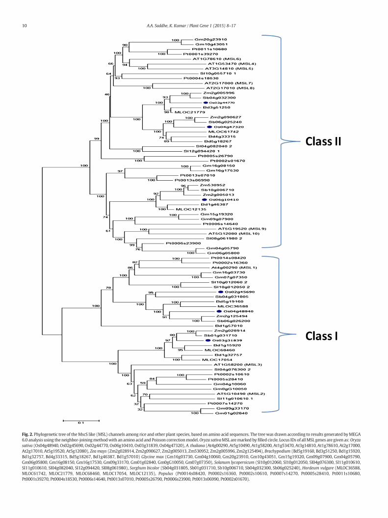
Fig. 2. Phylogenetic tree of theMscS like (MSL) channels among rice and other plant species, based on amino acid sequences. The treewas drawn according to results generated byMEGA6.0 analysis using the neighbor-joiningmethodwith an amino acid and Poisson correctionmodel.Oryza sativaMSL aremarked by filled circle. Locus IDs of allMSL genes are given as:Oryzasativa (Os04g48940, Os02g45690, Os02g44770, Os06g10410, Os03g31839, Os04g47320), A. thaliana (At4g00290, At5g10490, At1g58200, At1g53470, At3g14810, At1g78610, At2g17000,At2g17010, At5g19520, At5g12080), Zea mays (Zm2g028914, Zm2g090627, Zm2g005013, Zm530952, Zm2g005996, Zm2g125494), Brachypodium (Bd5g19160, Bd3g51250, Bd1g15920,Bd1g32757, Bd4g33315, Bd5g18267, Bd1g46387, Bd1g57010) Glycine max (Gm16g03730, Gm04g10060, Gm20g23910, Gm10g43051, Gm15g19320, Gm09g07900, Gm04g05790,Gm06g05800, Gm16g08150, Gm16g17530, Gm09g33170, Gm01g02840, Gm0gG10050, Gm07g07350), Solanum lycopersicum (Sl10g012060, Sl10g012050, Sl04g076300, Sl11g010610,Sl11g010610, Sl04g082040, Sl12g094420, Sl08g061980), Sorghum bicolor (Sb04g031805, Sb01g031710, Sb10g006710, Sb04g032300, Sb06g025240), Hordeum vulgare (MLOC36588,MLOC61742, MLOC21779, MLOC68460, MLOC17054, MLOC12135), Populus (Pt0014s08420, Pt0002s16360, Pt0002s10610, Pt0007s14270, Pt0005s28410, Pt0011s10680,Pt0001s39270, Pt0004s18530, Pt0006s14640, Pt0013s07010, Pt0005s26790, Pt0006s23900, Pt0013s06990, Pt0002s01670).
10 A.A. Saddhe, K. Kumar / Plant Gene 1 (2015) 8–17
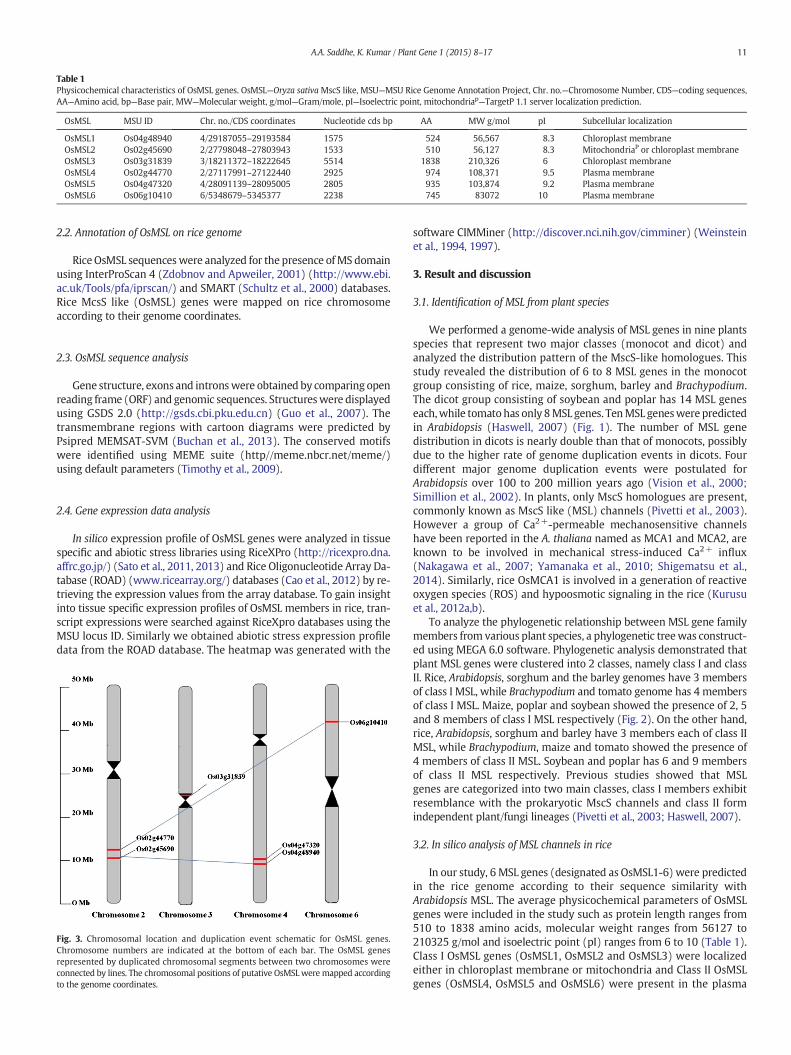
Table 1Physicochemical characteristics of OsMSL genes. OsMSL—Oryza sativaMscS like, MSU—MSU Rice Genome Annotation Project, Chr. no.—Chromosome Number, CDS—coding sequences,AA—Amino acid, bp—Base pair, MW—Molecular weight, g/mol—Gram/mole, pI—Isoelectric point, mitochondriap—TargetP 1.1 server localization prediction.
OsMSL MSU ID Chr. no./CDS coordinates Nucleotide cds bp AA MW g/mol pI Subcellular localization
OsMSL1 Os04g48940 4/29187055–29193584 1575 524 56,567 8.3 Chloroplast membraneOsMSL2 Os02g45690 2/27798048–27803943 1533 510 56,127 8.3 MitochondriaP or chloroplast membraneOsMSL3 Os03g31839 3/18211372–18222645 5514 1838 210,326 6 Chloroplast membraneOsMSL4 Os02g44770 2/27117991–27122440 2925 974 108,371 9.5 Plasma membraneOsMSL5 Os04g47320 4/28091139–28095005 2805 935 103,874 9.2 Plasma membraneOsMSL6 Os06g10410 6/5348679–5345377 2238 745 83072 10 Plasma membrane
11A.A. Saddhe, K. Kumar / Plant Gene 1 (2015) 8–17
2.2. Annotation of OsMSL on rice genome
RiceOsMSL sequenceswere analyzed for the presence ofMS domainusing InterProScan 4 (Zdobnov and Apweiler, 2001) (http://www.ebi.ac.uk/Tools/pfa/iprscan/) and SMART (Schultz et al., 2000) databases.Rice McsS like (OsMSL) genes were mapped on rice chromosomeaccording to their genome coordinates.
2.3. OsMSL sequence analysis
Gene structure, exons and intronswere obtained by comparing openreading frame (ORF) and genomic sequences. Structureswere displayedusing GSDS 2.0 (http://gsds.cbi.pku.edu.cn) (Guo et al., 2007). Thetransmembrane regions with cartoon diagrams were predicted byPsipred MEMSAT-SVM (Buchan et al., 2013). The conserved motifswere identified using MEME suite (http//meme.nbcr.net/meme/)using default parameters (Timothy et al., 2009).
2.4. Gene expression data analysis
In silico expression profile of OsMSL genes were analyzed in tissuespecific and abiotic stress libraries using RiceXPro (http://ricexpro.dna.affrc.go.jp/) (Sato et al., 2011, 2013) and Rice Oligonucleotide Array Da-tabase (ROAD) (www.ricearray.org/) databases (Cao et al., 2012) by re-trieving the expression values from the array database. To gain insightinto tissue specific expression profiles of OsMSL members in rice, tran-script expressions were searched against RiceXpro databases using theMSU locus ID. Similarly we obtained abiotic stress expression profiledata from the ROAD database. The heatmap was generated with the
Fig. 3. Chromosomal location and duplication event schematic for OsMSL genes.Chromosome numbers are indicated at the bottom of each bar. The OsMSL genesrepresented by duplicated chromosomal segments between two chromosomes wereconnected by lines. The chromosomal positions of putative OsMSL were mapped accordingto the genome coordinates.
software CIMMiner (http://discover.nci.nih.gov/cimminer) (Weinsteinet al., 1994, 1997).
3. Result and discussion
3.1. Identification of MSL from plant species
We performed a genome-wide analysis of MSL genes in nine plantsspecies that represent two major classes (monocot and dicot) andanalyzed the distribution pattern of the MscS-like homologues. Thisstudy revealed the distribution of 6 to 8 MSL genes in the monocotgroup consisting of rice, maize, sorghum, barley and Brachypodium.The dicot group consisting of soybean and poplar has 14 MSL geneseach,while tomatohas only 8MSL genes. TenMSL geneswere predictedin Arabidopsis (Haswell, 2007) (Fig. 1). The number of MSL genedistribution in dicots is nearly double than that of monocots, possiblydue to the higher rate of genome duplication events in dicots. Fourdifferent major genome duplication events were postulated forArabidopsis over 100 to 200 million years ago (Vision et al., 2000;Simillion et al., 2002). In plants, only MscS homologues are present,commonly known as MscS like (MSL) channels (Pivetti et al., 2003).However a group of Ca2+-permeable mechanosensitive channelshave been reported in the A. thaliana named as MCA1 and MCA2, areknown to be involved in mechanical stress-induced Ca2+ influx(Nakagawa et al., 2007; Yamanaka et al., 2010; Shigematsu et al.,2014). Similarly, rice OsMCA1 is involved in a generation of reactiveoxygen species (ROS) and hypoosmotic signaling in the rice (Kurusuet al., 2012a,b).
To analyze the phylogenetic relationship between MSL gene familymembers from various plant species, a phylogenetic treewas construct-ed using MEGA 6.0 software. Phylogenetic analysis demonstrated thatplant MSL genes were clustered into 2 classes, namely class I and classII. Rice, Arabidopsis, sorghum and the barley genomes have 3 membersof class I MSL, while Brachypodium and tomato genome has 4 membersof class I MSL. Maize, poplar and soybean showed the presence of 2, 5and 8 members of class I MSL respectively (Fig. 2). On the other hand,rice, Arabidopsis, sorghum and barley have 3 members each of class IIMSL, while Brachypodium, maize and tomato showed the presence of4 members of class II MSL. Soybean and poplar has 6 and 9 membersof class II MSL respectively. Previous studies showed that MSLgenes are categorized into two main classes, class I members exhibitresemblance with the prokaryotic MscS channels and class II formindependent plant/fungi lineages (Pivetti et al., 2003; Haswell, 2007).
3.2. In silico analysis of MSL channels in rice
In our study, 6 MSL genes (designated as OsMSL1-6) were predictedin the rice genome according to their sequence similarity withArabidopsis MSL. The average physicochemical parameters of OsMSLgenes were included in the study such as protein length ranges from510 to 1838 amino acids, molecular weight ranges from 56127 to210325 g/mol and isoelectric point (pI) ranges from 6 to 10 (Table 1).Class I OsMSL genes (OsMSL1, OsMSL2 and OsMSL3) were localizedeither in chloroplast membrane or mitochondria and Class II OsMSLgenes (OsMSL4, OsMSL5 and OsMSL6) were present in the plasma
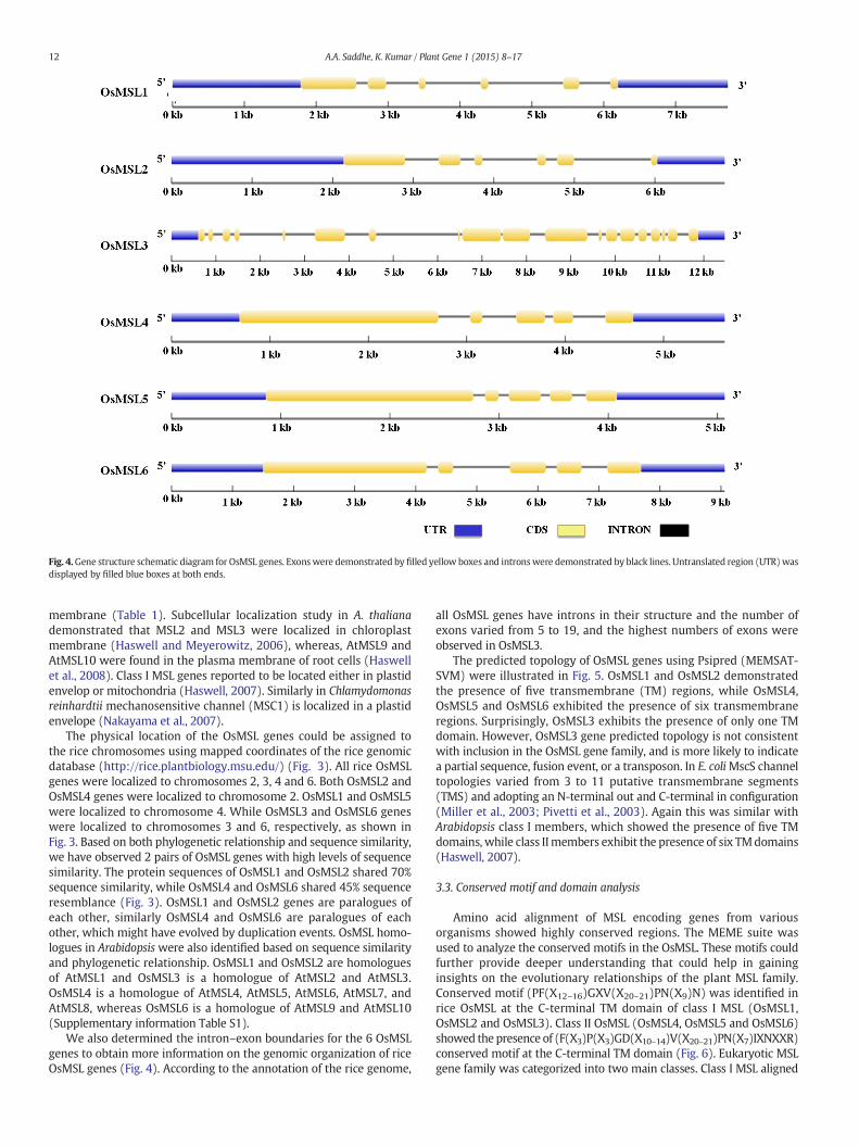
Fig. 4.Gene structure schematic diagram for OsMSL genes. Exonswere demonstrated by filled yellow boxes and intronswere demonstrated by black lines. Untranslated region (UTR)wasdisplayed by filled blue boxes at both ends.
12 A.A. Saddhe, K. Kumar / Plant Gene 1 (2015) 8–17
membrane (Table 1). Subcellular localization study in A. thalianademonstrated that MSL2 and MSL3 were localized in chloroplastmembrane (Haswell and Meyerowitz, 2006), whereas, AtMSL9 andAtMSL10 were found in the plasma membrane of root cells (Haswellet al., 2008). Class I MSL genes reported to be located either in plastidenvelop or mitochondria (Haswell, 2007). Similarly in Chlamydomonasreinhardtii mechanosensitive channel (MSC1) is localized in a plastidenvelope (Nakayama et al., 2007).
The physical location of the OsMSL genes could be assigned tothe rice chromosomes using mapped coordinates of the rice genomicdatabase (http://rice.plantbiology.msu.edu/) (Fig. 3). All rice OsMSLgenes were localized to chromosomes 2, 3, 4 and 6. Both OsMSL2 andOsMSL4 genes were localized to chromosome 2. OsMSL1 and OsMSL5were localized to chromosome 4. While OsMSL3 and OsMSL6 geneswere localized to chromosomes 3 and 6, respectively, as shown inFig. 3. Based on both phylogenetic relationship and sequence similarity,we have observed 2 pairs of OsMSL genes with high levels of sequencesimilarity. The protein sequences of OsMSL1 and OsMSL2 shared 70%sequence similarity, while OsMSL4 and OsMSL6 shared 45% sequenceresemblance (Fig. 3). OsMSL1 and OsMSL2 genes are paralogues ofeach other, similarly OsMSL4 and OsMSL6 are paralogues of eachother, which might have evolved by duplication events. OsMSL homo-logues in Arabidopsiswere also identified based on sequence similarityand phylogenetic relationship. OsMSL1 and OsMSL2 are homologuesof AtMSL1 and OsMSL3 is a homologue of AtMSL2 and AtMSL3.OsMSL4 is a homologue of AtMSL4, AtMSL5, AtMSL6, AtMSL7, andAtMSL8, whereas OsMSL6 is a homologue of AtMSL9 and AtMSL10(Supplementary information Table S1).
We also determined the intron–exon boundaries for the 6 OsMSLgenes to obtain more information on the genomic organization of riceOsMSL genes (Fig. 4). According to the annotation of the rice genome,
all OsMSL genes have introns in their structure and the number ofexons varied from 5 to 19, and the highest numbers of exons wereobserved in OsMSL3.
The predicted topology of OsMSL genes using Psipred (MEMSAT-SVM) were illustrated in Fig. 5. OsMSL1 and OsMSL2 demonstratedthe presence of five transmembrane (TM) regions, while OsMSL4,OsMSL5 and OsMSL6 exhibited the presence of six transmembraneregions. Surprisingly, OsMSL3 exhibits the presence of only one TMdomain. However, OsMSL3 gene predicted topology is not consistentwith inclusion in the OsMSL gene family, and is more likely to indicatea partial sequence, fusion event, or a transposon. In E. coliMscS channeltopologies varied from 3 to 11 putative transmembrane segments(TMS) and adopting an N-terminal out and C-terminal in configuration(Miller et al., 2003; Pivetti et al., 2003). Again this was similar withArabidopsis class I members, which showed the presence of five TMdomains, while class IImembers exhibit the presence of six TMdomains(Haswell, 2007).
3.3. Conserved motif and domain analysis
Amino acid alignment of MSL encoding genes from variousorganisms showed highly conserved regions. The MEME suite wasused to analyze the conserved motifs in the OsMSL. These motifs couldfurther provide deeper understanding that could help in gaininginsights on the evolutionary relationships of the plant MSL family.Conserved motif (PF(X12–16)GXV(X20–21)PN(X9)N) was identified inrice OsMSL at the C-terminal TM domain of class I MSL (OsMSL1,OsMSL2 and OsMSL3). Class II OsMSL (OsMSL4, OsMSL5 and OsMSL6)showed the presence of (F(X3)P(X3)GD(X10–14)V(X20–21)PN(X7)IXNXXR)conserved motif at the C-terminal TM domain (Fig. 6). Eukaryotic MSLgene family was categorized into two main classes. Class I MSL aligned

Fig. 5.A cartoonof the transmembrane helix topology summarizing the linear coordinates for the helices and indicating the protein's extra- and intercellular regions. Transmembranehelixregion represented by filled yellow boxes with labels S1, S2, S3, S4, S5 and S6. Membrane regions were represented by filled black boxes. N and C terminal regions were labeled andrepresented as single line.
13A.A. Saddhe, K. Kumar / Plant Gene 1 (2015) 8–17
with E. coli MscS and its C‐terminal TM domains resemble TM3 of MscS.Similar motifs have been reported in bacterial MscS channel as well.The conserved motif and consensus sequence (PF(X12–16)GXV(X20–21)PN(X9)N) were recognized at the C‐terminal TM domain and their sur-rounding sequences in E. coli (Pivetti et al., 2003). The MscS family has agood conservation throughout the TM3 helix which is rich in glycineand alanine residues (Bass et al., 2002; Pivetti et al., 2003; Miller et al.,2003). InterProScan analysis has combined different protein signaturerecognition methods into one resource including Superfamily, Pfam,Gene3D, Panther, SMART, Prosite and PIR (Protein Information Re-sources). The superfamily database of structural and functional proteinannotations showed that the MS channel and like Sm (LSM) domainwere present in all rice OsMSL sequences. The Pfam analysis (collectionof protein families) confirmed the presence of MS channel domain in allOsMSL. Alongwith theMS channel domain, OsMSL3 has a unique ammo-nia transferase like plant mobile domain, transposase MuDR plant do-main, zinc finger, PMZ-type domain and zinc finger SWIM-type domain.Protein information resource (PIR) database showed that rice OsMSL4,OsMSL5 and OsMSL6 have a unique MscS-like plant/fungi domainwhich is restricted to the plant and fungi lineages. To evaluate thesignaturemotif of all OsMSL,we performed aMEME suit analysis and rep-resented the distribution of motif to respective OsMSL gene (Supplemen-tary information Fig. S1). Motif 6 is the most commonly occurringsequence and is present in all OsMSL genes. Motif 1 consists of the MSchannel domain, uniformly distributed to all OsMSL except OsMSL3.Motif 10 is restricted to OsMSL1 and OMSL2. Motifs 2, 3, 5, 7 and 9 werepresent in OsMSL4, OsMSL5 and OsMSL6.Motifs 4 and 8were distributed
to OsMSL3, OsMSL4, OsMSL5, and OsMSL6 (Fig. 7a). The motifsequences and residue counts were diagrammatically represented inFig. 7b. Motifs 2 and 3 showed a protein kinase C phosphorylation site.As predicted, OsMSL4, OsML5 and OsMSL6 were located in plasmamembrane, which might be regulated by phosphorylation by proteinkinase C.
3.4. Gene expression analysis
Organ-specific expression of the 6 OsMSL genes under normalgrowth conditions in the field were obtained from the RiceXprodatabase (http://ricexpro.dna.affrc.go.jp/). The data obtained as OsMSLexpression level in different tissues and abiotic stress conditions (cold,drought and salt) of rice plant was retrieved as heat maps (Fig. 8a andb). All the OsMSL genes were expressed in various tissues/organsthroughout the plant life (Fig. 8a). In silico gene expression analysisrevealed that OsMSL3 has a uniform expression pattern in almost alltissues and organs of rice. Moreover, significant expression level ofOsMSL (OsMSL1, OsMSL2, OsMSL3, OsMSL5 andOsMSL6)was observedin reproductive organs such as inflorescence, anther, pistil, ovary,embryo and endosperm,while OsMSL4 showedno significant transcriptexpressions in tissue specific libraries. E. coli RpoS sigma factor controlsMscS gene expression at the transcriptional level, and activated by highosmolarity in stationary phase (Stokes et al., 2003). The MS channelgene family protects prokaryote from hypoosmotic stress and act assafety valve (Martinac et al., 1987; Booth et al., 2005). Eukaryotic MSLproved to have a diverse distribution and function besides serving as a
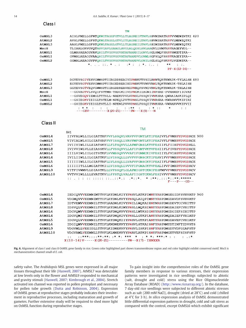
Fig. 6. Alignment of class I and class II OsMSL gene family in rice. Green color highlighted part shows transmembrane region and red color highlight exhibit conserved motif. MscS ismechanosensitive channel small of E. coli.
14 A.A. Saddhe, K. Kumar / Plant Gene 1 (2015) 8–17
safety valve. The Arabidopsis MSL genes were expressed in all majortissues throughout their life (Haswell, 2007). AtMSL7 was detectableat low levels only in the flower and AtMSL9 responded to mechanicaland gravity stimuli (Haswell, 2007; Kimbrough et al., 2004). Stretchactivated ion channel was reported in pollen protoplast and necessaryfor pollen tube growth (Dutta and Robinson, 2004). Expressionof OsMSL genes at reproductive stages probably indicates their involve-ment in reproductive processes, including maturation and growth ofgametes. Further extensive study will be required to shed more lighton OsMSL function during reproductive stages.
To gain insight into the comprehensive roles of the OsMSL genefamily members in response to various stresses, their expressionpatterns were investigated in rice seedlings subjected to abiotic(salt, drought and cold) stress using the Rice OligonucleotideArray Database (ROAD) (http://www.ricearray.org/). In the database,7 day-old rice seedlings were subjected to different abiotic stressessuch as salt (200 mM NaCl), drought (dried at 28°C) and cold (chilledat 4°C for 3 h). In silico expression analysis of OsMSL demonstratedlittle differential expression patterns in drought, cold and salt stress ascompared with the control, except OsMSL6 which exhibit significant
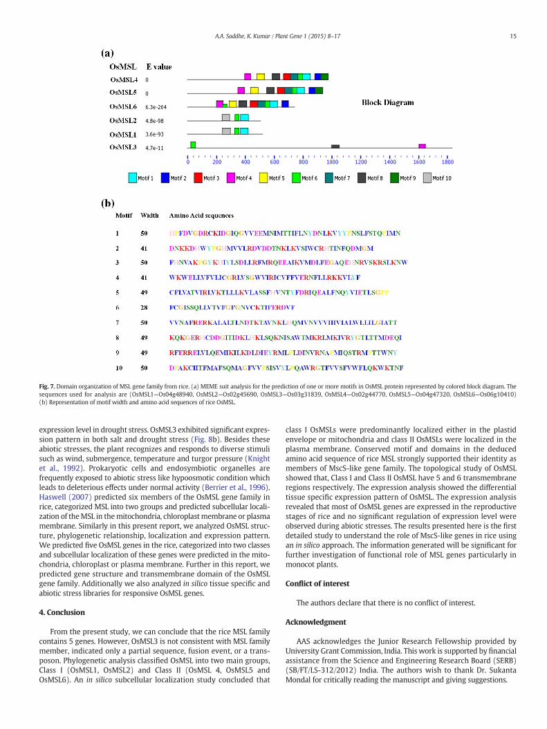
Fig. 7. Domain organization of MSL gene family from rice. (a) MEME suit analysis for the prediction of one or more motifs in OsMSL protein represented by colored block diagram. Thesequences used for analysis are (OsMSL1—Os04g48940, OsMSL2—Os02g45690, OsMSL3—Os03g31839, OsMSL4—Os02g44770, OsMSL5—Os04g47320, OsMSL6—Os06g10410)(b) Representation of motif width and amino acid sequences of rice OsMSL.
15A.A. Saddhe, K. Kumar / Plant Gene 1 (2015) 8–17
expression level in drought stress. OsMSL3 exhibited significant expres-sion pattern in both salt and drought stress (Fig. 8b). Besides theseabiotic stresses, the plant recognizes and responds to diverse stimulisuch as wind, submergence, temperature and turgor pressure (Knightet al., 1992). Prokaryotic cells and endosymbiotic organelles arefrequently exposed to abiotic stress like hypoosmotic condition whichleads to deleterious effects under normal activity (Berrier et al., 1996).Haswell (2007) predicted six members of the OsMSL gene family inrice, categorized MSL into two groups and predicted subcellular locali-zation of theMSL in themitochondria, chloroplastmembrane or plasmamembrane. Similarly in this present report, we analyzed OsMSL struc-ture, phylogenetic relationship, localization and expression pattern.We predicted five OsMSL genes in the rice, categorized into two classesand subcellular localization of these genes were predicted in the mito-chondria, chloroplast or plasma membrane. Further in this report, wepredicted gene structure and transmembrane domain of the OsMSLgene family. Additionally we also analyzed in silico tissue specific andabiotic stress libraries for responsive OsMSL genes.
4. Conclusion
From the present study, we can conclude that the rice MSL familycontains 5 genes. However, OsMSL3 is not consistent with MSL familymember, indicated only a partial sequence, fusion event, or a trans-poson. Phylogenetic analysis classified OsMSL into two main groups,Class I (OsMSL1, OsMSL2) and Class II (OsMSL 4, OsMSL5 andOsMSL6). An in silico subcellular localization study concluded that
class I OsMSLs were predominantly localized either in the plastidenvelope or mitochondria and class II OsMSLs were localized in theplasma membrane. Conserved motif and domains in the deducedamino acid sequence of rice MSL strongly supported their identity asmembers of MscS-like gene family. The topological study of OsMSLshowed that, Class I and Class II OsMSL have 5 and 6 transmembraneregions respectively. The expression analysis showed the differentialtissue specific expression pattern of OsMSL. The expression analysisrevealed that most of OsMSL genes are expressed in the reproductivestages of rice and no significant regulation of expression level wereobserved during abiotic stresses. The results presented here is the firstdetailed study to understand the role of MscS-like genes in rice usingan in silico approach. The information generated will be significant forfurther investigation of functional role of MSL genes particularly inmonocot plants.
Conflict of interest
The authors declare that there is no conflict of interest.
Acknowledgment
AAS acknowledges the Junior Research Fellowship provided byUniversity Grant Commission, India. This work is supported by financialassistance from the Science and Engineering Research Board (SERB)(SB/FT/LS-312/2012) India. The authors wish to thank Dr. SukantaMondal for critically reading the manuscript and giving suggestions.
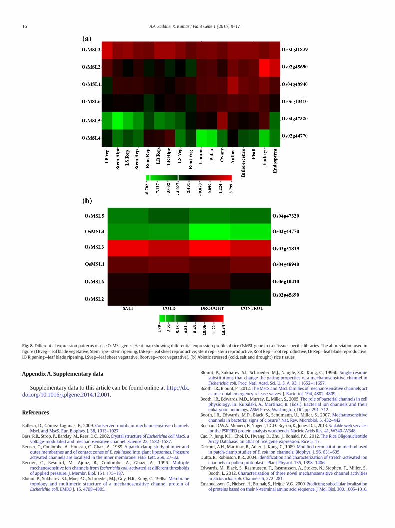
Fig. 8. Differential expression patterns of rice OsMSL genes. Heat map showing differential expression profile of rice OsMSL gene in (a) Tissue specific libraries. The abbreviation used infigure (LBveg—leaf blade vegetative, Stemripe—stemripening, LSRep—leaf sheet reproductive, Stem rep—stemreproductive, RootRep—root reproductive, LB Rep—leaf blade reproductive,LB Ripening—leaf blade ripening, LSveg—leaf sheet vegetative, Rootveg—root vegetative). (b) Abiotic stressed (cold, salt and drought) rice tissues.
16 A.A. Saddhe, K. Kumar / Plant Gene 1 (2015) 8–17
Appendix A. Supplementary data
Supplementary data to this article can be found online at http://dx.doi.org/10.1016/j.plgene.2014.12.001.
References
Balleza, D., Gómez-Lagunas, F., 2009. Conserved motifs in mechanosensitive channelsMscL and MscS. Eur. Biophys. J. 38, 1013–1027.
Bass, R.B., Strop, P., Barclay,M., Rees, D.C., 2002. Crystal structure of Escherichia coliMscS, avoltage-modulated and mechanosensitive channel. Science 22, 1582–1587.
Berrier, C., Coulombe, A., Houssin, C., Ghazi, A., 1989. A patch-clamp study of inner andouter membranes and of contact zones of E. coli fused into giant liposomes. Pressureactivated channels are localized in the inner membrane. FEBS Lett. 259, 27–32.
Berrier, C., Besnard, M., Ajouz, B., Coulombe, A., Ghazi, A., 1996. Multiplemechanosensitive ion channels from Escherichia coli, activated at different thresholdsof applied pressure. J. Membr. Biol. 151, 175–187.
Blount, P., Sukharev, S.I., Moe, P.C., Schroeder, M.J., Guy, H.R., Kung, C., 1996a. Membranetopology and multimeric structure of a mechanosensitive channel protein ofEscherichia coli. EMBO J. 15, 4798–4805.
Blount, P., Sukharev, S.I., Schroeder, M.J., Nangle, S.K., Kung, C., 1996b. Single residuesubstitutions that change the gating properties of a mechanosensitive channel inEscherichia coli. Proc. Natl. Acad. Sci. U. S. A. 93, 11652–11657.
Booth, I.R., Blount, P., 2012. The MscS andMscL families of mechanosensitive channels actas microbial emergency release valves. J. Bacteriol. 194, 4802–4809.
Booth, I.R., Edwards, M.D., Murray, E., Miller, S., 2005. The role of bacterial channels in cellphysiology. In: Kubalski, A., Martinac, B. (Eds.), Bacterial ion channels and theireukaryotic homologs. ASM Press, Washington, DC, pp. 291–312.
Booth, I.R., Edwards, M.D., Black, S., Schumann, U., Miller, S., 2007. Mechanosensitivechannels in bacteria: signs of closure? Nat. Rev. Microbiol. 5, 432–442.
Buchan, D.W.A.,Minneci, F., Nugent, T.C.O., Bryson, K., Jones, D.T., 2013. Scalableweb servicesfor the PSIPRED protein analysis workbench. Nucleic Acids Res. 41, W340–W348.
Cao, P., Jung, K.H., Choi, D., Hwang, D., Zhu, J., Ronald, P.C., 2012. The Rice OligonucleotideArray Database: an atlas of rice gene expression. Rice 5, 17.
Delcour, A.H., Martinac, B., Adler, J., Kung, C., 1989. Modified reconstitution method usedin patch-clamp studies of E. coli ion channels. Biophys. J. 56, 631–635.
Dutta, R., Robinson, K.R., 2004. Identification and characterization of stretch‐activated ionchannels in pollen protoplasts. Plant Physiol. 135, 1398–1406.
Edwards, M., Black, S., Rasmussen, T., Rasmussen, A., Stokes, N., Stephen, T., Miller, S.,Booth, I., 2012. Characterization of three novel mechanosensitive channel activitiesin Escherichia coli. Channels 6, 272–281.
Emanuelsson, O., Nielsen, H., Brunak, S., Heijne, V.G., 2000. Predicting subcellular localizationof proteins based on their N-terminal amino acid sequence. J. Mol. Biol. 300, 1005–1016.
17A.A. Saddhe, K. Kumar / Plant Gene 1 (2015) 8–17
Finn, R.D., Clements, J., Eddy, S.R., 2011. HMMER web server: interactive sequencesimilarity searching. Nucleic Acids Res. 39, W29–W37.
Goldberg, T., Hecht, M., Hamp, T., Karl, T., Yachdav, G., Ahmed, N., Altermann, U., Angerer,P., Ansorge, S., Balasz, K., Bernhofer, M., Betz, A., Cizmadija, L., Do, K.T., Gerke, J., Greil,R., Joerdens, V., Hastreiter, M., Hembach, K., Herzog, M., Kalemanov, M., Kluge, M.,Meier, A., Nasir, H., Neumaier, U., Prade, V., Reeb, J., Sorokoumov, A., Troshani, I.,Vorberg, S., Waldraff, S., Zierer, J., Nielsen, H., Rost, B., 2014. LocTree3 prediction of lo-calization. Nucleic Acids Res. 42, W350–W355.
Guo, A.Y., Zhu, Q.H., Chen, X., Luo, J.C., 2007. GSDS: a gene structure display server. YiChuan 29, 1023–1026.
Gustin, M.C., Zhou, X.L., Martinac, B., Kung, C., 1988. A mechanosensitive ion channel inthe yeast plasma membrane. Science 242, 762–765.
Hamill, O.P., Martinac, B., 2001. Molecular basis of mechanotransduction in living cells.Physiol. Rev. 81, 685–740.
Haswell, E.S., 2007. MscS-like proteins in plants. In: Hamill, O.P. (Ed.), MechanosensitiveIon Channels, Part A. Academic Press, San Diego, CA, pp. 329–359.
Haswell, E.S., Meyerowitz, E.M., 2006. MscS-like proteins control plastid size and shape inArabidopsis thaliana. Curr. Biol. 16, 1–11.
Haswell, E.S., Peyronnet, R., Barbier-Brygoo, H., Meyerowitz, E.M., Frachisse, J.M., 2008.Two homologs provide mechanosensitive channel activities in the Arabidopsis root.Curr. Biol. 18, 730–734.
Iscla, I., Wray, R., Blount, P., 2008. On the structure of the N-terminal domain of the MscLchannel: helical bundle or membrane interface. Biophys. J. 95, 2283–2291.
Jensen, G.S., Haswell, E.S., 2012. Functional analysis of conserved motifs in themechanosensitive channel homolog MscS-Like2 from Arabidopsis thaliana. PLoSONE 7, 1–10.
Kimbrough, J.M., Salinas‐Mondragon, R., Boss, W.F., Brown, C.S., Sedero, V., H.W., 2004.The fast and transient transcriptional network of gravity and mechanical stimulationin the Arabidopsis root apex. Plant Physiol. 136, 2790–2805.
Kloda, A., Martinac, B., 2001. Mechanosensitive channels in archaea. Cell Biochem.Biophys. 34, 349–381.
Kloda, A., Martinac, B., 2002. Common evolutionary origins of mechanosensitive ionchannels in archaea, bacteria and cell walled Eukarya. Archaea 1, 35–44.
Knight, M.R., Smith, S.M., Trewavas, A.J., 1992. Wind‐induced plant motion immediatelyincreases cytosolic calcium. Proc. Natl. Acad. Sci. U. S. A. 89, 4967–4971.
Kurusu, T., Iida, H., Kuchitsu, K., 2012a. Roles of a putative mechanosensitive plasmamembrane Ca2+-permeable channel OsMCA1 in generation of reactive oxygenspecies and hypo-osmotic signaling in rice. Plant Signal. Behav. 7, 796–798.
Kurusu, T., Nishikawa, D., Yamazaki, Y., Gotoh, M., Nakano, M., Hamada, H., Yamanaka, T.,Iida, K., Nakagawa, Y., Saji, H., Shinozaki, K., Iida, H., Kuchitsu, K., 2012b. Plasmamembrane protein OsMCA1 is involved in regulation of hypo-osmotic shock-induced Ca2+ influx and modulates generation of reactive oxygen species incultured rice cells. BMC Plant Biol. 12, 1–15.
Levina, N., Totemeyer, S., Stokes, N.R., Louis, P., Jones, M.A., Booth, I.R., 1999. Protection ofEscherichia coli cells against extreme turgor by activation of MscS and MscLmechanosensitive channels: identification of genes required for MscS activity.EMBO J. 18, 1730–1737.
Maksaev, G., Haswell, E.S., 2012. MscS-Like10 is a stretch-activated ion channel fromArabidopsis thaliana with a preference for anions. Proc. Natl. Acad. Sci. U. S. A. 109,19015–19020.
Martinac, B., 1993. Mechanosensitive ion channels: biophysics and physiology. In:Jackson, M.B. (Ed.), Thermodynamics of membrane receptors and channels. CRCPress, Boca Raton, FL, pp. 327–351.
Martinac, B., Kloda, A., 2003. Evolutionary origins of mechanosensitive ion channels. Prog.Biophys. Mol. Biol. 82, 11–24.
Martinac, B., Buechner, M., Delcour, A.H., Adler, J., Kung, C., 1987. Pressure-sensitive ionchannel in Escherichia coli. Proc. Natl. Acad. Sci. U. S. A. 84, 2297–2301.
Miller, S., Bartlett, W., Chandrasekaran, S., Simpson, S., Edwards, M., Booth, I.R., 2003.Domain organization of the MscS mechanosensitive channel of Escherichia coli.EMBO J. 22, 36–46.
Nakagawa, Y., Katagiri, T., Shinozaki, K., Qi, Z., Tatsumi, H., et al., 2007. Arabidopsis plasmamembrane protein crucial for Ca2+ influx and touch sensing in roots. Proc. Natl.Acad. Sci. U. S. A. 104, 3639–3644.
Nakayama, Y., Fujiu, K., Sokabe, M., Yoshimura, K., 2007. Molecular and electrophysiologicalcharacterization of a mechanosensitive channel expressed in the chloroplasts ofChlamydomonas. Proc. Natl. Acad. Sci. U. S. A. 104, 5883–5888.
Pivetti, C.D., Yen, M.R., Miller, S., Busch, W., Tseng, Y.H., Booth, I.R., Saier Jr., M.H., 2003.Two families of mechanosensitive channel proteins. Microbiol. Mol. Biol. Rev. 67,66–85.
Reuter, M., Hayward, J., Black, S., Miller, S., Dryden, F., Booth, R., 2014. Mechanosensitivechannels and bacterial cell wall integrity: does life end with a bang or a whimper?J. R. Soc. Interface 11, 1–9.
Sachs, F., Morris, C.E., 1998. Mechanosensitive ion channels in nonspecialized cells. Rev.Physiol. Biochem. Pharmacol. 132, 1–77.
Sackin, H., 1995. Mechanosensitive channels. Annu. Rev. Physiol. 57, 333–353.Sato, Y., Antonio, B., Namiki, N., Takehisa, H., Minami, H., Kamatsuki, K., Sugimoto, K.,
Shimizu, Y., Hirochika, H., Nagamura, Y., 2011. RiceXpro: a platform for monitoringgene expression in japonica rice grown under natural field conditions. NucleicAcids Res. 39, D1141–D1148.
Sato, Y., Takehisa, H., Kamatsuki, K., Minami, H., Namiki, N., Ikawa, H., Ohyanagi, H.,Sugimoto, K., Antonio, B., Nagamura, Y., 2013. RiceXPro Version 3.0: expanding theinformatics resource for rice transcriptome. Nucleic Acids Res. 41, D1206–D1213.
Schultz, J., Copley, R.R., Doerks, T., Ponting, C.P., Bork, P., 2000. SMART: a web-based toolfor the study of genetically mobile domains. Nucleic Acids Res. 28, 231–234.
Shigematsu, H., Iida, K., Nakano, M., Chaudhuri, P., Iida, H., et al., 2014. Structuralcharacterization of the mechanosensitive channel candidate MCA2 from Arabidopsisthaliana. PLoS ONE 9, e87724.
Simillion, C., Vandepoele, K., Van Montagu, M.C., Zabeau, M., Van de Peer, Y., 2002. Thehidden duplication past of Arabidopsis thaliana. Proc. Natl. Acad. Sci. U. S. A. 99,13627–13632.
Steinbacher, S., Bass, R., Strop, P., Rees, D.C., 2007. Structures of the prokaryoticmechanosensitive channels MscL and MscS. Curr. Top. Membr. 58, 1–24.
Stokes, N.R., Murray, H.D., Subramaniam, C., Gourse, R.L., Louis, P., Bartlett, W., Miller, S.,Booth, I.R., 2003. A role for mechanosensitive channels in survival of stationaryphase: regulation of channel expression by RpoS. Proc. Natl. Acad. Sci. U. S. A. 100,15959–15964.
Sukharev, S.I., Blount, P., Martinac, B., Blattner, F.R., Kung, C., 1994. A large-conductancemechanosensitive channel in E. coli encoded by mscL alone. Nature 368, 265–268.
Sukharev, S., Sigurdson,W.J., Kung, C., Sachs, F., 1999. Energetic and spatial parameters forgating of the bacterial large conductance mechanosensitive channel. MscL J. Gen.Physiol. 113, 525–540.
Tamura, K., Stecher, G., Peterson, D., Filipski, A., Kumar, S., 2013. MEGA6: MolecularEvolutionary Genetics Analysis version 6.0. Mol. Biol. Evol. 30, 2725–2729.
Timothy, L., Mikael Bodén, B., Buske, F.A., Frith, M., Grant, C.E., Clementi, L., Ren, J., Li,W.W., Noble, W.S., 2009. MEME SUITE: tools for motif discovery and searching.Nucleic Acids Res. 37, 202–208.
Veley, K.M., Haswell, E.S., 2012. Plastids and pathogens: mechanosensitive channels andsurvival in a hypoosmotic world. Plant Signal. Behav. 7, 668–671.
Vision, T.J., Brown, D.G., Tanksley, S.D., 2000. The origins of genomic duplications inArabidopsis. Science 290, 2114–2117.
Weinstein, J.N., et al., 1994. Predictive statistics and artificial intelligence in the U.S.National cancer institute's drug discovery program for cancer and AIDS. Stem Cells12, 13–22.
Weinstein, J.N., Myers, T.G., O'Connor, P.M., Friend, S.H., Fornace Jr., A.J., Kohn, K.W., Fojo,T., Bates, S.E., Rubinstein, L.V., Anderson, N.L., Buolamwini, J.K., van Osdol, W.W.,Monks, A.P., Scudiero, D.A., Sausville, E.A., Zaharevitz, D.W., Bunow, B.,Viswanadhan, V.N., Johnson, G.S., Wittes, R.E., Paull, K.D., 1997. An information-intensive approach to the molecular pharmacology of cancer. Science 17, 343–349.
Wilson, M., Haswell, E., 2012. A role for mechanosensitive channels in chloroplast andbacterial fission. Plant Signal. Behav. 7, 157–160.
Wilson, M.E., Jensen, G.S., Haswell, E.S., 2011. Two mechanosensitive channel homologsinfluence division ring placement in Arabidopsis chloroplasts. Plant Cell 23,2939–2949.
Yamanaka, T., Nakagawa, Y., Mori, K., Nakano, M., Imamura, T., et al., 2010. MCA1 andMCA2 that mediate Ca2+ uptake have distinct and overlapping roles in Arabidopsis.Plant Physiol. 152, 1284–1296.
Zdobnov, E.M., Apweiler, R., 2001. InterProScan—an integration platform for thesignature-recognition methods in InterPro. Bioinformatics 17, 847–848.