2,2,2-Trifluoroethanol Changes the Transition Kinetics and Subunit Interactions in the Small...
14
2,2,2-Trifluoroethanol Changes the Transition Kinetics and Subunit Interactions in the Small Bacterial Mechanosensitive Channel MscS Bradley Akitake,* Robin E. J. Spelbrink, y Andriy Anishkin,* J. Antoinette Killian, y Ben de Kruijff, y and Sergei Sukharev* *Department of Biology, University of Maryland, College Park, Maryland; and y Department of Biochemistry of Membranes, Institute of Biomembranes, Utrecht University, Utrecht, The Netherlands ABSTRACT 2,2,2-Trifluoroethanol (TFE), a low-dielectric solvent, has recently been used as a promising tool to probe the strength of intersubunit interactions in membrane proteins. An analysis of inner membrane proteins of Escherichia coli has identified several SDS-resistant protein complexes that separate into subunits upon exposure to TFE. One of these was the homo-heptameric stretch-activated mechanosensitive channel of small conductance (MscS), a ubiquitous component of the bacterial turgor-regulation system. Here we show that a substantial fraction of MscS retains its oligomeric state in cold lithium- dodecyl-sulfate gel electrophoresis. Exposure of MscS complexes to 10–15 vol % TFE in native membranes or nonionic detergent micelles before lithium-dodecyl-sulfate electrophoresis results in a complete dissociation into monomers, suggesting that at these concentrations TFE by itself disrupts or critically compromises intersubunit interactions. Patch-clamp analysis of giant E. coli spheroplasts expressing MscS shows that exposure to TFE in lower concentrations (0.5–5.0 vol %) causes leftward shifts of the dose-response curves when applied extracellularly, and rightward shifts when added from the cytoplasmic side. In the latter case, TFE increases the rate of tension-dependent inactivation and lengthens the process of recovery to the resting state. MscS responses to pressure ramps of different speeds indicate that in the presence of TFE most channels reside in the resting state and only at tensions near the activation threshold does TFE dramatically speed up inactivation. The effect of TFE is reversible as normal channel activity returns 15–30 min after a TFE washout. We interpret the observed midpoint shifts in terms of asymmetric partitioning of TFE into the membrane and distortion of the bilayer lateral pressure profile. We also relate the increased rate of inactivation and subunit separation with the capacity of TFE to perturb buried interhelical contacts in proteins and discuss these effects in the framework of the proposed gating mechanism of MscS. INTRODUCTION Since the mid-1960s, halogenated alcohols such as 2,2,2- trifluoroethanol (TFE) have been known to exert strong effects on protein secondary structure. More recently, these solvents have found new applications in the study of mem- brane proteins. Having a lower dielectric constant than water (1), TFE is often chosen as a nonpolar medium for spec- troscopic determination of peptide conformations (2–4) and helical propensities (5,6). TFE also serves as a nonpolar cosolvent in studies of conformational equilibria and protein folding kinetics (7,8). Although TFE is fully miscible with water at any ratio, the molecule forms microscopic clusters in aqueous solutions with the highest propensity for aggregation near 30 vol % (1,9). At these concentrations, TFE strongly stabilizes the a-helical and b-sheet structures of many soluble and am- phiphilic peptides by reducing solvation of the backbone amide groups thus destabilizing extended coil conformations (10,11). TFE has also been proposed to associate with apolar side chains, providing a nonaqueous matrix for the hydro- phobic collapse of polypeptides (12–14). TFE was shown to stabilize the secondary and tertiary structures of globular proteins subjected to denaturing agents or elevated tempera- tures (15). Finally, TFE has been shown to accelerate protein folding (7) and disfavor partially folded intermediates even at low concentrations (8). In contrast to the stabilizing effects observed in soluble proteins, TFE predominantly destabilizes integral membrane proteins and their complexes. The bacterial potassium chan- nel KcsA has been well studied in this regard. KcsA retains its tetrameric structure in nonionic detergents and even in SDS (16); however, it is completely disrupted into monomers by 20 vol % TFE present in a DDM detergent solution (17). Further increase of TFE to 35 vol % under such conditions leads to a reversible loss of secondary structure (18). Surrounding phos- pholipids, especially PE, stabilize the liposome-reconstituted KcsA complex against TFE, despite the fact that TFE concen- trations above 20 vol % severely perturb membranes them- selves (17). TFE’s ability to separate hydrophobic polypeptide chains has been utilized to improve the quality of samples for two-dimensional electrophoresis of membrane protein mix- tures (19,20). More recently, a new proteomic approach to identify partners in stably associated detergent-resistant complexes has been designed. In this procedure, a change of protein mobility in gels upon exposure to TFE indicated that the components had altered their oligomeric state (21). Such analysis of the Escherichia coli inner membrane has Submitted October 3, 2006, and accepted for publication January 9, 2007. B. Akitake and R. E. J. Spelbrink contributed equally to this work. Address reprint requests to Sergei Sukharev, Tel.: 301-405-6923; E-mail: [email protected]. Ó 2007 by the Biophysical Society 0006-3495/07/04/2771/14 $2.00 doi: 10.1529/biophysj.106.098715 Biophysical Journal Volume 92 April 2007 2771–2784 2771
Transcript of 2,2,2-Trifluoroethanol Changes the Transition Kinetics and Subunit Interactions in the Small...

2,2,2-Trifluoroethanol Changes the Transition Kinetics and SubunitInteractions in the Small Bacterial Mechanosensitive Channel MscS
Bradley Akitake,* Robin E. J. Spelbrink,y Andriy Anishkin,* J. Antoinette Killian,y
Ben de Kruijff,y and Sergei Sukharev**Department of Biology, University of Maryland, College Park, Maryland; and yDepartment of Biochemistry of Membranes,Institute of Biomembranes, Utrecht University, Utrecht, The Netherlands
ABSTRACT 2,2,2-Trifluoroethanol (TFE), a low-dielectric solvent, has recently been used as a promising tool to probe thestrength of intersubunit interactions in membrane proteins. An analysis of inner membrane proteins of Escherichia coli hasidentified several SDS-resistant protein complexes that separate into subunits upon exposure to TFE. One of these was thehomo-heptameric stretch-activated mechanosensitive channel of small conductance (MscS), a ubiquitous component of thebacterial turgor-regulation system. Here we show that a substantial fraction of MscS retains its oligomeric state in cold lithium-dodecyl-sulfate gel electrophoresis. Exposure of MscS complexes to 10–15 vol % TFE in native membranes or nonionicdetergent micelles before lithium-dodecyl-sulfate electrophoresis results in a complete dissociation into monomers, suggestingthat at these concentrations TFE by itself disrupts or critically compromises intersubunit interactions. Patch-clamp analysis ofgiant E. coli spheroplasts expressing MscS shows that exposure to TFE in lower concentrations (0.5–5.0 vol %) causes leftwardshifts of the dose-response curves when applied extracellularly, and rightward shifts when added from the cytoplasmic side. Inthe latter case, TFE increases the rate of tension-dependent inactivation and lengthens the process of recovery to the restingstate. MscS responses to pressure ramps of different speeds indicate that in the presence of TFE most channels reside in theresting state and only at tensions near the activation threshold does TFE dramatically speed up inactivation. The effect of TFE isreversible as normal channel activity returns 15–30 min after a TFE washout. We interpret the observed midpoint shifts in termsof asymmetric partitioning of TFE into the membrane and distortion of the bilayer lateral pressure profile. We also relate theincreased rate of inactivation and subunit separation with the capacity of TFE to perturb buried interhelical contacts in proteinsand discuss these effects in the framework of the proposed gating mechanism of MscS.
INTRODUCTION
Since the mid-1960s, halogenated alcohols such as 2,2,2-
trifluoroethanol (TFE) have been known to exert strong
effects on protein secondary structure. More recently, these
solvents have found new applications in the study of mem-
brane proteins. Having a lower dielectric constant than water
(1), TFE is often chosen as a nonpolar medium for spec-
troscopic determination of peptide conformations (2–4) and
helical propensities (5,6). TFE also serves as a nonpolar
cosolvent in studies of conformational equilibria and protein
folding kinetics (7,8).
Although TFE is fully miscible with water at any ratio, the
molecule forms microscopic clusters in aqueous solutions
with the highest propensity for aggregation near 30 vol %
(1,9). At these concentrations, TFE strongly stabilizes the
a-helical and b-sheet structures of many soluble and am-
phiphilic peptides by reducing solvation of the backbone
amide groups thus destabilizing extended coil conformations
(10,11). TFE has also been proposed to associate with apolar
side chains, providing a nonaqueous matrix for the hydro-
phobic collapse of polypeptides (12–14). TFE was shown to
stabilize the secondary and tertiary structures of globular
proteins subjected to denaturing agents or elevated tempera-
tures (15). Finally, TFE has been shown to accelerate protein
folding (7) and disfavor partially folded intermediates even at
low concentrations (8).
In contrast to the stabilizing effects observed in soluble
proteins, TFE predominantly destabilizes integral membrane
proteins and their complexes. The bacterial potassium chan-
nel KcsA has been well studied in this regard. KcsA retains its
tetrameric structure in nonionic detergents and even in SDS
(16); however, it is completely disrupted into monomers by 20
vol % TFE present in a DDM detergent solution (17). Further
increase of TFE to 35 vol % under such conditions leads to a
reversible loss of secondary structure (18). Surrounding phos-
pholipids, especially PE, stabilize the liposome-reconstituted
KcsA complex against TFE, despite the fact that TFE concen-
trations above 20 vol % severely perturb membranes them-
selves (17).
TFE’s ability to separate hydrophobic polypeptide chains
has been utilized to improve the quality of samples for
two-dimensional electrophoresis of membrane protein mix-
tures (19,20). More recently, a new proteomic approach to
identify partners in stably associated detergent-resistant
complexes has been designed. In this procedure, a change
of protein mobility in gels upon exposure to TFE indicated
that the components had altered their oligomeric state (21).
Such analysis of the Escherichia coli inner membrane has
Submitted October 3, 2006, and accepted for publication January 9, 2007.
B. Akitake and R. E. J. Spelbrink contributed equally to this work.
Address reprint requests to Sergei Sukharev, Tel.: 301-405-6923; E-mail:
� 2007 by the Biophysical Society
0006-3495/07/04/2771/14 $2.00 doi: 10.1529/biophysj.106.098715
Biophysical Journal Volume 92 April 2007 2771–2784 2771

identified ;60 oligomeric proteins. One of these proteins is
the mechanosensitive channel of small conductance (MscS),
a ubiquitous component of the bacterial osmoregulation sys-
tem and a highly convenient model system for mechanistic
studies of mechanosensitive channel gating.
MscS, a product of E. coli mscS (formerly yggB) gene, is a
stretch-activated (mechanosensitive) channel that acts as a
release valve for small intracellular osmolytes in the event of
acute osmotic downshock (22). Purification and reconstitu-
tion experiments proved that the channel opens in response
to membrane tension transmitted directly trough the lipid
bilayer (23,24). Functional patch-clamp analysis of MscS
responses to pulses of hydrostatic pressure across the mem-
brane indicate an adaptive multistate behavior, featuring
tension-dependent transitions from the resting to open and
then to inactivated states (22,25,26). The solved three-
dimensional structure of MscS (27) revealed a heptameric
assembly of identical subunits, each comprised of three
transmembrane helices (TM1–TM3). The C-terminal ends
of each subunit contribute to a large, hollow, cytoplasmic
domain. The third transmembrane helix (TM3) lines the
conducting pore and bears a characteristic kink at the cyto-
plasmic side (27). The MscS crystal structure laid the
groundwork for several hypotheses about its gating mech-
anism, with proposed conformational transitions of either
smaller (28) or larger scale (26,29,30). Thermodynamic
analysis of dose-response curves, however, strongly sug-
gested that the lateral protein expansion associated with the
opening transition is large (;8–18 nm2) and must involve a
substantial rearrangement of interhelical interactions (23,26).
In this work, we studied the oligomerization state and
functional behavior of MscS in the presence of TFE. We
report the conditions at which oligomeric MscS complexes
remain stable in the presence of ionic detergents and the range
of TFE concentrations at which breakdown into individual
subunits occurs. We provide the first evidence that TFE, at
concentrations much lower than those required for subunit
separation, changes the equilibrium and transition kinetics
between the functional states by reversibly driving the chan-
nel into the inactivated state. This new data suggests that TFE
can be used for controlled perturbations of interhelical inter-
actions in functional studies of membrane proteins.
METHODS
Materials
Electrophoresis setups were purchased from Bio-Rad Laboratories (Emmen,
The Netherlands). Lithium dodecyl sulfate (LDS) was purchased from USB
(Cleveland, OH). Octylglucoside was obtained from LabScientific (Living-
ston, NJ). Ni21 nitrilotriacetic acid agarose was obtained from Qiagen
Benelux N.V. (The Netherlands). Anti-his6-C-term antibodies were purchased
from Invitrogen (The Netherlands). Isopropyl-b-D-thiogalactopyranoside
was obtained from Calbiochem (Los Angeles, CA). 2,2,2-Trifluoroethanol
(TFE) was purchased from Merck (Darmstadt, Germany). 1,1,1,3,3,3-
hexafluoroisopropanol was purchased from Acros Organics (Deventer, The
Netherlands). Coomassie Brilliant Blue G-250 was purchased from ICN
Biomedicals (Aurora, OH). Lithium dodecyl sulfate-polyacrylamide gel
electrophoresis (LDS-PAGE) gradient gels were cast using a Hoefner SG30
gel maker while nongradient LDS gels were cast on BioRad Protean III
casting systems. All other chemicals were of the highest quality commer-
cially available.
Strains and expression constructs
PB111, a plasmid containing MscS with a C-terminal 6His tag, was a gift of
Dr. Paul Blount (UT Southwestern, Dallas, TX). MJF465, a triple E. coli
mutant (mscL�, mscS�, mscK�) (22), used in our work as a host strain was
kindly provided by Dr. Ian Booth (University of Aberdeen, Scotland). The
MscS S95C/I97C double mutant was generated with a single pair of com-
plementary primers using a QuikChange mutagenesis kit (Stratagene, La
Jolla, CA) and verified using automated sequencing.
Preparation of membrane vesicles
The PB111 construct containing MscS-his6 was transformed and expressed
in MJF465 cells (22). Cells were grown from overnight culture in 800 ml
Luria-Bertani medium at 37�C to an OD600 of 0.6 and induced with 0.8 mM
isopropyl-b-D-thiogalactopyranoside for 1 h. Cells were collected by cen-
trifugation. The cell-pellet was washed with 50 ml of 50 mM potassium
phosphate buffer pH 8 containing 5 mM MgCl2 and resuspended in the same
buffer. The suspension was passed twice through a French press at 1.1 kbar.
Unbroken cells were removed by low-speed centrifugation and membrane
vesicles were collected by ultracentrifugation in a Ti60 rotor (45k rpm,
45 min, 4�C), resulting in ;0.6 g of cell membranes (wet weight). Mem-
brane pellets were stored at –80�C until either being resuspended in 50 mM
phosphate buffer pH 8 or used for the purification of MscS-his6.
Purification of MscS-his6
His-tagged MscS was purified essentially as in Sukharev (23). An amount of
0.6 g of membrane pellet was dissolved in 8 ml of 50 mM potassium
phosphate buffer pH 8, 300 mM NaCl, 20 mM imidazole, and 3% (w/v)
octylglucoside. This solution was cleared from insoluble particles by ultra-
centrifugation (45,000 rpm, 45 min, 4�C). The resulting solution was incu-
bated with 0.5 ml Ni21 nitrilotriacetic acid slurry on ice for 1 h. The slurry
was poured into a column and eluted by gravity. The gel bed was washed
with 10 volumes of 300 mM NaCl, 50 mM potassium phosphate buffer pH
8, 20 mM imidazole, and 1% (w/v) octylglucoside. Elution was performed
stepwise with buffers containing 50, 75, and 200 mM imidazole, using two
gel-bed volumes for each step. Aliquots were run on an 11% SDS-PAGE gel
and stained with Coomassie G-250. Fractions containing purified MscS were
pooled and supplemented with 0.1% (w/v) Triton X-100. The protein solu-
tion was stored at 4�C.
TFE-induced dissociation of MscS detectedby LDS-PAGE
Twenty-microliter samples of either MscS (0.3 mg/ml) or a membrane
preparation from MJF465 cells containing roughly 4 mg/ml total protein
were added to solutions of TFE in water for a total volume of 30 ml. The
samples were incubated at ambient temperature for 1 h. Samples were cooled
on ice before addition of 7.5 ml ice-cold LDS-PAGE gel loading buffer.
Samples were run on either 9.5% continuous or 8–18% gradient LDS-PAGE
gels. In several experiments, TFE-exposed membrane vesicles were spun
down and the TFE-containing buffer was carefully removed before disso-
lution in LDS.
To facilitate detection of oligomeric MscS, electrophoresis was per-
formed at low temperature. Precipitation of dodecyl sulfate was prevented
2772 Akitake et al.
Biophysical Journal 92(8) 2771–2784

by replacing sodium dodecyl sulfate with lithium dodecyl sulfate in the gels
and buffers. Otherwise, the gels and buffers were identical to those com-
monly used in SDS-PAGE. Electrophoresis setups, gels, and buffers were
chilled before use and cooled continuously throughout each run. Gels were
run at 120 V until the blue dye-front reached the edge of the gel. Gels
were stained with Coomassie Brilliant Blue G-250 in the case of purified
protein or subjected to Western-blotting with anti-his6-COOH antibodies in
the case of inner membrane vesicles. Precision Plus All-Blue protein stan-
dards were from BioRad Laboratories.
Electrophysiology
Patch-clamp recordings of MscS were performed using bacterial strains,
equipment, and general techniques as previously described (26). Briefly,
PB111, a plasmid construct containing MscS with a C-terminal his6 tag, was
transformed and expressed in MJF465 strain (22). Voltage-clamp recordings
were taken at 130 mV (as measured in the pipette) from excised membrane
patches of giant Escherichia coli spheroplasts. Patches and MscS activity
were stimulated by reproducible ramps and pulses of negative pressure ap-
plied with a high-speed pressure-clamp apparatus HSPC-1 (ALA Scientific,
Westbury, NY). Recording was conducted in symmetrical potassium buffer
(200 mM KCl, 90 mM MgCl2, 10 mM CaCl2, and 5 mM HEPES titrated to
pH 7.4 with KOH). TFE solutions were created by adding 991% TFE
(Sigma, St. Louis, MO) to the recording buffer for final concentrations of
0.5, 1.0, 2.0, 3.0, and 5.0 vol %. TFE solutions were made fresh before each
experiment and solutions older than 3 h were discarded.
Membrane patches were exposed to TFE from the cytoplasmic (bath) or
periplasmic (pipette) faces. Exposure to TFE from the bath occurred after
establishment of a gigaOhm seal and patch excision. Recording buffer in the
bath chamber (;4 ml) was replaced with three chamber volumes of TFE
solution through perfusion. The total time of perfusion was 3 min, after
which the system was allowed to rest for an additional 3 min before stim-
ulation. After cytoplasmic exposure, TFE could be ‘‘washed out’’ using the
same perfusion technique with recording buffer replacing the TFE solution.
Exposure to TFE from the pipette was accomplished by filling the electrode
with TFE solution (1–5 vol %) behind a 3–5 mm plug of pipette solution
with 300 mM sucrose to delay the onset of exposure. This diffusion-limited
delay (2–10 min) provided time to take control measurements.
Data collection and analysis
Axon pClamp 9.2 software (Axon Instruments, Foster City, CA) was
employed to record integral or single-channel current with a bandwidth of
5–10 kHz at a sampling rate of 30 kHz. The pClamp software was also used
to control the pressure application via output commands to the pressure
clamp in episodic stimulation mode. Two-channel recordings of current and
pressure versus time were then analyzed with Axon Clampfit 9.2. The
maximal current (Gmax) achieved by the MscS population was calculated
from traces as the average conductance after the pressure ramp reached its
plateau. The midpoint pressure of activation (p1/2) was identified as the pres-
sure at which the MscS population reached 1/2 Gmax. Fitting of the inac-
tivation and recovery kinetics was also performed in Clampfit using built-in
fit protocols. A standard exponential function with one or two terms was
employed with a Levenberg-Marquardt search method.
Hydrophobicity analysis of MscS surfaces
The crystal structure of MscS (1MXM.pdb) (27) was used for mapping
the hydrophobic and hydrophilic areas on the solvent-accessible surfaces of
the entire protein. Estimations of the atomic solvent-exposed areas were
performed using the web-based GETAREA program (31) with a probe
radius of 1.4 A. The hydration energy was computed as the product of the
exposed area for each individual atom and the corresponding atomic
solvation energy parameter of Eisenberg (32). Hydration energies per
amino-acid residue were introduced into the PDB structure file using the
PDBAN program custom written in MatLab (The MathWorks, Natick, MA).
The solvation energy density was mapped on the MscS solvent-accessible
surface and visualized with color-code using VMD (32,33).
RESULTS
TFE-induced dissociation of MscS oligomers
To assess the stability of MscS oligomers, the protein, either
as a membrane preparation containing MscS-his6 or in puri-
fied, detergent-solubilized form, was incubated with varying
concentrations of TFE before separation by LDS-PAGE. To
assign the multimeric state of the gel-separated complexes,
we attempted two sets of molecular weight markers. The first
set was a commercial Precision Blue set (Bio-Rad) consist-
ing of fully denatured soluble proteins (left side on all gels).
As a second set we utilized disulfide-crosslinked subunits of
the MscS S95C/I97C double cysteine mutant that formed
ladders of products ranging from monomers to heptamers
under nonreducing conditions (right side, Fig. 1 A).
Electrophoresis on E. coli membranes overexpressing
MscS-his6 was performed using a gradient-gel to allow for
adequate resolution in the high-molecular-weight region.
LDS-PAGE followed by Western-blotting with anti-his6-
C-term antibodies revealed three bands (Fig. 1 A, lane 1).
According to the soluble marker scale (left side), the upper
band ran at 300 kDa, the second, most intensive band
appeared to be close to 250 kDa, and one lightly stained
band at 25 kDa. Boiling the sample before electrophoresis
produced a single band of monomeric MscS at 25 kDa
(Fig. 1 A, lane 2).
Since the mobility of membrane proteins in dodecyl-
sulfate gels may deviate considerably from that of soluble
proteins, electrophoresis standards made of soluble proteins
may not provide accurate estimations of molecular weight.
Therefore we utilized a double-cysteine mutant of MscS,
which spontaneously cross-links under ambient atmospheric
oxygen, to compare the migration patterns of known cova-
lent homooligomers of MscS and assess the oligomeric state
of the observed high-molecular-weight bands in unboiled
MscS samples. Fig. 1 A, lane 3, shows that the covalent
oligomers migrate mainly as two bands at the same location
as the regular MscS oligomers. When the double-cysteine
mutant was boiled before loading, a ladder of denatured,
covalent oligomers was observed (Fig. 1 A, lane 4). The
exact sequence-based molecular weights for these bands are
presented in parentheses on the right side of the gel. The
difference between the two scales shows that in an 8–18%
polyacrylamide gel, denatured MscS monomers and dimers
run slightly faster than soluble proteins of similar sizes,
whereas larger cross-links (43–73) migrate slower. As ex-
pected, boiling the double-cysteine mutant in the presence of
DTT caused most of the higher MW bands to disappear and
the monomer band to increase in intensity (Fig. 1 A, lane 5).
TFE Changes Kinetics and Subunit Interactions in MscS 2773
Biophysical Journal 92(8) 2771–2784

We presume that the positions of covalently cross-linked
oligomers of MscS itself (Fig. 1 A, lane 4) give more reliable
estimations of MW than the soluble protein standards.
Migration of the bands in this sample suggest that the upper
band in lanes 1 and 3 represent intact heptamers, whereas
the most intensively stained band near the 250 kDa soluble
marker arises from tetramers of MscS subunits that partially
retain tertiary structure. Therefore, to interpret these data, we
propose assignment of molecular weights according to the
disulfide-cross-linked multimers of MscS (Fig. 1 A, right).Using this interpretation, heptameric MscS is observed to run
at a higher molecular weight than its covalently-linked, de-
natured heptamer. This result may seem surprising because
compactly folded (nondenatured) proteins usually migrate in
gels faster than their denatured counterparts. However, na-
tive MscS contains a bulky cagelike C-terminal domain, a
feature that may cause the native form to migrate slower than
the denatured protein.
To test whether MscS oligomers can be dissociated by
exposure to TFE, membrane vesicles of a strain over-
expressing MscS-his6 were incubated with TFE for 1 h at
ambient temperature, before being subjected to electropho-
resis on continuous LDS-PAGE gels. Fig. 1 B shows that the
upper bands disappear from the gel after exposure to TFE
while a monomeric band appears. Both oligomeric forms of
the protein disappear at concentrations of TFE .10 vol %,
although some signal remains at high molecular weight. This
residual signal may be the result of MscS aggregation.
Aggregation may also explain the relatively low intensity of
the monomeric band since such an effect was observed
previously for KcsA upon exposure to high concentrations
of TFE (17). To verify that the observed decomposition of
MscS complexes to monomers is specifically due to the
presence of TFE, but not a result of the combined action of
TFE and LDS, in a separate experiment we pelleted the TFE-
exposed membranes and carefully removed the TFE-
containing buffer before adding the LDS sample buffer.
This procedure led to a dilution of the residual TFE by at
least 10-times. The resultant pattern of bands in the gel was
similar to that in Fig. 1 B showing a breakdown between 10
and 15 vol % TFE (data not shown). This suggests that TFE
present around and inside the membrane is, by itself, capable
of disrupting intersubunit interactions in MscS.
To establish whether the effect of TFE on the MscS-his6
protein is dependent on the membrane context or it is an
intrinsic property of the protein, preparations of purified
FIGURE 1 (A) Migration of MscS-
6his and MscS-his6 S95C/I97C in E.
coli membranes as visualized on anti-
his6-C-term Western-blot of an 8–18%
LDS-PAGE gradient gel. Lane 1 con-
tains membrane vesicles of MJF465
overexpressing MscS-his6. The sample
was kept on ice after exposure to LDS
gel loading buffer. Lane 2 shows the
same sample after boiling for 5 min
upon addition of LDS-PAGE gel load-
ing buffer. Lane 3 was loaded with
membranes of MJF465 overexpressing
MscS-his6 S95C/I97C. The sample has
undergone the same treatment as that in
lane 1. Lanes 4 and 5 show MscS-his6
S95C/I97C loaded in membrane vesi-
cles after boiling without and with 14
1mM of DTT, respectively. Positions
of soluble molecular weight markers
indicated on the left are used to provi-
sionally designate the band positions.
Marker positions based on disulfide-
crosslinked MscS subunits providing
better estimations for actual molecular
weights are shown on the right with
calculated molecular weights in paren-
theses (kDa). For reasons of legibility
the pentamer and hexamer have been
omitted. (B) TFE-induced dissociation
of MscS-6his in E. coli membrane vesicles as visualized by anti-his6-C-term Western blotting on a continuous 9.5% LDS-PAGE gel. Lanes 1–8 were loaded
with membrane vesicles of MJF465 overexpressing MscS-his6 that were incubated for 1 h at room temperature with the indicated percentages of TFE.
Densitometry indicated that the total protein intensity in lane 8 is reduced to a third compared to lane 1, presumably due to monomer aggregation. A molecular
weight marker is shown on the left. (C) TFE-induced dissociation of purified oligomeric MscS-6his in 1% (w:v) octylglucoside and 0.1% w:v TX100 on a
continuous 9.5% LDS-PAGE gel. Aliquots of purified MscS-his6 protein solution (0.3 mg/ml) were incubated with the indicated percentages of TFE for 1 h at
room temperature and analyzed by LDS-PAGE. The gel was stained with Coomassie Brilliant Blue G-250.
2774 Akitake et al.
Biophysical Journal 92(8) 2771–2784

protein in octylglucoside were also subjected to TFE-
induced dissociation. The addition of minor amounts of
Triton X-100 (0.1% w/v) was found to improve the stability
of the purified protein in LDS-PAGE. Under these condi-
tions, purified MscS migrates as a group of four bands with
the most dense one, presumably tetrameric, migrating as the
lower oligomer band seen in the membrane preparation gel
(Fig. 1 C, lane 1). Exposure of MscS to 2–6 vol % TFE
causes some bands to disappear, while simultaneously in-
creasing the intensity of the heptameric and likely pentam-
eric bands (Fig. 1 C, lanes 6–11). Apparently, even low
amounts of TFE are sensed by the protein, causing it to
migrate more slowly, likely due to the effect of ‘‘swelling’’
of hydrophobic cavities and voids (34).
Increasing the TFE concentration to 10 vol % causes com-
plete dissociation of MscS into monomers (Fig. 1 C, lane 15).
In this case no significant loss of protein was observed. The
concentration of TFE resulting in a complete dissociation of
MscS in detergent micelles was slightly lower than that
required to achieve the same result in native membranes.
Nevertheless, these concentrations are similar, which sug-
gests that TFE-induced dissociation is an intrinsic property
of the protein, which may be slightly stabilized by the lipid
bilayer as compared to detergent micelles. The ability to dis-
sociate MscS is not exclusive to TFE, as other alcohols such
as 1,1,1,3,3,3-hexafluoroisopropanol produce the same effect
on MscS albeit at lower concentrations (data not shown).
TFE effects on MscS activation by pressure ramps
As was shown previously (26), MscS steeply activates in
response to 1 s duration, linear ramps of negative pressure
followed by a plateau (Fig. 2). After reaching saturating
pressure, MscS stays open for the duration of pressure stim-
ulus. In control experiments with a large number of chan-
nels per patch (50 or more), maximal current (Gmax) of the
population reproduced itself within 10%. Using a typical size
of patch pipettes, the midpoint pressure of activation (p1/2)
varied in the range between 120 and 170 mm Hg; how-
ever, within each patch, sequential sweeps grouped tightly
around a single midpoint with ,2% deviation around the
mean (26).
We tested the effects of TFE on MscS function in a range
of concentrations between 0.5 and 5 vol %. Lower concen-
trations had no observable effect, whereas higher concen-
trations of TFE mechanically destabilized patches, thus
precluding reliable measurements. Patches exposed to 1 vol
% TFE from the pipette (periplasmic side of the membrane)
displayed a slight (;5 mm Hg) leftward shift of the dose-
response curves without any significant effect on Gmax. The
time for development of the leftward shift at this concentra-
tion was long (.1 h). When the concentration of TFE was
increased to 3–5 vol %, larger decreases in p1/2 (leftward
shifts) of ;20 mm Hg were observed. The ratio of midpoints
for 5 vol % TFE in the pipette, as compared to control, was
0.93 6 0.04 (n ¼ 3). These concentration-dependent shifts
occurred reproducibly in the course of 45-min incubations
(Fig. 2). During most experiments Gmax, and the correspond-
ing number of active channels in the population, remained
essentially constant, falling well within previously estab-
lished levels of control variability (8–10%).
Perfusion of TFE from the bath (cytoplasmic side of the
membrane) even at low concentrations (0.5–2 vol %)
invariably shifted p1/2 to the right by ;10–40 mm Hg
(Fig. 3, A–C). The peak ratio of midpoints for 2 vol % TFE in
the bath, relative to control, was 1.13 6 0.08 (n ¼ 4). The
presence of TFE in the bath appears to make the midpoint
less stable from trace to trace when compared to controls. In
all bath-perfusion experiments the initial and fastest mid-
point movement was always to the right. However, in very
long experiments (.2 h), p1/2 and Gmax were observed to
slowly return to the untreated level. We subsequently found
that TFE is very volatile and evaporates from a 35 mm Petri
dish filled with 5 vol % TFE at a rate of ;2 ml/min. In the
course of 100 min its concentration is thus expected to drop
by 80–90%. It was observed that the return of p1/2 and Gmax
to control values occurs roughly within this time frame.
TFE presented to the cytoplasmic side reproducibly
decreased Gmax of the MscS population as measured by
standard 1 s ramps of pressure. A measurable decline
(.10%) was observed at 0.5 vol %, with nearly complete
silencing of the entire population by a 5 vol % solution (Fig.
3 C). The concentration of TFE that causes 50% inactivation
appears to fall between 0.7 and 1 vol %, due to natural
variability in patches and spheroplast preparations. This
FIGURE 2 Dose-response curves of MscS measured with TFE in the
pipette. The tip of the pipette was first filled ;5 mm with recording solution
plus sucrose without TFE. The pipette was then backfilled with recording
solution plus 5 vol % TFE. Shown are three traces taken immediately after
patch formation before TFE could diffuse into the tip (right), after 20
(middle) and after 45 min (left) of incubation. The bottom trace shows the
linear ramp of the pressure gradient and the scale of the midpoint shift upon
TFE application.
TFE Changes Kinetics and Subunit Interactions in MscS 2775
Biophysical Journal 92(8) 2771–2784

concentration-dependent process of silencing was not instant
but developed within the course of 7–20 min (Fig. 3 D).
To verify that the decrease in Gmax was not due to a drastic
change in single-channel conductance, we performed mea-
surements of I-V curves in the presence and absence of TFE
(Fig. 4 A). The single-channel conductance in the presence of
3 vol % TFE in the bath was essentially the same as in
control except for a small deviation at strongly depolarizing
voltages (�80 mV pipette) where the open state current
becomes noisy due to the increased presence of subconduct-
ing states. The pipette electrode potential has been tested
independently in the presence of 5 vol % TFE, and we
observed no systematic deviation .61 mV.
To further demonstrate that the observed reduction of
Gmax in the presence of TFE was not caused by the right shift
of the activation curve, we stimulated the TFE-silenced
population with a double-ramp protocol (Fig. 4 B). Before
TFE application, the patch was tested with a saturating ramp
of pressure followed by a plateau evoking a ;4.09 nA
current. After exposure to 2 vol % TFE for 15 min, the
current stimulated by the same ramp fell to 0.44 nA.
Additional pressure applied in the form of a second ramp to a
higher plateau did not evoke any extra activity. The inset in
Fig. 4 B shows expanded segments of these traces to
illustrate again that the single-channel amplitudes before and
after TFE addition are identical.
TFE-induced silencing was also found to be reversible. A
washout of TFE with recording solution returned 80–100%
of the inactivated population back to the active state even
after complete silencing with the highest concentration of
TFE tested (5 vol %). On washout, p1/2 typically shifted back
to the left, returning to a pressure close to the control (before
FIGURE 3 Effects of TFE on dose-response curves of MscS measured in excised inside-out patches. TFE was applied to the cytoplasmic side (bath). After
equilibration, 1 s linear ramps of negative pressure reaching saturating level followed by a 1.5 s plateau were applied in 3 min intervals. Population responses to
0.5, 2.0, and 5.0 vol % TFE are presented in panels A–C, respectively. A right shift of the activation curve (midpoint change from 156 to 180 mm Hg) in the
presence of TFE is seen in panel A. Time course of current decline in the traces taken at 6–27 min time points (B) indicate MscS inactivation at saturating
pressures in the presence of TFE, not observed in controls. The midpoints for these traces are 161 (control), 161 (6 min), 163 (9 min), 166 (12 min), and 179 (27
min) mm Hg. Arrow in panel C points to a single channel transiently activated after exposure to 5 vol % TFE followed by complete inactivation. Plot of
maximal current achieved by channel population as a percentage of the maximal current before TFE addition (D). The curves in all panels represent inward
currents at 130 mV pipette potential.
2776 Akitake et al.
Biophysical Journal 92(8) 2771–2784

TFE exposure). A time course for the return of channel
activity, after partial silencing with 3 vol % TFE and
washout, is shown in Fig. 4 C. Only after 20 min did Gmax
return to the control level. This reproducible result suggests a
slow process of TFE cleansing from some reservoir, possibly
the lipid bilayer.
Stimulation by fast ramps and pulses: effects ofTFE on inactivation and recovery
To address the nature of the TFE-silenced state of MscS, we
investigated population responses to pressure ramps applied
with different speeds as well as responses to steeply applied
stimuli (pulses). Previously published data (26) demon-
strated that the MscS population responds fully to fast (,3 s)
ramps of saturating pressure, but with slower ramps (10–90
s), only a fraction of population reaches the conductive state.
The part of the population that does not conduct appears to
inactivate while the ramp passes slowly through a range of
intermediate pressures. Fig. 5 A depicts MscS responses to
short pressure ramps in the presence and absence of TFE.
The set of control experiments without TFE (shaded)
demonstrates that 0.1, 0.5, 1, and 2 s ramps evoke essentially
the same maximal current from the MscS population as our
fastest (hardware-limited) test pressure pulses (10 ms rise
time, 250 ms duration). Upon addition of 3 vol % TFE to the
same patch (bath perfusion), a 2 s ramp was observed to
evoke ,8% of the original Gmax. Progressively faster stimuli
were found to activate larger fractions of channels popula-
tion. A declining slope of Gmax during the pressure plateau at
the end of each ramp reveals an increased propensity to
inactivation. We know from the previous studies (22,25,26)
that MscS displays the tendency to inactivate when subjected
to intermediate pressure stimuli (above the threshold and
below saturation). In the inactivated state, the channel does
not conduct and is no longer responsive to even saturating
stimuli. Traces recorded from the same patch with rectan-
gular steps of subsaturating pressure (Fig. 5 B) show that
indeed, 3 vol % TFE increases the rate of inactivation ;10
times. These data presented in Fig. 5 reveal that MscS
channels do not inactivate spontaneously from their resting
state upon exposure to TFE as sharply applied stimuli can
elicit activation of the channel population. At subsaturating
pressures, TFE speeds up the process of inactivation, which
appears to be the reason for the decreased fraction of active
channels at slower rates of stimulus application.
Recovery of the MscS population from the inactivated to
the resting state was also found to be influenced by TFE.
Previous experiments revealed that this process is kinetically
FIGURE 4 Experiments illustrating the nature of Gmax reduction in the
presence of TFE. (A) Current-to-voltage relationships for MscS in control
(shaded circles) and in the presence of 3 vol % TFE (solid). (B) Effect of
additional pressure on a TFE-silenced MscS population. The shaded trace
represents the 4.09 nA MscS current in response to a 1 s pressure ramp to
160 mm Hg followed by a 1.5 s plateau, recorded before TFE application.
The solid trace shows the 0.44 nA response of the same patch 15 min after
application of 2 vol % TFE in the bath. The pressure protocol was extended
by an additional ramp reaching higher pressure (200 mm Hg), which did not
produce any additional activity indicating that the active part of channel
population is fully saturated by the first stimulus, whereas the rest is in an
inactivated state. (C) The kinetics of recovery after TFE washout. The
shaded trace represents the response of a freshly excised patch without
TFE, whereas the lower trace (0 min) shows residual current after a 20
min exposure to 3 vol % TFE. A washout resulted in a gradual return of
the population current to a pre-TFE level, within ;20 min. Shifting of the
midpoint to the left occurs concomitantly with the recovery. The mid-
points values are 131 (6 min), 127 (9 min), 122 (21 min), and 119 mm Hg
in control.
TFE Changes Kinetics and Subunit Interactions in MscS 2777
Biophysical Journal 92(8) 2771–2784

complex, with full recovery taking ;3 min under zero
applied pressure (26). A typical response of WT MscS to an
intermediate stimulus, followed by a series of short saturat-
ing stimuli designed to test the kinetics of recovery, is shown
in Fig. 6, A and B. An applied 25 s step of subsaturating
pressure initially opens ;95% of channel population. This
spike of channel activity decays almost monoexponentially
with a characteristic inactivation time (ti). The ti in MscS is
not constant and becomes longer with increasing amplitude
of the intermediate pressure stimulus (26). By the end of a
25 s intermediate stimulus, the current approaches the base-
line signifying that the entire population is now in a non-
conductive state. A short (0.25 s) test pulse of saturating
pressure immediately after the 30 s step (Fig. 6 B) reveals
that most of the population is now unresponsive to the
stimulus with the exception of a small variable fraction (;0–
15%) that still responds to the saturating pressure. A train of
test pulses spaced at 1, 10, 30, and 60 s after the intermediate
pulse illustrates the kinetics of recovery. Recovery appears to
be a multiexponential process with at least two components
(t1r and t2r). We observed a relatively fast component in the
beginning (t1r ¼ 1.8 s, ;85–90% Gmax), followed by a
much slower recovery to the initial Gmax (t2r ¼ 18.9 s).
Although the control curve presented here is fit relatively
well with two exponents, a third component with a longer
characteristic time but smaller contribution may exist.
After perfusion of 0.5 vol % TFE on the cytoplasmic side,
Gmax measured with a 1 s ramp stabilized at 75–90% of its
initial level. Experiments were carried out only after stabi-
lization of Gmax. Even at this low concentration of TFE,
inactivation after a stimulus near p1/2 was on average 2.6 6
0.8 times faster (mean 6 SD, n ¼ 6).
TFE markedly slows down the process of recovery from
the inactivated state. Fig. 5 C shows the normalized conduc-
tance of the channel population as a function of time after the
intermediate stimulus. The recovery curve from the TFE
treated population was fit with a single exponent producing a
characteristic tr of 10.6 s. The recovery data for the TFE-
treated population was fit better with one exponent than with
two. This suggests a delay in the onset of the second, longer
recovery component, observed in the control. For compar-
ison, the initial part of the control recovery curve was fit
with a single exponent producing characteristic time of tr of
2.4 s. The fast stage of recovery of the TFE-treated popula-
tion to 80% Gmax was therefore 4.2 6 0.4 times slower than
untreated control (n ¼ 7) (Fig. 6 C).
DISCUSSION
The results described above depict two types of events taking
place at different concentrations of TFE in the aqueous
solution. At lower TFE concentrations (1–5 vol %), we ob-
serve a dramatic effect on the kinetics of channel redistri-
bution between the functional states, whereas at higher
concentrations (10–15 vol %) MscS channels dissociate into
monomeric form. It appears that the nature of these two
effects is qualitatively the same and rests primarily on the
capability of TFE to partition into membranes or detergent
micelles and to perturb buried interhelical contacts.
Previous work (21) identified MscS as part of an olig-
omeric protein complex that survives solubilization in SDS
at room temperature but becomes dissociated by TFE. In this
study, we showed that MscS forms stable oligomers in cold,
ionic-detergent (LDS) gel electrophoresis. Previously, olig-
omeric MscS could only be visualized by using Blue-Native
PAGE (35).
Exposure of the protein in membrane vesicles to 15 vol %
TFE was found to result in dissociation of oligomeric MscS
FIGURE 5 MscS responses to ramps and pulses. (A) Increasing the speed of linear ramps from zero to a saturating pressure (200 mm Hg) evokes
progressively larger current in patches treated with 3 vol % TFE on the cytoplasmic side (solid traces). Ramp speed was changed after stabilization of Gmax
under the 1 s stimulus, with each subsequent stimulus separated by 3 min intervals. A control set of traces taken before TFE perfusion (shaded) shows no
difference in the maximal current at this range of ramp speeds. (B) Responses to a step of subsaturating pressure (160 mm Hg) recorded on the same patch
before (shaded) and after TFE perfusion (solid). Arrowheads indicate the maximal level of conductance upon application of the pressure stimulus. The current
decay time (ti) decreased from 2.8 s in control to 0.25 s, with TFE illustrating a higher rate of inactivation.
2778 Akitake et al.
Biophysical Journal 92(8) 2771–2784

into its monomeric subunits. A similar behavior was ob-
served at 10 vol % for the purified, detergent-stabilized
protein. This effect of TFE on MscS could potentially arise
from two mechanisms. First, TFE could act via the lipid-
phase by changing the packing properties of the bilayer as
was observed for KcsA (17). Second, TFE may dissociate
protein complexes by simply weakening the contacts be-
tween the subunits and/or associated lipids. Since we ob-
serve dissociation in MscS at approximately the same
concentration, both in the context of the E. coli inner
membrane and in detergent micelles, it seems likely that TFE
works mainly by the latter mechanism, although the com-
plexes are slightly more resistant to TFE when surrounded
by the native lipid bilayer. Removal of free TFE from the
system before membrane solubilization in LDS does not
change the outcome, suggesting that TFE by itself critically
compromises intersubunit interactions already in the mem-
brane, and the dissociation of MscS does not appear to be a
result of cooperative action between TFE and the detergent.
The existing data indicates a clear difference between
TFE’s effects on soluble and membrane-embedded proteins.
The ability for TFE to stabilize helical conformations in
peptides and accelerate protein folding has been explained
by aggregation of TFE around the protein backbone, local
exclusion of water from the competition for hydrogen bonds,
and possibly by lowering the effective dielectric constant of
the solvent (36). This mechanism is consistent with TFE’s
tendency to form microscopic clusters in aqueous solutions
(1,9), partition into hydrophobic protein crevices (34), and
promote desolvation of protein surfaces that normally form
buried contacts (14,36). At the same concentrations (15–30
vol %) that stabilize soluble proteins, TFE completely dis-
rupts KcsA and MscS as well as many other membrane
complexes (17,21).
Soluble proteins are stabilized by the formation of a
dehydrated core. They are held together by hydrophobic
interactions as well as strong polar interactions in a largely
nonaqueous environment. TFE does not interact strongly
FIGURE 6 Effect of TFE on the rate of MscS inactivation and recovery. Inside-out patches containing ;125 active MscS channels were subjected to a
pressure step of 25 s duration, during which the channels transiently open and display inactivation kinetics. The pressure stimulus is followed by a train of 0.25
s test pulses, which permit our monitoring the time course of recovery (A). A single test pulse of saturating pressure (preceding the stimulus by 3 min) indicates
the maximal current attainable in the patch (Gmax). (B) Representative current traces recorded before (shaded) and after (solid) perfusion of 0.5 vol % TFE in
the bath. (C) The time course of recovery before (shaded circles) and after perfusion of 0.5 vol % TFE. The recovery kinetics was fit with one and two
exponents, with and without TFE, respectively.
TFE Changes Kinetics and Subunit Interactions in MscS 2779
Biophysical Journal 92(8) 2771–2784

with hydrophobic side chains (15), and thus does not unfold
the hydrophobic core of a soluble protein until the concen-
tration in the surrounding aqueous solution exceeds 50%.
Membrane proteins, on the other hand, have an inverted
design when compared to typical soluble proteins (37,38).
They have water-filled cavities with hydrocarbon-exposed
hydrophobic rims, and are stabilized by interactions with the
surrounding lipids. The lipid bilayer could be considered a
two-dimensional anisotropic solvent for membrane proteins
where the lipids exist in a liquid crystalline state. Lipid tails
are relatively large and do not easily intercalate between the
helices thus preserving interhelical contacts. In contrast, TFE
is small and thus capable of wedging between helices and
separating them. Helical separation may be initiated primar-
ily at the membrane boundaries where the TFE concentration
is expected to be the highest.
In the transmembrane part of the MscS crystal structure
solved by Bass and co-workers (27) (Fig. 7) only the central
helices (TM3) form intersubunit contacts. The peripheral
helices TM1 and TM2 do not form a continuous lipid-facing
wall, but protrude outward at an angle, forming deep hydro-
phobic crevices. Given that tilting of individual transmem-
brane helices in the bilayer is energetically unfavorable
(39,40), the absence of tilt-stabilizing helical contacts be-
tween the TM1-TM2 pairs suggests that this unusual angle
could be a result of delipidation. Several independent MD
simulations showed that when embedded in lipids, without
tension, this structure quickly collapses (30,41). This sug-
gests that 1), the resting conformation should be more com-
pact, consistent with the hypothesis proposed by Booth and
co-workers, and supported by cross-linking studies (42,43);
and 2), under certain conditions the peripheral helices can
detach from the pore-lining TM3s, thus forming crevices.
As shown by the color-coded map of the protein surface
(Fig. 7 B), the crevices are largely hydrophobic and could be
occupied by an apolar solvent such as TFE. Previous mea-
surements of the adiabatic compressibility demonstrated an
increase of protein (lactalbumin) volume in the presence of
10–20 vol % of TFE indicating induction of packing defects
and preferential accumulation of the co-solvent in hydro-
phobic crevices (34). For membrane proteins, partitioning of
TFE into the lipid would increase the chance of penetration
into interhelical gaps and the crystal structure suggests where
these gaps may form in MscS.
Based on the above considerations and previous work
(26,42), our model of the MscS native resting state is
schematically represented as a compact conformation with
the TM1-TM2 pairs packed along the TM3s (Fig. 8 A). In
the resting state, the TM1-TM2-TM3 interactions are strong
enough to transmit mechanical forces from the lipid bilayer
to the gate. Applied tension expands the entire barrel
making it conductive (Fig. 8 B). A subsequent detachment
of the pore-lining TM3 helices from the peripheral helices,
accompanied by kink formation at Glycine-113, leads
the channel into a tension-insensitive inactivated state
(Fig. 8 C).
FIGURE 7 The crystal structure of
MscS (A) (1MXM.pdb) and a vertical
slice through the channel shown in a
space-filled representation (B). The sol-
vent-accessible surfaces were created
with the probe of 1.4 A in radius and
colored according the normalized con-
sensus hydrophobicity scale (32). The
most hydrophobic areas (the pore con-
striction, TM2-TM3 crevices, and the
distal parts of pore vestibules) are yellow
and most hydrophilic regions (rings of
charges in the vestibules and the equa-
torial part of the cytoplasmic cage) are
blue. Computation and averaging of
solvation parameters was performed us-
ing HISTAN (see Methods). The pore
with the hydrophobic gate (denoted as
G) is formed by the seven TM3 helices
contributed from each subunit. Deep
hydrophobic crevices separating the
TM3 barrel from the peripheral TM1-
TM2 helices might be a result of
delipidation. In the crystal structure these
crevices are likely filled by the detergent,
whereas in our experiments, these spaces
can potentially be occupied by TFE. It is
thus possible that the peripheral helices
in the TFE-induced inactivated state are
less tilted than in the crystal structure. The putative positions of membrane boundaries are shown by horizontal lines. The position of the outer boundary implies that
26 N-terminal residues missing from the crystal structure may form a periplasmic extension of the transmembrane domain.
2780 Akitake et al.
Biophysical Journal 92(8) 2771–2784

Why are the dose-response curves susceptible to pertur-
bation by low concentrations of TFE, and how does TFE
promote inactivation of MscS? It is likely that the membrane
acts as an apolar reservoir attracting TFE. Although the exact
partitioning coefficient of TFE between membranes and
aqueous solutions has not yet been measured, it is known
that log P octanol/water is 0.41, indicating ;2.5-times
higher preference for the bulk organic phase. Having an OH
group with hydrogen-bonding capacity, TFE, like ethanol
(44,45), may preferentially accumulate at the polar-apolar
interface of the membrane, where its concentration could be
higher than that in the bulk. Intercalation of TFE into the
interfacial layer may additionally change the dipole and the
surface components of the membrane boundary potential,
thus perturbing lipid-lipid interactions and local interactions
with proteins. At concentrations of 10 vol % and above, TFE
perturbs phosphatidylcholine liposomes based on perme-
ability tests. Inclusion of phosphatidylethanolamine was
found to make the bilayer more resistant to permeabilization
by TFE (17).
The partitioning of TFE is clearly reflected by measurable
shifts in the MscS activation dose-response curves. These
shifts are dependent on the membrane face (cytoplasmic or
periplasmic) to which TFE is applied (Figs. 2 and 3). One
possible mechanism for TFE action is illustrated in Fig. 8
combined with a schematic representation of the functional
cycle of MscS. When adding TFE to the periplasmic face of
the patch (pipette), TFE intercalates into the outer leaflet and
increases its area. Because the two leaflets of the membrane
are area-coupled by the common midplane, the expansion of
the outer leaflet of the membrane must create tension in the
inner leaflet (46,47). Since the gate in MscS is located more
toward the cytoplasm (27), channel activation is likely to be
sensitive to tension in the inner leaflet (Fig. 8 B). Extra
tension in the inner leaflet, created by TFE intercalation,
should promote early activation of the MscS population.
This was indeed the observed result, as addition of TFE to
the periplasmic face caused a leftward-shift of the dose-
response curves (Fig. 2). In contrast, when TFE is presented
to the cytoplasmic face of the patch (bath perfusion),
partitioning of TFE increases lateral pressure in the inner
leaflet (Fig. 8 A), causing a right-shift of the activation curve
(Fig. 3, A and B). Increased pressure caused by TFE
intercalation partially negates the applied tension. The fact
that the magnitude of the right-shift is not always stable
suggests that TFE can, given sufficient time, redistribute
between the leaflets thus dissipating the asymmetric area
perturbation. This interpretation, however, needs to be taken
with caution, as it has not been demonstrated that excised
patches of bacterial membrane lack lipid reservoirs at the
edges, which may allow independent area expansion of each
of the leaflets, thus uncoupling them. However, because the
inner E. coli membrane is densely packed with integral
proteins (50% by weight), it may be assumed that this greatly
impedes slippage of the two leaflets, making this system
similar to a closed liposome in terms of its response to
amphipath incorporation.
Early data on the modulation of MscS-like channels by
chlorpromazine, trinitrophenol, and lysophosphatidylcholine
(LPC) showed that these substances invariably activate the
FIGURE 8 A schematic illustration of the hypothetical conformations of the transmembrane domain of MscS in the closed (A), open (B), and inactivated (C)
states and the mechanism of TFE-induced perturbations. The gate of MscS is located at the cytoplasmic side and thus the channel is predicted to be more
sensitive to increased tension in the inner leaflet. Intercalation of TFE in the inner leaflet (A) would create an extra lateral pressure shifting the equilibrium to the
closed state. Addition of TFE to the periplasmic side expands the outer leaflet and produces tension in the inner leaflet, promoting opening (B). When present on
the cytoplasmic side in sufficiently high concentration, under membrane tension TFE penetrates the crevices between the TM1-TM2 pair of helices and TM3
facilitating the transition to the inactivated state (C). The black arrows show the locations of TFE-induced tension/pressure components, which superimpose
with the externally applied far-field tension used as a stimulus.
TFE Changes Kinetics and Subunit Interactions in MscS 2781
Biophysical Journal 92(8) 2771–2784

channels when presented from the cytoplasmic side (48). In
this respect, the action of these amphipaths is distinct from
the observed inhibitory action of TFE, which lowers the
activation threshold only when presented to the periplasmic
side. This difference is the focus of further investigation. A
strong activating effect of externally applied lysolipids has
also been reported for the large mechanosensitive channel
MscL. Spontaneous activation was observed in the presence
of large concentrations of LPC, an effect that only occurred
when LPC is applied asymmetrically (49). In this regard
LPC, like TFE, may strongly perturb leaflet area. However, it
is not known if TFE causes the same spontaneous positive
curvature, a feature characteristic of LPC.
The increased propensity to inactivation in the presence of
TFE can be explained by partial separation of TM1-TM2
pairs from the gate-forming TM3 helices and stabilization of
this state by intercalating TFE. As illustrated by data in Fig.
5, TFE does not drive MscS inactivation at low tension, thus
its partitioning into the interhelical crevices (at low concen-
trations) does not seem to occur spontaneously. Instead, TFE
partitioning appears to be critically facilitated by membrane
tension that, in the framework of our gating hypothesis (Fig.
8), normally drives TM2-TM3 separation. TFE occupying
voids in the molecule would stabilize the inactivated state,
preventing fast reassociation of the TM1-TM2 pairs with
TM3 and thus recovery (Fig. 6). Such an effect would also
result in a less compact conformation of the MscS channel
consistent with a slight upshift of MscS bands observed in
gel electrophoresis upon addition of TFE (Fig. 1 C). The
sidedness of the inactivating effect of TFE, shown to be
active only from the cytoplasmic side (Figs. 2–44), supports
the proposed location of the crevices as being accessible only
from the cytoplasmic face. TFE added to the pipette does not
cause inactivation, presumably because, after traversing the
membrane core, it does not substantially accumulate in the
cytoplasmic leaflet, as it would quickly partition out into
the TFE-free aqueous compartment.
At the present stage we cannot firmly exclude that TFE in
some way modifies the cytoplasmic ‘‘cage’’ domain leading
to inactivation. It has been previously shown that the channel
propensity to inactivation depends on the state of this cage
domain, which can be altered either by truncating mutations
(50) or by high-molecular-weight co-solvents (51). Addi-
tionally, it has been demonstrated that concentrations of TFE
as low as 3–5 vol % can influence conformational distribu-
tions in soluble proteins (52,53). Besides the TM2-TM3
crevices, other apolar solvent-accessible areas of MscS, such
as the pore vestibules (Fig. 7), could potentially act as sites of
TFE accumulation. Although possible at higher concentra-
tions, accumulation in the pore does not seem to occur in the
tested range of 0.5–5 vol % as TFE was not observed to
interfere with the single-channel conductance (Fig. 4, A and
B, inset). The slow onset of TFE action on wash-in (Figs. 2
and 3) and slow return of channel activity on washout (Fig. 4
C) are also more consistent with TFE partitioning into and
out of a relatively large hydrophobic reservoir, a role more
likely to be served by the membrane itself. A detailed
comparison of the water-membrane partitioning coefficients
with the concentration dependencies of their membrano-
tropic actions for TFE and similar compounds is a current
research focus and may clarify the above issues.
CONCLUSIONS AND PROSPECTS
We have identified two ranges of concentrations for TFE,
which cause separable effects on the MscS channel. The
lower range (0.5–5 vol %) dramatically affects the kinetics of
inactivation under tension with no effect on the oligomeric
state of the channel complex. The higher range (10–15 vol
%) causes larger perturbations, ultimately leading to subunit
separation. The data above suggests that TFE can be used not
only as a protein-denaturing proteomics tool, but also as a
perturbing agent that biases membrane proteins toward
specific conformational states or reduces transition barriers.
We observed that the effects of TFE are consistent with the
existing models of MscS activation and inactivation (26).
Future projects will certainly require a more quantitative
analysis of TFE partitioning into cell membranes, liposomes,
monolayers, and micelles and its effects on lateral pressure.
A detailed kinetic analysis of MscS inactivation/recovery
in the presence of different concentrations of TFE may sug-
gest the characteristic times, distances, and pathways of TFE
redistribution between the lipid bilayer and protein. Further
understanding of the structural organization of MscS could
be obtained by using TFE to probe the strength of inter-
subunit interactions in mutants with perturbed or stabilized
helical contacts, thus the location of crevices filled with
TFE in the inactivated state can be further specified. Also,
perturbing the tight TM3-TM3 knob-into-hole packing in
the resting state (27,28) with mutations may weaken the
complex against TFE. If a decrease in stability is not ob-
served in such mutants, we should search for alternative
intersubunit contacts, not seen in the delipidated crystal
structure.
As more atomic structures of membrane proteins become
available and more realistic force fields for molecular
simulations are developed, the utilization of nonaqueous
co-solvents will become more useful and interpretable.
Parameters for MD simulations of proteins in the presence of
TFE are already available (36,54). The merging of compu-
tation with experimental research will be a powerful strategy
in studies of function-defining conformational transitions in
membrane proteins.
REFERENCES
1. Hong, D. P., M. Hoshino, R. Kuboi, and Y. Goto. 1999. Clustering offluorine-substituted alcohols as a factor responsible for their markedeffects on proteins and peptides. J. Am. Chem. Soc. 121:8427–8433.
2782 Akitake et al.
Biophysical Journal 92(8) 2771–2784
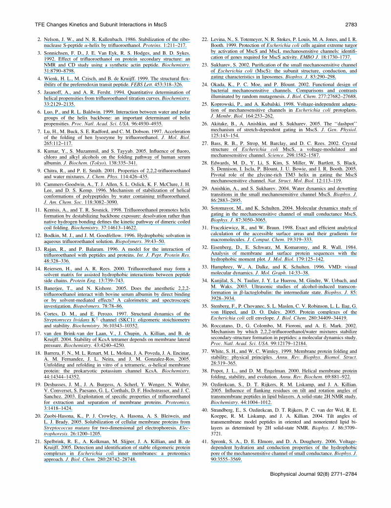
2. Nelson, J. W., and N. R. Kallenbach. 1986. Stabilization of the ribo-nuclease S-peptide a-helix by trifluoroethanol. Proteins. 1:211–217.
3. Sonnichsen, F. D., J. E. Van Eyk, R. S. Hodges, and B. D. Sykes.1992. Effect of trifluoroethanol on protein secondary structure: anNMR and CD study using a synthetic actin peptide. Biochemistry.31:8790–8798.
4. Wienk, H. L., M. Czisch, and B. de Kruijff. 1999. The structural flex-ibility of the preferredoxin transit peptide. FEBS Lett. 453:318–326.
5. Jasanoff, A., and A. R. Fersht. 1994. Quantitative determination ofhelical propensities from trifluoroethanol titration curves. Biochemistry.33:2129–2135.
6. Luo, P., and R. L. Baldwin. 1999. Interaction between water and polargroups of the helix backbone: an important determinant of helixpropensities. Proc. Natl. Acad. Sci. USA. 96:4930–4935.
7. Lu, H., M. Buck, S. E. Radford, and C. M. Dobson. 1997. Accelerationof the folding of hen lysozyme by trifluoroethanol. J. Mol. Biol.265:112–117.
8. Kumar, Y., S. Muzammil, and S. Tayyab. 2005. Influence of fluoro,chloro and alkyl alcohols on the folding pathway of human serumalbumin. J. Biochem. (Tokyo). 138:335–341.
9. Chitra, R., and P. E. Smith. 2001. Properties of 2,2,2-trifluoroethanoland water mixtures. J. Chem. Phys. 114:426–435.
10. Cammers-Goodwin, A., T. J. Allen, S. L. Oslick, K. F. McClure, J. H.Lee, and D. S. Kemp. 1996. Mechanism of stabilization of helicalconformations of polypeptides by water containing trifluoroethanol.J. Am. Chem. Soc. 118:3082–3090.
11. Kentsis, A., and T. R. Sosnick. 1998. Trifluoroethanol promotes helixformation by destabilizing backbone exposure: desolvation rather thannative hydrogen bonding defines the kinetic pathway of dimeric coiledcoil folding. Biochemistry. 37:14613–14622.
12. Bodkin, M. J., and J. M. Goodfellow. 1996. Hydrophobic solvation inaqueous trifluoroethanol solution. Biopolymers. 39:43–50.
13. Rajan, R., and P. Balaram. 1996. A model for the interaction oftrifluoroethanol with peptides and proteins. Int. J. Pept. Protein Res.48:328–336.
14. Reiersen, H., and A. R. Rees. 2000. Trifluoroethanol may form asolvent matrix for assisted hydrophobic interactions between peptideside chains. Protein Eng. 13:739–743.
15. Banerjee, T., and N. Kishore. 2005. Does the anesthetic 2,2,2-trifluoroethanol interact with bovine serum albumin by direct bindingor by solvent-mediated effects? A calorimetric and spectroscopicinvestigation. Biopolymers. 78:78–86.
16. Cortes, D. M., and E. Perozo. 1997. Structural dynamics of theStreptomyces lividans K1 channel (SKC1): oligomeric stoichiometryand stability. Biochemistry. 36:10343–10352.
17. van den Brink-van der Laan, V., J. Chupin, A. Killian, and B. deKruijff. 2004. Stability of KcsA tetramer depends on membrane lateralpressure. Biochemistry. 43:4240–4250.
18. Barrera, F. N., M. L. Renart, M. L. Molina, J. A. Poveda, J. A. Encinar,A. M. Fernandez, J. L. Neira, and J. M. Gonzalez-Ros. 2005.Unfolding and refolding in vitro of a tetrameric, a-helical membraneprotein: the prokaryotic potassium channel KcsA. Biochemistry.44:14344–14352.
19. Deshusses, J. M., J. A. Burgess, A. Scherl, Y. Wenger, N. Walter,V. Converset, S. Paesano, G. L. Corthals, D. F. Hochstrasser, and J. C.Sanchez. 2003. Exploitation of specific properties of trifluoroethanolfor extraction and separation of membrane proteins. Proteomics.3:1418–1424.
20. Zuobi-Hasona, K., P. J. Crowley, A. Hasona, A. S. Bleiweis, andL. J. Brady. 2005. Solubilization of cellular membrane proteins fromStreptococcus mutans for two-dimensional gel electrophoresis. Elec-trophoresis. 26:1200–1205.
21. Spelbrink, R. E., A. Kolkman, M. Slijper, J. A. Killian, and B. deKruijff. 2005. Detection and identification of stable oligomeric proteincomplexes in Escherichia coli inner membranes: a proteomicsapproach. J. Biol. Chem. 280:28742–28748.
22. Levina, N., S. Totemeyer, N. R. Stokes, P. Louis, M. A. Jones, and I. R.Booth. 1999. Protection of Escherichia coli cells against extreme turgorby activation of MscS and MscL mechanosensitive channels: identifi-cation of genes required for MscS activity. EMBO J. 18:1730–1737.
23. Sukharev, S. 2002. Purification of the small mechanosensitive channelof Escherichia coli (MscS): the subunit structure, conduction, andgating characteristics in liposomes. Biophys. J. 83:290–298.
24. Okada, K., P. C. Moe, and P. Blount. 2002. Functional design ofbacterial mechanosensitive channels. Comparisons and contrastsilluminated by random mutagenesis. J. Biol. Chem. 277:27682–27688.
25. Koprowski, P., and A. Kubalski. 1998. Voltage-independent adapta-tion of mechanosensitive channels in Escherichia coli protoplasts.J. Membr. Biol. 164:253–262.
26. Akitake, B., A. Anishkin, and S. Sukharev. 2005. The ‘‘dashpot’’mechanism of stretch-dependent gating in MscS. J. Gen. Physiol.125:143–154.
27. Bass, R. B., P. Strop, M. Barclay, and D. C. Rees. 2002. Crystalstructure of Escherichia coli MscS, a voltage-modulated andmechanosensitive channel. Science. 298:1582–1587.
28. Edwards, M. D., Y. Li, S. Kim, S. Miller, W. Bartlett, S. Black,S. Dennison, I. Iscla, P. Blount, J. U. Bowie, and I. R. Booth. 2005.Pivotal role of the glycine-rich TM3 helix in gating the MscSmechanosensitive channel. Nat. Struct. Mol. Biol. 12:113–119.
29. Anishkin, A., and S. Sukharev. 2004. Water dynamics and dewettingtransitions in the small mechanosensitive channel MscS. Biophys. J.86:2883–2895.
30. Sotomayor, M., and K. Schulten. 2004. Molecular dynamics study ofgating in the mechanosensitive channel of small conductance MscS.Biophys. J. 87:3050–3065.
31. Fraczkiewicz, R., and W. Braun. 1998. Exact and efficient analyticalcalculation of the accessible surface areas and their gradients formacromolecules. J. Comput. Chem. 19:319–333.
32. Eisenberg, D., E. Schwarz, M. Komaromy, and R. Wall. 1984.Analysis of membrane and surface protein sequences with thehydrophobic moment plot. J. Mol. Biol. 179:125–142.
33. Humphrey, W., A. Dalke, and K. Schulten. 1996. VMD: visualmolecular dynamics. J. Mol. Graph. 14:33–38.
34. Kanjilal, S., N. Taulier, J. Y. Le Huerou, M. Gindre, W. Urbach, andM. Waks. 2003. Ultrasonic studies of alcohol-induced transcon-formation in b-lactoglobulin: the intermediate state. Biophys. J. 85:3928–3934.
35. Stenberg, F., P. Chovanec, S. L. Maslen, C. V. Robinson, L. L. Ilag, G.von Hippel, and D. O. Daley. 2005. Protein complexes of theEscherichia coli cell envelope. J. Biol. Chem. 280:34409–34419.
36. Roccatano, D., G. Colombo, M. Fioroni, and A. E. Mark. 2002.Mechanism by which 2,2,2-trifluoroethanol/water mixtures stabilizesecondary-structure formation in peptides: a molecular dynamics study.Proc. Natl. Acad. Sci. USA. 99:12179–12184.
37. White, S. H., and W. C. Wimley. 1999. Membrane protein folding andstability: physical principles. Annu. Rev. Biophys. Biomol. Struct.28:319–365.
38. Popot, J. L., and D. M. Engelman. 2000. Helical membrane proteinfolding, stability, and evolution. Annu. Rev. Biochem. 69:881–922.
39. Ozdirekcan, S., D. T. Rijkers, R. M. Liskamp, and J. A. Killian.2005. Influence of flanking residues on tilt and rotation angles oftransmembrane peptides in lipid bilayers. A solid-state 2H NMR study.Biochemistry. 44:1004–1012.
40. Strandberg, E., S. Ozdirekcan, D. T. Rijkers, P. C. van der Wel, R. E.Koeppe, R. M. Liskamp, and J. A. Killian. 2004. Tilt angles oftransmembrane model peptides in oriented and nonoriented lipid bi-layers as determined by 2H solid-state NMR. Biophys. J. 86:3709–3721.
41. Spronk, S. A., D. E. Elmore, and D. A. Dougherty. 2006. Voltage-dependent hydration and conduction properties of the hydrophobicpore of the mechanosensitive channel of small conductance. Biophys. J.90:3555–3569.
TFE Changes Kinetics and Subunit Interactions in MscS 2783
Biophysical Journal 92(8) 2771–2784
42. Miller, S., M. D. Edwards, C. Ozdemir, and I. R. Booth. 2003. Theclosed structure of the MscS mechanosensitive channel. Cross-linkingof single cysteine mutants. J. Biol. Chem. 278:32246–32250.
43. Edwards, M. D., I. R. Booth, and S. Miller. 2004. Gating the bacterialmechanosensitive channels: MscS a new paradigm? Curr. Opin.Microbiol. 7:163–167.
44. Barry, J. A., and K. Gawrisch. 1994. Direct NMR evidence for ethanolbinding to the lipid-water interface of phospholipid bilayers. Biochem-istry. 33:8082–8088.
45. Koenig, B. W., and K. Gawrisch. 2005. Lipid-ethanol interactionstudied by NMR on bicelles. J. Phys. Chem. B. 109:7540–7547.
46. Sheetz, M. P., and S. J. Singer. 1974. Biological membranes as bilayercouples. A molecular mechanism of drug-erythrocyte interactions.Proc. Natl. Acad. Sci. USA. 71:4457–4461.
47. Heerklotz, H. 2001. Membrane stress and permeabilization induced byasymmetric incorporation of compounds. Biophys. J. 81:184–195.
48. Martinac, B., J. Adler, and C. Kung. 1990. Mechanosensitive ionchannels of E. coli activated by amphipaths. Nature. 348:261–263.
49. Perozo, E., A. Kloda, D. M. Cortes, and B. Martinac. 2002. Physicalprinciples underlying the transduction of bilayer deformation forcesduring mechanosensitive channel gating. Nat. Struct. Biol. 9:696–703.
50. Schumann, U., M. D. Edwards, C. Li, and I. R. Booth. 2004. The conservedcarboxy-terminus of the MscS mechanosensitive channel is not essentialbut increases stability and activity. FEBS Lett. 572:233–237.
51. Grajkowski, W., A. Kubalski, and P. Koprowski. 2005. Surfacechanges of the mechanosensitive channel MscS upon its activation,inactivation, and closing. Biophys. J. 88:3050–3059.
52. Qin, Z., and T. C. Squier. 2001. Calcium-dependent stabilization of thecentral sequence between Met76 and Ser81 in vertebrate calmodulin.Biophys. J. 81:2908–2918.
53. Main, E. R., and S. E. Jackson. 1999. Does trifluoroethanol affectfolding pathways and can it be used as a probe of structure in transitionstates? Nat. Struct. Biol. 6:831–835.
54. Chitra, R., and P. E. Smith. 2001. A comparison of the properties of2,2,2-trifluoroethanol and 2,2,2-trifluoroethanol/water mixtures usingdifferent force fields. J. Chem. Phys. 115:5521–5530.
2784 Akitake et al.
Biophysical Journal 92(8) 2771–2784