Functional Integration of Calcium Regulatory Mechanisms at Purkinje Neuron Synapses

Importance of Shank3 Protein in Regulating MetabotropicGlutamate Receptor 5 (mGluR5) Expression and Signaling atSynapses*□S
Received for publication, May 6, 2011, and in revised form, July 18, 2011 Published, JBC Papers in Press, July 27, 2011, DOI 10.1074/jbc.M111.258384
Chiara Verpelli‡§1, Elena Dvoretskova¶1, Cinzia Vicidomini‡, Francesca Rossi‡, Michela Chiappalone¶,Michael Schoen�, Bruno Di Stefano**, Renato Mantegazza§, Vania Broccoli**, Tobias M. Bockers�,Alexander Dityatev¶2, and Carlo Sala‡§3
From the ‡Department of Pharmacology, CNR Institute of Neuroscience, University of Milan, Milan 20129, the §Department ofNeuromuscular Diseases and Neuroimmunology, Neurological Institute Foundation Carlo Besta, Milan 20133, the ¶Department ofNeuroscience and Brain Technologies, Istituto Italiano di Tecnologia, Genova 16163, �Institute of Anatomy and Cell Biology, UlmUniversity Faculty of Medicine, Ulm 89081, and the **Division of Neuroscience, San Raffaele Scientific Institute, Milan 20132, Italy
Shank3/PROSAP2 genemutations are associated with cogni-tive impairment ranging from mental retardation to autism.Shank3 is a large scaffold postsynaptic density protein impli-cated in dendritic spines and synapse formation; however, itsspecific functions have not been clearly demonstrated.We haveused RNAi to knockdown Shank3 expression in neuronal cul-tures and showed that this treatment specifically reduced thesynaptic expression of the metabotropic glutamate receptor 5(mGluR5), but did not affect the expression of other major syn-aptic proteins. The functional consequence of Shank3 RNAiknockdown was impaired signaling via mGluR5, as shown byreduction in ERK1/2 and CREB phosphorylation induced bystimulationwith (S)-3,5-dihydroxyphenylglycine (DHPG) as theagonist of mGluR5 receptors, impaired mGluR5-dependentsynaptic plasticity (DHPG-induced long-term depression), andimpaired mGluR5-dependent modulation of neural networkactivity. We also found morphological abnormalities in thestructure of synapses (spine number, width, and length) andimpaired glutamatergic synaptic transmission, as shown byreduction in the frequency of miniature excitatory postsyn-aptic currents (mEPSC). Notably, pharmacological augmen-tation of mGluR5 activity using 3-cyano-N-(1,3-diphenyl-1H-pyrazol-5-yl)-benzamide as the positive allostericmodulator of these receptors restored mGluR5-dependentsignaling (DHPG-induced phosphorylation of ERK1/2) andnormalized the frequency of mEPSCs in Shank3-knockeddown neurons. These data demonstrate that a deficit inmGluR5-mediated intracellular signaling in Shank3 knock-down neurons can be compensated by 3-cyano-N-(1,3-di-phenyl-1H-pyrazol-5-yl)-benzamide; this raises the possibil-
ity that pharmacological augmentation of mGluR5 activityrepresents a possible new therapeutic approach for patientswith Shank3 mutations.
Haploinsufficiency of the SHANK3/PROSAP2 gene, whichencodes a structural protein located in the postsynaptic density(PSD)4 and involved in spinemaintenance in hippocampal neu-rons (1), is likely to be an essential cause of the major neurolog-ical features associated with the 22q13 deletion/Phelan-McDermid syndrome. Disruption of SHANK3/PROSAP2 wasfirst reported by Bonaglia et al. (2). Its association with theneurological deficit related to the syndrome is strongly sup-ported by the observation that all 22q13 deletions analyzed,except one (3), concerned SHANK3 (4), as shown by both theidentification of a recurrent breakpoint within the SHANK3gene (5) and by the recent finding that SHANK3mutations canresult in language and/or social interaction impairment (6).More recently, other small interstitial deletions or missensemutations in SHANK3 have been strongly associated withautism spectrum disorder and mental retardation (7–9).The three genes, SHANK1, SHANK2, and SHANK3, encode
large scaffold proteins that contain an ankyrin repeat near theNterminus followed by an Src homology domain 3 domain, aPDZ domain, a long proline-rich region, and a sterile � motifdomain at the C terminus (10). These proteins molecularly linkthe two glutamate receptor subtypes, NMDA receptors andtype-I metabotropic GluRs (mGluRs). The Shank PDZ domainbinds to the C terminus of GKAP, which binds to the PSD-95�NMDA receptor complex. Homer interaction with the pro-line-rich domain ensures the association of Shank with type ImGluRs (mGluR1 and mGluR5) (11–13).Overexpression of Shank1 in rat hippocampal neurons accel-
erates the maturation of filopodia-like protrusions in maturespines and promotes the enlargement of mature spines (14–
* This work was supported in part by Telethon-Italy Grant GGP09196, Fonda-zione CARIPLO Project number 2009.264, RSTL-CNR, Regione LombardiaProject number SAL-50-16983 TERDISMENTAL, and an Italian Institute ofTechnology, Seed Grant (to C. S. and V. B.).Author’s Choice—Final version full access.
□S The on-line version of this article (available at http://www.jbc.org) containssupplemental Fig. S1.
1 Both authors contributed equally to this work.2 Supported by a grant from the San Paolo Foundation and the Italian Insti-
tute of Technology.3 To whom correspondence should be addressed: Via Vanvitelli 32, 20129
Milano, Italy. Tel.: 39-02-50317096; Fax: 39-02-7490574; E-mail: [email protected].
4 The abbreviations used are: PSD, postsynaptic density; mGluR, metabo-tropic glutamate receptor; DHPG, (S)-3,5-dihydroxyphenylglycine; CREB,cAMP-response element-binding protein; CDPPB, 3-cyano-N-(1,3-diphen-yl-1H-pyrazol-5-yl)-benzamide; DIV, days in vitro; TTX, tetrodotoxin; APV,2-amino-5-phosphonopentanoic acid; mEPSC, miniature excitatory post-synaptic current; HBS, HEPES-buffered saline; ANOVA, analysis of variance;LTD, long-term depression.
THE JOURNAL OF BIOLOGICAL CHEMISTRY VOL. 286, NO. 40, pp. 34839 –34850, October 7, 2011Author’s Choice © 2011 by The American Society for Biochemistry and Molecular Biology, Inc. Printed in the U.S.A.
OCTOBER 7, 2011 • VOLUME 286 • NUMBER 40 JOURNAL OF BIOLOGICAL CHEMISTRY 34839
at FA
C M
ED
ICIN
A E
CH
IRU
RG
IA, on January 10, 2012
ww
w.jbc.org
Dow
nloaded from
http://www.jbc.org/content/suppl/2011/07/26/M111.258384.DC1.html Supplemental Material can be found at:

16). In contrast, mice lacking Shank1 display smaller dendriticspines, weaker synaptic transmission, and altered spatial learn-ing (17). Shank3 overexpression in rat cerebellar granule cellsinduces dendritic spine and synapse formation, whereasShank3 knockdown in hippocampal neurons reduces the num-ber of dendritic spines (1). Both Shank1 and Shank3 may formstructural frameworks in the PSDvia differentmolecularmech-anisms (18, 19).Several splice variants have been described for all three
genes, in particular six intragenic promoters generating multi-ple splicing variants have been identified for SHANK3 (20, 21).The functional role for all these splice variants remains to bedetermined; however, one can postulate that, depending on theintroduced mutations, the resulting truncated proteins mighthave different functional consequences, such as gain or loss ofspecific functions.This might explain the contradictory results recently pub-
lished on Shank3 partial-KOmice.Wang et al. (21) andBozdagiet al. (54) showed an alteration in hyppocampal synapse prop-erties, whereas Peca et al. (22) found clear alterations only in thecortico-striatal synapses. Finally Bangash et al. (23) described again-of-function phenotype for Shank3 protein missing theC-terminal fragment, which reduce specifically NR1 at syn-apses. With the aim of understanding the function of Shank3and its isoform(s) in the overall neuronal network toward theidentification of therapeutic target(s), for patients affected withMRand autismdue to SHANK3mutations, we have studied thesynaptic molecular pathways in cultured murine Shank3knockdown neurons.Rather than using Shank partial knockout mice, we knocked
down the expression of all the major Shank3 splice variants inneuronal cultures through RNA interference (RNAi). Our datashow that knockdown of Shank3 expression in rat and/ormouse hippocampal cell cultures induces a specific reductionin expression of mGluR5 receptors, as well as a reduction in(S)-3,5-dihydroxyphenylglycine (DHPG) (a group I mGluRagonist)-induced ERK1/2 and CREB phosphorylation and inmGluR5-dependent synaptic plasticity andmodulation of neu-ral network activity.Notably, pharmacological augmentation ofmGluR5 activity using 3-cyano-N-(1,3-diphenyl-1H-pyrazol-5-yl)-benzamide (CDPPB) as a positive allosteric modulatorpotentiated mGluR5-dependent signaling (DHPG-inducedphosphorylation of ERK1/2) and restored synaptic physiologyin neurons knocked down for Shank3. Thus, unlike Shank1,which in associationwithHomer acts as a structural frameworkat synapses (19), Shank3 could act as a signaling scaffold plat-form at synapses.
EXPERIMENTAL PROCEDURES
Neuronal Cultures—Rat hippocampal or cortical neuronalcultures were prepared from 18- to 19-day-old rat embryos(pregnant female rats were obtained from Charles River Labo-ratories). Neurons were plated at high density (750–1000 cells/mm2) and medium density (150–200 cells/mm2) and grown asdescribed (24) using B27 prepared in the laboratory. Neuronswere plated onto 6-well plastic tissue culture plates (Iwaki,Bibby Sterilin), or 18-mm diameter coverslips and grown on12-well plastic tissue culture plates (Iwaki, Bibby Sterilin). Pri-
mary mouse hippocampal neuronal cultures were prepared asdescribed previously (25). Hippocampi from 1- to 3-day-oldC57BL6/J mouse pups were dissected at 4 °C, and plated at adensity of 300/mm2 in Neurobasal-A medium supplementedwith 2% B27 supplement and 25 �g/ml of FGF2 (both fromInvitrogen) on 18-mm diameter round glass coverslips (Men-zel-Glaser) coated overnight with 100 �g/ml of poly-L-lysineand 40�g/ml of laminin (both fromSigma). Fromculture day 3,the medium was supplemented with 0.5 �M AraC (Sigma) toprevent glial cell proliferation. Neurons were transfected usingcalcium phosphate precipitation as per the protocol describedby Sala et al. (14). Cultures were infected with lentivirusexpressing shRNA specific for luciferase (shCtrl) or Shank3(shShank3) on day 7 in vitro (DIV) and used for experiments on13–15 DIV. Cells were stimulated with 100 �M DHPG, 100 �M
NMDA, or 50 mM KCl at 15 DIV for 30 min. To reduce endog-enous synaptic activity, 2 �M tetrodotoxin (TTX) was added tocultures 12 h before stimulation. For the biochemical experi-ments with CDPPB (Calbiochem), neurons were treated for12 h with 100 nM or 1 �M CDPPB before DHPG stimulation.RNA Interference and Relevant Plasmids—For plasmid-
based RNA inhibition, Shank3 and luciferase (26) oligonucleo-tides were annealed and inserted into the HindIII/BglII sites ofthe pLVTHM vector for lentivirus production of the shRNA.We used the following siRNA sequence that targets exon 21 ofthe rat andmouse SHANK3 gene (GenBankTM accession num-ber NM_021676 and NM_021423.3): 5�-GGAAGTCACCA-GAGGACAAGA-3�. The Shank3 rescue (Shank3r), R87C(Shank3R87Cr), and InsG (Shank3InsGr) constructs resistantto interference by siRNAwere generated by changing six nucle-otides of the siRNA target site, without changing the amino acidsequence of the resultant protein. Shank3 R87C and InsGmutants have been described elsewhere (6).Real Time-PCR (RT-PCR)—Total mRNA was extracted
using the RNeasy Plus kit (Qiagen, Valencia, CA). cDNA wassynthesized from DNase I-treated RNA using the QuantiTectreverse transcription kit (Qiagen,) according to the manufac-turer’s instructions. mRNA transcripts were quantified byTaqMan Q-PCR 3 (Applied Biosystems) on a Prism 7900 ther-mal cycler and sequence detector (Applied Biosystems). Allprimers and probes were from Applied Biosystems. Reactionswere performed in triplicate. Average �-Ct values normalizedto GAPDH or cyclophilin A (housekeeping genes) were used tocalculate gene-fold induction in treated samples, relativeto control set to 1.Antibodies—The following antibodies were used: rabbit anti-
Shank3 (Santa Cruz Biotechnology, H-160); guinea pig anti-Shank3 (27); rabbit anti-ERK1/2, rabbit anti-pERK 1/2, rabbitanti-eEF2, and rabbit anti-GFP 1:500 (Cell Signaling Technol-ogy); rabbit anti-mGluR1, rabbit anti-mGluR5, rabbit anti-mGluR4, rabbit anti-GluR1, and rabbit anti-GluR2/3 (MilliporeBioscience Research Reagents); mouse anti-GluR2,mouse anti-NR2B, mouse anti-Shank1, mouse anti-Shank2, mouse anti-Pan Shank, and mouse anti-PSD95 (NeuroMab, University ofCalifornia, Davis/NIH NeuroMab Facility); rabbit anti-GKAP(gifts from Morgan Sheng, Genentech); rabbit anti-Homer(gifts from Eunjoon Kim, KAIST, South Corea); rabbit anti-IRSp53 and mouse anti-Abi1 (gifts from Giorgio Scita, IFOM-
Shank3 Regulates mGluR5 Signaling
34840 JOURNAL OF BIOLOGICAL CHEMISTRY VOLUME 286 • NUMBER 40 • OCTOBER 7, 2011
at FA
C M
ED
ICIN
A E
CH
IRU
RG
IA, on January 10, 2012
ww
w.jbc.org
Dow
nloaded from

IEO, Italy); mouse anti-synaptophysin,mouse anti-�-actin, andmouse anti-�-tubulin (Sigma); secondary FITC-, Cy3- andCy5-conjugated anti-mouse, anti-rabbit, anti-guinea pig, oranti-goat antibodies (Jackson ImmunoResearch); secondaryHRP-conjugated anti-mouse, anti-rabbit, anti-guinea pig, oranti-goat (GE Healthcare).Western Blotting—Western blotting was performed as
described in Ref. 28. The signal was detected using an ECLdetection system (PerkinElmer Life Sciences). The intensity ofthe bands was measured with ImageJ software. Signals of thedetected proteins were normalized according to signals foractin or tubulin; the intensity of phosphospecific ERK1/2 andCREB immunoreactivity was normalized to the total ERK1/2andCREB signal in the same lane. Changes in protein levels andin ERK1/2 and CREB phosphorylation were compared withthose of untreated samples and were expressed as fold-increaseor decrease. The results are shown as mean � S.E.Immunocytochemistry—Neurons were fixed in 4% parafor-
maldehyde and 4% sucrose at room temperature or in 100%methanol at �20 °C. Primary and secondary antibodies wereapplied in GDB buffer (30 mM phosphate buffer, pH 7.4, con-taining 0.2% gelatin, 0.5% Triton X-100, and 0.8 MNaCl) for 2 hat room temperature or overnight at 4 °C.To study ERK1/2 phosphorylation, neuronswere transfected
with the pEGFP, Shank3r, Shank3R87Cr, or Shank3InsGr vec-tors alone or in combination with Shank3 shRNA at a ratio of1:2. Confocal images were acquired as described below. Cellbodies were manually traced using MetaMorph software(Molecular Devices) on the GFP channel. The average intensityof the fluorescent signal obtained with antibody againstpERK1/2 in the transfected neurons was divided by the averageintensity of the pERK1/2 signal in adjacent untransfectedneurons.To estimate cell surface expression of the GluR1 subunit of
AMPA receptors and its down-regulation by DHPG (29),untreated or DHPG-treated cultures were briefly washed withPBS and incubated with antibody against an extracellularepitope of the GluR1 subunit (Alomone Labs, agc-004, 16�g/ml) for 15min at 37 °C. For treatment, either 100�MDHPG� 50 �M 2-amino-5-phosphonopentanoic acid (APV) or 50 �M
APV alone were added to the culture medium for 10 min at37 °C. Following incubationwith anti-GluR1 antibody, cultureswere fixed with 4% formaldehyde. Anti-rabbit Alexa 546-con-jugated antibody (Invitrogen) was applied under nonpermeabi-lizing conditions for 1 h at RT.All images were collected using a Leica TCS SP5 confocal
microscope and �60 oil immersion objective at 1024 � 1024pixel resolution. The quantitative analysis was performed usingaZ-series projection of five images taken at 0.8-�mdepth inter-vals. All analyses were performed using NIH ImageJ software.The threshold for detection of GluR1-positive fluorescent clus-ters was fixed at twice the level of background fluorescenceobtained from a region of diffuse fluorescence within the den-dritic shaft. Only clusters lying along secondary dendriticbranches were counted; regions in which the identification ofneuronal processes was ambiguous were excluded from thequantification. We quantified the number of GluR1 immuno-
reactive clusters per 100-�m dendritic length within a givenfield.Measurement ofDendritic SpineMorphology—Neuronswere
cotransfected with a shRNA vectors and DsRed at a ratio of 2:1(7.5 �g of total DNA/well in 12-well plates) on 7 DIV and fixedon 18 DIV. Labeled transfected neurons were randomly chosenfor quantification in at least four independent experiments foreach construct.Confocal images of 1024 � 1024 pixels were obtained with a
LSM 510 Meta confocal microscope (Carl Zeiss, a gift fromFondazione Monzino) and a �63 objective with sequentialacquisition settings. Each image was a Z-series projection of7–15 images, each averaged two to four times, and taken at0.4–0.7-�m depth intervals. Morphometric measurementswere made using MetaMorph software (Molecular Devices).Individual dendrites were selected randomly and their spineswere traced manually. The maximum length and head width ofeach spine were measured and archived automatically (30).Electrophysiology—Cells were used for electrophysiological
recordings 3–4 days after transfection. For rescue experiments,the infected neurons were transfected on 11 DIVwith pcDNA3(Mock) or Shank3 resistant to shShank3 (Shank3r) clonedin pcDNA3. A vector expressing a red fluorescent proteintdTomato (31) was co-transfected in these experiments toallow the identification of transfected neurons.For pharmacological rescue of frequency of mEPSCs, the
allosteric modulator of mGluR5 (CDPPB, 1 �M, dissolved in0.1% dimethyl sulfoxide) was applied overnight on 13 DIV andduring recordings of mEPSCs on 14 DIV. As a vehicle control,0.1% dimethyl sulfoxide was used.Whole cell recordings from pyramidal-like neurons were
obtained as previously described (29). Electrodes with a resis-tance in the range of 3–6 megohms were filled with a solutionthat contained 130 mM CsMeSO4, 8 mM NaCl, 4 mM MgATP,0.3 mM NaGTP, 0.5 mM EGTA, and 10 mM HEPES, pH 7.25.Cells were perfused continuously with HEPES-buffered saline(HBS) of the following composition: 119mMNaCl, 5mMKCl, 2mM CaCl2, 2 mM MgCl2, 25 mM HEPES, 33 mM D-glucose, 0.5�M TTX citrate (Tocris), and 0.05 mM picrotoxin (Tocris), pH7.35. The osmolarity of HBS was adjusted to that of the culturemedium on the day of recording. The osmolarity of the elec-trode solution was 10 mosmol less than that of HBS. Whenused, DHPG (100 �M, Tocris) was added to HBS. To preventany contribution of NMDA receptor-dependent long-termdepression (LTD) and thus elicit pure mGluR-dependent LTD,50 �M APV (Sigma) was co-applied with DHPG or appliedalone as a control. Data were digitized at 10 kHz. Continuousrecording of mEPSCs was made using an EPC10 USB patchclamp amplifier and PATCHMASTER software (HEKA Elek-tronik). Detection and measurements of mEPSCs, which werecollected over a 3-min period, were performed using Mini-Analysis software (Synaptosoft, Leonia, NJ) after filtering tracesat 1 kHz and using a detection threshold of 6 pA (i.e. above 4times the standard deviation of baseline noise) and visual veri-fication of all detected events. Only cells with a holding currentless than �100 pA were analyzed.Microelectrode Array (MEA) Recordings and Analysis—Mi-
croelectrode arrays (Multichannel Systems, MCS, Reutlingen,
Shank3 Regulates mGluR5 Signaling
OCTOBER 7, 2011 • VOLUME 286 • NUMBER 40 JOURNAL OF BIOLOGICAL CHEMISTRY 34841
at FA
C M
ED
ICIN
A E
CH
IRU
RG
IA, on January 10, 2012
ww
w.jbc.org
Dow
nloaded from

Germany) consisted of 60 TiN/SiN planar round electrodes (30�m diameter, 200-�m center to center interelectrode distance)arranged in an 8 � 8 square grid excluding corners. Dissociatedneurons from C57BL6/J mice at postnatal day 1 were infected on8–10 DIV as described above. The activity of all cultures wasrecorded using the MEA60 System (MCS). After �1200 amplifi-cation, signals were sampled at 10 kHz and acquired through thedata acquisition card and MC_Rack software (MCS). To reducethermal stress to the cells during the experiment,MEAswere keptat 37 °C by means of a controlled thermostat (MCS) and coveredby flexiblepolydimethylsiloxane lids (32), toavoidevaporationandprevent changes in osmolarity. There was one recording sessionper culture; the session included 30 min of baseline recordings inthe absence ofDHPGand three consecutive 30-min recordings inthe presence of 1, 10, and 100 �M DHPG. Only the last 20 min ofeach episode were analyzed to exclude the initial part of record-ings, during which neuronal activity may have been influenced bythemechanical disturbances evoked by injection of the drug.Onlycultures in whichmore than 70% Shank3 expressionwas knockedout were included in the analysis.Data analysis was performed off-line using custom software,
SPYCODE, developed in MATLAB� (The Mathworks) (33);this software collects a series of tools for processing of multi-channel neural recordings. Data were imported into MATLABfrom mcd files (MCS format), and spikes were detected usingthe precise timing spike detection algorithm (34). Spike trainswere analyzed using a custom burst detection method (35), theparameters of which are directly estimated from the inter-spike
interval distribution of each channel. Following the burst detec-tion procedure, several measures describing spike and burststatistics were extracted; these includedmean firing rate, meanbursting rate, mean burst duration, mean frequency intra burst(spikes/sec), and percentage and frequency of out-burst spikes(i.e. spikes not included in bursts over the total). Two-wayANOVAwith repeatedmeasures followed byHolm-Sidak pair-wise comparison of groups was used for statistical evaluation ofthe DHPG and shShank3 effects.
RESULTS
To understand the role of Shank3 in synapse formation andfunction, we knocked down Shank3 using RNA interference.Expressed via a lentiviral vector, Shank3 shRNA (shShank3)strongly reduced the levels of endogenous Shank3 mRNA andprotein, but not those of other Shank family members, inmurine hippocampal cultures; a control shRNA (shCtrl) had nosuch effect (Fig. 1, A, C, and D). Interestingly, Shank3 shRNAknocked down all fourmajor Shank3 isoforms in a dose-depen-dent manner (Fig. 1B). We used two different antibodies in ourimmunoblotting analysis, one against the N-terminal domainand one against the C-terminal domain (see supplemental Fig.1A). Although the antibodies gave a different pattern in bothcases all the major bands were knocked down except for threeminor bands revealed with the N-terminal antibody (see sup-plemental Fig. 1C).Knocking Down Shank3 Reduces mGluR5 Expression—It has
been proposed that Shank3 plays an important role in assem-
FIGURE 1. Knocking down Shank3 reduces mGluR5 expression. A, histogram showing mean � S.E. for four independent experiments, of mRNA levels of theindicated proteins (normalized against those of uninfected neurons) in hippocampal neurons infected or not with a lentivirus expressing shShank3 or controlshRNA (shCtrl); the level of Shank3 mRNA was significantly lower in the shShank3-infected neurons than in uninfected or shCtrl-infected neurons, *, p�0.01, Student’st test. B, Western blot of hippocampal neurons infected with 3 and 6�l of lentivirus preparations expressing shShank3 or shCtrl and Shank3; Shank3 was detected usingrabbit H-160 anti-Shank3 antibody. The four major bands recognized by the antibody are indicated by the arrows. C, Western blot of hippocampal neurons infectedat 7 DIV with a lentivirus expressing shShank3 or shCtrl, as indicated above the panels, and analyzed with the antibodies against the proteins indicated on the left sideof the panels. D, histogram showing mean � S.E. for four independent experiments, of protein levels (normalized against those of the uninfected neurons) in thehippocampal neurons infected with a lentivirus expressing shShank3 or shCtrl. The expression levels of Shank3 and mGluR5 were significantly lower in the shShank3-infected neurons than in uninfected and shCtrl-infected neurons; *, p � 0.01, Student’s t test. E, Western blot of synaptosomes obtained from hippocampal neuronsinfected with a lentivirus expressing shShank3 or shCtrl and analyzed with the antibodies against the proteins indicated on the left side of the panel. F, histogramshowing mean � S.E. for three independent experiments, of protein levels (normalized against those of the shCtrl infected neurons) in synaptosomes obtained fromhippocampal neurons infected with a lentivirus expressing shShank3 or shCtrl. The expression levels of Shank3 and mGluR5 were significantly lower in the shShank3-infected neurons than in uninfected and shCtrl-infected neurons; *, p � 0.01, Student’s t test.
Shank3 Regulates mGluR5 Signaling
34842 JOURNAL OF BIOLOGICAL CHEMISTRY VOLUME 286 • NUMBER 40 • OCTOBER 7, 2011
at FA
C M
ED
ICIN
A E
CH
IRU
RG
IA, on January 10, 2012
ww
w.jbc.org
Dow
nloaded from

bling the PSD and in forming excitatory synapses via its multi-ple protein-protein interactions (1, 10). Accordingly, we exam-ined the effect of Shank3 knockdown on the proteincomposition of excitatory synapses using total lysates and syn-aptosomes from hippocampal cultures infected with shShank3or shCtrl. Immunoblotting with anti-Shank3 antibody con-firmed a strong reduction in Shank3 protein in the synapto-somal fraction of shShank3-infected neurons (Fig. 1, C and D).Immunoblotting for other glutamate receptors, scaffold pro-teins, and signaling molecules indicated that there was a signif-icant reduction of mGluR5 in shShank3-infected hippocampalneurons, both in the total lysate and in the synaptosomal frac-tion (mean � S.E. for normalized intensity in the total lysate,0.58� 0.03; in the synaptosomal fraction, 0.37� 0.05, p� 0.01,Student’s t test). Thus, the level of mGluR5, which bindsdirectly to Shank3 (12), was reduced in shShank3-infected hip-pocampal neurons. In the same preparations, we detected nosignificant difference in the abundance of a number of otherproteins that are known to be associated with synapses and/orthe PSD, including NMDA and AMPA receptors, PSD-95 andIRSp53 (Fig. 1,C andD). Also unchangedwere the total levels ofGKAP andHomer scaffold proteins, which can interact directlywith Shank3 (11, 12). The reduction in mGluR5 was notdependent on the decrease in mRNA expression as measuredby RT-PCR (Fig. 1A).In contrast to the drastic decrease in numbers of Shank3
clusters, the numbers of synaptophysin and PSD-95 clusterswere not modified by shShank3 treatment (Fig. 2,A and B). Wepreviously showed that the inhibition of Shank3 expression bytransfected shRNA in hippocampal neurons reduces dendriticspine numbers (1). Here, we confirm the reduction in spinenumbers from 4.9 � 1.0 spines for 10-�m dendrites in shCtrl-infected neurons to 3.1 � 0.2 spines/10-�m dendrite in neu-rons infected with shShank3 (Fig. 2C). Spine width and lengthwere also significantly reduced and increased, respectively, inneurons infected with shShank3, suggesting that the remainingspines are smaller and more immature (Fig. 2D).Knockdown of Shank3 Alters the mGluR5 Pathway—The
activation of group I mGluRs can lead to a form of long-termdepression (mGluR-LTD) that requires rapid translation ofpre-existing dendritic mRNA and involves several signalingmolecules including ERK1/2 and CREB (36). We investigatedthe effect of DHPG, the agonist of group I mGluRs, on therelevant signaling pathway by measuring the phosphorylationof ERK1/2 and CREB in response to stimulation of the neuronswith DHPG.shShank3 infection impaired ERK1/2 and CREB phosphory-
lation induced in neurons stimulated with 100 �M DHPG; KCldepolarization orNMDA stimulation had no such effect (Fig. 3,A and B). The reduction in DHPG-induced ERK1/2 phosphor-ylation in the shShank3-transfected neurons was rescued byoverexpression of shShank3-resistant Shank3 (Shank3r, seealso Fig. 4A) (Fig. 3,C andD). The reduction in DHPG-inducedERK1/2 phosphorylation in these neurons could also be res-cued by the overexpression of full-length mGluR5 (data notshown). However, the overexpression of two shShank3-resis-tant Shank3 mutants, Shank3R87Cr and Shank3InsGr (Fig.4A), which have been found in patients with autism (6) was not
able to rescue the ERK1/2 phosphorylation induced by 100 �M
DHPG (Fig. 4, B and C).To further investigatewhether Shank3deficiency affects syn-
aptic transmission at glutamatergic synapses, we recordedmEPSCs in pyramidal-like cells in shShank3- and shCtrl-treated cultures. Knockdown of Shank3 expression stronglyreduced the frequency of mEPSCs, but neither their amplitudenor their time course were affected (Fig. 5, A–F). The observedreduction in mEPSC frequency in shShank3-treated cultures isconsistent with the reported increase in mEPSC frequencywhen Shank3 is overexpressed in aspiny cerebellar neurons (1).The reduction in mEPSC frequency in our cultures could beprevented by transfection of shShank3-treated neurons withthe shShank3-resistant form of Shank3 (Fig. 5, G–I), confirm-ing the specificity of shShank3 treatment.Activation of mGluR5 could lead to a postsynaptic LTD that
ismediated by reduced synaptic expression of AMPA receptors(37, 38). To investigate the functional consequences of Shank3deficiency for mGluR5-dependent synaptic plasticity, we stim-ulated shShank3- and shCtrl-treated cultures with 100 �M
DHPG. As previously reported for uninfected cultured neurons
FIGURE 2. A, confocal microscopy images of hippocampal neurons infected ornot at 7 DIV with a lentivirus expressing shShank3 or shCtrl and stained after1 week with antibodies specific for Shank3, synaptophysin, and PSD-95 (asindicated above the panels). Scale bar � 5 �m. B, histogram showing mean �S.E. for four independent experiments, of numbers of Shank3, synaptophysin,and PSD-95 clusters in hippocampal neurons infected with lentivirus express-ing shShank3 or shCtrl; at least five neurons were considered for each exper-iment. The number of Shank3 clusters was significantly lower in theshShank3-infected neurons than in uninfected and shCtrl-infected neurons;*, p � 0.01, Student’s t test. C, confocal microscopy images showing dendriticspine morphology in hippocampal neurons infected with shShank3. Hip-pocampal neurons at 7 DIV were transfected with GFP-containing shCtrl orshShank3 cDNAs, plus DsRed-expressing cDNA. After a week, neurons werefixed and stained for GFP and DsRed. D, quantification of number (per 10 �m),length, and width of dendritic spines (� S.E.). Over 14 transfected neuronsfrom four independent experiments were measured for each transfection.Dendritic spine number, length, and width were significantly different in theshShank3-transfected and shCtrl-transfected neurons; **, p � 0.05; *, p �0.01, Student’s t test. Scale bar � 10 �m.
Shank3 Regulates mGluR5 Signaling
OCTOBER 7, 2011 • VOLUME 286 • NUMBER 40 JOURNAL OF BIOLOGICAL CHEMISTRY 34843
at FA
C M
ED
ICIN
A E
CH
IRU
RG
IA, on January 10, 2012
ww
w.jbc.org
Dow
nloaded from

(29), DHPG induced LTD in the frequency of mEPSC in neu-rons infected with the control lentivirus. However, LTD wasimpairedwhen expression of Shank3was knocked down (Fig. 6,A–D).We also found that shShank3 treatment affectedDHPG-induced down-regulation in the number of GluR1-immunore-active clusters on the plasma membranes of dendrites (Fig. 6, Eand F); a similar down-regulation has been reported for unin-fected cultured neurons (29). Furthermore, the density ofGluR1-immunoreactive clusters under basal conditions, i.e. in
neurons not treated with DHPG, was lower in shShank3-treated cultures than in controls. This finding corroborates theobserved reduction in the frequency of mEPSCs in shShank3-treated cultures under basal conditions.Knockdown of Shank3 Impairs DHPG-induced Modulation
of Neuronal Network Activity—To investigate the functionaleffects of Shank3 deficiency at the neural network level, weperformed multisite recordings of neuronal activity using mul-tielectrode arrays (Fig. 7). No difference between shShank3-
FIGURE 3. Knockdown of Shank3 alters the mGluR5 pathway. A, Western blot of hippocampal neurons infected at 7 DIV with shCtrl or shShank3. After 1week, neurons were treated with 100 �M DHPG, 100 �M NMDA, or 50 mM KCl for 30 min, as indicated above the panels, and analyzed Shank3, pERK1/2, ERK1/2,pCREB, and CREB expression, as indicated on the left of the panels. B, histogram showing mean � S.E., for five independent experiments, of pERK1/2 and pCREBlevels (normalized against total ERK and CREB and untreated control (NT) values). The levels of pERK1/2 and pCREB were significantly lower in the shShank3-infected neurons than in the shCtrl-infected neurons after DHPG treatment; *, p � 0.01, Student’s t test. C, confocal microscopy images of hippocampal neuronstransfected at 7 DIV with shCtrl, shShank3, or shShank3 plus Shank3r, as indicated on the left side of the panels, and treated on 14 DIV with 100 �M DHPG or 50mM KCl, as indicated above the panels (NT, untreated). The neurons were fixed and stained for GFP and pERK1/2. D, histogram showing mean � S.E. values forpERK1/2 signals in each condition of transfection and treatment quantified as described under “Experimental Procedures.” The level of pERK1/2 after DHPGtreatment was significantly lower in the shShank3-transfected neurons than in the shCtrl- and shShank3 plus Shank3r-transfected neurons; *, p � 0.05,Student’s t test. Scale bar � 20 �m.
Shank3 Regulates mGluR5 Signaling
34844 JOURNAL OF BIOLOGICAL CHEMISTRY VOLUME 286 • NUMBER 40 • OCTOBER 7, 2011
at FA
C M
ED
ICIN
A E
CH
IRU
RG
IA, on January 10, 2012
ww
w.jbc.org
Dow
nloaded from

and shCtrl-treated cultures was detected under basal condi-tions for any of the parameters of network activity evaluated(Fig. 7A, left panels, two-way ANOVA, p 0.05). However,DHPG-induced modulation of network activity was moreprominent in shCtrl- than in shShank3-treated cultures. Interms of the total spiking rate in all active channels, there was atendency, albeit not significant, toward lower activity inshShank3-treated cultures (Fig. 7B). However, the dose-depen-dent up-regulation of the number of bursts and spikes out-of-bursts by DHPG was significantly greater in control than inshShank3-treated cultures (Fig. 7, C and E). Also, there was no
DHPG-induced reduction in the spiking frequency withinbursts after knockdown of Shank3 (Fig. 7D).Restoration of Frequency of mEPSCs and DHPG-induced
Phosphorylation of ERK1/2 in shShank3-treated Cultured Hip-pocampal Neurons by the Allosteric mGluR5 Agonist CDPPB—To investigate whether the decrease in DHPG-induced ERK1/2phosphorylation and mEPSC frequency due to reduction ofmGluR5 protein expression in shShank3-treated cultures canbe pharmacologically rescued, we augmented the activity ofmGluR5 using CDPPB as an allosteric positive modulator ofthis receptor. Overnight treatment with CDPPB restored the
FIGURE 4. Shank3 mutations found in autistic patients do not rescue shShank3-induced defects in the mGluR5 pathway. A, Western blot of COS-7 cellstransfected with HA-Shank3, GFP-Shank3r, GFP-Shank3R87Cr, or GFP-Shank3InsGr plus or minus shShank3, as indicated above the panels, and analyzed withanti-HA, anti-GFP, or anti-tubulin antibodies. B, confocal microscopy images of hippocampal neurons transfected at 7 DIV with shShank3 � Shank3R87Cr orshShank3 � Shank3InsGr, as indicated on the left side of the panels, and treated or not at 14 DIV with 100 �M DHPG or 50 mM KCl, as indicated above the panels;the cells were fixed and stained for GFP and pERK1/2. C, histogram showing mean � S.E. values for pERK1/2 in each condition of transfection and treatmentquantified as described under “Experimental Procedures.” The level of pERK1/2 was significantly lower after DHPG treatment in the shShank3-, shShank3 �Shank3R87Cr-, and shShank3 � Shank3InsGr-transfected neurons than in the shCtrl-transfected neurons; *, p � 0.05, Student’s t test. Scale bar � 20 �m.
Shank3 Regulates mGluR5 Signaling
OCTOBER 7, 2011 • VOLUME 286 • NUMBER 40 JOURNAL OF BIOLOGICAL CHEMISTRY 34845
at FA
C M
ED
ICIN
A E
CH
IRU
RG
IA, on January 10, 2012
ww
w.jbc.org
Dow
nloaded from
frequency of mEPSCs (Fig. 8, A and B). Notably, we also foundthat theDHPG-induced phosphorylation of ERK1/2 in neuronsinfected with shShank3 was rescued by overnight treatmentwith CDPPB (Fig. 8, C and D). These data suggest that themGluR type I signaling that is altered by the knockdown ofShank3 can be potentially restored by CDPPB.
DISCUSSION
We have studied the role of Shank3 in synapse function tobetter understand the pathogenesis of the neurological symp-toms of patients affected by Phelan-McDermid syndrome (2,39). For this purpose, we used a specific shRNA to knockdownthe expression of themajor splice variants of Shank3 in culturedmurine neurons. Our immunoblotting study using two differ-
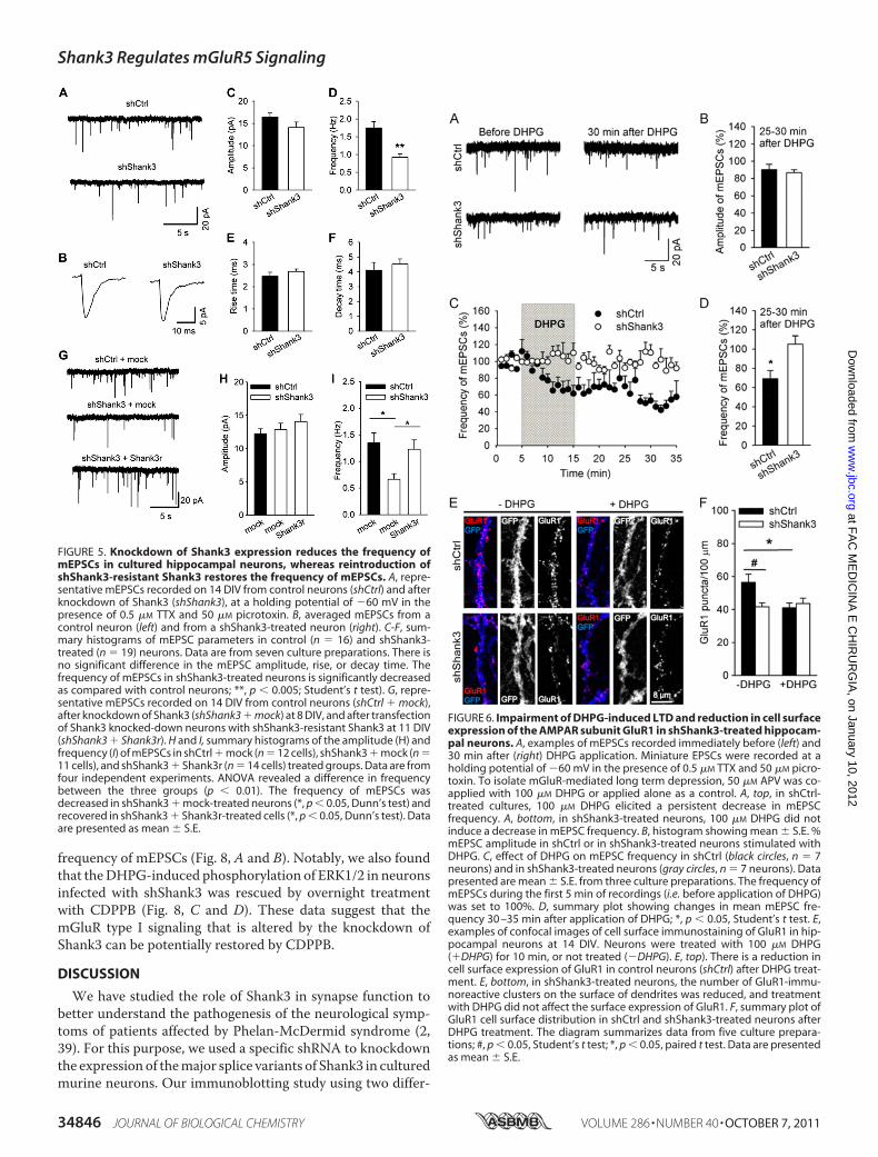
FIGURE 5. Knockdown of Shank3 expression reduces the frequency ofmEPSCs in cultured hippocampal neurons, whereas reintroduction ofshShank3-resistant Shank3 restores the frequency of mEPSCs. A, repre-sentative mEPSCs recorded on 14 DIV from control neurons (shCtrl) and afterknockdown of Shank3 (shShank3), at a holding potential of �60 mV in thepresence of 0.5 �M TTX and 50 �M picrotoxin. B, averaged mEPSCs from acontrol neuron (left) and from a shShank3-treated neuron (right). C-F, sum-mary histograms of mEPSC parameters in control (n � 16) and shShank3-treated (n � 19) neurons. Data are from seven culture preparations. There isno significant difference in the mEPSC amplitude, rise, or decay time. Thefrequency of mEPSCs in shShank3-treated neurons is significantly decreasedas compared with control neurons; **, p � 0.005; Student’s t test). G, repre-sentative mEPSCs recorded on 14 DIV from control neurons (shCtrl � mock),after knockdown of Shank3 (shShank3 � mock) at 8 DIV, and after transfectionof Shank3 knocked-down neurons with shShank3-resistant Shank3 at 11 DIV(shShank3 � Shank3r). H and I, summary histograms of the amplitude (H) andfrequency (I) of mEPSCs in shCtrl � mock (n � 12 cells), shShank3 � mock (n �11 cells), and shShank3 � Shank3r (n � 14 cells) treated groups. Data are fromfour independent experiments. ANOVA revealed a difference in frequencybetween the three groups (p � 0.01). The frequency of mEPSCs wasdecreased in shShank3 � mock-treated neurons (*, p � 0.05, Dunn’s test) andrecovered in shShank3 � Shank3r-treated cells (*, p � 0.05, Dunn’s test). Dataare presented as mean � S.E.
FIGURE 6. Impairment of DHPG-induced LTD and reduction in cell surfaceexpression of the AMPAR subunit GluR1 in shShank3-treated hippocam-pal neurons. A, examples of mEPSCs recorded immediately before (left) and30 min after (right) DHPG application. Miniature EPSCs were recorded at aholding potential of �60 mV in the presence of 0.5 �M TTX and 50 �M picro-toxin. To isolate mGluR-mediated long term depression, 50 �M APV was co-applied with 100 �M DHPG or applied alone as a control. A, top, in shCtrl-treated cultures, 100 �M DHPG elicited a persistent decrease in mEPSCfrequency. A, bottom, in shShank3-treated neurons, 100 �M DHPG did notinduce a decrease in mEPSC frequency. B, histogram showing mean � S.E. %mEPSC amplitude in shCtrl or in shShank3-treated neurons stimulated withDHPG. C, effect of DHPG on mEPSC frequency in shCtrl (black circles, n � 7neurons) and in shShank3-treated neurons (gray circles, n � 7 neurons). Datapresented are mean � S.E. from three culture preparations. The frequency ofmEPSCs during the first 5 min of recordings (i.e. before application of DHPG)was set to 100%. D, summary plot showing changes in mean mEPSC fre-quency 30 –35 min after application of DHPG; *, p � 0.05, Student’s t test. E,examples of confocal images of cell surface immunostaining of GluR1 in hip-pocampal neurons at 14 DIV. Neurons were treated with 100 �M DHPG(�DHPG) for 10 min, or not treated (�DHPG). E, top). There is a reduction incell surface expression of GluR1 in control neurons (shCtrl) after DHPG treat-ment. E, bottom, in shShank3-treated neurons, the number of GluR1-immu-noreactive clusters on the surface of dendrites was reduced, and treatmentwith DHPG did not affect the surface expression of GluR1. F, summary plot ofGluR1 cell surface distribution in shCtrl and shShank3-treated neurons afterDHPG treatment. The diagram summarizes data from five culture prepara-tions; #, p � 0.05, Student’s t test; *, p � 0.05, paired t test. Data are presentedas mean � S.E.
Shank3 Regulates mGluR5 Signaling
34846 JOURNAL OF BIOLOGICAL CHEMISTRY VOLUME 286 • NUMBER 40 • OCTOBER 7, 2011
at FA
C M
ED
ICIN
A E
CH
IRU
RG
IA, on January 10, 2012
ww
w.jbc.org
Dow
nloaded from

ent anti-Shank3 antibodies, one against theN terminus and oneagainst the C terminus, indicated that, at least in neuronal cul-tures, Shank3 shRNA knocked down all the major Shank3-spe-cific bands. AsWang et al. (21) recently described six intragenicpromoters that could potentially generate multiple splicingvariants coding for several isoforms of Shank3, we cannotexclude that someminor splice variants remain at undetectablelevels beyond shSank3 inhibition.Although the proteins encoded by these three SHANK genes
are structurally similar, some evidence suggests that they differin function, in synapse-targeting properties, and in bindingpartners. For example, the overexpression of Shank1 inducesmaturation of dendritic spines without increasing their num-bers, whereas the overexpression of Shank3 induces the forma-tion of new synapses and dendritic spines (1, 16). Shank1 tar-geting to synapses is dependent on the PDZ domain, but thetargeting of Shank2 and Shank3 depends on their C-terminaldomain, including the sterile � motif domain (24, 40). Shank2and Shank3 multimerize and form a platform or framework inthe PSD that depends on Zn2� binding to the sterile � motifdomain (18, 41). In contrast, Shank1 does not bind Zn2�,, butforms a large framework complex with Homer in the PSD (19).
The specific function of Shank proteins in dendritic spines isprobably related to the fact that all three protein variantsbind directly to a number of proteins involved in actinremodeling, such as cortactin, Abp1, IRsP53, and SPIN90(11, 42–44); they also interact indirectly with actin-remod-eling proteins through binding Homer, oligophrenin, andCdC42 (45, 46). Available data suggest that Shank proteinsfunctionally link glutamate receptors to the cytoskeleton,thereby regulating the size and dimensions of excitatory syn-apses and dendritic spines (47).Shank2 and -3 can also bind to Ab1 and LAPSER1, two pro-
teins that translocate from the PSD to the nucleus in an activity-dependent manner and induce gene transcription and transla-tion (48, 49). Finally, the fact that simple deletions of eitherSHANK3 or SHANK2, but not, up to now, SHANK1, have beenclearly implicated in the pathogenesis of mental retardation,autism and, more recently, schizophrenia, suggest that thethree proteins may have different functions that cannot com-pensate for each other. Thus, it is important to note that we didnot observe any type of compensation by the other SHANKmember as neither Shank1 nor Shank2 expression increasedupon Shank3 knockdown.
FIGURE 7. Knockdown of Shank3 impairs DHPG-induced modulation of neuronal network activity. A, representative 60-electrode recordings of activityof cultured mouse neurons before and 5 min after application of 100 �M DHPG. Each horizontal line corresponds to one electrode; short vertical intervalscorrespond to detected action potentials. The duration of each recording is 30 s. Following application of 100 �M DHPG, a greater increase in the number ofbursts occurs in the shCtrl-treated cultures compared with the shShank3-treated cultures. B-E, changes in the mean firing rate (B), bursting rate (C), intra-burstfiring rate (D), and percentage of out-burst spikes (E) for active electrodes after the application of DHPG to shCtrl- and shShank3-treated cultures. There wereno differences between shCtrl- and shShank3-treated groups in baseline activity; therefore, the values of parameters estimated under the baseline conditionwere set to 100% for each culture. Two-way ANOVA with repeated measures (DHPG concentrations of 1, 10, and 100 �M) revealed a significant effect of DHPG(p � 0.01), but not of shShank3 or shShank3 � DHPG on the mean firing rate (B). Significant effects of DHPG (p � 0.01) and shShank3 (p � 0.05) on the meanbursting rate were detected in C, and significant effects of shShank3 � DHPG (p � 0.05) on the intra-burst firing rate (D) and % of out-burst spikes (E) weredetected. *, p � 0.05; **, p � 0.01, Holm-Sidak post hoc t test, significant differences between shCtrl- (n � 10 cultures) and shShank3-treated cultures (n � 7cultures).
Shank3 Regulates mGluR5 Signaling
OCTOBER 7, 2011 • VOLUME 286 • NUMBER 40 JOURNAL OF BIOLOGICAL CHEMISTRY 34847
at FA
C M
ED
ICIN
A E
CH
IRU
RG
IA, on January 10, 2012
ww
w.jbc.org
Dow
nloaded from

We found that knockdown of Shank3 specifically impairedmGluR5 signaling at synapses. In hippocampal neurons knockeddown for Shank3, mGluR5 protein, but not its mRNA, is specifi-cally reduced in the total lysate and in the synaptosomes, suggest-ing that Shank3 is somehow involved inmGluR5protein stabiliza-tion. Previous work has shown that mGluR5 binds directly toShank3 or indirectly throughHomer cross-linking (12). However,because we did not find any change in Homer expression, it ispossible that thedirectbindingofShank3 tomGluR5 is involved inthis phenomenon. Both Shank3 and mGluR5 can be degraded byproteasomes following ubiquitination, suggesting that their inter-action can reciprocally modulate their ubiquitination and stabili-zation (50, 51). However, we did not find any change in Shank3
protein expression in themGluR5knock-outmice.5Thus, Shank3mightactasa stabilizationplatformformGluR5.Wealsoobserveda reduction in cell surface expression of GluR1 in shShank3-treated neuronswithout a reduction in its protein expression. Thereduction in GluR1 cell surface expression correlated with thereducedmEPSCfrequency.The impairedDHPG-dependentLTDobserved in shShank3-treated neurons did not result in anychanges in cell surface expression of GluR1, which is down-regu-lated by DHPG in shCtrl-treated neurons. Our findings thatCDPPB, an allosteric mGluR5 agonist, was able to rescue themEPSC frequency in neurons knocked down for Shank3 suggestthat Shank3 regulates AMPA receptor trafficking in an mGluR5-dependent manner. The reduction in cell surface GluR1 expres-sion and in frequency of mEPSCs after knockdown of Shank3might reflect impairment in activity-dependent synaptic recruit-ment of AMPA receptors at basal conditions.Despite the observed reduction inmEPSC frequency and cell
surface expression of GluR1-positive clusters in shShank3-treated neurons, our multielectrode recordings did not revealsignificant changes in the spiking patterns of neurons underbasal conditions. This is not surprising, because the connectiv-ity between cultured neurons is highly redundant. Therefore,despite the differences in synaptic activity between control andShank3-knockdown neurons in TTX-treated cultures, thecomposite postsynaptic potentials might well exceed thethreshold for spike generation in these cells in the absence ofTTX. Application of DHPG to cultured neurons led to a strongincrease in the bursting rate, as previously reported for hip-pocampal slices (52). Importantly, DHPG-induced up-regula-tion of bursting was reduced in Shank3-knockdown neurons.This result confirms the importance of Shank3 in the regulationof mGluR5-dependent signaling under physiological condi-tions, i.e. in the absence of TTX. It also demonstrates the poten-tial importance of Shank3 in mGluR5 activity-induced shapingof neural network activity.In a recent analysis of the role of Shank3 mutations when
overexpressed in hippocampal neurons, Durand et al. (53)showed that all mutations analyzed modify Shank3 functions.Here, we have analyzed two of themutations studied byDurandet al. (their R12C corresponds to our R87C and theirShank3STOP corresponds to our Shank3Ins). Both mutationswere shown by Durand et al. (53) to affect the ability of Shank3proteins to increase the dimension of dendritic spines andmodify synaptic properties. Interestingly, in our study, theexpression of mutated forms of Shank3 that mimic the muta-tions found in autistic patients was not able to rescue DHPG-dependent ERK1/2 phosphorylation. Thus, reduction inShank3 expression, which occurs in 22q13/Phelan-McDermidsyndrome, and functional mutations in Shank3, which occur insome autistic patients, might both induce alterations inmGluR5 signaling at synapses.Four recent studies highlight the importance of Shank3 at the
molecular and behavioral levels (21–23, 54). Two of themshowed that Shank3 heterozygous and homozygous male micedisplayed abnormal social behavior, communication pattern,
5 C. Verpelli and C. Sala, unpublished results.
FIGURE 8. Restoration of frequency of mEPSCs and DHPG-induced phos-phorylation of ERK1/2 in shShank3-treated cultured hippocampal neu-rons by the allosteric mGluR5 agonist CDPPB. A and B, neurons wereinfected with lentiviruses at 8 DIV and treated overnight with 1 �M CDPPB at13 DIV. On 14 DIV, mEPSCs were recorded at a holding potential of �60 mV inthe presence of 0.5 �M TTX and 50 �M picrotoxin. Summary graphs of theamplitude (A) and frequency (B) of mEPSCs in shCtrl � vehicle (n � 11 cells),shShank3 � vehicle (n � 14 cells), and shShank3 � CDPPB (n � 14 cells)-treated groups. Data are from four culture preparations. The frequency ofmEPSCs is significantly decreased in shShank3 � vehicle as compared withshCtrl � vehicle-treated neurons and recovers significantly in shShank3 �CDPPB-treated cells; *, p � 0.05, Dunn’s test. Data are presented as mean �S.E. C, Western blot of hippocampal neurons infected at 7 DIV with shCtrl orshShank3 and treated after 1 week as indicated above the panels. The pro-teins analyzed are indicated to the left of the panels. D, histogram showingmean � S.E. for five independent experiments, of ERK1/2 phosphorylation,normalized to total ERK and untreated (NT) values. The level of pERK1/2 afterDHPG treatment was significantly lower in the shShank3-infected neuronsthan in the shCtrl-infected neurons; *, p � 0.01, Student’s t test.
Shank3 Regulates mGluR5 Signaling
34848 JOURNAL OF BIOLOGICAL CHEMISTRY VOLUME 286 • NUMBER 40 • OCTOBER 7, 2011
at FA
C M
ED
ICIN
A E
CH
IRU
RG
IA, on January 10, 2012
ww
w.jbc.org
Dow
nloaded from

and learning and memory, as compared with wild-type litter-mate controls (21, 54). These studies revealed a strong impair-ment in basal synaptic transmission in CA3-CA1 connections,a reduction in GluR1 clusters and protein levels in the hip-pocampus, and an alteration in activity-dependent AMPARsynaptic plasticity (21, 54). However, the reduction in mEPSCamplitude and the “compensatory” increase in mEPSC fre-quency in Shank3 heterozygousmice reported by these authorswere not seen in our experiments. Most importantly in thiscontext, it should be noted that in our experiments, in contrastto the other studies, we knocked down all detectable Shank3splice variants through shRNA treatment; this led to a suppres-sion of Shank3 expression by 70–80% rather than by 50% as inShank3 heterozygous mice. This could lead to such a strongreduction of mEPSC amplitudes that EPSCs would decreasebelow the detection limit, resulting in a reduction of mEPSCfrequency, as we report here. Another difference is our obser-vation that Shank3 plays a role in LTD induced by the mGluRtype 1 agonist, DHPG, whereas Bozdagi and colleagues (54)found that Shank3 heterozygous mice have a normal LTDinduced by paired-pulse low-frequency stimulation. In view ofour present study, this is not surprising considering the role ofShank3 in regulation of mGluR5 expression and consideringthat previous studies in mGluR5 knock-out mice have alsoshown normal paired-pulse low-frequency stimulation-in-duced LTD (55), but impaired DHPG-induced LTD (56).Peca et al. (22) have instead reported that mice genetically
deleted of two major Shank3 splice variants exhibit self-injuri-ous repetitive grooming and deficits in social interaction andthese behavioral defects are caused by major alteration in thestriatal synapses and cortico-striatal circuits, but not in the hip-pocampus. Thus, it is possible that the remaining Shank3 splicevariant(s) might be sufficient tomaintain normal synapse func-tion and structure in thehippocampus. Paradoxically, the remain-ing Shank3 protein described by Bangash et al. (23), whichmissesthe C-terminal fragment, has a gain-of-function phenotype byreducing the NR1 subunit of NMDA receptors specifically at syn-apses, but not affecting synaptic AMPAR function and composi-tion. Although this pathway should obviously also be investigatedin vivo, our data strongly suggest nevertheless that knocking downall themajor Shank3 splice variants strongly affects the expressionof mGluR5 receptor at the synapses.The mGluR5 receptor plays a major role in synaptic plasticity
(57). It has been clearly demonstrated that antagonism or geneticdeletion of mGluR5 impairs both acquisition and extinction ofhippocampal-dependent learning tasks, such as the radial armmaze and theMorriswatermaze, by impairing both the late phaseof hippocampal long term potentiation and mGluR-dependentLTD (58–60). The occurrence ofmGluR-dependent LTD inCA1relies on activation of both ERK and PI3K-mTOR pathways (36,61). A role formGluR-LTDhas been demonstrated for the forma-tion of object recognitionmemory (62, 63).The existence of a link between mGluR-LTD and cognitive
disease is suggested by the finding that both hippocampal andcerebellar mGluR-LTD are altered in fragile X syndrome, amouse model of mental retardation and autism that has led tothe development of novel therapeutics for this syndrome thatact on mGluR5 (64). In contrast to our finding in Shank3-
knockdown neurons, mGluR-LTD is enhanced in the fragile Xsyndrome mouse model (65). This enhancement occursbecause, in the absence of fragile X mental retardation protein,as in fragile X syndrome, there is a loss of steady-state transla-tional suppression that leads to increased protein levels of frag-ile X mental retardation protein targeting specific mRNAs,such as those coding for activity-regulated cytoskeleton-asso-ciated protein that may enhance the magnitude of LTD (66).Notably, the use of mGluR5 antagonists or genetic reduction
of mGluR5 (in mice that are heterozygous for mGluR5) canreverse multiple phenotypes in mice deficient in FMR1, a geneencoding the fragile X mental retardation protein; these phe-notypes include increased dendritic spine density and deficitsin experience-dependent plasticity in the visual cortex and hip-pocampal-dependent learning (67, 68).Based on this finding, we tested whether the reduced
mGluR5 activity in Shank3-knockdownneurons can be rescuedby an allosteric agonist ofmGluR5, such as CDPPB (69, 70), andfound that both ERK1/2 phosphorylation and mEPSC fre-quency were rescued by overnight treatment with CDPPB.CDPPB has been shown to be brain-penetrant and to reverseamphetamine-induced locomotor activity and amphetamine-in-duced deficits in prepulse inhibition in rats (70), two models thatare sensitive to antipsychotic drug treatment. These results dem-onstrate that positive allosteric modulation of mGluR5 producesbehavioral effects andsuggest thatmGluR5activity couldbeenvis-aged as a potential therapeutic target. Therefore, these findingsopen new possibilities for the pharmacological treatment ofpatients affected by Shank3 gene deletion andmutation.
Acknowledgment—We thank N. Tonna for some preliminary data.
REFERENCES1. Roussignol, G., Ango, F., Romorini, S., Tu, J. C., Sala, C., Worley, P. F.,
Bockaert, J., and Fagni, L. (2005) J. Neurosci. 25, 3560–35702. Bonaglia, M. C., Giorda, R., Borgatti, R., Felisari, G., Gagliardi, C.,
Selicorni, A., and Zuffardi, O. (2001) Am. J. Hum. Genet. 69, 261–2683. Wilson, H. L., Crolla, J. A., Walker, D., Artifoni, L., Dallapiccola, B., Ta-
kano, T., Vasudevan, P.,Huang, S.,Maloney, V., Yobb, T.,Quarrell, O., andMcDermid, H. E. (2008) Eur. J. Hum. Genet. 16, 1301–1310
4. Wilson, C. A., Doms, R. W., and Lee, V. M. (2003) J. Neurosci. Res. 74,361–369
5. Bonaglia, M. C., Giorda, R., Mani, E., Aceti, G., Anderlid, B. M., Baroncini,A., Pramparo, T., and Zuffardi, O. (2006) J. Med. Genet. 43, 822–828
6. Durand, C. M., Betancur, C., Boeckers, T. M., Bockmann, J., Chaste, P.,Fauchereau, F., Nygren, G., Rastam, M., Gillberg, I. C., Anckarsater, H.,Sponheim, E., Goubran-Botros, H., Delorme, R., Chabane, N., Mouren-Simeoni, M. C., de Mas, P., Bieth, E., Roge, B., Heron, D., Burglen, L.,Gillberg, C., Leboyer, M., and Bourgeron, T. (2007)Nat. Genet. 39, 25–27
7. Moessner, R.,Marshall, C. R., Sutcliffe, J. S., Skaug, J., Pinto, D., Vincent, J.,Zwaigenbaum, L., Fernandez, B., Roberts, W., Szatmari, P., and Scherer,S. W. (2007) Am. J. Hum. Genet. 81, 1289–1297
8. Delahaye, A., Toutain, A., Aboura, A., Dupont, C., Tabet, A. C., Ben-zacken, B., Elion, J., Verloes, A., Pipiras, E., and Drunat, S. (2009) Eur.J. Med. Genet. 52, 328–332
9. Gauthier, J., Spiegelman, D., Piton, A., Lafreniere, R. G., Laurent, S., St-Onge, J., Lapointe, L., Hamdan, F. F., Cossette, P., Mottron, L., Fombonne,E., Joober, R., Marineau, C., Drapeau, P., and Rouleau, G. A. (2009) Am. J.Med. Genet. B Neuropsychiatr. Genet. 150, 421–424
10. Sheng, M., and Kim, E. (2000) J. Cell Sci. 113, 1851–185611. Naisbitt, S., Kim, E., Tu, J. C., Xiao, B., Sala, C., Valtschanoff, J., Weinberg,
Shank3 Regulates mGluR5 Signaling
OCTOBER 7, 2011 • VOLUME 286 • NUMBER 40 JOURNAL OF BIOLOGICAL CHEMISTRY 34849
at FA
C M
ED
ICIN
A E
CH
IRU
RG
IA, on January 10, 2012
ww
w.jbc.org
Dow
nloaded from

R. J., Worley, P. F., and Sheng, M. (1999) Neuron 23, 569–58212. Tu, J. C., Xiao, B., Naisbitt, S., Yuan, J. P., Petralia, R. S., Brakeman, P.,
Doan, A., Aakalu, V. K., Lanahan, A. A., Sheng, M., and Worley, P. F.(1999) Neuron 23, 583–592
13. Boeckers, T. M., Winter, C., Smalla, K. H., Kreutz, M. R., Bockmann, J.,Seidenbecher, C., Garner, C. C., and Gundelfinger, E. D. (1999) Biochem.Biophys. Res. Commun. 264, 247–252
14. Sala, C., Piech, V., Wilson, N. R., Passafaro, M., Liu, G., and Sheng, M.(2001) Neuron 31, 115–130
15. Sala, C., Futai, K., Yamamoto, K.,Worley, P. F., Hayashi, Y., and Sheng,M.(2003) J. Neurosci. 23, 6327–6337
16. Sala, C., Roussignol, G., Meldolesi, J., and Fagni, L. (2005) J. Neurosci. 25,4587–4592
17. Hung, A. Y., Futai, K., Sala, C., Valtschanoff, J. G., Ryu, J., Woodworth,M. A., Kidd, F. L., Sung, C. C., Miyakawa, T., Bear, M. F., Weinberg, R. J.,and Sheng, M. (2008) J. Neurosci. 28, 1697–1708
18. Baron, M. K., Boeckers, T. M., Vaida, B., Faham, S., Gingery, M., Sawaya,M. R., Salyer, D., Gundelfinger, E. D., and Bowie, J. U. (2006) Science 311,531–535
19. Hayashi, M. K., Tang, C., Verpelli, C., Narayanan, R., Stearns, M. H., Xu,R. M., Li, H., Sala, C., and Hayashi, Y. (2009) Cell 137, 159–171
20. Maunakea, A. K., Nagarajan, R. P., Bilenky, M., Ballinger, T. J., D’Souza, C.,Fouse, S. D., Johnson, B. E., Hong, C., Nielsen, C., Zhao, Y., Turecki, G., Dela-ney, A., Varhol, R., Thiessen, N., Shchors, K., Heine, V. M., Rowitch, D. H.,Xing, X., Fiore, C., Schillebeeckx, M., Jones, S. J., Haussler, D., Marra, M. A.,Hirst, M.,Wang, T., and Costello, J. F. (2010)Nature 466, 253–257
21. Wang, X., McCoy, P. A., Rodriguiz, R. M., Pan, Y., Je, H. S., Roberts, A. C.,Kim, C. J., Berrios, J., Colvin, J. S., Bousquet-Moore, D., Lorenzo, I., Wu,G., Weinberg, R. J., Ehlers, M. D., Philpot, B. D., Beaudet, A. L., Wetsel,W. C., and Jiang, Y. H. (2011) Hum. Mol. Genet. 20, 3093–3108
22. Peca, J., Feliciano, C., Ting, J. T., Wang, W., Wells, M. F., Venkatraman,T. N., Lascola, C. D., Fu, Z., and Feng, G. (2011) Nature 472, 437–442
23. Bangash, M. A., Park, J. M., Melnikova, T., Wang, D., Jeon, S. K., Lee, D.,Syeda, S., Kim, J., Kouser, M., Schwartz, J., Cui, Y., Zhao, X., Speed, H. E.,Kee, S. E., Tu, J. C., Hu, J. H., Petralia, R. S., Linden, D. J., Powell, C. M.,Savonenko, A., Xiao, B., and Worley, P. F. (2011) Cell 145, 758–772
24. Romorini, S., Piccoli, G., Jiang, M., Grossano, P., Tonna, N., Passafaro, M.,Zhang, M., and Sala, C. (2004) J. Neurosci. 24, 9391–9404
25. Dityatev, A., Dityateva, G., and Schachner,M. (2000)Neuron 26, 207–21726. Seeburg, D. P., and Sheng, M. (2008) J. Neurosci. 28, 6583–659127. Beri, S., Tonna, N., Menozzi, G., Bonaglia, M. C., Sala, C., and Giorda, R.
(2007) J. Neurochem. 101, 1380–139128. Verpelli, C., Piccoli, G., Zanchi, A., Gardoni, F., Huang, K., Brambilla, D.,
Di Luca, M., Battaglioli, E., and Sala, C. (2010) J. Neurosci. 30, 5830–584229. Moult, P. R., Gladding, C. M., Sanderson, T. M., Fitzjohn, S. M., Bashir,
Z. I., Molnar, E., and Collingridge, G. L. (2006) J. Neurosci. 26, 2544–255430. Piccoli, G., Verpelli, C., Tonna, N., Romorini, S., Alessio, M., Nairn, A. C.,
Bachi, A., and Sala, C. (2007) J. Proteome Res. 6, 3203–321531. Cancedda, L., Fiumelli, H., Chen, K., and Poo,M.M. (2007) J. Neurosci. 27,
5224–523532. Blau, A., Neumann, T., Ziegler, C., and Benfenati, F. (2009) J. Biosci. 34,
59–6933. Bologna, L. L., Nieus, T., Tedesco, M., Chiappalone, M., Benfenati, F., and
Martinoia, S. (2010) Neuroscience 165, 692–70434. Maccione, A., Gandolfo, M., Massobrio, P., Novellino, A., Martinoia, S.,
and Chiappalone, M. (2009) J. Neurosci. Methods 177, 241–24935. Pasquale, V., Martinoia, S., and Chiappalone, M. (2010) J. Comput. Neu-
rosci. 29, 213–22936. Gallagher, S. M., Daly, C. A., Bear, M. F., and Huber, K. M. (2004) J. Neu-
rosci. 24, 4859–486437. Collingridge, G. L., Peineau, S., Howland, J. G., and Wang, Y. T. (2010)
Nat. Rev. Neurosci. 11, 459–47338. Luscher, C., and Huber, K. M. (2010) Neuron 65, 445–45939. Wilson, H. L., Wong, A. C., Shaw, S. R., Tse, W. Y., Stapleton, G. A.,
Phelan, M. C., Hu, S., Marshall, J., and McDermid, H. E. (2003) J. Med.Genet. 40, 575–584
40. Boeckers, T. M., Liedtke, T., Spilker, C., Dresbach, T., Bockmann, J.,Kreutz, M. R., and Gundelfinger, E. D. (2005) J. Neurochem. 92, 519–524
41. Grabrucker, A. M., Knight, M. J., Proepper, C., Bockmann, J., Joubert, M.,Rowan, M., Nienhaus, G. U., Garner, C. C., Bowie, J. U., Kreutz, M. R.,Gundelfinger, E. D., and Boeckers, T. M. (2011) EMBO J. 30, 569–581
42. Bockmann, J., Kreutz, M. R., Gundelfinger, E. D., and Bockers, T. M.(2002) J. Neurochem. 83, 1013–1017
43. Qualmann, B., Boeckers, T. M., Jeromin, M., Gundelfinger, E. D., andKessels, M. M. (2004) J. Neurosci. 24, 2481–2495
44. Kim, S.M., Choi, K. Y., Cho, I. H., Rhy, J. H., Kim, S. H., Park, C. S., Kim, E.,and Song, W. K. (2009) J. Neurochem. 109, 1106–1117
45. Govek, E. E., Newey, S. E., Akerman, C. J., Cross, J. R., Van der Veken, L.,and Van Aelst, L. (2004) Nat. Neurosci. 7, 364–372
46. Shiraishi-Yamaguchi, Y., Sato, Y., Sakai, R., Mizutani, A., Knopfel, T.,Mori, N., Mikoshiba, K., and Furuichi, T. (2009) BMC Neurosci. 10, 25
47. Boeckers, T. M., Bockmann, J., Kreutz, M. R., and Gundelfinger, E. D.(2002) J. Neurochem. 81, 903–910
48. Proepper, C., Johannsen, S., Liebau, S., Dahl, J., Vaida, B., Bockmann, J.,Kreutz,M. R., Gundelfinger, E. D., and Boeckers, T.M. (2007)EMBO J. 26,1397–1409
49. Schmeisser, M. J., Grabrucker, A. M., Bockmann, J., and Boeckers, T. M.(2009) J. Biol. Chem. 284, 29146–29157
50. Moriyoshi, K., Iijima, K., Fujii, H., Ito, H., Cho, Y., andNakanishi, S. (2004)Proc. Natl. Acad. Sci. U.S.A. 101, 8614–8619
51. Gong, Y., Lippa, C. F., Zhu, J., Lin, Q., and Rosso, A. L. (2009) Brain Res.1292, 191–198
52. Quitsch, A., Berhorster, K., Liew, C.W., Richter, D., and Kreienkamp, H. J.(2005) J. Neurosci. 25, 479–487
53. Durand, C. M., Perroy, J., Loll, F., Perrais, D., Fagni, L., Bourgeron, T.,Montcouquiol, M., and Sans, N. (2011) Mol. Psychiatry doi:10.1038/mp.2011.57
54. Bozdagi, O., Sakurai, T., Papapetrou, D., Wang, X., Dickstein, D. L., Taka-hashi, N., Kajiwara, Y., Yang,M., Katz, A.M., Scattoni, M. L., Harris, M. J.,Saxena, R., Silverman, J. L., Crawley, J. N., Zhou, Q., Hof, P. R., and Bux-baum, J. D. (2010)Mol. Autism 1, 15
55. Volk, L. J., Daly, C. A., and Huber, K. M. (2006) J. Neurophysiol. 95,2427–2438
56. Huber, K. M., Roder, J. C., and Bear, M. F. (2001) J. Neurophysiol. 86,321–325
57. Simonyi, A., Schachtman, T. R., and Christoffersen, G. R. (2010) Eur.J. Pharmacol. 639, 17–25
58. Lu, Y. M., Jia, Z., Janus, C., Henderson, J. T., Gerlai, R., Wojtowicz, J. M.,and Roder, J. C. (1997) J. Neurosci. 17, 5196–5205
59. Naie, K., and Manahan-Vaughan, D. (2004) Cereb. Cortex 14, 189–19860. Manahan-Vaughan, D., and Braunewell, K. H. (2005) Cereb. Cortex 15,
1703–171361. Hough, C. D.,Woods, D. F., Park, S., and Bryant, P. J. (1997)Genes Dev. 11,
3242–325362. Jo, J., Ball, S. M., Seok, H., Oh, S. B., Massey, P. V., Molnar, E., Bashir, Z. I.,
and Cho, K. (2006) Nat. Neurosci. 9, 170–17263. Massey, P. V., and Bashir, Z. I. (2007) Trends Neurosci. 30, 176–18464. Bassell, G. J., and Warren, S. T. (2008) Neuron 60, 201–21465. Huber, K. M., Gallagher, S. M., Warren, S. T., and Bear, M. F. (2002) Proc.
Natl. Acad. Sci. U.S.A. 99, 7746–775066. Park, S., Park, J. M., Kim, S., Kim, J. A., Shepherd, J. D., Smith-Hicks, C. L.,
Chowdhury, S., Kaufmann, W., Kuhl, D., Ryazanov, A. G., Huganir, R. L.,Linden, D. J., and Worley, P. F. (2008) Neuron 59, 70–83
67. Berry-Kravis, E., Hessl, D., Coffey, S., Hervey, C., Schneider, A., Yuhas, J.,Hutchison, J., Snape, M., Tranfaglia, M., Nguyen, D. V., and Hagerman, R.(2009) J. Med. Genet. 46, 266–271
68. Hagerman, R. J., Berry-Kravis, E., Kaufmann,W. E., Ono, M. Y., Tartaglia,N., Lachiewicz, A., Kronk, R., Delahunty, C.,Hessl, D., Visootsak, J., Picker,J., Gane, L., and Tranfaglia, M. (2009) Pediatrics 123, 378–390
69. Kinney, G. G., O’Brien, J. A., Lemaire, W., Burno, M., Bickel, D. J., Cle-ments, M. K., Chen, T. B., Wisnoski, D. D., Lindsley, C. W., Tiller, P. R.,Smith, S., Jacobson, M. A., Sur, C., Duggan, M. E., Pettibone, D. J., Conn,P. J., andWilliams, D. L., Jr. (2005) J. Pharmacol. Exp. Ther. 313, 199–206
70. Uslaner, J. M., Parmentier-Batteur, S., Flick, R. B., Surles, N. O., Lam, J. S.,McNaughton, C. H., Jacobson, M. A., and Hutson, P. H. (2009) Neurop-harmacology 57, 531–538
Shank3 Regulates mGluR5 Signaling
34850 JOURNAL OF BIOLOGICAL CHEMISTRY VOLUME 286 • NUMBER 40 • OCTOBER 7, 2011
at FA
C M
ED
ICIN
A E
CH
IRU
RG
IA, on January 10, 2012
ww
w.jbc.org
Dow
nloaded from

Supplementary Figure 1. (A) Schematic representation of Shank3 protein and localization of the epitopes for the N-terminal and C-terminal antibodies. (B) Mouse hippocampal neurons were infected with lentivirus preparations expressing shShank3 or shCtrl and Shank3 levels were determined by Western blot using rabbit H-160 Shank3 specific antibodies. (C) Rat hippocampal neurons were infected with lentivirus preparations expressing shShank3 or shCtrl and Shank3 levels were determined by Western blot using guinea pig anti-Shank3 antibodies. The expressions of all five major bands (arrows) recognized by the antibodies were proportionally reduced by the shShank3 lentivirus. The asterisks indicate three other minor bands whose expression was not modify by shShank3 lentivirus.
at FA
C M
ED
ICIN
A E
CH
IRU
RG
IA, on January 10, 2012
ww
w.jbc.org
Dow
nloaded from
Copyright © 2022 FDOKUMEN


![Synthesis, radiolabeling, in vitro and in vivo evaluation of [18F]-FPECMO as a positron emission tomography radioligand for imaging the metabotropic glutamate receptor subtype 5](https://static.fdokumen.com/doc/165x107/6344ee6af474639c9b049b2a/synthesis-radiolabeling-in-vitro-and-in-vivo-evaluation-of-18f-fpecmo-as-a-positron.jpg)


![In vivo positron emission tomography imaging with [ 11 C]ABP688: binding variability and specificity for the metabotropic glutamate receptor subtype 5 in baboons](https://static.fdokumen.com/doc/165x107/6316b26ad18b031ae106426d/in-vivo-positron-emission-tomography-imaging-with-11-cabp688-binding-variability.jpg)