Importance in development and disease Dissertation Thesis
137
MASARYK UNIVERSITY Faculty of Science erRORs in cellular crosstalk- Importance in development and disease Zankruti Dave Dissertation Thesis Brno 2020 Supervisor: Prof. Mgr. Vítězslav Bryja, PhD
-
Upload
khangminh22 -
Category
Documents
-
view
5 -
download
0
Transcript of Importance in development and disease Dissertation Thesis

MASARYK UNIVERSITY
Faculty of Science
erRORs in cellular crosstalk- Importance in development and
disease
Zankruti Dave
Dissertation Thesis
Brno 2020
Supervisor: Prof. Mgr. Vítězslav Bryja, PhD


BIBLIOGRAPHIC ENTRY
Author: Zankruti Dave
Faculty of Science, Masaryk University, Department of Experimental Biology, Section of Animal Physiology and Immunology
Thesis title: erRORs in cellular crosstalk – importance in development and disease
Degree Program: Animal Physiology, Immunology and Developmental Biology
Supervisor: Prof. Mgr. Vítězslav Bryja, PhD.
Academic Year: 2019/2020
Number of Pages: 77
Key words: ROR1, ROR2, crosstalk, Wnt, chronic lymphocytic leukemia, Lyn, BMP signaling


BIBLIOGRAFICKÝ ZÁZNAM
Autor: Zankruti Dave, M.Sc
Přírodovědecká fakulta, Masarykova univerzita, Ústav experimentální biologie, Oddělení fyziologie a imunologie živočichů
Název dizertační práce:
erRORs in cellular crosstalk – importance in development and disease
Studijní program: Fyziologie, imunologie a vývojová biologie živočichů
Vedoucí práce: Prof. Mgr. Vítězslav Bryja, Ph.D.
Akademický rok: 2019/2020
Počet stran: 77
Klíčová slova: ROR1, ROR2, funkční interakce, Wnt, chronická lymfocytární leukémie, Lyn, BMP signalizace

PERMISSIONS Published articles have been reproduced with permission from the publisher.

ABSTRACT (Czech)
Buněčná signalizace je fascinující svou mírou komplexity.
Většina proteinů integruje vstupy z několika různých zdrojů a vytváří
výstup, který je pro buňku výhodný. Ve své práci se zaměřuji na dva
proteiny z rodiny tyrozinových kináz, receptor tyrosine kinase-orphan
receptor-1 a 2 (ROR1 a ROR2). Tyto proteiny slouží jako receptory
v rámci Wnt signální dráhy, která je jednou z nejdůležitějších
signalizací pro udržení buněčné homeostázy a během embryonálního
vývoje. Hlavním, spojujícím tématem této studie je otázka, jak proteiny
z ROR rodiny umožňují rychlou komunikaci v buňkách – ROR
receptory jsou křižovatkou několika buněčných signálních drah, což
buňkám přináší výhodu modulování odpovědí na externí a interní
signály. V prvním manuskriptu popsaném v této práci jsem se zaměřila
na popsání role ROR1 v kontextu chronické lymfocytární leukémie
(CLL) a ve druhém manuskriptu na roli ROR2 v kontextu vývoje
končetin.
CLL maligní buňky nesou zvýšenou hladinu ROR1 proteinu,
který patří mezi embryonální proteiny. Ve své práci jsem popisovala
interakci ROR1 s kinázou Lyn z rodiny Src kináz, nezávislou na jeho
primárním ligandu Wnt5a. Analyzovali jsme efekt kinázy Lyn na post-
translační modifikace ROR1 a s využitím CRISPR metodiky pro vyřazení
exprese Lyn kinázy jsme odhalili možné mechanismy propojení Wnt-
ROR a B-buněčné signalizace v CLL buňkách.
Správný vývoj končetin závisí na koordinaci několika signálních
drah, mezi které patří BMP, Wnt, Notch a TGF- Ve své práci jsme
poskytli důkazy propojující ligand Noggin, antagonistu BMP
signalizace, a Wnt-ROR2 dráhu.


ABSTRACT
Cellular signaling is a fascinating phenomenon because of the scale of
complexity involved in it. Most proteins have to integrate inputs from
multiple sources and generate an output beneficial to the cell. In my
thesis, I have focused on two proteins of the receptor tyrosine kinase
family, receptor tyrosine kinase-orphan receptor-1 and 2 (ROR1 &
ROR2). These proteins serve as receptors in the Wnt signaling pathway,
which is one of the most important pathways during embryonic
development and for cellular homeostasis. The overarching theme of my
thesis is how the ROR family of proteins facilitates rapid intracellular
communication by engaging in crosstalks with other pathways, offering
an advantage to the cell to modulate its external as well as internal
response. In manuscript 1, I have described the role of ROR1 in the
context of chronic lymphocytic leukemia (CLL) and in manuscript 2 that
of ROR2 in the context of limb development.
In CLL, malignant cells upregulate ROR1, an embryonic protein.
I have characterized the interaction of ROR1 and Src family kinase-Lyn,
independent of its primary ligand WNT5a. By analyzing the interaction
of these proteins when over-expressed and by using a CRISPR knock out
of LYN in a B-cell line, we have uncovered a possible crosstalk
mechanism between WNT-ROR and B-cell receptor (BCR) signaling in
CLL cells.
Normal limb development hinges on the co-ordination of
multiple signaling pathways such as bone morphogenetic factor (BMP),
WNT, Notch and transforming growth factor-beta (TGF-). In my work
related to ROR2 we provide evidence for crosstalk between the
antagonist of the BMP pathway - Noggin and WNT-ROR2 signaling.


LIST OF PUBLICATIONS INCLUDED IN THESIS 1) Zankruti Dave, Olga Vondálová Blanářová, Štěpán Čada, Pavlína
Janovská, Nikodém Zezula, Martin Běhal, Kateřina Hanáková, Sri
Ranjani Ganji, Pavel Krejci, Kristína Gömöryová, Helena Peschelová,
Michael Šmída, Zbyněk Zdráhal, Šárka Pavlová, Jana Kotašková, Šárka
Pospíšilová, and Vítězslav Bryja. Lyn controls chemotaxis and motility
of CLL cells via phosphorylation of ROR1.
bioRxiv 2020.05.29.124156; doi: https://doi.org/10.1101/2020.05.29.124156
I designed and carried out most of the experiments, analyzed the results
and wrote the manuscript.
2) Bernatik, O., Radaszkiewicz, T., Behal, M., Dave, Z., Witte, F., Mahl,
A., Cernohorsky, N. H., Krejci, P., Stricker, S., & Bryja, V. (2017). A Novel
Role for the BMP Antagonist Noggin in Sensitizing Cells to Non-
canonical Wnt-5a/Ror2/Disheveled Pathway Activation. Frontiers in
Cell and Developmental Biology, 5, 47.
https://doi.org/10.3389/fcell.2017.00047
I carried out the supplementary experiments to confirm the main
findings.


LIST OF OTHER PUBLICATIONS 1) Pospichalova, V., Svoboda, J., Dave, Z., Kotrbova, A., Kaiser, K.,
Klemova, D., Ilkovics, L., Hampl, A., Crha, I., Jandakova, E., Minar, L.,
Weinberger, V., & Bryja, V. (2015). Simplified protocol for flow
cytometry analysis of fluorescently labeled exosomes and microvesicles
using dedicated flow cytometer. Journal of Extracellular Vesicles, 4,
25530. https://doi.org/10.3402/jev.v4.25530
2) Harnoš, J., Cañizal, M., Jurásek, M., Kumar, J., Holler, C., Schambony,
A., Hanáková, K., Bernatík, O., Zdráhal, Z., Gömöryová, K., Gybeľ, T.,
Radaszkiewicz, T. W., Kravec, M., Trantírek, L., Ryneš, J., Dave, Z.,
Fernández-Llamazares, A. I., Vácha, R., Tripsianes, K., Hoffmann, C., …
Bryja, V. (2019). Dishevelled-3 conformation dynamics analyzed by
FRET-based biosensors reveals a key role of casein kinase 1. Nature
Communications, 10(1), 1804. https://doi.org/10.1038/s41467-019-
09651-7
3) Kotrbová, A., Štěpka, K., Maška, M., Pálenik, J. J., Ilkovics, L.,
Klemová, D., Kravec, M., Hubatka, F., Dave, Z., Hampl, A., Bryja, V.,
Matula, P., & Pospíchalová, V. (2019). TEM ExosomeAnalyzer: a
computer-assisted software tool for quantitative evaluation of
extracellular vesicles in transmission electron microscopy
images. Journal of Extracellular Vesicles, 8(1), 1560808.
https://doi.org/10.1080/20013078.2018.1560808


Table of Contents
1) Introduction 1
2) Receptor Tyrosine Kinases 4
2.1) ROR1 and ROR2 6
2.1.1) Structure 8
2.2) Receptors of Wnts 11
2.2.1) Canonical Wnt pathway 13
2.2.2) Non-Canonical Wnt pathway 14
2.2.3) ROR signaling in limb development 19
3) Chronic Lymphocytic Leukemia 22
3.1) IGHV status 25
3.2) Cytogenetic aberrations 26
3.3) B-cell receptor signaling in CLL 27
3.3.1) BCR pathway 28
3.3.2) Lyn kinase 30
3.3.3) Regulation of Lyn 33
4) Current therapeutic strategies in CLL 35
4.1) Targeting BCR 35
4.2) Targeting ROR1 37
5) Aims 40 6) Results and Discussion 41 7) Conclusions 50 8) Acknowledgements 52 9) References 58

LIST OF ABBREVIATIONS
Acute lymphoblastic leukemia ALL
Adenomatous polyposis coli APC Adenosine Triphosphate ATP Ataxia-telangiectasia mutated ATM B-cell lymphoma 2 BCL2 B-cell receptor BCR
Beta-transducin repeats containing protein -TrCP Bone morphogenetic protein BMP Brachydactyly B1 BDB1
Bruton’s tyrosine kinase BTK C-C chemokine receptor type 7 CCR7 C-C motif chemokine ligand-19 CCL19 c-casitas B lineage lymphoma c-CBL c-Jun N-terminal kinases JNK C-terminal Src kinase CSK Canal associated neuron abnormal migration – 1 CAM-1
Casein kinase CK Chimeric antigen receptor T cells CART Chronic Lymphocytic Leukemia CLL
Cluster of differentiation CD Colon carcinoma kinase-4 CCK4 Convergent extensions CE Csk homologous kinase CHK Cysteine rich domain CRD
Dedicator of cytokinesis-2 DOCK-2 Digit crescent DG Dishevelled Dvl Fibroblast growth factor FGF Frizzled domain Fzd Glycogen synthase kinase GSK Hematopoietic lineage specific protein-1 HS-1 Hematopoietic Stem Cell HSC
Immunoglobulin Ig
Immunoglobulin heavy chain variable region IGHV
Immunoreceptor tyrosine-based activation motifs ITAM Immunoreceptor tyrosine-based inhibitory motifs ITIM

Low density lipoprotein receptor related protein LRP Lymphoid enhancer-binding factor LEF Lyn knock out LKO Mantle cell lymphoma MCL
microRNA miRNA
Minimal residual disease MRD Monoclonal antibody mAb Muscle specific receptor kinase MuSK
Mutated CLL mCLL Myeloid differentiation primary response MYD Neurotrophic tropomyosin receptor kinase related NTRKR
Nuclear factor kappa-light-chain-enhancer of activated B cells NFB Phalyx-forming region PFR phosphatidyl inositol 3 kinase PI3K Phospho-tyrosine pY phospholipase C-2 PLC-2 Planar cell polarity. PCP Post-translational modification PTM
Proline rich domain PRD
Protein kinase C PKC Protein tyrosine phosphatase non-receptor PTPN Receptor Tyrosine Kinase RTK
Receptor tyrosine kinase orphan receptor ROR
Recessive robinow syndrome RRS
Serine/Threonine S/T
Short nucleotide polymorphisms SNP Src family kinase SFK Src homology SH Transforming growth factor TGF Tropomyosine receptor kinase TRK
Tyrosine kinase TK Tyrosine kinase domain TK Tyrosine-protein like kinase-7 PTK-7 Unmutated CLL uCLL Zinc finger protein ZNF −associated protein, molecular weight 70kDa ZAP-70


1
1. Introduction
he life of a cell is defined by 3 tightly regulated processes:
proliferation, differentiation, apoptosis. It is mindboggling to
even imagine that all the information needed by the cell to
carry out these processes is encoded and stored in its DNA— a
repository of information. To carry out each or all of these in a regulated
manner, cells rely on proteins - the work horses of a cell. Evolution has
fine-tuned these processes so fantastically that a cell can co-ordinate
multiple events occurring simultaneously; one would therefore think
that almost half of the DNA might be utilized in encoding proteins.
Instead, a germ line cell in humans uses up to only 2% of its 3 billion
base pairs to encode roughly 20,000 proteins.
Cancer is the end result of deregulated cellular signaling, when
a cell forgets to stop proliferating or has found means to overcome
apoptosis allowing cells to divide at a frantic pace. This is generally due
to the perturbation of dedicated signal transduction pathways.
Cancerous cells have an advantage over normal cells because they have
bypassed the regulatory mechanisms controlling the multiple activities
of a cell: cell cycle, division, migration or apoptosis. These rogue cells
either overexpress proteins which confer them with properties to
enhance proliferation capacity or repress proteins which instruct the
cell to self-abort in case of gross mistakes. In cancer, genetic insults
causing point mutations or large-scale chromosomal translocations
generally tend to strike a certain class of proteins and affect pathways
that play an important role in growth, development and homeostasis.
T

2
A normal adult, on average, has about 400 different types of cells
(Vickaryous and Hall 2006) and cancer is broadly categorized into 4
types based upon the cell type of origin: carcinomas in epithelial tissues,
sarcomas in mesenchymal tissues, neuroblastomas or glioblastomas in
the nervous tissues, and leukemias and lymphomas in hematopoietic
tissues (Sever and Brugge 2015). Among leukemias, chronic lymphocytic
leukemia (CLL) is the most common, prevalent in the western
countries, affecting every 4,1 out of 100,000 individuals diagnosed at a
median age of 72 years (Hallek 2019). Among the many abnormal
features of CLL, one of the most striking is the expression of an
embryonic protein receptor tyrosine kinase-orphan receptor (ROR1), a
receptor tyrosine kinase (RTK) on CLL cells (S. Baskar et al. 2008;
Fukuda et al. 2008; Klein et al. 2001; Rosenwald et al. 2001). For this very
reason, understanding ROR1 biology and using ROR1 as a lucrative
therapeutic target has been an area of intense research in the past
decade (Choi et al. 2015; Hudecek et al. 2010). Extant therapies already
exploit the dependence of CLL on the B-cell receptor (BCR) pathway for
survival and proliferation; key kinases of this pathway are targeted in
CLL patients (Ferrer and Montserrat 2018; ten Hacken et al. 2019).
However, there are a host of problems accompanying the present forms
of therapy: side effects, financial burden and resistance to drugs. Thus,
there is an urgent need for alternative targets in CLL, to alleviate the
problems associated with current therapies. Recent evidence suggesting
a communication between ROR1 and BCR pathway components thus
warrants further research (Bicocca et al. 2012; Karvonen, Chiron, et al.
2017; Q. Zhang et al. 2019).

3
With regards to cancer, ROR1 and its paralog ROR2 are unique
cancer markers in their own right (Borcherding et al. 2014; Rebagay et
al. 2012); however, these proteins also have very important roles to play
during embryonic development. Understanding signaling through
RORs has been the focus of my study while the overarching theme of
my thesis is cellular crosstalk. Since my primary project focuses on a
study of ROR1 and Lyn in the context of CLL and my secondary project
relates to Wnt5a-ROR2 signaling in the context of limb development,
in the following chapters I have tried to introduce each of these topics
individually and also attempted to provide a more general background
about them.

4
2. Receptor Tyrosine Kinases Proteins converse with each other by means of various post-
translational modifications (PTMs), namely, phosphorylation,
ubiquitination, acylation, glycosylation or methylation (Fabbro, Cowan-
Jacob, and Moebitz 2015). Among these, phosphorylation is the most
common form of modification found in a cell and almost 2% of the
human genome is dedicated to protein kinases, a specialized group of
enzymes that catalyse this process. Kinases are a specialized group of
enzymes which work by transferring the -phosphate group of the
adenosine triphosphate (ATP) molecule to amino acids having a free
hydroxyl group, namely serine, threonine or tyrosine. Protein kinases
can be divided into two major classes: Serine/Threonine (S/T) kinases
and Tyrosine kinases (TKs). The human genome encodes 58 types of
receptor tyrosine kinases (RTKs), which are broadly classified into 20
subfamilies (Fig 1), and 32 non-receptor tyrosine kinases which are
classified into 10 subfamilies (Blume-Jensen and Hunter 2001). It was
previously believed that tyrosine kinases were unique to metazoans and
that the emergence of these enzymes aided the process of
multicellularity; however, this theory was abandoned when it was
discovered that a unicellular organism like the choanoflagellate,
Monosiga brevicollis, also has a complex tyrosine kinase system
comprising of 128 tyrosine kinases (Manning et al. 2008). Nonetheless,
a general consensus in the field is that the ability to phosphorylate
tyrosine residues gives the cells additional signaling bandwidth (Mayer
2008) enabling it to forge new networks without disrupting the existing
signaling networks which rely on Ser/Thr kinases.

5
Fig 1: Scheme of Receptor tyrosine kinase families and their general structural features of, borrowed from the review (Lemmon and Schlessinger 2010)
The discovery and meticulous study of every new member of the RTK
family showed that members of this family are key regulators of the cell-
cycle, proliferation, differentiation and migration. Further, any changes
in the distribution, expression, or regulation of RTKs leads to disease
(Blume-Jensen and Hunter 2001; Lemmon and Schlessinger 2010). Thus,
it is not at all surprising that a significant number of cancers result from
mutations that impair the function of RTKs. In the case of CLL,
malignant cells rely on RTKs like ROR1, VEGF, insulin-like growth
factor-1 (IGF-1), and AXL for survival and evasion of apoptosis (Ghosh

6
and Kay 2013). Among these, ROR1 is of particular interest as it is
primarily an embryonic protein expressed by CLL cells (S. Baskar et al.
2008; Fukuda et al. 2008). It is highly expressed during embryonic
development with greatly reduced to no expression in tissues after birth
(Al-Shawi et al. 2001; Masiakowski and Carroll 1992). However, though
ROR1 expression was not detected on adult brain, lung, heart tissues, it
was detected in several parts of the gut, pancreas and parathyroid gland
(Balakrishnan et al. 2017).
2.1 ROR1 and ROR2
The ROR family comprises of ROR1 and its paralog ROR2, both type I
transmembrane RTKs. They were discovered in a neuroblastoma cell
line SH-SY5Y, using degenerate oligonucleotides as probes during a PCR
screen of the kinase domain during a search for additional RTKs, which
could be close relatives of the tropomyosin receptor kinase (Trk) family,
that play a role in the development of the nervous system. Owing to the
manner of their discovery, they were also initially known as
neurotrophic tropomyosin receptor kinase related (NTRKR) 1 and 2,
respectively (Masiakowski and Carroll 1992). The ROR1 gene, located on
chromosome 1, encodes a protein that is 937aa long and ROR2 on
chromosome 9 encodes a 943aa protein. Overall, ROR proteins share
about 58% amino acid identity with predicted molecular weights of
about 102 kDa, but their observed molecular weight is close to 130kDa
due to N-glycosylation, a PTM. It has been shown that ROR1 undergoes
multiple N-glycosylations (as well as mono-ubiquitination) which
influence the trafficking of ROR1 to the cell membrane and that these
modifications may play a role in ROR1 signaling (Kaucká et al. 2011).

7
Orthologs of ROR1 and ROR2 have been found in rat and mouse (mRor1
and mRor2) (Masiakowski and Carroll 1992; Oishi et al. 1999), in
D.melanogaster ( Dror and Dnrk ) (Oishi et al. 1997; Wilson, Goberdhan,
and Steller 1993) although, Dnrk may actually be the true ortholog of
drosophila MuSK receptor (Sossin 2006). In C.elegans, only a single
ortholog has been found known as CAM-1 (canal associated neuron
abnormal migration) (Forrester et al. 1999). RORs are highly expressed
at all embryonic stages, in cells belonging to all the 3 germ layers, but
the most prominent role they play is in neurogenesis and skeletal system
development. Their expression is however repressed to a large extent in
adult tissues (Balakrishnan et al. 2017; Rebagay et al. 2012). Mutations in
ROR2 have been known to cause heritable skeletal development
disorders: the autosomal recessive Robinow syndrome (RRS) — a
skeletal dysplasia and the autosomal dominant brachydactyly B1 (BDB1),
which causes developmental deformities in the fingers and toes (Afzal
et al. 2000; Afzal and Jeffery 2003). The mutations that cause RRS can
be found scattered all over the ROR2 sequence and they usually include
frame-shift, nonsense or missense mutations. BDB1 results from
mutations limited to two hotspots that give rise to a truncated protein,
almost always lacking the S/T and PRD (Stricker, Rauschenberger, and
Schambony 2017). I will delve deeper into the role of RORs in the context
of limb development in a later section. An autosomal recessive mutation
in ROR1 (pR736T) has been found to cause deafness due to inner ear
malformation and auditory neuropathy (Diaz-Horta et al. 2016). Both
the ROR proteins are implicated in cancer; initially it was thought that
ROR1 is seen to be upregulated in hematological malignancies such as
CLL, acute lymphoblastic leukemia (ALL) and mantle cell lymphoma
(MCL) while ROR2 plays a more prominent role in solid tumors such as

8
osteosarcoma or renal cell carcinoma (Rebagay et al. 2012). It is now
understood that both RORs are expressed in a wide variety of tumors
and their expression is generally related to worse overall survival (Saleh
et al. 2019).
RORs owe their classification as orphan receptors to the
considerable gap between their discovery and identification of their
ligands; this same gap also made it difficult to assess the possible roles
of ROR’s. Owing to their similarities to the Trk neurotropin receptors
and muscle-specific receptor kinase (MuSK) family, it was speculated
that RORs may play a role in synapse development(Forrester et al. 1999).
It is now known that ROR1 and ROR2 are receptors of WNT ligands,
specifically WNT5A, which constitutes a major pathway in embryonic
development (Ho et al. 2012; Oishi et al. 2003). This relationship is also
relevant to CLL since WNT signaling pathway has an important role to
play in the progression of the disease (Janovská and Bryja 2017).
2.1.1 Structure
As is the case with members of the RTK family, the RORs have a very
generic molecular architecture: an extracellular domain that can
respond to ligands, a transmembrane domain and an intracellular
tyrosine kinase domain (Fig 2). In case of human ROR1 and ROR2, the
extracellular domain has 3 subdomains: an immunoglobulin (Ig) like
domain, a cysteine-rich domain (CRD) like the one found in members
of the Frizzled family, and a kringle domain. The presence of a kringle
domain distinguishes the RORs from the rest of the RTK family; these
domains are highly folded structures, rich in cysteine residues found
predominantly in blood coagulation factors where they help in protein-

9
protein interactions. A short transmembrane domain is followed by an
intracellular domain which again is divided into 4 subdomains: a
tyrosine kinase (TK) domain, a proline rich domain (PRD) flanked by
serine/threonine (S/T) domains on each side.
Fig 2: Comparison of the domains of ROR, its C.elegans homolog CAM-1 and receptor
tyrosine kinase muscle specific kinase (MuSK). Figure borrowed from review on the
evolutionary divergence of tyrosine kinase domains(Bainbridge et al. 2014).
The phosphorylation of tyrosine residues in the TK domain opens up 2
possibilities: either it facilitates the stimulation of the inherent catalytic
activity of the TK or it serves to recruit adaptor proteins possessing the
phosphotyrosine recognizing domains e.g Src-homology 2 (SH2)
domain (Hubbard, Mohammadi, and Schlessinger 1998).

10
Interestingly, TK domains of ROR1 and ROR2 lack key amino acid
residues required for kinase activity in its catalytic loop (Bainbridge et
al. 2014; Masiakowski and Carroll 1992). Among RTKs, the kinase
domain is the most useful domain to trace the evolutionary history of a
receptor. Thus, a thorough comparison of protein sequences of the
catalytic or kinase domain in 65 kinases revealed a very tight
conservation in certain stretches of amino acids (Hanks and Hunter
1995). Accordingly, there are about 40 residues which are conserved
across all tyrosine kinases, except in the case of ROR1 and ROR2 which
have differences in 7 and 5 amino acids, respectively. The kinase domain
itself has about 11 sub-domains and it folds itself to give rise to 2 lobes –
the N-terminal lobe and the C-terminal lobe. Generally, the N-terminal
lobe comprises of subdomain I-IV and the residues here are involved in
anchoring the ATP molecule and stabilizing it while the C-terminal lobe
is involved in binding to the peptide substrate and carrying out the
transfer. Subdomain I forms the glycine rich loop and has the ‘GxGxxG’
sequence which is highly conserved and present in all tyrosine kinases,
including S/T kinases. This stretch of amino acids is involved in
stabilizing the ATP molecule and orienting it correctly for the phospho-
transfer to occur with the middle glycine residue (GxGxxG) playing an
important role in doing this. Crucially, in ROR1, this residue at position
482 changes to a cysteine and in ROR2 to aspartate. Two other
significant changes are in the C-terminal lobe containing the ‘HRD’ and
the ‘DFG’ motifs; in ROR1/2 where this changes to ‘HKD’ and ‘DLG’.
Even so, ROR2 has been shown to have some kinase activity in vitro but
ROR1 lacks any (Masiakowski and Carroll 1992; A. Mikels, Minami, and
Nusse 2009). Dror and CAM-1, on the other hand, retain the consensus
sequence as well as the kinase activity (Bainbridge et al. 2014).

11
2.2 Receptors of WNTs
The pathways on which the cells rely to undergo the regular cell-cycle,
maintain homeostasis, growth, division, or apoptosis are made up of
many individual components working together in a controlled fashion.
Most genetic insults to a cell are well tolerated and might not do much
long-term damage; cells do have very stringent modes of control for
such scenarios and can trigger apoptosis to deal with the problem. Even
so, there are some genes, called proto-oncogenes which are pivotal to a
cell and any mutation in these genes would turn them into oncogenes,
which results into cancer. Under normal circumstances though, these
proto-oncogenes are involved in key pathways, especially important for
the normal development of mammalian embryos. These proto-
oncogenes could be receptors, growth factors, cytoplasmic components
or nuclear factors and are a part of some important pathways such as
transforming growth factor – beta / bone morphogenetic protein (TGF-
/BMP), Hippo, Notch-Delta and WNT signaling pathways (Nusse and
Clevers 2017; Nusse and Varmus 1992).
Of these, the Wnt pathway genes were instrumental in establishing the
connection between key role players in development and oncogenesis.
Historically, tumorigenic viruses played a key role in aiding the
discovery of cellular oncogenes (Rijsewijk et al. 1987). By means of
transduction, these viruses lead to the expression of the proto-oncogene
in a modified form which helps the cell turn into tumorigenic form. A
study employing tumorigenic viruses led to the discovery of the int-1
gene, which upon transduction, led to the formation of tumors in the
mammary glands of mice (Nusse and Varmus 1982). It was later

12
discovered to be the homolog of the drosophila segment polarity gene
wingless, which if mutated was lethal zygotically (Nüsslein-volhard and
Wieschaus 1980; Sharma and Chopra 1976). Thus, Wnt is actually a
portmanteau of wingless and int-1. Wnts are small (42-48kDa), cysteine
rich, lipid modified secreted glycoproteins. In higher vertebrates, Wnts
form a large family comprising 19 members orchestrating different
functions in a cell such as, differentiation, polarity, migration and
proliferation (Kestler and Kühl 2008).
Broadly speaking, Wnts can be divided into 2 groups: one set of Wnts
(Wnt-1/3a/8/8b) can induce a secondary body axis formation in Xenopus
embryos and has the ability to transform cells; and the other set (Wnt-
4/5a/11) controls movements of cells and cell adhesion. There is also
evidence to suggest that these 2 sets of Wnts can antagonize each other
(Kestler and Kühl 2008; Kühl et al. 2000). Wnt pathways are broadly
classified as the canonical pathway, which culminates into the
stabilization of the -catenin protein, and the non-canonical pathway
that is -catenin independent. The complexity increases further with
regards to their receptors. The foremost receptor of Wnt, identified in
drosophila, was the Frizzled (Fzd) protein, (Bhanot et al. 1996), which
itself is a family of 10 members (Huang and Klein 2004). In addition,
there are co-receptors involved lending specificity with regards to the
function of the individual Wnts or Fzds in the canonical or the non-
canonical pathway. Lastly, the non-canonical pathways employ Wnts as
ligands but have receptors other than Fzd. This initial classification of
Wnts as canonical or non-canonical seems like an over-simplification of
a very complex event, since the same Wnt can have very different
outcomes based on its spatio-temporal distribution. Thus, it has been

13
suggested that the specificity of a signal is determined by (or in relation
to) the receptors / co-receptors and not the Wnt ligand per se
(Amerongen 2012; A. J. Mikels and Nusse 2006).
2.2.1 Canonical Wnt Pathway
A major goal of the canonical pathway is the cytoplasmic stabilization
of -catenin. In the absence of Wnt initiation, a group of 3 proteins –
adenomatous polyposis coli (APC), Axin, casein kinase – 1 epsilon (CK-
1) and glycogen synthase kinase 3b (GSK-3), come together to form
the ‘destruction complex’ and bind to -catenin. Ck-1 & Gsk-3 then
sequentially phosphorylate -catenin close to its N-terminal. The
phosphorylation now primes -catenin for ubiquitination by beta-
transducin repeats-containing protein (-TrCP), a subunit of an E3
ligase which results into the subsequent proteasomal degradation of it
(Fig 3). The binding of a Wnt ligand to its receptor Fzd, in the presence
of a co-receptor low density lipoprotein receptor related protein (LRP-
5/6), leads to the recruitment of disheveled (DVL) protein. The ensuing
cascade of events culminates into the disbanding of the destruction
complex, allowing the cytoplasmic accumulation of -catenin, which
then moves into the nucleus and binds to transcription factors of the T-
cell transcription factor/Lymphoid enhancer binding factor (TCF/LEF)
family and activates transcription (Kestler and Kühl 2008).
14
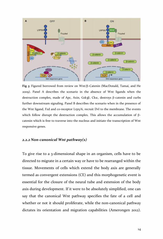
Fig 3: Figured borrowed from review on Wnt/-Catenin (MacDonald, Tamai, and He
2009). Panel A describes the scenario in the absence of Wnt ligands when the
destruction complex, made of Apc, Axin, Gsk3, Ck1, destroys -catenin and curbs
further downstream signaling. Panel B describes the scenario when in the presence of
the Wnt ligand, Fzd and co-receptor Lrp5/6, recruit Dvl to the membrane. The events
which follow disrupt the destruction complex. This allows the accumulation of -
catenin which is free to traverse into the nucleus and initiate the transcription of Wnt
responsive genes.
2.2.2 Non-canonical Wnt pathway(s)
To give rise to a 3-dimensional shape in an organism, cells have to be
directed to migrate in a certain way or have to be rearranged within the
tissue. Movements of cells which extend the body axis are generally
termed as convergent extensions (CE) and this morphogenetic event is
essential for the closure of the neural tube and extension of the body
axis during development. If it were to be absolutely simplified, one can
say that the canonical Wnt pathway specifies the fate of a cell and
whether or not it should proliferate, while the non-canonical pathway
dictates its orientation and migration capabilities (Amerongen 2012).

15
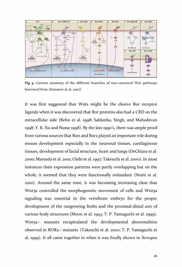
The non-canonical Wnt pathways encompass a set of pathways which
are usually triggered into action by Wnt or Fzd but downstream of that
they encompass a diverse range of receptors, cytoplasmic effectors and
transcription factors, each with a varying outcome, as can be seen in Fig
4. However, a unifying factor for all of these non-canonical Wnt
pathway sub-types is the lack of -catenin dependent transcription and
antagonism of the canonical pathway (Veeman, Axelrod, and Moon
2003).
The first clue regarding Wnts playing a role in gastrulation movements
came from studies in Xenopus embryos where overexpression of
XWnt5a, caused defects in the convergent extension movements, but
not the fates of cells (Moon et al. 1993). This observation suggested the
possibility of the existence of a pathway in vertebrates that was very
similar to Drosophila planar cell polarity (PCP). PCP, in flies, dictates
the polarity of a cell within a plane of tissue e.g: the arrangement of the
ommatidium or individual optical units which make up the compound
eye or the arrangement of the hair cells on the wing, in a fly. It was well
established that Fzd and Dvl played important roles in the PCP pathway
(Axelrod et al. 1998) but the ligand of this pathway remained obscure
until it was discovered in Drosophila that it was wingless which was the
ligand (Bhanot et al. 1996; Deardorff et al. 1998). In time, the list of non-
canonical Wnt pathways was extended to include the Wnt/PCP,
Wnt/Calcium pathway, Wnt/ c-Jun N-terminal kinases (JNK) pathway
and many others, though the latter ones are less well characterized. For
the purpose of this thesis, I will only discuss the Wnt5a-ROR axis.
16
Fig 4: Cartoon summary of the different branches of non-canonical Wnt pathways
borrowed from (Semenov et al. 2007)
It was first suggested that Wnts might be the elusive Ror receptor
ligands when it was discovered that Ror proteins also had a CRD on the
extracellular side (Rehn et al. 1998; Saldanha, Singh, and Mahadevan
1998; Y. K. Xu and Nusse 1998). By the late 1990’s, there was ample proof
from various sources that Ror1 and Ror2 played an important role during
mouse development especially in the neuronal tissues, cartilaginous
tissues, development of facial structure, heart and lungs (DeChiara et al.
2000; Matsuda et al. 2001; Oishi et al. 1997; Takeuchi et al. 2000). In most
instances their expression patterns were partly overlapping but on the
whole, it seemed that they were functionally redundant (Nomi et al.
2001). Around the same time, it was becoming increasing clear that
Wnt5a controlled the morphogenetic movement of cells and Wnt5a
signaling was essential in the vertebrate embryo for the proper
development of the outgrowing limbs and the proximal-distal axis of
various body structures (Moon et al. 1993; T. P. Yamaguchi et al. 1999).
Wnt5a-/- mutants recapitulated the developmental abnormalities
observed in ROR2-/- mutants (Takeuchi et al. 2000; T. P. Yamaguchi et
al. 1999). It all came together in when it was finally shown in Xenopus

17
that XRor2 was a receptor of the non-canonical Wnt5a and that it played
a role in CE movements (Hikasa et al. 2002; Schambony and Wedlich
2007) and that mROR2 and Wnt5a worked synergistically in activating
the JNK pathway. Ror2 was recognized as a bona fide receptor of Wnt5a
and not simply a co-receptor of Fzd (A. J. Mikels and Nusse 2006; Oishi
et al. 2003).
Over the years, various attempts were made to understand how Wnt5a
signaled through the Ror receptors. It was shown that Wnt5a induced
homodimerization and activation of Ror2 (Liu et al. 2008) and that upon
stimulation by Wnt5a, Ror2 underwent phosphorylation by CK1e and
GSK-3 on Ser/Thr residues (and not tyrosine residues) (Kani et al. 2004;
H. Yamamoto et al. 2007); Gsk-3 could specifically phosphorylate
serine 834 in Ror2 (Grumolato et al. 2010). The ability of Rors to function
as typical tyrosine kinases has always been disputed (as explained in the
‘structure’ section ) however it has been shown that Ror2 kinase activity
is required for signaling through Wnt5a (A. Mikels, Minami, and Nusse
2009). It was not long before Ror1 was also found to be a receptor of
Wnt5a. In a span of 5 years it was shown that Ror1 was a receptor of
Wnt5a (Fukuda et al. 2008), its interaction with Ror1/2 heterodimers
played an important role in synaptogenesis in hippocampal neurons
(Paganoni, Bernstein, and Ferreira 2010), and that Wnt5a-Ror signaling
axis was important for proper tissue morphogenesis (Ho et al. 2012).
Interestingly, while the first study showed that Wnt5a-Ror1 interaction
induced the activation of nuclear factor kappa-light-chain-enhancer of
activated B cells (NFkB), the last study found that among all the
downstream effectors of Wnt-Ror signaling which were reported
previously such as phosphorylation(s) of c-Jun, protein kinase C (PKC),

18
vang-like protein 2 (Vangl2) or Dvl (Gao et al. 2011; Oishi et al. 2003; X.
Zhang et al. 2007) or antagonism of canonical Wnt signaling (A. J.
Mikels and Nusse 2006) it was only phosphorylation of Dvl which was
affected. These studies highlight that the Wnt5a-Ror axis forms an
independent branch among the non-canonical pathways, though it
frequently can cooperate with the Wnt/PCP branch (Gao et al. 2011), and
that Wnt5a-Ror downstream signaling will differ among cell types. I
believe the initial intense scrutiny regarding the role of ROR2 in non-
canonical signaling was prompted by the fact that mutations in Ror2
were known to be responsible for the severe skeletal defects in RRS and
BDB1. Once it was discovered the ROR1 is a unique marker on CLL cells,
the focus shifted to also understanding this signaling branch better,
especially in CLL cells. First, it was shown that autocrine WNT5a by
regulating ROR1 activity conferred CLL cells with higher basal motility
and rendered them unable to respond to chemokines, and that
inhibiting the Wnt/PCP pathway in these cells restored migratory
defects (Janovska et al. 2016). It has also been shown that Wnt5a
promotes the interaction of ROR1 to intracellular proteins such as
dedicator of cytokinesis 2 (DOCK2), 14-3-3, hematopoietic-lineage-
specific protein 1 (HS1) and cortactin to activate Rho-GTPases,
ultimately leading to an enhanced rate of proliferation and migration in
CLL cells (M. Hasan et al. 2017; M. K. Hasan et al. 2018, 2019; J Yu et al.
2017). There have been reports suggesting that Wnt5a enhanced the
migration and proliferation in CLL cells by promoting the hetero-
oligomerization of ROR1 and ROR2 through the interaction of their
respective kringle domains (Jian Yu et al. 2016), however this has been
disputed since NMR studies show that these domains are not involved
in the interaction (Ma et al. 2019).

19
In general, a considerable body of work has helped us to understand
signaling through RORs via its ligand Wnt5a; however, significant gaps
of knowledge remain in our understanding of the importance of their
intracellular domains or any alternative modes of signaling since these
receptors do have other domains on the extracellular side which can
possibly interact with a wide repertoire of ligands.
2.2.3 ROR signaling in limb development
The skeletal system is composed of bone and cartilage made of dense
and semi-rigid connective tissues, respectively. It can be divided into
the axial and appendicular skeleton wherein the head and body trunk
are a part of the axial skeleton; forelimbs and hindlimbs part of the
appendicular skeleton. One of the first observable phenotypes in Ror-/-
or Wnt5a-/- mice were the skeletal defects (DeChiara et al. 2000; T. P.
Yamaguchi et al. 1999). While genetically Ror2 deficient mice exhibited
skeletal abnormalities (Takeuchi et al. 2000), Ror1 deficient mice did not
display any obvious skeletal defects but they died at birth due to
respiratory failure (Nomi et al. 2001). In a different study, though Ror1
mice were observed to have skeletal defects, they were limited to the
axial skeleton and the Ror1 deficient pups were severely growth
compromised (Lyashenko et al. 2010).
Between the RORs, ROR2 was firmly implicated to have a more
prominent role in skeletal development when it was discovered to
harbor mutations which caused RRS and BDB1 (Afzal et al. 2000; Afzal
and Jeffery 2003; Oldridge et al. 2000; Schwabe et al. 2000). RRS is
characterized by severe craniofacial malformations, overall skeletal

20
defects which affect the axial and appendicular system, heart defects,
and genital hypoplasia. On the other hand, brachydactylies are a group
of disorders characterized by the shortening of the limbs. Of the 5 types
(A-E), BDB1 subtype is the most severe which results from mutations in
ROR2 (Stricker and Mundlos 2011). For the purpose of this thesis, I will
discuss signaling events during limb development which can help us
appreciate the molecular milieu of ROR2 signaling.
During limb development, mesenchymal cells from the lateral plate
mesoderm initiate the formation of a limb bud. The limb bud then has
to coordinate development along 3 axes: proximo-distal(P-D), anterior-
posterior (A-P), and dorso-ventral (D-V). Each of these is under the
control of different signaling center: the apical ectodermal ridge (AER)
controls the P-D axis, the zone of polarizing activity (ZPA) controls the
A-P axis and the D-V axis is under the control of Wnts from the
overlying ectoderm (Petit, Sears, and Ahituv 2017). While the three axes
have independent signaling mechanisms, they have to be coordinated
spatio-temporally for correct limb formation (Spielmann and Stricker
2016). The limb can be divided into 3 regions from proximal to distal
end- stylopod, zeugopod and autopod. In the developing mouse
embryo, Ror1 expression is restricted to the proximal regions of the limb
while Ror2 expression can be detected throughout the limb, especially
in the distal regions (Matsuda et al. 2001). It is the autopod that is most
severely affected in BDB1 (Stricker, Rauschenberger, and Schambony
2017).
Bone formation is called ossification that can occur via
intramembranous ossification and endochondral ossification (Kamizaki

21
et al. 2020). Intramembranous ossification involves bone formation
directly from the connective tissue and is commonly seen in flat bones
while in endochondral ossification, commonly observed in long bones,
eg. Limb bones, the cartilage is laid first as a template. Chondrogenesis
is the process by which the cartilage is formed. It starts with the
condensation of undifferentiated mesenchymal stem cells into
aggregates which reflect the pattern of the future limb. These cells,
called chondrocytes, undergo progressive differentiation giving rise to
pre-hypertrophic and then hypertrophic chondrocytes; this entire
process is controlled by BMP signaling. It has been shown that Ror2
plays a role in chondrocyte differentiation but not so much in
proliferation (Schwabe et al. 2004). Mesenchymal cells which surround
the cartilage form the perichondrium and these cells express Wnt5a;
overexpression of Wnt5a prolongs the differentiation of
prehypertrophic to hypertrophic chondrocytes (Hartmann 2002;
Hartmann and Tabin 2000). The formation of the individual digits at
limb extremities is dependent on a signaling center called the phalynx-
forming region or digit crescent (PFR/DC) and it is characterized by a
highly active BMP signaling pathway (Witte et al. 2010). The AER
maintains an active fibroblast growth factor (FGF) signal to maintain
the cells in an undifferentiated state but AER signals are important for
the digit outgrowth driven by BMP signals. Ror2 and Wnt5a co-operate
to inhibit Wnt/-catenin signals from the ectoderm, a failure of which
leads to a break-down of the PFR/DC ultimately affecting the phalanges
as evinced by the BDB1 phenotype. However, the exact molecular and
biochemical details of these signaling crosstalks are yet to be elucidated.

22
3. Chronic Lymphocytic Leukemia CLL was first described in 1960’s by Dr William Dameshek, from Boston
and Dr D. Galton, from London (Dameshek 1967; G Galton 1966), almost
simultaneously, as an immunoproliferative disorder in which the B-cells
are immune-incompetent. The characteristic of CLL is accumulation in
peripheral blood, of small, mature looking, CD5+ B cells, which have
undergone clonal proliferation. The presence of CD5 on B-cells is an
obvious anomaly since it is actually a T-cell antigen (Burgess et al. 1992).
These cells have faulty apoptotic mechanisms and tend to accumulate
within the blood, spleen, bone marrow and lymph nodes and cause
lymphocytosis, splenomegaly and lymphadenopathy. For a very long
time, since the course of the disease is slow, it was believed the CLL
results due to the accumulation of faulty cells rather than being a
proliferative disorder. However it was shown that in fact CLL cells do
proliferate at an astounding rate (Messmer et al. 2005). In fact, the
prognosis is usually worse in patients in whom this rate is greater than
0.35%. These cells also express CD19; a biomarker of B-cells, and CD23;
a low affinity receptor of IgE. CD23 is used to distinguish CLL from other
lymphoproliferative disorders, mainly mantle cell lymphoma (MCL)
(DiRaimondo et al. 2002; Kilo and Dorfman 1996). CLL cells also express
lower levels of IgM and IgD in comparison to normal B-cells.
A perplexing aspect of CLL is that in some patients, the disease may
remain indolent for years and the patient may actually succumb due to
natural causes or some other ailment. These individuals may not even
know that they have CLL, if it were not for some routine blood check.
In other cases, the course of CLL may turn aggressive which leads to a
greatly decreased life expectancy, in some cases despite the therapy.

23
This heterogenous nature of CLL prompted Rai and colleagues in 1975
to devise a staging system of CLL based on the symptoms of the patients
(Rai et al. 1975). This staging system was revised and improved further
by Binet and colleagues (Binet et al. 1977) and has been used ever since
to stratify CLL patients into risk groups (as shown in Table 1) thereby
helping clinicians in identifying those who need treatment versus (vs)
those who just need to be under observation.
Table 1: Borrowed from the review – Chronic Lymphocytic Leukemia, A clinical review.
(Nabhan and Rosen 2014). It summarizes the criteria suggested by Rai and Binet to
stratify CLL patients into risk groups.
a-Note that the overall survival has improved over the years due to improved therapies
b-nodal areas such as cervical, axillary, inguinal, spleen and liver.
CLL is of unknown etiology and there are many theories about its cell of
origin. The identification of the cell of origin is imperative to understand
the pathobiology of the disease. Many groups have tried to hypothesize
about or identify the cellular origins of CLL. One view which has
prevailed in the field is that the disease develops from a self-renewing
hematopoietic stem cell (HSC) which may turn rogue and as a result be
the CLL cell of origin (Kikushige et al. 2011). Historically, it was also
believed that CLL arose from naïve, antigen inexperienced B-cells;

24
however, work by multiple groups has shown that since B-CLL cells are
functionally similar to the ones from splenic marginal zone, so they
must have arisen from antigen- experienced marginal zone, CD27+
memory B cells (Chiorazzi, Rai, and Ferrarini 2005; Damle et al. 2002;
Klein et al. 2001).
According to the Armitage and Doll ‘multistep model of carcinogenesis’,
it is not a single mutational event but a series of critical mutational
events which cause the transformation of a normal cell into a cancerous
one (Armitage and Doll 1954). Likewise, many factors contribute not just
to the development of the disease but also its course. In the case of CLL,
it is now understood that though the disease may be initiated by
changes in the genetic material it is the burden of additional factors /
mutations that makes it more aggressive. That genetics and hereditary
also play a role is evinced by the fact that approximately 9% of CLL
patients had a relative who also suffered from the disease. In addition,
in cases of familial CLL, about 30 genetic loci were found to carry short
nucleotide polymorphisms (SNPs)(Kipps et al. 2017). Curiously, gender
seems to play a role in CLL, with women having a much slower course
of the disease and a better chance at overall survival (D. Catovsky,
Fooks, and Richards 1989). Important hallmarks of CLL include: the
mutational status of the immunoglobulin heavy chain variable region
gene (IGHV); chromosomal changes in untreated patients such as
deletion of the long arm of chromosome 13 (del13q), trisomy 12, deletion
of the long arm of chromosome 11(del11q) or deletion of the short arm of
chromosome 17(del17p); expression of somatically mutated genes
especially NOTCH1, myeloid differentiation primary response
(MYD88), TP53, ZNF292, or PTPN11 (Hallek 2019). All together these

25
observations suggest that the central pathways on which a cell depends,
such as DNA damage and repair, RNA processing, MAPK signaling are
disrupted.
3.1 IGHV status
The course that CLL might take can be gauged by the mutational status
of the immunoglobulin heavy chain variable region gene. If a sequence
differs from the germline sequence more than 2%, it is considered
mutated. Many CLL patients, grouped as mCLL, harbor more than this
2% of mutations in their VH genes, and they have been observed to have
the less aggressive form of the disease and thus prolonged survival
(Damle et al. 1999). The other group of patients with < 2% or no
mutations in VH genes are referred to as uCLL, u referring to the
unmutated status. They usually suffer the more aggressive form of the
disease and can have decreased life expectancy (Hamblin et al. 1999).
Since there are 2 subtypes it was also believed that uCLL arose from
naïve or pre-germinal center B cells while mCLL arose from post-
germinal center B cells. uCLL cells have a greater capacity to proliferate
and can respond to immune-stimulation but are also prone to apoptosis
(Longo et al. 2007). Contrarily, mCLL do not respond to external
stimulus as much, but they do have a very active intracellular signaling
(Chiorazzi, Rai, and Ferrarini 2005). Interestingly, it was a study of the
mutational status of the IGHV that gave clues to researchers about the
difference in genders as well; the occurrence of uCLL is higher in males
(Daniel Catovsky, Wade, and Else 2014). Irrespective of the subtype, CLL
cells do have a very limited repertoire of antibodies, so much so that

26
about every 1 in 75 CLL patients will have nearly identical antibodies, a
phenomenon referred to as stereotypy (Widhopf et al. 2004).
3.2 Cytogenetic aberrations
In terms of genetic aberrations or alterations, CLL patients have some
classic cytogenetic lesions, namely: deletion of 13q14, trisomy 12,
deletion 11q22-23 and deletion of 17p13; however, these may not be the
primary triggers of CLL. Nonetheless, the presence of these cytogenetic
abnormalities does influence how patients will respond to treatments
hence it is important that CLL patients are tested for these lesions before
embarking upon treatments (Hallek et al. 2008).
The most common genetic insult found in CLL patients is the del13q,
specifically involving band 14. The frequency of this abnormality is close
to 50%, which means that the disruption of genes in this cluster, must
offer some advantage to the cell. Rightly so, this cluster has been found
to encode genes controlling cell cycle and apoptosis in B-cells. This
particular region has been found to encode a non-transcribed gene and
two micro RNAs (miRNA) 15-a and 16-1 (Calin et al. 2002, 2005; Veronese
et al. 2015),which serve to repress the expression of B-cell lymphoma 2
(BCL2) protein and -associated protein, molecular weight 70kDa (ZAP-
70), respectively. Lack of these miRNA controls allows the cells to
express BCL2 and ZAP70 which are anti-apoptotic (Klein et al. 2010).The
worst are the del17p and del11q22-23 lesions, since these stretches contain
p53 and ataxia-telangiectasia mutated (ATM) genes, respectively
(Döhner et al. 2000; Stilgenbauer et al. 2002). p53 protein is a well

27
characterized tumor suppressor and ATM kinase is involved in the DNA
damage repair pathway.
In spite of all the above mentioned molecular and genetic abnormalities
found in CLL, none of them are the direct causes nor are they specific to
CLL. The correct method of CLL diagnosis, as recommended by the
World Health Organization (WHO), international workshop on chronic
lymphocytic leukemia (iwCLL) and National Family Caregivers
Association (NCCN), relies on immunophenotyping to distinguish it
from other B-cell lymphoproliferative disorders (Rawstron et al. 2018).
Thus, in addition to the minimum set of markers such as CD19, CD5,
CD23, CD20, kappa & lambda, the assessment of the patient sample
should include CD200, CD10 and ROR1. The inclusion of ROR1 makes
sense since it was shown that higher levels of cell surface ROR1 were
associated with a rapid disease progression (Cui et al. 2016).
3.3 B-Cell Receptor signaling in CLL
There is no doubt that CLL is a BCR-dependent malignancy. Within
lymphoid tissues, CLL cells actively proliferate within areas termed as
psuedofollicles and BCR signaling is the most prominent pathway active
in these cells (Burger and Chiorazzi 2013). B-cells rely on this pathway
for their survival and a functional BCR is imperative for the survival of
B-cells (Lam, Kühn, and Rajewsky 1997). In a normal scenario, BCR
signaling allows for the expansion of a foreign antigen activated B-cell
and removal of B-cells which react to self-antigens. With an increase in
the knowledge about this pathway, it became clearer that in CLL a lot of
the BCR pathway components signal aberrantly which is advantageous

28
for the malignant cells; CLL-BCRs can be active even in the absence of
an external antigen, known as tonic stimulation (Burger and Chiorazzi
2013).
3.3.1 BCR Pathway
For about 500 years, vertebrates have relied on the adaptive immune
branch to protect themselves from an increasingly complex
environment. One of the key players in this branch of immune system
is the immunoglobulin (Ig) molecule, commonly referred to as an
antibody. Higher classes of vertebrates usually have 5 different isotypes
of Igs: IgM, IgD, IgG, IgA, IgE. Not only do these Ig molecules serve as
circulating effectors to stimulate other components of the immune
system, but also, they serve as antigen receptors on the surfaces of B-
cells (Schroeder 2015). The default antibody that a B-cell is equipped
with, when it is ‘born’ is the IgM; it is present even before the B-cell ever
encounters an external antigen. In a normal cell, this surface Ig molecule
is usually paired with a heterodimer comprising of cluster of
differentiation (CD) 79A-79B, also known as Ig/Ig, wherein the IgM
binds externally to the antigen while the Ig/Ig heterodimer handles
the intracellular signaling. The cytoplasmic domains of both these Ig
associated proteins contain multiple phosphorylatable sites known as
immunotyrosine based activation motifs (ITAMs). Upon antigen
stimulation, the BCR components undergo rearrangements in the
plasma membrane such that the tyrosines in the ITAMs are positioned
to get phosphorylated. These phosphorylations are carried out by Src-
family kinase (SFK) family members which are found to be pre-
associated with the BCRs, primarily Lyn (Burkhardt et al. 1991; T.

29
Yamamoto, Yamanashi, and Toyoshima 1993), and others like Fyn and
Blk. The tyrosine kinase Syk, which is a target of Lyn (Kurosaki et al.
1994), has 2 SH2 domains but lacks the N-terminal acylation to help it
anchor itself to the membrane. The SFK phosphorylated ITAM motifs
provide a binding site for Syk via its SH2 domain, such that gets apposed
to its activator Lyn.
Fig 5: Cartoon summary of the BCR pathway. Stimulation of the BCR by an antigen,
leads to the activation of various kinases downstream of the primary kinases Lyn and
Syk. Figure borrowed from review on B-cell receptor signaling by ten Hacken and Burger
(2016).
These SFKs, which fall under the class non-RTKs, are also responsible
for phosphorylating and activating further downstream kinases of the
BCR pathway which ensures that the incoming signal gets amplified and

30
is propagated via 3 main routes: phospholipase C-2 (PLC-2),
phosphatidyl inositol 3 kinase (PI3K) and Bruton’s tyrosine kinase
(BTK). The ensuing cascade of events leads ultimately to signals which
promote the survival and proliferation of B-cells (Burger and Chiorazzi
2013; Woyach, Johnson, and Byrd 2012). The importance of this pathway
is underscored by the current number of drugs which target the
individual pathway components. I will discuss more on that in the next
section but first I would like to highlight the importance of Lyn, a crucial
regulator of the BCR pathway.
3.3.2 Lyn kinase
Lyn is a member of a family of non-receptor tyrosine kinases, composed
of 9 structurally related members, which include Src, Yes, Fyn and Fgr
(which form the SrcA subfamily), Blk, Lck, Lyn and Hck (which form
the SrcB subfamily) and Yrk (Parsons and Parsons 2004). These
proteins, collectively referred to as the SFKs play an important role in
modulating the signals propagated by multiple RTKs on the membrane.
Lyn, short for Lck/Yes-related novel tyrosine kinase, is a SFK which was
discovered using v-yes DNA as a probe (Y Yamanashi et al. 1987) and its
gene is localized on human chromosome 8 (mouse chromosome 4). In
a mature B-cell, BCR signaling requires Lyn in its capacity as a positive
facilitator of the pathway through phosphorylations of ITAMs in Ig,
Ig and CD19; however, its role as a negative regulator is far more
important, whereby it phosphorylates immunoreceptor tyrosine-based
inhibitory motifs (ITIMs) in cell surface receptors which helps to end
the BCR signaling. Due to this dual role Lyn has been described as a
cellular signaling rheostat (Lowell 2004; Y. Xu et al. 2005).

31
It was known that the signal transducing entity in BCR, the Ig and Ig
heterodimer, lacked catalytic domains and therefore an alternate
protein was required to interpret the incoming signal and pass it
forward. By drawing parallels with the role of Lck in T cells and its
association with T cell receptors, it was discovered that Lyn physically
associates with IgM and mediates IgM signaling (Yuji Yamanashi et al.
1991). Subsequently it was found to associate with Syk, HS1, Vav, PI3K
and BTK. Lyn also co-operates heavily with the co-receptor CD19 to
form a signal amplification loop, wherein Lyn phosphorylates tyrosine
residues on CD19, creating an additional site for Lyn to bind and
undergo further activation in proximity of the BCR (Fujimoto et al. 2000;
Gauld and Cambier 2004). While its positive role in BCR signal
modulation can be shared to some extent by the other two SFKs, its role
as a negative modulator of this pathway is irreplaceable as was
highlighted by the work which shed light on the interaction of Lyn and
CD22 (Smith et al. 1998). As mentioned above, Lyn and CD19 enter into
a kind of activation loop. Among the many targets of Lyn, which it can
phosphorylate, one is CD22. The phosphorylation of CD22 initiates the
recruitment of SHP-1, a phosphatase of CD19 which ultimately leads to
the levels of activation signals going down. This finding was also
supported further by studies in Lyn-/- mice in which the B-cell
development was not affected but they were found to be
hyperproliferative, leading to autoimmune disorders (DeFranco, Chan,
and Lowell 1998; Hibbs et al. 1995). B-cell development was more or less
normal in these mice because at during this stage Lyn plays a minor role,
albeit a positive one, along with Fyn and Blk as a redundant SFKs (Y. Xu
et al. 2005). Surprisingly enough, the phenotype of mice overexpressing

32
Lyn (Lyn up/up ) recapitulated the effects of the Lyn-/- mice (Hibbs et al.
2002).
Owing to splice variants of its transcripts on exon 2 Lyn protein
can have 2 isoforms, p53 and p56 (also known as LynB and LynA,
respectively).The 2 isoforms were believed to be functionally redundant,
until it was shown in mast cells that the 2 isoforms have different roles
in signaling and associate with different effectors (Alvarez-Errico et al.
2010). Lyn is highly expressed in all hematopoietic cells of myeloid and
lymphoid lineage, except T cells, although it does play an important role
as a negative regulator of Th2 immune responses as seen in Lyn-/- mice
(Beavitt et al. 2005).
All in all, these studies highlight the importance of Lyn in B-cells
and suggest that Lyn fine-tunes BCR signaling by forming and
coordinating an extremely complex network of targets, activators and
regulators as shown in Fig 6.
Fig 6: Graphical summary of the positive, through CD19, and negative regulation,
through CD22, of BCR pathway by Lyn. Figure borrowed from review on Src family
kinases in B-cells.(Gauld and Cambier 2004)

33
3.3.3 Regulation of Lyn (and other SFKs)
In my work I have utilized pharmacological inhibition or Lyn deletion
mutants to control the kinase activity of Lyn and thereby influence its
interaction with ROR1. Hence, I would like to elaborate on how SFKs are
physiologically regulated.
Among the SFK family members there exists a considerable
amount of structural homology within their domains and thus, all SFKs
are regulated the same way. Each member has the following functional
domains: an N-terminal SH4 domain, unique for each kinase and which
hosts one or two acylation sites for both myristoyl and palmitoyl groups
to help it remain anchored to the membrane ( with the exception of Blk
which only has myristoyl groups), SH3 domains, SH2 domain, a linker
region and a kinase domain ( also referred to as the catalytic domain).
SH2 and SH3 domains are protein-protein interaction domains wherein
SH2 domains binds to phosphor-tyrosine (pY) residues (Moran et al.
1990) and SH3 binds to proline rich domains (PRD) (Ren et al. 1993).
The catalytic activity of SFKs has to be very tightly regulated and this is
done by a series of phosphorylations and dephosphorylations to keep it
active or inactive. To suppress the kinase activity, the catalytic domain
is maintained in a closed conformation (shown in Fig 7). This is
achieved by means of phosphorylating a Y residue at position 527, close
to the C-terminal. By convention, this position refers to the one
observed in c-Src but it differs slightly in each SFK. In the case of Lyn, it
is Y508. This phosphorylated Y527 can now bind to the SH2 domain and
keep the catalytic domain closed and the two PTKs responsible for this

34
phosphorylation are C-terminal Src kinase (CSK) and Csk homologous
kinase (CHK).
Fig 7: Regulation of Src family kinases. Figure on the left side shows SFK in an inactive
state kept phosphorylation of Y(508 in Lyn) residue in the C-terminal. Figure on the
right side shows an activated kinase in which the C-term phosphate is removed by a
phosphatase first, which allows the Y (397 in Lyn) in the kinase domain to become
accessible. Figure borrowed from a review on Src family kinases (Salter and Kalia 2004)
To activate the SFK, this Y527 has to first be dephosphorylated, which is
carried out by different phosphatases. Alternatively, even the binding of
external ligands to SH3/SH2 domains can interfere with this inactivating

35
intramolecular interaction, thereby opening up the catalytic domain
which exposes Y416 (Y396 in Lyn). Phosphorylation of this residue not
only dislodges it from the site to which SFK substrates can bind but also
completely activates the SFK (Salter and Kalia 2004).

36
4. Current therapeutic strategies in CLL
The objective of any treatment related to cancer is to achieve a minimal
residual disease (MRD) negative state. Since CLL is such a complex
disease, clinicians have found it beneficial to target it from multiple
directions using a wide array of available drugs. Thus most current
treatment options include combinations of cytostatic agents eg:
chlorambucil, fludarabine; monoclonal antibodies (mAb) eg: rituximab,
alemtuzumab; agents targeting BCR or BCL2 signaling eg: ibrutinib,
idelalisib, venetoclax; chimeric antigen receptor T cells (CART) therapy
(Hallek 2019). Overall it was observed that the addition of agents
targeting kinases in BCR pathway, to the combination treatments, was
more beneficial.
4.1 Targeting BCR as a therapeutic strategy in CLL
Given the importance of this pathway it was natural for it to be the focus
of therapies in CLL and hence this field has exploded in the recent years.
As can be seen in Fig 8, a host of small molecule kinase inhibitors have
been developed to target SYK , BTK and PI3K known as fostamatinib,
ibrutinib and idelalisib, respectively (Byrd et al. 2013; Friedberg et al.
2010; Furman et al. 2014). However, notwithstanding the evidence of
how efficient they are resistance to these drugs has been observed in
clinic. Point mutations in BTK (C481S) or activating mutations in PLC
have been observed (ten Hacken et al. 2019). BTK is a member of the Tec
family comprising of 5 other members, all of which are highly expressed
in cells of the hematopoietic system and play important roles in growth
and differentiation (Mano 1999). Members of this family are known to

37
mediate signals emerging from phosphotyrosine and phospholipid-
based systems and thus additional problems arise when drugs like
ibrutinib cross-react with other members of the family or other kinases
e.g: epidermal growth factor receptor (EGFR) (Byrd et al. 2016).
Moreover, these drugs are associated with expensive treatments, very
nasty side effects such a bleeding diathesis and arrythmias, and disease
relapse post discontinuation, hence there is a dire need of alternate
targets.
Fig 8: Cartoon summary of BCR kinases targeted for therapy in CLL. Figure borrowed
from review on the importance of BCR in CLL (ten Hacken et al. 2019)
Given the importance of BCR to CLL cells, it was not long before
the role of Lyn in CLL came under scrutiny. Even though BCR
stimulation in B-CLL cells failed to stimulate Lyn (Kawauchi,
Ogasawara, and Yasuyama 2002), Lyn was found to be highly expressed
in B-CLL cells as compared to normal B-cells (Contri et al. 2005; Hussein

38
et al. 2009). CLL cells thrive in areas of the bone marrow called
pseudofollicles and the microenvironment these CLL cells is made up of
mesenchymal stromal cells, nurse-like cells derived from monocytes
and T cells (ten Hacken and Burger 2016). CLL cells need input from all
of these to survive and this was evident from the observation that
culturing of CLL cells in vitro was a near impossible task, due to the
tendency of CLL cells to undergo apoptosis (Collins et al. 1989), unless
the culture was supported with factors normally found in the tumor
microenvironment (Nguyen et al. 2016). The importance of the
microenvironment on CLL cells was also gleaned from the observation
that CLL cells can have distinct biologies based on their location,
peripheral blood vs lymph nodes or bone marrow microenvironments
(Hayden et al. 2012). Given the importance of Lyn to BCR and cells of
the myeloid lineage, Nguyen et al attempted to uncover the role of Lyn
in influencing the interactions which occur in these
microenvironments. Indeed, its importance was underscored when it
was found that macrophages without Lyn failed to support the growth
of CLL cells. Thus targeting Lyn in CLL would be an option worth
exploring (Wiestner 2012).
4.2 Targeting ROR1 as a therapeutic strategy in CLL
The first line of treatment in CLL patients includes a combination of
cytostatic reagents, such as fludarabine and cyclophosphamide, and
mAbs, such as rituximab. The disadvantage of current mAbs which are
approved as first or second line of treatment in CLL, such as rituximab
(mAb against CD20) and alemtuzumab (mAb against CD52) is that
these target the normal B-cells leading to an overall

39
immunosuppression in patients (Yang et al. 2011). This need for a CLL
specific target paved the way for research into developing mAbs to
target other CLL-cell specific targets.
Two independent gene expression profiling studies had already
categorized ROR1 as a marker which was upregulated in CLL cells (Klein
et al. 2001; Rosenwald et al. 2001) but it was few more years before it was
proposed by different groups that ROR1 would be an ideal target to treat
this disease (S. Baskar et al. 2008; Amir H. Daneshmanesh et al. 2008;
Fukuda et al. 2008). ROR1 CLL cells did indeed undergo apoptosis when
treated with ROR1 siRNA which gave further impetus to this idea
(Choudhury et al. 2010) and within time different groups had developed
mAbs (Sivasubramanian Baskar et al. 2012; Choi et al. 2015; A. H.
Daneshmanesh et al. 2012) or CAR-T (Hudecek et al. 2010) against ROR1
which showed an apoptotic effect specifically towards CLL cells. One of
these anti-ROR1 mAbs, cirmtuzumab ( also known as UC-961), is
currently in clinical trials and has shown promising results (Choi et al.
2018).
It must be mentioned that B-cells have been observed to express
ROR1 normally during an intermediate stage of development and these
B-cells are termed hematogones (Broome et al. 2011; Hudecek et al.
2010). In light of this knowledge, a very interesting connection was made
between ROR1 and the pre-BCR in a subset of acute lymphoblastic
leukemia (ALL) patients, specifically ones which carried the genetic
abnormality t(1;19) (Bicocca et al. 2012). ALL is childhood malignancy
and about 5% of ALL patients carry this translocation t(1;19) and a study
of cells which carry this genetic abnormality showed that these were
cells which were arrested at a later stage of B-cell development,
compared to the other 95%. In this elegant study, the authors observed

40
that crosstalk between ROR1 and pre-BCR promoted the survival of CLL
cells by activating the Akt pathway via ROR1/MEK/ERK when only the
pre-BCR was inhibited, but a synergistic effect leading to cell death
when ROR1 and pre-BCR, both were inhibited. This line of thought was
further supported by a study which showed that downregulation of
ROR1 had a synergistic effect with BCR inhibition in BCR sensitive cells
(Karvonen, Chiron, et al. 2017). It was shown that NFB signaling was
downstream of ROR1 and this was affected when ROR1 was targeted.
This observation falls in line with the previous one implicating Akt lying
downstream of ROR1 since it is known that Akt lies upstream of NFkB
and regulates it (Scheid and Woodgett 2000).
In light of these discoveries, there has been an increasing
interest in pursuing combinatorial treatments targeting ROR1 and BCR
(Karvonen, Niininen, et al. 2017). However, how these crosstalks are
molecularly orchestrated is yet to be understood. In our work, we have
tried to decipher the crosstalk between Lyn and ROR1. We prioritized
our focus on Lyn owing to its importance in BCR pathway and prior
evidence which pointed to the relationship between ROR1 / ROR2 with
Src (Akbarzadeh et al. 2008; Gentile et al. 2014).

41
5. AIMS
1. Given the importance of ROR1 and Lyn in CLL and the problems of
drug resistance in disease relapse patients there is a need for additional
therapeutic targets. We wanted to ascertain if and how these tyrosine
kinases interacted intracellularly and what is the consequence of such
interaction.
Our main aims were
- To confirm interaction of ROR1 and Lyn
- To identify phosphorylations, if any, on ROR1 since Lyn is a
tyrosine kinase
- To identify any molecular and functional consequences of this
interaction/phosphorylation
2. Given the importance of ROR2 and Noggin mutations in causing BDB1
and BDB2, respectively, we wanted to ascertain if these proteins
interacted.
Our main aims were
- To confirm a genetic interaction of Noggin and Ror2
- To characterize a functional interaction of Noggin and Ror2

42
6. RESULTS & DISCUSSION
Article 1
(BIORXIV/2020/124156) Lyn controls chemotaxis and motility of CLL cells via phosphorylation of
ROR1.
Our study is the first to show that Lyn can interact with ROR1 and
phosphorylate it. By utilizing ROR1 intracellular domain-deletion
mutants, we were able to show that ROR1 could interact with Lyn in the
absence of its PRD and that Lyn could phosphorylate wild type (WT)
ROR1 on tyrosine residues. We further corroborated this result by
testing mutant forms of Lyn which either lacked the kinase domain,
possessed a kinase dead domain, or a constitutively active Lyn kinase;
indeed, the tyrosine (Y) phosphorylation of ROR1 depended on an active
kinase. This result was also supported by our experiments using
pharmacological inhibition of Lyn using Dasatinib, a pan-Src kinase
inhibitor. Two other SFKs are also found in B-cells, Fyn and Blk and
hence we wondered if ROR1 could also interact them. Fyn interacted
with ROR1 but could not phosphorylate it on tyrosine residues, while
Blk did not interact with ROR1 (data not shown in manuscript), proving
that ROR1- Lyn interaction was very specific.
Previously, ROR1 and ROR2 have been reported to interact with
Src (Akbarzadeh et al. 2008; Gentile et al. 2014; T. Yamaguchi et al. 2012).
In work shown by Akbarzadeh et al, it was shown that Src recruitment
and activation was incumbent upon Wnt5a stimulation of ROR2. In our
study we observed that Wnt5a was dispensable as far as ROR1 and Lyn

43
interaction was concerned. By means of immunocytochemistry we
could observe that this interaction occurred at or close to the plasma
membrane. While ROR1 is a RTK which spans the membrane; Lyn, as
well as other members of the SFK, usually stay anchored to the
membrane via their N-terminal dual fatty-acylation (Kovarova et al.
2001). In the work by Yamaguchi et al, the authors showed that in lung
adenocarcinoma cells, ROR1 was required to sustain the EGFR-ERBB3-
PI3K survival signaling. In the study, they also showed that Src and ROR1
could interact, but the interaction was dispensable for the role of ROR1
to cooperate with EGFR. Importantly, the authors claim that ROR1
kinase activity was required for c-Src activation, but this can be debated
on two accounts. One, ROR1 has been shown to lack in vitro kinase
activity (Bicocca et al. 2012; Masiakowski and Carroll 1992), hence has
been classified as a pseudokinase (Gentile et al. 2014). ROR1 can possibly
bind to ATP, since it retains the conserved lysine (K) in the b3-strand
(Rajakulendran and Sicheri 2010) but it lacks the conserved residues
needed to phosphorylate targets. Secondly, in the experiments carried
out by the authors, they do not address the issue of SFKs being capable
of undergoing autophosphorylation. Hence to ascertain if ROR1 indeed
is required for Src phosphorylation, additional experiments and
rigorous controls are warranted.
Using immunoprecipitation-mass spectrometry (IP-MS) we were able to
identify the residues phosphorylated by Lyn, namely Y645 and Y646.
These tyrosines are a part of a triad of tyrosines is commonly seen in
RTK family members with the consensus sequence ‘YxxxYY’ and they
are crucial for the autoregulation of the RTK (Hubbard, Mohammadi,
and Schlessinger 1998). In enzymatically active TKs, phosphorylation of

44
all 3 residues is required for the activation loop to undergo a
conformational change and make space to accommodate an ATP
molecule and a substrate. We have identified that Lyn can
phosphorylate at least 2 residues in this triad. Phosphorylation of the
second Y residue in the triad, in our case Y645, is essential for the RTK
to get out of its auto-inhibitory state. This observation becomes relevant
in light of the evidence that of ROR1 in CLL patients has been found to
be heavily phosphorylated (Hojjat-Farsangi et al. 2013). The same group
reported phosphorylation on residues Y641 Y646 and S652 but these
results were only presented in a conference perhaps as preliminary
results (Hojjat-Farsangi et al. 2012). On the other hand, this very triad
‘YxxxYY’, was found to be phosphorylated also by Src (Gentile et al.
2014). In that study, the RTK MET was shown to utilize ROR1, in its
capacity as a pseudokinase, to help drive tumorigenesis. Their work
demonstrated that phosphorylation of ROR1 in different domains
influenced different outcomes in a cell. The influence of MET on cell
proliferation was incumbent on phosphorylations in the PRD while the
capacity of a malignant cell to invade tissues was influenced by the
phosphorylation of the ‘pseudo’kinase domain by Src. Importantly, Src
needed the PRD of ROR1 to be present in order for it to phosphorylate
the kinase domain Y residues; Lyn somehow surpasses this need. This is
possible since individual SFK members do have different preferences of
sequences to bind to (Alexandropoulos, Cheng, and Baltimore 1995;
Zhou et al. 1993).
8 out of 58 human RTKs are most likely pseudokinases. Coincidentally,
4 of these happen to be receptors in the Wnt signaling pathway: ROR1,
ROR2, tyrosine-protein like kinase-7 (PTK7)/colon carcinoma kinase

45
(CCK4), and muscle specific kinase (MuSK). There is some evidence to
support the idea that these pseudokinases may function to allosterically
activate other kinases or may serve as scaffolding proteins (Mendrola et
al. 2013). ROR1 has been shown to play a scaffolding role in lung
adenocarcinoma cells (T. Yamaguchi et al. 2016) and hence the
possibility that ROR1 might serve such a function in CLL did not seem
too far-fetched. Once again, we relied on IP-MS to reveal binding
partners of ROR1 phosphorylated by Lyn. Among the many binding
partners which came up, one was Dvl2 which has been shown to work
downstream of ROR1, but another interesting candidate was c-casitas B
lineage lymphoma (c-Cbl) protein. c-Cbl is a proto-oncogene with E3
ligase activity known to negatively regulate RTKs by targeting them for
degradation (Kaabeche et al. 2004) or endocytosis (Petrelli et al. 2002).
It shares a very interesting relationship with Lyn, wherein not only does
it serve as its substrate in B-cells post BCR engagement (Tohru Tezuka
et al. 1996) but it can also target Lyn for degradation (Kaabeche et al.
2004; Shao et al. 2004). c-CBL is highly expressed in hematopoietic cells
and cells of the testis (Thien and Langdon 2001). Backed by our MS
results we sought to confirm this interaction in vitro and found that
indeed ROR1 could interact with c-Cbl but only after undergoing
phosphorylation by Lyn. Further study is definitely needed to
understand the downstream consequences of this interaction. In CLL c-
Cbl plays a role as an adaptor protein more prominently than that of an
E3 ligase (Martini et al. 2018). Structurally c-Cbl is a multi-domain
protein consisting of a N-terminal tyrosine-kinase binding domain
(TKB), a RING finger motif, a PRD and a ubiquitin associated domain
(UBA) close to the C-term. The TKB and PRD can engage in protein-
protein interactions while a number of phosphorylatable tyrosine and

46
serine residues provide an opportunity for c-Cbl to be differentially
regulated (Schmidt and Dikic 2005). A lot of work has been done to
show that Wnt5a exerts its effects in CLL through the promotion of
ROR1 interaction with various partners that play a role in the
modulation of cytoskeleton and migration (M. Hasan et al. 2017; M. K.
Hasan et al. 2019; J Yu et al. 2017). Naturally, it would be critical to
understand how this new interaction influences ROR1 response to
Wnt5a.
To further our understanding of how Lyn controls ROR1, we sought to
create Lyn deficient cells, using CRISPR-Cas technology. For this we
used a commercial cell line, derived from a patient suffering from MCL,
called Maver-1. Our Lyn knock-out (LKO) cells pointed to a defect in
ROR1 protein trafficking since ROR1 mRNA levels were lower in LKO
cells, but protein levels were more or less equal, or even higher in some
cases. Moreover, flow cytometric analysis showed that the LKO
expressed higher levels of surface level ROR1. A similar observation was
made by Bicocca et al when they treated cells with Dasatinib to inhibit
Lyn activity; they observed an increase in ROR1 levels. If c-Cbl plays a
role in ROR1 endocytosis and trafficking, this phenotype of ROR1 in the
absence of Lyn also makes sense, since ROR1-c-Cbl interaction is very
much dependent on Lyn. An increase in ROR1 surface levels would also
increase the chances of ROR1 becoming available for Wnt5a to exert its
effects. Our lab had previously shown that autocrine Wnt5a signaling
deregulated chemotaxis of leukemic cells in a way that CLL cells
displaying high levels of Wnt5a had greater levels of basal migration and
displayed defective response to chemokine stimulation (Janovska et al.
2016). Surprisingly, our LKOs displayed lowered surface levels of C-C

47
chemokine receptor type 7 (CCR7), a receptor for chemokine CCL19,
compared to the wild type (WT) cells. In trans-well migration assays our
LKOs showed reduced chemotaxis to CCL19 stimulation. These results
suggest that Lyn plays a pivotal role in balancing pathways mediating
basal migration (Wnt5a-ROR1) vs chemotaxis (CCL19-CCR7). Our
experiments with CLL patient samples support this model since CLL
cells which displayed higher levels of ROR1, were least chemotactic.
Conversely, CLL cells which displayed higher levels of pLyn, which
indicates the kinase active Lyn phosphorylated at Y397, were the most
chemotactic.
In conclusion, our work provides evidence that ROR1 is indeed engaged
in a crosstalk with BCR. Do cancer cells use such crosstalk mechanisms
as a backup to rewire pro-survival signaling? It may be the case, as seen
in t(1;19) ALL cells where ROR1 is the backup to activate the PI3K/Akt
pathway when input from pre-BCR is blocked (Bicocca et al. 2012). A
similar phenomenon has been observed in MCL cells in which ROR1
complexes with CD19 to activate PI3k/Akt independent of BCR/BTK axis
(Q. Zhang et al. 2019). Further understanding of how this crosstalk
benefits CLL, as well as MCL, cells will help us understand the best way
to target this interaction. Phosphorylation of ROR1 in the PRD has been
reported extensively (Gentile et al. 2014; M. Hasan et al. 2017; M. K.
Hasan et al. 2019; Karvonen et al. 2018) in comparison to
phosphorylation in TKD of ROR1. Clearly, they do represent two
different modes of ROR1 regulation but is one necessary for the other or
do they represent independent modes of regulation is yet to be defined.

48
Article II
(published in 2017)
A Novel Role for the BMP Antagonist Noggin in Sensitizing Cells to Non-
canonical Wnt-5a/Ror2/Disheveled Pathway Activation
During development different signaling pathways need to coordinate
for proper development, more so since a lot of signaling pathways end
up sharing common effector molecules. Hence crosstalk among
pathway components is common. Cell communication is facilitated by
the interaction of a host of growth factors such as BMP, WNT,
Hedgehog, Notch, FGF and their receptors during mammalian limb
development. In this paper we provided evidence of a crosstalk, between
an antagonist of the BMP pathway, Noggin and the non-canonical
Wnt5a pathway (Bernatik et al. 2017). Brachydactylies are classified as a
molecular disease family owing to overlapping phenotypes among the
subtypes. Thus, it is reasonable to hypothesize that mutations within
closely interacting pathway components would give rise to overlapping
phenotypes. Phenotypic resemblances in BDB1 and BDB2, which are
caused by mutations in ROR2 and NOGGIN, respectively, prompted us
to ask if they genetically interacted. We were able to confirm their
interaction genetically in mice, by analyzing the phenotypes of Ror2 /
Noggin compound heterozygotes and Noggin heterozygote in a Ror2-/-
background. In the ROR2-/- background, the Noggin heterozygosity was
not well tolerated and exacerbated the shortening or absence of the
phalanges phenotype. Our results indicated that Ror2 and Noggin not
only interacted genetically, but also functionally.
49
We were unable to observe a direct interaction of Noggin-Ror2 but
instead Noggin could potentiate the Wnt5a-Ror2-Dvl axis of signaling.
In the presence of Noggin, the threshold of Wnt5a required to activate
Dvl in mouse embryonic fibroblasts (MEF) was lowered. We were able
to demonstrate using the Ror double knock out (DKO) MEFs that this
Noggin-Wnt5a synergism was dependent on Ror1/Ror2. We even used
pharmacological inhibition of BMP signaling, to recapitulate the Noggin
effect on activation of Dvl2 by Wnt5a-ROR2 (data not shown). Finally,
using a rat chondrosarcoma (RCS) cell line, we were able to show that
pre-treatment of these RCS cells with FGF2 was a requirement to
observe the Noggin potentiation of Wnt5a-Ror2 signaling.
In conclusion, our work demonstrates that Noggin exerts an indirect
control over the sensitization of cells towards Wnt5a-ROR2 signaling
and that this control by Noggin is dependent on FGF. However, at the
molecular level how exactly does this interaction work, needs to be
elucidated. Ror2 has been shown to interact with the Ser/Thr kinase
Bmp receptor type 1 b(Bmpr1b) and get phosphorylated by it,
independent of its ligand growth differentiation factor 5 (GDF5) and this
interaction inhibits downstream signaling through Bmpr1b (Sammar et
al. 2004). At this point, we can only speculate that treatment with
Noggin, may interfere with some such intracellular ROR2 interaction,
thereby freeing up Ror2 to interact with Wnt5a and signal through it. In
the limb, the PCP pathway has been shown to operate downstream of
Wnt5a-Ror2 axis and mutations in Vangl2 have also shown BDB1 like
effects (Wang et al. 2011). Just like the BMP pathway, the developing
limb relies on the PCP pathway for proper digit shaping and outgrowth.
Clearly, the pathways have to complement one another. Interestingly,

50
halving the dose of Bmp4 can alleviate the severe symptoms due to loss
of Vangl2 (Wang et al. 2011). In future, it would be interesting to probe
for how this Noggin synergism can affect Wnt5a-Ror2 signaling to the
PCP pathway. The positive effect of FGF2 pretreatment suggests that in
vivo it is possible that the FGF2 induced growth arrest in cells makes
them responsive to differentiation signals promoted by ROR2, since
ROR2 has been shown to have a positive effect on chondrocyte
differentiation (Schwabe et al. 2004). In a complex process like limb
development which is under the influence of multiple pathways which
often work in opposition to one another, it is not too far-fetched to
hypothesize that crosstalk between individual pathway components
could potentially modulate signaling.

51
7. Conclusions
We provide evidence that both the ROR receptors by means of engaging
in crosstalks with other pathways have profound effects on cellular
signaling. ROR1-BCR communication is more direct through the action
of Lyn and has important consequences for CLL/MCL while Noggin-
ROR2 relationship seems to be more indirect but could play an
important role in the developing limb.
Based on the evidence so far, it seems that ROR1 functions largely as a
pseudokinase and serves as scaffold protein role while ROR2 can have
kinase independent as well as dependent roles. In cancer, RORs play an
important role in regulating cell migration and they are heavily
implicated in tumorigenesis and metastasis and owing to their
uniqueness as oncofetal antigens, they are prime candidates for drug
development. However, signaling through RORs is yet to be well
understood. So far, only Wnt ligands have been shown to signal through
ROR-CRD domains. The presence of Ig and kringle domains on the
extracellular side raise the possibility of additional ligands of RORs.
Intracellularly, RORs can be regulated by phosphorylations on TKD,
PRD as well as the S/T domains. Do the different domain
phosphorylations represent a means to interact with different classes of
effector proteins or protein regulatory mechanisms? There is evidence
to suggest that RORs can engage in intracellular interactions even in the
absence of their cognate ligands.
Our results with ROR1-Lyn are backed by research which has
shown that, in the event of inhibition of BCR signaling, cancer cells use
52
ROR1 as a back-up mechanism to transmit pro-survival signals. It has
been shown that Wnt5a promotes the interaction of its receptor ROR1
with proteins which affect cellular cytoskeleton, such as cortactin and
HS1, or which can function as molecular adaptors, such as 14-3-3. Is Lyn
the missing link in this equation since cortactin and HS-1 are substrates
of Lyn? Surely these new molecular connections facilitate us with
greater insight into how convoluted signaling in cancer cells can be.
Thus, targeting just one axis may not be enough. Moreover, evidence for
such an intracellular interaction provides targets for the development of
small molecule inhibitors in instances where other forms of treatments
can be prohibitively expensive. Our results with Noggin-Wnt5a-Ror2
signaling highlight that the strength of cell signaling is not just
dependent on the individual ligand - receptor pair but can be influenced
by many other factors which can potentiate or hamper signaling.

53
8. Acknowledgements
It would be foolish of me to believe, even for a second, that I have gotten
this far solely on the basis of my own endeavors. I have had the fortune
of being in the company of some of the best that humankind has to offer
and though words will not do enough justice, this is all I have to thank
them with.
None of this would have been possible without Vita. The sanskrit word
for a teacher is ‘guru’, the remover of darkness (meaning ignorance) and
in these past few years, Vita really has been a guru for me. Vita, you have
been incredibly understanding and generous in your guidance and
scientifically given me ample amount of space to come into my own.
Over the past few years, I have learnt so much from you and for that I
shall be indebted to you forever. Really, thank you for everything.
Bryjalab is not just Vita though. I am also forever indebted to my friends
and colleagues in Bryjalab; a group of unique and amazing individuals.
My interaction(s) with each member has definitely impacted my
scientific and personal growth. They welcomed me so readily and
helped me blend into the group with so much ease, it was unbelievable.
My first thankyou vote goes to Karol because he was my savior when I
first stepped into Brno. A walking-talking thesaurus in his own right, I
grew accustomed to that loud voice in the corridor or office and I have
been missing it ever since I left. Thank you for the Frodo-Sam moment
in Krknose . Bara was so kind and patient during the time of my
settling in and help with the visa office or student dorm or anything
related to official documents needed from me. Most importantly, with

54
her around, I did not have to worry about vegetarian food for me during
get togethers. I am glad you came back. The people I first started to work
with, Vendy and Mates, both fantastic teachers. If my IP and WB results
are any good, its thanks to them! Vendy thank you for trusting me with
the exosomes work and a lot many other things over the years. Tomek,
my fellow Wntsapper. I have been in awe of your multi-tasking and DIY
skills since the time I met you. Thank you introducing me to beer. I have
not yet learnt to appreciate it as well as you would like but getting there!
My ‘deer’ James, a fellow BSB and F.R.I.E.N.D.S lover, thank you for
introducing me to some great music and appreciating my crappy sense
of humor. You have been so helpful with so many things, especially the
humongous task of helping us move! Last, but by no means the least,
Paja. A walking talking repository of practically everything related to
CLL (and Brno). I could not have done a lot of things without your help
and guidance. Olga thank you for always being so kind and patient with
me. Anicka, Petra and Marek the masters students who evolved into
Phd students and a bunch of fantastic office mates. Petra thanks to you
I was introduced to the wonders of Policka ;)! You have always been so
kind with your generous compliments and smiles across the desk.
Thank you also for periodically bringing in the perfect tonic - Tonique.
Marek, I should actually thank you for tolerating me as your desk mate.
I took the liberty to spread my mess and you never complained. Thanks
for also making me appreciate the importance of adventure in life.
Anicka, you have introduced me to the BEST thing ever – Maximus. I
have to still take you up on the offer of going skiing but thank you for
patiently teaching me the joys of ice-skating. You made it look so
effortless and elegant. Lucka, what can I say, are you an angel? You
effuse warmth and joy, and your organization skills have always put me

55
to shame. Thank you for your help with EVERYTHING. You are the
BEST lab manager. Kristina thanks for always being so kind and
pleasant to work with and Tomas, thanks for being the CRISPR nerd
and the perfectionist that you are . My dear Labradors, you guys were
single handedly responsible for one of the best things I got to do while
in Czech- visit Krknose. Thank you for organizing that trip and all the
other random get-togethers or bbqs. Stepan, thank you for putting up
with me during the difficulties of generating the Lyn knock-outs. Petra
K, miss your mischief and smiles, Misa G, who ALWAYS had a smile on
her face when you walked into the lunchroom and the only other person
who understood the pains of being short, Nikodem, a.k.a Mr Helpful
and always eager to do something new. Mirek, thank you for trying to
encourage my sense of humor with your smiles and laughter, but more
importantly for visiting me in Uppsala! Pavlina M and Lenka thanks for
being awesome lab techs and making sure that we kept a clean lab.
Everything that you did in the background made our life so much easier.
Alka, your entry almost overlapped my exit but it was certainly nice
getting to know you. A big thank you to all the lab alumni as well -
Katka S, Ondra, Simca and Igor. Katka S, thank you for being so
appreciative, gentle and joyful always and Ondra for always having a
useful suggestion every time I ran into a roadblock. Kasia, thank you
for all the scrumptious baked products. You have a gift! Honza K, my
favorite stranger and the best guitar player in the department. Katka
and Mina, thanks for all the short weekend trips and ESPECIALLY for
helping me get to the top in Krknose. I would not have been able to
ascend that monster without your help! Pavel D, I always looked
forward to bumping into you in the corridor and carrying out our daily
ritual of saying ‘Hi’ to one another . Nadia and I endured an obvious

56
language barrier, but I have to thank her since she always found a way
to help. Lenka, Stasia, Jipro, Jipa, and all my other co-workers,
temporary bachelors/masters/high school students, thank you all for
making the department such a lively place to work in.
I landed in Brno thanks to WntsApp so how could I forget them? First
thanks to all the amazing supervisors of Wntsapp with their helpful and
critical comments during the meetings and organizing some fantastic
courses/workshops. Ingrid, Lena, Luca and Anna-Iris, thank you for
all the help during my secondment in Utrecht and Lelystad. My
Wntsapp colleagues Jana, Chelo, Alessandra, Lena, Gianmarco,
Nicola, Luca, Davide, Tomasso, Michael, Tomica and Shane with all
the usual shenanigans made all the meetings so memorable. I will
cherish the memories of all our times together. Brno also introduced me
to some very interesting people outside of my usual lab arena. Amrita
drove me crazy with her madness, but I am glad I had her around.
Sonali, thank you for all the chai, ‘charchas’ and introducing me to the
badminton group: Nandan, Anna, Neha and JD. Saturdays had turned
into so much fun thanks to you guys. Thanks for all the coffee(s) and
conversations, especially Neha and JD. Carlos & Kira, I am so glad I
found you guys. Jiri, Helena, Lida and Hinek, you guys were more than
just landlords and provided me with a comfortable home. For the past
one and half year, I have been working in IGP – Neuro-Oncology,
Uppsala (Sweden) all thanks to Michael. Thank you for being so
understanding and incredibly supportive. Tack for allt! My friends and
colleagues at IGP have been so helpful these past few months especially
Milena, Joaquin, Varsha, Anders and Grzegorz, to name a few.

57
3 important people have supplemented me with unconditional love and
always put a lot of faith in me which has propelled me this far. I am
blessed to have a lovely family and THE MOST amazing parents one
could ask for. Maa and Papa, thank you (a thousand times over) for
being my support, guidance and everything-under-the-sun system. Ishu
thank you for being such an understanding and loving brother. Your
numerous ‘Its ok, shit happens’ assurances have helped me quite a few
times in these past few years. Again, family is not just Maa-Papa-Ishan!!
Lipi, thanks for being the adorable person you are. You are indeed a
welcome addition to our crazy family . A big thankyou to my dear
Pune family: Amma, Appa, Subhashbhaiya, Kalpana and Vrushabh.
I would also like to thank my extended family, my mashi-masa, mama-
mami, fois-fuas, members of saptarshi gang, the Santacruz Trivedis,
Nikaka, Abhikaka-Kalyanikaki and especially Pka. Your blessings
and good-wishes have always been a source of encouragement and have
brought me this far. A big thank you for that. My dearest Pallavi, what
would I do without you! I am incredibly lucky to have a friend like you.
Thank you for everything over the years. The friendship, calls, chats,
laughter, advise, complaints. My Pittsburgh ashrama gang and
‘chronicles’ gang, thank you for being there for me during the roughest
moments of my life. Hari-Chandan, मम प्रिय ममत्र: अहम ्न जानामम क िं
रणीयिं तव दयािं ितत रोमम! Uppsala friends Tati & Mats thank you for
everything.
Last, but by no means the least, I would like to thank Sujikoo,
my life partner and best friend. We made Brno-Uppsala work
for 5 years and make it look so easy and doable, when in fact it was not.

58
All of this would not have been possible if it were not for your
accommodating, encouraging and supportive nature. You have played a
huge role in all my decisions in the past few years and have always given
me advise which has been in my best interest. Thank you not just for
putting up with me, always, but also for bringing out the best in me.

59
References
Afzal, Ali R. et al. 2000. ‘Recessive Robinow
Syndrome, Allelic to Dominant
Brachydactyly Type B, Is Caused by
Mutation of ROR2’. Nature Genetics
25(4): 419–22.
Afzal, Ali R., and Steve Jeffery. 2003. ‘One
Gene, Two Phenotypes:ROR2
Mutations in Autosomal Recessive
Robinow Syndrome and Autosomal
Dominant Brachydactyly Type B’.
Human Mutation 22(1): 1–11.
http://doi.wiley.com/10.1002/humu.1
0233.
Akbarzadeh, Shiva et al. 2008. ‘The Deleted
in Brachydactyly B Domain of ROR2
Is Required for Receptor Activation
by Recruitment of Src’ ed. Richard
Steinhardt. PLoS ONE 3(3): e1873.
http://dx.plos.org/10.1371/journal.po
ne.0001873 (July 17, 2018).
Al-Shawi, R., S. V. Ashton, C. Underwood,
and J. P. Simons. 2001. ‘Expression of
the Ror1 and Ror2 Receptor Tyrosine
Kinase Genes during Mouse
Development’. Development Genes
and Evolution 211(4): 161–71.
Alexandropoulos, Konstantina, Genhong
Cheng, and David Baltimore. 1995.
‘Proline-Rich Sequences That Bind
to Src Homology 3 Domains with
Individual Specificities’. Proceedings
of the National Academy of Sciences
of the United States of America
92(8): 3110–14.
Alvarez-Errico, Damiana et al. 2010.
‘Functional Analysis of Lyn Kinase A
and B Isoforms Reveals Redundant
and Distinct Roles in FcεRI-
Dependent Mast Cell Activation’.
The Journal of Immunology 184(9):
5000–5008.
Amerongen, Renée. 2012. ‘Alternative Wnt
Pathways and Receptors.’ Cold
Spring Harbor perspectives in biology
4(a007914): 1–18.
Armitage, P., and R. Doll. 1954. ‘The Age
Distribution of Cancer and a Multi-
Stage Theory of Carcinogenesis’.
British Journal of Cancer 8(1): 1–12.
http://www.nature.com/articles/bjc1
9541 (March 17, 2020).
Axelrod, Jeffrey D. et al. 1998. ‘Differential
Recruitment of Dishevelled Provides
Signaling Specificity in the Planar
Cell Polarity and Wingless Signaling
Pathways’. Genes and Development
12(16): 2610–22.
Bainbridge, Travis W. et al. 2014.
‘Evolutionary Divergence in the
Catalytic Activity of the CAM-1,
ROR1 and ROR2 Kinase Domains’.
PLoS ONE 9(7).
Balakrishnan, Ashwini et al. 2017. ‘Analysis
of ROR1 Protein Expression in
Human Cancer and Normal Tissues’.
Clinical Cancer Research 23(12):
3061–71.
http://clincancerres.aacrjournals.org
/ (April 2, 2020).
Baskar, S. et al. 2008. ‘Unique Cell Surface
Expression of Receptor Tyrosine
Kinase ROR1 in Human B-Cell
Chronic Lymphocytic Leukemia’.
Clinical Cancer Research 14(2): 396–

60
404.
http://clincancerres.aacrjournals.org
/cgi/doi/10.1158/1078-0432.CCR-07-
1823.
Baskar, Sivasubramanian et al. 2012.
‘Targeting Malignant B Cells with an
Immunotoxin against ROR1’. mAbs
4(3): 349–61.
Beavitt, Sarah-Jane E. et al. 2005. ‘Lyn-
Deficient Mice Develop Severe,
Persistent Asthma: Lyn Is a Critical
Negative Regulator of Th2
Immunity’. The Journal of
Immunology 175(3): 1867–75.
Bernatik, Ondrej et al. 2017. ‘A Novel Role
for the BMP Antagonist Noggin in
Sensitizing Cells to Non-Canonical
Wnt-5a/Ror2/Disheveled Pathway
Activation’. Frontiers in Cell and
Developmental Biology 5(MAY): 47.
http://journal.frontiersin.org/article
/10.3389/fcell.2017.00047/full (March
8, 2020).
Bhanot, Purnima et al. 1996. ‘A New
Member of the Frizzled Family from
Drosophila Functions as a Wingless
Receptor’. Nature 382(6588): 225–31.
Bicocca, Vincent T. et al. 2012. ‘Crosstalk
between ROR1 and the Pre-B Cell
Receptor Promotes Survival of t(1;19)
Acute Lymphoblastic Leukemia’.
Cancer Cell 22(5): 656–67.
https://www.sciencedirect.com/scie
nce/article/pii/S153561081200390X
(June 13, 2019).
Binet, J. L. et al. 1977. ‘A Clinical Staging
System for Chronic Lymphocytic
Leukemia.Prognostic Significance’.
Cancer 40(2): 855–64.
http://doi.wiley.com/10.1002/1097-
0142%28197708%2940%3A2%3C855
%3A%3AAID-
CNCR2820400239%3E3.0.CO%3B2-1.
Blume-Jensen, Peter, and Tony Hunter.
2001. ‘Oncogenic Kinase Signalling’.
Nature 411(6835): 355–65.
http://www.nature.com/articles/350
77225.
Borcherding, Nicholas, David Kusner,
Guang-Hui Liu, and Weizhou
Zhang. 2014. ‘ROR1, an Embryonic
Protein with an Emerging Role in
Cancer Biology’. Protein & Cell 5(7):
496–502.
http://link.springer.com/10.1007/s13
238-014-0059-7 (October 29, 2018).
Broome, H. Elizabeth et al. 2011. ‘ROR1 Is
Expressed on Hematogones (Non-
Neoplastic Human B-Lymphocyte
Precursors) and a Minority of
Precursor-B Acute Lymphoblastic
Leukemia’. Leukemia Research
35(10): 1390–94.
http://dx.doi.org/10.1016/j.leukres.20
11.06.021.
Burger, Jan A, and Nicholas Chiorazzi. 2013.
‘B Cell Receptor Signaling in
Chronic Lymphocytic Leukemia’.
Trends in Immunology 34(12): 592–
601.
http://www.ncbi.nlm.nih.gov/pubm
ed/23928062 (September 5, 2018).
Burgess, Kristine E., Masahiro Yamamoto,
K. V.S. Prasad, and Christopher E.
Rudd. 1992. ‘CD5 Acts as a Tyrosine
Kinase Substrate within a Receptor
Complex Comprising T-Cell
Receptor ζ Chain/CD3 and Protein-

61
Tyrosine Kinases P56lck and
P59fyn’. Proceedings of the National
Academy of Sciences of the United
States of America 89(19): 9311–15.
Burkhardt, A. L., M. Brunswick, J. B. Bolen,
and J. J. Mond. 1991. ‘Anti-
Immunoglobulin Stimulation of B
Lymphocytes Activates Src-Related
Protein-Tyrosine Kinases’.
Proceedings of the National Academy
of Sciences of the United States of
America 88(16): 7410–14.
Byrd, John C. et al. 2013. ‘Targeting BTK
with Ibrutinib in Relapsed Chronic
Lymphocytic Leukemia’. New
England Journal of Medicine 369(1):
32–42.
———. 2016. ‘Acalabrutinib (ACP-196) in
Relapsed Chronic Lymphocytic
Leukemia’. New England Journal of
Medicine 374(4): 323–32.
http://www.nejm.org/doi/10.1056/N
EJMoa1509981 (May 21, 2020).
Calin, George Adrian et al. 2002. ‘Frequent
Deletions and Down-Regulation of
Micro- RNA Genes MiR15 and MiR16
at 13q14 in Chronic Lymphocytic
Leukemia’. Proceedings of the
National Academy of Sciences
99(24): 15524–29.
www.pnas.orgcgidoi10.1073pnas.242
606799 (March 17, 2020).
———. 2005. ‘A MicroRNA Signature
Associated with Prognosis and
Progression in Chronic Lymphocytic
Leukemia’. New England Journal of
Medicine 353(17): 1793–1801.
www.bioconductor.org (March 17,
2020).
Catovsky, D., J. Fooks, and S. Richards.
1989. ‘Prognostic Factors in Chronic
Lymphocytic Leukaemia: The
Importance of Age, Sex and
Response to Treatment in Survival:
A REPORT FROM THE MRC CLL 1
TRIAL’. British Journal of
Haematology 72(2): 141–49.
http://doi.wiley.com/10.1111/j.1365-
2141.1989.tb07674.x (March 16,
2020).
Catovsky, Daniel, Rachel Wade, and
Monica Else. 2014. ‘The Clinical
Significance of Patients’ Sex in
Chronic Lymphocytic Leukemia’.
Haematologica 99(6): 1088–94.
http://www.haematologica.org/cgi/
doi/10.3324/haematol.2013.101378
(March 16, 2020).
Chiorazzi, Nicholas, Kanti R. Rai, and
Manlio Ferrarini. 2005. ‘Chronic
Lymphocytic Leukemia’. New
England Journal of Medicine 352(8):
804–15.
http://www.nejm.org/doi/abs/10.105
6/NEJMra041720 (March 15, 2020).
Choi, Michael Y. et al. 2015. ‘Pre-Clinical
Specificity and Safety of UC-961, a
First-In-Class Monoclonal Antibody
Targeting ROR1’. Clinical
Lymphoma, Myeloma and Leukemia
15(S): S167–69.
http://www.ncbi.nlm.nih.gov/pubm
ed/26297272 (March 1, 2019).
———. 2018. ‘Phase I Trial: Cirmtuzumab
Inhibits ROR1 Signaling and
Stemness Signatures in Patients with
Chronic Lymphocytic Leukemia’.
Cell Stem Cell 22(6): 951-959.e3.

62
http://www.ncbi.nlm.nih.gov/pubm
ed/29859176 (March 1, 2019).
Choudhury, Aniruddha et al. 2010.
‘Silencing of ROR1 and FMOD with
SiRNA Results in Apoptosis of CLL
Cells’. British Journal of
Haematology 151(4): 327–35.
http://doi.wiley.com/10.1111/j.1365-
2141.2010.08362.x (April 23, 2020).
Collins, Russell J. et al. 1989. ‘Spontaneous
Programmed Death (Apoptosis) of
B‐chronic Lymphocytic Leukaemia
Cells Following Their Culture in
Vitro’. British Journal of
Haematology 71(3): 343–50.
http://doi.wiley.com/10.1111/j.1365-
2141.1989.tb04290.x (April 22, 2020).
Contri, Antonella et al. 2005. ‘Chronic
Lymphocytic Leukemia B Cells
Contain Anomalous Lyn Tyrosine
Kinase, a Putative Contribution to
Defective Apoptosis.’ The Journal of
clinical investigation 115(2): 369–78.
http://www.ncbi.nlm.nih.gov/pubm
ed/15650771 (March 4, 2019).
Cui, Bing et al. 2016. ‘High-Level ROR1
Associates with Accelerated Disease
Progression in Chronic Lymphocytic
Leukemia’. Blood 128(25): 2931–40.
https://ashpublications.org/blood/ar
ticle/128/25/2931/35700/Highlevel-
ROR1-associates-with-accelerated-
disease (March 29, 2020).
Dameshek, William. 1967. ‘Special Article:
Chronic Lymphocytic Leukemia—an
Accumulative Disease of
Immunologically Incompetent
Lymphocytes’. Blood 29(4): 566–84.
https://ashpublications.org/blood/ar
ticle/29/4/566/38657/Special-
Article-Chronic-Lymphocytic-
Leukemiaan.
Damle, Rajendra N. et al. 1999. ‘Ig V Gene
Mutation Status and CD38
Expression as Novel Prognostic
Indicators in Chronic Lymphocytic
Leukemia’. Blood 94(6): 1840–47.
Damle, Rajendra N et al. 2002. ‘B-Cell
Chronic Lymphocytic Leukemia
Cells Express a Surface Membrane
Phenotype of Activated, Antigen-
Experienced B Lymphocytes’. Blood
99(11): 4087–93.
https://ashpublications.org/blood/ar
ticle-
pdf/99/11/4087/1685844/h811020040
87.pdf (April 21, 2020).
Daneshmanesh, A. H. et al. 2012.
‘Monoclonal Antibodies against
ROR1 Induce Apoptosis of Chronic
Lymphocytic Leukemia (CLL) Cells’.
Leukemia 26(6): 1348–55.
http://www.nature.com/articles/leu
2011362.
Daneshmanesh, Amir H. et al. 2008. ‘Ror1, a
Cell Surface Receptor Tyrosine
Kinase Is Expressed in Chronic
Lymphocytic Leukemia and May
Serve as a Putative Target for
Therapy’. Int J Cancer 123(5): 1190–
95.
http://www.ncbi.nlm.nih.gov/pubm
ed/18546292 (March 1, 2019).
Deardorff, Matthew A., Change Tan, Leslee
J. Conrad, and Peter S. Klein. 1998.
‘Frizzled-8 Is Expressed in the
Spemann Organizer and Plays a Role
in Early Morphogenesis’.

63
Development 125(14): 2687–2700.
DeChiara, Thomas M. et al. 2000. ‘Ror2,
Encoding a Receptor-like Tyrosine
Kinase, Is Required for Cartilage and
Growth Plate Development’. Nature
Genetics 24(3): 271–74.
DeFranco, Anthony L., Vivien W.F. Chan,
and Clifford A. Lowell. 1998.
‘Positive and Negative Roles of the
Tyrosine Kinase Lyn in B Cell
Function’. Seminars in Immunology
10(4): 299–307.
Diaz-Horta, Oscar et al. 2016. ‘ROR1 Is
Essential for Proper Innervation of
Auditory Hair Cells and Hearing in
Humans and Mice’. Proceedings of
the National Academy of Sciences
113(21): 5993–98.
http://www.pnas.org/lookup/doi/10.
1073/pnas.1522512113 (April 3, 2020).
DiRaimondo, Francesco et al. 2002. ‘The
Clinical and Diagnostic Relevance of
CD23 Expression in the Chronic
Lymphoproliferative Disease’.
Cancer 94(6): 1721–30.
http://doi.wiley.com/10.1002/cncr.10
401 (March 18, 2020).
Döhner, Hartmut et al. 2000. ‘Genomic
Aberrations and Survival in Chronic
Lymphocytic Leukemia’. New
England Journal of Medicine 343(26):
1910–16.
https://www.nejm.org/doi/pdf/10.10
56/NEJM200012283432602?articleTo
ols=true (March 26, 2020).
Fabbro, Doriano, Sandra W Cowan-Jacob,
and Henrik Moebitz. 2015. ‘Ten
Things You Should Know about
Protein Kinases: IUPHAR Review 14’.
British Journal of Pharmacology
172(11): 2675–2700.
http://doi.wiley.com/10.1111/bph.1309
6 (April 7, 2020).
Ferrer, Gerardo, and Emili Montserrat.
2018. ‘Critical Molecular Pathways in
CLL Therapy’. Molecular Medicine
24(1): 9.
https://molmed.biomedcentral.com
/articles/10.1186/s10020-018-0001-1
(April 22, 2020).
Forrester, Wayne C., Megan Dell, Elliot
Perens, and Gian Garriga. 1999. ‘A C.
Elegans Ror Receptor Tyresine
Kinase Regulates Cell Motility and
Asymmetric Cell Division’. Nature
400(6747): 881–85.
Friedberg, Jonathan W. et al. 2010.
‘Inhibition of Syk with Fostamatinib
Disodium Has Significant Clinical
Activity in Non-Hodgkin Lymphoma
and Chronic Lymphocytic
Leukemia’. Blood 115(13): 2578–85.
Fujimoto, Manabu, Jonathan C Poe, Minoru
Hasegawa, and Thomas F Tedder.
2000. ‘CD19 Regulates Intrinsic B
Lymphocyte Signal Transduction
and Activation through a Novel
Mechanism of Processive
Amplification’. In Immunologic
Research, , 281–98.
http://www.ncbi.nlm.nih.gov/pubm
ed/11339363 (April 21, 2020).
Fukuda, Tetsuya et al. 2008. ‘Antisera
Induced by Infusions of Autologous
Ad-CD154-Leukemia B Cells Identify
ROR1 as an Oncofetal Antigen and
Receptor for Wnt5a’. Proceedings of
the National Academy of Sciences

64
105(8): 3047–52.
http://www.pnas.org/cgi/doi/10.1073
/pnas.0712148105 (June 12, 2018).
Furman, Richard R. et al. 2014. ‘Idelalisib
and Rituximab in Relapsed Chronic
Lymphocytic Leukemia’. New
England Journal of Medicine 370(11):
997–1007.
http://www.nejm.org/doi/10.1056/N
EJMoa1315226 (April 19, 2020).
G Galton, D A. 1966. 94 Public Health
Service Current Progress:
Lymphocytic Leukemia 1005 The
Pathogenesis of Chronic
Lymphocytic Leukemia.
Gao, Bo et al. 2011. ‘Wnt Signaling
Gradients Establish Planar Cell
Polarity by Inducing Vangl2
Phosphorylation through Ror2’.
Developmental Cell 20(2): 163–76.
Gauld, Stephen B, and John C Cambier.
2004. ‘Src-Family Kinases in B-Cell
Development and Signaling’.
Oncogene 23(48 REV. ISS. 7): 8001–
6. www.nature.com/onc (October 21,
2019).
Gentile, Alessandra et al. 2014. ‘The ROR1
Pseudokinase Diversifies Signaling
Outputs in MET-Addicted Cancer
Cells’. International Journal of
Cancer 135(10): 2305–16.
http://doi.wiley.com/10.1002/ijc.288
79.
Ghosh, Asish K, and Neil E Kay. 2013.
‘Critical Signal Transduction
Pathways in CLL’. In Advances in
Experimental Medicine and Biology, ,
215–39.
http://link.springer.com/10.1007/978
-1-4614-8051-8_10 (March 27, 2020).
Grumolato, Luca et al. 2010. ‘Canonical and
Noncanonical Wnts Use a Common
Mechanism to Activate Completely
Unrelated Coreceptors’. Genes and
Development 24(22): 2517–30.
http://www.genesdev.org. (April 16,
2020).
ten Hacken, Elisa, and Jan A. Burger. 2016.
‘Microenvironment Interactions and
B-Cell Receptor Signaling in Chronic
Lymphocytic Leukemia:
Implications for Disease
Pathogenesis and Treatment’.
Biochimica et Biophysica Acta (BBA)
- Molecular Cell Research 1863(3):
401–13.
https://www.sciencedirect.com/scie
nce/article/pii/S0167488915002451
(October 29, 2018).
ten Hacken, Elisa, Maria Gounari, Paolo
Ghia, and Jan A. Burger. 2019. ‘The
Importance of B Cell Receptor
Isotypes and Stereotypes in Chronic
Lymphocytic Leukemia’. Leukemia
33(2): 287–98.
http://www.nature.com/articles/s413
75-018-0303-x (March 4, 2019).
Hallek, Michael et al. 2008. ‘Guidelines for
the Diagnosis and Treatment of
Chronic Lymphocytic Leukemia: A
Report from the International
Workshop on Chronic Lymphocytic
Leukemia Updating the National
Cancer Institute-Working Group
1996 Guidelines’. Blood 111(12): 5446–
56.
https://ashpublications.org/blood/ar
ticle-

65
pdf/111/12/5446/1298113/zh802508005
446.pdf (March 26, 2020).
———. 2019. ‘Chronic Lymphocytic
Leukemia: 2020 Update on
Diagnosis, Risk Stratification and
Treatment’. American Journal of
Hematology 94(11): 1266–87.
Hamblin, Terry J. et al. 1999. ‘Unmutated Ig
VH Genes Are Associated With a
More Aggressive Form of Chronic
Lymphocytic Leukemia’. Blood
94(6): 1848–54.
https://ashpublications.org/blood/ar
ticle/94/6/1848/52015/Unmutated-
Ig-VH-Genes-Are-Associated-With-
a-More (March 19, 2020).
Hanks, S K, and T Hunter. 1995. ‘Protein
Kinases 6. The Eukaryotic Protein
Kinase Superfamily: Kinase
(Catalytic) Domain Structure and
Classification.’ The FASEB Journal
9(8): 576–96. www.fasebj.org (April
6, 2020).
Hartmann, C. 2002. ‘Wnt-Signaling and
Skeletogenesis’. Journal of
Musculoskeletal Neuronal
Interactions 2(3): 274–76.
http://www.stanford.edu/ (May 16,
2020).
Hartmann, C, and C J Tabin. 2000. ‘Dual
Roles of Wnt Signaling during
Chondrogenesis in the Chicken
Limb’. Development 127(14): 3141 LP –
3159.
http://dev.biologists.org/content/127
/14/3141.abstract (May 10, 2020).
Hasan, Md Kamrul et al. 2018. ‘Wnt5a
Induces ROR1 to Recruit DOCK2 to
Activate Rac1/2 in Chronic
Lymphocytic Leukemia.’ Blood
132(2): 170–78.
http://www.ncbi.nlm.nih.gov/pubm
ed/29678828 (October 30, 2018).
———. 2019. ‘Wnt5a Causes ROR1 to
Complex and Activate Cortactin to
Enhance Migration of Chronic
Lymphocytic Leukemia Cells’.
Leukemia 33(3): 653–61.
http://www.nature.com/articles/s413
75-018-0306-7 (January 2, 2020).
Hasan, MK et al. 2017. ‘Wnt5a Induces
ROR1 to Complex with HS1 to
Enhance Migration of Chronic
Lymphocytic Leukemia Cells’.
Nature Publishing Group 31: 2615–22.
www.nature.com/leu (April 24,
2020).
Hayden, Rachel E et al. 2012. ‘Treatment of
Chronic Lymphocytic Leukemia
Requires Targeting of the Protective
Lymph Node Environment with
Novel Therapeutic Approaches’.
Leukemia and Lymphoma 53(4): 537–
49.
https://doi.org/10.3109/10428194.2011
.610014 (April 22, 2020).
Hibbs, Margaret L. et al. 1995. ‘Multiple
Defects in the Immune System of
Lyn-Deficient Mice, Culminating in
Autoimmune Disease’. Cell 83(2):
301–11.
https://linkinghub.elsevier.com/retr
ieve/pii/009286749590171X (April
20, 2020).
———. 2002. ‘Sustained Activation of Lyn
Tyrosine Kinase in Vivo Leads to
Autoimmunity’. Journal of
Experimental Medicine 196(12): 1593–

66
1604.
Hikasa, Hiroki, Mikihito Shibata, Ichiro
Hiratani, and Masanori Taira. 2002.
‘The Xenopus Receptor Tyrosine
Kinase Xror2 Modulates
Morphogenetic Movements of the
Axial Mesoderm and
Neuroectoderm via Wnt Signaling’.
Development 129(22): 5227–39.
Ho, H.-Y. H. et al. 2012. ‘Wnt5a-Ror-
Dishevelled Signaling Constitutes a
Core Developmental Pathway That
Controls Tissue Morphogenesis’.
Proceedings of the National Academy
of Sciences 109(11): 4044–51.
http://www.ncbi.nlm.nih.gov/pubm
ed/22343533 (July 20, 2018).
Hojjat-Farsangi, Mohammad et al. 2012.
‘Phosphorylation of Receptor
Tyrosine Kinase ROR1 At Tyrosine
641, 646 and Serine 652 Residues
Might Be of Importance for the
Survival of CLL Leukemic Cells’.
Blood 120(21): 3885–3885.
http://www.bloodjournal.org/conte
nt/120/21/3885?sso-checked=true
(March 7, 2019).
———. 2013. ‘The Tyrosine Kinase Receptor
ROR1 Is Constitutively
Phosphorylated in Chronic
Lymphocytic Leukemia (CLL) Cells’
ed. Matthaios Speletas. PLoS ONE
8(10): e78339.
http://dx.plos.org/10.1371/journal.po
ne.0078339 (November 4, 2018).
Huang, Hui Chuan, and Peter S. Klein.
2004. ‘The Frizzled Family: Receptor
for Multiple Signal Transduction
Pathways’. Genome Biology 5(7): 234.
Hubbard, Stevan R, Moosa Mohammadi,
and Joseph Schlessinger. 1998.
‘Autoregulatory Mechanisms in
Protein-Tyrosine Kinases’. Journal of
Biological Chemistry 273(20): 11987–
90.
http://www.ncbi.nlm.nih.gov/pubm
ed/9575136 (August 29, 2018).
Hudecek, Michael et al. 2010. ‘The B-Cell
Tumor-Associated Antigen ROR1
Can Be Targeted with T Cells
Modified to Express a ROR1-Specific
Chimeric Antigen Receptor’. Blood
116(22): 4532–41.
www.bloodjournal.org (July 18,
2018).
Hussein, Kais et al. 2009. ‘Opposite
Expression Pattern of Src Kinase Lyn
in Acute and Chronic
Haematological Malignancies’.
Annals of Hematology 88(11): 1059–
67.
Janovska, Pavlina et al. 2016. ‘Autocrine
Signaling by Wnt-5a Deregulates
Chemotaxis of Leukemic Cells and
Predicts Clinical Outcome in
Chronic Lymphocytic Leukemia.’
Clinical cancer research : an official
journal of the American Association
for Cancer Research 22(2): 459–69.
http://www.ncbi.nlm.nih.gov/pubm
ed/26240275 (April 24, 2018).
Janovská, Pavlína, and Vítězslav Bryja. 2017.
‘Wnt Signalling Pathways in Chronic
Lymphocytic Leukaemia and B-Cell
Lymphomas’. British Journal of
Pharmacology 174(24): 4701–15.
http://doi.wiley.com/10.1111/bph.1394
9 (July 16, 2018).

67
Kaabeche, Karim et al. 2004. ‘Cbl-Mediated
Degradation of Lyn and Fyn Induced
by Constitutive Fibroblast Growth
Factor Receptor-2 Activation
Supports Osteoblast Differentiation.’
The Journal of biological chemistry
279(35): 36259–67.
http://www.ncbi.nlm.nih.gov/pubm
ed/15190072 (September 5, 2018).
Kamizaki, Koki, Mitsuharu Endo, Yasuhiro
Minami, and Yasuhiro Kobayashi.
2020. ‘Role of Noncanonical Wnt
Ligands and Ror-Family Receptor
Tyrosine Kinases in the
Development, Regeneration, and
Diseases of the Musculoskeletal
System’. Developmental Dynamics:
dvdy.151.
https://onlinelibrary.wiley.com/doi/
abs/10.1002/dvdy.151 (January 21,
2020).
Kani, Shuichi et al. 2004. ‘The Receptor
Tyrosine Kinase Ror2 Associates
with and Is Activated by Casein
Kinase I∈’. Journal of Biological
Chemistry 279(48): 50102–9.
Karvonen, Hanna, David Chiron, et al. 2017.
‘Crosstalk between ROR1 and BCR
Pathways Defines Novel Treatment
Strategies in Mantle Cell
Lymphoma’. Blood Advances 1(24):
2257–68.
http://www.ncbi.nlm.nih.gov/pubm
ed/29296874 (June 14, 2018).
Karvonen, Hanna et al. 2018. ‘Interaction
between ROR1 and MuSK Activation
Complex in Myogenic Cells’. FEBS
Letters 592(3): 434–45.
http://doi.wiley.com/10.1002/1873-
3468.12966 (July 19, 2018).
Karvonen, Hanna, Wilhelmiina Niininen,
Astrid Murumägi, and Daniela
Ungureanu. 2017. ‘Targeting ROR1
Identifies New Treatment Strategies
in Hematological Cancers’.
Biochemical Society Transactions 45:
457–64.
http://www.biochemsoctrans.org/co
ntent/45/2/457.full-text.pdf (June 13,
2019).
Kaucká, M. et al. 2011. ‘Post-Translational
Modifications Regulate Signalling by
Ror1’. Acta Physiologica 203(3): 351–
62.
Kawauchi, Kiyotaka, Toshie Ogasawara,
and Masako Yasuyama. 2002.
‘Activation of Extracellular Signal-
Regulated Kinase through B-Cell
Antigen Receptor in B-Cell Chronic
Lymphocytic Leukemia.’
International journal of hematology
75(5): 508–13.
Kestler, Hans A, and Michael Kühl. 2008.
‘From Individual Wnt Pathways
towards a Wnt Signalling Network’.
Philosophical Transactions of the
Royal Society B: Biological Sciences
363(1495): 1333–47.
https://royalsocietypublishing.org/d
oi/10.1098/rstb.2007.2251 (April 12,
2020).
Kikushige, Yoshikane et al. 2011. ‘Self-
Renewing Hematopoietic Stem Cell
Is the Primary Target in
Pathogenesis of Human Chronic
Lymphocytic Leukemia’. Cancer Cell
20(2): 246–59.
Kilo, Mary N., and David M. Dorfman.

68
1996. ‘The Utility of Flow Cytometric
Immunophenotypic Analysis in the
Distinction of Small Lymphocytic
Lymphoma/Chronic Lymphocytic
Leukemia From Mantle Cell
Lymphoma’. American Journal of
Clinical Pathology 105(4): 451–57.
Kipps, Thomas J. et al. 2017. ‘Chronic
Lymphocytic Leukaemia’. Nature
Reviews Disease Primers 3(Cll).
Klein, Ulf et al. 2001. ‘Gene Expression
Profiling of B Cell Chronic
Lymphocytic Leukemia Reveals a
Homogeneous Phenotype Related to
Memory B Cells’. Journal of
Experimental Medicine 194(11): 1625–
38.
———. 2010. ‘The DLEU2/MiR-15a/16-1
Cluster Controls B Cell Proliferation
and Its Deletion Leads to Chronic
Lymphocytic Leukemia’. Cancer Cell
17(1): 28–40.
Kovarova, M. et al. 2001. ‘Structure-
Function Analysis of Lyn Kinase
Association with Lipid Rafts and
Initiation of Early Signaling Events
after Fcvarepsilon Receptor I
Aggregation’. Molecular and Cellular
Biology 21(24): 8318–28.
Kühl, Michael et al. 2000. ‘The Wnt/Ca2+
Pathway’. Trends in Genetics 16(7):
279–83.
https://linkinghub.elsevier.com/retr
ieve/pii/S016895250002028X.
Kurosaki, Tomohiro et al. 1994. ‘Syk
Activation by the Src-Family
Tyrosine Kinase in the B Cell
Receptor Signaling’. Journal of
Experimental Medicine 179(5): 1725–
29. https://rupress.org/jem/article-
pdf/179/5/1725/496364/1725.pdf
(April 21, 2020).
Lam, Kong Peng, Ralf Kühn, and Klaus
Rajewsky. 1997. ‘In Vivo Ablation of
Surface Immunoglobulin on Mature
B Cells by Inducible Gene Targeting
Results in Rapid Cell Death’. Cell
90(6): 1073–83.
https://linkinghub.elsevier.com/retr
ieve/pii/S0092867400803736 (March
26, 2020).
Lemmon, Mark A, and Joseph Schlessinger.
2010. ‘Cell Signaling by Receptor
Tyrosine Kinases’. Cell 141(7): 1117–34.
https://www.cell.com/action/showP
df?pii=S0092-8674%2810%2900665-
3 (May 6, 2019).
Liu, Yan, Boris Rubin, Peter V.N. Bodine,
and Julia Billiard. 2008. ‘Wnt5a
Induces Homodimerization and
Activation of Ror2 Receptor
Tyrosine Kinase’. Journal of Cellular
Biochemistry 105(2): 497–502.
Longo, P. G. et al. 2007. ‘The Akt Signaling
Pathway Determines the Different
Proliferative Capacity of Chronic
Lymphocytic Leukemia B-Cells from
Patients with Progressive and Stable
Disease’. Leukemia 21(1): 110–20.
Lowell, Clifford A. 2004. ‘Src-Family
Kinases: Rheostats of Immune Cell
Signaling’. Molecular Immunology
41(6–7): 631–43.
Lyashenko, Natalia et al. 2010. ‘Mice
Lacking the Orphan Receptor Ror1
Have Distinct Skeletal
Abnormalities and Are Growth
Retarded’. Developmental Dynamics

69
239(8): 2266–77.
Ma, Xiaofang, Bin Liu, Jiahui Yang, and
Kaifeng Hu. 2019. ‘Solution
Structure, Dynamics and Function
Investigation of Kringle Domain of
Human Receptor Tyrosine Kinase-
like Orphan Receptor 1’. Journal of
Biomolecular Structure and
Dynamics: 1–11.
https://www.tandfonline.com/actio
n/journalInformation?journalCode=
tbsd20 (July 25, 2019).
MacDonald, Bryan T., Keiko Tamai, and Xi
He. 2009. ‘Wnt/β-Catenin Signaling:
Components, Mechanisms, and
Diseases’. Developmental Cell 17(1):
9–26.
Manning, Gerard, Susan L. Young, W. Todd
Miller, and Yufeng Zhai. 2008. ‘The
Protist, Monosiga Brevicollis, Has a
Tyrosine Kinase Signaling Network
More Elaborate and Diverse than
Found in Any Known Metazoan’.
Proceedings of the National Academy
of Sciences of the United States of
America 105(28): 9674–79.
Mano, Hiroyuki. 1999. ‘Tec Family of
Protein-Tyrosine Kinases: An
Overview of Their Structure and
Function’. Cytokine and Growth
Factor Reviews 10(3–4): 267–80.
Martini, Veronica et al. 2018. ‘Abnormal
Regulation of BCR Signalling by C-
Cbl in Chronic Lymphocytic
Leukaemia’. Oncotarget 9(63):
32219–31.
http://www.oncotarget.com/fulltext
/25951 (October 15, 2018).
Masiakowski, P., and R. D. Carroll. 1992. ‘A
Novel Family of Cell Surface
Receptors with Tyrosine Kinase-like
Domain’. Journal of Biological
Chemistry 267(36): 26181–90.
Matsuda, Takeru et al. 2001. ‘Expression of
the Receptor Tyrosine Kinase Genes,
Ror1 and Ror2, during Mouse
Development’. Mechanisms of
Development 105(1–2): 153–56.
Mayer, Bruce J. 2008. ‘Clues to the
Evolution of Complex Signaling
Machinery’. Proceedings of the
National Academy of Sciences of the
United States of America 105(28):
9453–54.
Mendrola, Jeannine M., Fumin Shi, Jin H.
Park, and Mark A. Lemmon. 2013.
‘Receptor Tyrosine Kinases with
Intracellular Pseudokinase
Domains’. Biochemical Society
Transactions 41(4): 1029–36.
Messmer, Bradley T. et al. 2005. ‘In Vivo
Measurements Document the
Dynamic Cellular Kinetics of
Chronic Lymphocytic Leukemia B
Cells’. Journal of Clinical
Investigation 115(3): 755–64.
http://www.jci.org/articles/view/234
09.
Mikels, Amanda J., and Roel Nusse. 2006.
‘Purified Wnt5a Protein Activates or
Inhibits β-Catenin–TCF Signaling
Depending on Receptor Context’ ed.
Alfonso Martinez Arias. PLoS
Biology 4(4): e115.
https://dx.plos.org/10.1371/journal.p
bio.0040115.
Mikels, Amanda, Yasuhiro Minami, and
Roel Nusse. 2009. ‘Ror2 Receptor

70
Requires Tyrosine Kinase Activity to
Mediate Wnt5A Signaling’. Journal
of Biological Chemistry 284(44):
30167–76. http://www.jbc.org/
(March 9, 2020).
Moon, R. T. et al. 1993. ‘Xwnt-5A: A
Maternal Wnt That Affects
Morphogenetic Movements after
Overexpression in Embryos of
Xenopus Laevis’. Development 119(1):
97–111.
Moran, Michael F. et al. 1990. ‘Src
Homology Region 2 Domains Direct
Protein-Protein Interactions in
Signal Transduction’. Proceedings of
the National Academy of Sciences of
the United States of America 87(21):
8622–26.
Nabhan, Chadi, and Steven T. Rosen. 2014.
‘Chronic Lymphocytic Leukemia’.
JAMA 312(21): 2265.
http://jama.jamanetwork.com/articl
e.aspx?doi=10.1001/jama.2014.14553
(March 18, 2020).
Nguyen, Phuong-Hien et al. 2016. ‘LYN
Kinase in the Tumor
Microenvironment Is Essential for
the Progression of Chronic
Lymphocytic Leukemia’. Cancer Cell
30(4): 610–22.
http://linkinghub.elsevier.com/retri
eve/pii/S1535610816304421 (July 23,
2018).
Nomi, Masashi et al. 2001. ‘Loss of MRor1
Enhances the Heart and Skeletal
Abnormalities in MRor2-Deficient
Mice: Redundant and Pleiotropic
Functions of MRor1 and MRor2
Receptor Tyrosine Kinases’.
Molecular and Cellular Biology
21(24): 8329–35. http://mcb.asm.org/
(April 15, 2020).
Nusse, Roel, and Hans Clevers. 2017.
‘Wnt/β-Catenin Signaling, Disease,
and Emerging Therapeutic
Modalities’. Cell 169(6): 985–99.
Nusse, Roel, and Harold E. Varmus. 1982.
‘Many Tumors Induced by the
Mouse Mammary Tumor Virus
Contain a Provirus Integrated in the
Same Region of the Host Genome’.
Cell.
———. 1992. ‘Wnt Genes’. Cell 69(7): 1073–
87.
https://linkinghub.elsevier.com/retr
ieve/pii/009286749290630U.
Nüsslein-volhard, Christiane, and Eric
Wieschaus. 1980. ‘Mutations
Affecting Segment Number and
Polarity in Drosophila’. Nature
287(5785): 795–801.
Oishi, Isao et al. 1997. ‘A Novel Drosophila
Receptor Tyrosine Kinase Expressed
Specifically in the Nervous System.
Unique Structural Features and
Implication in Developmental
Signaling’. Journal of Biological
Chemistry 272(18): 11916–23.
———. 1999. ‘Spatio-Temporally Regulated
Expression of Receptor Tyrosine
Kinases, MRor1, MRor2, during
Mouse Development: Implications
in Development and Function of the
Nervous System’. Genes to Cells 4(1):
41–56.
http://doi.wiley.com/10.1046/j.1365-
2443.1999.00234.x (July 20, 2018).
———. 2003. ‘The Receptor Tyrosine

71
Kinase Ror2 Is Involved in Non-
Canonical Wnt5a/JNK Signalling
Pathway’. Genes to Cells 8(7): 645–
54.
http://doi.wiley.com/10.1046/j.1365-
2443.2003.00662.x (March 3, 2020).
Oldridge, Michael et al. 2000. ‘Dominant
Mutations in ROR2, Encoding an
Orphan Receptor Tyrosine Kinase,
Cause Brachydactyly Type B’. Nature
Genetics 24(3): 275–78.
Paganoni, S., J. Bernstein, and A. Ferreira.
2010. ‘Ror1-Ror2 Complexes
Modulate Synapse Formation in
Hippocampal Neurons’.
Neuroscience 165(4): 1261–74.
Parsons, Sarah J., and J. Thomas Parsons.
2004. ‘Src Family Kinases, Key
Regulators of Signal Transduction’.
Oncogene 23(48 REV. ISS. 7): 7906–
9.
Petit, Florence, Karen E. Sears, and Nadav
Ahituv. 2017. ‘Limb Development: A
Paradigm of Gene Regulation’.
Nature Reviews Genetics 18(4): 245–
58.
Petrelli, Annalisa et al. 2002. ‘The
Endophilin-CIN85-Cbl Complex
Mediates Ligand-Dependent
Downregulation of c-Met’. Nature
416(6877): 187–90.
Rai, KR et al. 1975. ‘Clinical Staging of
Chronic Lymphocytic Leukemia’.
Blood 46(2): 219–34.
https://ashpublications.org/blood/ar
ticle/46/2/219/8021/Clinical-staging-
of-chronic-lymphocytic-leukemia.
Rajakulendran, Thanashan, and Frank
Sicheri. 2010. ‘Allosteric Protein
Kinase Regulation by Pseudokinases:
Insights from STRAD’. Science
Signaling 3(111): pe8–pe8.
https://stke.sciencemag.org/lookup/
doi/10.1126/scisignal.3111pe8.
Rawstron, Andy C. et al. 2018.
‘Reproducible Diagnosis of Chronic
Lymphocytic Leukemia by Flow
Cytometry: An European Research
Initiative on CLL (ERIC) & European
Society for Clinical Cell Analysis
(ESCCA) Harmonisation Project’.
Cytometry Part B - Clinical
Cytometry 94(1): 121–28.
http://doi.wiley.com/10.1002/cyto.b.
21595 (April 25, 2020).
Rebagay, Guilly, Su Yan, Cheng Liu, and
Nai-Kong Cheung. 2012. ‘ROR1 and
ROR2 in Human Malignancies:
Potentials for Targeted Therapy’.
Frontiers in Oncology 2(34): 1–8.
http://www.ncbi.nlm.nih.gov/pubm
ed/22655270 (November 5, 2018).
Rehn, Marko, Taina Pihlajaniemi, Kay
Hofmann, and Philipp Bucher. 1998.
‘The Frizzled Motif: In How Many
Different Protein Families Does It
Occur?’ Trends in Biochemical
Sciences 23(11): 415–17.
Ren, Ruibao, Bruce J. Mayer, Piera
Cicchetti, and David Baltimore.
1993. ‘Identification of a Ten-Amino
Acid Proline-Rich SH3 Binding Site’.
Science 259(5098): 1157–61.
Rijsewijk, Frans et al. 1987. ‘The Drosophila
Homology of the Mouse Mammary
Oncogene Int-1 Is Identical to the
Segment Polarity Gene Wingless’.
Cell 50(4): 649–57.
72
Rosenwald, Andreas et al. 2001. ‘Relation of
Gene Expression Phenotype to
Immunoglobulin Mutation
Genotype in B Cell Chronic
Lymphocytic Leukemia’. Journal of
Experimental Medicine 194(11): 1639–
47.
Saldanha, Jose, Juswinder Singh, and
Daruka Mahadevan. 1998.
‘Identification of a Frizzled-like
Cysteine Rich Domain in the
Extracellular Region of
Developmental Receptor Tyrosine
Kinases’. Protein Science 7(7): 1632–
35.
http://www.ncbi.nlm.nih.gov/pubm
ed/9684897 (April 14, 2020).
Saleh, Ramy R. et al. 2019. ‘Prognostic
Value of Receptor Tyrosine Kinase-
like Orphan Receptor (ROR) Family
in Cancer: A Meta-Analysis’. Cancer
Treatment Reviews 77: 11–19.
https://linkinghub.elsevier.com/retr
ieve/pii/S0305737219300696 (June
10, 2019).
Salter, Michael W., and Lorraine V. Kalia.
2004. ‘SRC Kinases: A Hub for
NMDA Receptor Regulation’. Nature
Reviews Neuroscience 5(4): 317–28.
Sammar, Marei et al. 2004. ‘Modulation of
GDF5/BRI-b Signalling through
Interaction with the Tyrosine Kinase
Receptor Ror2’. Genes to Cells 9(12):
1227–38.
http://doi.wiley.com/10.1111/j.1365-
2443.2004.00799.x (May 17, 2020).
Schambony, Alexandra, and Doris Wedlich.
2007. ‘Wnt-5A/Ror2 Regulate
Expression of XPAPC through an
Alternative Noncanonical Signaling
Pathway’. Developmental Cell 12(5):
779–92.
Scheid, Michael P., and James R. Woodgett.
2000. ‘Protein Kinases: Six Degrees
of Separation?’ Current Biology 10(5):
R191–94.
https://linkinghub.elsevier.com/retr
ieve/pii/S0960982200003493 (April
26, 2020).
Schmidt, Mirko H.H., and Ivan Dikic. 2005.
‘The Cbl Interactome and Its
Functions’. Nature Reviews
Molecular Cell Biology 6(12): 907–18.
Schroeder, Harry W. 2015. ‘The Evolution
and Development of the Antibody
Repertoire’. Frontiers in Immunology
6(FEB).
Schwabe, Georg C. et al. 2004. ‘Ror2
Knockout Mouse as a Model for the
Developmental Pathology of
Autosomal Recessive Robinow
Syndrome’. Developmental
Dynamics 229(2): 400–410.
http://doi.wiley.com/10.1002/dvdy.10
466 (May 10, 2020).
Schwabe, Georg C et al. 2000. ‘Distinct
Mutations in the Receptor Tyrosine
Kinase Gene ROR2 Cause
Brachydactyly Type B’. American
Journal of Human Genetics 67(4):
822–31.
Semenov, Mikhail V., Raymond Habas,
Bryan T. MacDonald, and Xi He.
2007. ‘SnapShot: Noncanonical Wnt
Signaling Pathways’. Cell 131(7):
1378.e1-1378.e2.
https://linkinghub.elsevier.com/retr
ieve/pii/S0092867407016091 (June

73
18, 2019).
Sever, Richard, and Joan S. Brugge. 2015.
‘Signal Transduction in Cancer’.
Cold Spring Harbor Perspectives in
Medicine 5(4): a006098–a006098.
http://perspectivesinmedicine.cshlp.
org/lookup/doi/10.1101/cshperspect.a
006098 (March 11, 2020).
Shao, Yuan, Chun Yang, Chris Elly, and
Yun-Cai Liu. 2004. ‘Differential
Regulation of the B Cell Receptor-
Mediated Signaling by the E3
Ubiquitin Ligase Cbl’. Journal of
Biological Chemistry 279(42): 43646–
53.
http://www.jbc.org/lookup/doi/10.10
74/jbc.M404082200 (April 28, 2020).
Sharma, R. P., and V. L. Chopra. 1976.
‘Effect of the Wingless (Wg1)
Mutation on Wing and Haltere
Development in Drosophila
Melanogaster’. Developmental
Biology 48(2): 461–65.
Smith, Kenneth G.C. et al. 1998. ‘Inhibition
of the B Cell by CD22: A
Requirement for Lyn’. Journal of
Experimental Medicine 187(5): 807–
11.
Sossin, Wayne S. 2006. ‘Tracing the
Evolution and Function of the Trk
Superfamily of Receptor Tyrosine
Kinases’. Brain, Behavior and
Evolution 68(3): 145–56.
https://www.karger.com/Article/Ful
lText/94084.
Spielmann, Malte, and Sigmar Stricker.
2016. ‘Limb Development’. In
Epstein’s Inborn Errors of
Development, eds. Robert P.
Erickson and Anthony J. Wynshaw-
Boris. Oxford University Press.
http://oxfordmedicine.com/view/10.
1093/med/9780199934522.001.0001/
med-9780199934522 (May 16, 2020).
Stilgenbauer, S., L. Bullinger, P. Lichter,
and H. Döhner. 2002. ‘Genetics of
Chronic Lymphocytic Leukemia:
Genomic Aberrations and VH Gene
Mutation Status in Pathogenesis and
Clinical Course’. Leukemia 16(6):
993–1007.
http://www.nature.com/articles/240
2537 (March 19, 2020).
Stricker, Sigmar, and Stefan Mundlos. 2011.
‘Mechanisms of Digit Formation:
Human Malformation Syndromes
Tell the Story’. Developmental
Dynamics 240(5): 990–1004.
http://dx.doi.org/10.1016/B978-0-12-
385975-4.00013-9 (May 9, 2020).
Stricker, Sigmar, Verena Rauschenberger,
and Alexandra Schambony. 2017. 123
Current Topics in Developmental
Biology ROR-Family Receptor
Tyrosine Kinases. 1st ed. Elsevier Inc.
http://dx.doi.org/10.1016/bs.ctdb.201
6.09.003.
Takeuchi, Shigeto et al. 2000. ‘Mouse Ror2
Receptor Tyrosine Kinase Is
Required for the Heart Development
and Limb Formation’. Genes to Cells
5(1): 71–78.
http://doi.wiley.com/10.1046/j.1365-
2443.2000.00300.x (April 13, 2020).
Thien, Christine B. F., and Wallace Y.
Langdon. 2001. ‘Cbl: Many
Adaptations to Regulate Protein
Tyrosine Kinases’. Nature Reviews

74
Molecular Cell Biology 2(4): 294–307.
www.nature.com/reviews/molcellbi
o (March 30, 2020).
Tohru Tezuka, By et al. 1996. ‘Physical and
Functional Association of the Cbl
Protooncogene Product with an Src-
Family Protein Tyrosine Kinase,
P53/56 Lyn, in the B Cell Antigen
Receptor-Mediated Signaling.’
Journal of Experimental Medicine
183: 675–80.
Veeman, Michael T., Jeffrey D. Axelrod, and
Randall T. Moon. 2003. ‘A Second
Canon: Functions and Mechanisms
of β-Catenin-Independent Wnt
Signaling’. Developmental Cell 5(3):
367–77.
Veronese, A. et al. 2015. ‘Allele-Specific Loss
and Transcription of the MiR-15a/16-
1 Cluster in Chronic Lymphocytic
Leukemia’. Leukemia 29(1): 86–95.
http://www.nature.com/articles/leu
2014139 (March 17, 2020).
Vickaryous, Matthew K., and Brian K. Hall.
2006. ‘Human Cell Type Diversity,
Evolution, Development, and
Classification with Special Reference
to Cells Derived from the Neural
Crest’. Biological Reviews of the
Cambridge Philosophical Society
81(3): 425–55.
http://doi.wiley.com/10.1017/S146479
3106007068 (March 28, 2020).
Wang, Bing et al. 2011. ‘Disruption of PCP
Signaling Causes Limb
Morphogenesis and Skeletal Defects
and May Underlie Robinow
Syndrome and Brachydactyly Type
B’. Human Molecular Genetics 20(2):
271–85.
https://academic.oup.com/hmg/arti
cle-abstract/20/2/271/653583 (May
17, 2020).
Widhopf, George F. et al. 2004. ‘Chronic
Lymphocytic Leukemia B Cells of
More than 1% of Patients Express
Virtually Identical
Immunoglobulins’. Blood 104(8):
2499–2504.
https://ashpublications.org/blood/ar
ticle/104/8/2499/19108/Chronic-
lymphocytic-leukemia-B-cells-of-
more-than (March 16, 2020).
Wiestner, Adrian. 2012. ‘Emerging Role of
Kinase-Targeted Strategies in
Chronic Lymphocytic Leukemia’.
Blood 120(24): 4684–91.
Wilson, C., D. C.I. Goberdhan, and H.
Steller. 1993. ‘Dror, a Potential
Neurotrophic Receptor Gene,
Encodes a Drosophila Homolog of
the Vertebrate Ror Family of Trk-
Related Receptor Tyrosine Kinases.’
Proceedings of the National Academy
of Sciences 90(15): 7109–13.
http://www.pnas.org/cgi/doi/10.1073
/pnas.90.15.7109 (April 5, 2020).
Witte, Florian et al. 2010. ‘Receptor
Tyrosine Kinase-like Orphan
Receptor 2 (ROR2) and Indian
Hedgehog Regulate Digit Outgrowth
Mediated by the Phalanx-Forming
Region’. Proceedings of the National
Academy of Sciences of the United
States of America 107(32): 14211–16.
Woyach, Jennifer A., Amy J. Johnson, and
John C. Byrd. 2012. ‘The B-Cell
Receptor Signaling Pathway as a

75
Therapeutic Target in CLL’. Blood
120(6): 1175–84.
Xu, Y. K., and R. Nusse. 1998. ‘The Frizzled
CRD Domain Is Conserved in
Diverse Proteins Including Several
Receptor Tyrosine Kinases [1]’.
Current Biology 8(12): R405–6.
Xu, Yuekang et al. 2005. ‘Lyn Tyrosine
Kinase: Accentuating the Positive
and the Negative’. Immunity 22(1):
9–18.
https://www.sciencedirect.com/scie
nce/article/pii/S1074761304003814
(November 5, 2018).
Yamaguchi, Terry P., Allan Bradley,
Andrew P. McMahon, and Steven
Jones. 1999. ‘A Wnt5a Pathway
Underlies Outgrowth of Multiple
Structures in the Vertebrate
Embryo’. Development 126(6): 1211–
23.
Yamaguchi, Tomoya et al. 2012. ‘NKX2-
1/TITF1/TTF-1-Induced ROR1 Is
Required to Sustain EGFR Survival
Signaling in Lung Adenocarcinoma’.
Cancer Cell 21(3): 348–61.
https://www.sciencedirect.com/scie
nce/article/pii/S1535610812000736?vi
a%3Dihub (May 16, 2019).
———. 2016. ‘ROR1 Sustains Caveolae and
Survival Signalling as a Scaffold of
Cavin-1 and Caveolin-1’. Nature
Communications 7(1): 10060.
www.nature.com/naturecommunica
tions (April 28, 2020).
Yamamoto, Hiroyuki et al. 2007. ‘Wnt5a
Modulates Glycogen Synthase
Kinase 3 to Induce Phosphorylation
of Receptor Tyrosine Kinase Ror2’.
Genes to Cells 12(11): 1215–23.
http://doi.wiley.com/10.1111/j.1365-
2443.2007.01128.x (June 18, 2019).
Yamamoto, Tadashi, Yuji Yamanashi, and
Kumao Toyoshima. 1993.
‘Association of Src‐Family Kinase
Lyn with B‐Cell Antigen Receptor’.
Immunological Reviews 132(1): 187–
206.
Yamanashi, Y et al. 1987. ‘The Yes-Related
Cellular Gene Lyn Encodes a
Possible Tyrosine Kinase Similar to
P56lck.’ Molecular and Cellular
Biology 7(1): 237–43.
Yamanashi, Yuji et al. 1991. ‘Association of B
Cell Antigen Receptor with Protein
Tyrosine Kinase Lyn’. Science
251(4990): 192–94.
Yang, Jiahui et al. 2011. ‘Therapeutic
Potential and Challenges of
Targeting Receptor Tyrosine Kinase
ROR1 with Monoclonal Antibodies
in B-Cell Malignancies’. PLoS ONE
6(6).
Yu, J et al. 2017. ‘Wnt5a Induces ROR1 to
Associate with 14-3-3ζ for Enhanced
Chemotaxis and Proliferation of
Chronic Lymphocytic Leukemia
Cells’. Leukemia 31(12): 2608–14.
http://www.ncbi.nlm.nih.gov/pubm
ed/28465528 (March 3, 2019).
Yu, Jian et al. 2016. ‘Wnt5a Induces
ROR1/ROR2 Heterooligomerization
to Enhance Leukemia Chemotaxis
and Proliferation’. Journal of Clinical
Investigation 126(2): 585–98.
Zhang, Qian et al. 2019. ‘ROR1/CD19
Receptor Complex Promotes Growth
of Mantle Cell Lymphoma Cells

76
Independently of the B Cell
Receptor–BTK Signaling Pathway’.
The Journal of Immunology 203(8):
2043–48.
http://www.jimmunol.org/content/2
03/8/2043 (December 4, 2019).
Zhang, Xian et al. 2007. ‘Dishevelled
Promotes Axon Differentiation by
Regulating Atypical Protein Kinase
C’. Nature Cell Biology 9(7): 743–54.
Zhou, Songyang et al. 1993. ‘SH2 Domains
Recognize Specific Phosphopeptide
Sequences’. Cell 72(5): 767–78.

77
I
1
Lyn controls chemotaxis and motility of CLL cells via phosphorylation of ROR1. 1
Zankruti Dave1, Olga Vondálová Blanářová1, Štěpán Čada1, Pavlína Janovská1, Nikodém 2
Zezula1, Martin Běhal1, Kateřina Hanáková2, Sri Ranjani Ganji2, Pavel Krejci3,4, Kristína 3
Gömöryová1, Helena Peschelová2, Michael Šmída2, Zbyněk Zdráhal2,5, Šárka Pavlová2,6, 4
Jana Kotašková2,6, Šárka Pospíšilová2,6, and Vítězslav Bryja1,7* 5
6
1) Department of Experimental Biology, Faculty of Science, Masaryk University, Brno, Czech 7
Republic 8
2) Central European Institute of Technology, Masaryk University (CEITEC), Brno, Czech Republic 9
3) Department of Biology, Faculty of Medicine, Masaryk University, Brno, Czech Republic 10
4) International Clinical Research Center, St. Anne's University Hospital, Brno, Czech Republic 11
5) National Centre for Biomolecular Research, Faculty of Science, Masaryk University, Brno, Czech 12
Republic 13
6) Department of Internal Medicine – Hematology and Oncology, University Hospital Brno and Faculty 14
of Medicine, Masaryk University, Brno, Czech Republic 15
7) Department of Cytokinetics, Institute of Biophysics, Academy of Sciences of the Czech Republic 16
v.v.i., Brno, Czech Republic 17
Corresponding author (*): 18
Prof. Vítězslav Bryja, PhD. 19
Department of Experimental Biology, Faculty of Science, Masaryk University 20
Kotlářská 2, 611 37 21
Brno Czech Republic 22
Tel:+420-549493291 23
e-mail: [email protected] 24
25
Abstract word count: 168 words 26
Main text word count: 4183 words 27
Number of Figures: 6 28
Number of tables: 2 29
Running title: Lyn controls chemotaxis and motility of CLL cells via phosphorylation of ROR1. 30
Key words: Signaling, Chronic Lymphocytic Leukemia, Cross-talk, 31
32
.CC-BY-NC-ND 4.0 International licensewas not certified by peer review) is the author/funder. It is made available under aThe copyright holder for this preprint (whichthis version posted May 31, 2020. . https://doi.org/10.1101/2020.05.29.124156doi: bioRxiv preprint

2
Abstract 33
Chronic lymphocytic leukemia (CLL) and mantle cell lymphoma (MCL) are malignancies 34
characterized by the dependence on B-cell receptor (BCR) signaling and by the high 35
expression of the cell surface receptor ROR1. Both, BCR and ROR1 are therapeutic targets 36
in these diseases and the understanding of their mutual cross talk is thus of direct 37
therapeutic relevance. In this study we analyzed the role of Lyn, a kinase from the Src 38
family, as a mediator of the BCR-ROR1 crosstalk. We confirm the functional interaction 39
between Lyn and ROR1 and demonstrate that Lyn kinase efficiently phosphorylates ROR1 40
in its kinase domain and aids the recruitment of an E3 ligase c-CBL. The absence of Lyn in 41
Lyn KO Maver-1 cells produced by CRISPR-Cas9 resulted in the increased ROR1 cell 42
surface levels and deregulated migratory properties. Similar correlations between ROR1 43
surface dynamics, levels of active Lyn and chemotactic properties were confirmed in primary 44
CLL samples. Our data establish Lyn-mediated phosphorylation of ROR1 as a point of 45
crosstalk between BCR and ROR1 signaling pathways. 46
47
48
49
50
51
52
53
54
55
56
57
58
59
.CC-BY-NC-ND 4.0 International licensewas not certified by peer review) is the author/funder. It is made available under aThe copyright holder for this preprint (whichthis version posted May 31, 2020. . https://doi.org/10.1101/2020.05.29.124156doi: bioRxiv preprint

3
Introduction 60
The ROR protein family comprises of ROR1 and ROR2, which are both type 1 61
transmembrane receptors. Upon discovery, ROR proteins were referred to as orphan 62
receptors on account of the lack of identity of their ligands. However, subsequent studies 63
identified their ligands to be the Wnt proteins, mostly Wnt-5a protein1,2. Wnt-5a/ROR 64
pathway is an essential signaling pathway that controls cell polarity and migration during 65
embryonic development and tissue homeostasis 3,4. During embryonic development, RORs 66
are highly and uniformly expressed, most prominently in the skeletal and neural tissues, but 67
postnatally their expression becomes highly restricted 5. 68
Interestingly, ROR1 or ROR2 upregulation has been observed in many cancers: 69
ROR1 is upregulated in solid tumors or hematologic malignancies while ROR2 is 70
overexpressed in osteosarcomas or renal cell carcinomas 6. High expression of ROR1 is 71
typical for some B-cell lymphomas such as mantle cell lymphoma (MCL)7 and chronic 72
lymphocytic leukemia(CLL)8,9. CLL is a form of hematologic cancer which is manifested as a 73
steady accumulation of mature CD5+ B-cells in the bone marrow, lymphoid tissues and 74
peripheral blood. It is the most common form of adult leukemia in the western hemisphere, 75
with an incidence of 5 per 100 000 each year and an average median age of on-set around 76
70 years. Most of the CLL cases remain asymptomatic for a long time, in which case 77
therapeutic intervention is not necessary. However, part of the CLL cases progress rapidly, 78
require treatment and their overall life expectancy is decreased 10. 79
CLL cells are in most cases highly ROR1 positive 1,8 and there are currently several 80
therapies in development that target ROR1 11,12. Another typical feature of CLL is the 81
dependency on the B-cell receptor signaling (BCR) pathway that promotes survival and 82
proliferation of CLL cells 13,14. Modern treatments in CLL are thus designed to target the BCR 83
pathway components of which some examples include: Bruton tyrosine kinase (BTK) 84
inhibitor ibrutinib, and PI3K targeted by idelalisib 10. Importantly, there are several pieces of 85
evidence that propose a mutual crosstalk between ROR1 and BCR signaling 15–17. Given the 86
.CC-BY-NC-ND 4.0 International licensewas not certified by peer review) is the author/funder. It is made available under aThe copyright holder for this preprint (whichthis version posted May 31, 2020. . https://doi.org/10.1101/2020.05.29.124156doi: bioRxiv preprint

4
importance of both pathways in the novel therapeutic strategies for CLL/MCL, identification 87
of the molecular basis of such crosstalk would be of utmost importance. 88
In this study we have analyzed the role of Lyn, a kinase from the Src family, as a 89
candidate for such a function. It is the predominant Src family kinase in lymphoid cells and it 90
plays a dual role as the positive as well as negative regulator of the BCR pathway 18. We 91
focused on Lyn because of prior studies which identified the important role of Src in the 92
regulation of ROR1 or ROR2 in other experimental systems 19,20. Further, Lyn as an 93
important component of BCR pathway, has been found to be over expressed in CLL patients 94
21. We were able to confirm the mechanistic and functional interaction between Lyn and 95
ROR1 in several cell types including MCL cell line Maver-1 and primary CLL. Our study 96
establishes Lyn-mediated phosphorylation of ROR1 as another point of crosstalk between 97
BCR and ROR1 signaling pathways. 98
99
Materials and Methods 100
Cell culture 101
All cell lines used in the experiments were grown at 37°C and 5% CO2. HEK-293T wild type 102
cells (ATCC-CRL-11268, LGC Standards, Manassas, VA) were cultured in DMEM medium 103
(Thermo Fischer Scientific, USA) supplemented with 10% FBS (#10270, Gibco, Thermo 104
Fisher Scientific) and 1% Penicillin/Streptomycin (#sv30010, HyClone, GE Healthcare, 105
Chicago, IL). The mantle cell lymphoma cell line Maver-1 (#ACC 717, DSMZ Gmbh, 106
Braunschweig, Germany) was cultured in HyCloneTM RPMI 1640 medium (GE Healthcare) 107
supplemented with 10% heat-inactivated FBS and 1% Penicillin/Streptomycin. 108
109
Primary CLL samples 110
Patient samples were obtained from Dept. of Internal Medicine – Hematology and Oncology, 111
University Hospital Brno. B cells were isolated from the peripheral blood of CLL patients 112
.CC-BY-NC-ND 4.0 International licensewas not certified by peer review) is the author/funder. It is made available under aThe copyright holder for this preprint (whichthis version posted May 31, 2020. . https://doi.org/10.1101/2020.05.29.124156doi: bioRxiv preprint

5
undergoing monitoring and treatment at the hospital, as described here 22. CLL samples 113
were obtained after written informed consent in accordance to the Declaration of Helsinki 114
and by following protocols approved by the ethical committee of the University Hospital, 115
Brno. Primary CLL cells were grown in HyCloneTM RPMI 1640 medium (GE Healthcare) 116
supplemented with 10% heat-inactivated FBS and 1% Penicillin/Streptomycin at 37°C and 117
5% CO2. 118
119
Transfections, treatments and plasmids 120
Transient transfections of HEK-293T cells were carried out using polyethyleneimine (PEI 1 121
mg/ ml); for transfections in a 10 cm plate, a total of 6 µg of DNA and for the 24 well plate a 122
total of 0.2 µg of DNA per well was used. The DNA:PEI ratios were kept at 1:3 (w/v) in all 123
cases. All the ROR1 plasmids, apart from the ROR1-v5-his, were provided by Prof. Paolo 124
Comoglio and have been described previously 20. All the Lyn expression plasmids were a 125
kind gift from Dr. Naoto Yamaguchi 23. Dasatinib (#sc-218081, Santa Cruz Biotechnology, 126
Santa Cruz, CA) was used to inhibit the kinase activity of Lyn. Further details are provided in 127
the online Supplementary Materials and Methods. 128
129
Immunoprecipitation, western blotting and immunocytochemistry 130
An extensive description is provided in the online Supplementary Materials and Methods but, 131
in brief, for the immunoprecipitation experiments, transfected cells were first washed in cold 132
PBS and then lysed in cold 0.5% NP-40 Lysis buffer and kept at 4°C. Prior to lysis, the buffer 133
was supplemented with 1mM Na3VO4, 1mM DTT, 1mM NaF and with cOmpleteTM protease 134
inhibitor cocktail and phosphatase inhibitor cocktail set II (Merck, Kenilworth, NJ). For the 135
western blotting, samples were loaded on 8% gels and separated by SDS-PAGE followed by 136
the transfer done on to Immobilon-P® (Merck) PVDF membranes at 106 V for 75 minutes. 137
.CC-BY-NC-ND 4.0 International licensewas not certified by peer review) is the author/funder. It is made available under aThe copyright holder for this preprint (whichthis version posted May 31, 2020. . https://doi.org/10.1101/2020.05.29.124156doi: bioRxiv preprint

6
For immunocytochemistry, HEK-293T cells were grown on glass coverslips, transfected, and 138
immunostained. The images were taken using Leica SP8 confocal microscope. 139
140
Generation Lyn KO Maver-1 cell line 141
The knockout of Lyn gen in Maver-1 was performed using Crispr/Cas9 system. Selection of 142
the Lyn KO was based on the results of western blot and confirmed using next generation 143
sequencing of PCR product as described here 24. A detailed description can be found in the 144
online supplementary methods. 145
146
Quantitative real-time PCR 147
Quantitative real-time qPCR was used to assess the relative gene expression of ROR1 in 148
WT and Lyn KO Maver-1 cells. Data were analyzed by Delta-Delta CT method and are 149
shown as 2-ΔΔCT 25. A detailed description can be found in the online supplementary methods. 150
151
Transwell migration assay 152
Cell migration assays were carried out using HTS Transwell 24-well plates with a 5 µm pore 153
size polycarbonate membranes (Corning, New York, NY). 0.2 x 10⁶ WT or Lyn KO Maver-1 154
cells or 1 x 10⁶ primary CLL cells were seeded into the transwell upper inserts while media 155
were supplemented with chemokine CCL19 (#361-MI-025, R&D Systems, Minneapolis, MN) 156
at 200 ng/mL or 0.1% BSA in PBS (control) in the lower chamber. After 3 h (Maver-1 cells) 157
or 6 h (primary CLL cells) at 37°C with 5% CO2, the cells in the lower chamber were 158
collected and counted using the Accuri C6 Flow Cytometer (Becton Dickinson, Franklin 159
Lakes, NJ). 160
161
BCR Stimulation assay 162
Maver-1 cells (1x 10⁶) were subjected to BCR stimulation for 4 min at 37°C, with 10 μg/mI 163
F(ab’)2 Anti-human anti-IgM-UNLB (#2022-01, Southern Biotech, Birmingham, AL) 164
resuspended in PBS. The control samples were treated with 0.1% BSA in PBS. Post 165
.CC-BY-NC-ND 4.0 International licensewas not certified by peer review) is the author/funder. It is made available under aThe copyright holder for this preprint (whichthis version posted May 31, 2020. . https://doi.org/10.1101/2020.05.29.124156doi: bioRxiv preprint

7
stimulation the cells were immediately spun at 200 g for 5’ and washed with PBS. The cells 166
were finally pelleted and resuspended in 1x Laemmli buffer. The samples were sonicated for 167
15 s and further subjected to Western blotting. 168
Flow cytometric analysis of surface expression of CCR7 and ROR1 169
Cells either from culture or from transwell assay were washed in PBS and incubated in 2% 170
FBS in PBS with anti-CCR7-FITC (1:25, #561271, BD Biosciences) and anti-ROR1-APC 171
(1:25, #130-119-860, Miltenyi Biotec, Bergish Gladbach, Germany) antibodies on ice for 20 172
minutes. The cells were washed and resuspended in PBS and analyzed using Accuri C6 173
Flow Cytometer (BD Biosciences). Data were analyzed using NovoExpress (ACEA 174
Biosciences, Inc., San Diego, CA) and presented as a median fluorescence intensity (MFI 175
index) or as a fold of MFI of wt cells (Maver Lyn KO cells) or as a ratio of MFI of cells from 176
lower and upper compartment. 177
178
Mass spectrometry 179
Unbiased identification of ROR1 phosphorylation sites and ROR1 interaction partners was 180
performed by mass spectrometry. A detailed description can be found in the online 181
supplementary methods. 182
183
Statistics 184
All statistical tests were performed using GraphPad Prism software 6.0 (GraphPad Prism 185
Software, Inc., San Diego, CA). Number of replicates, format of data visualization and 186
statistical tests used for comparison are indicated in the individual figure legends. 187
188
Results 189
Lyn interacts with the Wnt-5a receptor ROR1 190
ROR1 has been reported earlier to interact with the members of the Src kinase family 20,26. In 191
order to test if this holds true for ROR1 and Lyn, we overexpressed both proteins in HEK-192
293T cells and performed immunoprecipitation experiments. We observed a strong and 193
.CC-BY-NC-ND 4.0 International licensewas not certified by peer review) is the author/funder. It is made available under aThe copyright holder for this preprint (whichthis version posted May 31, 2020. . https://doi.org/10.1101/2020.05.29.124156doi: bioRxiv preprint

8
specific pulldown of ROR1 by anti-Lyn antibody and vice versa (Fig. 1a and 1b). In order to 194
visualize this interaction intracellularly we used immunocytochemistry. Overexpressed 195
ROR1 and Lyn co-localized in the cell membrane and filopodia of HEK-293T cells (Fig. 1c). 196
Next, we attempted to identify the domains of ROR1 involved in its interaction with Lyn. For 197
this purpose, we used a set of ROR1 deletion mutants 20 (Fig. 1d). Lyn was able to interact 198
with the WT ROR1 as well as with the ROR1 lacking the C-terminal tail formed by two 199
Ser/Thr-rich and one Pro-rich (PRD) domains. Further deletion of the complete intracellular 200
domain of ROR1 abolished the binding to Lyn (Fig. 1e). These results indicated that the 201
ROR1 kinase domain, and/or nearly adjacent regions, represent a crucial interaction 202
interface for Lyn. 203
204
Lyn phosphorylates ROR1 205
Lyn is a tyrosine kinase and as such we wanted to test whether ROR1 represents its 206
substrate. ROR1, a member of the receptor tyrosine kinase family, has a considerable 207
number of tyrosine (Y) residues, which can be phosphorylated. Thus, we utilized a set of Lyn 208
plasmids [23] (schematized in Fig. 2a) to test this hypothesis. Lyn activity is regulated by 209
phosphorylation: Phosphorylation of the Y508 at its C-terminus keeps Lyn inactive and 210
dephosphorylation of this site is necessary for the activation of Lyn. On the other hand, 211
(auto)phosphorylation at Y396 turns it into an active kinase 27,28. We used the following Lyn 212
variants: WT Lyn; kinase domain deleted (Δ, aa 1-298) mutant lacking a significant portion of 213
the kinase domain; kinase active Lyn (KA, aa 1-506) lacking the C-terminal inhibitory Y508 214
and allowing the constant activation of Lyn; and kinase dead Lyn (KD, aa 1-506, K275A) 215
Lys->Ala mutation in the ATP binding pocket rendering Lyn kinase dead. 216
WT Lyn and kinase-active (KA) Lyn triggered a strong phosphorylation of ROR1 that could 217
be detected by phospho-tyrosine (pY) specific antibody of immunoprecipitated ROR1 (Fig 218
2b). In contrast, Lyn Δ and Lyn KD failed to phosphorylate ROR1 (Fig. 2b), which suggests 219
that the phosphorylation depends on Lyn kinase activity. Interestingly, all 4 different forms of 220
.CC-BY-NC-ND 4.0 International licensewas not certified by peer review) is the author/funder. It is made available under aThe copyright holder for this preprint (whichthis version posted May 31, 2020. . https://doi.org/10.1101/2020.05.29.124156doi: bioRxiv preprint
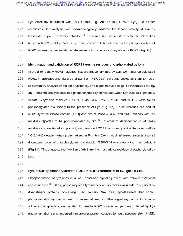
9
Lyn efficiently interacted with ROR1 (see Fig. 2b; IP ROR1, WB: Lyn). To further 221
corroborate the analysis, we pharmacologically inhibited the kinase activity of Lyn by 222
Dasatinib, a pan-Src family inhibitor 29. Dasatinib did not interfere with the interaction 223
between ROR1 and Lyn WT or Lyn KA, however, it did interfere in the phosphorylation of 224
ROR1 as seen by the substantial decrease of tyrosine phosphorylation on ROR1 (Fig. 2c). 225
226
Identification and validation of ROR1 tyrosine residues phosphorylated by Lyn 227
In order to identify ROR1 residues that are phosphorylated by Lyn, we immunoprecipitated 228
ROR1 in presence and absence of Lyn from HEK-293T cells and subjected them to mass-229
spectrometry analysis of phosphorylation(s). The experimental design is schematized in Fig. 230
3a. Proteomic analysis detected phosphorylated tyrosines only when Lyn was co-expressed. 231
In total 6 tyrosine residues - Y459, Y645, Y646, Y666, Y828, and Y836 - were found 232
phosphorylated exclusively in the presence of Lyn (Fig. 3b). Three residues are part of 233
ROR1 tyrosine kinase domain (TKD) and two of those – Y645 and Y646 overlap with the 234
residues reported to be phosphorylated by Src 20. In order to decipher which of those 235
residues are functionally important, we generated ROR1 individual point mutants as well as 236
Y645/Y646 double mutant (schematized in Fig. 3c). Even though all tested mutants showed 237
decreased levels of phosphorylation, the double Y645/Y646 was clearly the most deficient 238
(Fig 3d). This suggests that Y645 and Y646 are the most critical residues phosphorylated by 239
Lyn. 240
241
Lyn-induced phosphorylation of ROR1 induces recruitment of E3 ligase c-CBL 242
Phosphorylation at tyrosines is a well described signaling event with various functional 243
consequences 30. Often, phosphorylated tyrosines serve as molecular motifs recognized by 244
downstream proteins containing SH2 domain. We thus hypothesized that ROR1 245
phosphorylation by Lyn will lead to the recruitment of further signal regulators. In order to 246
address this question, we decided to identify ROR1 interaction partners induced by Lyn 247
phosphorylation using unbiased immunoprecipitation coupled to mass spectrometry (IP/MS). 248
.CC-BY-NC-ND 4.0 International licensewas not certified by peer review) is the author/funder. It is made available under aThe copyright holder for this preprint (whichthis version posted May 31, 2020. . https://doi.org/10.1101/2020.05.29.124156doi: bioRxiv preprint

10
We overexpressed ROR1 either alone or with WT or Lyn KD; pcDNA and Lyn WT-only 249
transfected cells served as a control. Design of the experiment is schematized in Fig. 4a. 250
IP/MS analysis identified 13 proteins that were uniquely detected as binding partners of 251
ROR1 phosphorylated by Lyn (Fig. 4b). Among the hits (Fig. 4c), c-Casitas B lineage 252
lymphoma (c-CBL) protein attracted our attention. c-CBL is an E3 ligase that recognizes pY 253
motifs31 and often downregulates its targets by triggering them for degradation 32 or 254
endocytosis 33. Of note, it is a known binding partner of Lyn 32, as well as its substrate 34. 255
We overexpressed the combinations of c-CBL, ROR1 and Lyn plasmids in HEK-293T cells 256
and performed a set of immunoprecipitation assays. In line with the mass spectrometry data, 257
c-CBL efficiently interacted with ROR1 only when WT Lyn was present. The interaction 258
between ROR1 and c-CBL was dependent on the Lyn-mediated phosphorylation of ROR1 259
since in the presence of the Lyn KD the binding to ROR1 was reduced (Fig. 4d, IP ROR1, 260
WB V5, lanes 6 vs. 8). Lyn was a part of the complex since it was pulled down both by 261
ROR1 and c-CBL (Fig. 4d, IP ROR1 & IP V5). Of note, co-expression of c-CBL clearly 262
attenuated the phosphorylation of ROR1 by Lyn and level of active Lyn itself (Fig. 4d, IP 263
ROR1 and IP pY, WB ROR1, Lyn and pY). Altogether, this data opens the possibility that the 264
consequence of phosphorylation-induced recruitment of c-CBL is the inactivation of the 265
phosphorylated ROR1, similar to a described c-CBL function in other RTKs targeted by c-266
CBL 31. 267
268
Lyn KO cells display increased surface levels of ROR1 269
Our findings reported in Figs. 1 – 4 showed that Lyn can efficiently phosphorylate ROR1 that 270
can be subsequently recognized by c-CBL. ROR1 and Lyn are important regulators of 271
signaling pathways driving chronic lymphocytic leukemia (CLL) 1,8,21,35 and several 272
lymphomas, namely mantle cell lymphoma (MCL) 15. Both, Lyn and ROR1, have been 273
evaluated as therapeutic targets in these malignancies and the understanding of their mutual 274
cross talk is thus of direct therapeutic relevance. In order to find out if Lyn controls ROR1 275
biology in CLL/MCL we decided to generate Lyn-deficient Maver-1 cells. Maver-136 are of 276
.CC-BY-NC-ND 4.0 International licensewas not certified by peer review) is the author/funder. It is made available under aThe copyright holder for this preprint (whichthis version posted May 31, 2020. . https://doi.org/10.1101/2020.05.29.124156doi: bioRxiv preprint

11
MCL origin, express high levels of ROR1 and Lyn, and respond well to BCR activation 37. 277
We produced Maver-1 Lyn knockout (KO) cells using the Crispr-Cas9 system. We validated 278
four independent clones of Maver-1 Lyn KO cells by western blotting (Fig. 5a) and 279
sequencing (Supplementary Table 1). Functionally, Lyn KO Maver-1 cells are deficient in the 280
activation of BCR signaling triggered by IgM. As shown in Fig. 5b, activation of several BCR 281
components downstream of Lyn, namely Syk, PLCγ, PI3K, as well as phosphorylation of Lyn 282
substrate HS1 was dramatically reduced in Lyn KO cells. These observations are in line with 283
what is already known about the role of Lyn in the BCR signaling cascade 18. 284
After this initial characterization of the BCR signaling deficiency in Lyn KO cells we focused 285
on their ROR1 status. As shown in Fig. 5c, global ROR1 protein levels determined by 286
Western blotting were comparable in WT and Lyn KO cells, despite the fact that ROR1 287
mRNA expression, determined by qPCR, was slightly lower in the Lyn KO cells (Fig. 5d). 288
However, when we assessed cell surface ROR1 by flow cytometry we could observe a 289
consistent increase in surface ROR1 in all four clones (Fig. 5e). This suggested that 290
endogenous Lyn controls ROR1 trafficking or endocytosis to reduce ROR1 availability on the 291
surface. 292
ROR1 and its ligand Wnt-5a were shown to control CLL cell migration and chemotaxis 38,39. 293
More specifically, Wnt-5a-ROR1 axis increases basal migration and reduces chemotaxis 38. 294
Surprisingly, Lyn KO Maver-1 cells showed not only increased cell surface ROR1 (Fig. 5e) 295
but also much lower levels of CCR7 (Fig. 5f), a receptor for chemokine CCL19 and essential 296
component of CCL19-induced chemotaxis 40. Despite some variability in the individual Lyn 297
KO clones, the observed trends showing higher ROR1 and lower CCR7 were always the 298
same (Fig. 5 e/f, histograms in the bottom part of the panel). 299
To address whether the changes in ROR1 and CCR7 levels translate into the changes in the 300
migratory behavior of WT and Lyn KO Maver-1 cells we used transwell assays without 301
(basal motility) or with CCL19 in the bottom chamber (chemotaxis). Lyn KO cells had 302
significantly higher basal motility (Fig. 5g, i). This feature was preserved across individual 303
Lyn KO clones and reflected variability of the cell surface ROR1 (Fig. 5g, ii) and the cell 304
.CC-BY-NC-ND 4.0 International licensewas not certified by peer review) is the author/funder. It is made available under aThe copyright holder for this preprint (whichthis version posted May 31, 2020. . https://doi.org/10.1101/2020.05.29.124156doi: bioRxiv preprint

12
surface CCR7 (Fig. 5g, iii). On the other hand, Lyn KO cells showed reduced chemotaxis to 305
CCL19 (Fig. 5h, i) that correlated negatively with cell surface ROR1 (Fig. 5h, ii) and 306
positively with the cell surface CCR7 (Fig. 5h, iii). Altogether, the analysis of the migratory 307
properties of Lyn KO Maver-1 cells suggested that Lyn, via control of cell surface levels of 308
ROR1 and CCR7, controls the migratory modes of lymphoid cells. Specifically, it suggests 309
that Lyn controls a balance between two migration modes – Wnt-5a/ROR1-mediated basal 310
migration (attenuated by Lyn) and CCL19/CCR7-mediated chemotaxis (promoted by Lyn). 311
312
Cell surface ROR1 is upregulated during CLL migration 313
Functional experiments with Lyn KO cells indicate that cell surface ROR1 can be under a 314
dynamic control during cell migration. In order to test whether this applies also to primary 315
CLL samples we performed a set of experiments in the patient cohort specified in Fig. 6a. 316
Cells from these patients were analyzed by Western blotting by probing for phospho-Y396-317
Lyn, which is auto-phosphorylated and as such represents a good hallmark for Lyn 318
activation (Fig. 6b). p-Lyn levels positively correlated with the phosphorylation of HS1 on 319
tyrosine 397, which is a well described target of Lyn 41 (Fig. 6c). On the other hand, ROR1 320
levels assessed by Western blotting did not correlate with active Lyn (Fig. 6d). 321
Chemotactic properties of CLL cells were analyzed in transwell assays as the migratory 322
response to the chemokine CCL19. In parallel, we analyzed the surface levels of ROR1 and 323
CCR7 in the non-migratory (upper chamber) and migratory (lower chamber) CLL cells (for 324
schematics see Fig. 6e). In some cases, because of low number of migrating cells we were 325
unable to assess in parallel all the functional parameters (migration, CCR7 and ROR1 levels 326
in lower and upper chamber, pLyn levels; see Supplementary Table 2 for details) and as 327
such the number of samples in individual analyses presented below slightly varies. 328
Surprisingly, ROR1 surface expression was clearly increased in the cells that passed 329
through the transwell membrane (Fig. 6f). No such increase was observed for CCR7, which 330
is a CCL19 receptor (Fig. 6g). Interestingly, the CLL samples that upregulated ROR1 most 331
efficiently, were the least chemotactic (Fig. 6h). CCR7 showed an opposite behavior to 332
.CC-BY-NC-ND 4.0 International licensewas not certified by peer review) is the author/funder. It is made available under aThe copyright holder for this preprint (whichthis version posted May 31, 2020. . https://doi.org/10.1101/2020.05.29.124156doi: bioRxiv preprint

13
ROR1 and CCR7 levels correlated positively with the chemotaxis (Fig. 6i). This data shows 333
that even in primary CLL cells, similar to Maver-1 cells, ROR1 surface levels are dynamically 334
regulated during cell migration and that ROR1 and CCR7 show opposite behavior, in this 335
respect. 336
It remained to be analyzed whether the balance between ROR1-driven and CCR7-controlled 337
migration can be correlated with the activity of Lyn in primary CLL cells. pLyn-high CLL 338
samples (based on Fig. 6b), were in general more chemotactic (Fig. 6j), which is in line with 339
the positive role of Lyn in chemotaxis identified in Maver-1 cells. On the other hand, pLyn 340
levels negatively correlated with the capacity of CLL cells to upregulate ROR1 during 341
migration (Fig. 6k). Altogether, this suggests that the function of Lyn that switches between 342
ROR1- and CCR7-mediated migratory modes, uncovered in Maver-1 cells, is conserved 343
also in primary CLL cells. 344
345
Discussion 346
A significant amount of research and preclinical development is being conducted on 347
developing monoclonal antibodies to target surface ROR1 in CLL and other malignancies 348
11,42,43. Also, a considerable body of work has helped us to understand signaling on the 349
extracellular side of ROR1 via Wnt5a in CLL cells 39,44. However, significant gaps of 350
knowledge remain in our understanding of the importance of the intracellular domains of 351
ROR1, especially the TKD, as well as of the regulation of ROR1 levels on the cell surface. 352
Our study is the first to show that the Src family kinase Lyn, an important component of BCR 353
signaling, phosphorylates ROR1 intracellularly and controls its surface levels. 354
The phosphorylation of ROR1 by Lyn identifies a novel crosstalk between ROR1 and 355
BCR signaling. This crosstalk can be of particular importance in CLL and MCL where both, 356
ROR1 and BCR pathways, represent therapeutic targets. It has been shown earlier that in 357
these malignancies the non-canonical Wnt pathway and BCR signaling can be targeted in a 358
combinatorial manner. Namely, it has been observed in vivo in the mouse models of CLL 359
where BTK inhibitor ibrutinib and anti-ROR1 antibody45 or casein kinase 1 (CK1) inhibitors 46 360
.CC-BY-NC-ND 4.0 International licensewas not certified by peer review) is the author/funder. It is made available under aThe copyright holder for this preprint (whichthis version posted May 31, 2020. . https://doi.org/10.1101/2020.05.29.124156doi: bioRxiv preprint

14
showed synergistic effects. Similar behavior has been observed by Karvonen and 361
colleagues in the in vitro model of MCL 15. Our data suggest that active BCR (correlating 362
with high Lyn activity) negatively controls the surface ROR1. This is an interesting 363
observation in the context of the recent report showing that in MCL ROR1/CD19 membrane 364
complex can functionally compensate for BCR/BTK activity and activate pro-survival and 365
pro-proliferative PI3K-Akt and MEK-Erk cascades 17. Lyn action towards ROR1 can explain 366
how BCR inhibited cells with low Lyn activation “switch” to the survival mode dependent on 367
ROR1. In addition, study by Zhang et al. opens the possibility that ROR1 and BCR-centered 368
complexes in MCL and CLL share even more components than Lyn described in this study. 369
Our analysis of Lyn KO Maver-1 cells has identified different migratory properties in 370
comparison to WT cells. Lyn KO cells had higher motility in the absence of external stimuli, a 371
feature correlating with the increased cell surface ROR1, but failed to respond to 372
physiological chemotactic stimulus CCL19, a feature correlating with the decrease in CCR7, 373
a CCL19 receptor. Of interest, similar behavior – i.e. deregulated motility and decreased 374
chemotaxis - was described for aggressive CLL characterized by high Wnt5a expression 38. 375
This opens the possibility that basal motility, promoted by Wnt5a/ROR1, and chemotaxis 376
represent distinct migratory modes that are coordinated by Lyn activity. In vivo phenotype of 377
Lyn KO leukemic lymphocytes in the TCL1 mouse model, which were more efficient in the 378
spleen infiltration than wt CLL cells 35 supports this view. 379
We demonstrate that at least one consequence of Lyn-induced ROR1 380
phosphorylation is the recruitment of c-Cbl. c-Cbl, a member of a family of RING finger E3 381
ligases, has been shown to be upregulated in CLL 47. Out of three 3 different family 382
members - Cbl (a.k.a c-Cbl or RNF55), Cbl-b (RNF56) and Cbl-c (RNF57), Cbl and Cbl-b 383
are known to be highly expressed in B and T lymphocytes. It is tempting to speculate that 384
Lyn activity towards ROR1-induced migration is not limited to the regulation of the interaction 385
with c-CBL but includes also action towards other cytoskeletal modulators of migration such 386
as HS1 and cortactin. Both these proteins serve in CLL cells as substrates of Lyn 41,48 and at 387
the same time were found to dynamically interact with ROR1 and control ROR1-induced 388
.CC-BY-NC-ND 4.0 International licensewas not certified by peer review) is the author/funder. It is made available under aThe copyright holder for this preprint (whichthis version posted May 31, 2020. . https://doi.org/10.1101/2020.05.29.124156doi: bioRxiv preprint

15
migration 44,49. HS1-deficient leukemic cells in the mouse model of CLL are more aggressive 389
compared to Lyn wt mainly due to preferential homing to bone marrow 50 – the molecular 390
mechanism is not known but loss of Lyn capacity to control Wnt5a-driven migration is one 391
possible explanation. 392
In addition to Lyn, ROR1 has been shown to get phosphorylated also by other TK’s, 393
namely two receptor TK (RTK)s - Met and Src20, and MuSK51. In the study by Gentile et al 20, 394
it was shown that ROR1 is first phosphorylated by Met kinase in its PRD and this helps 395
recruit Src which then leads to the phosphorylation of ROR1 in the kinase domain. It remains 396
to be tested whether some membrane associated TKs, such as Axl 52 or ZAP70 53 can 397
synergize with Lyn in the regulation of ROR1. 398
In summary, our study is the first to show the interaction between ROR1, important 399
BCR kinase Lyn and c-Cbl. Our work also provides a molecular mechanism of the crosstalk 400
for two signaling pathways essential for CLL: BCR signaling and the non-canonical Wnt 401
pathway. This crosstalk mechanism provides a basis for the rational combinational therapies 402
targeting BCR and non-canonical Wnt in CLL and MCL. 403
404
Acknowledgement 405
VB, PK and ZZ gratefully acknowledge the support of the Czech Science Foundation 406
(the projects GA17-09525S, 17-16680S, GA19-20123S). ZD was supported by the 407
European Union Grant FP7 Marie Curie ITN 608180 ‘Wntsapp’. CIISB (LM2018127) and 408
NCMG (LM2015091) research infrastructures funded by MEYS CR are acknowledged for 409
the financial support of the measurements at the Proteomics and Genomics Core Facilities. 410
Further supported by Ministry of Education, Youth and Sports of the Czech Republic 411
(National Program of Sustainability II projects LQ1605 and LQ1601), by the Ministry of 412
Health of the Czech Republic (FNBr 65269705) and by European Structural and Investment 413
Funds, Operational Programme Research, Development and Education – "Preclinical 414
Progression of New Organic Compounds with Targeted Biological Activity” (PreclinProgress; 415
CZ.02.1.01/0.0/0.0/16_025/0007381). 416
.CC-BY-NC-ND 4.0 International licensewas not certified by peer review) is the author/funder. It is made available under aThe copyright holder for this preprint (whichthis version posted May 31, 2020. . https://doi.org/10.1101/2020.05.29.124156doi: bioRxiv preprint

16
417
Conflict of interest 418
Authors declare that they have no conflict of interest. 419
420
.CC-BY-NC-ND 4.0 International licensewas not certified by peer review) is the author/funder. It is made available under aThe copyright holder for this preprint (whichthis version posted May 31, 2020. . https://doi.org/10.1101/2020.05.29.124156doi: bioRxiv preprint
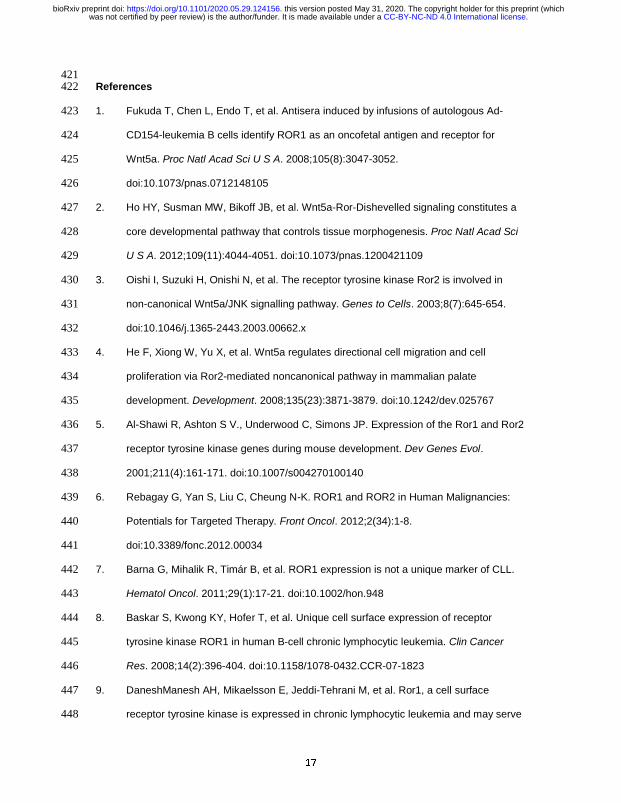
17
421 References 422
1. Fukuda T, Chen L, Endo T, et al. Antisera induced by infusions of autologous Ad-423
CD154-leukemia B cells identify ROR1 as an oncofetal antigen and receptor for 424
Wnt5a. Proc Natl Acad Sci U S A. 2008;105(8):3047-3052. 425
doi:10.1073/pnas.0712148105 426
2. Ho HY, Susman MW, Bikoff JB, et al. Wnt5a-Ror-Dishevelled signaling constitutes a 427
core developmental pathway that controls tissue morphogenesis. Proc Natl Acad Sci 428
U S A. 2012;109(11):4044-4051. doi:10.1073/pnas.1200421109 429
3. Oishi I, Suzuki H, Onishi N, et al. The receptor tyrosine kinase Ror2 is involved in 430
non-canonical Wnt5a/JNK signalling pathway. Genes to Cells. 2003;8(7):645-654. 431
doi:10.1046/j.1365-2443.2003.00662.x 432
4. He F, Xiong W, Yu X, et al. Wnt5a regulates directional cell migration and cell 433
proliferation via Ror2-mediated noncanonical pathway in mammalian palate 434
development. Development. 2008;135(23):3871-3879. doi:10.1242/dev.025767 435
5. Al-Shawi R, Ashton S V., Underwood C, Simons JP. Expression of the Ror1 and Ror2 436
receptor tyrosine kinase genes during mouse development. Dev Genes Evol. 437
2001;211(4):161-171. doi:10.1007/s004270100140 438
6. Rebagay G, Yan S, Liu C, Cheung N-K. ROR1 and ROR2 in Human Malignancies: 439
Potentials for Targeted Therapy. Front Oncol. 2012;2(34):1-8. 440
doi:10.3389/fonc.2012.00034 441
7. Barna G, Mihalik R, Timár B, et al. ROR1 expression is not a unique marker of CLL. 442
Hematol Oncol. 2011;29(1):17-21. doi:10.1002/hon.948 443
8. Baskar S, Kwong KY, Hofer T, et al. Unique cell surface expression of receptor 444
tyrosine kinase ROR1 in human B-cell chronic lymphocytic leukemia. Clin Cancer 445
Res. 2008;14(2):396-404. doi:10.1158/1078-0432.CCR-07-1823 446
9. DaneshManesh AH, Mikaelsson E, Jeddi-Tehrani M, et al. Ror1, a cell surface 447
receptor tyrosine kinase is expressed in chronic lymphocytic leukemia and may serve 448
.CC-BY-NC-ND 4.0 International licensewas not certified by peer review) is the author/funder. It is made available under aThe copyright holder for this preprint (whichthis version posted May 31, 2020. . https://doi.org/10.1101/2020.05.29.124156doi: bioRxiv preprint

18
as a putative target for therapy. Int J Cancer. 2008;123(5):1190-1195. 449
doi:10.1002/ijc.23587 450
10. Hallek M. Chronic lymphocytic leukemia: 2020 update on diagnosis, risk stratification 451
and treatment. Am J Hematol. 2019;94(11):1266-1287. doi:10.1002/ajh.25595 452
11. Choi MY, Widhopf GF, Wu CCN, et al. Pre-clinical Specificity and Safety of UC-961, a 453
First-In-Class Monoclonal Antibody Targeting ROR1. Clin Lymphoma, Myeloma Leuk. 454
2015;15(S):S167-S169. doi:10.1016/j.clml.2015.02.010 455
12. Hudecek M, Schmitt TM, Baskar S, et al. The B-cell tumor-associated antigen ROR1 456
can be targeted with T cells modified to express a ROR1-specific chimeric antigen 457
receptor. Blood. 2010;116(22):4532-4541. doi:10.1182/blood-2010-05-283309 458
13. ten Hacken E, Gounari M, Ghia P, Burger JA. The importance of B cell receptor 459
isotypes and stereotypes in chronic lymphocytic leukemia. Leukemia. 2019;33(2):287-460
298. doi:10.1038/s41375-018-0303-x 461
14. Burger JA, Chiorazzi N. B cell receptor signaling in chronic lymphocytic leukemia. 462
Trends Immunol. 2013;34(12):592-601. doi:10.1016/j.it.2013.07.002 463
15. Karvonen H, Chiron D, Niininen W, et al. Crosstalk between ROR1 and BCR 464
pathways defines novel treatment strategies in mantle cell lymphoma. Blood Adv. 465
2017;1(24):2257-2268. doi:10.1182/bloodadvances.2017010215 466
16. Karvonen H, Niininen W, Murumägi A, Ungureanu D. Targeting ROR1 identifies new 467
treatment strategies in hematological cancers. Biochem Soc Trans. 2017;45:457-464. 468
doi:10.1042/BST20160272 469
17. Zhang Q, Wang HY, Liu X, et al. ROR1/CD19 Receptor Complex Promotes Growth of 470
Mantle Cell Lymphoma Cells Independently of the B Cell Receptor–BTK Signaling 471
Pathway. J Immunol. 2019;203(8):2043-2048. doi:10.4049/jimmunol.1801327 472
18. Xu Y, Harder KW, Huntington ND, Hibbs ML, Tarlinton DM. Lyn Tyrosine Kinase: 473
Accentuating the Positive and the Negative. Immunity. 2005;22(1):9-18. 474
doi:10.1016/J.IMMUNI.2004.12.004 475
19. Akbarzadeh S, Wheldon LM, Sweet SMM, Talma S, Mardakheh FK, Heath JK. The 476
.CC-BY-NC-ND 4.0 International licensewas not certified by peer review) is the author/funder. It is made available under aThe copyright holder for this preprint (whichthis version posted May 31, 2020. . https://doi.org/10.1101/2020.05.29.124156doi: bioRxiv preprint

19
deleted in Brachydactyly B domain of ROR2 is required for receptor activation by 477
recruitment of Src. PLoS One. 2008;3(3):e1873. doi:10.1371/journal.pone.0001873 478
20. Gentile A, Lazzari L, Benvenuti S, Trusolino L, Comoglio PM. The ROR1 479
pseudokinase diversifies signaling outputs in MET-addicted cancer cells. Int J Cancer. 480
2014;135(10):2305-2316. doi:10.1002/ijc.28879 481
21. Contri A, Brunati AM, Trentin L, et al. Chronic lymphocytic leukemia B cells contain 482
anomalous Lyn tyrosine kinase, a putative contribution to defective apoptosis. J Clin 483
Invest. 2005;115(2):369-378. doi:10.1172/JCI22094 484
22. Kaucka M, Plevova K, Pavlova S, et al. The planar cell polarity pathway drives 485
pathogenesis of chronic lymphocytic leukemia by the regulation of b-lymphocyte 486
migration. Cancer Res. 2013;73(5):1491-1501. doi:10.1158/0008-5472.CAN-12-1752 487
23. Kasahara K, Nakayama Y, Ikeda K, et al. Trafficking of Lyn through the Golgi caveolin 488
involves the charged residues on alphaE and alphaI helices in the kinase domain. J 489
Cell Biol. 2004;165(5):641-652. doi:10.1083/jcb.200403011 490
24. Pavlova S, Smardova J, Tom N, Trbusek M. Detection and Functional Analysis of 491
TP53 Mutations in CLL. In: Methods in Molecular Biology. Vol 1881. Humana Press 492
Inc.; 2019:63-81. doi:10.1007/978-1-4939-8876-1_6 493
25. Livak KJ, Schmittgen TD. Analysis of Relative Gene Expression Data Using Real-494
Time Quantitative PCR and the 2 C T Method. METHODS. 2001;25:402-408. 495
doi:10.1006/meth.2001.1262 496
26. Yamaguchi T, Yanagisawa K, Sugiyama R, et al. NKX2-1/TITF1/TTF-1-Induced 497
ROR1 Is Required to Sustain EGFR Survival Signaling in Lung Adenocarcinoma. 498
Cancer Cell. 2012;21(3):348-361. doi:10.1016/j.ccr.2012.02.008 499
27. Brown MT, Cooper JA. Regulation, substrates and functions of src. Biochim Biophys 500
Acta - Rev Cancer. 1996;1287(2-3):121-149. doi:10.1016/0304-419X(96)00003-0 501
28. Xu W, Doshi A, Lei M, Eck MJ, Harrison SC. Crystal structures of c-Src reveal 502
features of its autoinhibitory mechanism. Mol Cell. 1999;3(5):629-638. 503
doi:10.1016/S1097-2765(00)80356-1 504
.CC-BY-NC-ND 4.0 International licensewas not certified by peer review) is the author/funder. It is made available under aThe copyright holder for this preprint (whichthis version posted May 31, 2020. . https://doi.org/10.1101/2020.05.29.124156doi: bioRxiv preprint

20
29. Das J, Chen P, Norris D, et al. 2-Aminothiazole as a novel kinase inhibitor template. 505
Structure-activity relationship studies toward the discovery of N-(2-chloro-6-506
methylphenyl)-2-[[6- [4-(2-hydroxyethyl)-1-piperazinyl]-2-methyl-4-pyrimidinyl]amino]-507
1, 3-thiazole-5-carboxamide (Dasatinib, BMS - 354825) as a potent pan-src kinase 508
inhibitor. J Med Chem. 2006;49(23):6819-6832. doi:10.1021/jm060727j 509
30. Hunter T. The Genesis of Tyrosine Phosphorylation. Cold Spring Harb Perspect Biol. 510
2014;6(5):a020644-a020644. doi:10.1101/cshperspect.a020644 511
31. Mohapatra B, Ahmad G, Nadeau S, et al. Protein tyrosine kinase regulation by 512
ubiquitination: Critical roles of Cbl-family ubiquitin ligases. Biochim Biophys Acta - Mol 513
Cell Res. 2013;1833(1):122-139. doi:10.1016/J.BBAMCR.2012.10.010 514
32. Kaabeche K, Lemonnier J, Le Mée S, Caverzasio J, Marie PJ. Cbl-mediated 515
degradation of Lyn and Fyn induced by constitutive fibroblast growth factor receptor-2 516
activation supports osteoblast differentiation. J Biol Chem. 2004;279(35):36259-517
36267. doi:10.1074/jbc.M402469200 518
33. Petrelli A, Gilestro GF, Lanzardo S, Comoglio PM, Migone N, Giordano S. The 519
endophilin-CIN85-Cbl complex mediates ligand-dependent downregulation of c-Met. 520
Nature. 2002;416(6877):187-190. doi:10.1038/416187a 521
34. Tohru Tezuka B, Umemori H, Fusaki N, Takata M, Kurosakifl T. Physical and 522
Functional Association of the cbl Protooncogene Product with an Src-Family Protein 523
Tyrosine Kinase, p53/56 lyn, in the B Cell Antigen Receptor-mediated Signaling. J 524
Exp Med. 1996;183:675-680. 525
35. Nguyen P-H, Fedorchenko O, Rosen N, et al. LYN Kinase in the Tumor 526
Microenvironment Is Essential for the Progression of Chronic Lymphocytic Leukemia. 527
Cancer Cell. 2016;30(4):610-622. doi:10.1016/j.ccell.2016.09.007 528
36. Zamò A, Ott G, Katzenberger T, et al. Establishment of the MAVER-1 cell line, a 529
model for leukemic and aggressive mantle cell lymphoma. Haematologica. 530
2006;91(1):40-47. http://www.ncbi.nlm.nih.gov/pubmed/16434369. Accessed 531
September 24, 2018. 532
.CC-BY-NC-ND 4.0 International licensewas not certified by peer review) is the author/funder. It is made available under aThe copyright holder for this preprint (whichthis version posted May 31, 2020. . https://doi.org/10.1101/2020.05.29.124156doi: bioRxiv preprint

21
37. Rauert-Wunderlich H, Rudelius M, Ott G, Rosenwald A. Targeting protein kinase C in 533
mantle cell lymphoma. Br J Haematol. 2016;173(3):394-403. doi:10.1111/bjh.13973 534
38. Janovska P, Poppova L, Plevova K, et al. Autocrine Signaling by Wnt-5a Deregulates 535
Chemotaxis of Leukemic Cells and Predicts Clinical Outcome in Chronic Lymphocytic 536
Leukemia. Clin Cancer Res. 2016;22(2):459-469. doi:10.1158/1078-0432.CCR-15-537
0154 538
39. Yu J, Chen L, Chen Y, et al. Wnt5a induces ROR1 to associate with 14-3-3ζ for 539
enhanced chemotaxis and proliferation of chronic lymphocytic leukemia cells. 540
Leukemia. 2017;31(12):2608-2614. doi:10.1038/leu.2017.132 541
40. Kobayashi D, Endo M, Ochi H, Hojo H, Miyasaka M, Hayasaka H. Regulation of 542
CCR7-dependent cell migration through CCR7 homodimer formation. Sci Rep. 543
2017;7(1):8536. doi:10.1038/s41598-017-09113-4 544
41. Ten Hacken E, Scielzo C, Bertilaccio MTS, et al. Targeting the LYN/HS1 signaling 545
axis in chronic lymphocytic leukemia. Blood. 2013;121(12):2264-2273. 546
doi:10.1182/blood 547
42. Baskar S, Wiestner A, Wilson WH, Pastan I, Rader C. Targeting malignant B cells 548
with an immunotoxin against ROR1. MAbs. 2012;4(3):349-361. 549
doi:10.4161/mabs.19870 550
43. Qi J, Li X, Peng H, et al. Potent and selective antitumor activity of a T cell-engaging 551
bispecific antibody targeting a membrane-proximal epitope of ROR1. Proc Natl Acad 552
Sci. 2018;115(24):E5467-E5476. doi:10.1073/pnas.1719905115 553
44. Hasan M, Yu J, Chen L, et al. Wnt5a induces ROR1 to complex with HS1 to enhance 554
migration of chronic lymphocytic leukemia cells. Nat Publ Gr. 2017;31:2615-2622. 555
doi:10.1038/leu.2017.133 556
45. Yu J, Chen L, Cui B, et al. Cirmtuzumab inhibits Wnt5a-induced Rac1 activation in 557
chronic lymphocytic leukemia treated with ibrutinib. Leukemia. 2017;31(6):1333-1339. 558
doi:10.1038/leu.2016.368 559
46. Janovska P, Verner J, Kohoutek J, et al. Casein kinase 1 is a therapeutic target in 560
.CC-BY-NC-ND 4.0 International licensewas not certified by peer review) is the author/funder. It is made available under aThe copyright holder for this preprint (whichthis version posted May 31, 2020. . https://doi.org/10.1101/2020.05.29.124156doi: bioRxiv preprint

22
chronic lymphocytic leukemia. Blood. 2018;131(11):1206-1218. doi:10.1182/blood-561
2017-05-786947 562
47. Mankai A., Eveillard J-R, Buhe V, et al. Is the c-Cbl Proto-Oncogene Involved in 563
Chronic Lymphocytic Leukemia? Ann N Y Acad Sci. 2007;1107(1):193-205. 564
doi:10.1196/annals.1381.021 565
48. Martini V, Gattazzo C, Frezzato F, et al. Cortactin, a Lyn substrate, is a checkpoint 566
molecule at the intersection of BCR and CXCR4 signalling pathway in chronic 567
lymphocytic leukaemia cells. Br J Haematol. 2017;178(1):81-93. 568
doi:10.1111/bjh.14642 569
49. Hasan MK, Rassenti L, Widhopf GF, Yu J, Kipps TJ. Wnt5a causes ROR1 to complex 570
and activate cortactin to enhance migration of chronic lymphocytic leukemia cells. 571
Leukemia. 2019;33(3):653-661. doi:10.1038/s41375-018-0306-7 572
50. Scielzo C, Bertilaccio MTS, Simonetti G, et al. HS1 has a central role in the trafficking 573
and homing of leukemic B cells. Blood. 2010;116(18):3537-3546. doi:10.1182/blood-574
2009-12-258814 575
51. Karvonen H, Summala K, Niininen W, Barker HR, Ungureanu D. Interaction between 576
ROR1 and MuSK activation complex in myogenic cells. FEBS Lett. 2018;592(3):434-577
445. doi:10.1002/1873-3468.12966 578
52. Ghosh AK, Secreto C, Boysen J, et al. The novel receptor tyrosine kinase Axl is 579
constitutively active in B-cell chronic lymphocytic leukemia and acts as a docking site 580
of nonreceptor kinases: implications for therapy. Blood. 2011;117(6):1928-1937. 581
doi:10.1182/blood-2010-09-305649 582
53. Dürig J, Nückel H, Cremer M, et al. ZAP-70 expression is a prognostic factor in 583
chronic lymphocytic leukemia. Leukemia. 2003;17(12):2426-2434. 584
doi:10.1038/sj.leu.2403147 585
586
.CC-BY-NC-ND 4.0 International licensewas not certified by peer review) is the author/funder. It is made available under aThe copyright holder for this preprint (whichthis version posted May 31, 2020. . https://doi.org/10.1101/2020.05.29.124156doi: bioRxiv preprint

23
587
Figure legends 588
Figure 1: Lyn interacts with ROR1. a, b) Lyn and ROR1 were overexpressed in HEK-293T 589
cells. co-IP and Western blot analysis showing a pull-down of ROR1 when the lysates were 590
immunoprecipitated with Lyn (a) and a pull-down of Lyn when the lysates were 591
immunoprecipitated with ROR1 (b). c) Representative images of immuno-cytochemistry 592
analysis of HEK-293T cells overexpressing ROR1 (green) and Lyn (red) in the indicated 593
combinations. Co-localization of ROR1 and Lyn is observed at the membrane. Scale bar 7.5 594
μm. d) Scheme of ROR1 mutants used for domain mapping. e) Lyn was co-expressed with 595
the ROR1 intracellular deletion mutants in WT HEK-293T cells. Immunoprecipitation was 596
done using ROR1 as the bait. WB – Western blotting, IP – immunoprecipitation, TCL – total 597
cell lysate. Results in all panels are representative of at least 3 biological replicates. 598
599
Figure 2: Lyn phosphorylates ROR1. Indicated combinations of Lyn and ROR1 plasmids 600
were overexpressed in HEK-293T cells. ROR1 was immunoprecipitated and the binding of 601
Lyn and Y phosphorylation was assessed by Western blotting. a) General scheme of the Lyn 602
mutants used in b/c. b) Only the Lyn mutants with the intact kinase activity were able to 603
phosphorylate ROR1 on its tyrosine residues. c) Small molecule inhibitor of Lyn, Dasatinib 604
(Das, 0.2 µM), did not affect the interaction of ROR1 and Lyn, however it did block the ability 605
of Lyn to phosphorylate ROR1. WB – Western blotting, IP – immunoprecipitation, TCL – total 606
cell lysate. Results in all panels are representative of at least 3 biological replicates. 607
608
Figure 3: Mapping of the ROR1 residues phosphorylated by Lyn 609
a) Scheme of the experimental set-up for mass-spec analysis of ROR1 phosphorylation. 610
Indicated combinations were transfected in HEK-293T cells. ROR1 was immunoprecipitated, 611
separated on SDS-PAGE and bands corresponding to ROR1 were analyzed by MS/MS. b) 612
ROR1 tyrosine (Y) residues that were found phosphorylated only when Lyn was 613
coexpressed as identified by MS/MS analysis. The phospho-peptide signal intensity (up) and 614
.CC-BY-NC-ND 4.0 International licensewas not certified by peer review) is the author/funder. It is made available under aThe copyright holder for this preprint (whichthis version posted May 31, 2020. . https://doi.org/10.1101/2020.05.29.124156doi: bioRxiv preprint

24
the position (bottom) of each detected phospho-tyrosine is presented. c) Schematics of the 615
point mutants of ROR1 made for the validation experiments. d) Phosphorylation analysis of 616
the point mutants showed that Y645/646F ROR1 mutation almost completely eliminated the 617
phosphorylation by Lyn. WB – Western blotting, IP – immunoprecipitation, TCL – total cell 618
lysate. Results in d are representative of at least 3 biological replicates. 619
620
Figure 4: Lyn induced phosphorylation of ROR1 triggers interaction with the E3 ligase 621
c-CBL 622
a) Scheme of the experimental set-up for the analysis of ROR1 interacting partners by 623
MS/MS. ROR1 and Lyn WT and Lyn KO were overexpressed in HEK-293T. ROR1 was 624
immunoprecipitated and the protein composition of the pulldown was analyzed by MS/MS. 625
b) Upset plot demonstrating the numbers of proteins identified as ROR1 interactors in 626
ROR1, ROR1+WT Lyn and ROR1+Lyn kinase dead (KD) conditions. Only proteins absent in 627
the control pulldowns (pcDNA and Lyn expression) were considered. c) List of ROR1 628
interactors identified only when phosphorylated by Lyn. d) Analysis of the interactions and 629
phosphorylation status of ROR1 and c-CBL. Indicated combinations were overexpressed in 630
HEK-293T, immunoprecipitated (IP) as indicated and subsequently analysed by WB. Lyn 631
promotes the interaction of ROR1 with c-Cbl. WB – Western blotting, IP – 632
immunoprecipitation, TCL – total cell lysate. Results in d are representative of at least 3 633
biological replicates. 634
635
Figure 5: Lyn KO cells display increased surface levels of ROR1 636
Lyn gene in Maver-1 cells was targeted by Crispr/Cas9. Parental WT and four clones of Lyn 637
KO cells coded #1E3, #1C2, #1E10, and #2F5 were analyzed functionally. a) Western blot 638
analysis of expression of Lyn in wt and KO cells. b) Activation of BCR signaling after 639
incubation with IgM antibody in WT and Lyn KO (#1E3) cells was assessed by Western 640
blotting of pPLCγ, pSYK, pLyn, pHS1, pPI3K and Lyn. GAPDH was used as a loading 641
control. c) Expression of ROR1 protein in WT and Lyn KO clones was analyzed by Western 642
.CC-BY-NC-ND 4.0 International licensewas not certified by peer review) is the author/funder. It is made available under aThe copyright holder for this preprint (whichthis version posted May 31, 2020. . https://doi.org/10.1101/2020.05.29.124156doi: bioRxiv preprint

25
blotting. β-actin was used as a loading control. Representative blots from 3 independent 643
experiments. d) Expression of ROR1 mRNA in WT and Lyn KO cells. Graphs show mean ± 644
SD of 3 independent experiments. Differences were analyzed by Kruskal-Wallis test (Dunn’s 645
multiple comparison test). e, f) Basal surface expression of ROR1 (e) and CCR7 (f) in 646
Maver-1 WT and individual clones of Lyn KO cells was analyzed by flow cytometry. Mean ± 647
SD from 3 independent experiments and Kruskal-Wallis test (Dunn’s multiple comparison 648
test). Representative histograms for all analyzed clones are shown. g, h) Basal motility (g) 649
or chemotaxis towards CCL19 (h) of Maver wt and Lyn KO cells was analyzed in a transwell 650
assay. Number of cells that migrated to lower chamber of transwell plate after 3 hours of 651
incubation is indicated. Panel i shows mean ± SD from 6 independent experiments for WT 652
and KO clone #1E3, Mann-Whitney nonparametric test. Other panels show variability among 653
4 different clones; migratory properties are plotted with the surface expression of ROR1 654
(panel ii) and CCR7 (panel iii) in Maver-1 WT and Lyn KO cells. 655
656
Figure 6: Correlation of Lyn activity with the chemotactic properties of primary CLL 657
cells 658
a) Table with basic clinical characteristics of the patient cohort used for functional analysis of 659
the primary CLL cells. Rai stage at the time of sampling, IGHV status and cytogenetic 660
analysis are indicated (n=13). b) Protein levels of ROR1, pLyn, Lyn and pHS1 expression in 661
the panel of CLL primary cells was analyzed by Western blotting. c, d) Correlation of pLyn 662
expression with pHS1 (c) and ROR1 (d). e) Scheme of the transwell assay indicating the 663
upper and lower compartment that was used for the separate analysis of the surface 664
markers. f,g) Surface expression of ROR1 (f) and CCR7 (g) in the upper (●) and lower (●) 665
compartments of transwell chamber in the panel of primary CLL cells was analyzed by flow-666
cytometry. Wilcoxon matched pairs signed test (n=9). h, i) Correlation of the change in the 667
surface expression of ROR1 (h) and CCR7 (i) during migration represented as the ratio of 668
receptor levels in the lower:upper compartment with the chemotactic properties of cells 669
(expressed as the number of cells in the lower chamber) in the panel of primary CLL cells. j) 670
.CC-BY-NC-ND 4.0 International licensewas not certified by peer review) is the author/funder. It is made available under aThe copyright holder for this preprint (whichthis version posted May 31, 2020. . https://doi.org/10.1101/2020.05.29.124156doi: bioRxiv preprint

26
Correlation of the chemotaxis towards CCL19 with pLyn levels (WB). k) Correlation of ROR1 671
surface levels dynamics (expressed as the ratio of ROR1 surface levels in the lower:upper 672
well of transwell) with pLyn levels (WB). h-k) Pearson correlation coefficient. 673
674
.CC-BY-NC-ND 4.0 International licensewas not certified by peer review) is the author/funder. It is made available under aThe copyright holder for this preprint (whichthis version posted May 31, 2020. . https://doi.org/10.1101/2020.05.29.124156doi: bioRxiv preprint

ROR1
Figure 1
ROR1 + - + + - +Lyn - + + - + +
TCLIP Lyn
WB: ROR1
WB: Lyn
a. b.
c.
d.
ROR1 WT
Extra cellular domain Intra cellular domain
TM domain Kinase domain
ROR1 ΔCyto
ROR1 ΔTail
e.
TCL IP ROR1
ROR1 + - + + - +Lyn - + + - + +
WB: ROR1
WB: Lyn
130
55
IP ROR1
WB: ROR1
WB: Lyn
TCL
ROR1 - + Lyn + - - - + + + + - - - + + +
kDa kDa
aa 1-937
aa 1-750
aa 1-443
Δ Cy
to
Δ Ta
il
+ Δ Cy
to
Δ Ta
il
- + Δ Cy
to
Δ Ta
il
+ Δ Cy
to
Δ Ta
il
130
55
50
13080
55
ROR1
ROR1 Lyn
Lyn
Lyn
DRAQ5
DRAQ5
DRAQ5
ROR1 + Lyn
ROR1 + Lyn
ROR1 + Lyn
kDa
ROR1
Lyn
ROR1
+ Ly
n.CC-BY-NC-ND 4.0 International licensewas not certified by peer review) is the author/funder. It is made available under a
The copyright holder for this preprint (whichthis version posted May 31, 2020. . https://doi.org/10.1101/2020.05.29.124156doi: bioRxiv preprint

SH4
a.
Figure 2
Wild type (aa 1-512)
Kinase active (aa 1-506; KA)
Kinase deleted (aa 1-298; Δ)
Kinase dead (aa 1-506; K275A; KD)
HA tag
WB: pY
WB: ROR1
Das
IP V
5
WB: Lyn
WB: ROR1
TCL
WB: Lyn
TKD
K275M
Phosphorylation
WB: Lyn
WB: ROR1
WB: pY
IP R
OR1
TCL
b.
WB: ROR1
130
55
55
130
55
130
130
130
c.
WB: Lyn
ROR1 + - - - - + + + +
Lyn - Wt Δ KA KD + Δ KA KD
55
35
35
ROR1-v5 Lyn
+ - - + + + +
- + KA + KA + KA
SH3 SH2
P
SH4 SH3 SH2
SH4 SH3 SH2
SH4 SH3 SH2
kDa
130
kDa
P
P
P
Lyn
.CC-BY-NC-ND 4.0 International licensewas not certified by peer review) is the author/funder. It is made available under aThe copyright holder for this preprint (whichthis version posted May 31, 2020. . https://doi.org/10.1101/2020.05.29.124156doi: bioRxiv preprint

a.
Figure 3
b.
d.pcDNA ROR1 ROR1 + Lyn
Transfection
Immunoprecipitation
Coommassie staining Of gel
WB: pY
WB: ROR1IP R
OR1
Mass-spec analysis
TCL
WB: ROR1
pcDNA ROR1 ROR1+Lyn
WB: pY
WB: pLyn
WB: ROR1
WB: Lyn
WB: pY
WB: pLyn
WB: ROR1
WB: Lyn
IP R
OR1
IP Ly
nWB: ROR1
WB: Lyn
TCL
55
55
130
130
130
130
55
55
ROR1Lyn - - - - - + + + + + +
130
55
+ Y641
F
Y645
F
Y646
F
Y645
/646
F
- + Y641
F
Y645
F
Y646
F
Y645
/646
F
kDa
WT ROR1 SREIYSADYYRVQSK
ROR1 Y641F SREIFSADYYRVQSK
ROR1 Y645F SREIYSADFYRVQSK
ROR1 Y646F SREIYSADYFRVQSK
ROR1 Y645/646F SREIYSADFFRVQSK
641
645
646
c.
ROR1 WT
.CC-BY-NC-ND 4.0 International licensewas not certified by peer review) is the author/funder. It is made available under aThe copyright holder for this preprint (whichthis version posted May 31, 2020. . https://doi.org/10.1101/2020.05.29.124156doi: bioRxiv preprint

a.
pcDNA ROR1 Lyn
ROR1+
Lyn
Mass-spec analysis
IP ROR1
Figure 4
b.
WB: pY
WB: V5
WB: Lyn
WB: pLyn
WB: ROR1
d.
IP V
5 WB: ROR1
WB: Lyn
WB: v5
IP p
Y
WB: c-Cbl
WB: Lyn
WB: ROR1
ROR1 + - - + + + + +
Lyn - + - + - + KD KD
c-Cbl-v5 - - + - + + - +
IP R
OR1
TCL
WB: ROR1
WB: Lyn
WB: c-Cbl
130kDa
55
120
55
130
130
55
120
130
55
120
130
55
120
ID Name Full name
Q16850 CYP51A1 Lanosterol 14-alpha demethylase
O14641 DVL2 Segment polarity protein dishevelled homolog DVL-2
Q8N4V1 MMGT1 Membrane magnesium transporter 1
Q8WVC6 DCAKD Dephospho-CoA kinase domain-containing protein
P01860 IGHG3 Immunoglobulin heavy constant gamma 3
Q9ULX6 AKAP8L A-kinase anchor protein 8-like
H3BRN7 CLN6 Ceroid-lipofuscinosisneuronal protein 6
Q9NR12 PDLIM7 PDZ and LIM domain protein 7
Q8TC12 RDH11 Retinol dehydrogenase 11
Q15392 DHCR24 Delta(24)-sterol reductase
Q9BSJ8 ESYT1 Extended synaptotagmin-1
P22681 CBL E3 ubiquitin-protein ligase CBL
P30876 POLR2B DNA-directed RNA polymerase II subunit RPB2
c.
WB: pY
WB: ROR1
WB: Lyn
WB: ROR1
WB: Lyn
IP R
OR1
TCL
ROR1+
Lyn KD
.CC-BY-NC-ND 4.0 International licensewas not certified by peer review) is the author/funder. It is made available under aThe copyright holder for this preprint (whichthis version posted May 31, 2020. . https://doi.org/10.1101/2020.05.29.124156doi: bioRxiv preprint

ROR1
wt#1
E3#1
C2#1
E10#2
F50.0
0.2
0.4
0.6
0.8
1.0
1.2 **
LYN KO
Figure 5a.
e. f.
WT
#1C2
#1E3
#1E1
0
#2F5
WB: Lyn
WB: β-ACTIN
55
c.
WB: ROR1
WB: Lyn55
130
WT
#1C2
#1E3
#1E1
0
#2F5
b.
WB: pLyn
WB: Lyn
WT Lyn KO #1E3
WB: GAPDH
WB: PPLCγ
WB: pSYK
WB: pHS1
WB: pPI3K55
kDa
155
72
80
56
56
37
g.
h.
d.
WB: β-ACTIN
kDa kDa
ii.
surfa
ce C
CR7
(fold
of w
t)
iii.i.
ii. iii.i.
cell
coun
t
wt
LYN KO #1E3
Lyn KO Lyn KO .CC-BY-NC-ND 4.0 International licensewas not certified by peer review) is the author/funder. It is made available under aThe copyright holder for this preprint (whichthis version posted May 31, 2020. . https://doi.org/10.1101/2020.05.29.124156doi: bioRxiv preprint

Figure 6
a
e.
# SexRai
stage IGHV Cytogenetics1 M III mut ∆11q2 M IV unmut ∆13q3 M II unmut normal karyotype4 F I unmut ∆13q5 M I unmut ∆13q6 M II unmut ∆13q7 M IV unmut ∆13q8 M IV unmut ∆11q9 F III unmut tris12
10 F III mut tris12
11 F IV mut ∆11q
12 F III unmut tris1213 M II mut normal karyotype
f.
i.h.
RO
R1
low
er/u
pper
(MFI
)
g.
CCR
7 lo
wer
(MFI
)
c.
k.
d.
j.
pLyn (WB)0 5 10 15
0
50000
100000
2
3
5
6
9
10
1112 13
r=0.86p=0.0029
RO
R1
low
er/u
pper
(MFI
)
12987654321 1110 13
WB: ROR1
WB: pHS1
WB: pLyn
WB: Lyn
WB: GAPDH
kDa
130
70
55
55
35
uppercompartment
lowercompartment
uppercompartment
lowercompartment
CCL19
6 h
Chemotaxis (CCL19)
MFI
ROR1 upper
ROR1 lower
0
1000
2000
30000.0078
MFI
CCR7 upper
CCR7 lower
0.9375
b. .CC-BY-NC-ND 4.0 International licensewas not certified by peer review) is the author/funder. It is made available under aThe copyright holder for this preprint (whichthis version posted May 31, 2020. . https://doi.org/10.1101/2020.05.29.124156doi: bioRxiv preprint

II

ORIGINAL RESEARCHpublished: 04 May 2017
doi: 10.3389/fcell.2017.00047
Frontiers in Cell and Developmental Biology | www.frontiersin.org 1 May 2017 | Volume 5 | Article 47
Edited by:
Gregory Kelly,
University of Western Ontario, Canada
Reviewed by:
Terry Van Raay,
University of Guelph, Canada
Ole-Morten Seternes,
University of Tromsø, Norway
*Correspondence:
Sigmar Stricker
Vitezslav Bryja
Specialty section:
This article was submitted to
Signaling,
a section of the journal
Frontiers in Cell and Developmental
Biology
Received: 30 January 2017
Accepted: 13 April 2017
Published: 04 May 2017
Citation:
Bernatik O, Radaszkiewicz T, Behal M,
Dave Z, Witte F, Mahl A,
Cernohorsky NH, Krejci P, Stricker S
and Bryja V (2017) A Novel Role for
the BMP Antagonist Noggin in
Sensitizing Cells to
Non-canonical Wnt-5a/Ror2/
Disheveled Pathway Activation.
Front. Cell Dev. Biol. 5:47.
doi: 10.3389/fcell.2017.00047
A Novel Role for the BMP AntagonistNoggin in Sensitizing Cells toNon-canonical Wnt-5a/Ror2/Disheveled Pathway ActivationOndrej Bernatik 1, Tomasz Radaszkiewicz 1, Martin Behal 1, Zankruti Dave 1, Florian Witte 2,
Annika Mahl 2, Nicole H. Cernohorsky 3, Pavel Krejci 1, 3, Sigmar Stricker 2* and
Vitezslav Bryja 1, 4*
1 Faculty of Sciences, Institute of Experimental Biology, Masaryk University, Brno, Czechia, 2 Institute for Chemistry and
Biochemistry, Freie Universität Berlin, Berlin, Germany, 3Department of Biology, Faculty of Medicine, Masaryk University,
Brno, Czechia, 4Department of Cytokinetics, Institute of Biophysics AS CR, v.v.i., Brno, Czechia
Mammalian limb development is driven by the integrative input from several signaling
pathways; a failure to receive or a misinterpretation of these signals results in skeletal
defects. The brachydactylies, a group of overlapping inherited human hand malformation
syndromes, are mainly caused by mutations in BMP signaling pathway components.
Two closely related forms, Brachydactyly type B2 (BDB2) and BDB1 are caused by
mutations in the BMP antagonist Noggin (NOG) and the atypical receptor tyrosine kinase
ROR2 that acts as a receptor in the non-canonical Wnt pathway. Genetic analysis
of Nog and Ror2 functional interaction via crossing Noggin and Ror2 mutant mice
revealed a widening of skeletal elements in compound but not in any of the single
mutants, thus indicating genetic interaction. Since ROR2 is a non-canonical Wnt co-
receptor specific for Wnt-5a we speculated that this phenotype might be a result of
deregulated Wnt-5a signaling activation, which is known to be essential for limb skeletal
elements growth and patterning. We show that Noggin potentiates activation of the
Wnt-5a-Ror2-Disheveled (Dvl) pathway in mouse embryonic fibroblast (MEF) cells in a
Ror2-dependent fashion. Rat chondrosarcoma chondrocytes (RCS), however, are not
able to respond to Noggin in this fashion unless growth arrest is induced by FGF2. In
summary, our data demonstrate genetic interaction between Noggin and Ror2 and show
that Noggin can sensitize cells to Wnt-5a/Ror2-mediated non-canonical Wnt signaling,
a feature that in cartilage may depend on the presence of active FGF signaling. These
findings indicate an unappreciated function of Noggin that will help to understand BMP
and Wnt/PCP signaling pathway interactions.
Keywords: noggin, Wnt5a, non-canonical Wnt pathways, BMP signaling, brachydactyly, Ror2

Bernatik et al. Crosstalk of Noggin and Non-canonical Wnt Pathway
INTRODUCTION
Limb bud development and the concomitant formation oflimb skeletal structures are regulated by the intricate interplayand integration of various signaling pathways, with majorroles played by the Shh, BMP, FGF, and Wnt/β-cateninpathways (reviewed for example in, Robert, 2007; Zuniga,2015). The BMP signaling pathway is of pivotal importanceespecially for skeletal development. The analysis of inheritablehuman hand malformation syndromes has been instrumentalin understanding the contribution of BMP signaling andother pathways for skeletal development. One example arethe brachydactylies, a group of inheritable syndromes thatare characterized by shortening or absence of phalanges.Most brachydactyly subtypes are caused by mutations inBMP signaling components or factors that, at different levels,intersect with BMP signaling. Therefore brachydactylies havebeen interpreted in terms of a molecular disease family (Strickerand Mundlos, 2011). This hypothesis predicts that overlappingphenotypes are likely caused by mutations affecting componentsthat show a close functional interaction within a commonsignaling network.
Intriguingly, two closely related brachydactyly subtypes,BDB1 and BDB2, are caused by mutations in ROR2 or NOGGIN,respectively (Oldridge et al., 2000; Lehmann et al., 2007). WhileNOG is well known as a secreted BMP antagonist, ROR2 isan atypical receptor tyrosine kinase that is involved in theinhibition of Wnt/β-catenin signaling (Mikels and Nusse, 2006).In developing digits, Ror2-mediated Wnt/β-catenin inhibitionallows BMP-mediated digit outgrowth (Witte et al., 2010).In addition, Ror2 is a Wnt (co)receptor, mainly for Wnt-5a, acting in non-canonical Wnt signaling (Oishi et al., 2003;Schambony and Wedlich, 2007). Recently, activation of thenon-canonical Wnt/planar cell polarity (PCP) pathway by Wnt-5a and ROR2 was shown to be critically involved in theregulation of limb skeleton development (Gao et al., 2011;Wang et al., 2011; Ho et al., 2012; Kuss et al., 2014).Moreover, a separate set of mutations in ROR2 causes autosomalrecessive Robinow syndrome (RS), which is characterized bydiverse malformations including the axial and limb skeleton(Afzal et al., 2000; van Bokhoven et al., 2000). A dominantform of RS is caused by mutations in Wnt/PCP componentsDVL1, DVL3, and WNT-5A, it is therefore believed that thedevelopmental defects seen in Robinow syndrome are causedby a deregulation of Wnt-5a/Ror2/PCP signaling (Stricker et al.,2017).
The skeletal elements of the limbs are formed byendochondral ossification. In this process a cartilage templateis formed that mediates growth of the skeletal element andbecomes later replaced by bone. This process is dependent onthe formation of stacked columns of proliferating chondrocytesoriented perpendicular to the longitudinal axis of the growingskeletal element (Romereim and Dudley, 2011). Deregulation ofPCP signaling in proliferating chondrocytes leads to perturbationof column formation, and to arbitrary chondrocyte orientationthat ultimately leads to skeletal malformations typically resultingin a shortening and widening of the skeletal elements (Ahrens
et al., 2009; Li and Dudley, 2009; Kuss et al., 2014; Romereimet al., 2014).
Based on the close phenotypic overlap of humanbrachydactyly-causing mutations in ROR2 and NOG, wehypothesized that NOG may directly interact with the Wnt-5a/Ror2 pathway. We show here a subtle genetic interactionof Noggin with Ror2 during mouse limb development.Mechanistically, we provide evidence that Noggin can sensitizecells to Wnt/PCP pathway activation mediated by ROR2,providing first evidence for a yet uncharacterized level ofcross-talk between BMP and Wnt/PCP signaling.
MATERIALS AND METHODS
Mouse Lines and Phenotypical AnalysisRor2+/− (Takeuchi et al., 2000) and Nog+/− (McMahon et al.,1998) were maintained as heterozygous lines and intercrossedto yield compound mutants. Timed matings were set up andembryos were collected at E18.5. Skeletal preparations wereperformed as described previously (Mundlos, 2000). All animalprocedures were carried out in accordance with European Unionand German law. Animals were maintained in the SPF mousefacility of the Max Planck Institute for Molecular Genetics, Berlinunder license from the Landesamt für Gesundheit und Soziales(LAGeSo) under license numbers ZH120 and G0346/13.
Cell Culture and TreatmentsRor1−/− Ror2−/− mouse embryonic fibroblasts (MEF) werederived from Ror1 flox/flox Ror2 flox/flox MEF cells as describedpreviously (Ho et al., 2012). MEF and RCS cells were propagatedin DMEM, 10% FCS, 2 mM L-glutamine, 50 units/ml penicillin,and 50 units/ml streptomycin. RCS cells were seeded in 24-well plates, grown for 24 h and treated as indicated. Followingreagents: Wnt-5a (R&D systems, 645-WN-010), Noggin (R&DSystems, 1967-NG-025), FGF2 (5 ng/ml, R&D Systems) andWnt-C59 5 µM (Tocris Bioscience, 5148) were used fortreatment. Wnt-5a conditioned media was produced from LWnt-5a cells (ATCCCRL-2814) according to ATCC instructions.RCS cells intended for WB analysis were treated by FGF2 for48 h, then were treated by the porcupine inhibitor Wnt-C59 (toreduce background autocrine Wnt activity), Noggin and Wnt-5a in indicated doses for additional 24 h. Total time of FGF2treatment was 72 h.
Western BlottingLysates for western blotting were prepared as follows: Growthmedium was removed and cells were directly lysed in 100mMTris/HCl (pH 6.8), 20% glycerol, 1% SDS, 0.01% bromophenolblue and 1% 2-mercaptoethanol.Western blotting was performedaccording to manufacturer’s instructions with minor adjustments[SDS-PAGE run on 150 V, transfer onto PVDF membrane1 h on 100 V, both steps on ice (BIO-RAD)]. Antibodieswere from Santa Cruz Biotechnologies: anti-Dvl2 (dephospho-Dvl2)–sc8026, anti-beta-Actin–sc1615-R, anti-Dvl3 sc8027 andfrom Cell Signaling Technologies: anti-Dvl2–CS3224. Anti-Ror2antibody was a gift from Henry Ho (UC Davis) (Ho et al.,2012). Phosphorylation status of Dvl2 and Dvl3 was quantified
Frontiers in Cell and Developmental Biology | www.frontiersin.org 2 May 2017 | Volume 5 | Article 47

Bernatik et al. Crosstalk of Noggin and Non-canonical Wnt Pathway
by densitometric analysis of Western Blot in three independentreplicates using Fiji distribution of ImageJ software as described(Bernatik et al., 2014). For pDvl/Dvl rations the peak area forthe upper band representing P-Dvl was divided by the peak areaof the lower band (Dvl). Data was analyzed by paired t-test(GraphPad Prism).
Dual Luciferase AssayRCS cells were transfected using pRLtkLuc and Super8XTopFlash plasmid. 9µg Super8X TopFlash and 3 µg pRLtkLucplasmid were mixed with 38.4 µl of Fugene6 (E2691, Promega)in 1200 µl of DMEM. Cells were treated by 0.3% collagenasetype II (GIBCO, cat.no.17101015) before transfection, 50 µl oftransfection mixture and 500 µl of collagenase treated RCS cellsin DMEM were used per 1 well of 24 well plate. Transfectionwas carried out overnight, cells were treated according to theexperimental scheme for 20 h, and samples were processedby Dual-Luciferase R© Reporter Assay System according to themanufacturer instructions (Promega, E1960).
RESULTS
Noggin Genetically Interacts with Ror2To get a first indication whether Ror2 and Noggin mightfunctionally interact we generated compound mutants forRor2 and Noggin. Ror2+/− mice (Takeuchi et al., 2000)were crossed to Noggin+/− mice (Brunet et al., 1998;McMahon et al., 1998). Heterozygous inactivation of eitherRor2 or Noggin does not result in any skeletal alteration(Figure 1A). In Ror2+/−;Nog+/−compound heterozygotes theoverall appearance of the limb skeleton was normal; howeverthe skeletal elements of the stylopod (the humerus) and thezeugopod (radius and ulna) showed a consistent small lateralexpansion (Figure 1A, width of skeletal elements in wild type andsingle mutants indicated in yellow, width in compound mutantindicated in orange). All skeletal elements showed a tendencytoward widening at both metaphyseal sides, however statisticalsignificance was only reached for the distal humerus and radius,respectively. This feature was not seen in single heterozygotes,indicating genetic interaction between Nog and Ror2.
Ror2−/− mice are a model for RRS, recapitulating severalof its features including mesomelic limb shortening as well asmild brachydactyly (Schwabe et al., 2004). Ror2−/− mice haveshortened digits, however all phalanges (two in the thumb/digit 1,three in digits 2–5) as well as the interphalangeal joints separatingthe phalanges are present (Takeuchi et al., 2000; Schwabe et al.,2004; Schwarzer et al., 2009) (Figure 1B). Noggin heterozygousmice have phenotypically normal digits. When one allele ofNoggin was removed on the Ror2−/− background, shorteningof phalanges was further increased. In digit 3 the appearanceof 3 individual phalanges, which were smaller than those inthe Ror2−/−, was preserved. In digits 2 and 5 loss of oneNoggin allele on the Ror2−/− background led to loss of anindividual phalanx 2, concomitant with a longer phalanx 3,indicating failure of distal joint formation. Distal joint fusion isalso a feature seen sometimes in BDB1 (ROR2 mutation) andfrequently in BDB2 (NOG mutation). In addition, joint fusions
are the hallmark of proximal symphalangism 1A (SYM1A) andmultiple synostosis syndrome (SYSN1), two conditions causedby a different set of NOG mutations (Stricker and Mundlos,2011). Altogether the compound mutants support the notion ofa genetic and functional interaction of Ror2 and Nog in skeletaldevelopment.
Noggin Potentiates Wnt/PCP Signaling in aRor2-Dependent MannerIn digit formation, Ror2 acts in part via inhibition of β-cateninsignaling leading to derepression of BMP/SMAD signaling ina structure called phalanx-forming region (Witte et al., 2010).Evidence however has accumulated that in addition or in parallelto this function Ror2 and its paralog Ror1 are both required forWnt-5a/PCP signaling activation during digit development (Gaoet al., 2011; Ho et al., 2012). Our genetic interaction experimentscannot distinguish the origin of the interaction seen, i.e., whetherit originated from Nog function in the BMP pathway, or a yetuncharacterized role in the Wnt-5a/PCP pathway. Noggin thusmight not only influence activity of BMP, but also of Wnt-5a-Ror2 pathway. To test if Noggin is able to activate Ror2we treated mouse embryonal fibroblasts (MEF) with increasingdoses of Noggin. The activation of endogenous Ror2 can bemonitored as a phosphorylation-dependent mobility shift onWestern blotting (Oishi et al., 2003). As we show in Figure 2A,even in the highest concentrations used (1,500 ng/ml) Noggin didnot induce phosphorylation of Ror2 and was unable to promotephosphorylation of Ror2 induced by its cognate ligand Wnt-5a.This suggests that at the receptor level Noggin is unable to acteither directly as a ligand for Ror2, or indirectly.
In the next step we tested if Noggin can promote anyof the Ror2-downstream events. A robust readout of non-canonical Wnt pathways activation is the Wnt-5a-inducedphosphorylation of Disheveled (Dvl) 2, an event dependenton the Ror1 and Ror2 receptors (Ho et al., 2012). We tookadvantage of an anti-Dvl2 antibody that recognizes only theinactive, dephosphorylated form of Dvl2 in MEF cells (Gonzalez-Sancho et al., 2013). Disappearance of non-phosphorylatedDvl2 currently represents one of the most sensitive tools forvisualization of Dvl2 phosphorylation and hence Wnt/PCPpathway activation. When we treated MEF cells with increasingdoses of Wnt-5a, the non-phospho Dvl2 signal disappeared(Figure 2Bi), indicative of activated Wnt-5a-Ror-Dvl signaling.No such phenotype was observed when cells were treatedby Noggin, confirming our previous observation that Nogginitself is not able to activate signaling via Ror2 (Figure 2Bii).However, when cells were treated with 100 ng/ml of Noggin,we could clearly observe stronger effects of Wnt-5a on Dvl2activation (compare Figure 2Bi vs. Figure 2Biii). This indicatesthat Noggin can sensitizeMEF cells toWnt-5a/Ror2 signaling. Toconfirm this observation, we treated cells with 25 ng/ml of Wnt-5a, which is a suboptimal dose unable to trigger Dvl2 activation(Figure 2Bi). When cells pre-treated by 25 ng/ml ofWnt-5a weresupplemented with increasing doses of Noggin, activation ofDvl2 was observed in a dose dependent manner (Figure 2Biv),
Frontiers in Cell and Developmental Biology | www.frontiersin.org 3 May 2017 | Volume 5 | Article 47

Bernatik et al. Crosstalk of Noggin and Non-canonical Wnt Pathway
FIGURE 1 | Noggin genetically interacts with Ror2. Skeletal preparations of E18.5 embryos of the indicated allelic combinations are shown. Cartilage stains blue,
bone stains red. (A) Top panel: Limbs of compound Ror2 and Noggin heterozygous mutants have a normal appearance. Ror2−/− skeletal elements are visibly
shortened and enlarged. Bottom panel: magnifications of humerus and radius/ulna. The width of the wild type or single heterozygous skeletal elements is indicated by
a yellow line on either side of the ossification center. Width of the double heterozygous or Ror2−/− skeletal elements is indicated by orange line for comparison. A
quantification of skeletal element width is shown right; significant effects were observed for the distal humerus and distal radius (p < 0,05; student’s t-test). (B) Digit
development in compound mutants. Ror2−/− digits are shortened, but individual phalanges (p1, p2, and p3) are present, separated by synovial joints. In
Ror2−/−;Nog+/− animals, the medial phalange (p2) shows additional shortening, which in digits 2 and 5 leads to distal symphalangism of p2 and p3.
indicating that presence of Noggin can reveal biological activityof previously sub-threshold Wnt-5a concentrations.
All these data suggest that Noggin, despite its inability toactivate Ror2 on its own, can efficiently potentiate the Wnt-5a-Ror2 signaling axis and sensitize cells to low amounts ofWnt-5a. Ror2 can have redundant function with closely relatedRor1 (Ho et al., 2012) that can also bind Wnt-5a. To confirmthat the effects of Noggin are indeed dependent on Ror1/Ror2,Ror1−/− Ror2−/− double knockout MEF cells were isolatedfrom conditional Ror1/Ror2 knockout mice (as described in Hoet al., 2012). Individual clones were tested by Western blotting(Figure 2C) and one of the Ror1/Ror2 double negative clones(#13) was further used for functional analysis. When Ror1/Ror2-deficient MEF cells were treated with 30 ng/ml of Wnt-5a and500 ng/ml of Noggin simultaneously, no shift of Dvl2 mobility(upper panels) or effects on non-phospho Dvl2 (middle panel)was observed, in contrast to wt MEF where Dvl2 was activated by
the combination of Wnt-5a (30 ng/ml) and Noggin (500 ng/ml)(Figure 2D). This data show that Noggin is able to potentiate theactivation of the Wnt-5a-Ror2 signaling circuit and demonstratethat the observed Noggin/Wnt-5a synergism toward Dvl2 isdependent on Ror1/Ror2.
FGF2-Induced Chondrocyte Growth ArrestEnables Noggin-Mediated Wnt/PCPPotentiation in RCS CellsThe genetic interaction between Ror2 and Noggin observed inmice as well as the skeletal involvement in human syndromescharacterized by NOG and ROR2 mutations pointed towardthe importance of a functional Noggin-Ror2 interaction forskeletal development. To test the Noggin-Ror2 synergy in amodel system that is more relevant to skeletal developmentwe decided to use the rat chondrosarcoma (RCS) cell line.
Frontiers in Cell and Developmental Biology | www.frontiersin.org 4 May 2017 | Volume 5 | Article 47

Bernatik et al. Crosstalk of Noggin and Non-canonical Wnt Pathway
FIGURE 2 | Noggin potentiates Wnt/PCP signaling. (A) MEF cells were treated by Wnt-5a conditioned medium (CM) and stimulated by increasing doses of
Noggin protein. Activation of Ror2 was analyzed as a phosphorylation-dependent shift by Western blotting. Noggin alone, in contrast to Wnt-5a CM, is not able to
trigger activation of Ror2. (B) MEF cells were treated with increasing doses of Wnt-5a (0, 25, 50, 100, 150, and 200 ng/ml) and Noggin (0, 25, 50, 100, 150, and 200
ng/ml) for 2 h. The activation of Wnt signaling was assessed by Western blotting as a decrease in the signal of dephospho-Dvl2. Wnt-5a could cause phosphorylation
of Dvl2 visible as a disappearance of dephospho-Dvl2 signal (i), whereas Noggin is inactive in the same assay (ii). Interestingly, pre-treatment of MEF cells by Noggin
(100 ng/ml) enhanced the effect of Wnt-5a (iii). On the other hand, Noggin, in a dose-dependent manner, potentiated the response to suboptimal doses of Wnt-5a
(25 ng/ml), which are otherwise ineffective—see lane 2 in panel “i” (iv). Actin is used as a loading control. (C) Ror1flox/flox; Ror2flox/flox MEF cells were treated by
tamoxifen and Ror1−/−; Ror2 −/− isogenic MEF line was isolated by serial dilutions method. The presence of Ror2 was tested by Western blotting and the clone no.
13 used for further studies is indicated. (D) MEF wt and MEF Ror1−/−; Ror2−/− (Ror1/2 dKO) cells were treated by combinations of Noggin and Wnt-5a as
indicated. Noggin itself cannot stimulate activation of Dvl2—visible as a phosphorylation-dependent shift of Dvl2 (upper blots) or decrease in dephospho-Dvl2 (middle
blots) signal. Noggin, however, increases activity of suboptimal dose of Wnt-5a (30 ng/ml), an effect that is lost in Ror1−/− Ror2−/− MEF cells.
RCS chondrocytes maintain a fully differentiated proliferatingchondrocyte phenotype in culture, manifested by abundantproduction of cartilaginous extracellular matrix rich in sulfatedproteoglycans and collagen type 2, but not collagen type 10characteristic for hypertrophic chondrocytes (Mukhopadhyayet al., 1995). Moreover, RCS chondrocytes faithfully recapitulateFGF-receptor 3 (FGFR3) signaling in the growth plate cartilage.Many essential features of FGFR3 signaling in the growth platecartilage, such as the FGF-mediated chondrocyte growth-arresthave been unraveled using the RCS chondrocyte model system(Aikawa et al., 2001; Dailey et al., 2003; Krejci et al., 2005).
To define this experimental system, we first investigatedwhether the FGF-induced growth arrest in RCS cells is influencedby addition of Noggin andWnt-5a. Noggin, Wnt-5a and/or theircombination did not induce a growth arrest by themselves, andalso did not modulate the FGF-induced growth arrest of RCScells (Figure 3A). We also wanted to exclude that any possibleobservations in RCS cells are caused by modulation of canonicalWnt pathway that was shown to oppose Wnt/PCP pathway inchondrogenesis. Since it was shown that RCS cells are responsiveto canonical Wnt ligands, e.g., Wnt3a (Krejci et al., 2012), we
tested whether Noggin andWnt-5a treatment alters the canonicalWnt pathway in RCS cells using TopFlash reporter assay. Theseresults (Figure 3B) showed that Noggin and Wnt-5a could notactivate or inhibit the canonical Wnt pathway even though RCScells responded well to canonical Wnt ligands such as Wnt-3a(Figure 3B). We conclude that combined treatment of RCS cellswith Noggin/Wnt-5a does not influence FGF2 induced growtharrest or the canonical Wnt signaling pathway in RCS cells.
Finally, we analyzed whether RCS cells respond to combinedNoggin/Wnt-5a treatment similarly to MEF cells. As we couldnot detect any signal by using the dephospho-Dvl2 antibodyused in MEF cells (not shown), we have used an alternativereadout—electrophoretic mobility shift of Dvl induced by Wnt-5a. Such mobility shift indeed represents a phosphorylationand can be effectively abrogated by alkaline phosphatase (AP)treatment (Figure 3C). Using this readout we next tested whetherNoggin could potentiate the response to Wnt-5a in RCS cellssimilarly as was observed in MEF cells. When RCS cells weretreated by combination of Wnt-5a and Noggin, no potentiationof Wnt-5a-Ror2 signaling was observed (Figure 3D, quantifiedin Figure 3E), and only the highest dose of Wnt-5a triggered
Frontiers in Cell and Developmental Biology | www.frontiersin.org 5 May 2017 | Volume 5 | Article 47

Bernatik et al. Crosstalk of Noggin and Non-canonical Wnt Pathway
FIGURE 3 | Positive effects of Noggin on Wnt/PCP activation in chondrocytes is induced by co-stimulation with FGF2. RCS cells were treated by FGF2 to
induce growth arrest, and then treated by the porcupine inhibitor Wnt-C59 (5 µM) to reduce the autocrine Wnt activity and by Noggin and Wnt-5a as indicated.
Timepoints are specified in Materials and Methods section. (A) Wnt-5a, Noggin and their combination does not alleviate growth arrest of RCS cells induced by FGF2
(72 h), graph shows average and SD from two independent experiments, ***p < 0.001 [One-way ANOVA (ANalysis Of VAriance) with post-hoc Tukey test]. (B)
Treatment of RCS cells by FGF2, Noggin and Wnt-5a does not activate canonical Wnt pathway analyzed by TopFlash reporter system. Treatment with Wnt3a was
used as a positive control. RLU—relative light units, graph shows average and SD from two independent experiments, ***p < 0.001 [One-way ANOVA (ANalysis Of
VAriance) with post-hoc Tukey test]. (C) Alkaline phosphatase (AP) treatment can remove the electrophoretic mobility shift of Dvl3 induced by FGF2/Noggin/Wnt-5a
treatment, which suggests that the mobility changes (used in D–G) are caused by phosphorylation. (D) Wnt-5a can activate downstream signaling—visible as
phosphorylation-dependent shift (p-Dvl) of Dvl2 and Dvl3—at 50 ng/ml and this effect is not positively modulated by the addition of Noggin (125 ng/ml). (E)
Quantification of p-Dvl/Dvl ratios for Dvl2 and Dvl3 from three independent experiments. (F) Similar experiment as in (D) but RCS cells were pre-treated also by FGF2
(5 ng/ml) for total 72 h to induce FGFR3-mediated growth arrest. Under these conditions 24 h treatment by C59, Noggin (125 ng/ml) and Wnt-5a can dramatically
induce the Wnt-5a-induced activation of Dvl2 and Dvl3. (G) Quantification of three independent experiments. *p < 0.01 (paired t-test).
phosphorylation of Dvl2 and Dvl3. However, when RCS cellswere pre-treated with FGF2 for 2 days in order to inducegrowth arrest (Krejci et al., 2010), Noggin dramatically improvedthe response of RCS cells to low doses of Wnt-5a (Figure 3F,quantified in Figure 3G). Importantly, acute treatment of RCScells with FGF2, Noggin and Wnt-5a was unable to induce
such “sensitization” (data not shown). These data thus arguethat the synergism between Noggin and Wnt-5a-Ror2 is not aproximal effect of FGF2-induced signaling or an inhibition ofthe canonical branch of Wnt signaling but is rather induced bycell changes caused by prolonged FGF2 treatment and cell cyclearrest.
Frontiers in Cell and Developmental Biology | www.frontiersin.org 6 May 2017 | Volume 5 | Article 47

Bernatik et al. Crosstalk of Noggin and Non-canonical Wnt Pathway
DISCUSSION
Signaling pathways do not operate as standalone units butfunctionally cooperate and interact. Inspired by the phenotypicresemblance of BDB1 and BDB2, inheritable syndromes causedby mutations in ROR2 or NOGGIN, respectively, we decidedto study how Noggin, an inhibitor of BMP pathway, and non-canonical Wnt signaling, driven by Ror2 receptor, can interact.We could show that Noggin increased biological activity ofWnt-5a and rendered cells sensitive to Wnt-5a concentrationsotherwise not causing cellular responses. This function wasdependent on the presence of Ror2, but Noggin did not elicit asignal on its own via Ror2.
Our study does not elucidate the molecular mechanismbehind this interaction. One mechanism may involve BMPreceptor type 1 b (Bmpr1b), which ismutated in BDA2 (Lehmannet al., 2003). In vitro, Ror2 and Bmpr1b were shown to interactand Ror2 is phosphorylated by Bmpr1b (Sammar et al., 2004,2009). The functional consequence of this phosphorylationremains unclear but one can speculate that the effects ofBmpr1b on Ror2 are controlled by BMP ligands, whose activeconcentration is controlled by Noggin. Another possibility,which we were, however, not able to prove (data not shown)can be formation of Noggin-Wnt-5a-Ror2 ternary complex withthe increased signaling capacity in comparison to Wnt-5a-Ror2only. As another alternative, Noggin can, via regulation of BMPpathway, control signaling competence or cell surface amountof Ror2—here a possible point of crosstalk can be representedby Smurf family E3-ligases, which were reported to controlboth BMP pathway (negatively) as well as Wnt/PCP pathway(positively) (Narimatsu et al., 2009).
The importance of the BMP pathway and its tight regulationby antagonists for digit development is underscored by thefact that the majority of human brachydactylies are causedby mutations in different members of this signaling network(reviewed in Stricker and Mundlos, 2011). A necessity forintegration of BMP and Wnt/β-catenin pathways has beenreported for numerous developmental processes (Itasaki andHoppler, 2010). For example, in digit outgrowth, BMP/SMADsignaling is fine-tuned by inhibition from the Wnt/β-cateninpathway, which itself is kept in check by Ror2 (Witte et al.,2010). Non-canonical (or alternative) Wnt pathways regulateentirely different aspects of tissue development compared tothe Wnt/β-catenin pathway, but are connected with the BMPpathway as well, albeit the connection has not been studied tothe same depth (Narimatsu et al., 2009; Schille et al., 2016).In developing limbs, Wnt/PCP signaling was involved in bothdigit shaping and outgrowth (Gao et al., 2011; Wang et al.,2011; Ho et al., 2012). Altogether this substantiates that bothBMP and non-canonical Wnt pathways are required and actin concert during the establishment of the limb skeleton. Ror2appears to be a pivotal intersection point between these twopathways.
Our work on RCS chondrocytes, a cell model for chondrocyte
growth and differentiation that to some extent recapitulatethe behavior of developing limb growth plate cartilage (Krejciet al., 2012) showed that Noggin could potentiate Wnt-5a-Ror2
pathway activity much more effectively when growth arrest
was induced by FGF2 stimulation. It was previously shownin RCS chondrocytes that the FGF pathway can stimulatephosphorylation of LRP6, a co-receptor of the Wnt/β-cateninpathway (Krejci et al., 2012; Buchtova et al., 2015). We speculated
that FGF signaling might be involved in activation of Wnt-5a-Ror2 in RCS cells, as it is known that Wnt/β-catenin and non-
canonical Wnt pathways receptors can be activated by common
mechanisms (Bryja et al., 2009; Grumolato et al., 2010). However,Wnt/β-catenin is likely not involved in the Noggin/Wnt-5a/Ror2 crosstalk in RCS cells because no differences inthe activity analyzed by the TopFlash reporter system wereobserved.
Where can such FGF-dependent Noggin-induced activationof Wnt-5a-Ror2 signaling pathway in chondrocytes take place invivo? In limb cartilage development, Wnt/PCP signaling appearsto be involved at two steps: during condensation of cartilageelements, especially the digits (Gao et al., 2011; Wang et al.,2011; Ho et al., 2012), and for establishing cartilage growthplate morphology (Ahrens et al., 2009; Li and Dudley, 2009;Kuss et al., 2014; Romereim et al., 2014). In the first scenario,Wnt-5a is required for digit formation, and mice deficient forWnt-5a form rudimentary digits (Yamaguchi et al., 1999). TheWnt-5a null phenotype is recapitulated by either Ror1/Ror2double null mutants (Ho et al., 2012) or Ror2/Vangl2 doublenull mutants (Gao et al., 2011), clearly establishing that a Wnt-5a/Ror2/PCP pathway is necessary for digit formation. Noggin isexpressed in forming cartilage condensations (Brunet et al., 1998)and could hence facilitate this process. During digit outgrowth,FGFs are expressed in the apical ectodermal ridge (AER). FGFsignaling from the AER is thought to keep distal mesenchymalcells proliferating and undifferentiated (ten Berge et al., 2008). Invitro, FGFs inhibit chondrogenesis (Buchtova et al., 2015), buton the other hand application of FGF beads can induce ectopicdigit formation in vivo (Montero et al., 2001). One possibilityis that FGF signaling that acts at a distance from the AER onprechondrogenic cells provides competence for Noggin activitytoward the Wnt-5a/Ror2/PCP pathway, and is thus enforcingPCP signaling in cells undergoing chondrogenic differentiation.In the growth plate, both Wnt-5a and Ror2 are essential forcellular polarity (Yang et al., 2003; Schwabe et al., 2004), andWnt-5a acts via a PCP pathway (Gao et al., 2011; Kuss et al., 2014).Noggin is expressed throughout the growth plate (Brunet et al.,1998), and FGF signaling, which is a major regulator of growthplate chondrocyte proliferation, is active here as well (Hortonet al., 2007).
In summary our data pinpoint a novel, yet unappreciated rolefor Noggin in sensitizing cells toWnt-5a. The cellular mechanismby which Noggin accomplishes this effect on the Wnt-5a-Ror2pathway remains to be elucidated.
AUTHOR CONTRIBUTIONS
PK, SS, and VB designed research; MB, TR, OB, ZD, FW, AM,NC, and PK performed research; all authors analyzed data; andOB, SS, and VB wrote the paper.
Frontiers in Cell and Developmental Biology | www.frontiersin.org 7 May 2017 | Volume 5 | Article 47

Bernatik et al. Crosstalk of Noggin and Non-canonical Wnt Pathway
ACKNOWLEDGMENTS
This work was supported by the Czech Science Foundation(15-21789S, 17-16680S, 17-09525S). TR and ZD are supportedby the Marie Curie ITN WntsApp. PK was supported by
Ministry of Education, Youth and Sports of the CzechRepublic (KONTAKT II LH15231) and Ministry of Healthof the Czech Republic (15-33232A, 15-34405A). This workwas supported by the Deutsche Forschungsgemeinschaft(DFG SFB 577).
REFERENCES
Afzal, A. R., Rajab, A., Fenske, C. D., Oldridge, M., Elanko, N., Ternes-
Pereira, E., et al. (2000). Recessive robinow syndrome, allelic to dominant
brachydactyly type B, is caused by mutation of ROR2. Nat. Genet. 25, 419–422.
doi: 10.1038/78107
Ahrens, M. J., Li, Y., Jiang, H., and Dudley, A. T. (2009). Convergent extension
movements in growth plate chondrocytes require gpi-anchored cell surface
proteins. Development 136, 3463–3474. doi: 10.1242/dev.040592
Aikawa, T., Segre, G. V., and Lee, K. (2001). Fibroblast growth factor inhibits
chondrocytic growth through induction of p21 and subsequent inactivation of
cyclin E-Cdk2. J. Biol. Chem. 276, 29347–29352. doi: 10.1074/jbc.M101859200
Bernatik, O., Sedova, K., Schille, C., Ganji, R. S., Cervenka, I., Trantirek, L.,
et al. (2014). Functional analysis of dishevelled-3 phosphorylation identifies
distinct mechanisms driven by casein kinase 1 and frizzled5. J. Biol. Chem. 289,
23520–23533. doi: 10.1074/jbc.M114.590638
Brunet, L. J., McMahon, J. A., McMahon, A. P., and Harland, R. M. (1998). Noggin,
cartilage morphogenesis, and joint formation in the mammalian skeleton.
Science 280, 1455–1457. doi: 10.1126/science.280.5368.1455
Bryja, V., Andersson, E. R., Schambony, A., Esner, M., Bryjova, L., Biris, K. K.,
et al. (2009). The extracellular domain of Lrp5/6 inhibits noncanonical Wnt
signaling in vivo.Mol. Biol. Cell 20, 924–936. doi: 10.1091/mbc.E08-07-0711
Buchtova, M., Oralova, V., Aklian, A., Masek, J., Vesela, I., Ouyang, Z., et al.
(2015). Fibroblast growth factor and canonical WNT/beta-catenin signaling
cooperate in suppression of chondrocyte differentiation in experimental
models of FGFR signaling in cartilage. Biochim. Biophys. Acta 1852, 839–850.
doi: 10.1016/j.bbadis.2014.12.020
Dailey, L., Laplantine, E., Priore, R., and Basilico, C. (2003). A network
of transcriptional and signaling events is activated by FGF to induce
chondrocyte growth arrest and differentiation. J. Cell Biol. 161, 1053–1066.
doi: 10.1083/jcb.200302075
Gao, B., Song, H., Bishop, K., Elliot, G., Garrett, L., English, M. A.,
et al. (2011). Wnt signaling gradients establish planar cell polarity by
inducing Vangl2 phosphorylation through Ror2. Dev. Cell 20, 163–176.
doi: 10.1016/j.devcel.2011.01.001
Gonzalez-Sancho, J.M., Greer, Y. E., Abrahams, C. L., Takigawa, Y., Baljinnyam, B.,
Lee, K. H., et al. (2013). Functional consequences of Wnt-induced dishevelled
2 phosphorylation in canonical and noncanonical Wnt signaling. J. Biol. Chem.
288, 9428–9437. doi: 10.1074/jbc.M112.448480
Grumolato, L., Liu, G., Mong, P., Mudbhary, R., Biswas, R., Arroyave, R.,
et al. (2010). Canonical and noncanonical Wnts use a common mechanism
to activate completely unrelated coreceptors. Genes Dev. 24, 2517–2530.
doi: 10.1101/gad.1957710
Ho, H. Y., Susman, M. W., Bikoff, J. B., Ryu, Y. K., Jonas, A. M., Hu, L., et al.
(2012). Wnt-5a-Ror-Dishevelled signaling constitutes a core developmental
pathway that controls tissue morphogenesis. Proc. Natl. Acad. Sci. U.S.A. 109,
4044–4051. doi: 10.1073/pnas.1200421109
Horton, W. A., Hall, J. G., and Hecht, J. T. (2007). Achondroplasia. Lancet 370,
162–172. doi: 10.1016/S0140-6736(07)61090-3
Itasaki, N., and Hoppler, S. (2010). Crosstalk between Wnt and bone
morphogenic protein signaling: a turbulent relationship. Dev. Dyn. 239, 16–33.
doi: 10.1002/dvdy.22009
Krejci, P., Aklian, A., Kaucka, M., Sevcikova, E., Prochazkova, J., Masek, J.
K., et al. (2012). Receptor tyrosine kinases activate canonical WNT/beta-
catenin signaling via MAP Kinase/LRP6 pathway and direct beta-catenin
phosphorylation. PLoS ONE 7:e35826. doi: 10.1371/journal.pone.0035826
Krejci, P., Masri, B., Fontaine, V., Mekikian, P. B., Weis, M., Prats, H., et al. (2005).
Interaction of fibroblast growth factor and C-natriuretic peptide signaling in
regulation of chondrocyte proliferation and extracellular matrix homeostasis. J.
Cell Sci. 118, 5089–5100. doi: 10.1242/jcs.02618
Krejci, P., Murakami, S., Prochazkova, J., Trantirek, L., Chlebova, K., Ouyang,
Z., et al. (2010). NF449 is a novel inhibitor of fibroblast growth
factor receptor 3 (FGFR3) signaling active in chondrocytes and multiple
myeloma cells. J. Biol. Chem. 285, 20644–20653. doi: 10.1074/jbc.M109.
083626
Kuss, P., Kraft, K., Stumm, J., Ibrahim, D., Vallecillo-Garcia, P., Mundlos, S.,
et al. (2014). Regulation of cell polarity in the cartilage growth plate and
perichondrium of metacarpal elements by HOXD13 and WNT-5A. Dev. Biol.
385, 83–93. doi: 10.1016/j.ydbio.2013.10.013
Lehmann, K., Seemann, P., Silan, F., Goecke, T. O., Irgang, S., Kjaer, K. W., et al.
(2007). A new subtype of brachydactyly type B caused by point mutations in
the bone morphogenetic protein antagonist NOGGIN. Am. J. Hum. Genet. 81,
388–396. doi: 10.1086/519697
Lehmann, K., Seemann, P., Stricker, S., Sammar, M., Meyer, B., Suring, K.,
et al. (2003). Mutations in bone morphogenetic protein receptor 1B cause
brachydactyly type A2. Proc. Natl. Acad. Sci. U.S.A. 100, 12277–12282.
doi: 10.1073/pnas.2133476100
Li, Y., and Dudley, A. T. (2009). Noncanonical frizzled signaling regulates
cell polarity of growth plate chondrocytes. Development 136, 1083–1092.
doi: 10.1242/dev.023820
McMahon, J. A., Takada, S., Zimmerman, L. B., Fan, C. M., Harland, R. M., and
McMahon, A. P. (1998). Noggin-mediated antagonism of BMP signaling is
required for growth and patterning of the neural tube and somite. Genes Dev.
12, 1438–1452. doi: 10.1101/gad.12.10.1438
Mikels, A. J., and Nusse, R. (2006). Purified Wnt-5a protein activates or inhibits
beta-catenin-TCF signaling depending on receptor context. PLoS Biol. 4:e115.
doi: 10.1371/journal.pbio.0040115
Montero, J. A., Ganan, Y., Macias, D., Rodriguez-Leon, J., Sanz-Ezquerro, J. J.,
Merino, R., et al. (2001). Role of FGFs in the control of programmed cell death
during limb development. Development 128, 2075–2084.
Mukhopadhyay, K., Lefebvre, V., Zhou, G., Garofalo, S., Kimura, J. H., and de
Crombrugghe, B. (1995). Use of a new rat chondrosarcoma cell line to delineate
a 119-base pair chondrocyte-specific enhancer element and to define active
promoter segments in the mouse pro-alpha 1(II) collagen gene. J. Biol. Chem.
270, 27711–27719. doi: 10.1074/jbc.270.46.27711
Mundlos, S. (2000). Skeletal morphogenesis. Methods Mol. Biol. 136, 61–70.
doi: 10.1385/1-59259-065-9:61
Narimatsu, M., Bose, R., Pye, M., Zhang, L., Miller, B., Ching, P., et al. (2009).
Regulation of planar cell polarity by Smurf ubiquitin ligases. Cell 137, 295–307.
doi: 10.1016/j.cell.2009.02.025
Oishi, I., Suzuki, H., Onishi, N., Takada, R., Kani, S., Ohkawara, B.,
et al. (2003). The receptor tyrosine kinase Ror2 is involved in non-
canonical Wnt-5a/JNK signalling pathway. Genes Cells 8, 645–654.
doi: 10.1046/j.1365-2443.2003.00662.x
Oldridge, M., Fortuna, A. M., Maringa, M., Propping, P., Mansour, S., Pollitt,
C., et al. (2000). Dominant mutations in ROR2, encoding an orphan
receptor tyrosine kinase, cause brachydactyly type B. Nat. Genet. 24, 275–278.
doi: 10.1038/73495
Robert, B. (2007). Bone morphogenetic protein signaling in limb
outgrowth and patterning. Dev. Growth Differ. 49, 455–468.
doi: 10.1111/j.1440-169X.2007.00946.x
Romereim, S. M., Conoan, N. H., Chen, B., and Dudley, A. T. (2014). A dynamic
cell adhesion surface regulates tissue architecture in growth plate cartilage.
Development 141, 2085–2095. doi: 10.1242/dev.105452
Romereim, S. M., and Dudley, A. T. (2011). Cell polarity: the missing link in
skeletal morphogenesis? Organogenesis 7, 217–228. doi: 10.4161/org.7.3.18583
Frontiers in Cell and Developmental Biology | www.frontiersin.org 8 May 2017 | Volume 5 | Article 47

Bernatik et al. Crosstalk of Noggin and Non-canonical Wnt Pathway
Sammar, M., Sieber, C., and Knaus, P. (2009). Biochemical and functional
characterization of the Ror2/BRIb receptor complex. Biochem. Biophys. Res.
Commun. 381, 1–6. doi: 10.1016/j.bbrc.2008.12.162
Sammar, M., Stricker, S., Schwabe, G. C., Sieber, C., Hartung, A., Hanke, M.,
et al. (2004). Modulation of GDF5/BRI-b signalling through interaction
with the tyrosine kinase receptor Ror2. Genes Cells 9, 1227–1238.
doi: 10.1111/j.1365-2443.2004.00799.x
Schambony, A., and Wedlich, D. (2007). Wnt-5A/Ror2 regulate expression of
XPAPC through an alternative noncanonical signaling pathway. Dev. Cell 12,
779–792. doi: 10.1016/j.devcel.2007.02.016
Schille, C., Bayerlova, M., Bleckmann, A., and Schambony, A. (2016). Ror2
signaling is required for local upregulation of GDF6 and activation of
BMP signaling at the neural plate border. Development 143, 3182–3194.
doi: 10.1242/dev.135426
Schwabe, G. C., Trepczik, B., Suring, K., Brieske, N., Tucker, A. S., Sharpe, P.
T., et al. (2004). Ror2 knockout mouse as a model for the developmental
pathology of autosomal recessive Robinow syndrome. Dev. Dyn. 229, 400–410.
doi: 10.1002/dvdy.10466
Schwarzer, W., Witte, F., Rajab, A., Mundlos, S., and Stricker, S. (2009). A gradient
of ROR2 protein stability and membrane localization confers brachydactyly
type B or Robinow syndrome phenotypes. Hum. Mol. Genet. 18, 4013–4021.
doi: 10.1093/hmg/ddp345
Stricker, S., and Mundlos, S. (2011). Mechanisms of digit formation:
human malformation syndromes tell the story. Dev. Dyn. 240, 990–1004.
doi: 10.1002/dvdy.22565
Stricker, S., Rauschenberger, V., and Schambony, A. (2017). ROR-Family
Receptor Tyrosine Kinases. Curr. Top. Dev. Biol. 123, 105–142.
doi: 10.1016/bs.ctdb.2016.09.003
Takeuchi, S., Takeda, K., Oishi, I., Nomi,M., Ikeya,M., Itoh, K., et al. (2000).Mouse
Ror2 receptor tyrosine kinase is required for the heart development and limb
formation. Genes Cells 5, 71–78. doi: 10.1046/j.1365-2443.2000.00300.x
ten Berge, D., Brugmann, S. A., Helms, J. A., and Nusse, R. (2008). Wnt and FGF
signals interact to coordinate growth with cell fate specification during limb
development. Development 135, 3247–3257. doi: 10.1242/dev.023176
van Bokhoven, H., Celli, J., Kayserili, H., van Beusekom, E., Balci, S., Brussel,
W., et al. (2000). Mutation of the gene encoding the ROR2 tyrosine kinase
causes autosomal recessive Robinow syndrome. Nat. Genet. 25, 423–426.
doi: 10.1038/78113
Wang, B., Sinha, T., Jiao, K., Serra, R., and Wang, J. (2011). Disruption of PCP
signaling causes limb morphogenesis and skeletal defects and may underlie
Robinow syndrome and brachydactyly type B. Hum. Mol. Genet. 20, 271–285.
doi: 10.1093/hmg/ddq462
Witte, F., Chan, D., Economides, A. N., Mundlos, S., and Stricker, S. (2010).
Receptor tyrosine kinase-like orphan receptor 2 (ROR2) and Indian hedgehog
regulate digit outgrowth mediated by the phalanx-forming region. Proc. Natl.
Acad. Sci. U.S.A. 107, 14211–14216. doi: 10.1073/pnas.1009314107
Yamaguchi, T. P., Bradley, A., McMahon, A. P., and Jones, S. (1999). A Wnt-5a
pathway underlies outgrowth of multiple structures in the vertebrate embryo.
Development 126, 1211–1223.
Yang, Y., Topol, L., Lee, H., and Wu, J. (2003). Wnt-5a and Wnt5b exhibit
distinct activities in coordinating chondrocyte proliferation and differentiation.
Development 130, 1003–1015. doi: 10.1242/dev.00324
Zuniga, A. (2015). Next generation limb development and evolution: old questions,
new perspectives. Development 142, 3810–3820. doi: 10.1242/dev.125757
Conflict of Interest Statement: The authors declare that the research was
conducted in the absence of any commercial or financial relationships that could
be construed as a potential conflict of interest.
Copyright © 2017 Bernatik, Radaszkiewicz, Behal, Dave, Witte, Mahl, Cernohorsky,
Krejci, Stricker and Bryja. This is an open-access article distributed under the terms
of the Creative Commons Attribution License (CC BY). The use, distribution or
reproduction in other forums is permitted, provided the original author(s) or licensor
are credited and that the original publication in this journal is cited, in accordance
with accepted academic practice. No use, distribution or reproduction is permitted
which does not comply with these terms.
Frontiers in Cell and Developmental Biology | www.frontiersin.org 9 May 2017 | Volume 5 | Article 47


![TITLE OF [THESIS, DISSERTATION]](https://static.fdokumen.com/doc/165x107/6333df734cd921f2410d11da/title-of-thesis-dissertation.jpg)