Impact on Disease Development, Genomic Location and Biological Function of Copy Number Alterations...
11
Impact on Disease Development, Genomic Location and Biological Function of Copy Number Alterations in Non- Small Cell Lung Cancer Yen-Tsung Huang 1,2 , Xihong Lin 2 , Lucian R. Chirieac 3 , Ray McGovern 2 , John C. Wain 4,5 , Rebecca S. Heist 4,6 , Vidar Skaug 7 , Shanbeh Zienolddiny 7 , Aage Haugen 7 , Li Su 6 , David C. Christiani 1,6,8 * 1 Department of Epidemiology, Harvard School of Public Health, Boston, Massachusetts, United States of America, 2 Department of Biostatistics, Harvard School of Public Health, Boston, Massachusetts, United States of America, 3 Department of Pathology, Brigham and Women’s Hospital, Massachusetts General Hospital, Boston, Massachusetts, United States of America, 4 Cancer Center, Massachusetts General Hospital, Boston, Massachusetts, United States of America, 5 Thoracic Surgery Unit, Massachusetts General Hospital, Boston, Massachusetts, United States of America, 6 Department of Environmental Health, Harvard School of Public Health, Boston, Massachusetts, United States of America, 7 Department of Biological and Chemical Working Environment, National Institute of Occupational Health, Oslo, Norway, 8 Pulmonary and Critical Care Unit, Massachusetts General Hospital, Boston, Massachusetts, United States of America Abstract Lung cancer, of which more than 80% is non-small cell, is the leading cause of cancer-related death in the United States. Copy number alterations (CNAs) in lung cancer have been shown to be positionally clustered in certain genomic regions. However, it remains unclear whether genes with copy number changes are functionally clustered. Using a dense single nucleotide polymorphism array, we performed genome-wide copy number analyses of a large collection of non-small cell lung tumors (n = 301). We proposed a formal statistical test for CNAs between different groups (e.g., non- involved lung vs. tumors, early vs. late stage tumors). We also customized the gene set enrichment analysis (GSEA) algorithm to investigate the overrepresentation of genes with CNAs in predefined biological pathways and gene sets (i.e., functional clustering). We found that CNAs events increase substantially from germline, early stage to late stage tumor. In addition to genomic position, CNAs tend to occur away from the gene locations, especially in germline, non- involved tissue and early stage tumors. Such tendency decreases from germline to early stage and then to late stage tumors, suggesting a relaxation of selection during tumor progression. Furthermore, genes with CNAs in non-small cell lung tumors were enriched in certain gene sets and biological pathways that play crucial roles in oncogenesis and cancer progression, demonstrating the functional aspect of CNAs in the context of biological pathways that were overlooked previously. We conclude that CNAs increase with disease progression and CNAs are both positionally and functionally clustered. The potential functional capabilities acquired via CNAs may be sufficient for normal cells to transform into malignant cells. Citation: Huang Y-T, Lin X, Chirieac LR, McGovern R, Wain JC, et al. (2011) Impact on Disease Development, Genomic Location and Biological Function of Copy Number Alterations in Non-Small Cell Lung Cancer. PLoS ONE 6(8): e22961. doi:10.1371/journal.pone.0022961 Editor: Pan-Chyr Yang, National Taiwan University Hospital, Taiwan Received March 28, 2011; Accepted July 2, 2011; Published August 2, 2011 Copyright: ß 2011 Huang et al. This is an open-access article distributed under the terms of the Creative Commons Attribution License, which permits unrestricted use, distribution, and reproduction in any medium, provided the original author and source are credited. Funding: This study is supported by US National Institutes of Health (http://www.nih.gov/) grants no. CA092824 (D.C.C.), CA074386 (D.C.C.), CA090578 (D.C.C.); and Norwegian Cancer Society (http://www.kreftforeningen.no/english)(A.H.). The funders had no role in study design, data collection and analysis, decision to publish, or preparation of the manuscript. Competing Interests: The authors have declared that no competing interests exist. * E-mail: [email protected] Introduction Lung cancer, of which more than 80% is non-small cell type (NSCLC), is the second most common cancer and the leading cause of cancer-related death in the United States [1]. It has been shown in previous studies that NSCLC tumor has more genomic alterations in specific region of the chromosomes, including the copy number gains of partial or whole chromosomal arms on 1q, 3q, 5p and 8q, and the copy losses on 3p, 6q, 8p, 9p, 13q and 17q [2,3]. That is, copy number alterations in lung cancer do not occur randomly in the genome but are positionally clustered. However, where the non-randomness comes from, and furthermore, whether the genes with copy number changes are also functionally clustered remains unclear. The goal of this study is to characterize the genome-wide copy number profiles in non-small cell lung cancer both positionally and functionally. We have collected 301 NSCLC tumor samples along with 63 paired blood samples and paired adjacent 50 normal tissue samples. Among the tumor samples, a subset of them are late-stage (n = 25). With the heterogeneity of those samples, we are able to establish a genomic model of disease development from germline genome (blood) or pre-cancerous genome (adjacent non-involved tissue), to early stage tumor genome and then to late stage tumor genome. This model also enables us to study the trends in the genome-wide copy number alterations (CNAs) pattern and its selection effects. In addition to focusing on the CNAs profile in tumor samples like previous studies, here we further investigated the difference of CNAs in non-involved tissue, early stage and late stage as well as between adenocarcinoma and squamous cell carcinoma. In order to perform a formal statistical test of genome- wide CNAs pattern between different groups, we proposed a permutation-based global test, in which multiple comparisons, PLoS ONE | www.plosone.org 1 August 2011 | Volume 6 | Issue 8 | e22961
Transcript of Impact on Disease Development, Genomic Location and Biological Function of Copy Number Alterations...

Impact on Disease Development, Genomic Location andBiological Function of Copy Number Alterations in Non-Small Cell Lung CancerYen-Tsung Huang1,2, Xihong Lin2, Lucian R. Chirieac3, Ray McGovern2, John C. Wain4,5, Rebecca S.
Heist4,6, Vidar Skaug7, Shanbeh Zienolddiny7, Aage Haugen7, Li Su6, David C. Christiani1,6,8*
1 Department of Epidemiology, Harvard School of Public Health, Boston, Massachusetts, United States of America, 2 Department of Biostatistics, Harvard School of Public
Health, Boston, Massachusetts, United States of America, 3 Department of Pathology, Brigham and Women’s Hospital, Massachusetts General Hospital, Boston,
Massachusetts, United States of America, 4 Cancer Center, Massachusetts General Hospital, Boston, Massachusetts, United States of America, 5 Thoracic Surgery Unit,
Massachusetts General Hospital, Boston, Massachusetts, United States of America, 6 Department of Environmental Health, Harvard School of Public Health, Boston,
Massachusetts, United States of America, 7 Department of Biological and Chemical Working Environment, National Institute of Occupational Health, Oslo, Norway,
8 Pulmonary and Critical Care Unit, Massachusetts General Hospital, Boston, Massachusetts, United States of America
Abstract
Lung cancer, of which more than 80% is non-small cell, is the leading cause of cancer-related death in the United States.Copy number alterations (CNAs) in lung cancer have been shown to be positionally clustered in certain genomicregions. However, it remains unclear whether genes with copy number changes are functionally clustered. Using adense single nucleotide polymorphism array, we performed genome-wide copy number analyses of a large collection ofnon-small cell lung tumors (n = 301). We proposed a formal statistical test for CNAs between different groups (e.g., non-involved lung vs. tumors, early vs. late stage tumors). We also customized the gene set enrichment analysis (GSEA)algorithm to investigate the overrepresentation of genes with CNAs in predefined biological pathways and gene sets(i.e., functional clustering). We found that CNAs events increase substantially from germline, early stage to late stagetumor. In addition to genomic position, CNAs tend to occur away from the gene locations, especially in germline, non-involved tissue and early stage tumors. Such tendency decreases from germline to early stage and then to late stagetumors, suggesting a relaxation of selection during tumor progression. Furthermore, genes with CNAs in non-small celllung tumors were enriched in certain gene sets and biological pathways that play crucial roles in oncogenesis andcancer progression, demonstrating the functional aspect of CNAs in the context of biological pathways that wereoverlooked previously. We conclude that CNAs increase with disease progression and CNAs are both positionally andfunctionally clustered. The potential functional capabilities acquired via CNAs may be sufficient for normal cells totransform into malignant cells.
Citation: Huang Y-T, Lin X, Chirieac LR, McGovern R, Wain JC, et al. (2011) Impact on Disease Development, Genomic Location and Biological Function of CopyNumber Alterations in Non-Small Cell Lung Cancer. PLoS ONE 6(8): e22961. doi:10.1371/journal.pone.0022961
Editor: Pan-Chyr Yang, National Taiwan University Hospital, Taiwan
Received March 28, 2011; Accepted July 2, 2011; Published August 2, 2011
Copyright: � 2011 Huang et al. This is an open-access article distributed under the terms of the Creative Commons Attribution License, which permitsunrestricted use, distribution, and reproduction in any medium, provided the original author and source are credited.
Funding: This study is supported by US National Institutes of Health (http://www.nih.gov/) grants no. CA092824 (D.C.C.), CA074386 (D.C.C.), CA090578 (D.C.C.);and Norwegian Cancer Society (http://www.kreftforeningen.no/english)(A.H.). The funders had no role in study design, data collection and analysis, decision topublish, or preparation of the manuscript.
Competing Interests: The authors have declared that no competing interests exist.
* E-mail: [email protected]
Introduction
Lung cancer, of which more than 80% is non-small cell type
(NSCLC), is the second most common cancer and the leading
cause of cancer-related death in the United States [1]. It has been
shown in previous studies that NSCLC tumor has more genomic
alterations in specific region of the chromosomes, including the
copy number gains of partial or whole chromosomal arms on 1q,
3q, 5p and 8q, and the copy losses on 3p, 6q, 8p, 9p, 13q and 17q
[2,3]. That is, copy number alterations in lung cancer do not occur
randomly in the genome but are positionally clustered. However,
where the non-randomness comes from, and furthermore, whether
the genes with copy number changes are also functionally clustered
remains unclear. The goal of this study is to characterize the
genome-wide copy number profiles in non-small cell lung cancer
both positionally and functionally.
We have collected 301 NSCLC tumor samples along with 63
paired blood samples and paired adjacent 50 normal tissue
samples. Among the tumor samples, a subset of them are late-stage
(n = 25). With the heterogeneity of those samples, we are able to
establish a genomic model of disease development from germline
genome (blood) or pre-cancerous genome (adjacent non-involved
tissue), to early stage tumor genome and then to late stage tumor
genome. This model also enables us to study the trends in the
genome-wide copy number alterations (CNAs) pattern and its
selection effects. In addition to focusing on the CNAs profile in
tumor samples like previous studies, here we further investigated
the difference of CNAs in non-involved tissue, early stage and late
stage as well as between adenocarcinoma and squamous cell
carcinoma. In order to perform a formal statistical test of genome-
wide CNAs pattern between different groups, we proposed a
permutation-based global test, in which multiple comparisons,
PLoS ONE | www.plosone.org 1 August 2011 | Volume 6 | Issue 8 | e22961

correlation of copy numbers and location of probe loci are fully
adjusted.
Gene set enrichment analysis (GSEA) was originally developed
for analyses of expression arrays and was used to identify the over-
representation of genes belonging to a particular biological
category that are associated with biological phenotypes (e.g.,
stage, histology) [4]. Molecular Signature Database (MSigDB) is a
collection of curated gene sets for use with GSEA. Here we show
that with modification of the permutation scheme, GSEA can be
adapted to explore the over-representation of genes with CNAs on
pre-defined MSigDB gene sets (i.e., ‘‘functional clustering’’).
In the ‘‘chromosomal theory of cancer’’, tumorigenesis is
initiated by aneuploidies [5,6]. For tumorigenesis, six necessary
acquired capabilities have been proposed: self-sufficiency in
growth signals, insensitivity to anti-growth signals, evading
apoptosis, limitless replicative potential, sustained angiogenesis
and tissue invasion and metastasis [7]. Since we hypothesize that
there is functional clustering of genes with CNAs, we sought to
investigate whether CNAs are a sufficient mechanistic strategy to
acquire the above capabilities; that is, whether the functional
clustering of genes with CNAs provides supporting evidence for
the chromosomal theory of cancer.
Materials and Methods
Ethics StatementWritten informed consent was obtained from all patients. The
study was approved by the institutional review boards of MGH,
the Harvard School of Public Health, and the Norwegian Data
Inspectorate, and The Local Regional Committee for Medical
Research.
Study population and specimensA series of 301 snap-frozen tumor samples from NSCLC
patients was collected during surgery or biopsy from the
Massachusetts General Hospital (MGH), Boston, MA and the
National Institute of Occupational Health, Oslo, Norway. We also
included 50 additional specimens of paired non-neoplastic lung
parenchyma from the Norwegian patients and 63 paired blood
samples from the MGH patients, all of which were used as the
reference group of copy number estimation.
DNA quality, histopathology and genechipDNA samples were extracted from tumor and non-neoplastic
lung parenchyma after manual microdissection of 5-m histopath-
ologic sections. For DNAs from MGH patients, a pathologist
(L.R.C.) who had no knowledge of the clinical and genetic
information reviewed all sections for each patient. Each specimen
was evaluated for amount and quality of tumor cells and
histologically classified using the WHO criteria. The Norwegian
specimens were all resected collected and prepared in the same
way. Specimens with lower than 70% cancer cellularity,
inadequate DNA concentration (,50 ng/mL), or a smearing
pattern in gel electrophoresis were not included for genotyping. A
total of 414 DNA samples (301 from tumors, 63 from paired blood
samples and 50 from paired non-involved lung samples) were
hybridized onto Affymetrix 250K Nsp GeneChipH, which
contains 262,264 probes (256,554 probes on somatic chromo-
somes and 5,710 probes on sex chromosome).
Data preprocessingCopy numbers were obtained with dChip software [8]. The
probe intensities were calculated by model-based expression
after invariant set normalization. For each SNP in each sample,
the raw copy number was computed as signal624(mean signal
of reference samples at this SNP) using blood and non-
neoplastic tissue samples as the referent. Inferred copy numbers
were computed from the raw copy numbers by median
smoothing with the window of 11 SNPs for each locus of
262,264 SNPs. Only 256,554 probes on somatic chromosomes
were analyzed. The SNP probes were mapped to the RefSeq
genes with 2 kb extension both upstream and downstream using
the UCSC Genome Browser. Among the 256,554 probes on
somatic chromosomes, 104,256 probes were mapped to 11,700
genes.
Statistical analysisCopy number gains and losses were analyzed separately. Copy
number gains were defined as inferred copy numbers (CN) $2.7
and copy number losses were defined as inferred copy numbers
#1.3. The cut-offs were chosen to detect copy number $3 and #1
by tolerating 30% normal tissue contamination. Note that 70%
cancer cellularity was the threshold for our pathological check of
quality. The prevalence of the subjects with CNAs was plotted
across the genome. For each locus, the numbers of patients having
CNAs were assumed to follow a binomial distribution with the
sample size as the total number of subjects and the null probability
estimated empirically from the data: total probes with CN$2.7 (or
CN#1.3)4(256,5546sample size). Significance in genome-wide
copy number alterations was determined by calculating the exact p
values for each of the 256,554 loci, and q values were calculated to
control for multiple comparisons across the genome using the false
discovery rate [9,10]. For each gene mapped by multiple probes,
the probe with the highest proportion of samples having CNAs, or
equivalently, the smallest p value was chosen to represent the
CNAs feature of the gene.
Here we proposed a permutation-based global test for the
genome-wide CNAs patterns between two groups were different,
we applied two-sample tests for binomial data by calculating the
standardized difference of two proportions for each locus as:
di~ p1i{p2ið Þ� ffiffiffiffiffiffiffiffiffiffiffiffiffiffiffiffiffiffiffiffiffiffiffiffiffiffiffiffiffiffiffiffiffiffiffiffiffiffiffiffiffiffiffiffiffiffiffiffiffiffiffiffiffiffiffiffiffiffiffiffiffiffi
p1i 1{p1ið Þ=n1zp2i 1{p2ið Þ=n2
pwhere pji is the
estimated proportion (stabilized by adding 0.5 in the numerator) of
CN gains (or losses) for group j at locus i and nj is the sample size in
group j. We summed up di2 over i across the 256,554 loci to
calculate the observed total standardized squared difference
(Dobserved) across the genome. By permuting the two groups and
carrying out the above procedure for 10,000 times, we obtained a
non-parametric null distribution (Dnull). Then p values were
obtained by comparing Dobserved and Dnull. The advantage of this
proposed test is that it provides a valid global test for the overall
genome-wide difference by accounting for multiple comparisons
and correlation of CNAs between different loci.
Using the global test described above, we tested the genome-
wide CNAs patterns between blood and tumors, non-involved
lung and tumors, early stage and late stage tumors, early stage
adenocarcinoma and squamous cell carcinoma tumors (Figure 1).
To further confirm the results, we performed the following
matched analyses. Since the blood and non-involved lung samples
were paired to subset of tumor samples, we can compare the
difference of genome-wide CNAs restricted to those with available
samples on blood and tumors or on non-involved and tumors. For
each late stage tumor, we selected one corresponding early stage
tumor sample with closest smoking pack-years. The distribution of
gender, histology and smoking pack-years showed no significant
difference in the matched early and late stage tumors. The
matched analyses showed similar results to those in Figure 1.
(Figure S2)
Copy Number Alterations of Lung Cancer
PLoS ONE | www.plosone.org 2 August 2011 | Volume 6 | Issue 8 | e22961

Figure 1. Genome-wide copy number alterations in blood, non-involved lung tissue and tumor of NSCLC patients. The x axisrepresents genomic locations, which were ordered by the somatic chromosomes. The y axis represents the prevalence (%) of NSCLC patients havingcopy number $2.7 (red or pink) and #1.3 (blue or light blue) in non-involved lung tissue and total tumor (A), early stage tumor and late stage tumor(B), early stage tumor of adenocarcinoma (C) and early stage tumor of squamous cell carcinoma (SCC) (D). The corresponding plots of 2log10(q
Copy Number Alterations of Lung Cancer
PLoS ONE | www.plosone.org 3 August 2011 | Volume 6 | Issue 8 | e22961

Both the total probes (TP) and the probes locating within genes
(GP) in which CNAs were detected were calculated for each
individual. Comparison of TP and GP across different subgroups
allows studying the pattern of selection of genomic regions where
CNAs occur, under the assumption that the probes on the chip
were chosen randomly without considering linkage disequilibrium.
The ratio of GP vs. TP (termed as G/T ratio) was calculated to
estimate the selection of CNAs with respect to the gene location.
Under the null hypothesis that CNAs occur randomly relative to
where genes locate, we would expect the null ratio of 104,256/
256,554 = 40.64%, where 104,256 is the number of probes located
within genes on the chip. By comparing the G/T ratios to the null
ratio 40.64%, we were able to test whether CNAs occurred
preferentially away from genes. Comparing G/T in different
subgroups (e.g., germline vs. tumor) enabled us to investigate the
magnitude of this preferential selection among different groups.
The comparisons of TP, GP or G/T ratios between two groups
were performed using unpaired two-sided student t test assuming
unequal variances.
Gene set analyses were performed using the modified Gene Set
Enrichment Analysis (GSEA) algorithm. GSEA was originally
proposed for gene expressions between groups [4]. Since we did
not attempt to associate the CNAs with other covariates but
simply investigate the enrichment of CNAs in a single group, we
modified the algorithm regarding the generation of null
distribution of the enrichment score. We are interested in
whether CNAs in a gene set are significantly higher than other
gene sets. Instead of permuting the group label, we permuted the
gene labels for 20,000 times to create the null distribution. The
discovery (n = 151) and validation sets (n = 150) randomly picked
from the 301 tumors were similar in many demographic and
clinical characteristics. (Table S1) Primary analysis was done
using the discovery data and validation was performed using the
validation dataset. Only gene sets that were significant (p,0.05)
in both sets were reported. The gene sets analyzed in this study
were taken from the Molecular Signatures Database (MSigDB) of
the Harvard/MIT Broad Institute, including gene families,
curated gene sets and gene ontology gene sets. Only 1619 gene
sets with at least 15 gene members in our data were analyzed to
achieve robustness.
Results
CNAs and disease developmentA series of 301 tumor samples was collected from NSCLC
patients, the characteristics of which are shown in Table S1. The
genome of blood or non-involved lung tissue had substantially
fewer CNAs events than did the tumor genome, especially in
copy number gains (losses: p = 0.038 in blood vs. tumor, p = 0.40
in non-involved tissue vs. tumor; gains: p,0.0001 in both)
(Figure 1A). The false discovery rates (q values) of the 256,554
loci for blood, non-involved tissue, tumors (in total, by clinical
stage or by histology) are shown in Figure S1. There were
substantial CNAs on chromosomes 3, 5 and 8, illustrated in
Figures S3, S4, and S5. Because AffymetrixH 250K Nsp
GeneChip probes were selected randomly across the genome, it
is reasonable to assume that the number of the probes that detect
copy number alterations is proportional to the genomic span of
CNAs events. The average number of probes that detect copy
number gains was 718 in blood and non-involved tissue, which
was much lower than the 19,469 in tumor (p,2.20610216)
(Figure 2A). The pattern was also found in copy number losses
(950 vs. 2,586, p = 0.0029) (Figure 2B). Furthermore, there are
more copy number gains than copy number losses in tumors
(p,2.20610216), which suggests that the copy number losses are
more deleterious [11].
The prevalence of CNAs events among NSCLC patients was
associated with clinical stage, especially in amplification. The
proportion of patients with copy number alterations in late stage
tumor was more than twice of that in early stage. (Figure 1B)
Between the two groups, we performed the global tests for the
paired difference of the proportion of CNAs for each locus across
the genome, and showed highly significant difference in gains
(,0.0001) and a marginally significant difference in losses
(p = 0.046) after accounting for multiple comparisons. Similarly,
the average number of probes that detect copy number gains was
14,029 in early stage and 45,792 in late stage (p = 4.94610214)
(Figure 2A). For copy number losses, they were 2,419 and 4,395,
respectively (p = 0.076) (Figure 2B). Excluding those with adjuvant
chemo- or radio-therapy still preserved the significant trend and
the corresponding numbers (p value) were 8,501 and 41,608
(p = 5.4361025) in gains and 2,099 and 5,947 (p = 0.017).
Adenocarcinoma and squamous cell carcinoma subtypes show a
significant difference in testing paired proportion (Figure 1C and
1D) (p = 0.016 in gains and p = 0.027 in losses), but no difference
in total CNAs events (Figure 2A and 2B) (p = 0.44 in gains and
p = 0.29 in losses), indicating that the genome-wide CNAs patterns
of the two cell types may be different even though the numbers of
total events are similar.
CNAs selection of gene location and diseasedevelopment
By calculating the G/T ratio (see Materials and Methods), we
investigated the selection of CNAs with respect to gene locations
during cancer development. In blood or non-involved tissues, the
G/T ratios were lower than the null (40.64%) in gains (31.71%,
p = 0.00098) and losses (30.38%, p = 0.0014) (Figure 2E and 2F),
which indicates that CNAs events are more likely to happen
outside genes in the germline as a result of natural selection. In
tumor genome, the selection effect still exists even though it has
been relaxed to certain extent. That is, the G/T ratios in tumors
were significantly higher than those in germline (p = 3.2861025 in
gains, p = 0.015 in losses), but they were still significantly lower
than the null ratio (39.16%, p = 0.0068 in gains; 37.17%,
p = 0.0052 in losses). However, such a selection effect was not
observed in late stage tumors, i.e., CNAs events have a similar
chance to occur within and outside genes.
CNAs in oncogenes and tumor suppressor genes104,256 (40.64%) out of 256,554 probes of somatic chromo-
somes on the chip were mapped to 11,700 genes with 2 kb
extension both upstream and downstream to include promoter
and flanking regions. There were 32 known oncogenes in which
.10% of patients had copy number gains (Table 1) and 16 tumor
suppressor genes in which .1% patients had copy number losses
(Table 2). We also identified 45 genes (including oncogenes and
values) were shown in Fig. S1. The p values of comparing genome-wide CNAs patterns between non-involved tissue samples and total tumors are,0.0001 for gains and 0.40 for losses by the permutation-based global test with details described in Methods. The p values comparing early stageand late stage tumors are ,0.0001 for gains and 0.046 for losses; the p values comparing early stage adenocarcinoma (C) and early stage squamouscell carcinoma (D) are 0.016 for gains and 0.027 for losses.doi:10.1371/journal.pone.0022961.g001
Copy Number Alterations of Lung Cancer
PLoS ONE | www.plosone.org 4 August 2011 | Volume 6 | Issue 8 | e22961

non-oncogenes) with .35% (p#1.50610242) having copy
number amplifications (Table S2) and 9 genes (CSMD1, SGCZ,
PDZRN3, NISCH, CACNA2D3, UBE2E2, MCPH1, PHF7 and
DOCK5) with .10% (p#1.53610221) having copy number
deletions.
Gene sets enriched with CNAs genesSince the genes with CNAs were under selection, we
hypothesized that these genes should be involved in similar
biologic functions, which subsequently favor fitness of cells during
tumorigenesis and/or cancer cell proliferation. Therefore, we
investigated further whether genes with CNAs were enriched in
1619 predefined gene sets. To avoid false positive findings when
testing 1619 gene sets, the analyses were done with a discovery-
and-validation process. In the discovery set, the genes with copy
number amplifications were significantly enriched in 152 gene sets
(p,0.05); 119 of them were validated in the validation set at
significance level of 0.05. For copy number deletions, 109 gene sets
were found in the discovery set (p,0.05) and 52 were validated.
We also investigated the 119 and 52 validated gene sets in early
stage and late stage tumors and only those significantly enriched in
both subgroups are reported (89 in gains and 27 in losses; Tables
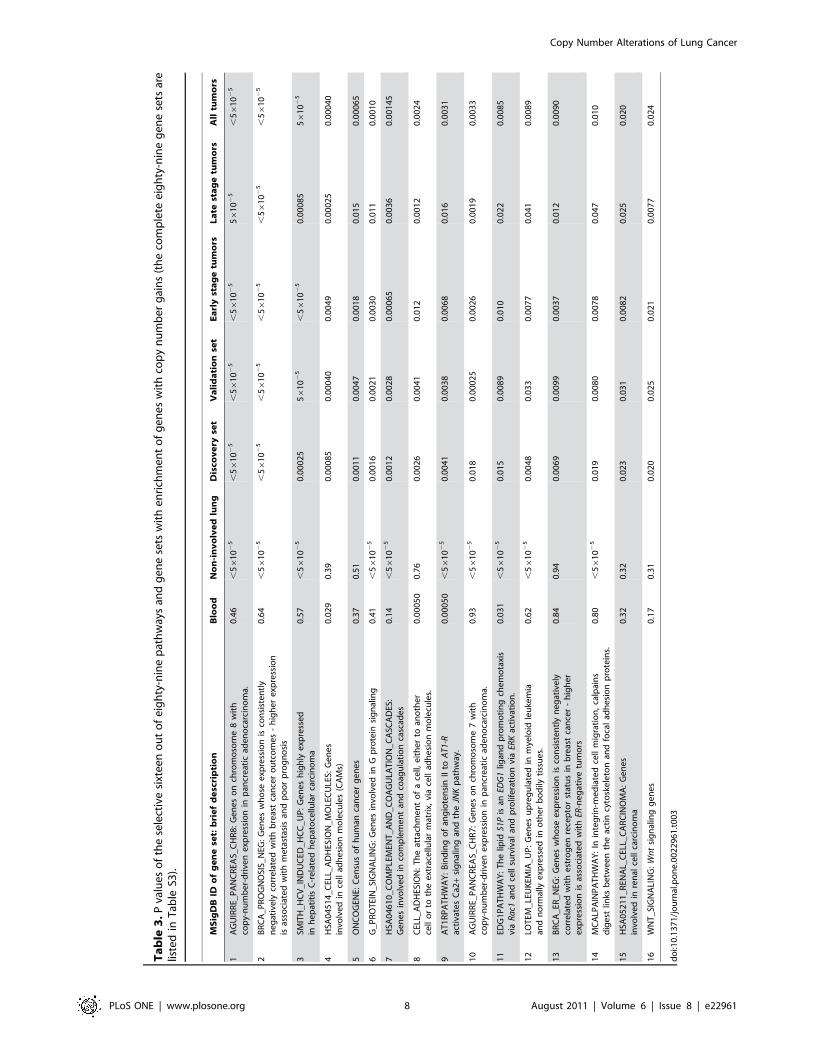
S3 and S4) We present 26 gene sets with particular relevance to
tumor biology that have enrichment of copy number gains or
losses in our samples in Tables 3 and 4 and the corresponding
gene set enrichment plots in Figures S6 and S7. Further
investigating the gene set enrichment in the blood and non-
involved lung tissue, we found many of the 89 and 27 validated
gene sets were also enriched in the genome of non-involved lung
tissue, including gains in G protein signaling pathway, EDG1
pathway, integrin-mediated cell migration pathway and losses in
regulations of autophagy and mitotic cell cycle. (Tables S3 and S4)
Figure 2. Total probes (TP) and probes locating within genes (GP) in which CNAs were detected, and the mean of G/T ratios. A, B,Counts of the total probes (TP) where CNAs events (A: copy number gains, B: copy number losses) occur were plotted for blood and non-involvedtissue, total tumor, early stage tumor, late stage tumor, early stage adenocarcinoma (ACA) and early stage squamous cell carcinoma (SCC). C, D,Counts of the probes within genes (GP) in which CNAs events (C: copy number gains, D: copy number losses) were detected in the same sixsubgroups. E, F, Mean and its 95% confidence interval of G/T ratios in the six subgroups for copy number gains (E) and losses (F); and the dashed linesrepresent the null G/T ratio on the chip (104,256/256,554 = 40.64%). Non-tumor: blood (n = 63) and non-involved tissue (n = 50); All tumor: total 301NSCLC tumors; Early tumor: stage I and II NSCLC tumors (n = 246); Late tumor: stage III and IV NSCLC tumors (n = 25); Early ACA: early stageadenocarcinoma tumors (n = 208); Early SCC: early stage squamous cell carcinoma tumors (n = 93).doi:10.1371/journal.pone.0022961.g002
Copy Number Alterations of Lung Cancer
PLoS ONE | www.plosone.org 5 August 2011 | Volume 6 | Issue 8 | e22961

Discussion
The genome-wide CNAs pattern from our analyses is similar to
those published in previous literatures [2,3,12]. Many of the
oncogenes with copy number amplifications reported here is also
consistent with previous studies [13,14,15,16]. The major strength
of this study is its large sample size, availability of paired blood and
non-involved tissue samples and detailed demographic/clinical
information, discovery-validation process and the novel statistical
analyses. The proposed global test for genome-wide CNAs provide
us the opportunities to test CNAs difference by simultaneously
taking the genomic locations, correlation of copy numbers and
multiple comparisons into account. The customized GSEA for
CNAs, on the other hand, can serve as a useful tool to analyze the
genome-wide copy numbers in the functional and biological
context, linking the sophisticated CNAs data to the knowledge of
gene categories, biological pathways and previous studies. There
are still limitations in our study. First, blood and non-involved
tissue samples can be obtained from only subset of the 301
patients. Second, we are unable to collect the CNAs data from
normal subjects or patients without lung cancer, which may
provide us a better insight into how the genome-wide CNAs
profile in NSCLC patients differs from that in normal subjects or
non-cancer patients. Thirdly, even though the gene sets analyses
can serve to formulate biological hypotheses, further investigation
to study the roles of gene sets/pathways with CNAs in
tumorigenesis is required.
The DNA materials analyzed in this study all come from
NSCLC patients, so the genome of blood and non-involved tissue
may not be viewed as a normal genome. We use DNAs from
blood, non-involved lung tissue, early-stage tumor and late-stage
tumor to represent the sequential stages of cancer development
and progression. We discovered that copy number alterations
increase with cancer development, but that selection with respect
Table 1. Thirty two oncogenes with .10% NSCLC patients having copy number gains.
Symbol Full name of oncogene Position % of patients with CN gains P value*
1 TRIO Triple functional domain 5p15.2 38.21 3.30610250
2 EGFR epidermal growth factor receptor 7p12 26.58 2.26610223
3 ERBB4 v-erb-a erythroblastic leukemia viral oncogene homolog 4 2q33.3–34 26.58 2.26610223
4 RUNX1T1 Runt-related transcription factor 1 8q22 26.25 9.97610223
5 ABL2 v-abl Abelson murine leukemia viral oncogene homolog 2 1q24–25 23.59 7.66610218
6 ETV5 ets variant 5 3q28 22.92 1.07610216
7 ETV1 ets variant 1 7p21.3 22.26 1.38610215
8 A2BP1 ataxin 2-binding protein 1 16p13.3 22.26 1.38610215
9 MYC v-myc myelocytomatosis viral oncogene homolog (avian) 8q24.21 21.59 1.66610214
10 TERT telomerase reverse transcriptase 5p15.33 20.60 5.97610213
11 AKT3 v-akt murine thymoma viral oncogene homolog 3 1q43–44 20.60 5.97610213
12 KRAS v-Ki-ras2 Kirsten rat sarcoma viral oncogene homolog 12p12.1 20.27 1.90610212
13 ETV6 ets variant 6 12p13 19.93 5.90610212
14 PTPN1 protein tyrosine phosphatase, non-receptor type 1 20q13.1–13.2 19.27 5.37610211
15 PIK3CA phosphoinositide-3-kinase, catalytic, alpha polypeptide 3q26.3 18.60 4.50610210
16 PGR progesterone receptor 11q22–23 16.61 1.5861027
17 BRAF v-raf murine sarcoma viral oncogene homolog B1 7q34 15.95 9.2861027
18 ERG v-ets erythroblastosis virus E26 oncogene homolog (avian) 21q22.3 15.61 2.1761026
19 CSMD1 CUB and Sushi multiple domains 1 8p23.2 15.28 4.9761026
20 ELF5 E74-like factor 5 (ets domain transcription factor) 11p13–12 14.95 1.1161025
21 PDE4D phosphodiesterase 4D 5q12 14.62 2.4161025
22 FGFR1 fibroblast growth factor receptor 1 8p11.2–11.1 14.62 2.4161025
23 EPHA3 EPH receptor A3 3p11.2 14.62 2.4161025
24 CDK6 cyclin-dependent kinase 6 7q21–q22 14.62 2.4161025
25 ESR2 Estrogen receptor 2 14q23.2 14.62 2.4161025
26 JAK2 Janus kinase 2 9p24 14.62 2.4161025
27 WWOX WW domain containing oxidoreductase 16q23.3–24.1 13.95 0.000106
28 PDGFRA platelet-derived growth factor receptor, alpha polypeptide 4q11–13 13.95 0.000106
29 CCND2 cyclin D2 12p13 13.95 0.000106
30 CCNE1 cyclin E1 19q12 13.62 0.000214
31 RBL1 retinoblastoma-like 1 20q11.2 11.30 0.0136
32 MET met proto-oncogene (hepatocyte growth factor receptor) 7q31 11.30 0.0136
*P values were calculated to test the significance of observing the percentage (%) of patients with copy number gains (copy number $2.7) on the oncogenes, giventhat the null probability of copy number gains is 0.076, which is empirically estimated from the data.doi:10.1371/journal.pone.0022961.t001
Copy Number Alterations of Lung Cancer
PLoS ONE | www.plosone.org 6 August 2011 | Volume 6 | Issue 8 | e22961

to gene location decreases. That is, there is a monotonic increase
in copy number alterations from blood and non-involved lung
tissue, to early stage and then to late stage tumors. (Figures 2A and
2B) On the other hand, copy number changes tend to occur away
from gene location in blood or non-involved tissue, but this trend
decreases in tumors, especially in late stage. (Figures 2E and 2F)
The increase of CNAs reflects the accumulation of somatic copy
number changes due to genomic instability, in which cellular
hypoxic stress in cancer might play a key role via perturbation of
DNA replication and replication of non-contiguous DNA
segments [17,18].
Similarly, we expect to see the accumulation of the number of
gene sets hit by CNAs from blood, non-involved tissue and then to
tumor, rather than the abrupt occurrence in tumor. Out of our
reported 89 gene sets of copy number gains, the numbers of
significant gene sets are 7 in blood, 46 in non-involved tissue, 89 in
tumors. (Table S3) Out of the reported 27 gene sets of copy
number losses, the numbers are 2 in blood, 8 in non-involved
tissue, 27 in tumors. (Table S4)
Selection of CNAs with respect to gene location during evolution
is also reported in Drosophila [11]. Here we show a similar selection
effect in the tumor genome even though it is relaxed to some extent.
We hypothesize that the selection in tumor occurs during early
cancer development on top of the consequence of evolutionary
selection as reflected in germline. These findings illustrate that the
purifying selection occurring in the germline as the result of species
evolution may also occur in tumor as a selecting effect during
oncogenesis and tumor progression. We also hypothesize that the
biological pathways with CNAs we observe in Tables S3 and S4 are
the consequence of such selection. That is, the large-scale alterations
may hit many different gene and biological pathways randomly, but
only the cells acquiring the growth advantage via CNAs (e.g.,
amplified oncogene signaling pathway or deleted tumor suppressor
gene pathway) will survive and become dominant. The finding that
CNAs tend to occur away from the gene also reflects the non-
randomness of CNAs occurrence.
Oncogenes can mimic normal growth signaling such that the
cancer cell reduces the dependence on exogenous growth
stimulation. Thus, the amplification of oncogenes is an essential
step in tumorigenesis. In lung cancer, KRAS, MYC, EGFR, and
ERBB family are well-known oncogenes [19,20,21,22], and all of
them were found to be highly significant in copy number gains.
Furthermore, genes with copy number amplifications were also
over-represented in the oncogenes as a category defined by the
census of human cancer genes [23]. This finding suggests that
oncogenesis may also result from different sets of oncogenes in
addition to the above well-known ones.
Genes with CNAs in NSCLC are also found to be highly
associated with genes involved in other cancers, including liver,
breast, kidney and pancreatic cancers. We found that genes with
copy number gains in NSCLC are more likely to be the highly
expressed genes in hepatitis C-related hepatocellular carcinoma
[24] (p = 0.00005) and renal cell carcinoma [25], [26] (p = 0.020).
Genes with copy number amplifications are also significantly
enriched in the genes of poor prognosis signature of breast cancer
[27] (p,0.00005), which may explain the poorer prognosis of lung
cancer than breast cancer. Moreover, our analysis showed the
genes with copy number gains in lung were enriched in the genes
on chromosomes 7 and 8, shown to have copy-number-driven
expression in pancreatic adenocarcinoma [28] (p = 0.0033 and
,0.00005, respectively), and those with copy number losses were
enriched in the genes with CNAs on chromosome 9, also
associated with gene expression in pancreatic tumor
(p,0.00005). This suggests that the gene dosage, as it were, of
genes with CNAs in NSCLC may also be positively related to
expression level. These results also suggest that cancer cells
emerging from different tissue origins share similar machinery for
oncogenesis and tumor invasion.
Table 2. Sixteen tumor suppressor genes with .1% NSCLC patients having copy number losses.
Symbol Full name of tumor suppressor gene Position% of patients withcopy number losses P value*
1 CDKN2A cyclin-dependent kinase inhibitor 2A 9p12 9.63 1.87610219
2 FHIT Fragile histidine triad gene 3p14.2 6.64 6.33610211
3 BRCA2 breast cancer 2, early onset 13q12.3 4.65 3.261026
4 TGFBR2 transforming growth factor, beta receptor II 3p22 4.31 1.5561025
5 FLCN Folliculin 17p11.2 3.65 0.00029
6 RB1 retinoblastoma 1 13q14.2 2.66 0.012
7 NF2 neurofibromin 2 (merlin) 22q12.2 2.66 0.012
8 PTEN phosphatase and tensin homolog 10q23.3 1.66 0.19
9 EP300 E1A binding protein p300 22q13.2 1.66 0.19
10 FBXW7 F-box and WD repeat domain containing 7 4q31.3 1.66 0.19
11 APC adenomatous polyposis coli 5q21–q22 1.66 0.19
12 NOTCH1 Notch homolog 1, translocation-associated 9q34.3 1.66 0.19
13 FAT3 FAT tumor suppressor homolog 3 11q14.3 1.33 0.36
14 SMARCB1 SWI/SNF related, matrix associated, actin dependentregulator of chromatin, subfamily b, member 1
22q11 1.33 0.36
15 SMAD4 mothers against decapentaplegic homolog 4 18q21.1 1.33 0.36
16 SMAD2 mothers against decapentaplegic homolog 2 18q21.1 1.33 0.36
*P values were calculated to test the significance of observing the percentage (%) of patients with copy number losses (copy number #1.3) on the tumor suppressorgenes, given that the null probability of copy number losses is 0.010, which is empirically estimated from the data.doi:10.1371/journal.pone.0022961.t002
Copy Number Alterations of Lung Cancer
PLoS ONE | www.plosone.org 7 August 2011 | Volume 6 | Issue 8 | e22961
Ta
ble
3.
Pva
lue
so
fth
ese
lect
ive
sixt
ee
no
ut
of
eig
hty
-nin
ep
ath
way
san
dg
en
ese
tsw
ith
en
rich
me
nt
of
ge
ne
sw
ith
cop
yn
um
be
rg
ain
s(t
he
com
ple
tee
igh
ty-n
ine
ge
ne
sets
are
liste
din
Tab
leS3
).
MS
igD
BID
of
ge
ne
set:
bri
ef
de
scri
pti
on
Blo
od
No
n-i
nv
olv
ed
lun
gD
isco
ve
ryse
tV
ali
da
tio
nse
tE
arl
yst
ag
etu
mo
rsL
ate
sta
ge
tum
ors
All
tum
ors
1A
GU
IRR
E_P
AN
CR
EAS_
CH
R8
:G
en
es
on
chro
mo
som
e8
wit
hco
py-
nu
mb
er-
dri
ven
exp
ress
ion
inp
ancr
eat
icad
en
oca
rcin
om
a.0
.46
,56
10
25
,56
10
25
,56
10
25
,56
10
25
561
02
5,
561
02
5
2B
RC
A_
PR
OG
NO
SIS_
NEG
:G
en
es
wh
ose
exp
ress
ion
isco
nsi
ste
ntl
yn
eg
ativ
ely
corr
ela
ted
wit
hb
reas
tca
nce
ro
utc
om
es
-h
igh
er
exp
ress
ion
isas
soci
ate
dw
ith
me
tast
asis
and
po
or
pro
gn
osi
s
0.6
4,
561
02
5,
561
02
5,
561
02
5,
561
02
5,
561
02
5,
561
02
5
3SM
ITH
_H
CV
_IN
DU
CE
D_
HC
C_
UP
:G
en
es
hig
hly
exp
ress
ed
inh
ep
atit
isC
-re
late
dh
ep
ato
cellu
lar
carc
ino
ma
0.5
7,
561
02
50
.00
02
556
10
25
,56
10
25
0.0
00
85
561
02
5
4H
SA0
45
14
_C
ELL_
AD
HES
ION
_M
OLE
CU
LES:
Ge
ne
sin
volv
ed
ince
llad
he
sio
nm
ole
cule
s(C
AM
s)0
.02
90
.39
0.0
00
85
0.0
00
40
0.0
04
90
.00
02
50
.00
04
0
5O
NC
OG
ENE:
Ce
nsu
so
fh
um
anca
nce
rg
en
es
0.3
70
.51
0.0
01
10
.00
47
0.0
01
80
.01
50
.00
06
5
6G
_P
RO
TEI
N_
SIG
NA
LIN
G:
Ge
ne
sin
volv
ed
inG
pro
tein
sig
nal
ing
0.4
1,
561
02
50
.00
16
0.0
02
10
.00
30
0.0
11
0.0
01
0
7H
SA0
46
10
_C
OM
PLE
MEN
T_
AN
D_
CO
AG
ULA
TIO
N_
CA
SCA
DES
:G
en
es
invo
lve
din
com
ple
me
nt
and
coag
ula
tio
nca
scad
es
0.1
4,
561
02
50
.00
12
0.0
02
80
.00
06
50
.00
36
0.0
01
45
8C
ELL_
AD
HES
ION
:T
he
atta
chm
en
to
fa
cell,
eit
he
rto
ano
the
rce
llo
rto
the
ext
race
llula
rm
atri
x,vi
ace
llad
he
sio
nm
ole
cule
s.0
.00
05
00
.76
0.0
02
60
.00
41
0.0
12
0.0
01
20
.00
24
9A
T1
RP
AT
HW
AY
:B
ind
ing
of
ang
iote
nsi
nII
toA
T1-R
acti
vate
sC
a2+
sig
nal
ing
and
the
JNK
pat
hw
ay.
0.0
00
50
,56
10
25
0.0
04
10
.00
38
0.0
06
80
.01
60
.00
31
10
AG
UIR
RE_
PA
NC
REA
S_C
HR
7:
Ge
ne
so
nch
rom
oso
me
7w
ith
cop
y-n
um
be
r-d
rive
ne
xpre
ssio
nin
pan
cre
atic
ade
no
carc
ino
ma.
0.9
3,
561
02
50
.01
80
.00
02
50
.00
26
0.0
01
90
.00
33
11
EDG
1P
AT
HW
AY
:T
he
lipid
S1P
isan
EDG
1lig
and
pro
mo
tin
gch
em
ota
xis
via
Ra
c1an
dce
llsu
rviv
alan
dp
rolif
era
tio
nvi
aER
Kac
tiva
tio
n.
0.0
31
,56
10
25
0.0
15
0.0
08
90
.01
00
.02
20
.00
85
12
LOT
EM_
LEU
KEM
IA_
UP
:G
en
es
up
reg
ula
ted
inm
yelo
idle
uke
mia
and
no
rmal
lye
xpre
sse
din
oth
er
bo
dily
tiss
ue
s.0
.62
,56
10
25
0.0
04
80
.03
30
.00
77
0.0
41
0.0
08
9
13
BR
CA
_ER
_N
EG:
Ge
ne
sw
ho
see
xpre
ssio
nis
con
sist
en
tly
ne
gat
ive
lyco
rre
late
dw
ith
est
rog
en
rece
pto
rst
atu
sin
bre
ast
can
cer
-h
igh
er
exp
ress
ion
isas
soci
ate
dw
ith
ER-n
eg
ativ
etu
mo
rs
0.8
40
.94
0.0
06
90
.00
99
0.0
03
70
.01
20
.00
90
14
MC
ALP
AIN
PA
TH
WA
Y:
Inin
teg
rin
-me
dia
ted
cell
mig
rati
on
,ca
lpai
ns
dig
est
links
be
twe
en
the
acti
ncy
tosk
ele
ton
and
foca
lad
he
sio
np
rote
ins.
0.8
0,
561
02
50
.01
90
.00
80
0.0
07
80
.04
70
.01
0
15
HSA
05
21
1_
REN
AL_
CEL
L_C
AR
CIN
OM
A:
Ge
ne
sin
volv
ed
inre
nal
cell
carc
ino
ma
0.3
20
.32
0.0
23
0.0
31
0.0
08
20
.02
50
.02
0
16
WN
T_
SIG
NA
LIN
G:
Wn
tsi
gn
alin
gg
en
es
0.1
70
.31
0.0
20
0.0
25
0.0
21
0.0
07
70
.02
4
do
i:10
.13
71
/jo
urn
al.p
on
e.0
02
29
61
.t0
03
Copy Number Alterations of Lung Cancer
PLoS ONE | www.plosone.org 8 August 2011 | Volume 6 | Issue 8 | e22961

Ta
ble
4.
Pva
lue
so
fth
ese
lect
ive
ten
ou
to
ftw
en
tyse
ven
pat
hw
ays
and
ge
ne
sets
wit
he
nri
chm
en
to
fg
en
es
wit
hco
py
nu
mb
er
loss
es
(th
eco
mp
lete
twe
nty
-se
ven
ge
ne
sets
are
liste
din
Tab
leS4
).
MS
igD
BID
of
ge
ne
set:
bri
ef
de
scri
pti
on
Blo
od
No
n-i
nv
olv
ed
lun
gD
isco
ve
ryse
tV
ali
da
tio
nse
tE
arl
yst
ag
etu
mo
rsL
ate
sta
ge
tum
ors
All
tum
ors
1A
GU
IRR
E_P
AN
CR
EAS_
CH
R9
:G
en
es
on
chro
mo
som
e9
wit
hco
py-
nu
mb
er-
dri
ven
exp
ress
ion
inp
ancr
eat
icad
en
oca
rcin
om
a.0
.25
0.5
7,
561
02
556
10
25
,56
10
25
,56
10
25
,56
10
25
2H
SA0
40
80
_N
EUR
OA
CT
IVE_
LIG
AN
D_
REC
EPT
OR
_IN
TER
AC
TIO
N:
Ge
ne
sin
volv
ed
inn
eu
roac
tive
ligan
d-r
ece
pto
rin
tera
ctio
n0
.06
10
.00
05
00
.00
07
0,
561
02
5,
561
02
50
.00
42
561
02
5
3C
ELL_
CEL
L_SI
GN
ALI
NG
:G
en
es
ann
ota
ted
by
the
GO
term
GO
:00
07
26
7.
An
yp
roce
ssth
atm
ed
iate
sth
etr
ansf
er
of
info
rmat
ion
fro
mo
ne
cell
toan
oth
er.
0.8
00
.06
10
.00
15
0.0
13
0.0
01
70
.00
30
0.0
00
70
4H
SA0
41
40
_R
EGU
LAT
ION
_O
F_A
UT
OP
HA
GY
:G
en
es
invo
lve
din
reg
ula
tio
no
fau
top
hag
y0
.16
,56
10
25
0.0
00
65
0.0
04
60
.00
09
50
.00
01
00
.00
08
5
5SI
GN
AL_
TR
AN
SDU
CT
ION
:G
en
es
invo
lve
din
sig
nal
tran
sdu
ctio
n0
.21
0.2
30
.01
20
.00
19
0.0
11
0.0
22
0.0
01
5
6C
ELL_
SUR
FAC
E_R
ECEP
TO
R_
LIN
KED
_SI
GN
AL_
TR
AN
SDU
CT
ION
_G
O_
00
07
16
6:
Ge
ne
san
no
tate
db
yth
eG
Ote
rmG
O:0
00
71
66
.A
ny
seri
es
of
mo
lecu
lar
sig
nal
sin
itia
ted
by
the
bin
din
go
fan
ext
race
llula
rlig
and
toa
rece
pto
ro
nth
esu
rfac
eo
fth
eta
rge
tce
ll.
0.3
50
.03
40
.02
10
.00
17
0.0
27
0.0
00
75
0.0
03
1
7C
ELL_
CEL
L_A
DH
ESIO
N:
Ge
ne
san
no
tate
db
yth
eG
Ote
rmG
O:0
01
63
37
.T
he
atta
chm
en
to
fo
ne
cell
toan
oth
er
cell
via
adh
esi
on
mo
lecu
les.
0.5
30
.06
90
.00
71
0.0
08
50
.00
28
0.0
00
35
0.0
03
9
8H
DA
CI_
CO
LON
_C
UR
24
HR
S_D
N:
Do
wn
reg
ula
ted
by
curc
um
inat
24
hrs
inSW
26
0co
lon
carc
ino
ma
cells
0.9
2,
561
02
50
.00
70
0.0
02
00
.00
94
0.0
37
0.0
05
0
9R
EGU
LAT
ION
_O
F_M
ITO
TIC
_C
ELL
_C
YC
LE:
Ge
ne
san
no
tate
db
yth
eG
Ote
rmG
O:0
00
73
46
.A
ny
pro
cess
that
mo
du
late
sth
era
teo
re
xte
nt
of
pro
gre
ssth
rou
gh
the
mit
oti
cce
llcy
cle
.
0.6
4,
561
02
50
.00
10
0.0
49
0.0
07
50
.01
70
.00
53
10
CH
RO
MA
TIN
_M
OD
IFIC
AT
ION
:G
en
es
ann
ota
ted
by
the
GO
term
GO
:00
16
56
8.
Th
eal
tera
tio
no
fD
NA
or
pro
tein
inch
rom
atin
,w
hic
hm
ayre
sult
inch
ang
ing
the
chro
mat
inst
ruct
ure
.
0.9
80
.71
0.0
19
0.0
22
0.0
49
0.0
09
50
.02
1
do
i:10
.13
71
/jo
urn
al.p
on
e.0
02
29
61
.t0
04
Copy Number Alterations of Lung Cancer
PLoS ONE | www.plosone.org 9 August 2011 | Volume 6 | Issue 8 | e22961

Our gene set analysis suggests that cells may acquire the
common capacities of tumor [7] through CNAs: gains in
oncogenes (self-sufficiency in growth signals; evading apoptosis),
gains in Calpain pathway (insensitivity to anti-growth signals; tissue
invasion and metastasis, sustained angiogenesis), gains in EDG1
pathway (evading apoptosis and self-sufficiency in growth signals),
gains in ERBB signaling (self-sufficiency in growth signals), gains in
WNT signaling (self-sufficiency in growth signals), losses in
regulation of autophagy (evading apoptosis), gains in telomerase
reverse transcriptase (TERT) (limitless replicative potential), and
losses in tight junction and cell adhesions (tissue invasion and
metastasis). Such results provide supporting evidence of the
‘chromosomal cancer theory’ [5,6]: cancer is a disease caused by
aneuploidy or large-scale alterations of chromosome. It is because
the large-scale duplication or deletion is more likely to alter the
copy number of significant numbers of genes in numerous
biological pathways. However, our results do not disagree with
the notion that duplication/deletion of a gene play a critical role in
cancer development. The occurrence of aneuploidy or large-scale
alterations requires an environment with genome instability, which
is likely to be facilitated by duplication/deletion or mutation of
genes critical in DNA repair, recombination and duplication.
Thus, they do not exclude the possibilities that CNAs may be
initiated by single or small sets of mutated genes and require
further investigation.
The finding that CNAs occur preferentially in certain regions
and chromosome arms (i.e., positional clustering) is consistent with
previous studies [2,3,12]. Another finding that genes with CNAs
tend to involve in certain biological functions and pathways (i.e.,
functional clustering) is novel and can provide a possible
explanation to the positional clustering: the CNAs hot spots/
regions in NSCLC may harbor genes that execute similar
biological functions or belong to the crucial pathways. That is,
our work is more providing biological insight into the positional
clustering rather than distinguishing the functional clustering from
positional clustering.
We conclude that CNAs events increase but their selection with
respect to gene location decreases from germline to early stage
tumors, and then to late stage tumors, and that CNAs in NSCLC
tumor are both positionally and functionally clustered. The functional
characteristics of gene sets enriched with CNAs genes provide us
better insight into the mechanisms of oncogenesis and cancer
progression, and have the potential for improving patient
management by drug targeting in the future for this prevalent
and devastating disease.
Supporting Information
Figure S1 Significance of genome-wide copy number alterations
in blood, non-involved lung tissue and tumor of NSCLC patients.
The x axis represents genomic locations, which were ordered by
the somatic chromosomes. The y axis represents 2log10(q values)
of NSCLC patients having copy number $2.7 (red) and #1.3
(blue) in blood (A), non-involved lung tissue (B), total tumor (C),
early stage tumor (D) and late stage tumor (E), early stage tumor of
adenocarcinoma (F) and early stage tumor of squamous cell
carcinoma (G).
(TIFF)
Figure S2 The comparisons of copy number alterations in
paired or matched samples. The proportion of copy numbers
$2.7 (red and pink) and #1.3 (blue and light blue) in paired blood
and tumor samples (A), paired non-involved lung and tumor
samples (B), and matched early stage and late stage tumors (C).
The p values indicate the statistical significance from the global
CNAs test between two groups.
(TIFF)
Figure S3 Copy numbers in chromosome 3. The main panel
represents the copy numbers of blood, non-involved lung tissue
and tumor as indicated. The bottom panel illustrates copy
numbers with their corresponding colors (red color indicates copy
number gains and blue color indicates copy number losses). The
right panel shows the corresponding 2log10(q values) for each
locus on the chromosome; the red and blue colors indicate the
2log10(q values) for copy number gains and losses, respectively.
(TIFF)
Figure S4 Copy numbers in chromosome 5. The main panel
represents the copy numbers of blood, non-involved lung tissue
and tumor as indicated. The bottom panel illustrates copy
numbers with their corresponding colors (red color indicates copy
number gains and blue color indicates copy number losses). The
right panel shows the corresponding 2log10(q values) for each
locus on the chromosome; the red and blue colors indicate the
2log10(q values) for copy number gains and losses, respectively.
(TIFF)
Figure S5 Copy numbers in chromosome 8. The main panel
represents the copy numbers of blood, non-involved lung tissue
and tumor as indicated. The bottom panel illustrates copy
numbers with their corresponding colors (red color indicates
copy number gains and blue color indicates copy number losses).
The right panel shows the corresponding 2log10(q values) for
each locus on the chromosome; the red and blue colors indicate
the 2log10(q values) for copy number gains and losses,
respectively.
(TIFF)
Figure S6 Gene set enrichment plot for the 16 selective gene sets
with copy number gains (corresponding to Table 3). For each
panel, the top indicates the location of genes from the gene set
within the list sorted by the proportion of patients with copy
number gains (left to right: higher proportion to lower); the bottom
is the plot of running enrichment scores.
(TIFF)
Figure S7 Gene set enrichment plot for the selective 10 gene sets
with copy number losses (corresponding to Table 4). For each
panel, the top indicates the location of genes from the gene set
within the list sorted by the proportion of patients with copy
number losses; the bottom is the plot of running enrichment
scores.
(TIFF)
Table S1 Characteristics of the discovery and validation tumor
samples*.
(DOC)
Table S2 Forty-five genes with .35% patients having copy
number gains.
(DOC)
Table S3 Eighty-nine pathways and gene sets with enrichment
of genes with copy number gains (with p,0.05 in discovery set,
validation set, early stage and late stage tumors).
(DOC)
Table S4 Twenty-seven pathways and gene sets with enrich-
ment of genes with copy number losses (with p,0.05 in discovery
set, validation set, early stage and late stage tumors).
(DOC)
Copy Number Alterations of Lung Cancer
PLoS ONE | www.plosone.org 10 August 2011 | Volume 6 | Issue 8 | e22961

Acknowledgments
We are indebted to the participants of Molecular and Genetic Analysis of
Lung Cancer Study; to Dr. GC Yuan of Harvard School of Public Health
(HSPH) and Michael Wu of University of North Carolina at Chapel Hill,
for their comments on GSEA analyses; to Dr. Oliver Hoffman of HSPH
Bioinformatics Core Facility for gene mapping; to Dr. Cheng Li of HSPH
for his advice on data preprocessing; to the HSPH Lung Cancer Study
Group; to Drs. Panos Fidias, Eugene Mark, Bruce A. Chabner and the
physicians and surgeons of the Massachusetts General Hospital Cancer
Center; to Dr. Lodve Stangeland of Haukeland University Hospital,
Norway for recruiting patients; and to Dr. Edward Fox and the Microarray
Core Facility of Dana Farber Cancer Institute.
Author Contributions
Conceived and designed the experiments: Y-TH LRC JCW RSH VS SZ
AH DCC. Performed the experiments: Y-TH VS SZ LS. Analyzed the
data: Y-TH XL RM. Contributed reagents/materials/analysis tools: Y-TH
LRC RM JCW RSH AH LS DCC. Wrote the paper: Y-TH XL LRC RM
JCW RSH VS SZ AH LS DCC.
References
1. Jemal A, Siegel R, Ward E, Hao Y, Xu J, et al. (2009) Cancer statistics, 2009.
CA Cancer J Clin 59: 225–249.
2. Weir BA, Woo MS, Getz G, Perner S, Ding L, et al. (2007) Characterizing the
cancer genome in lung adenocarcinoma. Nature 450: 893–898.
3. Kim TM, Yim SH, Lee JS, Kwon MS, Ryu JW, et al. (2005) Genome-wide
screening of genomic alterations and their clinicopathologic implications in non-
small cell lung cancers. Clin Cancer Res 11: 8235–8242.
4. Subramanian A, Tamayo P, Mootha VK, Mukherjee S, Ebert BL, et al. (2005)
Gene set enrichment analysis: a knowledge-based approach for interpreting
genome-wide expression profiles. Proc Natl Acad Sci U S A 102: 15545–15550.
5. Duesberg P, Li R, Fabarius A, Hehlmann R (2005) The chromosomal basis of
cancer. Cell Oncol 27: 293–318.
6. Duesberg P (2005) Does aneuploidy or mutation start cancer? Science 307: 41.
7. Hanahan D, Weinberg RA (2000) The hallmarks of cancer. Cell 100: 57–70.
8. Zhao X, Li C, Paez JG, Chin K, Janne PA, et al. (2004) An integrated view of
copy number and allelic alterations in the cancer genome using single nucleotide
polymorphism arrays. Cancer Res 64: 3060–3071.
9. Benjamini Y, Hochberg Y (1995) Controlling the False Discovery Rate: A
Practical and Powerful Approach to Multiple Testing. J Roy Statist Soc Ser B
57: 289–300.
10. Storey J (2002) A direct approach to false discovery rates. Journal of the Royal
Statistical Society, Series B 64: 479–498.
11. Emerson JJ, Cardoso-Moreira M, Borevitz JO, Long M (2008) Natural selection
shapes genome-wide patterns of copy-number polymorphism in Drosophila
melanogaster. Science 320: 1629–1631.
12. Dehan E, Ben-Dor A, Liao W, Lipson D, Frimer H, et al. (2007) Chromosomal
aberrations and gene expression profiles in non-small cell lung cancer. Lung
Cancer 56: 175–184.
13. Okudela K, Suzuki M, Kageyama S, Bunai T, Nagura K, et al. (2007) PIK3CA
mutation and amplification in human lung cancer. Pathol Int 57: 664–671.
14. Kang JU, Koo SH, Kwon KC, Park JW, Jung SS (2008) Gain of the EGFR gene
located on 7p12 is a frequent and early event in squamous cell carcinoma of the
lung. Cancer Genet Cytogenet 184: 31–37.
15. Kang JU, Koo SH, Kwon KC, Park JW, Kim JM (2008) Gain at chromosomal
region 5p15.33, containing TERT, is the most frequent genetic event in early
stages of non-small cell lung cancer. Cancer Genet Cytogenet 182: 1–11.16. Yamamoto H, Shigematsu H, Nomura M, Lockwood WW, Sato M, et al. (2008)
PIK3CA mutations and copy number gains in human lung cancers. Cancer Res68: 6913–6921.
17. Hastings PJ, Lupski JR, Rosenberg SM, Ira G (2009) Mechanisms of change ingene copy number. Nat Rev Genet 10: 551–564.
18. Shlien A, Malkin D (2009) Copy number variations and cancer. Genome Med 1:
62.19. Mitsuuchi Y, Testa JR (2002) Cytogenetics and molecular genetics of lung
cancer. Am J Med Genet 115: 183–188.20. Osada H, Takahashi T (2002) Genetic alterations of multiple tumor suppressors
and oncogenes in the carcinogenesis and progression of lung cancer. Oncogene
21: 7421–7434.21. Sanchez-Cespedes M (2003) Dissecting the genetic alterations involved in lung
carcinogenesis. Lung Cancer 40: 111–121.22. Panani AD, Roussos C (2006) Cytogenetic and molecular aspects of lung cancer.
Cancer Lett 239: 1–9.23. Futreal PA, Coin L, Marshall M, Down T, Hubbard T, et al. (2004) A census of
human cancer genes. Nat Rev Cancer 4: 177–183.
24. Smith MW, Yue ZN, Geiss GK, Sadovnikova NY, Carter VS, et al. (2003)Identification of novel tumor markers in hepatitis C virus-associated hepatocel-
lular carcinoma. Cancer Res 63: 859–864.25. Cohen HT, McGovern FJ (2005) Renal-cell carcinoma. N Engl J Med 353:
2477–2490.
26. Kanehisa M, Araki M, Goto S, Hattori M, Hirakawa M, et al. (2008) KEGG forlinking genomes to life and the environment. Nucleic Acids Res 36: D480–484.
27. van ’t Veer LJ, Dai H, van de Vijver MJ, He YD, Hart AA, et al. (2002) Geneexpression profiling predicts clinical outcome of breast cancer. Nature 415:
530–536.28. Aguirre AJ, Brennan C, Bailey G, Sinha R, Feng B, et al. (2004) High-resolution
characterization of the pancreatic adenocarcinoma genome. Proc Natl Acad
Sci U S A 101: 9067–9072.
Copy Number Alterations of Lung Cancer
PLoS ONE | www.plosone.org 11 August 2011 | Volume 6 | Issue 8 | e22961