Impact of Common Variation in Bone-Related Genes on Type 2 Diabetes and Related Traits
11
Impact of Common Variation in Bone-Related Genes on Type 2 Diabetes and Related Traits Liana K. Billings, 1,2,3 Yi-Hsiang Hsu, 4,5,6 Rachel J. Ackerman, 1 Josée Dupuis, 6,7 Benjamin F. Voight, 1,2,8 Laura J. Rasmussen-Torvik, 9 Serge Hercberg, 10 Mark Lathrop, 11 Daniel Barnes, 12 Claudia Langenberg, 12 Jennie Hui, 13,14,15 Mao Fu, 16 Nabila Bouatia-Naji, 17 Cecile Lecoeur, 17 Ping An, 18 Patrik K. Magnusson, 19 Ida Surakka, 20,21 Samuli Ripatti, 20,21 Lene Christiansen, 22 Christine Dalgård, 23 Lasse Folkersen, 24 Elin Grundberg, 25,26 the MAGIC Investigators,* the DIAGRAM1 Consortium,* the MuTHER Consortium,* the ASCOT Investigators,* the GEFOS Consortium, 27, * Per Eriksson, 24 Jaakko Kaprio, 20,28,29 Kirsten Ohm Kyvik, 30,31 Nancy L. Pedersen, 19 Ingrid B. Borecki, 18 Michael A. Province, 19 Beverley Balkau, 32 Philippe Froguel, 17,33 Alan R. Shuldiner, 16,34 Lyle J. Palmer, 35 Nick Wareham, 12 Pierre Meneton, 36 Toby Johnson, 37 James S. Pankow, 38 David Karasik, 4,6 James B. Meigs, 2,6 Douglas P. Kiel, 2,4,6 and Jose C. Florez 1,2,3,8 Exploring genetic pleiotropy can provide clues to a mechanism underlying the observed epidemiological association between type 2 diabetes and heightened fracture risk. We examined genetic vari- ants associated with bone mineral density (BMD) for association with type 2 diabetes and glycemic traits in large well-phenotyped and -genotyped consortia. We undertook follow-up analysis in ;19,000 individuals and assessed gene expression. We queried single nucleotide polymorphisms (SNPs) associated with BMD at levels of genome-wide significance, variants in linkage disequilib- rium (r 2 . 0.5), and BMD candidate genes. SNP rs6867040, at the ITGA1 locus, was associated with a 0.0166 mmol/L (0.004) in- crease in fasting glucose per C allele in the combined analysis. Genetic variants in the ITGA1 locus were associated with its ex- pression in the liver but not in adipose tissue. ITGA1 variants appeared among the top loci associated with type 2 diabetes, fast- ing insulin, b-cell function by homeostasis model assessment, and 2-h post–oral glucose tolerance test glucose and insulin levels. ITGA1 has demonstrated genetic pleiotropy in prior studies, and its suggested role in liver fibrosis, insulin secretion, and bone heal- ing lends credence to its contribution to both osteoporosis and type 2 diabetes. These findings further underscore the link be- tween skeletal and glucose metabolism and highlight a locus to direct future investigations. Diabetes 61:2176–2186, 2012 S tudies show that adults with type 2 diabetes have a higher fracture rate than those without diabetes (1–5). A meta-analysis of 16 studies revealed a 1.7 (95% CI 1.3–2.2) relative risk of hip fracture for people with diabetes compared with those without diabetes (6). The higher fracture rate persisted even after considering factors including, but not limited to, falls, impaired vision, and weight (4). Quantitative computed tomography studies show increased bone porosity in individuals with type 2 From the 1 Center for Human Genetic Research, Massachusetts General Hos- pital, Boston, Massachusetts; the 2 Department of Medicine, Harvard Med- ical School, Boston, Massachusetts; the 3 Diabetes Research Center (Diabetes Unit), Massachusetts General Hospital, Boston, Massachusetts; the 4 Hebrew SeniorLife Institute for Aging Research and Harvard Medical School, Boston, Massachusetts; the 5 Molecular and Integrative Physiolog- ical Sciences Program, Harvard School of Public Health, Boston, Massa- chusetts; the 6 Framingham Heart Study, Framingham, Massachusetts; the 7 Department of Biostatistics, Boston University School of Public Health, Boston, Massachusetts; the 8 Broad Institute of Harvard and Massachusetts Institute of Technology, Cambridge, Massachusetts; the 9 Department of Preventive Medicine, Northwestern University Feinberg School of Medi- cine, Chicago, Illinois; 10 INSERM, National Institute of Agronomic Re- search, University of Paris, Bobigny, France; the 11 National Genotyping Center, Atomic Energy Commission, Institute of Genomics, Evry, France; the 12 Medical Research Council Epidemiology Unit, Institute of Metabolic Sci- ence, Addenbrooke’s Hospital, Cambridge, U.K.; 13 Molecular Genetics, PathWest Laboratory Medicine of Western Australia, Nedlands, Western Australia, Australia; the 14 School of Population Health and School of Pathology and Laboratory Medicine, University of Western Australia, Nedlands, Western Australia, Australia; the 15 Busselton Population Medical Research Founda- tion, Sir Charles Gairdner Hospital, Nedlands, Western Australia, Australia; the 16 Department of Medicine, University of Maryland School of Medicine, Baltimore, Maryland; the 17 National Center for Scientific Research, UMR 8199, Genomics and Metabolic Diseases, Lille Pasteur Institute, Lille Nord de France University, Lille, France; the 18 Division of Statistical Genomics and Department of Genetics, Washington University School of Medicine, St. Louis, Missouri; the 19 Department of Medical Epidemiology and Biostatis- tics, Karolinska Institute, Stockholm, Sweden; the 20 Institute for Molecular Medicine Finland, University of Helsinki, Helsinki, Finland; the 21 Public Health Genomics Unit, National Institute for Health and Welfare, Helsinki, Finland; the 22 Danish Twin Registry, Epidemiology, Institute of Public Health, University of Southern Denmark, Odense, Denmark; the 23 Depart- ment of Environmental Medicine, Institute of Public Health, University of Southern Denmark, Odense, Denmark; the 24 Atherosclerosis Research Unit, Department of Medicine, Karolinska Institute, Stockholm, Sweden; the 25 Wellcome Trust Sanger Institute, Hinxton, U.K.; the 26 Department of Twin Research and Genetic Epidemiology, King’s College London, London, U.K.; 27 Erasmus Medical College (Coordinating Center), Rotterdam, the Netherlands; the 28 Unit for Child and Adolescent Mental Health, National Institute for Health and Welfare, Helsinki, Finland; the 29 Department of Public Health, University of Helsinki, Helsinki, Finland; the 30 Institute of Re- gional Health Services Research, University of Southern Denmark, Odense, Denmark; the 31 Odense Patient Data Explorative Network, Odense University Hospital, Odense, Denmark; 32 CESP Center for Research in Ep- idemiology and Health of Populations, U1018, Epidemiology of Diabetes, Obesity and Chronic Kidney Disease Over the Life Course, INSERM, Villejuif, France, and Université Paris-Sud 11, UMRS 1018, Villejuif, France; 33 Genomic Medicine, Hammersmith Hospital, Imperial College London, London, U.K.; the 34 Geriatric Research, Education and Clinical Center, Baltimore VA Medical Center, Baltimore, Maryland; the 35 Ontario Institute for Cancer Research, Toronto, Ontario, Canada; the 36 Cordeliers Center of Research, INSERM, Paris, France; the 37 Clinical Pharmacology and the Genome Centre, William Harvey Research Institute, Barts and London School of Medicine and Dentistry, Queen Mary University of London, London, U.K.; and the 38 Division of Epidemiology and Community Health, University of Minnesota, Minneapolis, Minnesota. Corresponding author: Jose C. Florez, jcfl[email protected]. Received 27 October 2011 and accepted 9 March 2012. DOI: 10.2337/db11-1515 This article contains Supplementary Data online at http://diabetes .diabetesjournals.org/lookup/suppl/doi:10.2337/db11-1515/-/DC1. *A complete list of the MAGIC Investigators, the DIAGRAM+ Consortium, the MuTHER Consortium, and the GEFOS Consortium can be found in the Supplementary Data online. A complete list of the ASCOT Investigators can be found in ref. 51. Ó 2012 by the American Diabetes Association. Readers may use this article as long as the work is properly cited, the use is educational and not for profit, and the work is not altered. See http://creativecommons.org/licenses/by -nc-nd/3.0/ for details. 2176 DIABETES, VOL. 61, AUGUST 2012 diabetes.diabetesjournals.org ORIGINAL ARTICLE
-
Upload
hms-harvard -
Category
Documents
-
view
4 -
download
0
Transcript of Impact of Common Variation in Bone-Related Genes on Type 2 Diabetes and Related Traits

Impact of Common Variation in Bone-Related Genes onType 2 Diabetes and Related TraitsLiana K. Billings,
1,2,3Yi-Hsiang Hsu,
4,5,6Rachel J. Ackerman,
1Josée Dupuis,
6,7Benjamin F. Voight,
1,2,8
Laura J. Rasmussen-Torvik,9Serge Hercberg,
10Mark Lathrop,
11Daniel Barnes,
12
Claudia Langenberg,12
Jennie Hui,13,14,15
Mao Fu,16
Nabila Bouatia-Naji,17
Cecile Lecoeur,17
Ping An,18
Patrik K. Magnusson,19
Ida Surakka,20,21
Samuli Ripatti,20,21
Lene Christiansen,22
Christine Dalgård,23
Lasse Folkersen,24
Elin Grundberg,25,26
the MAGIC Investigators,* the DIAGRAM1 Consortium,* the
MuTHER Consortium,* the ASCOT Investigators,* the GEFOS Consortium,27,
* Per Eriksson,24
Jaakko Kaprio,20,28,29
Kirsten Ohm Kyvik,30,31
Nancy L. Pedersen,19
Ingrid B. Borecki,18
Michael A. Province,19
Beverley Balkau,32
Philippe Froguel,17,33
Alan R. Shuldiner,16,34
Lyle J. Palmer,35
Nick Wareham,12
Pierre Meneton,36
Toby Johnson,37
James S. Pankow,38
David Karasik,4,6
James B. Meigs,2,6
Douglas P. Kiel,2,4,6
and Jose C. Florez1,2,3,8
Exploring genetic pleiotropy can provide clues to a mechanismunderlying the observed epidemiological association between type2 diabetes and heightened fracture risk. We examined genetic vari-ants associated with bone mineral density (BMD) for associationwith type 2 diabetes and glycemic traits in large well-phenotypedand -genotyped consortia. We undertook follow-up analysis in;19,000 individuals and assessed gene expression. We queriedsingle nucleotide polymorphisms (SNPs) associated with BMD atlevels of genome-wide significance, variants in linkage disequilib-rium (r2 . 0.5), and BMD candidate genes. SNP rs6867040, at theITGA1 locus, was associated with a 0.0166 mmol/L (0.004) in-crease in fasting glucose per C allele in the combined analysis.Genetic variants in the ITGA1 locus were associated with its ex-pression in the liver but not in adipose tissue. ITGA1 variantsappeared among the top loci associated with type 2 diabetes, fast-ing insulin, b-cell function by homeostasis model assessment, and2-h post–oral glucose tolerance test glucose and insulin levels.ITGA1 has demonstrated genetic pleiotropy in prior studies, and
its suggested role in liver fibrosis, insulin secretion, and bone heal-ing lends credence to its contribution to both osteoporosis andtype 2 diabetes. These findings further underscore the link be-tween skeletal and glucose metabolism and highlight a locus todirect future investigations. Diabetes 61:2176–2186, 2012
Studies show that adults with type 2 diabetes havea higher fracture rate than those without diabetes(1–5). A meta-analysis of 16 studies revealed a 1.7(95% CI 1.3–2.2) relative risk of hip fracture for
people with diabetes compared with those without diabetes(6). The higher fracture rate persisted even after consideringfactors including, but not limited to, falls, impaired vision,and weight (4). Quantitative computed tomography studiesshow increased bone porosity in individuals with type 2
From the 1Center for Human Genetic Research, Massachusetts General Hos-pital, Boston, Massachusetts; the 2Department of Medicine, Harvard Med-ical School, Boston, Massachusetts; the 3Diabetes Research Center(Diabetes Unit), Massachusetts General Hospital, Boston, Massachusetts;the 4Hebrew SeniorLife Institute for Aging Research and Harvard MedicalSchool, Boston, Massachusetts; the 5Molecular and Integrative Physiolog-ical Sciences Program, Harvard School of Public Health, Boston, Massa-chusetts; the 6Framingham Heart Study, Framingham, Massachusetts; the7Department of Biostatistics, Boston University School of Public Health,Boston, Massachusetts; the 8Broad Institute of Harvard and MassachusettsInstitute of Technology, Cambridge, Massachusetts; the 9Department ofPreventive Medicine, Northwestern University Feinberg School of Medi-cine, Chicago, Illinois; 10INSERM, National Institute of Agronomic Re-search, University of Paris, Bobigny, France; the 11National GenotypingCenter, Atomic Energy Commission, Institute of Genomics, Evry, France;the 12Medical Research Council Epidemiology Unit, Institute of Metabolic Sci-ence, Addenbrooke’s Hospital, Cambridge, U.K.; 13Molecular Genetics, PathWestLaboratory Medicine of Western Australia, Nedlands, Western Australia,Australia; the 14School of Population Health and School of Pathology andLaboratory Medicine, University of Western Australia, Nedlands, WesternAustralia, Australia; the 15Busselton Population Medical Research Founda-tion, Sir Charles Gairdner Hospital, Nedlands, Western Australia, Australia;the 16Department of Medicine, University of Maryland School of Medicine,Baltimore, Maryland; the 17National Center for Scientific Research, UMR8199, Genomics and Metabolic Diseases, Lille Pasteur Institute, Lille Nordde France University, Lille, France; the 18Division of Statistical Genomicsand Department of Genetics, Washington University School of Medicine, St.Louis, Missouri; the 19Department of Medical Epidemiology and Biostatis-tics, Karolinska Institute, Stockholm, Sweden; the 20Institute for MolecularMedicine Finland, University of Helsinki, Helsinki, Finland; the 21PublicHealth Genomics Unit, National Institute for Health and Welfare, Helsinki,Finland; the 22Danish Twin Registry, Epidemiology, Institute of PublicHealth, University of Southern Denmark, Odense, Denmark; the 23Depart-ment of Environmental Medicine, Institute of Public Health, University ofSouthern Denmark, Odense, Denmark; the 24Atherosclerosis Research Unit,
Department of Medicine, Karolinska Institute, Stockholm, Sweden; the25Wellcome Trust Sanger Institute, Hinxton, U.K.; the 26Department of TwinResearch and Genetic Epidemiology, King’s College London, London,U.K.; 27Erasmus Medical College (Coordinating Center), Rotterdam, theNetherlands; the 28Unit for Child and Adolescent Mental Health, NationalInstitute for Health and Welfare, Helsinki, Finland; the 29Department ofPublic Health, University of Helsinki, Helsinki, Finland; the 30Institute of Re-gional Health Services Research, University of Southern Denmark,Odense, Denmark; the 31Odense Patient Data Explorative Network, OdenseUniversity Hospital, Odense, Denmark; 32CESP Center for Research in Ep-idemiology and Health of Populations, U1018, Epidemiology of Diabetes,Obesity and Chronic Kidney Disease Over the Life Course, INSERM,Villejuif, France, and Université Paris-Sud 11, UMRS 1018, Villejuif,France; 33Genomic Medicine, Hammersmith Hospital, Imperial CollegeLondon, London, U.K.; the 34Geriatric Research, Education and ClinicalCenter, Baltimore VA Medical Center, Baltimore, Maryland; the 35OntarioInstitute for Cancer Research, Toronto, Ontario, Canada; the 36CordeliersCenter of Research, INSERM, Paris, France; the 37Clinical Pharmacologyand the Genome Centre, William Harvey Research Institute, Barts andLondon School of Medicine and Dentistry, Queen Mary University of London,London, U.K.; and the 38Division of Epidemiology and Community Health,University of Minnesota, Minneapolis, Minnesota.
Corresponding author: Jose C. Florez, [email protected] 27 October 2011 and accepted 9 March 2012.DOI: 10.2337/db11-1515This article contains Supplementary Data online at http://diabetes
.diabetesjournals.org/lookup/suppl/doi:10.2337/db11-1515/-/DC1.*A complete list of the MAGIC Investigators, the DIAGRAM+ Consortium, the
MuTHER Consortium, and the GEFOS Consortium can be found in theSupplementary Data online. A complete list of the ASCOT Investigatorscan be found in ref. 51.
� 2012 by the American Diabetes Association. Readers may use this article aslong as the work is properly cited, the use is educational and not for profit,and the work is not altered. See http://creativecommons.org/licenses/by-nc-nd/3.0/ for details.
2176 DIABETES, VOL. 61, AUGUST 2012 diabetes.diabetesjournals.org
ORIGINAL ARTICLE
diabetes, suggesting that bone integrity is compromised andthereby causing increased bone fragility (7–9), but it remainsunclear what may be causing the decreased bone integrity.Despite the generally increased bone mineral density (BMD)of individuals with type 2 diabetes (1), for the same BMDmeasurement, people with type 2 diabetes have a higher riskof fracture (10). Basic science studies reveal further evi-dence of a link between bone-derived hormones and glucoseregulation. Mice lacking osteocalcin, an osteoblast-specificsecreted molecule, have glucose intolerance (11,12).
The relationship between osteoporosis and type 2 dia-betes raised by these epidemiological studies, and intriguingnew molecular data, hint to a common mechanism impli-cated in the pathogenesis of both disorders. Discoveringgenetic determinants that exhibit genetic pleiotropy (de-fined as one gene influencing multiple phenotypic traits)may point to a common underlying mechanism. Approxi-mately 16.9% of the genes in the National Human GenomeResearch Institute’s catalog of published genome-wide as-sociation studies (GWASs) are estimated to be pleiotropic(13). GWASs reveal genetic variants that are associatedwith BMD (a quantitative endophenotype for osteoporosisand a surrogate for fracture risk) (10,14–18). Some of theseloci are also associated with traits seemingly unrelatedto BMD (Table 1). However, common genetic variants
influencing BMD have not been studied systematically forassociation with type 2 diabetes and other glycemic traits.
We therefore performed a comprehensive evaluation ofthe influence of BMD-related genetic loci on diabetes-related phenotypes. After examining an extensive list ofBMD-related single nucleotide polymorphisms (SNPs) forassociation with type 2 diabetes and quantitative glycemictraits in large GWAS meta-analysis datasets, our top SNPswere selected for in silico replication in additional cohorts,cis-gene expression analyses, and BMI association. In thisstudy, we aimed to underscore the genetic determinants thatare shared between osteoporosis and type 2 diabetes andprovide clues into a commonmechanism that may contributeto both diseases. Furthermore, through this systematic ex-ploration, we have generated testable hypotheses for repli-cation by independent cohorts and experimental follow-up.
RESEARCH DESIGN AND METHODS
SNP selection. In total, 1,778 SNPs were collated for association with type 2diabetes and glycemic traits (Fig. 1). The SNP selection is described below.
A total of 35 SNPs initially were selected based on BMD GWASs in pop-ulations of European ancestry (14–17). If multiple SNPs were listed for onegene per trait, SNPs were kept for analysis if the correlation was low (pairwiselinkage disequilibrium [LD] r2 , 0.5); if r2 $ 0.5, only the SNP with the lowestP value was kept unless the study indicated that multiple correlated SNPs had ahigh degree of explanatory power of the variance for the trait. We removed
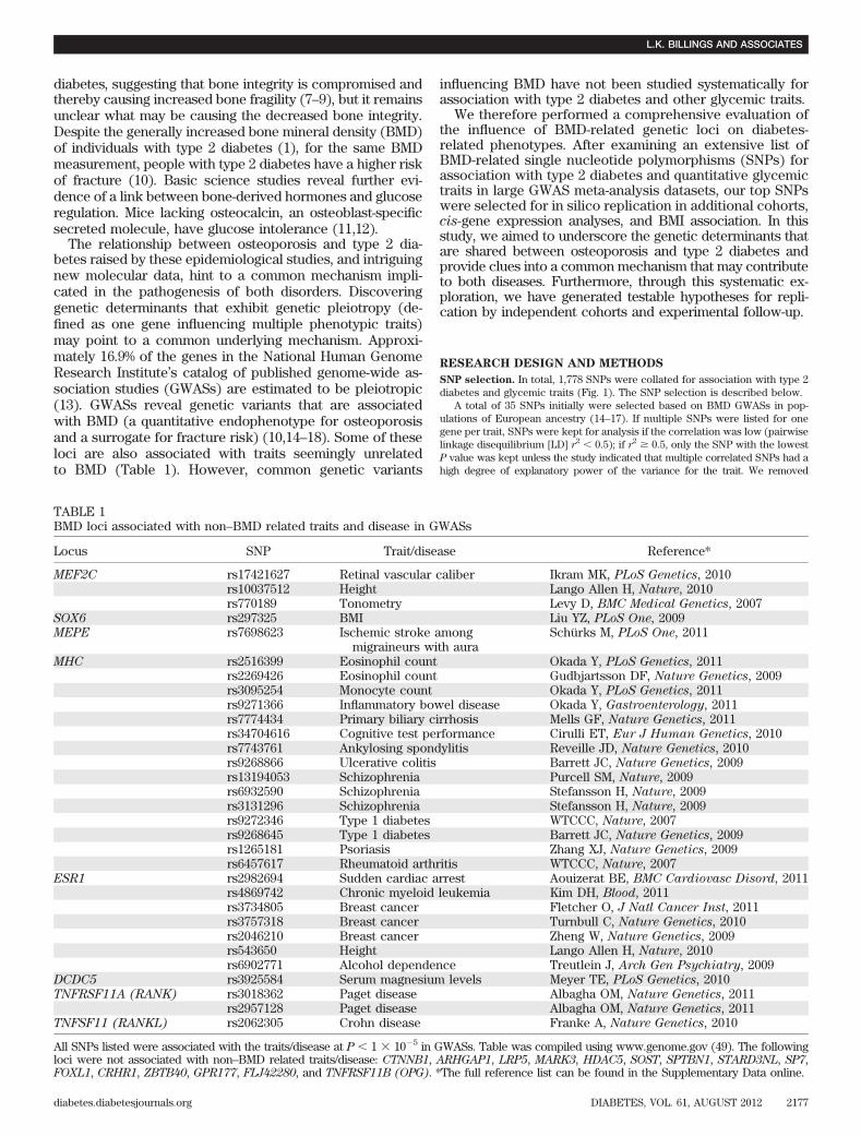
TABLE 1BMD loci associated with non–BMD related traits and disease in GWASs
Locus SNP Trait/disease Reference*
MEF2C rs17421627 Retinal vascular caliber Ikram MK, PLoS Genetics, 2010rs10037512 Height Lango Allen H, Nature, 2010rs770189 Tonometry Levy D, BMC Medical Genetics, 2007
SOX6 rs297325 BMI Liu YZ, PLoS One, 2009MEPE rs7698623 Ischemic stroke among
migraineurs with auraSchürks M, PLoS One, 2011
MHC rs2516399 Eosinophil count Okada Y, PLoS Genetics, 2011rs2269426 Eosinophil count Gudbjartsson DF, Nature Genetics, 2009rs3095254 Monocyte count Okada Y, PLoS Genetics, 2011rs9271366 Inflammatory bowel disease Okada Y, Gastroenterology, 2011rs7774434 Primary biliary cirrhosis Mells GF, Nature Genetics, 2011rs34704616 Cognitive test performance Cirulli ET, Eur J Human Genetics, 2010rs7743761 Ankylosing spondylitis Reveille JD, Nature Genetics, 2010rs9268866 Ulcerative colitis Barrett JC, Nature Genetics, 2009rs13194053 Schizophrenia Purcell SM, Nature, 2009rs6932590 Schizophrenia Stefansson H, Nature, 2009rs3131296 Schizophrenia Stefansson H, Nature, 2009rs9272346 Type 1 diabetes WTCCC, Nature, 2007rs9268645 Type 1 diabetes Barrett JC, Nature Genetics, 2009rs1265181 Psoriasis Zhang XJ, Nature Genetics, 2009rs6457617 Rheumatoid arthritis WTCCC, Nature, 2007
ESR1 rs2982694 Sudden cardiac arrest Aouizerat BE, BMC Cardiovasc Disord, 2011rs4869742 Chronic myeloid leukemia Kim DH, Blood, 2011rs3734805 Breast cancer Fletcher O, J Natl Cancer Inst, 2011rs3757318 Breast cancer Turnbull C, Nature Genetics, 2010rs2046210 Breast cancer Zheng W, Nature Genetics, 2009rs543650 Height Lango Allen H, Nature, 2010rs6902771 Alcohol dependence Treutlein J, Arch Gen Psychiatry, 2009
DCDC5 rs3925584 Serum magnesium levels Meyer TE, PLoS Genetics, 2010TNFRSF11A (RANK) rs3018362 Paget disease Albagha OM, Nature Genetics, 2011
rs2957128 Paget disease Albagha OM, Nature Genetics, 2011TNFSF11 (RANKL) rs2062305 Crohn disease Franke A, Nature Genetics, 2010
All SNPs listed were associated with the traits/disease at P , 1 3 1025 in GWASs. Table was compiled using www.genome.gov (49). The followingloci were not associated with non–BMD related traits/disease: CTNNB1, ARHGAP1, LRP5, MARK3, HDAC5, SOST, SPTBN1, STARD3NL, SP7,FOXL1, CRHR1, ZBTB40, GPR177, FLJ42280, and TNFRSF11B (OPG). *The full reference list can be found in the Supplementary Data online.
L.K. BILLINGS AND ASSOCIATES
diabetes.diabetesjournals.org DIABETES, VOL. 61, AUGUST 2012 2177

rs6696981 (ZBTB40), rs4879055 and rs6929137 (ESR1), rs6993813 and rs6469804(TNFRSF11), rs9594759 (RANKL), rs1107748 (SOST), rs2566755 (GPR177), andrs7781370 (FLJ42280) (14,15,17). The final list of 26 BMD genome wide–associated SNPs was examined for association with type 2 diabetes and glycemictraits (Table 2).
Since the index SNPmay not be the causal variant and other genetic variantsin the region may have a stronger influence on the traits examined, we testedthe region around the index variant by selecting SNPs in moderate-to-strong LD(r2 . 0.5). We chose variants in moderate-to-strong LD, rather than all of thevariants in this region, to base our exploration on variants with a higher priorprobability of true association and reduce the multiple testing burden. All SNPsthat were 50 kilobases (kb) upstream and downstream from and in moderate-to-strong LD with the 26 BMD-related SNPs were tested for association with type 2diabetes and glycemic traits. These SNPs were identified using SNP Annotationand Proxy Search, SNAP (http://www.broadinstitute.org/mpg/snap/) (19) (Sup-plementary Table 1).
In addition to selecting the 26 SNPs associated at genome-wide significancewith BMD and the surrounding region, we selected candidate genes that werefound to be associated (P , 2.39 3 1026 after Bonferroni correction) withBMD in the GEFOS (Genetic Factors for Osteoporosis) Consortium (20). Thisarticle identifies nine candidate genes, including ESR1, LRP4, ITGA1, LRP5,SOST, SPP1, TNFRSF11A (RANK), TNRFSF11B, and TNFSF11 (RANKL).For each gene, we identified all SNPs within and 20 kb upstream and down-stream of any transcript of the gene. All SNPs within those boundaries thatwere genotyped or imputed in the consortia were tested for association withtype 2 diabetes and glycemic traits (Supplementary Table 1).Study populations. We tested SNPs in the DIAGRAM+ (Diabetes GeneticsReplication and Meta-analysis) Consortium (21) for association with type 2diabetes and in MAGIC (Meta-Analyses of Glucose and Insulin-Related TraitsConsortium) (22–24) for association with seven glycemic quantitative traits.These traits included fasting glucose, fasting insulin, homeostasis modelassessments of b-cell function (HOMA-B) and insulin resistance (HOMA-IR)(25), hemoglobin A1C (HbA1c), and glucose and insulin levels 2 h post–glucoseload (2-h glucose and 2-h insulin). The DIAGRAM+ Consortium combinedcase-control data from eight type 2 diabetes GWASs with up to 42,542 case and98,912 control subjects of European ancestry (21). MAGIC combined datafrom multiple GWASs that identified loci that affect quantitative glycemic traits.Its discovery sample included up to 46,186 individuals from 17 population-basedcohorts and 4 case-control studies (22–24). It is noteworthy that the FraminghamHeart Study (FHS), Diabetes Epidemiology: Collaborative Analysis of Diagnostic
Criteria in Europe (deCODE) Study, Erasmus Rucphen Family Study, andTwinsUK Study provided data to both MAGIC and the BMD datasets from wherethe genome-wide–associated SNPs were selected. Using FHS as a representativecohort of European descent that contained both BMD and glycemic values, wefound phenotypic correlations, r of 0.11–0.16, between bone (femoral neck andlumbar spine BMD) and glycemic traits (glucose and insulin). Since the pheno-typic correlation is low, we would not necessarily expect to see a genetic asso-ciation solely based on the fact that a small portion of the participants wereassessed for both traits. In addition, examining the associations using meta-analyses of large consortia, rather than in the subset of overlapping participants,provides a more powerful approach.
The study protocols were approved by the institutional review board of therespective cohorts’ institutions, and informed consent was obtained from eachsubject prior to participation.Testing for association. After the collation of the index, LD-based, and gene-based BMD-related SNPs, we tested 1,778 unique SNPs for association withtype 2 diabetes and glycemic traits. We obtained effect estimates and P valuesfrom GWAS meta-analyses provided by DIAGRAM+ and MAGIC. We deter-mined which SNPs to examine in follow-up studies by calculating a significancethreshold for each group of SNPs selected (index, LD-based, and gene-based).We used a Bonferroni correction for the estimated number of independent testsafter taking LD into account determined using a method proposed by Nyholt(26) and Li and Ji (27). For our primary analyses, we used a stricter thresholdby accounting for the number of traits tested. We evaluated 26 BMD SNPsfor association with type 2 diabetes and seven glycemic traits (26 tests multi-plied by 8 traits = 208), yielding thresholds to declare statistical significance atP = 2.4 3 1024 (0.05/208 tests). For the LD- and gene-based secondary analyses,we corrected for the number of independent SNPs tested but not for the numberof traits examined. The P value threshold for the 513 LD-based SNPs (188 in-dependent tests) was 2.6 3 1024 and for the 1,318 candidate gene–based SNPs(651 independent tests), 7.73 1025. A study-wide P value of 6.03 1025 for 1,778total SNPs (830 independent tests) determined significance for the combinedmeta-analysis (described below).Follow-up strategy. To follow up the BMD-related SNPs associated with type2 diabetes and glycemic traits, we combined in silico GWAS data from 12additional cohorts of 19,417 nondiabetic participants (Amish Family DiabetesStudy, Atherosclerosis Risk in Communities Study [ARIC], Anglo-ScandinavianCardiac Outcomes Trial [ASCOT], Busselton Health Study [BHS], Data Fromthe Epidemiological Study on the Insulin Resistance Syndrome [DESIR] Study,French Obese Study, Family Heart Study [FamHS], Fenland Study, FinnishTwins Study, Swedish Twins Study, GEMINAKAR Study, and the Supplé-mentation en Vitamines et Minéraux Antioxydants [SU.VI.MAX]) Study (de-tailed in Supplementary Table 2). We then combined the discovery andreplication meta-analysis results for overall association using METAL (28).
Follow-up SNPs were examined by cis-expression quantitative trait loci(eQTL) analysis in metabolically relevant tissues, liver, and adipose. Liver tissuesamples came from the Advanced Study of Aortic Pathology (ASAP) cohort of211 healthy adults undergoing aortic valve surgery. Each biopsy was taken inRNAlater (Ambion, Austin, TX). RNA quality was analyzed with an Agilent 2100bioanalyzer (Agilent Technologies, Inc., Palo Alto, CA), and quantity was mea-sured by NanoDrop (Thermo Scientific, Waltham, MA). RNA was purified usingthe RNeasy Mini kit (QIAGEN, Hilden, Germany), including treatment withRNasefree DNase set (QIAGEN) according to the manufacturer’s instructions.Expression profiling was done on the Affymetrix GeneChip Human Exon 1.0 STarray (Affymetrix, Inc., Santa Clara, CA). Expression data were preprocessedusing the robust multiarray analysis algorithm with quantile normalization, log2transformation, and the “extended” set of meta probe sets. Genotyping of theDNA samples was done using Illumina 610wQuad arrays (Illumina, Inc., SanDiego, CA). SNPs were imputed using MACH 1.0 software with a readabilitystrength quality score $0.6. Each SNP was encoded as 0, 1, or 2 depending ongenotype, and a linear regression model was fitted (29).
Adipose tissue samples came from the Multiple Tissue Human ExpressionResource (MuTHER) (30) of 776 healthy female adult twins. RNA was extractedfrom homogenized subcutaneous adipose tissue samples using TRIzol Reagent(Invitrogen, Grand Island, NY) according to protocol provided by the manufac-turer. RNA quality was assessed with the Agilent 2100 BioAnalyzer, and theconcentrations were determined using NanoDrop ND-1000 (Thermo Scientific).Whole-genome expression profiling of the samples was performed using theIllumina Human HT-12 V3 BeadChips according to the protocol supplied by themanufacturer. Log2-transformed expression signals were normalized separatelyper tissue as follows: quantile normalization was performed across technicalreplicates of each individual followed by quantile normalization across all indi-viduals. Subject DNA was genotyped using a combination of Illumina arrays(HumanHap300, HumanHap610Q, 1M-Duo, and 1.2MDuo 1M). Untyped HapMap2(http://hapmap.ncbi.nlm.nih.gov) SNPs were imputed using the IMPUTE soft-ware package (version 2) (31). Association between all SNPs (minor allelefrequency [MAF] .5%, IMPUTE INFO .0.8) within a gene or within 1 MB of
FIG. 1. Study schema. A staged approach was used to examine BMD-related SNPs for association with type 2 diabetes and related traits.BMD-related SNPs were collated from BMD GWASs (14–17), nearbySNPs (650 kb) in moderate-to-high LD (r2 > 0.5), and SNPs from can-didate genes (620 kb) identified in GEFOS (20). A total of 1,778 SNPswere tested for association with type 2 diabetes in DIAGRAM+ (21) andseven glycemic traits in MAGIC (22,24). Thirteen SNPs were taken for-ward for follow-up in a replication cohort (N = 19,417), cis-eQTL analysisin liver and adipose tissue, and association with BMI.
BONE-RELATED GENES AND GLYCEMIC TRAITS
2178 DIABETES, VOL. 61, AUGUST 2012 diabetes.diabetesjournals.org
the gene transcription start or end site and normalized expression values wereperformed using the polygenic linear model incorporating a kinship matrix inGenABEL followed by the ProbABEL mmscore score test with imputed geno-types. Age and experimental batch were included as cofactors.
We also tested SNPs that were associated with fasting glucose for associ-ation with BMI using in silico GWAS data from the GIANT (the Genetic In-vestigation of Anthropometric Traits) Consortium (32) and for association withfemoral neck and lumbar spine BMD in GEFOS (16).
RESULTS
A total of 26 SNPs associated with BMD at genome-widelevels of significance were tested for association with type 2diabetes and seven continuous glycemic parameters. Noneof the SNPs reached the a priori P value threshold of 2.4 31024 using conservative Bonferroni correction. Three SNPswere nominally associated (P , 0.05) with two diabetes-related traits: the hip BMD-raising allele (G) of SNP rs87939(CTNNB1) was nominally associated with lower fasting in-sulin and lower HOMA-IR, the hip BMD-raising allele (A) ofSNP rs1366594 (MEF2C) was associated with higher fastinginsulin and higher HOMA-IR, and the spine BMD-raising al-lele (A) of SNP rs1999805 (ESR1) was associated with lowerfasting insulin and lower HOMA-IR (Table 2).
We examined 513 SNPs in moderate-to-strong LD (r2 $0.5) with the BMD index SNPs for association with type 2
diabetes and glycemic traits. None of the SNPs reachedour prespecified P value threshold (P = 2.6 3 1024). The Gallele at SNP rs2070852 (ARHGAP1), a near-perfect proxyfor the index SNP rs7932354 (T) (r2 = 0.96), was associatedwith higher fasting glucose (b = 0.0104 mmol/L [SE 0.004],P = 9.0 3 1023) (as would be predicted by the nominalassociation of the index SNP with the same trait). Theminor alleles of three SNPs, rs4081640, rs2371445, andrs2371446, in strong LD (r2 . 0.8) with the index SNPrs487939 (CTNNB1), were associated with lower fastinginsulin (20.016 [0.005], P , 0.002) and HOMA-IR (20.016[0.005], P , 0.005) at slightly higher levels of significancecompared with the index SNP. Likewise, the major allelesof three SNPs at ESR1 (rs3020348, rs3020349, andrs2982554) were associated with lower fasting insulin(20.01 [0.004], P , 0.01) at a slightly higher level of signifi-cance than the index SNP rs1999805 (r2 . 0.9). No otherSNPs correlated with the BMD-related index SNPs achievedsignificance levels ,0.01 (Supplementary Table 1).
We examined 1,318 SNPs from nine BMD candidate genesfor association with type 2 diabetes and glycemic traits(Supplementary Table 1). Thirteen SNPs at the locus ITGA1were associated with fasting glucose at significance levelsbelow our prespecified (Bonferroni-corrected) threshold of
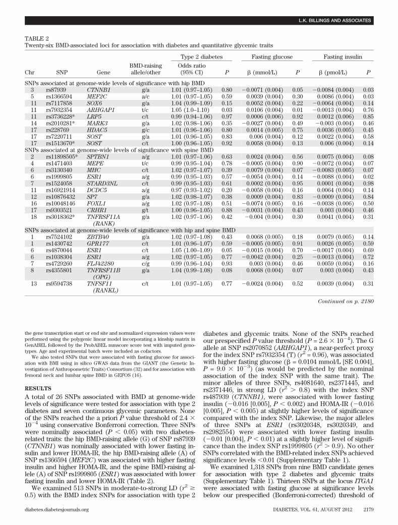
TABLE 2Twenty-six BMD-associated loci for association with diabetes and quantitative glycemic traits
Chr SNP GeneBMD-raisingallele/other
Type 2 diabetes Fasting glucose Fasting insulin
Odds ratio(95% CI) P b (mmol/L) P b (pmol/L) P
SNPs associated at genome-wide levels of significance with hip BMD3 rs87939 CTNNB1 g/a 1.01 (0.97–1.05) 0.80 20.0071 (0.004) 0.05 20.0084 (0.004) 0.035 rs1366594 MEF2C a/c 1.01 (0.97–1.05) 0.59 0.0039 (0.004) 0.30 0.0086 (0.004) 0.0311 rs7117858 SOX6 g/a 1.04 (0.99–1.09) 0.15 0.0052 (0.004) 0.22 20.0064 (0.004) 0.1411 rs7932354 ARHGAP1 t/c 1.05 (1.0–1.10) 0.03 0.0106 (0.004) 0.01 20.0013 (0.004) 0.7611 rs3736228* LRP5 c/t 0.99 (0.94–1.06) 0.97 0.0006 (0.006) 0.92 0.0012 (0.006) 0.8514 rs2010281* MARK3 g/a 1.02 (0.98–1.06) 0.35 20.0027 (0.004) 0.49 20.003 (0.004) 0.4617 rs228769 HDAC5 g/c 1.01 (0.96–1.06) 0.80 0.0014 (0.005) 0.75 0.0036 (0.005) 0.4517 rs7220711 SOST g/a 1.01 (0.96–1.05) 0.83 0.006 (0.004) 0.12 0.0022 (0.004) 0.5817 rs1513670* SOST c/t 1.00 (0.96–1.05) 0.92 0.0058 (0.004) 0.13 0.006 (0.004) 0.14
SNPs associated at genome-wide levels of significance with spine BMD2 rs11898505* SPTBN1 a/g 1.01 (0.97–1.06) 0.63 0.0024 (0.004) 0.56 0.0075 (0.004) 0.084 rs1471403 MEPE t/c 0.99 (0.95–1.04) 0.78 20.0005 (0.004) 0.90 20.0072 (0.004) 0.076 rs3130340 MHC c/t 1.02 (0.97–1.07) 0.39 0.0079 (0.004) 0.07 20.0083 (0.005) 0.076 rs1999805 ESR1 a/g 0.99 (0.95–1.03) 0.57 20.0054 (0.004) 0.14 20.0088 (0.004) 0.027 rs1524058 STARD3NL c/t 0.99 (0.95–1.03) 0.61 0.0002 (0.004) 0.95 0.0001 (0.004) 0.9811 rs16921914 DCDC5 a/g 0.97 (0.93–1.02) 0.20 20.0058 (0.004) 0.16 0.0064 (0.004) 0.1412 rs10876432 SP7 g/a 1.02 (0.98–1.07) 0.38 0.0009 (0.004) 0.83 20.0009 (0.004) 0.8416 rs10048146 FOXL1 a/g 1.02 (0.97–1.08) 0.51 20.0074 (0.005) 0.16 20.0038 (0.006) 0.5017 rs9303521 CRHR1 g/t 1.00 (0.96–1.05) 0.88 20.0031 (0.004) 0.43 0.003 (0.004) 0.4618 rs3018362* TNFRSF11A
(RANK)
g/a 1.02 (0.97–1.06) 0.42 20.004 (0.004) 0.30 0.0041 (0.004) 0.31
SNPs associated at genome-wide levels of significance with hip and spine BMD1 rs7524102 ZBTB40 g/a 1.02 (0.97–1.08) 0.43 0.0068 (0.005) 0.18 0.0079 (0.005) 0.141 rs1430742 GPR177 c/t 1.01 (0.96–1.07) 0.59 20.0005 (0.005) 0.91 0.0026 (0.005) 0.596 rs4870044 ESR1 c/t 1.05 (1.00–1.09) 0.05 20.0015 (0.004) 0.70 20.0017 (0.004) 0.696 rs1038304 ESR1 a/g 1.02 (0.97–1.05) 0.77 20.0042 (0.004) 0.25 20.0013 (0.004) 0.727 rs4729260 FLJ42280 c/g 0.99 (0.96–1.04) 0.93 0.003 (0.004) 0.46 0.0059 (0.004) 0.168 rs4355801 TNFRSF11B
(OPG)
g/a 1.04 (0.99–1.08) 0.08 0.0068 (0.004) 0.07 0.003 (0.004) 0.43
13 rs9594738 TNFSF11
(RANKL)
c/t 1.01 (0.97–1.05) 0.77 20.0024 (0.004) 0.52 0.0039 (0.004) 0.31
Continued on p. 2180
L.K. BILLINGS AND ASSOCIATES
diabetes.diabetesjournals.org DIABETES, VOL. 61, AUGUST 2012 2179

7.73 1025, of which 8 were below the study-wide significancethreshold (Table 3 and Fig. 2). By assembling an in silicoreplication sample of 19,417 individuals, we achieved .75%power (a = 0.05) to detect 1 SD difference in fasting glucose.Therefore, the top 13 ITGA1 SNPs were examined for asso-ciation with fasting glucose in the 12 additional cohorts. Themajor C allele of SNP rs6867040 was nominally associatedwith higher fasting glucose (P = 0.03) in a directionally con-sistent manner. None of the 13 SNPs reached genome-widesignificance (P , 5 3 1028) in the combined meta-analysis(Table 3). It is notable that variants in this locus, ITGA1, werenoted to be among the top 10 most significant associations forfive additional traits: type 2 diabetes, fasting insulin, HOMA-B,and 2-h glucose and insulin levels (Table 4).
To investigate the mechanism by which ITGA1 mightinfluence type 2 diabetes and related traits, we examinedthe effect of these 13 SNPs on cis-gene expression of ITGA1in liver and adipose tissue using eQTL analysis. ITGA1 ex-pression was measured in adipose tissue using a 50–basepair probe (chromosome 5:52,284,986–52,285,035) availableon the Illumina array and in liver tissue with a set of probescovering the length of the ITGA1 region (including the genePELO) on the Affymetrix array. The major allele of six SNPswas associated with increased expression (b ranged from
0.089 to 0.107 [SE 0.043–0.044]) of ITGA1/PELO in livertissue at P , 0.05, but no SNPs were associated withITGA1 expression in adipose tissue (Table 5). Of note, inadipose tissue, the major alleles of the 13 SNPs werehighly associated with lower PELO expression (effectestimates ;0.05 [SE ;0.01], lowest P , 2.0 3 1024). Todetermine whether PELO or ITGA1 gene expression wasdriving the association seen in liver tissue of the ITGA1expression, we examined probes for each exon in-dividually. We noted that for all of the genetic variants, theSNPs appeared to have a stronger association with theITGA1-specific probes than PELO-specific probes (an ex-ample figure of one of the SNPs, rs10512997, is provided inthe Supplementary Data). ITGA1 and PELO are bothexpressed in liver, adipose, and pancreatic islets, althoughITGA1 appears to have higher expression in these tissues(Supplementary Data).
We examined 13 SNPs in ITGA1 for association withBMI in the GIANT Consortium and BMD in the GEFOSConsortium. The major allele of seven SNPs was asso-ciated with higher BMI at P , 0.05 (Table 5). None ofthese SNPs were associated with femoral neck andlumbar spine BMD, although they trended toward low-ering BMD.
TABLE 2Continued
HOMA-B HOMA-IR HbA1c 2-h glucose 2-h insulin
Refb P b P b (%) P
b(mmol/L) P
b(mmol/L) P
20.0026 (0.003) 0.42 20.0103 (0.004) 0.01 20.0042 (0.006) 0.49 0.003 (0.019) 0.87 20.0073 (0.012) 0.54 (16)0.0032 (0.003) 0.34 0.01 (0.004) 0.02 20.0001 (0.006) 0.99 0.0167 (0.02) 0.40 20.004 (0.012) 0.75 (16)
20.0076 (0.004) 0.04 20.0052 (0.005) 0.26 20.0071 (0.007) 0.33 0.0075 (0.022) 0.73 20.0205 (0.014) 0.14 (16)20.0044 (0.004) 0.22 0.0013 (0.004) 0.76 20.0002 (0.007) 0.98 20.0028 (0.02) 0.89 0.0089 (0.013) 0.49 (16)
0.0 (0.006) 0.99 0.0024 (0.007) 0.71 0.0018 (0.009) 0.85 20.0172 (0.03) 0.57 0.0136 (0.019) 0.47 (17)0.0003 (0.004) 0.94 20.0015 (0.004) 0.73 20.0014 (0.007) 0.84 20.0085 (0.02) 0.67 20.0036 (0.012) 0.77 (15)0.0029 (0.004) 0.47 0.0028 (0.005) 0.56 0.0063 (0.008) 0.41 0.0071 (0.023) 0.76 20.0066 (0.015) 0.65 (16)
20.0004 (0.003) 0.92 0.0032 (0.004) 0.44 0.0052 (0.006) 0.42 20.0079 (0.019) 0.68 20.0153 (0.012) 0.21 (15)0.0045 (0.004) 0.21 0.0078 (0.004) 0.07 0.0019 (0.006) 0.76 0.0017 (0.02) 0.93 20.0002 (0.013) 0.99 (15)
20.0006 (0.004) 0.87 0.0073 (0.004) 0.10 0.0167 (0.007) 0.01 20.0522 (0.021) 0.01 20.0243 (0.013) 0.05 (16)20.0029 (0.004) 0.40 20.0061 (0.004) 0.15 0.0035 (0.006) 0.58 0.0014 (0.02) 0.94 0.0 (0.012) 1.00 (16)20.0032 (0.004) 0.40 20.0057 (0.005) 0.23 0.0096 (0.008) 0.23 0.0245 (0.023) 0.28 20.0125 (0.014) 0.37 (14)20.0051 (0.003) 0.13 20.0086 (0.004) 0.03 0.0115 (0.006) 0.06 0.024 (0.02) 0.23 0.001 (0.012) 0.93 (14)0.0013 (0.003) 0.71 0.0013 (0.004) 0.75 20.0052 (0.006) 0.39 20.0106 (0.02) 0.59 0.0073 (0.013) 0.57 (16)0.0068 (0.004) 0.06 0.0046 (0.005) 0.30 0.0058 (0.007) 0.42 20.0167 (0.021) 0.42 20.0046 (0.014) 0.74 (16)
20.0022 (0.004) 0.56 20.002 (0.005) 0.67 0.0019 (0.007) 0.78 20.0166 (0.021) 0.43 20.0064 (0.013) 0.62 (15)20.0013 (0.005) 0.78 20.0036 (0.006) 0.53 0.0116 (0.01) 0.24 20.0467 (0.026) 0.08 20.0159 (0.018) 0.38 (16)0.0015 (0.004) 0.66 0.0026 (0.004) 0.54 20.0052 (0.006) 0.41 0.0491 (0.02) 0.01 0.0221 (0.013) 0.09 (16)
0.0068 (0.003) 0.05 0.0034 (0.004) 0.42 0.0082 (0.007) 0.22 0.0117 (0.02) 0.55 20.0039 (0.012) 0.75 (15)
0.0013 (0.005) 0.78 0.0067 (0.006) 0.23 0.0022 (0.008) 0.79 20.0235 (0.026) 0.37 20.0134 (0.016) 0.40 (14)0.0015 (0.004) 0.72 0.0033 (0.005) 0.51 20.0119 (0.008) 0.12 20.03 (0.024) 0.22 20.0225 (0.015) 0.14 (16)
20.0024 (0.004) 0.50 20.0025 (0.004) 0.56 0.0024 (0.007) 0.73 20.022 (0.021) 0.30 0.0112 (0.013) 0.39 (14)0.0015 (0.003) 0.65 20.002 (0.004) 0.61 0.006 (0.006) 0.31 0.0096 (0.020) 0.62 0.0128 (0.012) 0.28 (14)0.0043 (0.004) 0.25 0.0054 (0.004) 0.23 0.0004 (0.007) 0.95 0.0243 (0.021) 0.25 0.0002 (0.013) 0.99 (16)
20.0029 (0.003) 0.38 0.0033 (0.004) 0.41 20.0032 (0.006) 0.60 0.0267 (0.019) 0.16 20.0132 (0.012) 0.27 (17)
0.0058 (0.003) 0.09 0.006 (0.004) 0.14 0.0113 (0.006) 0.06 20.016 (0.019) 0.40 0.0101 (0.012) 0.41 (14)
SEs are shown below the effect estimate; conversion factor (mmol/L 3 18 = mg/L). Ref, article where the genome-wide association for therespective SNP was described. *SNP also is associated with low trauma fracture. Chr, chromosome.
BONE-RELATED GENES AND GLYCEMIC TRAITS
2180 DIABETES, VOL. 61, AUGUST 2012 diabetes.diabetesjournals.org

DISCUSSION
By exploring genetic pleiotropy, we revealed a locus thatmay provide clues to a mechanism underlying the observedepidemiological association between type 2 diabetes andheightened fracture risk. We compiled a comprehensive listof BMD-related SNPs composed of genetic variants associ-ated with BMD at levels of genome-wide significance, var-iants in moderate-to-strong LD with the index SNPs, and
SNPs in BMD candidate genes. By examining these BMD-related SNPs for association with type 2 diabetes andglycemic traits, we discovered that SNPs in the ITGA1 locus,a BMD candidate gene, are suggestively associated withfasting glucose at study-wide levels of significance. Themajor alleles of these 13 highly correlated SNPs (CEUHapMap [Utah residents with ancestry from northern andwestern Europe] r
2 . 0.7) consistently raised fasting
TABLE 3SNPs in ITGA1 associated with fasting glucose Stage 1 and taken forward for replication
SNP FunctionEffect/other
allele
Stage 1(up to 46,262 participants)
Replication (Stage 2)(up to 19,417participants)
Combined(up to 64,188participants)
b (SE) P value b (SE) P value b (SE) P value
rs6881900 Intronic enhancer a/g 0.0167 (0.004) 3.131025 0.0092 (0.006) 0.14 0.0151 (0.003) 9.131026
rs17209725 Intronic c/t 0.0164 (0.004) 3.931025 0.0109 (0.006) 0.08 0.0154 (0.003) 6.231026
rs17209760 Intronic enhancer c/g 0.0164 (0.004) 3.931025 0.0108 (0.006) 0.08 0.0154 (0.003) 6.331026
rs10512997 Intronic c/t 0.0164 (0.004) 3.931025 0.0088 (0.006) 0.15 0.0148 (0.003) 1.431025
rs7716758 Upstream a/t 0.0165 (0.004) 4.131025 0.0113 (0.006) 0.07 0.0156 (0.003) 5.131026
rs12188019 Intronic enhancer t/c 0.0163 (0.004) 4.331025 0.0109 (0.006) 0.08 0.0154 (0.003) 6.731026
rs10940273 Intronic c/a 0.0176 (0.004) 4.531025 0.0103 (0.007) 0.15 0.0165 (0.004) 9.631026
rs6878212 Intronic t/a 0.0163 (0.004) 4.731025 0.0109 (0.006) 0.08 0.0153 (0.003) 6.831026
rs6867040 Intronic enhancer c/t 0.0165 (0.004) 6.731025 0.0142 (0.007) 0.03 0.0166 (0.004) 2.331026
rs6450088 Intronic a/g 0.0158 (0.004) 6.731025 0.0104 (0.006) 0.10 0.0148 (0.003) 1.431025
rs12153381 Intronic enhancer c/t 0.0157 (0.004) 6.931025 0.0092 (0.006) 0.14 0.0144 (0.003) 1.631025
rs10512998 Intronic enhancer a/t 0.0156 (0.004) 7.231025 0.0092 (0.006) 0.14 0.0143 (0.003) 1.731025
rs11886 Intronic t/g 0.0156 (0.004) 7.431025 0.0094 (0.006) 0.13 0.0144 (0.003) 1.731025
rs13179969* Intronic g/a 20.0013 (0.004) 0.76
Eight SNPs in ITGA1 were associated with fasting glucose below our study-wide P value threshold (P = 6.0 3 1025, in boldface type) in the 21discovery cohorts of MAGIC. The top 13 SNPs were promoted for follow-up with fasting glucose in 12 additional cohorts with in silicogenotype data. A combined analysis was then performed. SNP function was determined using FastSNP search (50). bs are expressed in mmol/L(conversion: mmol/L 3 18 = mg/L). Boldfaced alleles are the major allele per HapMap CEU. *rs13179969 major allele (G) was associated withlower lumbar spine BMD (b = 20.07 g/cm2) in a candidate gene study at study-wide significance (P = 9.6 3 1027) (20).
FIG. 2. SNPs at BMD-associated ITGA1 associated with fasting glucose. Thirteen SNPs (red diamonds) in ITGA1 were associated with fastingglucose levels (P < 7.7 3 10
25) in the MAGIC discovery cohorts, with 1 SNP (rs6867040) replicating at nominal significance (P < 0.05) in 12
replication cohorts. SNP rs13179969 (blue diamond) (ITGA1) was associated with lumbar spine BMD in GEFOS at 9.6 3 1027
(20). This SNP is notassociated with fasting glucose in MAGIC. LD is indicated by size of the diamond.
L.K. BILLINGS AND ASSOCIATES
diabetes.diabetesjournals.org DIABETES, VOL. 61, AUGUST 2012 2181
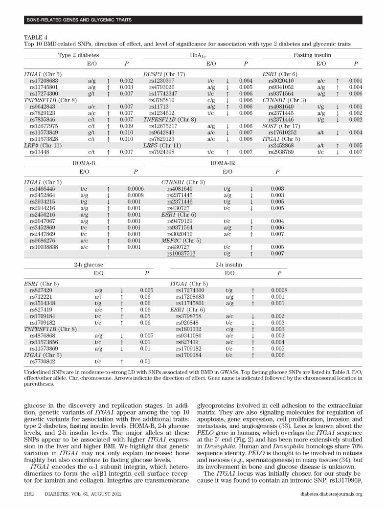
glucose in the discovery and replication stages. In addi-tion, genetic variants of ITGA1 appear among the top 10genetic variants for association with five additional traits:type 2 diabetes, fasting insulin levels, HOMA-B, 2-h glucoselevels, and 2-h insulin levels. The major alleles at theseSNPs appear to be associated with higher ITGA1 expres-sion in the liver and higher BMI. We highlight that geneticvariation in ITGA1 may not only explain increased bonefragility but also contribute to fasting glucose levels.
ITGA1 encodes the a-1 subunit integrin, which hetero-dimerizes to form the a1b1-integrin cell surface recep-tor for laminin and collagen. Integrins are transmembrane
glycoproteins involved in cell adhesion to the extracellularmatrix. They are also signaling molecules for regulation ofapoptosis, gene expression, cell proliferation, invasion andmetastasis, and angiogenesis (33). Less is known about thePELO gene in humans, which overlaps the ITGA1 sequenceat the 59 end (Fig. 2) and has been more extensively studiedin Drosophila. Human and Drosophila homologs share 70%sequence identity. PELO is thought to be involved in mitosisand meiosis (e.g., spermatogenesis) in many tissues (34), butits involvement in bone and glucose disease is unknown.
The ITGA1 locus was initially chosen for our study be-cause it was found to contain an intronic SNP, rs13179969,
TABLE 4Top 10 BMD-related SNPs, direction of effect, and level of significance for association with type 2 diabetes and glycemic traits
Type 2 diabetes HbA1c Fasting insulin
E/O P E/O P E/O P
ITGA1 (Chr 5) DUSP3 (Chr 17) ESR1 (Chr 6)rs17208683 a/g ↑ 0.002 rs1230397 t/c ↓ 0.004 rs3020410 a/c ↑ 0.001rs11745801 a/g ↑ 0.003 rs4793026 a/g ↓ 0.005 rs9341052 a/g ↑ 0.004rs17274300 g/t ↑ 0.007 rs17742347 t/c ↑ 0.006 rs9371564 a/g ↑ 0.006
TNFRSF11B (Chr 8) rs3785810 c/g ↓ 0.006 CTNNB1 (Chr 3)rs9642843 a/c ↑ 0.007 rs11713 a/g ↑ 0.006 rs4081640 t/g ↓ 0.001rs7829123 a/c ↑ 0.007 rs1234612 t/c ↓ 0.006 rs2371445 a/g ↓ 0.002rs7835846 c/t ↑ 0.007 TNFRSF11B (Chr 8) rs2371446 t/g ↓ 0.002rs12677975 c/t ↑ 0.009 rs12675217 a/g ↓ 0.006 SOST (Chr 17)rs11573849 g/t ↑ 0.010 rs9642843 a/c ↓ 0.007 rs17610252 a/t ↓ 0.004rs11573828 c/t ↑ 0.010 rs7829123 a/c ↓ 0.008 ITGA1 (Chr 5)
LRP4 (Chr 11) LRP5 (Chr 11) rs2452868 a/t ↑ 0.005rs13448 c/t ↑ 0.007 rs7924398 t/c ↑ 0.007 rs2938789 t/c ↓ 0.007
HOMA-B HOMA-IR
E/O P E/O P
ITGA1 (Chr 5) CTNNB1 (Chr 3)rs1466445 t/c ↑ 0.0006 rs4081640 t/g ↓ 0.003rs2452864 a/g ↓ 0.0008 rs2371445 a/g ↓ 0.003rs2934215 t/g ↓ 0.001 rs2371446 t/g ↓ 0.005rs2934216 a/g ↑ 0.001 rs430727 t/c ↓ 0.005rs2456216 a/g ↑ 0.001 ESR1 (Chr 6)rs2047067 a/g ↑ 0.001 rs9479129 t/c ↓ 0.004rs2452869 t/c ↑ 0.001 rs9371564 a/g ↑ 0.006rs2447869 t/c ↑ 0.001 rs3020410 a/c ↑ 0.007rs9686276 a/c ↑ 0.001 MEF2C (Chr 5)rs10038838 a/c ↑ 0.001 rs430727 t/c ↑ 0.005
rs10037512 t/g ↑ 0.007
2-h glucose 2-h insulin
E/O P E/O P
ESR1 (Chr 6) ITGA1 (Chr 5)rs827420 a/g ↓ 0.005 rs17274300 t/g ↑ 0.0008rs712221 a/t ↑ 0.06 rs17208683 a/g ↑ 0.001rs1514348 t/g ↑ 0.06 rs11745801 a/g ↑ 0.001rs827419 a/c ↑ 0.06 ESR1 (Chr 6)rs1709184 t/c ↑ 0.05 rs3798758 a/c ↓ 0.002rs1709182 t/c ↑ 0.06 rs926848 t/c ↓ 0.003
TNFRSF11B (Chr 8) rs1801132 c/g ↑ 0.003rs4876868 a/g ↓ 0.005 rs9341086 a/c ↓ 0.003rs11573856 t/c ↑ 0.01 rs827419 a/c ↑ 0.004rs11573869 a/g ↓ 0.01 rs1709182 t/c ↑ 0.005
ITGA1 (Chr 5) rs1709184 t/c ↑ 0.006rs7730842 t/c ↑ 0.01
Underlined SNPs are in moderate-to-strong LD with SNPs associated with BMD in GWASs. Top fasting glucose SNPs are listed in Table 3. E/O,effect/other allele. Chr, chromosome. Arrows indicate the direction of effect. Gene name is indicated followed by the chromosomal location inparentheses.
BONE-RELATED GENES AND GLYCEMIC TRAITS
2182 DIABETES, VOL. 61, AUGUST 2012 diabetes.diabetesjournals.org

whose G major allele had been associated with lowerlumbar spine BMD at levels of study-wide significance (P =9.6 3 1027) (20). This SNP was not associated with fastingglucose in our study, nor is it in strong LD with the 13SNPs followed up in this study (r2 , 0.05, HapMap CEU)(Fig. 2). Despite low LD between these SNPs, they point toa locus, ITGA1, in which in vivo and in vitro models havea suggested role in both bone disease and glucose homeo-stasis. Null ITGA1 mice have impaired fracture healing andcartilage remodeling (35), although it is not yet clear whatrole this gene product has on BMD or bone structure inanimal models. Furthermore, integrins have been examinedin an effort to culture and expand human b-cells for humantransplantation ex vivo (36). The a1b1-integrins appear toplay a role in b-cell insulin secretion, migration, and mes-enchymal transformation (37).
The mechanism by which ITGA1 may influence fastingglucose is not entirely clear. Fasting glucose is an estimateof hepatic glucose production after an overnight fast andcan indicate hepatic and peripheral insulin resistance (38).Our follow-up gene expression studies suggest that ITGA1genetic variation may affect fasting glucose via the liverrather than adipose tissue. We found that the major allelesof six of the SNPs tested were correlated with increasedhepatic expression of ITGA1 (P , 0.05). In addition, thesame top three SNPs were associated with both type 2diabetes and 2-h insulin level, suggesting that the mecha-nism may involve insulin resistance. Some studies suggesta role of integrins in insulin resistance (39). Integrins arethought to play a key role in the evolution of liver fibrosisbrought on by inflammation as seen in insulin resistance–associated nonalcoholic steatohepatitis (39).
In mice, the influence of ITGA1 and ITGA2 (encodingthe a-2 component of a2b1-integrin) on the effect of in-flammation on insulin resistance in muscle induced by a high-fat diet has been examined recently (40). The high-fat dietinduced extracellular matrix changes by increasing collagenaccumulation in muscle. The itga2
2/2 mice on a high-fatdiet had lower basal glucose than itga2
+/+ mice, suggestingthat the extracellular matrix–integrin signaling plays a rolein insulin resistance in muscle. The same observation wasnot seen for itga12/2 and itga1
+/+ mice. Given our study’sgene expression findings and the role of integrins in the liver
in response to inflammation and insulin resistance, furtherinvestigation of the liver in itga1 null mice in response toinflammation could reveal more information about the roleof ITGA1 in hepatic glucose production. Ultimately, ourstudy remains hypothesis generating and highlights a novellocus that links BMD and fasting glucose that warrants fur-ther investigation.
This study suggests that ITGA1 may exhibit geneticpleiotropy through its association with BMD and fastingglucose. True pleiotropy is difficult to confirm, especially ifa causal relationship exists between fasting glucose andBMD, as such a finding suggests the possibility of a medi-ating effect of one phenotype on the other. The evidence ofsuch a causal relationship between fasting glucose andbone density is not completely consistent. Although invitro studies show that chronic hyperglycemia may impairosteoblast function (41,42), clinical studies demonstratethat individuals with type 2 diabetes have lower bone turn-over (43), which usually indicates a more optimal skeletalstate. On the other hand, those with poorly controlled di-abetes have been shown to have improvement in BMDmeasured by bone densitometry after 1 year of tightenedcontrol (44). Therefore, it is not possible to clearly establisha direct link between hyperglycemia and BMD (45). Like-wise, if there was a common intermediate phenotype drivingthe relationship between BMD and fasting glucose, then ourfindings may not indicate true genetic pleiotropy. BMI couldbe considered a potential intermediate phenotype because itis correlated with both type 2 diabetes pathogenesis andBMD (46,47). We examined the ITGA1-related SNPs forassociation with BMI in the GIANT Consortium (32). Severalof the variants reached a nominal level of significance(lowest P = 0.007) for association with BMI (Table 4). Thesedata suggest that ITGA1 may act on BMD or fasting glucosethrough the intermediate phenotype of BMI. Although theITGA1 locus has not been associated with BMI in the past,the intronic SNP rs7723398 (r2 , 0.3 per CEU with the SNPsfollowed up in this study) has been found to be associatedwith another anthropometric trait, brachial circumference(P = 9.7 3 1026
), in a Croatian population (48).The strengths of our study include the comprehensive
bone-related SNP selection from recently published GWASdata and the ability to test them in very large, well-phenotyped
TABLE 5Association of ITGA1 genetic variation with ITGA1 RNA expression and BMI
SNPEffect/other
allele
Association of SNPs with ITGA1 eQTL Association of SNPs with BMI
Liver Adipose BMI
b (SE) P b (SE) P b (SE) P
rs6867040* c/t 0.073 (0.043) 0.09 20.018 (0.013) 0.21 0.012 (0.005) 0.03
rs7716758 a/t 0.064 (0.043) 0.15 20.015 (0.014) 0.16 0.011 (0.005) 0.04
rs12188019* t/c 0.071 (0.043) 0.10 20.018 (0.013) 0.17 0.011 (0.005) 0.04
rs17209725* c/t 0.084 (0.043) 0.05 20.018 (0.013) 0.18 0.011 (0.005) 0.04
rs6878212* t/a 0.07 (0.043) 0.11 20.018 (0.013) 0.17 0.011 (0.005) 0.04
rs17209760* c/g 0.084 (0.043) 0.05 20.018 (0.013) 0.18 0.011 (0.005) 0.04
rs6450088 a/g 0.094 (0.043) 0.03 20.018 (0.014) 0.34 0.009 (0.005) 0.07rs11886* t/g 0.089 (0.043) 0.04 20.015 (0.013) 0.25 0.01 (0.005) 0.06rs10940273 c/a 0.078 (0.044) 0.08 20.018 (0.014) 0.20 0.015 (0.006) 0.007
rs10512998 a/t 0.107 (0.043) 0.01 20.015 (0.013) 0.28 0.01 (0.005) 0.05rs12153381 c/t 0.1 (0.043) 0.02 20.015 (0.013) 0.26 0.01 (0.005) 0.05rs6881900 a/g 0.099 (0.043) 0.02 20.015 (0.013) 0.28 0.01 (0.005) 0.05rs10512997 c/t 0.095 (0.043) 0.03 20.015 (0.013) 0.28 0.01 (0.005) 0.05
*SNP overlies both ITGA1 and PELO gene. The boldfaced P values denote nominal significance (P , 0.05).
L.K. BILLINGS AND ASSOCIATES
diabetes.diabetesjournals.org DIABETES, VOL. 61, AUGUST 2012 2183

type 2 diabetes and glycemic traits consortia. We wereable to replicate our findings from the discovery phase inan additional ;19,000 individuals. We also followed up thegenetic variants with eQTL analysis and other relatedtraits. Our results may help explain the, as yet not quitewell understood, epidemiological link between type 2 dia-betes and bone disease. This study has highlighted the ne-cessity to examine genetic variants not reaching thegenome-wide significance threshold because this mayuncover potential findings buried in the P value distribution.Given that the MAGIC discovery dataset has been publishedsince the completion of our analyses, further studies likeours can be pursued (www.magicinvestigators.org). Fur-thermore, the BMD-related locus that was associated withfasting glucose was selected from a candidate gene study.This illustrates the importance of examining candidate genesin discovering genetic pleiotropy rather than solely exam-ining loci associated at levels of genome-wide significance.
We are limited by having chosen SNPs from GWASs ex-amining only BMD. Even though BMD is predictive offracture in people with type 2 diabetes (10), studies showthat individuals with type 2 diabetes have a higher risk offracture despite higher BMD in general (4). By examininggenetic variants related to BMD only, we may miss the non–BMD related genetic contribution to fracture risk. In addi-tion, our findings do not explain the observed paradox ofgenerally higher BMD and yet higher fracture risk amongpeople with type 2 diabetes (4). A direct genetic test of thisparadox using ITGA1 SNPs is not possible because theSNPs that influence fasting glucose and BMD at this locusare not correlated. In addition, a follow-up study examiningfracture-related genetic variants for association with type 2diabetes and glycemic traits will be warranted when largefracture GWASs become available. In a similar manner, theexamination of glycemia-related SNPs for association withBMD and fracture phenotypes may further explain the re-lationship between bone disease and type 2 diabetes, andthese studies are currently under way.
Despite the large sample size, none of the SNPs reachedgenome-wide significance in the combined analysis. Wemay need a larger sample size to determine if the ITGA1SNPs that were associated with fasting glucose will repli-cate in other populations and attain genome-wide signifi-cance because our replication sample may have been toosmall to detect the association found in the discovery stage.We estimate that we need an additional 12,000 participantsto see an association between the ITGA1 SNPs and fastingglucose at the same effect sizes seen in the discovery stage.Fortunately, ongoing deployment of the custom-madeMetabo-Chip (comprising .200,000 SNPs related to cardio-vascular disease, obesity, and type 2 diabetes) across manythousands of samples with relevant phenotypes may providesufficient power to uncover novel associations at genome-wide significance levels. The ITGA1 SNPs rs6881900 andrs10940273, found to be associated with fasting glucose inour study, are present in the Metabo-Chip. This provides anexciting opportunity to understand the relationship of ITGA1with glycemic traits, as well as other metabolic phenotypes incardiovascular disease and obesity.
In sum, we have identified a new locus candidate, ITGA1,influencing both fasting glucose and BMD, that may begin toexplain the genetic contribution to the epidemiological obser-vations linking type 2 diabetes and osteoporosis. The ongoinganalysis of Metabo-Chip genotypes across large samples willhelp determine if ITGA1 proves to be a new locus associatedwith fasting glucose at levels of genome-wide significance.
New insights into the genetic pleiotropy of both disease statesmay further underscore the link between skeletal and glucosemetabolism, highlight the complexity of this relationship, pro-vide a focus for future investigations, raise awareness foradverse effects in one system while treating another, and re-veal potential targets for disease therapies in both diseases.
ACKNOWLEDGMENTS
L.K.B. has received support from National Research Ser-vice Award Institutional Training Grant T32-DK-007028-35to the Massachusetts General Hospital, National Institutesof Health (NIH) Loan Repayment Award National Instituteof Diabetes and Digestive and Kidney Diseases (NIDDK) 1-L30-DK-089944-01, the Endocrine Society Lilly EndocrineScholars Award, and a Doris Duke Charitable FoundationDistinguished Scientist Clinical Award to David Altshuler.Y.-H.H. was supported by NIH/National Institute of Arthritisand Musculoskeletal and Skin Diseases (NIAMS) Grant R21-AR-056405. J.D. has received support from NIDDK R01-DK-078616. J.B.M. was supported by NIDDK R01-DK-078616and NIDDK K24-DK-080140. D.P.K. has received supportfrom NIAMS and National Institute on Aging Grant R01-AR/AG-41398. J.C.F. was supported by NIDDK R01-DK-078616 and a Doris Duke Charitable Foundation ClinicalScientist Development Award.
J.C.F. has received consulting honoraria from Novartis,Eli Lilly, and Pfizer. No other potential conflicts of interestrelevant to this article were reported.
L.K.B. wrote the manuscript and researched data. Y.-H.H.researched data, contributed to discussion, and reviewedand edited the manuscript. R.J.A. formatted the tables andreviewed and edited the manuscript. J.D. performed themeta-analysis and reviewed and edited the manuscript. B.F.V.,L.J.R.-T., S.H., M.L., D.B., C.La., J.H., M.F., N.B.-N., C.Le.,P.A., P.K.M., I.S., S.R., L.C., C.D., J.K., K.O.K., N.L.P., I.B.B.,M.A.P., B.B., P.F., A.R.S., L.J.P., N.W., P.M., T.J., and J.S.P.researched and provided data from their respective cohortsand reviewed and edited the manuscript. L.F., E.G., and P.E.researched and provided eQTL analysis and reviewed andedited the manuscript. D.K., J.B.M., and D.P.K. contributedto discussion and reviewed and edited the manuscript. J.C.F.contributed to discussion and wrote the manuscript. L.K.B.is the guarantor of this work and, as such, had full access toall of the data in the study and takes responsibility for theintegrity of the data and the accuracy of the data analysis.
Parts of this study were presented in abstract form at the12th International Congress of Human Genetics Meeting,Montreal, Quebec, Canada, 11–15 October 2011.
The authors would like to thank Denis Rybin at BostonUniversity School of Public Health for creating Fig. 2. Theauthors acknowledge the contribution of the GIANTConsortium, which provided summary statistics for theassociation between selected SNPs and BMI. Individual cohortacknowledgments are as follows: GEFOS Consortium (www.gefos.org) is funded by the European Commission (HEALTH-F2-2008-201865-GEFOS). ARIC is carried out as a collaborativestudy supported by National Heart, Lung, and Blood Institute(NHLBI) contracts (HHSN268201100005C, HHSN268201100006C, HHSN268201100007C, HHSN268201100008C, HHSN268201100009C, HHSN268201100010C, HHSN268201100011C,and HHSN268201100012C), R01-HL-087641, R01-HL-59367,and R01-HL-086694, National Human Genome Research Insti-tute contract U01-HG-004402, and NIH contract HHSN268200625226C. The authors thank the staff and partici-pants of the ARIC study for their important contributions.
BONE-RELATED GENES AND GLYCEMIC TRAITS
2184 DIABETES, VOL. 61, AUGUST 2012 diabetes.diabetesjournals.org

Infrastructure of the ARIC study was partly supported byGrant UL1-RR-025005, a component of the NIH and NIHRoadmap for Medical Research. The Fenland Study is fundedby the Wellcome Trust and the Medical Research Council.The authors are grateful to all the volunteers for their timeand help and to the general practitioners and practice staff forhelp with recruitment. The authors thank the Fenland StudyInvestigators, Fenland Study Co-ordination Team and the Ep-idemiology Field, Data, and Technical Teams. Biochemicalassays were performed by the National Institute for HealthResearch, Cambridge Biomedical Research Centre, Core Bio-chemistry Assay Laboratory, and the Cambridge UniversityHospitals National Health Service Foundation Trust, Depart-ment of Clinical Biochemistry. The BHS acknowledges thegenerous support for the 1994/1995 follow-up study fromHealthway, Western Australia, and the numerous Busseltoncommunity volunteers who assisted with data collection andthe study participants from the Shire of Busselton. The BHSis supported by the Great Wine Estates of the MargaretRiver region of Western Australia. The AMISH Cohort wassupported by NIH research grants U01-HL-72515, R01-DK-04261, and R01-AG-18728; University of Maryland GeneralClinical Research Center Grant M01-RR-16500; the Mid-Atlantic Nutrition and Obesity Research Center (P30-DK-072488); the Baltimore Diabetes Research and TrainingCenter (P60-DK-079637); and the Baltimore Veterans Ad-ministration Medical Center Geriatric Research and Educa-tion Clinical Center. French genetic studies (DESIR andFrench Obese) were supported in part by the Conseil Re-gional Nord-Pas-de-Calais: Fonds européen de développe-ment économique et regional, Genome Quebec–GenomeCanada and the British Medical Research Council. Theauthors acknowledge INSERM (employer of N.B.-N.).FamHS work was supported in part by NIH grants 5-R01-HL-08770003 and 5-R01-HL-08821502 from the NHLBI(to M.A.P.) and 5-R01-DK-07568102 and 5-R01-DK-06833603from NIDDK (to I.B.B.). The GenomEUtwin project is sup-ported by the European Commission under the programQuality of Life and Management of the Living Resourcesof Fifth Framework Programme (no. QLG2-CT-2002-01254)and the European Community’s Seventh Framework Pro-gramme (FP7/2007-2013), ENGAGE Consortium, GrantAgreement HEALTH-F4-2007-201413. The Swedish TwinCohort would like to acknowledge the Swedish ResearchCouncil and the Swedish Foundation for Strategic Re-search. The Finnish Twin Cohort would like to acknowl-edge the Center of Excellence in Complex DiseaseGenetics of the Academy of Finland. The GEMINAKARstudy was supported by the Danish Medical Research Coun-cil, the Danish Heart Association, the Danish Diabetes As-sociation, and GenomEUtwin. The FHS component of thiswork was supported by the NHLBI’s FHS (Contract N01-HC-25195), its contract with Affymetrix, Inc. for genotyp-ing services (Contract N02-HL-6-4278), and the resourcesof the FHS SNP Health Association Resource (SHARe)project, the Boston University Linux Cluster for GeneticAnalysis (LinGA) funded by the NIH National Centerfor Research Resources Shared Instrumentation Grant1-S10-RR-163736-01A1, and the Robert Dawson EvansEndowment of the Department of Medicine at BostonUniversity School of Medicine and Boston Medical Cen-ter. The ASAP study liver eQTL data were supportedby the Swedish Research Council (12660), the Swed-ish Heart-Lung Foundation, the European Commission(FAD, Health-F2-2008-200647), and a donation by FredrikLundberg.
REFERENCES
1. Vestergaard P. Discrepancies in bone mineral density and fracture risk inpatients with type 1 and type 2 diabetes—a meta-analysis. Osteoporos Int2007;18:427–444
2. Schwartz AV, Sellmeyer DE, Ensrud KE, et al.; Study of OsteoporoticFeatures Research Group. Older women with diabetes have an increasedrisk of fracture: a prospective study. J Clin Endocrinol Metab 2001;86:32–38
3. Strotmeyer ES, Cauley JA, Schwartz AV, et al. Nontraumatic fracture riskwith diabetes mellitus and impaired fasting glucose in older white andblack adults: the health, aging, and body composition study. Arch InternMed 2005;165:1612–1617
4. Bonds DE, Larson JC, Schwartz AV, et al. Risk of fracture in women withtype 2 diabetes: the Women’s Health Initiative Observational Study. J ClinEndocrinol Metab 2006;91:3404–3410
5. Melton LJ 3rd, Leibson CL, Achenbach SJ, Therneau TM, Khosla S. Frac-ture risk in type 2 diabetes: update of a population-based study. J BoneMiner Res 2008;23:1334–1342
6. Janghorbani M, Van Dam RM, Willett WC, Hu FB. Systematic review of type1 and type 2 diabetes mellitus and risk of fracture. Am J Epidemiol 2007;166:495–505
7. Burghardt AJ, Issever AS, Schwartz AV, et al. High-resolution peripheralquantitative computed tomographic imaging of cortical and trabecularbone microarchitecture in patients with type 2 diabetes mellitus. J ClinEndocrinol Metab 2010;95:5045–5055
8. Petit MA, Paudel ML, Taylor BC, et al.; Osteoporotic Fractures in Men(MrOs) Study Group. Bone mass and strength in older men with type 2diabetes: the Osteoporotic Fractures in Men Study. J Bone Miner Res 2010;25:285–291
9. Melton LJ 3rd, Riggs BL, Leibson CL, et al. A bone structural basis forfracture risk in diabetes. J Clin Endocrinol Metab 2008;93:4804–4809
10. Schwartz AV, Vittinghoff E, Bauer DC, et al.; Study of Osteoporotic Frac-tures (SOF) Research Group; Osteoporotic Fractures in Men (MrOS) Re-search Group; Health, Aging, and Body Composition (Health ABC) ResearchGroup. Association of BMD and FRAX score with risk of fracture in olderadults with type 2 diabetes. JAMA 2011;305:2184–2192
11. Ferron M, Wei J, Yoshizawa T, et al. Insulin signaling in osteoblastsintegrates bone remodeling and energy metabolism. Cell 2010;142:296–308
12. Lee NK, Sowa H, Hinoi E, et al. Endocrine regulation of energy metabolismby the skeleton. Cell 2007;130:456–469
13. Sivakumaran S, Agakov F, Theodoratou E, et al. Abundant pleiotropy inhuman complex diseases and traits. Am J Hum Genet 2011;89:607–618
14. Styrkarsdottir U, Halldorsson BV, Gretarsdottir S, et al. Multiple geneticloci for bone mineral density and fractures. N Engl J Med 2008;358:2355–2365
15. Styrkarsdottir U, Halldorsson BV, Gretarsdottir S, et al. New sequencevariants associated with bone mineral density. Nat Genet 2009;41:15–17
16. Rivadeneira F, Styrkársdottir U, Estrada K, et al.; Genetic Factors forOsteoporosis (GEFOS) Consortium. Twenty bone-mineral-density lociidentified by large-scale meta-analysis of genome-wide association studies.Nat Genet 2009;41:1199–1206
17. Richards JB, Rivadeneira F, Inouye M, et al. Bone mineral density, oste-oporosis, and osteoporotic fractures: a genome-wide association study.Lancet 2008;371:1505–1512
18. Hsu YH, Zillikens MC, Wilson SG, et al. An integration of genome-wideassociation study and gene expression profiling to prioritize the discoveryof novel susceptibility loci for osteoporosis-related traits. PLoS Genet2010;6:e1000977
19. Johnson AD, Handsaker RE, Pulit SL, Nizzari MM, O’Donnell CJ, de BakkerPI. SNAP: a web-based tool for identification and annotation of proxySNPs using HapMap. Bioinformatics 2008;24:2938–2939
20. Richards JB, Kavvoura FK, Rivadeneira F, et al.; Genetic Factors for Os-teoporosis Consortium. Collaborative meta-analysis: associations of 150candidate genes with osteoporosis and osteoporotic fracture. Ann InternMed 2009;151:528–537
21. Voight BF, Scott LJ, Steinthorsdottir V, et al.; MAGIC investigators; GIANTConsortium. Twelve type 2 diabetes susceptibility loci identified throughlarge-scale association analysis. Nat Genet 2010;42:579–589
22. Dupuis J, Langenberg C, Prokopenko I, et al.; DIAGRAM Consortium;GIANT Consortium; Global BPgen Consortium; Anders Hamsten on behalfof Procardis Consortium; MAGIC investigators. New genetic loci impli-cated in fasting glucose homeostasis and their impact on type 2 diabetesrisk. Nat Genet 2010;42:105–116
23. Soranzo N, Sanna S, Wheeler E, et al.; WTCCC. Common variants at 10genomic loci influence hemoglobin A₁(C) levels via glycemic and
L.K. BILLINGS AND ASSOCIATES
diabetes.diabetesjournals.org DIABETES, VOL. 61, AUGUST 2012 2185

nonglycemic pathways [corrected in: Diabetes 2011;60:1050–1051]. Di-abetes 2010;59:3229–3239
24. Saxena R, Hivert MF, Langenberg C, et al.; GIANT consortium; MAGICinvestigators. Genetic variation in GIPR influences the glucose and insulinresponses to an oral glucose challenge. Nat Genet 2010;42:142–148
25. Matthews DR, Hosker JP, Rudenski AS, Naylor BA, Treacher DF, TurnerRC. Homeostasis model assessment: insulin resistance and beta-cellfunction from fasting plasma glucose and insulin concentrations in man.Diabetologia 1985;28:412–419
26. Nyholt DR. A simple correction for multiple testing for single-nucleotidepolymorphisms in linkage disequilibrium with each other. Am J HumGenet 2004;74:765–769
27. Li J, Ji L. Adjusting multiple testing in multilocus analyses using the ei-genvalues of a correlation matrix. Heredity (Edinb) 2005;95:221–227
28. Willer CJ, Li Y, Abecasis GR. METAL: fast and efficient meta-analysis ofgenomewide association scans. Bioinformatics 2010;26:2190–2191
29. Folkersen L, van’t Hooft F, Chernogubova E, et al.; BiKE and ASAP studygroups. Association of genetic risk variants with expression of proximalgenes identifies novel susceptibility genes for cardiovascular disease[corrected in: Circ Cardiovasc Genet 2010;3:e5]. Circ Cardiovasc Genet2010;3:365–373
30. Nica AC, Parts L, Glass D, et al.; MuTHER Consortium. The architecture ofgene regulatory variation across multiple human tissues: the MuTHERstudy. PLoS Genet 2011;7:e1002003
31. Marchini J, Howie B, Myers S, McVean G, Donnelly P. A new multipointmethod for genome-wide association studies by imputation of genotypes.Nat Genet 2007;39:906–913
32. Speliotes EK, Willer CJ, Berndt SI, et al.; MAGIC; Procardis Consortium.Association analyses of 249,796 individuals reveal 18 new loci associatedwith body mass index. Nat Genet 2010;42:937–948
33. Kufe DW, Pollock RE, Weichselbaum RR, et al. (Eds.). Holland-Frei
Cancer Medicine. 6th ed. Hamilton, Ontario, Canada, BC Decker, 200334. Shamsadin R, Adham IM, von Beust G, Engel W. Molecular cloning, ex-
pression and chromosome location of the human pelota gene PELO.Cytogenet Cell Genet 2000;90:75–78
35. Ekholm E, Hankenson KD, Uusitalo H, et al. Diminished callus size andcartilage synthesis in alpha 1 beta 1 integrin-deficient mice during bonefracture healing. Am J Pathol 2002;160:1779–1785
36. Kaido T, Yebra M, Cirulli V, Rhodes C, Diaferia G, Montgomery AM. Impactof defined matrix interactions on insulin production by cultured humanbeta-cells: effect on insulin content, secretion, and gene transcription.Diabetes 2006;55:2723–2729
37. Kaido T, Yebra M, Cirulli V, Montgomery AM. Regulation of human beta-cell adhesion, motility, and insulin secretion by collagen IV and its receptoralpha1beta1. J Biol Chem 2004;279:53762–53769
38. Turner RC, Holman RR. Insulin rather than glucose homoeostasis in thepathophysiology of diabetes. Lancet 1976;1:1272–1274
39. Patsenker E, Stickel F. Role of integrins in fibrosing liver diseases. AmJ Physiol Gastrointest Liver Physiol 2011;301:G425–G434
40. Kang L, Ayala JE, Lee-Young RS, et al. Diet-induced muscle insulin re-sistance is associated with extracellular matrix remodeling and interactionwith integrin alpha2beta1 in mice. Diabetes 2011;60:416–426
41. Balint E, Szabo P, Marshall CF, Sprague SM. Glucose-induced inhibition ofin vitro bone mineralization. Bone 2001;28:21–28
42. Botolin S, McCabe LR. Chronic hyperglycemia modulates osteoblast geneexpression through osmotic and non-osmotic pathways. J Cell Biochem2006;99:411–424
43. Vestergaard P, Rejnmark L, Mosekilde L. Are antiresorptive drugs effectiveagainst fractures in patients with diabetes? Calcif Tissue Int 2011;88:209–214
44. Gregorio F, Cristallini S, Santeusanio F, Filipponi P, Fumelli P. Osteopeniaassociated with non-insulin-dependent diabetes mellitus: what are thecauses? Diabetes Res Clin Pract 1994;23:43–54
45. Schwartz AV, Sellmeyer DE. Diabetes, fracture, and bone fragility. CurrOsteoporos Rep 2007;5:105–111
46. Glauber HS, Vollmer WM, Nevitt MC, Ensrud KE, Orwoll ES. Body weightversus body fat distribution, adiposity, and frame size as predictors ofbone density. J Clin Endocrinol Metab 1995;80:1118–1123
47. Vazquez G, Duval S, Jacobs DR Jr, Silventoinen K. Comparison of bodymass index, waist circumference, and waist/hip ratio in predicting incidentdiabetes: a meta-analysis. Epidemiol Rev 2007;29:115–128
48. Polasek O, Marusić A, Rotim K, et al. Genome-wide association study ofanthropometric traits in Korcula Island, Croatia. Croat Med J 2009;50:7–16
49. Hindorff LA, MacArthur J (European Bioinformatics Institute), Wise A, et al.A catalog of published genome-wide association studies. Available fromwww.genome.gov/gwasstudies. Accessed 23 August 2011
50. Yuan HY, Chiou JJ, Tseng WH, et al. FASTSNP: an always up-to-date andextendable service for SNP function analysis and prioritization. NucleicAcids Res 2006;34(Web Server issue):W635–W641
51. Sever PS, Dahlof B, Poulter NR, et al. Prevention of coronary and strokeevents with atorvastatin in hypertensive patients who have average orlower-than-average cholesterol concentrations, in the Anglo-ScandinavianCardiac Outcomes Trial–Lipid Lowering Arm (ASCOT-LLA): a multicentrerandomised controlled trial. Lancet 2003;361:1149–1158
BONE-RELATED GENES AND GLYCEMIC TRAITS
2186 DIABETES, VOL. 61, AUGUST 2012 diabetes.diabetesjournals.org