Bio Fuel Oil - Upgrading by Hot Filtration and Novel ... - CORDIS

Impact Directly Over the Cardiac Silhouette IsNecessary to Produce Ventricular Fibrillationin an Experimental Model of Commotio CordisMark S. Link, MD, FACC,* Barry J. Maron, MD, FACC,† Brian A. VanderBrink, BS,*Masaaki Takeuchi, MD,* Natesa G. Pandian, MD, FACC,* Paul J. Wang, MD, FACC,*N. A. Mark Estes, III, MD, FACC*Boston, Massachusetts and Minneapolis, Minnesota
OBJECTIVES In an experimental model of sudden death from chest wall impact (commotio cordis), wesought to define the chest wall areas important in the initiation of ventricular fibrillation (VF).
BACKGROUND Sudden death can result from an innocent chest blow by a baseball or other projectile.Observations in humans suggest that these lethal blows occur over the precordium. However,the precise location of impact relative to the risk of sudden death is unknown.
METHODS Fifteen swine received 178 chest impacts with a regulation baseball delivered at 30 mph atthree sites over the cardiac silhouette (i.e., directly over the center, base or apex of the leftventricle [LV]) and four noncardiac sites on the left and right chest wall. Chest blows weregated to the vulnerable portion of the cardiac cycle for the induction of VF.
RESULTS Only chest impacts directly over the heart triggered VF (12 of 78: 15% vs. 0 of 100 fornoncardiac sites: p , 0.0001). Blows over the center of the heart (7 of 23; 30%) were morelikely to initiate VF than impacts at other precordial sites (5 of 55; 9%, p 5 0.02). Peak LVpressures generated instantaneously by the chest impact were directly related to the risk of VF(p , 0.0006).
CONCLUSIONS For nonpenetrating, low-energy chest blows to cause sudden death, impact must occurdirectly over the heart. Initiation of VF may be mediated by an abrupt and substantial increasein intracardiac pressure. Prevention of sudden death from chest blows during sports requiresthat protective equipment be designed to cover all portions of the chest wall that overlie theheart, even during body movements and positional changes that may occur with athleticactivities. (J Am Coll Cardiol 2001;37:649–54) © 2001 by the American College ofCardiology
Commotio cordis, or sudden death due to relatively low-energy chest wall impact without structural injury to thesternum, ribs or the heart itself, has been reported withincreasing frequency in young individuals participating insporting activities (1–4). These tragic events predominantlyoccur in youths age 4 to 16 years. While most commonlydue to chest blows from baseballs, commotio cordis has alsobeen described in hockey, softball, lacrosse, karate, fist fightsor in any situation in which a relatively hard projectile orbody part strikes the unprotected chest wall. In a series of 70victims of commotio cordis, all impacts appeared to be overor to the left of the sternum (1,3) although the exact impactsite was difficult to assess with precision. Victims’ collapseimmediately after the blow, and the most common initialrecorded rhythm is ventricular fibrillation (VF) (1,5–10).Some form of chest wall protection had been present in aminority of the victims; however, there is evidence in severalcases that critical areas of the precordium neverthelessbecame exposed and struck during sporting play (1,3).
In an experimental model of commotio cordis employinga baseball propelled at 30 mph, we have demonstrated theimportance that the precise timing of the blow plays in theinitiation of VF (11) in which impact is required during anarrow window of repolarization (10 to 30 ms before the Twave peak). However, the precise areas of the chest wallmost vulnerable to sudden death from impact, as well as thehemodynamic and cellular determinants of the VF, are notcompletely understood. Such information is crucial to theoptimal design of protective devices for young people inorganized sports and the prevention of sudden death fromchest wall blows.
METHODS
Animals. Juvenile domesticated swine, four to eight weeksold and weighing 8 to 12 kg, were used in this study. Theresearch protocol was approved by the Animal ResearchCommittee of the New England Medical Center in con-formity with the regulations of the Association for Assess-ment and Accreditation of Laboratory Animal Care. Ani-mals were anesthetized with ketamine and isoflurane andintubated; anesthesia was maintained with 1% to 2% isoflu-rane mixed with oxygen and nitrous oxide. Millar Mikrotip(Houston, Texas) pressure catheters were placed in the leftventricle (LV), and a transesophageal echocardiographic
From the *Center for the Cardiovascular Evaluation of Athletes, The CardiacArrhythmia Center, New England Medical Center, Tufts University School ofMedicine, Boston, Massachusetts, and the †Cardiovascular Research Division,Minneapolis Heart Institute Foundation, Minneapolis, Minnesota. Supported by theNational Operating Committee on Standards for Athletic Equipment (NOCSAE),Overland Park, Kansas and the Earl P. Charlton Research Fund of Tufts University.
Manuscript received May 23, 2000; revised manuscript received September 14,2000, accepted October 18, 2000.
Journal of the American College of Cardiology Vol. 37, No. 2, 2001© 2001 by the American College of Cardiology ISSN 0735-1097/01/$20.00Published by Elsevier Science Inc. PII S0735-1097(00)01142-6
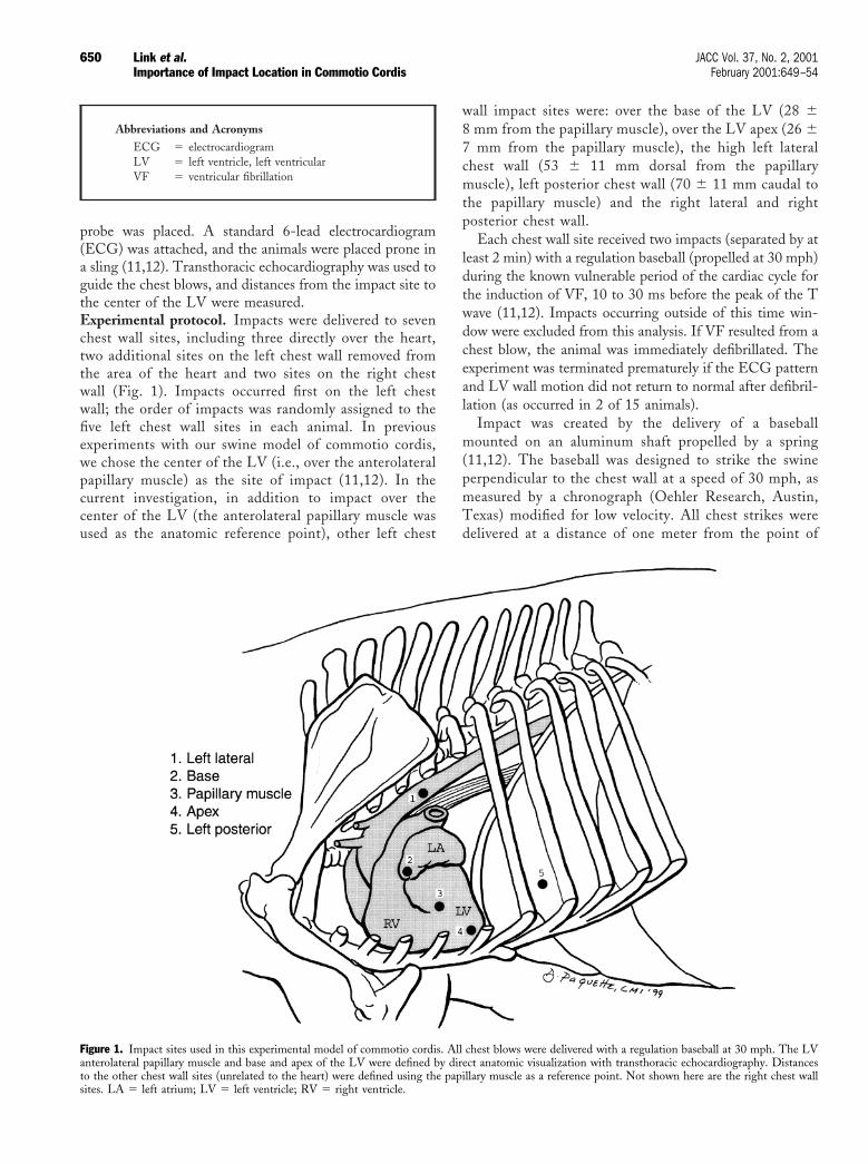
probe was placed. A standard 6-lead electrocardiogram(ECG) was attached, and the animals were placed prone ina sling (11,12). Transthoracic echocardiography was used toguide the chest blows, and distances from the impact site tothe center of the LV were measured.Experimental protocol. Impacts were delivered to sevenchest wall sites, including three directly over the heart,two additional sites on the left chest wall removed fromthe area of the heart and two sites on the right chestwall (Fig. 1). Impacts occurred first on the left chestwall; the order of impacts was randomly assigned to thefive left chest wall sites in each animal. In previousexperiments with our swine model of commotio cordis,we chose the center of the LV (i.e., over the anterolateralpapillary muscle) as the site of impact (11,12). In thecurrent investigation, in addition to impact over thecenter of the LV (the anterolateral papillary muscle wasused as the anatomic reference point), other left chest
wall impact sites were: over the base of the LV (28 68 mm from the papillary muscle), over the LV apex (26 67 mm from the papillary muscle), the high left lateralchest wall (53 6 11 mm dorsal from the papillarymuscle), left posterior chest wall (70 6 11 mm caudal tothe papillary muscle) and the right lateral and rightposterior chest wall.
Each chest wall site received two impacts (separated by atleast 2 min) with a regulation baseball (propelled at 30 mph)during the known vulnerable period of the cardiac cycle forthe induction of VF, 10 to 30 ms before the peak of the Twave (11,12). Impacts occurring outside of this time win-dow were excluded from this analysis. If VF resulted from achest blow, the animal was immediately defibrillated. Theexperiment was terminated prematurely if the ECG patternand LV wall motion did not return to normal after defibril-lation (as occurred in 2 of 15 animals).
Impact was created by the delivery of a baseballmounted on an aluminum shaft propelled by a spring(11,12). The baseball was designed to strike the swineperpendicular to the chest wall at a speed of 30 mph, asmeasured by a chronograph (Oehler Research, Austin,Texas) modified for low velocity. All chest strikes weredelivered at a distance of one meter from the point of
Abbreviations and AcronymsECG 5 electrocardiogramLV 5 left ventricle, left ventricularVF 5 ventricular fibrillation
Figure 1. Impact sites used in this experimental model of commotio cordis. All chest blows were delivered with a regulation baseball at 30 mph. The LVanterolateral papillary muscle and base and apex of the LV were defined by direct anatomic visualization with transthoracic echocardiography. Distancesto the other chest wall sites (unrelated to the heart) were defined using the papillary muscle as a reference point. Not shown here are the right chest wallsites. LA 5 left atrium; LV 5 left ventricle; RV 5 right ventricle.
650 Link et al. JACC Vol. 37, No. 2, 2001Importance of Impact Location in Commotio Cordis February 2001:649–54
release of the ball, allowing a constant and reproduciblevelocity of impact.Data acquisition and analysis. Continuous six-leadECGs were collected using MacLab Chart softwareand ADInstruments (Mountain View, California) ana-logue to digital converter. Left ventricular pressuremonitoring was performed with Millar catheters andconverted from analogue to digital with the ADInstru-ment converter. Electrocardiograms and pressure re-cordings were sampled at 1,000 Hz and recorded on acomputer (MacIntosh G3). Ventricular fibrillation wasdefined as a rapid polymorphic ventricular arrhythmiathat required defibrillation, while nonsustained polymor-phic ventricular tachycardia was defined as a rapid poly-morphic ventricular arrhythmia that spontaneously ter-minated. Criteria for ST segment elevation were $1-mmelevation after impact. Criteria for bundle branch blockincluded the characteristic bundle branch block mor-phology and doubling of QRS duration after impact.Transesophageal echocardiograms were recorded in realtime and later analyzed for wall motion abnormalitiesand LV function by an observer blinded to the site ofimpact.Statistical analysis. Continuous data were analyzed withthe Student t test; categorical data were assessed with thechi-square or Fisher exact test. Univariate logistic regressionwas performed for the comparison of LV pressure andrisk of VF. A p value of #0.05 was considered statisticallysignificant. Data are presented as mean 6 standarddeviation.
RESULTS
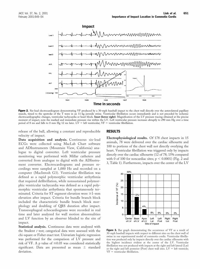
Electrophysiological results. Of 178 chest impacts in 15animals, 78 were delivered over the cardiac silhouette and100 to portions of the chest wall not directly overlying theheart. Ventricular fibrillation was triggered only by impactsdirectly over the cardiac silhouette (12 of 78; 15% comparedwith 0 of 100 for noncardiac sites; p , 0.0001) (Fig. 2 and3, Table 1). Furthermore, impacts over the center of the LV
Figure 2. Six-lead electrocardiogram demonstrating VF produced by a 30-mph baseball impact to the chest wall directly over the anterolateral papillarymuscle, timed to the upstroke of the T wave in an 11-kg juvenile swine. Ventricular fibrillation occurs immediately and is not preceded by ischemicelectrocardiographic changes, ventricular tachycardia or heart block. Inset (lower right). Magnification of the LV pressure tracing obtained at the precisemoment of impact; note the marked and immediate pressure rise within the LV. Left ventricular pressure increases abruptly to 290 mm Hg over a timeperiod of 8 ms and falls to 0 mm Hg 12 ms later. LV 5 left ventricular; VF 5 ventricular fibrillation.
Figure 3. Bar graph demonstrating the occurrence of VF as a result of30-mph baseball impacts with respect to different sites on the chest wall ofswine in our experimental model of commotio cordis. Ventricular fibrilla-tion was produced only by impacts directly over the cardiac silhouette withthe highest incidence evident at the center of the LV. Ventricularfibrillation was not produced with impacts at the right and left lateral (Lat)or the right and left posterior (Post) chest wall sites. LV 5 left ventricle;VF 5 ventricular fibrillation.
651JACC Vol. 37, No. 2, 2001 Link et al.February 2001:649–54 Importance of Impact Location in Commotio Cordis

were more likely to produce VF (7 of 23; 30%) than impactsto other precordial sites (5 of 55; 9%, p 5 0.02), includingthe apex of the LV (1 of 25; 4%, p 5 0.02) and the base ofthe LV (4 of 30; 13%, p 5 0.17). None of the blows tononcardiac sites including the left lateral (n 5 26), leftposterior (n 5 25), right posterior (n 5 24) and right lateralchest wall (n 5 25) generated VF. In addition, nonsustainedpolymorphic ventricular tachycardia (mean of 3 6 1 beats)occurred only with impacts over the cardiac silhouette: 3 of16 at the center (19%), 2 of 26 at the base (8%) and 1 of 24at the apex of the LV (4%).
Of those 166 impacts that did not cause VF, ST segmentelevation ($1 mm) occurred in 17 (10%), all with blowsdirectly over the cardiac silhouette (p , 0.0001 when
compared with nonprecordial impact sites) (Table 1). Tran-sient heart block (mean duration of 2 s) was observed with4 of the 166 impacts not causing VF and only with impactsover the heart (p 5 0.02 when compared with noncardiacimpact sites) (Table 1). Left bundle branch blocks occurredin 20 of 166 of the impacts not causing VF and were alsolimited to impacts over the cardiac silhouette (p , 0.0001when compared with noncardiac impact sites) (Table 1).LV pressure. Peak LV pressures became markedly elevatedas an immediate consequence of chest impacts directly overthe center (280 6 36 mm Hg), base (258 6 60 mm Hg) andapex (224 6 48 mm Hg) of the LV (Fig. 2). A univariatelogistic regression model revealed these peak pressures to behighly predictive of the initiation of VF (p 5 0.0006, odds
Figure 4. Logistic regression plot showing the significant correlation between the risk of VF induction with baseball impact to the chest wall at 30 mphand the peak LV pressure resulting immediately after the blow. LV 5 left ventricular; VF 5 ventricular fibrillation.
Table 1. Risk of Inducing Electrophysiologic Abnormalities With a Baseball Propelled at 30 mph and Impacting the Chest Wall atDifferent Anatomic Sites During the Vulnerable Time Period for Induction of Ventricular Fibrillation (10 to 30 ms Before the Twave Peak)
Sites of Chest Impacts
Cardiac Noncardiac
Center ofLV
Base ofLV
Apex ofLV
LeftLateralChest
LeftPosterior
Chest
RightLateralChest
RightPosterior
Chest
Induction of VF 7/23 4/30 1/25 0/26 0/25 0/25 0/24(30%)* (13%) (4%)
Induction of PMVT† 3/16 2/26 1/24 0/26 0/25 0/25 0/24(19%) (8%) (4%)
ST elevation† 7/16 5/26 5/24 0/26 0/25 0/25 0/24(44%) (19%) (21%)
Transient HB† 1/16 2/26 1/24 0/27 0/25 0/25 0/24(6%) (8%) (4%)
LBBB† 8/16 5/26 5/24 0/26 0/25 0/25 0/24(50%) (19%) (21%)
Peak LV pressure(in mm Hg)‡
280 6 36* 258 6 60a 224 6 48b 160 6 47 118 6 21 110 6 23 98 6 16
*Significantly different than all impact sites except for the base of the LV; †Expressed only for those impacts that did not produce VF; ‡Measured immediately after chest impact.a, significantly different than impacts at all other sites except for the center of the LV; b, significantly different than impacts at all sites.
HB 5 heart block; LBBB 5 left bundle branch block; LV 5 left ventricle; PMVT 5 nonsustained polymorphic ventricular tachycardia; VF 5 ventricular fibrillation.
652 Link et al. JACC Vol. 37, No. 2, 2001Importance of Impact Location in Commotio Cordis February 2001:649–54
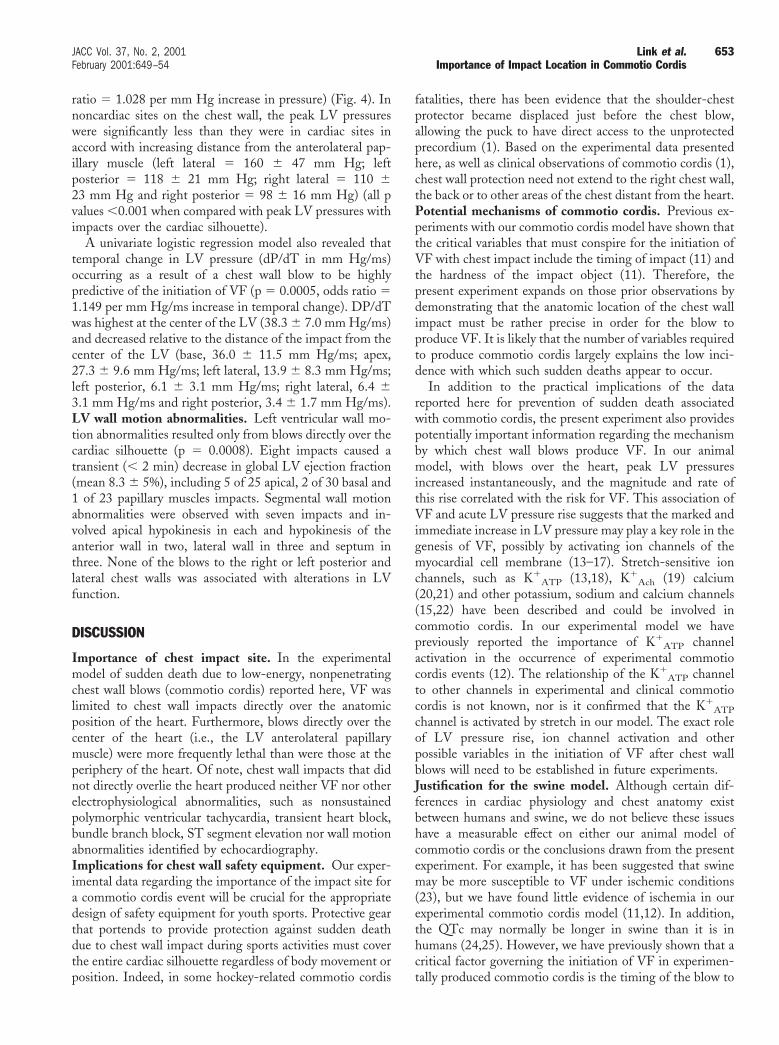
ratio 5 1.028 per mm Hg increase in pressure) (Fig. 4). Innoncardiac sites on the chest wall, the peak LV pressureswere significantly less than they were in cardiac sites inaccord with increasing distance from the anterolateral pap-illary muscle (left lateral 5 160 6 47 mm Hg; leftposterior 5 118 6 21 mm Hg; right lateral 5 110 623 mm Hg and right posterior 5 98 6 16 mm Hg) (all pvalues ,0.001 when compared with peak LV pressures withimpacts over the cardiac silhouette).
A univariate logistic regression model also revealed thattemporal change in LV pressure (dP/dT in mm Hg/ms)occurring as a result of a chest wall blow to be highlypredictive of the initiation of VF (p 5 0.0005, odds ratio 51.149 per mm Hg/ms increase in temporal change). DP/dTwas highest at the center of the LV (38.3 6 7.0 mm Hg/ms)and decreased relative to the distance of the impact from thecenter of the LV (base, 36.0 6 11.5 mm Hg/ms; apex,27.3 6 9.6 mm Hg/ms; left lateral, 13.9 6 8.3 mm Hg/ms;left posterior, 6.1 6 3.1 mm Hg/ms; right lateral, 6.4 63.1 mm Hg/ms and right posterior, 3.4 6 1.7 mm Hg/ms).LV wall motion abnormalities. Left ventricular wall mo-tion abnormalities resulted only from blows directly over thecardiac silhouette (p 5 0.0008). Eight impacts caused atransient (, 2 min) decrease in global LV ejection fraction(mean 8.3 6 5%), including 5 of 25 apical, 2 of 30 basal and1 of 23 papillary muscles impacts. Segmental wall motionabnormalities were observed with seven impacts and in-volved apical hypokinesis in each and hypokinesis of theanterior wall in two, lateral wall in three and septum inthree. None of the blows to the right or left posterior andlateral chest walls was associated with alterations in LVfunction.
DISCUSSION
Importance of chest impact site. In the experimentalmodel of sudden death due to low-energy, nonpenetratingchest wall blows (commotio cordis) reported here, VF waslimited to chest wall impacts directly over the anatomicposition of the heart. Furthermore, blows directly over thecenter of the heart (i.e., the LV anterolateral papillarymuscle) were more frequently lethal than were those at theperiphery of the heart. Of note, chest wall impacts that didnot directly overlie the heart produced neither VF nor otherelectrophysiological abnormalities, such as nonsustainedpolymorphic ventricular tachycardia, transient heart block,bundle branch block, ST segment elevation nor wall motionabnormalities identified by echocardiography.Implications for chest wall safety equipment. Our exper-imental data regarding the importance of the impact site fora commotio cordis event will be crucial for the appropriatedesign of safety equipment for youth sports. Protective gearthat portends to provide protection against sudden deathdue to chest wall impact during sports activities must coverthe entire cardiac silhouette regardless of body movement orposition. Indeed, in some hockey-related commotio cordis
fatalities, there has been evidence that the shoulder-chestprotector became displaced just before the chest blow,allowing the puck to have direct access to the unprotectedprecordium (1). Based on the experimental data presentedhere, as well as clinical observations of commotio cordis (1),chest wall protection need not extend to the right chest wall,the back or to other areas of the chest distant from the heart.Potential mechanisms of commotio cordis. Previous ex-periments with our commotio cordis model have shown thatthe critical variables that must conspire for the initiation ofVF with chest impact include the timing of impact (11) andthe hardness of the impact object (11). Therefore, thepresent experiment expands on those prior observations bydemonstrating that the anatomic location of the chest wallimpact must be rather precise in order for the blow toproduce VF. It is likely that the number of variables requiredto produce commotio cordis largely explains the low inci-dence with which such sudden deaths appear to occur.
In addition to the practical implications of the datareported here for prevention of sudden death associatedwith commotio cordis, the present experiment also providespotentially important information regarding the mechanismby which chest wall blows produce VF. In our animalmodel, with blows over the heart, peak LV pressuresincreased instantaneously, and the magnitude and rate ofthis rise correlated with the risk for VF. This association ofVF and acute LV pressure rise suggests that the marked andimmediate increase in LV pressure may play a key role in thegenesis of VF, possibly by activating ion channels of themyocardial cell membrane (13–17). Stretch-sensitive ionchannels, such as K1
ATP (13,18), K1Ach (19) calcium
(20,21) and other potassium, sodium and calcium channels(15,22) have been described and could be involved incommotio cordis. In our experimental model we havepreviously reported the importance of K1
ATP channelactivation in the occurrence of experimental commotiocordis events (12). The relationship of the K1
ATP channelto other channels in experimental and clinical commotiocordis is not known, nor is it confirmed that the K1
ATPchannel is activated by stretch in our model. The exact roleof LV pressure rise, ion channel activation and otherpossible variables in the initiation of VF after chest wallblows will need to be established in future experiments.Justification for the swine model. Although certain dif-ferences in cardiac physiology and chest anatomy existbetween humans and swine, we do not believe these issueshave a measurable effect on either our animal model ofcommotio cordis or the conclusions drawn from the presentexperiment. For example, it has been suggested that swinemay be more susceptible to VF under ischemic conditions(23), but we have found little evidence of ischemia in ourexperimental commotio cordis model (11,12). In addition,the QTc may normally be longer in swine than it is inhumans (24,25). However, we have previously shown that acritical factor governing the initiation of VF in experimen-tally produced commotio cordis is the timing of the blow to
653JACC Vol. 37, No. 2, 2001 Link et al.February 2001:649–54 Importance of Impact Location in Commotio Cordis

a narrow time window of vulnerability on the upslope of theT wave, a variable independent of the absolute length of theQT interval (whether or not corrected for heart rate) (11).Furthermore, the use of swine to study the mechanisms ofVF and resuscitation is well established (23,26,27).
Although differences in the geometry of the human andporcine thoracic cages could conceivably affect the transmis-sion of impact energy to the myocardium, nevertheless, inboth circumstances chest blows occur perpendicular to thechest wall, directly over the heart and at a point where theheart is in close proximity to the chest wall withoutintervening pulmonary tissue. Therefore, we would expectthe kinetic energy transmitted by nonpenetrating chestblows to the heart to be similar in the precipitation ofcommotio cordis events in both humans and our experi-mental animal model.Conclusions. The precise location of chest wall impact is acritical determinant of commotio cordis events. To result inVF, the chest blow must occur directly over the anatomicposition of the heart and, in fact, is more likely to be lethalwhen impact is at the center of the cardiac silhouette.Incorporation of these observations into the design of chestwall protectors may prevent sudden deaths and make theathletic field safer for youthful participants in sports activ-ities.
AcknowledgmentsThe authors are indebted to Darisse A. Paquette, CMI, forher assistance with the figures and to Stacey E. Supran ofthe Division of Clinical Care Research at the New EnglandMedical Center for assistance with statistical analysis.
Reprint requests and correspondence: Dr. Mark S. Link, NewEngland Medical Center, NEMC Box #197, 750 WashingtonStreet, Boston, Massachusetts 02111. E-mail: [email protected].
REFERENCES
1. Maron BJ, Poliac LC, Kaplan JA, Mueller FO. Blunt impact to thechest leading to sudden death from cardiac arrest during sportsactivities. N Engl J Med 1995;333:337–42.
2. Adler P, Monticone RCJ. Injuries and deaths related to baseball. In:Kyle SB, editor. Youth Baseball Protective Equipment Project FinalReport. Washington, DC: United States Consumer Product SafetyCommission, 1996:1–43.
3. Maron BJ, Link MS, Wang PJ, Estes NAM. Clinical profile ofcommotio cordis: an under-appreciated cause of sudden death in theyoung during sports and other activities. J Cardiovasc Electrophysiol1999;10:114–20.
4. Maron BJ, Kyle SB, Gohman TE, Estes NAM, Link MS. The clinicalspectrum of commotio cordis: the first 100 cases from the U.S.Registry (abstr). Circulation 2000;102:II-609.
5. Kaplan JA, Karofsky PS, Volturo GA. Commotio cordis in twoamateur ice hockey players despite the use of commercial chestprotectors: case reports. J Trauma 1993;34:151–3.
6. Abrunzo TJ. Commotio cordis: the single, most common cause oftraumatic death in youth baseball. Am J Dis Child 1991;145:1279–82.
7. Dickman GL, Hassan A, Luckstead EF. Ventricular fibrillationfollowing baseball injury. Phys Sport Med 1978;6:85–6.
8. Maron BJ, Strasburger JF, Kugler JD, Bell BM, Brodkey FD, PoliacLC. Survival following blunt chest impact induced cardiac arrestduring sports activities in young athletes. Am J Cardiol 1997;79:840–1.
9. Link MS, Ginsburg SH, Wang PJ, Kirchhoffer JB, Estes NAM, ParrisYM. Commotio cordis: cardiovascular manifestations of a rare survi-vor. Chest 1998;114:326–8.
10. Deady B, Innes G. Sudden death of a young hockey player: case reportof commotio cordis. J Emerg Med 1999;17:459–62.
11. Link MS, Wang PJ, Pandian NG, et al. An experimental model ofsudden death due to low-energy chest wall impact (commotio cordis).N Engl J Med 1998;338:1805–11.
12. Link MS, Wang PJ, VanderBrink BA, et al. Selective activation of theK1ATP channel is a mechanism by which sudden death is produced bylow-energy chest-wall impact (commotio cordis). Circulation 1999;100:413–8.
13. Van Wagoner DR. Mechanosensitive gating of atrial ATP-sensitivepotassium channels. Circ Res 1993;72:973–83.
14. Franz MR, Cima R, Wang D, Profitt D, Kurz R. Electrophysiologicaleffects of myocardial stretch and mechanical determinants of stretch-activated arrhythmias. Circulation 1992;86:968–78.
15. Hu H, Sachs F. Stretch-activated ion channels in the heart. J Mol CellCardiol 1997;29:1511–23.
16. Kohl P, Hunter P, Noble D. Stretch-induced changes in heart rate andrhythm: clinical observations, experiments and mathematical models.Prog Biophys Mol Biol 1999;71:91–138.
17. Zabel M, Koller BS, Sachs F, Franz MR. Stretch-induced voltagechanges in the isolated beating heart: importance of the timing ofstretch and implications for stretch-activated ion channels. CardiovascRes 1996;32:120–30.
18. Kim SH, Cho KW, Chang SH, Kim SZ, Chae SW. Glibenclamidesuppresses stretch-activated ANP secretion: involvement of K1
ATP
channels and L-type Ca21 channel modulation. Eur J Physiol 1997;434:362–72.
19. Pleumsamran A, Kim D. Membrane stretch augments the cardiacmuscarinic K1 channel activity. J Membrane Biol 1995;148:287–97.
20. Matsuda N, Hagiwara N, Shoda M, Kasanuki H, Hosoda S. En-hancement of the L-type Ca21 current by mechanical stimulation insingle rabbit cardiac myocytes. Circ Res 1996;78:650–9.
21. Gannier F, White E, Lacampagne A, Garnier D, Le Guennec J-Y.Streptomycin reverses a large stretch induced increase in Ca21 inisolated guinea pig ventricular myocytes. Cardiovasc Res 1994;28:1193–8.
22. Ruknudin A, Sachs F, Bustamante JO. Stretch-activated ion channelsin tissue-cultured chick heart. Am J Physiol 1993;264:H960–72.
23. Verdouw PD, Hartog JM. Provocation and suppression of ventriculararrhythmias in domestic swine. In: Stanton HC, Mersmann HJ,editors. Swine in Cardiovascular Research. Boca Raton, FL: CRCPress, 1986:121–56.
24. Wiesfeld ACP, De Langen CDJ, Crijns HJGM, et al. Rate-dependent effects of the class III antiarrhythmic drug almokalant onrefractoriness in the pig. J Card Pharm 1996;27:594–600.
25. Rubio MD, Tovar P, Santisteban R, De Oliveira M, Castejon F.Differential study of electrocardiographic intervals in two neonatalcrossbred swine. Am J Vet Res 1993;54:322–6.
26. Hughes HC. Swine in cardiovascular research. Lab Anim Sci 1986;36:348–50.
27. Bowman TA, Hughes HC. Swine as an in vivo model for electro-physiologic evaluation of cardiac pacing parameters. Pacing ClinElectrophysiol 1984;7:187–94.
654 Link et al. JACC Vol. 37, No. 2, 2001Importance of Impact Location in Commotio Cordis February 2001:649–54
Copyright © 2022 FDOKUMEN