The HDAC6/APOBEC3G complex regulates HIV-1 infectiveness by inducing Vif autophagic degradation

Immunogenicity of Seven New Recombinant YellowFever Viruses 17D Expressing Fragments of SIVmac239Gag, Nef, and Vif in Indian Rhesus MacaquesMauricio A. Martins1, Myrna C. Bonaldo2, Richard A. Rudersdorf3, Shari M. Piaskowski3, Eva G. Rakasz3,
Kim L. Weisgrau3, Jessica R. Furlott3, Christopher M. Eernisse3, Marlon G. Veloso de Santana2,
Bertha Hidalgo7, Thomas C. Friedrich3,4, Maria J. Chiuchiolo5¤, Christopher L. Parks5, Nancy A. Wilson3,6,
David B. Allison7, Ricardo Galler2, David I. Watkins1*
1 Department of Pathology, University of Miami Miller School of Medicine, Miami, Florida, United States of America, 2 Laboratorio de Biologia Molecular de Flavivırus,
Instituto Oswaldo Cruz – FIOCRUZ, Rio de Janeiro, Brazil, 3 Wisconsin National Primate Research Center, University of Wisconsin-Madison, Madison, Wisconsin, United
States of America, 4 Department of Pathobiological Sciences, University of Wisconsin-Madison, Madison, Wisconsin, United States of America, 5 International AIDS Vaccine
Initiative, AIDS Vaccine Design and Development Laboratory, Brooklyn Army Terminal, Brooklyn, New York, United States of America, 6 Department of Medicine,
University of Wisconsin-Madison, Madison, Wisconsin, United States of America, 7 Section on Statistical Genetics, Department of Biostatistics, University of Alabama at
Birmingham, Birmingham, Alabama, United States of America
Abstract
An effective vaccine remains the best solution to stop the spread of human immunodeficiency virus (HIV). Cellular immuneresponses have been repeatedly associated with control of viral replication and thus may be an important element of theimmune response that must be evoked by an efficacious vaccine. Recombinant viral vectors can induce potent T-cellresponses. Although several viral vectors have been developed to deliver HIV genes, only a few have been advanced forclinical trials. The live-attenuated yellow fever vaccine virus 17D (YF17D) has many properties that make it an attractivevector for AIDS vaccine regimens. YF17D is well tolerated in humans and vaccination induces robust T-cell responses thatpersist for years. Additionally, methods to manipulate the YF17D genome have been established, enabling the generationof recombinant (r)YF17D vectors carrying genes from unrelated pathogens. Here, we report the generation of seven newrYF17D viruses expressing fragments of simian immunodeficiency virus (SIV)mac239 Gag, Nef, and Vif. Studies in Indianrhesus macaques demonstrated that these live-attenuated vectors replicated in vivo, but only elicited low levels of SIV-specific cellular responses. Boosting with recombinant Adenovirus type-5 (rAd5) vectors resulted in robust expansion of SIV-specific CD8+ T-cell responses, particularly those targeting Vif. Priming with rYF17D also increased the frequency of CD4+
cellular responses in rYF17D/rAd5-immunized macaques compared to animals that received rAd5 only. The effect of therYF17D prime on the breadth of SIV-specific T-cell responses was limited and we also found evidence that some rYF17Dvectors were more effective than others at priming SIV-specific T-cell responses. Together, our data suggest that YF17D – aclinically relevant vaccine vector – can be used to prime AIDS virus-specific T-cell responses in heterologous prime boostregimens. However, it will be important to optimize rYF17D-based vaccine regimens to ensure maximum delivery of allimmunogens in a multivalent vaccine.
Citation: Martins MA, Bonaldo MC, Rudersdorf RA, Piaskowski SM, Rakasz EG, et al. (2013) Immunogenicity of Seven New Recombinant Yellow Fever Viruses 17DExpressing Fragments of SIVmac239 Gag, Nef, and Vif in Indian Rhesus Macaques. PLoS ONE 8(1): e54434. doi:10.1371/journal.pone.0054434
Editor: Clive M. Gray, University of Cape Town, South Africa
Received August 28, 2012; Accepted December 11, 2012; Published January 15, 2013
Copyright: � 2013 Martins et al. This is an open-access article distributed under the terms of the Creative Commons Attribution License, which permitsunrestricted use, distribution, and reproduction in any medium, provided the original author and source are credited.
Funding: This research was supported by National Institutes of Health (NIH)/National Institute of Allergy and Infectious Disease (NIAID) grants R01 AI076114 andR01 AI049120 awarded to DIW. Funding also came from Coordenacao de Aperfeicoamento de Pessoal de Nıvel Superior (CAPES) and FIOCRUZ in Brazil. Part of thework done in FIOCRUZ was supported by the National Institute for Vaccine Technology (INCTV/CNPq). In addition, this work was supported by NCRR grant P51RR000167 to the Wisconsin National Primate Research Center, University of Wisconsin-Madison. This research was conducted in part at a facility constructed withsupport from Research Facilities Improvement Program grant numbers RR15459 and RR020141. The funders had no role in study design, data collection andanalysis, decision to publish, or preparation of the manuscript.
Competing Interests: The authors have declared that no competing interests exist.
* E-mail: [email protected]
¤ Current address: Belfer Gene Therapy Core Facility, Department of Genetic Medicine Weill Cornell Medical College, New York, New York, United States ofAmerica
Introduction
The HIV epidemic is one of the worst public health tragedies in
human history. More than 25 million human lives have been lost
due to Acquired Immune Deficiency Syndrome (AIDS)-related
causes since the first description of the disease in 1981 [1,2]. Sadly,
two thirds of the 33 million people currently infected with HIV
infection live in resource-poor regions of the globe where access to
standard healthcare is limited [2]. Despite advances in access to
anti-retroviral treatment (ART) in countries with the heaviest
burden of the disease, less than half of the population in need of
this therapy actually receives it [2]. A safe and effective HIV
vaccine is, therefore, the best long-term solution to ending the
pandemic [3].
PLOS ONE | www.plosone.org 1 January 2013 | Volume 8 | Issue 1 | e54434

Several lines of evidence implicate CD8+ T-cells in the control
of HIV-1 replication [4–10]. It is also becoming increasingly clear
that lentivirus-specific CD4+ T-cells contribute to an effective
antiviral immune response [11–14]. As a result, a myriad of
vaccination platforms have been developed to induce HIV-specific
cellular responses. Viral vectors are of particular interest since this
strategy exploits the proficiency with which viruses enter cells and
hijack the cellular machinery to promote maximal expression of
viral proteins [15]. Non-replicating adenoviruses and poxviruses,
for instance, have been heavily exploited as potential HIV vector
systems due to their good preclinical immunogenicity and safety
profiles [15–18]. However, the high prevalence worldwide of pre-
existing immunity to Ad5 and the safety concerns raised by the
rAd5-vectored HIV vaccine tested in STEP study have dampened
the enthusiasm toward this vector platform [19–22]. Furthermore,
modified vaccinia virus Ankara (MVA)- and canarypox (ALVAC)
expressing HIV immunogens have elicited low levels of cellular
immunity in most human clinical trials conducted so far [23–25].
Therefore, novel viral vectors capable of safely inducing robust T-
cell responses in humans are needed to advance HIV vaccine
design.
Historically, live-attenuated replicating viral vaccines have
provided the most effective protection against infection and
disease [18]. Notably, these vaccines elicit robust and durable
immune responses in immunized individuals, which is rarely
achieved by inactivated or subunit vaccines [18]. The live-
attenuated yellow fever vaccine virus YF17D, for instance, is one
of the most successful human vaccines ever developed. More than
400 million people have been vaccinated since its development in
the 1930s, with only rare adverse events [19,26–28]. Perhaps
because it can replicate in humans, YF17D triggers several innate
immune pathways, which likely contributes to its high immuno-
genicity [29,30]. Indeed, vaccination results in polyvalent adaptive
immune responses consisting of effector CD8+ T-cells and a mixed
TH1/TH2 CD4+ T-cell profile [28]. Importantly, southern Africa
and Asia – two regions with high incidence of HIV cases – are not
considered endemic regions for yellow fever and thus vaccine
coverage in these areas is low [31,32]. As a result, there is a low
prevalence of pre-existing immunity to the yellow fever virus in
these important target populations. Therefore, vector-specific
immunity is unlikely to affect the immunogenicity of a potential
rYF17D/HIV vaccine in those areas. These properties make
YF17D a promising vector platform for delivering HIV genes.
Several methods for manipulating the YF17D genome have
been developed [33–35], including the generation of rYF17D
viruses bearing insertions between the genes encoding Envelope
(E) and non-structural protein 1 (NS1) [36]. We have recently used
this methodology to create a recombinant YF17D virus expressing
a segment of SIVmac239 Gag. This virus replicated and elicited
CD8+ T-cell responses in rhesus macaques [37]. Here, we
employed this same methodology to generate seven new rYF17D
vectors expressing fragments of the SIVmac239 Gag, Nef, and Vif
proteins. We chose these antigens since vaccine-induced T-cells
targeting these proteins have been associated with control of viral
replication after a pathogenic SIV challenge [38]. The goal of this
study was, therefore, to evaluate the immunogenicity of these
seven rYF17D vectors alone or as a prime in a heterologous
prime/boost regimen.
Materials and Methods
Research Animals and Ethics StatementThe animals in this study were Indian rhesus macaques (Macaca
mulatta) and were part of the breeding colony of the Wisconsin
National Primate Research Center (WNPRC). All animals were
cared for in accordance with the guidelines of the Weatherall
report [39] under a protocol approved by the University of
Wisconsin Graduate School Animal Care and Use Committee
(animal welfare assurance no. A3368-01; protocol no. G00578).
Furthermore, the macaques in this study were managed according
to the animal husbandry program of the WNPRC, which aims at
providing consistent and excellent care to nonhuman primates at
the center. This program is employed by the Colony Management
Unit and is based on the laws, regulations, and guidelines
promulgated by the United States Department of Agriculture
(e.g., the Animal Welfare Act and its regulations, and the Animal
Care Policy Manual), Institute for Laboratory Animal Research
(e.g., Guide for the Care and Use of Laboratory Animals, 8th
edition), Public Health Service, National Research Council,
Centers for Disease Control, and the Association for Assessment
and Accreditation of Laboratory Animal Care (AAALAC)
International. The nutritional plan utilized by the WNPRC is
based on recommendations published by the National Research
Council. Specifically, macaques were fed twice daily with 2050
Teklad Global 20% Protein Primate Diet and food intake was
closely monitored by Animal Research Technicians. This diet was
also supplemented with a variety of fruits, vegetables, and other
edible objects as part of the environmental enrichment program
established by the Behavioral Management Unit. Paired/grouped
animals exhibiting stereotypes and/or incompatible behaviors
were reported to the Behavioral Management staff and managed
accordingly. All primary enclosures (i.e., stationary cages, mobile
racks, and pens) and animal rooms were cleaned daily with water
and sanitized at least once every two weeks. All macaques used in
this study were males, except for three females: r98010, r03105,
and r04149. Their average weight was 8.0 kg (range: 6.5–9.8 kg)
and their average age was 5 years (range: 4–11 years).
Vaccinations were performed under anesthesia (Ketamine admin-
istered at 5–12 mg/kg depending on the animal) and all efforts
were made to minimize suffering. None of the animals were
euthanized as part of this study. The macaques were typed for
common major histocompatibility complex (MHC) class I alleles
by sequence-specific PCR analysis [40,41] and were chosen based
on the expression of Mamu-A*01 and/or Mamu-A*02 (Table 1).
Generation of Recombinant YF17D Viruses ExpressingFragments of SIVmac239 Proteins
We designed seven codon-optimized minigenes encoding
fragments of the SIVmac239 Gag, Nef, and Vif proteins. Four
of these encoded segments of the Gag precursor protein (amino
acids 44–84, 76–123, 142–186, and 250–415); two encoded the
amino and carboxyl portions of Vif (1–110 and 102–214); and one
encoded the central part of Nef (45–210). We generated viable
recombinant YF17D viruses expressing these protein fragments
using a previously described methodology [36]. Briefly, we
duplicated functional motifs flanking the E and NS1 intergenic
regions in the YF17D backbone and fused them to the exogenous
minigenes, allowing for the correct processing of the viral
polyprotein precursor. The recombinant SIV protein fragments
are released into the lumen of the endoplasmic reticulum via the
action of host peptidases. After cloning the inserts into the junction
of E and NS1, we prepared full-length rYF17D genomic RNA by
in vitro transcription using SP6 RNA polymerase (AmpliScribe
SP6, Epicentre Technologies). These RNA preparations were used
to transfect Vero cells with LipofectAmine (Invitrogen) as
described elsewhere [42]. Viral stocks were prepared by infecting
Vero cell monolayers with the virus present in the supernatant
from the transfections described above.
Immunogenicity of rYF17D/SIV Vaccine Candidates
PLOS ONE | www.plosone.org 2 January 2013 | Volume 8 | Issue 1 | e54434
To confirm the presence and integrity of SIV inserts, we
extracted viral RNA (vRNA) from culture supernatants (QIAmp
Viral RNA kit, QIAGEN) and performed an amplification of the
viral E-NS1 genomic region encompassing the heterologous insert.
For the cDNA synthesis (SuperScript III Reverse Transcriptase,
Invitrogen), we used viral RNA as a template and a negative
strand YF17D-specific synthetic oligonucleotide (genome position
2772–2795). Subsequently, we performed PCR (Master Mix
Green Go Taq, Promega) in the presence of a positive strand
YF17D-specific primer (genome position 2122–2145). Amplifica-
tion products were further purified from excess primers with silica-
based kits (QIAGEN) and run on an agarose gel.
YF17D Viral Load AssayWe measured the replication of rYF17D viruses by isolating
vRNA from EDTA-anticoagulated plasma by proteinase K
digestion of the nucleocapsid in the presence of guanidine
hydrochloride as described previously [43]. Viral RNA was
reverse transcribed and amplified using the SuperScript III
PlatinumH One-Step Quantitative RT-PCR System (Invitrogen,
Carlsbad, CA) in a Roche LightCyclerH 480. The final reaction
mixtures (100-ml total volume) contained 0.2 mM (each) deox-
ynucleoside triphosphates, 3.5 mM MgS04, 750 ng of random
hexamer primers (Promega, Madison, WI), 4.0 ml of SuperScript
III reverse transcriptase and Platinum Taq DNA polymerase in a
single enzyme mix, 600 nM (each) amplification primers (forward
[YF-17D 10188], 59-GCGGATCACTGATTGGAATGAC-39,
and reverse [YF-17D 10264], 59-CGTTCGGATACGATGGAT-
GACTA-39), and 100 nM probe [6Fam]59-AATAGGGC-
CACCTGGGCCTCCC-39[TamraQ]. The reverse transcription
reaction was performed at 37uC for 15 minutes and then at 50uC
for 30 minutes. Taq polymerase was activated subsequently by
incubation at 95uC for 2 minutes after which 50 amplification
cycles were performed at 95uC for 15 seconds and 57uC for one
minute with ramp times set to 2.2uC/s. Ten-fold serial dilutions of
a YF17D in vitro transcript were used to generate a standard curve
for each run. The transcript was made using a MEGAscriptH(Ambion, Austin, TX) high yield transcription kit. The plasmid,
pCR-Blunt, containing a PCR amplicon with a sequence identical
to nucleotides 8621–10354 of GenBank accession number
U17066, served as the template for transcription. The limit of
detection under standard assay conditions was approximately 45
vRNA copies/ml of plasma.
Vaccination of Rhesus Macaques with rYF17D and rAd5Vectors
We immunized animals with rYF17D viruses via the subcuta-
neous route. For recipients of single constructs, we injected 105
plaque-forming units (PFU) diluted with PBS to 500 ml into the
animals’ right forearm. For recipients of all constructs, we injected
either 104 or 105 PFU of each rYF17D virus (using 27-gauge
needles) into seven different sites; right and left thighs; right and
left forearms; right and left shoulders; and left lower leg.
We subsequently boosted the four animals that received all
seven rYF17D viruses (either at 104 or 105 PFU of each) with
recombinant Adenovirus type-5 (rAd5). We used three rAd5
vectors encoding full-length (i) Gag, (ii) Nef, and (iii) a fusion of the
Vif, Tat, Rev, Vpr and Vpx proteins of SIVmac239. The rAd5/
Gag vector encoded a myristoylated Gag protein spanning amino
acids 1 to 508, lacking two C-terminal amino acids. The rAd5/Nef
vector encoded Nef lacking a myristoylation signal. The rAd5
vectors were constructed as described previously [44]. Briefly, they
Table 1. Animal details and vaccination doses.
Animal ID MHC-I rYF17D/SIV vectors Dose rAd5 vectors Dose
r04147 Mamu-A*02 Gag (44–84) 105 PFU N.A. N.A.
r04170 Mamu-A*01 Gag (76–123) 105 PFU N.A. N.A.
r04136 Mamu-A*01 Gag (142–186) 105 PFU N.A. N.A.
r05079 Mamu-A*01 Gag (250–415) 105 PFU N.A. N.A.
r05089 Mamu-A*02 Vif (1–110) 105 PFU N.A. N.A.
r04109 Mamu-A*01 Vif (102–214) 105 PFU N.A. N.A.
r05070 Mamu-A*02 Nef (45–210) 105 PFU N.A. N.A.
r05028 Mamu-A*01, A*02 All constructs 104 PFU each (i) Gag, (ii) Nef, and (iii) a fusion ofVif, Vpr, Vpx, Tat, and Rev
1011 VP each
rh2138 Mamu-A*02 All constructs 104 PFU each (i) Gag, (ii) Nef, and (iii) a fusion ofVif, Vpr, Vpx, Tat, and Rev
1011 VP each
r04137 Mamu-A*01, A*02 All constructs 105 PFU each (i) Gag, (ii) Nef, and (iii) a fusion ofVif, Vpr, Vpx, Tat, and Rev
1011 VP each
r98010 Mamu-A*02 All constructs 105 PFU each (i) Gag, (ii) Nef, and (iii) a fusion ofVif, Vpr, Vpx, Tat, and Rev
1011 VP each
r03105 Mamu-A*01 N.A. N.A. (i) Gag, (ii) Nef, and (iii) a fusion ofVif, Vpr, Vpx, Tat, and Rev
1011 VP each
r04149 Mamu-A*01 N.A. N.A. (i) Gag, (ii) Nef, and (iii) a fusion ofVif, Vpr, Vpx, Tat, and Rev
1011 VP each
r05073 Mamu-A*02 N.A. N.A. (i) Gag, (ii) Nef, and (iii) a fusion ofVif, Vpr, Vpx, Tat, and Rev
1011 VP each
r05086 Mamu-A*01 N.A. N.A. (i) Gag, (ii) Nef, and (iii) a fusion ofVif, Vpr, Vpx, Tat, and Rev
1011 VP each
N.A., not applicable.doi:10.1371/journal.pone.0054434.t001
Immunogenicity of rYF17D/SIV Vaccine Candidates
PLOS ONE | www.plosone.org 3 January 2013 | Volume 8 | Issue 1 | e54434
Figure 1. Relative sizes of SIV inserts and genome schematic of rYF17D/SIV vectors. A) Relative size and amino acid position of SIVmac239minigenes. We designed codon-optimized sequences spanning regions of the Gag, Vif, and Nef proteins. The Gag minigenes covered amino acids44–84 and 76–123 of Matrix (MA), 142–186 of Capsid (CA), and 250–415 of Capsid, p2, and Nucleocapsid (NC). The Vif ORF was split into twominigenes encoding amino acids 1–110 and 102–214. The Nef sequence spanned the central region of this protein (45–210). B) Genome schematic ofrecombinant YF17D vaccine vectors. We inserted the SIVmac239 minigenes in the junction between the YF17D E and NS1 genes using a previouslydescribed methodology [36]. C) Electrophoretic analysis of RT-PCR amplicons from viral RNA extracted from the seven rYF17D/SIV stocks. The YF17Dparental vaccine served as a negative control. We performed these amplifications using YF17D-specific primers flanking the E-NS1 genomic regionencompassing the SIVmac239 inserts.doi:10.1371/journal.pone.0054434.g001
Immunogenicity of rYF17D/SIV Vaccine Candidates
PLOS ONE | www.plosone.org 4 January 2013 | Volume 8 | Issue 1 | e54434
were generated in a DE1A, partial DE1B, and DE3 genetic
background using the AdEasy adenoviral vector system (Strate-
gene, La Jolla, CA). The SIVmac239 genes were cloned into the
E1 region of the rAd5 vector under the control of the hCMV
immediate-early promoter-enhancer and the SV40 stop-polyade-
nylation signal. All vectors were rescued in HEK 293 cells and
purified by double cesium chloride centrifugation. Dosing was
based on viral particles (VP), determined by spectrophotometry
[45]. We injected 1011 VP of each vector intramuscularly (using
27-gauge needles) at three different sites; right shoulder, left
forearm and right thigh. Of note, we rotated these sites based on
the previous vaccination scheme used for rYF17D so that each site
did not receive vectors encoding fragments of the same protein in
both vaccinations. We reasoned that this strategy would maximize
the dispersal of antigens among lymph nodes and thereby avoid
immunodominance.
IFN-c ELISPOT AssaysWe isolated peripheral blood mononuclear cells (PBMC) from
EDTA-treated blood by Ficoll-Paque PLUS (GE Health Sciences)
density centrifugation. We used PBMC or PBMC depleted of
CD8+ cells using magnetic bead separation (Miltenyi Biotec,
Auburn, CA), directly in precoated ELISpotPLUS kits (MABTECH
Inc., Mariemont, OH) for the detection of monkey IFN-caccording to the manufacturer’s protocols. Briefly, 105 PBMC
were used per well and incubated for 14 to 18 hours at 37uC in 5%
CO2. As a positive control, 5 mg/ml of concanavalin A (Sigma
Chemical, St. Louis, MO) was added to the cells, and a set of
negative control wells of media only was also included on each
plate. Peptide pools of ten 15mers overlapping by 11 amino acids
spanning all SIVmac239 proteins included in the vaccine regimens
were used at a concentration of 1.0 mM, while minimal optimal
peptides corresponding to SIVmac239 or YF17D epitopes were
used at a concentration of 10.0 mM. All SIV peptide pools were
obtained from the AIDS Research and Reference Reagent
Program, Division of AIDS, NIAID, NIH.
Test wells were run with two replicates, while control wells were
run with replicates of 2, 4, or 6, depending on the assay. Assay
results are shown as spot-forming cells (SFC) per 106 PBMC.
Responses comprising ,50 SFC per 106 cells were considered
negative and were not tested statistically. Positive responses were
determined using a one-tailed t test and an alpha level of 0.05,
where the null hypothesis was that the background level would be
greater than or equal to the treatment level. If determined to be
positive statistically, the values were reported as the average of the
test wells minus the average of all negative-control wells.
Intracellular Cytokine Staining (ICS) AssayWe performed multi-parameter ICS by incubating freshly
isolated PBMC from rhesus macaques with 1.0 mM of separate
peptide mixtures spanning the ORFs of Vif, Nef, Tat, and Rev.
Vpr and Vpx peptides were combined in one test. We divided the
Gag peptides in two tests spanning amino acids 1–291 and 281–
510; reactivity to this protein is reported as the sum of these two
tests. We used staphylococcal enterotoxin B (SEB) stimulation for
our positive control and tissue culture medium devoid of
stimulatory peptides for our negative control. We also added
anti-CD28 (clone L293; BD Biosciences), anti-CD49d (clone 9g;
Pharmigen), and anti-CD107a PE (clone H4A3; BD Biosciences)
antibodies and 5.0 mg of Brefeldin A and Golgi Stop (BD
Biosciences) to each test. We then placed the samples in a 5.0%
CO2 incubator at 37uC. After overnight incubation, we stained the
cells at room temperature with antibodies directed against the
surface molecules CD4 (PerCP Cy5.5; clone L200; BD Bioscienc-
es), CD8 (Pacific Blue; clone RPA-T8; BD Biosciences), CD14
(ECD; clone RMO52; Beckman Coulter), and CD19 (ECD; clone
JA.119; Beckman Coulter). After 20 minutes, we washed the cells
with FACS buffer (10% of fetal bovine serum in PBS 1X) and fixed
them with a 2.0% paraformaldehyde solution. After 30 minutes,
we washed off the paraformaldehyde and permeabilized the cells
with 0.1% Saponin buffer (FACS buffer containing saponin) and
stained them with antibodies directed against the intracellular
molecules IFN-c (PE-Cy7; clone 4S.B3; BD Biosciences), TNF-a(Alexa700; clone MAb11; BD Biosciences), IL-2 (APC; clone
MQ1-17H12; BD Biosciences), and MIP-1b (FITC; clone 24006;
R&D Systems). After a 45-minute incubation, we washed the cells
twice with Saponin buffer and fixed them with 2.0% paraformal-
dehyde. Samples were acquired using FACS DIVA version 6 on a
Special Order Research Product (SORP) BD LSR II equipped
with a 50 mW 405 nm violet, a 100 mW 488 nm blue, and a
50 mW 640 nm red laser and were analyzed by FlowJo 9.2
(TreeStar, Inc.). Analysis and presentation of distributions were
performed using Pestle and SPICE version 5.22023, downloaded
from http://exon.niaid.nih.gov/spice [46].
For data analysis, we first gated on forward scatter height (FSC-
H) versus forward scatter area (FSC-A) to remove doublets (Fig.
S1). Subsequently, we gated on the lymphocyte population and
then created a dump channel by excluding CD14+CD19+ events.
At this stage, we separated lymphocyte subsets based on their
expression of either CD4 or CD8 (excluding those expressing both
markers) and conducted our functional analyses within these two
compartments. After making gates for each function (IFN-c,
CD107a, TNF-a, MIP-1b, and IL-2), we used the Boolean gate
platform to generate a full array of possible combinations,
equating to 32 response patterns when testing five functions
(25 = 32). We used three criteria to determine positivity of
responses; i) background-subtracted responses had to be at least
2-fold higher than the background itself; ii) Boolean gates for each
response pattern had to contain $10 events; and iii) response
patterns had to be greater than matched values obtained in ICS
assays using PBMC from three SIV naıve Indian rhesus macaques
under the same stimulation conditions.
Table 2. Replication of rYF17D/SIV vectors in vivo1.
Days post vaccination
Animal IDInsert (aaposition) Dose (PFU) 3 5 7 14
r04147 Gag (44–84) 105 381 0 216 0
r04170 Gag (76–123) 105 85 131 738 0
r04136 Gag (142–186) 105 0 887 666 0
r05079 Gag (250–415) 105 192 537 2,049 0
r05089 Vif (1–110) 105 0 0 984 0
r04109 Vif (102–214) 105 57 90 119 0
r05070 Nef (45–210) 105 283 433 1,815 0
r05028 All constructs 104 each 0 0 0 0
rh2138 All constructs 104 each 0 0 828 0
r04137 All constructs 105 each 0 0 0 0
r98010 All constructs 105 each 224 0 0 0
1We measured replication of the rYF17D/SIV viruses using a qRT-PCR assay.Values represent vRNA copies/ml of plasma. The limit of quantitation understandard assay conditions was 45 vRNA copies/ml of plasma.doi:10.1371/journal.pone.0054434.t002
Immunogenicity of rYF17D/SIV Vaccine Candidates
PLOS ONE | www.plosone.org 5 January 2013 | Volume 8 | Issue 1 | e54434

Figure 2. Magnitude of SIV-specific T-cell responses in animals vaccinated with single rYF17D/SIV constructs. We carried out IFN-cELISPOT at days 14 (white bars) and 17 (black bars) after the rYF17D vaccination using peptide pools (ten 15mers overlapping by 11 amino acids ineach pool) spanning the regions of SIVmac239 Gag, Vif, and Nef encoded in each rYF17D vector. We adjusted these pools for each macaquedepending on the region and SIV protein expressed by the rYF17D viruses. Bar graphs indicate the magnitude of IFN-c-producing cells in PBMC (SFC/
Immunogenicity of rYF17D/SIV Vaccine Candidates
PLOS ONE | www.plosone.org 6 January 2013 | Volume 8 | Issue 1 | e54434
Statistical AnalysisTo compare SIV-specific T-cell responses induced by the
rYF17D/rAd5 and rAd5 regimens, we performed parametric t-
tests and nonparametric Wilcoxon-Mann Whitney tests. We
performed these tests on raw and log10-transformed data. In
addition, because some of the cells contained ‘zero values’, a
constant equal to 0.5 was added to every count prior to log-
transforming the data. The results did not vary significantly across
the different statistical tests. The only exception was the
comparison of the magnitude of Vif-specific responses between
rYF17D/rAd5 and rAd5 vaccinees at week 1 post rAd5. The
analysis with the parametric t-test yielded a p-value of 0.021,
compared to a p-value of 0.06 obtained with the nonparametric
Wilcoxon-Mann Whitney test. However, since the data at this time
point was highly skewed, we conducted a permutation t-test
(20,000 samples) on the log-transformed values to serve as an
additional sensitivity analysis and obtained a p-value of 0.096.
Thus, results from the non-parametric and long-transformed
parametric t-tests are likely more reliable.
Results
Generation of rYF17D Viruses Encoding Fragments ofSIVmac239 Gag, Vif, and Nef
We chose to generate rYF17D vectors expressing fragments of
the SIVmac239 Gag, Nef, and Vif proteins – instead of whole
ORFs – since large inserts might make rYF17D viruses unstable.
Additionally, delivering vaccine antigens as fragments rather than
full-length proteins might overcome immunodominance and
thereby broaden the repertoire of vaccine-induced T-cell respons-
es [47,48]. Thus, we split the SIVmac239 Gag precursor protein
into four minigenes spanning parts of Matrix (MA), Capsid (CA),
p2, and Nucleocapsid (NC); two minigenes encoded the amino
and carboxyl termini of the Vif ORF; and one construct covered
the central part of Nef (Fig. 1A). We then engineered the backbone
of seven different YF17D viruses to express each of these SIV
sequences between the E and NS1 genes as described elsewhere
(Fig. 1B) [36]. To check the presence of all SIV inserts in their
corresponding rYF17D viruses, we extracted vRNA from all
rYF17D/SIV stocks and carried out RT-PCR using primers
flanking the E/NS1 intergenic region. We obtained amplicons
matching the sizes of the SIVmac239 inserts in all rYF17D viruses
(Fig. 1C). In the case of rYF17D/Nef(45–210), we noticed a dim
PCR fragment of approximately 700 kilobases (Fig. 1C). Since this
might be a sign of mixed viral quasi-species, we also sequenced the
E/NS1 intergenic region of the rYF17D/Nef(45–210) stock – and
the other rYF17D/SIV stocks as well – and confirmed that all SIV
inserts were intact (data not shown).
In vivo Replicative Capacity of rYF17D Vectors EncodingSIV Inserts
To determine whether the rYF17D/SIV vectors can replicate
in vivo, we administered 105 PFU of each virus to seven macaques
via the subcutaneous route (Table 1). In parallel, we immunized
four animals with all seven rYF17D/SIV viruses given at two
different doses; r05028 and rh2138 received 104 PFU while
r04137 and r98010 received 105 PFU of each vaccine virus
(Table 1). We administered the viruses individually at seven
different anatomical sites to maximize their dispersal among
lymph nodes and thereby avoid immunodominance [48,49]. We
then collected plasma from all animals at several time points post
vaccination (p.v.) and measured viral replication by qRT-PCR. Of
note, none of the rYF17D/SIV-vaccinated macaques in this study
experienced any adverse events. Despite considerable animal to
animal variability in the number of vRNA copies/ml of plasma, all
animals that received single rYF17D/SIV vectors had at least one
positive viral load, which peaked at around day 7 p.v. (Table 2). In
contrast, however, we detected viral replication in only two of the
four macaques vaccinated with all of the constructs: rh2138 (852
vRNA copies/ml plasma) at day 7 p.v. and r98010 (175 vRNA
copies/ml plasma) at day 3 p.v. Given that only four macaques
were immunized with all rYF17D/SIV constructs, it was difficult
to assess whether these low levels of viral replication were due to
the concomitant administration of multiple rYF17D/SIV viruses
or simply to variability among animals. In summary, all seven
rYF17D/SIV vaccine viruses replicated in rhesus macaques
without causing any observable adverse events.
Immunogenicity of rYF17D Vectors Encoding SIV InsertsTo measure the immunogenicity of rYF17D/SIV viruses, we
obtained PBMC from all vaccinated animals and performed IFN-
c ELISPOT at days 14 and 17 p.v. We used pools of peptides (ten
15mers overlapping by 11 amino acids in each pool) spanning the
Gag, Vif, and Nef ORFs of SIVmac239. We adjusted these pools
for each macaque depending on the region and SIV protein
expressed by the rYF17D viruses. For animals that received all
constructs, we included all peptide pools in the IFN-c ELISPOT
assays. To measure CD4+ T-cell responses to each of these
antigens, we employed the same strategy described above using
PBMC depleted of CD8+ lymphocytes.
Depending on each animal’s MHC haplotype, we also
measured CD8+ T-cell responses to the Mamu-A*02-restricted
Gag71–79GY9, Vif97–104WY8, and Nef159–167YY9 epitopes using
individual minimal optimal peptides [50,51]. To monitor vector-
specific cellular responses, we used four YF17D-derived peptides
predicted to bind to Mamu-A*01 according to the MHC pathway
algorithm, LTPVTMAEV (LV91285–1293), VSPGNGWMI
(VI93250–3258), MSPKGISRM (MM92179–2187), and
TTPFGQQRVF (TF102853–2862) [52]. We have recently reported
that Mamu-A*01+ macaques vaccinated with YF17D and a
106 PBMC) in macaques immunized with rYF17D/Gag constructs (A), rYF17D/Vif constructs (B), and the rYF17D/Nef construct (C). Responsesmeasured at days 14 and 17 following vaccination with the rYF17D/SIV vectors are shown by white and black bars, respectively. Of note, the vaccinecandidates rYF17D/Gag(44–84), rYF17D/Vif(1–110), and rYF17D/Nef(45–210) encode three Mamu-A*02-restricted, CD8+ T-cell epitopes: Gag71–79GY9,Vif97–104WY8, and Nef159–167YY9, respectively [50,51]. Since the recipients of these vectors were Mamu-A*02+ (r04147, r05089, and r05070; Table 1), weused minimal optimal peptides in their IFN-c ELISPOT assays to determine the frequency of CD8+ T-cells recognizing the Gag71–79GY9, Vif97–104WY8,and Nef159–167YY9 epitopes. We also assessed vector-specific cellular responses by using synthetic peptides corresponding to four Mamu-A*01-restricted CD8+ T-cell epitopes in the backbone of YF17D identified in a previous study [37]. The amino acid sequence of these peptides and theircorresponding position in the YF17D polyprotein are as follows: LTPVTMAEV (LV91285–1293), VSPGNGWMI (VI93250–3258), MSPKGISRM (MM92179–2187),and TTPFGQQRVF (TF102853–2862). We measured responses to these four epitopes in the Mamu-A*01+ macaques r04170, r04136, r05079, and r04109.An asterisk (*) on top of a bar indicates a positive response detected in CD8-depleted PBMC, which likely represents SIV-specific CD4+ T-cells. Thenumber of IFN-c-producing cells in positive control wells stimulated with Concanavalin A at days 14 and 17 were 13,321 and 6,194 SFC/106 PBMC,respectively.doi:10.1371/journal.pone.0054434.g002
Immunogenicity of rYF17D/SIV Vaccine Candidates
PLOS ONE | www.plosone.org 7 January 2013 | Volume 8 | Issue 1 | e54434

Figure 3. Magnitude of SIV-specific T-cell responses in animals vaccinated with all seven rYF17D/SIV constructs. We used the sameapproach described in the legend of Figure 2 to measure SIV-specific T-cell responses in animals that were vaccinated with all seven rYF17D/SIVviruses. The only difference was that the IFN-c ELISPOT assays for these animals contained all peptide pools spanning the regions encoded in the Gag,
Immunogenicity of rYF17D/SIV Vaccine Candidates
PLOS ONE | www.plosone.org 8 January 2013 | Volume 8 | Issue 1 | e54434

rYF17D virus expressing a fragment of SIVmac239 Gag
recognized these four peptides in vivo [37].
Overall, the rYF17D viruses were poorly immunogenic
following a single vaccination (Figs. 2 and 3). We could not
detect Gag-specific T-cell responses in any of the animals that
received single constructs encoding fragments of this protein
(Fig. 2A). In contrast, macaques immunized with rYF17D/
Vif(1–110), rYF17D/Vif(102–214), and rYF17D/Nef(45–210)
developed low frequency T-cell responses to their corresponding
SIV inserts (Fig. 2B and C). Among recipients of all seven
rYF17D/SIV constructs, the frequency of SIV-specific cellular
responses was also low and ranged from undetectable to 178
SFC/106 PBMC in r04137 and rh2138, respectively (Fig. 3A
and B). We also noticed that rYF17D/SIV replication in vivo did
not correlate with their immunogenicity, as evidenced by
undetectable Gag-specific T-cells in animals that received
constructs encoding fragments of this protein despite measurable
replication of these attenuated viruses on at least two time
points (Table 2 and Fig. 2A).
Vif, and Nef inserts. We performed these analyses at days 14 (white bars) and 17 (black bars) after vaccination with rYF17D/SIV. Vaccinees r05028 andrh2138 received 104 PFU of each vaccine vector (A) while r04137 and r98010 received 105 PFU of each construct (B). Similar to the description in thelegend of Figure 2, we measured the frequency of SIV-specific CD8+ T-cells in Mamu-A*02+ macaques by using minimal optimal peptidescorresponding to the three Mamu-A*02-restricted epitopes Gag71–79GY9, Vif97–104WY8, and Nef159–167YY9. We also determined the magnitude ofvector-specific CD8+ T-cells in the Mamu-A*01+ macaques r05028 and r04137 by using synthetic peptides corresponding to the YF17D epitopesLV91285–1293, VI93250–3258, MM92179–2187, and TF102853–2862. An asterisk (*) on top of a bar indicates a positive response detected in CD8-depletedPBMC, which likely represents SIV-specific CD4+ T-cells. The average number of IFN-c-producing cells in positive control wells stimulated withConcanavalin A at days 14 and 17 were 10,980 and 9,269 SFC/106 PBMC, respectively.doi:10.1371/journal.pone.0054434.g003
Figure 4. Priming with rYF17D/SIV increased the frequency of SIV-specific T-cell responses after rAd5 boosting. A) Vaccinationscheme. Sixteen weeks after vaccination with all seven rYF17D constructs, we boosted r05028, rh2138, r04137, and r98010 with three rAd5 vectors(1011 VP each) encoding full-length (i) Gag, (i) Nef, and (iii) Vif fused to Vpr, Vpx, Tat, and Rev. To control for primary SIV-specific T-cell responsesinduced by these rAd5 vectors, we immunized four SIV naıve macaques (r03105, r04149, r05073, and r05086) with rAd5 alone. We subsequentlymeasured SIV-specific cellular responses in the PBMC of all animals in the weeks following the rAd5 vaccination using IFN-c ELISPOT and ICS. B and C)Magnitude of SIV-specific T-cell responses (SFC/106 PBMC) to Gag (black bars), Nef (gray bars), Vif (plaid bars), and the combination of Tat, Rev, Vpr,and Vpx (white bars) measured at one week after the rAd5 vaccination of (B) rYF17D/rAd5- and (C) rAd5-immunized macaques. We determined themagnitude of responses to each protein by adding SFC/106 PBMC values obtained in test wells containing peptide pools spanning the SIV insertsdescribed in Figure 1A. The average number of IFN-c-producing cells in positive control wells stimulated with Concanavalin A at week 1 post rAd5was 14,259 SFC/106 PBMC.doi:10.1371/journal.pone.0054434.g004
Immunogenicity of rYF17D/SIV Vaccine Candidates
PLOS ONE | www.plosone.org 9 January 2013 | Volume 8 | Issue 1 | e54434
Our analysis of vector-specific cellular responses among Mamu-
A*01+ vaccinees revealed that three macaques recognized the
YF17D-derived peptides described above: r04170, who was
immunized with rYF17D/Gag(76–123), recognized VI93250–3258,
MM92179–2187, and TF102853–2862 (Fig. 2A); r04109 was immu-
nized with rYF17D/Vif(102–214) and recognized VI93250–3258
only (Fig. 2B); and r05028– one of the recipients of 104 PFU of
each of the seven rYF17D/SIV constructs – recognized VI93250–
3258 and TF102853–2862 (Fig. 3A). These YF17D-specific CD8+ T-
cell responses were within the same range as those directed against
SIV proteins.
It is noteworthy that PBMC from several animals produced
significant levels of IFN-c in vitro in the absence of peptide
stimulation at the times we carried out the IFN-c ELISPOT assays
(group mean = 94 SFC/106 PBMC; range = 0 to 320 SFC/106
PBMC at day 14 p.v.). As a result, we may have missed low-
frequency T-cell responses induced by rYF17D/SIV vaccination
since this ‘‘spontaneous’’ production of IFN-c increased the
background of our IFN-c ELISPOT assays. We have recently
described this phenomenon in Indian rhesus macaques immunized
with YF17D and found that it is mediated, at least in part, by
activated CD8+cd+ and CD4+ T-cells [53].
In sum, a single immunization with attenuated rYF17D viruses
encoding fragments of Gag, Nef, and Vif induced low levels of
SIV-specific T-cell responses in approximately 50% of vaccinated
macaques.
Priming with rYF17D/SIV Vectors Increased theFrequency of Vif- and Nef-specific Cellular Responsesafter a rAd5 Boost
Previous experiments in mice aimed at testing the immunoge-
nicity of rYF17D in mixed-modality vaccine regimens have shown
that rYF17D elicited the highest levels of cellular immune
responses when it was given as a prime, but not as a boost (R.
Andino, personal communication). We therefore set out to test
whether the low-frequency SIV-specific T-cell responses induced
by rYF17D vaccination could be boosted by the administration of
a heterologous viral vector. To do that, we boosted r05028,
rh2138, r04137, and r98010– the four animals that received all
seven rYF17D/SIV constructs (Table 1) – with three rAd5 vectors
encoding full-length SIVmac239 (i) Gag, (ii) Nef, and (iii) a fusion
of the Vif, Vpr, Vpx, Tat, and Rev proteins (Fig. 4A). We
performed these vaccinations sixteen weeks after the rYF17D/SIV
prime, when the non-specific IFN-c production in the ELISPOT
assay had disappeared and none of the animals had detectable
SIV-specific T-cell responses in PBMC (data not shown). To
control for primary SIV-specific responses induced by the rAd5
vectors, we immunized four SIV naıve macaques - r03105,
r04149, r05073, and r05086– with the same rAd5 constructs
described above. We then measured cellular immune responses to
all vaccine-encoded SIV antigens in the ensuing weeks using IFN-
c ELISPOT and ICS (Fig. 4A). For sake of simplicity, we reported
immune responses directed against Tat, Rev, Vpr, and Vpx as a
single group since these antigens were not included in the
rYF17D/SIV prime.
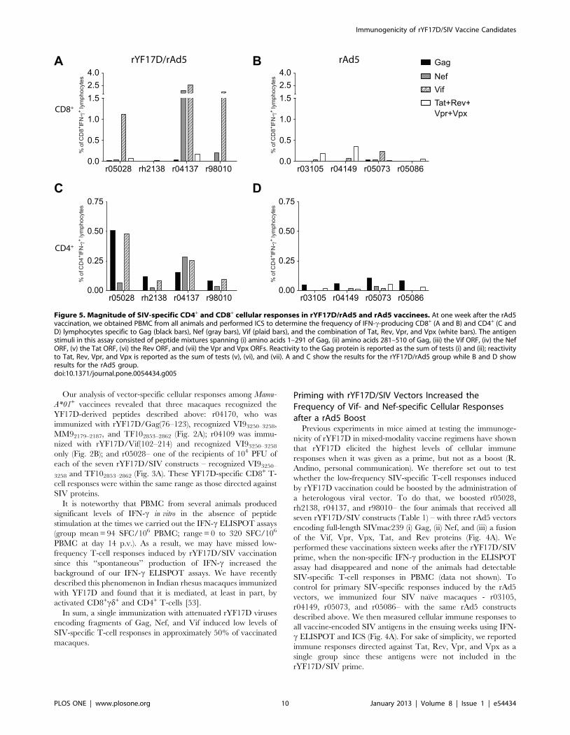
Figure 5. Magnitude of SIV-specific CD4+ and CD8+ cellular responses in rYF17D/rAd5 and rAd5 vaccinees. At one week after the rAd5vaccination, we obtained PBMC from all animals and performed ICS to determine the frequency of IFN-c-producing CD8+ (A and B) and CD4+ (C andD) lymphocytes specific to Gag (black bars), Nef (gray bars), Vif (plaid bars), and the combination of Tat, Rev, Vpr, and Vpx (white bars). The antigenstimuli in this assay consisted of peptide mixtures spanning (i) amino acids 1–291 of Gag, (ii) amino acids 281–510 of Gag, (iii) the Vif ORF, (iv) the NefORF, (v) the Tat ORF, (vi) the Rev ORF, and (vii) the Vpr and Vpx ORFs. Reactivity to the Gag protein is reported as the sum of tests (i) and (ii); reactivityto Tat, Rev, Vpr, and Vpx is reported as the sum of tests (v), (vi), and (vii). A and C show the results for the rYF17D/rAd5 group while B and D showresults for the rAd5 group.doi:10.1371/journal.pone.0054434.g005
Immunogenicity of rYF17D/SIV Vaccine Candidates
PLOS ONE | www.plosone.org 10 January 2013 | Volume 8 | Issue 1 | e54434
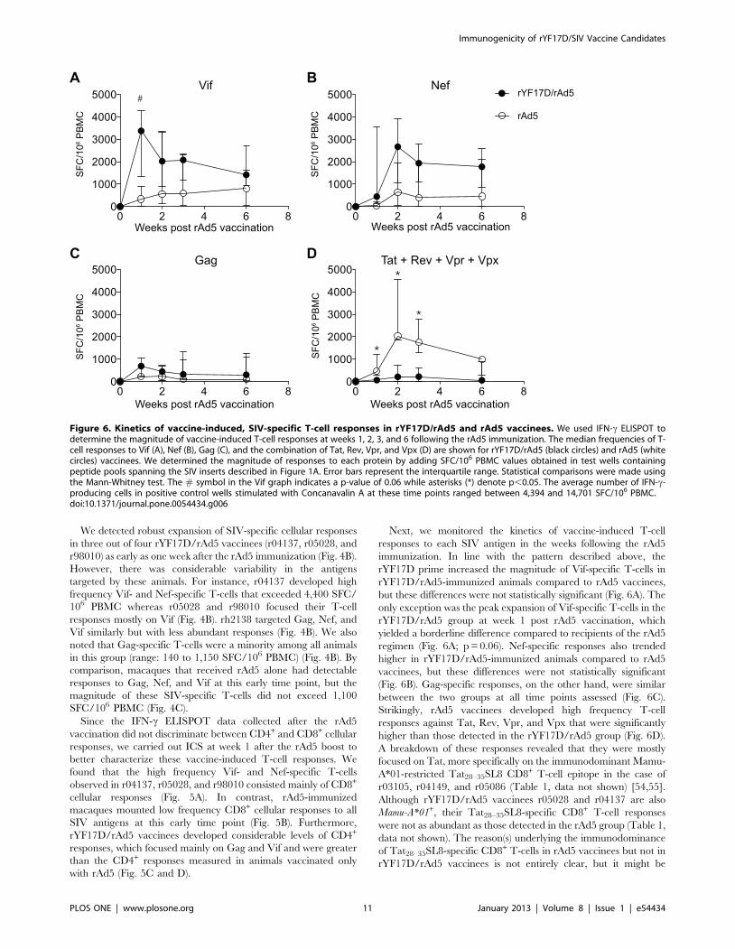
We detected robust expansion of SIV-specific cellular responses
in three out of four rYF17D/rAd5 vaccinees (r04137, r05028, and
r98010) as early as one week after the rAd5 immunization (Fig. 4B).
However, there was considerable variability in the antigens
targeted by these animals. For instance, r04137 developed high
frequency Vif- and Nef-specific T-cells that exceeded 4,400 SFC/
106 PBMC whereas r05028 and r98010 focused their T-cell
responses mostly on Vif (Fig. 4B). rh2138 targeted Gag, Nef, and
Vif similarly but with less abundant responses (Fig. 4B). We also
noted that Gag-specific T-cells were a minority among all animals
in this group (range: 140 to 1,150 SFC/106 PBMC) (Fig. 4B). By
comparison, macaques that received rAd5 alone had detectable
responses to Gag, Nef, and Vif at this early time point, but the
magnitude of these SIV-specific T-cells did not exceed 1,100
SFC/106 PBMC (Fig. 4C).
Since the IFN-c ELISPOT data collected after the rAd5
vaccination did not discriminate between CD4+ and CD8+ cellular
responses, we carried out ICS at week 1 after the rAd5 boost to
better characterize these vaccine-induced T-cell responses. We
found that the high frequency Vif- and Nef-specific T-cells
observed in r04137, r05028, and r98010 consisted mainly of CD8+
cellular responses (Fig. 5A). In contrast, rAd5-immunized
macaques mounted low frequency CD8+ cellular responses to all
SIV antigens at this early time point (Fig. 5B). Furthermore,
rYF17D/rAd5 vaccinees developed considerable levels of CD4+
responses, which focused mainly on Gag and Vif and were greater
than the CD4+ responses measured in animals vaccinated only
with rAd5 (Fig. 5C and D).
Next, we monitored the kinetics of vaccine-induced T-cell
responses to each SIV antigen in the weeks following the rAd5
immunization. In line with the pattern described above, the
rYF17D prime increased the magnitude of Vif-specific T-cells in
rYF17D/rAd5-immunized animals compared to rAd5 vaccinees,
but these differences were not statistically significant (Fig. 6A). The
only exception was the peak expansion of Vif-specific T-cells in the
rYF17D/rAd5 group at week 1 post rAd5 vaccination, which
yielded a borderline difference compared to recipients of the rAd5
regimen (Fig. 6A; p = 0.06). Nef-specific responses also trended
higher in rYF17D/rAd5-immunized animals compared to rAd5
vaccinees, but these differences were not statistically significant
(Fig. 6B). Gag-specific responses, on the other hand, were similar
between the two groups at all time points assessed (Fig. 6C).
Strikingly, rAd5 vaccinees developed high frequency T-cell
responses against Tat, Rev, Vpr, and Vpx that were significantly
higher than those detected in the rYF17D/rAd5 group (Fig. 6D).
A breakdown of these responses revealed that they were mostly
focused on Tat, more specifically on the immunodominant Mamu-
A*01-restricted Tat28–35SL8 CD8+ T-cell epitope in the case of
r03105, r04149, and r05086 (Table 1, data not shown) [54,55].
Although rYF17D/rAd5 vaccinees r05028 and r04137 are also
Mamu-A*01+, their Tat28–35SL8-specific CD8+ T-cell responses
were not as abundant as those detected in the rAd5 group (Table 1,
data not shown). The reason(s) underlying the immunodominance
of Tat28–35SL8-specific CD8+ T-cells in rAd5 vaccinees but not in
rYF17D/rAd5 vaccinees is not entirely clear, but it might be
Figure 6. Kinetics of vaccine-induced, SIV-specific T-cell responses in rYF17D/rAd5 and rAd5 vaccinees. We used IFN-c ELISPOT todetermine the magnitude of vaccine-induced T-cell responses at weeks 1, 2, 3, and 6 following the rAd5 immunization. The median frequencies of T-cell responses to Vif (A), Nef (B), Gag (C), and the combination of Tat, Rev, Vpr, and Vpx (D) are shown for rYF17D/rAd5 (black circles) and rAd5 (whitecircles) vaccinees. We determined the magnitude of responses to each protein by adding SFC/106 PBMC values obtained in test wells containingpeptide pools spanning the SIV inserts described in Figure 1A. Error bars represent the interquartile range. Statistical comparisons were made usingthe Mann-Whitney test. The # symbol in the Vif graph indicates a p-value of 0.06 while asterisks (*) denote p,0.05. The average number of IFN-c-producing cells in positive control wells stimulated with Concanavalin A at these time points ranged between 4,394 and 14,701 SFC/106 PBMC.doi:10.1371/journal.pone.0054434.g006
Immunogenicity of rYF17D/SIV Vaccine Candidates
PLOS ONE | www.plosone.org 11 January 2013 | Volume 8 | Issue 1 | e54434
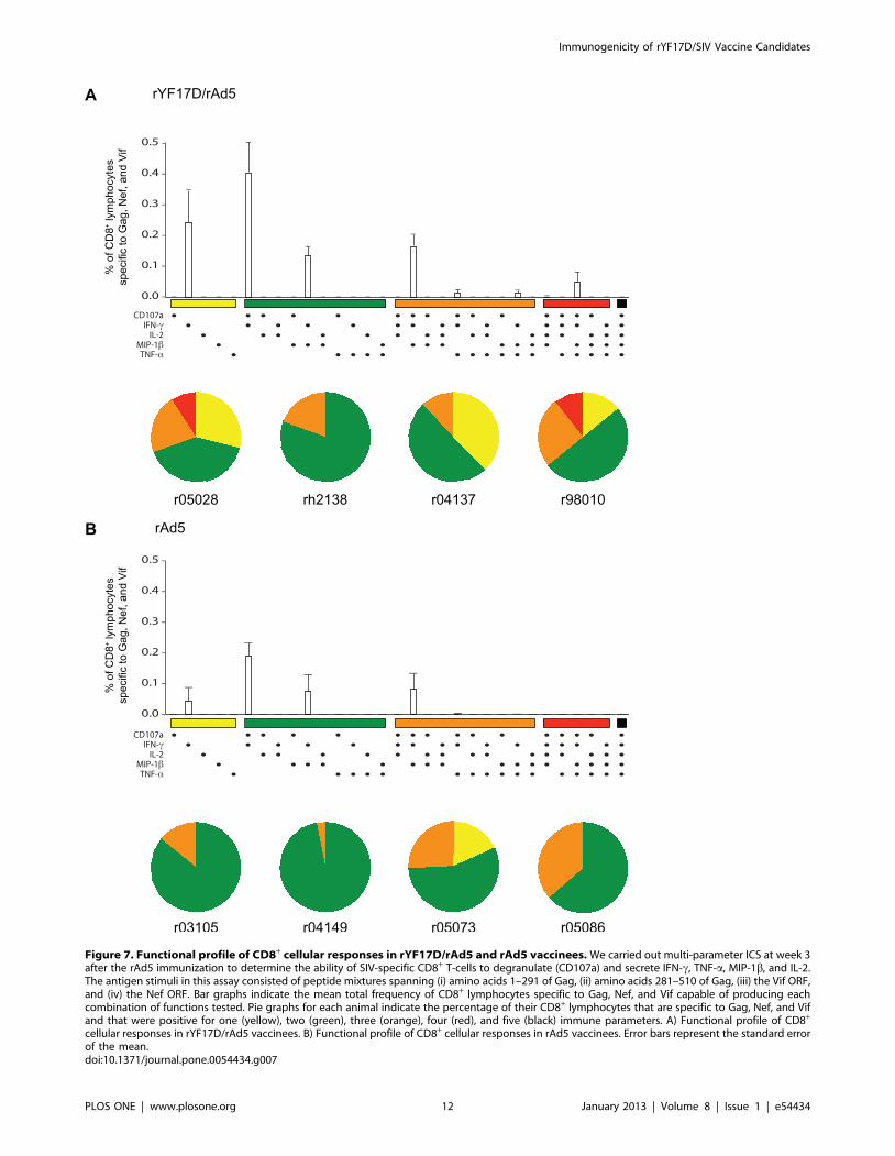
Figure 7. Functional profile of CD8+ cellular responses in rYF17D/rAd5 and rAd5 vaccinees. We carried out multi-parameter ICS at week 3after the rAd5 immunization to determine the ability of SIV-specific CD8+ T-cells to degranulate (CD107a) and secrete IFN-c, TNF-a, MIP-1b, and IL-2.The antigen stimuli in this assay consisted of peptide mixtures spanning (i) amino acids 1–291 of Gag, (ii) amino acids 281–510 of Gag, (iii) the Vif ORF,and (iv) the Nef ORF. Bar graphs indicate the mean total frequency of CD8+ lymphocytes specific to Gag, Nef, and Vif capable of producing eachcombination of functions tested. Pie graphs for each animal indicate the percentage of their CD8+ lymphocytes that are specific to Gag, Nef, and Vifand that were positive for one (yellow), two (green), three (orange), four (red), and five (black) immune parameters. A) Functional profile of CD8+
cellular responses in rYF17D/rAd5 vaccinees. B) Functional profile of CD8+ cellular responses in rAd5 vaccinees. Error bars represent the standard errorof the mean.doi:10.1371/journal.pone.0054434.g007
Immunogenicity of rYF17D/SIV Vaccine Candidates
PLOS ONE | www.plosone.org 12 January 2013 | Volume 8 | Issue 1 | e54434
related to a shift in the immunodominance hierarchy toward Vif
and Nef epitopes caused by the rYF17D prime.
Together, these results suggest that the rYF17D prime increased
the frequency of SIV-specific CD4+ and CD8+ T-cell responses
after rAd5 boosting. However, given the high animal to animal
variability observed in this study, a larger adequately powered trial
of rYF17D/SIV vaccine candidates may be required to confirm
these findings. The brisk expansion of CD8+ T-cells targeting Vif,
and to a lower extent Nef, seen early after the rAd5 boost might
also indicate that rYF17D vectors encoding fragments of these
proteins were more effective than the ones encoding segments of
Gag at priming SIV-specific T-cell responses.
Functional Profile of Vaccine-induced T-cell ResponsesThe ability of HIV-specific CD8+ T-cells to degranulate and
simultaneously produce multiple cytokines and chemokines has
been associated with delayed disease progression in HIV-1-
infected individuals [56]. We, therefore, used multi-parameter
ICS to determine the functional profile of vaccine-induced CD8+
T-cell responses in all animals. Since qualitative features of CD8+
T-cells targeting Gag, Nef, and Vif did not vary within each group
(data not shown), we decided to compare the total sum of CD8+ T-
cell responses specific to these three antigens between rYF17D/
rAd5 and rAd5 vaccinees at week 3 after the rAd5 vaccination. In
agreement with the immunogenicity data presented above, the
total frequency of CD8+ T-cells recognizing Gag, Nef, and Vif was
higher among macaques that were immunized with rYF17D/
rAd5 compared to those that received rAd5 only (Fig. 7A and B).
We also noticed that the majority of SIV-specific CD8+ T-cells in
both groups produced IFN-c either in combination with the
degranulation marker CD107a or with MIP-1b (Fig. 7A and B).
However, compared to rAd5-immunized animals, rYF17D/rAd5
vaccinees developed SIV-specific CD8+ T-cells with a slightly
increased functional quality, as seen by higher frequencies of
CD8+ T-cells staining positive for three and four immune
parameters (Fig. 7A and B). We also assessed qualitative features
of vaccine-induced CD4+ T-cell responses in rYF17D/rAd5 and
rAd5 vaccinees. We carried out this analysis at week 1 after the
rAd5 boost – the peak expansion of CD4+ T-cell responses in both
groups (Fig. 5C and D). In addition to secreting IFN-c, SIV-
specific CD4+ T-cells in rYF17D/rAd5 vaccinees were also
capable of producing IL-2, TNF-a, and MIP-1b in multiple
combinations (Fig. S2A). In contrast, rAd5-immunized macaques
mounted SIV-specific CD4+ T-cell responses with a more limited
functional profile (Fig. S2B). Together, these results suggest that
priming with rYF17D improved the functionality of SIV-specific
CD8+ and CD4+ T-cells that expanded after the rAd5 boost.
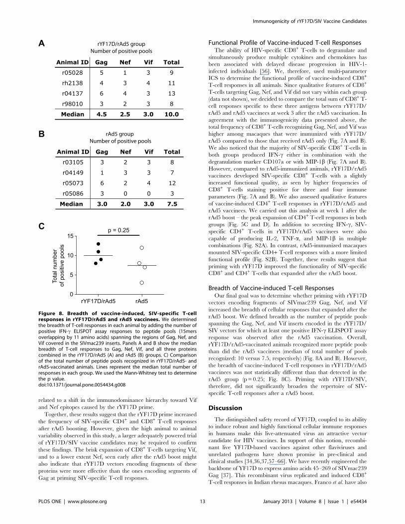
Breadth of Vaccine-induced T-cell ResponsesOur final goal was to determine whether priming with rYF17D
vectors encoding fragments of SIVmac239 Gag, Nef, and Vif
increased the breadth of cellular responses that expanded after the
rAd5 boost. We defined breadth as the number of peptide pools
spanning the Gag, Nef, and Vif inserts encoded in the rYF17D/
SIV vectors for which at least one positive IFN-c ELISPOT assay
response was observed after the rAd5 vaccination. Overall,
rYF17D/rAd5-vaccinated animals recognized more peptide pools
than did the rAd5 vaccinees (median of total number of pools
recognized: 10 versus 7.5, respectively) (Fig. 8A and B). However,
the breadth of vaccine-induced T-cell responses in rYF17D/rAd5
vaccinees was not statistically different than that detected in the
rAd5 group (p = 0.25; Fig. 8C). Priming with rYF17D/SIV,
therefore, did not significantly broaden the repertoire of SIV-
specific T-cell responses after a rAd5 boost.
Discussion
The distinguished safety record of YF17D, coupled to its ability
to induce robust and highly functional cellular immune responses
in humans make this live-attenuated virus an attractive vector
candidate for HIV vaccines. In support of this notion, recombi-
nant live YF17D-based vaccines against other flaviviruses and
unrelated pathogens have shown promise in pre-clinical and
clinical studies [34,36,37,57–66]. We have recently engineered the
backbone of YF17D to express amino acids 45–269 of SIVmac239
Gag [37]. This recombinant virus replicated and induced CD8+
T-cell responses in Indian rhesus macaques. Franco et al. have also
Figure 8. Breadth of vaccine-induced, SIV-specific T-cellresponses in rYF17D/rAd5 and rAd5 vaccinees. We determinedthe breadth of T-cell responses in each animal by adding the number ofpositive IFN-c ELISPOT assay responses to peptide pools (15mersoverlapping by 11 amino acids) spanning the regions of Gag, Nef, andVif covered in the SIVmac239 inserts. Panels A and B show the medianbreadth of T-cell responses to Gag, Nef, Vif, and all three proteinscombined in the rYF17D/rAd5 (A) and rAd5 (B) groups. C) Comparisonof the total number of peptide pools recognized in rYF17D/rAd5- andrAd5-vaccinated animals. Lines represent the median total number ofresponses in each group. We used the Mann-Whitney test to determinethe p value.doi:10.1371/journal.pone.0054434.g008
Immunogenicity of rYF17D/SIV Vaccine Candidates
PLOS ONE | www.plosone.org 13 January 2013 | Volume 8 | Issue 1 | e54434

demonstrated that a rYF17D encoding HIV Gag p24 induced
balanced CD4+ and CD8+ T-cell responses in BALB/c mice [67].
We, therefore, attempted to expand upon these studies by creating
seven new rYF17D viruses expressing fragments of the SIV-
mac239 Gag, Nef, and Vif proteins. We chose these antigens since
vaccine-induced T-cell responses to these proteins have been
associated with control of viral replication in a recent SIV efficacy
trial [38]. Additionally, a number of studies have linked Gag-
specific T-cell responses to lower chronic phase viral loads in HIV-
1-infected patients [68–70].
We vaccinated a total of eleven Indian rhesus macaques with
the rYF17D vectors encoding fragments of SIVmac239 Gag, Nef,
and Vif; seven animals received individual constructs while four
macaques received all seven rYF17D/SIV vectors. We detected
transient viremia in all animals that received single constructs, but
only two macaques that were immunized with the seven rYF17D/
SIV vectors had positive viral loads. An evaluation of cellular
immunity induced by a single vaccination with these vectors
revealed low frequency T-cell responses directed against Vif and
Nef, while Gag-specific responses were nearly absent. A potential
caveat in this analysis relates to the sensitivity of our IFN-cELISPOT assay, which might have been reduced since PBMC
from these animals produced high background levels of IFN-c at
days 14 and 17 post vaccination. We have recently described this
phenomenon, which occurs after YF17D vaccination of rhesus
macaques and appears to be caused by the activation of CD8+cd+
and CD4+ T-cells [53]. Since this ‘‘spontaneous’’ production of
IFN-c increased the background of our ELISPOT assays during
the first few weeks after vaccination, we likely missed low-
frequency SIV-specific T-cell responses induced by the rYF17D/
SIV vectors.
It is also possible that the insertion of SIV sequences in the E/
NS1 intergenic region attenuated the rYF17D/SIV constructs
even further and thus decreased their immunogenicity compared
to the parental YF17D vaccine. Along these lines, a rYF17D
virus expressing enhanced green fluorescent protein in this same
genomic region demonstrated delayed replication kinetics in vitro
and induced significantly lower titers of neutralizing antibodies
in mice compared to the parental YF17D virus [36]. Further-
more, we have recently evaluated the replication of YF17D and
a rYF17D expressing a fragment of SIVmac239 Gag encoding
amino acids 45–269 in Indian rhesus macaques using the same
qRT-PCR assay employed in this study [37]. We found that
positive viral loads came up earlier and at greater magnitudes in
animals that received the parental vaccine compared to
recipients of the recombinant construct. If lower replication
fitness is indeed limiting the magnitude of SIV-specific cellular
responses generated by the rYF17D/SIV candidates, additional
booster doses might improve the immunogenicity of these
vaccine viruses.
It is important to address genetic stability during the develop-
ment of live-attenuated RNA virus vaccines. In this regard, we
have tested the genetic integrity of the rYF17D/SIV viruses by
serially passaging them in Vero cells (Bonaldo et al., unpublished
data). Electrophoretic analysis of RT-PCR amplicons from viral
RNA extracted at the 15th passage revealed that four of the seven
rYF17D/SIV viruses were stable at this time point. The rYF17D/
Nef(45–210) construct yielded a unique gel pattern, containing the
amplicon corresponding to the SIV insert and two smaller, less
intense fragments. We are currently investigating whether these
extra bands are the result of a mixed viral population. We also
found that two constructs – rYF17D/Gag(76–123) and rYF17D/
Vif(1–110) – lost their inserts at the 10th passage. Although this
may explain why we could not detect Gag-specific responses in
r04170– the rYF17D/Gag(76–123)-vaccinated macaque, it does
not account for the low frequency of SIV-specific T-cell responses
induced by the other stable rYF17D constructs. Additionally,
rYF17D/Vif(1–110) – one of the genetically unstable viruses –
induced detectable Vif-specific cellular responses in r05089 (200
SFC/106 PBMC). Therefore, the rYF17D/SIV vectors were
poorly immunogenic even though the majority of these viruses
were stable in vitro.
Our next step was to test whether the low frequency T-cell
responses to Gag, Nef, and Vif induced by vaccination with the
rYF17D/SIV constructs could be boosted by a heterologous virus
boost. To do that, we immunized eight animals with rAd5 vectors
encoding full-length Gag, Nef, and a fusion of the Vif, Tat, Rev,
Vpr, and Vpx proteins. Four of the animals had been primed with
all seven rYF17D/SIV vectors, while the other four macaques
were SIV naıve and served as controls for primary responses
induced by the rAd5 (Table 1, Fig. 4A). We found evidence that
the rAd5 vaccination boosted SIV-specific cellular responses in
animals that had received the mixture of seven rYF17D/SIV
vectors, as seen by the robust expansion of Vif-specific T-cells in
r05028 and r98010, as well as the high magnitude of Nef-specific
T-cells in r04137. On one hand, this is an encouraging finding
since it suggests that YF17D – a clinically relevant vector platform
for inducing HIV-specific T-cell responses – effectively primed
SIV-specific cellular responses and thus is compatible with
heterologous prime boost vaccine regimens. On the other hand,
the heterogeneity in the magnitude and specificity of the responses
that expanded after the rAd5 boost suggests that some rYF17D/
SIV constructs were more effective than others at priming SIV-
specific T-cells. Furthermore, the immunogenicity of the rYF17D/
SIV prime did not predict the expansion of anamnestic responses
after the rAd5 boost. Macaque r04137, for instance, had no
detectable cellular responses to the SIV antigens following the
rYF17D/SIV prime and yet this animal mounted the highest
frequency of Vif- and Nef-specific T-cells in the rYF17D/rAd5
group (Figs. 3B and 4B). Conversely, rYF17D/SIV vaccination
elicited positive IFN-c ELISPOT responses to Nef and Vif in
rh2138 but this animal did not develop high levels of T-cell
responses to these two SIV antigens at week 1 following the rAd5
boost (Figs. 3A and 4B). The reasons for this high animal to animal
variability are not entirely clear, but these results suggest that a
more thorough investigation of the immunogenicity and in vivo
replicative capacity of these rYF17D/SIV viruses is warranted,
especially when rYF17D/SIV vectors are administered simulta-
neously.
We also noticed a trend toward broader T-cell responses among
rYF17D/rAd5 vaccineees compared to the rAd5 group (median of
total number of pools recognized: 10 versus 7.5, respectively).
However, this difference did not achieve statistical difference
(p = 0.25). The low immunogenicity achieved by the rYF17D/SIV
vectors during the priming stage and the small sample size of our
experimental groups (n = 4) likely contributed to the comparable
T-cell breadth observed in rYF17D/rAd5 and rAd5 vaccinees.
Additionally, the fact that rYF17D/rAd5 vaccinees were primed
with rYF17D/SIV vectors encoding fragments of Gag, Nef, and
Vif and subsequently boosted with rAd5 expressing full-length (i)
Gag, (ii) Nef, and (iii) Vif fused to Tat, Rev, Vpr, and Vpx might
have restricted the repertoire of vaccine-induced T-cell responses
by favoring the expansion of T-cells targeting dominant epitopes,
as suggested by previous studies [47–49,71]. Thus, a rYF17D/SIV
prime followed by a heterologous virus boost regimen encoding
the same SIV minigenes might result in broader SIV-specific T-
cell responses.
Immunogenicity of rYF17D/SIV Vaccine Candidates
PLOS ONE | www.plosone.org 14 January 2013 | Volume 8 | Issue 1 | e54434
In summary, the goal of the present study was to evaluate the
immunogenicity of live-attenuated rYF17D/SIV viruses express-
ing fragments of SIV Gag, Nef, and Vif in rhesus macaques. We
found evidence that these vaccine viruses replicated in vivo, but
they engendered low levels of SIV-specific cellular responses.
Boosting with rAd5 vectors resulted in robust expansion of SIV-
specific T-cells, particularly those targeting Vif and, to a lesser
extent, Nef. These anamnestic responses comprised CD4+ and
CD8+ T-cells capable of performing up to four functions after
stimulation with synthetic peptides. However, priming with
rYF17D/SIV had a limited effect on the breadth of SIV-specific
T-cell responses that expanded after the rAd5 boost. It is
important to note that these rYF17D/SIV vectors are in their
first generation and thus there is still room for improvement. For
example, a vaccination regimen comprised of two or three doses of
rYF17D/SIV might increase the immunogenicity of these vaccine
vectors. In support of this, Santos et al. reported that revaccination
of YF17D-immune human subjects with the parental YF17D
strain resulted in a 3-fold increase in the percentage of activated
CD8+ T-cells in peripheral blood [72]. Additionally, modification
of the SIV inserts increased the genetic integrity of rYF17D/
Gag(76–123) and rYF17D/Vif(1–110) – the two recombinant
viruses that became unstable after 10 passages in vitro (Bonaldo
et al. unpublished data). We are also testing whether macaques
immunized with an improved rYF17D/rAd5 regimen encoding
matched SIV minigenes can control viral replication after a
pathogenic SIV challenge (Martins et al., unpublished data).
Optimized rYF17D/HIV vectors may, therefore, be useful for
inducing cellular immune responses against the AIDS virus.
Supporting Information
Figure S1 Gating strategy for analysis of multi-func-tional ICS. A representative example of negative (medium only)
and positive (SEB-stimulated) CD8+ cellular responses in one
rYF17D/rAd5 vaccinee (r05028). These data were obtained at
week 3 after the rAd5 boost.
(EPS)
Figure S2 Functional profile of CD4+ cellular responsesin rYF17D/rAd5 and rAd5 vaccinees. We carried out multi-
parameter ICS at week 1 after the rAd5 immunization to
determine the ability of SIV-specific CD4+ T-cells to degranulate
(CD107a) and secrete IFN-c, TNF-a, MIP-1b, and IL-2. The
antigen stimuli in this assay consisted of peptide mixtures spanning
(i) amino acids 1–291 of Gag, (ii) amino acids 281–510 of Gag, (iii)
the Vif ORF, and (iv) the Nef ORF. Bar graphs indicate the mean
total frequency of CD4+ lymphocytes specific to Gag, Nef, and Vif
capable of producing each combination of functions tested. Pie
graphs for each animal indicate the percentage of their CD4+
lymphocytes that are specific to Gag, Nef, and Vif and that were
positive for one (yellow), two (green), three (orange), four (red), and
five (black) immune parameters. A) Functional profile of CD4+
cellular responses in rYF17D/rAd5 vaccinees. B) Functional
profile of CD4+ cellular responses in rAd5 vaccinees. Error bars
represent the standard error of the mean.
(EPS)
Acknowledgments
The authors would like to thank Chrystal Glidden, Gretta Borchardt, and
Debra Fisk for MHC typing of animals. We are also thankful to Saverio
Capuano III and the veterinary staff of the Wisconsin National Primate
Research Center for performing animal procedures.
Author Contributions
Conceived and designed SIVmac239 minigenes: TCF DIW. Generated
rYF17D/SIV vectors: MCB MGVS RG. Oversaw statistical analyses: BH
DBA. Conceived and designed the experiments: MAM NAW DIW.
Performed the experiments: MAM RR SMP JRF CME MGVS KLW.
Analyzed the data: MAM RR SMP EGR NAW. Contributed reagents/
materials/analysis tools: MJC CLP. Wrote the paper: MAM DIW.
References
1. (1981) Pneumocystis pneumonia–Los Angeles. MMWR Morb Mortal Wkly Rep
30: 250–252.
2. HIV/AIDS JUNPo (2010) Global report: UNAIDS report on the global AIDS
pandemic 2010.
3. McElrath MJ, Haynes BF (2010) Induction of immunity to human immuno-
deficiency virus type-1 by vaccination. Immunity 33: 542–554.
4. Allen TM, Altfeld M, Geer SC, Kalife ET, Moore C, et al. (2005) Selective
escape from CD8+ T-cell responses represents a major driving force of human
immunodeficiency virus type 1 (HIV-1) sequence diversity and reveals
constraints on HIV-1 evolution. J Virol 79: 13239–13249.
5. Carrington M, O’Brien SJ (2003) The influence of HLA genotype on AIDS.
Annu Rev Med 54: 535–551.
6. Friedrich TC, Valentine LE, Yant LJ, Rakasz EG, Piaskowski SM, et al. (2007)
Subdominant CD8+ T-cell responses are involved in durable control of AIDS
virus replication. J Virol 81: 3465–3476.
7. Goulder PJ, Watkins DI (2008) Impact of MHC class I diversity on immune
control of immunodeficiency virus replication. Nat Rev Immunol 8: 619–630.
8. Jin X, Bauer DE, Tuttleton SE, Lewin S, Gettie A, et al. (1999) Dramatic rise in
plasma viremia after CD8(+) T cell depletion in simian immunodeficiency virus-
infected macaques. J Exp Med 189: 991–998.
9. Matano T, Shibata R, Siemon C, Connors M, Lane HC, et al. (1998)
Administration of an anti-CD8 monoclonal antibody interferes with the
clearance of chimeric simian/human immunodeficiency virus during primary
infections of rhesus macaques. J Virol 72: 164–169.
10. Pereyra F, Jia X, McLaren PJ, Telenti A, de Bakker PI, et al. (2010) The major
genetic determinants of HIV-1 control affect HLA class I peptide presentation.
Science 330: 1551–1557.
11. Boaz MJ, Waters A, Murad S, Easterbrook PJ, Vyakarnam A (2002) Presence of
HIV-1 Gag-specific IFN-gamma+IL-2+ and CD28+IL-2+ CD4 T cell responses
is associated with nonprogression in HIV-1 infection. J Immunol 169: 6376–
6385.
12. Giraldo-Vela JP, Rudersdorf R, Chung C, Qi Y, Wallace LT, et al. (2008) The
major histocompatibility complex class II alleles Mamu-DRB1*1003 and -
DRB1*0306 are enriched in a cohort of simian immunodeficiency virus-infected
rhesus macaque elite controllers. J Virol 82: 859–870.
13. Rosenberg ES, Billingsley JM, Caliendo AM, Boswell SL, Sax PE, et al. (1997)
Vigorous HIV-1-specific CD4+ T cell responses associated with control of
viremia. Science 278: 1447–1450.
14. Sacha JB, Chung C, Rakasz EG, Spencer SP, Jonas AK, et al. (2007) Gag-
specific CD8+ T lymphocytes recognize infected cells before AIDS-virus
integration and viral protein expression. J Immunol 178: 2746–2754.
15. Liu MA (2010) Immunologic basis of vaccine vectors. Immunity 33: 504–515.
16. Barouch DH (2010) Novel adenovirus vector-based vaccines for HIV-1. Curr
Opin HIV AIDS 5: 386–390.
17. Pantaleo G, Esteban M, Jacobs B, Tartaglia J (2010) Poxvirus vector-based HIV
vaccines. Curr Opin HIV AIDS 5: 391–396.
18. Robert-Guroff M (2007) Replicating and non-replicating viral vectors for
vaccine development. Curr Opin Biotechnol 18: 546–556.
19. Abbink P, Lemckert AA, Ewald BA, Lynch DM, Denholtz M, et al. (2007)
Comparative seroprevalence and immunogenicity of six rare serotype recom-
binant adenovirus vaccine vectors from subgroups B and D. J Virol 81: 4654–
4663.
20. Buchbinder SP, Mehrotra DV, Duerr A, Fitzgerald DW, Mogg R, et al. (2008)
Efficacy assessment of a cell-mediated immunity HIV-1 vaccine (the Step Study):
a double-blind, randomised, placebo-controlled, test-of-concept trial. Lancet
372: 1881–1893.
21. McElrath MJ, De Rosa SC, Moodie Z, Dubey S, Kierstead L, et al. (2008) HIV-
1 vaccine-induced immunity in the test-of-concept Step Study: a case-cohort
analysis. Lancet 372: 1894–1905.
22. Thorner AR, Vogels R, Kaspers J, Weverling GJ, Holterman L, et al. (2006) Age
dependence of adenovirus-specific neutralizing antibody titers in individuals
from sub-Saharan Africa. J Clin Microbiol 44: 3781–3783.
23. Hanke T, Goonetilleke N, McMichael AJ, Dorrell L (2007) Clinical experience
with plasmid DNA- and modified vaccinia virus Ankara-vectored human
immunodeficiency virus type 1 clade A vaccine focusing on T-cell induction.
J Gen Virol 88: 1–12.
Immunogenicity of rYF17D/SIV Vaccine Candidates
PLOS ONE | www.plosone.org 15 January 2013 | Volume 8 | Issue 1 | e54434

24. Nitayaphan S, Pitisuttithum P, Karnasuta C, Eamsila C, de Souza M, et al.
(2004) Safety and immunogenicity of an HIV subtype B and E prime-boostvaccine combination in HIV-negative Thai adults. J Infect Dis 190: 702–706.
25. Russell ND, Graham BS, Keefer MC, McElrath MJ, Self SG, et al. (2007) Phase2 study of an HIV-1 canarypox vaccine (vCP1452) alone and in combination
with rgp120: negative results fail to trigger a phase 3 correlates trial. J AcquirImmune Defic Syndr 44: 203–212.
26. Barrett AD, Teuwen DE (2009) Yellow fever vaccine - how does it work and whydo rare cases of serious adverse events take place? Curr Opin Immunol 21: 308–
313.
27. Monath TP (2005) Yellow fever vaccine. Expert Rev Vaccines 4: 553–574.
28. Pulendran B (2009) Learning immunology from the yellow fever vaccine: innate
immunity to systems vaccinology. Nat Rev Immunol 9: 741–747.
29. Querec T, Bennouna S, Alkan S, Laouar Y, Gorden K, et al. (2006) Yellow fever
vaccine YF-17D activates multiple dendritic cell subsets via TLR2, 7, 8, and 9 tostimulate polyvalent immunity. J Exp Med 203: 413–424.
30. Querec TD, Pulendran B (2007) Understanding the role of innate immunity in
the mechanism of action of the live attenuated Yellow Fever Vaccine 17D. AdvExp Med Biol 590: 43–53.
31. Barnett ED (2007) Yellow fever: epidemiology and prevention. Clin Infect Dis44: 850–856.
32. Organization WH (2009) WHO Vaccine-preventable diseases: monitoring
system.
33. Arroyo J, Miller C, Catalan J, Myers GA, Ratterree MS, et al. (2004)
ChimeriVax-West Nile virus live-attenuated vaccine: preclinical evaluation ofsafety, immunogenicity, and efficacy. J Virol 78: 12497–12507.
34. Bonaldo MC, Garratt RC, Caufour PS, Freire MS, Rodrigues MM, et al. (2002)Surface expression of an immunodominant malaria protein B cell epitope by
yellow fever virus. J Mol Biol 315: 873–885.
35. McAllister A, Arbetman AE, Mandl S, Pena-Rossi C, Andino R (2000)
Recombinant yellow fever viruses are effective therapeutic vaccines fortreatment of murine experimental solid tumors and pulmonary metastases.
J Virol 74: 9197–9205.
36. Bonaldo MC, Mello SM, Trindade GF, Rangel AA, Duarte AS, et al. (2007)
Construction and characterization of recombinant flaviviruses bearing insertionsbetween E and NS1 genes. Virol J 4: 115.
37. Bonaldo MC, Martins MA, Rudersdorf R, Mudd PA, Sacha JB, et al. (2010)
Recombinant yellow fever vaccine virus 17D expressing simian immunodefi-
ciency virus SIVmac239 gag induces SIV-specific CD8+ T-cell responses inrhesus macaques. J Virol 84: 3699–3706.
38. Martins MA, Wilson NA, Reed JS, Ahn CD, Klimentidis YC, et al. (2010) T-cell
correlates of vaccine efficacy after a heterologous simian immunodeficiency viruschallenge. J Virol 84: 4352–4365.
39. Weatherall D (2006) The use of non-human primates in research: A workinggroup report. Final Report December 2006. FRS FMedSci.
40. Kaizu M, Borchardt GJ, Glidden CE, Fisk DL, Loffredo JT, et al. (2007)
Molecular typing of major histocompatibility complex class I alleles in the Indian
rhesus macaque which restrict SIV CD8+ T cell epitopes. Immunogenetics 59:693–703.
41. Loffredo JT, Maxwell J, Qi Y, Glidden CE, Borchardt GJ, et al. (2007) Mamu-
B*08-positive macaques control simian immunodeficiency virus replication.
J Virol 81: 8827–8832.
42. Caufour PS, Motta MC, Yamamura AM, Vazquez S, Ferreira II, et al. (2001)Construction, characterization and immunogenicity of recombinant yellow fever
17D-dengue type 2 viruses. Virus Res 79: 1–14.
43. Cline AN, Bess JW, Piatak MJ, Lifson JD (2005) Highly sensitive SIV plasma
viral load assay: practical considerations, realistic performance expectations, andapplication to reverse engineering of vaccines for AIDS. J Med Primatol 34:
303–312.
44. Winstone N, Wilson AJ, Morrow G, Boggiano C, Chiuchiolo MJ, et al. (2011)
Enhanced control of pathogenic Simian immunodeficiency virus SIVmac239replication in macaques immunized with an interleukin-12 plasmid and a DNA
prime-viral vector boost vaccine regimen. J Virol 85: 9578–9587.
45. Mittereder N, March KL, Trapnell BC (1996) Evaluation of the concentration
and bioactivity of adenovirus vectors for gene therapy. J Virol 70: 7498–7509.
46. Roederer M, Nozzi JL, Nason MC (2011) SPICE: exploration and analysis ofpost-cytometric complex multivariate datasets. Cytometry A 79: 167–174.
47. Liu Y, Li F, Liu Y, Hong K, Meng X, et al. (2011) HIV fragment gag vaccineinduces broader T cell response in mice. Vaccine 29: 2582–2589.
48. Rodriguez F, Harkins S, Slifka MK, Whitton JL (2002) Immunodominance in
virus-induced CD8(+) T-cell responses is dramatically modified by DNA
immunization and is regulated by gamma interferon. J Virol 76: 4251–4259.
49. Im EJ, Hong JP, Roshorm Y, Bridgeman A, Letourneau S, et al. (2011)Protective efficacy of serially up-ranked subdominant CD8+ T cell epitopes
against virus challenges. PLoS Pathog 7: e1002041.
50. Loffredo JT, Sidney J, Wojewoda C, Dodds E, Reynolds MR, et al. (2004)
Identification of seventeen new simian immunodeficiency virus-derived CD8+ Tcell epitopes restricted by the high frequency molecule, Mamu-A*02, and
potential escape from CTL recognition. J Immunol 173: 5064–5076.
51. Vogel TU, Friedrich TC, O’Connor DH, Rehrauer W, Dodds EJ, et al. (2002)Escape in one of two cytotoxic T-lymphocyte epitopes bound by a high-
frequency major histocompatibility complex class I molecule, Mamu-A*02: aparadigm for virus evolution and persistence? J Virol 76: 11623–11636.
52. Peters B, Bui HH, Sidney J, Weng Z, Loffredo JT, et al. (2005) A computational
resource for the prediction of peptide binding to Indian rhesus macaque MHCclass I molecules. Vaccine 23: 5212–5224.
53. Neves PC, Rudersdorf RA, Galler R, Bonaldo MC, de Santana MG, et al.(2010) CD8+ gamma-delta TCR+ and CD4+ T cells produce IFN-gamma at 5–
7 days after yellow fever vaccination in Indian rhesus macaques, before theinduction of classical antigen-specific T cell responses. Vaccine 28: 8183–8188.
54. Allen TM, O’Connor DH, Jing P, Dzuris JL, Mothe BR, et al. (2000) Tat-
specific cytotoxic T lymphocytes select for SIV escape variants during resolutionof primary viraemia. Nature 407: 386–390.
55. Mothe BR, Horton H, Carter DK, Allen TM, Liebl ME, et al. (2002)Dominance of CD8 responses specific for epitopes bound by a single major
histocompatibility complex class I molecule during the acute phase of viral
infection. J Virol 76: 875–884.56. Betts MR, Nason MC, West SM, De Rosa SC, Migueles SA, et al. (2006) HIV
nonprogressors preferentially maintain highly functional HIV-specific CD8+ Tcells. Blood 107: 4781–4789.
57. Appaiahgari MB, Vrati S (2010) IMOJEV((R)): a Yellow fever virus-based novelJapanese encephalitis vaccine. Expert Rev Vaccines 9: 1371–1384.
58. Bonaldo MC, Garratt RC, Freire MS, Galler R (2006) Expression of foreign
protein epitopes at the surface of recombinant yellow fever 17D viruses based onthree-dimensional modeling of its envelope protein. Cell Biochem Biophys 44:
313–324.59. Guirakhoo F, Pugachev K, Zhang Z, Myers G, Levenbook I, et al. (2004) Safety
and efficacy of chimeric yellow Fever-dengue virus tetravalent vaccine
formulations in nonhuman primates. J Virol 78: 4761–4775.60. Guy B, Nougarede N, Begue S, Sanchez V, Souag N, et al. (2008) Cell-mediated
immunity induced by chimeric tetravalent dengue vaccine in naive or flavivirus-primed subjects. Vaccine 26: 5712–5721.
61. Guy B, Guirakhoo F, Barban V, Higgs S, Monath TP, et al. (2010) Preclinicaland clinical development of YFV 17D-based chimeric vaccines against dengue,
West Nile and Japanese encephalitis viruses. Vaccine 28: 632–649.
62. Guy B, Barrere B, Malinowski C, Saville M, Teyssou R, et al. (2011) Fromresearch to phase III: preclinical, industrial and clinical development of the
Sanofi Pasteur tetravalent dengue vaccine. Vaccine 29: 7229–7241.63. Monath TP, Guirakhoo F, Nichols R, Yoksan S, Schrader R, et al. (2003)
Chimeric live, attenuated vaccine against Japanese encephalitis (ChimeriVax-
JE): phase 2 clinical trials for safety and immunogenicity, effect of vaccine doseand schedule, and memory response to challenge with inactivated Japanese
encephalitis antigen. J Infect Dis 188: 1213–1230.64. Monath TP, Liu J, Kanesa-Thasan N, Myers GA, Nichols R, et al. (2006) A live,
attenuated recombinant West Nile virus vaccine. Proc Natl Acad Sci U S A 103:6694–6699.
65. Smith HL, Monath TP, Pazoles P, Rothman AL, Casey DM, et al. (2011)
Development of antigen-specific memory CD8+ T cells following live-attenuatedchimeric West Nile virus vaccination. J Infect Dis 203: 513–522.
66. Tao D, Barba-Spaeth G, Rai U, Nussenzweig V, Rice CM, et al. (2005) Yellowfever 17D as a vaccine vector for microbial CTL epitopes: protection in a rodent
malaria model. J Exp Med 201: 201–209.
67. Franco D, Li W, Qing F, Stoyanov CT, Moran T, et al. (2010) Evaluation ofyellow fever virus 17D strain as a new vector for HIV-1 vaccine development.
Vaccine 28: 5676–5685.68. Edwards BH, Bansal A, Sabbaj S, Bakari J, Mulligan MJ, et al. (2002)
Magnitude of functional CD8+ T-cell responses to the gag protein of human
immunodeficiency virus type 1 correlates inversely with viral load in plasma.J Virol 76: 2298–2305.
69. Kiepiela P, Ngumbela K, Thobakgale C, Ramduth D, Honeyborne I, et al.(2007) CD8+ T-cell responses to different HIV proteins have discordant
associations with viral load. Nat Med 13: 46–53.70. Rolland M, Heckerman D, Deng W, Rousseau CM, Coovadia H, et al. (2008)
Broad and Gag-biased HIV-1 epitope repertoires are associated with lower viral
loads. PLoS One 3: e1424.71. Liu J, Ewald BA, Lynch DM, Nanda A, Sumida SM, et al. (2006) Modulation of
DNA vaccine-elicited CD8+ T-lymphocyte epitope immunodominance hierar-chies. J Virol 80: 11991–11997.
72. Santos AP, Bertho AL, Dias DC, Santos JR, Marcovistz R (2005) Lymphocyte
subset analyses in healthy adults vaccinated with yellow fever 17DD virus. MemInst Oswaldo Cruz 100: 331–337.
Immunogenicity of rYF17D/SIV Vaccine Candidates
PLOS ONE | www.plosone.org 16 January 2013 | Volume 8 | Issue 1 | e54434
Copyright © 2022 FDOKUMEN