Immune Modulation with Sulfasalazine Attenuates Immunopathogenesis but Enhances Macrophage-Mediated...
14
Immune Modulation with Sulfasalazine Attenuates Immunopathogenesis but Enhances Macrophage- Mediated Fungal Clearance during Pneumocystis Pneumonia Jing Wang 1 , Francis Gigliotti 1,2 , Samir P. Bhagwat 1 , Thaddeus C. George 3 , Terry W. Wright 1,2 * 1 Department of Pediatrics, University of Rochester School of Medicine and Dentistry, Rochester, New York, United States of America, 2 Department of Microbiology and Immunology, University of Rochester School of Medicine and Dentistry, Rochester, New York, United States of America, 3 Amnis Corporation, Seattle, Washington, United States of America Abstract Although T cells are critical for host defense against respiratory fungal infections, they also contribute to the immunopathogenesis of Pneumocystis pneumonia (PcP). However, the precise downstream effector mechanisms by which T cells mediate these diverse processes are undefined. In the current study the effects of immune modulation with sulfasalazine were evaluated in a mouse model of PcP-related Immune Reconstitution Inflammatory Syndrome (PcP-IRIS). Recovery of T cell-mediated immunity in Pneumocystis-infected immunodeficient mice restored host defense, but also initiated the marked pulmonary inflammation and severe pulmonary function deficits characteristic of IRIS. Sulfasalazine produced a profound attenuation of IRIS, with the unexpected consequence of accelerated fungal clearance. To determine whether macrophage phagocytosis is an effector mechanism of T cell-mediated Pneumocystis clearance and whether sulfasalazine enhances clearance by altering alveolar macrophage phagocytic activity, a novel multispectral imaging flow cytometer-based method was developed to quantify the phagocytosis of Pneumocystis in vivo. Following immune reconstitution, alveolar macrophages from PcP-IRIS mice exhibited a dramatic increase in their ability to actively phagocytose Pneumocystis. Increased phagocytosis correlated temporally with fungal clearance, and required the presence of CD4 + T cells. Sulfasalazine accelerated the onset of the CD4 + T cell-dependent alveolar macrophage phagocytic response in PcP-IRIS mice, resulting in enhanced fungal clearance. Furthermore, sulfasalazine promoted a TH2-polarized cytokine environment in the lung, and sulfasalazine-enhanced phagocytosis of Pneumocystis was associated with an alternatively activated alveolar macrophage phenotype. These results provide evidence that macrophage phagocytosis is an important in vivo effector mechanism for T cell-mediated Pneumocystis clearance, and that macrophage phenotype can be altered to enhance phagocytosis without exacerbating inflammation. Immune modulation can diminish pulmonary inflammation while preserving host defense, and has therapeutic potential for the treatment of PcP-related immunopathogenesis. Citation: Wang J, Gigliotti F, Bhagwat SP, George TC, Wright TW (2010) Immune Modulation with Sulfasalazine Attenuates Immunopathogenesis but Enhances Macrophage-Mediated Fungal Clearance during Pneumocystis Pneumonia. PLoS Pathog 6(8): e1001058. doi:10.1371/journal.ppat.1001058 Editor: Stuart M. Levitz, UMass Medical Center, United States of America Received March 19, 2010; Accepted July 22, 2010; Published August 19, 2010 Copyright: ß 2010 Wang et al. This is an open-access article distributed under the terms of the Creative Commons Attribution License, which permits unrestricted use, distribution, and reproduction in any medium, provided the original author and source are credited. Funding: This study was supported by National Institute of Health grants R01 HL083761 and R01 HL092797, and a grant from the Strong Children’s Research Center. The funders had no role in study design, data collection and analysis, decision to publish, or preparation of the manuscript. Competing Interests: Dr. George is an employee of Amnis Corporation, producers of the ImageStream imaging flow cytometer. * E-mail: [email protected] Introduction Pneumocystis (Pc) is an opportunistic fungal respiratory pathogen that causes life-threatening pneumonia in patients suffering from defects in cell-mediated immunity, including those with acquired immunodeficiency syndrome (AIDS) and immunosuppression secondary to chemotherapy or organ transplantation. Pneumocystis pneumonia (PcP) remains a leading cause of death among HIV- infected patients and a significant cause of AIDS-related mortality and morbidity [1]. For example, mortality rates of 50% or higher have been reported for AIDS patients with severe PcP [2,3], and despite major advances in health care, the mortality associated with PcP has changed little over the past 25 years. In addition, as more powerful anti-inflammatory treatments are developed for various underlying diseases, more cases of PcP are occurring in non-HIV patients and in previously unreported clinical settings [4–6]. Recent studies also indicate that Pc colonization can exacerbate chronic obstructive pulmonary disease [7]. Therefore, improving the treatment of patients suffering from both HIV- and non HIV-related PcP remains a central concern of the health care community. Although the direct pathogenic capabilities of the Pneumocystis organism itself are poorly understood, the role of the host’s immune response as a major contributor to PcP-related lung injury has come to the forefront. In patients, the clinical severity of PcP is dictated by the degree of pulmonary inflammation, rather than by the organism lung burden [8–14]. Specifically, T cell and neutrophilic responses have been linked to PcP-related lung injury in patients [10,15]. A clinical example of the immunopathogenic nature of PcP is the severe disease that has been reported in AIDS patients following successful anti-retroviral treatment [16–18]. This distinct clinical syndrome, termed Immune Reconstitution PLoS Pathogens | www.plospathogens.org 1 August 2010 | Volume 6 | Issue 8 | e1001058
Transcript of Immune Modulation with Sulfasalazine Attenuates Immunopathogenesis but Enhances Macrophage-Mediated...

Immune Modulation with Sulfasalazine AttenuatesImmunopathogenesis but Enhances Macrophage-Mediated Fungal Clearance during PneumocystisPneumoniaJing Wang1, Francis Gigliotti1,2, Samir P. Bhagwat1, Thaddeus C. George3, Terry W. Wright1,2*
1 Department of Pediatrics, University of Rochester School of Medicine and Dentistry, Rochester, New York, United States of America, 2 Department of Microbiology and
Immunology, University of Rochester School of Medicine and Dentistry, Rochester, New York, United States of America, 3 Amnis Corporation, Seattle, Washington, United
States of America
Abstract
Although T cells are critical for host defense against respiratory fungal infections, they also contribute to theimmunopathogenesis of Pneumocystis pneumonia (PcP). However, the precise downstream effector mechanisms by which Tcells mediate these diverse processes are undefined. In the current study the effects of immune modulation withsulfasalazine were evaluated in a mouse model of PcP-related Immune Reconstitution Inflammatory Syndrome (PcP-IRIS).Recovery of T cell-mediated immunity in Pneumocystis-infected immunodeficient mice restored host defense, but alsoinitiated the marked pulmonary inflammation and severe pulmonary function deficits characteristic of IRIS. Sulfasalazineproduced a profound attenuation of IRIS, with the unexpected consequence of accelerated fungal clearance. To determinewhether macrophage phagocytosis is an effector mechanism of T cell-mediated Pneumocystis clearance and whethersulfasalazine enhances clearance by altering alveolar macrophage phagocytic activity, a novel multispectral imaging flowcytometer-based method was developed to quantify the phagocytosis of Pneumocystis in vivo. Following immunereconstitution, alveolar macrophages from PcP-IRIS mice exhibited a dramatic increase in their ability to activelyphagocytose Pneumocystis. Increased phagocytosis correlated temporally with fungal clearance, and required the presenceof CD4+ T cells. Sulfasalazine accelerated the onset of the CD4+ T cell-dependent alveolar macrophage phagocytic responsein PcP-IRIS mice, resulting in enhanced fungal clearance. Furthermore, sulfasalazine promoted a TH2-polarized cytokineenvironment in the lung, and sulfasalazine-enhanced phagocytosis of Pneumocystis was associated with an alternativelyactivated alveolar macrophage phenotype. These results provide evidence that macrophage phagocytosis is an important invivo effector mechanism for T cell-mediated Pneumocystis clearance, and that macrophage phenotype can be altered toenhance phagocytosis without exacerbating inflammation. Immune modulation can diminish pulmonary inflammationwhile preserving host defense, and has therapeutic potential for the treatment of PcP-related immunopathogenesis.
Citation: Wang J, Gigliotti F, Bhagwat SP, George TC, Wright TW (2010) Immune Modulation with Sulfasalazine Attenuates Immunopathogenesis but EnhancesMacrophage-Mediated Fungal Clearance during Pneumocystis Pneumonia. PLoS Pathog 6(8): e1001058. doi:10.1371/journal.ppat.1001058
Editor: Stuart M. Levitz, UMass Medical Center, United States of America
Received March 19, 2010; Accepted July 22, 2010; Published August 19, 2010
Copyright: � 2010 Wang et al. This is an open-access article distributed under the terms of the Creative Commons Attribution License, which permitsunrestricted use, distribution, and reproduction in any medium, provided the original author and source are credited.
Funding: This study was supported by National Institute of Health grants R01 HL083761 and R01 HL092797, and a grant from the Strong Children’s ResearchCenter. The funders had no role in study design, data collection and analysis, decision to publish, or preparation of the manuscript.
Competing Interests: Dr. George is an employee of Amnis Corporation, producers of the ImageStream imaging flow cytometer.
* E-mail: [email protected]
Introduction
Pneumocystis (Pc) is an opportunistic fungal respiratory pathogen
that causes life-threatening pneumonia in patients suffering from
defects in cell-mediated immunity, including those with acquired
immunodeficiency syndrome (AIDS) and immunosuppression
secondary to chemotherapy or organ transplantation. Pneumocystis
pneumonia (PcP) remains a leading cause of death among HIV-
infected patients and a significant cause of AIDS-related mortality
and morbidity [1]. For example, mortality rates of 50% or higher
have been reported for AIDS patients with severe PcP [2,3], and
despite major advances in health care, the mortality associated
with PcP has changed little over the past 25 years. In addition, as
more powerful anti-inflammatory treatments are developed for
various underlying diseases, more cases of PcP are occurring in
non-HIV patients and in previously unreported clinical settings
[4–6]. Recent studies also indicate that Pc colonization can
exacerbate chronic obstructive pulmonary disease [7]. Therefore,
improving the treatment of patients suffering from both HIV- and
non HIV-related PcP remains a central concern of the health care
community.
Although the direct pathogenic capabilities of the Pneumocystis
organism itself are poorly understood, the role of the host’s
immune response as a major contributor to PcP-related lung
injury has come to the forefront. In patients, the clinical severity of
PcP is dictated by the degree of pulmonary inflammation, rather
than by the organism lung burden [8–14]. Specifically, T cell and
neutrophilic responses have been linked to PcP-related lung injury
in patients [10,15]. A clinical example of the immunopathogenic
nature of PcP is the severe disease that has been reported in AIDS
patients following successful anti-retroviral treatment [16–18].
This distinct clinical syndrome, termed Immune Reconstitution
PLoS Pathogens | www.plospathogens.org 1 August 2010 | Volume 6 | Issue 8 | e1001058

Inflammatory Syndrome (IRIS) or Immunorestitution Disease
(IRD), occurs when CD4+ T cell-mediated immunity is restored
following a period of immunosuppression. The recovery of
immune function restores protective adaptive immunity, but does
so at the cost of initiating a severe immunopathological response to
a pre-existing Pc infection. An IRIS-like presentation of PcP has
also been described in non-HIV infected patients following the
successful tapering of steroid therapy or bone marrow engraftment
[19,20]. Importantly, patients with non-HIV presentations of PcP
and IRIS seem to develop a more fulminant and acutely
immunopathogenic disease than patients with a classical AIDS-
related presentation in which CD4+ T cell function is chronically
and profoundly depressed [10,21–24].
The immunopathogenesis of PcP has been confirmed by
controlled studies in Pc-infected severe combined immunodeficient
(SCID) mice. Following adoptive transfer of normal splenocytes
these mice develop disease that is pathologically similar to clinical
reports of IRIS. When the host’s immune system is restored, an
intense T cell-mediated immune response brings about organism
clearance with the undesired consequence of severe lung damage
and respiratory deterioration [25–30]. Our laboratory has
demonstrated that CD4+ T cells predominate in the lungs at the
time of maximal injury and that depleting this population prevents
the onset of acute disease [28,31]. Other studies have demon-
strated that CD4+ T cells are robustly pathogenic in the setting of
immune recovery and PcP [26,27,32,33]. While existing evidence
unquestionably demonstrates that T cell responses are directly
involved in both the clearance of Pc and the generation of
immune-mediated lung injury, the specific downstream effector
mechanisms have not been elucidated. Alveolar macrophages
(AMs) are likely involved in both of these processes, but their in vivo
role remains incompletely defined.
Sulfasalazine (SSZ) is a potent anti-inflammatory drug com-
monly used to treat the inflammatory consequences of Crohn’s
disease and Rheumatoid Arthritis [34–36]. SSZ modulates
immune responses by altering macrophage and T cell responses
[37–39]. Many effects of SSZ are related to its function as a potent
inhibitor of NF-kB [39,40], a signaling pathway that is important
for inflammatory responses to Pc [38,41,42,43]. Therefore, we
hypothesized that the potent immunomodulatory properties of
SSZ could alleviate lung injury and improve outcome in a mouse
model of PcP-related IRIS. SSZ was highly effective for
attenuating the immune-mediated lung injury associated with
PcP, with the unexpected finding that SSZ also accelerated fungal
clearance. Moreover, we developed a multispectral imaging flow
cytometer-based method to assess Pc phagocytosis in vivo. Using
this technology we established that the macrophage is the
downstream effector for CD4+ T cell-dependent clearance of Pc
from the lung, and that SSZ enhances clearance by promoting
AM phagocytosis.
Results
Sulfasalazine markedly reduces the severity of PcPSSZ is used clinically to treat conditions in which inflammation is
integral to pathogenesis. To test the efficacy of SSZ for reducing the
severity of PcP-related IRIS, infected SCID mice were immunolog-
ically reconstituted with wild type splenocytes, and then treated with
either SSZ or PBS vehicle beginning at day 1 post-reconstitution
(PR). Respiratory rates and body weights were monitored non-
invasively, and dynamic lung compliance and resistance were
measured at 13, 18 and 25 days PR. These times correspond to
the early, peak, and resolution phases of PcP in this model. As
expected, the PBS-treated mice with IRIS exhibited progressive
disease that was characterized by dramatic weight loss and elevated
respiratory rates. These mice lost an average of 1762% body weight
by day 12, and 2463% by day 16 PR. Thereafter, the mice began to
gain weight coincident with the resolution of disease (Figure 1A).
These mice also exhibited elevated respiratory rates, which increased
dramatically to an average of 44169 respirations per minute at day
12 and 487613 respirations per minute by day 17 PR (Figure 1B). In
contrast, the SSZ-treated mice exhibited only slight variations in body
weight and respiratory rate over this same period, and had a generally
healthy appearance.
Direct pulmonary function measurements were taken at days
13, 18, and 25 PR. Dynamic lung compliance and lung resistance
are derived from pressure and volume measurements recorded on
live ventilated mice. Lung compliance is a measure of the lungs
ability to stretch during the respiratory cycle, and is considered a
measure of alveolar health. Mice with PcP have reduced
compliance compared to healthy mice, indicating that the lungs
are less elastic and generate greater pressure during respiration.
Lung resistance is a measure of air flow limitation to and from the
gas exchange surface, and can be negatively affected by airway
and alveolar inflammation. Mice with PcP have increased
resistance compared to healthy mice. Both of these measures are
good indicators of the severity of PcP. PBS-treated mice with IRIS
developed a drastic deterioration of pulmonary function over the
course of the study. A severe reduction in dynamic lung
compliance was observed by day 13 PR, and by day 18 these
mice demonstrated a 59% deficit in lung compliance (Figure 1C).
In contrast, the SSZ-treated mice suffered only a 19% reduction in
lung compliance over this same period, and recovered to nearly
normal pulmonary function by day 25 PR. Similarly, a dramatic
difference in resistance values was observed between the SSZ- and
PBS-treated IRIS mice (Figure 1D). The SSZ-treated mice
exhibited significantly lower lung resistance than the vehicle
group at all time points, supporting the conclusion that SSZ
decreases the magnitude of the pulmonary function deficits
associated with PcP, and attenuates overall disease severity.
Author Summary
Pneumocystis is a fungal respiratory pathogen that causeslife-threatening pneumonia (PcP) in immunosuppressedpatients. PcP remains an infectious complication of AIDSand cancer, and is emerging in previously unrecognizedclinical settings. Despite dramatic advances in health careand the availability of antibiotics to treat this infection,mortality rates have improved little over the past 25 years.T cell-mediated immunity is critical for host defenseagainst respiratory fungal infections. However, T cells alsocause PcP-related inflammation and lung injury. Theresults of the current study indicate that the immuneresponse to Pneumocystis can be modulated to reducetissue damaging inflammation while enhancing anti-fungalhost defense. Alveolar macrophages recognize and elim-inate pathogens from the lung and also regulateinflammation. We have identified alveolar macrophagesas the effector cells for T cell-dependent clearance ofPneumocystis from the lung, and demonstrated thatmacrophage phenotype can be altered to enhancemicrobe elimination without promoting inflammatoryinjury. These results suggest that the effector mechanismof T cell-mediated fungal clearance is distinct from theeffector mechanism of T cell-mediated lung inflammationand injury. This conceptual advance can be exploited todevelop more effective therapeutic strategies to blockinflammation while preserving host defense.
Dual Benefit of Immune Modulation during PcP
PLoS Pathogens | www.plospathogens.org 2 August 2010 | Volume 6 | Issue 8 | e1001058
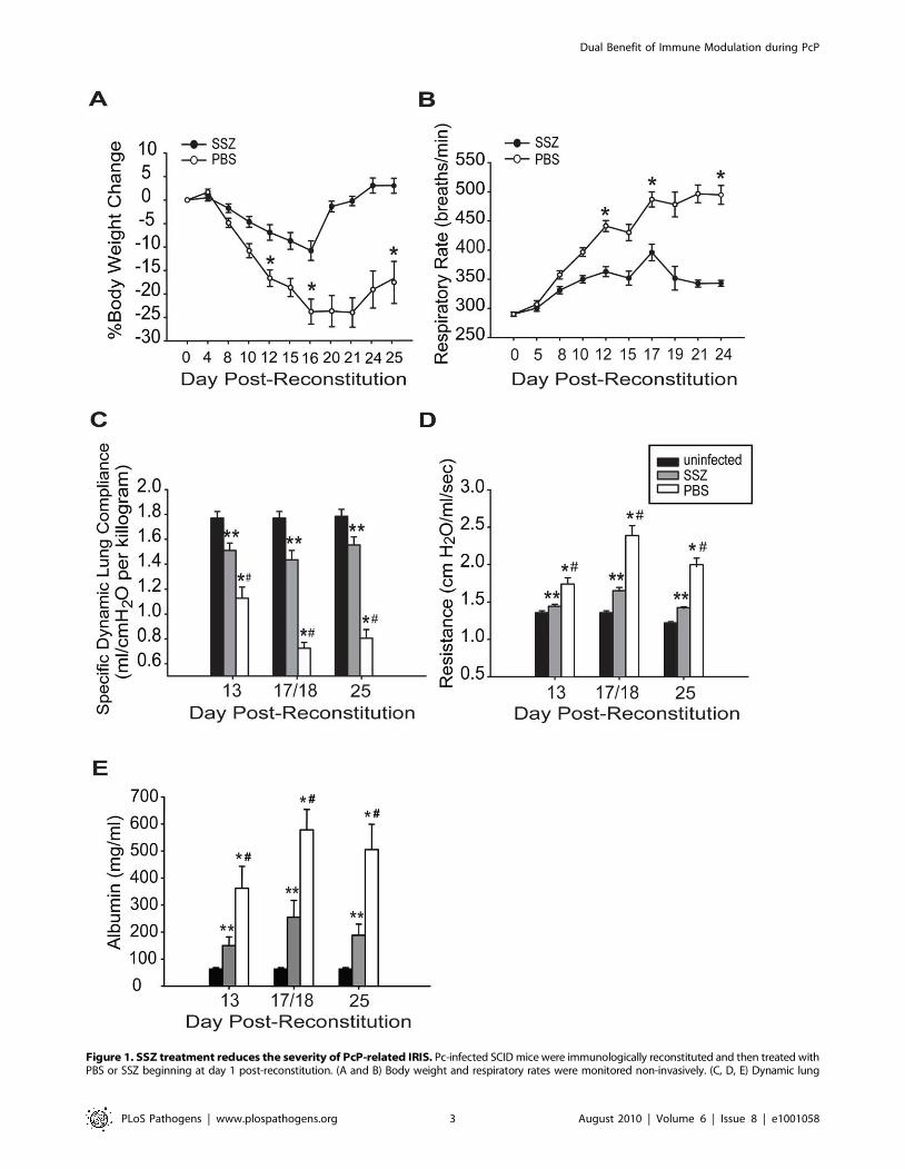
Figure 1. SSZ treatment reduces the severity of PcP-related IRIS. Pc-infected SCID mice were immunologically reconstituted and then treated withPBS or SSZ beginning at day 1 post-reconstitution. (A and B) Body weight and respiratory rates were monitored non-invasively. (C, D, E) Dynamic lung
Dual Benefit of Immune Modulation during PcP
PLoS Pathogens | www.plospathogens.org 3 August 2010 | Volume 6 | Issue 8 | e1001058

To determine the effect of SSZ treatment on epithelial damage
and alveolar permeability during PcP, albumin content was
measured in the bronchoalveolar lavage (BAL) fluid. Elevated
levels of albumin in the BAL fluid indicates damage to the tight
junctions between alveolar epithelial cells and serves as a marker
for the severity of PcP. PBS-treated mice with PcP had
significantly elevated albumin levels on days 13 and 18 PR as
compared with normal uninfected mice (Figure 1E). In contrast,
the SSZ-treated mice had lower albumin levels than PBS-treated
mice at both time points (Figure 1E). While albumin levels
returned toward baseline in both groups by day 25, they remained
significantly lower in the SSZ-treated mice. These data demon-
strate that SSZ attenuates damage to the alveolar-capillary barrier,
which contributes to the preservation of pulmonary function
during PcP.
Sulfasalazine reduces pulmonary inflammation duringPcP-related IRIS
Total cell counts, differentials, and flow cytometry were also
performed on BAL cells from experimental mice. PBS-treated
IRIS mice had significantly elevated numbers of total BAL cells
compared to SSZ-treated mice at all time points (Table 1).
Differential staining revealed that the reduced number of cells in
SSZ-treated mice relative to PBS-treated mice was mainly a
reflection of fewer lymphocytes and neutrophils at all time points
(Table 1). As neutrophil numbers are predictive of disease severity,
fewer neutrophils in SSZ-treated mice was also an indicator of less
severe disease. Despite a reduction in total cells, it was notable that
SSZ-treated mice had more AMs than PBS-treated mice at day
18. Flow cytometry analyses revealed that SSZ-treated mice had
fewer CD4+ and CD8+ T cells than PBS-treated mice at 13 and 18
days PR. By day 25 PR the reduced BAL cells in SSZ-treated mice
was mainly a reflection of reduced CD42/CD82 lymphocytes
(possibly B cells) and neutrophils.
Histological examination of hematoxylin and eosin stained lung
sections showed that the PBS-treated mice exhibited a more
intense, wide-spread inflammatory response than the SSZ-treated
mice. Pulmonary inflammation in PBS-treated mice was charac-
terized by the accumulation of monocytic and polymorphonuclear
aggregates throughout the alveoli, although the inflammation
appeared less intense by day 25 (Figure 2A–C). In contrast,
evidence of inflammation in the SSZ-treated mice at days 13 and
18 PR was mostly limited to perivascular and peribronchial
regions, and the alveoli were mainly free from cellular infiltrates
(Figure 2D, E). Furthermore, the inflammation was almost
completely resolved by day 25 in the SSZ-treated mice (Figure 2F).
Sulfasalazine inhibits lung chemokine and cytokineproduction during PcP
Elevated cytokine production is a characteristic of PcP-related
inflammation. To determine whether the reduced severity of PcP
observed in SSZ-treated mice was associated with blunted
chemokine and cytokine production in the lung, MCP-1,
RANTES, TNF-a, and IFN-c levels were measured in the BAL
fluid. Cytokine and chemokine levels were elevated above control
levels in the lungs of PBS-treated IRIS mice, consistent with the
physiological and histological data showing severe inflammatory
disease. In contrast, lung levels of MCP-1, RANTES, TNF-a and
IFN-c in the SSZ-treated group were all significantly lower than
the PBS-treated group at all time points (Figure 3A–D). Reduced
cytokine levels in the SSZ-treated mice were associated with
reduced inflammatory disease in these mice.
Sulfasalazine enhances the host’s ability to clear the Pcinfection
To study the effect of SSZ on fungal clearance, the lungs of
experimental mice were examined for Pc burden using real-time
PCR. Both PBS- and SSZ-treated mice had significant lung Pc
burdens on day 13 PR (Figure 4A). Although the PBS-treated mice
showed little reduction in Pc burden by day 18 (log 6.7760.07),
the SSZ-treated mice exhibited a 4-log reduction between days 13
and 18 PR (log 2.2560.42) (Figure 4A). By day 25 PR, Pc burdens
in the PBS-treated IRIS mice were reduced by an average of 2
logs, while Pc was undetectable in nearly all individual SSZ-
treated animals (Figure 4A). The above data were combined from
8 independent experiments. The enhanced clearance of Pc in
SSZ-treated mice was confirmed by enumerating Pc cysts in the
lung homogenates with Gomori’s methenamine silver stain (data
not shown). Therefore, SSZ accelerates the fungal clearance
kinetics in this model.
Table 1. Cellular composition of BAL fluid* from SSZ- and PBS-treated IRIS mice.
Cell Type Day13a Day18 Day25
SSZ PBS SSZ PBS SSZ PBS
Total cells (105) 1.9960.21 11.261.97b 7.9761.06 17.963.11c 5.0660.35 13.062.26d
Macrophages (105) 0.9360.21 1.9860.31e 5.0760.46 3.9060.86f 3.6560.23 4.3060.67
Lymphocytes (105) 0.2760.08 2.8160.63g 1.8260.46 7.2461.36h 1.1360.24 7.3861.51i
CD4+ T cells (105) 0.0460.01 0.3960.1j 0.4760.14 0.8960.17 0.1660.11 0.2560.06
CD8+ T cells (105) 0.1160.06 0.7960.2k 0.6460.34 1.6660.41l 0.6160.37 0.4960.26
Neutrophils (105) 0.6660.11 6.1061.55m 0.8160.27 6.3661.05n 0.160.04 1.0960.61o
*BAL fluid was isolated from one-half of the lung.adays post-immune reconstitution.b–oP,0.05 as compared with uninfected mice and SSZ-treated mice at the same time point, n$18/time point/group, data is pooled from three independent
experiments.doi:10.1371/journal.ppat.1001058.t001
compliance, lung resistance and BAL albumin content were measured at 13, 18, and 25 days post-reconstitution. Values are mean 61 standard errormeasurement (SEM) (n$15 for day 13, n$37 for day 18, and n$15 for day 25). ** and #, P,0.05 as compared to uninfected mice at the same time point.*, P,0.05 as compared to SSZ treated mice at the same time point. Mean represents combined data from eight independent experiments.doi:10.1371/journal.ppat.1001058.g001
Dual Benefit of Immune Modulation during PcP
PLoS Pathogens | www.plospathogens.org 4 August 2010 | Volume 6 | Issue 8 | e1001058

Enhanced Pc clearance in SSZ-treated mice requires CD4+
T cell mediated immunityAdaptive immunity is critical for the clearance of Pc from the
lungs. To determine whether the mechanism by which SSZ
enhances Pc clearance requires the adaptive immune system, non-
reconstituted Pc-infected SCID mice were administered either
PBS vehicle, SSZ (200 mg/kg/day), or trimethoprim sulfameth-
oxazole (TMP-SMX) (16 mg/kg/day). As expected, PBS-treated
SCID mice had high Pc burdens at both 13 and 20 days post-
treatment (Figure 4B). Similarly, the SSZ-treated group also had
high Pc burdens at both time points and did not clear the Pc
infection (Figure 4B), demonstrating that SSZ had no direct Pc
killing effect. In contrast, the TMP-SMX-treated group showed
reduced Pc burden at both time points (Figure 4B). The role of
CD4+ T cells in host defense against Pc has been well-
documented. To determine whether the mechanism of SSZ-
enhanced clearance is CD4+ T cell-dependent, Pc-infected SCID
mice were immune reconstituted and then CD4+ T cell depleted.
The Pc burdens of PBS- and SSZ-treated mice were determined at
days 18 and 25 PR. Neither PBS- nor SSZ-treated mice cleared
the Pc infection in the absence of CD4+ T cells (Figure 4C). Thus,
the mechanism by which SSZ enhances Pc clearance requires a
CD4+ T cell response. These data demonstrate that SSZ does not
have direct anti-Pc activity, and that SSZ-enhanced clearance
arises from modulation of the CD4+ T cell-mediated immune
response.
Sulfasalazine enhances CD4+ T cell-dependent AMphagocytosis
Although it has been inferred that AMs are responsible for
CD4+ T cell-mediated clearance of Pc, there is little in vivo
evidence to support this. Therefore, a multispectral imaging flow
cytometry-based method was developed to quantify AM phago-
cytosis of Pc in vivo. The Amnis ImageStream, which combines
digital imaging with traditional flow cytometry, allowed for dual
staining of AM surface markers and internalized Pc. Because of the
large number of cells that can be rapidly evaluated, a quantitative
assessment of AM internalization of Pc was generated. This
method was used to assess AM phagocytic activity in SSZ and PBS
treated animals at various time points. CD11c was used as a
marker for AMs, and a pool of five monoclonal antibodies that
recognize the surface of mouse Pc were used to determine
internalization of Pc relative to the AM. Figure 5 (Panel A) shows
representative images of brightfield (BF), CD11c (green), Pc (red),
and CD11c-Pc merged. The no internalization control is a
representative cell with CD11c signal, but no Pc internalization
(Figure 5A). Control staining was performed without inclusion of
anti-Pc antibodies. As expected, no red signal was observed in
these cells. The flow cytometer data were then quantified for AMs
from individual SSZ or PBS treated mice at days 13, 17/18, and
21 PR. The percentage of AMs with internalized Pc in SSZ-
treated mice was significantly higher than in PBS-treated mice at
day 17/18 PR (Figure 5B). Furthermore, when the absolute
number of AM with internalized Pc was determined, the
difference was even more striking. SSZ-treated mice had a 9-fold
greater number of AM with internalized Pc than PBS-treated mice
at days 17/18 PR (Figure 5C). Importantly the number of AM
with internalized Pc increased dramatically in PBS-treated mice at
day 21 PR, coincident with Pc clearance and recovery from
infection. These data indicate that AM-mediated clearance also
occurs in PBS-treated mice, but that SSZ treatment accelerates
this T cell-dependent process.
A CD4+ T cell response was required for Pc clearance in both
PBS and SSZ-treated animals. To determine whether CD4+ T
cells are required for AM phagocytosis of Pc, infected SCID mice
were immune reconstituted and given SSZ, SSZ plus anti-CD4
antibody, PBS, or PBS plus anti-CD4 antibody. As expected, SSZ-
treated mice exhibited a large increase in AM phagocytosis of Pc
at day 17/18 PR (Figure 5D), and also exhibited significant fungal
clearance at this time with an average Pc burden of log 2.861.1 (as
assessed by kexin gene copies). In contrast, SSZ-treated mice that
were depleted of CD4+ T cells displayed nearly undetectable levels
of AM phagocytosis of Pc (Figure 5D), and maintained high
average Pc lung burdens of log 7.560.1 (p,0.05 compared to
SSZ-treated mice). Likewise, PBS-treated animals displayed
increased AM phagocytosis at day 21 PR, but phagocytosis was
nearly undetectable in the absence of CD4+ T cells (Figure 5D).
Figure 2. SSZ reduces pulmonary inflammation during PcP-related IRIS. Pc-infected SCID mice were immunologically reconstituted, andthen treated with PBS (Panels A–C) or SSZ (Panels D–F) beginning at day 1 post-reconstitution. Lung sections from mice at days 13 (A, D), 18 (B, E) or25 (c, F) post-reconstitution were stained with Hematoxylin and Eosin. Representative pictures were taken by microscopy under 406magnification.Arrows denote peribronchiolar regions in each section, while asterisks denote alveolar regions.doi:10.1371/journal.ppat.1001058.g002
Dual Benefit of Immune Modulation during PcP
PLoS Pathogens | www.plospathogens.org 5 August 2010 | Volume 6 | Issue 8 | e1001058

Figure 4. SSZ enhances Pc clearance through a CD4-dependent mechanism. (A) Pc burden in immune reconstituted SCID mice thatreceived SSZ- (=) or PBS (&). Values are mean 61 SEM (n$15 for day 13, n$37 for day 18, and n$15 for day 25). ** and #, P,0.05 as compared touninfected mice at the same time point. *, P,0.05 as compared to SSZ treated mice at the same time point. Mean represents combined data fromeight independent experiments. (B) Pc burden in non-reconstituted SCID mice after 13 and 20 days of SSZ (=), PBS (&), or TMP-SMX (e) treatment.Values are mean 61 SEM (n$11 for day 13, and n$17 for day 20). *, P,0.05 as compared to SSZ and PBS treated mice at the same time point. Meanrepresents combined data from three independent experiments. (C) Pc burden in immune reconstituted, CD4-depleted SCID mice receiving SSZ (=)or PBS (&). Values are mean 61 SEM (n$8 for day 18 and n$14 for day 23). Mean represents combined data from four independent experiments.doi:10.1371/journal.ppat.1001058.g004
Figure 3. SSZ reduces inflammatory chemokine and cytokine production during PcP-related IRIS. Pc-infected SCID mice wereimmunologically reconstituted, and then treated with PBS or SSZ. (A, B, C, and D) MCP-1, RANTES, TNF-a, and IFN-c levels were measured in the BALfluid. Values are mean 61 SEM (n$15/time point/group). ** and #, P,0.05 as compared to uninfected mice at the same time point. *, P,0.05 ascompared to SSZ treated mice at the same time point. Mean represents combined data from three independent experiments.doi:10.1371/journal.ppat.1001058.g003
Dual Benefit of Immune Modulation during PcP
PLoS Pathogens | www.plospathogens.org 6 August 2010 | Volume 6 | Issue 8 | e1001058

Consistent with these results, PBS-treated mice had lower average
Pc burdens than the PBS plus anti-CD4 group (log 6.760.5 versus
log 7.660.01; p,0.05). Together, these data demonstrate that
AM phagocytosis is the effector mechanism for CD4+ T cell-
dependent clearance of Pc from the lung, and that SSZ alters the
lung immune response in a manner that accelerates the
macrophage-mediated phagocytosis of Pc.
In order to validate the ImageStream data, confocal microscopy was
used to confirm the internalization and localization of Pc within AMs.
BAL cells were recovered from PBS- and SSZ-treated mice at time
points when AMs are actively phagocytosing Pc. Based on the data in
Figure 5, the time points chosen were day 21 PR for PBS-treated mice,
and day 17 PR for SSZ-treated mice. Cells were stained with DAPI
(blue), anti-Pc antibody (green), anti-CD11c antibody (gray), and anti-
lysosomal-associated membrane protein-1 (LAMP-1) antibody (red).
LAMP-1 is a lysosome-specific protein, and was used to co-localize Pc
staining to the phagolysosome inside AMs. As shown in Figure 6, we
found that Pc signal co-localized with LAMP-1 signal inside CD11c+
AMs from both PBS-(Day 21) and SSZ-(Day 17) treated animals. The
‘‘Control’’ Panel shows a direct comparison between an AM with Pc
and one without Pc next to each other in the same field. The cell
without Pc (top) shows more diffuse LAMP-1 staining, while the
bottom cell shows more focal LAMP-1 staining that co-localizes with
Pc signal. This panel demonstrates differential staining and specificity
of LAMP-1 and Pc antibodies. These data validate the quantitative
ImageStream data as a measure of Pc internalization.
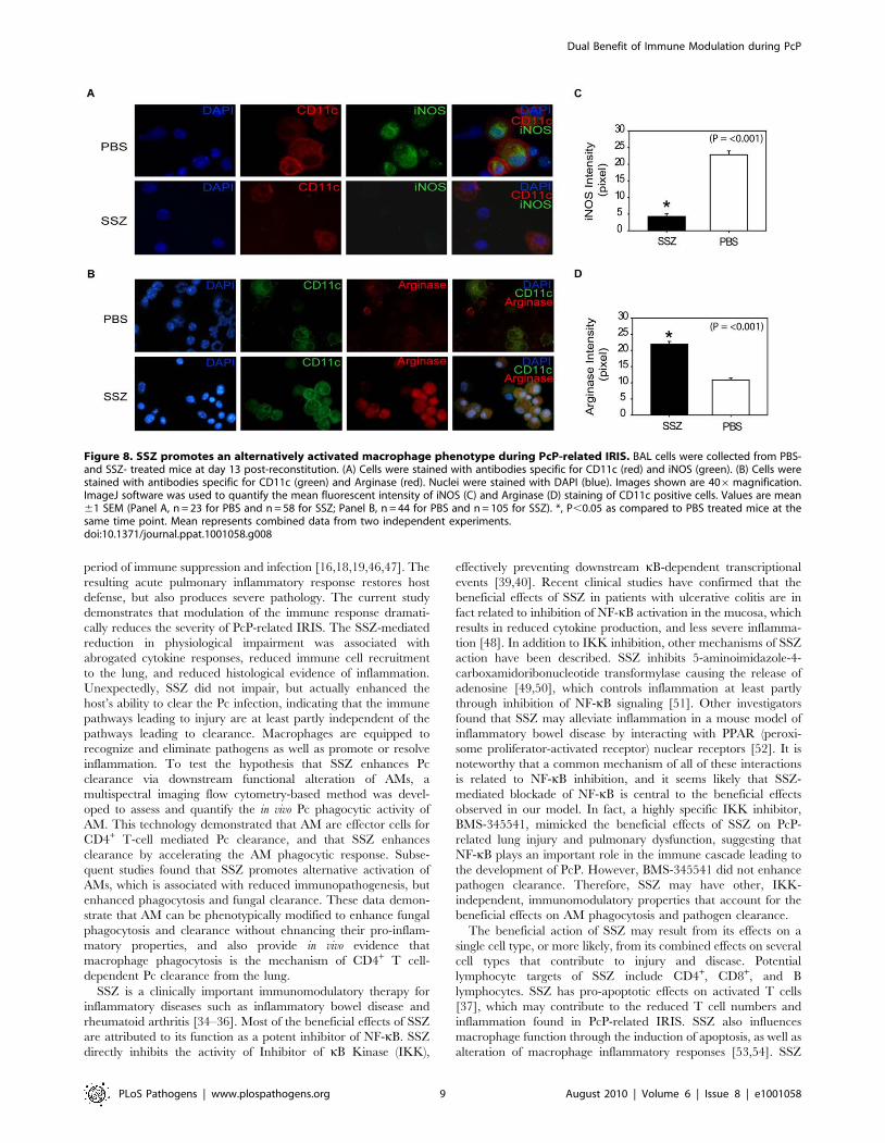
Sulfasalazine promotes a TH2 lung cytokine environmentand alternative activation of alveolar macrophages
Macrophages are immune effector cells for T cell-dependent
responses, and distinct macrophage phenotypes with differential
effects on host defense and inflammation have been identified
[44,45]. Classically activated macrophages (CAM) are induced by
TH1 cytokines, produce inducible nitric oxide synthase (INOS),
and are pro-inflammatory. In contrast, alternatively activated
macrophages (AAM) are induced by TH2 cytokines, produce
arginase (ARG), are highly phagocytic, and produce anti-
inflammatory mediators. Our studies have demonstrated that
SSZ has profound effects on CD4+ T cell-dependent macrophage
responses to Pc. Therefore, to determine whether SSZ alters PcP-
Figure 5. SSZ enhances CD4+ T cell-dependent AM phagocytosis of Pc. BAL cells were collected from PBS- and SSZ- treated mice at day 13,17 or 18 (17/18) and 21 post-reconstitution. Cells were stained with antibodies specific for CD11c (green) and Pc (red). (A) Imaging flow cytometrywas used to quantify Pc internalization by AMs. Representative images of brightfield (BF), CD11c, Pc, and merged CD11c/Pc are shown for SSZ or PBS-treated mice following immune reconstitution. The no internalization control is a representative CD11c+ cell without internalized Pc. ImageStreamIDEAS software was used to quantify (B) percentile and (C) number of AMs with Internalized Pc in immune reconstituted SCID mice treated with SSZ(=) or PBS (N). Values are mean 61 SEM (n$5 for day 13, n$9 for day 17/18, and n$9 for day 21). *, P,0.05 as compared to PBS-treated mice at thesame time point. **, P,0.05 as compared to PBS-treated mice at day 17/18. Mean represents combined data from three independent experiments.(D) Quantification of in vivo Pc phagocytosis in the presence or absence of CD4+ T cells. Reconstituted SCID mice were treated with PBS- (N), PBS +anti-CD4 (=), SSZ (&), or SSZ + anti-CD4 (e). Phagocytosis was measured at 17/18 or 21 days post-reconstitution. Values are mean +1 SEM (n$5 forday 13, n$9 for day 17/18, and n$8 for day 21). *, P,0.05 as compared to PBS treated, PBS + anti-CD4-treated, and SSZ + anti-CD4-treated mice atthe same time point. **, P,0.05 as compared to PBS + anti-CD4-treated mice at the same time point. Mean represents combined data from threeindependent experiments. Note that values for PBS treated, PBS + anti-CD4-treated, and SSZ + anti-CD4-treated mice at day 17/18 are allindistinguishable, and the symbols overlap.doi:10.1371/journal.ppat.1001058.g005
Dual Benefit of Immune Modulation during PcP
PLoS Pathogens | www.plospathogens.org 7 August 2010 | Volume 6 | Issue 8 | e1001058

related IRIS by modulating the polarity of the T helper response
and subsequent AM effector phenotype, TH cytokine levels and
macrophage activation state were assessed in experimental mice.
SSZ treatment caused a dramatic decrease in lung IFN-cproduction (Figure 3), with a concomitant increase in lung IL-4
production compared to PBS-treated mice (Figure 7A). Thus SSZ
produced a significant shift in the IL-4 to IFN-c ratio in the lungs
(Figure 7B), effectively creating a pro-TH2 lung cytokine
environment. In contrast, PBS-treated IRIS mice exhibited a
pro-TH1 lung cytokine environment.
To determine whether SSZ treatment altered AM phenotype,
AMs from SSZ- and PBS-treated IRIS mice at day 13 PR were
assessed for INOS and ARG protein expression. Because other cell
types were present in the BAL fluid from mice with PcP, CD11c
was used as a surface marker for AMs. CD11c positive AMs from
PBS-treated IRIS mice stained intensely for INOS (Figure 8A), but
weakly for ARG (Figure 8B). In contrast, CD11c positive AMs
from SSZ-treated mice stained weakly for INOS (Figure 8A), but
intensely for ARG (Figure 8B). Measurement of mean fluorescent
intensity of INOS and ARG staining in CD11c positive cells was
used to quantify the differential expression of CAM and AAM
markers in SSZ- and PBS-treated mice (Figure 8C, D). These data
demonstrate that SSZ promotes alternative activation of AMs,
which is associated with reduced immunopathogenesis but
enhanced phagocytosis and accelerated fungal clearance.
Discussion
IRIS is a clinical manifestation of PcP that occurs in certain
patients when cell-mediated immunity is restored following a
Figure 6. Co-localization of internalized Pc with the lysosomal protein LAMP-1. BAL cells were collected from PBS-treated mice at day 21and SSZ-treated mice at day 17 post-reconstitution. Cells were stained with antibodies specific for CD11c (gray), Pc (green) and LAMP-1 (red). Nucleiwere stained with DAPI (blue). Control image compares two AM in the same field; one with internalized Pc and one without. The scale bar represents10 mM. Images are representative of two independent experiments.doi:10.1371/journal.ppat.1001058.g006
Figure 7. SSZ promotes a TH2 cytokine environment during PcP-related IRIS. Pc-infected SCID mice were immunologically reconstituted,and then treated with PBS or SSZ. (A) IL-4 levels and (B) IL-4/IFN-c ratios were measured in the lung homogenates of experimental mice at indicatedtime points. Values are mean 61 SEM (n$10/time point/group). *, P,0.05 as compared to PBS treated mice at the same time point. Mean representscombined data from five independent experiments.doi:10.1371/journal.ppat.1001058.g007
Dual Benefit of Immune Modulation during PcP
PLoS Pathogens | www.plospathogens.org 8 August 2010 | Volume 6 | Issue 8 | e1001058
period of immune suppression and infection [16,18,19,46,47]. The
resulting acute pulmonary inflammatory response restores host
defense, but also produces severe pathology. The current study
demonstrates that modulation of the immune response dramati-
cally reduces the severity of PcP-related IRIS. The SSZ-mediated
reduction in physiological impairment was associated with
abrogated cytokine responses, reduced immune cell recruitment
to the lung, and reduced histological evidence of inflammation.
Unexpectedly, SSZ did not impair, but actually enhanced the
host’s ability to clear the Pc infection, indicating that the immune
pathways leading to injury are at least partly independent of the
pathways leading to clearance. Macrophages are equipped to
recognize and eliminate pathogens as well as promote or resolve
inflammation. To test the hypothesis that SSZ enhances Pc
clearance via downstream functional alteration of AMs, a
multispectral imaging flow cytometry-based method was devel-
oped to assess and quantify the in vivo Pc phagocytic activity of
AM. This technology demonstrated that AM are effector cells for
CD4+ T-cell mediated Pc clearance, and that SSZ enhances
clearance by accelerating the AM phagocytic response. Subse-
quent studies found that SSZ promotes alternative activation of
AMs, which is associated with reduced immunopathogenesis, but
enhanced phagocytosis and fungal clearance. These data demon-
strate that AM can be phenotypically modified to enhance fungal
phagocytosis and clearance without ehnancing their pro-inflam-
matory properties, and also provide in vivo evidence that
macrophage phagocytosis is the mechanism of CD4+ T cell-
dependent Pc clearance from the lung.
SSZ is a clinically important immunomodulatory therapy for
inflammatory diseases such as inflammatory bowel disease and
rheumatoid arthritis [34–36]. Most of the beneficial effects of SSZ
are attributed to its function as a potent inhibitor of NF-kB. SSZ
directly inhibits the activity of Inhibitor of kB Kinase (IKK),
effectively preventing downstream kB-dependent transcriptional
events [39,40]. Recent clinical studies have confirmed that the
beneficial effects of SSZ in patients with ulcerative colitis are in
fact related to inhibition of NF-kB activation in the mucosa, which
results in reduced cytokine production, and less severe inflamma-
tion [48]. In addition to IKK inhibition, other mechanisms of SSZ
action have been described. SSZ inhibits 5-aminoimidazole-4-
carboxamidoribonucleotide transformylase causing the release of
adenosine [49,50], which controls inflammation at least partly
through inhibition of NF-kB signaling [51]. Other investigators
found that SSZ may alleviate inflammation in a mouse model of
inflammatory bowel disease by interacting with PPAR (peroxi-
some proliferator-activated receptor) nuclear receptors [52]. It is
noteworthy that a common mechanism of all of these interactions
is related to NF-kB inhibition, and it seems likely that SSZ-
mediated blockade of NF-kB is central to the beneficial effects
observed in our model. In fact, a highly specific IKK inhibitor,
BMS-345541, mimicked the beneficial effects of SSZ on PcP-
related lung injury and pulmonary dysfunction, suggesting that
NF-kB plays an important role in the immune cascade leading to
the development of PcP. However, BMS-345541 did not enhance
pathogen clearance. Therefore, SSZ may have other, IKK-
independent, immunomodulatory properties that account for the
beneficial effects on AM phagocytosis and pathogen clearance.
The beneficial action of SSZ may result from its effects on a
single cell type, or more likely, from its combined effects on several
cell types that contribute to injury and disease. Potential
lymphocyte targets of SSZ include CD4+, CD8+, and B
lymphocytes. SSZ has pro-apoptotic effects on activated T cells
[37], which may contribute to the reduced T cell numbers and
inflammation found in PcP-related IRIS. SSZ also influences
macrophage function through the induction of apoptosis, as well as
alteration of macrophage inflammatory responses [53,54]. SSZ
Figure 8. SSZ promotes an alternatively activated macrophage phenotype during PcP-related IRIS. BAL cells were collected from PBS-and SSZ- treated mice at day 13 post-reconstitution. (A) Cells were stained with antibodies specific for CD11c (red) and iNOS (green). (B) Cells werestained with antibodies specific for CD11c (green) and Arginase (red). Nuclei were stained with DAPI (blue). Images shown are 406magnification.ImageJ software was used to quantify the mean fluorescent intensity of iNOS (C) and Arginase (D) staining of CD11c positive cells. Values are mean61 SEM (Panel A, n = 23 for PBS and n = 58 for SSZ; Panel B, n = 44 for PBS and n = 105 for SSZ). *, P,0.05 as compared to PBS treated mice at thesame time point. Mean represents combined data from two independent experiments.doi:10.1371/journal.ppat.1001058.g008
Dual Benefit of Immune Modulation during PcP
PLoS Pathogens | www.plospathogens.org 9 August 2010 | Volume 6 | Issue 8 | e1001058

blocked TNF production and also abrogated IL-12 expression and
NO production by stimulated macrophages [55]. Modification of
macrophage IL-12 may represent a mechanism by which SSZ
alters the nature of the T cell response during IRIS. NF-kB is also
involved in pulmonary epithelial cell inflammatory responses to Pc
[38,41,56], providing another potential target for the action of
SSZ. While the immunopathology associated with PcP and IRIS
requires T cells, other cell types likely contribute to the overall
disease process, and therefore the effectiveness of SSZ reported
here likely results from multiple points of action.
Our studies have found that SSZ produces a TH2 shift in the
lung cytokine environment during PcP-related IRIS, and that this
shift is reflected in the phenotype of alveolar macrophages. TH2
cytokines lead to alternative activation of macrophages, and
consistent with a TH2 cytokine shift we found that AMs isolated
from SSZ-treated mice express high levels of the AAM marker
ARG, but low levels of the CAM marker INOS (Figure 8). In
contrast, AMs from PBS-treated IRIS mice display a CAM
phenotype with high expression of INOS. It is notable that despite
a well-documented role for INOS in host defense, these data
suggest that enhanced Pc phagocytosis in SSZ-treated mice is
associated with an alternatively activated AM phenotype with low
expression of INOS. Based on our results, we believe that
increased phagocytosis by alternatively activated macrophages is
the mechanism of enhanced Pc clearance. However, a role for
INOS in Pc killing cannot be excluded. Although we observe less
staining in AMs from SSZ-treated mice, they are not totally devoid
of INOS protein. More extensive studies will be required to
determine the contribution of INOS in this model.
Although we have not demonstrated that the TH2 shift is
solely responsible for the beneficial effects of SSZ during PcP, it
is possible that TH2 cytokines acting through AAM effectors
can increase fungal clearance while reducing immunopatho-
genesis. For example, TH2 cytokines enhance macrophage
phagocytosis of Candida albicans by inducing macrophage
expression of mannose receptor (MR) [57,58] and dectin-1
[59]. These pattern recognition molecules are markers of the
AAM phenotype, and have known roles in anti-fungal host
defense. Similarly a TH2 shift in SSZ-treated mice could
enhance phagocytosis of Pc by eliciting AAM with increased
expression of MR and dectin-1. In addition, a TH2 shift may
also attenuate the immunopathogenesis of PcP by reducing the
production of pro-inflammatory TH1 cytokines, while enhanc-
ing production of anti-inflammatory TH2 cytokines. Elevated
lung levels of the TH1 cytokines TNF-a and IFN-c are
associated with PcP-related lung injury and respiratory impair-
ment [28]. In contrast, TH2-derived AAMs produce the potent
anti-inflammatory cytokines IL-10 and TGF- b [44], which can
dampen inflammatory responses and may contribute to the
reduced inflammation and injury in SSZ-treated mice. Impor-
tantly, the anti-inflammatory potential of AAMs has been
established in vivo by studies showing that the adoptive transfer
of in vitro programmed AAMs attenuates immunopathogenesis
in mouse models of inflammatory disease [60]. Although these
findings are consistent with a SSZ-induced shift in the polarity
of the T cell response, further studies are required to establish
whether TH2 cytokines and alternative activation of AMs are
directly responsible for the beneficial effects of SSZ during PcP.
Clinical studies have found that the severity of PcP correlates
with the degree of pulmonary inflammation, but not with
organism burden [9–14]. Controlled animal studies support these
clinical observations, and have provided direct evidence that the
immune response is a major pathogenic component of PcP
[26,28–31,61]. Consequently, antibiotic treatment does not always
produce rapid improvement of patients with severe PcP, because
organisms and antigen may continue to drive the pathological
immune response. The efficacy of SSZ in dramatically attenuating
the severity of PcP supports the contention that adjunctive
immunomodulatory therapy that target the T cell response is
critical to optimal treatment of patients. Currently, adjunctive
corticosteroids are commonly used for the clinical treatment of
PcP. The broad anti-inflammatory and immunosuppressive
properties of steroids are presumed to provide benefit, but
concrete evidence that steroids improve survival is lacking. Our
group has recently published a study demonstrating that specific
disruption of the T cell response to Pc with anti-CD3 antibody has
beneficial effects in a mouse model of PcP-related IRIS [62].
While both SSZ and anti-CD3 altered the T cell response to Pc
and reduced immunopathogenesis, they produced differential
outcomes with respect to fungal clearance. Anti-CD3 produced a
profound inhibition of T cell responses which reduced disease, but
also prevented the clearance of Pc from the lung. In contrast, SSZ
dampened PcP-related immunopathogenesis without suppressing
TH responses to a degree that prevented eradication of the
organism. SSZ not only reduced T cell-mediated inflammation,
but altered the nature of the T cell response by promoting TH2
lung cytokine environment and alternative activation of macro-
phages. It is likely that the preservation of TH2 responses
combined with a shift in the polarization of AMs in SSZ-treated
mice is responsible for the differential effects of SSZ and anti-CD3.
Another important aspect of our work is the development of a
multispectral imaging flow cytometer-based method to assess the in
vivo phagocytic activity of AM during a T cell-mediated immune
response by quantifying the percentage of AMs that contain
internalized Pc. Understanding the mechanisms controlling Pc
phagocytosis is an area of great interest, and many investigators
have utilized various techniques to perform in vitro assessments of
Pc phagocytosis [63–70]. However, demonstrating an in vivo role
for AM phagocytosis in the clearance of Pc has been more difficult.
AM with associated Pc have been observed in the BAL fluid of
patients and animals [71–74]. However, this was in the setting of
active PcP, the level of phagocytosis appeared low, and the
significance to organism clearance was not determined. Others
have performed in vivo assessments of phagocytosis immediately
(within 24 hours) following a bolus inoculation of labeled Pc
[75,76]. In addition, short-term depletion of AMs in rats reduced
the clearance of Pc over the initial 24 hours post-inoculation [77].
While these studies were able to demonstrate a role for AM in vivo,
the timing indicates that the investigators were evaluating the
innate immune response to a bolus inoculation of Pc, rather than
the CD4+ T cell-mediated response which is required for natural
clearance of Pc from the lung. Using this new technology we were
able to develop an assay to show that AMs are effector cells for the
clearance of Pc during a natural CD4+ T cell-mediated immune
response in vivo. The advantages of these ImageStream-based data
are that: 1) internalized Pc was distinguished from attached Pc; 2)
a large number of AM from each animal was rapidly assessed to
provide quantification of the phagocytic response; 3) the
dependence of phagocytic activity on the presence of CD4+ T
cells was demonstrated; and 4) the CD4+ T cell-dependent
increase in phagocytic activity correlated with the clearance
kinetics of Pc. Importantly, the ImageStream data was validated
using confocal microscopy to co-localize intracellular Pc with the
lysosome protein LAMP-1. These data indicate that Pc is located
within the phagolysosome of AM, consistent with phagocytosis of
the pathogen. The multispectral imaging flow cytometry technol-
ogy should provide a valuable tool for further study of Pc
phagocytosis in vivo.
Dual Benefit of Immune Modulation during PcP
PLoS Pathogens | www.plospathogens.org 10 August 2010 | Volume 6 | Issue 8 | e1001058
In summary, the results of this study indicate that the immune
response to Pc can be modulated in a manner that reduces
inflammatory consequences of PcP while enhancing the pathogen
clearance through increased AM phagocytic capacity. We also
developed a method for in vivo quantification of AM phagocytosis
of Pc, and provide evidence that the macrophage is the ultimate
effector for the CD4+ T cell-mediated clearance of Pc from the
lungs. Immune modulation of T cell and AM functions should be
considered potential therapeutic targets for the treatment of
immune complications of PcP. Macrophages are equipped to
recognize and eliminate pathogens as well as promote and/or
resolve inflammation. Our results indicate that the phagocytic
function of macrophages can be enhanced with a concomitant
reduction in their pro-inflammatory properties. Enhancement of
AM-mediated clearance of Pc may prove less inflammatory and
generally superior to antibiotic therapy alone.
Methods
Pc source animalsSevere combined immunodeficient (SCID) mice on a C.B-17
background (C. B-Igh-1b/Icr Tac-Prkdcscid) were purchased from
Taconic (Hudson, NY), or obtained from a breeding colony at the
University of Rochester. The mice were housed using micro-
isolator technology and fed sterilized food and water. To induce
infection SCID mice were co-housed with Pc-infected SCID mice.
Pc organisms were isolated as previously described [38]. Pc cysts
were enumerated by standard Gomori’s methenamine silver stain.
Sulfasalazine and trimethoprim-sulfamethoxazoleadministration
Sulfasalazine (SSZ) (Sigma, St. Louis, MO) was administered
once daily by intra-peritoneal (i.p.) injection at a dose of 200 mg
per kg of body weight. Trimethoprim Sulfamethoxazole (TMP-
SMX) (SICOR Pharmaceuticals, Inc. Irvine, CA) was adminis-
tered once daily i.p. at a dose calculated to give 16 mg per kg of
body weight of the Trimethoprim component of the drug
combination. This dose was based on the therapeutic dose given
to humans for the treatment of PcP.
Mouse model of PcP-related IRISTo induce infection SCID mice were intra-nasally inoculated
with 16105 purified Pc based on cyst count. Three weeks later the
mice were immunologically reconstituted with 56107 congenic
spleen cells from normal C.B-17 mice.
In vivo CD4+ T cell depletionCD4+ T cells were depleted by i.p. injection of monoclonal
antibody specific for mouse CD4 (clone GK1.5, ATCC TIB207).
Antibody injections (250 mg per mouse) were given one day prior
to and one day after immune reconstitution. Thereafter, antibody
was administrated every four days for the duration of the
experiment.
Physiologic assessment of pulmonary function in live,ventilated mice
Lung compliance and resistance were measured in live
ventilated mice using a whole body plethysmograph (BUXCO
Electronics Inc., Wilmington, NC) connected to a Harvard rodent
ventilator (Harvard Apparatus, Southnatic, MA) as previously
described [78]. Dynamic lung compliance was normalized to the
peak body weight of the animal. Respiratory rates were measured
using whole body unrestrained chambers (BUXCO Electronics
Inc). Data was collected and analyzed using the Biosystems XA
software package (BUCXO Electronics Inc.).
Bronchoalveolar lavage (BAL) and lung tissue preparationRight lung lobes were lavaged with four, one-ml aliquots of 1X
Hank’s balanced salt solution. Cell-free lavage fluid (approximate-
ly 3.5 ml per mouse) was frozen at 280uC. BAL cells were
enumerated, and then differentials and multi-parameter flow
cytometric analyses were performed. Anti-CD4-Fluorescein (clone
RM4-4) and anti-CD8a-Peridinin Chlorophyll-a Protein (clone
53-6.7), were purchased from BD Biosciences (San Diego, CA).
The anti-CD4 clone RM4-4 was used to confirm CD4+ cell
depletion in vivo because it is not blocked by the CD4-depleting
antibody (clone GK1.5). Cells were analyzed on a FACSCalibur
(BD Biosciences, San Jose, CA), with at least 10,000 events
routinely analyzed for each Pc-infected mouse. At least 5,000
events were analyzed from uninfected control mice.
For fixation the lungs were inflated with 15 cm gravity flow-
pressure of 10% formalin (Sigma, St. Louis, MO). The lungs were
fixed in situ for 10 minutes under gravity flow pressure, and then
removed from the animal and placed in fixative for 16 h at 4uC.
Lung tissue was embedded in paraffin and 4 mM sections were cut.
Hematoxylin and eosin was used to visualize tissue.
Cytokine ELISAs of BAL fluidTotal protein concentration was determined in cell-free lavage
by the colorimetric assay of Lowry. Albumin concentration was
determined using the Mouse Albumin ELISA Quantitation kit
from Bethyl Laboratories (Montgomery, TX). TNF-a, IFN-c,
MCP-1, and RANTES ELISA kits were used according to the
manufacturer’s instructions (R&D, Minneapolis, MN).
Real-time PCR assessment of lung Pc burdenSince Pc cannot be cultured, a real-time PCR method was used
to quantify lung burden. For quantification of Pc burden in right
lung lobes, quantitative PCR using TaqMan primer/fluorogenic
probe chemistry and an Applied Biosystems Prism 7000 Sequence
Detection System (Applied Biosystems, Foster City, CA) was
performed with a primer/probe set specific for the mouse Pc kexin
gene as previously described [78].
In vivo assessment of macrophage phagocytosis of Pcusing ImageStream
For quantitation of Pc phagocytosis, an ImageStream multi-
spectral imaging flow cytometer (Amnis Corporation, Seattle, WA)
was used [79,80]. With this technology the number of BAL AM
with internalized Pc was directly quantified. CD11c was used as a
surface marker to identify AM, while anti-Pc antibodies were used
to stain internalized Pc. Whole lungs were lavaged and BAL cells
were washed with ice cold PBS with 1%BSA (PBA), and incubated
with mouse Fc Block (BD Biosciences, San Diego, CA) for 5 min
on ice. Cells were then surface stained with anti-CD11c-
phycoerythrin (clone HL3, BD Biosciences) for 30 minutes on
ice and washed with PBA. The cells were then permeabilized with
BD Cytofix/Cytoperm Fixation and Permeabilization Solution
(BD Biosciences), and incubated with a pool of five different anti-
Pneumocystis monoclonal antibodies for 30 minutes on ice. These
antibodies were generated in our laboratory and were chosen
because they recognize five different epitopes on the surface of Pc
as determined by western blot and IFA (4F11, 2B5, 3D6, 1F1,
1F5). Characterization of antibody 4F11 has been published [81],
but the remaining antibodies have not been further characterized.
Following a wash step, the cells were incubated with Alexa Fluor
Dual Benefit of Immune Modulation during PcP
PLoS Pathogens | www.plospathogens.org 11 August 2010 | Volume 6 | Issue 8 | e1001058

647 goat anti-mouse IgG (H+L) (Invitrogen Molecular Probes,
OR) for 30 minutes on ice. Stained cells were washed, pelleted,
and resuspended in 50 ml of ice cold 1% paraformaldehyde in PBS
(Electron Microscopy Sciences, PA). Samples were stored at 4uCin the dark until analyzed. Twenty thousand to forty thousand
event image files were collected for each sample on an Image-
Stream100 using 200 mW of 488 nm and 90 mW of 658 nm laser
power.
The data obtained were analyzed using the ImageStream Data
Exploration and Analysis Software (IDEAS, Amnis), which
quantifies morphometric and photometric parameters on a per-
cell basis for large populations of collected events [79,82]. Single
AM cells were gated as those events with normal brightfield area,
high brightfield aspect ratio and CD11c positive staining. Each
analyzed file contained at least 5000 AM events, enabling routine
statistical analysis. AM that had phagocytosed Pc were identified
by gating on CD11c+ events with high Pc-AF647 Max Pixel
values (discriminates punctuate Pc from diffuse background
staining and autofluorescence) and high Pc Internalization values.
The Internalization score is a ratio of the intensity of bright red
staining inside the cell (defined by eroding the CD11c mask 6
pixels) to the bright red staining in the membrane (defined by
subtracting the latter mask from the CD11c mask dilated 3
pixels), and the higher the score the greater the concentration of
Pc inside the cell. In order to condition the measurement to Pc
particles in sharp focus (and thus in the central focal plane of the
cell), only the mean of the upper quartile pixel intensities,
weighted by the max pixel intensity, is used to compute the ratio
[83]. The percentage of AM with internalized Pc was derived
directly from the ImageStream data. The absolute number of AM
with internalized Pc that was recovered from each animal was
calculated by multiplying this percentage by the total number of
AM recovered.
Confocal fluorescence microscopyCells were stained with the identical antibodies under the
identical conditions described for ImageStream analyses with the
following exceptions. Anti-mouse CD11c-AlexaFluor 488 (Invi-
trogen Molecular Probes, OR) was used instead of CD11c-PE.
Also, in some experiments, biotinylated anti-mouse CD107a
(LAMP-1) (Biolegend, CA) was used to co-localize intracellular Pc
with lysosomes. Cells were first stained with anti-CD11c and anti-
Pc antibodies as described above. After a wash and second
permeabilization step, cells were stained with biotinylated anti-
CD107a antibody for 30 minutes on ice followed by streptavidin
conjugated with PE-Texas Red (BD Bioscience, CA). After
fixation, cells were centrifuged onto glass slides, mounted with
anti-fade Vectashield (Vector Laboratories, CA), and cover-
slipped for optimal imaging. Cells were imaged using an FV1000
Olympus Laser Scanning Confocal Microscope using an I681
inverted microscope and a 606 objective with zoom of 4. Lasers
used were 405, 488, 559, and 635 optimized to reduce photo-
bleaching and used sequentially. Differential Interference Con-
trast (DIC) was performed using the 559 laser. Pixel dwell times
were 8 us/pixel and 102461024 pixel format for high resolution
imaging. Parameters were maintained consistent throughout
imaging. All the images presented are the originals without
post-processing.
Analyses of alveolar macrophage phenotypeBAL cells were collected and centrifuged onto glass slides. Cells
were fixed with 3% paraformaldehyde and initially stained with
hamster anti-mouse CD11c (Abcam, MA) followed by either goat
anti-hamster AF594 or goat anti-hamster AF488 (Invitrogen
Molecular Probe, Oregon) secondary antibody. After permeabi-
lization with 0.2% Triton 62100 in phosphate buffered saline,
cells were stained with rabbit anti-mouse iNOS (Abcam, MA) with
goat anti-rabbit AF488 (Invitrogen Molecular Probe, Oregon)
secondary antibody, or goat anti-mouse Arginase (Santa Cruz
Biotechnology, CA) with donkey anti-goat AF546 (Invitrogen
Molecular Probe, Oregon) secondary antibody. Slides were
mounted with anti-fade Vectashield (Vector Laboratories, CA)
and coverslipped for optimal imaging. A Nikon Eclipse E400
fluorescence microscope was used for photomicroscopy. All
photographs for a given protein were taken with identical exposure
settings. The ImageJ software (National Institutes of Health) was
used to quantify the mean fluorescent intensity of iNOS and
Arginase staining in CD11c positive alveolar macrophages.
Statistical analysesOne-way analysis of variance was performed with the SigmaStat
2.0 software (Jandel, San Rafael, Calif.). The Student-Newman-
Keuls method was used for all pair-wise multiple comparisons.
Ethics statementAll animal protocols were pre-approved by University Com-
mittee on Animal Resources (UCAR) at the University of
Rochester Medical Center according to the guidelines of the
Association for Assessment and Accreditation of Laboratory
Animal Care International.
Acknowledgments
We thank Dr. Timothy Bushnell for kindly providing professional support
to help us develop Image Stream-based protocols, and to Dr. Linda
Callahan for assistance with confocal microscopy. We also thank Dr.
Hongyue Wang for expert statistical analyses. Nabilah Khan, Jane Malone
and Bradley Buchheit contributed expert technical support for all animal
studies.
Author Contributions
Conceived and designed the experiments: JW FG SPB TWW. Performed
the experiments: JW SPB. Analyzed the data: JW FG SPB TCG TWW.
Wrote the paper: JW FG SPB TCG TWW.
References
1. Santos J, Palacios R, Ruiz J, Gonzalez M, Marquez M (2005) Study of patientsdiagnosed with advanced HIV in the HAART era—OMEGA Cohort. Int J STD
AIDS 16: 252–255.
2. Forrest DM, Seminari E, Hogg RS, Yip B, Raboud J, et al. (1998) The incidenceand spectrum of AIDS-defining illnesses in persons treated with antiretroviral
drugs. Clin Infect Dis 27: 1379–1385.
3. Staikowsky F, Lafon B, Guidet B, Denis M, Mayaud C, et al. (1993) Mechanical
ventilation for Pneumocystis carinii pneumonia in patients with the acquiredimmunodeficiency syndrome. Is the prognosis really improved? Chest 104: 756–762.
4. Arend SM, Kroon FP, van’t Wout JW (1995) Pneumocystis carinii pneumonia in
patients without AIDS, 1980 through 1993. An analysis of 78 cases. Arch Intern
Med 155: 2436–2441.
5. Kaur N, Mahl TC (2004) Pneumocystis carinii pneumonia with oral candidiasisafter infliximab therapy for Crohn’s disease. Dig Dis Sci 49: 1458–1460.
6. Sepkowitz KA, Brown AE, Armstrong D (1995) Pneumocystis carinii pneumonia
without acquired immunodeficiency syndrome. More patients, same risk. ArchIntern Med 155: 1125–1128.
7. Morris A, Sciurba FC, Lebedeva IP, Githaiga A, Elliott WM, et al. (2004)Association of Chronic Obstructive Pulmonary Disease Severity and Pneumocystis
Colonization. Am J Respir Crit Care Med 170: 408–413.
8. Benfield TL, van Steenwijk R, Nielsen TL, Dichter JR, Lipschik GY, et al.(1995) Interleukin-8 and eicosanoid production in the lung during moderate to
severe Pneumocystis carinii pneumonia in AIDS: a role of interleukin-8 in the
pathogenesis of P. carinii pneumonia. Respir Med 89: 285–290.
Dual Benefit of Immune Modulation during PcP
PLoS Pathogens | www.plospathogens.org 12 August 2010 | Volume 6 | Issue 8 | e1001058

9. Jensen BN, Lisse IM, Gerstoft J, Borgeskov S, Skinhoj P (1991) Cellular
profiles in bronchoalveolar lavage fluid of HIV-infected patients with
pulmonary symptoms: relation to diagnosis and prognosis. AIDS 5:
527–533.
10. Limper AH, Offord KP, Smith TF, Martin WJ (1989) Pneumocystis carinii
pneumonia. Differences in lung parasite number and inflammation in patients
with and without AIDS. Am Rev Respir Dis 140: 1204–1209.
11. Lipschik GY, Doerfler ME, Kovacs JA, Travis WD, Andrawis VA, et al. (1993)
Leukotriene B4 and interleukin-8 in human immunodeficiency virus-related
pulmonary disease. Chest 104: 763–769.
12. Mason GR, Hashimoto CH, Dickman PS, Foutty LF, Cobb CJ (1989)
Prognostic implications of bronchoalveolar lavage neutrophilia in patients with
Pneumocystis carinii pneumonia and AIDS. Am Rev Respir Dis 139: 1336–1342.
13. Smith RL, El-Sadr WM, Lewis ML (1988) Correlation of bronchoalveolar
lavage cell populations with clinical severity of Pneumocystis carinii pneumonia.
Chest 93: 60–64.
14. Benfield TL, Vestbo J, Junge J, Nielsen TL, Jensen AB, et al. (1995) Prognostic
value of interleukin-8 in AIDS-associated Pneumocystis carinii pneumonia.
Am J Respir Crit Care Med 151: 1058–1062.
15. Vahid B, Bibbo M, Marik PE (2007) Role of CD8 lymphocytes and neutrophilic
alveolitis in Pneumocystis jiroveci pneumonia. Scandinavian Journal of Infectious
Diseases 39: 612–614.
16. Barry SM, Lipman MC, Deery AR, Johnson MA, Janossy G (2002) Immune
reconstitution pneumonitis following Pneumocystis carinii pneumonia in HIV-
infected subjects. HIV Med 3: 207–211.
17. Dore GJ, Li Y, McDonald A, Ree H, Kaldor JM (2002) Impact of highly active
antiretroviral therapy on individual AIDS-defining illness incidence and survival
in Australia. J Acquir Immune Defic Syndr 29: 388–395.
18. Wu AK, Cheng VC, Tang BS, Hung IF, Lee RA, et al. (2004) The unmasking of
Pneumocystis jiroveci pneumonia during reversal of immunosuppression: case
reports and literature review. BMC Infect Dis 4: 57.
19. Sharma S, Nadrous HF, Peters SG, Tefferi A, Litzow MR, et al. (2005)
Pulmonary complications in adult blood and marrow transplant recipients:
autopsy findings. Chest 128: 1385–1392.
20. Slivka A, Wen PY, Shea WM, Loeffler JS (1993) Pneumocystis carinii pneumonia
during steroid taper in patients with primary brain tumors. Am J Med 94:
216–219.
21. Mansharamani NG, Balachandran D, Vernovsky I, Garland R, Koziel H (2000)
Peripheral blood CD4 + T-lymphocyte counts during Pneumocystis carinii
pneumonia in immunocompromised patients without HIV infection. Chest
118: 712–720.
22. Bachelez H, Schremmer B, Cadranel J, Mouly F, Sarfati C, et al. (1997)
Fulminant Pneumocystis carinii pneumonia in 4 patients with dermatomyositis.
Arch Intern Med 157: 1501–1503.
23. Sepkowitz KA (1993) Pneumocystis carinii pneumonia in patients without AIDS.
Clin Infect Dis 17 Suppl 2: S416–S422.
24. Kovacs JA, Hiemenz JW, Macher AM, Stover D, Murray HW, et al. (1984)
Pneumocystis carinii pneumonia: a comparison between patients with the acquired
immunodeficiency syndrome and patients with other immunodeficiencies. Ann
Intern Med 100: 663–671.
25. Hori S, Carvalho TL, Demengeot J (2002) CD25+CD4+ regulatory T cells
suppress CD4+ T cell-mediated pulmonary hyperinflammation driven by
Pneumocystis carinii in immunodeficient mice. Eur J Immunol 32: 1282–1291.
26. Roths JB, Sidman CL (1992) Both immunity and hyperresponsiveness to
Pneumocystis carinii result from transfer of CD4+ but not CD8+ T cells into severe
combined immunodeficiency mice. J Clin Invest 90: 673–678.
27. Swain SD, Meissner NN, Harmsen AG (2006) CD8 T cells modulate CD4 T-
cell and eosinophil-mediated pulmonary pathology in Pneumocystis pneumonia in
B-cell-deficient mice. Am J Pathol 168: 466–475.
28. Wright TW, Gigliotti F, Finkelstein JN, McBride JT, An CL, et al. (1999)
Immune-mediated inflammation directly impairs pulmonary function, contrib-
uting to the pathogenesis of Pneumocystis carinii pneumonia. J Clin Invest 104:
1307–1317.
29. Wright TW, Johnston CJ, Harmsen AG, Finkelstein JN (1999) Chemokine gene
expression during Pneumocystis carinii-driven pulmonary inflammation. Infect
Immun 67: 3452–3460.
30. Wright TW, Notter RH, Wang Z, Harmsen AG, Gigliotti F (2001) Pulmonary
inflammation disrupts surfactant function during Pneumocystis carinii pneumonia.
Infect Immun 69: 758–764.
31. Bhagwat SP, Gigliotti F, Xu H, Wright TW (2006) Contribution of T cell subsets
to the pathophysiology of Pneumocystis-related immunorestitution disease.
Am J Physiol Lung Cell Mol Physiol 291: L1256–L1266.
32. Roths JB, Sidman CL (1993) Single and combined humoral and cell-mediated
immunotherapy of Pneumocystis carinii pneumonia in immunodeficient scid mice.
Infect Immun 61: 1641–1649.
33. Swain SD, Han S, Harmsen A, Shampeny K, Harmsen AG (2007) Pulmonary
Hypertension Can Be a Sequela of Prior Pneumocystis Pneumonia. Am J Pathol
171: 790–799.
34. O’Dell JR, Petersen K, Leff R, Palmer W, Schned E, et al. (2006) Etanercept in
combination with sulfasalazine, hydroxychloroquine, or gold in the treatment of
rheumatoid arthritis. J Rheumatol 33: 213–218.
35. Plosker GL, Croom KF (2005) Sulfasalazine: a review of its use in the
management of rheumatoid arthritis. Drugs 65: 1825–1849.
36. Brookes MJ, Green JR (2004) Maintenance of remission in Crohn’s disease:current and emerging therapeutic options. Drugs 64: 1069–1089.
37. Liptay S, Bachem M, Hacker G, Adler G, Debatin KM, et al. (1999) Inhibition
of nuclear factor kappa B and induction of apoptosis in T-lymphocytes bysulfasalazine. Br J Pharmacol 128: 1361–1369.
38. Wang J, Gigliotti F, Maggirwar S, Johnston C, Finkelstein JN, et al. (2005)
Pneumocystis carinii activates the NF-kappaB signaling pathway in alveolarepithelial cells. Infect Immun 73: 2766–2777.
39. Weber CK, Liptay S, Wirth T, Adler G, Schmid RM (2000) Suppression of NF-
kappaB activity by sulfasalazine is mediated by direct inhibition of IkappaBkinases alpha and beta. Gastroenterology 119: 1209–1218.
40. Wahl C, Liptay S, Adler G, Schmid RM (1998) Sulfasalazine: a potent and
specific inhibitor of nuclear factor kappa B. J Clin Invest 101: 1163–1174.
41. Evans SE, Hahn PY, McCann F, Kottom TJ, Pavlovic ZV, et al. (2005)
Pneumocystis cell wall beta-glucans stimulate alveolar epithelial cell chemokine
generation through nuclear factor-kappaB-dependent mechanisms. Am J RespirCell Mol Biol 32: 490–497.
42. Lebron F, Vassallo R, Puri V, Limper AH (2003) Pneumocystis carinii cell wall
beta-glucans initiate macrophage inflammatory responses through NF-kappaBactivation. J Biol Chem 278: 25001–25008.
43. Zhang J, Zhu J, Imrich A, Cushion M, Kinane TB, et al. (2004) Pneumocystis
activates human alveolar macrophage NF-kappaB signaling through mannose
receptors. Infect Immun 72: 3147–3160.
44. Martinez FO, Helming L, Gordon S (2008) Macrophage activation andpolarization. Annu Rev Immuno 27: 451–483.
45. Gordon S (2007) The macrophage: past, present and future. Eur J Immunol 37
Suppl 1: S9–17.
46. Cheng VC, Hung IF, Wu AK, Tang BS, Chu CM, et al. (2004) Lymphocytesurge as a marker for immunorestitution disease due to Pneumocystis jiroveci
pneumonia in HIV-negative immunosuppressed hosts. Eur J Clin MicrobiolInfect Dis 23: 512–514.
47. Chen CS, Boeckh M, Seidel K, Clark JG, Kansu E, et al. (2003) Incidence, risk
factors, and mortality from pneumonia developing late after hematopoietic stemcell transplantation. Bone Marrow Transplant 32: 515–522.
48. Gan HT, Chen YQ, Ouyang Q (2005) Sulfasalazine inhibits activation of
nuclear factor-kappaB in patients with ulcerative colitis. J Gastroenterol Hepatol20: 1016–1024.
49. Baggott JE, Morgan SL, Ha T, Vaughn WH, Hine KJ (1992) Inhibition of
folate-dependent enzymes by non-steroidal anti-infalmmatory drugs. Biochem J282: 197–202.
50. Gadangi P, Longaker M, Naime D, Levin RI, Recht PA, et al. (1996) The anti-inflammatory mechanism of sulfasalazine is related to adenosine release at
inflamed sites. J Immunol 156: 1937–1941.
51. Lukashev D, Ohta A, Apasov S, Chen JF, Sitkovsky M (2004) Cutting Edge:Physiologic Attenuation of Proinflammatory Transcription by the Gs Protein-
Coupled A2A Adenosine Receptor In Vivo. J Immunol 173: 21–24.
52. Rousseaux C, Lefebvre B, Dubuquoy L, Lefebvre P, Romano O, et al. (2005)Intestinal antiinflammatory effect of 5-aminosalicylic acid is dependent on
peroxisome proliferator-activated receptor-{gamma}. J Exp Med 201:
1205–1215.
53. Salh B, Assi K, Huang S, O’Brien L, Steinbrecher U, et al. (2002) Dissociated
ROS production and ceramide generation in sulfasalazine-induced cell death inRaw 264.7 cells. J Leukoc Biol 72: 790–799.
54. Oshikawa K, Sugiyama Y (2003) Regulation of toll-like receptor 2 and 4 gene
expression in murine alveolar macrophages. Exp Lung Res 29: 401–412.
55. Hasko G, Szabo C, Nemeth ZH, Deitch EA (2001) Sulphasalazine inhibitsmacrophage activation: inhibitory effects on inducible nitric oxide synthase
expression, interleukin-12 production and major histocompatibility complex IIexpression. Immunology 103: 473–478.
56. Wang J, Gigliotti F, Bhagwat SP, Maggirwar SB, Wright TW (2007) Pneumocystis
stimulates MCP-1 production by alveolar epithelial cells through a JNK-dependent mechanism. Am J Physiol Lung Cell Mol Physiol 292: L1495–L1505.
57. Coste A, Dubourdeau M, Linas MD, Cassaing S, Lepert JC, et al. (2003)
PPARgamma promotes mannose receptor gene expression in murine macro-phages and contributes to the induction of this receptor by IL-13. Immunity 19:
329–339.
58. Coste A, Lagane C, Filipe C, Authier H, Gales A, et al. (2008) IL-13 attenuatesgastrointestinal candidiasis in normal and immunodeficient RAG-2(-/-) mice via
peroxisome proliferator-activated receptor-gamma activation. J Immunol 180:4939–4947.
59. Gales A, Conduche A, Bernad J, Lefevre L, Olagnier D, et al. (2010)
PPARgamma controls dectin-1 expression required for host antifungal defenseagainst Candida albicans. PLoS Pathog 6(1): e1000714.
60. Hunter MM, Wang A, Parhar KS, Johnston MJ, Van RN, et al. (2010) In vitro-
derived alternatively activated macrophages reduce colonic inflammation inmice. Gastroenterology 138: 1395–1405.
61. Gaut P, Daar ES (1999) Editorial response: Prophylaxis for Pneumocystis carinii
pneumonia—an evolving tale of two populations. Clin Infect Dis 29: 784–786.
62. Bhagwat SP, Wright TW, Gigliotti F (2010) Anti-CD3 antibody decreases
inflammation and improves outcome in a murine model of Pneumocystis
pneumonia. J Immunol 184: 497–502.
63. Ezekowitz RA, Williams DJ, Koziel H, Armstrong MY, Warner A, et al. (1991)
Uptake of Pneumocystis carinii mediated by the macrophage mannose receptor.
Nature 351: 155–158.
Dual Benefit of Immune Modulation during PcP
PLoS Pathogens | www.plospathogens.org 13 August 2010 | Volume 6 | Issue 8 | e1001058

64. Forte M, Rahelu M, Stubberfield C, Tomkins L, Pithie A, et al. (1991) In-vitro
interaction of human macrophages with Pneumocystis carinii. Int J Exp Pathol 72:589–598.
65. Koziel H, Eichbaum Q, Kruskal BA, Pinkston P, Rogers RA, et al. (1998)
Reduced binding and phagocytosis of Pneumocystis carinii by alveolar macro-phages from persons infected with HIV-1 correlates with mannose receptor
downregulation. J Clin Invest 102: 1332–1344.66. Lasbury ME, Lin P, Tschang D, Durant PJ, Lee CH (2004) Effect of
bronchoalveolar lavage fluid from Pneumocystis carinii-infected hosts on phagocytic
activity of alveolar macrophages. Infect Immun 72: 2140–2147.67. Limper AH, Standing JE, Hoyte JS (1996) The role of alveolar macrophages in
Pneumocystis carinii elimination from the lower respiratory tract. J EukaryotMicrobiol 43: 12S.
68. Masur H, Jones TC (1978) The interaction in vitro of Pneumocystis carinii withmacrophages and L-cells. J Exp Med 147: 157–170.
69. Steele C, Marrero L, Swain S, Harmsen AG, Zheng M, et al. (2003) Alveolar
macrophage-mediated killing of Pneumocystis carinii f. sp. muris involves molecularrecognition by the Dectin-1 beta-glucan receptor. J Exp Med 198: 1677–1688.
70. Zhang J, Zhu J, Bu X, Cushion M, Kinane TB, et al. (2005) Cdc42 and RhoBactivation are required for mannose receptor-mediated phagocytosis by human
alveolar macrophages. Mol Biol Cell 16: 824–834.
71. Baskerville A, Dowsett AB, Cook RW, Dennis MJ, Cranage MP, et al. (1991)Pneumocystis carinii pneumonia in simian immunodeficiency virus infection:
immunohistological and scanning and transmission electron microscopicalstudies. J Pathol 164: 175–184.
72. Chen W, Mills JW, Harmsen AG (1992) Development and resolution ofPneumocystis carinii pneumonia in severe combined immunodeficient mice: a
morphological study of host inflammatory responses. Int J Exp Pathol 73:
709–720.73. Wehle K, Blanke M, Koenig G, Pfitzer P (1991) The cytological diagnosis of
Pneumocystis carinii by fluorescence microscopy of Papanicolaou stainedbronchoalveolar lavage specimens. Cytopathology 2: 113–120.
74. Wehle K, Schirmer M, Dunnebacke-Hinz J, Kupper T, Pfitzer P (1993)
Quantitative differences in phagocytosis and degradation of Pneumocystis carinii by
alveolar macrophages in AIDS and non-HIV patients in vivo. Cytopathology 4:
231–236.
75. Empey KM, Hollifield M, Schuer K, Gigliotti F, Garvy BA (2004) Passive
immunization of neonatal mice against Pneumocystis carinii f. sp. muris enhances
control of infection without stimulating inflammation. Infect Immun 72:
6211–6220.
76. Weaver T, Hall CL, Kachel DL, Ward RP, Williams MD, et al. (1996)
Assessment of in vivo attachment/phagocytosis by alveolar macrophages.
J Immunol Methods 193: 149–156.
77. Limper AH, Hoyte JS, Standing JE (1997) The role of alveolar macrophages in
Pneumocystis carinii degradation and clearance from the lung. J Clin Invest 99:
2110–2117.
78. Wright TW, Pryhuber GS, Chess PR, Wang Z, Notter RH, et al. (2004) TNF
receptor signaling contributes to chemokine secretion, inflammation, and
respiratory deficits during Pneumocystis pneumonia. J Immunol 172: 2511–2521.
79. George TC, Basiji DA, Hall BE, Lynch DH, Ortyn WE, et al. (2004)
Distinguishing modes of cell death using the ImageStream multispectral imaging
flow cytometer. Cytometry A 59: 237–245.
80. Ortyn WE, Hall BE, George TC, Frost K, Basiji DA, et al. (2006) Sensitivity
measurement and compensation in spectral imaging. Cytometry A 69: 852–862.
81. Wells J, Gigliotti F, Simpson-Haidaris PJ, Haidaris CG (2004) Epitope mapping
of a protective monoclonal antibody against Pneumocystis carinii with shared
reactivity to Streptococcus pneumoniae surface antigen PspA. Infect Immun 72:
1548–1556.
82. George TC, Fanning SL, Fitzgeral-Bocarsly P, Medeiros RB, Highfill S, et al.
(2006) Quantitative measurement of nuclear translocation events using similarity
analysis of multispectral cellular images obtained in flow. Journal of
Immunological Methods 311: 117–129.
83. Megjugorac NJ, Jacobs ES, Izaguirre AG, George TC, Gupta G, et al. (2007)
Image-Based Study of Interferongenic Interactions between Plasmacytoid
Dendritic Cells and HSV-Infected Monocyte-Derived Dendritic Cells*.
Immunological Investigations 36: 739–761.
Dual Benefit of Immune Modulation during PcP
PLoS Pathogens | www.plospathogens.org 14 August 2010 | Volume 6 | Issue 8 | e1001058