A Deep Modality-Specific Ensemble for Improving Pneumonia ...
15
Citation: Rajaraman, S.; Guo, P.; Xue, Z.; Antani, S.K. A Deep Modality-Specific Ensemble for Improving Pneumonia Detection in Chest X-rays. Diagnostics 2022, 12, 1442. https://doi.org/10.3390/ diagnostics12061442 Academic Editor: Henk A. Marquering Received: 17 May 2022 Accepted: 8 June 2022 Published: 11 June 2022 Publisher’s Note: MDPI stays neutral with regard to jurisdictional claims in published maps and institutional affil- iations. Copyright: © 2022 by the authors. Licensee MDPI, Basel, Switzerland. This article is an open access article distributed under the terms and conditions of the Creative Commons Attribution (CC BY) license (https:// creativecommons.org/licenses/by/ 4.0/). diagnostics Article A Deep Modality-Specific Ensemble for Improving Pneumonia Detection in Chest X-rays Sivaramakrishnan Rajaraman * ,† , Peng Guo † , Zhiyun Xue and Sameer K. Antani Computational Health Research Branch, National Library of Medicine, National Institutes of Health, Bethesda, MD 20894, USA; [email protected] (P.G.); [email protected] (Z.X.); [email protected] (S.K.A.) * Correspondence: [email protected] † These authors contributed equally to this work. Abstract: Pneumonia is an acute respiratory infectious disease caused by bacteria, fungi, or viruses. Fluid-filled lungs due to the disease result in painful breathing difficulties and reduced oxygen intake. Effective diagnosis is critical for appropriate and timely treatment and improving survival. Chest X-rays (CXRs) are routinely used to screen for the infection. Computer-aided detection methods using conventional deep learning (DL) models for identifying pneumonia-consistent manifestations in CXRs have demonstrated superiority over traditional machine learning approaches. However, their performance is still inadequate to aid in clinical decision-making. This study improves upon the state of the art as follows. Specifically, we train a DL classifier on large collections of CXR images to develop a CXR modality-specific model. Next, we use this model as the classifier backbone in the RetinaNet object detection network. We also initialize this backbone using random weights and ImageNet-pretrained weights. Finally, we construct an ensemble of the best-performing models resulting in improved detection of pneumonia-consistent findings. Experimental results demonstrate that an ensemble of the top-3 performing RetinaNet models outperformed individual models in terms of the mean average precision (mAP) metric (0.3272, 95% CI: (0.3006,0.3538)) toward this task, which is markedly higher than the state of the art (mAP: 0.2547). This performance improvement is attributed to the key modifications in initializing the weights of classifier backbones and constructing model ensembles to reduce prediction variance compared to individual constituent models. Keywords: chest X-ray; deep learning; modality-specific knowledge; object detection; RetinaNet; ensemble learning; pneumonia; mean average precision 1. Introduction Pneumonia is an acute respiratory infectious disease that can be caused by various pathogens such as bacteria, fungi, or viruses [1]. The infection affects the alveoli in the lungs by filling them up with fluid or pus, thereby resulting in reduced intake of oxygen and causing difficulties in breathing. The potency of the disease depends on several factors including age, health, and the source of infection. According to the World Health Organi- zation (WHO) report (https://www.who.int/news-room/fact-sheets/detail/pneumonia, accessed on 11 December 2021), pneumonia is reported to be an infectious disease that results in a higher mortality rate, particularly in children. About 22% of all deaths in pediatrics from 1 to 5 years of age are reported to result from this infection. Effective diagnosis and treatment of pneumonia are therefore critical to improving patient care and survival rate. Chest X-rays (CXRs) are commonly used to screen for pneumonia infection [2,3]. Anal- ysis of CXR images can be particularly challenging in low and middle-income countries due to a lack of expert resources, socio-economic factors, etc. [4]. Computer-aided detection systems using conventional deep learning (DL) methods, a sub-class of machine learning (ML) algorithms can alleviate this burden and have demonstrated superiority over tradi- tional machine learning methods in detecting disease regions of interest (ROIs) [5,6]. Such Diagnostics 2022, 12, 1442. https://doi.org/10.3390/diagnostics12061442 https://www.mdpi.com/journal/diagnostics
-
Upload
khangminh22 -
Category
Documents
-
view
1 -
download
0
Transcript of A Deep Modality-Specific Ensemble for Improving Pneumonia ...

Citation: Rajaraman, S.; Guo, P.;
Xue, Z.; Antani, S.K. A Deep
Modality-Specific Ensemble for
Improving Pneumonia Detection in
Chest X-rays. Diagnostics 2022, 12,
1442. https://doi.org/10.3390/
diagnostics12061442
Academic Editor: Henk A. Marquering
Received: 17 May 2022
Accepted: 8 June 2022
Published: 11 June 2022
Publisher’s Note: MDPI stays neutral
with regard to jurisdictional claims in
published maps and institutional affil-
iations.
Copyright: © 2022 by the authors.
Licensee MDPI, Basel, Switzerland.
This article is an open access article
distributed under the terms and
conditions of the Creative Commons
Attribution (CC BY) license (https://
creativecommons.org/licenses/by/
4.0/).
diagnostics
Article
A Deep Modality-Specific Ensemble for Improving PneumoniaDetection in Chest X-raysSivaramakrishnan Rajaraman *,† , Peng Guo †, Zhiyun Xue and Sameer K. Antani
Computational Health Research Branch, National Library of Medicine, National Institutes of Health,Bethesda, MD 20894, USA; [email protected] (P.G.); [email protected] (Z.X.); [email protected] (S.K.A.)* Correspondence: [email protected]† These authors contributed equally to this work.
Abstract: Pneumonia is an acute respiratory infectious disease caused by bacteria, fungi, or viruses.Fluid-filled lungs due to the disease result in painful breathing difficulties and reduced oxygen intake.Effective diagnosis is critical for appropriate and timely treatment and improving survival. ChestX-rays (CXRs) are routinely used to screen for the infection. Computer-aided detection methodsusing conventional deep learning (DL) models for identifying pneumonia-consistent manifestationsin CXRs have demonstrated superiority over traditional machine learning approaches. However,their performance is still inadequate to aid in clinical decision-making. This study improves upon thestate of the art as follows. Specifically, we train a DL classifier on large collections of CXR imagesto develop a CXR modality-specific model. Next, we use this model as the classifier backbone inthe RetinaNet object detection network. We also initialize this backbone using random weights andImageNet-pretrained weights. Finally, we construct an ensemble of the best-performing modelsresulting in improved detection of pneumonia-consistent findings. Experimental results demonstratethat an ensemble of the top-3 performing RetinaNet models outperformed individual models interms of the mean average precision (mAP) metric (0.3272, 95% CI: (0.3006,0.3538)) toward this task,which is markedly higher than the state of the art (mAP: 0.2547). This performance improvement isattributed to the key modifications in initializing the weights of classifier backbones and constructingmodel ensembles to reduce prediction variance compared to individual constituent models.
Keywords: chest X-ray; deep learning; modality-specific knowledge; object detection; RetinaNet;ensemble learning; pneumonia; mean average precision
1. Introduction
Pneumonia is an acute respiratory infectious disease that can be caused by variouspathogens such as bacteria, fungi, or viruses [1]. The infection affects the alveoli in thelungs by filling them up with fluid or pus, thereby resulting in reduced intake of oxygenand causing difficulties in breathing. The potency of the disease depends on several factorsincluding age, health, and the source of infection. According to the World Health Organi-zation (WHO) report (https://www.who.int/news-room/fact-sheets/detail/pneumonia,accessed on 11 December 2021), pneumonia is reported to be an infectious disease thatresults in a higher mortality rate, particularly in children. About 22% of all deaths inpediatrics from 1 to 5 years of age are reported to result from this infection. Effectivediagnosis and treatment of pneumonia are therefore critical to improving patient care andsurvival rate.
Chest X-rays (CXRs) are commonly used to screen for pneumonia infection [2,3]. Anal-ysis of CXR images can be particularly challenging in low and middle-income countriesdue to a lack of expert resources, socio-economic factors, etc. [4]. Computer-aided detectionsystems using conventional deep learning (DL) methods, a sub-class of machine learning(ML) algorithms can alleviate this burden and have demonstrated superiority over tradi-tional machine learning methods in detecting disease regions of interest (ROIs) [5,6]. Such
Diagnostics 2022, 12, 1442. https://doi.org/10.3390/diagnostics12061442 https://www.mdpi.com/journal/diagnostics

Diagnostics 2022, 12, 1442 2 of 15
algorithms (i) automatically detect pneumonia-consistent manifestations on CXRs; and(ii) can support clinical-decision making by facilitating swift referrals for critical cases toimprove patient care.
1.1. Related Works
A study of the literature reveals several studies that propose automated methodsusing DL models for detecting pneumonia-consistent manifestations on CXRs. How-ever, DL models vary in their architecture and learn discriminative features from dif-ferent regions in the feature space. They are observed to be highly sensitive to datafluctuations resulting in poor generalizability due to varying degrees of biases and vari-ances. An approach to achieving a low bias and variance and ensuring reliable out-comes is using ensemble learning which is an established ML paradigm that combinespredictions from multiple diverse DL models and improves performance compared toindividual constituent models [7]. The authors of [8] proposed an ensemble of Faster-RCNN [9], Yolov5 [8], and EfficientDet [8] models to localize and predict boundingboxes containing pneumonia-consistent findings in the publicly available VinDr-CXR [8]dataset and reported a mean Average Precision (mAP) of 0.292. The following meth-ods used ensembled object detection models to detect pneumonia-consistent findingsusing the CXR collection hosted for the RSNA Kaggle pneumonia detection challenge(https://www.kaggle.com/c/rsna-pneumonia-detection-challenge accessed on 3 March2022). The current state-of-the-art method according to the challenge leaderboard (https://www.kaggle.com/competitions/rsna-pneumonia-detection-challenge/leaderboard ac-cessed on 3 March 2022) has a mAP of 0.2547. In [10], an ensemble of RetinaNet [11]and Mask RCNN models with ResNet-50 and ResNet-101 classifier backbones delivered aperformance with a mAP of 0.2283 using the RSNA Kaggle pneumonia detection challengeCXR dataset. Another study [12] proposed a weighted-voting ensemble of the predic-tions from Mask R-CNN and RetinaNet models to achieve an mAP of 0.2174 in detectingpneumonia-consistent manifestations. These studies used the randomized test set splitfrom the challenge-provided training data. This is a serious concern since the organizershave not made the blinded test set used during the challenge available for further use. Thiscripples follow-on research, such as ours, from making fair comparisons.
1.2. Rationale for the Study
All above studies used off-the-shelf DL object detection models with ImageNet [13]pretrained classifier backbones. However, ImageNet is a collection of stock photographicimages whose visual characteristics, including shape and texture among others, are distinctfrom CXRs. As well, the disease-specific ROIs in CXRs are relatively small and many gounnoticed which may result in suboptimal predictions [14]. Our prior works and otherliterature have demonstrated that the knowledge transferred from DL models that areretrained on a large collection of CXR images is shown to improve performance on relevanttarget medical visual recognition tasks [15–17]. To the best of our knowledge, we observedthat no literature discussed the use of CXR modality-specific backbones in object detectionmodels, particularly applied to detecting pneumonia-consistent findings in CXRs.
1.3. Contributions of the Study
Our study improves upon the state-of-the-art as follows:
(i). To the best of our knowledge, this is the first study that studies the impact of usingCXR modality-specific classifier backbones in a RetinaNet-based object detectionmodel, particularly applied to detecting pneumonia-consistent findings in CXRs.
(ii). We train state-of-the-art DL classifiers on large collections of CXR images to developCXR modality-specific models. Next, we use these models as the classifier backbonein the RetinaNet object detection network. We also initialize this backbone usingrandom weights and ImageNet-pretrained weights to compare detection performance.
Diagnostics 2022, 12, 1442 3 of 15
Finally, we construct an ensemble of the aforementioned models resulting in improveddetection of pneumonia-consistent findings.
(iii). Through this approach, we aim to study the combined benefits of various weight ini-tializations for classifier backbones and construct an ensemble of the best-performingmodels to improve detection performance. The models’ performance is evaluated interms of mAP and statistical significance is reported in terms of confidence intervals(CIs) and p-values.
Section 2 discusses the datasets, model architecture, training strategies, loss functions,evaluation metrics, statistical methods, and computational resources, Section 3 elaborateson the results and Section 4 concludes this study.
2. Materials and Methods2.1. Data Collection and Preprocessing
The following data collections are used for this study:
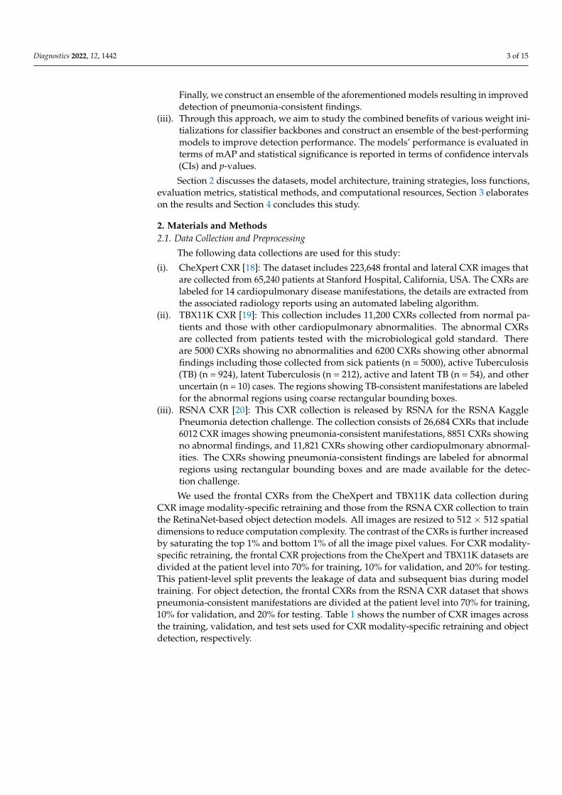
(i). CheXpert CXR [18]: The dataset includes 223,648 frontal and lateral CXR images thatare collected from 65,240 patients at Stanford Hospital, California, USA. The CXRs arelabeled for 14 cardiopulmonary disease manifestations, the details are extracted fromthe associated radiology reports using an automated labeling algorithm.
(ii). TBX11K CXR [19]: This collection includes 11,200 CXRs collected from normal pa-tients and those with other cardiopulmonary abnormalities. The abnormal CXRsare collected from patients tested with the microbiological gold standard. Thereare 5000 CXRs showing no abnormalities and 6200 CXRs showing other abnormalfindings including those collected from sick patients (n = 5000), active Tuberculosis(TB) (n = 924), latent Tuberculosis (n = 212), active and latent TB (n = 54), and otheruncertain (n = 10) cases. The regions showing TB-consistent manifestations are labeledfor the abnormal regions using coarse rectangular bounding boxes.
(iii). RSNA CXR [20]: This CXR collection is released by RSNA for the RSNA KagglePneumonia detection challenge. The collection consists of 26,684 CXRs that include6012 CXR images showing pneumonia-consistent manifestations, 8851 CXRs showingno abnormal findings, and 11,821 CXRs showing other cardiopulmonary abnormal-ities. The CXRs showing pneumonia-consistent findings are labeled for abnormalregions using rectangular bounding boxes and are made available for the detec-tion challenge.
We used the frontal CXRs from the CheXpert and TBX11K data collection duringCXR image modality-specific retraining and those from the RSNA CXR collection to trainthe RetinaNet-based object detection models. All images are resized to 512 × 512 spatialdimensions to reduce computation complexity. The contrast of the CXRs is further increasedby saturating the top 1% and bottom 1% of all the image pixel values. For CXR modality-specific retraining, the frontal CXR projections from the CheXpert and TBX11K datasets aredivided at the patient level into 70% for training, 10% for validation, and 20% for testing.This patient-level split prevents the leakage of data and subsequent bias during modeltraining. For object detection, the frontal CXRs from the RSNA CXR dataset that showspneumonia-consistent manifestations are divided at the patient level into 70% for training,10% for validation, and 20% for testing. Table 1 shows the number of CXR images acrossthe training, validation, and test sets used for CXR modality-specific retraining and objectdetection, respectively.

Diagnostics 2022, 12, 1442 4 of 15
Table 1. Patient-level dataset splits show the number of images for CXR modality-specific retrainingand object detection. Note: TBX11K and RSNA datasets have one image per patient.
DatasetTrain Validation Test
Abnormal Normal Abnormal Normal Abnormal Normal
CXR Modality-specific retraining
CheXpert 13,600 13,600 1700 1700 1700 1700
TBX11k 3040 3040 380 380 380 380
RetinaNet-based object detection
Dataset Train Validation Test
RSNA 4212 600 1200
2.2. Model Architecture2.2.1. CXR Modality-Specific Retraining
The ImageNet-pretrained DL models, viz., VGG-16, VGG-19, DenseNet-121, ResNet-50,EfficientNet-B0, and MobileNet have demonstrated promising performance in several medicalvisual recognition tasks [14,19,21–23]. These models are further retrained on a large collectionof CXR images to classify them as showing cardiopulmonary abnormal manifestations or noabnormalities. Such retraining helps the models to learn CXR image modality-specific featuresthat can be transferred and fine-tuned to improve performance in a relevant task using CXRimages. The best-performing model with the learned CXR image modality-specific weights isused as the classifier backbone to train the RetinaNet-based object detection model towarddetecting pneumonia-consistent manifestations. Figure 1 shows the block diagram illustratingthe steps involved in CXR image modality-specific retraining.
Diagnostics 2022, 12, x FOR PEER REVIEW 4 of 14
Table 1. Patient-level dataset splits show the number of images for CXR modality-specific retraining and object detection. Note: TBX11K and RSNA datasets have one image per patient.
Dataset Train Validation Test
Abnormal Normal Abnormal Normal Abnormal Normal CXR Modality-specific retraining
CheXpert 13600 13600 1700 1700 1700 1700 TBX11k 3040 3040 380 380 380 380
RetinaNet-based object detection Dataset Train Validation Test RSNA 4212 600 1200
2.2. Model Architecture 2.2.1. CXR Modality-Specific Retraining
The ImageNet-pretrained DL models, viz., VGG-16, VGG-19, DenseNet-121, ResNet-50, EfficientNet-B0, and MobileNet have demonstrated promising performance in several medical visual recognition tasks [14,19,21–23]. These models are further retrained on a large collection of CXR images to classify them as showing cardiopulmonary abnormal manifestations or no abnormalities. Such retraining helps the models to learn CXR image modality-specific features that can be transferred and fine-tuned to improve performance in a relevant task using CXR images. The best-performing model with the learned CXR image modality-specific weights is used as the classifier backbone to train the RetinaNet-based object detection model toward detecting pneumonia-consistent manifestations. Fig-ure 1 shows the block diagram illustrating the steps involved in CXR image modality-specific retraining.
Figure 1. Steps illustrating CXR image modality-specific retraining of the ImageNet-pretrained models.
2.2.2. RetinaNet Architecture We used RetinaNet as the base object detection architecture in our experiments. The
architecture of the RetinaNet model is shown in Figure 2. As a single-stage object detection structure, RetinaNet shares a similar concept of “anchor proposal” with [24]. It used a feature pyramid network (FPN) [25] where features on each of the image scales are com-puted separately in the lateral connections and then summed up through convolutional operations via the top-down pathways. The FPN network combines low-resolution fea-tures with strong semantic information, and high-resolution features with weak semantics through top-down paths and horizontal connections. Thus, feature maps with rich seman-tic information are obtained that would prove beneficial for detecting relatively smaller
Figure 1. Steps illustrating CXR image modality-specific retraining of the ImageNet-pretrained models.
2.2.2. RetinaNet Architecture
We used RetinaNet as the base object detection architecture in our experiments. Thearchitecture of the RetinaNet model is shown in Figure 2. As a single-stage object de-tection structure, RetinaNet shares a similar concept of “anchor proposal” with [24]. Itused a feature pyramid network (FPN) [25] where features on each of the image scales arecomputed separately in the lateral connections and then summed up through convolu-tional operations via the top-down pathways. The FPN network combines low-resolutionfeatures with strong semantic information, and high-resolution features with weak seman-tics through top-down paths and horizontal connections. Thus, feature maps with richsemantic information are obtained that would prove beneficial for detecting relativelysmaller ROIs consistent with pneumonia compared to the other parts of the CXR image.Furthermore, when trained to minimize the focal loss [5], the RetinaNet was reported todeliver significant performance focusing on hard, misclassified examples.

Diagnostics 2022, 12, 1442 5 of 15
Diagnostics 2022, 12, x FOR PEER REVIEW 5 of 14
ROIs consistent with pneumonia compared to the other parts of the CXR image. Further-more, when trained to minimize the focal loss [5], the RetinaNet was reported to deliver significant performance focusing on hard, misclassified examples.
Figure 2. Method flowchart for the RetinaNet network.
2.2.3. Ensemble of RetinaNet Models with Various Backbones We initialized the weights of the VGG-16 and ResNet-50 classifier backbones used in
the RetinaNet model using three strategies: (i) Random weights; (ii) ImageNet-pretrained weights, and (iii) CXR image modality-specific retrained weights as discussed in Section 2.2.1. Each model is trained for 80 epochs and the model weights (snapshots) are stored at the end of each epoch. Varying modifications of the RetinaNet model classifier backbones and loss functions are mentioned in Table 2.
Table 2. RetinaNet model classifier backbones with varying weight initializations and loss func-tions. The loss functions mentioned are used for classification. For bounding box regression, only the smooth-L1 loss function [26] is used in all cases.
ResNet-50 Backbone and Classification Loss Functions
VGG-16 Backbone and Classification Loss Functions
ResNet-50 with random weights + focal loss VGG-16 with random weights + focal loss ResNet-50 with random weights + focal Tversky loss
VGG-16 with random weights + focal Tversky loss
ResNet-50 with ImageNet pretrained weights + focal loss
VGG-16 with ImageNet pretrained weights + focal loss
ResNet-50 with ImageNet pretrained weights + focal Tversky loss
VGG-16 with ImageNet pretrained weights + focal Tversky loss
ResNet-50 with CXR image modality-specific weights + focal loss
VGG-16 with CXR image modality-specific weights + focal loss
ResNet-50 with CXR image modality-specific weights + focal Tversky loss
VGG-16 with CXR image modality-specific weights + focal Tversky loss
We adopted the non-maximum suppression (NMS) in the RetinaNet training with an IoU threshold of 0.5 and evaluated the models using all the predictions with a confidence score over 0.9. A weighted averaging ensemble is constructed using (i) the top-3 perform-ing models from the 12 RetinaNet models mentioned in Table 2, and (ii) the top-3 per-forming snapshots (model weights) using each classifier backbone. We empirically as-signed the weights as 1, 0.9, and 0.8 for the predictions of the 1st, 2nd, and 3rd best per-forming models. A schematic of the ensemble procedure is shown in Figure 3. An ensem-bled bounding box is generated if the IOU of the weighted average of the predicted bounding boxes and the ground truth (GT) boxes is greater than 0.5. The ensembled model is evaluated based on the mean average precision (mAP) metric.
Figure 2. Method flowchart for the RetinaNet network.
2.2.3. Ensemble of RetinaNet Models with Various Backbones
We initialized the weights of the VGG-16 and ResNet-50 classifier backbones used inthe RetinaNet model using three strategies: (i) Random weights; (ii) ImageNet-pretrainedweights, and (iii) CXR image modality-specific retrained weights as discussed in Sec-tion 2.2.1. Each model is trained for 80 epochs and the model weights (snapshots) arestored at the end of each epoch. Varying modifications of the RetinaNet model classifierbackbones and loss functions are mentioned in Table 2.
Table 2. RetinaNet model classifier backbones with varying weight initializations and loss functions.The loss functions mentioned are used for classification. For bounding box regression, only thesmooth-L1 loss function [26] is used in all cases.
ResNet-50 Backbone and ClassificationLoss Functions
VGG-16 Backbone and ClassificationLoss Functions
ResNet-50 with random weights + focal loss VGG-16 with random weights + focal loss
ResNet-50 with random weights + focalTversky loss
VGG-16 with random weights + focalTversky loss
ResNet-50 with ImageNet pretrainedweights + focal loss
VGG-16 with ImageNet pretrainedweights + focal loss
ResNet-50 with ImageNet pretrainedweights + focal Tversky loss
VGG-16 with ImageNet pretrainedweights + focal Tversky loss
ResNet-50 with CXR image modality-specificweights + focal loss
VGG-16 with CXR image modality-specificweights + focal loss
ResNet-50 with CXR image modality-specificweights + focal Tversky loss
VGG-16 with CXR image modality-specificweights + focal Tversky loss
We adopted the non-maximum suppression (NMS) in the RetinaNet training with anIoU threshold of 0.5 and evaluated the models using all the predictions with a confidencescore over 0.9. A weighted averaging ensemble is constructed using (i) the top-3 performingmodels from the 12 RetinaNet models mentioned in Table 2, and (ii) the top-3 performingsnapshots (model weights) using each classifier backbone. We empirically assigned theweights as 1, 0.9, and 0.8 for the predictions of the 1st, 2nd, and 3rd best performing models.A schematic of the ensemble procedure is shown in Figure 3. An ensembled bounding boxis generated if the IOU of the weighted average of the predicted bounding boxes and theground truth (GT) boxes is greater than 0.5. The ensembled model is evaluated based onthe mean average precision (mAP) metric.

Diagnostics 2022, 12, 1442 6 of 15Diagnostics 2022, 12, x FOR PEER REVIEW 6 of 14
Figure 3. Method Schematic of the ensemble approach.
2.2.4. Loss Functions and Evaluation Metrics CXR image Modality-Specific Retraining
During CXR image modality-specific retraining, the DL models are retrained on a combined selection of the frontal CXR projections from the CheXpert and TBX11K da-tasets (details in Table 1). The training is performed for 128 epochs to minimize the cate-gorical cross-entropy (CCE) loss. The CCE loss is the most commonly used loss function in classification tasks, and it helps to measure the distinguishability between two discrete probability distributions. It is expressed as shown in Equation (1).
𝐶𝐶𝐸 = − 𝑦 𝑙𝑜𝑔𝑦^ (1)
Here, 𝑦^ denotes the kth scalar value in the model output, 𝑦 denotes the corre-sponding target, and the 𝑜𝑢𝑡𝑝𝑢𝑡 𝑠𝑖𝑧𝑒 denotes the number of scalar values in the model output. The term 𝑦 denotes the probability that event k occurs and the sum of all 𝑦 =1. The minus sign in the CCE loss equation ensures the loss is minimized when the distri-butions become less distinguishable. We used a stochastic gradient descent optimizer with an initial learning rate of 1 × 10⁻⁴ and momentum of 0.9 to reduce the CCE loss and improve performance. Callbacks are used to store the model checkpoints and the learning rate is reduced after a patience parameter of 10 epochs when the validation performance ceased to improve. The weights of the model that delivered a superior performance with the val-idation set are used to predict the test set. The models are evaluated in terms of accuracy, the area under the receiver-operating characteristic curve (AUROC), the area under the precision-recall (PR) curve (AUPRC), sensitivity, precision, F-score, Matthews correlation coefficient (MCC), and Kappa statistic. RetinaNet-Based Detection of Pneumonia-Consistent Findings
Considering medical images, the disease ROIs span a relatively smaller portion of the whole image. This results in a considerably high degree of imbalance in the fore-ground ROI and the background pixels. These issues are particularly prominent in appli-cations such as detecting cardiopulmonary manifestations like pneumonia where the number of pixels showing pneumonia-consistent manifestations is markedly lower com-pared to the total number of image pixels. Generalized loss functions such as balanced
Figure 3. Method Schematic of the ensemble approach.
2.2.4. Loss Functions and Evaluation MetricsCXR Image Modality-Specific Retraining
During CXR image modality-specific retraining, the DL models are retrained on acombined selection of the frontal CXR projections from the CheXpert and TBX11K datasets(details in Table 1). The training is performed for 128 epochs to minimize the categoricalcross-entropy (CCE) loss. The CCE loss is the most commonly used loss function inclassification tasks, and it helps to measure the distinguishability between two discreteprobability distributions. It is expressed as shown in Equation (1).
CCEloss = −output size
∑k=1
yklogy ˆk (1)
Here, y ˆk denotes the kth scalar value in the model output, yk denotes the corresponding
target, and the output size denotes the number of scalar values in the model output. Theterm yk denotes the probability that event k occurs and the sum of all yk = 1. The minus signin the CCE loss equation ensures the loss is minimized when the distributions become lessdistinguishable. We used a stochastic gradient descent optimizer with an initial learningrate of 1 × 10⁻⁴ and momentum of 0.9 to reduce the CCE loss and improve performance.Callbacks are used to store the model checkpoints and the learning rate is reduced aftera patience parameter of 10 epochs when the validation performance ceased to improve.The weights of the model that delivered a superior performance with the validation set areused to predict the test set. The models are evaluated in terms of accuracy, the area underthe receiver-operating characteristic curve (AUROC), the area under the precision-recall(PR) curve (AUPRC), sensitivity, precision, F-score, Matthews correlation coefficient (MCC),and Kappa statistic.
RetinaNet-Based Detection of Pneumonia-Consistent Findings
Considering medical images, the disease ROIs span a relatively smaller portion of thewhole image. This results in a considerably high degree of imbalance in the foreground ROIand the background pixels. These issues are particularly prominent in applications suchas detecting cardiopulmonary manifestations like pneumonia where the number of pixelsshowing pneumonia-consistent manifestations is markedly lower compared to the totalnumber of image pixels. Generalized loss functions such as balanced cross-entropy loss do

Diagnostics 2022, 12, 1442 7 of 15
not take this data imbalance into account. This may lead to a learning bias and subsequentadversity in learning the minority ROI pixels. Appropriate selection of the loss function istherefore critical for improving detection performance. In this regard, the authors of [11]proposed the focal loss for object detection, an extension of the cross-entropy loss, whichalleviates this learning bias by giving importance to the minority ROI pixels while down-weighting the majority background pixels. Minimizing the focal loss thereby reduces theloss contribution from majority background examples and increases the importance ofcorrectly detecting the minority disease-positive ROI pixels. The focal loss is expressed asshown in Equation (2).
Focal loss(pt) = −αt(1− pt)γ log(pt) (2)
Here, pt denotes the probability the object detection model predicts for the GT. The pa-rameter γ decides the rate of down-weighting the majority (background non-ROI) samples.The equation converges to the conventional cross-entropy loss when γ = 0. We empiricallyselected the value of γ = 2 which delivered superior detection performance.
Another loss function called the Focal Tversky loss function [27], a generalizationof the focal loss function, is proposed to tackle the data imbalance problem and is givenin Equation (3). The Focal Tversky loss function generalizes the Tversky loss which isbased on the Tversky index that helps achieve a superior tradeoff between recall andprecision when trained on class-imbalanced datasets. The Focal Tversky loss function usesa smoothing parameter γ that controls the non-linearity of the loss at different values of theTversky index to balance between the minority pneumonia-consistent ROI and majoritybackground classes. In Equation (3), TI denotes the Tversky index, expressed as shown inEquation (4).
FTlossc = ∑c
1− TIγc (3)
TIc =∑M
i=1 ticgic+ ∈∑M
i=1 ticgic + α ∑Mi=1 tic gic + β ∑M
i=1 ticgic+ ∈(4)
Here, gic and tic denote the ground truth and predicted labels for the pneumoniaclass c, where gic and tic ∈ {0,1}. That is, tic denotes the probability that the pixel i belongsto the pneumonia class c and tic denotes the probability that the pixel i belongs to thebackground class c. The same holds for gic and gic . The term M denotes the total number ofimage pixels. The term ∈ provides numerical stability to avoid divide-by-zero errors. Thehyperparameters α and β are tuned to emphasize recall under class-imbalanced trainingconditions. The Tversky index is adapted to a loss function by minimizing ∑c 1− TIc. Afterempirical evaluations, we fixed the value of γ = 4/3, α = 0.7 and β = 0.75.
As is known, the loss function within RetinaNet is a summation of a couple of lossfunctions, one for classification and the other for bounding box regression. We left theSmooth-L1 loss that is used for bounding box regression unchanged. For classification, weexplored the performance with focal loss and focal Tversky loss functions individually fortraining the RetinaNet models with varying weight initializations. We used the boundingbox annotations [20] associated with the RSNA CXRs showing pneumonia-consistentmanifestations as the GT bounding boxes and measured its agreement with that generatedby the models initialized with random weights, ImageNet-pretrained, and CXR imagemodality-specific retrained classifier backbones. Let TP, FP, and FN denote the true positives,false positives, and false negatives, respectively. Given a pre-defined IOU threshold, apredicted bounding box is considered to be TP if it overlaps with the GT bounding box bya value equal to or exceeding this threshold. FP denotes that the predicted bounding boxhas no associated GT bounding box. FN denotes the GT bounding box has no associatedpredicted bounding box. The mAP is measured as the area under the precision-recall curve(AUPRC) as shown in Equation (5). Here, P denotes precision which measures the accuracyof predictions, and R denotes recall which measures how well the model identifies all the

Diagnostics 2022, 12, 1442 8 of 15
TPs. They are computed as shown in Equations (6) and (7). The value of mAP lies in therange [0, 1].
mean average precision (mAP) =∫ 1
0P(R)dR (5)
Precision (P) =TP
TP + FP(6)
Recall =TP
(TP + FN)(7)
We used a Linux system with 1080Ti GPU, the Tensorflow backend (v. 2.6.2) withKeras, and CUDA/CUDNN libraries for accelerating the graphical processing unit (GPU)toward training the object detection models that are configured in the Python environment.
2.3. Statistical Analysis
We evaluated statistical significance using the mAP metric achieved by the modelstrained with various weight initializations and loss functions. The 95% confidence intervals(CIs) are measured as the binomial interval using the Clopper-Pearson method.
3. Results and Discussion
We organized the results from our experiments into the following sections: Evaluatingthe performance of (i) CXR image modality-specific retrained models and (ii) RetinaNetobject detection models using classifier backbones with varying weight initializations andloss functions.
3.1. Classification Performance during CXR Image Modality-Specific Retraining
Recall that the ImageNet-pretrained DL models are retrained on the combined selec-tion of CXRs from the CheXpert and TBX11K collection. Such retraining is performed toconvert the weight layers specific to the CXR image modality and let the models learn CXRmodality-specific features to improve performance when the learned knowledge is trans-ferred and fine-tuned for a related medical image visual recognition task. The performanceachieved by the CXR image modality-specific retrained models using the hold-out test setis listed in Table 3 and the performance curves are shown in Figure 4. The no-skill line inFigure 4 denotes the performance when a classifier would fail to discriminate between thenormal and abnormal CXRs and therefore would predict a random outcome or a specificcategory under all circumstances.
Diagnostics 2022, 12, x FOR PEER REVIEW 9 of 14
Figure 4. The collection of performance curves for the CXR image modality-specific retrained mod-els. The performance is recorded at the optimal classification threshold measured with the valida-tion data. (a) ROC and (b) PR curves.
We could observe from Table 3 that the CXR image modality-specific retrained VGG-16 model demonstrates the best performance compared to other models in terms of all metrics except sensitivity. Of these, the MCC metric is a good measure to use because unlike F-score because it considers a balanced ratio of TPs TNs, FPs, and FNs. We noticed that the differences in the MCC values achieved by the various CXR image modality-spe-cific retrained models are not significantly different (p > 0.05). Based on its performance, we used VGG-16 as the backbone for the RetinaNet detector. However, to enable fair com-parison with other conventional RetinaNet-based results, we included the ResNet-50 backbone for detecting pneumonia-consistent manifestations. The VGG-16 and ResNet-50 classifier backbones are also initialized with random and ImageNet-pretrained weights for further comparison.
3.2. Detection Performance Using RetinaNet Models and their Ensembles Recall that the RetinaNet models are trained with different initializations of the clas-
sifier backbones. The performance achieved by these models using the hold-out test set is listed in Table 4. Figure 5 shows the PR curves obtained with the RetinaNet model using varying weight initializations for the selected classifier backbones. These curves show the precision and recall value of the model’s bounding box predictions on every sample in the test set. We observe from Table 4 that the RetinaNet model with the CXR image modality-specific retrained ResNet-50 classifier backbone and trained using the focal loss function demonstrates superior performance in terms of mAP. Figure 6 shows the bounding box predictions of the top-3 performing RetinaNet models for a sample CXR from the hold-out test set.
Table 4. Performance of RetinaNet with the varying weight initializations for the classifier back-bones and training losses. The values in parenthesis denote the 95% CI for the mAP metric. Bold numerical values denote superior performance.
Models AUPRC (mAP) ResNet-50 with random weights + focal loss 0.2763 (0.2509, 0.3017) ResNet-50 with random weights + focal Tversky loss 0.2627 (0.2377, 0.2877) ResNet-50 with ImageNet pretrained weights + focal loss 0.2719 (0.2467, 0.2971) ResNet-50 with ImageNet pretrained weights + focal Tversky loss 0.2737 (0.2484, 0.2990) ResNet-50 with CXR image modality-specific weights + focal loss 0.2865 (0.2609, 0.3121) ResNet-50 with CXR image modality-specific weights + focal Tversky loss 0.2859 (0.2603, 0.3115) VGG-16 with random weights + focal loss 0.2549 (0.2302, 0.2796) VGG-16 with random weights + focal Tversky loss 0.2496 (0.2251, 0.2741)
Figure 4. The collection of performance curves for the CXR image modality-specific retrained models.The performance is recorded at the optimal classification threshold measured with the validationdata. (a) ROC and (b) PR curves.

Diagnostics 2022, 12, 1442 9 of 15
Table 3. Performance of the CXR image modality-specific retrained models with the hold-out test set.Bold numerical values denote superior performance. The values in parenthesis denote the 95% CI forthe MCC metric.
Models Accuracy AUROC AUPRC Sensitivity Precision F-Score MCC Kappa
VGG-16 0.7834 0.8701 0.8777 0.8303 0.7591 0.7931 0.5693(0.5542, 0.5844) 0.5668
VGG-19 0.7743 0.8660 0.8727 0.8389 0.7429 0.7880 0.5532(0.5380, 0.5684) 0.5486
DenseNet-121 0.7738 0.8582 0.8618 0.8264 0.7477 0.7851 0.5507(0.5355, 0.5659) 0.5476
ResNet-50 0.7685 0.8586 0.8646 0.8207 0.7431 0.7800 0.5400(0.5248, 0.5552) 0.5370
EfficientNet-B0 0.7553 0.8568 0.8612 0.8678 0.7084 0.7800 0.5240(0.5088, 0.5392) 0.5106
MobileNet 0.7584 0.8609 0.8655 0.8726 0.7104 0.7832 0.5309(0.5157, 0.5461) 0.5168
We could observe from Table 3 that the CXR image modality-specific retrained VGG-16 model demonstrates the best performance compared to other models in terms of allmetrics except sensitivity. Of these, the MCC metric is a good measure to use becauseunlike F-score because it considers a balanced ratio of TPs TNs, FPs, and FNs. We noticedthat the differences in the MCC values achieved by the various CXR image modality-specific retrained models are not significantly different (p > 0.05). Based on its performance,we used VGG-16 as the backbone for the RetinaNet detector. However, to enable faircomparison with other conventional RetinaNet-based results, we included the ResNet-50backbone for detecting pneumonia-consistent manifestations. The VGG-16 and ResNet-50classifier backbones are also initialized with random and ImageNet-pretrained weights forfurther comparison.
3.2. Detection Performance Using RetinaNet Models and Their Ensembles
Recall that the RetinaNet models are trained with different initializations of the clas-sifier backbones. The performance achieved by these models using the hold-out test setis listed in Table 4. Figure 5 shows the PR curves obtained with the RetinaNet modelusing varying weight initializations for the selected classifier backbones. These curvesshow the precision and recall value of the model’s bounding box predictions on everysample in the test set. We observe from Table 4 that the RetinaNet model with the CXRimage modality-specific retrained ResNet-50 classifier backbone and trained using the focalloss function demonstrates superior performance in terms of mAP. Figure 6 shows thebounding box predictions of the top-3 performing RetinaNet models for a sample CXRfrom the hold-out test set.
We used two approaches to combine the bounding box predictions. They are (i) usingthe bounding box predictions from the top-3 performing RetinaNet models, viz., ResNet-50 with CXR image modality-specific weights + focal loss, ResNet-50 with CXR imagemodality-specific weights + focal Tversky loss, and ResNet-50 with random weights + focalloss; and, (ii) using the bounding box predictions from the top-3 performing snapshots(weights) within each model. The results are presented in Table 5 and Figure 7. A weightedaveraging ensemble of the bounding boxes is generated when the IoU of the predictedbounding boxes is greater than the threshold value which is set at 0.5. Recall that themodels are trained for 80 epochs and a snapshot (i.e., the model weights) is stored at theend of each epoch. We observed that the ensemble of the top-3 performing RetinaNetmodels delivered superior performance in terms of mAP metric compared to other modelsand ensembles. Figure 8 shows a sample CXR image with GT and predicted bounding

Diagnostics 2022, 12, 1442 10 of 15
boxes using the weighted averaging ensemble of the top-3 individual models and the top-3snapshots of the best-performing model.
Table 4. Performance of RetinaNet with the varying weight initializations for the classifier backbonesand training losses. The values in parenthesis denote the 95% CI for the mAP metric. Bold numericalvalues denote superior performance.
Models AUPRC (mAP)
ResNet-50 with random weights + focal loss 0.2763 (0.2509, 0.3017)
ResNet-50 with random weights + focal Tversky loss 0.2627 (0.2377, 0.2877)
ResNet-50 with ImageNet pretrainedweights + focal loss 0.2719 (0.2467, 0.2971)
ResNet-50 with ImageNet pretrained weights + focal Tversky loss 0.2737 (0.2484, 0.2990)
ResNet-50 with CXR image modality-specificweights + focal loss 0.2865 (0.2609, 0.3121)
ResNet-50 with CXR image modality-specificweights + focal Tversky loss 0.2859 (0.2603, 0.3115)
VGG-16 with random weights + focal loss 0.2549 (0.2302, 0.2796)
VGG-16 with random weights + focal Tversky loss 0.2496 (0.2251, 0.2741)
VGG-16 with ImageNet pretrained weights + focal loss 0.2734 (0.2481, 0.2987)
VGG-16 with ImageNet pretrained weights + focal Tversky loss 0.2666 (0.2415, 0.2917)
VGG-16 with CXR image modality-specificweights + focal loss 0.2686 (0.2435, 0.2937)
VGG-16 with CXR image modality-specificweights + focal Tversky loss 0.2648 (0.2398, 0.2898)
Diagnostics 2022, 12, x FOR PEER REVIEW 10 of 14
VGG-16 with ImageNet pretrained weights + focal loss 0.2734 (0.2481, 0.2987) VGG-16 with ImageNet pretrained weights + focal Tversky loss 0.2666 (0.2415, 0.2917) VGG-16 with CXR image modality-specific weights + focal loss 0.2686 (0.2435, 0.2937) VGG-16 with CXR image modality-specific weights + focal Tversky loss 0.2648 (0.2398, 0.2898)
Figure 5. PR curves of the RetinaNet models initialized with varying weights for the classifier back-bones. (a) ResNet-50 with CXR image modality-specific weights + focal Tversky loss; (b) ResNet-50 with CXR image modality-specific weights + focal loss, and (c) ResNet-50 with random weights + focal loss.
Figure 6. Bounding box predictions of the RetinaNet models initialized with varying weights for the classifier backbones. Green boxes denote the model predictions and red boxes denote the ground truth. (a) A sample CXR with ground truth bounding boxes. (b) ResNet-50 with CXR image modal-ity-specific weights + focal Tversky loss; (c) ResNet-50 with CXR image modality-specific weights + focal loss, and (d) ResNet-50 with random weights + focal loss.
Figure 5. PR curves of the RetinaNet models initialized with varying weights for the classifierbackbones. (a) ResNet-50 with CXR image modality-specific weights + focal Tversky loss; (b) ResNet-50 with CXR image modality-specific weights + focal loss, and (c) ResNet-50 with random weights +focal loss.

Diagnostics 2022, 12, 1442 11 of 15
Diagnostics 2022, 12, x FOR PEER REVIEW 10 of 14
VGG-16 with ImageNet pretrained weights + focal loss 0.2734 (0.2481, 0.2987) VGG-16 with ImageNet pretrained weights + focal Tversky loss 0.2666 (0.2415, 0.2917) VGG-16 with CXR image modality-specific weights + focal loss 0.2686 (0.2435, 0.2937) VGG-16 with CXR image modality-specific weights + focal Tversky loss 0.2648 (0.2398, 0.2898)
Figure 5. PR curves of the RetinaNet models initialized with varying weights for the classifier back-bones. (a) ResNet-50 with CXR image modality-specific weights + focal Tversky loss; (b) ResNet-50 with CXR image modality-specific weights + focal loss, and (c) ResNet-50 with random weights + focal loss.
Figure 6. Bounding box predictions of the RetinaNet models initialized with varying weights for the classifier backbones. Green boxes denote the model predictions and red boxes denote the ground truth. (a) A sample CXR with ground truth bounding boxes. (b) ResNet-50 with CXR image modal-ity-specific weights + focal Tversky loss; (c) ResNet-50 with CXR image modality-specific weights + focal loss, and (d) ResNet-50 with random weights + focal loss.
Figure 6. Bounding box predictions of the RetinaNet models initialized with varying weights for theclassifier backbones. Green boxes denote the model predictions and red boxes denote the groundtruth. (a) A sample CXR with ground truth bounding boxes. (b) ResNet-50 with CXR image modality-specific weights + focal Tversky loss; (c) ResNet-50 with CXR image modality-specific weights + focalloss, and (d) ResNet-50 with random weights + focal loss.
Table 5. Ensemble performance with the top-3 performing models (from Table 4) and the top-3snapshots for each of the models trained with various classifier backbones and weight initializa-tions. Values in parenthesis denote the 95% CI for the mAP metric. Bold numerical values denotesuperior performance.
Ensemble Method mAP
Top-3 model ensemble (ResNet-50 with CXR imagemodality-specific weights + focal loss,ResNet-50 with CXR image modality-specific weights + focalTversky loss, and ResNet-50 with random weights + focal loss
0.3272 (0.3006, 0.3538)
Ensemble of the top-3 snapshots for each model
ResNet-50 with random weights + focal loss 0.2777 (0.2523, 0.3031)
ResNet-50 with random weights + focal Tversky loss 0.2630 (0.2380, 0.2880)
ResNet-50 with ImageNet pretrained weights + focal loss 0.2788 (0.2534, 0.3042)
ResNet-50 with ImageNet pretrained weights + focal Tversky loss 0.2812 (0.2557, 0.3067)
ResNet-50 with CXR image modality-specific weights + focal loss 0.2973 (0.2714, 0.3232)
ResNet-50 with CXR image modality-specific weights + focalTversky loss 0.2901 (0.2644, 0.3158)
VGG-16 with random weights + focal loss 0.2633 (0.2383, 0.2883)
VGG-16 with random weights + focal Tversky loss 0.2556 (0.2309, 0.2803)
VGG-16 with ImageNet pretrained weights + focal loss 0.2823 (0.2568, 0.3078)
VGG-16 with ImageNet pretrained weights + focal Tversky loss 0.2715 (0.2463, 0.2967)
VGG-16 with CXR image modality-specific weights + focal loss 0.2813 (0.2558, 0.3068)
VGG-16 with CXR image modality-specific weights + focalTversky loss 0.2698 (0.2446, 0.2950)

Diagnostics 2022, 12, 1442 12 of 15
Diagnostics 2022, 12, x FOR PEER REVIEW 11 of 14
We used two approaches to combine the bounding box predictions. They are (i) using the bounding box predictions from the top-3 performing RetinaNet models, viz., ResNet-50 with CXR image modality-specific weights + focal loss, ResNet-50 with CXR image modality-specific weights + focal Tversky loss, and ResNet-50 with random weights + focal loss; and, (ii) using the bounding box predictions from the top-3 performing snapshots (weights) within each model. The results are presented in Table 5 and Figure 7. A weighted averaging ensemble of the bounding boxes is generated when the IoU of the predicted bounding boxes is greater than the threshold value which is set at 0.5. Recall that the models are trained for 80 epochs and a snapshot (i.e., the model weights) is stored at the end of each epoch. We observed that the ensemble of the top-3 performing RetinaNet models delivered superior performance in terms of mAP metric compared to other models and ensembles. Figure 8 shows a sample CXR image with GT and predicted bounding boxes using the weighted averaging ensemble of the top-3 individual models and the top-3 snapshots of the best-performing model.
Table 5. Ensemble performance with the top-3 performing models (from Table 4) and the top-3 snapshots for each of the models trained with various classifier backbones and weight initializa-tions. Values in parenthesis denote the 95% CI for the mAP metric. Bold numerical values denote superior performance.
Ensemble Method mAP Top-3 model ensemble (ResNet-50 with CXR image modality-specific weights + focal loss, ResNet-50 with CXR image modality-specific weights + focal Tversky loss, and ResNet-50 with random weights + focal loss
0.3272 (0.3006, 0.3538)
Ensemble of the top-3 snapshots for each model ResNet-50 with random weights + focal loss 0.2777 (0.2523, 0.3031) ResNet-50 with random weights + focal Tversky loss 0.2630 (0.2380, 0.2880) ResNet-50 with ImageNet pretrained weights + focal loss 0.2788 (0.2534, 0.3042) ResNet-50 with ImageNet pretrained weights + focal Tversky loss 0.2812 (0.2557, 0.3067) ResNet-50 with CXR image modality-specific weights + focal loss 0.2973 (0.2714, 0.3232) ResNet-50 with CXR image modality-specific weights + focal Tversky loss 0.2901 (0.2644, 0.3158) VGG-16 with random weights + focal loss 0.2633 (0.2383, 0.2883) VGG-16 with random weights + focal Tversky loss 0.2556 (0.2309, 0.2803) VGG-16 with ImageNet pretrained weights + focal loss 0.2823 (0.2568, 0.3078) VGG-16 with ImageNet pretrained weights + focal Tversky loss 0.2715 (0.2463, 0.2967) VGG-16 with CXR image modality-specific weights + focal loss 0.2813 (0.2558, 0.3068) VGG-16 with CXR image modality-specific weights + focal Tversky loss 0.2698 (0.2446, 0.2950)
Figure 7. PR curves of the model ensembles. (a) PR curve obtained with the weighted-averaging ensemble of top-3 performing models (ResNet-50 with CXR modality-specific weights + focal loss, ResNet-50 with CXR modality-specific weights + focal Tversky loss, and ResNet-50 with random weights + focal loss and (b) PR curve obtained with the ensemble of top-3 performing snapshots while training the ResNet-50 with CXR modality-specific weights + focal loss model.
Figure 7. PR curves of the model ensembles. (a) PR curve obtained with the weighted-averagingensemble of top-3 performing models (ResNet-50 with CXR modality-specific weights + focal loss,ResNet-50 with CXR modality-specific weights + focal Tversky loss, and ResNet-50 with randomweights + focal loss and (b) PR curve obtained with the ensemble of top-3 performing snapshotswhile training the ResNet-50 with CXR modality-specific weights + focal loss model.
Diagnostics 2022, 12, x FOR PEER REVIEW 12 of 14
Figure 8. Bounding box predictions using the ensemble of RetinaNet models initialized with vary-ing weights for the classifier backbones. Green boxes denote the individual model predictions, blue boxes denote the ensemble predictions and red boxes denote the ground truth. (a) ResNet-50 with CXR image modality-specific weights + focal Tversky loss; (b) ResNet-50 with CXR image modality-specific weights + focal loss; (c) ResNet-50 with random weights + focal loss, and (d) the ensembled bounding box prediction.
4. Conclusions and Future Work In this study, we demonstrated the combined benefits of training CXR image modal-
ity-specific models, using them as backbones in an object detection model, evaluating them in different loss settings, and constructing ensembles of the best-performing models to improve performance in a pneumonia detection task. We observed that both CXR im-age modality-specific classifier backbones and ensemble learning improved detection per-formance compared to the individual constituent models. This study, however, suffers from the limitation that we have only investigated the effect of using CXR modality-spe-cific classifier backbones in a RetinaNet-based object detection model to improve detect-ing pneumonia-consistent findings. The efficacy of this approach in detecting other cardi-opulmonary disease manifestations is a potential avenue for future research. Additional diversity in the training process could be introduced by using CXR images and their dis-ease-specific annotations collected from multiple institutions. With the advent of high-performance computing and current advancements in DL-based object detection, future studies could explore the use of mask x-RCNN, transformer-based models, and other ad-vanced detection methods [28–31] and their ensembles in improving detection perfor-mance. Novel model optimization methods and loss functions can be proposed to further improve detection performance. However, the objective of this study is not to propose a new objection detection model but to validate the use of CXR modality-specific classifier backbones in existing models to improve performance. As the organizers of the RSNA Kaggle pneumonia detection challenge have not made the blinded GT annotations of the test set publicly available, we are unable to compare our results with the challenge lead-erboard. However, the performance of our method on a random split from the challenge-provided training set, where we sequester 10% of the images for testing, using 70% for training and 20% for validation, respectively, is markedly superior to the best performing method on the leaderboard.
Figure 8. Bounding box predictions using the ensemble of RetinaNet models initialized with varyingweights for the classifier backbones. Green boxes denote the individual model predictions, blueboxes denote the ensemble predictions and red boxes denote the ground truth. (a) ResNet-50 withCXR image modality-specific weights + focal Tversky loss; (b) ResNet-50 with CXR image modality-specific weights + focal loss; (c) ResNet-50 with random weights + focal loss, and (d) the ensembledbounding box prediction.

Diagnostics 2022, 12, 1442 13 of 15
4. Conclusions and Future Work
In this study, we demonstrated the combined benefits of training CXR image modality-specific models, using them as backbones in an object detection model, evaluating themin different loss settings, and constructing ensembles of the best-performing models toimprove performance in a pneumonia detection task. We observed that both CXR imagemodality-specific classifier backbones and ensemble learning improved detection perfor-mance compared to the individual constituent models. This study, however, suffers from thelimitation that we have only investigated the effect of using CXR modality-specific classifierbackbones in a RetinaNet-based object detection model to improve detecting pneumonia-consistent findings. The efficacy of this approach in detecting other cardiopulmonarydisease manifestations is a potential avenue for future research. Additional diversity inthe training process could be introduced by using CXR images and their disease-specificannotations collected from multiple institutions. With the advent of high-performancecomputing and current advancements in DL-based object detection, future studies couldexplore the use of mask x-RCNN, transformer-based models, and other advanced detectionmethods [28–31] and their ensembles in improving detection performance. Novel modeloptimization methods and loss functions can be proposed to further improve detectionperformance. However, the objective of this study is not to propose a new objection detec-tion model but to validate the use of CXR modality-specific classifier backbones in existingmodels to improve performance. As the organizers of the RSNA Kaggle pneumonia detec-tion challenge have not made the blinded GT annotations of the test set publicly available,we are unable to compare our results with the challenge leaderboard. However, the perfor-mance of our method on a random split from the challenge-provided training set, wherewe sequester 10% of the images for testing, using 70% for training and 20% for validation,respectively, is markedly superior to the best performing method on the leaderboard.
Author Contributions: Conceptualization, S.R., P.G. and S.K.A.; Data curation, S.R. and P.G.; Formalanalysis, S.R., P.G. and Z.X.; Funding acquisition, S.K.A.; Investigation, Z.X. and S.K.A.; Methodology,S.R. and P.G.; Project administration, S.K.A.; Resources, S.K.A.; Software, S.R. and P.G.; Supervision,Z.X. and S.K.A.; Validation, S.R., P.G. and S.K.A.; Visualization, S.R. and P.G.; Writing—original draft,S.R. and P.G.; Writing—review & editing, S.R., P.G., Z.X. and S.K.A. All authors have read and agreedto the published version of the manuscript.
Funding: This research was supported by the Intramural Research Program of the National Library ofMedicine, National Institutes of Health. The funders had no role in the study design, data collection,and analysis, decision to publish, or preparation of the manuscript.
Institutional Review Board Statement: Ethical review and approval were waived for this studybecause of the retrospective nature of the study and the use of anonymized patient data.
Informed Consent Statement: Patient consent was waived by the IRBs because of the retrospectivenature of this investigation and the use of anonymized patient data.
Data Availability Statement: The data required to reproduce this study is publicly available andcited in the manuscript.
Conflicts of Interest: The authors declare no conflict of interest.
References1. Kermany, D.S.; Goldbaum, M.; Cai, W.; Valentim, C.C.S.; Liang, H.; Baxter, S.L.; McKeown, A.; Yang, G.; Wu, X.; Yan, F.; et al.
Identifying Medical Diagnoses and Treatable Diseases by Image-Based Deep Learning. Cell 2018, 172, 1122–1131.e9. [CrossRef][PubMed]
2. Rajaraman, S.; Candemir, S.; Thoma, G.; Antani, S. Visualizing and Explaining Deep Learning Predictions for PneumoniaDetection in Pediatric Chest Radiographs. Proc. SPIE 2019, 10950, 109500S.
3. Nishio, M.; Noguchi, S.; Matsuo, H.; Murakami, T. Automatic Classification between COVID-19 Pneumonia, Non-COVID-19Pneumonia, and the Healthy on Chest X-Ray Image: Combination of Data Augmentation Methods. Sci. Rep. 2020, 10, 17532.[CrossRef]

Diagnostics 2022, 12, 1442 14 of 15
4. Balabanova, Y.; Coker, R.; Fedorin, I.; Zakharova, S.; Plavinskij, S.; Krukov, N.; Atun, R.; Drobniewski, F. Variability inInterpretation of Chest Radiographs among Russian Clinicians and Implications for Screening Programmes: Observational Study.BMJ 2005, 331, 379–382. [CrossRef] [PubMed]
5. Lin, T.Y.; Goyal, P.; Girshick, R.; He, K.; Dollár, P. Focal loss for dense object detection. In Proceedings of the IEEE InternationalConference on Computer Vision, Venice, Italy, 22–29 October 2017; pp. 2980–2988.
6. Rajaraman, S.; Candemir, S.; Kim, I.; Thoma, G.; Antani, S.; Rajaraman, S.; Candemir, S.; Kim, I.; Thoma, G.; Antani, S. Visualizationand Interpretation of Convolutional Neural Network Predictions in Detecting Pneumonia in Pediatric Chest Radiographs. Appl.Sci. 2018, 8, 1715. [CrossRef]
7. Mouhafid, M.; Salah, M.; Yue, C.; Xia, K. Deep Ensemble Learning-Based Models for Diagnosis of COVID-19 from Chest CTImages. Healthcare 2022, 10, 166. [CrossRef] [PubMed]
8. Pham, V.T.; Tran, C.M.; Zheng, S.; Vu, T.M.; Nath, S. Chest X-Ray Abnormalities Localization via Ensemble of Deep ConvolutionalNeural Networks. Int. Conf. Adv. Technol. Commun. 2021, 2021, 125–130. [CrossRef]
9. Xie, X.; Liao, Q.; Ma, L.; Jin, X. Gated Feature Pyramid Network for Object Detection. Lect. Notes Comput. Sci. 2018, 11259, 199–208.[CrossRef]
10. Mao, L.; Yumeng, T.; Lina, C. Pneumonia detection in chest X-rays: A deep learning approach based on ensemble RetinaNetand mask R-CNN. In Proceedings of the 8th International Conference on Advanced Cloud and Big Data, Taiyuan, China, 19–20September 2020; pp. 213–218. [CrossRef]
11. Lin, T.Y.; Goyal, P.; Girshick, R.; He, K.; Dollar, P. Focal Loss for Dense Object Detection. IEEE Trans. Pattern Anal. Mach. Intell.2020, 42, 318–327. [CrossRef] [PubMed]
12. Ko, H.; Ha, H.; Cho, H.; Seo, K.; Lee, J. Pneumonia detection with weighted voting ensemble of CNN models. In Proceedings ofthe 2nd International Conference on Artificial Intelligence and Big Data (ICAIBD), Chengdu, China, 26–29 May 2019; pp. 306–310.[CrossRef]
13. Fei-Fei, L.; Deng, J.; Li, K. ImageNet: Constructing a Large-Scale Image Database. J. Vis. 2010, 9, 1037. [CrossRef]14. Suzuki, K. Overview of Deep Learning in Medical Imaging. Radiol. Phys. Technol. 2017, 10, 257–273. [CrossRef] [PubMed]15. Rajaraman, S.; Sornapudi, S.; Kohli, M.; Antani, S. Assessment of an ensemble of machine learning models toward abnormality
detection in chest radiographs. In Proceedings of the 41st Annual International Conference of the IEEE Engineering in Medicineand Biology Society (EMBC), Berlin, Germany, 23–27 July 2019.
16. Rajaraman, S.; Folio, L.R.; Dimperio, J.; Alderson, P.O.; Antani, S.K. Improved Semantic Segmentation of Tuberculosis—ConsistentFindings in Chest x-Rays Using Augmented Training of Modality-Specific u-Net Models with Weak Localizations. Diagnostics2021, 11, 616. [CrossRef] [PubMed]
17. Yadav, O.; Passi, K.; Jain, C.K. Using deep learning to classify x-ray images of potential tuberculosis patients. In Proceedings ofthe IEEE International Conference on Bioinformatics and Biomedicine (BIBM), Madrid, Spain, 3–6 December 2018.
18. Irvin, J.; Rajpurkar, P.; Ko, M.; Yu, Y.; Ciurea-Ilcus, S.; Chute, C.; Marklund, H.; Haghgoo, B.; Ball, R.; Shpanskaya, K.; et al.CheXpert: A Large Chest Radiograph Dataset with Uncertainty Labels and Expert Comparison. Proc. Conf. AAAI Artif. Intell.2019, 33, 590–597. [CrossRef]
19. Liu, Y.; Wu, Y.H.; Ban, Y.; Wang, H.; Cheng, M.M. Rethinking computer-aided tuberculosis diagnosis. In Proceedings of the IEEEComputer Society Conference on Computer Vision and Pattern Recognition, Seattle, WA, USA, 14–19 June 2020.
20. Shih, G.; Wu, C.C.; Halabi, S.S.; Kohli, M.D.; Prevedello, L.M.; Cook, T.S.; Sharma, A.; Amorosa, J.K.; Arteaga, V.; Galperin-Aizenberg, M.; et al. Augmenting the National Institutes of Health Chest Radiograph Dataset with Expert Annotations of PossiblePneumonia. Radiol. Artif. Intell. 2019, 1, e180041. [CrossRef] [PubMed]
21. Rajaraman, S.; Jaeger, S.; Thoma, G.R.; Antani, S.K.; Silamut, K.; Maude, R.J.; Hossain, M.A. Understanding the Learned Behaviorof Customized Convolutional Neural Networks toward Malaria Parasite Detection in Thin Blood Smear Images. J. Med. Imaging2018, 5, 034501. [CrossRef] [PubMed]
22. Ganesan, P.; Rajaraman, S.; Long, R.; Ghoraani, B.; Antani, S. Assessment of data augmentation strategies toward performanceimprovement of abnormality classification in chest radiographs. In Proceedings of the 41st Annual International Conference ofthe IEEE Engineering in Medicine and Biology Society (EMBC), Berlin, Germany, 23–27 July 2019.
23. Rajaraman, S.; Antani, S.K.; Poostchi, M.; Silamut, K.; Hossain, M.A.; Maude, R.J.; Jaeger, S.; Thoma, G.R.; Hossain, A.;Maude, R.J.; et al. Pre-Trained Convolutional Neural Networks as Feature Extractors toward Improved Malaria Parasite Detectionin Thin Blood Smear Images. PeerJ 2018, 6, e4568. [CrossRef] [PubMed]
24. Ren, S.; He, K.; Girshick, R.; Sun, J. Faster R-CNN: Towards Real-Time Object Detection with Region Proposal Networks. IEEETrans. Pattern Anal. Mach. Intell. 2017, 39, 1137–1149. [CrossRef]
25. Lin, T.Y.; Dollár, P.; Girshick, R.; He, K.; Hariharan, B.; Belongie, S. Feature Pyramid Networks for Object Detection. In Proceedingsof the 30th IEEE Conference on Computer Vision and Pattern Recognition, Honolulu, HI, USA, 21–26 July 2017; pp. 936–944.[CrossRef]
26. Liu, C.; Yu, S.; Yu, M.; Wei, B.; Li, B.; Li, G.; Huang, W. Adaptive smooth L1 loss: A better way to regress scene texts withextreme aspect ratios. In Proceedings of the IEEE Symposium on Computers and Communications (ISCC), Athens, Greece,5–8 September 2021. [CrossRef]
27. Abraham, N.; Khan, N.M. A Novel Focal Tversky Loss Function with Improved Attention U-Net for Lesion Segmentation. InProceedings of the IEEE 16th International Symposium on Biomedical Imaging (ISBI), Venice, Italy, 8–11 April 2019.

Diagnostics 2022, 12, 1442 15 of 15
28. Qi, W.; Su, H. A Cybertwin Based Multimodal Network for ECG Patterns Monitoring Using Deep Learning. IEEE Trans. Ind.Inform. 2022, 3203, 1–9. [CrossRef]
29. Su, H.; Hu, Y.; Karimi, H.R.; Knoll, A.; Ferrigno, G.; De Momi, E. Improved Recurrent Neural Network-Based ManipulatorControl with Remote Center of Motion Constraints: Experimental Results. Neural Netw. 2020, 131, 291–299. [CrossRef]
30. Qi, W.; Aliverti, A. A Multimodal Wearable System for Continuous and Real-Time Breathing Pattern Monitoring during DailyActivity. IEEE J. Biomed. Health Inform. 2020, 24, 2199–2207. [CrossRef]
31. Su, H.; Mariani, A.; Ovur, S.E.; Menciassi, A.; Ferrigno, G.; De Momi, E. Toward Teaching by Demonstration for Robot-AssistedMinimally Invasive Surgery. IEEE Trans. Autom. Sci. Eng. 2021, 18, 484–494. [CrossRef]