Chandra X‐Ray Observations of the X‐Ray Faint Elliptical Galaxy NGC 4697

Imaging quality assessment of multiplexing x-ray radiography based on multi-beam x-ray source technology
J. Zhang1,2, R. Peng3, S. Chang1, J.P. Lu2, and O. Zhou2,3,4
(1) Department of Radiation Oncology, University of North Carolina, Chapel Hill, NC 27599
(2) Department of Physics and Astronomy, University of North Carolina, Chapel Hill, NC 27599 (3) Curriculum in Applied Science and Engineering, University of North Carolina, Chapel Hill, NC 27599
(4) Lineberger Comprehensive Cancer Center, University of North Carolina, Chapel Hill, NC 27599.
ABSTRACT
Multiplexing technique has been widely used in telecommunication, magnetic resonance imaging (MRI) and various spectroscopic applications to drastically increase system throughput. In the field of radiology, however, it was just getting started to attract researchers’ attention recently due to the development of multi-beam x-ray source technology, especially the emergence of carbon nanotube (CNT) field emission based multi-beam x-ray source. The CNT multi-beam x-ray source provides an ideal signal source for multiplexing x-ray imaging applications because of its capability of modulating x-ray radiation waveforms. The feasibility of multiplexing x-ray radiography has been successfully demonstrated experimentally using a CNT field emission enabled multi-beam x-ray imaging system. The idea of applying multiplexing radiography in computed tomography (CT) to speed up scanning speed has also been proposed. At the same time several simulation studies on the evaluation of multiplexing x-ray imaging performance have been reported. In this study, we reported our recent investigation on the imaging quality assessment of multiplexing x-ray radiography based on the simulation work stimulated by our previous experimental experience. A computer program was written to simulate the imaging process of the as-developed multi-beam x-ray imaging system. The impacts of different noise components on multiplexing imaging quality were studied. Our preliminary results indicated that the performance of multiplexing x-ray radiography is closely related to the noise environment and x-ray tube current stability. Under appropriate imaging conditions, multiplexing radiography has the potential to achieve higher imaging speed without significantly sacrificing the imaging quality. Keywords: multi-beam x-ray source technology, multiplexing x-ray radiography, quantum noise, electronic noise
1. INTRODUCTION
Multiplexing technique has been widely used in telecommunication [1, 2], magnetic resonance imaging (MRI) [3], and various kinds of spectroscopic applications including microscopy [4-6], optical spectroscopy [7], and mass spectrometry [8] to drastically increase system throughput. Conceptually speaking it is straightforward to apply the same multiplexing scheme in x-ray radiography to achieve higher imaging speed. However it was until very recently that multiplexing approach has been considered for x-ray radiography due to the limitation of conventional x-ray source technology. The conventional thermionic emission based x-ray tube typically only generates a single x-ray beam at a given time while multiple x-ray signals are generally required for multiplexing x-ray imaging applications. It also lacks the capability of modulating x-ray radiation waveforms in order to be used as a signal source for multiplexing.
It has been recently demonstrated that x-ray tubes using carbon nanotube (CNT) field emission cathodes have
several intrinsic advantages over the thermionic x-ray tubes, including the capability of programming x-ray radiation waveforms and potential for miniaturization [9, 10]. Because its small device size, multiple x-ray tube units can be integrated together to form an integrated x-ray source array, which provides an ideal x-ray signal source for potential multiplexing x-ray imaging applications [11, 12].
Multiplexing technique has great potential of speeding up x-ray imaging throughput, which is essential for
various imaging applications, such as computed tomography (CT). Boosted by the technology advancements of high
Medical Imaging 2010: Physics of Medical Imaging, edited by Ehsan Samei, Norbert J. Pelc,Proc. of SPIE Vol. 7622, 76224K · © 2010 SPIE · CCC code: 1605-7422/10/$18 · doi: 10.1117/12.844052
Proc. of SPIE Vol. 7622 76224K-1
Downloaded from SPIE Digital Library on 09 Apr 2010 to 152.2.176.242. Terms of Use: http://spiedl.org/terms

power x-ray tube, multi-slice x-ray detector, and high speed rotating gantry, CT scanning speed has been greatly improved during the past decade. However its intrinsic imaging throughput is still bottlenecked by its sequential data acquisition scheme. In a typical CT scan, up to a thousand projection images need to be acquired one at a time, in a sequential fashion, which in turn limits the overall imaging speed. In order to further the scanning speed, the idea of applying multiplexing technique to CT imaging to speed up its scanning speed has already been.
Several studies on the investigations of imaging performance of multiplexing radiography have been recently
published [13, 14]. In one paper, an analytical mathematical model was developed for the optimization of multiplexing coding patterns in a mixed noise environment. In another paper, the authors analyzed the system signal-to-noise ratio (SNR) of multiplexing imaging in quantum noise limited imaging systems. Based on those published studies, it is clear that the performance of multiplexing x-ray imaging depends on the noise environment of the x-ray imaging system. We have previously demonstrated the feasibility of multiplexing x-ray imaging experimentally using a CNT based multi-beam x-ray imaging system [15, 16]. Based on our observation during the previous study, in this paper, we reported some simulation works on the assessment of imaging performance of multiplexing x-ray radiography under different noise environments. We further investigated the cause of certain type of artifacts (ghost image) observed during our previous imaging experiment.
2. METHODS AND MATERIALS 2.1. Multi-beam x-ray imaging system: experimental setup and computer model
We have recently developed a multi-beam x-ray imaging system as a prototype stationary micro-CT scanner [17]. Meanwhile the as-developed imaging system also provides an ideal testing platform for multiplexing x-ray imaging applications. The multi-beam x-ray imaging system includes a linear multi-beam x-ray source array, a 3-axis rotary object table and a flat panel x-ray detector (Hamamatsu C7921CA), as illustrated in Figure 1a. The essential part of the system is the linear multi-beam x-ray source array, which comprises of a CNT field emission cathode array, an electron focusing assembly, and a molybdenum x-ray anode. The x-ray tube assembly is housed in a high vacuum chamber (10-9 Torr). The system is able to deliver up to 3 mA tube current from each individual CNT field emission cathode at 50 kV tube voltage with about 150 µm focal spot size.
Figure 1. (a) The as-developed multi-beam x-ray imaging system includes a CNT based field emission linear x-ray source array, a sample rotation stage, and an area x-ray detector. (b) A computer model was built based on the corresponding multi-beam x-ray imaging system for simulation purpose.
Each x-ray pixel of the multi-beam x-ray source array was individually controlled through a MOSFET based control circuitry [16]. In order to perform multiplexing x-ray imaging, a set of pre-programmed control signals (TTL
(b)
Proc. of SPIE Vol. 7622 76224K-2
Downloaded from SPIE Digital Library on 09 Apr 2010 to 152.2.176.242. Terms of Use: http://spiedl.org/terms

signals) was generated to trigger the MOSFET based control electronics connected to the corresponding CNT field emission cathode array inside the vacuum chamber. Once the 5V TTL signal opens up the gate of a MOSFET, a complete circuit is formed and x-ray radiation is generated from the corresponding x-ray focal spot.
In order to better assess the performance of multiplexing x-ray imaging under various imaging conditions, an
MATLAB based simulation program was written to simulate the imaging behavior of the multi-beam x-ray system. The imaging parameters, including x-ray tube current, exposure time, number of x-ray beams and noise composition (quantum noise and electronic noise), can be easily controlled to better evaluate the system imaging performance under different imaging environments. As depicted in Figure 1b, the simulated imaging system for this particular study has a linear x-ray source array with 7 x-ray pixels, a simple cylinder as the imaging object and a flat panel x-ray detector for imaging acquisition. The reason to use this 7-beam imaging configuration is to match the simulation results with the previously published experimental studies [16]. 2.2. Multiplexing radiography based on multi-beam x-ray source technology
In this study we focused on binary multiplexing radiography, especially a specific form of that called Hadamard multiplexing radiography, although other forms of multiplexing x-ray imaging have been previously reported [15]. Based on Hadamard transform, Hadamard multiplexing encodes signals using spatial modulation technique, which is inherently based on square waves (on/off state of the signal source). Compared with other multiplexing scheme such as frequency multiplexing, Hadamard multiplexing is able to achieve similar gain in imaging speed with much simplified multiplexing scheme. It also mitigates the stringent requirement on the x-ray detector speed.
Figure 2. Imaging flow chart for a general Hadamard multiplexing imaging process. A typical Hadamard multiplexing instrument consists of four essential components: a signal source (in this case
a multi-beam x-ray source), an encoding Hadamard mask (in this case a set of pre-programmed control signal based on the corresponding Hadamard matrix), a detector and a demultiplexing processor [18]. In Figure 2, we illustrated the imaging flow chart for a general Hadamard multiplexing x-ray imaging process. The Hadamard transform technique superimposes signals according to the Hadamard matrix. A controlling signal with pre-determined signal pattern and
Proc. of SPIE Vol. 7622 76224K-3
Downloaded from SPIE Digital Library on 09 Apr 2010 to 152.2.176.242. Terms of Use: http://spiedl.org/terms
pulse width was swept across the x-ray tube control circuit. A series of multiplexed images were acquired and stored in a computer. The original signals can then be directly recovered from the recorded multiplexed signals by applying the corresponding inversed Hadamard transformation. Mathematically, this recovering algorithm simply followed a certain procedure of addition and subtraction based on the inversed Hadamard matrix. 2.3. Noise model used for multiplexing x-ray imaging simulation
The performance of multiplexing x-ray radiography is closely related to the noise composition of the x-ray imaging system. In a typical x-ray imaging system, the imaging noise generally falls into the following two categories: photon noise and electronic noise. Photon noise occurs inherently in any x-ray imaging process due to the quantum nature of x-ray photons. Electronic noise, also called additive noise, on the other hand is related to the detector or to the electronics of the imaging system itself. Under normal imaging conditions, those two types of noises generally co-exist in any x-ray imaging system showing as a form of mixed noise. Most of the medical imaging x-ray systems are quantum noise limited devices. However, some of the x-ray imaging applications are electronic noise limited such as fluoroscopic procedures [19], x-ray cargo imaging [20] and other high sensitivity and high speed x-ray imaging applications.
Based on the reported results in the published literatures [13, 14], in the mixed noise scenario where quantum
noise and electronic noise co-exist, the performance of multiplexing x-ray imaging is determined by the relative ratio of those two types of noises. It has also been showing that in the case of quantum noise limited case, multiplexing imaging is generally outperformed by conventional sequential imaging method [14]. In our simulation, the system noise model is a simple combination of both photon and electronic noises. The photon noise was generated using a Poisson noise generator and its magnitude was proportional to the square root of total number of photons collected by the x-ray detector. On the other hand, the generation of electronic noise during the imaging process followed a typical Normal distribution (Gaussian distribution). The electronic noise was independent of x-ray exposure level (total number of incident x-ray photons). We also realized that in a real x-ray imaging system the situation will be much more complex and the noise model used for this simulation is only a gross approximation to the real noise scenario.
3. RESULTS AND DISCUSSIONS 3.1. Simulation of multiplexing/demultiplexing procedure
Although the simulated imaging system has the capability to handle arbitrary number of x-ray beams, here we only showed the results for the 7-beam case just to be able to compare the simulation results with our previous experimental results. A simple cylindrical rod phantom was used for all the simulated imaging purpose. In order to have a fair comparison, the simulation was done under the constant exposure constraint.
As shown in Figure 3a, a set of projection images from different viewing angles were first generated under
sequential imaging mode. The set of sequential images were used as the baseline images to evaluate the performance of the multiplexing imaging method. In Figure 3b, we showed a set of typical multiplexed images collected by the x-ray detector during multiplexing imaging process. In the case of Hadamard multiplexing of the order N=7, a certain combination of 4 x-ray pixels was turned on simultaneously for each of a total of 7 multiplexed exposures. It is clear from Figure 3b that each multiplexed image is a superimposed image of 4 individual projection images. By applying corresponding demultiplexing algorithm on the multiplexed images, the original individual projection can be recovered from the superimposed images, as demonstrated in Figure 3c. The demultiplexed images in Figure 3c, however, showed slightly higher noise level compared to their sequential counterparts in Figure 3a, which were taken using sequential method. This is due to the fact that the system noise, especially the quantum noise, was actually magnified during the multiplexing/demultiplexing process. The detailed quantitative results were discussed in the next section.
Proc. of SPIE Vol. 7622 76224K-4
Downloaded from SPIE Digital Library on 09 Apr 2010 to 152.2.176.242. Terms of Use: http://spiedl.org/terms
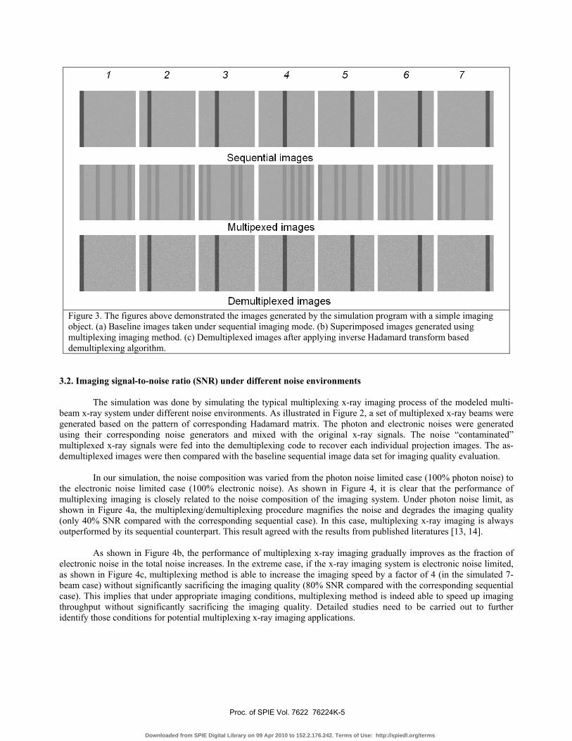
Figure 3. The figures above demonstrated the images generated by the simulation program with a simple imaging object. (a) Baseline images taken under sequential imaging mode. (b) Superimposed images generated using multiplexing imaging method. (c) Demultiplexed images after applying inverse Hadamard transform based demultiplexing algorithm.
3.2. Imaging signal-to-noise ratio (SNR) under different noise environments
The simulation was done by simulating the typical multiplexing x-ray imaging process of the modeled multi-beam x-ray system under different noise environments. As illustrated in Figure 2, a set of multiplexed x-ray beams were generated based on the pattern of corresponding Hadamard matrix. The photon and electronic noises were generated using their corresponding noise generators and mixed with the original x-ray signals. The noise “contaminated” multiplexed x-ray signals were fed into the demultiplexing code to recover each individual projection images. The as-demultiplexed images were then compared with the baseline sequential image data set for imaging quality evaluation.
In our simulation, the noise composition was varied from the photon noise limited case (100% photon noise) to
the electronic noise limited case (100% electronic noise). As shown in Figure 4, it is clear that the performance of multiplexing imaging is closely related to the noise composition of the imaging system. Under photon noise limit, as shown in Figure 4a, the multiplexing/demultiplexing procedure magnifies the noise and degrades the imaging quality (only 40% SNR compared with the corresponding sequential case). In this case, multiplexing x-ray imaging is always outperformed by its sequential counterpart. This result agreed with the results from published literatures [13, 14].
As shown in Figure 4b, the performance of multiplexing x-ray imaging gradually improves as the fraction of
electronic noise in the total noise increases. In the extreme case, if the x-ray imaging system is electronic noise limited, as shown in Figure 4c, multiplexing method is able to increase the imaging speed by a factor of 4 (in the simulated 7-beam case) without significantly sacrificing the imaging quality (80% SNR compared with the corresponding sequential case). This implies that under appropriate imaging conditions, multiplexing method is indeed able to speed up imaging throughput without significantly sacrificing the imaging quality. Detailed studies need to be carried out to further identify those conditions for potential multiplexing x-ray imaging applications.
Proc. of SPIE Vol. 7622 76224K-5
Downloaded from SPIE Digital Library on 09 Apr 2010 to 152.2.176.242. Terms of Use: http://spiedl.org/terms

Figure 4. Comparison of imaging qualities between sequential imaging and multiplexing imaging under different noise environments. (a) Photon noise limited case (b) Mixed noise (photon noise and electronic noise) case. (c) Electronic noise limited case.
3.3. Effect of tube current fluctuation: “ghost” image
A particular type of imaging artifacts, called “ghost image”, were observed during our multiplexing x-ray imaging test. In Figure 5a and 5b, we showed our experimental results of two demultiplexed images corresponding to two projection images from different x-ray sources. The white stripe in Figure 4b implied that that there was some kind of crosstalk happened between the two x-ray sources during the imaging process.
Figure 5: Demonstration of ghost image observed in our multiplexing experiment. The ghost image showing in (b) was caused by the crosstalk between different x-ray beams, in this case from the image showing in (a).
Further evidence indicated that the beam crosstalk was primarily caused by the current fluctuation of x-ray
tubes during the multiplexing process. If the tube current is unstable, images generated by different x-ray beams will
(a) (b)
Ghost image
(a) (b) (c)
Proc. of SPIE Vol. 7622 76224K-6
Downloaded from SPIE Digital Library on 09 Apr 2010 to 152.2.176.242. Terms of Use: http://spiedl.org/terms

interfere with each other and cause so called “ghost” image. In order to verify this, we performed another run of multiplexing x-ray imaging simulation. In this case, we manipulated the x-ray tube current, thus the x-ray exposure, by allowing 5% x-ray tube current fluctuation during the entire imaging process. As the images shown in Figure 6, we can clearly see the “ghost” images in the demultiplexed images, which agreed with our previous experimental observation.
Figure 6. The simulation results above demonstrated the ghost image caused by beam crosstalk during multiplexing imaging under the assumption of 5% current fluctuation.
4. CONCLUSIONS
We have developed a multi-beam x-ray imaging system based on carbon nanotube field emission enabled multi-beam x-ray source technology. This novel x-ray source allows us to perform multiplexing x-ray imaging which could not be done easily using conventional x-ray tubes. In this study we report our recent simulation work on the imaging quality assessment of multiplexing x-ray imaging under different noise environments. We are hoping that this study can provide us a guideline to better identify future multiplexing x-ray imaging related applications. Our simulation results indicated that the performance of multiplexing x-ray radiography is closely related to the noise composition and tube current stability of the x-ray imaging system. Under appropriate imaging conditions, multiplexing x-ray radiography has the potential to achieve higher imaging speed without significantly sacrificing the imaging quality. It could be used for applications such as image-guided radiation therapy (IGRT) for which sometimes imaging speed instead of imaging quality is the main concern of the task.
ACKNOWLEDGEMENTS This work was partially supported by NIH-NCI (U54CA119343), NIH-NIBIB (4R33EB004204-01) and Xintek, Inc.
REFERENCES 1. Weinstei.Sb and P.M. Ebert, Data Transmission by Frequency-Division Multiplexing Using Discrete
Fourier Transform. Ieee Transactions on Communication Technology, 1971. CO19(5): p. 628-&.
Proc. of SPIE Vol. 7622 76224K-7
Downloaded from SPIE Digital Library on 09 Apr 2010 to 152.2.176.242. Terms of Use: http://spiedl.org/terms

2. Chang, R.W., Synthesis of Band-Limited Orthogonal Signals for Multichannel Data Transmission. Bell System Technical Journal, 1966. 45(10): p. 1775-+.
3. Doerr, A., Multiplexing MRI. Nature Methods, 2008. 5(8): p. 668-668. 4. P.J. Treado and M.D. Morris, The Hadamard transform in chemical analysis and instrumentation.
Anal. Chem., 1989. 61: p. 732A-734A. 5. Q.S. Hanley, P.J. Verveer, and T.M. Jovin, Spectral imaging in a programmable array microscope by
hadamard transform fluorescence spectroscopy. Applied Spectroscopy, 1999. 53(1): p. 1-10. 6. E. Mei, The analysis of DNA and protein in a single cell by Hadamard transform microscope image.
Fresenius J. Anal. Chem., 1996. 354(250-253). 7. R.M. Hammaker, A.N. Mortensen, E.A. Orr, M.K. Bellamy, J.V. Paukstelis, and W.G. Fateley, Multi-
dimensional Hadamard transform spectrometry. J. Mol. Struct., 1995. 348: p. 135-138. 8. A. Brock, N. Rodriguez, and R.N. Zare, Hadamard transform time of flight mass spectrometry.
Analytical Chemistry, 1998. 70(18): p. 3735-3741. 9. G.Z. Yue, Q. Qiu, B. Gao, Y. Cheng, J. Zhang, H. Shimoda, S. Chang, J.P. Lu, and O. Zhou, Generation of
continuous and pulsed diagnostic imaging x-ray radiation using a carbon-nanotube-based field-emission cathode. Appl. Phys. Lett., 2002. 81(2): p. 355.
10. Y. Cheng, J. Zhang, Y.Z. Lee, B. Gao, S. Dike, W. Lin, J.P. Lu, and O. Zhou, Dynamic X-ray radiography using a carbon nanotube field emission x-ray source. Rev. Sci. Inst.. 2004. 75: p. 3264.
11. J. Zhang, G. Yang, Y. Cheng, B. Gao, Q. Qiu, Y. Z. Lee, J. P. Lu, and O. Zhou, Stationary scanning x-ray source based on carbon nanotube field emitters. Appl. Phys. Lett., 2005. 86: p. 184104.
12. Zhang, J., G. Yang, Y.Z. Lee, Y. Cheng, B. Gao, Q. Qiu, J.P. Lu, and O. Zhou, A multi-beam X-ray imaging system based on carbon nanotube field emitters, in Proceedings of the SPIE - The International Society for Optical Engineering. 2006, SPIE - The International Society for Optical Engineering. p. 614204-1-614204-614204-8.
13. Lalush, D.S., Binary matrices for multiplexed X-ray imaging: constant-time and constant-exposure models, in 2007 4th IEEE International Symposium on Biomedical Imaging: Macro to Nano. 2007, Ieee. p. 548-551.
14. De Man, B., N.J. Pelc, C. Dumoulin, and T. Bernstein, Propagation of quantum noise in multiplexed X-ray imaging, in Proceedings of the SPIE - The International Society for Optical Engineering. 2008, SPIE - The International Society for Optical Engineering. p. 69131U-1-69131U-69131U-9.
15. J. Zhang, G. Yang, Y.Z. Lee, S. Chang, J.P. Lu, and O. Zhou, Multiplexing radiography using a carbon nanotube based x-ray source. Appl. Phys. Lett., 2006: p. in press.
16. Zhang, J., G. Yang, S. Chang, J.P. Lu, and O. Zhou, Hadamard multiplexing radiography based on carbon nanotube field emission multi-pixel X-ray technology, in Proceedings of the SPIE - The International Society for Optical Engineering. 2008, SPIE - The International Society for Optical Engineering. p. 69131T-1-69131T-69131T-8.
17. Peng, R., J. Zhang, X. Calderon-Colon, S. Wang, S. Sultana, S. Chang, J.P. Lu, and O. Zhou, Stationary micro-CT scanner using a distributed multi-beam field emission x-ray source: a feasibility study, in Proceedings of the SPIE - The International Society for Optical Engineering. 2009, SPIE - The International Society for Optical Engineering. p. 725847 (9 pp.)-725847 (9 pp.).
18. M. Harwit and N.J.A. Sloane, Hadamard Transform Optics. 1979: Academic Press. 19. Yadava, G.K., A.T. Kuhls-Gilcrist, S. Rudin, V.K. Patel, K.R. Hoffmann, and D.R. Bednarek, A practical
exposure-equivalent metric for instrumentation noise in x-ray imaging systems. Physics in Medicine and Biology, 2008. 53(18): p. 5107-5121.
20. Gongyin, C., Understanding X-ray cargo imaging. Nuclear Instruments & Methods in Physics Research, Section B (Beam Interactions with Materials and Atoms), 2005. 241(1-4): p. 810-815.
Proc. of SPIE Vol. 7622 76224K-8
Downloaded from SPIE Digital Library on 09 Apr 2010 to 152.2.176.242. Terms of Use: http://spiedl.org/terms
Copyright © 2022 FDOKUMEN