#!$@leberry Finn edit raises eyebrows - Copley-Fairlawn City Schools

IM-D-08-00170R2
1
Suppression of IL-33 Bioactivity through Proteolysis by Apoptotic Caspases
Alexander U. Lüthi,1,8 Sean P. Cullen,1,8 Edel A. McNeela,2 Patrick J.
Duriez,1,5 Inna S. Afonina,1 Clare Sheridan,1 Gabriela Brumatti,1,6 Rebecca C.
Taylor,1,7 Kristof Kersse,3,4 Peter Vandenabeele,3,4 Ed C. Lavelle,2 and
Seamus J. Martin1,*
1Molecular Cell Biology Laboratory, Dept. of Genetics, The Smurfit Institute, Trinity College, Dublin 2, Ireland.2Adjuvant Research Group, School of Biochemistry and Immunology, Trinity College, Dublin 2, Ireland.3Department for Molecular Biomedical Research, VIB, Ghent, Belgium.4Department of Biomedical Molecular Biology, Ghent University, Ghent, Belgium.5Present address: CRUK Clinical Centre, The Somers Cancer Research Building, Southampton General Hospital, S016 6YD, UK.6Present address: Children's Cancer Centre, Murdoch Children’s Research Institute, Flemington Road Parkville, Victoria 3052, Australia.7Present address: The Salk Institute for Biological Studies, La Jolla, 10010 North Torrey Pines Road, La Jolla, CA 92037, USA.8These authors contributed equally to this work.
*Correspondence: [email protected]
Keywords: Apoptosis; caspases; cell death; inflammation; IL-33; Th2.
Character count (excluding spaces): 47,694.
Running head: IL-33 is regulated by apoptotic but not inflammatory caspases
* [C] ManuscriptClick here to view linked References

IM-D-08-00170R2
2
Summary
IL-33 is a member of the IL-1 family and is involved in polarization of T cells
towards a TH2 phenotype. It is widely believed that IL-33 is activated via
caspase-1-dependent proteolysis, similar to the pro-inflammatory
cytokines IL-1 and IL-18, but this remains unproven. In opposition to this
view, here we show that IL-33 is processed by caspases activated during
apoptosis (caspases -3 and -7) but is not a physiological substrate for
caspases associated with inflammation (caspase -1, -4 and -5).
Furthermore, caspase-dependent processing of IL-33 was not required for
ST2 receptor binding or ST2-dependent NFB activation. Indeed, caspase-
dependent proteolysis of IL-33 dramatically attenuated IL-33 bioactivity in
vitro and in vivo. These data suggest that IL-33 does not require
proteolysis for activation, but rather, that IL-33 bioactivity is diminished
through caspase-dependent proteolysis within apoptotic cells. Thus,
caspase-mediated proteolysis acts as a switch to dampen the pro-
inflammatory properties of IL-33.

IM-D-08-00170R2
3
INTRODUCTION
Caspases are highly specific proteases that have been implicated in apoptosis
and inflammation (Creagh et al., 2003; Martinon and Tschopp, 2004). Caspases
-1, -4, -5 are activated in response to pathogen products such as
lipopolysaccharide (LPS) that signal via members of the Toll-like receptor and
Nod-like receptor families (Martinon and Tschopp, 2004). Caspase-1 plays a
critical role in the innate immune response to infectious agents through
proteolytic processing of pro-IL-1 and pro-IL-18 to their mature forms (reviewed
in Creagh et al., 2003).
Recently, caspase-1 has also been implicated in the proteolytic maturation
of the novel IL-1 family cytokine, IL-33/IL-1F11 (Schmitz et al., 2005). IL-33 is a
ligand for the IL-1R family member ST2/T1, which has previously been linked
with maturation of TH2 cells and negative regulation of IL-1R and TLR4 signalling
( Xu et al., 1998; Meisel et al., 2001; Brint et al., 2004). Antagonistic antibodies
against ST2 or IgG-ST2 fusion proteins lead to enhancement of TH1 responses
and attenuation of TH2-associated effects (Lohning et al., 1998; Xu et al., 1998).
Relatively little is known concerning the factors that stimulate IL-33
production and secretion. An artificially-truncated form of this cytokine enhanced
production of TH2 cytokines from in vitro polarized TH2 cells and suppressed TH1
cytokine production (Schmitz et al., 2005). Administration of the same truncated
form of IL-33 in vivo induced expression of IL-4, IL-5 and IL-13 and also led to
eosinophilia, splenomegaly and increased levels of serum IgE and IgA (Schmitz
et al., 2005; Chackerian et al., 2007). IL-33 is also a potent activator of

IM-D-08-00170R2
4
eosinophils, basophils and mast cells and can promote in vitro maturation of the
latter from bone marrow precursors (Allakhverdi et al., 2007; Ali et al., 2007;
Pecaric-Petkovic et al., 2009).
The role of caspase-1, or other inflammatory caspases, in the maturation
of IL-33 remains enigmatic. High concentrations of caspase-1 have been
reported to cleave IL-33 in vitro and this has been proposed as a mechanism of
activation of this cytokine, similar to IL-1 (Schmitz et al., 2005). However, the
functional consequences of caspase-mediated proteolysis of IL-33 are not known
because the activity of the full-length cytokine has not been investigated. Nor is
it known whether IL-33 is cleaved by caspase-1 at physiological concentrations.
Here, we have examined the role of caspase-1, caspase-4 and caspase-5
in the maturation of IL-33. Surprisingly, we find little evidence that IL-33 is a
physiological substrate for the inflammatory caspases. Rather, we show that this
cytokine is efficiently processed by caspases-3 and -7, proteases that are
selectively activated during apoptosis. Furthermore, caspase-mediated
proteolysis of IL-33 increased dramatically attenuated IL-33 biological activity in
vitro and in vivo. Mutation of a single amino acid located within the caspase
cleavage site of IL-33 also eliminated the biological activity of this cytokine,
suggesting that this region is critical for IL-33 activity. Thus, IL-33 is
preferentially processed by caspases activated during apoptosis rather than
inflammation and this serves to reduce, rather than enhance, IL-33 activity in
vivo.

IM-D-08-00170R2
5
RESULTS
IL-33 is a poor substrate for caspase-1
To explore whether IL-33 is a physiological substrate for caspases activated
during inflammation, we incubated in vitro transcribed and translated human and
mouse IL-33 in the presence physiologically-relevant amounts of caspases -1, -4
and -5 (Figure 1A). We used non-saturating concentrations of caspase-1 that
achieved robust proteolysis of the known caspase-1 substrate, IL-1, and
equimolar amounts of caspases –4 and –5. All caspases were active as
indicated by hydrolysis of the synthetic peptide substrate WEHD-AMC (Figure
S1). However, while caspase-1 robustly cleaved IL-1, IL-33 was not readily
processed under the same conditions (Figure 1A). Caspases-4 and -5 also failed
to process IL-33 suggesting that, in comparison with IL-1, IL-33 is a poor
substrate for the inflammatory caspases.
IL-33 is a substrate for caspases activated during apoptosis
We next explored whether IL-33 could be cleaved by caspases that are activated
during apoptosis rather than inflammation. Caspase-3 and -7 act as the major
effector caspases within the cell death machinery (Walsh et al., 2008), thus we
used concentrations of caspases -3 and -7 that achieved robust, but incomplete,
proteolysis of their known substrates, RhoGDI2, co-chaperone p23 and XIAP
(Figure 1B). These concentrations were chosen to avoid using saturating, non-
physiological, amounts of the latter proteases.

IM-D-08-00170R2
6
As Figures 1C and 1D illustrate, caspases-3 and -7 readily processed
human and murine IL-33, with caspase-7 being more efficient in this regard.
Importantly, caspase -3 and -7 failed to cleave IL-1 under the same conditions
(Figure 1C). Whereas robust IL-33 processing was observed at low
concentrations (3-7 nM) of caspase-7, caspase-1 failed to cleave IL-33 even at
several-fold higher concentrations. Once again, caspase-1 readily processed IL-
1 under conditions where it failed to process IL-33 to any substantial degree
(Figures 1A and 1C). These data argue that IL-33 is preferentially cleaved by
caspases that are activated during apoptosis as opposed to inflammation.
Proteolysis of IL-33 in apoptotic cell-free extracts
To explore IL-33 processing further, we used a cell-free system based upon
cytosolic extracts derived from LPS-treated monocytic THP-1 cells, where
caspases-1, -4, and -5 can be activated by incubating these extracts at 37°C
(Yamin et al., 1996; Martinon et al., 2002). Upon incubation of THP-1 cell-free
extracts at 37°C, caspase-1 was processed to its active form and maturation of
endogenous IL-1 was readily detected (Figure 2A). As expected, caspase-3
was not activated under these conditions, as indicated by the failure of this
protease to undergo proteolytic processing (Figure 2A). In sharp contrast to the
robust processing of IL-1 seen under these conditions, processing of human or
mouse IL-33 was barely detectable (Figure 2B), again suggesting that IL-33 is a
poor substrate for caspase-1 and other caspases activated under inflammatory
conditions.

IM-D-08-00170R2
7
Using the same THP-1 cell-free system, caspases -3 and -7 can be
activated by addition of cytochrome c and dATP to the extracts, as the latter act
as co-factors for assembly of the Apaf-1/caspase-9 apoptosome, which activates
these caspases downstream (reviewed in Creagh et al., 2003). Under these
conditions, caspase-1 activation was attenuated and IL-1 proteolysis was much
less efficient, whereas caspase-3 was robustly activated (Figure 2A). In contrast
to the lack of processing of IL-33 under conditions where caspase-1 was clearly
activated, IL-33 was processed very efficiently upon activation of caspase-3 and -
7 through addition of cytochrome c and dATP (Figure 2B), again arguing that IL-
33 is preferentially cleaved by caspases activated during apoptosis but not
inflammation.
We also used a cell-free system based upon cytosolic extracts of Jurkat
cells, which are essentially devoid of caspase-1 (Chow et al., 1999). Addition of
cytochrome c and dATP to Jurkat extracts resulted in rapid activation of
caspases-3/-7 and proteolytic processing of known caspase substrates, such as
XIAP and co-chaperone p23 (Figure S2A). Proteolysis of human and murine IL-
33 was again readily observed under these conditions (Figure S2B). Taken
together with our earlier observations made using recombinant caspases (Figure
1), these results strongly suggest that IL-33 is a physiological substrate for
caspases activated during apoptosis, but is cleaved very inefficiently at
physiologically-relevant concentrations of caspase-1.

IM-D-08-00170R2
8
IL-33 is cleaved at a single site that is conserved between the human and
murine forms of this cytokine
It has been proposed that human IL-33 is cleaved by caspase-1 at Asp110 and
that this represents the biologically-active form of this cytokine (Schmitz et al.,
2005). However, this site is not conserved between the human and murine forms
of IL-33, making it highly unlikely that IL-33 is processed at this residue (Figure
2C). To identify the caspase-processing site within IL-33, we inspected the
human and mouse IL-33 sequences for conserved tetrapeptide motifs containing
Asp residues that may qualify as caspase cleavage motifs. Based upon the
approximate molecular weights of the caspase-mediated cleavage products of IL-
33 observed in our initial experiments (Figure 1C and Figure 2B), a conserved
caspase cleavage motif was located at Asp178 within human IL-33 (175DGVD178)
and Asp175 within murine IL-33 (172DGVD175) that was more likely to represent
the site of caspase-mediated proteolysis (Figure 2C). Notably, this site also
conforms to a consensus caspase -3/-7 DXXD cleavage motif, rather than the
WI/V/LXD motif preferred by caspase-1.
We therefore expressed truncations of human IL-33 corresponding to the
putative cleavage products generated through processing at Asp178. As can be
seen from Figure 2D, these truncated IL-33 proteins displayed precisely the
same SDS-PAGE mobilities as the caspase-7-cleaved form of IL-33.
Furthermore, the truncated IL-33 mutants failed to be further processed by
caspase-7 (Figure 2D), strongly suggesting that human IL-33 is processed at
Asp178 and not Asp110 as previously claimed. We also expressed recombinant

IM-D-08-00170R2
9
full-length GST-IL-33 in bacteria and cleaved this protein with caspase-7 (Figure
2E). The resulting fragments were then analysed using MALDI-TOF mass
spectrometry and the peptide coverage of these fragments strongly indicated that
the caspase cleavage site was located between amino acids 159 and 187
(Figure S3), which encompassed the conserved DGVD175/178 motif discussed
above. We then generated point mutations in human and murine IL-33
corresponding to the putative caspase cleavage site (Asp178 in human and
Asp175 in mouse) and these mutants were completely resistant to processing by
any of the caspases examined (Figure 2F and data not shown). Furthermore,
this point mutant was also completely protected from proteolysis in apoptotic
Jurkat cell-free extracts under conditions where wild-type IL-33 was completely
cleaved (Figure 2G).
Based upon the initial observations of Schmitz et al. (2005), all
investigations carried out to date with IL-33 have used an artificially-truncated
form of this cytokine, IL-33112-270, that was proposed to represent the caspase-
cleaved form of this protein. However, our experiments indicate that this form of
IL-33 would still contain the actual caspase cleavage site and therefore be
susceptible to caspase-mediated proteolysis. To confirm this, we also generated
the artificially-truncated form of IL-33 (amino acids 112-270) as well as the
D178A mutant form of this truncation. As Figure 2H shows, IL-33112-270 was
cleaved by caspase-7 whereas the IL-33112-270 D178A mutant was completely
resistant to proteolysis.

IM-D-08-00170R2
10
These data demonstrate that IL-33 is cleaved by caspase-3 and -7 within
a conserved motif at Asp178 in the human form of this cytokine (Asp175 in the
mouse). This has important implications, as all previous studies on IL-33 have
exclusively used a truncated form of this protein based on a predicted caspase
cleavage site (at Asp110) that has failed to be verified by our investigations and
is not conserved between human and mouse IL-33.
IL-33 is cleaved during apoptosis
To confirm that IL-33 is cleaved during apoptosis in a cellular context, we
transiently overexpressed FLAG-tagged IL-33 in human HeLa cells and induced
these cells to die by exposure to a panel of pro-apoptotic stimuli, including
Daunorubicin, TNF and Cisplatin (Figure 3A). Robust processing of IL-33 was
observed under conditions where apoptosis was initiated, but importantly, the IL-
33D178A point mutant was not cleaved under the same conditions (Figure 3B).
Furthermore, inhibition of caspase activation or activity in HeLa cells, through
overexpression of Bcl-xL or by inclusion of a poly-caspase inhibitor (z-VAD-fmk)
in the medium, also blocked apoptosis-associated proteolysis of IL-33 (Figures
3C and D). Thus, IL-33 is cleaved during apoptosis and this occurs at the same
site (Asp178) of caspase-mediated processing of IL-33 in vitro.
We also asked whether murine embryonic fibroblasts (MEFs) lacking
either CASP-1, CASP-3, or CASP-7 processed IL-33 during apoptosis. As
Figure 3E shows, whereas IL-33 was readily processed upon induction of
apoptosis in wild type MEFs, CASP-1-/- MEFs still retained the ability to process

IM-D-08-00170R2
11
this cytokine, underscoring our earlier observations that caspase-1 is unlikely to
cleave IL-33. In sharp contrast, processing of IL-33 was completely attenuated in
CASP-3-/- MEFs under the same conditions (Figure 3E). CASP-7-/- MEFs
exhibited processing of IL-33 similar to wild type cells (Figure 3E), most likely
because caspase-3 and caspase-7 exhibit functional redundancy with respect to
many caspase substrates, although this is unidirectional in many contexts
because caspase-3 is a more abundant enzyme in certain cell types (Walsh et
al., 2008).
Endogenous IL-33 is induced by LPS/PMA treatment but only released
during necrosis
To ask whether endogenous IL-33 behaved similarly to the overexpressed
cytokine, we initially explored conditions for induction of IL-33 protein expression
in THP-1 cells, as previous studies have not addressed this issue. As shown in
Figure 4A and 4B, THP-1 cells did not express IL-33 constitutively, but were
induced to do so upon stimulation with LPS/PMA, similar to IL-1. However, in
contrast to IL-1, IL-33 remained completely cell-associated and was not
detected in medium from LPS/PMA-stimulated cells (Figure 4B and 4C).
Furthermore, the form of IL-33 that was detected under these conditions was the
full-length form of this cytokine (Figure 4B).
We also immunoprecipitated endogenous IL-33 from LPS/PMA-treated
THP-1 cells to explore whether any of this cytokine could be found in a cleaved
form, but failed to detect any cleaved IL-33 under these conditions (Figure 4D).

IM-D-08-00170R2
12
However, upon induction of apoptosis in THP-1 cells using cytotoxic drugs,
endogenous IL-33 was processed to a species that ran at an identical mobility to
the caspase-3/7-cleaved form of IL-33 (Figure 4E). Similar results were also
obtained when apoptosis was induced by the physiological death receptor
ligands TNF, anti-Fas or TRAIL (Figure S4). We also asked whether IL-33 was
secreted during apoptosis, but again found that the majority of protein remained
cell-associated (Figure 4F, upper panels). In contrast, upon induction of necrosis
using hydrogen peroxide, sodium azide, streptolysin O, or a high concentration of
daunorubicin, IL-33 was now readily detected in medium (Figure 4F, lower
panels).
We also performed similar experiments using primary bone marrow-
derived mouse dendritic cells. Similar to what was observed in human THP-1
cells, IL-33 was upregulated upon LPS/PMA-treatment of murine DCs but was
not proteolytically processed or secreted under these conditions (Figure 4G and
4H). IL-1 was also induced upon LPS/PMA-treatment of DCs but, in contrast to
IL-33, was secreted under these conditions (Figure 4H). Upon triggering of
apoptosis in these cells, IL-33 was once again processed to a fragment
consistent with caspase-3/-7-dependent processing of this cytokine (Figure 4I).
Collectively, the above data indicate that IL-33 is processed in a caspase-
dependent manner during apoptosis, but not in response to a pro-inflammatory
stimulus (LPS) associated with caspase-1 activation.

IM-D-08-00170R2
13
IL-33 does not require proteolytic processing for activity
Certain members of the IL-1 family, such as IL-1 undergo restricted proteolysis
to convert their inactive precursors into the active cytokine (Mosley et al., 1987).
However, other cytokines in this family, such IL-1, display biological activity
irrespective of whether they are proteolytically processed or not (Mosley et al.,
1987). Because all previous studies on IL-33 have used an artificially truncated
form of this cytokine (IL-33112-270) that does not represent either the full-length or
the bona fide caspase-cleaved form of IL-33, it is therefore not clear how
proteolysis modulates the activity of this cytokine, as the biological activity of full
length IL-33 has not been assessed.
To explore the impact of caspase-mediated proteolysis on the biological
activity of IL-33, we generated recombinant full-length GST-IL-33 and incubated
this protein with caspase-7 to generate cleaved IL-33 protein (see Figure 2E).
Note that a GST-fusion protein was used due to the extreme insolubility of full
length untagged IL-33 when expressed in bacterial or yeast expression systems
(data not shown). We then compared the ability of full-length GST-IL-33, versus
the caspase-cleaved form of this protein, to promote NFB activation in an ST2-
receptor-dependent manner. For this purpose, we used HEK293T cells
transfected with the ST2 receptor along with a NFB-responsive promoter. As
Figure 5A illustrates, whereas we detected NFB activation in response to the
full-length IL-33 protein, the activity of the caspase-cleaved form of this protein
was dramatically attenuated. Similar results were also observed using the
artificially-truncated form of IL-33 (amino acids 112-270), which also exhibited

IM-D-08-00170R2
14
reduced activity upon caspase-mediated proteolysis (Figure 5A, bottom panel).
These data suggest, in direct opposition to the prevailing view, that caspase-
mediated proteolysis of IL-33 results in a decrease rather than an increase in the
activity of this cytokine. Moreover, our data also suggest that full-length IL-33 is
biologically active and does not require proteolytic processing for acquisition of
ST2-dependent receptor activation.
We also compared the activity of full length GST-IL-33 with the artificially-
truncated version of this protein (amino acids 112-270; Figure 5B) that is
currently used by most laboratories as ‘mature’ IL-33. As Figure 5C shows, IL-
33112-270 had comparable activity to full-length GST-IL-33 in the ST2-dependent
NFB reporter assay. However, as we have shown above, this truncated form of
IL-33 is not the form that would be produced through caspase-dependent
proteolysis. Therefore, we also generated recombinant forms of IL-33 equivalent
to the caspase-generated cleavage products (IL-33112-178 and IL-33179-270; Figure
5B) to ask whether these fragments could promote ST2-dependent NFB
activation. However, compared to either full length GST-IL-33 or the artificially-
truncated IL-33112-270, when expressed independently neither fragment was found
to be capable of promoting ST2-dependent NFB activation (Figure 5C).
Collectively, these data suggest that IL-33 is active as a full-length
molecule, or when artificially-truncated after amino acid 111, and that caspase-
mediated processing is not required for the production of mature IL-33. These
observations are reminiscent of the pattern of activity reported for IL-1 as this
cytokine displays biological activity both as a precursor as well as an N-

IM-D-08-00170R2
15
terminally-truncated protein (Mosley et al., 1987). Thus, the proposal that IL-33
is activated through proteolysis by caspase-1 (Schmitz et al., 2005), similar to IL-
1 and IL-18, appears unfounded. Indeed, proteolytic processing of full length
IL-33 by caspases considerably undermined the activity of this cytokine (Figure
5A), possibly through destabilizing the protein and/or by promoting the separation
of IL-33 into fragments that are incapable of promoting efficient ST2 receptor
stimulation (Figure 5C).
Pro-IL-33 can bind to the ST2 receptor
Because the preceding experiments indicated that pro-IL-33 possessed ST2-
dependent biological activity, this suggested that full length IL-33 was capable of
interacting with the ST2 receptor. To confirm this, we performed in vitro pulldown
assays where we incubated sepharose-immobilized full-length GST-IL-33, or
caspase-cleaved GST-IL-33, with a soluble Fc-ST2 fusion protein to determine
whether both forms of IL-33 bound to the ST2 receptor. As Figure 5D shows,
both forms of GST-IL-33 specifically captured Fc-ST2 in the assay. We also
carried out the reciprocal experiment where we immobilized Fc-ST2 on protein
A/G agarose and assessed the binding of soluble full-length GST-IL-33 or the
caspase-cleaved form of this protein (Figure 5E). Once again, we observed that
both the cleaved as well as the full-length forms of GST-IL-33 were able to
interact with the ST2 receptor.
We also carried out binding studies in ST2-transfected HEK293 cells to
compare binding of GST, GST-IL-33 and caspase-7-cleaved GST-IL-33. Dose-

IM-D-08-00170R2
16
dependent binding of full length GST-IL-33, but not GST, to ST2-transfected cells
was readily detected under these conditions. In agreement with the in vitro
pulldown experiments (Figure 5D and 5E), the caspase-cleaved form of GST-IL-
33 also bound to ST2-transfected cells, but with reduced efficiency (Figure 5F
and 5G). Because caspase-processed IL-33 was still capable of interacting with
the ST2 receptor, this suggests that the loss of biological activity observed
(Figure 5A) was unrelated to loss of receptor-binding per se but may be related
to reduced binding efficiency and/or other factors. Furthermore, although
receptor binding by the cleaved from of IL-33 was detected, it also remains
possible that the cleaved ligand may not activate the ST2 receptor efficiently and
this may be responsible for the reduced biological activity observed.
IL-33 stability is modulated through caspase-mediated proteolysis
To explore the consequences of caspase-mediated cleavage of IL-33 further, we
asked whether caspase-mediated proteolysis might destabilize this cytokine,
possibly by opening the molecule up to attack by serum proteases. To test this,
we used the serum protease -chymotrypsin as a probe for IL-33 stability as
many cytokines are rapidly inactivated through degradation in the peripheral
circulation (Shechter et al., 2001). As Figure 6A shows, whereas IL-33 was
relatively resistant to proteolysis by -chymotrypsin, pre-treatment of IL-33 with
caspase-7 rendered this cytokine much more susceptible to degradation by this
protease. Differential susceptibility of the caspase-cleaved form of IL-33, versus
the uncleaved form, to -chymotrypsin-mediated degradation was observed over

IM-D-08-00170R2
17
a wide concentration range (Figures 6A and 6B). Similar results were also
observed in response to proteinase K treatment (Figures 6C and 6D).
These data indicate that caspase-mediated proteolysis of IL-33 provokes
structural changes that render this cytokine more susceptible to serum protease-
mediated inactivation. This suggests that rather than completely abolishing the
biological activity of IL-33 (by blocking ST2 receptor binding), caspases may be
involved in reducing the half-life of IL-33, by increasing the sensitivity of this
cytokine to attack by serum proteases.
The caspase-cleaved form of IL-33 exhibits diminished activity in vivo
To ask whether the caspase-cleaved form of IL-33 was also less potent in vivo
we then compared the activity of both forms of IL-33 in a mouse model. Mice
treated with daily injections of IL-33 (i.p.) over a 6 day period exhibited dramatic
increases in splenic weight and cellularity (Figure 7A). Granulocyte numbers in
the peritoneal space, the peripheral blood and the spleen were highly elevated
(Figure 7B-D), with increases in eosinophil numbers particularly evident (Figure
7C and 7D). In addition, serum IL-4 and IL-5 levels were dramatically elevated in
response to IL-33, as previously reported (Figure 7E). Furthermore, IL-5 and IgA
levels were also greatly elevated in the lungs of IL-33-treated mice (Figure 7E).
Strikingly, all of these responses were significantly attenuated in mice treated
with an identical regime of caspase-cleaved IL-33 (Figure 7A-E). Furthermore,
whereas restimulation of splenocytes and mesenteric lymph node-derived
lymphocytes from IL-33-treated mice resulted in robust IL-5 production, these
IM-D-08-00170R2
18
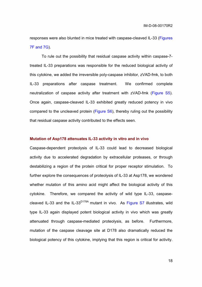
responses were also blunted in mice treated with caspase-cleaved IL-33 (Figures
7F and 7G).
To rule out the possibility that residual caspase activity within caspase-7-
treated IL-33 preparations was responsible for the reduced biological activity of
this cytokine, we added the irreversible poly-caspase inhibitor, zVAD-fmk, to both
IL-33 preparations after caspase treatment. We confirmed complete
neutralization of caspase activity after treatment with zVAD-fmk (Figure S5).
Once again, caspase-cleaved IL-33 exhibited greatly reduced potency in vivo
compared to the uncleaved protein (Figure S6), thereby ruling out the possibility
that residual caspase activity contributed to the effects seen.
Mutation of Asp178 attenuates IL-33 activity in vitro and in vivo
Caspase-dependent proteolysis of IL-33 could lead to decreased biological
activity due to accelerated degradation by extracellular proteases, or through
destabilizing a region of the protein critical for proper receptor stimulation. To
further explore the consequences of proteolysis of IL-33 at Asp178, we wondered
whether mutation of this amino acid might affect the biological activity of this
cytokine. Therefore, we compared the activity of wild type IL-33, caspase-
cleaved IL-33 and the IL-33D178A mutant in vivo. As Figure S7 illustrates, wild
type IL-33 again displayed potent biological activity in vivo which was greatly
attenuated through caspase-mediated proteolysis, as before. Furthermore,
mutation of the caspase cleavage site at D178 also dramatically reduced the
biological potency of this cytokine, implying that this region is critical for activity.

IM-D-08-00170R2
19
Using ST2-transfeced HEK293 cells, the IL-33D178A mutant was also found to be
inactive within the NFB reporter assay (Figure S7H).
Collectively, these data provide strong support for the idea that caspase-
3/-7 mediated cleavage of IL-33 diminishes, rather than increases, the biological
activity of this cytokine through destabilizing a region within IL-33 that is
important for ST2 receptor stimulation.
DISCUSSION
Here we have shown that IL-33 is efficiently cleaved by apoptosis-associated
caspases (caspases -3 and -7) but not inflammatory caspases –1, -4 or –5.
Caspases –3 and –7 cleave IL-33 at a motif (DGVD178 in human and DGVD175 in
mouse) that is fully conserved between the human and mouse forms of this
protein. Proteolysis of IL-33 was not required for ST2 receptor binding or ST2
receptor-dependent NFB activation. Caspase-dependent proteolysis of IL-33
attenuated ST2-dependent NFB activation and increased susceptibility of IL-33
to degradation by serum proteases. Furthermore, mutation of IL-33 at Asp178
abolished the biological activity of this cytokine, suggesting that proteolysis at this
site alters the conformation of a region within IL-33 that is critical for receptor
signalling.
Because caspases are activated during apoptosis but not necrosis, an
interesting implication of our experiments is that the half-life of IL-33 is reduced
during apoptosis. Consistent with this, IL-33 failed to undergo proteolytic
processing in necrotic cells (data not shown) but was readily cleaved during

IM-D-08-00170R2
20
apoptosis. Similar to IL-1 and IL-1, IL-33 does not possess a classical
secretory sequence and is therefore unlikely to be released from cells via the
classical ER-Golgi secretory pathway. Therefore, one possibility is that IL-33,
similar to the non-classical cytokine HMGB1 (Scaffidi et al., 2002), is released as
a consequence of necrosis. Because caspases do not become activated during
necrotic cell death (Kroemer and Martin, 2005), IL-33 is therefore likely to be
released from necrotic cells as a full-length active molecule. Caspase-dependent
proteolysis of IL-33 during apoptosis may therefore represent a means of
reducing the pro-inflammatory activity of this cytokine. Interestingly, it has been
demonstrated by several groups that apoptotic cells are much less pro-
inflammatory than necrotic cells and can even exhibit anti-inflammatory effects
that may dominate over necrotic cell-derived factors (Voll et al., 1997; Patel et al.,
2007). Thus, the proteolysis of IL-33 during apoptosis may contribute to the
damping down of the potentially pro-inflammatory effects of cell death.
Furthermore, because apoptotic cells are typically engulfed by phagocytes prior
to loss of plasma membrane integrity, this further reduces the possibility of
biologically active IL-33 being released from such cells. IL-33 may therefore
represent an endogenous ‘danger signal’ or ‘alarmin’ that is more potent when
released in the context of pathological cell death (necrosis) as opposed to
apoptosis which is more usually encountered in physiological settings.
IL-33 is a nuclear protein that has been reported to possess activity as a
regulator of transcription (Carriere et al., 2007). IL-1 also exhibits a nuclear
expression pattern and is reported to have intracellular activities (Maier et al.,

IM-D-08-00170R2
21
1994). Furthermore, pro-IL-1 is active as a full-length protein and is capable of
binding to the IL-1 receptor (Mosley et al., 1987). It is also suspected that the
major route of IL-1 release may be through necrosis. Thus, IL-33 and IL-1
share several features in common as both proteins are active as full-length
molecules but also undergo proteolytic processing under certain circumstances,
and both are released during necrosis.
In conclusion, here we have shown that IL-33 is active as a full-length
cytokine, similar to IL-1, and does not require proteolytic maturation by
inflammatory caspases for production of the biologically active cytokine.
However, IL-33 can be cleaved at physiological concentrations of caspase-3/-7,
which greatly attenuates the biological activity of this cytokine. Consistent with
this, IL-33 was processed at this cleavage motif within apoptotic but not necrotic
cells. Thus, contrary to the previous proposal that caspase-1 activates IL-33
(Schmitz et al., 2005), caspase-mediated proteolysis acts to suppress the pro-
inflammatory properties of this cytokine. Direct inactivation of a cytokine
represents a novel function for cell death-associated caspases and suggests that
caspases activated during apoptosis may actively disable molecules with pro-
inflammatory properties.

IM-D-08-00170R2
22
EXPERIMENTAL PROCEDURES
Reagents
Polyclonal antibodies were generated against hIL-33 by repeated immunization
of rabbits with the hIL-33 peptide 58CYFRRETTKRPSLKT72 (Sigma Genosys,
UK). Additional IL-33 antibodies were purchased from Alexis (UK) and R&D
systems (UK). Mouse anti-caspase-1 antibody was a kind gift from P.
Vandenabeele. Mouse IL-1 antibody was obtained from the National Institute
for Biological Standards and Control (UK). Antibodies specific to caspase-3,
caspase-7 and XIAP were obtained from BD (UK). Anti-human caspase-1
antibodies were from Santa Cruz (UK). Antibodies specific to caspase-4 and
caspase-5 were purchased from MBL (UK). Anti-IL-1 antibodies were from
R&D systems (UK), anti-caspase-9 monoclonal antibodies were from Oncogene
Research Products (UK). Anti-co-chaperone p23 antibodies were purchased
from Affinity Bioreagents (UK). Anti-actin antibody was purchased from ICN
(UK). Anti-GR-1-FITC antibody was purchased from ImmunoTools (Germany).
The peptides, z-YVAD-CHO, Ac-WEHD-AMC, Ac-DEVD-AFC zVAD-FMK were
all purchased from Bachem (UK). Unless otherwise indicated, all other reagents
were purchased from Sigma (Ireland) Ltd.
Primary cell culture
Bone marrow-derived DCs were generated from C57BL/6 mice as described
(Lavelle et al., 2001). WT, CASP-1-/-, CASP-3-/-, and CASP-7-/- MEFs were
derived from C57BL/6 mice.

IM-D-08-00170R2
23
Expression and purification of recombinant IL-33 and caspases
Recombinant GST-IL-33 and various His-tagged forms of IL-33 were expressed
and purified as described in the supplemental procedures. Recombinant poly-
histidine-tagged caspases –1, -4, -5, -3, and –7 were also expressed and purified
as described in the supplemental procedures.
Animals and in vivo treatment
C57BL/6 and Balb/c mice were obtained from Harlan U.K. Animal experiments
and maintenance were approved and regulated by the Trinity College Dublin
ethics committee and the Irish Department of Health.
Determination of Cytokine and IgA levels
Cytokines were detected by ELISA with paired antibodies for IL-4, IL-5, IL-6 (BD
Pharmingen, UK), IL-33 and IL-1 (R&D Systems, UK). IgA levels were
measured as described previously (Lavelle et al., 2001).
SUPPLEMENTAL DATA
The Supplemental Data include Supplemental Experimental Procedures and
seven figures and can be found with this article online at
http://www.immunity.com/supplemental/XXXXXX.
ACKNOWLEDGEMENTS

IM-D-08-00170R2
24
We thank Dr. Ken Yanagisawa for provision of ST2 expression plasmid and
Profs. Luke O’Neill and Kingston Mills for helpful discussions and for provision of
reagents. We thank Science Foundation Ireland (PI1/B038) and The Health
Research Board of Ireland (RP/2006/233) for their support of this work. S.J.M is
a Science Foundation Ireland Fellow. The authors have no conflicting financial
interests.

IM-D-08-00170R2
25
REFERENCES
Ali, S., Huber, M., Kollewe, C., Bischoff, S.C., Falk, W., Martin, M.U. (2007). IL-1receptor accessory protein is essential for IL-33-induced activation of T lymphocytes and mast cells. Proc Natl Acad Sci U S A 104, 18660-5.
Allakhverdi, Z., Smith, D.E., Comeau, M.R., and Delespesse, G. (2007). Cutting Edge: The ST2 ligand IL-33 potently activates and drives maturation of human mast cells. J Immunol 179, 2051-4.
Brint, E.K., Xu, D., Liu, H., Dunne, A., McKenzie, A.N., O'Neill, L.A., and Liew, F.Y. (2004). ST2 is an inhibitor of interleukin 1 receptor and Toll-like receptor 4 signaling and maintains endotoxin tolerance. Nat Immunol 5, 373-9.
Carriere, V., Roussel, L., Ortega, N., Lacorre, D.A., Americh, L., Aguilar, L., Bouche, G., and Girard, J.P. (2007). IL-33, the IL-1-like cytokine ligand for ST2 receptor, is a chromatin-associated nuclear factor in vivo. Proc Natl Acad Sci U S A 104, 282-7.
Chackerian, A.A., Oldham, E.R., Murphy, E.E., Schmitz, J., Pflanz, S., and Kastelein, R.A. (2007). IL-1 receptor accessory protein and ST2 comprise the IL-33 receptor complex. J Immunol 179, 2551-5.
Chow, S.C., Slee, E.A., MacFarlane, M., and Cohen, G.M. (1999). Caspase-1 is not involved in CD95/Fas-induced apoptosis in Jurkat T cells. Exp Cell Res246, 491-500.
Creagh, E.M., Conroy, H., and Martin, S.J. (2003). Caspase-activation pathways in apoptosis and immunity. Immunol Rev 193, 10-21.
Kroemer, G., Martin, S.J. (2005). Caspase-independent cell death. Nat Med. 11, 725-30
Lavelle, E. C., Grant, G., Pusztai, A., Pfuller, U., O'Hagan, D. T. (2001). The identification of plant lectins with mucosal adjuvant activity. Immunology 102, 77-86
Lohning, M., Stroehmann, A., Coyle, A.J., Grogan, J.L., Lin, S., Gutierrez-Ramos, J.C., Levinson, D., Radbruch, A., and Kamradt, T. (1998). T1/ST2 is preferentially expressed on murine Th2 cells, independent of interleukin 4, interleukin 5, and interleukin 10, and important for Th2 effector function. Proc Natl Acad Sci U S A 95, 6930-5.
Maier, J.A., Statuto, M., Ragnotti, G. (1994). Endogenous interleukin 1 alpha must be transported to the nucleus to exert its activity in human endothelial cells. Mol Cell Biol. 14, 1845-51.
Martinon, F., Burns, K., and Tschopp, J. (2002). The inflammasome: a molecular platform triggering activation of inflammatory caspases and processing of proIL-beta. Mol Cell 10, 417-26.
Martinon, F., and Tschopp, J. (2004). Inflammatory caspases: linking an intracellular innate immune system to autoinflammatory diseases. Cell 117, 561-74.
Meisel, C., Bonhagen, K., Lohning, M., Coyle, A.J., Gutierrez-Ramos, J.C., Radbruch, A., and Kamradt, T. (2001). Regulation and function of T1/ST2

IM-D-08-00170R2
26
expression on CD4+ T cells: induction of type 2 cytokine production by T1/ST2 cross-linking. J Immunol 166, 3143-50.
Mosley, B., Urdal, D.L., Prickett, K.S., Larsen, A., Cosman, D., Conlon, P.J., Gillis, S., Dower, S.K. (1987). The interleukin-1 receptor binds the human interleukin-1 alpha precursor but not the interleukin-1 beta precursor. J. Biol. Chem. 262, 2941-4.
Patel, V.A., Longacre-Antoni, A., Cvetanovic, M., Lee, D.J., Feng, L., Fan, H., Rauch, J., Ucker, D.S., and Levine, J.S. (2007). The affirmative response of the innate immune system to apoptotic cells. Autoimmunity 40, 274-80.
Pecaric-Petkovic, T., Didichenko, S.A., Kaempfer, S., Spiegl, N., and Dahinden, C.A. (2009) Human basophils and eosinophils are the direct target leukocytes of the novel IL-1 family member IL-33. Blood 113,:1526-34.
Scaffidi, P., Misteli, T., Bianchi, M.E. (2002). Release of chromatin protein HMGB1 by necrotic cells triggers inflammation. Nature 418, 191-5.
Schmitz, J., Owyang, A., Oldham, E., Song, Y., Murphy, E., McClanahan, T. K., Zurawski, G., Moshrefi, M., Qin, J., Li, X., et al. (2005). IL-33, an interleukin-1-like cytokine that signals via the IL-1 receptor-related protein ST2 and induces T helper type 2-associated cytokines. Immunity 23, 479-90.
Shechter, Y., Preciado-Patt, L., Schreiber, G., Fridkin, M. (2001). Prolonging the half-life of human interferon-alpha 2 in circulation: Design, preparation, and analysis of (2-sulfo-9-fluorenylmethoxycarbonyl)7-interferon-alpha 2. Proc Natl Acad Sci U S A. 98, 1212-7.
Voll, R.E., Herrmann, M., Roth, E.A., Stach, C., Kalden, J.R., and Girkontaite, I.(1997). Immunosuppressive effects of apoptotic cells. Nature 390, 350-351.
Walsh, J.G., Cullen, S.P., Sheridan, C., Lüthi, A.U., Gerner, C., and Martin, S.J. (2008) Executioner caspase-3 and caspase-7 are functionally distinct proteases. Proc Natl Acad Sci U S A. 105,12815-9.
Xu, D., Chan, W.L., Leung, B.P., Huang, F., Wheeler, R., Piedrafita, D., Robinson, J.H., and Liew, F.Y. (1998). Selective expression of a stable cell surface molecule on type 2 but not type 1 helper T cells. J Exp Med 187, 787-94.
Yamin, T.T., Ayala, J.M., and Miller, D.K. (1996). Activation of the native 45-kDa precursor form of interleukin-1-converting enzyme. J Biol Chem 271, 13273-82.

IM-D-08-00170R2
27
Figure Legends
Figure 1. Processing of IL-33 by apoptotic but not inflammatory caspases
(A) 35S-labeled hIL-33, mIL-33 and IL-1 were prepared by in vitro
transcription/translation and were then incubated with the indicated
concentrations of recombinant caspase-1, -4 and -5 for 2 h at 37°C followed by
analysis by SDS-PAGE and fluorography.
(B) Recombinant caspases –1, -3 and -7 were added to Jurkat cell-free extracts,
at the indicated concentrations, followed by incubation at 37°C for 2 h. Extracts
were then analysed by SDS-PAGE followed by immunoblotting for the indicated
substrate proteins.
(C) 35S-labeled hIL-33, mIL-33 and IL-1, prepared by in vitro
transcription/translation, were incubated with the indicated concentrations of
recombinant caspase-1, -3 and -7 for 2 h at 37°C followed by analysis by SDS-
PAGE/fluorography.
(D) Densitometric analysis of the gels presented in C. Scanned gels were
analysed using ImageJ software (http://rsb.info.nih.gov/ij/) and results are
expressed as % proteolysis of the full-length forms of each protein relative to the
untreated control.
Figure 2. Proteolysis of IL-33 at Asp178 occurs under conditions of
apoptotic but not inflammatory caspase activation
(A) Cell-free extracts derived from THP-1 cells were incubated at 37°C to permit
spontaneous activation of inflammatory caspases (‘Inflammasome’) or in the
presence of 50 µg/ml cytochrome c and 1 mM dATP to promote activation of
apoptotic caspases (‘Apoptosome’). As a control, caspase activation was
suppressed through addition of 5 µM YVAD-CHO. Extracts were then
immunoblotted for caspase-1, caspase-3 and IL-1, as indicated.
(B) 35S-labeled hIL-33, mIL-33 and IL-1 were added to THP-1 cell-free extracts
followed by treatment as described in A. Reactions were sampled at the
indicated times and were subsequently analysed by SDS-PAGE/fluorography.

IM-D-08-00170R2
28
(C) Schematic representation of human and murine IL-33 depicting potential
caspase cleavage motifs. Note that the proposed site of caspase-1-mediated
proteolysis (ALHD110; Schmitz et al., 2005) is not conserved between human and
mouse IL-33.
(D) 35S-labeled full length (FL) hIL-33 and the indicated IL-33 deletion mutants
were incubated in the presence of recombinant caspase-7 (40 nM) for 2 h at
37°C followed by analysis by SDS-PAGE/fluorography.
(E) Recombinant GST-IL-33 was incubated for 2 h at 37°C in the presence or
absence of recombinant caspase-7 (600 nM), as indicated, followed by SDS-
PAGE/Coomassie blue staining.
(F) 35S-labeled wild-type hIL-33 and IL-33D178A point mutant were incubated for 2
h at 37°C with recombinant caspase-3, -7 and –1, as shown. Reactions were
analysed by SDS-PAGE/fluorography.
(G) 35S-labeled wild-type hIL-33 and IL-33D178A point mutant were added to Jurkat
cell-free extracts followed by activation of apoptotic caspases by addition of
cytochrome c and dATP. Reactions were sampled at the indicated times and
were subsequently analysed by SDS-PAGE/fluorography. Samples of the same
reactions were also immunoblotted for caspase-3 and XIAP, as indicated.
(H) Recombinant IL-33112-270 and IL-33112-270 D178A point mutant were incubated
with recombinant caspase-7 (600 nM) for 4 h at 37°C followed by analysis by
SDS-PAGE/Coomassie blue staining.
Figure 3. IL-33 is cleaved during apoptosis
(A) HeLa cells were transfected with expression plasmids encoding either wild
type IL-33 (top panel), or IL-33D178A point mutant (bottom panel). 24 h later, cells
were then treated with Daunorubicin (Dauno; 5 M), TNF (10 ng/ml),
cycloheximide (CHX; 1 M) and cisplatin (50 µM) and incubated for a further 8 h
before assessment of apoptosis.
(B) Western blot analysis of cell lysates derived from HeLa cells transfected
either with wild type IL-33 (left panel) or the D178A point mutant (right panel),

IM-D-08-00170R2
29
followed by incubation in the presence or absence of Daunorubicin (Dauno),
TNF/cycloheximide, or Cisplatin at concentrations indicated in A.
(C) HeLa cells were transfected with an IL-33 expression plasmid for 24 h
followed by treatment for 8 h with Daunorubicin (5 µM) to induce apoptosis. In
parallel, HeLa cells were also treated with the poly-caspase inhibitor Z-VAD-fmk
(50 µM), or were transfected with a Bcl-xL expression plasmid as indicated.
(D) Cell lysates were generated from the cells treated in C and were
immunoblotted for the indicated proteins.
(E) Murine embryonic fibroblasts from wild type (WT), CASP-1 null, CASP-3 null,
or CASP-7 null mice were transfected with an expression plasmid encoding hIL-
33, followed by treatment for 24h with MG132 (0.2-0.4 µM) to facilitate IL-33
stabilization. Cells were then either left untreated, or were treated for 24-30 h
with Staurosporine (40-80 µM) or Etoposide (0.5 –1 mM) in order to achieve
equivalent levels of cell death, followed by preparation of cell lysates and
immunoblotting for the indicated proteins. The % of apoptotic cells in each
condition was assessed by direct cell counts on a minimum of 300 cells per
treatment using standard morphological criteria (bottom panel).
Figure 4. Endogenous IL-33 is upregulated in response to LPS and is
selectively released during necrosis
(A) THP-1 cells were either left untreated, or were treated with LPS (5 g/ml),
PMA (50 nM), TNF (50 ng/ml), IL-1(50 ng/ml), IL-1(50 ng/ml), IFN(250
ng/ml), or combinations of these agents, as indicated. 24 h later, lysates were
generated and immunoblotted for IL-33. Note that LPS/PMA induced strong
upregulation of endogenous IL-33.
(B) THP-1 cells were treated with PMA (50 nM) together with the indicated
concentrations of LPS. 24 h later, lysates were generated and immunoblotted for
the indicated proteins. As controls, in vitro transcribed/translated full length IL-
33, caspase-7-cleaved full length IL-33, and artificially truncated IL-33112-270 were
included to facilitate size comparison. Note that cleaved IL-33 was not detected
in LPS-stimulated cells.

IM-D-08-00170R2
30
(C) THP-1 cells were treated as in (B) then 24 h later, cells and medium were
harvested separately and cells were lysed in buffer containing 150 nM NaCl, 50
nM Tris pH 8, 1 % NP-40, 0.1 % SDS to facilitate measurment of cell-associated
cytokines. Cell-associated and secreted IL-33 and IL-1 levels were then
assessed by ELISA. Note that whereas IL-1 was secreted into the medium
under these conditions, IL-33 was not.
(D) Immunoprecipitation of endogenous IL-33. THP-1 cells (108) were treated
with LPS (5 µg/ml) and PMA (50 nM) for 24 h, followed by treatment with the
proteasome inhibitor MG132 (5 µM) for a further 24 h to stabilize endogenous IL-
33. Cells were lysed as in (C) then incubated overnight with Protein A/G
agarose-immobilized control or IL-33 antibody. Immune complexes were then
immunoblotted for IL-33 as indicated. The arrow indicates full-length IL-33 while
the asterix indicates antibody light chain used for the immunoprecipitation. As
controls, in vitro transcribed/translated full length IL-33 and caspase-7-cleaved
full length IL-33 were included to facilitate size comparison.
(E) THP-1 cells were treated with LPS (5 µg/ml) and PMA (50 nM) then 24 h
later, MG132 (5 µM) was added to stabilize endogenous IL-33. After a further 6
h, cells were treated with the apoptosis-inducing agents Actinomycin D (Act D; 5
µM), Daunorubicin (Dauno; 10 µM) Cisplatin (500 µM) or MG132 (100 µM) and
incubated for a further 36 h before assessment of apoptosis. The % of apoptotic
cells (counts were performed on a minimum of 300 cells per treatment) in each
condition is indicated at the bottom of the figure. Cell lysates were
immunoblotted for the indicated proteins.
(F) Upper panels, THP-1 cells were treated with LPS (5 µg/ml) and PMA (50 nM)
for 24 h. To induce apoptosis, cells were then treated with Act D (5 µM),
Daunorubicin (10 µM), Cisplatin (500 µM) or CHX (250 µM) for a further 12 h
before assessment of apoptosis by annexin V-FITC/ propidium iodide (PI)
staining and flow cytometry. Cells and medium were harvested separately and
cells were lysed as in (C). IL-33 and IL-1 in cell lysates and medium
were then assessed by ELISA. Lower panels, THP-1 cells were treated with
LPS/PMA for 24 h, as above. To induce necrosis, cells were then treated for 1 h

IM-D-08-00170R2
31
with Hydrogen Peroxide (H2O2; 20 mM), Sodium Azide (NaN2; 1 M), Steptolysin
O (SLO; 10 µg/ml) or a high dose of Daunorubicin (100 µM) followed by
assessment of necrosis using PI uptake in conjunction with flow cytometry. Cells
and medium were harvested separately and cells lysed as in (C). IL-33 and IL-
1 in the cell lysates and media were assessed by ELISA.
(G) Primary bone-marrow-derived mouse DCs were treated with 50 nM PMA,
either alone, or together with the indicated concentrations of LPS. 24 h later, cell
lysates were immunoblotted for the indicated proteins. As controls, in vitro
transcribed/translated full length IL-33 and caspase-7-cleaved full length IL-33
were included to facilitate size comparison.
(H) Primary bone-marrow-derived mouse DCs were treated with LPS/PMA as in
(G) then 24 h later, cells and medium were harvested separately and IL-33 and
IL-1 levels were measured by ELISA.
(I) Primary bone-marrow-derived mouse DCs were treated with LPS (5 µg/ml)
and PMA (50 nM) for 24 h, followed by treatment for a further 12 h with MG132
(0.2 µM) to stabilize IL-33 levels. Cells were then treated for 30 h with the
apoptosis-inducing agents Staurosporine (STS; 40 µM), TPCK (500 µM) or
MG132 (100 µM) before assessment of cell death by direct counts on a minimum
of 300 cells per treatment. Lysates were generated and immunoblotted for the
indicated proteins.
Figure 5. Caspase-mediated proteolysis of IL-33 attenuates the activity of
this cytokine in vitro
(A) Upper panel, HEK293T cells were transfected with a ST2L receptor
expression plasmid (200 ng per well of a 6 well plate) along with an NFB
luciferase reporter plasmid (10 ng). 24 h later, the indicated concentrations of full
length or caspase-7-cleaved GST-IL-33 were added for a further 8 h. Luciferase
activity was assayed, in triplicate, in cell lysates and normalised against empty
vector transfected cells. **p<0.01 by student’s t-test. Lower panel, the biological
activity of His-tagged recombinant IL-33112-270, or caspase-7-cleaved IL-33112-270
was assessed as above. **p<0.01 by student’s t-test.

IM-D-08-00170R2
32
(B) Schematic representation of IL-33 depicting the caspase cleavage site and
the various His-tagged IL-33 deletion mutants generated for this study.
(C) Cells were transfected as in A, followed by addition of the indicated molar
amounts of GST-IL-33, or the indicated deletion mutants of His-tagged IL-33, or
the control protein, PHAP. Cell lysates were assayed for luciferase activity 8 h
after addition of recombinant proteins. **p<0.01 by student’s t-test.
(D) Upper panel, capture of soluble ST2-Fc after incubation with sepharose-
immobilized GST, GST treated with caspase-7, GST-IL-33, or GST-IL-33 treated
with caspase-7, followed by probing for ST2. Lower panel, cleavage status of the
IL-33 used for the pulldown assay was revealed by immunoblotting. Note that
ST2-Fc was pulled down with the full length as well as the cleaved form of IL-33
(upper panel).
(E) Protein A/G immobilized ST2-Fc was used to assess binding of GST, GST-IL-
33 full-length or cleaved GST-IL-33. Note that both full-length as well as the
cleaved forms of IL-33 were captured by ST2 whereas the GST control was not.
(F) Recombinant GST, GST-IL-33 or caspase-7 cleaved GST-IL-33 (all at
125nM) were incubated for 1 h with HEK293 cells stably expressing the ST2L
receptor. Cells were then surface immunostained with anti-GST or anti-IL-33
antibodies where appropriate. Binding of IL-33 was detected using flow
cytometry analysis.
(G) HEK293 cells expressing the ST2L receptor were incubated with the
indicated concentrations of recombinant GST, GST-IL-33 or caspase-7 cleaved
GST-IL-33 for 1 h followed by immunostaining with anti-GST or anti-IL-33
antibodies where appropriate. Binding of IL-33 was detected using flow
cytometry analysis.
Figure 6. Caspase-dependent proteolysis of IL-33 increases susceptibility
to degradation by serum proteases
(A) Purified recombinant IL-33112-270, or caspase-cleaved IL-33112-270, were
incubated for 2 h at 37oC in the presence of the indicated concentrations of -
chymotrypsin, followed by analysis by SDS-PAGE/Coomassie blue staining.

IM-D-08-00170R2
33
(B) Purified recombinant IL-33112-270, or caspase-cleaved IL-33112-270, were
incubated for the indicated times at 37oC with -chymotrypsin (1 g/ml) followed
by analysis by SDS-PAGE/Coomassie blue staining. Histograms represent the
relative intensities of each IL-33 species normalized to the 0 h time point. Gels
were quantitated using Image-J software.
(C) Purified recombinant IL-33112-270 and caspase-cleaved IL-33112-270 were
incubated for 2 h at 37oC in the presence of the indicated concentrations of
, followed by analysis of cleavage reactions by SDS-
PAGE/Coomassie blue staining.
(D) Purified recombinant IL-33112-270, or caspase-cleaved IL-33112-270, were
incubated at 37oC with (25 g/ml) for the indicated times
followed by analysis by SDS-PAGE/Coomassie blue staining. Histograms
represent the relative intensities of each IL-33 species normalized to the 0 h time
point. Gels were quantitated using Image-J software.

IM-D-08-00170R2
34
Figure 7. Cleaved IL-33 displays diminished biological activity in vivo
C57BL/6 mice (5 per treatment group) were injected (i.p.) either with PBS, IL-
33112-270 (1 µg per mouse per day), or caspase-cleaved IL-33112-270 (1 µg per
mouse per day) or for 6 consecutive days. Note that the artificially-truncated IL-
33 was used here due to problems associated with purification of large quantities
of full length IL-33.
(A) Spleen size, weight and cellularity for each group of mice are shown.
Photographs show representative spleens for two mice per group. **p<0.01 by
student’s t-test.
(B) Peritoneal lavage-derived cells were enumerated by haemocytometer and
cytospins were also made. Cytospins were stained with hematoxylin and eosin
for assessment of cell morphology, arrows indicate granulocytes (top panels).
Numbers of individual cell types were assessed by cytospin analysis and are
expressed relative to the total number of cells found in the peritoneal lavages.
Granulocyte numbers were also determined by FSC/SSC analysis. **p<0.01 by
student’s t-test.
(C) Peripheral bloods were treated with FACS lysis solution to eliminate RBCs
followed by analysis by flow cytometry. Granulocyte numbers were scored
based on their high FSC/SSC properties, as shown. Eosinophil numbers were
determined by counting H&E-stained cytospin preparations of peripheral bloods.
(D) Spleen-derived granulocytes were enumerated as described in (C) and
neutrophil and eosinophil numbers were scored on H&E-stained cytospin
preparations. *p<0.05, **p<0.01 by student’s t-test.
(E) IL-4, IL-5 and IgA levels were determined by ELISA in plasma samples or
lung homogenates. Note that lung data are expressed per mg protein. *p<0.05,
**p<0.01 by student’s t-test.
Splenocytes (F) and mesenteric lymph node cells (G) (106 cells/ml) were
restimulated either with medium, 1 µg/ml anti-CD3, 1µg/ml anti-CD3 and 1 µg/ml
anti-CD28, or 1 µg/ml anti-CD3 and 20 ng/ml PMA, as indicated. Supernatants
were collected after 3 days and IL-5 concentrations were determined by ELISA.

Fig
ure
1
BA
35S
-hIL
-33
35S
-mIL
-33
35S
-IL
-1β
47.5 25
32.5
32.5
16.525
Con
c (n
M)
Mw
(kD
a)
16.5
32.5
16.525 6.5
6.5
XIA
P
Rh
o G
DI2
p23
0.62
5
1.25
20
0
2.5
5
10
Cas
pas
e-1
Cas
pas
e-3
Cas
pas
e-7
3.25
7.5
120
0
15
30
60
3.25
7.5
120
0
15
30
60
Mw
(kD
a)47
.5 6225
47.5
Con
c (n
M)
16.5
32.5
Mw
(kD
a)32
.5
C
0.62
5
1.25
20
0
2.5
5
10
Cas
pas
e-1
Cas
pas
e-3
Cas
pas
e-7
35S
-hIL
-33
35S
-mIL
-33
35S
-IL
-1β
6.5
16.525
32.5
16.525
16.525 6.5
32.5
3.25
7.5
120
015
3060
3.25
7.5
120
015
3060
Con
c (n
M)
D
Cas
pas
e-1
(nM
)
100 80 60 40 20 0
Proteolysis (%)
00.
631.
255
2.5
Cas
pas
e-3
(nM
)C
asp
ase-
7 (n
M)
030
6012
020
107.
515
3.25
hIL
-33
mIL
-33
IL-1β
200
2.5
510
200
2.5
510
200
2.5
510
Cas
pas
e-5
Cas
pas
e-4
Cas
pas
e-1
030
6012
07.
515
3.25
100 80 60 40 20 0
100 80 60 40 20 0
6.5
47.5
[E] Figure

Figure 2
B Control Inflammasome Apoptosome
35S-hIL-33
35S-mIL-33
35S-IL-1β
IL-1β
Casp-1
Casp-3
120
A
Time (min): 300 60 120300 60 90 120300 60 90
Control Inflammasome Apoptosome120Time (min): 300 60 120300 60 90 120300 60 90
32.5
47.5Mw (kDa)
16.56.5
32.5
25
16.5
32.5
25
16.5
32.5
47.5Mw (kDa)
16.5
6.5
32.5
25
16.5
32.5
25
16.5
6.5
hIL-33
mIL-33
TYND(131
)
DGVD(178
)
GVKD(244
)
ALHD(1
10)
C
D+
32.5
25
16.5
6.5
- +- +-Caspase-7:62
47.5
Caspase-7:
32.5
25
16.5
6.5
+-
- + - +Caspase-7:IL
-33W
T
IL-3
3D17
8A
F Caspase-3
35S-hIL-33
35S-hIL-33D178A
Caspase-7 Caspase-1
0 60 30 15 0 60 30 15 0 60 30 15Conc (nM)
35S-hIL-1β
32.5
16.5
25
6.5
32.5
16.5
25
6.5
32.5
16.5
25
6.5
Casp-3
XIAP
G
35S-hIL-33
35S-hIL-33D178A
Control ApoptosomeTime (min): 12030 60 900 1512030 60 900 15Mw (kDa)
32.5
16.5
25
6.5
32.5
16.5
25
6.5
47.5
32.5
16.5
25
47.5
32.5
6247.5
FL 1-17
8
179-
270
25
16.5
6.5
HE
(245
)(1
75)
(138
)
Mw (kDa)
Mw (kDa)Mw (kDa) ’
’
’

Figure 3
A
0
20
40
60
80
100
Untreat
ed
Dauno
TNF + C
HX
Cispla
tin
0
20
40
60
80
100
Cel
l dea
th (
%)
IL-33 wt
IL-33 D178A
0
20
40
60
80
100
+ + + +IL-33- - + -Bcl-x
L - - - +zVAD
Daunorubicin
- + + +Dauno
C
IL-33
p23
XIAP
Cispla
tin
Untreat
ed
Dauno
TNF + C
HX
Untreat
ed
Dauno
TNF + C
HX
Cispla
tin
IL-33wt IL-33D178A
Casp-3
Actin
B
Mw (kDa)32.5
16.5
25
25
62
32.5
62
IL-33
Casp-3
p23
XIAP
Actin
D
32.5
16.5
25
25
47.5
25
47.5
32.5
16.5
+ + + +IL-33- - + -Bcl-x
L - - - +zVAD- + + +Dauno
Cel
l dea
th (
%)
Cel
l dea
th (
%)
Mw (kDa)
32.5
E
0
20
40
60
80
100
Untreated STS Etoposide
MEF Killing assayMEFs transfected with IL-33 for 36 h then treated with 40 uM STS (80 uM for Casp-7 KO)
or 500 uM Etoposide (1 mM for Casp-7 KO) for 24 h (Casp-1 KO and Casp-7 KO) or 28 h (Casp-1 WT and Casp-3 KO). SC 22.1.9
MEF WT
Casp-1 KO
Casp-3 KO
Casp-7 KO
Casp-7-/-
MEFCasp-3-/-
MEFCasp-1-/-
MEFWT MEF
Untreated STS Etop0
20
40
60
80
100
Cel
l dea
th (
%)
Untreat
ed
STSEto
pUntre
ated
STSEto
pUntre
ated
STSEto
pUntre
ated
STSEto
pIL
-33
ITT
IL-3
3 IT
T + C
-7
Mw (kDa)
32.5
IL-33 25
16.5
6.5
32.5
25
25
Casp-1
Casp-3
Casp-7
p23
Rho GDI
Actin
47.5
32.5
25
16.5
25
62.5

Figure 4
0
20
40
60
80
100
Untreated
H2O2 NaN3SLO
Dauno
Assay for IL-33 distribution after necrosis in Dendritic Cells2 ml THP-1 cells at 10^6/ml were treated with the 5 ug/ml LPS + 50 nM
PMA for 24 h then treated with the indicated necrosis-inducing agents for 1 h in a vol of 0.5 ml. Medium and cells were
seperated and cells lysed in 0.5 ml before ELISA; SC 11.12.8
0
50
100
150
Untreated
H2O2 NaN3 SLODauno
IL-33 ELISA; IL-33 distribution after necrosis in THP-12 ml THP-1 cells at 10^6/ml were treated with the 5 ug/ml LPS + 50 nM
PMA for 24 h then treated with the indicated necrosis-inducing agents for 1 h in a vol of 0.5 ml. Medium and cells were
seperated and cells lysed in 0.5 ml before ELISA; SC 5.11.08
Cell-associated
Medium
0
20
40
60
80
Untreated
Act D
Daunorubicin
Cisplatin
CHX
IL-33 ELISA; IL-33 distribution after apoptosis of THP-12 ml THP-1 cells at 10^6/ml were treated with the 5 ug/ml LPS + 50 nM
PMA for 24 h then treated with the indicated drugs for 20 h in a vol of 0.5 ml. Medium and cells were
seperated and cells lysed in 0.5 ml before FACS analysis; SC 12.2.08
0
50
100
150
Untreated
H2O2 NaN3 SLODauno
IL-33 ELISA; IL-33 distribution after necrosis in THP-12 ml THP-1 cells at 10^6/ml were treated with the 5 ug/ml LPS + 50 nM
PMA for 24 h then treated with the indicated necrosis-inducing agents for 1 h in a vol of 0.5 ml. Medium and cells were
seperated and cells lysed in 0.5 ml before ELISA; SC 5.11.08
Cell-associated
Medium
0
50
100
150
Untreated
H2O2 NaN3 SLODauno
IL-33 ELISA; IL-33 distribution after necrosis in THP-12 ml THP-1 cells at 10^6/ml were treated with the 5 ug/ml LPS + 50 nM
PMA for 24 h then treated with the indicated necrosis-inducing agents for 1 h in a vol of 0.5 ml. Medium and cells were
seperated and cells lysed in 0.5 ml before ELISA; SC 5.11.08
Cell-associated
Medium
A
0
50
100
150
0
50
100
150
1 2 3 4 5 6
C
D
F
G H
0
1000
2000
3000
4000
5000
6000
7000
8000
0 0.1 0.5 1 5 10
Data 22
Lysate
0
50
100
150
Untreated
H2O2 NaN3 SLODauno
IL-33 ELISA; IL-33 distribution after necrosis in THP-12 ml THP-1 cells at 10^6/ml were treated with the 5 ug/ml LPS + 50 nM
PMA for 24 h then treated with the indicated necrosis-inducing agents for 1 h in a vol of 0.5 ml. Medium and cells were
seperated and cells lysed in 0.5 ml before ELISA; SC 5.11.08
Cell-associated
Medium
Mw (kDa)
32.5
25
6.5
17.5
Untreat
ed
LPSPM
ALPS/P
MA
TNFαTNFα
+ IL
-1β
IL-1αTNFα
+ IL
-1α
IFNγ
IL-33
100
pg
/ml
150
50
0
100
pg
/ml
150
50
0101 50.50.10LPS (µg/ml)
101 50.50.10LPS (µg/ml)
101 50.50.10LPS (µg/ml)
101 50.50.10LPS (µg/ml)
pg
/ml
0
200
400
600
800
ng
/ml
0
2
4
6
8
Cispla
tin
MG13
2
Dauno
Act D
Untreat
ed
Mw (kDa)
32.525
6.516.5
32.5
32.547.5
6.516.5
25
Casp-3
Casp-7
p23
IL-33
Cispla
tinCHX
Dauno
Act D
Untreat
ed
Cispla
tinCHX
Dauno
Act D
Untreat
ed
Cispla
tinCHX
Dauno
Act D
Untreat
ed
AxV
/PI-
po
siti
ve c
ells
(%
)
20
40
60
80
0
100p
g/m
l
150
50
0
4
ng
/ml
6
2
0
6n
g/m
l9
3
0
12
100
pg
/ml
150
50
0
PI-
po
siti
ve c
ells
(%
)
20
40
60
80
0
100
Untreat
edH 2
O 2
NaN3
SLO
Dauno
Untreat
edH 2
O 2
NaN3
SLO
Dauno
Untreat
edH 2
O 2
NaN3
SLO
Dauno
MediumCell-associated
MediumCell-associated
MediumCell-associated
MediumCell-associated
0 IL-3
3
IL-3
3 +
C-7
15100
IL-1β
Casp-3
Casp-7
IL-33
Mw (kDa)32.5
25
6.5
25
I
Cell-associated
Cell-associated
Medium
Medium
32.5
32.5
IL-3
3IL
-33
+ C-7
Untreat
ed
STSTPCK
MG13
2
IL-3
3IL
-33
+ C-7
Mw (kDa)32.5
25
6.517.5
Casp-3
IL-33
Actin
47.5
32.5
62.5
Actin
Casp-1
47
32.5
Actin
25 Casp-7
Rho GDI
47.5
32.5
25
p2325
Input (
2.5%
)
Control I
P
IL-3
3 IP
IL-3
3 (F
/L)
IL-3
3 +
C-7
IL-33 *IgL
THP-1 B
Mw (kDa)32.5
25
6.5
17.5
0
IL-33
10 5 1 0.5
0.1
IL-3
3 (1
-270
)
IL-3
3 +
Casp-7
IL-3
3 (1
12-2
70)
IL-1β
Casp-1
Actin
32.5
16.5
32.525
47.5
17.5
LPS (µg/ml)
THP-1
DC’s DC’s
E
THP-1
THP-1
IL-33 IL-33
IL-1β IL-1β
Cell Death (%) 5 90 96 94 93
Cell Death (%) 6 92 86 92
LPS (µg/ml)
IL-1β
IL-1β
IL-33
IL-33
IL-33 IL-33
IL-1β IL-1β
101 50.50.10LPS (µg/ml)
101 50.50.10LPS (µg/ml)
101 50.50.10LPS (µg/ml)
101 50.50.10LPS (µg/ml)
ng
/ml
0
2
4
6
8
ng
/ml
0
2
4
6
8
ng
/ml
0
10
20
30
40
50
ng
/ml
0
10
20
30
40
Cell-associated
Cell-associated
Medium
Medium
Apoptosis
Necrosis
Apoptosis Apoptosis
Necrosis Necrosis
200 8
200

Figure 5
A 4
2
1
0
3
2
1
0
C
D
E
ST2
IL-33GST
175
83
62
62
47.5
Mw (kD)
62
47.5
32.5
10% Input PulldownMr (kD)
0.5 0.21251020
Ligand concentration (nM)
GSTGST-IL-33Casp-7++ --
+ +- -+ + - -
GSTGST-IL-33Casp-7+--
+ +-+ - -
+--+ +-
+ - -
BIL-33
Caspase cleavage site’
270
270
270179
178112
112
1
-
++
10% Input ST2-Fc
Pulldown
0
1
2
3
NFκB
ind
uct
ion
(fo
ld m
ediu
m)
****** NFκB
ind
uct
ion
(fo
ld m
ediu
m)
**
**
**
**
** ****
NFκB
ind
uct
ion
(fo
ld m
ediu
m)
5 2102050100200
Ligand concentration (pM)
5 2102050 5 2102050 5 2102050 5 2102050 5 2102050
IL-33112-270
IL-331-270
IL-33112-178
IL-33179-270
PHAP I
nM
** ** **
**
**** ** **
** ****
**
3
****
****
**
**
F G 100
80
60
40
20
0No primary
antibody500 250 125 62.5
Mea
n F
luo
resc
ence
Inte
nsi
ty(a
rbit
ruar
y u
nit
s)
GSTGST-IL-33Cleaved GST-IL-33
Log IL-33 binding
Co
un
ts
GSTGST-IL-33Cleaved GST-IL-33
Untreated
Ligand concentration (nM)
150
60
90
120
30
010 10 101010
0 1 2 3 4

Figure 6
0 5 10 15 30 60 120 0 5 10 15 30 60 120
Proteinase K (25 ng/ml)
C
IL-33 (112-270) Cleaved IL-33 (112-270)
A
IL-33 (112-270) Cleaved IL-33 (112-270)
α-Chymotrypsin (1 µg/ml)
DB
0 5 10 15 30 60 120 0 5 10 15 30 60 120Time (min):
Proteinase Kα-Chymotrypsin
IL-33 (112-270) Cleaved IL-33 (112-270)IL-33 (112-270) Cleaved IL-33 (112-270)
16.5
25
6.5
16.5
25
6.5
16.5
25
6.5
16.5
25
6.5
Duration of α-Chymotrypsin treatment (min)
Duration of Proteinase K treatment (min)
Time (min):
Mr (kD) Mr (kD)
100
0
40
80
20
60
Rel
ativ
e in
tens
ity (
%)
120
100
0
40
80
20
60
Rel
ativ
e in
tens
ity (
%)
140
120
0 5 10 15 30 60 1200 5 10 15 30 60 120
Conc (µg/ml) Conc (ng/ml)4 0 0.25
2 1 0.5
4 0 0.25
2 1 0.5
0 100
50 25 12.5
6.25
0 100
50 25 12.5
6.25

IL-5
(ng
/ m
l)
pg /
mg
µg
/ mg
Figure 7
BA
C
D
0
PB
S
IL-3
3C
leav
ed
IL-3
340
30
20
10
Granulocytes
% e
vent
s
sple
en w
eigh
t (m
g)
0
8
6
4
2
Eosinophils
% c
ells
0
40
30
20
10
Granulocytes
% e
vent
s
0
8
6
4
2
Eosinophils
% c
ells
0
40
30
20
10
0
IgA
40
30
20
10
PB
S
IL-3
3C
leav
ed
IL-3
3
PB
S
IL-3
3C
leav
ed
IL-3
3
PB
S
IL-3
3
Cle
aved
IL
-33
PB
S
IL-3
3
Cle
aved
IL
-33
PB
S
IL-3
3
Cle
aved
IL
-33
8
0
6
4
Neutrophils
% c
ells
PB
S
IL-3
3C
leav
ed
IL-3
3
200
100
0
PBS
IL-33
Cleaved IL-33
pg /
mg
0
Lung
3
2
1
4
E
cells
per
spl
een
(x1
06 )
PB
S
IL-3
3C
leav
ed
IL-3
3
1
0
2
3
Serum
IL-4
IL-5IL-5
IgA
IL-4
G
Splenocytes
ng /
ml
mg
/ ml
0
5
4
3
2
1
0
2.5
2
1.5
1
pg /
ml
0
30
20
10
0.5
PB
S
IL-3
3
Cle
aved
IL
-33
0
4
2
6
3
1
IL-5
ng
/ ml
0
4
2
3
5
Peritoneal lavage
Total cells6
1
5
4
3
2
cells
per
ml (
x106 )
0
PB
S
IL-3
3C
leav
ed
IL-3
3
Spleen
Mesenteric Lymphnode
2
F
Peripheral Blood
14.6%
26.1%
21.0%
5
MediumαCD3αCD3 + αCD28αCD3 + PMA
FSC
SS
C
1000
800
600
400
200
1000
800
600
400
200
1000
800
600
400
200
0
0
0
10008006004002000
10008006004002000
10008006004002000
PBS IL-33 Cleaved IL-33
PBS
IL-33
CleavedIL-33
Splenocytes
50
PBS IL-33 Cleaved IL-33
MediumαCD3αCD3 + αCD28αCD3 + PMA
1
6
PBS IL-33 Cleaved IL-33
7Mesenteric lymph nodes
**
** *
**
*
****
**
**
*
1
3
2
cells
per
ml (
x106 )
0
Total cells100
0
80
60
40
20
Granulocytes
% e
vent
s
PB
S
IL-3
3C
leav
ed
IL-3
3
**LymphocytesMonocytes
EosinophilsNeutrophils

Supplemental DataSuppression of IL-33 Bioactivity through
Proteolysis by Apoptotic Caspases
Alexander U. Lüthi, Sean P. Cullen, Edel A. McNeela, Patrick J. Duriez, InnaS. Afonina, Clare Sheridan, Gabriela Brumatti, Rebecca C. Taylor, KristofKersse, Peter Vandenabeele, Ed C. Lavelle, and Seamus J. Martin
SUPPLEMENTAL FIGURES and METHODS
Supplemental Figure S1
Figure S1. Hydrolysis of a synthetic caspase substrate peptide byrecombinant caspases –1, -4 and -5Hydrolysis of the synthetic caspase substrate, WEHD-AMC, by recombinantcaspase-1, -4 and –5 (20nM each). Note that recombinant inflammatorycaspases cleave WEHD-AMC with different efficiencies. Active site titrations withzVAD-fmk confirmed that the molar amounts of each caspase were identical(data not shown).
[F] Supplemental Text and FiguresClick here to download [F] Supplemental Text and Figures: Supplemental Data_200dpi.pdf

Supplemental Figure S2
Supplemental Figure S2. Proteolysis of IL-33 in apoptotic Jurkat cell-freeextracts(A) Cell-free extracts derived from Jurkat cells were incubated at 37°C in thepresence (Apoptosome) or absence (Control) of cytochrome c/dATP, asindicated, followed by immunoblotting for caspase-3, caspase-9, XIAP or p23.(B) 35S-labeled hIL-33 or mIL-33 were added to Jurkat cell-free extracts whichwere treated as described in Figure 2B. Reactions were sampled at theindicated times and were subsequently analysed by SDS-PAGE followed byfluorography.

Supplemental Figure S3
Figure S3. Mass spectrometry analysis of caspase cleaved IL-33Recombinant GST-IL-33 was cleaved by caspase-7, as depicted in Figure 2E,followed by analysis of the cleavage products by MALDI-TOF massspectrometry. Mass spectrograms for each IL-33 species (i.e. full length, largeand small fragments) are shown, along with the corresponding peptide coverageof each. The peptide coverage (highlighted in bold) of each IL-33 speciesindicates that the site of caspase-7-mediated proteolysis lies between residues159 and 187.

Supplemental Figure S4

Figure S4. Proteolytic processing of IL-33 during apoptosis induced bydeath receptor ligands(A) THP-1 cells were treated with LPS (5 µg/ml) and PMA (50 nM) for 20 h toinduce expression of endogenous IL-33, MG132 (2 µM) was then added tostabilize IL-33. After a further 6 h, cells were treated with anti-Fas CH11 IgMantibody (2.5 µg/ml), or TNF (500 ng/ml) plus act D (300 nM), to induceapoptosis. 30 h after addition of pro-apoptotic stimuli, apoptosis was quantitatedby direct cell counts using standard morphological criteria (counts wereperformed on triplicate fields of a minimum of 100 cells per treatment). Celllysates were then generated and immunoblotted for the indicated proteins. Ascontrols, in vitro transcribed/translated full length IL-33 and caspase-7-cleavedfull length IL-33 were included to facilitate size comparison.(B) HeLa cells were transfected with 1 µg of expression plasmid encoding humanFLAG epitope-tagged IL-33. 24 h later, MG132 (1 µM) was added to stabilize IL-33, followed by treatment with with anti-Fas CH11 IgM antibody (0.5 µg/ml), orTNF (200 ng/ml) plus act D (300 nM), for 12 h to induce apoptosis. Apoptosiswas quantitated by direct cell counts using standard morphological criteria(counts were performed on triplicate fields of a minimum of 100 cells pertreatment). Cell lysates were then generated and immunoblotted for theindicated proteins. As controls, in vitro transcribed/translated full length IL-33 andcaspase-7-cleaved full length IL-33 were included to facilitate size comparison.(C) HeLa cells were transfected with 1 µg of expression plasmid encoding humanFLAG epitope-tagged IL-33. 24 h later, MG132 (1 µM) was added to stabilize IL-33, followed by treatment with with anti-Fas CH11 IgM antibody (0.5 µg/ml),TRAIL (100 ng/ml) plus act D (300 nM), or TNF (200 ng/ml) plus act D (300 nM),for 12 h to induce apoptosis. Apoptosis was quantitated by direct cell countsusing standard morphological criteria (counts were performed on triplicate fieldsof a minimum of 100 cells per treatment). Cell lysates were then generated andimmunoblotted for the indicated proteins. As controls, in vitrotranscribed/translated full length IL-33 and caspase-7-cleaved full length IL-33were included to facilitate size comparison.

Supplemental Figure S5
Figure S5. Elimination of residual caspase activity in caspase-treated IL-33preparations after treatment with an irreversible caspase inhibitor.Purified recombinant IL-33 was incubated with 300 nM caspase-7 at 37°C for 4hours then 30 minutes with 2 µM zVAD-fmk on ice. IL-33 pre- (open circles),post- caspase (closed circles) and post zVAD-fmk (filled triangles) addition wasassayed for DEVDase activity.

Supplemental Figure S6

Figure S6. Attenuation of IL-33 bioactivity through caspase-dependentproteolysis.To ensure that residual caspase activity in caspase-treated IL-33 preparationsdid not contribute to the reduced biological effects seen, recombinant IL-33 wastreated for 4 h with either heat-inactivated caspase-7 (300 nM), or activecaspase-7 (300 nM), followed by addition of 2 µM of the irreversible caspaseinhibitor, zVAD-fmk, to both IL-33 preparations to eliminate any residual caspaseactivity. Quenching of caspase activity was confirmed by assessing hydrolysis ofthe synthetic caspase substrate, DEVD-AMC, as shown in Figure S4. BALB/cmice (n=4 per group) were then injected (i.p.) with either PBS, or zVAD-fmk-treated IL-33 (0.8 µg per day per mouse), or zVAD-fmk-treated cleaved IL-33 (0.8µg per mouse), for 5 consecutive days.(A) Spleen size, weight and cellularity for each group of mice are shown.(B) Peritoneal lavage-derived cells were enumerated from cytospins as describedin Fig 6B. Percentages of individual cell types were multiplied by the totalnumber of cells (bottom middle panel) and granulocyte numbers were alsodetermined by FSC/SSC analysis.(C) Peripheral bloods were analysed as described in Fig 6C.(D) Spleen-derived granulocytes were enumerated as described in Fig 6C andneutrophil and eosinophil numbers were scored on H&E-stained cytospinpreparations.(E) IL-5, IL-6 and IgA levels were determined by ELISA in plasma samples orlung homogenates, as indicated.Splenocytes (F) and mesenteric lymph node cells (G) (106 cells/ml) wererestimulated either with medium, 1 µg/ml anti-CD3, 1µg/ml anti-CD3 and 1 µg/mlanti-CD28, or 1 µg/ml anti-CD3 and 20 ng/ml PMA, as indicated. Supernatantswere collected after 3 days and IL-5 concentrations were determined by ELISA.

Supplemental Figure S7

Figure S7. Mutation of the caspase cleavage site Asp178 is sufficient toabolish IL-33 biological activity in vivo.BALB/c mice (4 per treatment group) were injected (i.p.) either with PBS, IL-33112-270, caspase-cleaved IL-33112-270, IL-33112-270 D178A mutant, or caspase-7-treated IL-33112-270 D178A mutant. All IL-33 treatment groups received 0.8 µg permouse per day for 5 consecutive days.(A) Spleen size, weight and cellularity for each group of mice are shown.(B) Peritoneal lavage-derived cells were enumerated by haemocytometercounting. Granulocyte numbers were determined by FSC/SSC analysis.(C) Peripheral bloods were treated with FACS lysis solution to eliminate RBCsfollowed by analysis by flow cytometry. Granulocyte numbers were scoredbased on their high FSC/SSC properties, as shown. Eosinophil numbers weredetermined by counting H&E-stained cytospin preparations of peripheral bloods.(D) Spleen-derived granulocytes were enumerated by flow cytometry asdescribed in (C), and neutrophil and eosinophil numbers were scored on H&E-stained cytospin preparations.Inguinal lymph node-derived lymphocyte (E), splenocytes (F), and mesentericlymph node-derived lymphocytes (G), were restimulated either with medium, 1µg/ml anti-CD3, 1µg/ml anti-CD3 and 1 µg/ml anti-CD28, or 1 µg/ml anti-CD3 and20 ng/ml PMA, as indicated. Supernatants were collected after 3 days and IL-5concentration was determined by ELISA.(H) The indicated amounts of untreated or caspase-treated IL33wt or IL33D178A
mutant protein was added to the HEK ST2-NFκB reporter cell line as describedin Fig 5A.

SUPPLEMENTAL METHODS
Cell-free reactionsCell-free extracts were generated from exponentially growing healthy THP-1 cellsas described previously (Murphy et al., 2003; Slee et al., 1999). Briefly, THP-1cells were treated for 5 hours with 1 µg/ml LPS then harvested by centrifugationat 800 x g into a Dounce-type homogenizer. Three volumes of ice-cold cellextract buffer were added CEB (20 mM Hepes, pH 7.5, 10 mM KCl, 1.5 mMMgCl2, 1 mM EDTA, 1 mM EGTA, 1mM DTT, 100 µM PMSF, 10 µg/ml leupeptin,2 µg/ml aprotinin), and the cells were allowed to swell for 15–20 min on ice. Cellswere then lysed by homogenization with 10–15 strokes of a B-type pestle.Lysates were clarified by centrifugation at 15,000 x g for 30 min to remove nuclei,mitochondria, and other cellular debris. Extracts were then aliquoted and frozenat -70ºC prior to use. For in vitro activation of caspases involved in inflammation,THP-1 cell-free extracts were diluted to 80% in CEB and incubated at 37ºC for 2hours. To provoke apoptosome-dependent caspase activation, bovine heartcytochrome c and dATP were added to reactions to final concentrations of 50µg/ml and 1 mM, respectively.
Expression and purification of recombinant caspasesCaspase-1.p30, Caspase-4.p30, Caspase-5.p30, Caspase-3 and Caspase-7were produced by PCR-mediated amplification of the relevant coding sequencesfrom the respective full-length cDNA, followed by subcloning of the resulting PCRproducts in-frame with the His coding region of pET15b (Novagen, UK) forCaspase-1, pet23b (Novagen, UK) for Caspase-3 and -7, or pGEX4T1(Amersham, UK) for Caspase-4 and –5. Plasmids encoding His-tagged andwere transformed into Escherichia coli DH5α, GST-tagged fusion proteins weretransformed into BL21pLysS and bacteria were induced to express therecombinant proteins in the presence of 0.1 to 0.6 mM IPTG (Melford, UK).Recombinant caspases were subsequently purified using Ni2+ beads (Qiagen,UK) or Glutathione Sepharose 4B (Amersham, UK) according to standardprocedures.
Expression and purification of recombinant IL-33GST-IL-33 was generated by inserting the human IL-33 coding sequence into thepGEX4T2 bacterial expression vector. GST-IL-33 was expressed by addition of100 µM IPTG to exponentially growing cultures of DH5α strain E. coli followed byincubation for 1 h at room temperature. Bacteria were lysed by sonication andGST-IL-33 was captured using Glutathione Sepharose 4B, followed by elutioninto PBS, pH 7.2, in the presence of 20 mM reduced gluthathione. Protein wasthen extensively dialysed against PBS prior to use. His-tagged IL-33 deletionmutants were generated through inserting the relevant coding sequences intopET45b (Novagen, UK), followed by expression in BL21pLysS strain E. coli andwere purified using Ni2+ beads (Qiagen, UK) according to standard procedures.Proteins were then eluted from the affinity capture matrix and were extensively

dialysed against PBS, pH 7.2, followed by depletion of LPS by several rounds ofincubation to agarose-immobilized polymyxin B (Sigma). For caspase-mediatedprocessing, recombinant IL-33 was incubated with recombinant caspase-7(300nM) for 4 h at 37ºC, or with an identical concentration of heat-inactivatedcaspase-7 as a control.
Transient transfection and reporter gene assaysHeLa cells were seeded at a density of 2 x 10⁵ cells per well of a 6-well tissueculture plate and were transiently transfected 24 h later with GeneJuice (Merck,Ireland), according to the manufacturer's instructions. HEK293T cells wereseeded at a density of 4 x 10⁵ cells per well of a 6-well tissue culture plate 24 hprior to transfection. Cells were transfected with plasmids according to thestandard calcium phosphate precipitation method, and DNA complexes wereallowed to remain on cells for 14 h before replacing with fresh medium. For theluciferase reporter assay the cells were lysed in 200 µl RLS (100 mM HEPES, pH8, 2 mM MgCl2, 2% Triton X-100) of which 20 µl were assayed with 50 µl LAR(20mM N-Glycylglycine, 1mM MgCl2, 100 µM EDTA, 27.8 µg/ml ATP, 21.3 µg/mlCoenzyme A and 160 µg/ml beetle luciferin (Promega, UK). Luminescence wasmeasured in a Spectrafluor Plus (TECAN, UK).
Pulldown and immunprecipitation assaysGST-IL-33 (1 µg) was immobilised on 30 µl Glutathione Sepharose 4B(Amersham Biosciences, UK) by rotation at 4ºC for 30 minutes, followed byaddition of 1 µg of recombinant ST2.Fc (Alexis, UK) and further incubation for 4 hin 1 ml of reaction buffer (50 mM Tris, pH 7.6, 120 mM NaCl, 0.1% CHAPS). Thereciprocal pulldown experiments were performed under essentially the sameconditions.
For immunoprecipitation of endogenous IL-33, THP-1 cells (108) weretreated with LPS (5 µg/ml) and PMA (50 nM) for 24 h, followed by addition ofMG132 (5 µM) and incubation for a further 6 h to stabilize endogenous IL-33.Cells were then lysed in buffer containing 150 mM NaCl, 50 mM Tris pH 8, 1 %NP-40, 0.1 % SDS. The clarified supernatant was used for immunprecipitationby adding 3 µg of control or anti-IL-33 antibody and 30 µl A/G agarose (SantaCruz). After overnight incubation, the beads were washed extensively, andsamples were electrophoresed on 13 % SDS-PAGE followed by immunoblotting.
Caspase activity assaysFor the assessment of caspase activity, recombinant caspases -1, -3, -4 and –5and -7 were diluted to a final volume of 50 µl in CEB containing 50 µM Ac-WEHD-AMC (for the inflammatory caspases) or Ac-DEVD-AMC (for apoptoticcaspases). Samples were then measured over 20 minutes in an automatedfluorimeter (Spectrafluor Plus, TECAN, UK) at wavelengths of 360 nm(excitation) and 465 nm (emission). Caspases were active site titrated byincubation with a range of concentrations (0, 6.25, 12.5, 25, 50, 100 nM) of thepoly-caspase inhibitor zVAD-fmk for 30 min at 37ºC, followed by measurement of

residual caspase activity by monitoring the hydrolysis of WEHD-AMC or Ac-DEVD-AMC as described above.
Coupled in vitro transcription/translation reactionsIn vitro transcription/translation reactions were carried out using purified plasmidtemplates added to a rabbit reticulocyte lysate system (Promega, UK) with 35S-Methionine (Amersham, UK) as described previously (Slee et al., 1999).
Analysis of peripheral blood, spleen and peritoneal lavage samplesBlood was collected from the tail vein followed by addition of 100 µM EDTA as ananticoagulant. Red blood cells were lysed in 20 volumes of 150 mM NH4CL,10mM NaHCO3 and 100 µM EDTA. Cytospins were prepared and stained withhematoxylin/eosin and scored for lymphocytes, monocytes, neutrophils andeosinophils. For flow cytometry, cells were fixed in FACS lysis buffer (BD, UK)according to the manufacturer’s instructions and analysed on a FACScalibur (BD,UK).
IL-33-ST2 cell binding assayHEK293 cells stably over-expressing the ST2L receptor were incubated withGST, GST-IL-33 or caspase-7-cleaved GST-IL-33 at concentrations indicated inthe figure legends for 1 h followed by immunostaining with anti-GST or anti-IL-33antibodies. Binding of IL-33 was detected using flow cytometry analysis.
SUPPLEMENTAL REFERENCES
Murphy, B.M., O'Neill, A.J., Adrain, C., Watson, R.W., and Martin, S.J. (2003).The apoptosome pathway to caspase activation in primary human neutrophilsexhibits dramatically reduced requirements for cytochrome C. J. Exp. Med.197, 625-32.
Slee, E. A., Harte, M. T., Kluck, R. M., Wolf, B. B., Casiano, C. A., Newmeyer, D.D., Wang, H. G., Reed, J. C., Nicholson, D. W., Alnemri, E. S., et al. (1999).Ordering the cytochrome c-initiated caspase cascade: hierarchical activationof caspases-2, -3, -6, -7, -8, and -10 in a caspase-9-dependent manner. J CellBiol 144, 281-92.
Copyright © 2022 FDOKUMEN