Identification of Up-Regulated Low Molecular Weight Proteins in Human Adipocytes Treated with...
6
110 The Open Obesity Journal, 2010, 2, 110-115 1876-8237/10 2010 Bentham Open Open Access Identification of Up-Regulated Low Molecular Weight Proteins in Human Adipocytes Treated with Glycoxidized Albumin Nihar Ranjan Singh, Philippe Rondeau and Emmanuel Bourdon * Laboratoire de Biochimie et Génétique Moléculaire (LBGM), Groupe d'Etude sur l'Inflammation Chronique et l'Obésité (GEICO), Université de La Réunion, Saint Denis de La Réunion, France Abstract: Diabetes and Obesity are strongly associated with enhanced circulating advanced glycoxidation end products (AGEs), and adipose tissue constitutes a biological driver in the metabolic syndrome characterized by abdominal obesity, dyslipidemia and insulin resistance. We determined whether AGEs issued from glycoxidized albumin could induce expression of determinant proteins in human adipocyte cell lines (SW872). Cell proteins separation by 2D gels and mass spectrometry were combined to identify up-regulated proteins in SW872 treated with AGEs. Four low molecular weight proteins appeared as more expressed in adipocytes treated with glycated albumin than with native albumin. All four proteins have determinant role in adipocyte physiology as they exert antioxidant activities (superoxide dismutase and proteasome subunit) or energy production properties (phosphoglycerate mutase and triosephosphate isomerase). Proteomic analyses of SW872 cells have never been performed before. By using proteomic analyses, our results bring new evidence of AGEs-involvement in metabolic disorders at the adipocyte level. Keywords: Glycoxidation, albumin, adipocytes, oxidative stress, SW872, glycation, diabetes. 1. INTRODUCTION It is now well established that free radicals and reactive oxygen species (ROS) contribute to the development of several age-related diseases by inducing oxidative stress and oxidative damages [1]. The incidence of diabetes is inc- reasing, with a worldwide prevalence estimated to double by 2030, primarily because of sedentary lifestyle and obesity [2]. A strong link between obesity and diabetes is now well established with more than 70 percent of people with dia- betes being overweight. This disease, which is characterized by hyperglycemia and insulin resistance, is closely asso- ciated with severe complications. Indeed, diabetes increases the risk of developing cardiovascular disease, which repre- sents the leading cause of mortality in western countries [3]. Recently, a causal role was shown for ROS in multiple forms of insulin resistance, which was also associated with ele- vated formation of advanced glycation end products (AGEs) [4]. Adipocytes are known to express and secrete a variety of active molecules, so-called adipokines, which can regulate many biological processes such as insulin-sensitivity, appe- tite, immunity, reproduction. It is now well established that dysregulation of adipocyte functions by oxidative stress constitutes a key process leading to type 2 diabetes. It is thus not surprising that many diabetic therapies target adipose tissue (thiazolidinedione, physical activity, etc) [5,6]. SW872 cells constitute a liposarcoma cell line often used as a human adipocyte cell model [7-12]. Nonetheless, proteomic analy- ses of SW872 cells have never been performed before. In *Address correspondence to this author at the LBGM - Université de la Réunion - 15, avenue René Cassin – BP 7151 – Cedex 09, 97715 Saint Denis de La Réunion, France; Tel: (+262) 262 93 86 48; Fax: (+262) 262 93 82 37; E-mail: [email protected] this work, we determined whether AGEs issued from gly- coxidized albumin could induce expression of determinant proteins in human adipocyte cell lines (SW872). 2. MATERIALS AND METHODS In Vitro Glycoxidation of BSA Non defatted recombinant BSA (Sigma, cat # A2153) was dissolved in phosphate-buffered saline (PBS), pH 7.4, to yield a stock solution of 50 mg.mL -1 . This solution was prepared with 200 mM glucose solution in PBS, to form incubation mixtures of 50 mg.mL -1 BSA with 0 or 100 mM glucose. After being sterilized by filtration through 0.2 µm filters (Millipore), the solutions were incubated at 37°C for 50 days in capped vials. Reversible and unbound glucose were removed from BSA by extensive dialysis against PBS, pH 7.4. Albumin preparations were assessed for glucose content after dialyses and the later was below detectable levels. Samples were separated into aliquots and stored at – 80 °C in the dark prior to incubation with the SW872 cells. Endotoxin content was below detectable level (0.03 Endotoxate Unit/mL) as assessed with an in vitro toxicology assay kit (E-TOXATE, Sigma). Mass Spectrometry Studies of Native and Glycoxidized BSA Native BSA and glycoxidized BSA treated in the absence or the presence of 100 mM of glucose, were analyzed using mass spectrometry for molecular weight and extent of glyco- xidation determinations. ESI/MS was performed on Agilent 1100LC coupled to Bruker Esquire 3000plus operating in the positive ion mode as mentioned earlier [20].
-
Upload
ravenshawuniversity -
Category
Documents
-
view
2 -
download
0
Transcript of Identification of Up-Regulated Low Molecular Weight Proteins in Human Adipocytes Treated with...
110 The Open Obesity Journal, 2010, 2, 110-115
1876-8237/10 2010 Bentham Open
Open Access
Identification of Up-Regulated Low Molecular Weight Proteins in Human Adipocytes Treated with Glycoxidized Albumin
Nihar Ranjan Singh, Philippe Rondeau and Emmanuel Bourdon*
Laboratoire de Biochimie et Génétique Moléculaire (LBGM), Groupe d'Etude sur l'Inflammation Chronique et l'Obésité (GEICO), Université de La Réunion, Saint Denis de La Réunion, France
Abstract: Diabetes and Obesity are strongly associated with enhanced circulating advanced glycoxidation end products (AGEs), and adipose tissue constitutes a biological driver in the metabolic syndrome characterized by abdominal obesity, dyslipidemia and insulin resistance. We determined whether AGEs issued from glycoxidized albumin could induce expression of determinant proteins in human adipocyte cell lines (SW872).
Cell proteins separation by 2D gels and mass spectrometry were combined to identify up-regulated proteins in SW872 treated with AGEs. Four low molecular weight proteins appeared as more expressed in adipocytes treated with glycated albumin than with native albumin. All four proteins have determinant role in adipocyte physiology as they exert antioxidant activities (superoxide dismutase and proteasome subunit) or energy production properties (phosphoglycerate mutase and triosephosphate isomerase).
Proteomic analyses of SW872 cells have never been performed before. By using proteomic analyses, our results bring new evidence of AGEs-involvement in metabolic disorders at the adipocyte level.
Keywords: Glycoxidation, albumin, adipocytes, oxidative stress, SW872, glycation, diabetes.
1. INTRODUCTION
It is now well established that free radicals and reactive oxygen species (ROS) contribute to the development of several age-related diseases by inducing oxidative stress and oxidative damages [1]. The incidence of diabetes is inc-reasing, with a worldwide prevalence estimated to double by 2030, primarily because of sedentary lifestyle and obesity [2]. A strong link between obesity and diabetes is now well established with more than 70 percent of people with dia-betes being overweight. This disease, which is characterized by hyperglycemia and insulin resistance, is closely asso-ciated with severe complications. Indeed, diabetes increases the risk of developing cardiovascular disease, which repre-sents the leading cause of mortality in western countries [3]. Recently, a causal role was shown for ROS in multiple forms of insulin resistance, which was also associated with ele-vated formation of advanced glycation end products (AGEs) [4]. Adipocytes are known to express and secrete a variety of active molecules, so-called adipokines, which can regulate many biological processes such as insulin-sensitivity, appe-tite, immunity, reproduction. It is now well established that dysregulation of adipocyte functions by oxidative stress constitutes a key process leading to type 2 diabetes. It is thus not surprising that many diabetic therapies target adipose tissue (thiazolidinedione, physical activity, etc) [5,6]. SW872 cells constitute a liposarcoma cell line often used as a human adipocyte cell model [7-12]. Nonetheless, proteomic analy-ses of SW872 cells have never been performed before. In
*Address correspondence to this author at the LBGM - Université de la Réunion - 15, avenue René Cassin – BP 7151 – Cedex 09, 97715 Saint Denis de La Réunion, France; Tel: (+262) 262 93 86 48; Fax: (+262) 262 93 82 37; E-mail: [email protected]
this work, we determined whether AGEs issued from gly-coxidized albumin could induce expression of determinant proteins in human adipocyte cell lines (SW872).
2. MATERIALS AND METHODS
In Vitro Glycoxidation of BSA
Non defatted recombinant BSA (Sigma, cat # A2153) was dissolved in phosphate-buffered saline (PBS), pH 7.4, to yield a stock solution of 50 mg.mL-1. This solution was prepared with 200 mM glucose solution in PBS, to form incubation mixtures of 50 mg.mL-1 BSA with 0 or 100 mM glucose. After being sterilized by filtration through 0.2 µm filters (Millipore), the solutions were incubated at 37°C for 50 days in capped vials. Reversible and unbound glucose were removed from BSA by extensive dialysis against PBS, pH 7.4. Albumin preparations were assessed for glucose content after dialyses and the later was below detectable levels. Samples were separated into aliquots and stored at – 80 °C in the dark prior to incubation with the SW872 cells. Endotoxin content was below detectable level (0.03 Endotoxate Unit/mL) as assessed with an in vitro toxicology assay kit (E-TOXATE, Sigma).
Mass Spectrometry Studies of Native and Glycoxidized BSA
Native BSA and glycoxidized BSA treated in the absence or the presence of 100 mM of glucose, were analyzed using mass spectrometry for molecular weight and extent of glyco-xidation determinations. ESI/MS was performed on Agilent 1100LC coupled to Bruker Esquire 3000plus operating in the positive ion mode as mentioned earlier [20].
Glycoxidized Albumin Up-Regulates Adipocyte Proteins The Open Obesity Journal, 2010, Volume 2 111
Cell Culture
Human SW872 liposarcoma cells were purchased from American Type Culture Collection. Cells were seeded in six-multiwell plates and grown in Dulbecco’s modified Eagle medium (DMEM) containing 1.25% L-glutamine, 2% peni-cillin/streptomycin, and 10% fetal calf serum, in a humidi-fied incubator (5% CO2, 37°C). The culture plates were incubated until cells reach 75% confluency before adding BSAG0 or BSAG100 at a final concentration of 20 µM. Thereafter, cells were maintained in the humidified CO2 incubator for 16 h before further analyses.
Cell Lysate Preparation
After the incubation, the cells were washed twice with PBS. Then, cells were treated at 4°C during 30 min with 100 µL of lysis buffer containing 25 mM Tris-HCl, pH 8.3, 10 mM KCl, 1 mM DTT, 1 mM EDTA, 1% Triton X100. Cell lysates were then centrifuged at 15,000 rpm at 4°C for 20 min and the protein concentration were measured in the supernatant.
Two-Dimensional Gel Electrophoresis
The first dimension was performed utilizing Immobiline Drystrips (pH 6.2 -7.5, length 7 cm) and the Multiphor II isoelectric focusing system (Amersham Pharmacia Biotech). 0.5 mg protein from cell lysates was diluted in sample buffer (9 M urea, 2% Chaps, 2% Pharmalytes, 20 mM dithio-threitol, and bromophenol blue). Lysates from SW872 cells treated with native and glycated albumin were isolated and separated on Immobiline Drystrips. The Drystrip was rehy-drated in this solution in a re-swelling tray (GE Healthcare) overnight at room temperature and then focused for 50,000 Vxh-1 (23 h). After focusing, the Immobilines Drystrips were equilibrated for 10 min in equilibration buffer (50 mM Tris-HCl, pH 6.8, 6 M urea, 30% (v/v) glycerol, 1% w/v SDS) supplemented with 1% (w/v) dithiothreitol followed by 10 min in equilibration buffer containing 2.5% (w/v) iodoace-tamide. The second dimension, SDS-PAGE, was performed using a 12% (w/v) gel and the Protean II electrophoresis system (Bio-Rad). For each 2D experiment, two strips of 7 cm each were run side by side on the same gel. This experiment was replicated four times for consistency in
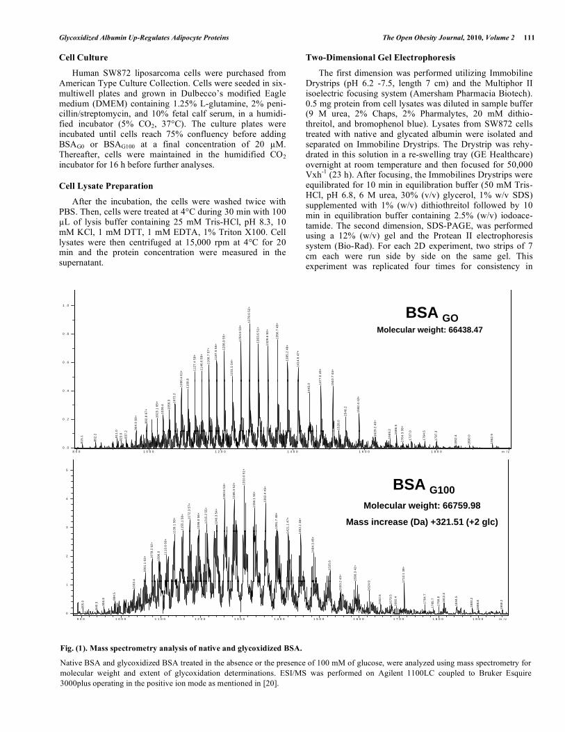
Fig. (1). Mass spectrometry analysis of native and glycoxidized BSA.
Native BSA and glycoxidized BSA treated in the absence or the presence of 100 mM of glucose, were analyzed using mass spectrometry for molecular weight and extent of glycoxidation determinations. ESI/MS was performed on Agilent 1100LC coupled to Bruker Esquire 3000plus operating in the positive ion mode as mentioned in [20].
53+
1278
.6 5
2+
51+
+ 7 49
+
1 . 0
Molecular weight:
BSA GO
0.4
61+
.8
1127
.4 5
9+
1146
.6 5
8+
1166
.7 5
7+
1187
.5 5
6+
1208
.9 5
5+
1231
.5 5
4+
1254
.6
1303
.6 5
1329
.6 5
0
1356
.
1385
.2 4
8+
1414
.6 4
7+
77.0
45+
10.7
44+
0 . 6
0 . 8
0 2
964.
0 69
+
992.
8 67
+
1023
.1 6
5+
1039
.4
1056
.0 1072
.2
1090
1108
1445
.0 147
151
1528
.0
1546
.2
1582
.4 4
2+
9.2
40+
.2 86.5
39+
0 5 3
0 . 2
0 . 4
815.
1
852.
2
911.
092
3.0
937.
2
162
1669
.
16
1704
.3 3
1727
.0
1764
.5
1797
.3
1852
.4
1890
.0
1952
.9
0 . 08 0 0 1 0 0 0 1 2 0 0 1 4 0 0 1 6 0 0 1 8 0 0 m / z
10.0
51+5
x 1 0
BSA
135.
1 59
+
1155
.1 5
8+ 1172
.3 5
7+
1196
.0 5
6+
1215
.2 5
5+
1240
.3 5
4+
1260
.6 5
3+
1285
.3 5
2+ 13
1336
.1 5
0+
1363
.4 4
9+
1391
.7 4
8+
421.
1 47
+
452.
1 46
+
3
4
BSA G100Molecular weight: 66759.98
Mass increase (Da) +321.51 (+2 glc)
.4
1061
.1 6
3+ 1078
.2 6
2+
1096
.0 1113
.6 6
0+
1 1 14 14
1484
.3 4
5+
1525
.6
2 43
+
1590
.3 4
2+
9 712.
1 39
+
2
905.
0
940.
3
956.
8 984.
5
1033
.
1553
.2
1624
.9
1652
.6
1679
.516
92.4
1
1764
.7
1785
.7
1799
.8
1813
.0
1844
.5
1883
.2
1898
.8
1959
.2
0
1
9 0 0 1 0 0 0 1 1 0 0 1 2 0 0 1 3 0 0 1 4 0 0 1 5 0 0 1 6 0 0 1 7 0 0 1 8 0 0 1 9 0 0 m / z
Molecular weight: 66438 47Molecular weight: 66438.47
112 The Open Obesity Journal, 2010, Volume 2 Singh et al.
results. These 2D gels were stained with silver nitrate follo-wing established protocols [13].
Identification of Proteins
Images generated from silver stained gels were com-pared. Co-localizing spots were excised from the silver stained gels and submitted separately for identification. Spots were sent for peptide mass fingerprinting identification by MALDI-TOF/TOF mass spectrometry, at the Plateforme Technologique de Biotechnologie Moléculaire (Université d'Angers, France). Signal quantifications from images were determined using the freeware ImageJ (version 1.32j), available from the internet website: http://rsb.info.nih.gov/ij/.
3. RESULTS
Mass Spectrometry Analysis of Native and Glycoxidized Albumin
Oxidative modifications of proteins could occur in dia-betes, which is one of the important pathological conditions associated with the early occurrence of vascular complica-tions, together with functional alterations of albumin. In diabetes, albumin undergoes increased glycation and gly-coxidation [14]. The glycation phenomenon corresponds to the non-enzymatic and non-oxidative covalent attachment of glucose molecule to protein [14]. Glycoxidation refers to the radical-mediated oxidation reaction of both free and protein-bound sugars [15]. Amadori rearrangement of the above glycated protein gives rise to deleterious, advanced gly-coxidation (also termed advanced glycation) end products (AGEs) [16,17].
Modifications of commercial BSA when incubated with 0 mM and 100 mM of glucose were assessed by ESI/MS, which indicate the number of glucose molecule condensed on the protein. The most abundant component of native and glycoxidized albumin is shown in Fig. (1). Additional impor-tant characterizations of structural modifications in HSA and
BSA after incubating with varied concentration of glucose were previously performed by our group [18-20]. As expected, non-enzymatic glycosylation of BSA induces an increase in molecular weight for albumin incu-bated with 100 mM of glucose, in comparison with native BSA. Here, the molecular mass represented by the most abundant component of non-modified BSA and glycoxidized albumin is 66438.47 Da and 66759.98 Da, respectively. This increase of 321.51 Da could correspond to the addition of two molecules of glucose unit to the glycoxidized albumin.
Protein Expression in SW872 Cells after Incubation with Native or Glycoxidized Albumin
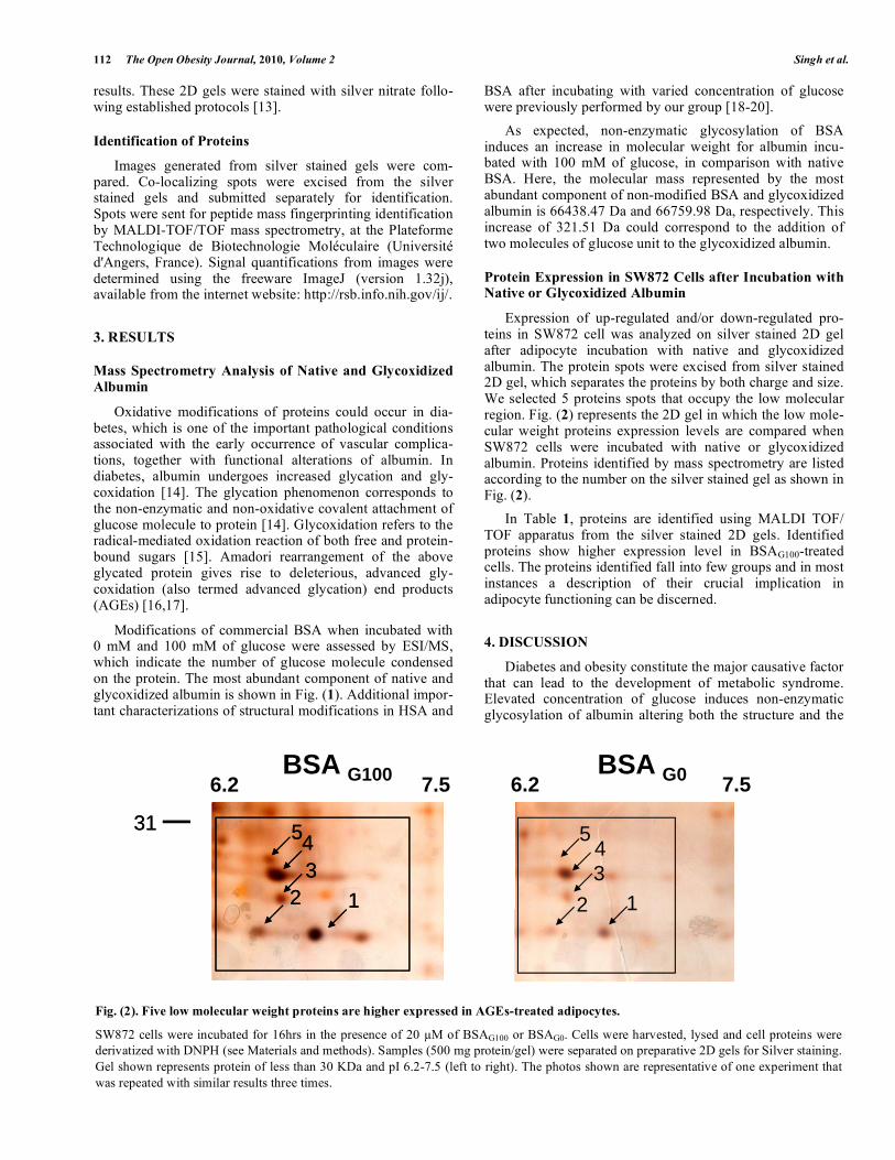
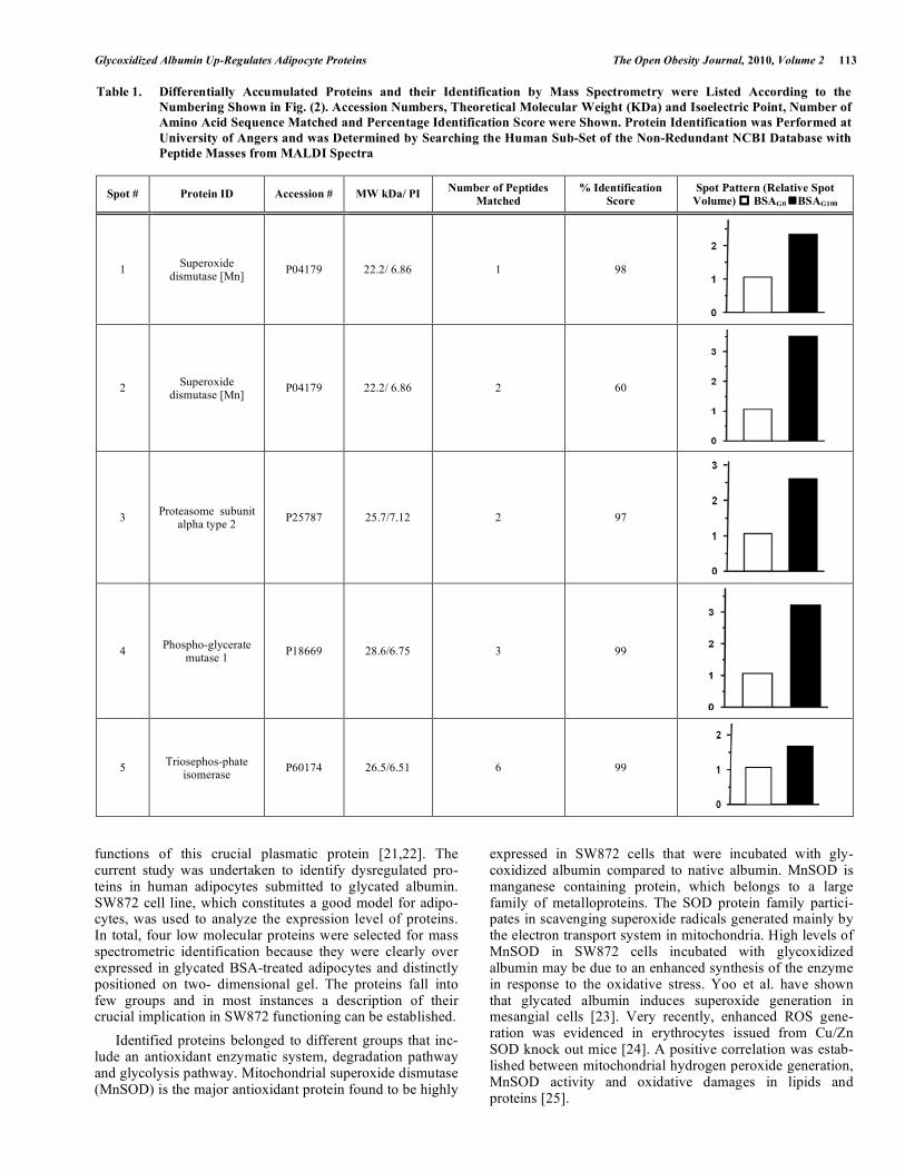
Expression of up-regulated and/or down-regulated pro-teins in SW872 cell was analyzed on silver stained 2D gel after adipocyte incubation with native and glycoxidized albumin. The protein spots were excised from silver stained 2D gel, which separates the proteins by both charge and size. We selected 5 proteins spots that occupy the low molecular region. Fig. (2) represents the 2D gel in which the low mole-cular weight proteins expression levels are compared when SW872 cells were incubated with native or glycoxidized albumin. Proteins identified by mass spectrometry are listed according to the number on the silver stained gel as shown in Fig. (2). In Table 1, proteins are identified using MALDI TOF/ TOF apparatus from the silver stained 2D gels. Identified proteins show higher expression level in BSAG100-treated cells. The proteins identified fall into few groups and in most instances a description of their crucial implication in adipocyte functioning can be discerned.
4. DISCUSSION
Diabetes and obesity constitute the major causative factor that can lead to the development of metabolic syndrome. Elevated concentration of glucose induces non-enzymatic glycosylation of albumin altering both the structure and the
Fig. (2). Five low molecular weight proteins are higher expressed in AGEs-treated adipocytes.
SW872 cells were incubated for 16hrs in the presence of 20 µM of BSAG100 or BSAG0. Cells were harvested, lysed and cell proteins were derivatized with DNPH (see Materials and methods). Samples (500 mg protein/gel) were separated on preparative 2D gels for Silver staining. Gel shown represents protein of less than 30 KDa and pI 6.2-7.5 (left to right). The photos shown are representative of one experiment that was repeated with similar results three times.
BSA G100 BSA G06.2 7.5
31
6.2 7.5
3131
1345
34
5
2 12
31
1345
34
5
2 12 12 12
Glycoxidized Albumin Up-Regulates Adipocyte Proteins The Open Obesity Journal, 2010, Volume 2 113
functions of this crucial plasmatic protein [21,22]. The current study was undertaken to identify dysregulated pro-teins in human adipocytes submitted to glycated albumin. SW872 cell line, which constitutes a good model for adipo-cytes, was used to analyze the expression level of proteins. In total, four low molecular proteins were selected for mass spectrometric identification because they were clearly over expressed in glycated BSA-treated adipocytes and distinctly positioned on two- dimensional gel. The proteins fall into few groups and in most instances a description of their crucial implication in SW872 functioning can be established. Identified proteins belonged to different groups that inc-lude an antioxidant enzymatic system, degradation pathway and glycolysis pathway. Mitochondrial superoxide dismutase (MnSOD) is the major antioxidant protein found to be highly
expressed in SW872 cells that were incubated with gly-coxidized albumin compared to native albumin. MnSOD is manganese containing protein, which belongs to a large family of metalloproteins. The SOD protein family partici-pates in scavenging superoxide radicals generated mainly by the electron transport system in mitochondria. High levels of MnSOD in SW872 cells incubated with glycoxidized albumin may be due to an enhanced synthesis of the enzyme in response to the oxidative stress. Yoo et al. have shown that glycated albumin induces superoxide generation in mesangial cells [23]. Very recently, enhanced ROS gene-ration was evidenced in erythrocytes issued from Cu/Zn SOD knock out mice [24]. A positive correlation was estab-lished between mitochondrial hydrogen peroxide generation, MnSOD activity and oxidative damages in lipids and proteins [25].
Table 1. Differentially Accumulated Proteins and their Identification by Mass Spectrometry were Listed According to the Numbering Shown in Fig. (2). Accession Numbers, Theoretical Molecular Weight (KDa) and Isoelectric Point, Number of Amino Acid Sequence Matched and Percentage Identification Score were Shown. Protein Identification was Performed at University of Angers and was Determined by Searching the Human Sub-Set of the Non-Redundant NCBI Database with Peptide Masses from MALDI Spectra
Spot # Protein ID Accession # MW kDa/ PI Number of Peptides Matched
% Identification Score
Spot Pattern (Relative Spot Volume) BSAG0 BSAG100
1 Superoxide dismutase [Mn] P04179 22.2/ 6.86 1 98
2 Superoxide dismutase [Mn] P04179 22.2/ 6.86 2 60
3 Proteasome subunit alpha type 2 P25787 25.7/7.12 2 97
4 Phospho-glycerate mutase 1 P18669 28.6/6.75 3 99
5 Triosephos-phate isomerase P60174 26.5/6.51 6 99
114 The Open Obesity Journal, 2010, Volume 2 Singh et al.
Similarly, there is a 3 fold increase in the expression levels of proteasomal subunit α2. Proteasome is a multi protein complex that is known to degrade altered protein. The 20S subunit of proteasome consists of two outer rings containing seven different α-subunits (α1-α7) and two middle central rings each is constituted by seven different β-subunits (β1-β7). 20S subunit is involved in the degradation of oxidized proteins [26]. Reports from our group showed that enhanced oxidative stress and oxidative damages in AGEs-treated cells were associated with impairements in the proteasomal activities [7,20]. Increased expression level of proteasome in SW872 cells are consistent with very recent data from Schimdt et al who evidenced induction of heat shock proteins and the proteasome system by two different types of AGEs in Caco-2 cells [27]. Phosphoglycerate mutase 1 and triosephosphate isome-rase proteins also appeared to accumulate more importantly in SW872 cells that were incubated with BSAG100 than with BSAG0. These enzymes are closely involved in the glycolysis pathway, where the glucose is oxidized to produce energy. These enzymes are closely related to the insulin sensibility in adipocytes, which constitutes a emerging issue in the obesity-associated diabetes pathology [28]. This study warrants further investigation. Nonetheless, we propose that AGEs-mediated enhancement in the accu-mulation of these four proteins in adipocytes may be involved in the adipose tissue inflammation inherent to obesity-associated disorders.
ACKNOWLEDGEMENTS
The authors would like to acknowledge the Plateforme Technologique de Biotechnologie Moléculaire (Université d'Angers, France) for mass spectrometry analyses. This work has been supported by the Ministère de l’Enseignement Supérieur et de la Recherche et de l’Outre Mer and the Université de La Réunion. NRS was supported by a fellowship from the Conseil Régional de La Réunion et de l’Union Européenne.
ABBREVIATIONS
AGEs = Advanced Glycated Endproducts BSA = Bovine Serum Albumin BSAGX = BSA incubated with x mM of glucose BSAG0 = BSA incubated without glucose OS = Oxidative Stress PAGE = Polyacrylamide Gel Electrophoresis ROS = Reactive Oxygen Species
REFERENCES [1] Droge W. Free radicals in the physiological control of cell function.
Physiol Rev 2002; 82: 47-95. [2] Wild S, Roglic G, Green A, Sicree R, King H. Global prevalence of
diabetes: estimates for the year 2000 and projections for 2030. Diabetes Care 2004; 27: 1047-53.
[3] Shoelson SE, Lee J, Goldfine AB. Inflammation and insulin resistance. J Clin Invest 2006; 116: 1793-801.
[4] Houstis N, Rosen ED, Lander ES. Reactive oxygen species have a causal role in multiple forms of insulin resistance. Nature 2006; 440: 944-8.
[5] Mayerson AB, Hundal RS, Dufour S, et al. The effects of rosiglitazone on insulin sensitivity, lipolysis, and hepatic and skeletal muscle triglyceride content in patients with type 2 diabetes. Diabetes 2002; 51: 797-802.
[6] Rahimi R, Nikfar S, Larijani B, Abdollahi M. A review on the role of antioxidants in the management of diabetes and its complications. Biomed Pharmacother 2005; 59: 365-73.
[7] Singh NR, Rondeau P, Hoareau L, Bourdon E. Identification of preferential protein targets for carbonylation in human mature adipocytes treated with native or glycated albumin. Free Radic Res 2007; 41: 1078-88.
[8] Wassef H, Bernier L, Davignon J, Cohn JS. Synthesis and secretion of apoC-I and apoE during maturation of human SW872 liposarcoma cells. J Nutr 2004; 134: 2935-41.
[9] Izem L, Morton RE. Possible role for intracellular cholesteryl ester transfer protein in adipocyte lipid metabolism and storage. J Biol Chem 2007; 282: 21856-65.
[10] Roche M, Tarnus E, Rondeau P, Bourdon E. Effects of nutritional antioxidants on AAPH- or AGEs-induced oxidative stress in human SW872 liposarcoma cells. Cell Biol Toxicol 2009; 25: 635-44.
[11] Carmel JF, Tarnus E, Cohn JS, Bourdon E, Davignon J, Bernier L. High expression of apolipoprotein E impairs lipid storage and promotes cell proliferation in human adipocytes. J Cell Biochem 2009; 106: 608-17.
[12] Tarnus E, Wassef H, Carmel JF, et al. Apolipoprotein E limits oxidative stress-induced cell dysfunctions in human adipocytes. FEBS Lett 2009; 583: 2042-8.
[13] Shevchenko AWM, Vorm O, Mann M. Mass spectrometric sequencing of proteins silver-stained polyacrylamide gels. Anal Chem 1996; 68: 850-8.
[14] Cohen MP. Intervention strategies to prevent pathogenetic effects of glycated albumin. Arch Biochem Biophys 2003; 419: 25-30.
[15] Pennathur S, Heinecke JW. Mechanisms for oxidative stress in diabetic cardiovascular disease. Antioxid Redox Signal 2007; 9: 955-69.
[16] Cohen MP, Shea E, Chen S, Shearman CW. Glycated albumin increases oxidative stress, activates NF-kappa B and extracellular signal-regulated kinase (ERK), and stimulates ERK-dependent transforming growth factor-beta 1 production in macrophage RAW cells. J Lab Clin Med 2003; 141: 242-9.
[17] Huebschmann AG, Regensteiner JG, Vlassara H, Reusch JE. Diabetes and advanced glycoxidation end products. Diabetes Care 2006; 29: 1420-32.
[18] Rondeau P, Armenta S, Caillens H, Chesne S, Bourdon E. Assessment of temperature effects on beta-aggregation of native and glycated albumin by FTIR spectroscopy and PAGE: relations between structural changes and antioxidant properties. Arch Biochem Biophys 2007; 460: 141-50.
[19] Chesne S, Rondeau P, Armenta S, Bourdon E. Effects of oxidative modifications induced by the glycation of bovine serum albumin on its structure and on cultured adipose cells. Biochimie 2006; 88: 1467-77.
[20] Rondeau P, Singh NR, Caillens H, Tallet F, Bourdon E. Oxidative stresses induced by glycoxidized human or bovine serum albumin on human monocytes. Free Radic Biol Med 2008; 45: 799-812.
[21] Bourdon E, Loreau N, Blache D. Glucose and free radicals impair the antioxidant properties of serum albumin. FASEB J 1999; 13: 233-44.
[22] Roche M, Rondeau P, Singh NR, Tarnus E, Bourdon E. The antioxidant properties of serum albumin. FEBS Lett 2008; 582: 1783-7.
[23] Yoo BS, Regnier FE. Proteomic analysis of carbonylated proteins in two-dimensional gel electrophoresis using avidin-fluorescein affinity staining. Electrophoresis 2004; 25: 1334-41.
[24] Grzelak A, Kruszewski M, Macierzynska E, et al. The effects of superoxide dismutase knockout on the oxidative stress parameters and survival of mouse erythrocytes. Cell Mol Biol Lett 2009; 14: 23-34.
[25] Jolitha AB, Subramanyam MV, Asha DS. Modification by vitamin E and exercise of oxidative stress in regions of aging rat brain: studies on superoxide dismutase isoenzymes and protein oxidation status. Exp Gerontol 2006; 41: 753-63.
Glycoxidized Albumin Up-Regulates Adipocyte Proteins The Open Obesity Journal, 2010, Volume 2 115
[26] Shringarpure R, Grune T, Davies KJ. Protein oxidation and 20S proteasome-dependent proteolysis in mammalian cells. Cell Mol Life Sci 2001; 58: 1442-50.
[27] Schmid K, Haslbeck M, Buchner J, Somoza V. Induction of heat shock proteins and the proteasome system by casein-N epsilon-
(carboxymethyl)lysine and N epsilon-(carboxymethyl)lysine in Caco-2 cells. Ann N Y Acad Sci 2008; 1126: 257-61.
[28] Scherer PE. Adipose tissue: from lipid storage compartment to endocrine organ. Diabetes 2006; 55: 1537-1545.
Received: June 24, 2009 Revised: October 09, 2009 Accepted: October 09, 2009 © Singh et al.; Licensee Bentham Open.
This is an open access article licensed under the terms of the Creative Commons Attribution Non-Commercial License (http://creativecommons.org/licenses/by-nc/3.0/), which permits unrestricted, non-commercial use, distribution and reproduction in any medium, provided the work is properly cited.