Clinical oncology for pancreatic and biliary cancers: Advances and current limitations
Upload
independentCategory
view
2download
0
Identification of Novel Cellular Targets in Biliary TractCancers Using Global Gene Expression Technology
Donna E. Hansel,* Ayman Rahman,*Manuel Hidalgo,† Paul J. Thuluvath,‡
Keith D. Lillemoe,§ Richard Shulick,§ Ja-Lok Ku,¶
Jae-Gahb Park,¶ Kohje Miyazaki,�
Raheela Ashfaq,** Ignacio I. Wistuba,††
Ram Varma,‡‡ Lesleyann Hawthorne,‡‡
Joseph Geradts,‡‡ Pedram Argani,* andAnirban Maitra*From the Departments of Pathology,* Oncology,†
Gastroenterology,‡ and Surgery,§ The Johns Hopkins University
School of Medicine, Baltimore, Maryland; the Cancer Research
Institute,¶ Seoul National University College of Medicine, Seoul,
Korea; Saga Medical School,� Saga, Japan; University of Texas
Southwestern Medical School,** Dallas, Texas; Catholic
University,†† Santiago, Chile; and Roswell Park Cancer Institute,‡‡
Buffalo, New York
Biliary tract carcinoma carries a poor prognosis, anddifficulties with clinical management in patients withadvanced disease are often due to frequent late-stagediagnosis, lack of serum markers, and limited infor-mation regarding biliary tumor pathogenesis. RNA-based global analyses of gene expression have led tothe identification of a large number of up-regulatedgenes in several cancer types. We have used the re-cently developed Affymetrix U133A gene expressionmicroarrays containing nearly 22,000 unique tran-scripts to obtain global gene expression profiles fromnormal biliary epithelial scrapings (n � 5), surgicallyresected biliary carcinomas (n � 11), and biliary cancercell lines (n � 9). Microarray hybridization data werenormalized using dCHIP (http://www.dCHIP.org) toidentify differentially up-regulated genes in primary bil-iary cancers and biliary cancer cell lines and their ex-pression profiles was compared to that of normal epi-thelial scrapings using the dCHIP software as well asSignificance Analysis of Microarrays or SAM (http://www-stat.stanford.edu/�tibs/SAM/). Comparison of thedCHIP and SAM datasets revealed an overlapping list of282 genes expressed at greater than threefold levels inthe cancers compared to normal epithelium (t-test P<0.1 in dCHIP, and median false discovery rate <10 inSAM). Several pathways integral to tumorigenesis wereup-regulated in the biliary cancers, including prolifera-tion and cell cycle antigens (eg, cyclins D2 and E2,cdc2/p34, and geminin), transcription factors (eg, ho-meobox B7 and islet-1), growth factors and growth fac-tor receptors (eg, hepatocyte growth factor, amphi-
regulin, and insulin-like growth factor 1 receptor), andenzymes modulating sensitivity to chemotherapeuticagents (eg, cystathionine � synthase, dCMP deaminase,and CTP synthase). In addition, we identified several“pathway” genes that are rapidly emerging as noveltherapeutic targets in cancer (eg, cytosolic phospho-lipase A2, an upstream target of the cyclooxygenasepathway, and ribosomal protein S6 kinase and eukary-otic translation initiation factor 4E, two importantdownstream mediators of the mitogenic Akt/mTOR sig-naling pathway). Overexpression of selected up-regu-lated genes was confirmed in tissue microarrays of bil-iary cancers by immunohistochemical analysis (n � 4)or in situ hybridization (n � 1), and in biliary cancercell lines by reverse transcriptase PCR (n � 2). Themajority of genes identified in the present study has notbeen previously reported in biliary cancers, and repre-sent novel potential screening and therapeutic targetsof this cancer type. (Am J Pathol 2003, 163:217–229)
Biliary tract carcinomas, which include cancers of thegallbladder and intra- and extrahepatic biliary tree, affect7500 individuals in the United States each year, andnearly 3500 patients die as a direct consequence of thislethal disease.1 Once established, biliary tract cancersare notoriously challenging to diagnose and treat. Atpresent, only surgical excision of detectable malignancyis associated with improvement in 5-year survival. Distantmetastases, extensive regional lymph node metastasis,and vascular encasement or invasion preclude resec-tion.2 In general, the outcome for patients with advancedbiliary tract cancer at any site is dismal, and neitherradiation nor conventional chemotherapy significantly im-proves survival or quality of life. Early diagnosis has asignificant impact on prognosis of biliary cancers. Forexample, patients with lesions confined to the gallbladdermucosa have a 32% 5-year survival rate, while patientswith more advanced lesions have only a 10% 5-yearsurvival rate.3 Thus, urgent efforts are needed for identi-fication of reliable tumor markers that will facilitate theearly detection of biliary cancer in at-risk individuals. In
Supported by the family of Margaret Lee. This work was also partiallysupported by shared resources of the Roswell Park Cancer Center Sup-port Grant P30 CA16056.
Accepted for publication April 4, 2003.
Address reprint requests to Anirban Maitra, M.D., 720 Rutland Avenue,Ross 632 Department of Pathology, Johns Hopkins University School ofMedicine, Baltimore, MD 21212. E-mail: [email protected].
American Journal of Pathology, Vol. 163, No. 1, July 2003
Copyright © American Society for Investigative Pathology
217
addition, we need to identify cancer-specific cellular tar-gets that would form the basis for novel therapeutic ap-proaches for established biliary tract malignancies.
Global expression platforms such as oligonucleotidemicroarrays are a robust technique for identification ofdifferentially up-regulated cancer-specific genes in tu-mors. This technology has recently been successfullyapplied for the identification of novel tumor markers in alarge variety of human neoplasms.4–7 To identify differ-entially expressed genes in biliary cancers versus non-neoplastic biliary epithelium, we applied RNA-basedglobal gene expression profiling using the recently de-veloped Affymetrix U133A expression microarrays to aseries of surgically resected biliary cancers, biliary can-cer cell lines, and biliary epithelial scrapings. We reportthe identification of 282 genes, conforming to a variety ofpotential tumorigenic pathways, expressed threefold orgreater in biliary cancers compared to normal biliaryepithelium. The majority of these genes has not beenpreviously described in biliary tract carcinoma and mayserve as potential therapeutic targets and novel tumormarkers for this lethal disease.
Materials and Methods
Non-Neoplastic Epithelial Scrapings, PrimaryBiliary Cancers, and Cell Lines
Permission for this study was obtained through The JohnsHopkins Joint Committee on Clinical Investigation. Freshscrapings from five non-neoplastic biliary epithelial sam-ples were collected as previously described8 from pa-tients undergoing Whipple resection or incidental chole-cystectomies for non-biliary disorders at The JohnsHopkins Hospital; this method has been shown to yieldhighly enriched sheets of epithelial cells without contam-inating stromal elements. The epithelial scrapings werecollected within 10 minutes of surgical resection andstored at �80°C, and included 3 extrahepatic biliary 2gallbladder epithelial samples. Eleven cancer sampleswere collected from patients undergoing surgery for bil-iary tract cancer, and included 7 gallbladder carcinomas,2 intrahepatic cholangiocarcinomas, and 2 distal bileduct carcinomas. All tumor samples were collected within10 minutes of surgical resection, snap-frozen in liquidnitrogen, and stored at �80°C. Hematoxylin and eosin-stained sections from adjacent frozen tissue were pre-pared before sample harvest to confirm the diagnosisand assess neoplastic cellularity. RNA was extractedfrom tumor samples containing �50% neoplastic cells onfrozen section examination.
The nine human biliary cancer cell lines used for thisstudy included EGI-1 and TFK-19,10 (obtained from theGerman Collection of Microorganisms and Cell CulturesDepartment, Braunschweig, Germany), HUH2811 andHUCCT-112 (obtained from the Health Science ResearchResources Bank, Osaka, Japan), SNU 245, SNU 308,and SNU 107913 (obtained from the Korean Cell LineBank, Seoul, Korea), GB-H3, and GB-D1.14 Of these,SNU308, GB-H3, and GB-D1 cell lines were derived from
gallbladder carcinomas, HuH28, HuCCT-1, and SNU1079 were derived from intrahepatic cholangiocarcino-mas, and EGI-1, TFK-1, and SNU 245 were derived fromextrahepatic biliary cancers. All cell lines except EGI-1were grown in RPMI (Life Technologies Inc., Gaithers-burg, MD) supplemented with 10% heat-inactivated fetalbovine serum (FBS; Life Technologies Inc.); EGI-I wasgrown in Dulbecco’s MEM (Life Technologies Inc.) sup-plemented with 10% FBS. Cells were incubated at 37°Cin a humidified atmosphere of 5% CO2 in air.
RNA Extraction and Hybridization
Sample preparation and processing procedure was per-formed at the Roswell Park Cancer Institute MicroarrayCore Facility, as described in the Affymetrix GeneChipExpression Analysis Manual (Affymetrix Inc., Santa Clara,CA). Briefly, frozen tumor tissues were crushed in TRIzol(Invitrogen Inc., Carlsbad, CA) by using a Polytron ho-mogenizer (Brinkman Instruments, Westbury, NY). TotalRNA was then extracted from the crushed tissue andcleaned using RNeasy columns according to manufac-turer’s protocol (Qiagen Inc., Valencia, CA). For biliarycancer cell lines and biliary epithelial scrapings, theRNeasy protocol for human cell lines was directly usedfor extraction of total RNA. The integrity of total RNA wasconfirmed in each case using the Agilent 2100 Bioana-lyzer (Agilent Technologies, Palo Alto, CA). Using 5 to 40�g of total RNA, double-stranded cDNA was synthesizedfollowing SuperScript Choice system (Invitrogen Inc.).T7-(dT24) oligomer was used for priming the first-strandcDNA synthesis. The resultant cDNA was purified usingPhase Lock Gel, phenol/chloroform extraction, and pre-cipitated with ethanol. The cDNA pellet was collectedand dissolved in appropriate volume. Using cDNA astemplate, cRNA was synthesized using a T7 MegaScriptIn-Vitro Transcription (IVT) kit (Ambion, Austin, TX). Bio-tinylated-11-CTP and 16-UTP ribonucleotides (Enzo Di-agnostics Inc., Farmingdale, NY) were added to the re-action as labeling reagents. IVT reactions were carriedout at 37°C for 6 hours and, the labeled cRNA obtainedwas purified using RNeasy columns (Qiagen Inc.). ThecRNA was fragmented in a fragmentation buffer (40mmol/L Tris-acetate, pH 8.1, 100 mmol/L KOAc, 30mmol/L MgOAc) for 35 minutes at 94°C. FragmentedcRNA (10 to 11 �g/probe array) was used to hybridize tohuman U133A GeneChip array at 45°C for 24 hours in ahybridization oven with constant rotation (60 rpm). Thechips were washed and stained using Affymetrix fluidicsstations. Staining was performed using streptavidin phy-coerythrin conjugate (SAPE; Molecular Probes, Eugene,OR), followed by the addition of biotinylated antibody tostreptavidin (Vector Laboratories, Burlingame, CA), andfinally with streptavidin phycoerythrin conjugate. Probearrays were scanned using fluorometric scanners(Hewlett Packard Gene Array Scanner; Hewlett PackardCorporation, Palo Alto, CA). The scanned images wereinspected and analyzed using established quality controlmeasures.
218 Hansel et alAJP July 2003, Vol. 163, No. 1

Data Filtering and Analysis
The 25 .CEL files generated by the Affymetrix MicroarraySuite (MAS) version 5.0 were converted into .DCP filesusing dCHIP (www.dCHIP.org), as described previouslyby Li and Wong.15 The .DCP files were normalized, andraw gene expression data generated using the dCHIPsystem of model-based analysis. To evaluate how the 25samples grouped together according to the similarity oftheir gene expression profiles, we used hierarchical clus-tering with the average linkage method, with a subset of2308 genes demonstrating the largest variation acrosssamples (SD/mean �1). For hierarchical cluster analysis,data were log-transformed, median-centered, and visu-alized using the CLUSTER and TREEVIEW programs.16
For comparison of global gene expression profiles be-tween normal and cancer samples, a two-pronged strat-egy was used. The first comparison was performed usingthe dCHIP software itself, wherein the five biliary epithelialscrapings were designated as “baseline” (B), and the 20biliary cancer specimens designated as “experiment”(E). Genes expressed threefold or higher in the cancersversus normal samples were then identified by definingthe appropriate filtering criteria in the dCHIP software(mean E/mean B �3; mean E � mean B � 100, P � 0.1,t-test). The second comparison was performed usingsignificance analysis of microarrays or SAM v1.13 (http://www-stat.stanford.edu/�tibs/SAM/),17,18 which containsa sliding scale for false discovery rate (FDR) of signifi-cantly up-regulated genes. The output criteria selectedfor SAM included threefold or greater expression in thebiliary cancers as compared to normal tissues, and asignificance threshold expected to produce a medianFDR of less than 10 genes.
Immunohistochemistry
A biliary cancer tissue microarray was generated from 40biliary tract cancers (15 gallbladder carcinomas, 10 in-trahepatic cholangiocarcinomas, and 15 extrahepaticcholangiocarcinomas), as previously described.19 Eachcancer specimen was represented by four 1.4-mm coreson the tissue microarrays, to obtain adequate represen-tation of neoplastic cells. In addition, non-neoplastic bil-iary tissues, and additional control tissues from extra-biliary sites were also included on the tissue array. Slideswere deparaffinized in fresh xylenes and rehydratedthrough sequential graded ethanol steps. Antigen re-trieval was performed by citrate buffer incubation (18mmol/L citric acid, 8.2 mmol/L sodium citrate, pH 6.0)using a household vegetable steamer (Black andDecker) for 60 minutes. Slides were incubated for 5 min-utes with 3% hydrogen peroxide, washed in TBS/T (20mmol/L Tris, 140 mmol/L NaCl, 0.1% Tween-20, pH 7.6),and incubated in appropriate antibody dilutions forcdc2(p34) (Zymed, South San Francisco, CA, 1:100),topoisomerase II � [topoII�] (Neomarkers, Fremont, CA,1:3200), bone morphogenetic protein receptor 1A[BMPR1A/Activin A receptor/ALK3] (Santa Cruz Biotech-nology, Inc., Santa Cruz, CA, 1:500), and geminin (1:500,
a generous gift from Dr. Anindya Dutta, Brigham andWoman’s Hospital, Boston, MA) for 60 minutes at roomtemperature. Normal saline was substituted for the pri-mary antibody in control sections. The avidin-biotin-per-oxidase complex method from DAKO (Glostrup, Den-mark) was used and slides were subsequentlycounterstained with hematoxylin. Assessment of immu-nohistochemical labeling in the tissue microarrays wasperformed by two of the authors (D.E.H. and A.M.). Forthe three nuclear markers (cdc2, geminin, and topo II�),scoring was performed as follows: negative, �5% nu-clear labeling; focal, 5 to 25% nuclear labeling; and dif-fuse, �25% nuclear labeling; for BMPR1A, labeling wasscored as negative (�5% cytoplasmic and membranousstaining), focal (5 to 25% cytoplasmic and membranousstaining), and diffuse (�25% cytoplasmic and membra-nous staining).
In Situ Hybridization
Non-radioisotopic in situ hybridization was performed forthe transcription factor islet-1 (ISL-1). Sense and anti-sense riboprobes were prepared from the correspondingsequence-verified I.M.A.G.E. clone (Invitrogen, Inc.), aspreviously described,20 and labeled using Digoxin RNALabeling mix (Boehringer Mannheim, Germany). Biliarycancer tissue microarrays were rehydrated through eth-anol gradients (as described above), incubated for 10minutes in 1% hydrogen peroxide (Super G Brand),washed in TBS, treated with proteinase K for 30 minutesat 37°C, and incubated overnight in 300 �l of dilutedsense or antisense probe at 50°C. The next day, slideswere washed in 2X SSC, treated with 250 �l Rnase A/T1cocktail (Ambion, Austin, TX) for 30 minutes at 37°C,washed in 2X SSC with 50% formamide (American Bio-analytical, Natick, MA), washed in 0.8X SSC, blocked,incubated with rabbit HRP-anti-DIG (DAKO), and devel-oped in the dark with biotinyl-tyramide (DAKO). Slideswere then counterstained as previously described.20 Thespecificity of hybridization was assessed by absence ofsignal in the sense riboprobe slide; the antisense ribo-probe slide was then used for analysis of transcript ex-pression pattern in neoplastic versus non-neoplastic bili-ary cells.
Semi-Quantitative RT-PCR
The differential expression of two genes, homeobox B7(HoxB7) and dickkopf 1 homolog (Dkk-1), was validatedby reverse transcriptase PCR. Total RNA was preparedfrom five biliary cancer cell lines (GB-H3, SNU1079,SNU308, SNU245, and HuCCT1), and three normal bili-ary epithelial scrapings using the RNeasy Mini kit (Qia-gen, Inc.). For cDNA preparation, a mix of 4 �g total RNAand Oligo (dT) primers (Invitrogen, Inc.) was incubated at70°C for 10 minutes to denature RNA and briefly placedon ice. A final buffer mixture of 1X First Strand Buffer, 20mmol/L DTT, and 2 mmol/L dNTP (Invitrogen) was added,samples were incubated at 42°C for 2 minutes, 200 unitsof Super Script II RNase H-reverse transcriptase (Invitro-
Gene Expression in Biliary Cancers 219AJP July 2003, Vol. 163, No. 1
gen, Inc.) were added, and samples were again incu-bated at 42°C for 50 minutes. A final incubation at 70°Cfor 15 minutes was performed. Polymerase chain reactionwas performed in a final mixture of 1X Platinum PCRSupermix (Invitrogen, Inc.), 400 nmol/L forward and re-verse primer, and 2 �l cDNA. PCR reactions were per-formed in a Thermo Hybaid MBS 0.2S cycler as follows:96°C for 5 minutes, 35 cycles of 94°C for 45 seconds,55°C for 45 seconds, and 72°C for 3 minutes, followed by72°C for 5 minutes, then 4°C. PCR products were visual-ized by 1% agarose gel electrophoresis, followed byeithidium bromide staining. The primer sequences wereas follows: HoxB7 foward primer 5�-AGCCTCAAGTTCG-GTTTTCG-3�, Hox B7 reverse primer 5�-GCGTCAGGTA-GCGATTGTAG-3�, Dkk-1 forward primer 5�-ACCAGAC-CATTGACAACTAC-3�, Dkk-1 reverse primer 5�-GTGTCT-AGCACAACACAATC-3�. Standardization was performedusing GAPDH; forward primer and reverse primers wereused as previously described.4
Results
Identification of Differentially Up-RegulatedGenes in Biliary Cancers
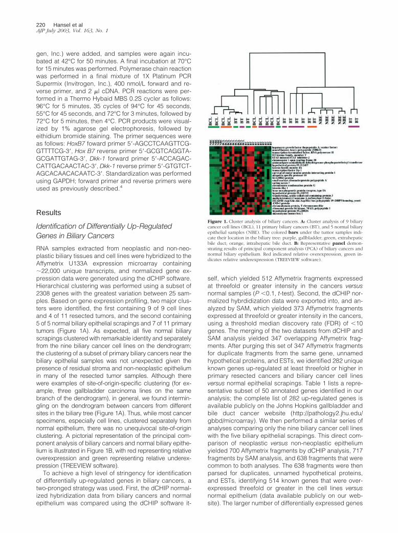
RNA samples extracted from neoplastic and non-neo-plastic biliary tissues and cell lines were hybridized to theAffymetrix U133A expression microarray containing�22,000 unique transcripts, and normalized gene ex-pression data were generated using the dCHIP software.Hierarchical clustering was performed using a subset of2308 genes with the greatest variation between 25 sam-ples. Based on gene expression profiling, two major clus-ters were identified, the first containing 9 of 9 cell linesand 4 of 11 resected tumors, and the second containing5 of 5 normal biliary epithelial scrapings and 7 of 11 primarytumors (Figure 1A). As expected, all five normal biliaryscrapings clustered with remarkable identity and separatelyfrom the nine biliary cancer cell lines on the dendrogram;the clustering of a subset of primary biliary cancers near thebiliary epithelial samples was not unexpected given thepresence of residual stroma and non-neoplastic epitheliumin many of the resected tumor samples. Although therewere examples of site-of-origin-specific clustering (for ex-ample, three gallbladder carcinoma lines on the samebranch of the dendrogram), in general, we found intermin-gling on the dendrogram between cancers from differentsites in the biliary tree (Figure 1A). Thus, while most cancerspecimens, especially cell lines, clustered separately fromnormal epithelium, there was no unequivocal site-of-originclustering. A pictorial representation of the principal com-ponent analysis of biliary cancers and normal biliary epithe-lium is illustrated in Figure 1B, with red representing relativeoverexpression and green representing relative underex-pression (TREEVIEW software).
To achieve a high level of stringency for identificationof differentially up-regulated genes in biliary cancers, atwo-pronged strategy was used. First, the dCHIP normal-ized hybridization data from biliary cancers and normalepithelium was compared using the dCHIP software it-
self, which yielded 512 Affymetrix fragments expressedat threefold or greater intensity in the cancers versusnormal samples (P �0.1, t-test). Second, the dCHIP nor-malized hybrdidization data were exported into, and an-alyzed by SAM, which yielded 373 Affymetrix fragmentsexpressed at threefold or greater intensity in the cancers,using a threshold median discovery rate (FDR) of �10genes. The merging of the two datasets from dCHIP andSAM analysis yielded 347 overlapping Affymetrix frag-ments. After purging this set of 347 Affymetrix fragmentsfor duplicate fragments from the same gene, unnamedhypothetical proteins, and ESTs, we identified 282 uniqueknown genes up-regulated at least threefold or higher inprimary resected cancers and biliary cancer cell linesversus normal epithelial scrapings. Table 1 lists a repre-sentative subset of 50 annotated genes identified in ouranalysis; the complete list of 282 up-regulated genes isavailable publicly on the Johns Hopkins gallbladder andbile duct cancer website (http://pathology2.jhu.edu/gbbd/microarray). We then performed a similar series ofanalyses comparing only the nine biliary cancer cell lineswith the five biliary epithelial scrapings. This direct com-parison of neoplastic versus non-neoplastic epitheliumyielded 700 Affymetrix fragments by dCHIP analysis, 717fragments by SAM analysis, and 638 fragments that werecommon to both analyses. The 638 fragments were thenparsed for duplicates, unnamed hypothetical proteins,and ESTs, identifying 514 known genes that were over-expressed threefold or greater in the cell lines versusnormal epithelium (data available publicly on our web-site). The larger number of differentially expressed genes
Figure 1. Cluster analysis of biliary cancers. A: Cluster analysis of 9 biliarycancer cell lines (BCL), 11 primary biliary cancers (BT), and 5 normal biliaryepithelial samples (NBE). The colored bars under the tumor samples indi-cate their location in the biliary tree: purple, gallbladder; green, extrahepaticbile duct; orange, intrahepatic bile duct. B: Representative panel demon-strating results of principal component analysis (PCA) of biliary cancers andnormal biliary epithelium. Red indicated relative overexpression, green in-dicates relative underexpression (TREEVIEW software).
220 Hansel et alAJP July 2003, Vol. 163, No. 1
when only cell lines were compared with normal epithe-lium possibly reflects either the “dilution” effect causedby non-neoplastic epithelial and stromal elements withinprimary cancers, or the effects of in vitro culture. In pass-ing, it should be mentioned that we also identified 513genes that were down-regulated threefold or greater inthe cancers versus normals; however, since the objectiveof the current study was to identify novel tumor markers,the ensuing discussion will focus on up-regulated genesonly.
Multiple classes of differentially up-regulated geneswere identified in this study and included proliferationand cell cycle antigens (cyclins D2 and E2, cdc2/p34, andgeminin),21–23 transcription factors (homeobox B7 andislet-1),24,25 growth factors and growth factor receptors(hepatocyte growth factor, amphiregulin, and insulin-likegrowth factor 1 receptor),26–28 and enzymes modulatingsensitivity to chemotherapeutic agents (cystathionine �synthase, dCMP deaminase, and CTP synthase)29–31 Sub-sets of genes involved in biologically relevant pathwayswere also identified, such as genes in the transforminggrowth factor � pathway (bone morphogenetic protein re-ceptor 1A [BMPR1A], mothers against decapentaplegic ho-molog 5 [MADH5], and TGF-� receptor-associated protein1 [TRAP-1]),32–34 or genes involved in steroid metabo-lism, particularly in the estrogen/androgen pathways (es-trogen-related receptor-�, sterol isomerase, and steroid-5-�-reductase).35–37 Interestingly, we identified severalgenes that are related to pathways rapidly emerging asnovel therapeutic targets in many cancer types. Notableamong these were cytosolic phospholipase A2, whichgenerates arachidonic acid, the substrate for the cyclo-oxygenase (COX) enzyme,38,39 and two important down-stream mediators of the tumorigenic Akt/mammalian tar-get of rapamycin (mTOR) pathway in humans, ribosomalprotein S6 kinase, and eukaryotic translation initiation factor4E.40
A literature search of PubMed (www.ncbi.nlm.nih.gov/PubMed) revealed that several of the genes up-regulatedin this study have previously been reported as overex-pressed, either singly or through global microarray ex-pression analyses in other cancer types, in principle val-idating our approach. A second PubMed search usingthe gene name and either “cholangiocarcinoma,” “bili-ary,” “bile duct,” or “gallbladder” in the text search re-vealed that only three up-regulated genes (hepatocytegrowth factor, proliferating cell nuclear antigen, and cytoso-lic phospholipase A2) have been previously reported asspecifically overexpressed in biliary cancers.41–43 Thus,the overwhelming majority of up-regulated genes re-ported in this study represent novel tumor markers andcellular targets for this cancer type.
Validation of Differentially Up-Regulated Genesin Biliary Cancers
The differential overexpression of a subset of genes (n �7) was validated using a combination immunohistochem-istry and in situ hybridization in archival biliary cancersand RT-PCR on biliary cancer cell lines. A biliary cancer
tissue microarray containing 40 biliary cancers was im-munolabeled with antibodies against BMPR1A, geminin,topoII�, and cdc2/p34 to confirm the overexpression ofthe corresponding protein product. The results of immu-nohistochemical analysis are summarized in Table 2.
BMPR1A is a member of the cell surface receptorTGF-� superfamily and is expressed in human skeletalmuscle, heart, and placenta under normal conditions.32
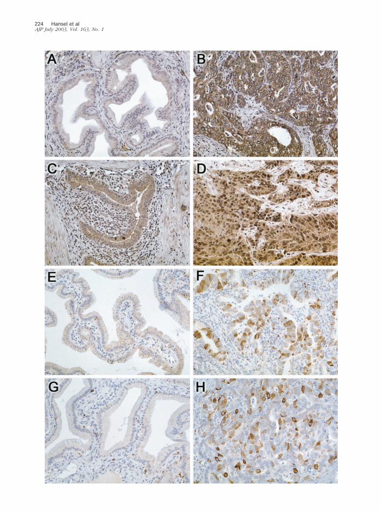
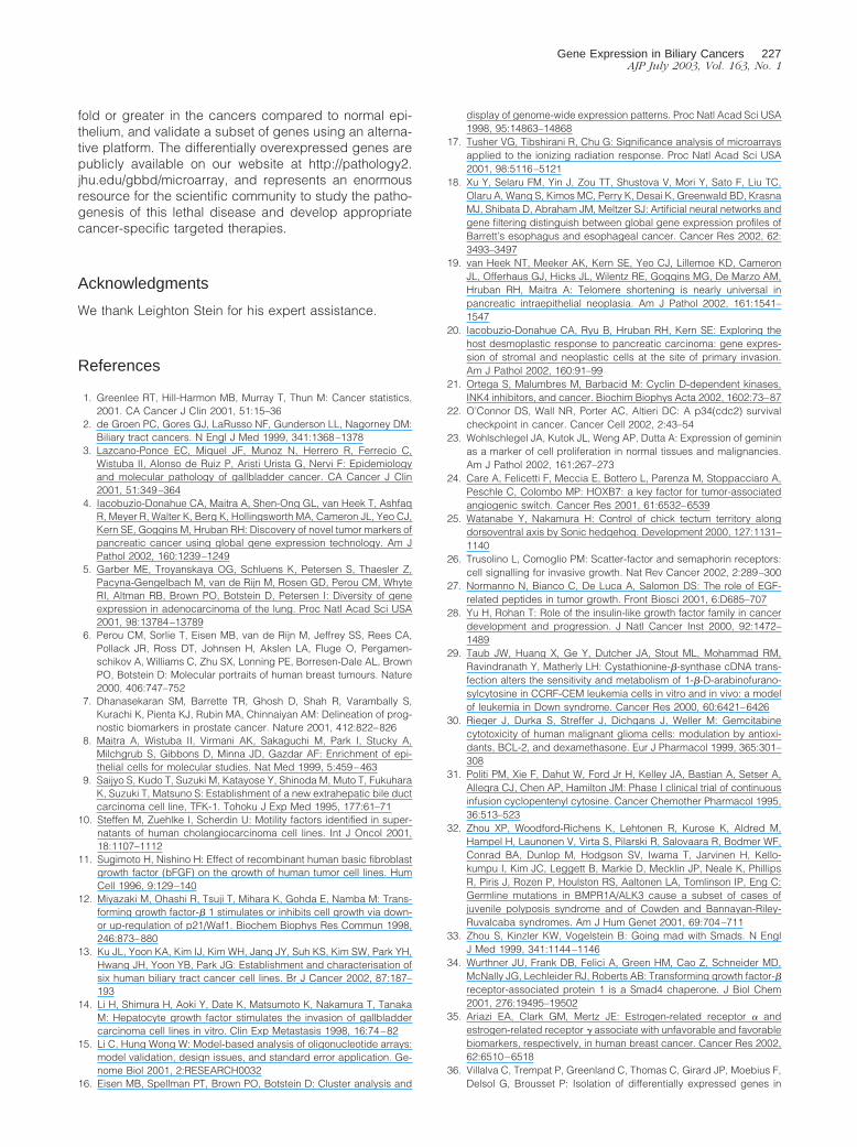
BMPR1A immunolabeling was absent in normal biliaryepithelium (�5% labeling) (Figure 2A). In contrast, 91%of cancers demonstrate diffuse or focal cell membraneand cytoplasmic immunolabeling of BMPR1A (Figure2B). Similarly, 85% of biliary cancers demonstrate diffuseor focal nuclear overexpression of the novel protein gemi-nin,23 while normal biliary epithelium was negative (Fig-ure 2, C and D). The cell cycle protein cdc2/p34 servesas a promoter of mitosis and is regulated by tyrosinephosphorylation.44 Normal biliary epithelium lacked nu-clear cdc2/p34 expression (Figure 2E), whereas 83% ofbiliary tract cancers demonstrated diffuse or focal nu-clear expression (Figure 2F). Similarly, labeling with to-poII�, which regulates the topology of DNA,45 was ab-sent in normal biliary tract epithelium (Figure 2G), but wasdiffusely or focally expressed in 71% of biliary cancers(Figure 2H) (Table 2).
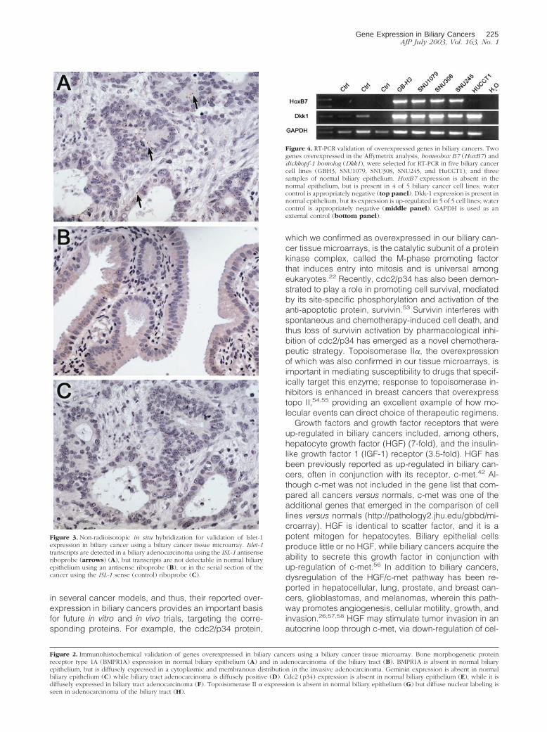
In situ hybridization for the transcription factor islet-1(ISL-1) was performed using the biliary cancer tissuemicroarray to confirm differential overexpression. ISL-1 isa downstream target of the Sonic hedgehog pathway,and is essential for development of the endocrine pan-creas.46,47 In situ hybridization for ISL-1 demonstratedneoplastic epithelial expression in 26 of 40 (65%) biliarytract carcinomas, while minimal expression was seen innon-neoplastic biliary epithelium (Figure 3,A and B); sensecontrol yielded no signal in the cancer cells (Figure 3C).
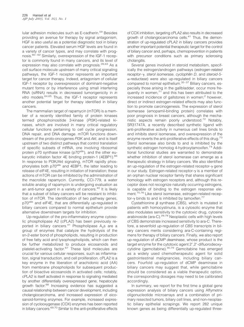
We additionally confirmed the overexpression of twogenes, homeobox B7 (HoxB7) and dickkopf 1 (Dkk-1), infive biliary cancer cell lines and three non-neoplasticbiliary epithelial scrapings by semi-quantitative RT-PCR.HoxB7 is a homeobox containing gene normally ex-pressed during development and aberrantly overex-pressed in several human cancers,48 whereas Dkk-1 is ap53-responsive gene that inhibits canonical Wnt signal-ing pathways.49 Both transcripts were expressed andup-regulated in the cancer cell lines we examined incomparison to normal biliary epithelium (Figure 4).
Discussion
Biliary tract carcinoma represents the second most com-mon primary cancer of the hepatobiliary system; eachyear as many as 3500 individuals succumb to this dis-ease in the United States alone, while mortality rates arefar higher in some parts of the world such as the Far East,Chile, and parts of Southeast Asia.2 Known risk factorsinclude primary sclerosing cholangitis, cholelithiasis,chronic parasitic infection, and chemical exposure.3 Al-though early preventative steps can be taken to lower therisk of developing biliary cancers, the overall prognosisonce cancer develops is poor, with only a 10% 5-year
Gene Expression in Biliary Cancers 221AJP July 2003, Vol. 163, No. 1
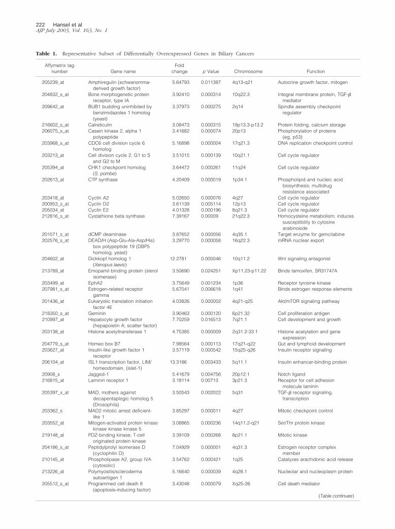
Table 1. Representative Subset of Differentially Overexpressed Genes in Biliary Cancers
Affymetrix tagnumber Gene name
Foldchange p Value Chromosome Function
205239_at Amphiregulin (schwanomma-derived growth factor)
5.64793 0.011387 4q13-q21 Autocrine growth factor, mitogen
204832_s_at Bone morphogenetic proteinreceptor, type IA
3.92410 0.000314 10q22.3 Integral membrane protein, TGF-�mediator
209642_at BUB1 budding uninhibited bybenzimidazoles 1 homolog(yeast)
3.37973 0.000275 2q14 Spindle assembly checkpointregulator
216602_s_at Calreticulin 3.08473 0.000315 19p13.3-p13.2 Protein folding, calcium storage206075_s_at Casein kinase 2, alpha 1
polypeptide3.41682 0.000074 20p13 Phosphorylation of proteins
(eg, p53)203968_s_at CDC6 cell division cycle 6
homolog5.16898 0.000004 17q21.3 DNA replication checkpoint control
203213_at Cell division cycle 2, G1 to Sand G2 to M
3.51015 0.000139 10q21.1 Cell cycle regulator
205394_at CHK1 checkpoint homolog(S. pombe)
3.64472 0.000261 11q24 Cell cycle regulator
202613_at CTP synthase 4.20409 0.000019 1p34.1 Phospholipid and nucleic acidbiosynthesis; multidrugresistance associated
203418_at Cyclin A2 5.02650 0.000076 4q27 Cell cycle regulator200953_s_at Cyclin D2 3.61139 0.005114 12p13 Cell cycle regulator205034_at Cyclin E2 4.01328 0.000196 8q21.3 Cell cycle regulator212816_s_at Cystathione beta synthase 7.39167 0.00009 21q22.3 Homocysteine metabolism; induces
susceptibility to cytosinearabinoside
201571_s_at dCMP deaminase 3.87652 0.000056 4q35.1 Target enzyme for gemcitabine202576_s_at DEAD/H (Asp-Glu-Ala-Asp/His)
box polypeptide 19 (DBP5homolog, yeast)
3.29770 0.000058 16q22.3 mRNA nuclear export
204602_at Dickkopf homolog 1(Xenopus laevis)
12.2781 0.000046 10q11.2 Wnt signaling antagonist
213789_at Emopamil binding protein (sterolisomerase)
3.50690 0.024251 Xp11.23-p11.22 Binds tamoxifen, SR31747A
203499_at EphA2 3.75649 0.001234 1p36 Receptor tyrosine kinase207981_s_at Estrogen-related receptor
gamma5.67041 0.006618 1q41 Binds estrogen response elements
201436_at Eukaryotic translation initiationfactor 4E
4.03826 0.000002 4q21-q25 Akt/mTOR signaling pathway
218350_s_at Geminin 3.90463 0.000120 6p21.32 Cell proliferation antigen210997_at Hepatocyte growth factor
(hepapoietin A; scatter factor)7.70259 0.016513 7q21.1 Cell development and growth
203138_at Histone acetyltransferase 1 4.75385 0.000009 2q31.2-33.1 Histone acetylation and geneexpression
204779_s_at Homeo box B7 7.98564 0.000113 17q21-q22 Gut and lymphoid development203627_at Insulin-like growth factor 1
receptor3.57119 0.000542 15q25-q26 Insulin receptor signaling
206104_at ISL1 transcription factor, LIM/homeodomain, (islet-1)
13.3186 0.003433 5q11.1 Insulin enhancer-binding protein
20908_s Jagged-1 5.41679 0.004756 20p12.1 Notch ligand216815_at Laminin receptor 1 3.18114 0.00713 3p21.3 Receptor for cell adhesion
molecule laminin205397_x_at MAD, mothers against
decapentaplegic homolog 5(Drosophila)
3.50543 0.002022 5q31 TGF-� receptor signaling,transcription
203362_s MAD2 mitotic arrest deficient-like 1
3.85297 0.000011 4q27 Mitotic checkpoint control
203552_at Mitogen-activated protein kinasekinase kinase kinase 5
3.08865 0.000236 14q11.2-q21 Ser/Thr protein kinase
219148_at PDZ-binding kinase; T-celloriginated protein kinase
3.39109 0.000268 8p21.1 Mitotic kinase
204186_s_at Peptidylprolyl isomerase D(cyclophilin D)
7.04929 0.000001 4q31.3 Estrogen receptor complexmember
210145_at Phospholipase A2, group IVA(cytosolic)
3.54762 0.000421 1q25 Catalyzes arachidonic acid release
213226_at Polymyositis/sclerodermaautoantigen 1
5.16640 0.000039 4q28.1 Nucleolar and nucleoplasm protein
205512_s_at Programmed cell death 8(apoptosis-inducing factor)
3.43048 0.000079 Xq25-26 Cell death mediator
(Table continues)
222 Hansel et alAJP July 2003, Vol. 163, No. 1
survival rate for advanced disease.3 New methods forearly detection, a better understanding of the biologicalmechanisms underlying cancer progression, and can-cer-targeted treatment modalities are urgently needed toreduce the mortality from this lethal disease.
The availability of global expression platforms such asoligonucleotide and cDNA microarrays has greatly facil-itated the identification of novel tumor markers in manycancer types. For example, oligonucleotide microarrayshave enabled us to identify nearly 100 novel tumor mark-ers with immediate diagnostic and therapeutic relevancein pancreatic adenocarcinomas.4 In this study, we iden-tify for the first time the up-regulation of a large number ofgenes in biliary cancers relative to non-neoplastic biliaryepithelium using the recently developed AffymetrixU133A expression microarrays. Of the 282 genes identi-fied as overexpressed threefold or greater in biliary can-cers, only three have been reported previously in thespecific context of biliary cancers.41–43 Thus, this studyhas enormous potential in terms of expanding our knowl-edge of biliary cancer pathogenesis and identification ofnovel cellular targets in this cancer type. In addition tousing the recently developed Affymetrix U133A microar-ray that probes for �22,000 transcripts on a single plat-form, other methodological strengths of our study includethe use of a large number of biliary cancer cell linesrepresenting different sites in the biliary tree, verificationof neoplastic cellularity of primary resected cancers us-
ing frozen section examination, and the appropriate useof enriched biliary epithelium (as opposed to “full thick-ness” biliary ductal sections) as controls for comparisonof gene expression. The dCHIP and SAM software usedfor data analysis in this study have emerged as two of themost widely used and robust programs for two-classcomparison of microarray expression data.17,18,50–52 Fi-nally, we have validated the gene expression data for asubset of genes using a second independent technique;all seven genes were confirmed as overexpressed inbiliary cancers compared to non-neoplastic biliary epi-thelium, irrespective of the detection technique (immuno-histochemistry, in situ hybridization, or RT-PCR). While weonly chose to identify, validate, and discuss overex-pressed genes in this study, it should be mentioned thatwe have also identified over 500 genes that are under-expressed threefold or more in the tumors compared tonormal samples; although not discussed in the context ofthis manuscript, these genes should serve as an invalu-able resource in future studies for identification of tumorsuppressor genes that are the target of genetic or epige-netic inactivation in biliary cancers.
Of the 282 genes identified as differentially overex-pressed in biliary cancers compared to normal epithe-lium, many conform to cellular processes intuitive to tu-mor cells, such as proliferation, DNA synthesis and RNAprocessing, and metabolism. A subset of these geneshave emerged as candidates for novel molecular therapy
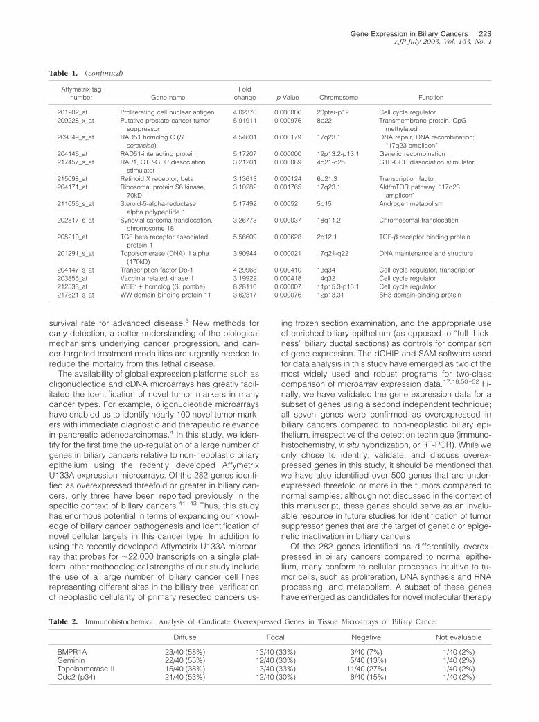
Table 2. Immunohistochemical Analysis of Candidate Overexpressed Genes in Tissue Microarrays of Biliary Cancer
Diffuse Focal Negative Not evaluable
BMPR1A 23/40 (58%) 13/40 (33%) 3/40 (7%) 1/40 (2%)Geminin 22/40 (55%) 12/40 (30%) 5/40 (13%) 1/40 (2%)Topoisomerase II 15/40 (38%) 13/40 (33%) 11/40 (27%) 1/40 (2%)Cdc2 (p34) 21/40 (53%) 12/40 (30%) 6/40 (15%) 1/40 (2%)
Table 1. (continued)
Affymetrix tagnumber Gene name
Foldchange p Value Chromosome Function
201202_at Proliferating cell nuclear antigen 4.02376 0.000006 20pter-p12 Cell cycle regulator209228_x_at Putative prostate cancer tumor
suppressor5.91911 0.000976 8p22 Transmembrane protein, CpG
methylated209849_s_at RAD51 homolog C (S.
cerevisiae)4.54601 0.000179 17q23.1 DNA repair, DNA recombination;
“17q23 amplicon”204146_at RAD51-interacting protein 5.17207 0.000000 12p13.2-p13.1 Genetic recombination217457_s_at RAP1, GTP-GDP dissociation
stimulator 13.21201 0.000089 4q21-q25 GTP-GDP dissociation stimulator
215098_at Retinoid X receptor, beta 3.13613 0.000124 6p21.3 Transcription factor204171_at Ribosomal protein S6 kinase,
70kD3.10282 0.001765 17q23.1 Akt/mTOR pathway; “17q23
amplicon”211056_s_at Steroid-5-alpha-reductase,
alpha polypeptide 15.17492 0.00052 5p15 Androgen metabolism
202817_s_at Synovial sarcoma translocation,chromosome 18
3.26773 0.000037 18q11.2 Chromosomal translocation
205210_at TGF beta receptor associatedprotein 1
5.56609 0.000628 2q12.1 TGF-� receptor binding protein
201291_s_at Topoisomerase (DNA) II alpha(170kD)
3.90944 0.000021 17q21-q22 DNA maintenance and structure
204147_s_at Transcription factor Dp-1 4.29968 0.000410 13q34 Cell cycle regulator, transcription203856_at Vaccinia related kinase 1 3.19922 0.000418 14q32 Cell cycle regulator212533_at WEE1� homolog (S. pombe) 8.28110 0.000007 11p15.3-p15.1 Cell cycle regulator217821_s_at WW domain binding protein 11 3.62317 0.000076 12p13.31 SH3 domain-binding protein
Gene Expression in Biliary Cancers 223AJP July 2003, Vol. 163, No. 1
224 Hansel et alAJP July 2003, Vol. 163, No. 1
in several cancer models, and thus, their reported over-expression in biliary cancers provides an important basisfor future in vitro and in vivo trials, targeting the corre-sponding proteins. For example, the cdc2/p34 protein,
which we confirmed as overexpressed in our biliary can-cer tissue microarrays, is the catalytic subunit of a proteinkinase complex, called the M-phase promoting factorthat induces entry into mitosis and is universal amongeukaryotes.22 Recently, cdc2/p34 has also been demon-strated to play a role in promoting cell survival, mediatedby its site-specific phosphorylation and activation of theanti-apoptotic protein, survivin.53 Survivin interferes withspontaneous and chemotherapy-induced cell death, andthus loss of survivin activation by pharmacological inhi-bition of cdc2/p34 has emerged as a novel chemothera-peutic strategy. Topoisomerase II�, the overexpressionof which was also confirmed in our tissue microarrays, isimportant in mediating susceptibility to drugs that specif-ically target this enzyme; response to topoisomerase in-hibitors is enhanced in breast cancers that overexpresstopo II,54,55 providing an excellent example of how mo-lecular events can direct choice of therapeutic regimens.
Growth factors and growth factor receptors that wereup-regulated in biliary cancers included, among others,hepatocyte growth factor (HGF) (7-fold), and the insulin-like growth factor 1 (IGF-1) receptor (3.5-fold). HGF hasbeen previously reported as up-regulated in biliary can-cers, often in conjunction with its receptor, c-met.42 Al-though c-met was not included in the gene list that com-pared all cancers versus normals, c-met was one of theadditional genes that emerged in the comparison of celllines versus normals (http://pathology2.jhu.edu/gbbd/mi-croarray). HGF is identical to scatter factor, and it is apotent mitogen for hepatocytes. Biliary epithelial cellsproduce little or no HGF, while biliary cancers acquire theability to secrete this growth factor in conjunction withup-regulation of c-met.56 In addition to biliary cancers,dysregulation of the HGF/c-met pathway has been re-ported in hepatocellular, lung, prostate, and breast can-cers, glioblastomas, and melanomas, wherein this path-way promotes angiogenesis, cellular motility, growth, andinvasion.26,57,58 HGF may stimulate tumor invasion in anautocrine loop through c-met, via down-regulation of cel-
Figure 3. Non-radioisotopic in situ hybridization for validation of Islet-1expression in biliary cancer using a biliary cancer tissue microarray. Islet-1transcripts are detected in a biliary adenocarcinoma using the ISL-1 antisenseriboprobe (arrows) (A), but transcripts are not detectable in normal biliaryepithelium using an antisense riboprobe (B), or in the serial section of thecancer using the ISL-1 sense (control) riboprobe (C).
Figure 4. RT-PCR validation of overexpressed genes in biliary cancers. Twogenes overexpressed in the Affymetrix analysis, homeobox B7 (HoxB7) anddickkopf-1 homolog (Dkk1), were selected for RT-PCR in five biliary cancercell lines (GBH3, SNU1079, SNU308, SNU245, and HuCCT1), and threesamples of normal biliary epithelium. HoxB7 expression is absent in thenormal epithelium, but is present in 4 of 5 biliary cancer cell lines; watercontrol is appropriately negative (top panel). Dkk-1 expression is present innormal epithelium, but its expression is up-regulated in 5 of 5 cell lines; watercontrol is appropriately negative (middle panel). GAPDH is used as anexternal control (bottom panel).
Figure 2. Immunohistochemical validation of genes overexpressed in biliary cancers using a biliary cancer tissue microarray. Bone morphogenetic proteinreceptor type 1A (BMPR1A) expression in normal biliary epithelium (A) and in adenocarcinoma of the biliary tract (B). BMPR1A is absent in normal biliaryepithelium, but is diffusely expressed in a cytoplasmic and membranous distribution in the invasive adenocarcinoma. Geminin expression is absent in normalbiliary epithelium (C) while biliary tract adenocarcinoma is diffusely positive (D). Cdc2 (p34) expression is absent in normal biliary epithelium (E), while it isdiffusely expressed in biliary tract adenocarcinoma (F). Topoisomerase II � expression is absent in normal biliary epithelium (G) but diffuse nuclear labeling isseen in adenocarcinoma of the biliary tract (H).
Gene Expression in Biliary Cancers 225AJP July 2003, Vol. 163, No. 1
lular adhesion molecules such as E-cadherin.59 Besidesproviding an avenue for therapy by signal antagonism,HGF is also useful as a potential diagnostic tool in biliarycancer patients. Elevated serum HGF levels are found ina variety of cancer types, and may correlate with prog-nosis.60–62 Similarly, overexpression of the IGF-1 recep-tor is commonly found in many cancers, and its level ofexpression may also correlate with prognosis.28,63 As acell surface molecule upstream of many critical signalingpathways, the IGF-1 receptor represents an importanttarget for cancer therapy. Indeed, antagonism of cellularIGF-1 receptor by overexpression of dominant-negativemutant forms or by interference using small interferingRNA (siRNA) results in decreased tumorigenicity in invitro models.64,65 Thus, the IGF-1 receptor representsanother potential target for therapy identified in biliarycancers.
The mammalian target of rapamycin (mTOR) is a mem-ber of a recently identified family of protein kinasestermed phosphoinositide 3-kinase (PI3K)-related ki-nases, which are involved in many critical regulatorycellular functions pertaining to cell cycle progression,DNA repair, and DNA damage. mTOR functions down-stream of the proto-oncogenes PI3K and Akt, and is itselfupstream of two distinct pathways that control translationof specific subsets of mRNA, one involving ribosomalprotein p70 synthase kinase (p70s6k), and the other eu-karyotic initiation factor 4E binding protein-1 (4EBP1).66
In response to PI3K/Akt signaling, mTOR rapidly phos-phorylates both p70s6k and 4EBP1, the latter leading torelease of eIF4E, resulting in initiation of translation; theseactions of mTOR can be inhibited by the administration ofthe macrolide rapamycin. Currently, CCI-779, a watersoluble analog of rapamycin is undergoing evaluation asan anti-tumor agent in a variety of cancers.40 It is likelythat a subset of biliary cancers will be resistant to inhibi-tion of mTOR. The identification of two pathway genes,p70s6k and eIF4E, that are differentially up-regulated inbiliary cancers compared to normal epithelium providealternative downstream targets for inhibition.
Up-regulation of the pro-inflammatory enzyme cytoso-lic phospholipase A2 (cPLA2) has been previously re-ported in biliary cancers.41 Phospholipase A2s are agroup of enzymes that catalyze the hydrolysis of thesn-2-ester bond of phospholipids, resulting in productionof free fatty acid and lysophospholipids, which can thenbe further metabolized to produce eicosanoids andplatelet-activating factor.67 These lipid molecules arecrucial for various cellular responses, such as inflamma-tion, signal transduction, and cell proliferation. cPLA2 is akey enzyme in the liberation of arachidonic acid (AA)from membrane phospholipids for subsequent produc-tion of bioactive eicosanoids in activated cells; notably,cPLA2 is itself activated in response to signaling mediatedby another differentially overexpressed gene, hepatocytegrowth factor.68 Increasing evidence has suggested acausal relationship between cancer development, includingcholangiocarcinoma, and increased expression of eico-sanoid-forming enzymes. For example, increased expres-sion of cyclooxygenase (COX) enzymes has been reportedin biliary cancers.69,70 Similar to the anti-proliferative effects
of COX inhibition, targeting cPLA2 also results in decreasedgrowth of cholangiocarcinoma cells.41 Thus, the demon-stration of up-regulated cPLA2 in biliary cancers providesanother important potential therapeutic target for the controlof biliary cancer and, perhaps, chemoprevention in patientswith precursor conditions such as primary sclerosingcholangitis.
Several genes involved in steroid metabolism, specif-ically the estrogen/androgen pathways (estrogen-relatedreceptor-�, sterol isomerase, cyclophilin D, and steroid-5-�-reductase) were also up-regulated in biliary cancerscompared to normal epithelium.35–37 Biliary cancers, es-pecially those arising in the gallbladder, occur more fre-quently in women,71 and this has been attributed to theincreased incidence of gallstones in women;2 however,direct or indirect estrogen-related effects may also func-tion to promote carcinogenesis. The expression of sterolisomerase (emopamil-binding protein) correlates withpoor prognosis in breast cancers, although the mecha-nistic aspects remain poorly understood.72 Notably,SR31747A, a recently described synthetic ligand withanti-proliferative activity in numerous cell lines binds toand inhibits sterol isomerase, and overexpression of theenzyme reverts the anti-proliferative effects of this drug.73
Sterol isomerase also binds to and is inhibited by thesynthetic estrogen homolog 4-hydroxytamoxifen.74 Addi-tional functional studies are warranted to demonstratewhether inhibition of sterol isomerase can emerge as atherapeutic strategy in biliary cancers. We also identifiedan up-regulation of the estrogen-related receptor-� genein our study. Estrogen-related receptor-� is a member ofan orphan nuclear receptor family that shares significanthomology with estrogen receptors and, although this re-ceptor does not recognize naturally occurring estrogens,is capable of binding to the estrogen response ele-ment.75,76 Like sterol isomerase, estrogen-related recep-tor-� binds to and is inhibited by tamoxifen.77
Cystathionine � synthase (CBS), which is mutated inpatients with homocystinuria, is a cytosolic enzyme thatalso modulates sensitivity to the cytotoxic drug, cytosinearabinoside (ara-C).29,78 Neoplastic cells with high levelsof CBS demonstrate increased sensitivity to ara-C. There-fore, a sevenfold up-regulation of CBS transcripts in bil-iary cancers merits considering ara-C-containing regi-mens for therapy of biliary cancers. Finally, we also reportup-regulation of dCMP deaminase, whose product is thetarget enzyme for the cytotoxic agent 2�,2�-difluorodeoxy-cytidine (gemcitabine).30,79 Gemcitabine has emergedas a widely used chemotherapeutic agent for solidgastrointestinal malignancies, including biliary can-cers. Fourfold up-regulation of dCMP deaminase inbiliary cancers may suggest that, while gemcitabineshould be considered as a viable therapeutic option,the corresponding dosages may need to be calibratedaccordingly.
In summary, we report for the first time a global geneexpression analysis of biliary cancers using Affymetrixoligonucleotide microarrays and a combination of pri-mary resected tumors, biliary cell lines, and non-neoplas-tic biliary epithelial scrapings. We report 282 uniqueknown genes as being differentially up-regulated three-
226 Hansel et alAJP July 2003, Vol. 163, No. 1
fold or greater in the cancers compared to normal epi-thelium, and validate a subset of genes using an alterna-tive platform. The differentially overexpressed genes arepublicly available on our website at http://pathology2.jhu.edu/gbbd/microarray, and represents an enormousresource for the scientific community to study the patho-genesis of this lethal disease and develop appropriatecancer-specific targeted therapies.
Acknowledgments
We thank Leighton Stein for his expert assistance.
References
1. Greenlee RT, Hill-Harmon MB, Murray T, Thun M: Cancer statistics,2001. CA Cancer J Clin 2001, 51:15–36
2. de Groen PC, Gores GJ, LaRusso NF, Gunderson LL, Nagorney DM:Biliary tract cancers. N Engl J Med 1999, 341:1368–1378
3. Lazcano-Ponce EC, Miquel JF, Munoz N, Herrero R, Ferrecio C,Wistuba II, Alonso de Ruiz P, Aristi Urista G, Nervi F: Epidemiologyand molecular pathology of gallbladder cancer. CA Cancer J Clin2001, 51:349–364
4. Iacobuzio-Donahue CA, Maitra A, Shen-Ong GL, van Heek T, AshfaqR, Meyer R, Walter K, Berg K, Hollingsworth MA, Cameron JL, Yeo CJ,Kern SE, Goggins M, Hruban RH: Discovery of novel tumor markers ofpancreatic cancer using global gene expression technology. Am JPathol 2002, 160:1239–1249
5. Garber ME, Troyanskaya OG, Schluens K, Petersen S, Thaesler Z,Pacyna-Gengelbach M, van de Rijn M, Rosen GD, Perou CM, WhyteRI, Altman RB, Brown PO, Botstein D, Petersen I: Diversity of geneexpression in adenocarcinoma of the lung. Proc Natl Acad Sci USA2001, 98:13784–13789
6. Perou CM, Sorlie T, Eisen MB, van de Rijn M, Jeffrey SS, Rees CA,Pollack JR, Ross DT, Johnsen H, Akslen LA, Fluge O, Pergamen-schikov A, Williams C, Zhu SX, Lonning PE, Borresen-Dale AL, BrownPO, Botstein D: Molecular portraits of human breast tumours. Nature2000, 406:747–752
7. Dhanasekaran SM, Barrette TR, Ghosh D, Shah R, Varambally S,Kurachi K, Pienta KJ, Rubin MA, Chinnaiyan AM: Delineation of prog-nostic biomarkers in prostate cancer. Nature 2001, 412:822–826
8. Maitra A, Wistuba II, Virmani AK, Sakaguchi M, Park I, Stucky A,Milchgrub S, Gibbons D, Minna JD, Gazdar AF: Enrichment of epi-thelial cells for molecular studies. Nat Med 1999, 5:459–463
9. Saijyo S, Kudo T, Suzuki M, Katayose Y, Shinoda M, Muto T, FukuharaK, Suzuki T, Matsuno S: Establishment of a new extrahepatic bile ductcarcinoma cell line, TFK-1. Tohoku J Exp Med 1995, 177:61–71
10. Steffen M, Zuehlke I, Scherdin U: Motility factors identified in super-natants of human cholangiocarcinoma cell lines. Int J Oncol 2001,18:1107–1112
11. Sugimoto H, Nishino H: Effect of recombinant human basic fibroblastgrowth factor (bFGF) on the growth of human tumor cell lines. HumCell 1996, 9:129–140
12. Miyazaki M, Ohashi R, Tsuji T, Mihara K, Gohda E, Namba M: Trans-forming growth factor-� 1 stimulates or inhibits cell growth via down-or up-regulation of p21/Waf1. Biochem Biophys Res Commun 1998,246:873–880
13. Ku JL, Yoon KA, Kim IJ, Kim WH, Jang JY, Suh KS, Kim SW, Park YH,Hwang JH, Yoon YB, Park JG: Establishment and characterisation ofsix human biliary tract cancer cell lines. Br J Cancer 2002, 87:187–193
14. Li H, Shimura H, Aoki Y, Date K, Matsumoto K, Nakamura T, TanakaM: Hepatocyte growth factor stimulates the invasion of gallbladdercarcinoma cell lines in vitro. Clin Exp Metastasis 1998, 16:74–82
15. Li C, Hung Wong W: Model-based analysis of oligonucleotide arrays:model validation, design issues, and standard error application. Ge-nome Biol 2001, 2:RESEARCH0032
16. Eisen MB, Spellman PT, Brown PO, Botstein D: Cluster analysis and
display of genome-wide expression patterns. Proc Natl Acad Sci USA1998, 95:14863–14868
17. Tusher VG, Tibshirani R, Chu G: Significance analysis of microarraysapplied to the ionizing radiation response. Proc Natl Acad Sci USA2001, 98:5116–5121
18. Xu Y, Selaru FM, Yin J, Zou TT, Shustova V, Mori Y, Sato F, Liu TC,Olaru A, Wang S, Kimos MC, Perry K, Desai K, Greenwald BD, KrasnaMJ, Shibata D, Abraham JM, Meltzer SJ: Artificial neural networks andgene filtering distinguish between global gene expression profiles ofBarrett’s esophagus and esophageal cancer. Cancer Res 2002, 62:3493–3497
19. van Heek NT, Meeker AK, Kern SE, Yeo CJ, Lillemoe KD, CameronJL, Offerhaus GJ, Hicks JL, Wilentz RE, Goggins MG, De Marzo AM,Hruban RH, Maitra A: Telomere shortening is nearly universal inpancreatic intraepithelial neoplasia. Am J Pathol 2002, 161:1541–1547
20. Iacobuzio-Donahue CA, Ryu B, Hruban RH, Kern SE: Exploring thehost desmoplastic response to pancreatic carcinoma: gene expres-sion of stromal and neoplastic cells at the site of primary invasion.Am J Pathol 2002, 160:91–99
21. Ortega S, Malumbres M, Barbacid M: Cyclin D-dependent kinases,INK4 inhibitors, and cancer. Biochim Biophys Acta 2002, 1602:73–87
22. O’Connor DS, Wall NR, Porter AC, Altieri DC: A p34(cdc2) survivalcheckpoint in cancer. Cancer Cell 2002, 2:43–54
23. Wohlschlegel JA, Kutok JL, Weng AP, Dutta A: Expression of gemininas a marker of cell proliferation in normal tissues and malignancies.Am J Pathol 2002, 161:267–273
24. Care A, Felicetti F, Meccia E, Bottero L, Parenza M, Stoppacciaro A,Peschle C, Colombo MP: HOXB7: a key factor for tumor-associatedangiogenic switch. Cancer Res 2001, 61:6532–6539
25. Watanabe Y, Nakamura H: Control of chick tectum territory alongdorsoventral axis by Sonic hedgehog. Development 2000, 127:1131–1140
26. Trusolino L, Comoglio PM: Scatter-factor and semaphorin receptors:cell signalling for invasive growth. Nat Rev Cancer 2002, 2:289–300
27. Normanno N, Bianco C, De Luca A, Salomon DS: The role of EGF-related peptides in tumor growth. Front Biosci 2001, 6:D685–707
28. Yu H, Rohan T: Role of the insulin-like growth factor family in cancerdevelopment and progression. J Natl Cancer Inst 2000, 92:1472–1489
29. Taub JW, Huang X, Ge Y, Dutcher JA, Stout ML, Mohammad RM,Ravindranath Y, Matherly LH: Cystathionine-�-synthase cDNA trans-fection alters the sensitivity and metabolism of 1-�-D-arabinofurano-sylcytosine in CCRF-CEM leukemia cells in vitro and in vivo: a modelof leukemia in Down syndrome. Cancer Res 2000, 60:6421–6426
30. Rieger J, Durka S, Streffer J, Dichgans J, Weller M: Gemcitabinecytotoxicity of human malignant glioma cells: modulation by antioxi-dants, BCL-2, and dexamethasone. Eur J Pharmacol 1999, 365:301–308
31. Politi PM, Xie F, Dahut W, Ford Jr H, Kelley JA, Bastian A, Setser A,Allegra CJ, Chen AP, Hamilton JM: Phase I clinical trial of continuousinfusion cyclopentenyl cytosine. Cancer Chemother Pharmacol 1995,36:513–523
32. Zhou XP, Woodford-Richens K, Lehtonen R, Kurose K, Aldred M,Hampel H, Launonen V, Virta S, Pilarski R, Salovaara R, Bodmer WF,Conrad BA, Dunlop M, Hodgson SV, Iwama T, Jarvinen H, Kello-kumpu I, Kim JC, Leggett B, Markie D, Mecklin JP, Neale K, PhillipsR, Piris J, Rozen P, Houlston RS, Aaltonen LA, Tomlinson IP, Eng C:Germline mutations in BMPR1A/ALK3 cause a subset of cases ofjuvenile polyposis syndrome and of Cowden and Bannayan-Riley-Ruvalcaba syndromes. Am J Hum Genet 2001, 69:704–711
33. Zhou S, Kinzler KW, Vogelstein B: Going mad with Smads. N EnglJ Med 1999, 341:1144–1146
34. Wurthner JU, Frank DB, Felici A, Green HM, Cao Z, Schneider MD,McNally JG, Lechleider RJ, Roberts AB: Transforming growth factor-�receptor-associated protein 1 is a Smad4 chaperone. J Biol Chem2001, 276:19495–19502
35. Ariazi EA, Clark GM, Mertz JE: Estrogen-related receptor � andestrogen-related receptor � associate with unfavorable and favorablebiomarkers, respectively, in human breast cancer. Cancer Res 2002,62:6510–6518
36. Villalva C, Trempat P, Greenland C, Thomas C, Girard JP, Moebius F,Delsol G, Brousset P: Isolation of differentially expressed genes in
Gene Expression in Biliary Cancers 227AJP July 2003, Vol. 163, No. 1
NPM-ALK-positive anaplastic large cell lymphoma. Br J Haematol2002, 118:791–798
37. Brawley OW, Ford LG, Thompson I, Perlman JA, Kramer BS: 5-�-reductase inhibition and prostate cancer prevention. Cancer Epide-miol Biomarkers Prev 1994, 3:177–182
38. Osterstrom A, Dimberg J, Fransen K, Soderkvist P: Expression ofcytosolic and group X secretory phospholipase A(2) genes in humancolorectal adenocarcinomas. Cancer Lett 2002, 182:175–182
39. Graff JR, Konicek BW, Deddens JA, Chedid M, Hurst BM, Colligan B,Neubauer BL, Carter HW, Carter JH: Expression of group IIa secre-tory phospholipase A2 increases with prostate tumor grade. ClinCancer Res 2001, 7:3857–3861
40. Hidalgo M, Rowinsky EK: The rapamycin-sensitive signal transduc-tion pathway as a target for cancer therapy. Oncogene 2000, 19:6680–6686
41. Wu T, Han C, Lunz JG, 3rd, Michalopoulos G, Shelhamer JH, Dem-etris AJ: Involvement of 85-kd cytosolic phospholipase A(2) andcyclooxygenase-2 in the proliferation of human cholangiocarcinomacells. Hepatology 2002, 36:363–373
42. Sirica AE, Lai GH, Zhang Z: Biliary cancer growth factor pathways,cyclo-oxygenase-2, and potential therapeutic strategies. J Gastroen-terol Hepatol 2001, 16:363–372
43. Batheja N, Suriawinata A, Saxena R, Ionescu G, Schwartz M, ThungSN: Expression of p53 and PCNA in cholangiocarcinoma and primarysclerosing cholangitis. Mod Pathol 2000, 13:1265–1268
44. Lee MG, Norbury CJ, Spurr NK, Nurse P: Regulated expression andphosphorylation of a possible mammalian cell-cycle control protein.Nature 1988, 333:676–679
45. Tsai-Pflugfelder M, Liu LF, Liu AA, Tewey KM, Whang-Peng J, Knut-sen T, Huebner K, Croce CM, Wang JC: Cloning and sequencing ofcDNA encoding human DNA topoisomerase II and localization of thegene to chromosome region 17q21–22. Proc Natl Acad Sci USA1988, 85:7177–7181
46. Wang M, Drucker DJ: The LIM domain homeobox gene isl-1 is apositive regulator of islet cell-specific proglucagon gene transcrip-tion. J Biol Chem 1995, 270:12646–12652
47. van den Brink GR, Hardwick JC, Tytgat GN, Brink MA, Ten Kate FJ,Van Deventer SJ, Peppelenbosch MP: Sonic hedgehog regulatesgastric gland morphogenesis in man and mouse. Gastroenterology2001, 121:317–328
48. Naora H, Yang YQ, Montz FJ, Seidman JD, Kurman RJ, Roden RB: Aserologically identified tumor antigen encoded by a homeobox genepromotes growth of ovarian epithelial cells. Proc Natl Acad Sci USA2001, 98:4060–4065
49. Shou J, Ali-Osman F, Multani AS, Pathak S, Fedi P, Srivenugopal KS:Human Dkk-1, a gene encoding a Wnt antagonist, responds to DNAdamage and its overexpression sensitizes brain tumor cells to apo-ptosis following alkylation damage of DNA. Oncogene 2002, 21:878–889
50. Ramalho-Santos M, Yoon S, Matsuzaki Y, Mulligan RC, Melton DA:“Stemness”: transcriptional profiling of embryonic and adult stemcells. Science 2002, 298:597–600
51. Storch KF, Lipan O, Leykin I, Viswanathan N, Davis FC, Wong WH,Weitz CJ: Extensive and divergent circadian gene expression in liverand heart. Nature 2002, 417:78–83
52. Huang Y, Prasad M, Lemon WJ, Hampel H, Wright FA, Kornacker K,LiVolsi V, Frankel W, Kloos RT, Eng C, Pellegata NS, de la ChapelleA: Gene expression in papillary thyroid carcinoma reveals highlyconsistent profiles. Proc Natl Acad Sci USA 2001, 98:15044–15049
53. O’Connor DS, Grossman D, Plescia J, Li F, Zhang H, Villa A, TogninS, Marchisio PC, Altieri DC: Regulation of apoptosis at cell division byp34cdc2 phosphorylation of survivin. Proc Natl Acad Sci USA 2000,97:13103–13107
54. Coon JS, Marcus E, Gupta-Burt S, Seelig S, Jacobson K, Chen S,Renta V, Fronda G, Preisler HD: Amplification and overexpression oftopoisomerase II� predict response to anthracycline-based therapyin locally advanced breast cancer. Clin Cancer Res 2002, 8:1061–1067
55. Di Leo A, Gancberg D, Larsimont D, Tanner M, Jarvinen T, Rouas G,Dolci S, Leroy JY, Paesmans M, Isola J, Piccart MJ: HER-2 amplifi-cation and topoisomerase II� gene aberrations as predictive markersin node-positive breast cancer patients randomly treated either with
an anthracycline-based therapy or with cyclophosphamide, metho-trexate, and 5-fluorouracil. Clin Cancer Res 2002, 8:1107–1116
56. Yokomuro S, Tsuji H, Lunz JG, 3rd, Sakamoto T, Ezure T, Murase N,Demetris AJ: Growth control of human biliary epithelial cells by inter-leukin 6, hepatocyte growth factor, transforming growth factor �1,and activin A: comparison of a cholangiocarcinoma cell line withprimary cultures of non-neoplastic biliary epithelial cells. Hepatology2000, 32:26–35
57. Haddad R, Lipson KE, Webb CP: Hepatocyte growth factor expres-sion in human cancer and therapy with specific inhibitors. AnticancerRes 2001, 21:4243–4252
58. Maulik G, Shrikhande A, Kijima T, Ma PC, Morrison PT, Salgia R: Roleof the hepatocyte growth factor receptor, c-met, in oncogenesis andpotential for therapeutic inhibition. Cytokine Growth Factor Rev 2002,13:41–59
59. Li G, Schaider H, Satyamoorthy K, Hanakawa Y, Hashimoto K, HerlynM: Down-regulation of E-cadherin and desmoglein 1 by autocrinehepatocyte growth factor during melanoma development. Oncogene2001, 20:8125–8135
60. Naughton M, Picus J, Zhu X, Catalona WJ, Vollmer RT, Humphrey PA:Scatter factor-hepatocyte growth factor elevation in the serum ofpatients with prostate cancer. J Urol 2001, 165:1325–1328
61. Junbo H, Li Q, Zaide W, Yunde H: Increased level of serum hepato-cyte growth factor/scatter factor in liver cancer is associated withtumor metastasis. In Vivo 1999, 13:177–180
62. Takigawa N, Segawa Y, Maeda Y, Takata I, Fujimoto N: Serum he-patocyte growth factor/scatter factor levels in small cell lung cancerpatients. Lung Cancer 1997, 17:211–218
63. Chakravarti A, Loeffler JS, Dyson NJ: Insulin-like growth factor recep-tor I mediates resistance to anti-epidermal growth factor receptortherapy in primary human glioblastoma cells through continued acti-vation of phosphoinositide 3-kinase signaling. Cancer Res 2002,62:200–207
64. Scotlandi K, Maini C, Manara MC, Benini S, Serra M, Cerisano V,Strammiello R, Baldini N, Lollini PL, Nanni P, Nicoletti G, Picci P:Effectiveness of insulin-like growth factor I receptor antisense strat-egy against Ewing’s sarcoma cells. Cancer Gene Ther 2002, 9:296–307
65. Nakamura K, Hongo A, Kodama J, Miyagi Y, Yoshinouchi M, Kudo T:Down-regulation of the insulin-like growth factor I receptor by anti-sense RNA can reverse the transformed phenotype of human cervicalcancer cell lines. Cancer Res 2000, 60:760–765
66. Aoki M, Blazek E, Vogt PK: A role of the kinase mTOR in cellulartransformation induced by the oncoproteins P3k and Akt. Proc NatlAcad Sci USA 2001, 98:136–141
67. Dennis EA: Diversity of group types, regulation, and function of phos-pholipase A2. J Biol Chem 1994, 269:13057–13060
68. Skouteris GG, Schroder CH: Cytosolic phospholipase A2 is activatedby the hepatocyte growth factor receptor-kinase in Madin Darbycanine kidney cells. J Cell Sci 1997, 110:1655–1663
69. Hayashi N, Yamamoto H, Hiraoka N, Dono K, Ito Y, Okami J, KondoM, Nagano H, Umeshita K, Sakon M, Matsuura N, Nakamori S,Monden M: Differential expression of cyclooxygenase-2 (COX-2) inhuman bile duct epithelial cells and bile duct neoplasm. Hepatology2001, 34:638–650
70. Yoon JH, Higuchi H, Werneburg NW, Kaufmann SH, Gores GJ: Bileacids induce cyclooxygenase-2 expression via the epidermal growthfactor receptor in a human cholangiocarcinoma cell line. Gastroen-terology 2002, 122:985–993
71. Henson DE, Albores-Saavedra J, Corle D: Carcinoma of thegallbladder: histologic types, stage of disease, grade, and survivalrates. Cancer 1992, 70:1493–1497
72. Simony-Lafontaine J, Esslimani M, Bribes E, Gourgou S, Lequeux N,Lavail R, Grenier J, Kramar A, Casellas P: Immunocytochemicalassessment of �-1 receptor and human sterol isomerase in breastcancer and their relationship with a series of prognostic factors. Br JCancer 2000, 82:1958–1966
73. Labit-Le Bouteiller C, Jamme MF, David M, Silve S, Lanau C, Dhers C,Picard C, Rahier A, Taton M, Loison G, Caput D, Ferrara P, Lupker J:Antiproliferative effects of SR31747A in animal cell lines are mediatedby inhibition of cholesterol biosynthesis at the sterol isomerase step.Eur J Biochem 1998, 256:342–349
228 Hansel et alAJP July 2003, Vol. 163, No. 1

74. Cho SY, Kim JH, Paik YK: Cholesterol biosynthesis from lanosterol:differential inhibition of sterol � 8-isomerase and other lanosterol-converting enzymes by tamoxifen. Mol Cells 1998, 8:233–239
75. Giguere V: To ERR in the estrogen pathway. Trends Endocrinol Metab2002, 13:220–225
76. Heard DJ, Norby PL, Holloway J, Vissing H: Human ERR�, a thirdmember of the estrogen receptor-related receptor (ERR) subfamily oforphan nuclear receptors: tissue-specific isoforms are expressedduring development and in the adult. Mol Endocrinol 2000, 14:382–392
77. Tremblay GB, Bergeron D, Giguere V: 4-Hydroxytamoxifen is an
isoform-specific inhibitor of orphan estrogen-receptor-related(ERR) nuclear receptors � and �. Endocrinology 2001, 142:4572–4575
78. Taub JW, Matherly LH, Stout ML, Buck SA, Gurney JG, RavindranathY: Enhanced metabolism of 1-�-D-arabinofuranosylcytosine in Downsyndrome cells: a contributing factor to the superior event-free sur-vival of Down syndrome children with acute myeloid leukemia. Blood1996, 87:3395–3403
79. Plunkett W, Huang P, Searcy CE, Gandhi V: Gemcitabine: preclinicalpharmacology and mechanisms of action. Semin Oncol 1996, 23:3–15
Gene Expression in Biliary Cancers 229AJP July 2003, Vol. 163, No. 1
Copyright © 2022 FDOKUMEN