Identification of Etoposide Glucuronide as a Major Metabolite of Etoposide in the Rat and Rabbit1
7
1988;48:1829-1834. Cancer Res Kenneth Hande, Lowell Anthony, Reta Hamilton, et al. Etoposide in the Rat and Rabbit Identification of Etoposide Glucuronide as a Major Metabolite of Updated version http://cancerres.aacrjournals.org/content/48/7/1829 Access the most recent version of this article at: E-mail alerts related to this article or journal. Sign up to receive free email-alerts Subscriptions Reprints and . [email protected] Department at To order reprints of this article or to subscribe to the journal, contact the AACR Publications Permissions . [email protected] Department at To request permission to re-use all or part of this article, contact the AACR Publications on July 14, 2014. © 1988 American Association for Cancer Research. cancerres.aacrjournals.org Downloaded from on July 14, 2014. © 1988 American Association for Cancer Research. cancerres.aacrjournals.org Downloaded from
-
Upload
independent -
Category
Documents
-
view
1 -
download
0
Transcript of Identification of Etoposide Glucuronide as a Major Metabolite of Etoposide in the Rat and Rabbit1
1988;48:1829-1834. Cancer Res Kenneth Hande, Lowell Anthony, Reta Hamilton, et al. Etoposide in the Rat and RabbitIdentification of Etoposide Glucuronide as a Major Metabolite of
Updated version
http://cancerres.aacrjournals.org/content/48/7/1829
Access the most recent version of this article at:
E-mail alerts related to this article or journal.Sign up to receive free email-alerts
Subscriptions
Reprints and
To order reprints of this article or to subscribe to the journal, contact the AACR Publications
Permissions
To request permission to re-use all or part of this article, contact the AACR Publications
on July 14, 2014. © 1988 American Association for Cancer Research. cancerres.aacrjournals.org Downloaded from on July 14, 2014. © 1988 American Association for Cancer Research. cancerres.aacrjournals.org Downloaded from
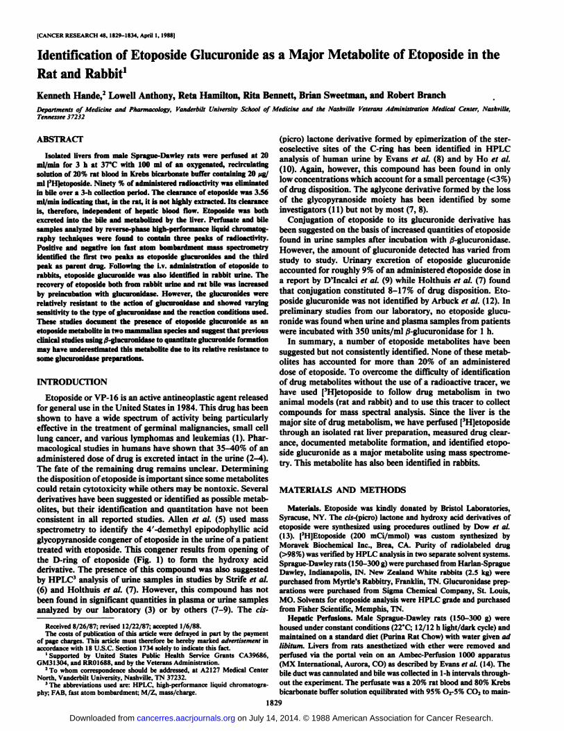
[CANCER RESEARCH 48, 1829-1834, April 1, 1988)
Identification of Etoposide Glucuronide as a Major Metabolite of Etoposide in theRat and Rabbit1
Kenneth I lande,2 Lowell Anthony, Reta Hamilton, Rita Bennett, Brian Sweetman, and Robert Branch
Departments of Medicine and Pharmacology, Vanderbilt University School of Medicine and the Nashville Veterans Administration Medical Center, Nashville,Tennessee 37232
ABSTRACT
Isolated livers from male Sprague-Dawley rats were perfused at 20ml/min for 3 h at 37°C with 100 ml of an oxygenated, recirculating
solution of 20% rat blood in Krebs bicarbonate buffer containing 20 Mg/ml [3H]etoposide. Ninety % of administered radioactivity was eliminated
in bile over a 3-h collection period. The clearance of etoposide was 3.56
ml/min indicating that, in the rat, it is not highly extracted. Its clearanceis, therefore, independent of hepatic blood flow. Etoposide was bothexcreted into the bile and metabolized by the liver. Perfusate and bilesamples analyzed by reverse-phase high-performance liquid chromatog-
raphy techniques were found to contain three peaks of radioactivity.Positive and negative ion fast atom bombardment mass spectromerryidentified the first two peaks as etoposide glucuronides and the thirdpeak as parent drug. Following the i.v. administration of etoposide torabbits, etoposide glucuronide was also identified in rabbit urine. Therecovery of etoposide both from rabbit urine and rat bile was increasedby preincubation with glucuronidase. However, the glucuronides wererelatively resistant to the action of glucuronidase and showed varyingsensitivity to the type of glucuronidase and the reaction conditions used.These studies document the presence of etoposide glucuronide as anetoposide metabolite in two mammalian species and suggest that previousclinical studies using 0-glucuronidase to quantitate glucuronide formation
may have underestimated this metabolite due to its relative resistance tosome glucuronidase preparations.
INTRODUCTION
Etoposide or VP-16 is an active antineoplastic agent releasedfor general use in the United States in 1984. This drug has beenshown to have a wide spectrum of activity being particularlyeffective in the treatment of germinal malignancies, small celllung cancer, and various lymphomas and leukemia s (1). Pharmacological studies in humans have shown that 35-40% of anadministered dose of drug is excreted intact in the urine (2-4).
The fate of the remaining drug remains unclear. Determiningthe disposition of etoposide is important since some metabolitescould retain cytotoxicity while others may be nontoxic. Severalderivatives have been suggested or identified as possible metabolites, but their identification and quantitation have not beenconsistent in all reported studies. Allen et al. (5) used massspectrometry to identify the 4'-demethyl epipodophyllic acid
glycopyranoside congener of etoposide in the urine of a patienttreated with etoposide. This congener results from opening ofthe D-ring of etoposide (Fig. 1) to form the hydroxy acidderivative. The presence of this compound was also suggestedby HPLC3 analysis of urine samples in studies by Strife et al.
(6) and Holthuis et al. (7). However, this compound has notbeen found in significant quantities in plasma or urine samplesanalyzed by our laboratory (3) or by others (7-9). The cis-
Received 8/26/87; revised 12/22/87; accepted 1/6/88.The costs of publication of this article were defrayed in part by the payment
of page charges. This article must therefore be hereby marked advertisement inaccordance with 18 U.S.C. Section 1734 solely to indicate this fact.
1Supported by United States Public Health Service Grants CA39686,GM31304, and RR01688, and by the Veterans Administration.
2To whom correspondence should be addressed, at A2127 Medical CenterNorth, Vanderbilt University, Nashville, TN 37232.
'The abbreviations used are: HPLC, high-performance liquid chromatogra-
phy; FAB, fast atom bombardment; M/Z, mass/charge.
(pierò) lactone derivative formed by epimerization of the ster-eoselective sites of the C-ring has been identified in HPLCanalysis of human urine by Evans et al. (8) and by Ho et al.(10). Again, however, this compound has been found in onlylow concentrations which account for a small percentage (<3%)of drug disposition. The aglycone derivative formed by the lossof the glycopyranoside moiety has been identified by someinvestigators (11) but not by most (7, 8).
Conjugation of etoposide to its glucuronide derivative hasbeen suggested on the basis of increased quantities of etoposidefound in urine samples after incubation with /3-glucuronidase.However, the amount of glucuronide detected has varied fromstudy to study. Urinary excretion of etoposide glucuronideaccounted for roughly 9% of an administered etoposide dose ina report by D'Incaici et al. (9) while Holthuis et al. (7) foundthat conjugation constituted 8-17% of drug disposition. Etoposide glucuronide was not identified by Arbuck et al. (12). Inpreliminary studies from our laboratory, no etoposide glucuronide was found when urine and plasma samples from patientswere incubated with 350 units/ml /3-glucuronidase for 1 h.
In summary, a number of etoposide metabolites have beensuggested but not consistently identified. None of these metabolites has accounted for more than 20% of an administereddose of etoposide. To overcome the difficulty of identificationof drug metabolites without the use of a radioactive tracer, wehave used [3H]etoposide to follow drug metabolism in two
animal models (rat and rabbit) and to use this tracer to collectcompounds for mass spectral analysis. Since the liver is themajor site of drug metabolism, we have perfused [3H]etoposide
through an isolated rat liver preparation, measured drug clearance, documented metabolite formation, and identified etoposide glucuronide as a major metabolite using mass spectrometry. This metabolite has also been identified in rabbits.
MATERIALS AND METHODS
Materials. Etoposide was kindly donated by Bristol Laboratories,Syracuse, NY. The ci's-(picro) lactone and hydroxy acid derivatives of
etoposide were synthesized using procedures outlined by Dow et al.(13). [3H]Etoposide (200 mCi/mmol) was custom synthesized by
Moravek Biochemical Inc., Brea, CA. Purity of radiolabeled drug(>98%) was verified by HPLC analysis in two separate solvent systems.Sprague-Dawley rats (150-300 g) were purchased from Harlan-SpragueDawley, Indianapolis, IN. New Zealand White rabbits (2.5 kg) werepurchased from Myrtle's Rabbitry, Franklin, TN. Glucuronidase prep
arations were purchased from Sigma Chemical Company, St. Louis,MO. Solvents for etoposide analysis were HPLC grade and purchasedfrom Fisher Scientific, Memphis, TN.
Hepatic Perfusions. Male Sprague-Dawley rats (150-300 g) werehoused under constant conditions (22°C;12/12 h light/dark cycle) and
maintained on a standard diet (Purina Rat Chow) with water given adlibitum. Livers from rats anesthetized with ether were removed andperfused via the portal vein on an Ambec-Perfusion 1000 apparatus(MX International, Aurora, CO) as described by Evans et al. (14). Thebile duct was cannulated and bile was collected in 1-h intervals throughout the experiment. The perfusate was a 20% rat blood and 80% Krebsbicarbonate buffer solution equilibrated with 95% O2-5% CO2 to main-
1829
on July 14, 2014. © 1988 American Association for Cancer Research. cancerres.aacrjournals.org Downloaded from
ETOPOSIDE METABOLISM
tain a pH of 7.4 at 37°C.The volume of the recirculating perfusate was
100 ml, and a constant flow of 20 ml/min was maintained. After 20min of equilibration, etoposide (2000 n% containing 10.0 ¿tCiI'll]
etoposide) was added in a single bolus to the reservoir. Serial perfusateand bile samples were collected over a 180-min period, centrifuged toremove red cells, and frozen at -70°Cuntil assayed. After 180 min, theliver was removed, weighed, homogenized, and stored at -70°C until
assayed.Chloroform and Ethyl Acetate Extraction. To determine the extrac
tion efficiency of chloroform and ethyl acetate, 500 n\ of a spiked blankbile sample or a 0- to 1-h bile sample from a liver perfused with [3H]-
etoposide were added to each solvent and vortexed. The organic phasewas removed and the bile sample again extracted with 4 ml of eitherchloroform or ethyl acetate. Aliquots (0.5 ml) of both the organic andremaining aqueous phases were removed, radioactivity was counted tocorrect for quench, and the proportion of counts in the organic phasewas determined.
Rabbits. New Zealand White rabbits were given 2 mg etoposidecontaining 14 /iCi of [3H]-labeled drug by a 5-min injection into the
marginal ear vein. Urine was collected daily for 7 days, total radioactivity was counted, and aliquots were analyzed by HPLC to separateparent drug from metabolites.
HPLC Analysis. Perfusate and bile samples were analyzed by modification of the HPLC method of Sinkule and Evans (15). To 0.5 ml ofperfusate was added 0.5 ml of saturated ammonium sulfate followed by4 ml of ethyl acetate. Samples were vortexed for 5 min and centrifugedat 3000 rpni x 10 min. The upper organic layer was collected andsaved. The aqueous layer was reextracted with 4 ml of ethyl acetate andthe resulting organic layer combined with the previously collected ethylacetate layer. The organic extract was evaporated to dryness under anitrogen stream and reconstituted in 200 n\ methanol. Twenty to 200til of this solution were used for HPLC analysis. Bile samples werecentrifuged and 10 to 50 /il directly injected onto the HPLC column.The HPLC apparatus consisted of a Waters M6000 pump, a Waters71OBinjector, and a 4.6- x 250-mm, 10-^m, ^Bondapak phenyl column
(Waters Associates, Milford, MA). Ultraviolet absorbance (254 nm) ofthe eluate was monitored by a Waters 490 variable wavelength detectorcoupled to a Waters data module. A mobile phase of water:acetonitrile:acetic acid (74:25:1) was used to separate etoposide fromits potential metabolites. Eluate from the column was collected in 0.6-
min fractions. Radioactivity in the eluate was determined by adding 5ml of Aquasol (New England Nuclear Corp., Boston, MA) and countingin a liquid scintillation counter using an external standardization procedure to correct for quench.
Kinetic Calculations. Half-life (/,,) measurements were estimated
using linear regression analysis of the logarithm of the perfusate concentration plotted against time. The volume of distribution (yd) wasestimated from the dose divided by the perfusate concentration back
extrapolated to time 0. Total clearance (Clt) was calculated from theformula
Clt = yd x 0.693
ETOPOSIDE
(MW sae)QLUCURONIOE
(MW 177)
ETOPOSIDE GLUCURONIDE(MW 764)
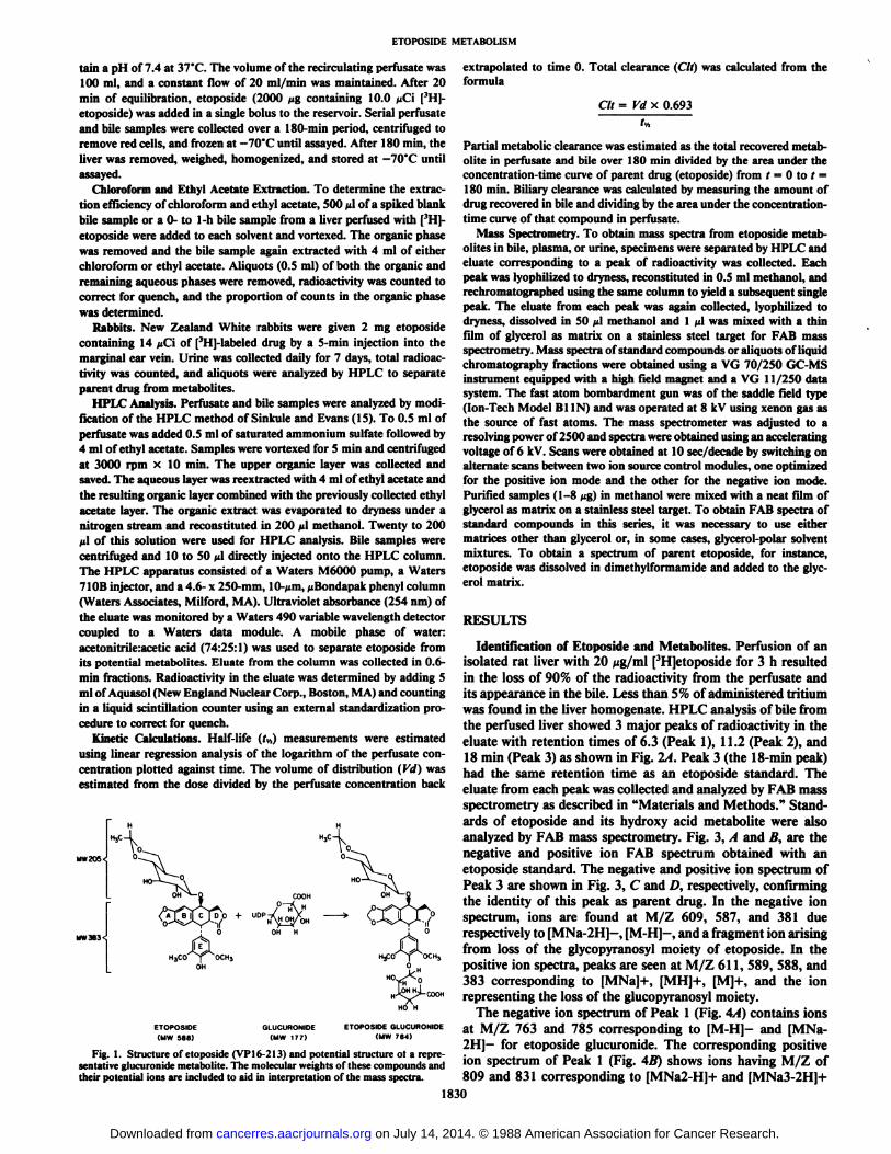
Fig. 1. Structure of etoposide (VP 16-2 13) and potential structure ot a representative glucuronide metabolite. The molecular weights of these compounds andtheir potential ions are included to aid in interpretation of the mass spectra.
Partial metabolic clearance was estimated as the total recovered metabolite in perfusate and bile over 180 min divided by the area under theconcentration-time curve of parent drug (etoposide) from / = 0 to / =180 min. Biliary clearance was calculated by measuring the amount ofdrug recovered in bile and dividing by the area under the concentration-time curve of that compound in perfusate.
Mass Spectrometry. To obtain mass spectra from etoposide metabolites in bile, plasma, or urine, specimens were separated by HPLC andeluate corresponding to a peak of radioactivity was collected. Eachpeak was lyophilized to dryness, reconstituted in 0.5 ml methanol, andrechromatographed using the same column to yield a subsequent singlepeak. The eluate from each peak was again collected, lyophilized todryness, dissolved in 50 ¿ilmethanol and 1 n\ was mixed with a thinfilm of glycerol as matrix on a stainless steel target for FAB massspectrometry. Mass spectra of standard compounds or aliquots of liquidchromatography fractions were obtained using a VG 70/250 GC-MSinstrument equipped with a high field magnet and a VG 11/250 datasystem. The fast atom bombardment gun was of the saddle field type(Ion-Tech Model BUN) and was operated at 8 kV using xenon gas asthe source of fast atoms. The mass spectrometer was adjusted to aresolving power of 2500 and spectra were obtained using an acceleratingvoltage of 6 kV. Scans were obtained at 10 sec/decade by switching onalternate scans between two ion source control modules, one optimizedfor the positive ion mode and the other for the negative ion mode.Purified samples (1-8 Mg)in methanol were mixed with a neat film ofglycerol as matrix on a stainless steel target. To obtain FAB spectra ofstandard compounds in this series, it was necessary to use eithermatrices other than glycerol or, in some cases, glycerol-polar solventmixtures. To obtain a spectrum of parent etoposide, for instance,etoposide was dissolved in dimethylformamide and added to the glycerol matrix.
RESULTS
Identification of Etoposide and Metabolites. Perfusion of anisolated rat liver with 20 /¿g/ml[3H]etoposide for 3 h resulted
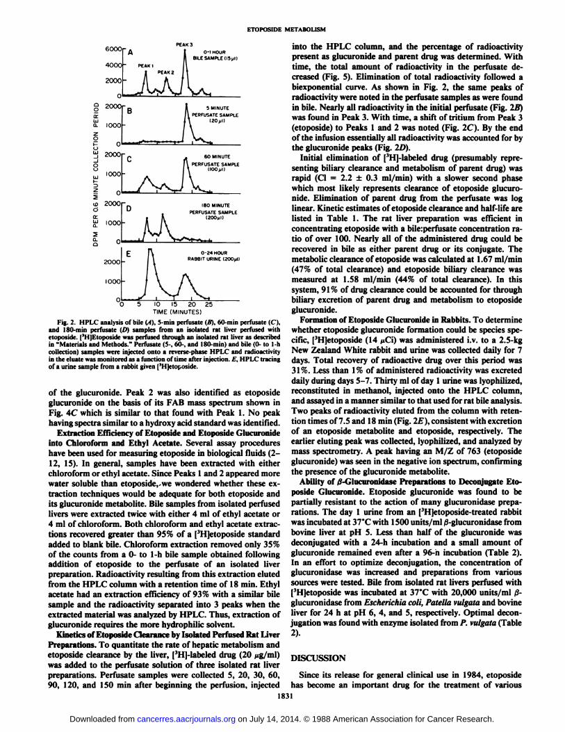
in the loss of 90% of the radioactivity from the perfusate andits appearance in the bile. Less than 5% of administered tritiumwas found in the liver homogenate. HPLC analysis of bile fromthe perfused liver showed 3 major peaks of radioactivity in theeluate with retention times of 6.3 (Peak 1), 11.2 (Peak 2), and18 min (Peak 3) as shown in Fig. 2A. Peak 3 (the 18-min peak)had the same retention time as an etoposide standard. Theeluate from each peak was collected and analyzed by FAB massspectrometry as described in "Materials and Methods." Stand
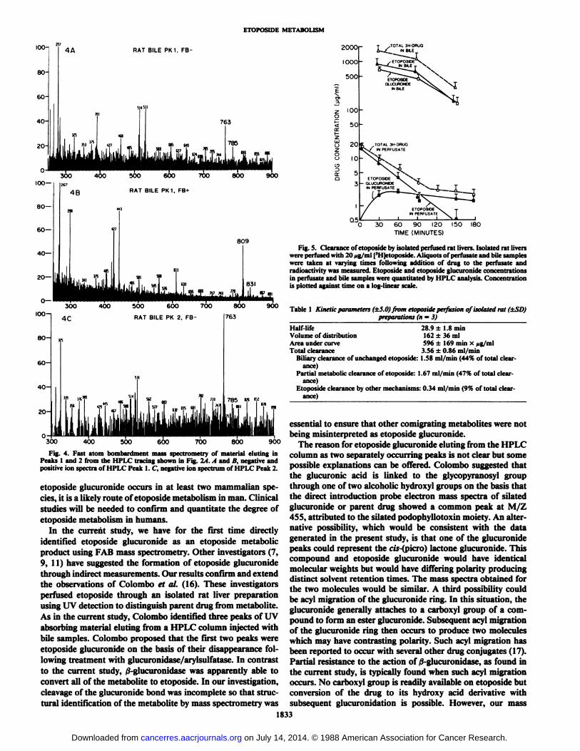
ards of etoposide and its hydroxy acid metabolite were alsoanalyzed by FAB mass spectrometry. Fig. 3, A and B, are thenegative and positive ion FAB spectrum obtained with anetoposide standard. The negative and positive ion spectrum ofPeak 3 are shown in Fig. 3, C and D, respectively, confirmingthe identity of this peak as parent drug. In the negative ionspectrum, ions are found at M/Z 609, 587, and 381 duerespectively to [MNa-2H]-, [M-H]-, and a fragment ion arising
from loss of the glycopyranosyl moiety of etoposide. In thepositive ion spectra, peaks are seen at M/Z 611, 589, 588, and383 corresponding to [MNa]+, [MH]+, [M]+, and the ionrepresenting the loss of the glucopyranosyl moiety.
The negative ion spectrum of Peak 1 (Fig. 4A) contains ionsat M/Z 763 and 785 corresponding to [M-H]- and [MNa-2H]—for etoposide glucuronide. The corresponding positive
ion spectrum of Peak 1 (Fig. 4B) shows ions having M/Z of809 and 831 corresponding to [MNa2-H]+ and [MNa3-2H]+
1830
on July 14, 2014. © 1988 American Association for Cancer Research. cancerres.aacrjournals.org Downloaded from
ETOPOSIDE METABOLISM
eoOOr o-i HOUR9ILE SAMPLE(ISjill
5 10 15 20 25TIME (MINUTES)
Fig. 2. HPLC analysis of bile (A), S-min perfusate (It). (>0min perfusate (C),and 180-min perfusate (/') samples from an isolated rat liver perfused withetoposide. (3H]Etoposide was perfused through an isolated rat liver as describedin "Materials and Methods." Perfusate (5-, 60-, and 180-min) and bile (0- to 1-hcollection) samples were injected onto a reverse-phase HPLC and radioactivityin the eluate was monitored as a function of time after injection. /•.'.HPLC tracingof a urine sample from a rabbit given ['Hjetoposide.
of the glucuronide. Peak 2 was also identified as etoposideglucuronide on the basis of its FAB mass spectrum shown inFig. 4C which is similar to that found with Peak 1. No peakhaving spectra similar to a hydroxy acid standard was identified.
Extraction Efficiency of Etoposide and Etoposide Glucuronideinto Chloroform and Ethyl Acetate. Several assay procedureshave been used for measuring etoposide in biological fluids (2-12, IS). In general, samples have been extracted with eitherchloroform or ethyl acetate. Since Peaks 1 and 2 appeared morewater soluble than etoposide,-we wondered whether these extraction techniques would be adequate for both etoposide andits glucuronide metabolite. Bile samples from isolated perfusedlivers were extracted twice with either 4 ml of ethyl acetate or4 ml of chloroform. Both chloroform and ethyl acetate extractions recovered greater than 95% of a [3H]etoposide standard
added to blank bile. Chloroform extraction removed only 35%of the counts from a 0- to 1-h bile sample obtained followingaddition of etoposide to the perfusate of an isolated liverpreparation. Radioactivity resulting from this extraction elutedfrom the HPLC column with a retention time of 18 min. Ethylacetate had an extraction efficiency of 93% with a similar bilesample and the radioactivity separated into 3 peaks when theextracted material was analyzed by HPLC. Thus, extraction ofglucuronide requires the more hydrophilic solvent.
Kinetics of Etoposide Clearance by Isolated Perfused Rat LiverPreparations. To quantitate the rate of hepatic metabolism andetoposide clearance by the liver, [3H]-labeled drug (20 /ig/ml)
was added to the perfusate solution of three isolated rat liverpreparations. Perfusate samples were collected 5, 20, 30, 60,90, 120, and 150 min after beginning the perfusion, injected
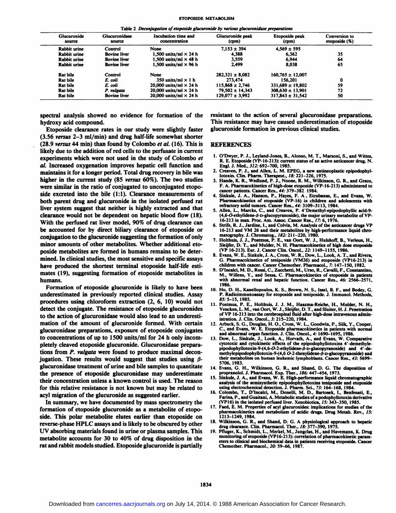
into the HPLC column, and the percentage of radioactivitypresent as glucuronide and parent drug was determined. Withtime, the total amount of radioactivity in the perfusate decreased (Fig. 5). Elimination of total radioactivity followed abiexponential curve. As shown in Fig. 2, the same peaks ofradioactivity were noted in the perfusate samples as were foundin bile. Nearly all radioactivity in the initial perfusate (Fig. 2/f)was found in Peak 3. With time, a shift of tritium from Peak 3(etoposide) to Peaks 1 and 2 was noted (Fig. 2C). By the endof the infusion essentially all radioactivity was accounted for bythe glucuronide peaks (Fig. 2D).
Initial elimination of [3H]-labeled drug (presumably repre
senting biliary clearance and metabolism of parent drug) wasrapid (Cl = 2.2 ±0.3 ml/min) with a slower second phasewhich most likely represents clearance of etoposide glucuronide. Elimination of parent drug from the perfusate was loglinear. Kinetic estimates of etoposide clearance and half-life arelisted in Table 1. The rat liver preparation was efficient inconcentrating etoposide with a bile:perfusate concentration ratio of over 100. Nearly all of the administered drug could berecovered in bile as either parent drug or its conjugate. Themetabolic clearance of etoposide was calculated at 1.67 ml/min(47% of total clearance) and etoposide biliary clearance wasmeasured at 1.58 ml/min (44% of total clearance). In thissystem, 91% of drug clearance could be accounted for throughbiliary excretion of parent drug and metabolism to etoposideglucuronide.
Formation of Etoposide Glucuronide in Rabbits. To determinewhether etoposide glucuronide formation could be species specific, [3H]etoposide (14 ¿iCi)was administered i.v. to a 2.5-kg
New Zealand White rabbit and urine was collected daily for 7days. Total recovery of radioactive drug over this period was31%. Less than 1% of administered radioactivity was excreteddaily during days 5-7. Thirty ml of day 1 urine was lyophilized,reconstituted in methanol, injected onto the HPLC column,and assayed in a manner similar to that used for rat bile analysis.Two peaks of radioactivity eluted from the column with retention times of 7.5 and 18 min (Fig. 21:). consistent with excretionof an etoposide metabolite and etoposide, respectively. Theearlier eluting peak was collected, lyophilized, and analyzed bymass spectrometry. A peak having an M/Z of 763 (etoposideglucuronide) was seen in the negative ion spectrum, confirmingthe presence of the glucuronide metabolite.
Ability of /3-Glucuronidase Preparations to Deconjugate Etoposide Glucuronide. Etoposide glucuronide was found to bepartially resistant to the action of many glucuronidase preparations. The day 1 urine from an [3H]etoposide-treated rabbitwas incubated at 37°Cwith 1500 units/ml /3-glucuronidase from
bovine liver at pH 5. Less than half of the glucuronide wasdeconjugated with a 24-h incubation and a small amount ofglucuronide remained even after a 96-n incubation (Table 2).In an effort to optimize deconjugation, the concentration ofglucuronidase was increased and preparations from varioussources were tested. Bile from isolated rat livers perfused with[3H]etoposide was incubated at 37°Cwith 20,000 units/ml ß-
glucuronidase from Escherichia coli, Patella vulgata and bovineliver for 24 h at pH 6, 4, and 5, respectively. Optimal deconjugation was found with enzyme isolated from /'. vulgata (Table
2).
DISCUSSION
Since its release for general clinical use in 1984, etoposidehas become an important drug for the treatment of various
1831
on July 14, 2014. © 1988 American Association for Cancer Research. cancerres.aacrjournals.org Downloaded from

ETOPOSIDE METABOLISM
3C100 RAT BILE PK3. FB- 587
Fig. 3. Fast atom bombardment mass spectra of etoposide standards and material dut ingin Peak 3 from the HPLC tracing (A). A andB, negative and positive ion spectrum of etoposide. C and I), comparable negative andpositive ion spectrum of Peak 3.
ETOPOSIDE STANDARD, FB-
700
200
3B1°°1 ETOPOSIDE
300 400 500 600
3D RAT BILE PK3, FB+
200 300 400 500 600
malignancies. Despite its widespread use, the metabolism ofthis drug has not been clearly defined. Urinary excretion ofetoposide accounts for only 35-40% of drug disposition (2-4).
The fate of the remaining 60% of an administered drug dosehas not been determined. Several etoposide metabolites haveeither been postulated or identified (5-9). However, clinicalstudies evaluating etoposide metabolism have not all found thesame metabolites and the degree to which some of these metabolites are formed is unclear. In no study has any metaboliteaccounted for more than 18% of an administered drug dose.Although most potential metabolites are noncytotoxic, Dow et
al. (13) have shown that some may retain cytotoxicity. Complete elaboration of the metabolism of etoposide and its routeof clearance is important in understanding its antineoplasticaction and in minimizing its toxicity. Since etoposide is a highlytoxic drug with a low therapeutic index, patients with renalinsufficiency or hepatic impairment may need dose adjustmentsin order to prevent accumulation of toxic drug or metabolite.The results of the present studies demonstrate metabolism ofetoposide to its glucuronide conjugate and indicate that formation of this metabolite accounts for a significant percentageof drug disposition in the rat and rabbit. Since formation of
1832
on July 14, 2014. © 1988 American Association for Cancer Research. cancerres.aacrjournals.org Downloaded from
ETOPOSIDE METABOLISM
100-
80-
60-
40-
RAT BILE PK1, FB-
763
400 500 600 700 800 900
Fig. 4. Fast atom bombardment mass spectrometry of material eluting inPeaks 1 and 2 from the HPLC tracing shown in Fig. TA. A and B, negative andpositive ion spectra of HPLC Peak 1. C, negative ion spectrum of HPLC Peak 2.
etoposide glucuronide occurs in at least two mammalian species, it is a likely route of etoposide metabolism in man. Clinicalstudies will be needed to confirm and quantitate the degree ofetoposide metabolism in humans.
In the current study, we have for the first time directlyidentified etoposide glucuronide as an etoposide metabolicproduct using FAB mass spectrometry. Other investigators (7,9, 11) have suggested the formation of etoposide glucuronidethrough indirect measurements. Our results confirm and extendthe observations of Colombo et al. (16). These investigatorsperfused etoposide through an isolated rat liver preparationusing UV detection to distinguish parent drug from metabolite.As in the current study, Colombo identified three peaks of UVabsorbing material eluting from a HPLC column injected withbile samples. Colombo proposed that the first two peaks wereetoposide glucuronide on the basis of their disappearance following treatment with glucuronidase/arylsulfatase. In contrastto the current study, /i-glucuronidase was apparently able to
convert all of the metabolite to etoposide. In our investigation,cleavage of the glucuronide bond was incomplete so that structural identification of the metabolite by mass spectrometry was
ozo
2000
1000
500-
100
5O
20
IO
5
3
T -TOTAL 3M-DRUG&-"Ã^. IN BILE_
fETOPOSOt
GLUCLflOCeN BILE
,TOTAL 3H-DHUG
IN PERFUSATE
30 60 90 120 150 180TIME (MINUTES)
Fig. S. Clearance of etoposide by isolated perfused rat livers. Isolated rat liverswere perfused with 20 »ig/ml['I I|ft»posidv.Aliquots of perfusate and bile samples
were taken at varying times following addition of drug to the perfusate andradioactivity was measured. Etoposide and etoposide glucuronide concentrationsin perfusate and bile samples were quantitated by HPLC analysis. Concentrationis plotted against time on a log-linear scale.
Table 1 Kinetic parameters (±5.0)from etoposide perfusion of isolated rat (±SD)preparations (n = 3)
Half-lifeVolume of distributionArea under curveTotal clearance
28.9 ±1.8 min162 ±36 ml596 ±169 min x. »ig/ml
3.56 ±0.86 ml/minBiliary clearance of unchanged etoposide: 1.58 ml/min (44% of total clear
ance)Partial metabolic clearance of etoposide: 1.67 ml/min (47% of total clear
ance)Etoposide clearance by other mechanisms: 0.34 ml/min (9% of total clear
ance)
essential to ensure that other comigrating metabolites were notbeing misinterpreted as etoposide glucuronide.
The reason for etoposide glucuronide eluting from the HPLCcolumn as two separately occurring peaks is not clear but somepossible explanations can be offered. Colombo suggested thatthe glucuronic acid is linked to the glycopyranosyl groupthrough one of two alcoholic hydroxyl groups on the basis thatthe direct introduction probe electron mass spectra of silatedglucuronide or parent drug showed a common peak at M/Z455, attributed to the silated podophyllotoxin moiety. An alternative possibility, which would be consistent with the datagenerated in the present study, is that one of the glucuronidepeaks could represent the m-(picro) lactone glucuronide. Thiscompound and etoposide glucuronide would have identicalmolecular weights but would have differing polarity producingdistinct solvent retention times. The mass spectra obtained forthe two molecules would be similar. A third possibility couldbe acyl migration of the glucuronide ring. In this situation, theglucuronide generally attaches to a carboxyl group of a compound to form an ester glucuronide. Subsequent acyl migrationof the glucuronide ring then occurs to produce two moleculeswhich may have contrasting polarity. Such acyl migration hasbeen reported to occur with several other drug conjugates (17).Partial resistance to the action of 0-glucuronidase, as found inthe current study, is typically found when such acyl migrationoccurs. No carboxyl group is readily available on etoposide butconversion of the drug to its hydroxy acid derivative withsubsequent glucuronidation is possible. However, our mass
1833
on July 14, 2014. © 1988 American Association for Cancer Research. cancerres.aacrjournals.org Downloaded from
ETOPOSIDE METABOLISM
Table 2 Deconjugation ofetoposide glucuronide by various glucuronidase preparations
GlucuronidesourceRabbit
urineRabbit urineRabbit urineRabbiturineRat
bileRat bileRat bileRat bileRat bileGlucuronidase
sourceControl
Bovine liverBovine liverBovineliverControl
E. coliE. coliP. vulgataBovine liverIncubation
time andconcentrationNone
1,500 units/ml x 24 h1,500 units/ml x 48 h1,500 units/ml x 96hNone
350 units/ml x 1 h20,000 units/ml x 24 h20,000 units/ml x 24 h20,000 units/ml x 24 hGlucuronide
peak(cpm)7,
153 ±3944,3883,559
2,499282,321
±8,082273,474
115,868 ±2,74679,502 ±14,343
129,077 ±3,992Etoposide
peak(cpm)4,569
±5956,3626,944
8,0381
60,765 ±12,007156,201
331, 689 ±19,802308,630 ±13,9013 17,843 ±31,542Conversion
toetoposide(%)3564
650
5972
50
spectral analysis showed no evidence for formation of thehydroxy acid compound.
Etoposide clearance rates in our study were slightly faster(3.56 versus 2-3 ml/min) and drug half-life somewhat shorter(28.9 versus 44 min) than found by Colombo et al. (16). This islikely due to the addition of red cells to the perfusate in currentexperiments which were not used in the study of Colombo etal. Increased oxygénation improves hepatic cell function andmaintains it for a longer period. Total drug recovery in bile washigher in the current study (85 versus 60%). The two studieswere similar in the ratio of conjugated to unconjugated etoposide excreted into the bile (1:1). Clearance measurements ofboth parent drug and glucuronide in the isolated perfused ratliver system suggest that neither is highly extracted and thatclearance would not be dependent on hepatic blood flow (18).With the perfused rat liver model, 90% of drug clearance canbe accounted for by direct biliary clearance of etoposide orconjugation to the glucuronide suggesting the formation of onlyminor amounts of other metabolites. Whether additional etoposide metabolites are formed in humans remains to be determined. In clinical studies, the most sensitive and specific assayshave produced the shortest terminal etoposide half-life estimates (19), suggesting formation ofetoposide metabolites inhumans.
Formation of etoposide glucuronide is likely to have beenunderestimated in previously reported clinical studies. Assayprocedures using chloroform extraction (2, 6, 10) would notdetect the conjugate. The resistance of etoposide glucuronidesto the action of glucuronidase would also lead to an underestimation of the amount of glucuronide formed. With certainglucuronidase preparations, exposure of etoposide conjugatesto concentrations of up to 1500 units/ml for 24 h only incompletely cleaved etoposide glucuronide. Glucuronidase preparations from P. vulgata were found to produce maximal decon-jugation. These results would suggest that studies using ß-glucuronidase treatment of urine and bile samples to quantitatethe presence of etoposide glucuronidase may underestimatetheir concentration unless a known control is used. The reasonfor this relative resistance is not known but may be related toacyl migration of the glucuronide as suggested earlier.
In summary, we have documented by mass spectrometry theformation of etoposide glucuronide as a metabolite of etoposide. This polar metabolite élûtesearlier than etoposide onreverse-phase HPLC assays and is likely to be obscured by otherUV absorbing materials found in urine or plasma samples. Thismetabolite accounts for 30 to 40% of drug disposition in therat and rabbit models studied. Etoposide glucuronide is partially
resistant to the action of several glucuronidase preparations.This resistance may have caused underestimation of etoposideglucuronide formation in previous clinical studies.
REFERENCES
1. O'Dwyer, P. J., Leyland-Jones, B., Alonso, M. T., Marsoni, S., and Wittes,R. E. Etoposide (VP-16-213): current Status of an active anticancer drug. N.Engl. J. Med., 312:692-700, 1985.
2. Creaven, P. J., and Allen, L. M. EPEG, a new antineoplastic epipodophyl-lotoxin. Clin. Pharm. Therapeut., /*: 221-226, 1975.
3. Hande, K. R., Wedlund, P. J., Noone, R. M., Wilkinson, G. R., and Greco,F. A. Pharmacokinetics of high-dose etoposide (VP-16-213) administered tocancer patients. Cancer Res., 44: 379-382 1984.
4. Sinkule, J. A., Hanson, P., Hayes, F. A, Etcubanas, E., and Evans, W.Pharmacokinetics of etoposide (VP-16) in children and adolescents withrefractory solid tumors. Cancer Res., 44: 3109-3113, 1984.
5. Allen, L., Marcks, C., and Creaven, P. 4'Demethyl-epipodophyllic acid-9-(4,6-0-ethylidene-/3-r>glucopyranoside), the major urinary metabolite of VP-16-213 in man. Proc. Am. Assoc. Cancer Res., 17:6, 1976.
6. Strife, R. J., Jardine, I., and Colvin, M. Analysis of the anticancer drugs VP16-213 and VM 26 and their metabolites by high-performance liquid diromatography. J. Chromatog., 182: 211-220, 1980.
7. Holthuis, J. J., Postmus, P. E., van Oort, W. J., Hulshoff, B., Verleun, H.,Sleijfer, D. T., and Mulder, N. H. Pharmacokinetics of high dose etoposide(VP 16-213). Eur. J. Cancer Clin. Oncol., 22:1149-1155, 1986.
8. Evans, W. E., Sinkule, J. A., Crom, W. R., Dow, L., Look, A. T., and Rivera,G. Pharmacokinetics of teniposide (VM26) and etoposide (VP16-213) inchildren with cancer. Cancer Chemother. Pharmacol., 7: 147-150, 1982.
9. D'Incaici, M. D., Rossi, C., Zucchetti, M., Urso, R., Cavalli, F., Constantino,
M., Willens, Y., and Sessa, C. Pharmacokinetics of etoposide in patientswith abnormal renal and hepatic function. Cancer Res., 46: 2566-2571,1986.
10. Ho, D. H., Kanellopoulos, K. S., Brown, N. S., Issel, B. F., and Bodey, G.P. Radioimmunoassay for etoposide and teniposide. J. Immunol. Methods,85: 5-15, 1985.
11. Postmus, P. E., Holthuis, J. J. M., Haazma-Reiche, H., Mulder, N. H.,Vencken, L. M., van Oort, W. J., Sleijfer, D. T., and Sluiter, H. J. Penetrationof VP 16-213 into the cerebrospinal fluid after high-dose intravenous administration. J. Clin. Oncol., 3: 215-220, 1984.
12. Arbuck, S. G., Douglas, H. O., Crom, W. L., Goodwin, P., Silk, Y., Cooper,C., and Evans, W. E. Etoposide Pharmacokinetics in patients with normaland abnormal organ function. J. Clin. Oncol., 4: 1690-1695, 1986.
13. Dow, L., Sinkule, J., Look, A., Horvath, A., and Evans, W. Comparativecytotoxic and cytokinetic effects of the epipodophyllotoxins 4'-demethyle-pipodophyllotoxin-9-(4,6-O-2-ethylidene-/3-D-glucopyranoside) and 4'-de-methylepipodophyllotoxin-9-(4,6 0-2-thenylidene-0-D-glucopyranoside) andtheir metabolites on human leukemic lymphoblasts. Cancer Res., 43: 5699-5706, 1983.
14. Evans, G. H., Wilkinson, G. R., and Shand, D. G. The disposition ofpropranolol. J. Pharmacol. Exp. Ther., 186:447-454, 1973.
15. Sinkule, J. A., and Evans, W. E. High-performance liquid Chromatographieanalysis of the semisynthetic epipodophyllotoxins teniposide and etoposideusing electrochemical detection. J. Pharm. Sci., 73: 164-168, 1984.
16. Colombo, T., D'Incaici, M., Doni-Ili. M. D., Bartosek, I., Benfenati, E.,
Farina, P., and Guaitani, A. Metabolic studies of a podophyllotoxin derivative(VP16) in the isolated perfused liver. Xenobiotica, 15: 343-350, 1985.
17. Faed, E. M. Properties of acyl glucuronides: implications for studies of thePharmacokinetics and metabolism of acidic drugs. Drug Metab. Rev., 15:1213-1249, 1984.
18. Wilkinson, G. R., and Shand, D. G. A physiological approach to hepaticdrug clearance. Clin. Pharmacol. Ther., IS: 377-390, 1975.
19. Pfluger, K., Schmidt, L., Merkel, M., Jungclas, H., and Havemann, K. Drugmonitoring ofetoposide (VP 16-213): correlation of pharmacokinetic parameters to clinical and biochemical data in patients receiving etoposide. CancerChemother. Pharmacol., 20: 59-66, 1987.
1834
on July 14, 2014. © 1988 American Association for Cancer Research. cancerres.aacrjournals.org Downloaded from