Identification and isolation of multipotential neural progenitor cells from the subcortical white...
9
NATURE MEDICINE • VOLUME 9 • NUMBER 4 • APRIL 2003 439 ARTICLES The adult human subcortical white matter harbors a popula- tion of mitotically competent glial progenitors that comprise as many as 3% of its cells 1,2 . These cells may be extracted from brain tissue using FACS after transfection with GFP-encoding plasmids driven by the promoter for CNP, an early oligoden- drocytic transcript 2,3 . The cells express the immature neural ganglioside recognized by monoclonal antibody A2B5 but do not express more mature markers of glial lineage. We previ- ously noted that when grown at high density, pCNP2:hGFP + progenitors gave rise to glia, largely oligodendrocytes. Nonetheless, in low-density culture after high-purity FACS, pCNP2:hGFP + cells often generated βIII-tubulin + neurons 2 . Because neurogenesis was never observed from pCNP2:hGFP + cells in higher-density or unsorted cultures, we postulated that the restriction of these progenitor cells to the oligodendroglial phenotype might be an effect of environmental cues rather than a function of autonomous commitment. Once isolated into high-purity, low-density culture, and therefore removed from any paracrine or autocrine influences, human subcortical pCNP2:hGFP + cells were able to generate neurons as well as glia 2 . It was subsequently reported 4 that glial progenitors from the postnatal rat optic nerve could also generate neurons after serum- or bone morphogenetic protein–induced phenotypic instruction and basic fibroblast growth factor (bFGF)–stimu- lated expansion. Similar work showed that progenitor cells of the adult rat forebrain parenchyma could also generate neu- rons after prolonged in vitro expansion in bFGF 8 . Taken to- gether, these findings indicated that glial progenitor cells might retain substantial phenotypic plasticity. We asked whether some fraction of the nominally glial prog- enitors of the adult human subcortical white matter might ac- tually be parenchymal neural stem cells. Specifically, we asked whether single, sorted WMPCs could generate multiple neural phenotypes, and if so, whether they were capable of expansion and self-renewal. In addition, we investigated whether this process requires de-differentiative reprogramming to an inter- mediate phenotype, or whether simply removing these cells from their local environment and mitotically expanding them in bFGF might suffice to permit these cells to act as multipo- tential progenitors. In doing so, we tested the hypothesis that the phenotypic plasticity of adult WMPCs might be tonically restricted by the adult parenchymal environment, rather than irreversibly lost with development. WMPCs were isolated by CNP- and A2B5-based sorting White matter was dissected from surgical samples taken at the time of temporal lobectomy for epilepsy, aneurysm, and post- traumatic decompression (n = 21). The tissues were dissected free of adjacent cortex and ventricular epithelium, and enzymati- cally dissociated to single-cell suspension as described 2 . The dis- sociates were plated onto laminin (100 µg/ml) in DMEM/F12/N1 supplemented with bFGF (20 ng/ml), NT3 (2 ng/ml) and platelet-derived growth factor (PDGF)-AA (20 ng/ml). To iden- tify oligodendrocyte progenitors, the dissociates were trans- Identification and isolation of multipotential neural progenitor cells from the subcortical white matter of the adult human brain MARTA C. NUNES 1 , NEETA SINGH ROY 1 , H. MICHAEL KEYOUNG 1 ,ROBERT R. GOODMAN 2 , GUY MCKHANN II 2 , LI JIANG 3 , JIAN KANG 3 , MAIKEN NEDERGAARD 3 & STEVEN A. GOLDMAN 1 1 Department of Neurology and Neuroscience, Cornell University Medical College, New York, New York, USA 2 Department of Neurosurgery, Columbia University College of Physicians and Surgeons, New York, New York, USA 3 Department of Anatomy and Cell Biology, New York Medical College, Valhalla, New York, USA M.C.N. and N.S.R. contributed equally to this work. Correspondence should be addressed to S.A.G.; e-mail: [email protected] Published online 10 March 2003; doi:10.1038/nm837 The subcortical white matter of the adult human brain harbors a pool of glial progenitor cells. These cells can be isolated by fluorescence-activated cell sorting (FACS) after either transfection with green fluorescent protein (GFP) under the control of the CNP2 promoter, or A2B5-targeted immunotagging. Although these cells give rise largely to oligodendrocytes, in low-density cul- ture we observed that some also generated neurons. We thus asked whether these nominally glial progenitors might include multipotential progenitor cells capable of neurogenesis. We found that adult human white-matter progenitor cells (WMPCs) could be passaged as neu- rospheres in vitro and that these cells generated functionally competent neurons and glia both in vitro and after xenograft to the fetal rat brain. WMPCs were able to produce neurons after their initial isolation and did not require in vitro expansion or reprogramming to do so. These ex- periments indicate that an abundant pool of mitotically competent neurogenic progenitor cells resides in the adult human white matter. © 2003 Nature Publishing Group http://www.nature.com/naturemedicine
-
Upload
independent -
Category
Documents
-
view
1 -
download
0
Transcript of Identification and isolation of multipotential neural progenitor cells from the subcortical white...

NATURE MEDICINE • VOLUME 9 • NUMBER 4 • APRIL 2003 439
ARTICLES
The adult human subcortical white matter harbors a popula-tion of mitotically competent glial progenitors that compriseas many as 3% of its cells1,2. These cells may be extracted frombrain tissue using FACS after transfection with GFP-encodingplasmids driven by the promoter for CNP, an early oligoden-drocytic transcript2,3. The cells express the immature neuralganglioside recognized by monoclonal antibody A2B5 but donot express more mature markers of glial lineage. We previ-ously noted that when grown at high density, pCNP2:hGFP+
progenitors gave rise to glia, largely oligodendrocytes.Nonetheless, in low-density culture after high-purity FACS,pCNP2:hGFP+ cells often generated βIII-tubulin+ neurons2.Because neurogenesis was never observed from pCNP2:hGFP+
cells in higher-density or unsorted cultures, we postulated thatthe restriction of these progenitor cells to the oligodendroglialphenotype might be an effect of environmental cues ratherthan a function of autonomous commitment. Once isolatedinto high-purity, low-density culture, and therefore removedfrom any paracrine or autocrine influences, human subcorticalpCNP2:hGFP+ cells were able to generate neurons as well asglia2. It was subsequently reported4 that glial progenitors fromthe postnatal rat optic nerve could also generate neurons afterserum- or bone morphogenetic protein–induced phenotypicinstruction and basic fibroblast growth factor (bFGF)–stimu-lated expansion. Similar work showed that progenitor cells ofthe adult rat forebrain parenchyma could also generate neu-rons after prolonged in vitro expansion in bFGF8. Taken to-
gether, these findings indicated that glial progenitor cellsmight retain substantial phenotypic plasticity.
We asked whether some fraction of the nominally glial prog-enitors of the adult human subcortical white matter might ac-tually be parenchymal neural stem cells. Specifically, we askedwhether single, sorted WMPCs could generate multiple neuralphenotypes, and if so, whether they were capable of expansionand self-renewal. In addition, we investigated whether thisprocess requires de-differentiative reprogramming to an inter-mediate phenotype, or whether simply removing these cellsfrom their local environment and mitotically expanding themin bFGF might suffice to permit these cells to act as multipo-tential progenitors. In doing so, we tested the hypothesis thatthe phenotypic plasticity of adult WMPCs might be tonicallyrestricted by the adult parenchymal environment, rather thanirreversibly lost with development.
WMPCs were isolated by CNP- and A2B5-based sortingWhite matter was dissected from surgical samples taken at thetime of temporal lobectomy for epilepsy, aneurysm, and post-traumatic decompression (n = 21). The tissues were dissected freeof adjacent cortex and ventricular epithelium, and enzymati-cally dissociated to single-cell suspension as described2. The dis-sociates were plated onto laminin (100 µg/ml) in DMEM/F12/N1supplemented with bFGF (20 ng/ml), NT3 (2 ng/ml) andplatelet-derived growth factor (PDGF)-AA (20 ng/ml). To iden-tify oligodendrocyte progenitors, the dissociates were trans-
Identification and isolation of multipotential neural progenitor cells from the subcortical white
matter of the adult human brain
MARTA C. NUNES1, NEETA SINGH ROY1, H. MICHAEL KEYOUNG1, ROBERT R. GOODMAN2, GUY MCKHANN II2, LI JIANG3, JIAN KANG3, MAIKEN NEDERGAARD3 & STEVEN A. GOLDMAN1
1Department of Neurology and Neuroscience, Cornell University Medical College, New York, New York, USA2Department of Neurosurgery, Columbia University College of Physicians and Surgeons, New York, New York, USA
3Department of Anatomy and Cell Biology, New York Medical College, Valhalla, New York, USAM.C.N. and N.S.R. contributed equally to this work.
Correspondence should be addressed to S.A.G.; e-mail: [email protected]
Published online 10 March 2003; doi:10.1038/nm837
The subcortical white matter of the adult human brain harbors a pool of glial progenitor cells.These cells can be isolated by fluorescence-activated cell sorting (FACS) after either transfectionwith green fluorescent protein (GFP) under the control of the CNP2 promoter, or A2B5-targetedimmunotagging. Although these cells give rise largely to oligodendrocytes, in low-density cul-ture we observed that some also generated neurons. We thus asked whether these nominallyglial progenitors might include multipotential progenitor cells capable of neurogenesis. Wefound that adult human white-matter progenitor cells (WMPCs) could be passaged as neu-rospheres in vitro and that these cells generated functionally competent neurons and glia bothin vitro and after xenograft to the fetal rat brain. WMPCs were able to produce neurons aftertheir initial isolation and did not require in vitro expansion or reprogramming to do so. These ex-periments indicate that an abundant pool of mitotically competent neurogenic progenitor cellsresides in the adult human white matter.
B-nm837.qxd 3/13/03 1:40 PM Page 439
©20
03 N
atu
re P
ub
lish
ing
Gro
up
h
ttp
://w
ww
.nat
ure
.co
m/n
atu
rem
edic
ine

440 NATURE MEDICINE • VOLUME 9 • NUMBER 4 • APRIL 2003
ARTICLES
fected with pCNP2:hGFP, the transcription of which resultsin GFP expression by oligodendrocyte progenitor cells2.
To avoid both the temporal lag between transfection andGFP expression and the inefficiency of plasmid transfec-tion, cultures were also sorted on the basis of A2B5 surfaceimmunoreactivity, which can serve as a surrogate markerfor pCNP2:hGFP+ WMPCs in vitro2. Immunostainingshowed that 84 ± 8.3% of pCNP2:GFP+ cells expressed A2B5(ref. 2). GFP-based FACS gated 0.49 ± 0.15% of all white-matter cells as pCNP2:hGFP+ (mean ± s.e.m.; n = 3 patients;Fig. 1a). Matched cultures transfected with pCMV:GFP hada net transfection efficiency of 13.1%. Thus, the predictedincidence of pCNP2:hGFP+ cells in the white matter was3.7% (= 1 ÷ 0.131 × 0.49), consistent with our prior esti-mates of the incidence of this phenotype2. From the samesamples, A2B5-based FACS gated an average of 3.1 ± 0.7%(n = 3) of the white-matter cell population (Fig. 1b). Thegreater than six-fold increase in net yield when A2B5 wasused (3.1% versus 0.49%) reflected the higher efficiencyof A2B5 immunodetection relative to pCNP2:hGFP plas-mid transfection. On this basis, we used immunomag-
netic sorting (IMS) to select A2B5+ cells from adult white-mat-ter dissociates. By IMS, the incidence of A2B5-sorted cells inwhite matter dissociates was 3.6 ± 0.3% (n = 21) with a medianof 3.1%. This improved yield was accomplished with no ap-preciable loss of cell-type specificity, in that the A2B5+ cellsoverlapped entirely with the sort profiles of pCNP2:hGFP+
cells and each isolate generated O4+ oligodendrocytes withsimilar efficiency (Fig. 1c–f). Thus, A2B5-based FACS and IMSidentified WMPCs homologous to those recognized bypCNP2:GFP-based FACS, while permitting higher-yield isola-tion of these cells.
Adult WMPCs gave rise to multipotent neurospheresTo assess the expansion capacity of pCNP2:hGFP- and A2B5-sorted cells, we propagated sorted isolates of each in suspen-sion6–8. The cells were distributed into 24-well plates at 50,000cells per 0.5 ml in serum-free media (SFM) supplemented withbFGF (20 ng/ml), NT3 (2 ng/ml) and PDGF-AA (20 ng/ml), acombination that permits the expansion of human WMPCs2.Seven days later, the cells were switched to SFM with bFGF alone(20 ng/ml)8. Over the next 10 d, neurospheres—spherical masses
of cells that expand from single parental prog-enitors—arose in these cultures, such that by 3weeks after sorting, there were 84.8 ± 9.0spheres/well (n = 4 patients). These neu-rospheres were typically >150 µm in diameterand included 46.5 ± 8.2 cells/sphere (Fig. 2a andb). Thus, single WMPCs of the adult humanbrain were capable of generating neurospheres.
To establish the lineage potential of singleadult human WMPCs, we dissociated the re-sultant primary neurospheres and passagedthem into new wells. Alternatively, some wereplated onto substrate to permit their differen-tiation. Immunostaining showed that bothpCNP2:hGFP+ and A2B5+ progenitor–derivedspheres gave rise to all major neural pheno-types (Fig. 2d and e). Among those cells pas-saged from primary spheres, secondaryspheres were observed to arise within twoweeks after passage. After expansion, thesesecondary spheres were similarly plated onsubstrate, raised for one to two weeks andfixed. Immunolabeling confirmed that virtu-ally all secondary spheres generated both neu-rons and glia together (Fig. 2c and e). Inaddition, when the mitotic marker BrdU wasadded to A2B5-sorted cells, BrdU-incorporat-ing neurons, oligodendrocytes and astrocytesall emerged from the spheres generated (Fig.2f–i). The persistence of mitotic neurogenesisand gliogenesis by single spheres indicatedthat they contained cycling multipotentialcells. The secondary spheres were probably ofclonal origin, given the low plating density ofthe single cells from which each was derivedand the fact that the sphere-forming cells orig-inated from primary spheres that had them-selves expanded from single-cell dissociates.These data indicate that single progenitor cellsof the adult human white matter are bothclonogenic and multipotent.
a b
c d
e f
Fig. 1 A2B5-based FACS selects oligodendrocyte progenitor cells. a and b, FACSgraphs showing the extraction of pCNP2:hGFP+ (a) and A2B5+ (b) WMPCs from an adulthuman white-matter dissociate. Forward scatter (FCS), a measure of cell size, is plottedagainst fluorescence intensity (Fl-1). When pCNP2:hGFP- and A2B5-based sorts were di-rectly compared, their plots showed overlapping profiles, but A2B5+ cells were >6-foldmore abundant than their pCNP2:hGFP+ counterparts, reflecting the higher efficiency ofA2B5 surface tagging. c–f, Progenitors sorted by pCNP2:hGFP (c and e) and A2B5 (d andf) gave rise to O4+ oligodendrocytes. A2B5-based surface antigen sorting may thus beused as a higher-yield alternative to pCNP2:hGFP transfection–based FACS for isolatingWMPCs. Scale bar, 24 µm.
B-nm837.qxd 3/13/03 1:40 PM Page 440
©20
03 N
atu
re P
ub
lish
ing
Gro
up
h
ttp
://w
ww
.nat
ure
.co
m/n
atu
rem
edic
ine
NATURE MEDICINE • VOLUME 9 • NUMBER 4 • APRIL 2003 441
ARTICLES
Single WMPCs remained multipotential with passageThe serial propagability of sorted WMPCs from neurospheresin low-density dissociates suggested the clonal derivation ofeach individual sphere9–11. To further validate the clonal originof neurons and glia arising within single spheres, we usedlentiviral GFP to genetically tag and follow single WMPCs.A2B5+ cells were tagged, 2–5 d after sorting, with a lentivirusexpressing GFP under cytomegalovirus (CMV) promoter con-trol12–14. At 10 PFU/cell, 23% of the cells expressed GFP by oneweek after sorting, yielding a mixture of GFP–, GFP+ and mixedspheres in the resultant cultures (Fig. 3a–b). These primaryspheres were triturated two to four weeks later to single-cellsuspensions and passaged into bFGF at ∼ 3,000 cells/well.Under these conditions, 40.8 ± 12.9 secondary spheres/wellwere generated, indicating a clonogenic cell incidence of 1.3%(n = 5). Of these secondary spheres, 47.2 ± 10.8% containedonly GFP+ cells (Fig. 3c–d) whereas 30.9 ± 6.9% harbored noGFP+ cells. The relative uniformity of GFP expression, or lackthereof, among the cells within a given sphere indicated thatmost spheres were clonally derived (P < 0.005 by χ2 analysis).This tested the null hypothesis that the spheres arose fromnon-clonal aggregation of two or more cells, each of which was
equally likely to be GFP+ or GFP–. When the single spheres wereplated onto polyornithine and fibronectin and their outgrowthassessed two weeks later, all gave rise to both neurons and glia(Fig. 3e–g). Because most secondary spheres were likely to havebeen clonally derived, and all included neurons as well as glia(38 of 38 spheres; n = 4 samples), single WMPCs must havegiven rise to neurons and glia together.
We next asked if the neurogenic capacity and multilineagecompetence of WMPCs were maintained with passage.Primary spheres were raised serially in bFGF/NT3/PDGF-AA for7 d, DMEM/F12/N1 with 15% serum/PDGF-AA for 4 d, andserum-free DMEM/F12/N1 with bFGF for 10 d. Cells were thendissociated and replated in bFGF at 3,000 cells/well in a 24-well plate. Secondary spheres arose within two weeks from 1.1± 0.3% of these cells (n = 8). After more than two weeks of fur-ther expansion, the secondary spheres were plated on polyor-nithine and fibronectin and were fixed and immunostainedtwo weeks later (seven to nine weeks after sorting). Whereasprimary spheres consisted of 21.7 ± 4.3% βIII-tubulin+ neu-rons, 17.7 ± 3.9% glial fibrillary acidic protein (GFAP)+ astro-cytes and 46.7 ± 5.9% O4+ oligodendrocytes (n = 3), secondaryspheres consisted of 16.0 ± 2.5% neurons, 19.3 ± 3.2% astro-
a b c
d e f
g h i
Fig. 2 Adult human WMPCs give rise to multipotential neurospheres. a, First-passage spheres generated from A2B5-sorted cells 2 weeks aftersorting. b, First-passage spheres arising from pCNP2:hGFP-sorted cells, at2 weeks. c, Second-passage sphere derived from an A2B5-sorted sample,at 3 weeks. d, Once plated onto substrate, the primary spheres differenti-ated into βIII-tubulin+ neurons (red), GFAP+ astrocytes (blue) and O4+
oligodendrocytes (green). e, Neurons (red), astrocytes (blue) and oligo-
dendrocytes (green) arose similarly from spheres derived frompCNP2:GFP-sorted WMPCs. f–h, BrdU incorporation (blue) showed thatnew neurons (f, βIII-tubulin (red); g, MAP2 (red)) and oligodendrocytes(h, O4 (green)) were generated in vitro. i, βIII-tubulin+ neurons (green)co-expressed neuronal Hu protein40,41 (red), yielding double labeling (yel-low). Nuclei were counterstained with DAPI (blue). Scale bars, 100 µm(a–e) or 40 µm (f–i).
B-nm837.qxd 3/13/03 1:40 PM Page 441
©20
03 N
atu
re P
ub
lish
ing
Gro
up
h
ttp
://w
ww
.nat
ure
.co
m/n
atu
rem
edic
ine

442 NATURE MEDICINE • VOLUME 9 • NUMBER 4 • APRIL 2003
ARTICLES
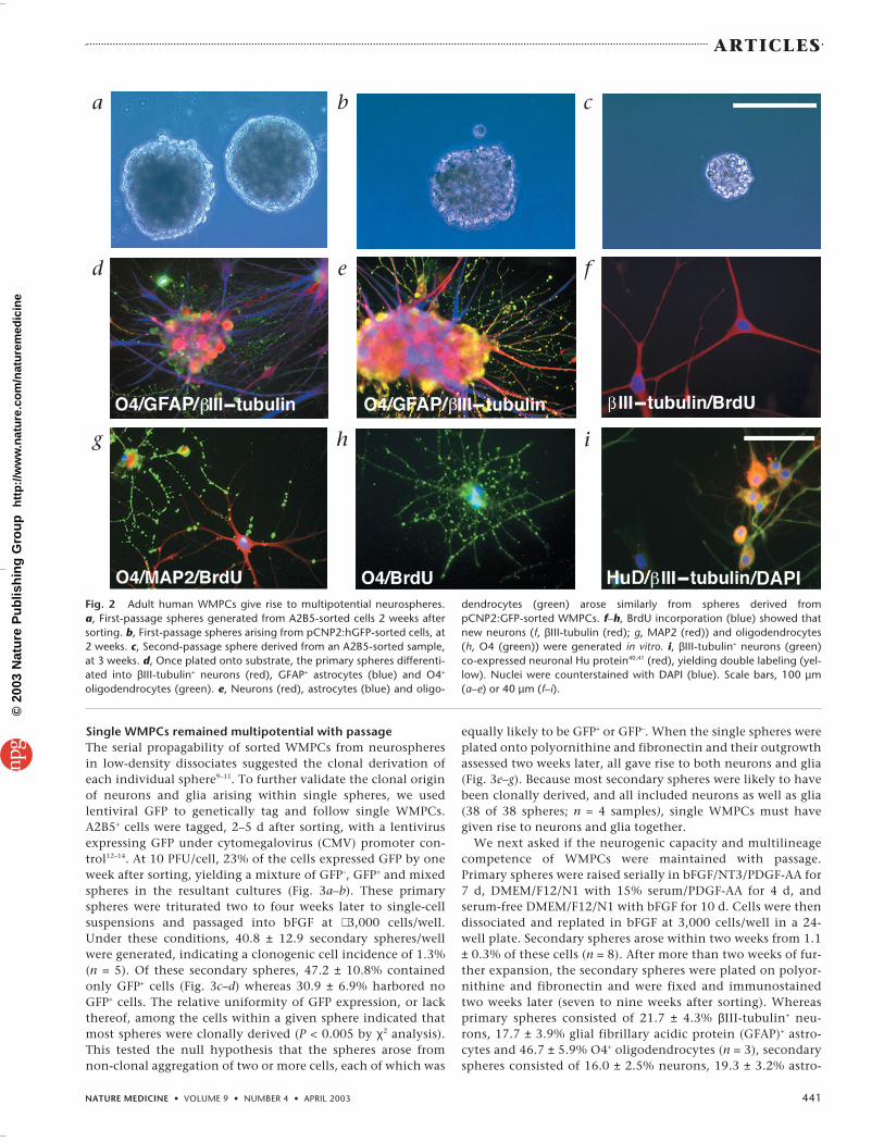
cytes and 46.4 ± 2.4% oligodendrocytes (n = 3). Most of theneurons were GABAergic, by virtue of their expression of glu-tamic acid decarboxylase-67 (GAD67) (Fig. 4a–c). Because therelative proportions of neurons, oligodendrocytes and astro-cytes in secondary spheres were similar to those in primaryspheres, we concluded that WMPCs retained multilineagecompetence with expansion.
WMPC-derived neurons become functionally matureThe calcium responses and membrane currents of WMPC-de-rived neurons were assessed to establish their ability to re-spond to depolarizing stimuli. Primary spheres (n = 12 fields,derived from 3 brains) were plated on fibronectin to permitneuronal outgrowth, and assessed 14 d later for their calciumresponses to depolarizing stimuli. The cultures were thenloaded with the calcium indicator dye Fluo-3 and serially ex-posed to both 100 µM glutamate and 60 mM potassium during confocal microscopy. Astrocytic responses to depolar-ization were minimal under these culture conditions, as pre-viously noted. In contrast, neuron-like cells displayed rapid,reversible, >100% elevations in cytosolic calcium in responseto potassium, consistent with the activity of neuronal volt-age-gated calcium channels (Fig. 4d–f). The neuronal pheno-type of these cells was then validated by immunostaining forβIII-tubulin.
We then asked whether WMPC-derived neurons would beable to develop the fast sodium currents and action potentialscharacteristic of electrophysiologically competent neurons.We used whole-cell patch-clamp recording during currentstimulation to assess the response of WMPC-derived neuronsthat arose from plated secondary spheres derived from A2B5-
sorted isolates. A total of 58 WMPC-derived fiber-bearing cellswere recorded, in 5 cultures derived from 3 patients. Of these,13 showed voltage-activated sodium ion currents (INa) of >100nA, and 7 had INa > 600, compatible with the fast sodium cur-rents of neuronal depolarization15,16. Accordingly, whereas twoof five cells with INa > 800 generated stimulus-evoked actionpotentials (Fig. 4g–h), none did so with INa < 800. In addition,none of 26 morphologically non-neuronal cells showed sub-stantial (≥100 pA) current-induced sodium currents. Together,these results indicated that neurons arising from adult humanWMPCs developed mature electrophysiologic functions, in-cluding both fast sodium currents and action potentials.
WMPCs generated neurons without reprogrammingGlial progenitor cells from the postnatal rat optic nerve cangenerate neurons, under conditions that have been describedas ‘reprogramming’ glial progenitors to multilineage compe-tence4. In that study, neurogenesis was achieved by first in-structing the cells to an intermediary astrocytic lineage usingeither serum or bone morphogenetic protein-2, followed bybFGF-stimulated mitogenesis. We asked whether such repro-gramming steps are required for the generation of neuronsfrom adult human WMPCs, or whether simple expansionunder minimal conditions in vitro, with the removal of thesecells from their environment, might be sufficient to permitneurogenesis by these cells. Sorted A2B5+ cells were cultured inseveral permutations of mitogenic and differentiative condi-tions to identify the minimal conditions permissive for lin-eage diversification. We compared the phenotypes generatedunder three conditions: (i) bFGF/NT3/PDGF-AA in SFM (com-posed of DMEM/F12/N1) for 7 d, followed by 15% FBS/ PDGF-
a c d
e f g
Fig. 3 Single lentiviral GFP-tagged WMPCs generated neurons andglia. A2B5-sorted WMPCs were infected with a lentivirus encoding en-hanced GFP14, 5 d after sorting. a and b, Secondary spheres subse-quently derived from infected cells harbored either GFP-tagged cells(arrowhead), untagged cells (arrow) or, less commonly, both. c and
d, GFP+ secondary sphere 1 week after plating. e and f, βIII-tubulin+ neu-rons (red) and GFAP+ astrocytes (blue) arising from a single clonally de-rived GFP+ secondary sphere. g, GFP+ (green) and O4+ (red)oligodendrocytes arising from a secondary sphere. Scale bars, 100 µm (aand b), 60 µm (c and d) or 40 µm (e–g).
b
B-nm837.qxd 3/13/03 1:41 PM Page 442
©20
03 N
atu
re P
ub
lish
ing
Gro
up
h
ttp
://w
ww
.nat
ure
.co
m/n
atu
rem
edic
ine
NATURE MEDICINE • VOLUME 9 • NUMBER 4 • APRIL 2003 443
ARTICLES
AA for 4 d and SFM with bFGF for twoweeks; (ii) bFGF/NT3/PDGF-AA inSFM continuously for three weeks;and (iii) bFGF alone in SFM for threeweeks. The first condition was in-tended to promote initial differentia-tion in serum, whereas the latter twogroups were designed to skip this glialdifferentiative step4.
The A2B5-sorted progenitorsyielded spheres under each of theseconditions; however, both the num-ber of spheres and the percentage ofneurons generated by each differedas a function of treatment. Culturesmaintained in base media alone or inbFGF-supplemented media had 5.9 ±1.7% and 7.2 ± 2.1% βIII-tubulin+
neurons, respectively (n = 3 patients).When matched WMPC-derivedspheres were sequentially raised inbFGF/NT3/PDGF-AA with 15% serumand bFGF, 18.2 ± 2.2% of the cellswere βIII-tubulin+ (Fig. 5a). A similarproportion of neurons (22.5 ± 1.9%;n = 3) was generated by those neu-rospheres maintained in SFM withbFGF/NT3/PDGF-AA. Serum expo-sure was therefore not required forA2B5+ cells to generate neurons.Indeed, no specific signals seemednecessary for neuronal instruction,besides those provided by PDGF andNT3. These data indicated that an-tecedent astrocytic differentiation wasnot a necessary prerequisite to neuro-genesis by adult WMPCs. These cellsrequired neither prolonged mitogenicexpansion, nor specific dedifferentia-tion steps, to generate neurons as wellas glia4.
Although both PDGF and NT3 pro-mote oligodendrocyte production byglial progenitors of the rat opticnerve17,18, each can induce neuronaldifferentiation in less-committed hip-pocampal and ventricular zone neuralprogenitors19,20. As such, their neuro-genic effects on adult WMPCs may re-flect the relatively undifferentiatedstate of these cells.
Only a fraction of A2B5+ cells were clonogenicWe next assessed the incidence of clonogenic and multipoten-tial progenitor cells within the larger pool of A2B5-sorted white-matter cells. We first assessed whether either the survival or themitotic competence of adult human WMPCs were dependent ondensity, by assessing the limiting dilution at which clonogenicprogenitors could be obtained from A2B5-sorted white-matterdissociates. A2B5+ cells were plated immediately after sorting, atdensities ranging from 100,000 to 1,000 cells/ml (0.5 ml cell sus-pension per well of a 24-well plate), in basal media supple-
mented with bFGF/NT-3/PDGF-AA. Under these conditions, theincidence of clonogenic progenitors was a curvilinear functionof the sorted cell density (R2 = 0.978; Fig. 5b). Whereas 186 ± 7.6spheres were generated at a density of 100,000 cells/ml (0.4%; n= 5 patients), only 6.5 ± 2.7 were noted at 10,000 cells/ml (0.1%)and no sphere generation was noted at or below 5,000 cells/ml.Thus, the expansion of purified WMPCs was density dependentand optimal at 50,000–100,000 cells/ml. Densities higher thanthe optimal range seemed to promote terminal differentiation ofthe progenitors.
Fig. 4 WMPC-derived neurons showed functionalmaturation in vitro. a–c, Neurons derived fromadult human WMPCs had a GABAergic phenotype.a, Outgrowth of a WMPC-derived neurosphere,stained for neuronal βIII-tubulin after 35 d in vitro.b, Immunostaining showed that all 9 neurons in the field were GAD67+ and were thus likely to beGABAergic. c, DAPI nuclear counterstaining showed the abundance of cells in the field. d–f, WMPC-derived neurons developed neuronal Ca2+ responses to depolarization. d, WMPC-derived cells loadedwith the calcium indicator dye Fluo-3, 10 d after plating of first-passage spheres derived from A2B5-sorted white matter (35 d in vitro total). Many fiber-bearing cells of both neuronal and glial morpholo-gies are apparent. e, The same field after exposure to 100 µM glutamate. f, The same field after exposureto a depolarizing stimulus of 60 mM KCl. Rapid, reversible, >100% elevations in cytosolic calcium oc-curred in response to K+, consistent with the activity of neuronal voltage-gated calcium channels. Scalebar, 80 µm. g and h, Whole-cell patch-clamp experiments detected voltage-gated sodium currents andaction potentials in WMPC-derived neurons. g, Representative cell, 14 d after plating of first-passagesphere derived from A2B5-sorted white matter. The cell was patch clamped in a voltage-clamped con-figuration and its responses to current injection were recorded. h, Action potentials (AP) were notedafter positive current injection, at INa >800 pA (left tracing). The fast negative deflections noted after de-polarization steps are typical of the voltage-gated sodium currents of mature neurons (right).
a b c
d e f
g h
B-nm837.qxd 3/13/03 1:41 PM Page 443
©20
03 N
atu
re P
ub
lish
ing
Gro
up
h
ttp
://w
ww
.nat
ure
.co
m/n
atu
rem
edic
ine

444 NATURE MEDICINE • VOLUME 9 • NUMBER 4 • APRIL 2003
ARTICLES
To assess whether clonogenic WMPCs were restricted to theA2B5+ population, we also cultured the A2B5-depleted pool re-maining after each sort. A2B5-depleted cultures did not give riseto any passageable neurospheres at any of the cell densities as-sessed over the range of 1,000–100,000 cells/ml (Fig. 5d). On thebasis of these studies, we concluded that only a fraction ofwhite-matter A2B5+ cells are actually clonogenic and multipo-tential progenitors, although all clonogenic WMPCs are A2B5+.
Adult WMPCs showed limited self-renewalWe next sought to define the extent to which WMPCs were self-renewing by assessing the extent to which WMPC-derived neu-rospheres were capable of repetitive passage. Primary sphereswere raised from three patients at an optimal initial density of100,000 cells/ml, under the conditions identified as most sup-portive of multilineage expansion (bFGF/NT3/PDGF-AA inDMEM/F12/N1). One month later, the spheres were dissociatedand replated. Secondary spheres were generated and were re-plated one month later at 1 × 10⁴–5 × 10⁴ cells/ml. These culturesgave rise to tertiary spheres over the following month, thoughwith less efficiency and a smaller volumetric expansion than sec-ondary spheres. Attempts at propagating these spheres as quater-nary spheres, after additional dissociation, were generallyunsuccessful. Given an apparent cell doubling time of 3–4 d(data not shown) and monthly passages spanning 8–10 dou-blings, we estimated that the tertiary spheres assessed onemonth after the last passage underwent a minimum of 16–24and no more than 30 doublings. This is well below the numberof doublings of which tissue-derived stem cells are typicallythought capable.
Our inability to successfully passage these cells beyond 16–24doublings called into question their ability to self-replicate forextended periods of time in vitro. Their limited replicative com-petence contrasted with that of neural progenitors sorted fromthe fetal human ventricular zone, which may be readily pas-saged for >60 doublings under analogous culture conditions21.
Such self-renewal capacity has been ascribed to sustained telom-erase activity in a number of developing systems, including thefetal human forebrain22,23. To assess whether the apparently fi-nite proliferative potential of adult human WMPCs reflected alack of telomerase activity, telomerase levels were assessed usingthe telomerase reverse transcriptase activity protocol (TRAP)assay23,24. We did not detect any telomerase activity in primary orsecondary WMPC-derived spheres, despite high-level activity ina variety of positive controls (see Supplementary Fig. 1 online).Their lack of extended replicative potential, coupled with theirlack of telomerase activity, suggests that adult WMPCs mightconstitute a pool of multipotential progenitors with a finite capacity for mitotic expansion, transitional between tissue-restricted stem cells and phenotypically committed progenitors.
WMPCs produced neurons and glia after fetal xenograftWe next assessed whether WMPCs were multipotential in vivo aswell as in vitro by evaluating their fate after engraftment to em-bryonic stage (E)17 fetal rat brains. Some A2B5-sorted cells weretransplanted 24–48 h after sorting to assess their lineage poten-tial upon initial isolation. These cells were maintained only inSFM during the period between isolation and xenograft and werenever exposed to any exogenous growth factors. Other cells weretransplanted 10 d after sorting, after maintenance inbFGF/NT3/PDGF-AA for 4 d and 15% serum/PDGF-AA followedby bFGF, for 3 days each. All donor cells were administered intoE17 rat embryos by intraventricular injection at 105 cells/animal.The recipients were killed and fixed four weeks after birth toevaluate the fate of the implanted human cells. Human donorcells were identified by immunolabeling of brain sections forhuman nuclear antigen (HNA).
In rats implanted with propagated WMPCs (Fig. 6) and theircounterparts injected with acutely isolated WMPCs (seeSupplementary Fig. 2 online), donor-derived migrants co-ex-pressing HNA with either nestin or doublecortin25 were found inthe host olfactory subpendyma and hippocampus (Fig. 6a and
*
*20
25
15
10
5
0
% o
f n
euro
ns
Control F F+N+P F+N+PCFBS+P
F
50
100
150
200
250
Num
ber o
f sph
eres
/wel
l
0 20 40 60 80 100
0
Cell number (103 / ml)
a b c
Fig. 5 WMPCs show density-dependent expansion and neurogenesis. a, WMPCs can generateneurons after initial isolation. When A2B5-sorted cells were maintained in base medium alone or inbFGF-supplemented medium, 5–7% βIII-tubulin/TuJ1+ neurons were observed in the culture.WMPC-derived spheres raised continuously in bFGF/NT3/PDGG-AA or sequentially inbFGF/NT3/PDGF-AA, 15% serum/PDGF-AA and bFGF, gave rise to progressively higher percent-ages of neurons (see text). *, P < 0.01 by one-way analysis of variance with Bonferroni t-test. F,bFGF; N, NT3; P, FDGF-AA; CFBS, characterized fetal bovine serum. b, Adult human WMPCs showdensity-dependent expansion, such that no sphere formation was observed below a cell density of10,000 cells/ml. The incidence of sphere formation was a curvilinear function of cell density (R2 =0.9781). c and d, Only A2B5-selected cells generated spheres. c, First-passage spheres generatedfrom A2B5+ cells 2 weeks after sorting. d, A2B5-depleted remainder of A2B5-cells, derived from thesame source culture as cells in (c), exhibited no evidence of sphere formation 2 weeks after sorting.
d
B-nm837.qxd 3/13/03 1:41 PM Page 444
©20
03 N
atu
re P
ub
lish
ing
Gro
up
h
ttp
://w
ww
.nat
ure
.co
m/n
atu
rem
edic
ine

NATURE MEDICINE • VOLUME 9 • NUMBER 4 • APRIL 2003 445
ARTICLES
b). In addition, abundant populations ofHNA+ βIII-tubulin+ neurons were found inthe olfactory subependyma and rostral mi-gratory stream as well as in the hippocam-pal alvius (Fig. 6e). WMPC-derived neuronswere also observed in the neostriatum, indi-cating striatal neuronal differentiation onthe part of some xenografted WMPCs (Fig.6f). These data showed that engrafted adulthuman WMPCs could integrate into theforebrain subventricular zone as neuronalprogenitor cells that then gave rise to bothgranule and striatal neurons. HumanWMPC-derived GFAP+ astrocytes and CNP+
oligodendrocytes were also common in re-cipient brains and were found primarilyalong the ventricles or in the subcorticalwhite matter (Fig. 6c and d). Thus, adulthuman WMPCs showed context-dependentdifferentiation after xenograft to the devel-oping rat brain and were competent to do soupon acute isolation, without the benefit ofhumoral instruction in vitro.
DiscussionThese observations suggest that the WMPCsof the adult human forebrain include multi-potential progenitor cells, capable of a finiteand limited degree of expansion and self-re-newal. These cells remain competent to re-spond to local instructive cues, with a widerange of lineage choices, upon xenograft aswell as in vitro. They are readily able to giverise to neurons and glia once they are re-moved from their native white-matter envi-ronment. The freshly isolated adult WMPCsin our study did not require prolonged ex-pansion to undergo neurogenesis in vitro,and seemed immediately competent to gen-erate neurons upon xenograft to the develop-ing brain.
Previous studies of the adult rat brain haveidentified parenchymal progenitor cells thatare able to give rise to neurons and glia aftera number of cell doublings, in the presenceof bFGF8. In addition, nominally committedglial progenitor cells derived from the neona-tal rat optic nerve have also been reported togive rise to neurons and oligodendrocytes4.The lineage diversification of these cellsseems to require a humorally directed repro-gramming of their phenotype, with the in-duction of an astrocytic intermediary on theway to neurogenesis. In the present study,adult human WMPCs did not seem to requireany such reprogramming or transdifferentia-tion to achieve multilineage competence.Similarly, they did not seem to pass throughan intermediate astrocytic stage before gener-ating neurons, oligodendrocytes and astro-cytes. Indeed, after their acute isolation andxenograft, A2B5-defined WMPCs were able to
a b
c d
e f
g h
Fig. 6 WMPCs engrafted into fetal rats give rise to neurons and glia in a site-specific man-ner. Sections from a rat brain implanted at E17 with A2B5-sorted WMPCs and killed 1 monthafter birth. Cells were maintained in culture for 10 d before implanting. a and b, Nestin+ (a)progenitors and doublecortin+ (b) migrants (red) each co-expressing HNA (green) in the hip-pocampal alvius. c, CNP+ (red) HNA+ (green) oligodendrocytes, found exclusively in the cor-pus callosum. d, Low-power image of GFAP+ (green) HNA+ (red) astrocytes (yellow,double-positive) along the ventricular wall. e, βIII-tubulin+ (green) and HNA+ (red) neuronsmigrating in a chain in the hippocampal alvius. f, βIII-tubulin+ and MAP2+ (inset) neurons inthe striatum, adjacent to the rostral migratory stream (green, bIII-tubulin and MAP2; red,HNA; yellow, double-stained human nuclei). g, Hu+ (red) HNA+ (green) neuron in the sep-tum. h, GAD67+ (red) HNA+ (green) striatal neuron. Insets (a–f) show orthogonal projectionsof a high-power confocal image of each identified cell (arrow). Scale bars, 40 µm (a–e) or 20 µm (f–h).
B-nm837.qxd 3/13/03 1:41 PM Page 445
©20
03 N
atu
re P
ub
lish
ing
Gro
up
h
ttp
://w
ww
.nat
ure
.co
m/n
atu
rem
edic
ine
446 NATURE MEDICINE • VOLUME 9 • NUMBER 4 • APRIL 2003
ARTICLES
generate all major neural phenotypes in vivo and in vitro, withoutany exogenous growth factor exposure. Nevertheless, because anaverage of 7% of A2B5-sorted white-matter cells co-expressedGFAP (data not shown), it is possible that some WMPCs exhibitastroglial features at some point during their ontogeny, muchlike subventricular neural progenitor cells26,27. This categoriza-tion notwithstanding, our results suggest that the WMPCs of theadult human brain are fundamentally tissue-specific progenitorcells that are tonically restricted to glial lineage by the localparenchymal environment, and do not require specific pheno-typic reprogramming for neuronal differentiation.
These data suggest that adult human WMPCs constitute apopulation of parenchymal glial progenitor cells whose in situfate is restricted by the local white-matter environment. Yet theprogenitor cell pool of the adult white matter may be heteroge-neous, and it is not clear whether all WMPCs have the same on-togeny or fate potential28–30. A minority of multipotentialprogenitor cells might still persist among a larger pool of morefundamentally lineage-restricted glial progenitors8. Theseparenchymal multipotent progenitors may constitute a rela-tively rare subpopulation, more akin to persistent stem cellsthan to any lineage-restricted derivatives31,32. In this regard, al-though we did not detect telomerase activity in sorted WMPCs,if the clonogenic portion of these represents only a small frac-tion of the total progenitor pool, then their numbers might havebeen below the detection threshold of our TRAP assay. Furtherstudy of the heterogeneity of the white-matter progenitor cellpopulation, and of the lineage competence of its constituentphenotypes, will be needed to define the spectrum of progenitorcell types in the adult brain. These considerations aside, multi-potential and neurogenic progenitors are abundant in the adulthuman white matter and are both extractable and expandable.These cells may prove to be important agents for both inductionand implantation strategies of cell-based neurological therapy.
MethodsTissue dissociation and culture. Adult subcortical white matter was surgi-cally obtained from 21 patients, including 14 undergoing epileptic resec-tions (age 1–50 years; 7 males and 7 females), one undergoing aneurysmalrepair (69-year-old male), 2 undergoing resections of a noncontiguous dys-plastic focus (20-year-old male and 36-year-old female) and 4 undergoingtraumatic temporal lobe decompressions (17–67 years old; all males).Samples were obtained from patients who consented to tissue use underprotocols approved by the New York Hospital–Cornell and ColumbiaPresbyterian Hospital Institutional Review Boards. The samples were dis-sected and dissociated to single-cell suspensions using papain and DNaseas described2,33,34. The cells were then suspended in DMEM/F12/N1 with ei-ther bFGF (20 ng/ml; Sigma, St. Louis, Missouri) alone or bFGF with NT-3(2 ng/ml; R&D Minneapolis, Minnesota) and PDGF-AA (20 ng/ml; Sigma),and plated in 100-mm suspension culture dishes (Corning, New York).
Magnetic separation of A2B5+ cells. The number of viable cells was deter-mined using calcein (Molecular Probes, Eugene, Oregon) 24–48 h after dis-sociation. The cells were then washed and incubated with A2B5 supernatant(clone 105; American Type Culture Collection, Manassas, Virginia) for 30–45min at 4 °C, washed 3 times with PBS containing 0.5% BSA and 2 mM EDTA,and incubated with microbead-tagged mouse-specific rat IgM (1:4; MiltenyiBiotech, Bergisch Gladbach, Germany) for 30 min at 4 °C. The A2B5+ cellswere washed, resuspended and separated using positive selection columns,type MS+ RS+ or LS+ VS+ (magnetic cell sorting (MACS); Miltenyi Biotech).For flow cytometry of matched samples, cells were incubated in FITC-labeledmouse-specific goat IgM at 1:50 before FACS.
Transfection and sorting. Samples were transfected with pCNP2:hGFPafter 2–6 d in vitro, using 2 µg of plasmid DNA and 10 µl of Lipofectin
(Gibco, Carlsbad, California) as described2,33,35. Sorting for pCNP2:hGFPand A2B5 immunofluorescence was performed on a Becton-DickinsonFACS Vantage (San Diego, California), also as described2,33,35. Untransfectedand IgM-exposed control cells were used to calibrate background; a false-positive rate of 1% was accepted as cutoff.
Generation of primary and secondary spheres. A2B5+ and A2B5-de-pleted white-matter cells were distributed to a 24-well plate directly aftersorting, at 100,000, 50,000, 25,000, 10,000, 5,000 and 1,000 cells/mlwith 0.5 ml/well of DMEM/F12/N1 with bFGF/NT3/PDGF-AA. The resultingWMPC-derived neurospheres were passaged at the 50- to 100-cell stage,by dissociation to single cells with trypsin and EDTA. The cells were platedat 3,000 cells/well. Three weeks later, the resultant secondary spheres wereeither dissociated and passaged again as tertiary spheres, or plated into 2%FBS with 20 ng/ml brain-derived neurotrophic factor on a polyornithineand fibronectin substrate and fixed 2 weeks later.
Lentiviral tagging and lineage analysis. A2B5-sorted cells were infected2–5 d after separation with lentivirus (108 PFU/ml) expressing GFP underCMV promoter control and a WPRE5 post-transcriptional regulatory ele-ment12,13. The lentivirus was generated by co-transfecting plasmidspCMV/DR8.91, pMD.G, and pHRCMVGFPwsin into 293T cells as de-scribed14. A2B5-sorted cells were exposed to lentivirus for 24 h in poly-brene-supplemented medium (8 µg/ml), then passaged into fresh mediumin 24-well plates. GFP expression by tagged cells was observed within 2 d.The primary spheres that arose in these cultures were dissociated 3 weekslater and replated at 3,000 cells/well; secondary spheres arose from thesewithin 2 weeks.
TRAP assay. Telomerase activity was determined using the TRAP assay23,24,described in detail in the material accompanying Supplementary Figure 1online.
In utero transplantation. Transuterine xenograft into E17 rat fetuses wasperformed as described21,36. Some cells were injected within 24–48 h aftersorting and others after 10 d in vitro in FGF2, PDGF-AA and NT3. Onemonth after implantation, the animals were perfusion-fixed by 4%paraformaldehyde. Experiments were conducted with the approval of theInstitutional Animal Care and Use Committee of the Weill Medical Collegeof Cornell University.
Immunocytochemistry. Xenografted rat brains were cryosectioned at 15µm, permeabilized with PBS, 0.1% saponin and 1% NGS, and blocked withPBS, 0.05% saponin and 5% NGS, each for 30 min. Sections were labeledwith HNA-specific mouse antibody (1:50; Chemicon, Temecula, California),then immunostained with βIII-tubulin–specific antibody TuJ1 (1:600;Covance, Princeton, New Jersey), MAP2-specific antibody AP-20 (1:50;Sigma), HuC/HuD-specific mouse monoclonal antibody 16A11 (25 µg/ml;H. Furneaux, Memorial Sloan-Kettering Cancer Center, New York), GAD67-specific rabbit antibody (1:100; Chemicon), GFAP-specific mouse antibodySMI 21 (1:1,000; Sternberger, Lutherville, Maryland), GFAP-specific rabbitantibody (1:400; Sigma), CNP-specific mouse antibody SMI 91 (1:1000Sternberger), human nestin–specific rabbit antibody (1:200; Chemicon), ordoublecortin-specific rabbit antisera (1:100; C. Walsh, Harvard MedicalSchool, Boston, Massachusetts). The sections were incubated with anti-body overnight at 4 °C. Species- and isotype-specific fluorescent secondaryantibodies were applied at 1:100 for 1.5 h at room temperature.
O4 and A2B5 were immunolabeled in vitro as described2. For multiple-antigen labeling, O4 was localized on live cells that were then fixed andstained for βIII-tubulin, MAP2, GFAP, Hu, GAD67 or BrdU. O4 supernatant(R. Bansal and S. Pfeiffer, University of Connecticut Health Center,Farmington, Connecticut) was used at 1:100 for 40 min at 4 °C. Antibodiesagainst βIII-tubulin, MAP-2, GFAP and BrdU (BrdU-specific rat antibody;1:200; Harlan, Indianapolis, Indiana) were incubated overnight at 4 °C.Fixed cultures were counterstained with DAPI (10 µg/ml; MolecularProbes).
Confocal imaging. In the xenografted brains, single cells that appeared co-labeled for both human- and cell-specific markers were evaluated by confo-cal imaging as described21,37. To be deemed double labeled, cells were
B-nm837.qxd 3/13/03 1:41 PM Page 446
©20
03 N
atu
re P
ub
lish
ing
Gro
up
h
ttp
://w
ww
.nat
ure
.co
m/n
atu
rem
edic
ine

NATURE MEDICINE • VOLUME 9 • NUMBER 4 • APRIL 2003 447
ARTICLES
required to have HNA-specific signal surrounded by neuronal or glial im-munoreactivity in every serially acquired 0.4-µm z-dimension optical sec-tion, as well as in each orthogonal side view thereof.
Calcium imaging. Outgrowths from both first- and second-passageWMPC-derived neurospheres were assessed 2–3 weeks after plating intoBDNF-supplemented DMEM/F12/N1 with 2% FBS. These mixed neuronaland glial outgrowths were challenged with 100 µM glutamate or 60 mMpotassium. Cytosolic calcium imaging was conducted using confocal mi-croscopy of cultures loaded with Fluo-3 acetoxymethylester (MolecularProbes)33,38,39. We previously reported that adult progenitor–derived humanneurons showed a mean calcium rise of >400% in response to 60 mMpotassium in vitro, compared with glial responses of <20%38. In this study,we assigned neuronal identity to cells with ≥2-fold calcium elevations to de-polarization.
Electrophysiology. Sister cultures to those subjected to calcium imagingwere assessed by whole-cell patch-clamp analysis. Whole-cell voltage-clamped recordings of fiber-bearing cells were conducted and analyzed asdescribed15,33. A holding potential of –60 mV and voltage steps of 10 mVwith 100-ms durations were applied to the recorded cells through thepatch electrodes. Signals were sampled every 50 µs.
Note: Supplementary information is available on the Nature Medicinewebsite.
AcknowledgmentsThis study was supported by US National Institute of Neurological Disordersand Stroke grants R01NS39559, R01NS33106 and R37/R01NS29813, andby the National Multiple Sclerosis Society, Christopher Reeve ParalysisFoundation, and Project ALS. N.S.R. and M.N. were supported in part by theAmerican Heart Association. M.N. was supported by the Fundação para aCiencia e Tecnologia and the Gulbenkian Foundation of Portugal. We thank D.Trono for the lentiviral plasmids; C. Walsh for anti-doublecortin antisera; H.Furneaux for Hu-specific monoclonal antibody 16A11; R. Bansal and S. Pfeifferfor the monoclonal O4 line; and A. Benraiss, M. Windrem and T. Takano foradvice and assistance.
Competing interests statementThe authors declare that they have no competing financial interests.
RECEIVED 4 DECEMBER 2002; ACCEPTED 19 FEBRUARY 2003
1. Scolding, N. et al. Oligodendrocyte progenitors are present in the normal adulthuman CNS and in the lesions of multiple sclerosis [see comments]. Brain 121,2221–2228 (1998).
2. Roy, N.S. et al. Identification, isolation, and promoter-defined separation of mitoticoligodendrocyte progenitor cells from the adult human subcortical white matter. J.Neurosci. 19, 9986–9995 (1999).
3. Gravel, M., DiPolo, A., Valera, P. & Braun, P. Four-kilobase sequence of the mouseCNP gene directs spatial and temporal expression of lacZ in transgenic mice. J.Neurosci. Res. 53, 393–404 (1998).
4. Kondo, T. & Raff, M. Oligodendrocyte precursor cells reprogrammed to becomemultipotential CNS stem cells. Science 289, 1754–1757 (2000).
5. Palmer, T.D., Markakis, E.A., Willhoite, A.R., Safar, F. & Gage, F.H. Fibroblastgrowth factor-2 activates a latent neurogenic program in neural stem cells from di-verse regions of the adult CNS. J. Neurosci. 19, 8487–8497 (1999).
6. Reynolds, B.A. & Weiss, S. Generation of neurons and astrocytes from isolated cellsof the adult mammalian central nervous system. Science 255, 1707–1710 (1992).
7. Vescovi, A.L., Reynolds, B.A., Fraser, D.D. & Weiss, S. bFGF regulates the prolifera-tive fate of unipotent (neuronal) and bipotent (neuronal/astroglial) EGF-generatedCNS progenitor cells. Neuron 11, 951–966 (1993).
8. Morshead, C.M. et al. Neural stem cells in the adult mammalian forebrain: a rela-tively quiescent subpopulation of subependymal cells. Neuron 13, 1071–1082(1994).
9. Vescovi, A. et al. Isolation and cloning of multipotential stem cells from the embry-
onic human CNS and establishment of transplantable human stem cells lines byepigenetic stimulation. Exp. Neurol. 156, 71–83 (1999).
10. Svendsen, C., Caldwell, M. & Ostenfeld, T. Human neural stem cells: isolation, ex-pansion and transplantation. Brain Pathol. 9, 499–513 (1999).
11. Carpenter, M. et al. In vitro expansion of a multipotent population of human neuralprogenitor cells. Exp. Neurol. 158, 265–278 (1999).
12. Han, J. et al. Transgene expression in the guinea pig cochlea mediated by alentivirus-derived gene transfer vector. Hum. Gene Ther. 10, 1867–1873 (1999).
13. Zufferey, R., Donello, J., Trono, D. & Hope, H. Woodchuck hepatitis virus posttran-scriptional regulatory element enhances expression of transgenes delivered byretroviral vectors. J. Virol. 73, 2886–2892 (1999).
14. Windrem, M. et al. Progenitor cells derived from the adult human subcortical whitematter disperse and differentiate as oligodendrocytes within demyelinated regionsof the rat brain. J. Neurosci. Res. 69, 966–975 (2002).
15. Kang, J., Jiang, L., Goldman, S.A. & Nedergaard, M. Astrocyte-mediated potentia-tion of inhibitory synaptic transmission. Nat. Neurosci. 1, 683–692 (1998).
16. Tse, F.W., Fraser, D.D., Duffy, S. & MacVicar, B.A. Voltage-activated K+ currents inacutely isolated hippocampal astrocytes. J. Neurosci. 12, 1781–1788 (1992).
17. Barres, B.A. et al. A crucial role for neurotrophin-3 in oligodendrocyte develop-ment. Nature 367, 371–375 (1994).
18. Raff, M.C., Lillien, L.E., Richardson, W.D., Burne, J.F. & Noble, M.D. Platelet-de-rived growth factor from astrocytes drives the clock that times oligodendrocyte de-velopment in culture. Nature 333, 562–565 (1988).
19. Vicario-Abejon, C., Johe, K.K., Hazel, T.G., Collazo, D. & McKay, R.D. Functions ofbasic fibroblast growth factor and neurotrophins in the differentiation of hip-pocampal neurons. Neuron 15, 105–114 (1995).
20. Johe, K.K., Hazel, T.G., Muller, T., Dugich-Djordjevic, M.M. & McKay, R.D. Singlefactors direct the differentiation of stem cells from the fetal and adult central ner-vous system. Genes Dev. 10, 3129–3140 (1996).
21. Keyoung, H.M. et al. Specific identification, selection and extraction of neural stemcells from the fetal human brain. Nat. Biotechnol. 19, 843–850 (2001).
22. Yashima, K. et al. Expression of the RNA component of telomerase during humandevelopment and differentiation. Cell Growth Differ. 9, 805–813 (1998).
23. Ostenfeld, T. et al. Human neural precursor cells express low levels of telomerase invitro and show diminishing cell proliferation with extensive axonal outgrowth fol-lowing transplantation. Exp. Neurol. 164, 215–226 (2000).
24. Kim, N. et al. Specific association of human telomerase activity with immortal cellsand cancer. Science 266, 2011–2015 (1994).
25. Gleeson, J., Lin, P., Flanagan, L. & Walsh, C. Doublecortin is a microtubule-associ-ated protein and is expressed widely by migrating neurons. Neuron 23, 257–271(1999).
26. Doetsch, F., Caille, I., Lim, D., Garcia-Verdugo, J. & Alvarez-Buylla, A.Subventricular zone astrocytes are neural stem cells in the adult mammalian brain.Cell 97, 703–716 (1999).
27. Alvarez-Buylla, A. & Garcia-Verdugo, J.M. Neurogenesis in adult subventricularzone. J. Neurosci. 22, 629–634 (2002).
28. Kukekov, V., Laywell, E., Thomas, L. & Steindler, D. A nestin-negative precursor cellfrom the adult mouse brain gives rise to neurons and glia. Glia 21, 399–407 (1997).
29. Levine, J.M., Reynolds, R. & Fawcett, J.W. The oligodendrocyte precursor cell inhealth and disease. Trends Neurosci. 24, 39–47 (2001).
30. Mason, J. & Goldman, J. A2B5+ and O4+ cycling progenitors in the adult forebrainwhite matter respond differentially to PDGF-AA, FGF-2, and IGF-1. Molec. Cell.Neurosci. 20, 30–42 (2002).
31. Jiang, Y. et al. Pluripotency of mesenchymal stem cells derived from adult marrow.Nature 418, 41–49 (2002).
32. Capela, A. & Temple, S. LeX/ssea-1 is expressed by adult mouse CNS stem cells,identifying them as nonependymal. Neuron 35, 865–875 (2002).
33. Roy, N.S. et al. In vitro neurogenesis by progenitor cells isolated from the adulthuman hippocampus. Nat. Med. 6, 271–277 (2000).
34. Roy, N.S. et al. Promoter-targeted selection and isolation of neural progenitor cellsfrom the adult human ventricular zone. J. Neurosci. Res. 59, 321–331 (2000).
35. Wang, S. et al. Isolation of neuronal precursors by sorting embryonic forebraintransfected with GFP regulated by the T α 1 tubulin promoter. Nat. Biotechnol. 16,196–201 (1998).
36. Brustle, O. et al. Chimeric brains generated by intraventricular transplantation offetal human brain cells into embryonic rats. Nat. Biotechnol. 16, 1040–1044(1998).
37. Benraiss, A., Chmielnicki, E., Roh, D. & Goldman, S.A. Adenoviral BDNF inducesboth neostriatal and olfactory neuronal recruitment from endogenous progenitorcells in the adult forebrain. J. Neurosci. 21, 6718–6731 (2001).
38. Kirschenbaum, B. et al. In vitro neuronal production and differentiation by precur-sor cells derived from the adult human forebrain. Cereb. Cortex 4, 576–589(1994).
39. Pincus, D.W. et al. FGF2/BDNF-associated maturation of new neurons generatedfrom adult human subependymal cells. Ann. Neurol. 43, 576–585 (1998).
40. Marusich, M., Furneaux, H., Henion, P. & Weston, J. Hu neuronal proteins are ex-pressed in proliferating neurogenic cells. J. Neurobiol. 25, 143–155 (1994).
41. Barami, K., Iversen, K., Furneaux, H. & Goldman, S.A. Hu protein as an earlymarker of neuronal phenotypic differentiation by subependymal zone cells of theadult songbird forebrain. J. Neurobiol. 28, 82–101 (1995).
B-nm837.qxd 3/13/03 1:41 PM Page 447
©20
03 N
atu
re P
ub
lish
ing
Gro
up
h
ttp
://w
ww
.nat
ure
.co
m/n
atu
rem
edic
ine