Identification and Characterization of Amentoflavone from Six Species of Juniperus Against H2O2...
15
Research Journal of Phytochemistry, 2015 ISSN 1819-3471 / DOI: 10.3923/rjphyto.2015. © 2015 Academic Journals Inc. Identification and Characterization of Amentoflavone from Six Species of Juniperus Against H 2 O 2 Induced Oxidative Damage in Human Erythrocytes and Leucocytes Souravh Bais and Yash Prashar Department of Pharmacology, Rayat Institute of Pharmacy, Railmajra, District SBS Nagar, Punjab, 144533, India Corresponding Author: Souravh Bais, Department of Pharmacology, Rayat Institute of Pharmacy, Railmajra, District SBS Nagar, Punjab, 144533, India ABSTRACT The aim of the present study was to investigate the protective effects of amentoflavone (AF), isolated from six species of Juniperus (AF1-AF6) (Cupressaceae) Juniperus communis L. (AF1), Juniperus wallichiana L. (AF2), Juniperus indica L. (AF3), Juniperus macropoda L. (AF4), juniperus recurva L. (AF5) and Juniperus polycarpos L. (AF6)] against H 2 O 2 induced oxidative damage in human erythrocytes and leucocytes. The experiment was set and designed as per method available in literature. Amentoflavone was isolated from aerial parts of Juniperus species (above six) and authenticated by TLC, IR, HPLC and physicochemical parameters. The two concentration of isolated AF (A = 250 μL, B = 500 μL) were used to assessed its protective effects against H 2 O 2 induced oxidative damage in human erythrocytes and leucocytes. The measurements of different antioxidant parameters like, CAT, SOD and GPx, LPO and GSH levels were performed under each set extracts. Juniperus communis L. (AF1 B ) (500 μL) and Juniperus indica L. (AF3 B ) (500 μL) were the most significant one on CAT (28.35±2.980 and 27.73±1.580 AU gG 1 Hb), GPx (128.53±3.10 and 41.7±1.420 μg gG 1 Hb) and SOD (1193.5±19.7 and 1532.6±17.60 U gG 1 Hb) enzyme systems of erythrocytes. Among plant extracts, Juniperus communis L. (AF1 B ) showed the highest activities on CAT (13.79±1.832 AU mgG 1 protein) and SOD (820±14.50 U mgG 1 protein) while Juniperus indica L. (AF3 B ) showed significant effects on Gpx (152.72±3.70 U mgG 1 protein), GSH (25.93±1.560 μg mgG 1 protein) and LPO (10.54±2.90 nmol mgG 1 protein) enzyme systems of leucocytes. The study concludes that isolated fractions of AF from Juniperus species (among six species), has a potential source of natural antioxidants for treatment and prevention of diseases in which oxidative stress takes place. Key words: Reactive oxygen species, H 2 O 2 , erythrocytes, leucocytes, Juniperus species INTRODUCTION Reactive Oxygen Species (ROS) such as O 2- and H 2 O 2 play an important role in normal cellular function and cellular signaling. At lower concentrations they participate in various cellular physiological reactions. The increase in ROS results in increased leukocyte and platelet activation and increased leukocyte recruitment (Schopfer et al., 2001; Patel et al., 2000; Cooper et al., 2002; Stokes et al., 2002a, b). The modification of cellular phenotype and increased levels of ROS is associated with oxidative stress and vascular disease formation and progression. Increased pro-oxidants are associated with vascular diseases and are thought to be an important step in vascular disease development, including atherosclerosis and hypertension (Landmesser et al., 1
-
Upload
rayatbahra -
Category
Documents
-
view
1 -
download
0
Transcript of Identification and Characterization of Amentoflavone from Six Species of Juniperus Against H2O2...

Research Journal of Phytochemistry, 2015ISSN 1819-3471 / DOI: 10.3923/rjphyto.2015.© 2015 Academic Journals Inc.
Identification and Characterization of Amentoflavone from SixSpecies of Juniperus Against H2O2 Induced Oxidative Damage inHuman Erythrocytes and Leucocytes
Souravh Bais and Yash PrasharDepartment of Pharmacology, Rayat Institute of Pharmacy, Railmajra, District SBS Nagar, Punjab, 144533,India
Corresponding Author: Souravh Bais, Department of Pharmacology, Rayat Institute of Pharmacy, Railmajra, District SBS Nagar, Punjab, 144533, India
ABSTRACTThe aim of the present study was to investigate the protective effects of amentoflavone (AF),
isolated from six species of Juniperus (AF1-AF6) (Cupressaceae) Juniperus communis L. (AF1),Juniperus wallichiana L. (AF2), Juniperus indica L. (AF3), Juniperus macropoda L. (AF4),juniperus recurva L. (AF5) and Juniperus polycarpos L. (AF6)] against H2O2 induced oxidativedamage in human erythrocytes and leucocytes. The experiment was set and designed as permethod available in literature. Amentoflavone was isolated from aerial parts of Juniperus species(above six) and authenticated by TLC, IR, HPLC and physicochemical parameters. The twoconcentration of isolated AF (A = 250 µL, B = 500 µL) were used to assessed its protective effectsagainst H2O2 induced oxidative damage in human erythrocytes and leucocytes. The measurementsof different antioxidant parameters like, CAT, SOD and GPx, LPO and GSH levels were performedunder each set extracts. Juniperus communis L. (AF1B) (500 µL) and Juniperus indica L. (AF3B)(500 µL) were the most significant one on CAT (28.35±2.980 and 27.73±1.580 AU gG1 Hb), GPx(128.53±3.10 and 41.7±1.420 µg gG1 Hb) and SOD (1193.5±19.7 and 1532.6±17.60 U gG1 Hb) enzymesystems of erythrocytes. Among plant extracts, Juniperus communis L. (AF1B) showed the highestactivities on CAT (13.79±1.832 AU mgG1 protein) and SOD (820±14.50 U mgG1 protein) whileJuniperus indica L. (AF3B) showed significant effects on Gpx (152.72±3.70 U mgG1 protein), GSH(25.93±1.560 µg mgG1 protein) and LPO (10.54±2.90 nmol mgG1 protein) enzyme systems ofleucocytes. The study concludes that isolated fractions of AF from Juniperus species (among sixspecies), has a potential source of natural antioxidants for treatment and prevention of diseasesin which oxidative stress takes place.
Key words: Reactive oxygen species, H2O2, erythrocytes, leucocytes, Juniperus species
INTRODUCTIONReactive Oxygen Species (ROS) such as O2- and H2O2 play an important role in normal cellular
function and cellular signaling. At lower concentrations they participate in various cellularphysiological reactions. The increase in ROS results in increased leukocyte and platelet activationand increased leukocyte recruitment (Schopfer et al., 2001; Patel et al., 2000; Cooper et al., 2002;Stokes et al., 2002a, b). The modification of cellular phenotype and increased levels of ROS isassociated with oxidative stress and vascular disease formation and progression. Increasedpro-oxidants are associated with vascular diseases and are thought to be an important step invascular disease development, including atherosclerosis and hypertension (Landmesser et al.,
1

Res. J. Phytochem., 2015
2003a, b). Multiple pro-oxidants and anti-oxidants participate in the normal physiologic balancethat is lost with oxidative stress. The herbal plants are valuable sources of antioxidants, as theirchemical constituents help to maintain the balance between prooxidants and antioxidants whichincrease the quality lifespan.
About 60-70 species of aromatic evergreen trees or shrubs constituting the genus Juniperus ofthe cypress family (Cupressaceae), is distributed throughout the Northern Hemisphere. Juniperis found in Himachal Pradesh at an altitude of 3000-4200 m. It is mainly distributed inManimahesh in Chamba, Kullu, Churdhar in Sirmour, Chhota and Bara Bhanghal in Kangra,Kinnaur and Pattan valley in Lahaul-Spiti districts. The plant also grows in Europe south-westernAsia and North America (Sharma and Lal, 2005). The juvenile leaves of juniper are needle like.Mature leaves are awl-shaped, spreading and arranged in pairs or in whorls of three. Some specieshave small, scale like leaves, often bearing an oil gland, which are pressed closely to the roundedor four-angled branchlets. Male and female reproductive structures usually are borne on separateplants. The reddish brown or bluish cones are fleshy and berry like and often have a grayish, waxycovering. They mature in 1-3 seasons and contain 1-12 seeds (Adams, 2014). Juniperus usedtraditionally, in the treatment of various disease and disorders (Table 1). The leaves of Juniperuscontain enriched amounts of phenols and flavonoids (Tavares et al., 2012). Amentoflavone is adimer of apigenin, found in several plants and reported as antiviral (Lin et al., 1999), antioxidant,antidepressant, anti-inflammatory and analgesic activity (Cholbi et al., 1991; Baureithel et al.,1997; Kim et al., 1998; Da Silva et al., 2000).
The aim of the present study was to investigate the protective effects of amentoflavoneisolated from six wild Juniperus species found in Himalaya region, used in traditional indianmedicine, including Juniperus communis L., Juniperus wallichiana L., Juniperus indica L.,Juniperus macropoda L., Juniperus recurva L. and Juniperus turbinate L. against H2O2 inducedoxidative damage in human erythrocytes and leucocytes.
MATERIALS AND METHODSChemicals: GPx and SOD activity were determined using commercial available enzyme kits suchas Ransel, RANDOX/RS 504 and Ransod, RANDOX/SD 125, RANDOX Laboratories, U.K.Hydrogen peroxide, 3% (Max Laboratories Pvt. Ltd.), pyrogallol (High media), amentoflavone(Highmedia) gallic acid (LOBA Chem) butylated hydroxyl anisole, potassium ferricyanide, nitroblue tetrazolium, thiobarbituric acid (TBA), trichloroacetic acid ethylene diamine tetra acetic acid,ammonium thiocyanate, potassium persulfate, ferrous chloride, quercetin and ascorbic acid wereobtained from Sigma Chemicals, USA and SD. LOBA-Chem Ltd., Baddi (H.P). All other chemicalsand reagents used for experimental work were of analytical grade (Fig. 1).
Plant material collection and authentication: All the species of Juniperus were collected inthe month of November-December from various locations of Punjab and Himachal Pradesh, frozenand freeze dried. Plants were identified by the chief scientist of NISCAIR and kept in theHerbarium (NISCAIR/RHMD/Consult/2014/2424-03) NISCAIR New Delhi.
Preparation of extract: Five hundred gram of air dried powdered leaves of Juniperus specieswere extracted with methanol. After the extraction, excess solvent was completely removed by
2

Res. J. Phytochem., 2015
3
Tabl
e 1:
Com
mon
nam
es, p
art o
r m
ater
ial,
habi
tat a
nd lo
calit
y, tr
aditi
onal
pre
para
tion(
s), r
epor
ted
activ
ities
and
trad
ition
al u
ses
of J
unip
erus
spe
cies
from
Him
alay
aSp
ecie
s an
d co
mm
on n
ame
Vou
cher
No.
Hab
itat a
nd lo
calit
yPa
rts
or m
ater
ials
Trad
ition
al u
ses
(s)
Trad
ition
al p
repa
ratio
nR
epor
ted
activ
ities
(Ref
eren
ces)
Juni
peru
s co
mm
unis
pal
asN
ISC
AIR
/RH
MD
/Sh
rub
fore
st a
nd,
Leav
es b
erri
esC
ough
, abd
omin
al d
isor
ders
, In
fusi
ons
deco
ctio
nA
ntifu
ngal
act
ivity
(Abb
assy
and
C
onsu
lt/20
14/2
424-
03te
mpe
ratu
re g
rass
land
sbr
anch
es a
nd c
ones
rena
l sup
pres
sion
, leu
corr
hoea
, br
anch
es b
oile
d in
wat
er
Mar
ei, 2
013)
, ana
lges
ic a
ctiv
itysk
in a
ffect
ions
, inf
antil
e an
d st
eam
inha
led
(Ban
erje
e et
al.,
201
2), H
epat
otu
berc
ulos
is, a
sthm
a an
d al
Prot
ectiv
e (M
anvi
and
Gar
g, 2
010)
so u
sed
in a
cute
and
A
nti-d
iabe
tic a
nd a
nti-
chro
nic c
ystit
is
hype
rlip
idem
ic (B
aner
jee e
t al.,
201
3)(G
umra
l et a
l., 2
015)
Ant
imic
robi
al a
ctiv
ity(P
epel
jnja
k et
al.,
200
5), a
ntio
xida
ntac
tivity
(Hof
erl e
t al.,
201
4),
Ant
i-hyp
er c
hole
ster
olem
ic a
ctiv
ity(A
kdog
an et
al.,
2012
) Neu
ropr
otec
tive
(Bai
s et a
l., 20
14a)
Ant
ibac
teria
l act
ivity
(Sat
i and
Josh
i, 201
0), a
nti-i
nfla
mm
ator
y (T
unon
et a
l., 1
995)
, ant
icat
alep
ticac
tivity
(Bai
s et
al.,
201
4b)
Juni
peru
s w
allic
hian
aN
ISC
AIR
/RH
MD
/Tr
ee (b
lue
pine
fore
st)
Leav
es b
erri
esD
isin
fect
ant a
nd tr
eatm
ent,
Infu
sion
s de
coct
ion
Ant
imic
robi
al a
ctiv
ityB
lack
juni
per
Con
sult/
2014
/242
4-03
GIT
ailm
ents
and
trea
tmen
t(H
aide
r et
al.,
201
3)of
uri
nary
trac
t inf
ectio
ns(S
eca
and
Silv
a, 2
006)
Juni
peru
s po
lyca
rpos
N
ISC
AIR
/RH
MD
/Tr
ee s
crub
land
sLe
aves
ber
ries
Com
mon
col
d, u
rina
ry in
fect
ions
Infu
sion
s de
coct
ion
Ant
imic
robi
al, a
ntifu
ngal
and
C
. koc
h Pe
rsia
n ju
inip
erC
onsu
lt/20
14/2
424-
03ur
ticar
ia, d
ysen
tery
, hem
orrh
age,
an
tioxi
dant
(Moe
in et
al.,
201
0)rh
eum
atic
art
hriti
s an
d to
rel
ieve
m
enst
rual
pai
n (S
eca
and
Silv
a, 2
006)
Juni
peru
s m
acro
poda
NIS
CA
IR/R
HM
D/
Shru
b no
t kno
wn
Leav
es b
erri
esTr
eatm
ent o
f uri
nary
trac
t In
fusi
ons
deco
ctio
nA
ntim
icro
bial
act
ivity
(Mis
hra
and
larg
e be
rry
juni
per
Con
sult/
2014
/242
4-03
infe
ctio
ns. A
cute
and
chr
onic
Cha
uhan
, 198
4)cy
stiti
s (B
aral
and
Kur
mi,
2006
)Ju
nipe
rus
indi
ca (D
hupi
)N
ISC
AIR
/RH
MD
/Sh
rub
fore
stLe
aves
ber
ries
Cur
ing
of c
ough
and
col
d, to
nsill
itis,
A
ntib
acte
rial
and
gyt
otox
ic a
ctiv
ityC
onsu
lt/20
14/2
424-
03he
adac
he, m
alar
ial f
ever
, nec
k pa
in,
(Gya
wal
i et a
l., 2
012)
to r
educ
e bl
ood
pres
sure
, che
st p
ains
, lu
ng d
isea
ses,
bro
nchi
tis a
nd r
espi
rato
ry
dise
ases
, for
ani
mal
s af
fect
ed b
y di
ffere
nt
kind
s of
inse
cts,
sca
bies
and
wou
nds
(Bar
al a
nd K
urm
i, 20
06)
Juni
peru
s re
curv
a N
ISC
AIR
/RH
MD
/Tr
ee fo
rest
and
Le
aves
ber
ries
Unk
now
nU
nkno
wn
Not
rep
orte
d ac
tivity
foun
d(C
OX’
s ju
nipe
r)C
onsu
lt/20
14/2
424-
03sc
rubl
ands
Res. J. Phytochem., 2015
HO
HO
OH
OHO
O
O
OOH
OH
(a)
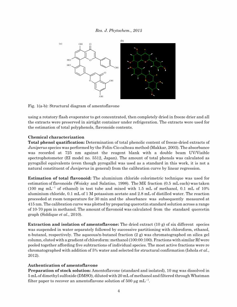
Fig. 1(a-b): Structural diagram of amentoflavone
using a rotatory flash evaporator to get concentrated, then completely dried in freeze drier and allthe extracts were preserved in airtight container under refrigeration. The extracts were used forthe estimation of total polyphenols, flavonoids contents.
Chemical characterizationTotal phenol quatification: Determination of total phenolic content of freeze-dried extracts ofJuniperus species was performed by the Folin-Cio calteau method (Makkar, 2003). The absorbancewas recorded at 725 nm against the reagent blank with a double beam UV/Visiblespectrophotometer (EI model no. 5512, Japan). The amount of total phenols was calculated aspyrogallol equivalents (even though pyrogallol was used as a standard in this work, it is not anatural constituent of Juniperus in general) from the calibration curve by linear regression.
Estimation of total flavonoid: The aluminium chloride colorimetric technique was used forestimation of flavonoids (Woisky and Salatino, 1998). The ME fraction (0.5 mL each) was taken(100 mg mLG1 of ethanol) in test tube and mixed with 1.5 mL of methanol, 0.1 mL of 10%aluminium chloride, 0.1 mL of 1 M potassium acetate and 2.8 mL of distilled water. The reactionproceeded at room temperature for 30 min and the absorbance was subsequently measured at415 nm. The calibration curve was plotted by preparing quercetin standard solution across a rangeof 10-70 ppm in methanol. The amount of flavonoid was calculated from the standard quercetingraph (Siddique et al., 2010).
Extraction and isolation of amentoflavone: The dried extract (10 g) of six different specieswas suspended in water separately followed by successive partitioning with chloroform, ethanol,n-butanol, respectively. The aqueous/n-butanol fraction (2 g) was chromatographed on silica gelcolumn, eluted with a gradient of chloroform: methanol (100:00:100). Fractions with similar Rf werepooled together affording five subtractions of individual species. The most active fractions were rechromatographed with addition of 5% water and selected for structural confirmation (Ishola et al.,2012).
Authentication of amentoflavonePreparation of stock solution: Amentoflavone (standard and isolated), 10 mg was dissolved in5 mL of dimethyl sulfoxide (DMSO), diluted with 20 mL of methanol and filtered through Whatmanfilter paper to recover an amentoflavone solution of 500 µg mLG1.
4
(b)

Res. J. Phytochem., 2015
Thin layer chromatography: Freshly coated with silica gel G254 plates were allowed to air dryat room temperature, transferred to an oven for activation and kept at 110°C for 30 min. Commonlyused solvent system for identification and quantification of flavonoids, benzene: pyridine: Formicacid (36:9:5) (BPF) was used for co-thin layer chromatograph to compare Rf values of standard andisolated amentoflavone (Ali et al., 2011). Stock solution (20 µL) was spotted; plates were placed intothe developing chamber and allowed to run until it reached a height of about 10 cm from the pointof spotting. Spots were viewed under U.V light and as developed in iodine vapour showed the spotwith different fluorescence. After development, the plates were kept in an oven maintained at110°C in order to achieve optimal color development.
Identification of amentoflavone: Melting point was determined with the help of a digitalmelting point apparatus (Microteknik, India). All selected fractions of six species were identifiedby spectral data (IR), which was in accordance with those previously described (Ishola et al., 2012;Markham et al., 1987; Zheng et al., 2011). The HPLC was used with C18 Column (250×4.6 mm,5 µm) column. The mobile phase consisted of methanol and phosphate buffer. The gradientcondition was 0-25 min with methanol 40-48% and 20-45 min with methanol 68-80%. The columntemperature was 40°C. The flow rate was 1.0 mL minG1 and the detection wavelength at 338 nm.
Calculation of the other parameters: All the physicochemical properties viz. MR (MolecularRefractivity), MV (Molecular Volume), Pc (Parachor), 0 (Index of refraction), ST (Surface Tension),D (density) and Pol (Polarizability) were calculated by ACD lab freeware (Chemsketch 5.0). All thetopological parameters and other descriptors were calculated by dragon 5.0 and some nonconventional parameter Viz. ASA (Approximate Surface Area) and SAG (Surface Area Grid) arecalculated by Hyperchem 6 (demo version).
Isolation of leucocytes: Human leukocytes were isolated from freshly sampled venous blood(12-15 mL) of healthy volunteers by using dextran (Macrodex: 6% dextran in 0.9% NaCl solution)and heparin tubes (25,000 IU mLG1).
Isolation of erythrocytes: Fresh blood samples from healthy volunteers (12-15 mL) werecollected and centrifuged at 3000 rpm for 15 min and plasma and buffy coats were removed. Redcells were washed with Phosphate Buffer Solution (PBS) (pH 7.00, containing 140 mM NaCl) 3times and erythrocytes were haemolyzed with ice-cold distilled water.
Preparation of incubations with Juniperus species (Isolated AF fractions)For erythrocytes:
C Group I: Control contains erythrocyte haemolysate 750 µL, PBS 1000 µL and distilled water250 µL
C Group II: H2O2, erythrocyte haemolysate 750 µL, H2O2 (10 mM) 50 µL, PBS 950 µL anddistilled water 250 µL
C Group III: C AF1A group; erythrocytes hemolysate 750 µL, H2O2 (10 mM) 50 µL, AF 1A 250 µL and PBS
950 µLC AF1B group; erythrocytes hemolysate 750 µL, H2O2 (10 mM) 50 µL, AF 1B 500 µL and PBS
950 µL
5

Res. J. Phytochem., 2015
C AF2A group; erythrocytes hemolysate 750 µL, H2O2 (10 mM) 50 µL, AF2A 250 µL and PBS950 µL
C AF2B group; erythrocytes hemolysate 750 µL, H2O2 (10 mM) 50 µL, AF2B 500 µL and PBS950 µL
C AF3A group; erythrocytes hemolysate 750 µL, H2O2 (10 mM) 50 µL, AF3A 250 µL and PBS950 µL
C AF3B group; erythrocytes hemolysate 750 µL, H2O2 (10 mM) 50 µL, AF3B 500 µL and PBS950 µL
C AF4A group; erythrocytes hemolysate 750 µL, H2O2 (10 mM) 50 µL, AF4A 250 µL and PBS950 µL
C AF4B group; erythrocytes hemolysate 750 µL, H2O2 (10 mM) 50 µL, AF4B 500 µL and PBS950 µL
C AF5A group; erythrocytes hemolysate 750 µL, H2O2 (10 mM) 50 µL, AF5A 250 µL and PBS950 µL
C AF5B group; erythrocytes hemolysate 750 µL, H2O2 (10 mM) 50 µL, AF5B 500 µL and PBS950 µL
C AF6A group; erythrocytes hemolysate 750 µL, H2O2 (10 mM) 50 µL, AF6A 250 µL and PBS950 µL
C AF6B group; erythrocytes hemolysate 750 µL, H2O2 (10 mM) 50 µL, AF6B 500 µL and PBS950 µL
For leucocytes:
C Group I: Control contains leucocytes haemolysate 750 µL, PBS 1000 µL and distilled water250 µL
C Group II: H2O2, leucocytes haemolysate 750 µL, H2O2 (10 mM) 50 µL, PBS 950 µL anddistilled water 250 µL
Group III: C AF1A group; leucocytes hemolysate 750 µL, H2O2 (10 mM) 50 µL, AF1A 250 µL and PBS
950 µLC AF1B group; leucocytes hemolysate 750 µL, H2O2 (10 mM) 50 µL, AF1B 500 µL and PBS
950 µLC AF2A group; leucocytes hemolysate 750 µL, H2O2 (10 mM) 50 µL, AF2A 250 µL and PBS
950 µLC AF2B group; leucocytes hemolysate 750 µL, H2O2 (10 mM) 50 µL, AF2B 500 µL and PBS
950 µLC AF3A group; leucocytes hemolysate 750 µL, H2O2 (10 mM) 50 µL, AF3A 250 µL and PBS
950 µLC AF3B group; leucocytes hemolysate 750 µL, H2O2 (10 mM) 50 µL, AF3B 500 µL and PBS
950 µL.C AF4A group; leucocytes hemolysate 750 µL, H2O2 (10 mM) 50 µL, AF4A 250 µL and PBS
950 µLC AF4B group; leucocytes hemolysate 750 µL, H2O2 (10 mM) 50 µL, AF4B 500 µL and PBS
950 µLC AF5A group; leucocytes hemolysate 750 µL, H2O2 (10 mM) 50 µL, AF5A 250 µL and PBS
950 µL
6

Res. J. Phytochem., 2015
C AF5B group; leucocytes hemolysate 750 µL, H2O2 (10 mM) 50 µL, AF5B 500 µL and PBS950 µL
C AF6A group; leucocytes hemolysate 750 µL, H2O2 (10 mM) 50 µL, AF6A 250 µL and PBS950 µL
C AF6B group; leucocytes hemolysate 750 µL, H2O2 (10 mM) 50 µL, AF6B 500 µL and PBS950 µL
Assays of antioxidant activityAssays of catalase activity: The reaction mixture consisted of 1 mL PBS (50 mM, pH 7.00) and2 mL diluted erythrocytes or leucocytes homogenate. The mixture was incubated at 25°C for 3 minand the reaction started by the addition of 1 mL of 30 mM H2O2. The decomposition of H2O2 wasfollowed directly by the decrease in absorbance at 240 nm at 25°C in a double beamspectrophotometer (EI model no. 7612, Japan) (Aebi, 1984). The results were expressed forerythrocytes as AU gG1 hemoglobin (Hb) and for the leucocytes as U mgG1 protein
Assays of SOD (Superoxide dismutase) activity: The SOD activity was determined using theRANDOX Ransod enzyme kit. This method employs xanthine and xanthine oxidase (XOD)generated superoxide radicals, which react with 2-(4-iodophenyl)-3-(4-nitropheno)-5-phenyltetrazolium-chloride to form the red formazon dye. The SOD activity was measured by thedegree of inhibition of this reaction (Konyalioglu and Karamenderes, 2005). The results wereexpressed for erythrocytes as U gG1 Hb and for leucocytes as U mgG1 protein.
Assays of GPx (Glutathion peroxidase) activity: The GPx activity was determined using theRANDOX Ransel enzyme kit. In this method, GPx catalyses the oxidation of GSH by hydrogenperoxide. In the presence of GSH reductase and reduced nicotinamide adenine dinucleotidephosphate (NADPH), the oxidised glutathione (GSSG) was immediately converted to the reducedform with a concomitant oxidation of NADPH to NADP+ (oxide form). The decrease in absorbancesat 340 nm was measured (Konyalioglu and Karamenderes, 2005). The results were expressed forerythrocytes as U gG1 Hb and for leucocytes as U mgG1 protein.
Determination of GSH (reduced glutathion) content in leucocytes and erythrocytes : TheGSH was determined by using 5, 5’ di-thio-bis-2-nitro benzoic acid (DTNB). In this method,molecule of DTNB was reduced to 2-nitro-5 mercapto benzoic acid (NMBA) by GSH. The NMBAwas deep yellow and this colour was used to measure SH groups by spectrophotometrically at412 nm erythrocyte and leucocyte homogenate samples (1 mL) were taken. Following this, i.e.,4 mL of 5% trichloroacetic acid (TCA) was added in centrifuge tubes. This mixture was centrifugedat 1000 rpm for 15 min. The PBS (50 mM, pH 8.00) 2 mL and 5 µM DTNB 250 µL were mixed witheach of 200 µL erythrocyte and leucocyte supernatants. The absorbance of the mixture wasmeasured against blank tube (added 200 µL distilled water instead of supernatant) at 412 nm(Duh and Yen, 1997). The results were expressed for erythrocytes as µg gG1 Hb and for leucocytesas µg mgG1 protein.
Determination of LPO (lipid peroxidase): The LPO was measured by TBA method(Satoh, 1978; Yagi, 1984), this method evaluates oxidative stress by measuring MDA, the lastproduct of lipid breakdown caused by oxidative stress. All experimental groups of erythrocyte andleucocyte homogenate samples were used. Test solutions (samples and standards) of 0.5 mL wereadded to 4.0 mL of N/12 H2 SO4 followed by the addition of 0.5 mL of 10% phosphotungustic acid
7

Res. J. Phytochem., 2015
and allowed to stand at room temperature for 5 min and then centrifuged for 10 min at 3,000 rpmand supernatant was discarded. The 2.5 mL N/12 H2SO4 and 0.2% TBA was added to these tubesand allowed to stand at boiling water bath for 60 min. After being cooled with tap water, 3 mL ofthe mixture of n butanol and HCl (15:1, v/v) was added and the mixture was shaken vigorously andabsorbance of the organic layer (upper layer) was measured at 532 nm (Duh and Yen, 1997). Theresults were expressed for erythrocytes as nmol gG1 Hb and for leucocytes as nmol mgG1 protein.
Determination of total protein concentration in erythrocyte: The Hb concentration wasdetermined by Drabkin method in erythrocyte hemolysate (Lowry et al., 1951).
Determination of Hb concentration in leukocytes: Total protein concentration in leucocytehaemolysate was evaluated by using bovine serum albumin as standard (Bauer et al., 1974).
RESULTS The results of the preliminary phytochemical screening of methanolic extract of Juniperus
showed the presence of alkaloids, tannins, flavonoids and terpenoids and steroids. The totalphenolic contents of juniperus species were estimated by folin catechu method in termspyrogallol equiv LG1. The contents of phenolic compounds were highest in Juniperus communis(2.14±0.21 pyrogallol equiv LG1) and the total flavonoid contents were highest in Juniperus indica(0.94±0.14 quercetin equiv LG1) (Table 2). Melting points of isolated AF fractions were shown inTable 3 which were matched with in a range of standard amentoflavone (218-220°C). The AFfractions (AF1-AF7) gave Rf value 0.17 compared to standard amentoflavone 0.17 in solvent systemof benzene: pyridine: formic acid (36:9:5) (BPF). The UV absorption maxima of isolated AF fractionswere 338 nm. The IR spectrum of isolated AF from different species of JC, JW, JP, JM, JI and JRshowed a strong bond of 3351, 1632, 1498 and 1452 cmG1 suggests presence of hydrogen OHstretching, chelated carbonyl C = O stretching and aromatic ring functionalities (C = C aromaticat 1608 cmG1) similar to standard amentoflavone.
Effects on neurochemical parameters: All the results indicated that all six extracts ofJuniperus species were found to be significant (p<0.001) on antioxidant enzyme systems oferythrocytes and leucocytes compared with the H2O2 group. Juniperus communis (AF1) andJuniperus indica (AF3) were the most effective one on CAT, GPx and SOD enzyme systems oferythrocytes (Table 4). Among plant extracts, (AF1) (500 µL) and (AF3) (500 µL) showed the highestsignificant activities on CAT, Juniperus communis (500 µL) on SOD, GPx and on LPO enzymesystem of leucocytes (Table 5). All extracts (six) of Juniperus sp. were found protective (250 and500 µL) on GSH levels of erythrocytes and leucocytes against H2O2 induced oxidative stress. AF6(24.54±1.210, 45.6±1.770 at (500 µL) and AF4 (25.96±1.760, 41.5±3.100 at (500 µL) have thesignificant activity on GSH levels of erythrocytes and leucocytes, respectively.
Table 2: Total phenolic contents from different species of JuniperusPlants name Total phenolic contents pyrogallol (equiv LG1) Total flavonoids quercetin (equiv LG1)Juniperus communis L. 2.14±0.21 0.484±0.29Juniperus wallichiana L. 0.89±0.69 0.250±0.12Juniperus polycarpos C. Koch 1.60±0.045 0.129±0.007Juniperus macropoda L. 1.29±0.67 0.560±0.19Juniperus indica L. 1.89±0.92 0.940±0.14Juniperus recurva L. 2.10±0.13 0.890±0.17All values expressed as Mean±SEM (n = 4)
8

Res. J. Phytochem., 2015
9
Tabl
e 3:
Var
ious
iden
tific
atio
n ch
arac
teri
stic
s of
am
ento
flavo
neIs
olat
ed fr
actio
ns o
f H
PLC
con
firm
atio
nam
ento
flavo
ne fr
om
Mel
ting
TLC
pro
file
UV
flo
w r
ate
= 1
mL
minG1
Juni
peru
s sp
ecie
spo
int (
°C)
(B.F
.)de
tect
ion
(nm
)W
= 3
38 n
mIR
Phys
icoc
hem
ical
pro
pert
ies
Ref
eren
ces
AF1
320-
322
0.16
*26
0 Li
near
ity-2
53-7
85 n
gIRνm
ax (K
Br)
M
olar
ref
ract
ivty
= 1
38.0
6 cm
3 , mol
ar,
Isho
la e
t al.
(201
2)R
ecov
ery
-97.
03%
cmG1 :3
082,
165
1,vo
lum
e =
324.
7 cm
3 , pol
arza
bilit
y =
54.7
2×10
G24 c
m3 ,
RSD
- 0.8
0% (
n =
5)16
09, 1
574,
149
3,
surf
ace
tens
ion
= 10
7.98
, ind
ex o
f ref
ract
ion
= 1.
793,
1423
, 135
8, 1
275,
m
olar
ref
ract
ivity
= 1
38.0
4, d
ensi
ty1.
656
g cm
G3 ,12
42, 1
165,
110
7,en
thal
py o
f vap
oriz
atio
n =
136.
9 K
J m
olG1
832
cmG1
AF2
317-
318
0.17
*26
0Li
near
ity-2
83-8
5 1n
gIRνm
ax (K
Br)
cmG1
Mol
ar r
efra
ctiv
ty =
138
.06
cm3 , m
olar
vol
ume
= 32
4.9
cm3 ,
Isho
la e
t al.
(201
2)R
ecov
ery-
98.0
6%30
81, 1
650,
160
9, 1
574,
pola
riza
bilit
y =
24.7
2×10
G24 c
m3 , s
urfa
ce te
nsio
n =
107.
98,
RSD
- 0.8
5% (n
= 5
)14
93, 1
423,
135
8, 1
285,
in
dex
of r
efra
ctio
n =
1.79
3, m
olar
ref
ract
ivity
= 1
38.0
4,12
42, 1
165,
110
7,de
nsity
1.65
6 g
cmG3 , e
ntha
lpy
of v
apor
izat
ion
= 13
6.9
KJ
molG1
832
cmG1
AF3
320-
321
0.17
*26
0Li
near
ity-2
43-7
86 n
gIRνm
ax (K
Br)
cmG1 :3
082,
M
olar
ref
ract
ivty
= 1
38.0
4 c
m3 , m
olar
vol
ume
= 32
4.5
cm3 ,
Isho
la e
t al.
(201
2)R
ecov
ery-
97.0
3%16
41, 1
609,
157
4, 1
493,
po
lari
zabi
lity
= 54
.72×
10G24
cm
3 , sur
face
tens
ion=
107.
98,
RSD
- 0.8
0% (n
=5)-
1423
, 135
8, 1
275,
124
2,
inde
x of
ref
ract
ion=
1.79
3, m
olar
ref
ract
ivity
=138
.04,
11
65, 1
107,
dens
ity1.
656
g cm
G3 , ent
halp
y of
vap
oriz
atio
n=13
6.9
KJ
molG1
832
cmG1
AF4
317-
180.
17*
260
Line
arity
-263
-715
ngIRνm
ax (K
Br)
cmG1 :3
080,
M
olar
ref
ract
ivty
=138
.06
cm3 , m
olar
vol
ume
= 32
4.9
cm3 ,
Rec
over
y-97
.03%
1651
, 160
9, 1
564,
149
3,
pola
riza
bilit
y =
54.7
2×10
G24 cm
3 , sur
face
tens
ion
= 10
7.90
, R
SD-0
.86%
(n =
5)
1423
, 135
8, 1
285,
124
2,
inde
x of
ref
ract
ion
= 1.
793,
mol
ar r
efra
ctiv
ity =
138
.04,
1165
, 110
7, 8
33 c
mG1
dens
ity1.
656
g cm
G3 , ent
halp
y of
vap
oriz
atio
n =
136.
9 K
J m
olG1
Isho
la e
t al.
(201
2)A
F531
5-31
70.
17*
260
Line
arity
-213
-765
ng
IRνm
ax (K
Br)
cmG1 :3
082,
M
olar
ref
ract
ivty
= 1
38.0
6 cm
3 , mol
ar v
olum
e =
324
.9 c
mG3 ,
Rec
over
y-97
.03%
1651
, 160
9, 1
574,
149
3,
pola
riza
bilit
y =
54.7
2×10
G24 c
m3 , s
urfa
ce te
nsio
n =
107.
98,
RSD
- 0.9
0% (n
= 5
)14
23, 1
358,
128
5, 1
242,
in
dex
of r
efra
ctio
n =
1.78
3, m
olar
ref
ract
ivity
= 1
38.0
4,
1165
, 110
7, 8
32 c
mG1
dens
ity1.
656
g cm
G3 , ent
halp
y of
vap
oriz
atio
n =
136.
9 K
J m
olG1
Isho
la e
t al.
(201
2)A
F631
6-18
0.17
*26
0Li
near
ity -2
03-8
85 n
gIRνm
ax (K
Br)
cmG1 :3
082,
M
olar
ref
ract
ivty
= 1
38.0
6 c
mG3 , m
olar
vol
ume
= 32
4.9
cmG3 ,
Rec
over
y-97
.03%
1651
, 160
9, 1
574,
149
2,
pola
riza
bilit
y =
54.7
2×10
G24 cm
3 , sur
face
tens
ion
= 10
7.98
, R
SD- 0
.90%
(n =
5)
1423
, 135
8, 1
285,
124
2,
inde
x of
ref
ract
ion
= 1.
793,
mol
ar r
efra
ctiv
ity =
138
.04,
11
65, 1
107,
832
cmG1
dens
ity1.
666
g cm
G3 , ent
halp
y of
vap
oriz
atio
n =
136.
9 K
J m
olG1
Isho
la e
t al.
(201
2)St
anda
rd31
8-32
00.
17*
260
Line
arity
153
-769
ng
IRνm
ax (K
Br)
cmG1 :3
082,
M
olar
ref
ract
ivty
= 1
38.0
4 cm
3 , mol
ar v
olum
e =
324.
9 cm
G3 , R
ecov
ery-
97.0
3%16
51, 1
609,
157
4, 1
493,
pola
riza
bilit
y =
54.7
2×10
G24 c
m3 , s
urfa
ce te
nsio
n =
107.
98,
RSD
- 0.9
0%14
23, 1
358,
128
5, 1
242,
inde
x of
ref
ract
ion
= 1.
793,
mol
ar r
efra
ctiv
ity =
138
.04,
1165
, 110
7, 8
33 c
mG1
dens
ity1.
656
g mG3 , e
ntha
lpy
of v
apor
izat
ion=
136.
9 K
J m
olG1
AF:
Am
ento
flavo
ne, R
SD: R
elat
ive
stan
dard
dev
iatio
n, T
LC: T
hin
laye
r chr
omat
ogra
phy,
HPL
C: H
igh
perf
orm
ance
liqu
id c
horo
mat
ogra
phy,
UV
: Ultr
avio
let,
IR: I
nfra
red

Res. J. Phytochem., 2015
Table 4: Effects of Juniperus species on erythrocytes antioxidant enzyme activity, glutathione and lipid peroxidase levels*
Groups and concentration CAT (AU gG1 Hb) SOD (U gG1 Hb) GSH (µg gG1 Hb) GPx (U gG1 Hb) LPO (nmol gG1 Hb)Juniperus aommunis (AF1)I 36.76±2.940a 2174.8±24.800a 25.90±1.270a 188.70±1.73a 35.80±1.360II 28.35±2.980a 1193.5±19.700a 43.83±1.980a 128.53±3.10a 19.23±2.380a
Juniperus wallichiana (AF2)I 45.65±1.470 5263.2±14.650a 33.70±2.110a 265.40±1.932a 41.76±3.920II 35.82±1.980a 2492.8±17.500a 41.80±5.100a 211.80±2.120a 32.70±2.560c
Juniperus indica (AF3)I 39.93±1.740b 1782.3±33.500a 16.21±1.720c 244.98±1.98a 31.78±2.880c
II 27.73±1.580a 1532.6±17.600a 41.70±1.420a 110.60±2.730a 17.38±2.710a
Juniperus macropoda (AF4)I 42.94±2.980 2556.2±34.50a 31.70±2.660a 288.40±3.200 39.62±2.870II 30.04±2.140a 1972.7±12.500a 41.50±3.100a 205.70±3.160a 29.50±1.770b
Juniperus polycarpos C. Koch (AF6)I 41.06±1.250c 2639.3±11.900a 38.90±1.620a 321.90±2.930b 38.50±2.910II 31.93±0.970a 1836.9±31.400a 45.60±1.770a 203.50±2.760a 27.67±1.970a
Juniperus recurva (AF5)I 43.25±3.56 2789.2±11.3 31.54±2.45 298.56±3.26b 31.34±3.32II 33.45±1.45a 1232.1±19.67b 38.70±4.34a 209.50±5.34a 21.70±5.12a
Control group 18.76±1.985a 1265.8±19.570a 48.29±1.720a 96.54±4.920a 16.53±1.970a
H2O2 group 52.67±1.250 2850.7±21.650 13.70±2.150 372.80±3.950 47.52±2.930*All the value expressed as Mean±Sem, (n = 3) at ap<0.001, bp<0.01, cp<0.05 when compared with H2O2 group (one- way ANOVA followed by Dunnets test),SOD: Superoxide dismutase, Gpx: Glutathion peroxidase, LPO Lipid peroxidase, CAT: Catalase, GSH: Glutathione
Table 5: Effects of Juniperus species on leucocytes antioxidant enzyme activity, Glutathione and lipid peroxidase levels*CAT LPO SOD GPx GSH
Groups and concentration (AU mgG1 protein) (nmol mgG1 protein) (U mgG1 protein) (U mgG1 protein) (µg mgG1 protein)Juniperus communis (AF1)I 15.21±0.3200a 27.30±2.100 1680±16.200a 208.54±1.620a 11.08±0.760II 13.79±1.832a 12.45±1.430a 8200±14.500a 152.72±3.700a 19.67±1.160c
Juniperus wallichiana (AF2)I 18.63±0.3420 26.93±2.620 1920±41.425a 299.62±4.320 13.67±0.980II 16.23±0.5260b 19.67±2.170c 1644±21.720a 201.67±1.830b 18.54±1.630c
Juniperus indica (AF3)I 18.82±0.4612 27.54±2.540 2547±21.980b 288.73±3.560b 19.73±0.630II 14.35±0.6120a 10.54±2.900a 2145±40.520a 162.53±1.783c 25.93±1.560c
Juniperus macropoda (AF4)I 17.36±0.5100 25.67±3.420 1932±30.320a 297.60±5.620 21.52±0.178b
II 15.82±0.5200b 16.31±1.760b 1045±19.670 198.78±3.800a 25.96±1.760a
Juniperus recurva (AF5)I 17.26±0.851 26.45±1.56 2354±22.13 301.82±2.67 14.32±1.56II 14.85±0.761a 17.27±0.972b 1572±29.12a 172.42±3.27a 16.72±2.9b
Juniperus polycarpos (AF6)I 15.73±0.9700b 23.93±1.980 2366±21.520a 225.45±2.730c 12.68±0.770II 14.53±1.431a 12.67±1.030a 1690±24.430a 194.43±1.980a 24.54±1.210a
Control group 10.48±0.6100 11.23±1.890 577.2±17.82 132.30±1.620a 29.56±3.980a
H2O2 group 21.67±0.7100 31.44±1.550 2789±78.210 311.31±2.892 9.67±2.670*All the value expressed as Mean±Sem, (n = 3), at ap<0.001, bp<0.01, cp<0.05 when compared with H2O2 group (one- way ANOVA followed by dunnets test),SOD: Superoxide dismutase, Gpx: Glutathion peroxidase, LPO Lipid peroxidase, CAT: Catalase, GSH: Glutathione
DISCUSSIONThe protective effect of methanolic extracts from different species of juniperus in different
concentration (250 and 500 µL) has been seen against H2O2 induced oxidative damage in humanerythrocytes and leucocytes. Juniperus species was used traditionally, in the treatment of variousdiseases and disorder like cough, abdominal disorders, renal suppression, leucorrhoea, skinaffections, infantile tuberculosis, asthma and also used in acute and chronic cystitis Juniper berriesare used commercially for the preparation essential oil (flavoring agents), gin and as a spice(Marse, 1991). According to traditional usage of Juniperus species, we evaluated methanolicextracts of these plants against H2O2 induced oxidative damage in human erythrocytes andleucocytes. Juniperus species are reported for presence of numerous chemicals i.e. volatile oils(Vichi et al., 2007), coumarines, bicyclic diterpenes (Teresa et al., 1980), flavonoids and phenoliccompounds (Lamer-Zarawska, 1975; Hiermann et al., 1996; Ilyas and Ilyas, 1990; Khare, 2007;Bais et al., 2015). The phenolic compounds obtained from four species of Juniperus have been
10

Res. J. Phytochem., 2015
reported as neuroprotective (Tavares et al., 2012; Bais and Mali, 2013). Present study also focusedon the effect of junipers against oxidative stress model. Human blood cells are the key fightersagainst various oxidative stress diseases and remain the primary target for natural products andplants. These cells are continuously exposed to radicals and are unable to replace damagedcomponents, especially polyunsaturated fatty acids chains from peroxidation (Bukowska, 2003).
During the reduction of molecular oxygen to water through acceptance of four electrons, activeoxygen species, such as superoxide anion radicals, H2O2 and OH• radicals are generated. Againstsuch active oxygen’s, cells have some defensive mechanisms including superoxide dismutase,catalase, several peroxidases and antioxidants, such as ascorbate, tocopherol, uric acid, β-caroteneand GSH (Izawa et al., 1995; Yen et al., 2003).
In response to oxidative stress, GSH protect the thiol groups on membrane surface. Thedecreased availability of GSH indicates an increased demand (GSH) in erythrocytes to regain thestorage mediated oxidative stress (Ault and Lawrence, 2003; Konyalioglu and Karamenderes,2005). In present study, all extracts had significant (p<0.001) protective effect on GSH level ascompared to the hydrogen peroxide group. Reactive oxygen species degrade polyunsaturated lipids,forming malondialdehyde (Schopfer et al., 2001). This is an end product of erythrocyte membranelipid peroxidation. In various clinical conditions in humans and in vitro studies in animalerythrocytes and leucocytes, MDA a highly reactive, bifunctional molecule has been shown tocross-link erythrocyte and leucocyte phospholipids and proteins to impair a variety of themembrane-related functions and ultimately leading to diminished erythrocytes and leucocytessurvival (Chiu et al., 1989; Sugihara et al., 1991; Ault and Lawrence, 2003). All extracts ofjuniperus species significantly decreased the level of LPO in dose dependent manner whencompared with H2O2 group and this showed the protective effect against oxidative damage.Juniperus communis (500 µL) and Juniperus indica (500 µL) have the lowest LPO levels onerythrocytes (Table 4). Juniperus polycarpos C. Koch (500 µL) and Juniperus indica (500 µL)showed highest protective effects on leucocytes (Table 5). As a conclusion, Juniperus speciesextracts are found to have protective effect on antioxidant enzyme systems, GSH and LPO levelsof erythrocytes and leucocytes against H2O2 Induced oxidative damage. Observed activity can beattributed to the phenol contents of these plants. The present results demonstrate that extracts ofJuniperus communis, Juniperus wallichiana, Juniperus indica, Juniperus macropoda, Juniperusrecurva and Juniperus polycarpos are potential sources of natural antioxidants. In future, furtherin vitro and in vivo studies are needed to assess the effects of these species on antioxidant systemof the body.
ACKNOWLEDGMENTSAuthors are thankful to the Principal, Rayat Institute of Pharmacy, Punjab for providing the
necessary facilities during this research work. Authors also wish to thanks Pinnacle BiomedicalResearch Institute, Bhopal.
REFERENCESAbbassy, M.A. and G.I. Marei, 2013. Antifungal and chemical composition of essential oils of
Juniperus communis L. and Thymus vulgaris L. against two phytopathogenic fungi. J. AppliedSci. Res., 9: 4584-4588.
Adams, R.P., 2014. Juniperus of the World: The Genus Juniperus. 4th Edn., Trafford Publishers,USA., pp: 1-25.
11

Res. J. Phytochem., 2015
Aebi, H., 1984. Catalase in vitro. Methods Enzymol., 105: 121-126.Akdogan, M., A. Koyu, M. Ciris and K. Yildiz, 2012. Anti-hypercholesterolemic activity of Juniperus
communis Lynn Oil in rats: A biochemical and histopathological investigation. Biomed. Res.,23: 321-328.
Ali, S.A., O.R. Abdulraheem, T.E. Alemika, I.M. Sule and M. Ilyas et al., 2011. Phytochemicalscreening and identification of compounds in the leaves of Callitris glauca (Cupresseceae). Res.J. Pharma. Biol. Chem. Sci., 2: 624-635.
Ault, J.G. and D.A. Lawrence, 2003. Glutathione distribution in normal and oxidatively stressedcells. Exp. Cell Res., 285: 9-14.
Bais, S.S. and P.Y. Mali, 2013. Protective effect of Amorphophallus campanulatus tuber extractsagainst H2O2 induced oxidative damage in human erythrocytes and leucocytes. Int. J. GreenPharm., 7: 111-116.
Bais, S., N.S. Gill, N. Rana and S. Shandil, 2014a. A phytopharmacological review on a medicinalplant: Juniperus communis. Int. Scholarly Res. Notices, Vol. 2014. 10.1155/2014/634723
Bais, S., N.S. Gill and N. Rana, 2014b. Effect of Juniperus communis extract on reserpine inducedcatalepsy. Inventi Impact: Ethnopharmacol., 2014: 117-120.
Bais, S., N.S. Gill and N. Kumar, 2015. Neuroprotective effect of Juniperus communis onchlorpromazine induced Parkinson disease in animal model. Chin. J. Biol., Vol. 2015.10.1155/2015/542542
Banerjee, S., A. Mukherjee and T.K. Chatterjee, 2012. Evaluation of analgesic activities ofmethanolic extract of medicinal plant Juniperus communis Linn. Int. J. Pharm. PharmaceuticalSci., 4: 547-550.
Banerjee, S., H. Singh and T.K. Chatterjee, 2013. Evaluation of anti-diabetic and anti-hyperlipidemic potential of methanolic extract of Juniperus communis (L.) instreptozotocinnicotinamide induced diabetic rats. Int. J. Pharm. Bio Sci., 4: 10-17.
Baral, S.R. and P.P. Kurmi, 2006. A Compendium of Medicinal Plants in Nepal. Rachana ShramaPublication, Kathmandu, ISBN-13: 9789994620272, Pages: 534.
Bauer, J.D., P.G. Ackerman and G. Toro, 1974. Clinical Laboratory Methods. CV Mosby Co., St.Louis, New York.
Baureithel, K.H., K.B. Buter, A. Engesser, W. Burkard and W. Schaffner, 1997. Inhibition ofbenzodiazepine binding in vitro by amentoflavone, a constituent of various species ofHypericum. Pharmaceutica Acta Helvetiae, 72: 153-157.
Bukowska, B., 2003. Effects of 2,4-D and its metabolite 2,4-dichlorophenol on antioxidant enzymesand level of glutathione in human erythrocytes. Comp. Biochem. Physiol. C Toxicol. Pharmacol.,135: 435-441.
Chiu, D., F. Kuypers and B. Lubin, 1989. Lipid peroxidation in human red cells. Semin. Hematol.,26: 257-276.
Cholbi, M.R., M. Paya and M.J. Alcaraz, 1991. Inhibitory effects of phenolic compounds onCCl4-induced microsomal lipid peroxidation. Cell. Mol. Life Sci., 47: 195-199.
Cooper, D., K.Y. Stokes, A. Tailor and D.N. Granger, 2002. Oxidative stress promotes bloodcell-endothelial cell interactions in the microcirculation. Cardiovascular Toxicol., 2: 165-180.
Da Silva, K.L., A.R. dos Santos, P.E. Mattos, R.A. Yunes, F. Delle-Monache and V. Cechinel-Filho,2000. Chemical composition and analgesic activity of Calophyllum brasiliense leaves. Therapie,56: 431-434.
Duh, P.D. and G.C. Yen, 1997. Antioxidative activity of three herbal water extracts. Food Chem.,60: 639-645.
12

Res. J. Phytochem., 2015
Gumral, N., D.D. Kumbul, F. Aylak, M. Saygin and E. Savik, 2015. Juniperus communis Linn oildecreases oxidative stress and increases antioxidant enzymes in the heart of rats administereda diet rich in cholesterol. Toxicol. Ind. Health, 31: 85-91.
Gyawali, R., M. Binita and T.M. Shrestha, 2012. Antibacterial and cytotoxic activity of juniperusindica bertol from nepalese Himalaya. Int. J. Pharma. Sci. Res., 3: 1104-1107.
Haider, S.Z., M. Mohan, S. Sah and R. Seth, 2013. Comparative study on composition andantimicrobial activity of Juniperus wallichiana essential oils against clinical isolates.Med. Plants-Int. J. Phytomedicines Related Ind., 5: 90-95.
Hiermann, A., A. Kompek, J. Reiner, H. Auer and M. Schubert-Zsilavecz, 1996. Investigation offlavonoid pattern in fruits of Juniperus communis L. Sci. Pharmaceutica, 64: 437-444.
Hoferl, M., I. Stoilova, E. Schmidt, J. Wanner and L. Jirovetz et al., 2014. Chemical compositionand antioxidant properties of juniper berry (Juniperus communis L.) essential oil. action of theessential oil on the antioxidant protection of Saccharomyces cerevisiae model organism.Antioxidants, 3: 81-98.
Ilyas, M. and N. Ilyas, 1990. Biflavones from the leaves of Juniperus communis and a survey onbiflavones of the Juniperus genus. Ghana J. Chem., 1: 143-147.
Ishola, I.O., M. Chatterjee, S. Tota, N. Tadigopulla, O.O. Adeyemi, G. Palit and R. Shukla, 2012.Antidepressant and anxiolytic effects of amentoflavone isolated from Cnestis ferruginea inmice. Pharmacol. Biochem. Behav., 103: 322-331.
Izawa, S., Y. Inoue and A. Kimura, 1995. Oxidative stress response in yeast: Effect of glutathioneon adaptation to hydrogen peroxide stress in Saccharomyces cerevisiae. FEBS Lett., 368: 73-76.
Khare, C.P., 2007. Indian Medicinal Plants: An Illustrated Dictionary. Springer Science andBusiness Media, New York, ISBN: 9780387706375, pp: 346-348.
Kim, H.K., K.H. Son, H.W. Chang, S.S. Kang and H.P. Kim, 1998. Amentoflavone, a plantbiflavone: A new potential anti-inflammatory agent. Arch. Pharm. Res., 21: 406-410.
Konyalioglu, S. and C. Karamenderes, 2005. The protective effects of Achillea L. species native inTurkey against H2O2-induced oxidative damage in human erythrocytes and leucocytes.J. Ethnopharmacol., 102: 221-227.
Lamer-Zarawska, E., 1975. Biflavonoids in Juniperus L. sp (Cupressaceae). Polish J. Pharmacol.Pharm., 27: 81-87.
Landmesser, U., S. Dikalov, S.R. Price, L. McCann and T. Fukai et al., 2003a. Oxidation oftetrahydrobiopterin leads to uncoupling of endothelial cell nitric oxide synthase inhypertension. J. Clin. Invest., 111: 1201-1209.
Landmesser, U., S. Dikalov, S.R. Price, L. McCann and T. Fukai et al., 2003b. Oxidation oftetrahydrobiopterin leads to uncoupling of endothelial cell nitric oxide synthase inhypertension. J. Clin. Invest., 111: 1201-1209.
Lin, Y.M., M.T. Flavin, R. Schure, F.C. Chen and R. Sidwell et al., 1999. Antiviral activities ofbiflavonoids. Planta Med., 65: 120-125.
Lowry, O.H., N.J. Rosebrough, A.L. Farr and R.J. Randall, 1951. Protein measurement with theFolin phenol reagent. J. Biol. Chem., 193: 265-275.
Makkar, H.P.S., 2003. Quantification of Tannins in Tree and Shrub Foliage: A Laboratory Manual.Kluwer Academic Publishers, Dordrecht, Netherlands, pp: 43-54..
Manvi and G.P. Garg, 2010. Screening and evaluation of pharmacognostic, phytochemical andhepatoprotective activity of Juniperus communis Linn. stems. Int. J. Pharma. Bio Sci., 1: 1-11.
13

Res. J. Phytochem., 2015
Markham, K.R., C. Sheppard and H. Geiger, 1987. 13C NMR studies of some naturally occurringamentoflavone and hinokiflavone biflavonoids. Phytochemistry, 26: 3335-3337.
Marse, H., 1991. Volatile Compounds in Foods and Beverages. 1st Edn., CRC Press, New York,pp: 94.
Mishra, P. and C.S. Chauhan, 1984. Antimicrobial studies of the essential oil of the berries ofJuniperus macropoda Boiss. Hindustan Antibiotics Bull., 26: 38-40.
Moein, M.R., Y. Ghasemi, S. Moein and M. Nejati, 2010. Analysis of antimicrobial, antifungal andantioxidant activities of Juniperus excelsa M. B subsp. Polycarpos (K. Koch) Takhtajanessential oil. Pharmacognosy Res., 2: 128-131.
Patel, R.P., D. Moellering, J. Murphy-Ullrich, H. Jo, J.S. Beckman and V.M. Darley-Usmar, 2000.Cell signaling by reactive nitrogen and oxygen species in atherosclerosis. Free Radical Biol.Med., 28: 1780-1794.
Pepeljnjak, S., I. Kosalec, Z. Kalodera and N. Blazevic, 2005. Antimicrobial activity of juniper berryessential oil (Juniperus communis L., Cupressaceae). Acta Pharm., 55: 417-422.
Sati, S.C. and S. Joshi, 2010. Antibacterial potential of leaf extracts of Juniperus communis L. fromKumaun Himalaya. Afr. J. Microbiol. Res., 4: 1291-1294.
Satoh, K., 1978. Serum lipid peroxide in cerebrovascular disorders determined by a newcolorimetric method. Clin. Chim. Acta, 90: 37-43.
Schopfer, P., C. Plachy and G. Frahry, 2001. Release of reactive oxygen intermediates(superoxide radicals, hydrogen peroxide and hydroxyl radicals) and peroxidase in germinatingradish seeds controlled by light, gibberellin and abscisic acid. Plant Physiol., 125: 1591-1602.
Seca, A.M.L. and A.M.S. Silva, 2006. The Chemical Composition of the Juniperus Genus(1970-2004). In: Recent Progress in Medicinal Plants: Phytomedicines, Govil, J.N., V.K. Singhand R. Bhardwaj (Eds.). Studium Press, Houston, TX., ISBN: 0-9761849-8-2, pp: 401-522.
Sharma, P.K. and B. Lal, 2005. Ethnobotanical notes on some medicinal and aromatic plants ofHimachal Pradesh. Indian J. Trad. Knowl., 4: 424-428.
Siddique, N.A., M. Mujeeb, A.K. Najmi and M. Akram, 2010. Evaluation of antioxidant activity,quantitative estimation of phenols and flavonoids in different parts of Aegle marmelos.Afr. J. Plant Sci., 4: 1-5.
Stokes, K.Y., D. Cooper, A. Tailor and D.N. Granger, 2002a. Hypercholesterolemia promotesinflammation and microvascular dysfunction: Role of nitric oxide and superoxide. Free RadicalBiol. Med., 33: 1026-1036.
Stokes, K.Y., E.C. Clanton, K.S. Bowles, J.W. Fuseler and D. Chervenak et al., 2002b. The role ofT-lymphocytes in hypercholesterolemia-induced leukocyte-endothelial interactions.Microcirculation, 9: 407-417.
Sugihara, T., W. Rawicz, E.A. Evans and R.P. Hebbel, 1991. Lipid hydroperoxides permitdeformation-dependent leak of monovalent cation from erythrocytes. Blood, 77: 2757-2763.
Tavares, L., G.J. McDougall, S. Fortalezas, D. Stewart, R.B. Ferreira and C.N. Santos, 2012. Theneuroprotective potential of phenolic-enriched fractions from four Juniperus species found inPortugal. Food Chem., 135: 562-570.
Teresa, J.D.P., A.F. Barrero, L. Muriel, A.S. Feliciano and M. Grande, 1980. New natural diterpeneacids from Juniperus communis. Phytochemistry, 19: 1153-1156.
Tunon, H., C. Olavsdotter and L. Bohlin, 1995. Evaluation of anti-inflammatory activity of someSwedish medicinal plants. Inhibition of prostaglandin biosynthesis and PAF-induced exocytosis.J. Ethnopharmacol., 48: 61-76
14

Res. J. Phytochem., 2015
Vichi, S., M. Riu-Aumatell, M. Mora-Pons, J.M. Guadayol, S. Buxaderas and E. Lopez-Tamames,2007. HS-SPME coupled to GC/MS for quality control of Juniperus communis L. berries usedfor gin aromatization. Food Chem., 105: 1748-1754.
Woisky, R.G. and A. Salatino, 1998. Analysis of propolis: Some parameters and procedures forchemical quality control. J. Apicult. Res., 37: 99-105.
Yagi, K., 1984. Assay for blood plasma or serum. Methods Enzymol., 105: 328-331.Yen, G.C., H.C. Chiang, C.H. Wu and C.T. Yeh, 2003. The protective effects of Aspergillus candidus
metabolites against hydrogen peroxide-induced oxidative damage to Int 407 cells. Food Chem.Toxicol., 41: 1561-1567.
Zheng, J.X., Y. Zheng, H. Zhi, Y. Dai and N.L. Wang et al., 2011. New 3 , 8 -linked biflavonoidsfrom Selaginella uncinata displaying protective effect against anoxia. Molecules, 16: 6206-6214.
15