Tracking plasma generated H2O2 from gas into liquid phase ...
Hydroxytyrosol glucuronides protect renal tubular epithelial cells against H2O2 induced oxidative...
8
Hydroxytyrosol glucuronides protect renal tubular epithelial cells against H 2 O 2 induced oxidative damage Monica Deiana a,⇑ , Alessandra Incani a , Antonella Rosa a , Angela Atzeri a , Debora Loru a , Barbara Cabboi a , M. Paola Melis a , Ricardo Lucas b , Juan C. Morales b , M. Assunta Dessì a a Dipartimento di Biologia Sperimentale, Sezione di Patologia Sperimentale, Università degli Studi di Cagliari, Cittadella Universitaria SS 554, 09042 Monserrato, Cagliari, Italy b Department of Bioorganic Chemistry, Instituto de Investigaciones Químicas, CSIC-Universidad de Sevilla, 49 Americo Vespucio, 41092 Sevilla, Spain article info Article history: Received 16 May 2011 Received in revised form 4 July 2011 Accepted 10 July 2011 Available online 26 July 2011 Keywords: Hydrogen peroxide Hydroxytyrosol glucuronides Lipid peroxidation MAPK Oxidative stress abstract Hydroxytyrosol (2-(3 0 ,4 0 -dihydroxyphenyl)ethanol; HT), the most active ortho-diphenolic compound, present either in free or esterified form in extravirgin olive oil, is extensively metabolized in vivo mainly to O-methylated, O-sulfated and glucuronide metabolites. We investigated the capacity of three glucuro- nide metabolites of HT, 3 0 -O-b-D-glucuronide and 4 0 -O-b-D-glucuronide derivatives and 2-(3 0 ,4 0 -dihy- droxyphenyl)ethanol-1-O-b-D-glucuronide, in comparison with the parent compound, to inhibit H 2 O 2 induced oxidative damage and cell death in LLC-PK1 cells, a porcine kidney epithelial cell line. H 2 O 2 treat- ment exerted a toxic effect inducing cell death, interacting selectively within the pro-death extracellular- signal relate kinase (ERK 1/2) and the pro-survival Akt/PKB signaling pathways. It also produced direct oxidative damage initiating the membrane lipid peroxidation process. None of the tested glucuronides exhibited any protection against the loss in renal cell viability. They also failed to prevent the changes in the phosphorylation states of ERK and Akt, probably reflecting their inability to enter the cells, while HT was highly effective. Notably, pretreatment with glucuronides exerted a protective effect at the high- est concentration tested against membrane oxidative damage, comparable to that of HT: the formation of malondialdehyde, fatty acid hydroperoxides and 7-ketocholesterol was significantly inhibited. Ó 2011 Elsevier Ireland Ltd. All rights reserved. 1. Introduction Hydroxytyrosol (2-(3 0 ,4 0 -dihydroxyphenyl)ethanol; HT) is the most potent antioxidant among the phenolics found in extravirgin olive oil. The biological activities of HT have been explored by sev- eral groups as recently reviewed by Granados-Principal et al. [1]. Two of the most relevant reported activities are antitumor and anti-inflammatory effects. The anticancer activity seems to be a re- sult of its capacity to exert cytotoxic effects, such as induction of apoptosis, cell cycle arrest and antiproliferative effects. The anti- inflammatory and antiplatelet aggregation action of HT, together with its antiatherogenic capacity and cardioprotective effects, are important in counteracting the development of cardiovascular diseases. HT biological effects stem mainly from its free radical scaveng- ing and metal chelating properties, most probably due to its ortho-diphenolic structure, whose high antioxidant activity may be explained by the high electron donating effect of the second hydroxyl group. At the same time it also shows effects on cell sig- naling pathways and on gene expression [1]. A daily intake of 25–50 ml of extravirgin olive oil, typical of Med- iterranean countries, may supply at least 1 mg of simple phenols, free HT and tyrosol, and 8 mg of their secoiridoids derivatives, mainly oleuropein and ligstroside-aglycones [2]. However, the majority of these complex polyphenols undergo gastro-intestinal biotransformation. HT and tyrosol conjugated forms, except oleu- ropein, are rapidly hydrolysed under gastric conditions, effectively increasing the relative amount of simple phenols, HT and tyrosol, entering the small and large intestine [3]. Oleuropein is not degraded under acidic conditions and is not absorbed in the paren- tal form in the small intestine; however, once reached the large intestine, it may be subjected to rapid degradation by the colonic microflora, to yield HT [3]. The absorption of HT takes place in the small intestine and the colon [4] through a passive diffusion mechanism [5]. In the process of crossing enterocytes, HT is subjected to a classic phase I/II bio- transformation, and then to an important first pass metabolism in the liver cells. This process leads to the formation of ortho- methyl derivatives (homovanillic alcohol), glucuronide and sulfate 0009-2797/$ - see front matter Ó 2011 Elsevier Ireland Ltd. All rights reserved. doi:10.1016/j.cbi.2011.07.002 Abbreviations: HT, hydroxytyrosol; HP, fatty acid hydroperoxides; UFA, unsaturated fatty acids; 7-keto, 7-ketocholesterol; Glu1, hydroxytyrosol 3 0 -O-b-D- glucuronide; Glu2, hydroxytyrosol 4 0 -O-b-D-glucuronide; Glu3, 2-(3 0 ,4 0 -dihydroxy- phenyl)ethanol-1-O-b-D-glucuronide; ERK, extracellular-signal related kinase. ⇑ Corresponding author. Tel.: +39 070 6754126; fax: +39 070 6754032. E-mail address: [email protected] (M. Deiana). Chemico-Biological Interactions 193 (2011) 232–239 Contents lists available at ScienceDirect Chemico-Biological Interactions journal homepage: www.elsevier.com/locate/chembioint
-
Upload
independent -
Category
Documents
-
view
2 -
download
0
Transcript of Hydroxytyrosol glucuronides protect renal tubular epithelial cells against H2O2 induced oxidative...

Chemico-Biological Interactions 193 (2011) 232–239
Contents lists available at ScienceDirect
Chemico-Biological Interactions
journal homepage: www.elsevier .com/locate /chembioint
Hydroxytyrosol glucuronides protect renal tubular epithelial cells against H2O2
induced oxidative damage
Monica Deiana a,⇑, Alessandra Incani a, Antonella Rosa a, Angela Atzeri a, Debora Loru a, Barbara Cabboi a,M. Paola Melis a, Ricardo Lucas b, Juan C. Morales b, M. Assunta Dessì a
a Dipartimento di Biologia Sperimentale, Sezione di Patologia Sperimentale, Università degli Studi di Cagliari, Cittadella Universitaria SS 554, 09042 Monserrato, Cagliari, Italyb Department of Bioorganic Chemistry, Instituto de Investigaciones Químicas, CSIC-Universidad de Sevilla, 49 Americo Vespucio, 41092 Sevilla, Spain
a r t i c l e i n f o
Article history:Received 16 May 2011Received in revised form 4 July 2011Accepted 10 July 2011Available online 26 July 2011
Keywords:Hydrogen peroxideHydroxytyrosol glucuronidesLipid peroxidationMAPKOxidative stress
0009-2797/$ - see front matter � 2011 Elsevier Irelandoi:10.1016/j.cbi.2011.07.002
Abbreviations: HT, hydroxytyrosol; HP, fattyunsaturated fatty acids; 7-keto, 7-ketocholesterol; Glglucuronide; Glu2, hydroxytyrosol 40-O-b-D-glucuroniphenyl)ethanol-1-O-b-D-glucuronide; ERK, extracellul⇑ Corresponding author. Tel.: +39 070 6754126; fax
E-mail address: [email protected] (M. Deiana).
a b s t r a c t
Hydroxytyrosol (2-(30 ,40-dihydroxyphenyl)ethanol; HT), the most active ortho-diphenolic compound,present either in free or esterified form in extravirgin olive oil, is extensively metabolized in vivo mainlyto O-methylated, O-sulfated and glucuronide metabolites. We investigated the capacity of three glucuro-nide metabolites of HT, 30-O-b-D-glucuronide and 40-O-b-D-glucuronide derivatives and 2-(30 ,40-dihy-droxyphenyl)ethanol-1-O-b-D-glucuronide, in comparison with the parent compound, to inhibit H2O2
induced oxidative damage and cell death in LLC-PK1 cells, a porcine kidney epithelial cell line. H2O2 treat-ment exerted a toxic effect inducing cell death, interacting selectively within the pro-death extracellular-signal relate kinase (ERK 1/2) and the pro-survival Akt/PKB signaling pathways. It also produced directoxidative damage initiating the membrane lipid peroxidation process. None of the tested glucuronidesexhibited any protection against the loss in renal cell viability. They also failed to prevent the changesin the phosphorylation states of ERK and Akt, probably reflecting their inability to enter the cells, whileHT was highly effective. Notably, pretreatment with glucuronides exerted a protective effect at the high-est concentration tested against membrane oxidative damage, comparable to that of HT: the formation ofmalondialdehyde, fatty acid hydroperoxides and 7-ketocholesterol was significantly inhibited.
� 2011 Elsevier Ireland Ltd. All rights reserved.
1. Introduction
Hydroxytyrosol (2-(30,40-dihydroxyphenyl)ethanol; HT) is themost potent antioxidant among the phenolics found in extravirginolive oil. The biological activities of HT have been explored by sev-eral groups as recently reviewed by Granados-Principal et al. [1].Two of the most relevant reported activities are antitumor andanti-inflammatory effects. The anticancer activity seems to be a re-sult of its capacity to exert cytotoxic effects, such as induction ofapoptosis, cell cycle arrest and antiproliferative effects. The anti-inflammatory and antiplatelet aggregation action of HT, togetherwith its antiatherogenic capacity and cardioprotective effects, areimportant in counteracting the development of cardiovasculardiseases.
HT biological effects stem mainly from its free radical scaveng-ing and metal chelating properties, most probably due to its
d Ltd. All rights reserved.
acid hydroperoxides; UFA,u1, hydroxytyrosol 30-O-b-D-de; Glu3, 2-(30 ,40-dihydroxy-ar-signal related kinase.: +39 070 6754032.
ortho-diphenolic structure, whose high antioxidant activity maybe explained by the high electron donating effect of the secondhydroxyl group. At the same time it also shows effects on cell sig-naling pathways and on gene expression [1].
A daily intake of 25–50 ml of extravirgin olive oil, typical of Med-iterranean countries, may supply at least 1 mg of simple phenols,free HT and tyrosol, and 8 mg of their secoiridoids derivatives,mainly oleuropein and ligstroside-aglycones [2]. However, themajority of these complex polyphenols undergo gastro-intestinalbiotransformation. HT and tyrosol conjugated forms, except oleu-ropein, are rapidly hydrolysed under gastric conditions, effectivelyincreasing the relative amount of simple phenols, HT and tyrosol,entering the small and large intestine [3]. Oleuropein is notdegraded under acidic conditions and is not absorbed in the paren-tal form in the small intestine; however, once reached the largeintestine, it may be subjected to rapid degradation by the colonicmicroflora, to yield HT [3].
The absorption of HT takes place in the small intestine and thecolon [4] through a passive diffusion mechanism [5]. In the processof crossing enterocytes, HT is subjected to a classic phase I/II bio-transformation, and then to an important first pass metabolismin the liver cells. This process leads to the formation of ortho-methyl derivatives (homovanillic alcohol), glucuronide and sulfate

M. Deiana et al. / Chemico-Biological Interactions 193 (2011) 232–239 233
derivatives [6] and glutathionyl conjugates [3], which are detect-able both in the plasma and in the urine [7]. Urinary recoveriesas high as 80% of the ingested amounts of HT have been reportedin humans [6,8]; over 90% of the urinary metabolites were conju-gates, mainly glucuronidated metabolites [8–10]. 30-O- and 40-O-b-glucuronide of HT have been detected in human urine afteringestion of olive oil [11]. As unconjugated HT represents a verysmall proportion of circulating species, the testing of the biologicalactivity of the conjugated metabolites is of great importance forthe understanding of its in vivo properties. Few in vitro studieshave been carried out on HT glucuronide conjugates and showedcontrasting results: the 30-O-b-glucuronide exerted a more potentradical scavenging activity than HT itself in a simple chemical sys-tem [7], while failed together with the 40-O-b-glucuronide to pro-tect LDL against oxidation [11].
The aim of this study was to examine the activity of the HT glu-curonide metabolites in comparison with the parent compoundagainst oxidative stress in vitro. H2O2 induced oxidative damageand cell death in LLC-PK1 cells, a porcine kidney epithelial cell linethat retains characteristics of the proximal tubular epithelium [12].This cell line has been used to study the mechanisms of kidney tox-icity [13,14]. The generation of H2O2 has been implicated in thepathogenesis of several forms of acute tubular cell injury, wherethe lipid peroxidation process plays a central role [15]. We havepreviously shown that H2O2 treatment in LLC-PK1 cells may inducea toxic effect both through a direct oxidative damage, initiating themembrane lipid peroxidation process [16], and inducing cell death,interacting selectively within signaling cascades that regulate cellsurvival following exposure to oxidative stress, as the pro-deathextracellular-signal relate kinase (ERK 1/2) and the pro-survivalAkt/PKB signaling pathways [17]. As 30-O- and 40-O-b-glucuronideof HT have been detected in human urine, after ingestion of oliveoil [11], three glucuronide metabolites of HT, 30-O-b-D-glucuronideand 40-O-b-D-glucuronide, as a mixture of isomers, and 2-(30,40-dihydroxyphenyl)ethanol-1-O-b-D-glucuronide (Fig. 1) have beensynthesized and tested, in order to investigate the influence ofthe conjugation and its position within the phenolic structure.
H2O2 induced toxicity was evaluated in terms of changes in thephosphorylation state of the kinases ERK 1/2 and Akt/PKB. H2O2 in-duced oxidative cell damage was also evaluated as malondialde-hyde (MDA) production and through the more sensible andprecise markers of the lipid peroxidation process, the modificationof the profile of the major oxidizable membrane lipids, unsaturatedfatty acids (UFA) and cholesterol.
2. Materials and methods
2.1. Chemicals
All solvents used were HPLC grade (Merck, Darmstadt, Germany).Fatty acid standards, cholesterol, 5-cholesten-3b-ol-7-one(7-keto), H2O2, 2-thiobarbituric acid (TBA), trichloroacetic acid
Fig. 1. Structure of hydroxytyrosol glucuronides Glu1, Glu2 and Glu3 and theirparent compound hydroxytyrosol (HT).
(TCA), 1,1,3,3-tetraethoxypropane (TEP), 3-(4,5-dimethylthiazol-2-yl)-2,5-diphenyltetrazolium bromide (MTT), horseradish peroxi-dase-conjugated goat anti-rabbit secondary antibody were pur-chased from Sigma Chemical (St. Louis, MO). Desferal (deferoxamine methanesulfonate) was purchased from CIBA-Geigy (Basel,Switzerland). HT was purchased from Cayman Chemical Company(Ann Arbor, MI). Nitrocellulose membranes (Hybond-ECL), en-hanced chemiluminescence reagent (ECL) and Hyperfilm-ECL werepurchased from Amersham Biosciences (Buckinghamshire, UK).Phospho-Akt (ser473) antibody and Akt antibody, were obtainedfrom Cell Signaling Technology, Inc. (Danvers, MA). Anti-phospho-MAP Kinase1/2 (Erk1/2) and anti-MAP Kinase 1/2 (Erk1/2) wereobtained from Upstate cell signaling solution (Hampshire UK). Cellculture materials were purchased from Invitrogen (Milano, Italy).
2.2. HT glucuronides and partition coefficient measurement
HT 30-O-b-D-glucuronide (Glu1) and HT 40-O-b-D-glucuronide(Glu2), as a 1.7:1 mixture of isomers, and 2-(30,40-dihydroxy-phenyl)ethanol-1-O-b-D-glucuronide (Glu3) have been synthesizedas previously described [18]. HT glucuronides, whose solubility inwater was greater than that of HT (5 g/100 ml = 0.32 M), were pre-pared in stock aqueous solutions (25 mM, pH 6.8).
Partition coefficient (log P) values for Glu1,2 and Glu3 were cal-culated using the Crippen’s fragmentation [19] in the ChemBio-Draw Ultra 11.0 software and compared with experimental log Pvalues for HT, previously determined [20,21].
2.3. Cell culture and experimental design
The LLC-PK1 cells (a porcine renal epithelial cell line with prox-imal tubule epithelial characteristics) were obtained from Euro-pean Collection of Cell Cultures (ECACC, Salisbury, UK). Frozenstocks were routinely thawed, grown in 75-cm2 tissue cultureflasks in an incubator with a humidified atmosphere of 5% CO2/95% air at 37 �C, and passaged once a week. The maintenance cul-ture medium was M199 supplemented with 10% fetal bovine ser-um, penicillin (100 U/ml)-streptomycin (100 lg/ml). Experimentswere performed with cells from passages 44 to 60. For the experi-ments the cells were harvested and subcultured in the mainte-nance culture medium in 96-well plates by inoculating 104 cellsin 200 ll/well, in 35-mm culture plates by inoculating 12.5 �104 cells in 2.5 ml/dish and in 90-mm culture dishes by inoculating5 � 105 cells in 10 ml/dish. After at least 24 h of growth the main-tenance culture medium was replaced, after two washes, for thedifferent experiments. The protein concentration in all experi-ments was determined by the Bradford protein assay (Sigma) [22].
2.4. Cytotoxic activity
LLC-PK1 cells grown for 24 h in 96-well plates were exposed tovarious concentrations (5–500 lM) of HT and its glucuronide con-jugates, in a volume of 200 ll/well, and the cytotoxicity was as-sessed by the MTT assay [23].
In order to assess the protective effect of HT and its glucuronideconjugates against H2O2 induced toxicity, cells were pre-treatedwith the tested compounds (5–10 lM; 24 h) prior to exposure toH2O2 (25 lM, 1 h). Cells were washed thoroughly with PBS follow-ing the removal of HT or metabolites and prior to the treatmentwith H2O2. After treatment the cells were washed with PBS priorto the addition of fresh medium and incubation for 24 h at 37 �C.After incubation the cytotoxic effect was assessed by the MTT as-say [23].

234 M. Deiana et al. / Chemico-Biological Interactions 193 (2011) 232–239
2.5. Immunoblotting
All experiments were performed on 70% confluent cells in 6-well plate. Three sets of cell treatments were performed: (a) cellsexposed to H2O2 (25 lM) in PBS for 1 h; (b) cells pre-treated withHT or its glucuronide metabolites (5–10 lM, 24 h) before the treat-ment with H2O2 (25 lM, 1 h); (c) control cells.
Following treatment, the cells were washed with ice-cold PBSprior to the addition of 150 ll lysis buffer for protein extraction[50 mM Tris base, Triton X-100 (1:100 v/v), 2 mM EDTA, 2 mM EGTA,150 mM NaCl, 0.5 mM PMSF, 1 mM sodium ortho-vanadate, 5 mMsodium pyrophosphate, 50 mM sodium fluoride, and mammalianprotease inhibitor cocktail (1:100 v/v)]. Cells were scraped on iceand lysates were incubated for 45 min on ice before centrifugationat 2000g at 4 �C for 5 min. The samples were boiled at 98 �C for3 min in boiling buffer (62.5 mM Tris, pH 6.8 containing 2% SDS,5% 2-mercaptoethanol, 10% glycerol, and 0.0025% bromophenolblue). The boiled samples were run on 8% SDS–polyacrylamide gels(20 lg/lane), and the proteins were transferred to nitrocellulosemembranes (Hybond-ECL) by semi-dry electroblotting (1.5 mA/cm2). The nitrocellulose membrane was then incubated in a blockingbuffer [TBS supplemented with 0.05% (v/v) Tween 20 (TTBS)] con-taining 4% (w/v) skimmed milk powder for 1 h at room temperature,followed by three 5 min washes in TTBS. The blots were then incu-bated with either anti-Akt (1:1000 dilution), anti-phospho-Akt(Ser473) (1:1000 dilution), anti-p42/44 MAPK (1:1000 dilution),anti-phospho-p42/44 MAPK (Thr202/Tyr204) (1:1000 dilution) inTTBS containing 1% (w/v) skimmed milk powder (antibody buffer)overnight at room temperature on a three-dimensional rocking ta-ble. The blots were washed twice for 10 min in TTBS and then incu-bated with goat anti-rabbit IgG conjugated to horseradishperoxidase (1:2000 dilution) in antibody buffer for 1 h. Finally, theblots were washed twice for 10 min in TTBS and exposed to ECL� re-agent for 1–2 min as described in the manufacturer’s protocol(Amersham Biosciences). The blots were exposed to Hyperfilm-ECL for 2–5 min in an auto-radiographic cassette and developed.The molecular weights of the bands were calculated from compari-son with pre-stained molecular weight markers that were run inparallel with the samples (molecular weight, 27,000–180,000 and6000–45,000; Sigma). Protein bands were quantified using QuantityOne software (Bio-Rad, Hertfordshire, UK).
2.6. Uptake of HT and glucuronides
Cells were incubated with 250 lM of HT or glucuronide conju-gates for 90 min at 37 �C. Following exposure, cells were washedwith ice-cold PBS and rapidly lysed on ice using aqueous methanol(50%, v/v) containing HCl (0.1%). Lysed cells were scraped and lefton ice to solubilize for 45 min and then centrifuged at 2000g for5 min at 4 �C to remove unbroken cell debris and nuclei. The super-natants were recovered and analyzed by an Agilent Technologies(Palo Alto, CA) 1100 liquid chromatograph equipped with a diodearray detector, HPLC-DAD, using a Agilent Technologies column,Eclipse XDB – C8, 150 � 4.6 mm, with a mobile phase of H2O/H3PO4(99/1):MeOH/CH3CN(50/50) 97:3, v:v at a flow rate of0.5 ml/min. Detection of the peaks was carried out at 280 nm.For each experiment a calibration curve was performed using stan-dard compounds (0–1 mM).
2.7. Antioxidant activity
Three sets of cell treatments were performed: (a) cells exposedto H2O2 (100 lM) in PBS for 1 h; (b) cells pre-treated with HT orglucuronide conjugates (5–10 lM) 30 min before H2O2 treatment;(c) control cells. After H2O2 treatment, the cells were scraped on iceand centrifuged at 2000g at 4 �C for 5 min. After centrifugation
pellets were separated from supernatants: the pellet was usedfor lipid analyses and for the evaluation of the protein concentra-tion by the Bradford protein assay [22], while MDA quantitationwas performed in the supernatant.
2.7.1. Determination of MDAMDA concentration was determined as MDA-TBA adduct by
HPLC [24]. 0.4 ml of the supernatant were added with 0.1 ml ofTCA (10%) and 0.2 ml of TBA (0.6%) and incubated at 90 �C for45 min. After centrifugation aliquots of the supernatant were in-jected into the HPLC-DAD system. Separation of MDA-TBA adductwas carried out using a Spherisorb column, Inertsil 5 ODS-2,250 � 4.6 mm, with a mobile phase of 450 mM KH2PO/MeOH(65/35, v/v) at a flow rate of 1 ml/min. Detection of the adduct peakwas carried out at 532 nm. For each experiment a calibration curvewas performed using standards (0–10 lM) of TEP.
2.7.2. Determination of fatty acids, cholesterol and oxidized productsTotal lipids were extracted from the cell pellet dissolved in 12 ml
of CHCl3/MeOH (2/1, v/v) solution as indicated by the Folch et al. pro-cedure [25]. Separation of cholesterol and free fatty acids was ob-tained by mild saponification [26] as follows: 6 ml of the CHCl3
fraction, containing the lipids, from each sample was dried downand dissolved in 5 ml of ethanol; 100 ll of Desferal solution(25 mg/ml of H2O), 1 ml of a water solution of ascorbic acid 25%(w/v), and 0.5 ml of 10 N KOH were added. The mixtures were leftin the dark at room temperature overnight. After addition of 10 mlof n-hexane and 7 ml of H2O, samples were centrifuged for 1 h at900g. The hexane phase with cholesterol was collected, the driedresidue was dissolved in 0.25 ml of MeOH and aliquots of the sam-ples were injected into the HPLC system. After addition of further10 ml of n-hexane to the mixtures, samples were acidified with37% HCl to pH 3–4 and then centrifuged for 1 h at 900g. The hexanephase with free fatty acid was collected, a part was evaporated andthe residue was dissolved in 0.25 ml of CH3CN with 0.14% (v/v)CH3COOH. Aliquots of the samples were injected into the HPLC sys-tem. The recovery of fatty acids and cholesterol was calculated byusing an external standard mixture. All solvents evaporation wasperformed under vacuum. Separation of UFA and cholesterol wascarried out with a HPLC-DAD system. Cholesterol, detected at203 nm, and 7-ketocholesterol (7-keto), detected at 245 nm, weremeasured using a Varian column (Middelburg, The Netherlands),Inertsil 5 ODS-3, 150 � 3 mm, and MeOH as mobile phase, at a flowrate of 0.4 ml/min. UFA, detected at 200 nm, and conjugated fattyacids hydroperoxides (HP), detected at 234 nm, were measuredusing a Varian column, Inertsil 5 ODS-2, 150 � 4.6 mm, with a mo-bile phase of CH3CN/H2O (70/30, v/v) containing CH3COOH 0.12%at a flow rate of 1.5 ml/min. The identification of the peaks was madeusing standard compounds and the second derivative as well as con-ventional UV spectra, generated using the Agilent ChemstationA.10.02. software, as detailed in a previous paper [16].
2.8. Statistical analysis
GraphPad INSTAT software (GraphPad software, San Diego, CA)was used to calculate the means and standard deviations of threeindependent experiments (n = 9 for each sample/condition). Evalu-ation of the statistical significance of differences was performedusing one-way analysis of variation (ANOVA) and the Bonferronipost test.
3. Results
HT glucuronides, that did not exert any cytotoxic effect in theconcentration range 5–500 lM, were examined in comparison
M. Deiana et al. / Chemico-Biological Interactions 193 (2011) 232–239 235
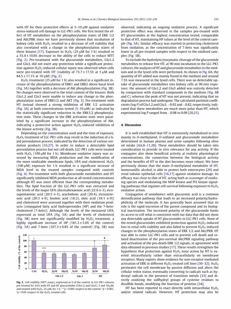
with HT for their protective effects at 5–10 lM against oxidativestress-induced cell damage in LLC-PK1 cells. We first tested the ef-fect of HT metabolites on the phosphorylation states of ERK 1/2and Akt/PBK since we have previously shown that incubation ofkidney cells with H2O2 resulted in a decrease of cell viability thatalso correlated with a change in the phosphorylation states ofthese kinases [17]. Exposure to H2O2 (25 lM for 1 h) resulted ina 55.4 ± 10.0% decrease in the ability of the cells to reduce MTT(Fig. 2). Pre-treatment with the glucuronide metabolites, Glu1,2and Glu3, did not exert any protection while a significant protec-tion against H2O2-induced cellular injury was observed when cellswere pre-treated with HT (viability of 73.7 ± 17.5% at 5 lM and84.5 ± 17.1% at 10 lM) (Fig. 2).
H2O2 treatment (25 lM for 1 h) also resulted in a significant in-crease of the phosphorylation of ERK1 and ERK2 above basal level(Fig. 3A) together with a decrease of Akt phosphorylation (Fig. 3B).No changes were observed in the total content of the kinases. BothGlu1,2 and Glu3 were unable to induce any change in the phos-phorylation states of ERK1/2 and AKT (Fig. 3). Pre-treatment withHT instead showed a strong inhibition of ERK 1/2 activation(Fig. 3A) at both concentrations tested (5–10 lM) as indicated bythe statistically significant reduction in the ERK1/2 phosphoryla-tion state. These changes in the ERK activation state were paral-leled by a significant increase in the phosphorylation of Akt,indicating a protective action against H2O2-induced inhibition ofthe kinase activity (Fig. 3B).
Depending on the concentration used and the time of exposure,H2O2 treatment of LLC-PK1 cells may result in the induction of a li-pid peroxidation process, indicated by the detection of various oxi-dation products [16,27]. In order to induce a detectable lipidperoxidation process but not cell death, LLC-PK1 cells were treatedwith H2O2 (100 lM for 1 h). Membrane oxidative injury was as-sessed by measuring MDA production and the modification ofthe more oxidizable membrane lipids, UFA and cholesterol. H2O2
(100 lM) exposure for 1 h resulted in a significant increase ofMDA level in the treated samples compared with controls(Fig. 4). Pre-treatment with both glucuronide metabolites and HTsignificantly inhibited MDA production at all tested concentrationsalthough HT was more efficient than the corresponding metabo-lites. The lipid fraction of the LLC-PK1 cells was extracted andthe levels of the major UFA (docosahexaenoic acid (22:6 n-3), eico-sapentaenoic acid (20:5 n-3), arachidonic acid (20:4), eicosatrie-noic acid (20:3 n-9), linoleic acid (18:2), oleic acid (18:1 n-9))and cholesterol were assessed together with their oxidation prod-ucts (conjugated fatty acid hydroperoxides (HP) and the 7-keto-cholesterol (7-keto)). Although the levels of the measured UFA,expressed as total UFA (Fig. 5A) and the levels of cholesterol(Fig. 5B) were not significantly modified by H2O2 treatment, ahighly significant increase of HP (181.3 ± 5.0% of the control)(Fig. 5A) and 7-keto (167.3 ± 0.8% of the control) (Fig. 5B) was
Fig. 2. Cell viability (MTT assay), expressed as % of the control, in LLC-PK1 culturespre-treated for 24 h with HT and HT glucuronides (Glu1,2 and Glu3), 5 and 10 lM,and treated with H2O2, 25 lM, for 1 h. ⁄⁄⁄p < 0.001 respect to the control; ap < 0.001,cp < 0.05 respect to the H2O2 treated cells.
observed, indicating an ongoing oxidative process. A significantprotective effect was observed in the samples pre-treated withHT glucuronides at the highest concentration tested, comparableto that of HT, maintaining HP values at the level of the control sam-ple (Fig. 5A). Similar efficacy was exerted in protecting cholesterolfrom oxidation, as the concentration of 7-keto was significantlylower in all pre-treated samples with respect to the oxidized sam-ples (Fig. 5B).
To exclude the hydrolytic/enzymatic cleavage of the glucuronidemetabolites to release free HT, at 90 min incubation in the LLC-PK1cultures, the analyses of HT and glucuronide metabolites in the med-ium and in the lysed cells were performed. As shown in Fig. 6A, thequantity of HT added was mainly found in the medium and around7.5% was measured in the lysed cells. There was no detectable up-take of glucuronide metabolites into kidney cells at 90 min expo-sure; the amount of Glu1,2 and Glu3 added was entirely detectedby comparison with standard compounds in the medium (Fig. 6Band C), whereas the peak of HT was not present, indicating that nodegradation process had undergone. The calculated partition coeffi-cients (log P) of Glu1,2 and Glu3,�0.92 and�0.82, respectively, indi-cated that these metabolites are much more polar than HT, whichexperimental log P ranged from �0.08 to 0.09 [20,21].
4. Discussion
It is well established that HT is extensively metabolized in vivomainly to O-methylated, O-sulfated and glucuronide metabolitesdetermined in human plasma and urine samples following oliveoil intake [4,6,8–11,28]. These metabolites should be taken intoconsideration to provide in vivo relevance for any activity. If theconjugates also show beneficial activity at realistic physiologicalconcentrations, the connection between the biological activityand the benefits of HT in the diet becomes more robust. We havepreviously shown that the main O-methylated metabolite of HT,homovanillic alcohol, is able to protect both enterocytes [29] andrenal tubular epithelial cells [16,17] against oxidative damage. Itsefficacy was close to that of HT, acting both as scavenger of oxidiz-ing species and modulating the MAP kinase and PI3 kinase signal-ing pathways that regulate cell survival following exposure to H2O2
oxidative action.Conjugation of xenobiotics with glucuronic acid is a common
detoxification pathway that leads to an increased polarity/hydro-philicity of the molecule. It has generally been assumed that itsrole is the rapid excretion of the parent compound and its biolog-ical inactivation. The increased polarity of the glucuronide limitsits access to cell what is consistent with our data that did not showany detectable uptake of HT glucuronides in LLC-PK1 cells. None ofthe tested glucuronides exhibited protection against H2O2-inducedloss in renal cells viability and also failed to prevent H2O2-inducedchanges in the phosphorylation states of ERK 1/2 and Akt/PKB. HTwas able to enter LLC-PK1 cells and to prevent cell death and re-lated deactivation of the pro-survival Akt/PKB signaling pathwayand activation of the pro-death ERK 1/2 signals, in agreement withdata obtained in previous studies [17]. These results strengthen thehypothesis that protection against H2O2 toxic action by HT is ex-erted intracellularly rather than extracellularly on membranereceptors. Many reports show evidence for non-receptor-mediatedactivation of ERK in different H2O2 treated cell lines [30–32]; H2O2
permeates the cell membrane by passive diffusion and alters thecellular redox status, eventually converting to radicals such as hy-droxyl radicals in the presence of transition metals [33] and di-rectly oxidizing the sulfhydryl groups of cysteine residues todisulfide bonds, modifying the function of proteins [34].
HT has been reported to react directly with intracellular H2O2
and/or related radicals. However, HT protection may also be

0.0
0.2
0.4
0.6
0.8
1.0
1.2
Rel
ativ
e ba
nd in
tens
ity
ERK 1 ERK 2
Ctrl H2O2 10µM 5µM 10µM 5µM 10µM 5µM
HT Glu 1,2 Glu 3
A Ctrl H2O2 10µM 5µM 10µM 5µM 10µM 5µM
HT Glu 1,2 Glu 3
ERK 1ERK 2
***
***
cb
aa
0.0
0.3
0.5
0.8
1.0
1.3
1.5
1.8
2.0
Rel
ativ
e ba
nd in
tens
ity
Ctrl H2O2 10µM 5µM 10µM 5µM 10µM 5µM
HT
Ctrl H2O2 10µM 5µM 10µM 5µM 10µM 5µM
Glu 1,2 Glu 3
HT Glu 1,2 Glu 3
Akt
***
aa
B
Fig. 3. Modulation of H2O2-induced ERK phosphorylation (A) and Akt de-phosphorylation (B) by HT and HT glucuronides (Glu1,2 and Glu3) in LLC-PK1 cells, treated with HTand HT glucuronides, 5 and 10 lM, 24 h prior to the addition of H2O2, 25 lM, for 1 h. ⁄⁄⁄p < 0.001 respect to the control; ap < 0.001, bp < 0.01, cp < 0.05 respect to the H2O2
treated cells.
0
50
100
150
200
250
MD
A (%
of t
he c
ontr
ol)
Ctrl H2O2 5µµM 10µM 5µM 10µM 5µM 10µM
HTGlu 1,2 Glu 3
***aa a
a
aa
Fig. 4. Values of MDA, expressed as % of the control value (0.7 ± 0.1 lmol/mgprotein), measured in LLC-PK1 cells after 1 h incubation with H2O2 and treated withHT or HT glucuronides (Glu1,2 and Glu3), 5 and 10 lM. ⁄⁄⁄p < 0.001 respect to thecontrol, ap < 0.001 respect to the H2O2 treated cells.
236 M. Deiana et al. / Chemico-Biological Interactions 193 (2011) 232–239
attributed to the rapid generation of the arylating catechol quinoneas an oxidation product [35]. Hydrophilic catechols, such as HT, arereadily oxidized in vivo to catechol quinone electrophiles, therebypossessing properties similar to those of lipophilic arylating
tocopherols quinones, including an ability to undergo Michaeladditions with cellular thiols such as cysteinyl containing proteins;their hydrophilicity allows the catechol quinone electrophile to re-act with available thiol nucleophilic groups in enzymes and signal-ing molecules within the hydrophilic environment of the cell [35].
Glucuronide metabolites of HT did not exert the same efficacyof the parent compound in protecting renal cells against H2O2-in-duced death, however, they were able to exert an antioxidant ef-fect. This is in agreement with reported data for other phenolicantioxidants in the literature. It has been shown that some flavo-noids after glucuronidation may still act as antioxidants in biolog-ical fluids [36–38], for example inhibiting xanthine oxidase andlipoxygenase [39], retaining a reduced cytoprotective capacityin vitro against H2O2 induced oxidative stress [40], and displayinga slightly decreased ability to scavenge radicals (TEAC test) [41].Even more relevant is the fact that quercetin and its glucuronidemetabolites, at physiological concentrations, can inhibit theexpression of key molecules involved in monocyte recruitmentduring the early stages of atherosclerosis [42]. The antioxidantactivity of HT glucuronides is controversial; the 30-O-b-glucuronideconjugate showed a more efficient radical scavenging potency(DPPH test) than HT itself [7]. However Khymenets et al. reportedthat 30-and 40-O-b-glucuronide metabolites, although slightly

0
40
80
120
160
200
0
20
40
60
80
100
120
140
HP
(% o
f th
e co
ntro
l)
UFA
tot
(% o
f th
e co
ntro
l)
UFA HP
Ctrl H2O2 5µµM 10µM 5µM 10µM 5µM 10µM
***a
a aaa
b
HTGlu 1,2 Glu 3
A
0
40
80
120
160
0
20
40
60
80
100
120
7-k
eto
(% o
f th
e co
ntro
l)
Cho
lest
erol
(% o
f th
e co
ntro
l)
Cholesterol 7-Keto
Ctrl H2O2 5µM 10µM 5µM 10µM 5µM 10µM
***
a
a
aa aa
HTGlu 1,2 Glu 3
B
Fig. 5. Values of the total UFA together with fatty acid hydroperoxides (HP) (A) and cholesterol and 7-ketocholesterol (7-keto) (B) measured in LLC-PK1 cells after 1 hincubation with H2O2 and treated with HT or HT glucuronides (Glu1,2 and Glu3), 5 and 10 lM, expressed as % of the control values (total UFA as sum of 22:6 n-3,11.3 ± 1.7 lg/mg; 20:5 n-3, 8.1 ± 0.9 lg/mg; 20:4, 40.9 ± 6.2 lg/mg; 20:3 n-9, 13.5 ± 1.1 lg/mg; 18:2, 19.6 ± 2.1 lg/mg; 18:1 n-9, 145.2 ± 10.2 lg/mg; HP, 0.2 ± 0.1 nmol/mg;cholesterol, 78.3 ± 5.2 lg/mg; 7-keto, 0.2 ± 0.1 lg/mg protein). ⁄⁄⁄p < 0.001 respect to the control, ap < 0.001 respect to the H2O2 treated cells.
min0 2.5 5 7.5 10 12.5 15 17.5 20 22.5
mAU
100
200
300
400
500
0min
5 10 15 20 25
mAU
100200300400500600
HT
HT
medium
cell lysate
280 nm
280 nm
A
B
C
min5 10 15 20 250
min0 2.5 5 7.5 10 12.5 15 17.5 20
Glu 3mAU
20406080
100120140
mAU
20406080
100120140
medium
cell lysate
280 nm
280 nm
min0 5 10 15 20 25
mAU
20
40
60
80
100
120
min0 5 10 15 20
mAU
20406080
100120140
Glu1,2
medium
cell lysate
280 nm
280 nm
Fig. 6. Chromatograms showing HT (A) and its glucuronide metabolites Glu1,2 (B) and Glu3 (C) detected in the medium and cell lysate after 90 min of incubation of LLC-PK1cells with 250 lM of each compound.
M. Deiana et al. / Chemico-Biological Interactions 193 (2011) 232–239 237
active, did not display the antiradical activity of the parent com-pound and appeared to maintain only some residual activity inprotecting LDL from Cu-mediated oxidation [11].
Our study assesses for the first time the ability of the glucuro-nide metabolites of HT to protect cells against the lipid peroxida-
tion process. We have shown that both a mixture of HT 30-O-b-D-glucuronide and 40-O-b-D-glucuronide, and 2-(30,40-dihydroxy-phenyl)ethanol-1-O-b-D-glucuronide exhibited a significant pro-tection against H2O2 induced membrane oxidative injury in renalcells comparable to that of the parent compound. Cell pre-treat-
238 M. Deiana et al. / Chemico-Biological Interactions 193 (2011) 232–239
ment with the glucuronide metabolites significantly inhibited theformation of MDA and fatty acid and cholesterol major oxidationproducts, HP and 7-keto. In fact, exposure of LLC-PK1 cells toH2O2 may also result in the disruption of the membrane structure,due to the lipid peroxidation process initiated by the oxidative at-tack from the aqueous phase [16].
The antioxidant action of a phenolic compound in a membranesystem depends both on the radical scavenging ability of aqueousradicals at the surface of the membrane and on the formation of li-pid peroxyl radicals within the membrane. HT has been shown toreact directly with H2O2 [42,43], to act as oxygen-centered radicalscavenger [44] in the reaction medium and near the membranesurface [45] and as scavenger of chain propagating lipid peroxylradicals generated from membrane UFA and cholesterol [29].
HT metabolites tested are highly polar compounds, as indicatedby the calculated log P and the high solubility in water, whichmakes it difficult to pass the cell membranes. Therefore, their pro-tective action is more likely to be due to the ability of scavengingthe initiating aqueous radicals originated from reaction withH2O2 both in the reaction medium and near the membrane surface.In our experimental conditions, the scavenging ability of the HTglucuronides does not seem to be influenced by the substitutionof the catechol structure. In fact, Glu1,2, where glucuronic acid isconjugated to one of the hydroxyl groups of the catechol, protectedcells as efficiently as Glu3, where the conjugation affects the ali-phatic chain and leaves intact the ortho-diphenolic structure. Incontrast, the activity of the glucuronide metabolites of severalflavonoids were reported to be highly dependent on the conjuga-tion position [39].
Our results have shown that HT glucuronide metabolites areable to protect renal cells against H2O2 induced membrane oxida-tive damage, more likely through a direct antioxidant action. More-over, Glu1,2 and Glu3 revealed no effect on cell death probably dueto their incapacity to interact with intracellular signaling path-ways. These findings are interesting in light of the fact that,although the glucuronide metabolites were less efficacious in ourrenal cell model, it should be noted that in vivo they would bepresent at greater concentrations than the unmetabolized parentcompound. Although further researches either in animal experi-mentations or ex vivo models for renal oxidative damage areneeded, these results suggest that HT glucuronide metaboliteswhen concentrated in the renal compartment [46] may signifi-cantly contribute, together with HT and other of its metabolites,to the protective action against H2O2 mediated renal diseaseswhere the lipid peroxidation process plays a central role.
Conflict of interest statement
The authors declare no conflict of interest.
References
[1] S. Granados-Principal, J.L. Quiles, C.L. Ramirez-Tortosa, P. Sanchez-Rovira, M.C.Ramirez-Tortosa, Hydroxytyrosol: from laboratory investigations to futureclinical trials, Nutr. Rev. 68 (2010) 191–206.
[2] R. de la Torre, Bioavailability of olive oil phenolic compounds in humans,Inflammopharmacology 16 (2008) 245–247.
[3] G. Corona, X. Tzounis, M.A. Dessi, M. Deiana, E.S. Debnam, F. Visioli, J.P.E.Spencer, The fate of olive oil polyphenols in the gastrointestinal tract:implications of gastric and colonic microflora-dependent biotransformation,Free Radic. Res. 40 (2006) 647–658.
[4] M.N. Vissers, P.L. Zock, A.J. Roodenburg, R. Leenen, M.B. Katan, Olive oil phenolsare absorbed in humans, J. Nutr. 132 (2002) 409–417.
[5] C. Manna, P. Galletti, G. Maisto, V. Cucciolla, S. D’Angelo, V. Zappia, Transportmechanism and metabolism of olive oil hydroxytyrosol in Caco-2 cells, FEBSLett. 470 (2000) 341–344.
[6] E. Miro-Casas, M.I. Covas, M. Farre, M. Fito, J. Ortuno, T. Weinbrenner, P. Roset,R. de la Torre, Hydroxytyrosol disposition in humans, Clin. Chem. 49 (2003)945–952.
[7] K.L. Tuck, P.J. Hayball, I. Stupans, Structural characterization of the metabolitesof hydroxytyrosol, the principal phenolic component in olive oil, in rats, J.Agric. Food Chem. 50 (2002) 2404–2409.
[8] E. Miro-Casas, M.I. Covas, M. Fito, M. Farre-Albadalejo, J. Marrugat, R. de laTorre, Tyrosol and hydroxytyrosol are absorbed from moderate and sustaineddoses of virgin olive oil in humans, Eur. J. Clin. Nutr. 57 (2003) 186–190.
[9] F. Visioli, C. Galli, F. Bornet, A. Mattei, R. Patelli, G. Galli, D. Caruso, Olive oilphenolics are dose-dependently absorbed in humans, FEBS Lett. 468 (2000)159–160.
[10] D. Caruso, F. Visioli, R. Patelli, C. Galli, G. Galli, Urinary excretion of olive oilphenols and their metabolites in humans, Metabolism 50 (2001) 1426–1428.
[11] O. Khymenets, M. Fito, S. Tourino, D. Munoz-Aguayo, M. Pujadas, J.L. Torres, J.Joglar, M. Farre, M.I. Covas, R. de la Torre, Antioxidant activities ofhydroxytyrosol main metabolites do not contribute to beneficial healtheffects after olive oil ingestion, Drug Metab. Dispos. 38 (2010) 1417–1421.
[12] A. Perantoni, J.J. Berman, Properties of Wilms’ tumor line (TuWi) and pigkidney line (LLC-PK1) typical of normal kidney tubular epithelium, In Vitro 15(1979) 446–454.
[13] S.H. Baek, X.L. Piao, U.J. Lee, H.Y. Kim, J.H. Park, Reduction of Cisplatin-inducednephrotoxicity by ginsenosides isolated from processed ginseng in culturedrenal tubular cells, Biol. Pharm. Bull. 29 (2006) 2051–2055.
[14] H.H. Cohly, A. Taylor, M.F. Angel, A.K. Salahudeen, Effect of turmeric, turmerinand curcumin on H2O2-induced renal epithelial (LLC-PK1) cell injury, FreeRadic. Biol. Med. 24 (1998) 49–54.
[15] A.K. Salahudeen, Role of lipid peroxidation in H2O2-induced renal epithelial(LLC-PK1) cell injury, Am. J. Physiol. 268 (1995) F30–38.
[16] M. Deiana, A. Incani, A. Rosa, G. Corona, A. Atzeri, D. Loru, M.P. Melis, M.A.Dessì, Protective effect of hydroxytyrosol and its metabolite homovanillicalcohol on H2O2 induced lipid peroxidation in renal tubular epithelial cells,Food Chem. Toxicol. 46 (2008) 2984–2990.
[17] A. Incani, M. Deiana, G. Corona, K. Vafeiadou, D. Vauzour, M.A. Dessì, J.P.E.Spencer, Involvement of ERK, Akt and JNK signaling in H2O2-induced cellinjury and protection by Hydroxytyrosol and its metabolite homovanillicalcohol, Mol. Nutr. Food Res. 54 (2010) 788–796.
[18] R. Lucas, D. Alcantara, J.C. Morales, A concise synthesis of glucuronidemetabolites of urolithin-B, resveratrol, and hydroxytyrosol, Carbohyd. Res.344 (2009) 1340–1346.
[19] A.K. Ghose, G.M. Crippen, Atomic physicochemical parameters for three-dimensional-structure-directed quantitative structure–activity relationships.2. Modeling dispersive and hydrophobic interactions, J. Chem. Inf. Comput. Sci.27 (1987) 21–35.
[20] I. Medina, S. Lois, D. Alcantara, R. Lucas, J.C. Morales, Effect of lipophilization ofhydroxytyrosol on its antioxidant activity in fish oils and fish oil-in-wateremulsions, J. Agric. Food Chem. 57 (2009) 9773–9779.
[21] S. Grasso, L. Siracusa, C. Spatafora, M. Renis, C. Tringali, Hydroxytyrosollipophilic analogues: enzymatic synthesis, radical scavenging activity and DNAoxidative damage protection, Bioorg. Chem. 35 (2007) 137–152.
[22] M.M. Bradford, A rapid and sensitive method for the quantitation ofmicrogram quantities of protein utilizing the principle of protein–dyebinding, Anal. Biochem. 72 (1976) 248–254.
[23] T. Mosmann, Rapid colorimetric assay for cellular growth and survival:application to proliferation and cytotoxicity assays, J. Immunol. Methods 65(1983) 55–63.
[24] J. Templar, S.P. Kon, T.P. Milligan, D.J. Newman, M.J. Raftery, Increased plasmamalondialdehyde levels in glomerular disease as determined by a fullyvalidated HPLC method, Nephrol. Dial. Transplant. 14 (1999) 946–951.
[25] J. Folch, M. Lees, G.H. Sloane Stanley, A simple method for the isolation andpurification of total lipides from animal tissues, J. Biol. Chem. 226 (1957) 497–509.
[26] S. Banni, B.W. Day, R.W. Evans, F.P. Corongiu, B. Lombardi, Liquidchromatography–mass spectrometric analysis of conjugated diene fattyacids in a partially hydrogenated fat, JAOCS 71 (1994) 1321–1325.
[27] X. Meng, W.B. Reeves, Effects of chloride channel inhibitors on H2O2-inducedrenal epithelial cell injury, Am. J. Physiol. Renal Physiol. 278 (2000) F83–90.
[28] E. Miro-Casas, M. Farre Albaladejo, M.I. Covas, J.O. Rodriguez, E. MenoyoColomer, R.M. Lamuela Raventos, R. de la Torre, Capillary gaschromatography–mass spectrometry quantitative determination ofhydroxytyrosol and tyrosol in human urine after olive oil intake, Anal.Biochem. 294 (2001) 63–72.
[29] M. Deiana, G. Corona, A. Incani, D. Loru, A. Rosa, A. Atzeri, M.P. Melis, M.A.Dessì, Protective effect of simple phenols from extravirgin olive oil againstlipid peroxidation in intestinal Caco-2 cells, Food Chem. Toxicol. 48 (2010)3008–3016.
[30] H.M. Lander, J.S. Ogiste, K.K. Teng, A. Novogrodsky, P21ras as a commonsignaling target of reactive free radicals and cellular redox stress, J. Biol. Chem.270 (1995) 21195–21198.
[31] R. Aikawa, I. Komuro, T. Yamazaki, Y. Zou, S. Kudoh, M. Tanaka, I. Shiojima, Y.Hiroi, Y. Yazaki, Oxidative stress activates extracellular signal-regulatedkinases through Src and Ras in cultured cardiac myocytes of neonatal rats, J.Clin. Invest. 100 (1997) 1813–1821.
[32] M.K. Abe, S. Kartha, A.Y. Karpova, J. Li, P.T. Liu, W.L. Kuo, M.B. Hershenson,Hydrogen peroxide activates extracellular signal-regulated kinase via proteinkinase C, Raf-1, and MEK 1, Am. J. Respir. Cell Mol. Biol. 18 (1998) 562–569.
[33] B. Halliwell, J.M.C. Gutteridge, Free Radicals in Biology and Medicine, third ed.,Clarendon Press, Oxford, 1999.

M. Deiana et al. / Chemico-Biological Interactions 193 (2011) 232–239 239
[34] Y. Sun, L.W. Oberley, Redox regulation of transcriptional activators, Free Radic.Biol. Med. 21 (1996) 335–348.
[35] D.G. Cornwell, J. Ma, Nutritional benefit of olive oil: the biological effects ofhydroxytyrosol and its arylating quinone adducts, J. Agric. Food Chem. 56(2008) 8774–8786.
[36] M. Harada, Y. Kan, H. Naoki, Y. Fukui, N. Kageyama, M. Nakai, W. Miki, Y. Kiso,Identification of the major antioxidative metabolites in biological fluids of therat with ingested (+)-catechin and (�)-epicatechin, Biosci. Biotech. Bioch. 63(1999) 973–977.
[37] Y. Miyake, K. Shimoi, S. Kumazawa, K. Yamamoto, N. Kinae, T. Osawa,Identification and antioxidant activity of flavonoid metabolites in plasma andurine of eriocitrin-treated rats, J. Agric. Food Chem. 48 (2000) 3217–3224.
[38] C. Morand, V. Crespy, C. Manach, C. Besson, C. Demigne, C. Remesy, Plasmametabolites of quercetin and their antioxidant properties, Am. J. Physiol. 275(1998) R212–R219.
[39] A.J. Day, Y. Bao, M.R. Morgan, G. Williamson, Conjugation position of quercetinglucuronides and effect on biological activity, Free Radic. Biol. Med. 29 (2000)1234–1243.
[40] D.E. Stevenson, J.M. Cooney, D.J. Jensen, R. Wibisono, A. Adaim, M.A. Skinner, J.Zhang, Comparison of enzymically glucuronidated flavonoids with flavonoidaglycones in an in vitro cellular model of oxidative stress protection, In vitroCell. Dev. Biol. 44 (2008) 73–80.
[41] S.E. Pollard, G.G. Kuhnle, D. Vauzour, K. Vafeiadou, X. Tzounis, M. Whiteman, C.Rice-Evans, J.P. Spencer, The reaction of flavonoid metabolites withperoxynitrite, Biochem. Biophys. Res. Commun. 350 (2006) 960–968.
[42] M. De Lucia, L. Panzella, A. Pezzella, A. Napolitano, M. d’Ischia, Oxidativechemistry of the natural antioxidant hydroxytyrosol: hydrogen peroxide-dependent hydroxylation and hydroxyquinone/o-quinone coupling pathways,Tetrahedron 62 (2006) 1273–1278.
[43] Y. O’Dowd, F. Driss, P.M. Dang, C. Elbim, M.A. Gougerot-Pocidalo, C. Pasquier, J.El-Benna, Antioxidant effect of hydroxytyrosol, a polyphenol from olive oil:scavenging of hydrogen peroxide but not superoxide anion produced byhuman neutrophils, Biochem. Pharmacol. 68 (2004) 2003–2008.
[44] S.J. Rietjens, A. Bast, G.R. Haenen, New insights into controversies on theantioxidant potential of the olive oil antioxidant hydroxytyrosol, J. Agric. FoodChem. 55 (2007) 7609–7614.
[45] F. Paiva-Martins, M.H. Gordon, P. Gameiro, Activity and location of olive oilphenolic antioxidants in liposomes, Chem. Phys. Lipids 124 (2003) 23–36.
[46] S. D’Angelo, C. Manna, V. Migliardi, O. Mazzoni, P. Morrica, G. Capasso, G.Pontoni, P. Galletti, V. Zappia, Pharmacokinetics metabolism ofhydroxytyrosol, a natural antioxidant from olive oil, Drug Metab. Dispos. 29(2001) 1492–1498.