Hypoxia-Induced miR-210 Modulates Tissue Response to Acute Peripheral Ischemia
53
1 1 Forum Original Research Communication Hypoxia-induced miR-210 modulates tissue response to acute peripheral ischemia Germana Zaccagnini 1,2 , Biagina Maimone 1,3 , Valeria Di Stefano 1 , Pasquale Fasanaro 2,4 , Simona Greco 1 , Alessandra Perfetti 1 , Maurizio C. Capogrossi 2 , Carlo Gaetano 5 , Fabio Martelli 1* 1) Molecular Cardiology Laboratory, IRCCS-Policlinico San Donato, 20097 Milan, Italy. 2) Laboratorio di Patologia Vascolare, Istituto Dermopatico dell’Immacolata-IRCCS, 00167 Rome, Italy. 3) Gruppo Ospedaliero San Donato Foundation, 20122, Milan, Italy. 4) Epigenetics & Regenerative Pharmacology, IRCCS Fondazione Santa Lucia, 00143, Rome, Italy. 5) Division of Cardiovascular Epigenetics, Department of Cardiology, Internal Medicine Clinic III, Goethe University, 60590 Frankfurt am Main, Germany, Running head: miR-210 and ischemia response *To whom correspondence should be addressed: Molecular Cardiology Laboratory, IRCCS- Policlinico San Donato, via Morandi 30, San Donato Milanese, 20097 Milan, Italy. Tel.: 390252774533; Fax: 390252774666; E-mail: [email protected]. Page 1 of 53 Antioxidants & Redox Signaling Hypoxia-induced miR-210 modulates tissue response to acute peripheral ischemia (doi: 10.1089/ars.2013.5206) This article has been peer-reviewed and accepted for publication, but has yet to undergo copyediting and proof correction. The final published version may differ from this proof.
-
Upload
independent -
Category
Documents
-
view
4 -
download
0
Transcript of Hypoxia-Induced miR-210 Modulates Tissue Response to Acute Peripheral Ischemia
1
1
Forum Original Research Communication
Hypoxia-induced miR-210 modulates tissue response to acute
peripheral ischemia
Germana Zaccagnini1,2
, Biagina Maimone1,3
, Valeria Di Stefano1, Pasquale Fasanaro
2,4,
Simona Greco1, Alessandra Perfetti
1, Maurizio C. Capogrossi
2, Carlo Gaetano
5, Fabio
Martelli1*
1) Molecular Cardiology Laboratory, IRCCS-Policlinico San Donato, 20097 Milan, Italy.
2) Laboratorio di Patologia Vascolare, Istituto Dermopatico dell’Immacolata-IRCCS,
00167 Rome, Italy.
3) Gruppo Ospedaliero San Donato Foundation, 20122, Milan, Italy.
4) Epigenetics & Regenerative Pharmacology, IRCCS Fondazione Santa Lucia, 00143,
Rome, Italy.
5) Division of Cardiovascular Epigenetics, Department of Cardiology, Internal Medicine
Clinic III, Goethe University, 60590 Frankfurt am Main, Germany,
Running head: miR-210 and ischemia response
*To whom correspondence should be addressed: Molecular Cardiology Laboratory, IRCCS-
Policlinico San Donato, via Morandi 30, San Donato Milanese, 20097 Milan, Italy. Tel.:
390252774533; Fax: 390252774666; E-mail: [email protected].
Page 1 of 53
Ant
ioxi
dant
s &
Red
ox S
igna
ling
Hyp
oxia
-ind
uced
miR
-210
mod
ulat
es ti
ssue
res
pons
e to
acu
te p
erip
hera
l isc
hem
ia (
doi:
10.1
089/
ars.
2013
.520
6)T
his
artic
le h
as b
een
peer
-rev
iew
ed a
nd a
ccep
ted
for
publ
icat
ion,
but
has
yet
to u
nder
go c
opye
ditin
g an
d pr
oof
corr
ectio
n. T
he f
inal
pub
lishe
d ve
rsio
n m
ay d
iffe
r fr
om th
is p
roof
.

2
2
WORD COUNT: 4328
REFERENCES NUMBER: 44
GRAYSCALE ILLUSTRATION: 4 (ONLINE 1 AND HARDCOPY 3)
COLOR ILLUSTRATION:12 (ONLINE 5 AND HARDCOPY 7)
Page 2 of 53
Ant
ioxi
dant
s &
Red
ox S
igna
ling
Hyp
oxia
-ind
uced
miR
-210
mod
ulat
es ti
ssue
res
pons
e to
acu
te p
erip
hera
l isc
hem
ia (
doi:
10.1
089/
ars.
2013
.520
6)T
his
artic
le h
as b
een
peer
-rev
iew
ed a
nd a
ccep
ted
for
publ
icat
ion,
but
has
yet
to u
nder
go c
opye
ditin
g an
d pr
oof
corr
ectio
n. T
he f
inal
pub
lishe
d ve
rsio
n m
ay d
iffe
r fr
om th
is p
roof
.

3
3
ABSTRACT
Aims: Peripheral Artery Disease is caused by the restriction or occlusion of arteries
supplying the leg. Better understanding of the molecular mechanisms underpinning tissue
response to ischemia is urgently needed to improve therapeutic options. The aim of this study
is investigating hypoxia-induced miR-210 regulation and role in a mouse model of hindlimb
ischemia.
Results: miR-210 expression was induced by femoral artery dissection. To study miR-210
role, its function was inhibited by the systemic administration of a miR-210 complementary
LNA-oligonucleotide (anti-miR-210). In the ischemic skeletal muscle, anti-miR-210 caused a
marked decrease of miR-210 compared to LNA-scramble control, while miR-210 target
expression increased accordingly. Histological evaluation of acute tissue damage showed that
miR-210 inhibition increased both apoptosis at 1 day and necrosis at 3 days. Capillary density
decrease caused by ischemia was significantly more pronounced in anti-miR-210 treated
mice; residual limb perfusion decreased accordingly. To investigate the molecular
mechanisms underpinning the increased damage triggered by miR-210 blockade, we tested
the impact of anti-miR-210 treatment on the transcriptome. Gene expression analysis
highlighted the deregulation of mitochondrial function and redox balance. Accordingly,
oxidative damage was more severe in the ischemic limb of anti-miR-210 treated mice and
miR-210 inhibition increased oxidative metabolism. Furthermore, oxidative-stress resistant
p66Shc
-null mice displayed decreased tissue damage following ischemia.
Innovation: This study identifies miR-210 as a crucial element in the adaptive mechanisms
to acute peripheral ischemia.
Conclusions: The physio-pathological significance of miR-210 is context dependent. In the
ischemic skeletal muscle it seems to be cytoprotective, regulating oxidative metabolism and
oxidative stress.
Page 3 of 53
Ant
ioxi
dant
s &
Red
ox S
igna
ling
Hyp
oxia
-ind
uced
miR
-210
mod
ulat
es ti
ssue
res
pons
e to
acu
te p
erip
hera
l isc
hem
ia (
doi:
10.1
089/
ars.
2013
.520
6)T
his
artic
le h
as b
een
peer
-rev
iew
ed a
nd a
ccep
ted
for
publ
icat
ion,
but
has
yet
to u
nder
go c
opye
ditin
g an
d pr
oof
corr
ectio
n. T
he f
inal
pub
lishe
d ve
rsio
n m
ay d
iffe
r fr
om th
is p
roof
.

4
4
INTRODUCTION
Peripheral Artery Disease is a frequent condition, affecting almost 6% of the US population
aged ≥40 (36), that is mostly caused by stenosis, embolism, or thrombosis involving the
arteries supplying the leg (37). Abrupt arterial occlusion leads to acute ischemia, while
restriction of blood flow due to arterial stenosis most commonly causes chronic ischemia.
According to several parameters such as the affected vessel, the degree of occlusion and the
presence of collaterals, Peripheral Artery Disease can cause from mild claudication to major
tissue loss and may require revascularization surgery (37, 43). Unfortunately, delayed
treatment of acute patients can result in morbidity, amputation, and/or death (27). Moreover,
chronic patients who benefit from successful revascularization often suffer from high rate of
recurrent symptoms or revision surgery and many still require progressive amputations (42).
Patients affected by Chronic Critical Limb Ischemia, the most severe form of the disease, are
particularly at risk. Within one year of diagnosis, 40-50 percent will experience an
amputation and 20-25 percent will die (42).
Recent evidence shows that ischemia induces profound changes in the expression of
microRNAs (miRNAs), small non-protein-coding RNAs that act as negative regulators of
gene expression (14). Hypoxia is a crucial component of both acute and chronic ischemia and
one specific miRNA, miR-210, is robustly up-regulated by both hypoxia and ischemia (12, 26,
7, 5). Between the articulate program of cellular adaptive mechanisms aimed at relieving
tissue hypoxia and removing irreversibly damaged cells, miR-210 can be considered a master
miRNA of hypoxic response, since it was found up-regulated by hypoxia in virtually all the
cells and tissues tested to date (12, 7). Mechanistically, miR-210 is a target of the Hypoxia
Inducible factor 1-alpha, that binds to its promoter and activates transcription, upon low
oxygen exposure (12). The instrumental role of miR-210 in the regulation of cell response to
Page 4 of 53
Ant
ioxi
dant
s &
Red
ox S
igna
ling
Hyp
oxia
-ind
uced
miR
-210
mod
ulat
es ti
ssue
res
pons
e to
acu
te p
erip
hera
l isc
hem
ia (
doi:
10.1
089/
ars.
2013
.520
6)T
his
artic
le h
as b
een
peer
-rev
iew
ed a
nd a
ccep
ted
for
publ
icat
ion,
but
has
yet
to u
nder
go c
opye
ditin
g an
d pr
oof
corr
ectio
n. T
he f
inal
pub
lishe
d ve
rsio
n m
ay d
iffe
r fr
om th
is p
roof
.

5
5
hypoxia is confirmed by pre-clinical and clinical evidences (21). Indeed, miR-210 has been
found to be up-regulated upon brain transient focal ischemia in rats, in mouse ischemic
wounds, after human myocardial infarction and was proposed as a blood biomarker in acute
cerebral ischemia (4, 28, 49, 3). miR-210 was also found up-regulated in most solid tumors
and its expression correlates with an adverse clinical outcome and with metastatic potential
(12, 26). In keeping with these data, miR-210 has been proposed as a novel tumor hypoxia
marker (18).
Evidence accumulated in numerous culture systems shows reduced survival of cells devoid of
miR-210 in hypoxia, but also in normoxia, in certain cell types (13, 17, 39, 45, 22, 41).
Furthermore, miR-210 has a crucial cytoprotective role in mesenchymal stem cells exposed
to anoxia after ischemia/reoxygenation preconditioning, supporting their survival after
transplantation in the infarcted heart (30). Although the molecular mechanisms supporting
these events are complex, miR-210 directly represses the apoptotic component CASP8AP2
(30) as well as other apoptosis-related genes such as DAPK1 and, at least in humans, AIFM3
(24, 35, 45). However, miR-210 function is complex and context-dependent, since miR-210
overexpression in normoxia induces apoptosis in certain cancer cell cultures (39, 17).
miR-210 also influences mitochondrial metabolism: targeting the iron-sulfur cluster scaffold
protein ISCU1/2, miR-210 inhibits the mitochondrial electron transport chain, prompting the
shift from mitochondrial respiration to glycolysis observed in hypoxia (17, 6, 8, 46, 15).
Accordingly, miR-210 can also modulate the mitochondrial generation of reactive oxygen
species (ROS) (17, 6, 8).
In this study, we investigated the regulation and the role of miR-210 in a rodent model of
acute hindlimb ischemia. We found that miR-210 blockade increased apoptosis and necrosis
as well as oxidative damage associated to ischemia.
Page 5 of 53
Ant
ioxi
dant
s &
Red
ox S
igna
ling
Hyp
oxia
-ind
uced
miR
-210
mod
ulat
es ti
ssue
res
pons
e to
acu
te p
erip
hera
l isc
hem
ia (
doi:
10.1
089/
ars.
2013
.520
6)T
his
artic
le h
as b
een
peer
-rev
iew
ed a
nd a
ccep
ted
for
publ
icat
ion,
but
has
yet
to u
nder
go c
opye
ditin
g an
d pr
oof
corr
ectio
n. T
he f
inal
pub
lishe
d ve
rsio
n m
ay d
iffe
r fr
om th
is p
roof
.
6
6
RESULTS
Hind-limb ischemia induces miR-210 expression. To evaluate whether miR-210 was
modulated following acute ischemia, the femoral artery of 2 months old CD1 male mice was
removed in order to induce unilateral hind-limb ischemia. Gastrocnemius muscles were
harvested at 0 (non ischemic) 1, 3, 7, 14 days after surgery, total RNA was extracted and
miR-210 levels were measured by qPCR. Following hindlimb ischemia, miR-210 expression
progressively increased up to day 7 and then declined at day 14 (Fig.1).
Functional miR-210 blockade by anti-miR-210. To investigate the role played by miR-210
in the tissue damage induced by ischemia, we focused our attention to the first 3 days after
femoral artery dissection, when the peak of tissue degeneration is reached and vascular and
muscular regeneration has not started yet (Fig. S1) (47). To this aim, we blocked miR-210
function by systemic administration of a 15mer complementary LNA oligonucleotide (anti-
miR-210) in ischemic mice. Twelve mg/kg of anti-miR-210 or scrambled (SCR) LNA
sequences were injected in the tail vein 2 days before surgery and mice were sacrificed 3 days
after femoral artery removal (i.e. 5 days after LNA-oligonucleotide injection). As shown in
Figure 2A, anti-miR-210 treatment efficiently inhibited miR-210 expression, both in ischemic
and in non-ischemic controlateral muscles. Indeed, upon anti-miR-210 treatment, miR-210
levels in the ischemic muscles were lower than these observed in untreated non-ischemic
muscles. Conversely, SCR treatment did not affect miR-210 induction upon femoral artery
removal, confirming the specificity of anti-miR-210 action. In keeping with these
observations, miR-210 levels were similarly decreased in the liver of anti-miR-210 treated
mice (not shown).
In order to evaluate whether the observed miR-210 decrease in anti-miR-210 treated mice
was functionally effective, a subset of well established miR-210 targets was measured in the
ischemic gastrocnemius muscle. We found that, to a different extent, most miR-210 targets
Page 6 of 53
Ant
ioxi
dant
s &
Red
ox S
igna
ling
Hyp
oxia
-ind
uced
miR
-210
mod
ulat
es ti
ssue
res
pons
e to
acu
te p
erip
hera
l isc
hem
ia (
doi:
10.1
089/
ars.
2013
.520
6)T
his
artic
le h
as b
een
peer
-rev
iew
ed a
nd a
ccep
ted
for
publ
icat
ion,
but
has
yet
to u
nder
go c
opye
ditin
g an
d pr
oof
corr
ectio
n. T
he f
inal
pub
lishe
d ve
rsio
n m
ay d
iffe
r fr
om th
is p
roof
.

7
7
were de-repressed in anti-miR-210 compared to SCR treated ischemic muscles, with the only
exception of MNT, that was decreased, albeit non significantly (Fig.2B and C). It has been
reported that miR-210 and miR-147b have similar functional activities (2). When miR-147b
levels were measured, we found that miR-147b was undetectable by qPCR in non ischemic
muscles. Following ischemia, miR-147b levels passed detection threshold (Fig. S2),
indicating that miR-147b and miR-210 are both induced by ischemia. However, miR-210
blockade by anti-miR-210 in ischemic muscles did not affect miR-147b levels compared to
ischemic SCR control (1.1 ±0.3 fold change, not significant). These data further confirm the
specificity of the treatment and indicate no compensatory super-induction of miR-147b in the
adopted experimental conditions.
Anti-miR-210 increases apoptosis induced by ischemia. Hindlimb ischemia has been
shown to induce cell death by both apoptosis and necrosis (47). Given the anti-apoptotic role
of miR-210 (12), we investigated whether miR-210 inhibition affected skeletal muscle
apoptotic response upon ischemia. To this aim, femoral artery was removed and ischemic
mice were analyzed 8 hours after surgery, when necrosis was not present yet. As expected,
TUNEL–positive nuclei were easily detectable in gastrocnemius muscle sections of SCR
treated mice and greatly increased when miR-210 was blocked (Fig.3A). As shown in the bar
graph (Fig.3B), the percentage of apoptotic nuclei was more than 3 fold higher in anti-miR-
210 than in SCR treated mice, indicating that miR-210 inhibition enhanced apoptosis
following acute hindlimb ischemia.
Anti-miR-210 increases ischemic damage. Ischemia leads to myofiber degeneration,
necrosis and decreased capillary density (47). Thus, we tested whether the higher apoptosis
levels induced by miR-210 inhibition were followed by more tissue damage. To this aim,
mice were injected with anti-miR-210 or SCR and gastrocnemius muscles were analyzed 1
and 3 days after femoral artery dissection.
Page 7 of 53
Ant
ioxi
dant
s &
Red
ox S
igna
ling
Hyp
oxia
-ind
uced
miR
-210
mod
ulat
es ti
ssue
res
pons
e to
acu
te p
erip
hera
l isc
hem
ia (
doi:
10.1
089/
ars.
2013
.520
6)T
his
artic
le h
as b
een
peer
-rev
iew
ed a
nd a
ccep
ted
for
publ
icat
ion,
but
has
yet
to u
nder
go c
opye
ditin
g an
d pr
oof
corr
ectio
n. T
he f
inal
pub
lishe
d ve
rsio
n m
ay d
iffe
r fr
om th
is p
roof
.

8
8
Morphometric analysis of non ischemic muscles did not show any overt difference between
SCR and anti-miR-210 treated mice (Fig.4A). As expected, upon femoral artery dissection,
areas of necrotic myofibers were present at both timepoints (Fig.4A). Necrotic areas were
evaluated by morphological criteria, differential eosin staining and presence of infiltrating
cells both into and near the degenerating myofibers. Quantification showed that the
percentage of necrotic areas was significantly higher in anti-miR-210 treated muscles both at
1 day and 3 days of ischemia (Fig.4B). These results were confirmed by in vivo systemic
administration of Evans Blue Dye (EBD), that can permeate and stain only damaged
myofibers (23). Indeed, myofibers devoid of an intact cell membrane allow EBD penetration
and emit red fluorescence in fluorescent microscopy while healthy myofibers remain dark
(Fig.5A). Figure 5B shows that the percentage of EBD positive areas was significantly higher
in anti-miR-210 treated muscles compared to SCR, confirming more extensive damage. To
further strengthen this notion, we measured capillary density in ischemic muscles. As
expected, capillary density decreased in ischemic respect to non ischemic gastrocnemius
muscles, in both groups. However, this decrease was significantly more pronounced in anti-
miR-210 treated mice (Fig.6 A e B). This difference in capillary density is likely functionally
relevant, since it was mirrored by a small but significant decrease of residual perfusion
(Fig.6C). Conversely, differences in arteriolar length density did not reach statistical
significance (not shown).
No difference in Hif pathway activation upon miR-210 inhibition in ischemic mice. It has
been shown in certain cell culture systems that miR-210 inhibition leads to decreased
hypoxia-inducible factor 1alpha (HIF1alpha; official symbol: Hif1a) stabilization following
ischemia, decreasing HIF1alpha-dependent response (39, 29). To test whether the increased
sensitivity to ischemic damage upon miR-210 inactivation was due to decreased HIF1alpha
pathway activation, the expression of several HIF1alpha target genes was measured. qPCR
Page 8 of 53
Ant
ioxi
dant
s &
Red
ox S
igna
ling
Hyp
oxia
-ind
uced
miR
-210
mod
ulat
es ti
ssue
res
pons
e to
acu
te p
erip
hera
l isc
hem
ia (
doi:
10.1
089/
ars.
2013
.520
6)T
his
artic
le h
as b
een
peer
-rev
iew
ed a
nd a
ccep
ted
for
publ
icat
ion,
but
has
yet
to u
nder
go c
opye
ditin
g an
d pr
oof
corr
ectio
n. T
he f
inal
pub
lishe
d ve
rsio
n m
ay d
iffe
r fr
om th
is p
roof
.
9
9
analysis shows that HIF1alpha pathway was similarly activated by ischemia in both SCR and
anti-miR-210 mice (Fig. S3A). Accordingly, similar Hif1alpha and HIF2alpha protein levels
were observed after miR-210 blocking in ischemic muscles compared to controls (Fig S3B),
indicating that miR-210 acted with different mechanisms in the context of peripheral acute
ischemia.
MiR-210 blockade increases oxidative stress. To investigate the molecular mechanisms
underpinning the increased apoptosis and tissue damage triggered by miR-210 blockade, we
tested the impact of miR-210 inhibition on the transcriptome. To this aim, we measured gene
expression profiles of gastrocnemius muscles derived from SCR and anti-miR-210 treated
mice, in the presence or absence of hindlimb ischemia for 3 days. Unsupervised hierarchical
clustering analysis segregated the profiles of SCR and anti-miR-210 samples, confirming the
biological relevance of miR-210 activity (Fig. S4A). Class comparison analysis of SCR vs
anti-miR-210 muscles was performed and qPCR validated (Supplementary Fig. 4B and C),
identifying 338 transcripts differentially expressed in non-ischemic tissues and 420
transcripts differentially expressed in the ischemic ones. As expected, the comparison
between ischemic vs non-ischemic SCR treated muscles identified a much higher number
(2340) of differentially expressed transcripts.
To facilitate the interpretation of the complex gene expression changes observed in SCR and
anti-210 treated groups, we used Ingenuity Pathway Analysis software (IPA), exploring for
enriched biological functions and pathways. We applied a low-stringency approach,
prioritizing sensitivity to maximize the identification of the potentially involved categories.
Among the top scoring functions, we identified mitochondrial dysfunction, glutathione
depletion, oxidative stress, NRF2-mediated Oxidative Stress Response and apoptosis, all
indicating that miR-210 inhibition may lead to increase oxidative tissue damage. To
experimentally validate these findings, we assayed protein nitrosylation, an oxidative damage
Page 9 of 53
Ant
ioxi
dant
s &
Red
ox S
igna
ling
Hyp
oxia
-ind
uced
miR
-210
mod
ulat
es ti
ssue
res
pons
e to
acu
te p
erip
hera
l isc
hem
ia (
doi:
10.1
089/
ars.
2013
.520
6)T
his
artic
le h
as b
een
peer
-rev
iew
ed a
nd a
ccep
ted
for
publ
icat
ion,
but
has
yet
to u
nder
go c
opye
ditin
g an
d pr
oof
corr
ectio
n. T
he f
inal
pub
lishe
d ve
rsio
n m
ay d
iffe
r fr
om th
is p
roof
.
10
10
marker. Figure 7A shows that nitrotyrosine staining was more pronounced and diffuse in anti-
miR-210 treated mice after 1 day of ischemia. A similar pattern was observed at 3 days, but
higher background was present, possibly due to the dominant presence of necrotic tissue (not
shown).
To validate this finding, we tested if it was true also in isolated cells. In particular, given the
implication of miR-210 in oxidative metabolism (5, 12), mitochondrial ROS were measured.
To this aim, miR-210 was inhibited in hypoxic cultured myotubes and mitochondrial oxidants
were assayed by MitoSOX fluorogenic dye staining. C2C12 murine myoblasts were
transfected with anti-miR-210 or scrambled LNA. After 48 hours of differentiation, myotubes
were cultured in hypoxic conditions for 24 hours and then stained with MitoSOX fluorescent
dye. In keeping with in vivo data, MitoSOX associated fluorescence was increased when
miR-210 was inhibited (Fig. S5). We conclude that miR-210 inhibition is associated to
increased oxidative stress that may, in turn, increase ischemia-induced muscle damage.
p66Shc
null mice are resistant to increased tissue damage upon miR-210 blockade.
p66Shc is a lifespan-regulating protein contributing to mitochondrial ROS metabolism and
regulating the mitochondrial apoptosis pathway (19). Indeed, p66Shc
null mice are resistant to
several ROS mediated injuries (19). In particular we previously demonstrated that 129 SvEv
p66Shc
null mice (p66Shc
-/-) are resistant to ischemia-induced oxidative damage in skeletal
muscles and vascular structures (47). In order to clarify whether mitochondrial ROS played a
causal role in the increased tissue damage induced by miR-210 blockade, we tested the effect
of miR-210 blocking in the presence or absence of p66Shc
. To this aim, both p66Shc
-/- and
p66Shc
wt mice were injected with SCR or anti-miR-210 LNA, 2 days before femoral artery
dissection. Next, mice were sacrificed 3 days after surgery for histological evaluation. When
p66Shc
wt gastrocnemius muscles were analyzed, a dramatic damage increase was observed in
anti-miR-210 muscles compared to SCR (Fig.7B). These data are in agreement with these
Page 10 of 53
Ant
ioxi
dant
s &
Red
ox S
igna
ling
Hyp
oxia
-ind
uced
miR
-210
mod
ulat
es ti
ssue
res
pons
e to
acu
te p
erip
hera
l isc
hem
ia (
doi:
10.1
089/
ars.
2013
.520
6)T
his
artic
le h
as b
een
peer
-rev
iew
ed a
nd a
ccep
ted
for
publ
icat
ion,
but
has
yet
to u
nder
go c
opye
ditin
g an
d pr
oof
corr
ectio
n. T
he f
inal
pub
lishe
d ve
rsio
n m
ay d
iffe
r fr
om th
is p
roof
.
11
11
obtained in CD1 wt mice (Fig.4), albeit the difference between anti-miR-210 and SCR treated
mice was even more dramatic, possibly due to mouse strain specificities. Conversely, p66Shc
-
/- muscles showed minimal tissue damage not only in SCR treated mice, but also upon miR-
210 blockade (Fig.7B). Similar results were obtained when capillary density was evaluated.
As previously published (47), capillary density of normoperfused gastrocnemius muscles was
significantly lower in p66Shc
-/- than in p66Shc
wt mice (Fig 7C). In ischemic p66Shc
wt mice,
capillary density was further decreased by anti-miR-210 treatment. Conversely, p66Shc
-/-
mice were resistant to the capillary density decrease induced by ischemia, and only minimal
capillary rarefaction was observed following miR-210 blocking (Fig 7C). In conclusion, these
data indicate that miR-210 blockade is virtually ineffective in p66Shc
-/- mice, indicating that
oxidative stress of mitochondrial origin is a crucial player in the increased muscle damage
observed upon miR-210 blockade in ischemic mice.
Anti-miR-210 treatment induces a shift from glycolytic to oxidative metabolism in
normoxic muscles. As previously noticed, histological analysis of non-ischemic skeletal
muscles derived from SCR and anti-miR-210 treated mice did not display any overt alteration
(Fig. 4 and 5) and no decrease in capillary density was observed (Fig.6). However previous
observations in cell culture showed that normoxic levels of miR-210 were functionally
relevant (13, 9, 25). In keeping with these observations, we found that upon miR-210
inhibition, miR-210 targets were de-repressed in normoperfused non-ischemic muscles as
well (Fig.S6). Thus, we investigated whether more subtle metabolic alterations were
occurring. To this aim, the Extensor Digitorum Longus (EDL) muscle, a fast-twitch muscle
composed mainly by type IIB (glycolitic) and type IIA (intermediate glycolitic/oxidative)
fibers (31) was analyzed using the NADH-TR-diaphorase staining (Fig.8A). The intensity of
the staining, that identifies mitochondria and sarcoplasmic reticulum, is related to the
Page 11 of 53
Ant
ioxi
dant
s &
Red
ox S
igna
ling
Hyp
oxia
-ind
uced
miR
-210
mod
ulat
es ti
ssue
res
pons
e to
acu
te p
erip
hera
l isc
hem
ia (
doi:
10.1
089/
ars.
2013
.520
6)T
his
artic
le h
as b
een
peer
-rev
iew
ed a
nd a
ccep
ted
for
publ
icat
ion,
but
has
yet
to u
nder
go c
opye
ditin
g an
d pr
oof
corr
ectio
n. T
he f
inal
pub
lishe
d ve
rsio
n m
ay d
iffe
r fr
om th
is p
roof
.

12
12
metabolism: a light staining indicates type IIB fibers with glycolytic metabolism, while a
darker staining indicates a more oxidative metabolism (type IIA fibers) (50). As shown in
Figure 8B, miR-210 down-modulation increased NADH-TR-diaphorase staining both at 2
and 5 days of anti-miR-210 treatment. Analysis of the individual fibers showed that anti-miR-
210 increased the fibers with a type IIA-pattern, while more glycolytic type IIB-like fibers
remained prevalent in the SCR-treated muscles (Fig.8C). However, anti-miR-210 treatment,
at least in the adopted conditions, did not alter myofiber specification. Indeed, the abundance
of fiber-type specific isoforms of myosin (MyH1, 4 and 7) and troponin (Tnn1 and 2) was
unchanged (not shown). In keeping with these observation, Aconitase activity was
significantly increased in anti-miR-210 EDL muscle extracts (Fig. 9A). In addition, Complex
I activity, measured in crude mitochondrial preparation of gastrocnemius muscles, showed a
significant increase when miR-210 was blocked (Fig. 9B).
In conclusion, these data indicated a shift towards a more oxidative metabolism, confirming
the relevance of miR-210 also in normoperfused skeletal muscles.
DISCUSSION
In this study, we identified an anti-apoptotic, pro-survival role of miR-210 in skeletal muscles
exposed acute ischemia. Specifically, acute pharmacological inhibition of miR-210 increased
the levels of apoptosis and necrosis in a mouse model of hindlimb ischemia. Our findings are
in keeping with numerous observations indicating that miR-210 inhibition increases apoptosis
and cell death in a variety of cell culture systems (12, 7, 26). We and others found that miR-
210 blockade induces endothelial cell apoptosis and increases cell death in hypoxia (13, 6).
Moreover, when miR-210 is blocked, cell death is significantly more pronounced in
differentiated myoblast cultures upon mitochondrial damage and oxidative stress (9). Our
observations are also in agreement with a previous in vivo study that used a reciprocal gain-
Page 12 of 53
Ant
ioxi
dant
s &
Red
ox S
igna
ling
Hyp
oxia
-ind
uced
miR
-210
mod
ulat
es ti
ssue
res
pons
e to
acu
te p
erip
hera
l isc
hem
ia (
doi:
10.1
089/
ars.
2013
.520
6)T
his
artic
le h
as b
een
peer
-rev
iew
ed a
nd a
ccep
ted
for
publ
icat
ion,
but
has
yet
to u
nder
go c
opye
ditin
g an
d pr
oof
corr
ectio
n. T
he f
inal
pub
lishe
d ve
rsio
n m
ay d
iffe
r fr
om th
is p
roof
.

13
13
of-function approach: It was found that miR-210 overexpression can inhibit apoptosis,
increase angiogenesis, and improve cardiac function in a murine model of myocardial
infarction (24).
Here, miR-210 was expressed at physiological levels and its function was inhibited by a
specific LNA-antisense sequence. The effectiveness of this approach was demonstrated not
only by the decreased miR-210 levels but, more importantly, by the de-repression of many
experimentally validated miR-210 target mRNAs. It is however worth noting that not all
targets were de-repressed and de-repression levels were highly variable. While different
explanations are possible, one should keep into consideration that certain targets may be
mostly affected at the translational level and that tissue specificity in miR-210 targeting
efficiency may be present (1).
It is worth noting that, while miR-210 was robustly induced by ischemia 3 days after femoral
artery dissection, its levels further increased at 7 days and remained high at 14 days. Later
miR-210 high levels may be due to the compound effect of the still unresolved ischemia with
myogenic regeneration, that, in the adopted experimental setting, is absent at day 3, starts at
day 7 and peaks at day 14 (GZ and FM, unpublished). Indeed, we have previously shown that
miR-210 is induced during myogenic differentiation and its levels were elevated in
regenerating skeletal muscles following cardiotoxin damage (9).
In a recent paper, Bertero et al. showed that miR-210 and miR-147b share a "minimal" 6-
nucleotides seed sequence and have similar functional activities in A549 adenocarcinoma cell
line (2). We found that miR-147b is induced by ischemia, increasing the number of
overlapping features between miR-147b and miR-210. However, we did not find evidence of
any compensatory mechanism of miR-147b overexpression triggered by miR-210 acute
inhibition. Further investigation is needed to ascertain whether the same is true upon
congenital or chronic miR-210 loss of function.
Page 13 of 53
Ant
ioxi
dant
s &
Red
ox S
igna
ling
Hyp
oxia
-ind
uced
miR
-210
mod
ulat
es ti
ssue
res
pons
e to
acu
te p
erip
hera
l isc
hem
ia (
doi:
10.1
089/
ars.
2013
.520
6)T
his
artic
le h
as b
een
peer
-rev
iew
ed a
nd a
ccep
ted
for
publ
icat
ion,
but
has
yet
to u
nder
go c
opye
ditin
g an
d pr
oof
corr
ectio
n. T
he f
inal
pub
lishe
d ve
rsio
n m
ay d
iffe
r fr
om th
is p
roof
.
14
14
We also investigated the molecular mechanisms underpinning miR-210 action in peripheral
acute ischemia. A hypoxia-induced positive feedback loop promoting HIF1alpha stability
through miR-210 has been observed, providing a possible explanation for the increased
apoptosis and tissue damage observed following miR-210 inhibition in ischemic muscles (39,
29). However, we did not find any significant difference in the activation of HIF1alpha-
pathway, possibly due to differences in the experimental systems. Indeed, the studies of
Puissegur et al. (39) and of Kelly et al. (29) were performed in cancer cell cultures exposed to
hypoxia, while, in our case, non transformed tissues were exposed to ischemia.
A possible mechanism involved in the regulation of cell survival by miR-210 is the direct
targeting of miR-210 of apoptotic genes such as CASP8AP2 (30) and DAPK1 (24).
Moreover, genome wide analysis of gene expression indicated that miR-210 inhibition
affected, directly or indirectly, several genes modulating mitochondrial function and
oxidative stress. Indeed, increased levels of oxidative damage were found in ischemic
muscles when miR-210 was blocked by anti-miR-210. These findings are in keeping with
numerous observations in culture systems. We and others found that anti-miR-210 increased
ROS levels in normoxic (9) and hypoxic differentiated myotubes and in endothelial cells (6),
as well as in other cell systems (17, 35). Moreover, experiments performed in both primary
endothelial and cancer cells show that miR-210 is a crucial regulator of mitochondrial
metabolism: by down-regulating the expression of ISCU1/2, NDUFA2, COX10 and SDH,
miR-210 disrupts the mitochondrial electron transport activity, repressing mitochondrial
oxidative phosphorylation (17, 15, 6, 46, 8). In support of a prominent role of miR-210 in the
regulation of metabolism, we found that, even in non ischemic muscles, miR-210 blockade
induced a shift towards a more oxidative metabolism.
The p66Shc
gene encodes an adaptor protein for cell signaling and its ablation in mice causes
life-span prolongation without overt pathological consequence, confers resistance to
Page 14 of 53
Ant
ioxi
dant
s &
Red
ox S
igna
ling
Hyp
oxia
-ind
uced
miR
-210
mod
ulat
es ti
ssue
res
pons
e to
acu
te p
erip
hera
l isc
hem
ia (
doi:
10.1
089/
ars.
2013
.520
6)T
his
artic
le h
as b
een
peer
-rev
iew
ed a
nd a
ccep
ted
for
publ
icat
ion,
but
has
yet
to u
nder
go c
opye
ditin
g an
d pr
oof
corr
ectio
n. T
he f
inal
pub
lishe
d ve
rsio
n m
ay d
iffe
r fr
om th
is p
roof
.
15
15
oxidative stress and correlates with reduced levels of apoptosis (19, 34). A fraction of p66Shc
localizes to mitochondria where it binds to cytochrome c and acts as oxidoreductase,
generating ROS and leading to organelle dysfunction and cell death (20). p66Shc
-/- mice are
resistant to several ROS mediated injuries; in particular we previously demonstrated that
p66Shc
-/- mice are resistant to ischemia induced oxidative damage on skeletal muscles and
vascular structures (47). We now show that p66Shc
-/- mice were also largely resistant to the
additional damage induced by miR-210 inhibition upon hindlimb ischemia, further
implicating mitochondrial dysfunction as an underpinning mechanism.
Finally miR-210 may also influence the function of inflammatory cells that, in turn, have a
great influence in tissue response to ischemia. Indeed, microRNA-210 negatively regulates
the production of proinflammatory cytokines by targeting NF-κB1 in murine macrophages
(40).
It has been reported that miR-210 inhibits DNA damage in hypoxic cells (11). Thus, it is
likely that DNA damage repair may be increased in ischemic limbs upon miR-210 blocking.
However, if present, this potentially positive effect seems outweighed by all the other
detrimental effects. One should also keep in mind that miR-210 function appears to be largely
context dependent. For instance, Biswas et al. showed that miR-210 attenuates keratinocyte
proliferation and impairs closure in a murine model of ischemic wounds (3). Thus, while
miR-210 seems to have a cytoprotective role, it might also have a detrimental role in the
ensuing regeneration. A specific experimental setting will be necessary to further explore this
issue. Another cautionary note is represented by the fact that, in our experimental setting, we
acutely blocked miR-210 function. Different results might be obtained upon congenital
deletion of this miRNAs, where compensatory mechanisms may be established.
Proposed mechanisms of miR-210 action in the specific context of acute peripheral ischemia
are summarized in Figure 10. In hypoxic cells, mitochondrial formation of ROS is increased
Page 15 of 53
Ant
ioxi
dant
s &
Red
ox S
igna
ling
Hyp
oxia
-ind
uced
miR
-210
mod
ulat
es ti
ssue
res
pons
e to
acu
te p
erip
hera
l isc
hem
ia (
doi:
10.1
089/
ars.
2013
.520
6)T
his
artic
le h
as b
een
peer
-rev
iew
ed a
nd a
ccep
ted
for
publ
icat
ion,
but
has
yet
to u
nder
go c
opye
ditin
g an
d pr
oof
corr
ectio
n. T
he f
inal
pub
lishe
d ve
rsio
n m
ay d
iffe
r fr
om th
is p
roof
.

16
16
and metabolism switches from oxidative phosphorylation to glycolysis, minimizing oxidative
damage (38). Thus, inhibition of mitochondrial respiration can be deleterious in normoxia
and positive in hypoxia. miR-210 is an integral part of oxidative phosphorylation inhibition:
we found that miR-210 blockade stimulates oxidative metabolism boosting ROS levels and
this has a negative impact on ischemic skeletal muscles.
In conclusion, we identified a crucial role played by miR-210 in the adaptive mechanisms to
acute peripheral ischemia. Together, our findings implicate miR-210 as a microRNA with
cytoprotective effects in the skeletal muscle, regulating oxidative metabolism and oxidative
stress.
Page 16 of 53
Ant
ioxi
dant
s &
Red
ox S
igna
ling
Hyp
oxia
-ind
uced
miR
-210
mod
ulat
es ti
ssue
res
pons
e to
acu
te p
erip
hera
l isc
hem
ia (
doi:
10.1
089/
ars.
2013
.520
6)T
his
artic
le h
as b
een
peer
-rev
iew
ed a
nd a
ccep
ted
for
publ
icat
ion,
but
has
yet
to u
nder
go c
opye
ditin
g an
d pr
oof
corr
ectio
n. T
he f
inal
pub
lishe
d ve
rsio
n m
ay d
iffe
r fr
om th
is p
roof
.
17
17
INNOVATION
We investigated the role of hypoxia-induced miR-210 in ischemia response. To this aim, we
used a mouse model of acute hindlimb ischemia and the systemic administration of a miR-
210 complementary LNA-oligonucleotide as inhibitory agent. We found that miR-210 has
cytoprotective effects in the skeletal muscle, regulating oxidative metabolism and oxidative
stress. Thus, miR-210 is a crucial element of the adaptive mechanisms to acute peripheral
ischemia. miR-210 is considered a master miRNA of hypoxic response, since it was found
up-regulated by hypoxia in virtually all the cells and tissues tested to date. Thus, with all due
limitations, our findings may not be restricted to peripheral ischemic disease.
Page 17 of 53
Ant
ioxi
dant
s &
Red
ox S
igna
ling
Hyp
oxia
-ind
uced
miR
-210
mod
ulat
es ti
ssue
res
pons
e to
acu
te p
erip
hera
l isc
hem
ia (
doi:
10.1
089/
ars.
2013
.520
6)T
his
artic
le h
as b
een
peer
-rev
iew
ed a
nd a
ccep
ted
for
publ
icat
ion,
but
has
yet
to u
nder
go c
opye
ditin
g an
d pr
oof
corr
ectio
n. T
he f
inal
pub
lishe
d ve
rsio
n m
ay d
iffe
r fr
om th
is p
roof
.

18
18
MATERIAL AND METHODS
Animal Model and Surgical Procedures. Unless differently specified, 2 months old CD1
male mice were used. For certain experiments, 2 to 3-month-old 129 Sv-Ev p66Shc
wt and
p66Shc
-/- mice were used (34). Experimental procedures complied with the Guidelines of the
Italian National Institutes of Health and with the Guide for the Care and Use of Laboratory
Animals (Institute of Laboratory Animal Resources, National Academy of Sciences, Bethesda,
Md) and were approved by the institutional Animal Care and Use Committee. Before all
surgical and perfusion procedures, mice were anesthetized with an intraperitoneal injection of
1 mg/Kg Medetomidine (Domitor, VETEM) and 75 mg/Kg Ketamine (Ketavet 100, Intervet
farmaceutici). Dissection of the left femoral artery and blood flow measurement by Laser
Doppler Perfusion Imaging (LDPI, Lisca) were previously described (10). In vivo down-
modulation of miR-210 was carried out by tail vein injection of Locked Nucleic Acid (LNA)
oligonucleotides against miR-210 (anti-miR-210) or scrambled (SCR) control sequence (In
vivo LNAmicroRNA Inhibitors, Exiqon). Specifically, the following 15mers LNA-enhanced
sequences with complete phosphothioate backbone were used: anti-miR-210,
GCTGTCACACGCACA; SCR, CGTCTAGCCACCTAG. Twelve mg/Kg of anti-miR-210
or SCR LNA were diluted in 200 µl of saline and injected in the tail vein of 2 different
groups of mice. Next, mice were divided into two subgroup, one group was sacrificed 2 days
after treatment (non- ischemic mice) and the second underwent femoral artery dissection
(ischemic mice). For RNA extraction, muscles were snap frozen in liquid nitrogen. For
histological analysis, mice were perfused via left ventricle with phosphate-buffered saline
(PBS), followed by 10% buffered formalin, at 100 mm/Hg for 10 min. Therefore
gastrocnemius muscles were harvested, fixed and paraffin embedded. For the evaluation of
myofibers permeability, 1% Evans Blue Dye (EBD) (Sigma, St Louis, MO, USA) in
phosphate-buffered saline (PBS, pH 7.5) was used. EBD solution 1% volume relative to body
Page 18 of 53
Ant
ioxi
dant
s &
Red
ox S
igna
ling
Hyp
oxia
-ind
uced
miR
-210
mod
ulat
es ti
ssue
res
pons
e to
acu
te p
erip
hera
l isc
hem
ia (
doi:
10.1
089/
ars.
2013
.520
6)T
his
artic
le h
as b
een
peer
-rev
iew
ed a
nd a
ccep
ted
for
publ
icat
ion,
but
has
yet
to u
nder
go c
opye
ditin
g an
d pr
oof
corr
ectio
n. T
he f
inal
pub
lishe
d ve
rsio
n m
ay d
iffe
r fr
om th
is p
roof
.

19
19
mass was injected intraperitoneally 16 hours before sacrifice. Thereafter gastrocnemius
muscles were harvested and frozen in OCT embedding medium.
Histological, morphometric and NADH-TR-diaphorase analyses. Apoptosis mediated by
DNAse I and II was identified by terminal deoxynucleotidyl transferase (TdT)-mediated
dUTP nick-endlabeling (TUNEL) assay (ApoAlert DNA fragmentation assay kit, Clontech)
according to the manufacturer’s instructions. Hematoxylin/eosine sections of paraffin
embedded gastrocnemius muscles were prepared as previously described (44). Necrotic
muscle fibers were identified by morphology, differential eosin staining, and presence of
infiltrating cells into and near the degenerating fibers on the whole section at 400x
magnification. Capillary density was measured counting the number of capillary profiles in
30-40 random fields/section, at 1000x magnification (48). Immunohistochemistry for
Nitrotyrosine (rabbit polyclonal antibody, Calbiochem) was carried out according to standard
procedures on 3 µm gastrocnemius sections.
Frozen sections (10µm) of EBD muscles were fixed in cold acetone (–20°C) for 10 min
washed in PBS and mounted with fluorescence mounting medium.
For NADH-TR-diaphorase staining, Estensor Digitorum Longus (EDL) muscles were
dissected, harvested and frozen in OCT embedding medium. Eight µm frozen sections were
fixed using 4% paraphormaldehyde for 10 minutes, incubated for 30 minutes at 37°C with
10mg/5ml NBT (N6876, Sigma) and 8mg/5ml NADH (N8129, Sigma) in 1:1 ratio and then
washed three times with water. Unbound NBT was removed by washes with 30, 60 and 90 %
acetone solutions in increasing and then decreasing concentrations. Samples were rinsed
several times with water and then mounted with aqueous mounting medium onto a labeled
glass slide. Whole sections were analyzed.
Page 19 of 53
Ant
ioxi
dant
s &
Red
ox S
igna
ling
Hyp
oxia
-ind
uced
miR
-210
mod
ulat
es ti
ssue
res
pons
e to
acu
te p
erip
hera
l isc
hem
ia (
doi:
10.1
089/
ars.
2013
.520
6)T
his
artic
le h
as b
een
peer
-rev
iew
ed a
nd a
ccep
ted
for
publ
icat
ion,
but
has
yet
to u
nder
go c
opye
ditin
g an
d pr
oof
corr
ectio
n. T
he f
inal
pub
lishe
d ve
rsio
n m
ay d
iffe
r fr
om th
is p
roof
.

20
20
A Zeiss Axioplan 2 fluorescence microscope with image analyzer KS300 software was used
to acquire images and to measure areas. All histological and morphometric analyses were
carried out by two blinded readers with comparable results.
Metabolic activity assays. Aconitase activity was measured in EDL muscle extracts using
the Aconitase Assay Kit (ab83459, Abcam, Cambridge, USA) according to manufacturer’s
instructions. Gastrocnemius muscles crude mitochondrial preparations were assayed for
Complex I activity using the Complex I Enzyme Activity Microplate Assay Kit (ab109721,
Abcam, Cambridge, USA) according to manufacturer’s instructions.
Cell Culture and MitoSOX assay. Mouse C2C12 myoblast cell line (ATCC) was cultured
in Dulbecco’s modified Eagle’s medium high glucose (DMEM), supplemented with 20%
FCS (growing medium) as described previously (33). For miR-210 down modulation, the
same LNA oligonucleotides against miR-210 or a scrambled sequence used in vivo (40nM,
both from Exiqon) were transfected using siRNA transfection reagent (SC29528, Santa Cruz
Biotechnology). After 16 hours, cells were washed and differentiation medium (DMEM
supplemented with 2% horse serum) was added. Forty eight hours later, differentiated
myotubes cells were exposed to 1% oxygen tension in a hypoxic incubator and maintained in
hypoxia for 24 hours. Mitochondrial oxidants were assayed as previously reported (9).
Briefly, cells were incubated with 5 µM MitoSOX (Molecular probes Invitrogen) for 10
minutes at 37°C in the hypoxic incubator. Then cells were fixed with 4% paraformaldehyde,
nuclei were stained with Hoechst 33342 and fluorescence was revealed by fluorescence
microscopy (Olympus IX51, with image analyzer Soft Imaging System Cell F) using the
same exposure conditions for each sample and quantified using Adobe Photoshop CS2.
Intensity of MitoSOX fluorescence was normalized for the number of Hoechst 33342 positive
nuclei.
Page 20 of 53
Ant
ioxi
dant
s &
Red
ox S
igna
ling
Hyp
oxia
-ind
uced
miR
-210
mod
ulat
es ti
ssue
res
pons
e to
acu
te p
erip
hera
l isc
hem
ia (
doi:
10.1
089/
ars.
2013
.520
6)T
his
artic
le h
as b
een
peer
-rev
iew
ed a
nd a
ccep
ted
for
publ
icat
ion,
but
has
yet
to u
nder
go c
opye
ditin
g an
d pr
oof
corr
ectio
n. T
he f
inal
pub
lishe
d ve
rsio
n m
ay d
iffe
r fr
om th
is p
roof
.

21
21
Gene expression profiles. Total RNA was extracted using TRIzol (Invitrogen, Paisley, UK)
and the TissueLyser system (Qiagen, Valencia, CA, USA). RNA was further purified using
the RNAeasy mini kit (Qiagen) following the RNA cleanup protocol as indicated by the
manufacturer. RNA purity and integrity were assessed by spectophotometric analysis and
agarose gel electrophoresis. Illumina MouseWG-6 v2.0 Expression BeadChips were used for
this study. Total RNA (300 ng) was analyzed according to the manufacturer’s instructions.
Gene expression profiles were analyzed using the class comparison between-groups function
of BRB-ArrayTools (Richard Simon and BRBArrayTools Development Team). False
discovery rate was computed per gene using the Benjamini and Hochberg method. The
complete dataset of our study is available from the National Center for Biotechnology
Information Gene Expression Omnibus database (GEO entry GSE43340). Data validation
was performed by quantitative real-time PCR (qPCR).
Pathway analysis was performed using Ingenuity Pathways Knowledge Base-v8.8 (Ingenuity
Systems) as reference set and assuming direct and indirect relationships. Fisher's exact test p-
value <0.05 was deemed as statistically significant.
miRNA and mRNA quantification. miRNA levels were analyzed using TaqMan
quantitative real-time PCR assay (1 ng per assay) and quantified with the 7900 HT Fast real
Time PCR System (Applied Biosystems, Foster City, CA, USA) as previously described (32).
Primers for miR-210, miR-147b, miR-16 and the reagents for reverse transcriptase and qPCR
reactions were all purchased from Applied Biosystems. miR-210 expression level in each
sample was normalized to miR-16 expression as, under the experimental conditions of the
present study, miR-16 was not modulated by ischemia or miR-210. mRNAs levels were
analyzed using the SYBR-GREEN qPCR method (5 ng per assay, Qiagen) and quantified
with 7900 HT Fast real Time PCR System (Applied Biosystems) as previously described (16).
Primer sequences are indicated in table S1. mRNA expression was normalized to RPL13
Page 21 of 53
Ant
ioxi
dant
s &
Red
ox S
igna
ling
Hyp
oxia
-ind
uced
miR
-210
mod
ulat
es ti
ssue
res
pons
e to
acu
te p
erip
hera
l isc
hem
ia (
doi:
10.1
089/
ars.
2013
.520
6)T
his
artic
le h
as b
een
peer
-rev
iew
ed a
nd a
ccep
ted
for
publ
icat
ion,
but
has
yet
to u
nder
go c
opye
ditin
g an
d pr
oof
corr
ectio
n. T
he f
inal
pub
lishe
d ve
rsio
n m
ay d
iffe
r fr
om th
is p
roof
.

22
22
levels, not modulated by ischemia or miR-210. For both miRNAs and mRNAs, elative
expression was calculated using the comparative Ct method (2–Delta Delta Ct). Heat maps
were generated using Genesis software (version 1.7.5, Graz University of Technology,
Institute for Genomics and Bioinformatics).
Western Blotting. Frozen tissues were homogenized in ice-cold RIPA buffer containing
1mM PMSF (Sigma) and Protease Inhibitor Cocktail (Thermo Scientific) (300µl buffer/5 mg
tissue) using TissueLyser system (Qiagen). The homogenate was centrifuged for 20 min at
12000 rpm at 4°C and then the supernatant was used for SDS-PAGE. Fifty µg of total
proteins after 5 min boiling in 4xLaemmli buffer were separated in SDS-polyacrylamide gels
and transferred to a nitrocellulose membrane (Bio-Rad) by standard procedures. The
membranes were incubated with the following antibodies: anti-P4hb (C-2, Santa Cruz
Biotechnologies), anti-α-tubulin (B-5-1-2, Sigma), anti-Rod1 (F-30, Santa Cruz
Biotechnologies), anti-Hif-1a (h1alpha67, Novus Biologicals), anti-Hif-2a (Novus
Biologicals). Horseradish peroxidase-linked secondary antibodies were diluted 1/2000 and
SuperSignal West Dura Extended Duration Substrate was used for chemiluminescence
development. Expression levels were scanned by Molecular Imager ChemiDoc XRS System
using the Quantity One software (BIO-RAD).
Statistical Analysis. Unless differently stated, variables were analyzed by Student t test and
ANOVA. A value of P<0.05 was deemed statistically significant. Results are reported as
mean ±SEM values.
Page 22 of 53
Ant
ioxi
dant
s &
Red
ox S
igna
ling
Hyp
oxia
-ind
uced
miR
-210
mod
ulat
es ti
ssue
res
pons
e to
acu
te p
erip
hera
l isc
hem
ia (
doi:
10.1
089/
ars.
2013
.520
6)T
his
artic
le h
as b
een
peer
-rev
iew
ed a
nd a
ccep
ted
for
publ
icat
ion,
but
has
yet
to u
nder
go c
opye
ditin
g an
d pr
oof
corr
ectio
n. T
he f
inal
pub
lishe
d ve
rsio
n m
ay d
iffe
r fr
om th
is p
roof
.

23
23
ACKNOWLEDGMENTS
Dr. Giovanni Pani (Università Cattolica School of Medicine, 00168 Rome, Italy) is
acknowledged for his invaluable advice on oxidative stress mouse models. This work was
supported by Ministero della Salute and Associazione Italiana per la Ricerca sul Cancro
(Grant AIRC IG-11436).
LIST OF ABBREVIATIONS
EBD=Evans Blue Dye; EDL=Estensor Digitorum Longus; LDPI= Laser Doppler Perfusion
Imaging; LNA= Locked Nucleic Acid; SCR= scrambled sequence
Page 23 of 53
Ant
ioxi
dant
s &
Red
ox S
igna
ling
Hyp
oxia
-ind
uced
miR
-210
mod
ulat
es ti
ssue
res
pons
e to
acu
te p
erip
hera
l isc
hem
ia (
doi:
10.1
089/
ars.
2013
.520
6)T
his
artic
le h
as b
een
peer
-rev
iew
ed a
nd a
ccep
ted
for
publ
icat
ion,
but
has
yet
to u
nder
go c
opye
ditin
g an
d pr
oof
corr
ectio
n. T
he f
inal
pub
lishe
d ve
rsio
n m
ay d
iffe
r fr
om th
is p
roof
.
24
24
REFERENCES
1. Bartel DP. MicroRNAs: target recognition and regulatory functions. Cell 136: 215-233, 2009.
2. Bertero T, Grosso S, Robbe-Sermesant K, Lebrigand K, Henaoui IS, Puissegur MP, Fourre S,
Zaragosi LE, Mazure NM, Ponzio G, Cardinaud B, Barbry P, Rezzonico R, and Mari B.
"Seed-Milarity" confers to hsa-miR-210 and hsa-miR-147b similar functional activity. PLoS
One 7: e44919, 2012.
3. Biswas S, Roy S, Banerjee J, Hussain SR, Khanna S, Meenakshisundaram G, Kuppusamy P,
Friedman A, and Sen CK. Hypoxia inducible microRNA 210 attenuates keratinocyte
proliferation and impairs closure in a murine model of ischemic wounds. Proc Natl Acad Sci
U S A 107: 6976-6981, 2010.
4. Bostjancic E, Zidar N, and Glavac D. MicroRNA microarray expression profiling in human
myocardial infarction. Dis Markers 27: 255-268, 2009.
5. Chan SY, and Loscalzo J. MicroRNA-210: a unique and pleiotropic hypoxamir. Cell Cycle 9:
1072-1083, 2010.
6. Chan SY, Zhang YY, Hemann C, Mahoney CE, Zweier JL, and Loscalzo J. MicroRNA-210
controls mitochondrial metabolism during hypoxia by repressing the iron-sulfur cluster
assembly proteins ISCU1/2. Cell Metab 10: 273-284, 2009.
7. Chan YC, Banerjee J, Choi SY, and Sen CK. miR-210: the master hypoxamir.
Microcirculation 19: 215-223, 2012.
8. Chen Z, Li Y, Zhang H, Huang P, and Luthra R. Hypoxia-regulated microRNA-210
modulates mitochondrial function and decreases ISCU and COX10 expression. Oncogene 29:
4362-4368, 2010.
9. Cicchillitti L, Di Stefano V, Isaia E, Crimaldi L, Fasanaro P, Ambrosino V, Antonini A,
Capogrossi MC, Gaetano C, Piaggio G, and Martelli F. Hypoxia-inducible Factor 1-alpha
Induces miR-210 in Normoxic Differentiating Myoblasts. J Biol Chem 287: 44761-44771,
2012.
Page 24 of 53
Ant
ioxi
dant
s &
Red
ox S
igna
ling
Hyp
oxia
-ind
uced
miR
-210
mod
ulat
es ti
ssue
res
pons
e to
acu
te p
erip
hera
l isc
hem
ia (
doi:
10.1
089/
ars.
2013
.520
6)T
his
artic
le h
as b
een
peer
-rev
iew
ed a
nd a
ccep
ted
for
publ
icat
ion,
but
has
yet
to u
nder
go c
opye
ditin
g an
d pr
oof
corr
ectio
n. T
he f
inal
pub
lishe
d ve
rsio
n m
ay d
iffe
r fr
om th
is p
roof
.
25
25
10. Couffinhal T, Silver M, Zheng LP, Kearney M, Witzenbichler B, and Isner JM. Mouse model
of angiogenesis. Am J Pathol 152: 1667-1679, 1998.
11. Crosby ME, Kulshreshtha R, Ivan M, and Glazer PM. MicroRNA regulation of DNA repair
gene expression in hypoxic stress. Cancer Res 69: 1221-1229, 2009.
12. Devlin C, Greco S, Martelli F, and Ivan M. miR-210: More than a silent player in hypoxia.
IUBMB Life 63: 94-100, 2011.
13. Fasanaro P, D'Alessandra Y, Di Stefano V, Melchionna R, Romani S, Pompilio G,
Capogrossi MC, and Martelli F. MicroRNA-210 modulates endothelial cell response to
hypoxia and inhibits the receptor tyrosine kinase ligand Ephrin-A3. J Biol Chem 283: 15878-
15883, 2008.
14. Fasanaro P, Greco S, Ivan M, Capogrossi MC, and Martelli F. microRNA: emerging
therapeutic targets in acute ischemic diseases. Pharmacol Ther 125: 92-104, 2010.
15. Fasanaro P, Greco S, Lorenzi M, Pescatori M, Brioschi M, Kulshreshtha R, Banfi C, Stubbs
A, Calin GA, Ivan M, Capogrossi MC, and Martelli F. An integrated approach for
experimental target identification of hypoxia-induced miR-210. J Biol Chem 284: 35134-
35143, 2009.
16. Fasanaro P, Romani S, Voellenkle C, Maimone B, Capogrossi MC, and Martelli F. ROD1 is a
seedless target gene of hypoxia-induced miR-210. PLoS One 7: e44651, 2012.
17. Favaro E, Ramachandran A, McCormick R, Gee H, Blancher C, Crosby M, Devlin C, Blick C,
Buffa F, Li JL, Vojnovic B, Pires das Neves R, Glazer P, Iborra F, Ivan M, Ragoussis J, and
Harris AL. MicroRNA-210 regulates mitochondrial free radical response to hypoxia and
krebs cycle in cancer cells by targeting iron sulfur cluster protein ISCU. PLoS One 5: e10345,
2010.
18. Gee HE, Camps C, Buffa FM, Patiar S, Winter SC, Betts G, Homer J, Corbridge R, Cox G,
West CM, Ragoussis J, and Harris AL. hsa-mir-210 is a marker of tumor hypoxia and a
prognostic factor in head and neck cancer. Cancer 116: 2148-2158, 2010.
Page 25 of 53
Ant
ioxi
dant
s &
Red
ox S
igna
ling
Hyp
oxia
-ind
uced
miR
-210
mod
ulat
es ti
ssue
res
pons
e to
acu
te p
erip
hera
l isc
hem
ia (
doi:
10.1
089/
ars.
2013
.520
6)T
his
artic
le h
as b
een
peer
-rev
iew
ed a
nd a
ccep
ted
for
publ
icat
ion,
but
has
yet
to u
nder
go c
opye
ditin
g an
d pr
oof
corr
ectio
n. T
he f
inal
pub
lishe
d ve
rsio
n m
ay d
iffe
r fr
om th
is p
roof
.
26
26
19. Gertz M, and Steegborn C. The Lifespan-regulator p66Shc in mitochondria: redox enzyme or
redox sensor? Antioxid Redox Signal 13: 1417-1428, 2010.
20. Giorgio M, Migliaccio E, Orsini F, Paolucci D, Moroni M, Contursi C, Pelliccia G, Luzi L,
Minucci S, Marcaccio M, Pinton P, Rizzuto R, Bernardi P, Paolucci F, and Pelicci PG.
Electron transfer between cytochrome c and p66Shc generates reactive oxygen species that
trigger mitochondrial apoptosis. Cell 122: 221-233, 2005.
21. Gorospe M, Tominaga K, Wu X, Fahling M, and Ivan M. Post-Transcriptional Control of the
Hypoxic Response by RNA-Binding Proteins and MicroRNAs. Front Mol Neurosci 4: 7,
2011.
22. Gou D, Ramchandran R, Peng X, Yao L, Kang K, Sarkar J, Wang Z, Zhou G, and Raj JU.
miR-210 has an antiapoptotic effect in pulmonary artery smooth muscle cells during hypoxia.
Am J Physiol Lung Cell Mol Physiol 303: L682-691, 2012.
23. Hamer PW, McGeachie JM, Davies MJ, and Grounds MD. Evans Blue Dye as an in vivo
marker of myofibre damage: optimising parameters for detecting initial myofibre membrane
permeability. J Anat 200: 69-79, 2002.
24. Hu S, Huang M, Li Z, Jia F, Ghosh Z, Lijkwan MA, Fasanaro P, Sun N, Wang X, Martelli F,
Robbins RC, and Wu JC. MicroRNA-210 as a novel therapy for treatment of ischemic heart
disease. Circulation 122: S124-131, 2010.
25. Huang X, Ding L, Bennewith KL, Tong RT, Welford SM, Ang KK, Story M, Le QT, and
Giaccia AJ. Hypoxia-inducible mir-210 regulates normoxic gene expression involved in
tumor initiation. Mol Cell 35: 856-867, 2009.
26. Huang X, Le QT, and Giaccia AJ. MiR-210--micromanager of the hypoxia pathway. Trends
Mol Med 16: 230-237, 2010.
27. Jaffery Z, Thornton SN, and White CJ. Acute limb ischemia. Am J Med Sci 342: 226-234,
2011.
Page 26 of 53
Ant
ioxi
dant
s &
Red
ox S
igna
ling
Hyp
oxia
-ind
uced
miR
-210
mod
ulat
es ti
ssue
res
pons
e to
acu
te p
erip
hera
l isc
hem
ia (
doi:
10.1
089/
ars.
2013
.520
6)T
his
artic
le h
as b
een
peer
-rev
iew
ed a
nd a
ccep
ted
for
publ
icat
ion,
but
has
yet
to u
nder
go c
opye
ditin
g an
d pr
oof
corr
ectio
n. T
he f
inal
pub
lishe
d ve
rsio
n m
ay d
iffe
r fr
om th
is p
roof
.

27
27
28. Jeyaseelan K, Lim KY, and Armugam A. MicroRNA expression in the blood and brain of rats
subjected to transient focal ischemia by middle cerebral artery occlusion. Stroke 39: 959-966,
2008.
29. Kelly TJ, Souza AL, Clish CB, and Puigserver P. A hypoxia-induced positive feedback loop
promotes hypoxia-inducible factor 1alpha stability through miR-210 suppression of glycerol-
3-phosphate dehydrogenase 1-like. Mol Cell Biol 31: 2696-2706, 2011.
30. Kim HW, Haider HK, Jiang S, and Ashraf M. Ischemic preconditioning augments survival of
stem cells via miR-210 expression by targeting caspase-8-associated protein 2. J Biol Chem
284: 33161-33168, 2009.
31. Klont RE, Brocks L, and Eikelenboom G. Muscle fibre type and meat quality. Meat Sci 49S1:
S219-229, 1998.
32. Magenta A, Cencioni C, Fasanaro P, Zaccagnini G, Greco S, Sarra-Ferraris G, Antonini A,
Martelli F, and Capogrossi MC. miR-200c is upregulated by oxidative stress and induces
endothelial cell apoptosis and senescence via ZEB1 inhibition. Cell Death Differ 18: 1628-
1639, 2011.
33. Martelli F, Cenciarelli C, Santarelli G, Polikar B, Felsani A, and Caruso M. MyoD induces
retinoblastoma gene expression during myogenic differentiation. Oncogene 9: 3579-3590,
1994.
34. Migliaccio E, Giorgio M, Mele S, Pelicci G, Reboldi P, Pandolfi PP, Lanfrancone L, and
Pelicci PG. The p66shc adaptor protein controls oxidative stress response and life span in
mammals. Nature 402: 309-313, 1999.
35. Mutharasan RK, Nagpal V, Ichikawa Y, and Ardehali H. microRNA-210 is upregulated in
hypoxic cardiomyocytes through Akt- and p53-dependent pathways and exerts cytoprotective
effects. Am J Physiol Heart Circ Physiol 301: H1519-1530, 2011.
36. Pande RL, Perlstein TS, Beckman JA, and Creager MA. Secondary prevention and mortality
in peripheral artery disease: National Health and Nutrition Examination Study, 1999 to 2004.
Circulation 124: 17-23, 2011.
Page 27 of 53
Ant
ioxi
dant
s &
Red
ox S
igna
ling
Hyp
oxia
-ind
uced
miR
-210
mod
ulat
es ti
ssue
res
pons
e to
acu
te p
erip
hera
l isc
hem
ia (
doi:
10.1
089/
ars.
2013
.520
6)T
his
artic
le h
as b
een
peer
-rev
iew
ed a
nd a
ccep
ted
for
publ
icat
ion,
but
has
yet
to u
nder
go c
opye
ditin
g an
d pr
oof
corr
ectio
n. T
he f
inal
pub
lishe
d ve
rsio
n m
ay d
iffe
r fr
om th
is p
roof
.

28
28
37. Peach G, Griffin M, Jones KG, Thompson MM, and Hinchliffe RJ. Diagnosis and
management of peripheral arterial disease. Bmj 345: e5208, 2012.
38. Prabhakar NR, and Semenza GL. Adaptive and maladaptive cardiorespiratory responses to
continuous and intermittent hypoxia mediated by hypoxia-inducible factors 1 and 2. Physiol
Rev 92: 967-1003, 2012.
39. Puissegur MP, Mazure NM, Bertero T, Pradelli L, Grosso S, Robbe-Sermesant K, Maurin T,
Lebrigand K, Cardinaud B, Hofman V, Fourre S, Magnone V, Ricci JE, Pouyssegur J,
Gounon P, Hofman P, Barbry P, and Mari B. miR-210 is overexpressed in late stages of lung
cancer and mediates mitochondrial alterations associated with modulation of HIF-1 activity.
Cell Death Differ 18: 465-478, 2011.
40. Qi J, Qiao Y, Wang P, Li S, Zhao W, and Gao C. microRNA-210 negatively regulates LPS-
induced production of proinflammatory cytokines by targeting NF-kappaB1 in murine
macrophages. FEBS Lett 586: 1201-1207, 2012.
41. Quero L, Dubois L, Lieuwes NG, Hennequin C, and Lambin P. miR-210 as a marker of
chronic hypoxia, but not a therapeutic target in prostate cancer. Radiother Oncol 101: 203-
208, 2011.
42. Setacci C, de Donato G, Teraa M, Moll FL, Ricco JB, Becker F, Robert-Ebadi H, Cao P,
Eckstein HH, De Rango P, Diehm N, Schmidli J, Dick F, Davies AH, Lepantalo M, and
Apelqvist J. Chapter IV: Treatment of critical limb ischaemia. Eur J Vasc Endovasc Surg 42
Suppl 2: S43-59, 2011.
43. Shanmugasundaram M, Ram VK, Luft UC, Szerlip M, and Alpert JS. Peripheral arterial
disease--what do we need to know? Clin Cardiol 34: 478-482, 2011.
44. Turrini P, Gaetano C, Antonelli A, Capogrossi MC, and Aloe L. Nerve growth factor induces
angiogenic activity in a mouse model of hindlimb ischemia. Neurosci Lett 323: 109-112, 2002.
45. Yang W, Sun T, Cao J, Liu F, Tian Y, and Zhu W. Downregulation of miR-210 expression
inhibits proliferation, induces apoptosis and enhances radiosensitivity in hypoxic human
hepatoma cells in vitro. Exp Cell Res 318: 944-954, 2012.
Page 28 of 53
Ant
ioxi
dant
s &
Red
ox S
igna
ling
Hyp
oxia
-ind
uced
miR
-210
mod
ulat
es ti
ssue
res
pons
e to
acu
te p
erip
hera
l isc
hem
ia (
doi:
10.1
089/
ars.
2013
.520
6)T
his
artic
le h
as b
een
peer
-rev
iew
ed a
nd a
ccep
ted
for
publ
icat
ion,
but
has
yet
to u
nder
go c
opye
ditin
g an
d pr
oof
corr
ectio
n. T
he f
inal
pub
lishe
d ve
rsio
n m
ay d
iffe
r fr
om th
is p
roof
.
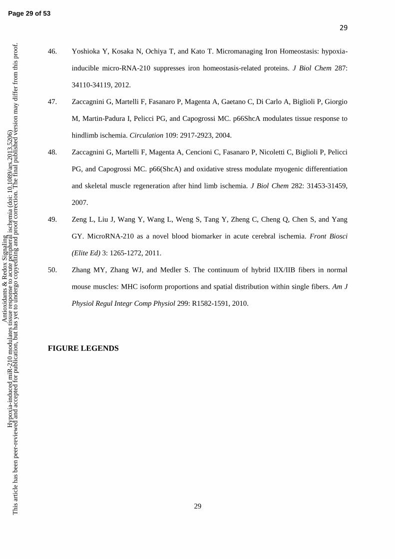
29
29
46. Yoshioka Y, Kosaka N, Ochiya T, and Kato T. Micromanaging Iron Homeostasis: hypoxia-
inducible micro-RNA-210 suppresses iron homeostasis-related proteins. J Biol Chem 287:
34110-34119, 2012.
47. Zaccagnini G, Martelli F, Fasanaro P, Magenta A, Gaetano C, Di Carlo A, Biglioli P, Giorgio
M, Martin-Padura I, Pelicci PG, and Capogrossi MC. p66ShcA modulates tissue response to
hindlimb ischemia. Circulation 109: 2917-2923, 2004.
48. Zaccagnini G, Martelli F, Magenta A, Cencioni C, Fasanaro P, Nicoletti C, Biglioli P, Pelicci
PG, and Capogrossi MC. p66(ShcA) and oxidative stress modulate myogenic differentiation
and skeletal muscle regeneration after hind limb ischemia. J Biol Chem 282: 31453-31459,
2007.
49. Zeng L, Liu J, Wang Y, Wang L, Weng S, Tang Y, Zheng C, Cheng Q, Chen S, and Yang
GY. MicroRNA-210 as a novel blood biomarker in acute cerebral ischemia. Front Biosci
(Elite Ed) 3: 1265-1272, 2011.
50. Zhang MY, Zhang WJ, and Medler S. The continuum of hybrid IIX/IIB fibers in normal
mouse muscles: MHC isoform proportions and spatial distribution within single fibers. Am J
Physiol Regul Integr Comp Physiol 299: R1582-1591, 2010.
FIGURE LEGENDS
Page 29 of 53
Ant
ioxi
dant
s &
Red
ox S
igna
ling
Hyp
oxia
-ind
uced
miR
-210
mod
ulat
es ti
ssue
res
pons
e to
acu
te p
erip
hera
l isc
hem
ia (
doi:
10.1
089/
ars.
2013
.520
6)T
his
artic
le h
as b
een
peer
-rev
iew
ed a
nd a
ccep
ted
for
publ
icat
ion,
but
has
yet
to u
nder
go c
opye
ditin
g an
d pr
oof
corr
ectio
n. T
he f
inal
pub
lishe
d ve
rsio
n m
ay d
iffe
r fr
om th
is p
roof
.
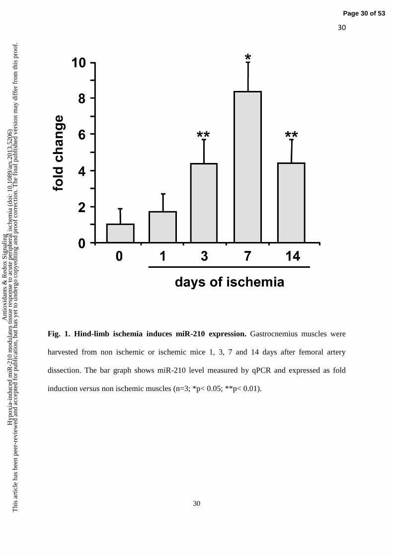
30
30
Fig. 1. Hind-limb ischemia induces miR-210 expression. Gastrocnemius muscles were
harvested from non ischemic or ischemic mice 1, 3, 7 and 14 days after femoral artery
dissection. The bar graph shows miR-210 level measured by qPCR and expressed as fold
induction versus non ischemic muscles (n=3; *p< 0.05; **p< 0.01).
Page 30 of 53
Ant
ioxi
dant
s &
Red
ox S
igna
ling
Hyp
oxia
-ind
uced
miR
-210
mod
ulat
es ti
ssue
res
pons
e to
acu
te p
erip
hera
l isc
hem
ia (
doi:
10.1
089/
ars.
2013
.520
6)T
his
artic
le h
as b
een
peer
-rev
iew
ed a
nd a
ccep
ted
for
publ
icat
ion,
but
has
yet
to u
nder
go c
opye
ditin
g an
d pr
oof
corr
ectio
n. T
he f
inal
pub
lishe
d ve
rsio
n m
ay d
iffe
r fr
om th
is p
roof
.
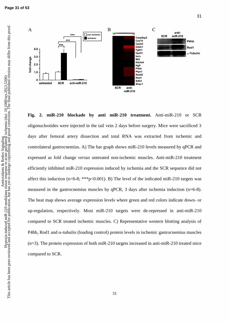
31
31
Fig. 2. miR-210 blockade by anti miR-210 treatment. Anti-miR-210 or SCR
oligonucleotides were injected in the tail vein 2 days before surgery. Mice were sacrificed 3
days after femoral artery dissection and total RNA was extracted from ischemic and
controlateral gastrocnemius. A) The bar graph shows miR-210 levels measured by qPCR and
expressed as fold change versus untreated non-ischemic muscles. Anti-miR-210 treatment
efficiently inhibited miR-210 expression induced by ischemia and the SCR sequence did not
affect this induction (n=6-8; ***p<0.001). B) The level of the indicated miR-210 targets was
measured in the gastrocnemius muscles by qPCR, 3 days after ischemia induction (n=6-8).
The heat map shows average expression levels where green and red colors indicate down- or
up-regulation, respectively. Most miR-210 targets were de-repressed in anti-miR-210
compared to SCR treated ischemic muscles. C) Representative western blotting analysis of
P4hb, Rod1 and -tubulin (loading control) protein levels in ischemic gastrocnemius muscles
(n=3). The protein expression of both miR-210 targets increased in anti-miR-210 treated mice
compared to SCR.
Page 31 of 53
Ant
ioxi
dant
s &
Red
ox S
igna
ling
Hyp
oxia
-ind
uced
miR
-210
mod
ulat
es ti
ssue
res
pons
e to
acu
te p
erip
hera
l isc
hem
ia (
doi:
10.1
089/
ars.
2013
.520
6)T
his
artic
le h
as b
een
peer
-rev
iew
ed a
nd a
ccep
ted
for
publ
icat
ion,
but
has
yet
to u
nder
go c
opye
ditin
g an
d pr
oof
corr
ectio
n. T
he f
inal
pub
lishe
d ve
rsio
n m
ay d
iffe
r fr
om th
is p
roof
.

32
32
Page 32 of 53
Ant
ioxi
dant
s &
Red
ox S
igna
ling
Hyp
oxia
-ind
uced
miR
-210
mod
ulat
es ti
ssue
res
pons
e to
acu
te p
erip
hera
l isc
hem
ia (
doi:
10.1
089/
ars.
2013
.520
6)T
his
artic
le h
as b
een
peer
-rev
iew
ed a
nd a
ccep
ted
for
publ
icat
ion,
but
has
yet
to u
nder
go c
opye
ditin
g an
d pr
oof
corr
ectio
n. T
he f
inal
pub
lishe
d ve
rsio
n m
ay d
iffe
r fr
om th
is p
roof
.
33
33
Fig. 3 Ischemia-induced apoptosis is increased by miR-210 inhibition. A) Representative
sections of gastrocnemius muscles obtained from both SCR and anti-miR-210 treated mice, at
8 hours of ischemia. TUNEL positive nuclei are stained in green and nuclei are counter-
stained by Hoechst 33342 (blue fluorescence). Magnification 400X; Calibration bar 25 µm. B)
The bar graph shows the percentage of TUNEL-positive nuclei (n=7; #p<0.02).
Page 33 of 53
Ant
ioxi
dant
s &
Red
ox S
igna
ling
Hyp
oxia
-ind
uced
miR
-210
mod
ulat
es ti
ssue
res
pons
e to
acu
te p
erip
hera
l isc
hem
ia (
doi:
10.1
089/
ars.
2013
.520
6)T
his
artic
le h
as b
een
peer
-rev
iew
ed a
nd a
ccep
ted
for
publ
icat
ion,
but
has
yet
to u
nder
go c
opye
ditin
g an
d pr
oof
corr
ectio
n. T
he f
inal
pub
lishe
d ve
rsio
n m
ay d
iffe
r fr
om th
is p
roof
.
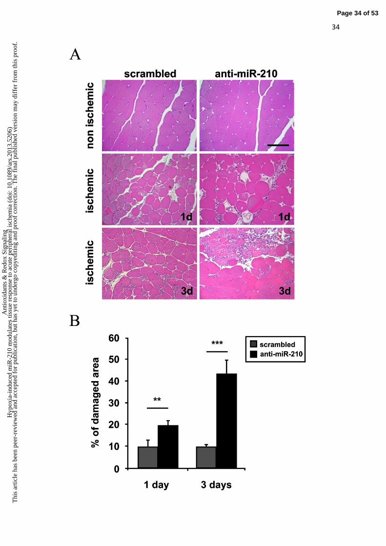
34
34
Page 34 of 53
Ant
ioxi
dant
s &
Red
ox S
igna
ling
Hyp
oxia
-ind
uced
miR
-210
mod
ulat
es ti
ssue
res
pons
e to
acu
te p
erip
hera
l isc
hem
ia (
doi:
10.1
089/
ars.
2013
.520
6)T
his
artic
le h
as b
een
peer
-rev
iew
ed a
nd a
ccep
ted
for
publ
icat
ion,
but
has
yet
to u
nder
go c
opye
ditin
g an
d pr
oof
corr
ectio
n. T
he f
inal
pub
lishe
d ve
rsio
n m
ay d
iffe
r fr
om th
is p
roof
.
35
35
Fig. 4. Ischemic muscle damage is increased by miR-210 inhibition. A) Representative
hematoxilin/eosin-stained sections of gastrocnemius muscles obtained from both SCR and
anti-miR-210 treated mice, 1 and 3 days after femoral artery dissection. Magnification 200x.
Calibration bar 100 µm. B) More necrosis in anti-miR-210 ischemic gastrocnemius muscles.
The bar graph shows the percentage of necrotic areas respect to whole section area (**p<0.01;
***p<0.0001, n=5-6).
Page 35 of 53
Ant
ioxi
dant
s &
Red
ox S
igna
ling
Hyp
oxia
-ind
uced
miR
-210
mod
ulat
es ti
ssue
res
pons
e to
acu
te p
erip
hera
l isc
hem
ia (
doi:
10.1
089/
ars.
2013
.520
6)T
his
artic
le h
as b
een
peer
-rev
iew
ed a
nd a
ccep
ted
for
publ
icat
ion,
but
has
yet
to u
nder
go c
opye
ditin
g an
d pr
oof
corr
ectio
n. T
he f
inal
pub
lishe
d ve
rsio
n m
ay d
iffe
r fr
om th
is p
roof
.

36
36
Fig. 5. Anti-miR-210 treatment increases ischemic muscle damage measured by EBD. A)
Representative EBD stained sections of gastrocnemius muscles 3 days after ischemia. EBD
was administered intraperitoneally, 16 hours before sacrifice. Red fluorescence indicates
damaged myofibers, permeable to EBD; conversely healthy myofibers are dark. Nuclei were
highlighted by Hoechst 33342 staining (blue fluorescence). Magnification 200X, Calibration
bar 100 µm. B) Bar graph showing the percentage of EBD positive areas respect to whole
area of the section (° p<0.04; n=7).
Page 36 of 53
Ant
ioxi
dant
s &
Red
ox S
igna
ling
Hyp
oxia
-ind
uced
miR
-210
mod
ulat
es ti
ssue
res
pons
e to
acu
te p
erip
hera
l isc
hem
ia (
doi:
10.1
089/
ars.
2013
.520
6)T
his
artic
le h
as b
een
peer
-rev
iew
ed a
nd a
ccep
ted
for
publ
icat
ion,
but
has
yet
to u
nder
go c
opye
ditin
g an
d pr
oof
corr
ectio
n. T
he f
inal
pub
lishe
d ve
rsio
n m
ay d
iffe
r fr
om th
is p
roof
.

37
37
Page 37 of 53
Ant
ioxi
dant
s &
Red
ox S
igna
ling
Hyp
oxia
-ind
uced
miR
-210
mod
ulat
es ti
ssue
res
pons
e to
acu
te p
erip
hera
l isc
hem
ia (
doi:
10.1
089/
ars.
2013
.520
6)T
his
artic
le h
as b
een
peer
-rev
iew
ed a
nd a
ccep
ted
for
publ
icat
ion,
but
has
yet
to u
nder
go c
opye
ditin
g an
d pr
oof
corr
ectio
n. T
he f
inal
pub
lishe
d ve
rsio
n m
ay d
iffe
r fr
om th
is p
roof
.
38
38
Fig. 6. MiR-210 blockade increases capillaries damage. A) Hematoxilin/eosin
representative stainings of both non ischemic (upper panels) and ischemic (lower panels)
gastrocnemius muscles 3 days after ischemia. Magnification 1000X. Calibration bar 50 µm.
Black arrows indicate capillaries. Yellow arrows indicate capillaries with a trapped
erythrocyte. B) Quantification of capillaries/mm2 (#p<0.04; N=8). C) Bar graph showing
Laser Doppler Perfusion Imaging measurement 3 days after femoral artery dissection.
Percentage of residual perfusion is expressed as ischemic/non ischemic ratio (*p<0.02, n=18).
Page 38 of 53
Ant
ioxi
dant
s &
Red
ox S
igna
ling
Hyp
oxia
-ind
uced
miR
-210
mod
ulat
es ti
ssue
res
pons
e to
acu
te p
erip
hera
l isc
hem
ia (
doi:
10.1
089/
ars.
2013
.520
6)T
his
artic
le h
as b
een
peer
-rev
iew
ed a
nd a
ccep
ted
for
publ
icat
ion,
but
has
yet
to u
nder
go c
opye
ditin
g an
d pr
oof
corr
ectio
n. T
he f
inal
pub
lishe
d ve
rsio
n m
ay d
iffe
r fr
om th
is p
roof
.
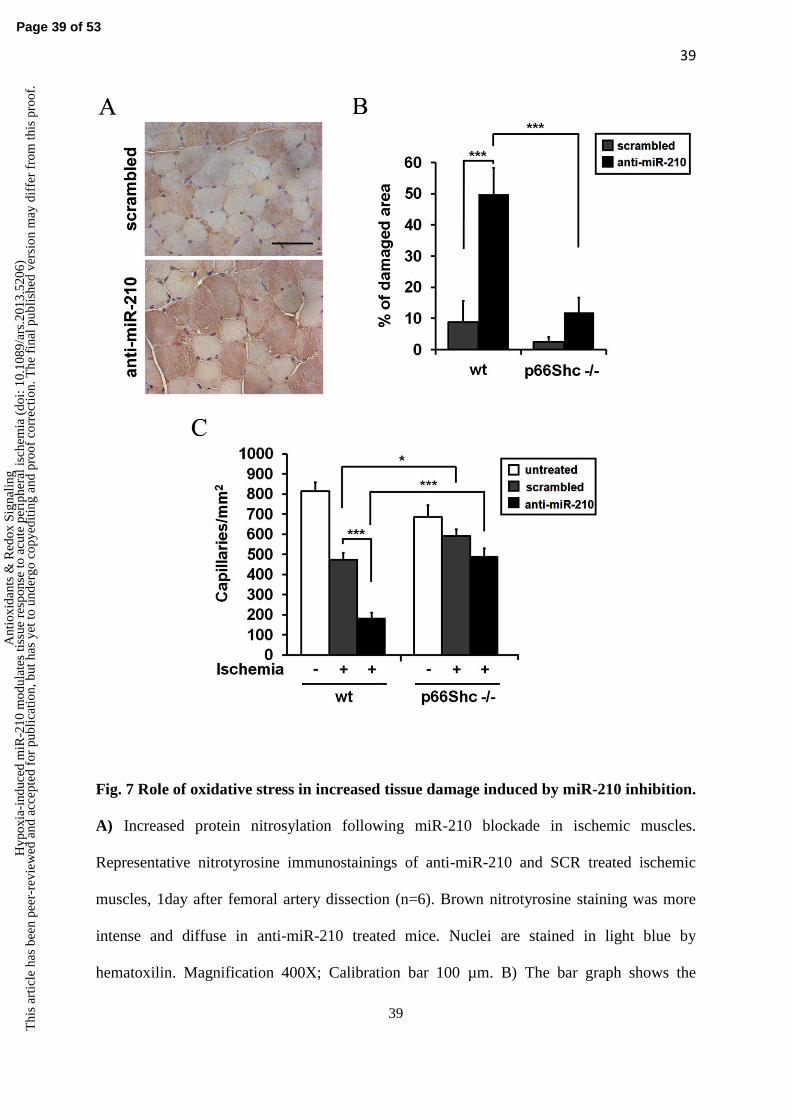
39
39
Fig. 7 Role of oxidative stress in increased tissue damage induced by miR-210 inhibition.
A) Increased protein nitrosylation following miR-210 blockade in ischemic muscles.
Representative nitrotyrosine immunostainings of anti-miR-210 and SCR treated ischemic
muscles, 1day after femoral artery dissection (n=6). Brown nitrotyrosine staining was more
intense and diffuse in anti-miR-210 treated mice. Nuclei are stained in light blue by
hematoxilin. Magnification 400X; Calibration bar 100 µm. B) The bar graph shows the
Page 39 of 53
Ant
ioxi
dant
s &
Red
ox S
igna
ling
Hyp
oxia
-ind
uced
miR
-210
mod
ulat
es ti
ssue
res
pons
e to
acu
te p
erip
hera
l isc
hem
ia (
doi:
10.1
089/
ars.
2013
.520
6)T
his
artic
le h
as b
een
peer
-rev
iew
ed a
nd a
ccep
ted
for
publ
icat
ion,
but
has
yet
to u
nder
go c
opye
ditin
g an
d pr
oof
corr
ectio
n. T
he f
inal
pub
lishe
d ve
rsio
n m
ay d
iffe
r fr
om th
is p
roof
.
40
40
percentage of necrotic areas in 3 days ischemic mice (***p<0.001; n= 6-7). Ischemic p66Shc
-
/- mice were resistant to increased tissue damage induced by miR-210 inhibition. C) The bar
graph represents the number of capillaries/mm2 in ischemic and non ischemic mice (*p< 0.03;
***p <0.00003; n=6-7). Ischemic p66Shc
-/- mice were resistant to capillary density decrease
upon miR-210 blockade.
Page 40 of 53
Ant
ioxi
dant
s &
Red
ox S
igna
ling
Hyp
oxia
-ind
uced
miR
-210
mod
ulat
es ti
ssue
res
pons
e to
acu
te p
erip
hera
l isc
hem
ia (
doi:
10.1
089/
ars.
2013
.520
6)T
his
artic
le h
as b
een
peer
-rev
iew
ed a
nd a
ccep
ted
for
publ
icat
ion,
but
has
yet
to u
nder
go c
opye
ditin
g an
d pr
oof
corr
ectio
n. T
he f
inal
pub
lishe
d ve
rsio
n m
ay d
iffe
r fr
om th
is p
roof
.
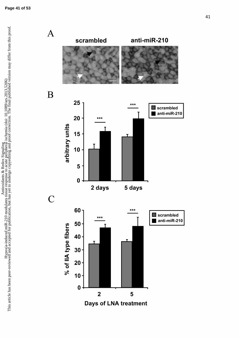
41
41
Page 41 of 53
Ant
ioxi
dant
s &
Red
ox S
igna
ling
Hyp
oxia
-ind
uced
miR
-210
mod
ulat
es ti
ssue
res
pons
e to
acu
te p
erip
hera
l isc
hem
ia (
doi:
10.1
089/
ars.
2013
.520
6)T
his
artic
le h
as b
een
peer
-rev
iew
ed a
nd a
ccep
ted
for
publ
icat
ion,
but
has
yet
to u
nder
go c
opye
ditin
g an
d pr
oof
corr
ectio
n. T
he f
inal
pub
lishe
d ve
rsio
n m
ay d
iffe
r fr
om th
is p
roof
.
42
42
Fig. 8. Anti-miR-210 shifts metabolism of EDL from glycolytic to oxidative. Frozen
sections of EDL muscle were stained with NADH-TR-diaphorase and analyzed to evaluate
metabolic differences between mice treated with anti-miR-210 and SCR. A) representative
pictures of day 2 muscles are shown. White arrows indicate type IIB fibers, while black
arrows indicate type IIA fibers. Calibration bar 50 µm. B) The bar graph shows increased
intensity of NADH-TR-diaphorase staining expressed in arbitrary units, at the indicated
timepoints (n=8; ***p<0.001). C) The bar graph shows the percentage of type IIA fibers,
indicating an increase of this fiber type in mice treated with anti-miR-210, for 2 or 5 days
(n=8; ***p<0.001).
Fig.9. Metabolic activity assays. A) The bar graph represents Aconitase activity fold change
in EDL muscle extracts(*p<0.01; n=9). B) The bar graph shows Complex I activity measured
in crude mitochondrial preparations of Gastrocnemius muscles (*p<0.05; n=5).
Fig.10. Model of the mechanisms underpinning miR-210 antiapoptotic, cytoprotective
function. Upon miR-210 blocking, mitochondrial oxidative phosphorylation is de-repressed,
increasing ROS levels and triggering cell death. Other sources of ROS, such as NADPH
oxidase (not indicated), may also contribute to determine ROS levels.
Supplementary figure legends
Fig.S1. Time course of capillary density in CD1 mice. The bar graph represents
quantification of capillaries/mm2 in normoperfused (normo) and ischemic CD1 mice at 1 and
3 days. Differences between ischemic and normoperfused muscles are statistically significant
(p<0.0001; n=7-8).
Page 42 of 53
Ant
ioxi
dant
s &
Red
ox S
igna
ling
Hyp
oxia
-ind
uced
miR
-210
mod
ulat
es ti
ssue
res
pons
e to
acu
te p
erip
hera
l isc
hem
ia (
doi:
10.1
089/
ars.
2013
.520
6)T
his
artic
le h
as b
een
peer
-rev
iew
ed a
nd a
ccep
ted
for
publ
icat
ion,
but
has
yet
to u
nder
go c
opye
ditin
g an
d pr
oof
corr
ectio
n. T
he f
inal
pub
lishe
d ve
rsio
n m
ay d
iffe
r fr
om th
is p
roof
.

43
43
Fig. S2. MiR-147b is induced upon ischemia. The levels of miR-147b were measured in the
gastrocnemius muscle by qPCR, 3 days after ischemia induction (n=8). In both SCR and anti-
miR-210 treated mice, miR-147b levels were undetectable in non ischemic muscles (Ct >35).
In ischemic muscles, miR-147b was detectable in both SCR and anti-miR-210 treated mice
(Ct<35). Ct levels of a control miRNA (miR-16) were similar in all groups. The red line
indicates detection threshold.
Page 43 of 53
Ant
ioxi
dant
s &
Red
ox S
igna
ling
Hyp
oxia
-ind
uced
miR
-210
mod
ulat
es ti
ssue
res
pons
e to
acu
te p
erip
hera
l isc
hem
ia (
doi:
10.1
089/
ars.
2013
.520
6)T
his
artic
le h
as b
een
peer
-rev
iew
ed a
nd a
ccep
ted
for
publ
icat
ion,
but
has
yet
to u
nder
go c
opye
ditin
g an
d pr
oof
corr
ectio
n. T
he f
inal
pub
lishe
d ve
rsio
n m
ay d
iffe
r fr
om th
is p
roof
.

44
44
Fig. S3. MiR-210 blockade does not affect Hif activation upon ischemia. A) The levels of
the indicated HIF1alpha species and targets were measured in the gastrocnemius muscle by
qPCR, 3 days after ischemia induction (n=8). The heat map shows average expression levels
in a base 2 logarithmic scale, where red color indicates up-regulation and green color down-
regulation. HIF1alpha targets were similarly induced in anti-miR-210 and SCR treated
ischemic muscles compared to controlateral non ischemic muscles. B) Representative
western blotting analysis of HIF1alpha and HIF2alpha and -tubulin (loading control) protein
levels in ischemic gastrocnemius muscles (n=3). Both HIF1alpha and HIF2alpha display
similar levels in anti-miR-210 and SCR treated mice.
Page 44 of 53
Ant
ioxi
dant
s &
Red
ox S
igna
ling
Hyp
oxia
-ind
uced
miR
-210
mod
ulat
es ti
ssue
res
pons
e to
acu
te p
erip
hera
l isc
hem
ia (
doi:
10.1
089/
ars.
2013
.520
6)T
his
artic
le h
as b
een
peer
-rev
iew
ed a
nd a
ccep
ted
for
publ
icat
ion,
but
has
yet
to u
nder
go c
opye
ditin
g an
d pr
oof
corr
ectio
n. T
he f
inal
pub
lishe
d ve
rsio
n m
ay d
iffe
r fr
om th
is p
roof
.

45
45
Fig. S4. Hierarchical clustering and qPCR validation of ischemic gastrocnemius gene
expression profiles. A) Hierarchical clustering obtained using centered correlation and
average linkage of non-ischemic (left panel) and ischemic (right) samples derived from SCR
Page 45 of 53
Ant
ioxi
dant
s &
Red
ox S
igna
ling
Hyp
oxia
-ind
uced
miR
-210
mod
ulat
es ti
ssue
res
pons
e to
acu
te p
erip
hera
l isc
hem
ia (
doi:
10.1
089/
ars.
2013
.520
6)T
his
artic
le h
as b
een
peer
-rev
iew
ed a
nd a
ccep
ted
for
publ
icat
ion,
but
has
yet
to u
nder
go c
opye
ditin
g an
d pr
oof
corr
ectio
n. T
he f
inal
pub
lishe
d ve
rsio
n m
ay d
iffe
r fr
om th
is p
roof
.
46
46
and anti-miR-210 treated muscles. B) Transcripts modulated upon ischemia (n=4) and (C)
transcripts differentially expressed in anti-miR-210 and SCR treated ischemic muscles (n=4-
6). The levels of the indicated transcripts was measured in the gastrocnemius muscle by
qPCR, 3 days after ischemia induction. The heat map shows average expression levels in a
base 2 logarithmic scale, where red color indicates up-regulation and green color down-
regulation.
Page 46 of 53
Ant
ioxi
dant
s &
Red
ox S
igna
ling
Hyp
oxia
-ind
uced
miR
-210
mod
ulat
es ti
ssue
res
pons
e to
acu
te p
erip
hera
l isc
hem
ia (
doi:
10.1
089/
ars.
2013
.520
6)T
his
artic
le h
as b
een
peer
-rev
iew
ed a
nd a
ccep
ted
for
publ
icat
ion,
but
has
yet
to u
nder
go c
opye
ditin
g an
d pr
oof
corr
ectio
n. T
he f
inal
pub
lishe
d ve
rsio
n m
ay d
iffe
r fr
om th
is p
roof
.
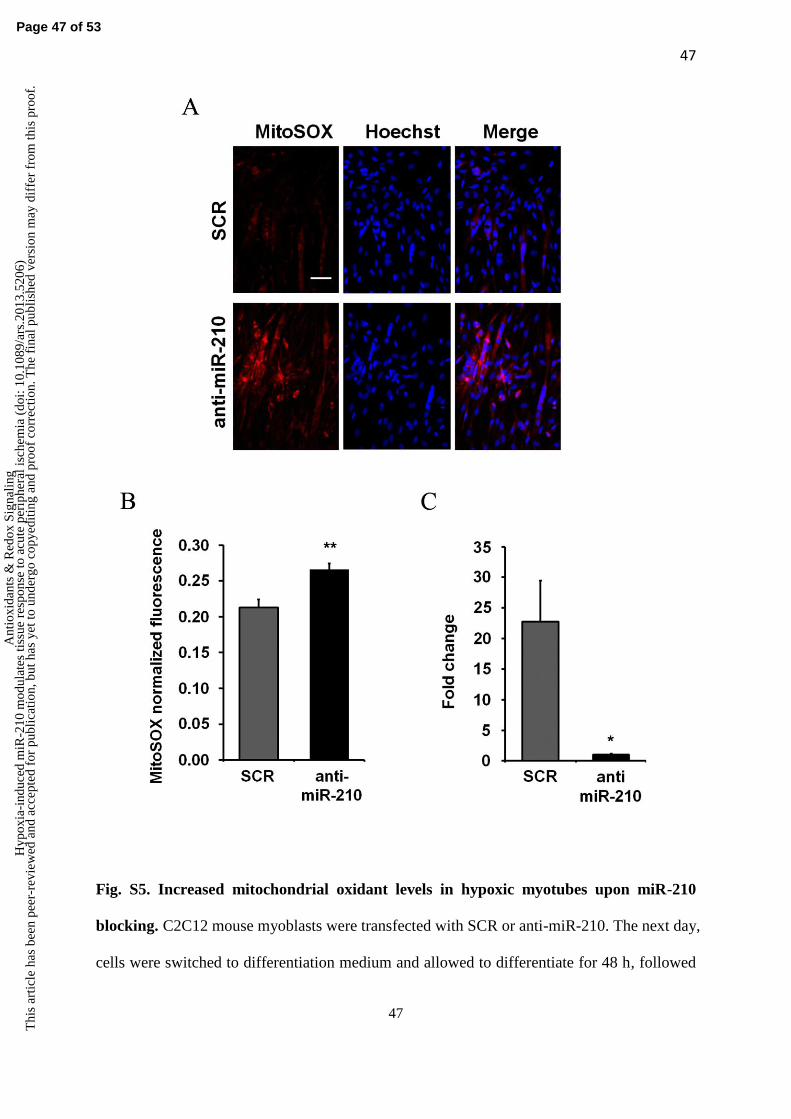
47
47
Fig. S5. Increased mitochondrial oxidant levels in hypoxic myotubes upon miR-210
blocking. C2C12 mouse myoblasts were transfected with SCR or anti-miR-210. The next day,
cells were switched to differentiation medium and allowed to differentiate for 48 h, followed
Page 47 of 53
Ant
ioxi
dant
s &
Red
ox S
igna
ling
Hyp
oxia
-ind
uced
miR
-210
mod
ulat
es ti
ssue
res
pons
e to
acu
te p
erip
hera
l isc
hem
ia (
doi:
10.1
089/
ars.
2013
.520
6)T
his
artic
le h
as b
een
peer
-rev
iew
ed a
nd a
ccep
ted
for
publ
icat
ion,
but
has
yet
to u
nder
go c
opye
ditin
g an
d pr
oof
corr
ectio
n. T
he f
inal
pub
lishe
d ve
rsio
n m
ay d
iffe
r fr
om th
is p
roof
.
48
48
by incubation in 1% O2 hypoxic conditions for 24 hours. Then, myotubes were stained with
MitoSOX and Hoechst 33342 dyes, and fluorescence was revealed by fluorescence
microscopy. A) Representative pictures display MitoSOX and Hoechst 33342 fluorescences,
either separate or merged. Calibration bar =50 m. B) The bar graph indicates MitoSOX
fluorescence intensity normalized for the number of Hoechst 33342-positive nuclei
(**p<0.002; n=6) MitoSOX associated fluorescence was increased when miR-210 was
inhibited. C) The bar graph shows miR-210 levels in C2C12 cells transfected with anti-
miR210 or SCR. MiR-210 levels were decreased efficiently by anti-miR210 (n=6; *, p<0.03)
Page 48 of 53
Ant
ioxi
dant
s &
Red
ox S
igna
ling
Hyp
oxia
-ind
uced
miR
-210
mod
ulat
es ti
ssue
res
pons
e to
acu
te p
erip
hera
l isc
hem
ia (
doi:
10.1
089/
ars.
2013
.520
6)T
his
artic
le h
as b
een
peer
-rev
iew
ed a
nd a
ccep
ted
for
publ
icat
ion,
but
has
yet
to u
nder
go c
opye
ditin
g an
d pr
oof
corr
ectio
n. T
he f
inal
pub
lishe
d ve
rsio
n m
ay d
iffe
r fr
om th
is p
roof
.

49
49
Fig. S6 miR-210 targets de-repression in normoperfused muscles upon miR-210
inhibition. The level of the indicated miR-210 targets was measured in non ischemic
gastrocnemius muscles by qPCR, 2 days after treatment with either anti-miR-210 or SCR
(n=4). The heat map shows average expression levels in a base 2 logarithmic scale, where
green and red colors indicate down- or up-regulation, respectively. MiR-210 targets were de-
repressed in anti-miR-210 compared to SCR treated muscles.
Page 49 of 53
Ant
ioxi
dant
s &
Red
ox S
igna
ling
Hyp
oxia
-ind
uced
miR
-210
mod
ulat
es ti
ssue
res
pons
e to
acu
te p
erip
hera
l isc
hem
ia (
doi:
10.1
089/
ars.
2013
.520
6)T
his
artic
le h
as b
een
peer
-rev
iew
ed a
nd a
ccep
ted
for
publ
icat
ion,
but
has
yet
to u
nder
go c
opye
ditin
g an
d pr
oof
corr
ectio
n. T
he f
inal
pub
lishe
d ve
rsio
n m
ay d
iffe
r fr
om th
is p
roof
.

50
50
Page 50 of 53
Ant
ioxi
dant
s &
Red
ox S
igna
ling
Hyp
oxia
-ind
uced
miR
-210
mod
ulat
es ti
ssue
res
pons
e to
acu
te p
erip
hera
l isc
hem
ia (
doi:
10.1
089/
ars.
2013
.520
6)T
his
artic
le h
as b
een
peer
-rev
iew
ed a
nd a
ccep
ted
for
publ
icat
ion,
but
has
yet
to u
nder
go c
opye
ditin
g an
d pr
oof
corr
ectio
n. T
he f
inal
pub
lishe
d ve
rsio
n m
ay d
iffe
r fr
om th
is p
roof
.

51
51
Fig.9. Metabolic activity assays. A) The bar graph represents Aconitase activity fold change in EDL
muscle extracts(*p<0.01; n=9). B) The bar graph shows Complex I activity measured in crude
mitochondrial preparations of Gastrocnemius muscles (*p<0.05; n=5).
Page 51 of 53
Ant
ioxi
dant
s &
Red
ox S
igna
ling
Hyp
oxia
-ind
uced
miR
-210
mod
ulat
es ti
ssue
res
pons
e to
acu
te p
erip
hera
l isc
hem
ia (
doi:
10.1
089/
ars.
2013
.520
6)T
his
artic
le h
as b
een
peer
-rev
iew
ed a
nd a
ccep
ted
for
publ
icat
ion,
but
has
yet
to u
nder
go c
opye
ditin
g an
d pr
oof
corr
ectio
n. T
he f
inal
pub
lishe
d ve
rsio
n m
ay d
iffe
r fr
om th
is p
roof
.
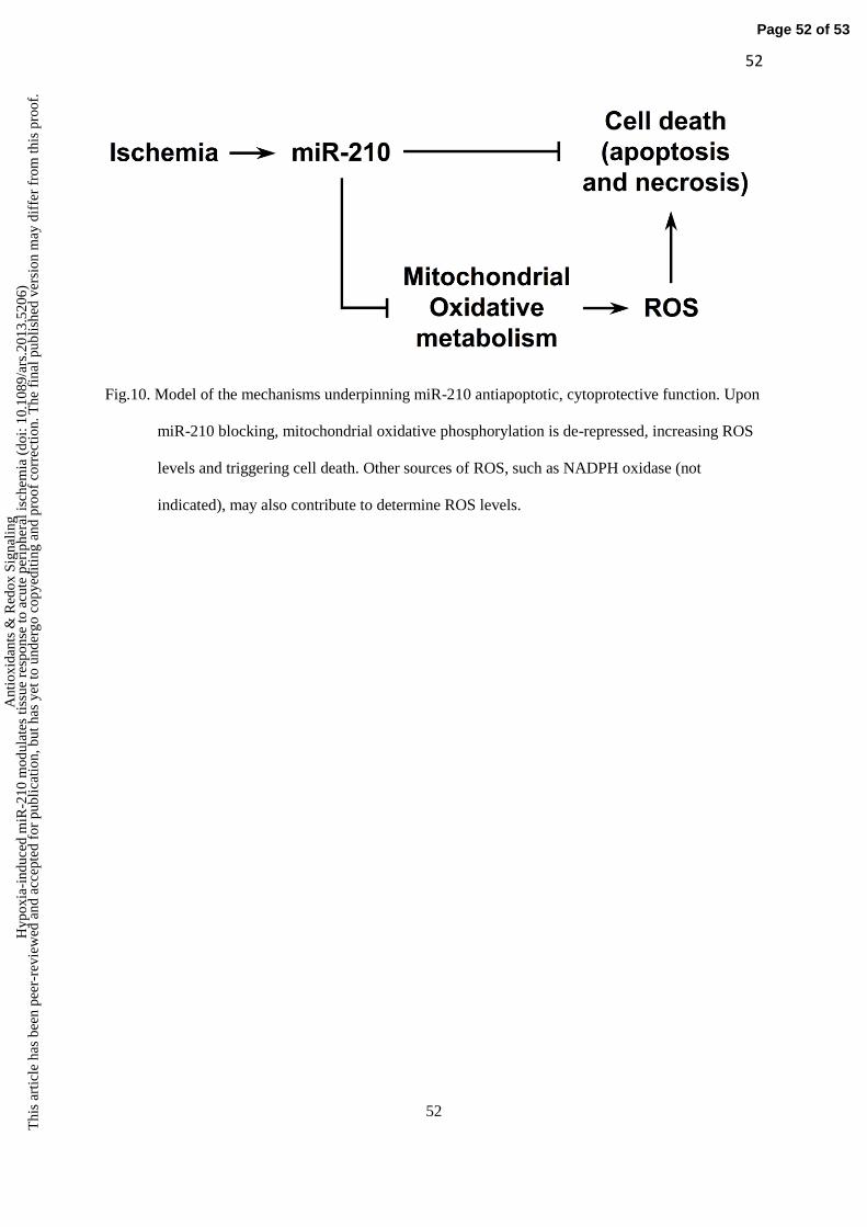
52
52
Fig.10. Model of the mechanisms underpinning miR-210 antiapoptotic, cytoprotective function. Upon
miR-210 blocking, mitochondrial oxidative phosphorylation is de-repressed, increasing ROS
levels and triggering cell death. Other sources of ROS, such as NADPH oxidase (not
indicated), may also contribute to determine ROS levels.
Page 52 of 53
Ant
ioxi
dant
s &
Red
ox S
igna
ling
Hyp
oxia
-ind
uced
miR
-210
mod
ulat
es ti
ssue
res
pons
e to
acu
te p
erip
hera
l isc
hem
ia (
doi:
10.1
089/
ars.
2013
.520
6)T
his
artic
le h
as b
een
peer
-rev
iew
ed a
nd a
ccep
ted
for
publ
icat
ion,
but
has
yet
to u
nder
go c
opye
ditin
g an
d pr
oof
corr
ectio
n. T
he f
inal
pub
lishe
d ve
rsio
n m
ay d
iffe
r fr
om th
is p
roof
.

53
53
Fig.S1. Time course of capillary density in CD1 mice. The bar graph represents quantification of
capillaries/mm2 in normoperfused (normo) and ischemic CD1 mice at 1 and 3 days.
Differences between ischemic and normoperfused muscles are statistically significant
(p<0.0001; n=7-8).
Page 53 of 53
Ant
ioxi
dant
s &
Red
ox S
igna
ling
Hyp
oxia
-ind
uced
miR
-210
mod
ulat
es ti
ssue
res
pons
e to
acu
te p
erip
hera
l isc
hem
ia (
doi:
10.1
089/
ars.
2013
.520
6)T
his
artic
le h
as b
een
peer
-rev
iew
ed a
nd a
ccep
ted
for
publ
icat
ion,
but
has
yet
to u
nder
go c
opye
ditin
g an
d pr
oof
corr
ectio
n. T
he f
inal
pub
lishe
d ve
rsio
n m
ay d
iffe
r fr
om th
is p
roof
.