Hydrostatic pressure and shear stress affect endothelin‐1 and nitric oxide release by endothelial...
24
Independent application of pressure and shear stress on endothelial cells in controlled bioreactors Federico Vozzi 1 , Francesca Bianchi 2 , Arti Ahluwalia 2 , Claudio Domenici 1 1) Biomimetic Materials and Tissue Engineering Laboratory, CNR Institute of Clinical Physiology, Via G. Moruzzi 1, Pisa, Italy 2) Research Center “E. Piaggio”, University of Pisa, Via Diotisalvi 2, Pisa, Italy Abstract The principal physical forces exerted on the blood vessel wall by the passage of intra-luminal blood are pressure and shear. In order to analyze their independent effects, these two stresses were applied to cultured cells by means of two different bioreactors: the Pressure Controlled Bioreactor (PCB) and the Laminar Flow Bioreactor (LFB), in which controlled levels of pressure and shear stress can be respectively generated. Using the bioreactor systems, Endothelin-1 and Nitric Oxide release from human umbilical vein endothelial cells (HUVEC) were measured in varying conditions of shear stress and pressure. As result, a decrease of Endothelin-1 production from the cells cultured in both bioreactors, with respect to the controls, was observed, whereas Nitric Oxide synthesis was up-regulated only in the presence of shear stress but not modulated by hydrostatic pressure. These results show that the two hemodynamic forces acting on blood vessels affect endothelial cell function in different ways, and that both should be considered when planning in vitro experiments in the presence of flow.
-
Upload
independent -
Category
Documents
-
view
4 -
download
0
Transcript of Hydrostatic pressure and shear stress affect endothelin‐1 and nitric oxide release by endothelial...

Independent application of pressure and shear stress on endothelial cells in controlled bioreactors
Federico Vozzi1, Francesca Bianchi2, Arti Ahluwalia2, Claudio Domenici1
1)Biomimetic Materials and Tissue Engineering Laboratory, CNR Institute of Clinical Physiology, Via G. Moruzzi 1, Pisa, Italy 2)Research Center “E. Piaggio”, University of Pisa, Via Diotisalvi 2, Pisa, Italy
Abstract
The principal physical forces exerted on the blood vessel wall by the passage of intra-luminal
blood are pressure and shear. In order to analyze their independent effects, these two stresses were
applied to cultured cells by means of two different bioreactors: the Pressure Controlled Bioreactor
(PCB) and the Laminar Flow Bioreactor (LFB), in which controlled levels of pressure and shear
stress can be respectively generated.
Using the bioreactor systems, Endothelin-1 and Nitric Oxide release from human umbilical vein
endothelial cells (HUVEC) were measured in varying conditions of shear stress and pressure. As
result, a decrease of Endothelin-1 production from the cells cultured in both bioreactors, with
respect to the controls, was observed, whereas Nitric Oxide synthesis was up-regulated only in the
presence of shear stress but not modulated by hydrostatic pressure.
These results show that the two hemodynamic forces acting on blood vessels affect endothelial
cell function in different ways, and that both should be considered when planning in vitro
experiments in the presence of flow.

1 Introduction
The most basic physical requirements for maintaining mammalian cells in culture are a pH value
of 7.2-7.4 and a temperature of 37 °C. For decades the backbone of in vitro cell culture has been
the incubator: an oven in which cell cultures can grow in an appropriate pH and temperature
controlled environment. In the last few years, owing to the failure of cell culture to predict in vivo
tissue responses, the issue of the environment discrepancy problem has been raised. The
prevailing view is that the classic in vitro culture environment does not adequately replicate the
physical forces and chemical concentration gradients present in vivo. As a result of this, several
alternative culture systems (bioreactors) have been developed in order to create dynamic cell
culture devices, which allow the physiological conditions of an organism to be simulated [1,2,3].
The goal of an effective bioreactor is to influence and control biological reactions and functions.
To accomplish this, in the design of the instrument, two critical aspects must be taken into
account. One of them is related to the suitable physical parameters (like geometrical dimensions,
flow rate, etc.) required to obtain the desired macro-kinetic environment. The other area of major
importance in bioreactor design involves the appropriate control of parameters which influence
biomolecular reactions including: controlled temperature, optimum pH-value, sufficient substrate
(usually a carbon source such as sugars), O2 and CO2 control, which can influence cell growth and
metabolite production. In addition, to be able to control these parameters, the bioreactor must be
capable of maintaining an appropriate cell phenotype for extended periods of time and eliminate
or reduce contamination by unwanted organisms. Among the different types of cell culture
bioreactors described in the literature, many are used in cardiovascular studies, such as laminar
flow bioreactor [4], perfusion bioreactor [5], multi-cue bioreactor [6] etc. While several groups
have performed in depth studies of endothelial cell function as a function of flow [7,8], most of
the recent systems reported are directed towards tissue engineering [9−12]. On the other hand
very little work has been done on the influence of pressure on endothelial cell structure and
function [13,14], often using in vitro systems as vessel simulators addressed to generate a

pulsatile flow, in combination with stretch, capable of simultaneously inducing shear stress and
pressure [15,14], or focusing on the characterization of the apparatus with respect to its
biomechanical and hemodynamic properties [16].
Endothelial cells cover the lumen of blood vessels and play an essential role in the regulation of
vessel homeostasis and of flow and blood pressure in the circulatory system [17,18]. The
endothelium is a dynamic system which is able to respond to different physical and chemical
conditions through the production of many vasoactive substances such as: Endothelins,
Prostaglandin and Nitric Oxide. These molecules act on the smooth muscle cells that form the
vessel side and together play a critical role in coordinating and maintaining vascular function.
Since its discovery, a great deal of effort has been devoted to understanding the role of Endothelin
(ET), a family of peptides involved in cardiovascular homeostasis, and their potential contribution
to many pathological processes [19,20]. The main vascular isoform is Endothelin-1 (ET-1), a 21
amino acid peptide [21]. In vivo studies have shown that ET-1 causes concentration dependent
arterial and venous constrictions and that it is more potent as a venoconstrictor [22]. The ET's
counter molecule is Nitric Oxide (NO), a potent vasodilator that acts on smooth muscle cells.
Moreover, it is demonstrated that the Endothelin-NO system participates in numerous physio-
pathological conditions, including hypertension, atherosclerosis and coronary artery disease [23].
Stress, in particular mechanical pressure and stretch, induces ET release in vivo [24]. A few
in vitro studies relating flow, pressure and pulsatility to ET and NO production have been
reported. Walshe et al. [25] described a pulsatile flow system in which a co-culture of endothelial
and smooth muscle cells showed an increase of both NO and ET-1 with flow. Dai et al. [26]
reported on a venous flow simulator in which endothelial cells up-regulated NO synthesis with the
flow, while there are no reported differences in ET-1 production. In Tsukurov et al. [27], the
combined pressurized flow and cyclic strain down-regulated ET-1 production after 48 h, but not at
24 h, whereas the results for NO production were not clear. Braddon et al. [28] used a parallel
flow chamber, where endothelial cells were cultured in tissue culture plastic, or a feeder layer of

fibroblasts, or on woven polymer. The results of their study showed that NO production increases
with flow. In general, it is unclear as to whether ET-1 and NO production is pressure or flow
dependent, particularly when high flow rates are used, or if they depend on factors such as
substrate deformation. In some reports it is unclear as to what forces are actually being imposed
on the cells, and which stimulus actually causes the up- or down-regulation of these substances.
This is because high wall shear stresses usually require the application of high pressure gradients
across a sample. In fact, most commercial laminar flow systems consist of very narrow chambers;
to obtain physiological wall shear stress values in the chambers it is necessary to apply high
pressure gradients which means generating pressure differences of up to 10 mmHg or more
between inlet and outlet. Thus cells are stimulated by both shear stress and pressure.
In order to obtain more information on cell response to acute pressure and shear stress stimuli, we
report here a study carried out using two new cell-culture bioreactors, which form part of an array
of systems developed at the Research Centre “E. Piaggio”, University of Pisa [29]. While the
basic fluid and environmental control system is identical for each reactor, the cell culture chamber
can be designed and configured according to experimental requirements. The Pressure Controlled
Bioreactor (PCB) and the Laminar Flow Bioreactor (LFB), were tested by evaluating the effect of
pressure and low shear stress on human umbilical vein endothelial cell cultures, through
measurement of ET-1 and NO levels. Using these two systems, and applying the two forces in
independent well-controlled experiments we show that it is possible to determine endothelial cell
response to hydrostatic pressure and to different levels of shear stress unequivocally.
2 Materials and methods
2.1 Cell culture
HUVEC (human umbilical vein endothelial cells) were isolated from fresh human umbilical cord
veinsby enzymatic treatment with collagenase. After pooling from from multiple donors, they
were used between passages 2 to 8. The cells were cultured as already described by Jaffe et al.
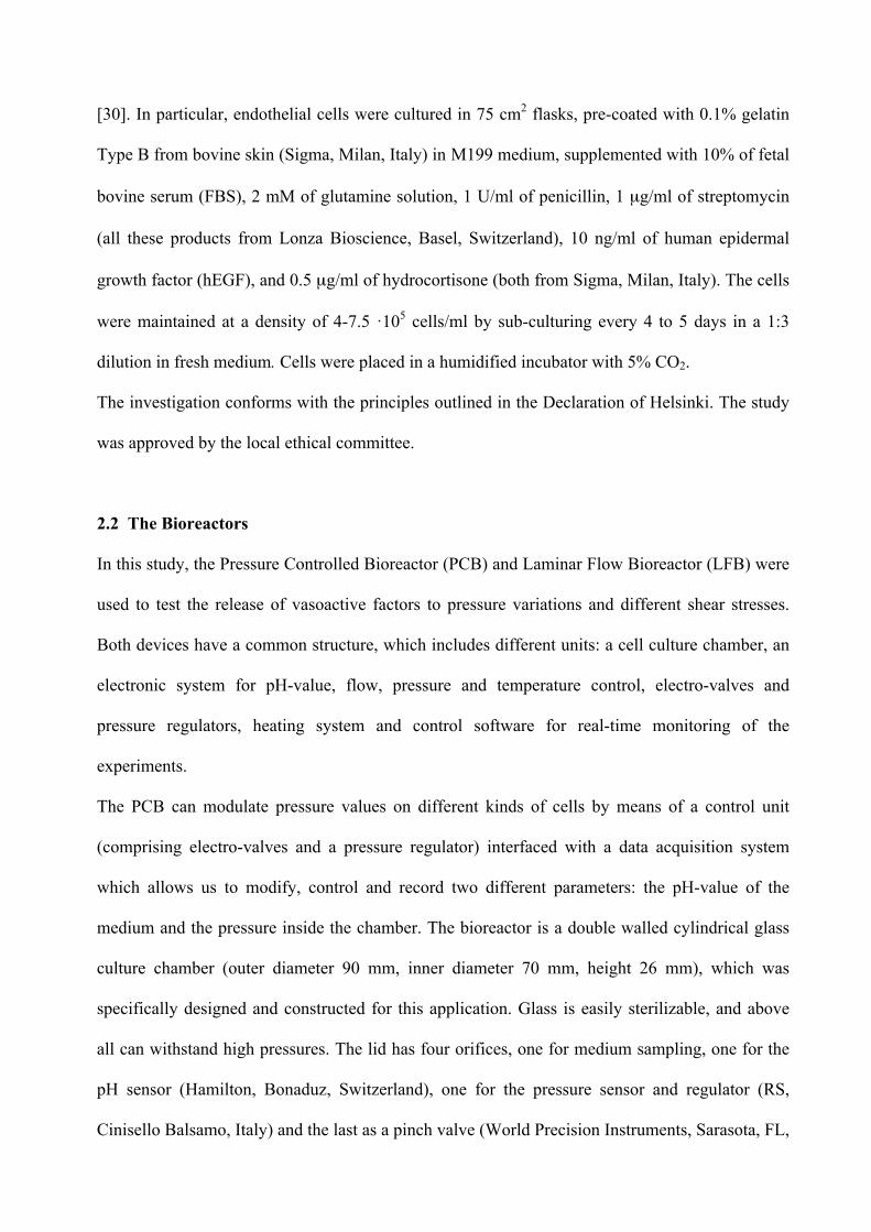
[30]. In particular, endothelial cells were cultured in 75 cm2 flasks, pre-coated with 0.1% gelatin
Type B from bovine skin (Sigma, Milan, Italy) in M199 medium, supplemented with 10% of fetal
bovine serum (FBS), 2 mM of glutamine solution, 1 U/ml of penicillin, 1 µg/ml of streptomycin
(all these products from Lonza Bioscience, Basel, Switzerland), 10 ng/ml of human epidermal
growth factor (hEGF), and 0.5 µg/ml of hydrocortisone (both from Sigma, Milan, Italy). The cells
were maintained at a density of 4-7.5 ·105 cells/ml by sub-culturing every 4 to 5 days in a 1:3
dilution in fresh medium. Cells were placed in a humidified incubator with 5% CO2.
The investigation conforms with the principles outlined in the Declaration of Helsinki. The study
was approved by the local ethical committee.
2.2 The Bioreactors
In this study, the Pressure Controlled Bioreactor (PCB) and Laminar Flow Bioreactor (LFB) were
used to test the release of vasoactive factors to pressure variations and different shear stresses.
Both devices have a common structure, which includes different units: a cell culture chamber, an
electronic system for pH-value, flow, pressure and temperature control, electro-valves and
pressure regulators, heating system and control software for real-time monitoring of the
experiments.
The PCB can modulate pressure values on different kinds of cells by means of a control unit
(comprising electro-valves and a pressure regulator) interfaced with a data acquisition system
which allows us to modify, control and record two different parameters: the pH-value of the
medium and the pressure inside the chamber. The bioreactor is a double walled cylindrical glass
culture chamber (outer diameter 90 mm, inner diameter 70 mm, height 26 mm), which was
specifically designed and constructed for this application. Glass is easily sterilizable, and above
all can withstand high pressures. The lid has four orifices, one for medium sampling, one for the
pH sensor (Hamilton, Bonaduz, Switzerland), one for the pressure sensor and regulator (RS,
Cinisello Balsamo, Italy) and the last as a pinch valve (World Precision Instruments, Sarasota, FL,

USA). Hot water from a thermostat flows through the glass jacket in order to maintain a
temperature of 37 °C. A steel frame holds the entire device together; Figure 1A shows the PCB
pressure and pH control circuit while Figure 2 reports the assembled bioreactor complete with
sensors and valves. Both the pH-value and pressure are controlled by the introduction of an
air/CO2 blend through the pressure regulator on the chamber cap. The PBC works under constant
over pressure, and the desired pressure is obtained either by changing the setting of the pinch
valve, or by changing the input pressure. The pH-value is controlled by regulating the amounts of
CO2 or air input into the chamber according to a feedback algorithm based on the output of the pH
meter. The signals are sent to a PC acquisition card. The pressure and pH in the chamber are
controlled by software developed in Visual Basic. Before starting, the system must be calibrated
introducing a constant pressure in the outlet from the electro-valve and verifying that no air losses
are present. After the calibration and the setting of the desired pressure and pH-value values, the
experiment can start. The pressure and pH control algorithms and electronics are described fully
in Mazzei et al. [29].
In the LFB system, a peristaltic pump and a fluid reservoir or mixing chamber is added to the
circuit to generate different rates of shear stress, while the pressure control circuit is bypassed.
The mixing chamber serves to oxygenate media, and in particular, to reduce pressure fluctuations
from the pump, the chamber is positioned between the pump and the LBF inlet. In this way it acts
as a pressure capacitor, smoothing any fluctuations from the pump. Graphs showing the minimal
pressure fluctuations using this configuration are reported in the supplementary data. The chamber
is fabricated in polydimethylsiloxane (PDMS, Sylgard 184®) (Dow Corning, Midland, MI, USA),
a biocompatible silicone polymer, through milli-molding [29]. The cell chamber, shown in
Figure 2A, is 160 mm long, 20 mm wide and 1 mm high, with a 150 µm deep rectangular
insertion for glass or plastic coverslips. Its particular shape was obtained after an accurate
modeling analysis performed with finite element software for simulation of fluid dynamic flow
(COMSOL Multiphysics, Stockholm, Sweden) in order to obtain a central region with a well-

defined and uniform wall shear stress. We used the single phase incompressible fluid flow regime
described by the Navier Stokes equations, imposing a fluid with the same density (1 g/cm3) and
viscosity as cell culture medium (1.4 mPa·s) at 37°C. No slip boundary conditions were imposed
at the walls and zero pressure at the outlet. The 3D model was solved parametrically and in steady
state conditions using different flows at the inlet boundary ranging from 1 to 20 ml/min. In the
central cell culture region the flow is fully developed and wall shear stress, τ , ranged from 0.07
to 1.4 dynes/cm2. In Figure 2B the simulation of velocity field in the LFB cell culture chamber is
reported for the xy plane at a flow rate of 15 ml/min, showing a uniform velocity and hence shear
stress in the central zone, where the maximum fluid velocity is 2.5 cm/s. At this flow rate the
pressure difference across the bioreactor is of the order of a few tenths of mmHg, as calculated
from the Finite Element Method (FEM) model (Figure 2C), and thus negligible. Further figures
(uniform wall shear stress and velocity profiles) can be found in the supplementary data.
Therefore with this geometry we can simulate different regions of the cardiovascular system
simply by adjusting flow rates. In theory a wide range of flow rates (laminar flow is ensured up to
240 mL/min) and shear stresses can be generated in the system, but the limiting factor is the
pressure difference generated by high flow rates which can give rise to leakage. The maximum
velocity is 50 mL/min, with a wall shear stress in the cell culture region of about 3.5 dynes/cm2.
In this work low flow rates were used to avoid significant pressure gradients across the bioreactor,
which could give rise to hydrostatic pressure effects.
2.3 Pressure variation experiments
When at confluence, HUVEC were seeded on glass slides (dimensions 50 mm x 25 mm),
purposely washed with ethanol 96% and sterilized under UV light for 15-20 min each side. The
slides were subsequently washed with sterile PBS to eliminate traces of ethanol. Successively, the
glass slides were treated with gelatin in the incubator for 15-20 min to improve cell adhesion.
After removal of the surplus gelatin, slides were seeded with the cell suspension (6400 cells/cm2

equivalent to 80000 cells/glass slide). About 48 hours after seeding, the experiments with the
bioreactor, whose components were previously sterilized with H2O2 gas plasma, were started.
Once assembled, the mixing chamber was filled with 10 ml of complete medium for each set of
experiments (3 slides for each). One slide was placed in the PCB which was filled with 10 mL of
complete medium and closed, ready for the experiments.
Endothelial cells are normally exposed to pressures ranging from 2-5 mmHg up to 140 mmHg,
although in cases of acute hypertension pressures can go up to 240 mmHg. In this work the value
of 0 mmHg (with respect to atmospheric pressure), identified as P0, was used to simulate normal
conditions (controls), while a “representative” physiological pressure (about 70 mmHg, identified
as P70) was used to stimulate the in vitro conditioned cells. Besides an increase in pressure, there
is also a slight increase in oxygen solubility. The Henry constant for oxygen is 1.3.10-6 M/mmHg,
thus the increase in oxygen concentration at 830 mmHg is only about 8%. Since the Km
(Micheles Menten constant for mammalian cells, 1 µM) is much lower than the predicted oxygen
concentration (around 0.2 mM), we can assume that the additional 70 mmHg in the physiological
pressure experiment does not contribute significantly to increase oxygen consumption, and the
only stimulus being applied is hydrostatic pressure. Samples were cultured in the bioreactor filled
with fresh medium and placed for 1 hour in the incubator at P0 (as controls), 1 hour at 70 mmHg
(P70) in the pressure bioreactor and, finally, for another hour at P0 in the incubator, to verify cell
recovery. In all cases, the entire volume of medium was collected for the biological tests (ET-1
and NO), and refreshed every hour. At the end of each experiment, cells were analyzed under a
microscope (Olympus IX-81, Olympus Italia, Segrate, Italy) in order to observe structural changes
and for counting using trypan blue exclusion. The experiment was replicated five times.
2.4 Shear stress experiments
For this set of experiments, cells at confluence were seeded on glass slides (dimensions 17 mm x
32 mm x 0.15 mm) with a cell suspension density of 6400 cells/cm2 (equivalent to about 35000

cells/glass slide), treated as previously described. About 48 hours after seeding, the experiments
started with the bioreactor. Once assembled, the mixing chamber was filled with 10 ml of fresh
complete medium M199 and sensors were connected for the control of air/CO2 inlet, thermostatic
system and peristaltic pump (Masterflex L/S, Cole-Palmer, Vernon Hills, IL, USA).
For ET-1 and NO quantization, the experiments were conducted at flow rates of 0 (control), 1.5,
5, and 15 ml/min (for 1 hour each one), corresponding to wall shear stress values of 0, 0.105,
0.35, and 1.05 dynes/cm2 respectively. It should be noted that these are fairly low shear stresses in
comparison to those typically encountered in vivo, with values varying from a few to tens of
dynes/cm2 as reported in literature [31, 32].
If high flow rates are used, then the pressure gradient generated across the slide in the LFB could
give rise to hydrostatic pressure induced effects on the cells, and it is impossible to determine the
effects of the two stimuli unambiguously.
At the end of each experiment samples were analyzed for ET-1 and NO, and cells were stained
with Coomassie brilliant blue stain or trypan blue for micrographs and counting. In order to
quantify the structural re-organization of cells after mechanical stress, the eccentricity values of
cells at the end of every experiment were also analyzed. The cell shapes were approximated to an
ellipse using image processing software [33] and the eccentricity was calculated applying the
formula:
⎟⎟⎠
⎞⎜⎜⎝
⎛−= 2
2
1abe
where e is the eccentricity, a and b are major (longest) axis and minor (shortest perpendicular to a)
axis, respectively. Three experiments were performed for each flow rate and the eccentricity values
were averaged over 60-100 cells in 5 different locations on each slide.
2.5 Biological assays

ET-1 and NO concentrations were assayed in both pressure studies and shear stress experiments.
The concentration of Endothelin-1 was assayed in medium samples by enzymatic immunoassay
with an ELISA kit (IBL America, Minneapolis MN, USA) based on a double-antibody sandwich
technique. Nitric Oxide concentrations were evaluated using a commercial colorimetric assay
(CaymanChem, Ann Arbor, MI, USA) based on the Griess reaction [34]. Fresh medium did not
contain detectable amounts of either compound.
2.6 Statistical tests
Statistical analysis was performed using the Student’s t-test. A p value of less than 0.05 was
considered statistically significant. Since different cell numbers were used in the LFB and PCB all
results are expressed as relative concentrations, with respect to controls.
3 Results
3.1 Pressure variation In each set of experiments, HUVEC cells at confluence on glass slides were subjected to pressure
cycles between high pressure (P70 = 70 mmHg higher with respect to control conditions P0) in the
PCB bioreactor chamber, and control conditions P0 = 0 mmHg, in a standard incubator. The ET-1
and NO concentrations after 1 hour of exposure at the different levels of pressure were evaluated.
The results of ET-1 dosage, shown in Figure 3A, demonstrated that HUVEC react to the pressure
imposed by down-regulating ET-1 production in comparison with control conditions. Moreover
after the second hour at P0, ET-1 levels were restored indicating that the down-regulation is
reversible. On the other hand, NO levels were not changed significantly between P0 and P70
(Figure 3B), and neither was cell morphology. The cells retained their typical cobblestone
appearance throughout the experiments, with no detectable changes in viable cell numbers.

3.2 Shear stress
The experiments carried out using the LFB highlighted the high sensitivity of endothelial cells to
flow. Even at shear stress values much lower than those typical of the in vivo environment, there
were significant variations in ET-1 and NO concentrations, as well as structural modifications
induced by flow. As reported in Figure 4A, shear stress induces down-regulation of ET-1 with
respect to static conditions. This result is in accordance with some literature reports [35,36]. In
particular, the decrease of ET-1 production has been related to a reduction of synthesis of mRNA
for ET-1 in HUVEC [37]. NO concentrations on the other hand increased with shear stress
(Figure 4B), as generally reported in the literature [38,39].
Flow also induced structural reorganization of endothelial cells, even at the low stresses used
here. Endothelial cells, which are known to respond strongly to flow [40], changed their
morphology becoming more elongated and aligning with the direction of flow. This well-known
phenomenon which has been ascribed to the reorganization of F-actin and microtubule networks,
is thought to be activated through shear stress sensitive mechanoreceptors. As can be observed in
the micrographs of Figure 5, HUVEC exposed to different levels of shear stress are elongated
along flow lines, and this effect increases with the increasing shear stress.
In order to compare the eccentricity data obtained using the LFB with eccentricity values of
endothelial cells in vivo, e, was calculated using the data reported in Kim et al. [41].
Interestingly, as shown in Figure 6, we observed that even at very low values of shear stress
(0.35 dynes/cm2) eccentricity is quite similar to that obtained for endothelial cells exposed to
higher shear stress in vivo, demonstrating again that these cells are extremely sensitive to flow.
This morphological effect was not observed in the PCB, indicating that tangential flow rather than
hydrostatic pressure is responsible for these observed cytoskeletal changes.

4 Discussion
Endothelial cells are known to undergo significant changes, such as increased membrane
permeability and expression of adhesion factors, under flow. At present there is some debate as to
whether fluid mechanical forces directly influence cell response to flow through mechano-
sensitive receptors, or if the increased mass transport brought about by convection is actually
responsible for most of the flow dependent phenomena observed in endothelial cells. Recently
Vandrangi et al. showed that even in the absence of mechano-sensitivity, mass-transfer limited
systems show shear and viscosity dependent behavior [42]. However mechanical stimulation due
to pressure variations have not been considered in this debate, probably due to the experimental
difficulties in decoupling hydrostatic pressure and viscous flow. In fact, as mentioned in the
introduction, the effect of pressure on HUVEC Endothelin and NO release has been little
investigated in the literature with respect to that of shear-stress. Here we show that the two stimuli
induce different response profiles in endothelial cells. As most reports do not observe any
morphological changes at shear stresses less than about 2 dyne/cm2, probably the most striking
result is the change in morphology and NO downregulation in the presence of flow. Quite likely
not only shear but also mass transfer effects, play a role in these cell responses. On the other hand
Endothelin−1 is stimulated by both shear and pressure, indicating that its pathway could indeed be
force, rather than transport modulated.
These results indicate that our system can be useful even for in vitro experiments simulating
pathological conditions. In fact, it has been widely confirmed that low shear stress contributes to
atherosclerosis development, giving rise to lipid accumulation and intimal thickening in blood
vessels [43]. Therefore these results, although preliminary, could open new important
perspectives regarding the differential role of pressure and flow in the study of cardiovascular
diseases [8].
5 Conclusion
The bioreactors described in this work, with the two different configurations, allow the action of
pressure and shear stress to be assessed independently. The experiments were performed using
the two bioreactor chambers, in which steps of hydrostatic pressure and different levels of shear
stress were respectively applied. Both the application of hydrostatic pressure and flow induced
shear stress resulted in a decrease in ET-1 production, with respect to the controls in static
conditions in an incubator. NO production and cell morphology were unaffected by pressure, but
highly sensitive to shear stress. Cells aligned with the direction of flow even at very low flow
rates, while their typical cobblestone morphology was unchanged by pressure.
We show here how the two principal hemodynamic forces which normally act on blood vessels,
affect endothelial cell function. This is the first report in which the two forces are considered
separately and provides an important advancement in the understanding of physiological and
pathological processes in vascular remodeling and adaptation in health and disease.
The authors declare no commercial or financial conflict of interest.
6 References
[1] Barron, V., Lyons, E., Stenson-Cox, C., McHugh, P.E. et al., Bioreactors for cardiovascular
cell and tissue growth: A review. Ann. Biomed. Eng. 2003, 31, 1017-1030.
[2] Morsi, Y.S., Yang, W.W., Owida, A., Wong, C.S., Development of a novel pulsatile
bioreactor for tissue culture. J. Artif. Organs 2007, 10, 109-‐114.
[3] Jaasma, M.J., Plunkett, N.A., O’Brien, F.J., Design and validation of a dynamic flow
perfusion bioreactor for use with compliant tissue engineering scaffolds. J. Biotechnol. 2008, 133,
490-496.
[4] Thompson, C.A., Colon-Hernandez, P., Pomerantseva, I., MacNeil, B.D. et al., A novel
pulsatile, laminar flow bioreactor for the development of tissue-engineered vascular structures.
Tissue Eng. 2002, 8, 1083-1088.
[5] Williams. C., Wick, T.M., Perfusion bioreactor for small diameter tissue-engineered arteries.
Tissue Eng. 2004, 10, 930-941.
[6] McCulloch, A.D., Harris, A.B., Sarraf, C.E., Eastwood, M., New multi-cue bioreactor for
tissue engineering of tubular cardiovascular samples under physiological conditions. Tissue Eng.
2004, 10, 565-573.
[7] Dewey, C.F. Jr., Effects of fluid flow on living vascular cells. J. Biomech. Eng. 1984, 106,
31-35.
[8] Li, Y.-S.J., Haga, J.H., Chien, S., Molecular basis of the effects of shear stress on vascular
endothelial cells. J. Biomech. 2005, 38, 1949-1971.
[9] Martin, I., Wendt, D., Heberer, M., The role of bioreactors in tissue engineering. Trends
Biotechnol. 2004, 22, 80-86.
[10] Poertner, R., Nagel-Heyer, S., Goepfert, C., Adamietz, P. et al.., Bioreactor design for tissue
engineering. J. Biosci. Bioeng. 2005, 100, 235-245.
[11] Chen, H.-C., Hu, Y.-C., Bioreactors for tissue engineering. Biotechnol. Lett. 2006, 28, 1415-
1423.
[12] Vinci, B., Cavallone, D., Vozzi, G., Domenici, C. et al., In vitro liver model using
microfabricated scaffolds in a modular bioreactor. Biotechnol. J. 2010, 5, 232-241.
[13] Dai, G., Tsukurov, O., Orkin, R.W., Abbott, W.M. et al., An in vitro cell culture system to
study the influence of external pneumatic compression on endothelial function. J. Vasc. Surg.
2000, 32, 977-897.
[14] Estrada, R., Giridharan, G.A., Nguyen, M.D., Roussel, T.J. et al., Endothelial cell culture
model for replication of physiological profiles of pressure, flow, stretch, and shear stress in vitro.
Anal. Chem. 2011, 83, 3170-3177.
[15] Peng, X., Recchia, F.A., Byrne, B.J., Wittstein, I.S. et al., In vitro system to study realistic
pulsatile flow and stretch signaling in cultured vascular cells. Am. J. Physiol. Cell Physiol. 2000,
279, C797-C805.
[16] Nakadate, H., Hirose, Y., Sekizuka, E., Minamitani, H., A new in vitro pulsatile perfusion
system that mimics physiological transmural pressure and shear stress in any size of in vivo
vessel. J. Biomech. Sci. Eng. 2008, 3, 25-37.
[17] Bilsel, A.S., Moini, H., Tetik, E., Aksungar, F. et al., 17β-estradiol modulates endothelin-1
expression and release in human endothelial cells. Cardiovasc. Res. 2000, 46, 579-584.

[18] Korff, T., Kimmina, S., Martiny-Baron, G., Augustin, H.G., Blood vessel maturation in a 3-
dimensional spheroidal coculture model: direct contact with smooth muscle cells regulates
endothelial cell quiescence and abrogates VEGF responsiveness. FASEB J. 2001, 15, 447-457.
[19] Yanagisawa, M., Kurihara, H., Kimura, S., Tomobe, Y. et al., A novel potent vasoconstrictor
peptide produced by vascular endothelial cells. Nature 1988, 332(6163), 411-415.
[20] Lüscher, T.F., Oemar, B.S., Boulanger, C.M., Hahn, A.W., Molecular and cellular biology
of endothelin and its receptors-Part I. J. Hypertens. 1993, 11, 7-11.
[21] Rubanyi, G.M., Polokoff, M.A., Endothelins: molecular biology, biochemistry,
pharmacology, physiology, and pathophysiology. Pharmacol. Rev. 1994, 46, 325-415.
[22] Johnson, R.J., Fink, G.D., Watts, S.W., Galligan, J.J., Endothelin receptor function in
mesenteric veins from deoxycorticosterone acetate salt-hypertensive rats. J. Hypertens. 2002, 20,
587-589.
[23] Schiffrin, E.L., Vascular endothelin in hypertension. Vascul. Pharmacol. 2005, 43, 19-29.
[24] Hasdai, D., Holmes, D.R. Jr., Garratt, K.N., Edwards, W.D. et al., Mechanical pressure and
stretch release endothelin-1 from human atherosclerotic coronary arteries in vivo. Circulation
1997, 95, 357-362.
[25] Walshe, T.E., Ferguson, G., Connell, P., O’Brien, C. et al., Pulsatile flow increases the
expression of eNOS, ET-1, and Prostacyclin in a novel in vitro coculture model of the retinal
vasculature. Invest. Ophthalmol. Vis. Sci. 2005, 46, 375-382.
[26] Dai, G., Tsukurov, O., Chen, M., Gertler, J.P. et al., Endothelial nitric oxide production
during in vitro simulation of external limb compression, Am. J. Physiol. Heart Circ. Physiol.
2002, 282, H2066-2075.
[27] Tsukurov, O.I., Kwolek, C.J., L'Italien, G.J., Benbrahim, A. et al., The response of adult
human saphenous vein endothelial cells to combined pressurized pulsatile flow and cyclic strain,
in vitro. Ann. Vasc. Surg. 2000, 14, 260-267.
[28] Braddon, L.G., Karoyli, D., Harrison, D.G., Nerem, R.M., Maintenance of a functional
endothelial cell monolayer on a fibroblast/polymer substrate under physiologically relevant shear
stress conditions. Tissue Eng. 2002, 8, 695-708.
[29] Mazzei, D., Vozzi, F., Cisternino, A., Vozzi, G. et al., A high-throughput bioreactor system
for simulating physiological environments, IEEE Trans. Ind. Electron. 2008, 55, 3273-3280.
[30] Jaffe, E.A., Nachman, R.L., Becker, C.G., Minick, C.R., Culture of human endothelial cells
derived from umbilical vein. Identification by morphologic and immunologic criteria. J. Clin.
Invest. 1973, 52, 2745-2756.
[31] Lipowsky, H.H., Kovalcheck, S,. Zweifach, B.W., The distribution of blood rheological
parameters in the microvasculature of cat mesentery. Circ. Res. 1978, 43, 738-749.
[32] Koutsiaris, A.G., Tachmitzi, S.V., Batis, N, Kotoula, M.G. et al., Volume flow and wall
shear stress quantification in the human conjunctival capillaries and post-capillary venules in
vivo. Biorheology 2007, 44, 375-386.
[33] Santarelli, M.F., Sani, L., Ahluwalia, A., Vozzi, G. et al., A new method for quantitative
cellular imaging on 3-D scaffolds using fluorescence microscopy. IEEE Trans. Nanobiosci. 2003,
2, 110-117.
[34] Green, L.C., Wagner, D.A., Glogowski, J., Skipper, P.L. et al., Analysis of nitrate, nitrite
and [15N]-nitrate in biological fluids. Anal. Biochem. 1982, 126, 131-138.

[35] Malek, A.M., Greene, A.L., Izumo, S., Regulation of endothelin 1 gene by fluid shear stress
is transcriptionally mediated and independent of protein kinase C and cAMP. Proc. Natl. Acad.
Sci. U.S.A. 1993, 90, 5999-6003.
[36] Kuchan, M.J., Frangos, J.A., Shear stress regulates endothelin-1 release via protein kinase C
and cGMP in cultured endothelial cells. Am. J. Physiol. 1993, 264, H150-156.
[37] Sharefkin, J.B., Diamond, S.L., Eskin, S.G., McIntire, L.V. et al., Fluid flow decreases
preproendothelin mRNA levels and suppresses endothelin-1 peptide release in cultured human
endothelial cells. J. Vasc. Surg. 1991, 14, 1-9.
[38] Ranjan, V., Xiao, Z., Diamond, S.L., Constitutive NOS expression in cultured endothelial
cells is elevated by fluid shear stress. Am. J. Physiol. 1995, 269, H550-555.
[39] Morawietz, H., Talanow, R., Szibor, M., Rueckschloss, U. et al., Regulation of the
endothelial system by shear stress in human endothelial cells. J. Physiol. 2000, 525, 761-770.
[40] Li, Y.-S.J., Haga, J.H., Chien, S., Molecular basis of the effects of shear stress on vascular
endothelial cells. J. Biomechanics 2005, 38, 1949-1971.
[41] Kim, D.W., Gotlieb, A.I., Langille, B.L., In vivo modulation of endothelial F-actin
microfilaments by experimental alterations in shear stress. Arterioscler. Thromb. Vasc. Biol.
1989, 9, 439-445.
[42] Vandrangi, P., Sosa, M., Shyy, J.Y.-J., Rodgers, V.G.J., Flow-dependent mass transfer may
trigger endothelial signaling cascades. PLoS ONE 2012, 7, e35260.
[43] Caro, C.G., Discovery of the role of wall shear in atherosclerosis. Arterioscler. Thromb.
Vasc. Biol. 2009, 29, 158-161.

FIGURE CAPTIONS Figure 1 - Schematic of pressure and pH control circuit for the Pressure Controlled Bioreactor,
PCB (A) Assembled set up of the PCB (B).
A
B

Figure 2: Open view of the Laminar Flow Bioreactor, LFB (the white blocks are used to hold
horizontal tubing in place) (A), flow streamlines in LFB (B), and simulation of
pressure field in LFB cell culture chamber at a flow rate of 15 ml/min (C).
A
B
C

Figure 2 - Effect of pressure on endothelial cell release of ET-1 (A) and NO (B). Data correspond
to 1h at P0 , 1h at P70 and 1h again at P0. Medium was refreshed every hour, so the
values indicate the level of NO release after each hour of incubation at the pressures
indicated. The asterisk in (A) indicates a statistically significant difference with
respect to the ET-1 concentration measured at P0 at the beginning of the experiment,
p<0.01 .
A
B

Figure 3 - Effect of shear stress on ET-1 (A) and NO (B) release by endothelial cells. Cells were
placed in fresh medium at the beginning of the experiment, and assayed at the end.
The asterisk * indicates a statistically significant difference with respect to ET-1 and
NO concentration at zero shear stress with p<0.01 , while ** indicates a statistically
significant difference with p<0.05 .
A
B

Figure 4 - Micrographs of HUVEC exposed to different levels of shear stress: 0 (A), 0.105 (B),
0.35 (C), and 1.05 (D) dynes/cm2. The scale bare is 100 µm, while the arrows
indicate the flow direction.
A B
D C

Figure 5 - Cell eccentricity calculated for different levels of shear stress and compared with
in vivo value estimated from the data reported in [41]. Eccentricity values present a
statistically significant difference with respect to the control at zero shear stress,
p<0.01 .
152 © 2013 Wiley-VCH Verlag GmbH & Co. KGaA, Weinheim
www.biotechnology-journal.com www.biotecvisions.com
BiotechnologyJournal Biotechnol. J. 2014, 99, 146–154
will always have problems decoupling biochemicaleffects (i.e. enhanced mass transport) from biophysicaleffects (forces, pressures, etc.). This was very elegantlyunderlined by Vandrangi et al. [43]. The authors showedthat even in the absence of mechano-sensitivity, masstransfer-limited systems show shear- and viscosity-dependent behavior [43]. However, mechanical stimula-tion due to pressure variations has not been considered inthis debate, probably due to the experimental difficultiesin decoupling pressure and viscous flow. Additionally, anincrease in pressure may also be accompanied by anincrease in dissolved oxygen concentration through Hen-ry’s law. In the PCB, the increase in pressure results in an8% increase in oxygen concentration, which according toour calculations, does not affect oxygen consumption bycells (Section 2.3). Interestingly, although we were unableto find any studies on ET-1 and oxygen, investigations byFismen et al. [44, 45] suggest that NO synthase activityand NO production by endothelial cells are not signifi-cantly correlated with oxygen concentration. Here weshow that flow and pressure induce different responseprofiles in endothelial cells. As most reports do notobserve any morphological changes at shear stresses lessthan about 2 dyne/cm2, probably the most striking resultis the change in morphology and NO regulation in thepresence of flow. Quite likely not only shear but also masstransfer effects play a role in these cell responses. On theother hand, ET-1 is stimulated by both shear and pres-sure, indicating that its pathway could indeed be forcerather than transport modulated.
These results indicate that our system can be usefuleven for in vitro experiments simulating pathological con-ditions. In fact, it has been widely confirmed that low
shear stress contributes to atherosclerosis development,giving rise to lipid accumulation and intimal thickening inblood vessels [46]. Therefore, these results suggest newimportant perspectives regarding the differential roles ofpressure and flow in the study of cardiovascular diseases[8].
The bioreactors described in this work, with the twodifferent configurations, allow the action of pressure andshear stress to be assessed independently. The experi-ments were performed using the two bioreactor cham-bers, in which steps of hydrostatic pressure and differentlevels of shear stress were respectively applied. Both theapplication of hydrostatic pressure and flow-inducedshear stress resulted in a decrease in ET-1 production,with respect to the controls in static conditions in an incu-bator. NO production and cell morphology were unaffect-ed by pressure, but highly sensitive to shear stress. Cellsaligned with the direction of flow, even at very low flowrates, while their typical cobblestone morphology wasunchanged by pressure.
We show here how the two principal hemodynamicforces, which normally act on blood vessels, affectendothelial cell function. This is the first report in whichthe two forces are considered separately, and provides animportant advancement in the understanding of physio-logical and pathological processes in vascular remodelingand adaptation in health and disease.
The authors declare no commercial or financial conflict ofinterest.
Figure 5. Micrographs of HUVECs exposed to different levels of shearstress: (A) 0, (B) 0.105, (C) 0.35, and (D) 1.05 dynes/cm2. Stained cellswere analyzed under a microscope to quantify their elongation. The scalebar represents 100 µm; the arrows indicate the flow direction. Three exper-iments were performed for each flow rate and the eccentricity values wereevaluated in five different locations on each sample.
Figure 6. Cell eccentricity calculated for different levels of shear stress andcompared with in vivo value estimated from the data reported in [42].Eccentricity values, averaged over 60–100 cells in five different locationson each sample, present a statistically significant difference with respectto the control at zero shear stress, p<0.01. The error bars indicate stan-dard deviation.