hydrolysing system of Pseudomonas putida RU-KM3S - CORE
201
Characterization of the hydantoin- hydrolysing system of Pseudomonas putida RU-KM3 S Thesis submitted in fulfilment of the requirements for the degree of Doctor of Philosophy of Rhodes University by Gwynneth Felicity Matcher April 2004
-
Upload
khangminh22 -
Category
Documents
-
view
0 -
download
0
Transcript of hydrolysing system of Pseudomonas putida RU-KM3S - CORE

Characterization of the hydantoin- hydrolysing system of
Pseudomonas putida RU-KM3S
Thesis submitted in fulfilment of the requirements for the degree of
Doctor of Philosophy of
Rhodes University
by Gwynneth Felicity Matcher
April 2004
ABSTRACT
The biocatalytic conversion of 5-monosubstituted hydantoin derivatives to optically pure
amino acids involves two reaction steps: the hydrolysis of hydantoin to N-carbamylamino
acid by an hydantoinase or dihydropyrimidinase enzyme, followed by conversion of the N-
carbamylamino acid to the corresponding amino acid by an N-carbamoylase enzyme. This
biocatalytic process has been successfully applied in several industrial processes for the
production of enantiomerically pure amino acids used in the synthesis of pharmaceuticals,
insecticides, hormones, and food additives.
P. putida RU-KM3S was selected for study based on inherent high levels of hydantoinase and
N-carbamoylase activity. Subsequent biocatalytic analysis of the enzyme activity within this
strain revealed unique properties thus prompting further characterization. The main focus of
this research was the isolation of the genes encoding the hydantoin-hydrolysing pathway in
RU-KM3S. A genomic library was constructed and screened for heterologous expression of
the hydantoin-hydrolysing enzymes. However, this approach was unsuccessful prompting
the use of transposon mutagenesis in order to circumvent the drawbacks associated with
complementation studies. The enzymes responsible for hydantoin-hydrolysis were identified
by insertional inactivation as a dihydropyrimidinase and β-ureidopropionase encoded by dhp
and bup respectively. A third open reading frame, encoding a putative transport protein, was
identified between the dhp and bup genes and appeared to share a promoter with bup.
Analysis of the amino acid sequence deduced from bup and dhp substantiated the distinctive
properties and potential industrial application of the L-enantioselective β-ureidopropionase
and provided targets for potential optimisation of the substrate-selectivity and activity of the
dihydropyrimidinase by site directed mutagenesis.
Several transposon-generated mutants with an altered phenotype for growth on minimal
medium with hydantoin as the sole source of nitrogen were also isolated. Analysis of the
insertion events in these mutants revealed disruptions of genes encoding key elements of
the Ntr global regulatory pathway. However, inactivation of these genes had no effect on the
dihydropyrimidinase and β-ureidopropionase activity levels. An additional mutant in which
the gene coding for the dihydrolipoamide succinyltransferase, which is involved in the TCA
cycle, was isolated with reduced levels of both dihydropyrimidinase and β-ureidopropionase
activities. These results indicated that the hydantoin-hydrolysis pathway in RU-KM3S is
regulated by carbon rather than nitrogen catabolite repression. This was confirmed by the
reduction of hydantoin-hydrolysis in cells grown in excess carbon as opposed to nitrogen.
Identification of a putative CRP-binding site within the promoter region of these enzymes
further supported the regulatory role of carbon catabolite repression (CCR). As CCR in
Pseudomonads is poorly understood, elucidation of the mechanism by which the hydantoin-
hydrolysing pathway in RU-KM3S is regulated would provide valuable insight into this
complex process.
I
TABLE OF CONTENTS
List of Figures .........................................................................................................................II List of Tables V List of Abbreviations VII Acknowledgements ............................................................................................................... VIII Research outputs IX
Chapter 1: Literature review ................................................................................... 1
Chapter 2: Identification and biocatalytic characterization of strain RU-KM3S 45
Chapter 3: Construction and screening of RU-KM3S genome library ...................64
Chapter 4: Mutagenesis of RU-KM3S .......................................................................................................... 89
Chapter 5: Structural and regulatory components of the hydantoin-hydrolysing
pathway of RU-KM3S ......................................................................................................................107
Chapter 6: Analysis of the bup and dhp genes from RU-KM3S 127
Chapter 7: General conclusions 149
Appendices ................................................................................................................ 158
References 174
II
LIST OF FIGURES
Figure 1.1 Enzymatic hydrolysis of hydantoin to form enantiomerically pure amino acids 5
Figure 1.2 Keto-enol-tautomerism of 5-monosubstituted hydantoins ..........................................24
Figure 1.3 Ribbon representation of the Thermus sp. D-hydantoinase quaternary structure (A),
monomer (B), and catalytic site (C) ............................................................................. 26
Figure 1.4 Hydrogen bond formation between the D-hydantoinase and D-5-monosubstituted
hydantoin substrate (A), and the steric clash occurring when an L-5-monosubstitued
hydantoin substrate is modelled into this site (B) ........................................................27
Figure 1.5 Ribbon representation of the D-carbamoylase quaternary structure (A), monomer (B),
and catalytic site (C) ....................................................................................................30
Figure 1.6 Model of the catalytic binding pocket of the D-carbamoylase with hydroxyphenyl-
hydantoin substrate (A) and the interactions of the substrate with the enzyme (B) 30
Figure 2.1 Rational development of a biocatalyst for production of industrially important organic
compounds ....................................................................................................................45
Figure 2.2 Production of N-carbamylglycine and glycine from hydantoin by RU-KM3S at various
phases of growth in complete medium .........................................................................50
Figure 2.3 The effect of divalent cations on hydantoinase and N-carbamoylase activity after
dialysis of cell free extracts of RU-KM3S against phosphate buffer containing
Na2EDTA .......................................................................................................................54
Figure 2.4 Effect of ATP on hydantoinase and N-carbamoylase activities in resting whole cell
biocatalytic assays of RU-KM3S ............................................................................................................................55
Figure 2.5 DNA fragments generated by restriction endonuclease digestion of the 16S rRNA
gene of RU-KM3S and separated electophoretically through an agarose gel 56
Figure 2.6 Restriction endonuclease map of the 16S rRNA gene from P. putida RU-KM3S
constructed from sequence data obtained from the parental and deletion constructs of
the PCR product in pUC18 57
Figure 2.7 Boxshade of the aligned 16S rRNA gene sequences of P. putida strains BH and
RU-KM3S ...................................................................................................................................................................................60
Figure 3.1 Electrophoresis of the native plasmid extracted from RU-KM3S as well as RU-KM3S
chromosomal DNA (A) and plasmid extraction supernatant for RU-KM3S and
GMPpc (B) 71
Figure 3.2 Partial digestion of RU-KM3S chromosomal DNA by addition of varying concentrations
of Sau 3A1 ..................................................................................................................... 72
Figure 3.3 Estimation of insertion frequency and average size of insert of the recombinant clones
in the constructed genomic library of RU-KM3S .......................................................................................73
Figure 3.4 Schematic diagram of the pBK-CMV vector in which the genomic library of RU-KM3S
was constructed .............................................................................................................76
Figure 3.5 Endonuclease digestion of plasmid preparations (A) probed with radioactive PCR
product of the insert from pGMLib15 (B) 77
III
Figure 3.6 Southern analysis of the chromosomal DNA flanking the sequence encoded by the
insert in pGMLib15 ........................................................................................................ 79
Figure 3.7 Graphical representation of the strategy used to determine the nucleotide sequence of
the RU-KM3S genomic fragment contained in pGMlib15 ........................................... 81
Figure 3.8 Restriction endonuclease digestion of pGMlib1 and pGMlib2 from initial culture of the
recombinant directly from the NCG-MM plate (A), and subsequent to sub-culturing in
Luria broth (B) 83
Figure 3.9 Comparison of the growth of E. coli DH5α transformants expressing pBK-CMV,
pGMlib1, or pGMlib2 over time .................................................................................... 84
Figure 4.1 Schematic map of the plasposon pTnMod-OKm ........................................................90
Figure 4.2 Optimisation of EMS mutagenesis treatment of RU-KM3S cells ................................94
Figure 4.3 Phenotypes of RU-KM3S wild type (WT) and corresponding chemically induced
mutant strains GMP1 and GMP2 on nutrient agar plates supplemented with and
without 0.005% 5-FU, in the presence and absence of hydantoin as inducer 95
Figure 4.4 Hydantoinase and N-carbamoylase activities, of induced and uninduced resting cells,
of wild type RU-KM3S and mutant strains GMP1 and GMP2 .................................... 96
Figure 4.5 Hydantoinase and N-carbamoylase activities in wild type RU-KM3S, parental strain
GMP1, and insertional mutant GMP5 .......................................................................... 99
Figure 4.6 Hydantoin-hydrolysing activity of wild type RU-KM3S and mutant strains GMP3 and
GMP4 (from phenotype grouping C) ............................................................................102
Figure 4.7 Hydantoin-hydrolysing activity of wild type RU-KM3S and mutant strains GMP6, GMP7
and GMP21 (from phenotype grouping A) 103
Figure 5.1 Schematic summary of the Ntr regulatory system in response to the nitrogen status of
the cell ............................................................................................................................108
Figure 5.2 Schematic summary of the CCR regulatory system in E. coli 110
Figure 5.3 Chromosomal map of P. aeruginosa PAO1 containing the genes encoding for the
dihydropyrimidinase, β-ureidopropionase, and probable transporter as well as relative
alignment of the genomic fragments isolated from mutants GMP4 and GMP3 115
Figure 5.4 Construction of dihydropyrimidinase knockout mutants by homologous recombination
of pGMKdhp with the chromosomal dihydropyrimidinase to produce truncated, non-
functional dhp genes ....................................................................................................117
Figure 5.5 Hydantoinase activity of RU-KM3S wild type, GMP1, GMP2 and their corresponding
mutants (GMP11, GMP12, GMP13) in which the gene encoding the
dihydropyrimidinase has been inactivated by targeted insertional mutation 118
Figure 5.6 The effect of ammonium sulphate, glucose and succinate on the hydantoin-
hydrolysing activity of RU-KM3S ............................................................................................................................120
Figure 5.7 Multiple alignment of the nucleotide sequence of the intergenic region between the
dhp and ORF1 of RU-KM3S, P. putida KT2440, and P. aeruginosa PAO1 with the
putative CRP binding site highlighted by alignment of a typical E. coli CRP-binding
site 122
IV
Figure 5.8 Conversion of hydantoin to N-carbamylglycine by dihydropyrimidinase from RU-KM3S
expressed in E. coli from its native promoter sequence from cultures grown in Luria
broth supplemented with ammonium sulphate, succinate, or glucose ......................123
Figure 6.1 Approach for determining the nucleotide sequence of the gene encoding the β-
ureidopropionase enzyme ............................................................................................128
Figure 6.2 Approach for determining the nucleotide sequence of the gene encoding ORF1 129
Figure 6.3 Approach for determining the nucleotide sequence of the gene encoding the
dihydropyrimidinase enzyme ........................................................................................131
Figure 6.4 Organisation of the gene clusters encoding enzymes involved in hydantoin- and
dihydropyrimidine-hydrolysis ........................................................................................133
Figure 6.5 Alignment of the N-terminal amino acid sequence of the β-ureidopropionase enzymes
from P. putida IFO 12996 and RU-KM3S ....................................................................................................... 136
Figure 6.6 Phylogenetic tree of β-ureidopropionases, as well as L- and D-enantioselective N-
carbamoylases .............................................................................................................. 137
Figure 6.7 Multiple alignment of the primary amino acid sequence of L-enantiospecific N-
carbamoylases and β-ureidopropionase enzymes ..................................................... 139
Figure 6.8 Multiple sequence alignment of the primary amino acid sequence of D-enantiospecific
hydantoinases and dihydropyrimidinases ....................................................................142
Figure A2.1 Typical standard curves for 0-50mM N-carbamylglycine (A) and 3-ureidopropionic acid
(B) with Ehrlichs reagent 160
Figure A2.2 Typical standard curves for 0-25mM glycine (A) and alanine (B) with Ninhydrin
reagent ...........................................................................................................................161
Figure A3.1 Plasmid maps of some constructs and vectors utilized in this study .........................165
Figure A6.1 Nucleotide and amino acid sequence of the P. putida RU-KM3S dihydropyrimidinase
and the upstream promoter region ...............................................................................170
Figure A6.2 Nucleotide and amino acid sequence of the putative transport protein (ORF1) from P.
putida RU-KM3S and the upstream promoter region 171
Figure A6.3 Nucleotide and amino acid sequence of the β-ureidopropionase from P. putida
RU-KM3S, ......................................................................................................................172
V
LIST OF TABLES
Table 1.1 Some examples of optically pure amino acids and their applications in
industry ...........................................................................................................................2
Table 1.2 Biochemical and genetic properties of bacterial D-hydantoinase enzymes 8
Table 1.3 Biochemical and genetic properties of bacterial L- and non-enantioselective
hydantoinase enzymes ................................................................................................. 13
Table 1.4 Biochemical and genetic properties of bacterial D-enantioselective N-carbamoylase
enzymes ........................................................................................................................ 18
Table 1.5 Biochemical and genetic properties of bacterial L-enantioselective N-carbamoylase
and β-ureidopropionase enzymes ................................................................................ 21
Table 1.6 Inducers of hydantoin-hydrolysing activities in bacterial isolates ...............................32
Table 2.1 Production of N-carbamylalanine and alanine from D/L-methylhydantoin and optically
pure L- or D-methylhydantoin by RU-KM3S 51
Table 2.2 Effect of metal ions on hydantoinase and N-carbamoylase activities in cell free
extracts (CFE) of RU-KM3S in the presence/absence of Na2EDTA .......................... 52
Table 2.3 DNA fragment lengths produced from restriction endonuclease digestion of the 16S
rRNA gene from P. putida strain BH downloaded from the RDP database ..............56
Table 2.4 Similarity rank of the 16S rRNA gene from strain RU-KM3S with corresponding gene
sequences submitted to the Ribosomal Database Project ........................................ 58
Table 3.1 Hydantoin-hydrolysing activity of wild type RU-KM3S and plasmid cured strain
GMPpc ...........................................................................................................................71
Table 3.2 Summary of the screening process for the genomic library of RU-KM3S in microtitre
plates ..............................................................................................................................74
Table 3.3 Biocatalytic activity of recombinant clones selected from the genomic library of RU-
KM3S as determined in microtitre plates ......................................................................75
Table 3.4 Encoded proteins to which the genomic fragment contained in the recombinant clone
pGMLib15 showed similarity and the gene products flanking the homologous protein
on the total genome sequences of P. putida, P. aeruginosa, and P. syringae 82
Table 3.5 Gene loci to which the genomic fragment of RU-KM3S contained within pGMLib2 had
a high degree of similarity .............................................................................................85
Table 4.1 Optimisation of the operational transposition frequencies by varying the ratio of RU-
KM3S cells to E. coli cells containing pTnMod-OKm and pRK2013 .......................... 97
Table 4.2 Insertional mutants of RU-KM3S, auxotrophic with respect to hydantoin, grouped
phenotypically based on the ability to utilize various sources of nitrogen, as well as the
presence or absence of hydantoinase and N-carbamoylase activity ........................ 101
VI
Table 5.1 Gene locus of insertional inactivation by pTnMod-OKm of mutant strains lacking N-
carbamoylase activity ....................................................................................................114
Table 5.2 Gene locus of insertional inactivation by pTnMod-OKm of mutant strains unable to
utilize hydantoin as a sole source of nitrogen .............................................................119
Table 6.1 Proteins from total genome projects to which the amino acid sequence of ORF1 had a
high degree of similarity ................................................................................................130
Table 6.2 Gene loci from total genome projects to which the nucleotide sequence upstream of
the dhp exhibited a high degree of similarity ...............................................................132
Table 6.3 Comparison of the primary amino acid sequences of L-stereoselective N-
carbamoylase and β-ureidopropionase enzymes with the β-ureidopropionase isolated
from P. putida RU-KM3S ...............................................................................................................................................135
Table 6.4 Comparison of the primary amino acid sequences of hydantoinase enzymes with the
dihydropyrimidinase isolated from P. putida RU-KM3S 140
Table A2.1 Reaction components for biocatalytic assay ............................................................... 159
Table A3.1 Primers utilized in this study .........................................................................................163
Table A3.2 Escherichia coli strains utilized in this study ............................................................... 164
Table A7.1 Accession numbers and references for the enzymes depicted in the phylogenetic tree
(Figure 6.5) ....................................................................................................................173
VII
LIST OF ABBREVIATIONS
5-FU 5-fluorouracil
AMM Minimal medium with ammonium as sole nitrogen source
ATP Adenosine triphosphate
BLAST Basic Local Alignment Search Tool
bp Base pair
cAMP Cyclic adenosine monophosphate
CAP Catabolite activator protein
CCR Carbon catabolite repression
CFE Cell-free extract
Crc Catabolite repression control protein
CRP cAMP receptor protein
CTAB Hexadecyltrimethyl-ammonium bromide
dddH2O Triple distilled water
DNA Deoxyribonucleic acid
EMS Ethylmethane sulfonate
GMM Minimal medium with glycine as sole nitrogen source
HMM Minimal medium with hydantoin as sole nitrogen source
IPTG Isopropyl-β-thiogalactosidase
mRNA Messenger RNA
Na2EDTA Ethylene diamine tetra-acetic acid
N-carbamoylase N-carbamoylamino acid amidohydrolase
NCBI National Centre for Biotechnology Information
NCG N-carbamylglycine
Ntr Global nitrogen regulatory system
OD600 Optical density at 600nm
ORF Open reading frame
PCR Polymerase chain reaction
RNA Ribonucleic acid
RNase Ribonuclease
rpm Revolutions per minute
SDS Sodium dodecyl sulphate
TCA Tricarboxylic acid cycle
TE buffer Tris-EDTA buffer
Tris Tris-2-amino-2-(hydroxymethyl)-1,3-propandiol
UMM Minimal medium with uracil as sole nitrogen source
Vfr Virulence factor regulator
X-gal 5-bromo-4-chloro-3-indolyl-β-D-galactopyranosidase
∆ Deletion
VIII
ACKNOWLEDGMENTS
My most sincere thanks to my wonderful supervisor Professor Dorrington, whose enthusiasm
and dedication to science has been an inspiration and who has guided and encouraged me
throughout my studies. Thank you for all your effort in providing a wonderful working
environment and for invaluable guidance and critical input into this project.
Thanks are also due to the members of Lab 417, past and present, for their friendship and
intellectual stimulation. To my friends Meesbah Jiwaji and Fritha Hennessy who kindly
proofread this thesis, provided valuable criticisms, technical assistance, and encouragement
where needed. In particular, I would like to thank Carol Hartley for helping me as I started
out on this project and Meesbah Jiwaji for her inexhaustible cheerfulness and
encouragement. To Dr Bradley for technical assistance with the phylogenetic tree.
For all their love and support, I would like to thank my very special family without whom I
would not have completed this project. For picking me up and encouraging me when times
were trying and loving me regardless of how difficult life became. I thank God for so richly
blessing me with such a wonderful family and some special friends.
For funding of this research, I gratefully acknowledge Rhodes University, AECI (Pty) Ltd.
South Africa, and the DACST Innovation Fund.
IX
RESEARCH OUTPUTS
Burton S.G., Dorrington R.A., Hartley C., Kirchmann K., Matcher G, Phehane V. (1998)
Production of enantiomerically pure amino acids: characterization of South African
hydantoinases and hydantoinase-producing bacteria. Journal of Molecular Catalysis B:
Enzymatic 5, 301-305
Buchanan K., Burton S.G., Dorrington R.A., Matcher G.F., Skepu Z. (2001) A novel
Pseudomonas putida strain with high levels of hydantoin-converting activity, producing L-
amino acids. Journal of Molecular Catalysis B: Enzymatic 11, 397-406
Matcher G.F., Burton S.G., Dorrington R.A. (2004) Mutational analysis of the hydantoin
hydrolysis pathway in Pseudomonas putida RU-KM3S. Applied Microbiology and
Biotechnology, In Press.
Chapter 1 : Literature review 1
1.1 INTRODUCTION
Natural living systems are intrinsically chiral with stereochemistry a characteristic feature of
enzymatic reactions, messenger-receptor interactions and metabolic processes.
Consequently, metabolic and regulatory systems within an organism are sensitive to
interaction with chiral compounds and often display different responses to the action of a pair
of enantiomers. Thus, when considering xenobiotics such as agrochemicals, food additives,
pharmaceuticals, flavours, and fragrances, the stereochemistry of the compounds must be
taken into account (Maier et al. 2001).
Numerous pharmacological studies on the relative activities of enantiomers of a target drug
have indicated that the (S)-isomer not only has a greater therapeutic effect than the (R)-
isomer (28-fold in the case of ibuprofen) but therapeutic concentrations in the blood are
reached more rapidly with optically pure compounds than with the racemic mixture (Zaks and
Dodds 1997). Furthermore, the enantioselectivity of drugs has implications in terms of
bioavailability, distribution, side-effects and even toxicity (Maier et al. 2001). D-enantiomer
pharmaceuticals have also been shown to be more stable against decomposition in the liver,
kidney, and bloodstream than their L-analogs (Bommarius et al. 1998).
The importance of utilizing the more biologically active enantiomer can be applied to
agrochemicals and crop-protection as well. Treatment with less- or non-active stereoisomers
not only increases the degree of pollution without any reciprocal benefits but may be toxic or
counterproductive (Maier et al. 2001). In order to produce balanced food or feed, cereals are
often supplemented with amino acids such as lysine (Demain 2000). However, addition of D-
amino acids results in a nutritionally poor product as mammals are unable to metabolise or
use these amino acids (Maier et al. 2001).
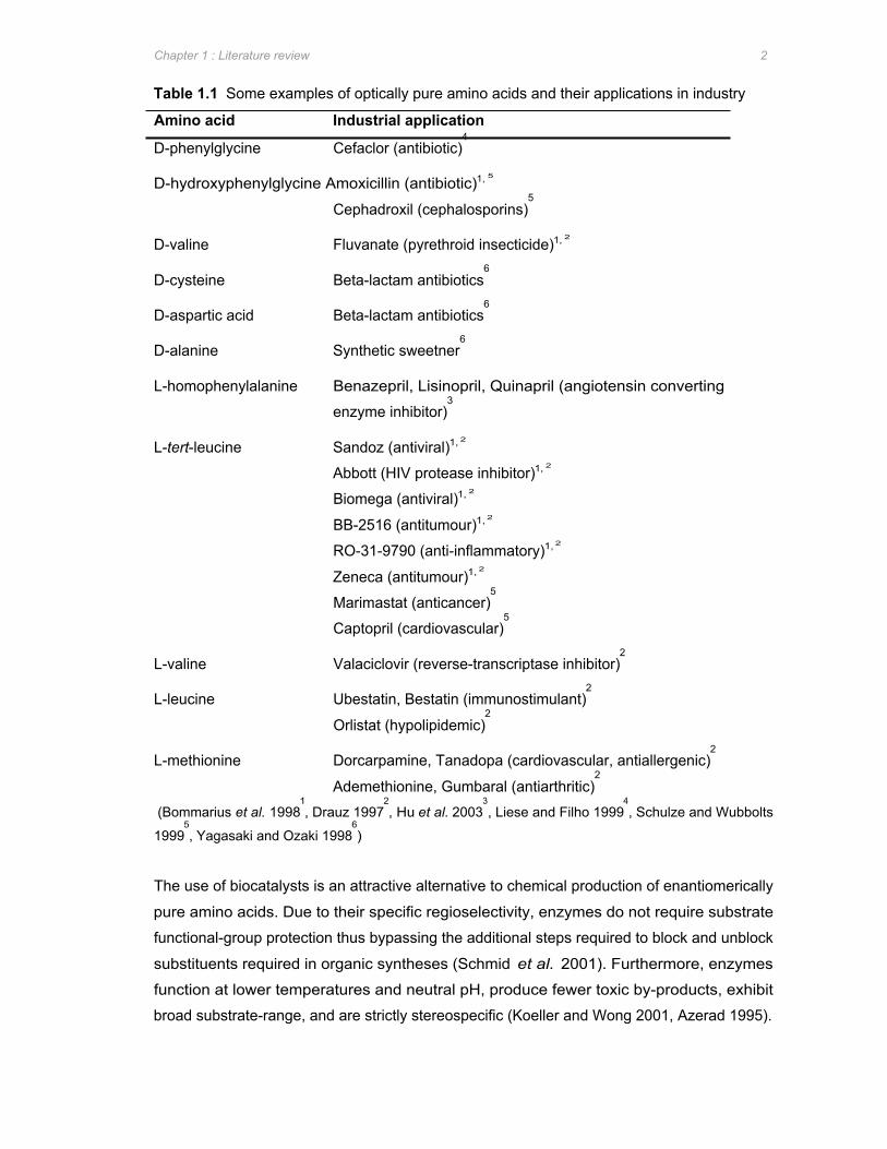
1.1.1 Chiral amino acids in industry Chiral precursors are used to synthesize complex enantiomerically pure compounds.
Interest in enantiomerically pure amino acids in particular is primarily due to the increasing
use of unnatural amino acids in the production of pharmaceuticals or pesticides resulting in
novel activity of the substances produced. A few of the chiral amino acids produced
industrially and the compounds for which they are used as precursors are listed in Table 1.1.
Chapter 1 : Literature review 2
Table 1.1 Some examples of optically pure amino acids and their applications in industry
Amino acid Industrial application
D-phenylglycine Cefaclor (antibiotic)4
D-hydroxyphenylglycine Amoxicillin (antibiotic)1, 5
Cephadroxil (cephalosporins)5
D-valine Fluvanate (pyrethroid insecticide)1, 2
D-cysteine Beta-lactam antibiotics6
D-aspartic acid Beta-lactam antibiotics6
D-alanine Synthetic sweetner6
L-homophenylalanine Benazepril, Lisinopril, Quinapril (angiotensin converting
enzyme inhibitor)3
L-tert-leucine Sandoz (antiviral)1, 2
Abbott (HIV protease inhibitor)1, 2
Biomega (antiviral)1, 2
BB-2516 (antitumour)1, 2
RO-31-9790 (anti-inflammatory)1, 2
Zeneca (antitumour)1, 2
Marimastat (anticancer)5
Captopril (cardiovascular)5
L-valine Valaciclovir (reverse-transcriptase inhibitor)2
L-leucine Ubestatin, Bestatin (immunostimulant)2
Orlistat (hypolipidemic)2
L-methionine Dorcarpamine, Tanadopa (cardiovascular, antiallergenic)2
Ademethionine, Gumbaral (antiarthritic)2
(Bommarius et al. 19981, Drauz 1997
2, Hu et al. 2003
3, Liese and Filho 1999
4, Schulze and Wubbolts
19995, Yagasaki and Ozaki 1998
6)
The use of biocatalysts is an attractive alternative to chemical production of enantiomerically
pure amino acids. Due to their specific regioselectivity, enzymes do not require substrate
functional-group protection thus bypassing the additional steps required to block and unblock
substituents required in organic syntheses (Schmid et al. 2001). Furthermore, enzymes
function at lower temperatures and neutral pH, produce fewer toxic by-products, exhibit
broad substrate-range, and are strictly stereospecific (Koeller and Wong 2001, Azerad 1995).

Chapter 1 : Literature review 3
1.1.2 Hydantoin-hydrolysis for the production of enantiopure amino acids Biocatalytic production of optically pure amino acids is primarily done via dynamic resolutions
and asymmetric synthesis (Schulze and Wubbolts 1999). The most prominent example of
dynamic kinetic resolution is the production of D-p-hydroxyphenylglycine from a benzylic
hydantoin derivative by the hydantoinase, N-carbamylamino acid amidohydrolase (N-
carbamoylase) and racemase enzymes (Schultze and Wubbolts 1999). Hydrolysis of
hydantoin derivatives to form enantiospecific amino acids has several advantages including a
potentially 100% yield with 100% enantiopure amino acid produced from a racemic substrate
(Altenbuchner et al. 2001). In addition, most D,L-5-monosubstituted hydantoin derivatives
can be readily synthesized from inexpensive chemical precursors (Syldatk et al. 1992a).
The use of hydantoin-hydrolysing enzymes has been found to be applicable to the production
of highly lipophilic, silicon-containing amino acids. This non-natural amino acid offers several
advantages including prevention of hydrophobic pocket collapse, higher lipophilicity, and
enhanced stability towards proteolytic degradation (Smith et al. 2001). D- and L-
enantioselective N-carbamoylases have also been shown to recognise the configuration of
the α-carbon as well as the β-carbon of some N-carbamylamino acids (Ogawa et al.
1999a,b). This is extremely valuable in the synthesis complex of α,β-diastereomeric amino
acids which can be found in four stereoisomers and which makes stereospecific synthesis of
these compounds difficult by conventional chemical or enzymatic methodologies (Ogawa et
al. 1999a,b, 2001).
D-p-hydroxyphenylglycine is the most important compound produced by the hydantoinase
process to date (Syldatk et al. 1999) with approximately 2000 tons produced each year
(Ogawa and Shimizu 2002). There are three main processes used by competing industrial
companies to produce D-p-hydroxyphenylglycine. Kanegafuchi Chemical Industries
hydrolyse 5-(4’-hydroxyphenyl)hydantoin with immobilized Bacillus brevis cells containing D-
hydantoinase while Snamprogetti use immobilized dihydropyrimidinase purified from calf
liver. In both processes the resultant N-carbamyl-D-p-hydroxyphenylglycine is then
chemically treated with HNO2 to produce D-p-hydroxyphenylglycine (Syldatk et al. 1990b).
Recordati and Degussa use resting cells of Agrobacterium radiobacter expressing both a D-
hydantoinase and D-carbamoylase thereby negating the necessity of treatment with HNO2
(Syldatk and Pietzsch 1995).
An L-stereoselective industrial process for the production of non-natural aromatic L-amino
acids has been developed by Rütgers using whole cells of Arthrobacter aurescens DSM
3745 and DSM 3747 both of which express an L-hydantoinase, L-carbamoylase, and
racemase enzymes (Syldatk and Pietzsch 1995). Amino acids produced via this method
Chapter 1 : Literature review 4
include L-tryptophan, L-phenylalanine, L-O-benzylserine, p-chloro-phenylalanine, p-
fluorophenyl-alanine, p-nitro-phenylalanine, 1’-naphthylalaine, 2’-naphthylalanine, 3,4-
dimethoxyphenyl-alanine, and 2’-thienylalanine (Syldatk and Pietzsch 1995).
1.2 HYDROLYSIS OF 5-MONOSUBSTITUTED HYDANTOINS
Hydantoin, also known as imidazolidine-2,4-dione or 2,4-diketotetrahydroimidazole, was first
discovered in 1861 by Baeyer as a reduction/hydrogenation product of the naturally occurring
cyclic amide allantoin (Syldatk and Pietzsch 1995). Subsequently, hydantoins have been
isolated from several natural sources such as white shoots of sugar beet and oriental plane
tree buds (Ware 1950). Hydantoin derivatives have long been of interest in industry and are
utilized as anticonvulsants in epilepsy treatment, as herbicides, fungicides, antimicrobial, and
virucidal agents (Syldatk et al. 1992a). However, as discussed above, the microbial
hydrolysis of 5-monosubstituted hydantoins resulting in the production of enantiomerically
pure D- and L-amino acids has gained in interest in recent years (Syldatk et al. 1999).
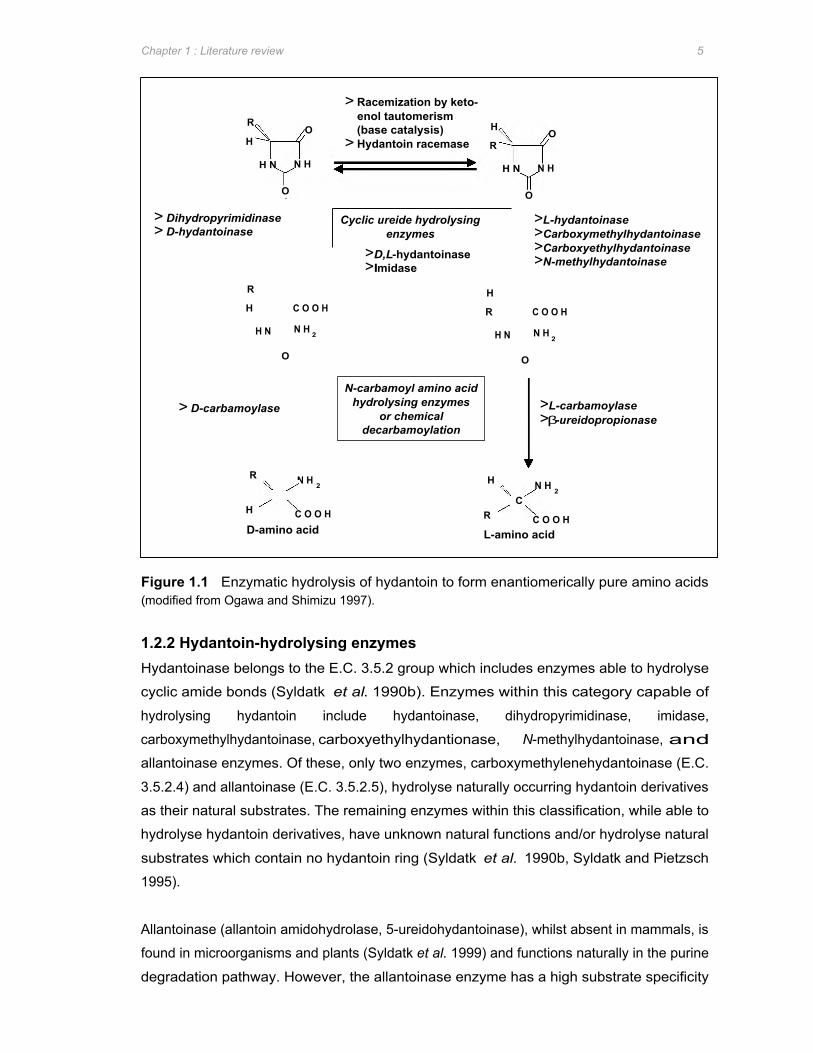
1.2.1 Enzymatic hydrolysis of hydantoins Microbial cleavage of hydantoins is widespread in nature and can be catalysed by a variety
of enzyme systems. Hydrolysis of 5-monosubstituted hydantoin derivatives to the
corresponding amino acids occurs via three reactions (Figure 1.1). Firstly, the ring structure
of the hydantoin molecule is opened by cleavage of the cyclic amide bond at position 2 of the
hydantoin ring. The resultant N-carbamylamino acid is then converted to the corresponding
amino acid either chemically or enzymatically. Depending on the stereospecificities of the
enzymes involved in these two reactions, the end product is either a D- or L-enantiomer.
The third component of the hydrolysis of racemic hydantoins to optically pure amino acids
involves spontaneous or enzymatic racemization of the unreacted hydantoin substrate
(Syldatk and Pietzsch 1995).
Of the enzymes listed in Figure 1.1, the dihydropyrimidinase, hydantoinase (D-, L-, non-
stereospecific), N-carbamoylamino acid amidohydrolase (D- or L-stereospecific), and β-
ureidopropionase are of particular interest due to the generally high activity levels and broad
substrate-selectivities observed.
N H H N H N N H
O O
> Racemization by keto-enol tautomerism
O (base catalysis) > Hydantoin racemase
R
H H
R O
> Dihydropyrimidinase > D-hydantoinase
Cyclic ureide hydrolysing enzymes
>L-hydantoinase >Carboxymethylhydantoinase >Carboxyethylhydantoinase >N-methylhydantoinase
>D,L-hydantoinase >Imidase
R H H C O O H R C O O H
H N N H 2 H N N H 2
D-amino acid L-amino acid
> D-carbamoylase
R
C H
N-carbamoyl amino acid hydrolysing enzymes
or chemical decarbamoylation
>L-carbamoylase >β-ureidopropionase
O
N H 2
O
C O O H R
H
C
C O O H
N H 2
Chapter 1 : Literature review 5
Figure 1.1 Enzymatic hydrolysis of hydantoin to form enantiomerically pure amino acids (modified from Ogawa and Shimizu 1997).
1.2.2 Hydantoin-hydrolysing enzymes Hydantoinase belongs to the E.C. 3.5.2 group which includes enzymes able to hydrolyse
cyclic amide bonds (Syldatk et al. 1990b). Enzymes within this category capable of
hydrolysing hydantoin include hydantoinase, dihydropyrimidinase, imidase,
carboxymethylhydantoinase, carboxyethylhydantionase, N-methylhydantoinase, and
allantoinase enzymes. Of these, only two enzymes, carboxymethylenehydantoinase (E.C.
3.5.2.4) and allantoinase (E.C. 3.5.2.5), hydrolyse naturally occurring hydantoin derivatives
as their natural substrates. The remaining enzymes within this classification, while able to
hydrolyse hydantoin derivatives, have unknown natural functions and/or hydrolyse natural
substrates which contain no hydantoin ring (Syldatk et al. 1990b, Syldatk and Pietzsch
1995).
Allantoinase (allantoin amidohydrolase, 5-ureidohydantoinase), whilst absent in mammals, is
found in microorganisms and plants (Syldatk et al. 1999) and functions naturally in the purine
degradation pathway. However, the allantoinase enzyme has a high substrate specificity

Chapter 1 : Literature review 6
and low enantioselectivity thus biotechnological application of this enzyme is limited (Syldatk
et al. 1999). The catalytic function of carboxymethylenehydantoinase (L-carboxymethyl-
hydantoin amidohydrolase) is postulated to be involved in a side reaction in the metabolism
of pyrimidines (Syldatk and Pietzsch 1995) but, besides the ability to hydrolyse carboxy-
methylhydantoin to L-aspartic acid, no investigation of the substrate spectrum or enzymatic
properties of this enzyme has been done (Syldatk et al. 1999). N-methylhydantoin
amidohydrolase (N-methylhydantoinase) is an ATP-dependent enzyme that hydrolyses N-
methylhydantoin to N-carbamoylsarcosine which is part of the creatinine degradation
pathway (Ogawa and Shimizu 1997). N-methylhydantoinase is able to hydrolyse hydantoin
derivatives L-specifically but is unable to hydrolyse dihydropyrimidines (Ogawa et al. 1995a),
and due to its ATP-dependence it is an unlikely candidate for industrial production of optically
pure amino acids (Syldatk et al. 1999). Imidase functions in the initial part of the cyclic amide
metabolic pathway. Whilst this enzyme was shown to hydrolyse simple cyclic imides and
cyclic ureides such as dihydrouracil and hydantoin, bulky imides and 5-monosubstituted
hydantoins were not hydrolysed (Soong et al. 2001). Lastly, carboxyethylhydantoinase has
been shown to be involved in the degradation of histidine and is classified as L-
stereoselective. However, very little research has been done on this enzyme to date
(Syldatk et al. 1999).
Of primary interest when considering enzymes capable of hydrolysing hydantoin derivatives
to the corresponding N-carbamylamino acids are the dihydropyrimidinase and hydantoinase
enzymes. Hydantoinase enzymes have been found in a wide variety of organisms including
plants (Eadie et al. 1949 cited in Syldatk et al. 1999, Morin 1993), animals (Bernheim and
Bernheim 1946, Wada 1934 cited in Syldatk et al. 1999, Cecere et al. 1975) and bacteria.
Hydantoinase enzymes have predominantly been characterized from bacterial isolates
including species such as Agrobacterium (Hils et al. 2001, Durham and Weber 1995,
Grifantini et al. 1998, Hartley et al. 1998), Arthobacter (Moller et al. 1988, Siemann et al.
1999, Gross et al. 1990, May et al. 1998a, Volkel and Wagner 1995), Bacillus (Luksa et al.
1997, Kim et al. 1997, Mukohara et al. 1994, Park et al. 1998), Pseudomonas (Buchanan et
al. 2001, Gokhale et al. 1996, Chien et al. 1998, LaPointe et al. 1994, Yokozeki et al. 1987b,
Sudge et al. 1998, Ishikawa et al. 1997), Thermus (Abendroth et al. 2000a), Flavobacterium
(Yokozeki et al. 1987d), and Methanococcus (Chung et al. 2002), and have been shown to
hydrolyse 5-monosubstituted hydantoin derivatives effectively.
Traditionally, hydantoinase enzymes have been classified according to their hydrolytic
enantioselectivity and grouped as L-, D-, or non-enantioselective hydantoinases respectively.
However, this is misleading as it implies that the hydantoinase enzymes differ solely in their
enantioselectivities which is not the case. Furthermore, enantioselectivity of hydantoinase
enzymes from Arthrobacter sp. DSM 3745 and Flavobacterium sp. have been shown to be

Chapter 1 : Literature review 7
substrate dependent (May et al. 1998a, Yokozeki et al. 1987e cited in Syldatk et al. 1999).
However, for the purposes of this literature review, hydantoinases have been grouped based
on their predominant enantioselectivity.
1.2.2.1 D-enantioselective hydantoinases D-enantioselective hydantoinases are often considered synonymous with
dihydropyrimidinases (Syldatk et al. 1999). This is due to the finding that isolated
dihydropyrimidinases were capable of hydrolysing 5-monsubstituted hydantoin derivatives as
well as their natural substrate dihydropyrimidines (Xu and West 1994). Also, several D-
hydantoinases which have subsequently been isolated are able to hydrolyse dihydrouracil as
a substrate (Lee et al. 1995, Sharma and Vohra 1997, Siemann et al. 1999, Durham and
Weber 1995, Ogawa et al. 1995b, Sudge et al. 1998). In contrast, the dihydropyrimidinase
from Clostridium uracilicum has been shown to be unable to hydrolyse hydantoin (Campbell
1958 cited in Syldatk et al. 1999) and an hydantoinase was isolated from Agrobacterium sp.
IP I-671 which was unable to hydrolyse dihydrouracil (Runser and Meyer 1993). Thus, while
the names D-hydantoinase and dihydropyrimidinase are often used interchangeably, these
are in fact two separate enzymes with different catalytic capabilities. D-hydantoinase
enzymes from a wide variety of bacterial species have been described in literature. Some of
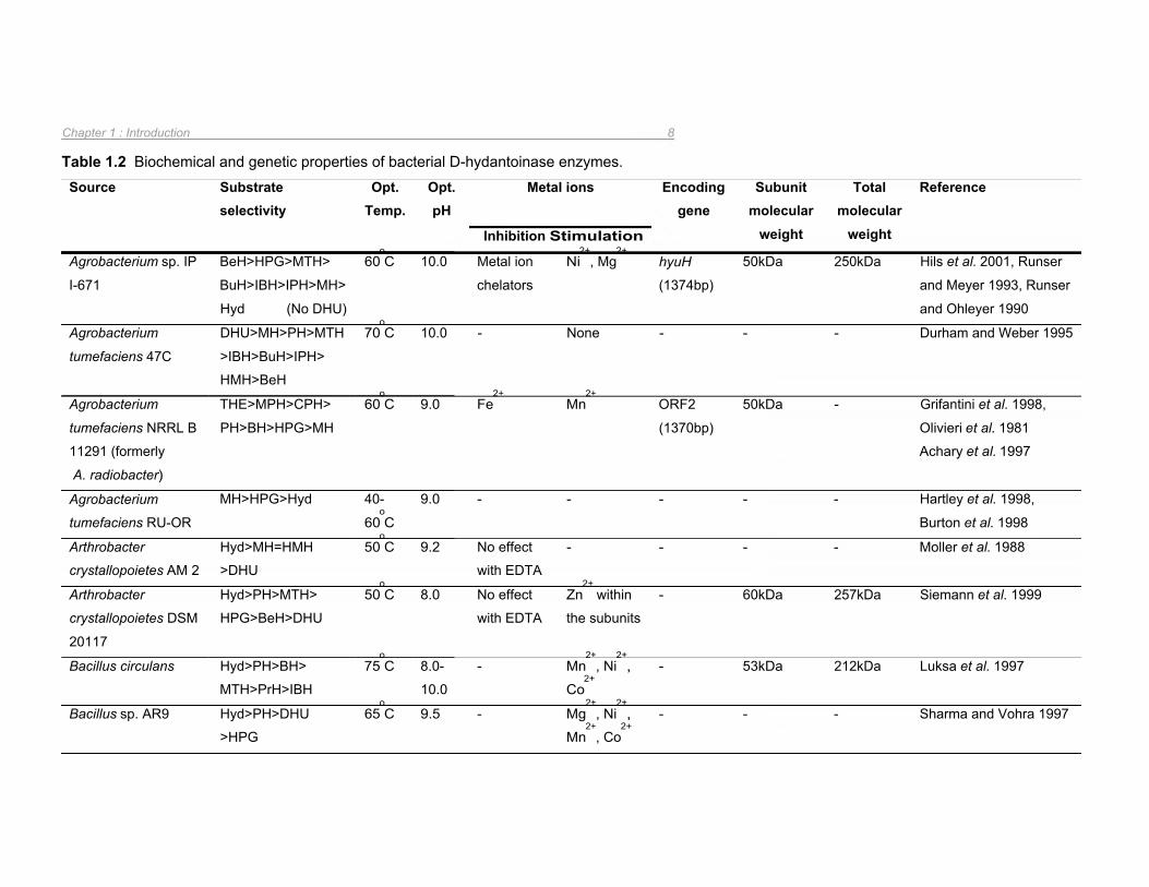
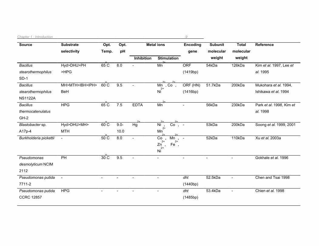
the characteristics of these enzymes are summarized in Table 1.2.
With the exception of the D-hydantoinases from Bacillus stearothermophilus SD-1 and
Burkholderia pickettii, which are dimeric in structure (Lee et al. 1995, Xu et al. 2003a), the
remaining D-hydantoinases analysed were found to form tetramers (Table 1.2). The
optimum temperature for the catalytic activity of these enzymes ranged from 30oC for
Pseudomonas desmolyticum NCIM 2212 (Gokhale et al. 1996) to 75oC for Bacillus circulans
(Luksa et al. 1997) with the majority of the D-hydantoinases functioning optimally at ~60oC
(Table 1.2). With respect to optimal pH for hydantoin hydrolysis by D-hydantoinases, the
highest activities generally occurred at alkali pH (pH 8-10) (Table 1.2).
In all instances where metal ion dependency has been investigated, D-hydantoinases have
been observed to require divalent metal ions as co-factors (Table 1.2). Consequently, these
enzymes are sensitive to metal ion chelators and are stimulated by metal ions such as Mn2+
and Co2+. This metal ion dependency was verified by crystallographic analysis of D-
hydantoinases from Thermus sp. (Abendroth et al. 2002a), Burkholderia pickettii (Xu et al.
2003a), and B. stearothermophilus SD-1 (Cheon et al. 2002) in which two metal ions were
identified within the catalytic site. These studies are described in greater detail in Section
1.3.1. In most cases, the co-factor was Zn2+
which could be replaced by other divalent
transition group metals such as Mn2+
or Co2+
(Abendroth et al. 2002a).
Encoding gene
Subunit molecular
weight
Total molecular
weight
Reference
Inhibition Stimulation
Metal ions
Ni2+
, Mg2+
hyuH
(1374bp)
50kDa 250kDa Hils et al. 2001, Runser
and Meyer 1993, Runser
and Ohleyer 1990
Metal ion
chelators
Fe2+
Mn2+
ORF2
(1370bp)
50kDa - Grifantini et al. 1998,
Olivieri et al. 1981
Achary et al. 1997
- - - - Moller et al. 1988 No effect
with EDTA
- 60kDa 257kDa Siemann et al. 1999 Zn2+
within No effect
with EDTA the subunits
- Mn2+
, Ni2+
,
Co2+
- 53kDa 212kDa Luksa et al. 1997
- Mg2+
, Ni2+
,
Mn2+
, Co2+
- - - Sharma and Vohra 1997
Chapter 1 : Introduction 8
Table 1.2 Biochemical and genetic properties of bacterial D-hydantoinase enzymes. Source Substrate
selectivity Opt.
Temp. Opt. pH
Agrobacterium sp. IP
I-671
BeH>HPG>MTH>
BuH>IBH>IPH>MH>
Hyd (No DHU)
60oC 10.0
Agrobacterium
tumefaciens 47C
DHU>MH>PH>MTH
>IBH>BuH>IPH>
HMH>BeH
70oC 10.0
Agrobacterium
tumefaciens NRRL B
11291 (formerly
A. radiobacter)
THE>MPH>CPH>
PH>BH>HPG>MH
60oC 9.0
Agrobacterium
tumefaciens RU-OR
MH>HPG>Hyd 40-
60oC
9.0
Arthrobacter
crystallopoietes AM 2
Hyd>MH=HMH
>DHU
50oC 9.2
Arthrobacter
crystallopoietes DSM
20117
Hyd>PH>MTH>
HPG>BeH>DHU
50oC 8.0
Bacillus circulans Hyd>PH>BH>
MTH>PrH>IBH
75oC 8.0-
10.0
Bacillus sp. AR9 Hyd>PH>DHU
>HPG
65oC 9.5
- None - - - Durham and Weber 1995
- - - - - Hartley et al. 1998,
Burton et al. 1998
Chapter 1 : Introduction 9
Source Substrate selectivity
Opt. Temp.
Opt. pH
Metal ions Encoding gene
Subunit molecular
weight
Total molecular
weight
Reference
Inhibition Stimulation
Bacillus
stearothermophilus
SD-1
Hyd>DHU>PH
>HPG
65oC 8.0 - Mn
2+ ORF
(1419bp)
54kDa 126kDa Kim et al. 1997, Lee et
al. 1995
Bacillus
stearothermophilus
NS1122A
MH>MTH>IBH>IPH>
BeH
60oC 9.5 - Mn
2+, Co
2+,
Ni2+
ORF (HN)
(1416bp)
51.7kDa 200kDa Mukohara et al. 1994,
Ishikawa et al. 1994
Bacillus
thermocatenulatus
GH-2
HPG 65oC 7.5 EDTA Mn
2+ - 56kDa 230kDa Park et al. 1998, Kim et
al. 1998
Blastobacter sp.
A17p-4
Hyd>DHU>MH>
MTH
60oC 9.0-
10.0
Hg2+
Ni2+
, Co2+
,
Mn2+
- 53kDa 200kDa Soong et al. 1999, 2001
Burkholderia pickettii - 50oC 8.0 - Co
2+, Mn
2+,
Zn2+
, Fe2+
,
Ni2+
- 52kDa 110kDa Xu et al. 2003a
Pseudomonas
desmolyticum NCIM
2112
PH 30oC 9.5 - - - - - Gokhale et al. 1996
Pseudomonas putida
7711-2
- - - - - dht
(1440bp)
52.5kDa - Chen and Tsai 1998
Pseudomonas putida
CCRC 12857
HPG - - - - dht
(1485bp)
53.4kDa - Chien et al. 1998

Chapter 1 : Introduction 10
Source Substrate selectivity
Opt. Temp.
Opt. pH
Metal ions Encoding gene
Subunit molecular
weight
Total molecular
weight
Reference
Inhibition Stimulation
Pseudomonas putida
DSM 84
DHU>methionine
>IPH (Hyd=poor)
55oC 9.0 Cu
2+ Mn
2+, Fe
2+ ORF1
(1104bp)
60kDa 230kDa Morin et al. 1986a,b,
LaPointe et al. 1994
Pseudomonas putida
IFO 12996
DHU>>MH>Hyd 45-
55oC
8.0-
9.0
Metal ions
chelators
- - - 190kDa Takahashi et al. 1978,
Ogawa et al. 1994c
Pseudomonas sp. AJ-
11220
CEH>MTH=hyd>CH
>PH>HPG>MH>BeH
(very low DHU)
43oC 8.0 Hg
2+ Mg
2+, Mn
2+,
Co2+
, Ni2+
,
Cu2+
- - Yokozeki et al. 1987b
Pseudomonas sp.
KBEL 101
HPG 30oC 8.0 - - - - - Kim and Kim 1993
Pseudomonas sp.
NCIM 5109
DHU>Hyd>PH>
HPG
30oC 9.0-
9.5
- - - - - Sudge et al. 1998
Thermus sp. Prefers 5-phenylic
substituted
hydantoins
- - - Mn2+
, Zn2+
within the
subunits
- - 50kDa Abendroth et al. 2000a,
2002a
(BeH= 5-benzylhydantoin; BuH= 5-(sec)-butylhydantoin; CEH= 5-cyanoethylhydantoin; CH= 5-carbamylethylhydantoin; CPH= chlorophenylhydantoin; DHU= dihydrouracil; HMH=hydroxymethylhydantoin; HPG= 5-hydroxyphenylglycine; IBH= 5-isobutylhydantoin; IPH= 5-isopropylhydantoin; MH= 5-methylhydantoin; MPH= methyoxyphenylhydantoin; MTH= 5-(2-methylthioethyl)hydantoin; PH= 5-phenylhydantoin; PrH= propylhydantoin; THE= thienylhydantoin, Opt. : optimal)
Chapter 1 : Introduction 11
The genes encoding D-hydantoinase enzymes have been isolated from P. putida DSM 84, B.
stearothermophilus SD-1, Agrobacterium sp. IP I-671, A. tumefaciens NRRL B11291, P.
putida CCRC 12857, P. putida 7711-2, and B. stearothermophilus NS1122A (Table 1.2).
The D-hydantoinase-encoding gene from Agrobacterium sp. IP I-671 was isolated by
screening a genomic phage library by plaque hybridisation with the previously isolated, DIG-
labelled N-carbamoylase gene as a probe (Hils et al. 2001). The genes encoding the
hydantoinase, N-carbamoylase and racemase enzymes in this strain were located as a
cluster on a native 190kb plasmid (Hils et al. 2001). However, the hydantoinase gene
isolated was not the only gene encoding for a hydantoin-hydrolysing enzyme present in
Agrobacterium sp. IP I-671 as inactivation of hyuH resulted in 15% residual D-hydantoinase
activity (Hils et al. 2001).
The amino acid sequence of the D-hydantoinase from B. stearothermophilus strains SD-1
(Kim et al. 1997) and NS1122A (Mukohara et al. 1994) were almost identical with the
majority of the few variations observed located in the C-terminal region (Kim et al. 1997).
However, despite the high similarity between these two enzymes, their biochemical
properties differed significantly including variations in oligomeric structure with the
hydantoinase from B. stearothermophilus SD-1 forming a dimer whilst the B.
stearothermophilus NS1122A hydantoinase formed a tetramer (Kim et al. 1997). This
suggested that the C-terminal region has an important role in the structural and/or catalytic
properties of these enzymes. The D-hydantoinase encoding gene from Bacillus
thermocatenulatus GH2, which was found have an identical nucleotide sequence to the
corresponding gene in B. stearothermophilus NS1122A, was digested with exonuclease III in
order to determine if the C-terminal region was important for enzyme activity (Kim and Kim
1998). Enzyme activity was still detected when up to 40 amino acid residues had been
deleted implying that the non-homologous C-terminal regions are not involved in the catalysis
reaction (Kim and Kim 1998). However, deletion of 11 to 12 amino acids from the C-terminal
end of the B. thermocatenulatus GH2 D-hydantoinase gave rise to a dimeric protein, as
opposed to the parental tetramer, while the dimeric nature of the B. stearothermophilus SD-1
hydantoinase remained unchanged (Kim and Kim 1998).
In contrast to the Bacillus hydantoinases, the C-terminus of the D-hydantoinase from P.
putida CCRC 12857 was found to be involved in the catalytic activity of the enzyme as
deletion of 32 amino acids from the C-terminal end resulted in loss of hydantoinase activity in
this strain (Chien et al. 1998). It is interesting that the D-hydantoinases for which the amino
acid sequences are known all consist of more than 450 amino acid residues except for the
hydantoinase from P. putida DSM 84 which was approximately 100 residues shorter in the
carboxyl terminus (LaPointe et al. 1994, Chien et al. 1998).
Chapter 1 : Introduction 12
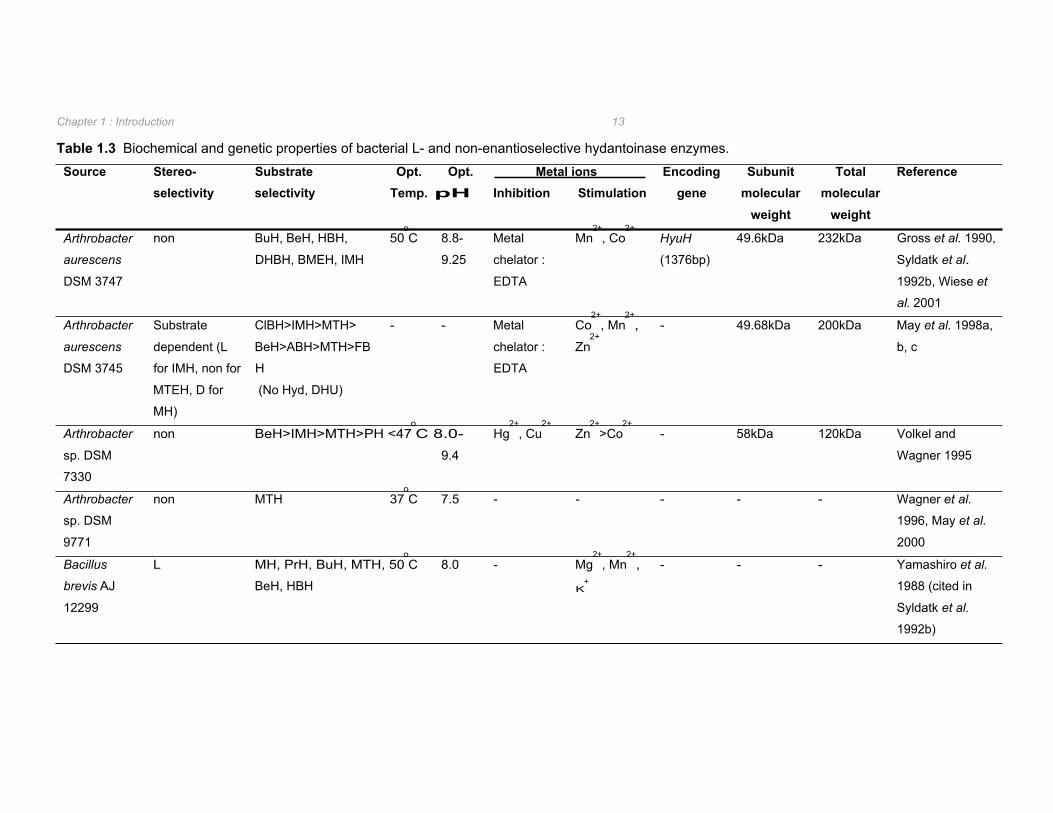
1.2.2.2. L- and non-enantioselective hydantoinases Production of L-amino acids from 5-monosubstituted hydantoin derivatives is performed by L-
enantioselective N-carbamoylases in conjunction with either an L- or non-enantioselective
hydantoinase. Significantly fewer L- and non-enantioselective hydantoinases have been
isolated in comparison to D-hydantoinases (Table 1.2 vs. Table 1.3). L-hydantoinases have
been reported from Bacillus (Yamashiro et al. 1988 cited in Syldatk et al. 1992b),
Flavobacterium (Yokozeki et al. 1987e cited in Syldatk et al. 1992b), and Methanococcus
(Chung et al. 2002) species whilst non-enantioselective hydantoinases were found in
Arthrobacter (Gross et al. 1990, Volkel and Wagner 1995, Wagner et al. 1996) and
Pseudomonas (Ishikawa et al. 1997) species.
The hydantoinases from Arthrobacter aurescens DSM 3745 and Flavobacterium sp. cannot
be conveniently classified as D-, L-, or non-enantioselective as the enantioselectivity
observed in these strains appears to be substrate dependent (May et al. 1998a, Yokozeki et
al. 1987e cited in Syldatk et al. 1999). Specifically, the hydantoinase from Flavobacterium
hydrolysed indolylmethylhydantoin L-enantioselectively and benzyloxymethylhydantoin D-
selectively (Yokozeki et al. 1987e cited in Syldatk et al. 1999). In the case of the
Arthrobacter hydantoinase, strict L-enantioselectivity was observed with D,L-5-indolylmethyl-
hydantoin whilst, when supplied with 5-(2-methylthioethyl)-hydantoin the D-enantiomer
intermediate was produced in 3-fold excess to that of the corresponding L-enantiomer (May
et al. 1998a). The substrate-dependent enantioselectivity of the hydantoinase from A.
aurescens DSM 3745, along with the inability to hydrolyse unsubstituted hydantoin
suggested that this enzyme should be classified as a new member of the EC group 3.5.2
(May et al. 1998a). This was substantiated by phylogenetic analysis of this enzyme with
dihydropyrimidinases, allantoinases, ureases, and dihydroorotases in which it forms a novel
branch of the phylogenetic tree (May et al. 1998d). Selected characteristic biochemical and
genetic properties of the L- and non-enantioselective hydantoinases reported in literature are
summarized in Table 1.3.
As with the D-hydantoinases, the optimum pH for the catalytic activity of L- and non-
enantioselective hydantoinases was found to be at slightly alkaline to alkaline pH (Table 1.3).
Optimum temperature ranged from 37oC for the non-selective hydantoinase from
Arthrobacter sp. DSM 9771 through to 80oC for the L-hydantoinase from Methanococcus
jannaschii (Table 1.3).
Source Stereo- selectivity
Substrate selectivity
Subunit Inhibition Stimulation gene molecular
weight
Total molecular
weight
Opt. Opt. Temp. pH
Metal ions Encoding Reference
Chapter 1 : Introduction 13
Table 1.3 Biochemical and genetic properties of bacterial L- and non-enantioselective hydantoinase enzymes.
Arthrobacter non BuH, BeH, HBH, 50oC 8.8- Metal Mn
2+, Co
2+ HyuH 49.6kDa 232kDa Gross et al. 1990,
aurescens DHBH, BMEH, IMH 9.25 chelator : (1376bp) Syldatk et al.
DSM 3747 EDTA 1992b, Wiese et
al. 2001
Arthrobacter
aurescens
DSM 3745
Substrate
dependent (L
for IMH, non for
MTEH, D for
MH)
ClBH>IMH>MTH>
BeH>ABH>MTH>FB
H
(No Hyd, DHU)
- - Metal
chelator :
EDTA
Co2+
, Mn2+
,
Zn2+
- 49.68kDa 200kDa May et al. 1998a,
b, c
Arthrobacter non BeH>IMH>MTH>PH <47oC 8.0- Hg
2+, Cu
2+ Zn
2+>Co
2+ - 58kDa 120kDa Volkel and
sp. DSM 9.4 Wagner 1995
7330
Arthrobacter non MTH 37oC 7.5 - - - - - Wagner et al.
sp. DSM 1996, May et al.
9771 2000
Bacillus L MH, PrH, BuH, MTH, 50oC 8.0 - Mg
2+, Mn
2+, - - - Yamashiro et al.
brevis AJ BeH, HBH K+ 1988 (cited in
12299 Syldatk et al.
1992b)
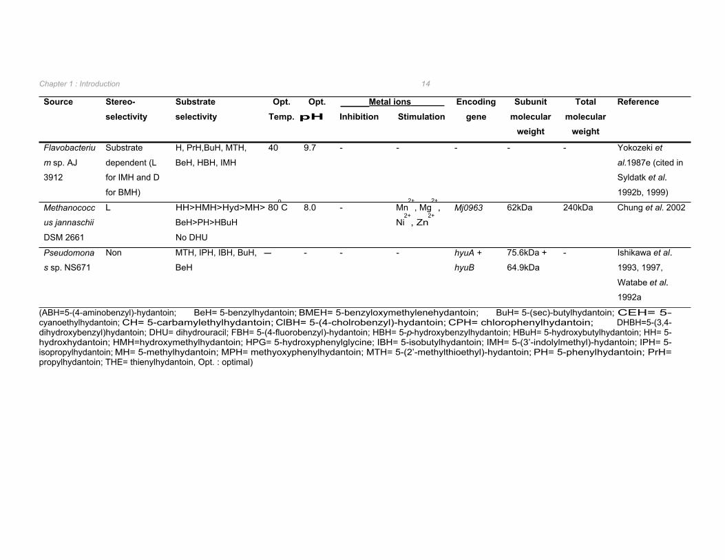
Source Stereo- selectivity
Substrate selectivity
Subunit Inhibition Stimulation gene molecular
weight
Total molecular
weight
Opt. Opt. Temp. pH
Metal ions Encoding Reference
Chapter 1 : Introduction 14
Flavobacteriu Substrate H, PrH,BuH, MTH, 40 9.7 - - - - - Yokozeki et
m sp. AJ dependent (L BeH, HBH, IMH al.1987e (cited in
3912 for IMH and D Syldatk et al.
for BMH) 1992b, 1999)
Methanococc L HH>HMH>Hyd>MH> 80oC 8.0 - Mn
2+, Mg
2+, Mj0963 62kDa 240kDa Chung et al. 2002
us jannaschii BeH>PH>HBuH Ni2+
, Zn2+
DSM 2661 No DHU
Pseudomona Non MTH, IPH, IBH, BuH, - - - - hyuA + 75.6kDa + - Ishikawa et al.
s sp. NS671 BeH hyuB 64.9kDa 1993, 1997,
Watabe et al.
1992a
(ABH=5-(4-aminobenzyl)-hydantoin; BeH= 5-benzylhydantoin; BMEH= 5-benzyloxymethylenehydantoin; BuH= 5-(sec)-butylhydantoin; CEH= 5- cyanoethylhydantoin; CH= 5-carbamylethylhydantoin; ClBH= 5-(4-cholrobenzyl)-hydantoin; CPH= chlorophenylhydantoin; DHBH=5-(3,4- dihydroxybenzyl)hydantoin; DHU= dihydrouracil; FBH= 5-(4-fluorobenzyl)-hydantoin; HBH= 5-p-hydroxybenzylhydantoin; HBuH= 5-hydroxybutylhydantoin; HH= 5-hydroxhydantoin; HMH=hydroxymethylhydantoin; HPG= 5-hydroxyphenylglycine; IBH= 5-isobutylhydantoin; IMH= 5-(3’-indolylmethyl)-hydantoin; IPH= 5-isopropylhydantoin; MH= 5-methylhydantoin; MPH= methyoxyphenylhydantoin; MTH= 5-(2’-methylthioethyl)-hydantoin; PH= 5-phenylhydantoin; PrH= propylhydantoin; THE= thienylhydantoin, Opt. : optimal)
Chapter 1 : Introduction 15
Metal ion dependency was also observed for both L- and non-enantioselective
hydantoinases (Table 1.3). The hydantoinase from A. aurescens DSM 3745 was subjected
to atomic adsorption spectrometry and inductive coupled plasma-atomic emission to
determine the metal ion content of the enzyme. From this analysis, 10mol zinc ions per 1mol
of active enzyme was observed and removal of these zinc ions resulted not only in loss of
activity but dissociation of the enzymes into its subunits as well (May et al. 1998b,c). Thus
zinc was found to be essential not only for catalytic activity but also for the stabilization of the
active quaternary structure of the hydantoinase (May et al. 1998b). Crystallographic analysis
of the A. aurescens DSM 3745 hydantoinase verified the presence of the metal ions in the
active site of the enzyme as is the case for the D-hydantoinases investigated (Abendroth et
al. 2002b). The function of the zinc ions within the active site is postulated to be activation of
water by lowering its pKa thus enabling the hydroxyl ion to perform nucleophilic attack on the
amide bond resulting in the hydrolytic cleavage of the hydantoin ring (May et al. 1998b,
Abendroth et al. 2002b).
The only L-enantioselective hydantoinase for which the encoding sequence has been
determined is that from M. jannaschii DSM 2661 (Table 1.3). This gene was amplified from
chromosomal DNA using sequence data obtained from the complete genome sequences of
M. jannaschii available on the NCBI database (Chung et al. 2002). M. jannaschii is a
thermostable methanogen with an optimal growth temperature of 85oC. The hydantoinase
from this organism was found to be extremely thermostable with an optimal catalytic activity
at 80oC and a half-life of 100 minutes at 90
oC (Chung et al. 2002).
The nucleotide sequence of the non-enantioselective hydantoinase from A. aurescens DSM
3747 was determined by screening a genomic library, prepared in phage λ-RESIII, with a
radiolabeled oligonucleotide probe deduced from the N-terminal amino acid sequence of the
hydantoinase purified from A. aurescens DSM 3745 (Wiese et al. 2001). Analysis of the
7,637bp plasmid thus isolated revealed the presence of hyuP, hyuA, hyuH, and hyuC genes
encoding for a putative permease, hydantoin racemase, hydantoinase, and N-carbamoylase
respectively. These genes were found to be arranged in an operon and orientated in the
same direction. The close proximity of the hyu genes to one another indicated that they are
co-transcribed into a polycistronic mRNA (Wiese et al. 2001).
The hydantoin-hydrolysing enzymes from Pseudomonas sp. NS671 were located on a
172Kbp native plasmid on which two genes, hyuA and hyuB, were found to encode the
hydantoinase enzyme (Watabe et al. 1992a). Thus, in contrast to all other reported
hydantoinases in which the enzymes are homodimers or homotetramers, the hydantoinase
from Pseudomonas sp. NS671 is comprised of two different subunits. The gene encoding
Chapter 1 : Introduction 16
the L-enantioselective N-carbamoylase was located in close proximity to the hyuA and hyuB
genes forming an operon. As with A. aurescens DSM 3747, these genes were orientated in
the same direction and were closely spaced suggesting that these open reading frames may
be translationally coupled (Watabe et al. 1992a).
1.2.3 N-carbamylamino acid-hydrolysing enzymes N-carbamylamino acids can be converted to their corresponding amino acids by treatment
with HNO2 (Snamprogetti and Kaneko processes for production of D-p-hydroxyphenyl-
hydantoin). However, this approach is not viable for amino acids such as tryptophan,
citrulline, or pyridylalanine which are unstable when treated with acid (Syldatk and Pietzsch
1995). The enzymatic cleavage of N-carbamylamino acids by N-carbamoylases (N-
carbamylamino acid amidohydrolases) provides an alternative approach. In addition, by
application of a strictly enantioselective N-carbamoylase, enantiopure amino acids can still
be produced even if a non-enantioselective hydantoinase is used for the hydrolysis of
hydantoin.
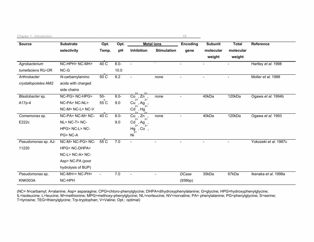
N-carbamoylases have been isolated from several bacterial sources (Table 1.4 and Table
1.5) and are classified as L- or D-enantioselective. No substrate dependent or non-
enantioselective N-carbamoylases have been reported to date. N-carbamoylases occur in
conjunction with D-, L-, or non-enantioselective hydantoinases with the exception of
Comomonas sp. E222c in which a D-carbamoylase exists independently of an hydantoinase
(Ogawa et al. 1993).
As D-carbamoylases were shown to be associated with dihydropyrimidinases, the natural
function of these enzymes was originally thought to be that of a β-ureidopropionase which is
responsible for the cleavage of N-carbamyl-β-alanine (β-ureidopropionic acid) to β-alanine in
the pyrimidine degradation pathway (Syldatk and Pietzsch 1995). This did indeed seem to
be the case with the D-enantioselective N-carbamoylase from Arthrobacter crytallopoietes
which was able to produce β-alanine from dihydrouracil (Moller et al. 1988). However, the D-
enantioselective N-carbamoylase from Blastobacter sp. A17p-4 was unable to hydrolyse β-
ureidopropionic acid (Ogawa and Shimizu 1997). Subsequent to this finding, the D-
enantioselective N-carbamoylases from Pseudomonas sp. KNK003A, and Agrobacterium
tumefaciens AM10 were also shown to be unable to hydrolyse β-ureidopropionic acid
(Ikenaka et al. 1998a, Sareen et al. 2001a). In order to determine if β-ureidopropionase does
cleave N-carbamylamino acids and with what enantioselectivity, the β-ureidopropionase from
Pseudomonas putida IFO 12996 was purified and analysed (Ogawa and Shimizu 1994).
From these studies, it was found that the β-ureidopropionase not only hydrolysed N-
carbamyl-α-amino acids but N-carbamyl-β- and γ-amino acids as well (Ogawa and Shimizu
Chapter 1 : Introduction 17
1994). Hydrolysis of N-carbamyl-α-amino acids by the β-ureidopropionase was shown to be
strictly L-enantioselective (Ogawa and Shimizu 1994) suggesting that the natural function of
L- rather than D-carbamoylases is in pyrimidine degradation. However, L-enantioselective
N-carbamoylases from Alcaligenes xyloxidans AKU 990, Arthrobacter aurescens DSM 3747,
Pseudomonas sp. NS671, and Bacillus kaustophilus CCRC11223 are unable to hydrolyse
the natural substrate of β-ureidopropionase (Ogawa et al. 1995c, Wilms et al. 1999, Ishikawa
et al. 1996, Hu et al. 2003). Furthermore, a β-ureidopropionase recently isolated from
Brevibacillus agri NCHU1002, while hydrolysing N-carbamyl-β-alanine, did not hydrolyse N-
carbamyl-α-amino acids (Kao and Hsu 2003). Thus the natural function of both L- and D-
enantioselective N-carbamoylases remains unclear.
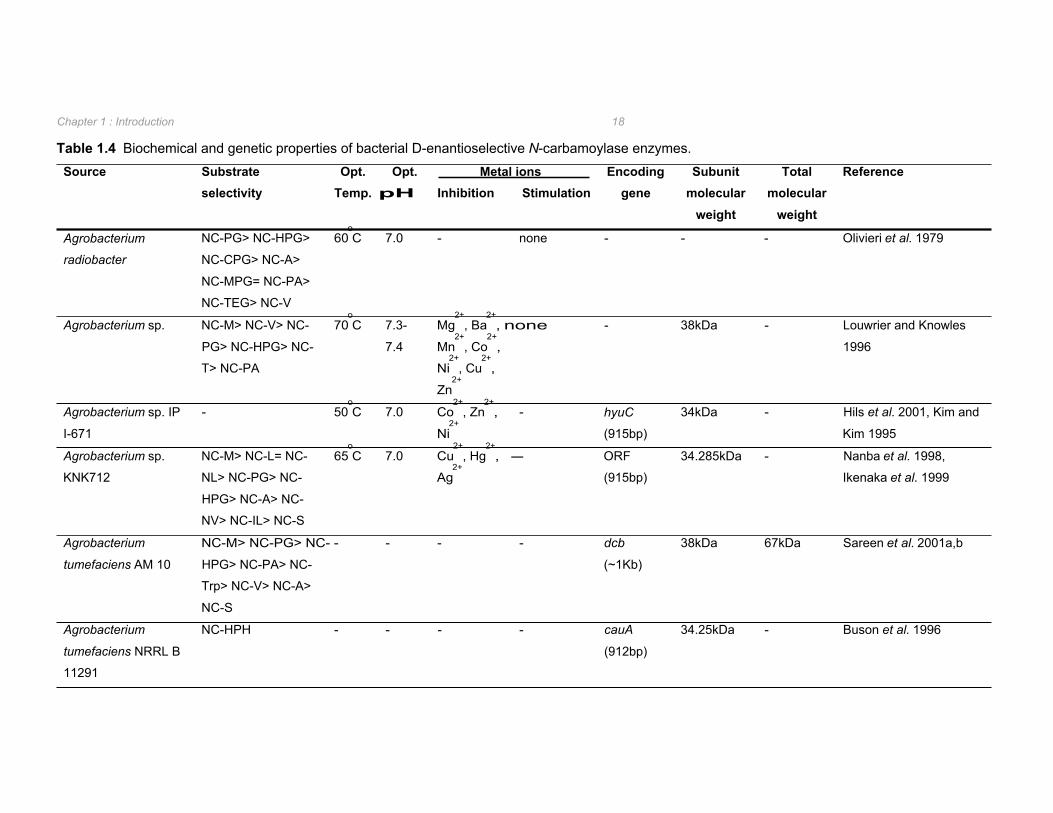
1.2.3.1 D-enantioselective N-carbamoylases D-enantioselective N-carbamoylases have been found in several Agrobacterium isolates, as
well as in Pseudomonas, Blastobacter, Comamonas, and Arthrobacter species. A selection
of the biochemical and genetic properties of these enzymes are listed in Table 1.4. D-
carbamoylases are found as homodimers or homotrimers with optimum pH and temperatures
for N-carbamylamino acid hydrolysis ranging from pH 7 to 9.2, and 40oC to 70
oC respectively
(Table 1.4). Inhibition of D-carbamoylases by metal ions has been well documented.
However, unlike hydantoinases, D-carbamoylases do not appear to be metalloenzymes as
indicated by the absence of sensitivity to metal ion chelators and minimal stimulation of
enzyme activity subsequent to the addition of metal ions to the biocatalytic reaction (Table
1.4).
The genes encoding for D-carbamoylases in Agrobacterium sp. KNK712 and Agrobacterium
tumefaciens NRRL B11291 were isolated by screening of the relevant genomic libraries
(Nanba et al. 1998, Buson et al. 1996). The gene sequences were observed to be similar
with nine residues difference between the amino acid sequences of the two D-
carbamoylases (Nanba et al. 1998). Agrobacterium sp. IP I-671 D-carbamoylase was also
found to have a high amino acid sequence identity with the above D-carbamoylases. This
was not surprising considering the D-carbamoylase encoding gene was amplified by PCR
from the chromosome of Agrobacterium sp. IP I-671 using oligonucleotides designed from
the aligned sequences of the previously isolated D-carbamoylases from Agrobacterium
strains NRRL B11291, KNK712 and 80/44-2A (Hils et al. 2001). The D-carbamoylases of
both A. tumefaciens NRRL B11291 and Agrobacterium sp. IP I-671 were found to be
encoded within the parental strain on 160kb and 190kb plasmids respectively (Hils et al.
2001).
Source Substrate selectivity
Subunit Inhibition Stimulation gene molecular
weight
Total molecular
weight
Opt. Opt. Temp. pH
Reference Metal ions Encoding
Chapter 1 : Introduction 18
Table 1.4 Biochemical and genetic properties of bacterial D-enantioselective N-carbamoylase enzymes.
Agrobacterium NC-PG> NC-HPG> 60oC 7.0 - none - - - Olivieri et al. 1979
radiobacter NC-CPG> NC-A>
NC-MPG= NC-PA>
NC-TEG> NC-V
Agrobacterium sp. NC-M> NC-V> NC- 70oC 7.3- Mg
2+, Ba
2+, none - 38kDa - Louwrier and Knowles
PG> NC-HPG> NC- 7.4 Mn2+
, Co2+
, 1996
T> NC-PA Ni2+
, Cu2+
,
Zn2+
Agrobacterium sp. IP - 50oC 7.0 Co
2+, Zn
2+, - hyuC 34kDa - Hils et al. 2001, Kim and
I-671 Ni2+
(915bp) Kim 1995
Agrobacterium sp. NC-M> NC-L= NC- 65oC 7.0 Cu
2+, Hg
2+, - ORF 34.285kDa - Nanba et al. 1998,
KNK712 NL> NC-PG> NC- Ag2+
(915bp) Ikenaka et al. 1999
HPG> NC-A> NC-
NV> NC-IL> NC-S
Agrobacterium NC-M> NC-PG> NC- - - - - dcb 38kDa 67kDa Sareen et al. 2001a,b
tumefaciens AM 10 HPG> NC-PA> NC- (~1Kb)
Trp> NC-V> NC-A>
NC-S
Agrobacterium NC-HPH - - - - cauA 34.25kDa - Buson et al. 1996
tumefaciens NRRL B (912bp)
11291
Chapter 1 : Introduction 19
Source Substrate selectivity
Opt. Temp.
Opt. pH
Metal ions Encoding gene
Subunit molecular
weight
Total molecular
weight
Reference Inhibition Stimulation
Agrobacterium NC-HPH> NC-MH> 40oC 8.0- - - - - - Hartley et al. 1998
tumefaciens RU-OR NC-G 10.0
Arthrobacter
crystallopoietes AM2
N-carbamylamino
acids with charged
side chains
50oC 9.2 - none - - - Moller et al. 1988
Blastobacter sp.
A17p-4
NC-PG> NC-HPG>
NC-PA> NC-NL>
NC-M> NC-L> NC-V
50-
55oC
8.0-
9.0
Co2+
, Zn2+
,
Cu2+
, Ag2+
,
Cd2+
, Hg2+
none - 40kDa 120kDa Ogawa et al. 1994b
Comamonas sp.
E222c
NC-PA> NC-M> NC-
NL> NC-T> NC-
HPG> NC-L> NC-
PG> NC-A
40oC 8.0-
9.0
Cu2+
, Zn2+
,
Cd2+
, Ag2+
,
Hg2+
, Co2+
,
Ni2+
none - 40kDa 120kDa Ogawa et al. 1993
Pseudomonas sp. AJ- NC-M> NC-PG> NC- 55oC 7.0 - - - - - Yokozeki et al. 1987c
11220 HPG> NC-DHPA>
NC-L> NC-A> NC-
Asp> NC-PA (poor
hydrolysis of BUP)
Pseudomonas sp. NC-MH>> NC-PH> - 7.0 - - DCase 35kDa 67kDa Ikenaka et al. 1998a
KNK003A NC-HPH (936bp)
(NC= N-carbamyl; A=alanine; Asp= asparagine; CPG=chloro-phenylglycine; DHPA=dihydroxyphenylalanine; G=glycine; HPG=hydroxyphenylglycine; IL=isoleucine; L=leucine; M=methionine; MPG=methoxy-phenylglycine; NL=norleucine; NV=norvaline; PA= phenylalanine; PG=phenylglycine; S=serine; T=tyrosine; TEG=thienylglycine; Trp-tryptophan; V=Valine; Opt.: optimal)
Chapter 1 : Introduction 20
The presence of five cysteine residues were identified in the amino acid sequence of the D-
carbamoylase from A. tumefaciens NRRL B11291 (Grifantini et al. 1996). Chemical
derivatization using acrylamide and Ellmans reagent revealed that none of these five
cysteines form disulphide bridges. However, Cys172
was identified as involved in the catalytic
action of the enzyme as site-directed mutagenesis of this residue led to complete inactivation
of the enzyme (Grifantini et al. 1996). The importance of this residue was verified by
crystallographic analysis and protein modelling which identified Cys172
, Glu47
, and Lys127
as
the catalytic triad involved in the hydrolysis of N-carbamylamino acids by the D-
carbamoylases from Agrobacterium sp. KNK712 and A. radiobacter 14924 (Nakai et al.
2000, Wang et al. 2001, Chen et al. 2003). This is discussed in more detail in Section 1.3.2.
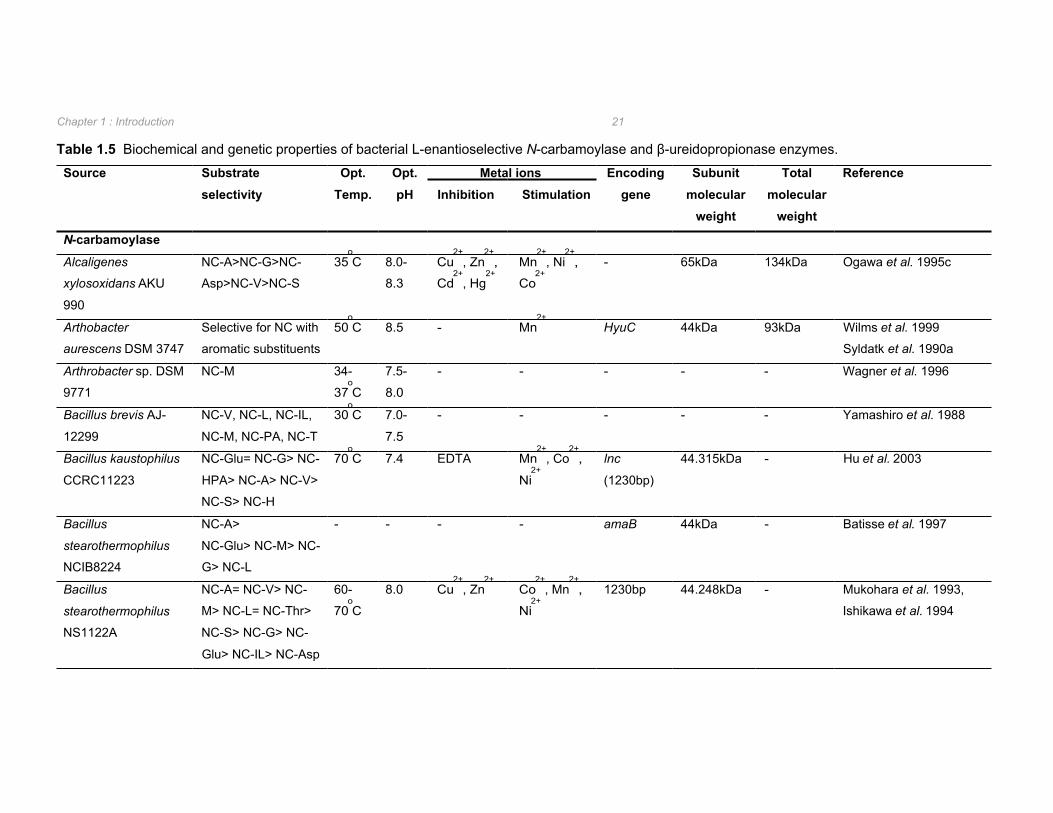
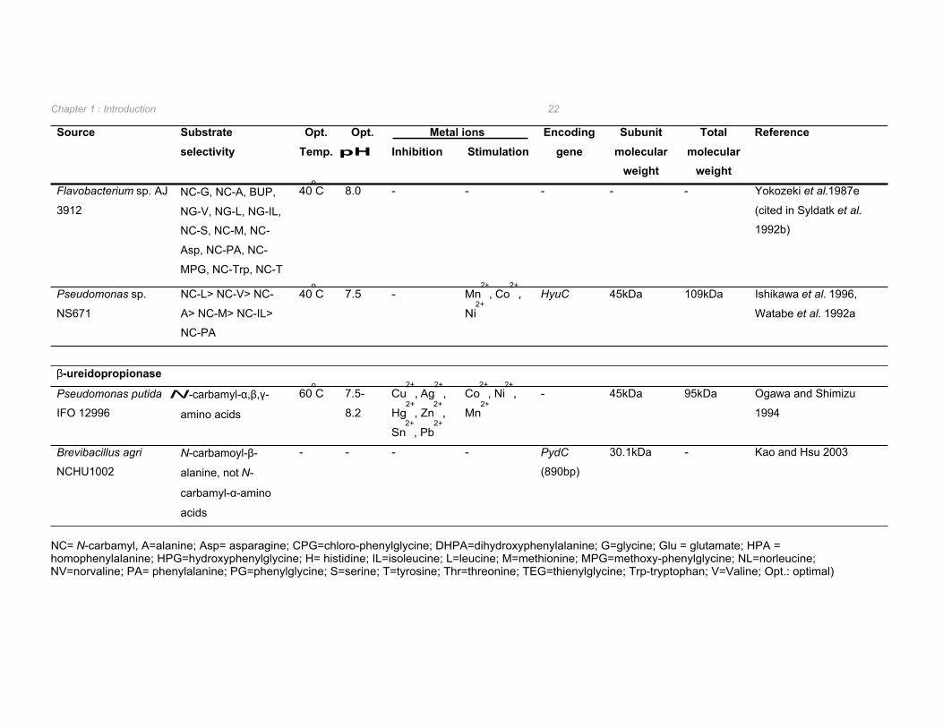
1.2.3.2 L-enantioselective N-carbamoylases L-enantioselective enzymes have been isolated and characterized from Alcaligenes, Bacillus
and Pseudomonas species and shown to hydrolyse a wide range of aliphatic and aromatic
N-carbamylamino acids (Table 1.5). However, due to the highly unstable nature of this
enzyme, only L-carbamoylases from Alcaligenes xylosoxidans AKU 990, Arthrobacter
aurescens DSM 3747, and Pseudomonas sp. NS671 have been purified (Ogawa et al.
1995c, Wilms et al. 1999, Ishikawa et al. 1996). In all cases the L-carbamoylase was found
to be a homodimer (Table 1.5).
Unlike the D-carbamoylases, the optimal pH for hydrolysis of N-carbamylamino acid
substrates by L-carbamoylases appears to be at a more neutral pH of 7.0 to 8.5 (Table 1.5).
Furthermore, metal ions were found to stimulate as well as inhibit L-carbamoylase activities
and inhibition by metal ion chelators was observed (Table 1.5) which is in contrast to the
results observed for D-carbamoylases (Table 1.4). The presence of metal ion chelators such
as EDTA, 8-hydroxyquinoline, and 2,2’-dipyridyl caused considerable inhibition of the β-
ureidopropionase from Pseudomonas putida IFO12996 indicating that, as is the case with L-
carbamoylases, the β-ureidopropionase is a metalloenzyme (Ogawa and Shimizu 1994).
Of the eight L-carbamoylases isolated, four have been shown not to hydrolyse N-carbamyl-β-
alanine whilst the remaining L-carbamoylases were not tested for the ability to hydrolyse the
natural substrate of β-ureidopropionase. This suggests that L-carbamoylases are not
synonymous with β-ureidopropionase. The narrow substrate range of the β-
ureidopropionase from Brevibacillus agri NCHU1002, which hydrolysed N-carbamyl-β-
alanine but not N-carbamyl-α-amino acids (Kao and Hsu 2003) supports this. In contrast, the
β-ureidopropionase from P. putida IFO 12990 was observed to have a very broad substrate
range with hydrolysis of N-carbamyl-α,β, and γ-amino acids with strict L-enantioselectivity
with respect to N-carbamyl-α-amino acid hydrolysis (Ogawa and Shimizu 1994).
Chapter 1 : Introduction 21
Table 1.5 Biochemical and genetic properties of bacterial L-enantioselective N-carbamoylase and β-ureidopropionase enzymes.
Source Substrate selectivity
Opt. Temp.
Opt. pH
Metal ions Encoding gene
Subunit molecular
weight
Total molecular
weight
Reference Inhibition Stimulation
N-carbamoylase
Alcaligenes
xylosoxidans AKU
990
NC-A>NC-G>NC-
Asp>NC-V>NC-S
35oC 8.0-
8.3
Cu2+
, Zn2+
,
Cd2+
, Hg2+
Mn2+
, Ni2+
,
Co2+
- 65kDa 134kDa Ogawa et al. 1995c
Arthobacter
aurescens DSM 3747
Selective for NC with
aromatic substituents
50oC 8.5 - Mn
2+ HyuC 44kDa 93kDa Wilms et al. 1999
Syldatk et al. 1990a
Arthrobacter sp. DSM
9771
NC-M 34-
37oC
7.5-
8.0
- - - - - Wagner et al. 1996
Bacillus brevis AJ-
12299
NC-V, NC-L, NC-IL,
NC-M, NC-PA, NC-T
30oC 7.0-
7.5
- - - - - Yamashiro et al. 1988
Bacillus kaustophilus
CCRC11223
NC-Glu= NC-G> NC-
HPA> NC-A> NC-V>
NC-S> NC-H
70oC 7.4 EDTA Mn
2+, Co
2+,
Ni2+
lnc
(1230bp)
44.315kDa - Hu et al. 2003
Bacillus
stearothermophilus
NCIB8224
NC-A>
NC-Glu> NC-M> NC-
G> NC-L
- - - - amaB 44kDa - Batisse et al. 1997
Bacillus
stearothermophilus
NS1122A
NC-A= NC-V> NC-
M> NC-L= NC-Thr>
NC-S> NC-G> NC-
Glu> NC-IL> NC-Asp
60-
70oC
8.0 Cu2+
, Zn2+
Co2+
, Mn2+
,
Ni2+
1230bp 44.248kDa - Mukohara et al. 1993,
Ishikawa et al. 1994
Source Substrate selectivity
Subunit Inhibition Stimulation gene molecular
weight
Total molecular
weight
Opt. Opt. Temp. pH
Metal ions Encoding Reference
Chapter 1 : Introduction 22
Flavobacterium sp. AJ
3912 NC-G, NC-A, BUP,
NG-V, NG-L, NG-IL,
NC-S, NC-M, NC-
Asp, NC-PA, NC-
MPG, NC-Trp, NC-T
40oC 8.0 - - - - - Yokozeki et al.1987e
(cited in Syldatk et al.
1992b)
Pseudomonas sp. NC-L> NC-V> NC- 40oC 7.5 - Mn
2+, Co
2+, HyuC 45kDa 109kDa Ishikawa et al. 1996,
NS671 A> NC-M> NC-IL> Ni2+
Watabe et al. 1992a
NC-PA
β-ureidopropionase
Pseudomonas putida N-carbamyl-α,β,γ- 60oC 7.5- Cu
2+, Ag
2+, Co
2+, Ni
2+, - 45kDa 95kDa Ogawa and Shimizu
IFO 12996 amino acids 8.2 Hg2+
, Zn2+
, Mn2+
1994
Sn2+
, Pb2+
Brevibacillus agri N-carbamoyl-β- - - - - PydC 30.1kDa - Kao and Hsu 2003
NCHU1002 alanine, not N- (890bp)
carbamyl-α-amino
acids
NC= N-carbamyl, A=alanine; Asp= asparagine; CPG=chloro-phenylglycine; DHPA=dihydroxyphenylalanine; G=glycine; Glu = glutamate; HPA = homophenylalanine; HPG=hydroxyphenylglycine; H= histidine; IL=isoleucine; L=leucine; M=methionine; MPG=methoxy-phenylglycine; NL=norleucine; NV=norvaline; PA= phenylalanine; PG=phenylglycine; S=serine; T=tyrosine; Thr=threonine; TEG=thienylglycine; Trp-tryptophan; V=Valine; Opt.: optimal)

Chapter 1 : Introduction 23
Five genes encoding L-carbamoylases have been isolated and found to encode enzymes
with similar predicted molecular weights of approximately 44kDa (Table 1.5). The HyuC from
Pseudomonas sp. NS671 was located on a 172Kbp plasmid downstream of the HyuA and
HyuB genes encoding for the hydantoinase (Watabe et al. 1992a). The purified enzyme was
strictly L-enantioselective with a broad substrate range and shown to be inhibited in the
presence of ATP (Ishikawa et al. 1996). Sequence analysis of the HyuC indicated a 43%
similarity to the HyuA and HyuB genes suggesting that these genes evolved from a common
ancestor by gene duplication (Watabe et al. 1992a).
The L-carbamoylase gene, amaB, from Bacillus stearothermophilus NCIB8224 was found to
be co-transcribed on a polycistronic mRNA with the amaA gene encoding an aminoacylase
(Batisse et al. 1997). The operon formed by the amaA and amaB genes in B.
stearothermophilus NCIB8224 was found, by Southern hybridisation and PCR analysis, to be
conserved in other B. stearothermophilus strains (Batisse et al. 1997).
Analysis of the amino acid sequence of the Bacillus kaustophilus L-carbamoylase revealed
the presence of six cysteine residues which, by treatment with excess dithiothreitol, were
shown to form three disulphide bridges (Hu et al. 2003). The amino acid sequence of L-
carbamoylases from Bacillus stearothermophilus NS1122A, Pseudomonas sp. NS671, A.
aurescens DSM 3747, and B. stearothermophilus NCIB8224 were shown to have 93.6, 44.3,
35.5 and 93.6% identity respectively with that of the L-carbamoylase from B. kaustophilus
(Hu et al. 2003). By alignment of the amino acid sequences from these L-carbamoylases,
the six cysteine residues found to form disulphide bridges in the B. kaustophilus L-
carbamoylase were shown to be conserved in the L-carbamoylases from both B.
stearothermophilus strains (Hu et al. 2003). While the L-carbamoylase from Pseudomonas
sp. NS671 contains five cysteine residues, and inhibition by the SH reagent p-
chloromercaribenzoate suggested the potential presence of two disulphide bridges (Ishikawa
et al. 1996), only two of the cysteine residues occur in positions relative to the cysteines in B.
kaustophilus L-carbamoylase. The L-carbamoylase from A. aurescens DSM 3747 does not
contain disulphide bridges as only one cysteine residue is present in its primary amino acid
sequence. The formation of disulphide bridges is thought to stabilize proteins at high
temperatures (Hu et al. 2003) and this is reflected in the thermostabilities of the L-
carbamoylases from B. kaustophilus and B. stearothermophilus in comparison to that of A.
aurescens and Pseudomonas sp. NS671 (Table 1.5). Increased overall hydrophobicity and
proline substitutions over the entire polypeptide chain were also suggested to contribute to
the higher thermostability of the L-carbamoylase from B. stearothermophilus NS1122A when
compared to Pseudomonas sp. NS671 (Mukohara et al. 1993).
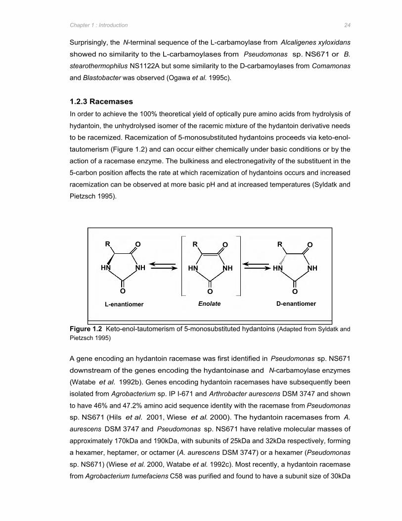
R O R R O O
HN NH NH HN
O O O
HN NH
L-enantiomer Enolate D-enantiomer
Chapter 1 : Introduction 24
Surprisingly, the N-terminal sequence of the L-carbamoylase from Alcaligenes xyloxidans
showed no similarity to the L-carbamoylases from Pseudomonas sp. NS671 or B.
stearothermophilus NS1122A but some similarity to the D-carbamoylases from Comamonas
and Blastobacter was observed (Ogawa et al. 1995c).
1.2.3 Racemases In order to achieve the 100% theoretical yield of optically pure amino acids from hydrolysis of
hydantoin, the unhydrolysed isomer of the racemic mixture of the hydantoin derivative needs
to be racemized. Racemization of 5-monosubstituted hydantoins proceeds via keto-enol-
tautomerism (Figure 1.2) and can occur either chemically under basic conditions or by the
action of a racemase enzyme. The bulkiness and electronegativity of the substituent in the
5-carbon position affects the rate at which racemization of hydantoins occurs and increased
racemization can be observed at more basic pH and at increased temperatures (Syldatk and
Pietzsch 1995).
Figure 1.2 Keto-enol-tautomerism of 5-monosubstituted hydantoins (Adapted from Syldatk and Pietzsch 1995)
A gene encoding an hydantoin racemase was first identified in Pseudomonas sp. NS671
downstream of the genes encoding the hydantoinase and N-carbamoylase enzymes
(Watabe et al. 1992b). Genes encoding hydantoin racemases have subsequently been
isolated from Agrobacterium sp. IP I-671 and Arthrobacter aurescens DSM 3747 and shown
to have 46% and 47.2% amino acid sequence identity with the racemase from Pseudomonas
sp. NS671 (Hils et al. 2001, Wiese et al. 2000). The hydantoin racemases from A.
aurescens DSM 3747 and Pseudomonas sp. NS671 have relative molecular masses of
approximately 170kDa and 190kDa, with subunits of 25kDa and 32kDa respectively, forming
a hexamer, heptamer, or octamer (A. aurescens DSM 3747) or a hexamer (Pseudomonas
sp. NS671) (Wiese et al. 2000, Watabe et al. 1992c). Most recently, a hydantoin racemase
from Agrobacterium tumefaciens C58 was purified and found to have a subunit size of 30kDa
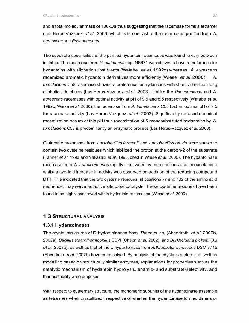
Chapter 1 : Introduction 25
and a total molecular mass of 100kDa thus suggesting that the racemase forms a tetramer
(Las Heras-Vazquez et al. 2003) which is in contrast to the racemases purified from A.
aurescens and Pseudomonas.
The substrate-specificities of the purified hydantoin racemases was found to vary between
isolates. The racemase from Pseudomonas sp. NS671 was shown to have a preference for
hydantoins with aliphatic substituents (Watabe et al. 1992c) whereas A. aurescens
racemized aromatic hydantoin derivatives more efficiently (Wiese et al. 2000). A.
tumefaciens C58 racemase showed a preference for hydantoins with short rather than long
aliphatic side chains (Las Heras-Vazquez et al. 2003). Unlike the Pseudomonas and A.
aurescens racemases with optimal activity at pH of 9.5 and 8.5 respectively (Watabe et al.
1992c, Wiese et al. 2000), the racemase from A. tumefaciens C58 had an optimal pH of 7.5
for racemase activity (Las Heras-Vazquez et al. 2003). Significantly reduced chemical
racemization occurs at this pH thus racemization of 5-monosubstituted hydantoins by A.
tumefaciens C58 is predominantly an enzymatic process (Las Heras-Vazquez et al. 2003).
Glutamate racemases from Lactobacillus fermenti and Lactobacillus brevis were shown to
contain two cysteine residues which labilized the proton at the carbon-2 of the substrate
(Tanner et al. 1993 and Yakasaki et al. 1995, cited in Wiese et al. 2000). The hydantoinase
racemase from A. aurescens was rapidly inactivated by mercuric ions and iodoacetamide
whilst a two-fold increase in activity was observed on addition of the reducing compound
DTT. This indicated that the two cysteine residues, at positions 77 and 182 of the amino acid
sequence, may serve as active site base catalysts. These cysteine residues have been
found to be highly conserved within hydantoin racemases (Wiese et al. 2000).
1.3 STRUCTURAL ANALYSIS
1.3.1 Hydantoinases The crystal structures of D-hydantoinases from Thermus sp. (Abendroth et al. 2000b,
2002a), Bacillus stearothermophilus SD-1 (Cheon et al. 2002), and Burkholderia pickettii (Xu
et al. 2003a), as well as that of the L-hydantoinase from Arthrobacter aurescens DSM 3745
(Abendroth et al. 2002b) have been solved. By analysis of the crystal structures, as well as
modelling based on structurally similar enzymes, explanations for properties such as the
catalytic mechanism of hydantoin hydrolysis, enantio- and substrate-selectivity, and
thermostability were proposed.
With respect to quaternary structure, the monomeric subunits of the hydantoinase assemble
as tetramers when crystallized irrespective of whether the hydantoinase formed dimers or
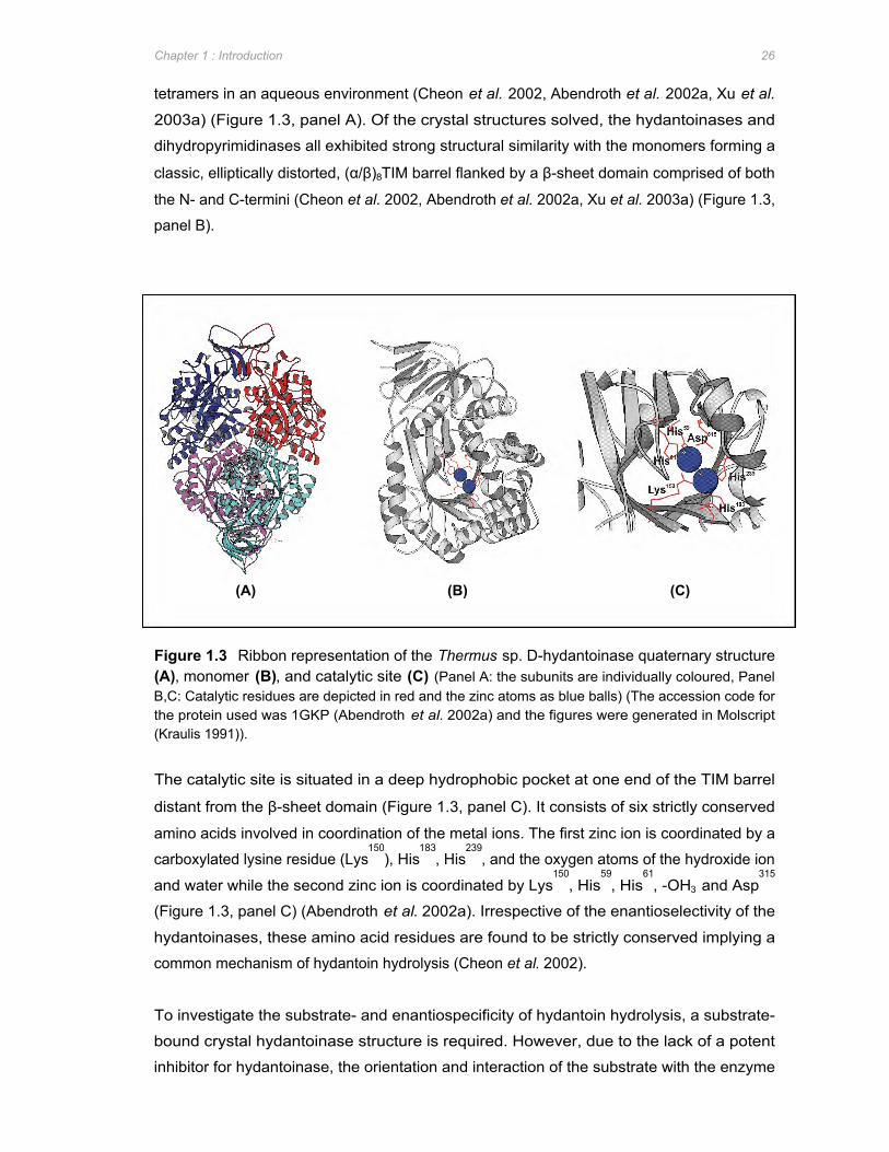
(A) (B) (C)
Chapter 1 : Introduction 26
tetramers in an aqueous environment (Cheon et al. 2002, Abendroth et al. 2002a, Xu et al.
2003a) (Figure 1.3, panel A). Of the crystal structures solved, the hydantoinases and
dihydropyrimidinases all exhibited strong structural similarity with the monomers forming a
classic, elliptically distorted, (α/β)8TIM barrel flanked by a β-sheet domain comprised of both
the N- and C-termini (Cheon et al. 2002, Abendroth et al. 2002a, Xu et al. 2003a) (Figure 1.3,
panel B).
Figure 1.3 Ribbon representation of the Thermus sp. D-hydantoinase quaternary structure (A), monomer (B), and catalytic site (C) (Panel A: the subunits are individually coloured, Panel B,C: Catalytic residues are depicted in red and the zinc atoms as blue balls) (The accession code for the protein used was 1GKP (Abendroth et al. 2002a) and the figures were generated in Molscript (Kraulis 1991)).
The catalytic site is situated in a deep hydrophobic pocket at one end of the TIM barrel
distant from the β-sheet domain (Figure 1.3, panel C). It consists of six strictly conserved
amino acids involved in coordination of the metal ions. The first zinc ion is coordinated by a
carboxylated lysine residue (Lys150
), His183
, His239
, and the oxygen atoms of the hydroxide ion
and water while the second zinc ion is coordinated by Lys150
, His59
, His61
, -OH3 and Asp315
(Figure 1.3, panel C) (Abendroth et al. 2002a). Irrespective of the enantioselectivity of the
hydantoinases, these amino acid residues are found to be strictly conserved implying a
common mechanism of hydantoin hydrolysis (Cheon et al. 2002).
To investigate the substrate- and enantiospecificity of hydantoin hydrolysis, a substrate-
bound crystal hydantoinase structure is required. However, due to the lack of a potent
inhibitor for hydantoinase, the orientation and interaction of the substrate with the enzyme
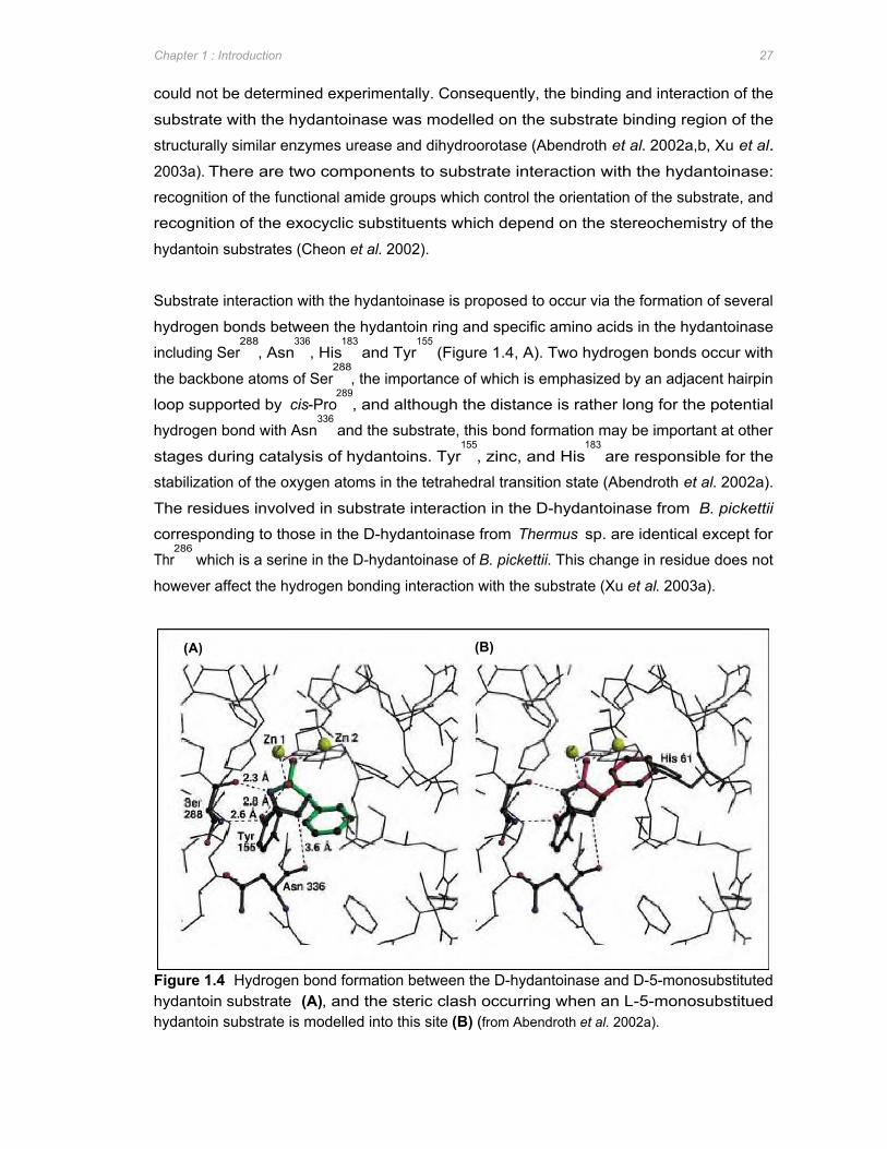
(A) (B)
Chapter 1 : Introduction 27
could not be determined experimentally. Consequently, the binding and interaction of the
substrate with the hydantoinase was modelled on the substrate binding region of the
structurally similar enzymes urease and dihydroorotase (Abendroth et al. 2002a,b, Xu et al.
2003a). There are two components to substrate interaction with the hydantoinase:
recognition of the functional amide groups which control the orientation of the substrate, and
recognition of the exocyclic substituents which depend on the stereochemistry of the
hydantoin substrates (Cheon et al. 2002).
Substrate interaction with the hydantoinase is proposed to occur via the formation of several
hydrogen bonds between the hydantoin ring and specific amino acids in the hydantoinase
including Ser288
, Asn336
, His183
and Tyr155
(Figure 1.4, A). Two hydrogen bonds occur with
the backbone atoms of Ser288
, the importance of which is emphasized by an adjacent hairpin
loop supported by cis-Pro289
, and although the distance is rather long for the potential
hydrogen bond with Asn336
and the substrate, this bond formation may be important at other
stages during catalysis of hydantoins. Tyr155
, zinc, and His183
are responsible for the
stabilization of the oxygen atoms in the tetrahedral transition state (Abendroth et al. 2002a).
The residues involved in substrate interaction in the D-hydantoinase from B. pickettii
corresponding to those in the D-hydantoinase from Thermus sp. are identical except for
Thr286
which is a serine in the D-hydantoinase of B. pickettii. This change in residue does not
however affect the hydrogen bonding interaction with the substrate (Xu et al. 2003a).
Figure 1.4 Hydrogen bond formation between the D-hydantoinase and D-5-monosubstituted hydantoin substrate (A), and the steric clash occurring when an L-5-monosubstitued hydantoin substrate is modelled into this site (B) (from Abendroth et al. 2002a).
Chapter 1 : Introduction 28
The pronounced hydrophobic pocket formed by the amino acid residues Leu64
, Cys95
, Phe152
,
Tyr155
, and Phe159
is proposed to be responsible for the enatio- and substrate-specificity
exhibited by the D-hydantoinase of Thermus sp. (Abendroth et al. 2002a). Thermus D-
hydantoinase readily hydrolyses 5-phenylic substituted hydantoins as these substrates are
easily accommodated in the hydrophobic pocket. In contrast, hydantoin derivatives with
benzylic or polar substituents were found to be hydrolysed to D-enantiomer intermediates
with significantly lower efficiency due to less productive interaction within the hydrophobic
pocket. While D-specific hydantoin substrates fit into this model well, the larger side-chains
of L-hydantoin derivatives clash sterically with His61 in the catalytic site of D-hydantoinase
from Thermus (Figure 1.4, panel A vs. B). However, hydantoin derivatives with small side
chains are readily hydrolysed but without enantioselectivity as neither enantiomer results in
steric clashes with the His61 residue (Abendroth et al. 2002a). The residues proposed to
form the hydrophobic pocket in B. stearothermophilus D-hydantoinase correspond to that of
Thermus D-hydantoinase with the exception of Met63
in place of Cys95
(Cheon et al. 2002).
In contrast to the D-hydantoinases described above, the L-hydantoinase from A. aurescens
contains no pronounced hydrophobic pocket and subsequent analysis of the structure
produced no explanations for the substrate-specificities observed from this enzyme. D/L-5-
indolylhydantoin is one of the preferred substrates of the L-hydantoinase. The proposed
orientation for the substrate interaction with the hydantoinase would result in no hydrophobic
interaction partners for the D-enantiomer of 5-indolylhydantoin whereas the L-enantiomer is
able to form hydrophobic interactions with the side chains of His62
and Ile95
(Abendroth et al.
2002b). Mutation of the L-hydantoinase from A. aurescens resulted in increased D-
enantiospecificity with respect to 5-methylthioethyl-hydantoin subsequent to a V154A point
mutation (May et al. 2000). This is most likely due to the increased space for the side chain
created by this mutation (Abendroth et al. 2002b). The point mutation I95F which resulted in
increased L-enantiospecificity of the enzyme (May et al. 2000) is most likely due to increased
hydrophobicity close to the side chain of L-5-methylthioethyl hydantoin resulting in increased
cleavage of the L-enantiomer (Abendroth et al. 2002b).
The exocyclic substituent recognition site in the D-hydantoinase from B. pickettii is formed by
the predominantly hydrophilic amino acid residues Thr62
, Ser64
, Gln93
, Phe150
, Tyr153
, and
Asn157
. This is in contrast to the D-hydantoinases from Thermus and B. stearothermophilus,
which have hydrophobic residues at these positions forming a pronounced hydrophobic
pocket. The presence of hydrophilic residues at the exocyclic recognition site of the D-
hydantoinase from B. pickettii suggest that the enzyme may have a preference for hydantoin
substrates with polar substituents (Xu et al. 2003a).
Chapter 1 : Introduction 29
Analysis of the crystal structures of hydantoinases not only sheds light on the catalytic
properties of these enzymes but also suggests factors likely to contribute toward
thermostability of the enzymes. Comparison of the crystal structure of the mesophylic D-
hydantoinase from B. pickettii with the thermophylic D-hydantoinase from Thermus sp. by Xu
et al. (2003a) revealed that the thermostability of the D-hydantoinase from Thermus sp.
appears to be due to a combination of several factors. These include the presence of
methionine and cysteine residues, which can be readily oxidized at high temperatures, on the
surface of the enzymes resulting in decreased thermostability. B. pickettii has more
methionine and cysteine residues on the surface of its D-hydantoinase than does Thermus
sp. Increased hydrophilic interactions, such as charged ion pairs and hydrogen-bonding
interactions, result in increased thermostability of enzymes. Thermus sp. D-hydantoinase
was found to contain 44 pairs of salt bridges and 777 hydrogen-bonding interactions
compared to the 38 salt bridges and 714 hydrogen bonds in the mesophylic D-hydantoinase.
Amino acid composition of the enzyme may also affect thermostability. Proline, for example,
has a rigid conformation and thus imposes constraints on the flexibility of proteins. Increased
proline content, as is the case in the D-hydantoinase from Thermus sp., results in enzymes
which resist unfolding at high temperatures (Xu et al. 2003a).
1.3.2 N-carbamoylases The crystal structures of the D-carbamoylases from Agrobacterium sp. KNK712 (Nakai et al.
2000) and A. radiobacter 14924 (Wang et al. 2001) have been solved. From these studies,
D-carbamoylases are found to consist of monomers with a sandwich of β-sheets surrounded
by 2 layers of α-helices (Figure 1.5, panel B). In crystal form, the D-carbamoylases form
tetrameric quaternary structures with subunits A+B and C+D forming tightly packed dimers
which then interact with one another rather loosely (Nakai et al. 2000, Wang et al. 2001)
(Figure 1.5, panel A). As is the case with the hydantoinase, the D-carbamoylases appear to
behave differently in aqueous environments where they form dimers or trimers as opposed to
the tetramers formed when crystallized (Nakai et al. 2000).
The catalytic residues of the D-carbamoylase enzymes were proposed to be Glu46
, Lys126
,
and Cys172
(Nakai et al. 2000, Wang et al. 2001, Chen et al. 2003) (Figure 1.5, panel C).
This is supported by site-directed mutagenesis of Cys172
resulting in loss of catalytic activity
(Grifantini et al. 1996, Chen et al. 2003). Two helices (α2, α6) and the loops connecting β5
and α4, β7 and α5, and β8 and α6 were found to form the pocket structure of the active site
(Nakai et al. 2000) (Figure 1.5, panel B). Helix α7 of the adjacent monomer was found to
cover the active site pocket thereby narrowing the entrance to the active site (Nakai et al.
2000). Further site-directed mutagenesis identified residues His129
, His144
, and His215
as
necessary to maintain the stable conformation of the catalytic cleft (Wang et al. 2001).
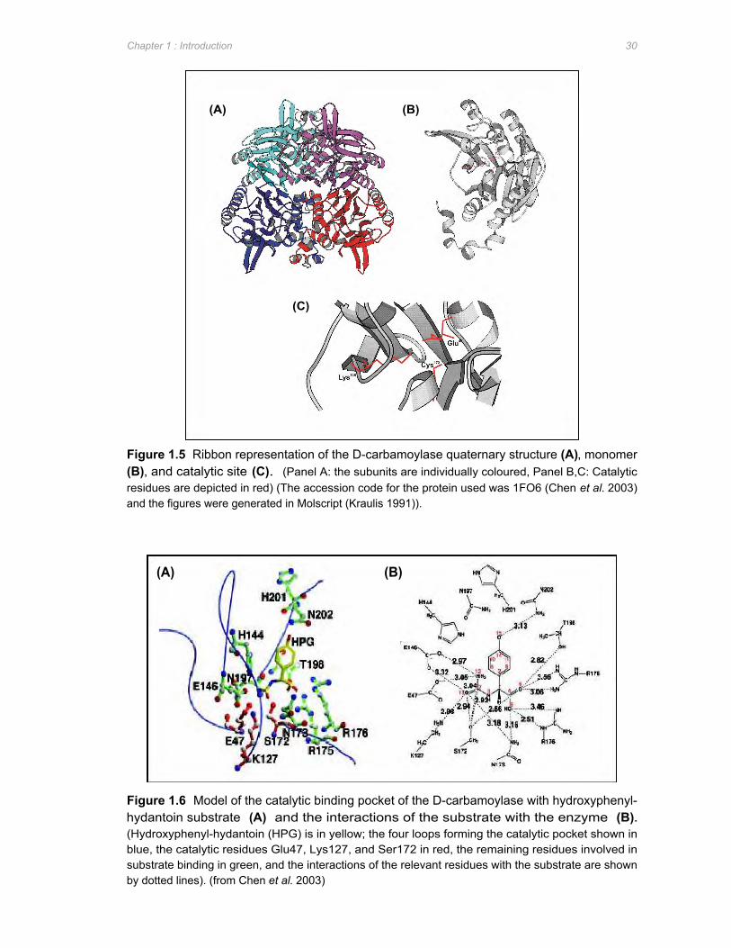
(A) (B)
(C)
(A) (B)
Chapter 1 : Introduction 30
Figure 1.5 Ribbon representation of the D-carbamoylase quaternary structure (A), monomer (B), and catalytic site (C). (Panel A: the subunits are individually coloured, Panel B,C: Catalytic residues are depicted in red) (The accession code for the protein used was 1FO6 (Chen et al. 2003) and the figures were generated in Molscript (Kraulis 1991)).
Figure 1.6 Model of the catalytic binding pocket of the D-carbamoylase with hydroxyphenyl-hydantoin substrate (A) and the interactions of the substrate with the enzyme (B). (Hydroxyphenyl-hydantoin (HPG) is in yellow; the four loops forming the catalytic pocket shown in blue, the catalytic residues Glu47, Lys127, and Ser172 in red, the remaining residues involved in substrate binding in green, and the interactions of the relevant residues with the substrate are shown by dotted lines). (from Chen et al. 2003)
Chapter 1 : Introduction 31
Based on substrate-enzyme modelling of the solved crystal structure of the D-carbamoylase
as well as site-directed mutagenesis studies, a mechanism by which N-carbamyl-D-amino
acids are hydrolysed to their corresponding D-amino acids by the D-carbamoylases was
proposed. Based on modelling, the side chains of Glu47
, Lys127
, and Cys172
were proposed to
cluster around the carbamoyl moiety of the substrate while the side chains of residues
Asn173
, Arg175
, and Arg176
interacted with the carboxyl group resulting in substrate specificity
(Figure 1.6, panel B) (Chen et al. 2003). Chen et al. (2003) also proposed that Cys172
, with a
nucleophilic Sγ, atom is central in attacking the C7 atom of the carbamoyl group whilst Glu47
functions as a general base and Lys127
stabilizes the tetrahedral transition state. Based on
this data, a possible catalytic mechanism consisting of an acylation reaction with the
carbamoyl moiety to cleave the susceptible C-N bond and produce a NH3, followed by
deacylation of the acyl-enzyme intermediate to form the D-amino acid and a CO2 molecule
was suggested (Chen et al. 2003).
To date, no crystal structure has been solved for an L-enantioselective N-carbamoylase.
1.4 REGULATION OF HYDANTOIN-HYDROLYSING ENZYME ACTIVITY
Overproduction of unwanted enzymes may endow the cell with an energetic burden or result
in hydrolytic activity which is detrimental to the cell, as is the case with D-hydantoinases
which hydrolyse dihydropyrimidines required for cellular function. In order to protect against
such effects, microorganisms possess regulatory mechanisms to control expression of
metabolic enzymes. These mechanisms include induction, feedback inhibition, and
nutritional regulation by carbon or nitrogen sources (Sanchez and Demain 2002). For
industrial applications, deregulation of these mechanisms by classical and genetic
manipulations is required such that over-production of the desired metabolite or enzyme is
achieved (Sanchez and Demain 2002).
1.4.1 Induction A wide range of hydantoin and pyrimidine derivatives have been shown to induce expression
of genes encoding hydantoin-hydrolysing enzymes with increases in enzyme activities from
5- to 30-fold compared to uninduced cells (Hartley et al. 1998, Wiese et al. 2001). Table 1.6
provides a summary of the hydantoin-hydrolysing enzymes reported to be subject to
inductive regulation and their corresponding inducers. Regulation of expression of the
hydantoin-hydrolysing genes by induction ensures that the hydantoinase and N-
carbamoylase enzymes are produced only when a suitable substrate is available. The D-
hydantoinase enzymes from Pseudomonas fluorescens DSM84, Agrobacterium tumefaciens
NRRL B11291, and Pseudomonas sp. NCIM 5109 are exceptions with constitutive

Chapter 1 : Introduction 32
expression of the hydantoinase genes in these isolates (Morin et al. 1986a, Deepa et al.
1993, Sudge et al. 1998).
Table 1.6 Inducers of hydantoin-hydrolysing activities in bacterial isolates Isolate Inducer Enzyme affected Reference
Agrobacterium tumefaciens 2-thiouracil D-hydantoinase Hartley et al. 1998
RU-OR D-carbamoylase
Arthrobacter aurescens DSM D,L-5-(3-indolylmethyl)- Hydantoinase Wiese et al. 2001
3747 3-N-methylhydantoin L-carbamoylase
Arthrobacter crystallopoietes Hydantoin D-hydantoinase Moller et al. 1988
AM2 DL-5-hydroxymethyl-
hydantoin
D-carbamoylase
Arthrobacter sp. BH20 DL-5-indolylmethyl-
hydantoin
Hydantoinase Syldatk et al. 1987
Bacillus brevis AJ-12299 DL-5-isopropyl-
hydantoin
D-hydantoinase Yamashiro et al.
1988
Flavobacterium sp. I-3 DL-5-indolylmethyl-
hydantoin
Hydantoinase
L-carbamoylase
Nishida et al. 1987
Pseudomonas putida RU-KM1 Hydantoin Hydantoinase Burton et al. 1998
D-carbamoylase
Pseudomonas putida RU-KM3S Hydantoin Hydantoinase Burton et al. 1998
L-carbamoylase
Pseudomonas sp. AJ-11220 DL-5-methylthioethyl- D-hydantoinase Yokozeki et al.
hydantoin D-carbamoylase 1987b
The hydantoin hydrolysing enzymes from Arthrobacter sp. DSM 3747 were induced by DL-5-
(3-indolylmethyl)hydantoin (Syldatk et al. 1990a). However, Syldatk et al. (1990a) reported a
decrease in hydantoin-hydrolysing activities over time due to degradation of the inducer.
Addition of higher concentrations of DL-5-(3-indolylmethyl)hydantoin was not feasible as it
resulted in significant growth retardation. Thus, in order to obtain optimum levels of
hydantoin-hydrolysis with maximum production of biomass, the inducer had to be
continuously fed into the growth medium (Syldatk et al. 1990a). An alternative is the use of
non-metabolizable inducers such as D,L-5-(3-indolylmethyl)-3-N-methylhydantoin (N-3-CH3-
IMH) (Syldatk et al. 1990a). Induction studies of A. aurescens DSM 3747 showed a 30-fold
increase in hydantoinase activity when N-3-CH3-IMH was supplied in the growth medium as
an inducer (Wiese et al. 2001). Similar results were obtained in Agrobacterium tumefaciens
Chapter 1 : Introduction 33
RU-OR in which the use of the non-metabolizable 2-thiouracil as inducer resulted in a 5- and
10-fold increase in hydantoinase and N-carbamoylase activity levels respectively (Hartley et
al. 1998).
Selection of inducer-independent strains, in order to reduce the costs of industrial production
of enantiopure amino acids, has been done in an Arthrobacter (Wagner et al. 1996) and an
Agrobacterium strain (Hartley et al. 1998). To select for an inducer-independent mutant,
Arthrobacter sp. DSM 7330 was subjected to extended selection pressure by repeated
cycles of growth in minimal medium with N-carbamoyl-L-methionine as a sole source of
nitrogen. The uninduced cells of the subsequently isolated mutant, DSM 9771, displayed a
3-fold increase in hydantoinase activity but somewhat lower N-carbamoylase activity when
compared to the induced parental cells. However, on induction, the hydantoinase and N-
carbamoylase activity in DSM 9771 was 5-fold higher than that of DSM 7330 under
corresponding conditions (Wagner et al. 1996).
Inducer-independent mutants were also generated from Agrobacterium tumefaciens RU-OR
wild type. Mutant strains RU-ORL5 and RU-ORLB3 were isolated by growth in the presence
of the toxic hydantoin analogue 5-fluorouracil in the absence of an inducer. This selection
was based on the principle that unless the cells were able to express the hydantoinase under
non-inducing conditions, hydrolysis of the toxic 5-fluorouracil to a non-toxic intermediate by
the hydantoinase enzyme would not occur resulting in cell death (Hartley et al. 1998).
Mutant strain RU-ORL5 showed constitutive expression of both the hydantoinase and N-
carbamoylase whilst mutant RU-ORLB3 was constitutive only with respect to hydantoinase
activity with N-carbamoylase activity still inducer-dependent (Hartley et al. 2001). This
suggested that while induction of hydantoinase and N-carbamoylase enzymes in RU-OR are
probably mediated by a common regulatory factor, the expression of the genes are not
transcriptionally linked (Hartley et al. 2001).
1.4.2 Catabolite repression Numerous studies have reported that the production of hydantoinase and N-carbamoylase
enzymes in microorganisms occurs only once late logarithmic to stationary phase of growth
has been reached (Hartley et al. 1998, Syldatk et al. 1990a, Sudge et al. 1998, Nishida et al.
1987, Gokhale et al. 1996). Furthermore, supplementation of various carbon and nitrogen
sources in the growth medium affects production of hydantoinase and N-carbamoylase
enzymes (Sudge et al. 1998, Morin et al. 1986a, Yamashiro et al. 1988, Syldatk et al. 1990a,
Nishida et al. 1987, Ishikawa et al. 1994). These results indicate that hydantoin-hydrolysing
enzymes are subject to catabolite repression in addition to regulation by induction.
Catabolite repression is a regulatory mechanism which functions to ensure organised and
Chapter 1 : Introduction 34
sequential utilization of nutrient sources. Nutrient sources which most readily supply carbon
or nitrogen and energy for growth are classified as “good” nutrient sources and are
metabolised by the cell first. Synthesis of enzymes for the metabolism of other, “poorer”
nutrient sources is then repressed until the primary substrate has been exhausted (Sanchez
and Demain 2002). Hydantoin derivatives are a poor nitrogen and carbon source and
expression of hydantoin-hydrolysing genes is likely to be subject to carbon and/or nitrogen
catabolic repression mechanisms.
Upstream of the genes encoding for the hydantoinase (hyuA and hyuB) and N-carbamoylase
(hyuC) in Pseudomonas sp. NS671, ORF1 was identified and found to share strong amino
acid similarity with the NifA and NtrC proteins (Watabe et al. 1992a) which are involved in
control of nitrogen fixation and regulation by functioning as transcriptional activators of σ54
RNA polymerase (Morett and Segovia 1993). The gene product of ORF1 may therefore be
involved in regulatory control of the hydantoin-hydrolysing gene cluster of Pseudomonas sp.
NS671 which is preceded by a putative σ54
-promoter region (Watabe et al. 1992a). A
putative σ54
-dependent promoter region has also been identified upstream of the hydantoin-
hydrolysing gene cluster of Arthrobacter aurescens DSM 3747 (Wiese et al. 2001). As
activation of σ54
-dependent promoters occurs via an activator protein which usually belongs
to the NtrC family (Snyder and Champress 2003), the presence of these regions upstream of
the genes encoding for hydantoin-hydrolysing enzymes suggests that these operons may be
regulated by nitrogen catabolite repression (Ntr system).
The production of hydantoinase and N-carbamoylase in Agrobacterium tumefaciens RU-OR
also appeared to be regulated by nitrogen catabolite repression as evidenced by the
dramatic decrease in activity in cells cultured with a good nitrogen source such as
(NH4)2SO4. This is substantiated by mutant strain RU-ORPN1 which was no longer subject
to nitrogen catabolite repression as, unlike the wild type, hydantoinase and N-carbamoylase
activities were observed in mid-log phase of growth (Hartley et al. 2001). Subsequent to
incubation for 30 minutes in minimal medium with (NH4)2SO4 as a nitrogen source, A.
tumefaciens RU-OR cells retained only a third and a half of the original hydantoinase and N-
carbamoylase activities respectively. This second regulatory mechanism was termed
ammonia shock and was shown to involve the enzymatic action of glutamine synthetase as
addition of the glutamine synthetase inhibitor D,L-methionine D,L-sulfoximine protected the
hydantoinase from ammonium shock (Hartley et al. 2001).
Glutamine synthetase activity is an integral part of the global nitrogen regulatory system in
prokaryotic cells (Merrick and Edwards 1995). To verify the importance of glutamine
synthetase in the activity of hydantoinase and N-carbamoylase enzymes in A. tumefaciens
Chapter 1 : Introduction 35
RU-OR, a glutamine auxotrophic mutant of RU-ORPN1, RU-ORPN1F9, was isolated which
was not sensitive to ammonia shock (Hartley et al. 2001). As a rule, the higher the degree of
adenylation of glutamine synthetase, the lower the level of glutamine synthesis (Merrick and
Edwards 1995). The adenylation of the glutamine synthetase in mutant strain RU-ORPN1F9
was found to be equivalent pre- and post-ammonia shock whilst in the wild type strain,
adenylation of glutamine synthetase occurred only after ammonia shock. Thus the glutamine
auxotrophy in RU-ORPN1F9 is due to decreased glutamine synthesis as a result of
abnormally high levels of adenylation of the glutamine synthetase enzyme (Hartley et al.
2001). The cellular ratio of α-2-ketoglutarate to glutamine reflects the nitrogen status of the
cell and affects the regulation of the nitrogen catabolite repression pathway (Merrick and
Edwards 1995). Thus a reduction in glutamine synthesis in mutant strain RU-ORPN1F9
resulted in an inability of the cell to detect a high nitrogen status and as a result regulation of
the hydantoin-hydrolysing enzymes did not occur (Hartley et al. 2001).
1.5 HETEROLOGOUS EXPRESSION OF HYDANTOIN-HYDROLYSING ENZYMES
Increasing interest has been directed at the heterologous expression of hydantoin-
hydrolysing enzymes. The reasons for this include: a) decreased costs of catalysis as
Escherichia coli, in contrast to many wild type organisms, grows rapidly to a high cell density
on inexpensive substrates (Baneyx 1999), b) expression of the genes can be readily
manipulated in E. coli (Weickert et al. 1996), c) enzymatic properties such as stability and
stereoselectivity can be improved by alteration of the recombinant gene by rational protein
design (if the structure and catalytic mechanisms have been elucidated) or by evolutionary
protein design (Syldatk et al. 1999).
Genes coding for hydantoinase and N-carbamoylase enzymes from several bacterial
sources have been heterologously expressed in E. coli with varying success. The D-
hydantoinase from B. stearothermophilus, for example, was found to be constitutively
expressed from its own promoter in E. coli and specific activities 30 times higher than that of
the wild type strain observed (Lee et al. 1996). Whilst the heterologous expression of A.
radiobacter D-hydantoinase and D-carbamoylase encoding genes under the control of their
corresponding wild type promoters resulted in the conversion of D,L-hydroxyphenylhydantoin
to D-hydroxyphenylglycine with a conversion yield of 97% and a productivity five times higher
than that obtained from the gene donor strain (Chao et al. 1999b). Recombinant expression,
also under its native promoter, of the D-carbamoylase from Pseudomonas sp. KNK003A in
E. coli resulted in activity levels 40-fold higher than that of the native strain (Ikenaka et al.
1998a).
Chapter 1 : Introduction 36
1.5.1 Expression systems As an alternative to expression of genes using native promoters, expression of hydantoinase
and N-carbamoylase in E. coli can be placed under the control of a promoter with high-level
production of protein but which is regulated, as opposed to constitutive, to minimize the
metabolic burden and possible toxic effects that expression of the gene may incur (Weickert
et al. 1996). Heterologous expression of P. putida CCRC 12857 D-hydantoinase under the
control of the T5lac promoter and lactose induction resulted in activity levels of 200U.L-1
which was approximately 20-fold higher than that of the wild type strain (Chien et al. 1998).
The cauA gene, encoding the D-carbamoylase from A. radiobacter NRRL B11291, under T7
promoter control resulted in production of D-carbamoylase at 40% of the total protein with a
specific activity of 0.7U (Buson et al. 1996).
High levels of expression of hydantoinase and N-carbamoylase enzymes in E. coli was,
however, found to result in the formation of insoluble aggregates of inactive proteins, termed
inclusion bodies (Buson et al. 1996, Chien et al. 1998, Hils et al. 2001, Chao et al. 2000a,
Wilms et al. 2001b) due to improperly folded proteins (Baneyx 1999). In addition, retarded
growth rates were also observed which may be attributed to the unregulated degradation of
pyrimidines in the cell by the overproduced hydantoinase enzyme (Mukohara et al. 1994,
Chien et al. 1998). Whilst the formation of inclusion bodies facilitates in the separation of the
expressed protein from the rest of the cellular material, the refolding process to resolubilize
the protein aggregates varies from case-to-case and costs vary greatly. In addition, the yield
of active proteins recovered from the inclusion bodies is usually very low (Hockney 1994).
A substitute for the positively regulated lac or T7 promoters, which exhibit high basal
expression levels under non-inducing conditions and often result in expression of proteins
that are unable to reach their native conformation and either partially or completely segregate
within inclusion bodies (Baneyx 1999), is the rhamnose-inducible rhaBAD promoter which is
one of the most tightly regulated gene promoters described in literature (Wilms et al. 2001a).
Wilms et al. (2001a) used the positively regulated E. coli rhaBAD promoter for the
heterologous expression of the A. aurescens L-carbamoylase gene. This promoter confers
two major advantages to the expression system namely, extremely tight regulation of
expression under non-inducing conditions and slow induction by L-rhamnose (Altenbuchner
et al. 2001). These characteristics facilitate in lowering inclusion-body formation and toxicity
of the expressed protein. In order to reduce production costs, the host E. coli strain used
contained a frameshift mutation in the rhamnose operon resulting in an inactive rhamnose
kinase. This mutation prevented hydrolysis of the rhamnose inducer by the cell thus
reducing the amount of inducer required for expression by a factor of approximately 20
(Wilms et al. 2001a).
Chapter 1 : Introduction 37
A further cost in the production of heterologously expressed proteins is the requirement of
antibiotics in the fermentations, however, removal of the selection pressure supplied by the
antibiotic resulted in dramatic increases in plasmid-free cells. This is partly due to
multimerization of the plasmids resulting in a reduction in the copy number available for
random distribution to the daughter cells. Thus Wilms et al. (2001a) integrated a cer site
onto the plasmid which is recognised by the chromosomally encoded integrase, XerCD,
which catalyses the formation of monomers from the plasmid multimers. As a result, plasmid
stability was enhanced and at the end of the fermentation without supplementation of an
antibiotic, over 90% of the cells still carried plasmids (Wilms et al. 2001a). The combination
of the above manipulations resulted in a space time yield of L-carbamoylase of 458U.L-1.h
-1
with nearly 50g of enzyme produced from a single 13 litre fermentation (Wilms et al. 2001a).
1.5.2 Ratio of N-carbamoylase to hydantoinase Expression and activity levels of the enzymes involved in hydantoin-hydrolysis does not
occur at equivalent rates with the hydrolysis of N-carbamylamino acids to amino acids by N-
carbamoylase identified as the rate limiting step in the reaction (Chao et al. 2000b, Grifantini
et al. 1998, Burton et al. 2001). In order to optimise the reaction cascade, the catalytic
efficiency of the N-carbamoylase enzyme must be improved or alternatively, the production
of N-carbamoylase relative to the hydantoinase needs to be increased. Manipulation of the
production levels of N-carbamoylase and hydantoinase enzymes relative to one another can
be achieved by expressing the encoding genes separately and then adjusting the loading
ratio of the individual cells according to the enzyme activity levels required. This approach
was used by Chao et al. (2000b) to express the D-hydantoinase and D-carbamoylase genes
from A. radiobacter NRRL B11291 under the control of the tac promoter in separate cells
lines. Optimal productivity was found at a ratio of D-carbamoylase to D-hydantoinase activity
of between 1 and 2. When the ratio was increased to 4 or decreased to less than 0.5, the
productivity observed dropped to 25% and 50% respectively (Chao et al. 2000b).
Equivalent loading of the separate cells expressing the D-hydantoinase and D-carbamoylase
genes, from B. stearothermophilus SD-1 and A. tumefaciens NRRL B11291 respectively,
such that the activities were the same (200 units/L) resulted in a product yield of 71% D-
hydroxyphenylglycine (D-HPG). However, when the activity of D-carbamoylase was
increased to 400 units/L, the production rate of D-HPG increased to 90% (Park et al. 2000).
Expression of the enzymes in two separate cell lines has a drawback in that the substrate
and intermediate have to diffuse across several membranes before complete conversion to
amino acids can occur thereby decreasing productivity (Park et al. 2000, Chao et al. 2000b).
In particular, the N-carbamylamino acid intermediate is easily transported out of the cell but
is not readily taken up, resulting in reduced productivity (Pietzsch et al. 2000).
Chapter 1 : Introduction 38
Co-expression of the hydantoinase and N-carbamoylase genes within the same E. coli strain
was studied by Park et al. (2000). They expressed the D-hydantoinase gene from Bacillus
stearothermophilus SD-1 and the N-carbamoylase from Agrobacterium tumefaciens NRRL
B11291 on separate plasmids under the control of the constitutive promoter isolated
upstream of the D-hydantoinase gene in B. stearothermophilus SD-1. The hydrolytic activity
of these co-expressed enzymes resulted in 98% conversion (6.74mM/g-cell/h in 15 hours) of
hydroxyphenylhydantoin to hydroxyphenylglycine compared to the 71% conversion
(2.57mM/g-cell/h in 15 hours) observed when the genes were expressed in separate cell
lines (Park et al. 2000).
An alternative approach is the expression of the hydantoinase and N-carbamoylase genes
from a single recombinant plasmid as a polycistronic mRNA. Consequently, E. coli
expression vectors were constructed with the genes encoding hydantoinase and N-
carbamoylase isolated from Agrobacterium tumefaciens NRRL B11291 such that these
genes were expressed as a polycistronic mRNA under the control of a constitutive promoter.
The order in which the hydantoin-hydrolysing genes were placed in relation to the promoter
was found to be important as the vector in which the N-carbamoylase gene was placed first
and the hydantoinase gene distal from the promoter resulted in better conversion of
hydroxyphenylhydantoin to hydroxyphenylglycine than when the hydantoinase gene
preceded the N-carbamoylase gene (Grifantini et al. 1998). Expression of the construct in
which the hydantoinase preceded the N-carbamoylase gene resulted in conversion yields
equivalent to that of the wild type A. tumefaciens NRRL B11291 with 30% of the substrate
converted to hydroxyphenylglycine after 22 hours. In contrast, the heterologous expression
of the N-carbamoylase and hydantoinase from polycistronic mRNA in which the N-
carbamoylase gene preceded the hydantoinase led to over 60% conversion of the substrate
to the corresponding amino acid (Grifantini et al. 1998).
The major limitation of the above heterologous expression systems is an inability to regulate
the levels of N-carbamoylase relative to the hydantoinase within the cell (Park et al. 2000).
This can be circumvented by expression of the relevant genes under the control of different
promoters with the gene product with the lowest activity combined with the strongest
promoter and visa versa. However, this approach requires the use of different inducers.
Alternatively, the genes can be placed under the control of the same promoter but encoded
on replicons with varying copy numbers (Wilms et al. 2001b). This approach was utilized by
Wilms et al. (2001b) for the expression of the genes encoding the hydantoinase (hyuH), N-
carbamoylase (hyuC) and racemase (hyuA) from Arthrobacter aurescens DSM 3747 under
the control of a rhamnose inducible E. coli promoter. When all three genes (hyuA, hyuC, and
hyuH) were separately coded for on pBR322, which has a copy number of 40-50, and co-
Chapter 1 : Introduction 39
expressed in the same E. coli cell, the racemase produced was soluble, the N-carbamoylase
resulted in the formation of small inclusion bodies, whilst expression of the hydantoinase
resulted in high levels of inclusion body formation. As over-expression of proteins often
leads to inclusion body formation, the number of hyuH gene copies within the cell needed to
be reduced.
Expression of the genes from alternative vectors such as pSC101 or pACYC184 which have
copy numbers of 5-10 and 10-15 respectively, as well as insertion of the hyuH gene into the
E. coli chromosome was therefore investigated. The optimal expression system was found
to consist of the hyuA and hyuC genes in pBR322 vectors and insertion of the hyuH gene
into the vector pSC101 resulting in productivity levels of 1.2mmol/h/g, compared to the
0.3mmol/h/g obtained with the wild type A. aurescens DSM 3747, with the hydantoinase as
the limiting activity. Further optimisation of this system was done by replacing the hyuH gene
from A. aurescens DSM 3747 with the corresponding gene from A. aurescens DSM 3745
(Wilms et al. 2001b) which differ from one another by 8 amino acid residues (Wilms et al.
1999). With this optimised catalytic cascade, a productivity of 1.9mmol/h/g was obtained
which is 6-fold higher than that of the wild type (Wilms et al. 2001b).
1.5.3 Co-overexpression with molecular chaperones Despite the optimisation of the heterologous expression of various hydantoinase and N-
carbamoylases in E. coli, a significant proportion of the expressed protein still remains within
the insoluble fraction of the cell lysate as inclusion bodies. Many proteins have been found
to require the presence of molecular chaperone proteins in order to attain their biologically
active native conformation. Molecular chaperones function by temporarily stabilizing the
unfolded or partially folded target proteins thus minimizing the formation of incorrect inter-
and intra-molecular interactions which result in deposition of the misfolded proteins in
inclusion bodies (Hockney 1994). Thus over-expression of hydantoinase and/or N-
carbamoylase encoding genes in E. coli result in high levels of nascent protein formation
which, due to their rapid accumulation, may not be adequately chaperoned (Chao et al.
2000a). Accordingly, over-expression of molecule chaperones were investigated to
determine if an increase in these proteins would facilitate correct folding of the
heterologously expressed hydantoin-hydrolysing enzymes.
Co-expression of the N-carbamoylase from Agrobacterium radiobacter NRRL B11291 in E.
coli with a plasmid encoding for the GroEL/ES molecular chaperones resulted in a 3-5-fold
increase in enzyme activity and a significant reduction in inclusion body formation (Chao et
al. 2000a). A similar result was obtained with expression of the dcb gene from
Agrobacterium tumefaciens AM 10 in E. coli JM109. Prior to co-expression with the
Chapter 1 : Introduction 40
molecular chaperones GroEL/ES only 10% of the N-carbamoylase protein produced was
present in the soluble fraction. Subsequent to co-transformation with a plasmid encoding for
the GroEL/ES molecular chaperones, 60% solubilization of the total expressed N-
carbamoylase was observed with a 6.2-fold increase in activity compared to cells grown
under the same conditions but without co-expression of the molecular chaperones (Sareen et
al. 20001b). In contrast, co-expression of the GroEL/ES chaperones with the hydantoinase
isolated from A. radiobacter NRRL B11291 did not result in any significant improvement in
enzyme activity or solubility. However, co-expression with the molecular chaperones
DnaK/DnaJ resulted in an increase in hydantoinase activity by a factor of 3.8 and a reduction
in inclusion bodies observed (Chao et al. 2000a). While co-expression of molecular
chaperones was found to increase the solubility and biological activity of the hydantoinase
and N-carbamoylase enzymes in these cases, it is unlikely to provide a universal solution as
the beneficial effect of over-expression of GroEL/ES or DnaK/DnaJ molecular chaperones is
highly dependent on the folding pathway of the target protein (Hockney 1994, Baneyx 1999).
1.6 GENETIC MANIPULATIONS FOR IMPROVED BIOCATALYSTS
1.6.1 Fusion proteins Fusion proteins were originally constructed to facilitate purification and/or immobilization of a
target protein by supplying an affinity tag for specific absorption to a matrix. An
advantageous side-effect of recombinant fusion proteins is that, in certain instances, the
solubility of the target protein, which would normally aggregate in the E. coli cytoplasm, was
found to be increased (Baneyx 1999). The hydantoinase (HyuH) from A. aurescens DSM
3747 was fused to the maltose-binding protein (MalE) resulting in enhanced solubility of the
hydantoinase. In addition, the specific activity of the MalE-HyuH fusion protein was found to
be equivalent to that of the wild type despite the fact that the protein had approximately
doubled in size (Pietzsch et al. 2000). In comparison, fusion of the phenylhydantoinase from
E. coli K12 with the maltose-binding protein resulted in a 350- to 600-fold increase in
hydroxyphenylhydantoin hydrolysis when compared to that of the wild type cells (Kim et al.
2000c).
The affinity of the fused protein for immobilized ligands can be used to purify the target
protein, however, binding usually occurs with low affinity which prohibits the use of stringent
wash conditions. This can be remedied by the fusion of polypeptide tags, composed of
amino acids such as histidine, which can be efficiently purified by metal affinity
chromatography (Baneyx 1999). In addition, the smaller size of the affinity tag (6 His
residues of 0.84kDa vs. ~40kDa maltose-binding protein) reduces potential interference of
the tag with the structure and function of the recombinant protein (Sheibani 1999). However,
Chapter 1 : Introduction 41
His6-tagging of the hydantoinase from A. aurescens DSM 3747 resulted in high levels of
inclusion body formation and a loss of activity of over 80% during elution from the affinity
column with imidazole (Pietzsch et al. 2000, Ragnitz et al. 2001). Loss of activity subsequent
to imidazole treatment may have been due to the removal of the zinc ions which are crucial
for the biocatalytic activity of hydantoinases (Altenbuchner et al. 2001). In contrast, the N-
carbamoylase containing a His6-tag could be eluted from the metal affinity column with
imidazole as the chelating agent without loss in specific activity. Tagging of the N-
carbamoylase with six aspartate residues did not affect the specific activity of the enzyme
either but in both instances, tagging the N-carbamoylase allowed for a one-step purification
procedure as apposed to the three steps required for wild type enzyme (Pietzsch et al.
2000). A His6-tag was also fused to the N-carbamoylase isolated from B. kaustophilus
CCRC 11223 and a 430-fold increase in specific activity observed (Hu et al. 2003).
An alternative use of fusion protein technology is the construction of bifunctional enzymes in
which two enzymes with different catalytic properties are fused together. This has the
obvious advantage of only one protein having to be produced and purified as opposed to two
in addition to potential increases in reaction kinetics of the fusion enzyme compared to that of
the two individual enzymes (Altenbuchner et al. 2001). Kim et al. (2000a) constructed a
bifunctional enzyme (CAB-HYD) by fusion of the N-carbamoylase from A. radiobacter NRRL
B11291 to the N-terminus of the D-hydantoinase from B. stearothermophilus SD-1 such that
complete conversion of hydantoin derivatives to the corresponding amino acids by a single
enzyme could be achieved. Whilst the constructed fusion protein did exhibit bifunctional
activity, CAB-HYD was found to be subject to extensive proteolysis. An increase in CAB-
HYD protein was detected subsequent to expression of the fusion protein in the protease
deficient E. coli strain BL21 but over 95% of the detected protein occurred in the insoluble
fraction of the cell lysate (Kim et al. 2000a). High levels of proteolysis and inclusion body
formation may be attributed to low structural stability of CAB-HYD and DNA-shuffling was
therefore used to isolate a mutant of CAB-HYD with improved stability. The resultant
enzyme, called F11, was isolated with increased resistance to proteolytic degradation and a
6-fold increase in production of D-amino acids compared of CAB-HYD. Sequence analysis
of the gene encoding F11 revealed 19 point mutations, distributed predominantly in the
hydantoinase domain, of which 8 were silent mutations (Kim et al. 2000a).
A related hydantoinase from B. thermocatenulatus GH2 was also fused to the A. radiobacter
N-carbamoylase resulting in a bifunctional enzyme (CAB-HYD1) which showed improved
catalytic performance when compared to CAB-HYD (Kim et al. 2000b). The different
properties of CAB-HYD and CAB-HYD1 appear to be as a result of differences in the primary
structure of the hydantoinase domain. Sequence analysis of the two hydantoinases revealed
Chapter 1 : Introduction 42
17 amino acid residues difference when conservative substitutions were considered (Kim et
al. 2000a). The major disadvantage of protein fusion to generate bifunctional enzymes is the
lack of flexibility with respect to optimisation of reaction conditions if the two fused enzymes
have different optimal conditions of catalytic activity as is often the case with hydantoinase
and N-carbamoylase enzymes (Altenbuchner et al. 2001).
1.6.2 Directed evolution Optimisation of hydrolysis of hydantoin to optically pure amino acids by hydantoinase and N-
carbamoylase enzymes by manipulation of culture, expression, and reaction conditions can
only be carried to a certain extent. Beyond this, optimisation of the catalytic action of the
enzyme itself is required by alteration of the enzymes primary structure such that a desired
physical characteristic (e.g. thermostability, etc) is achieved. However, despite enormous
developments in research, a complete understanding of the relationship between amino acid
sequence, structure, and catalytic mechanism/function is incomplete. Consequently, rational
design of new proteins by site-directed mutagenesis is not always successful (Arnold and
Moore 1997). By contrast, directed evolution involves the generation of a random library of
mutants, via random mutagenesis (e.g. error-prone PCR) or recombination of gene
fragments, followed by screening for a representative with the desired altered characteristic
(Zhao et al. 2002).
The N-carbamoylase from A. tumefaciens NRRL B11291 has low oxidative and
thermostability in comparison to the hydantoinase enzyme. Consequently, directed evolution
of the N-carbamoylase by DNA shuffling over two evolutionary generations was done.
Evolved enzyme 2S3 was selected from the second generation of mutants subsequent to
screening of the mutant enzymes for increased thermostability and oxidative stability by
incubation at 70oC for 2 hours and treatment with hydrogen peroxide for 30 minutes
respectively (Oh et al. 2002a). Analysis of the evolved enzyme 2S3 revealed an 8-fold
increase in thermostability and an oxidative stability approximately 18-fold higher than that of
the wild type enzyme (Oh et al. 2002b). Sequence analysis of 2S3 revealed the amino acid
residue alterations in 8 positions. In order to determine the significance of each of the amino
acid residue alterations, the mutations were individually carried out on the wild type enzyme
and the resultant effect on thermostability and oxidative stability observed. Substitutions
Q23L, H58Y, M184L, and T262A were found to contribute towards thermostability and
oxidative stability with T262A resulting in the most significant increase in stability.
Substitutions V40A and G75S were found to enhance oxidative stability alone (Oh et al.
2002a). However, the enhanced stability of the evolved enzyme 2S3 appeared to come at
the cost of reduced activity levels as the kcat/Km of 2S3 was approximately 70% that of the
wild type enzyme (Oh et al. 2002b).
Chapter 1 : Introduction 43
Directed evolution was also applied to the hydantoinase from Arthrobacter sp. DSM 9771 in
order to generate mutant enzymes with increased activity levels and/or inverted
enantioselectivity (May et al. 2000). The first generation of mutants were produced by
random mutagenesis via error-prone PCR and five mutants were isolated with altered levels
of activity with D- or L-5-(2-methylthioethyl)hydantoin (MTEH) as substrates. Mutant
enzymes 1CF3, 1CG7, and 14CB10 were isolated with significantly increased D-
enantioselectivity (90% enantiomeric excess (ee)D at 30% conversion) than the wild type
enzyme. Sequence analysis of these three enzymes revealed a single amino acid
substitution of V154A. Mutant enzymes 19AG11 and 11DH7, with the mutation I95L, were
slightly more active toward L-MTEH than D-MTEH exhibiting 20% eeD compared to 40% eeD
for the wild type. In the second generation of random mutagenesis in which 11DH7 was
used as the parent, mutant enzyme 22CG2 (carrying the mutation V180A) was isolated and
found to be have the same enantioselectivity as the parent but was 4-fold more active (May
et al. 2000).
However, the number of amino acid substitutions achieved by PCR mutagenesis is limited
due to the low error rate obtained. Consequently, May et al. (2000) carried out saturation
mutagenesis, in which a specific amino acid residue is targeted for mutation, with mutant
enzyme 22CG2 as the parent. As the amino acid residue at position 95 had been identified
as responsible for a slight shift in enantioselectivity in mutants 19AG11 and 11DH7,
saturation mutagenesis was used to generate a library in which all 20 amino acids had been
substituted into this position. The subsequently isolated mutant enzyme Q2H4 was found to
produce N-carbamoyl-L-methionine with an eeL of 20% compared to the 20% eeD and 40%
eeD of the parental and wild type enzymes respectively. In addition to inversion of
enantioselectivity, the single amino acid substitution of I95F in mutant enzyme Q2H4 also
resulted in approximately 1.5-fold increase in activity compared to that of the parent (May et
al. 2000).
1.7 RESEARCH PROPOSAL
Several hydantoin-hydrolysing microorganisms were isolated from soil samples collected
from the Grahamstown and Hogsback area (Eastern Cape, South Africa) by the Rhodes
Hydantoinase Research Group. Isolates were screened for the ability to utilize hydantoin as
the sole source of nitrogen. Of 146 isolates, six grew well with hydantoin, N-
carbamylglycine, methylhydantoin, or N-carbamylalanine as sole sources of nitrogen and
were subjected to biocatalytic assays to determine their hydantoin-hydrolysing enzyme
activity levels. One of these six isolates, designated RU-KM3S, exhibited high levels of N-
Chapter 1 : Introduction 44
carbamylglycine and glycine production from hydantoin in resting cell assays and was
subsequently selected for further investigation (Burton et al. 1998). The central objective of
this research project was to determine if the hydantoin-hydrolysing enzyme system in RU-
KM3S had potential for industrial application. To achieve this, the following objectives were
identified:
Research objectives 1) Identification of the environmental isolate in order to eliminate the possibility of
RU-KM3S being a pathogen. This information will also facilitate in the establishing
the potential novelty of the hydantoin-hydrolysing enzyme system.
2) Establishment of potential industrial application of the hydantoin-hydrolysing
enzyme system by biochemical characterization of the relevant enzymes.
3) Isolation of the genes coding for the hydantoinase and N-carbamoylase enzymes
and comparison of the deduced nucleotide/amino acid sequence to those in
literature to determine if the enzymes are indeed novel.
4) Investigation of the mechanisms by which the hydantoin-hydrolysing system in
RU-KM3S is regulated.
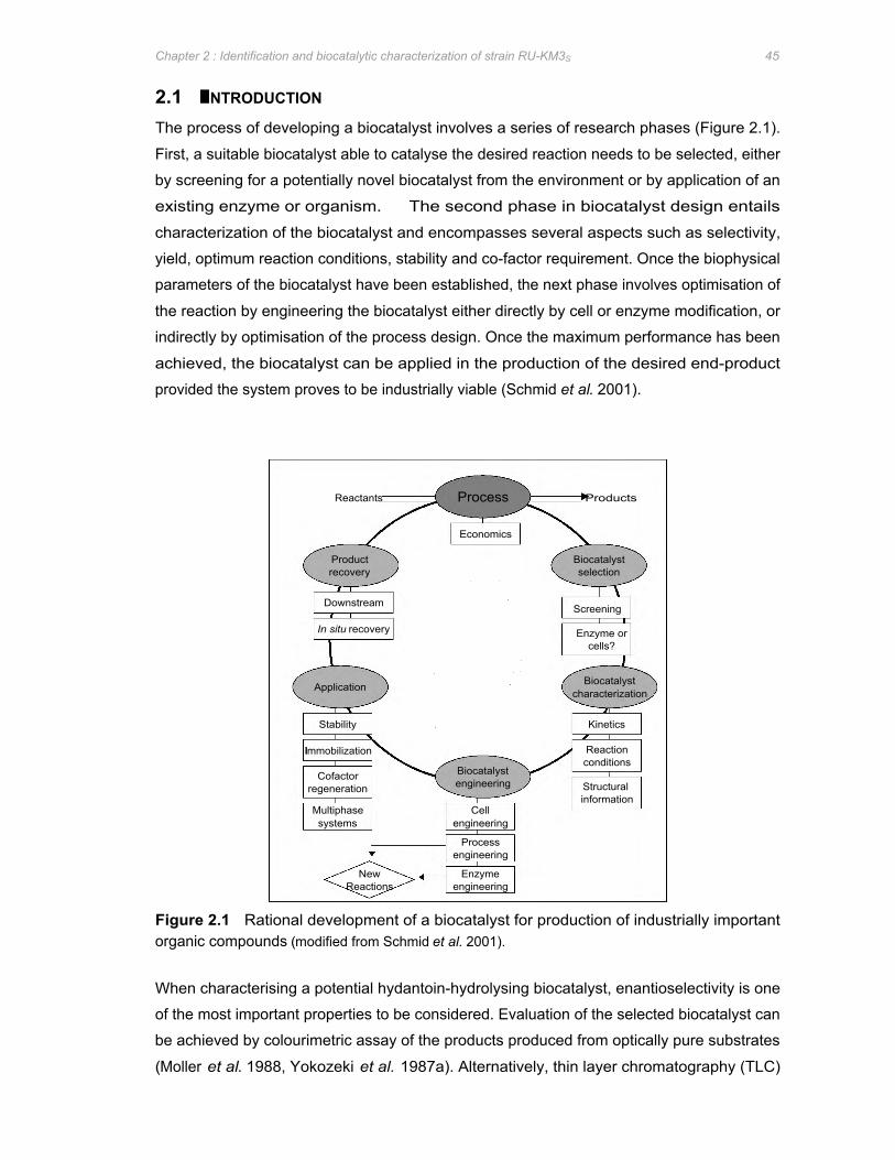
Immobilization
Cofactor regeneration
Multiphase systems
Application
In situ recovery
Stability
Downstream
Product recovery
Reactants Process Products
New Reactions
Cell engineering
Process engineering
Enzyme engineering
Biocatalyst engineering
Economics
Biocatalyst characterization
Screening
Biocatalyst selection
Enzyme or cells?
Structural information
Reaction conditions
Kinetics
Chapter 2 : Identification and biocatalytic characterization of strain RU-KM3S 45
2.1 INTRODUCTION
The process of developing a biocatalyst involves a series of research phases (Figure 2.1).
First, a suitable biocatalyst able to catalyse the desired reaction needs to be selected, either
by screening for a potentially novel biocatalyst from the environment or by application of an
existing enzyme or organism. The second phase in biocatalyst design entails
characterization of the biocatalyst and encompasses several aspects such as selectivity,
yield, optimum reaction conditions, stability and co-factor requirement. Once the biophysical
parameters of the biocatalyst have been established, the next phase involves optimisation of
the reaction by engineering the biocatalyst either directly by cell or enzyme modification, or
indirectly by optimisation of the process design. Once the maximum performance has been
achieved, the biocatalyst can be applied in the production of the desired end-product
provided the system proves to be industrially viable (Schmid et al. 2001).
Figure 2.1 Rational development of a biocatalyst for production of industrially important organic compounds (modified from Schmid et al. 2001).
When characterising a potential hydantoin-hydrolysing biocatalyst, enantioselectivity is one
of the most important properties to be considered. Evaluation of the selected biocatalyst can
be achieved by colourimetric assay of the products produced from optically pure substrates
(Moller et al. 1988, Yokozeki et al. 1987a). Alternatively, thin layer chromatography (TLC)
Chapter 2 : Identification and biocatalytic characterization of strain RU-KM3S 46
and high performance liquid chromatography (HPLC) using chiral supports (Morin et al.
1993, Syldatk et al. 1987, Hartley et al. 1998) allow for direct enantiomeric analysis of the
products. Enantiospecificity of hydantoin-hydrolysis products can also be accurately
determined by nuclear magnetic resonance (NMR) studies and infra-red spectroscopy
(Yokozeki et al. 1987a), or X-ray diffraction patterns of the amino acid products (Sano et al.
1977). However, these methods require expensive equipment as well as the purification of
the products from the reaction mixtures (Syldatk et al. 1990b). Alternatively, elucidation of
hydantoinase enantioselectivity can be determined via the reverse hydantoinase reaction: a
cell lysate containing hydantoinase activity was subjected to non-denaturing PAGE followed
by incubation with D- or L-carbamyltryptophan which is enzymatically converted to insoluble
indolymethylhydantoin and visualized as a precipitant (May et al. 1998e).
Besides enantioselectivity, factors such as metal ion- and ATP-dependency of hydantoin-
hydrolysing enzyme systems are also addressed in this chapter. The requirement of metal
ions for hydantoinase and N-carbamoylase activity has been well documented, however,
which metal ions are stimulatory and which are inhibitory with respect to enzyme activity
varies greatly between isolates. There are two major approaches to determining if the
hydantoinase or N-carbamoylase enzymes of a particular bacterial strain are metal ion
dependent or not. Firstly, the metal ions may be removed from the reaction mixture using
EDTA to chelate any metal ions present (Louwrier and Knowles 1997) or via dialysis of the
reaction mixture against a buffer solution which is free of metal ions (Syldatk et al. 1987).
The second approach involves the addition of the appropriate metal ion to the reaction
mixture and measurement of the effect of the metal on enzyme activity (Ishikawa et al. 1994).
Finally, identification of a microbial isolate with biocatalytic potential may provide information
as to the potential novelty of the hydantoin-hydrolysing enzyme system. In addition, the
potential pathogenicity of environmental isolates will influence the choice between using the
native organism or a recombinant host in the development of an industrial biocatalytic
process. Traditionally, microbial identification was based on phenotypic characteristics
determined by metabolic reactions and microscopy (Brunk et al. 1996). However,
identification of bacterial strains by these means are not always conclusive due to common
phenotypic parameters between several species, as well as expression of the tested
characteristics on plasmids which can undergo abrupt and multiple changes or be lost
altogether. In contrast, the analysis of the genotype of an organism does not rely on
expression of particular genes and is not subject to phenotypic variation (Towner and
Cockayne 1993). Due to the fundamental role of ribosomes in protein synthesis, rRNA
genes are highly conserved and are not affected by environmental conditions (Rosello-Mora
and Amann, 2001) yet sufficient differentiation exists between isolates to allow for
unambiguous identification (Brunk et al. 1996). In this study, the strain of interest was
Chapter 2 : Identification and biocatalytic characterization of strain RU-KM3S 47
initially identified based on phenotypic characteristics. This information was used to select
16S rRNA gene sequences from strains of the same species, from which primers were
designed and amplification of the 16S rRNA gene sequence from the environmental isolate
was done. The nucleotide sequence analysis of the resultant PCR product was then
compared to a comprehensive sequence dataset of approximately 16 277 prokaryotic entries
which is readily available on the Ribosomal Database Project (RDP) databases (Cole et al.
2003).
As described in Chapter 1, strain RU-KM3S was isolated from environmental samples and
selected due to its relatively high productivity in whole cell biocatalytic assays using
hydantoin as a substrate (Burton et al. 1998). The aim of the research described in this
chapter was to establish the presence of unique properties in the RU-KM3S hydantoin-
hydrolysing enzyme system. This was done through characterization of the RU-KM3S
enzyme system in terms of enantioselectivity, metal ion inhibition/stimulation, ATP-
dependence, and the identification of the strain by determining the 16S rRNA gene
sequence. Previous and concomitant characterization of RU-KM3S by Z. Skepu and K.
Buchanan with respect to substrate-selectivity, induction, and optimum reaction conditions on
hydantoin-hydrolysing activity are also discussed (Buchanan et al. 2001).
2.2 METHODS AND MATERIALS
2.2.1 Chemicals and kits D-L-methylhydantoin was prepared as a 1:1 mixture of D- and L-methylhydantoin (Toronto
Research Chemicals Ltd.). Hydantoin, N-carbamylglycine, glycine, and alanine were
obtained from Aldrich, ICN Biomedicals Inc., Saarchem, and Sigma respectively. Adenosine
triphosphate (ATP) was obtained from Boehringer Mannheim. The growth medium nutrient
broth was supplied by Merck.
2.2.2 Hydantoinase and N-carbamoylase enzyme assays 2.2.2.1 Resting cell biocatalytic assay A detailed methodology for resting cell biocatalytic assays is described in Appendix 2.1.
Unless otherwise stated, a starter culture of strain RU-KM3S was generated in hydantoin
minimal medium (HMM) (Appendix 1) and used to seed nutrient broth supplemented with
0.1% hydantoin as an inducer such that the OD600 was 0.02. The seeded cultures were then
incubated at 28oC, shaking at 200rpm, until the culture reached stationary phase of growth
(after approximately 20 hours). Absorbance readings were determined at 600nm using the
Shimadzu UV-visible spectrophotometer with the corresponding uninoculated medium as a
blank.
Chapter 2 : Identification and biocatalytic characterization of strain RU-KM3S 48
Once optimum OD600 was reached, the cells were harvested by centrifugation and
resuspended in 0.1M phosphate buffer (pH 8.0). A final concentration of 20mg wet cell
mass/ml was aliquoted into each reaction mixture with either 0.1M phosphate buffer (cell
blank) or the appropriate substrate in 0.1M phosphate buffer (e.g. 50mM hydantoin or 25mM
N-carbamylglycine). Following incubation at 40oC for 3 hours, the reaction mixtures were
centrifuged in a Heraeus microfuge (13 000rpm unless otherwise stated) and the supernatant
analysed for N-carbamylamino acids and amino acids using the Ehrlich’s and ninhydrin
colorimetric assays respectively. The units of activity were measured as μ mol/ml of product
and expressed as an average of 3 replicates. Hydantoinase activity was calculated as the
total amount of N-carbamylamino acid and amino acid produced from an hydantoin substrate
whilst N-carbamoylase activity was expressed as μ mol/ml amino acid produced from the N-
carbamylamino acid intermediate.
2.2.2.2 Cell-free extracts Induced RU-KM3S cells were cultured and harvested as described above. The resuspended
cells were then disrupted by a single pass through a Yeda-press (LINCA Lamon
Instrumentations, Tel Aviv) at 15psi., 4oC and at a flow rate of approximately 1 drop per
second. The resultant cell-free extract was then utilized in biocatalytic reactions as
described above.
2.2.3 Metal ion dependence To determine the effect of metal ions on hydantoinase and N-carbamoylase activities, the
metal ions Mg2+
, Mn2+
, Zn2+
, Co2+
, Cu2+
, Fe2+
, or Co2+
and Mg2+
were added to each
biocatalytic reaction triplicate set at a concentration of 2.5mM together with or without 2.5mM
Na2EDTA. To obtain dialysed cell free extracts, the harvested RU-KM3S cells were disrupted
as described above and the cell-free extract subjected to dialysis with SpectraforR dialysis
tubing (MWco 6-8000) against 0.1M phosphate buffer, pH 8.0, with or without 2.5mM
Na2EDTA for 90 minutes at 4oC, changing the buffer every half hour. In order to determine
the stimulatory effect of the respective metal ions on hydantoinase and N-carbamoylase
activities, 2.5mM Mg2+
, Mn2+
, Zn2+
, Co2+
, Cu2+
, Fe2+
, or Co2+
and Mg2+
were added to the
respective biocatalytic reaction sample sets after dialysis against 0.1M phosphate buffer
containing 2.5mM Na2EDTA as described above. Controls were established as undialysed
cell-free extract, cell-free extract dialysed without addition of any metal ions, and cell-free
extract allowed to stand at 4oC for the time taken for dialysis. Hydantoin was used as a
substrate at a final concentration of 50mM and the reactions were incubated at 40oC for 3
hours before carrying out Ehrlich’s and ninhydrin assays (Appendix 2.1).
Chapter 2 : Identification and biocatalytic characterization of strain RU-KM3S 49
2.2.4 Effect of ATP In order to determine the ATP requirements of the hydantoinase and N-carbamoylase in RU-
KM3S, ATP was added to the resting whole cell biocatlytic reactions (prepared as described
above) at a final concentration of 1.5mM, 3mM, and 6mM. Hydantoin, at a final
concentration of 50mM, was used as a substrate and the reaction mixtures incubated at 40oC
for 3 hours before carrying out Ehrlich’s and ninhydrin assays to determine levels of N-
carbamylglycine and glycine production respectively (Appendix 2).
2.2.5 Strain identification Genomic DNA was extracted from RU-KM3S by the detergent lysis/CTAB and organic
solvent extraction method as described in Appendix 5.2. Polymerase chain reaction (PCR)
amplification of the 16S rRNA gene from the prepared genomic DNA was done with universal
primers for the 16S rRNA coding region, CH1 and CH3 (Appendix 3, Table A3.1) using Vent
DNA Polymerase (New England Biolabs) under standard amplification parameters and an
annealing temperature of 63oC (Appendix 5.3). The resultant 1300bp PCR product was
cloned into the Bam HI site of vector pUC18 (Appendix 3, Figure A3.1) as follows: the PCR
product was digested with Bam HI restriction endonuclease (flanking sites generated by the
primers CH1 and CH3) and ligated with Bam HI digested pUC18, that had been treated with
Shrimp Alkaline Phosphatase (USB), using T4 DNA Ligase (Promega) as per manufacturers
specifications. The resultant plasmid clones were then transformed into competent E. coli
DH5α (supE44 ∆lacU169(∅80lacZΔM15)hsdR17 recA1 endA1 gyrA96 thi-1 relA1) and
colonies hosting insert-containing plasmids selected, using the blue/white selection provided
by β-galactosidase activity, on Luria agar plates containing 100μ g/ml ampicillin, 40μ g/ml 5-
bromo-4-chloro-3-indolyl-β-D-galactopyranosidase (X-gal) (Biosolve, Ltd), and 50μ g/ml
isopropyl-β-thiogalactosidase (IPTG) (Roche).
The High Pure Plasmid Isolation Kit (Roche) was used to purify the correct recombinant
clones, pGMS1 and pGMS2 in which the insert was subcloned into the pUC18 vector in both
orientations. The nucleotide sequence of the PCR product was determined using the Alf
Express Automated DNA Sequencing service (University of Cape Town, South Africa) and
ABI Prism Big Dye Terminator (Amersham) procedure. The 5’ and 3’ end of the 16S rRNA
gene was sequenced using primers specific for the pUC18 vector, pUCF and pUCR
(Appendix 3, Table A3.1). In order to generate overlapping sequence from both strands,
deletion constructs of pGMS1 and pGMS2 were constructed by restriction endonuclease
digestion with Hinc II (USB), Sma I (USB), or Csp 451 (Promega) followed by recirculirization
using T4 DNA Ligase. The nucleotide sequence of the resultant plasmids (pGMS1ΔHinc II,
pGMS1ΔSma I, pGMS2ΔSma I, pGMS2ΔCsp 451) was determined as above using primers
pUCF or pUCR where applicable.

Pro
duct
( m
ol/m
l)
40
60
50
30
20
10
0 0 4 6 8 10 14 18 20 21
N-carbamoylglycine Glycine OD 600
Time (Hours)
3.5
3
2.5
2
0.5
0
1.5
1
Abs
orba
nce
(OD
600)
Chapter 2 : Identification and biocatalytic characterization of strain RU-KM3S 50
Sequence data obtained for the 16S rRNA gene from RU-KM3S was subjected to
comparative analysis with the small subunit rRNA gene sequences available on the
Ribosomal Database Project II (http://rdp.cme.msu.edu/html/) (Cole et al. 2003). The
sequence alignment was done using ClustalW software (Thompson et al. 1994) located at
the BCM Search Launcher website (http://searchlauncher.bcm.tmc.edu) and presented in
boxshade format (http://www.ch.embnet.org/software/BOX form.html).
2.3 RESULTS
2.3.1 Growth phase for optimal hydantoin hydrolysis It has been well documented that maximum hydantoinase and N-carbamoylase activities
occur in cells during stationary phase of growth. Accordingly, the hydantoinase and N-
carbamoylase activities of RU-KM3S were determined in cells harvested at different stages of
growth in complete medium containing hydantoin as an inducer. During early (after 4 hours)
and mid-logarithmic phase (after 8 hours), low levels of hydantoin-hydrolysing activity were
observed (Figure 2.2). Production of glycine reached optimal levels after 14 hours of growth,
whilst maximum levels of hydantoinase activity were observed in cells harvested in mid- to
late-stationary phase after 21 hours of growth (Figure 2.2). Consequently, all subsequent
biocatalytic assays were carried out using cells harvested after 20 hours of growth in nutrient
broth at 28oC.
Figure 2.2 Production of N-carbamylglycine and glycine from hydantoin by RU-KM3S at various phases of growth in complete medium. (Error bars represent the standard error of the mean, n=3)
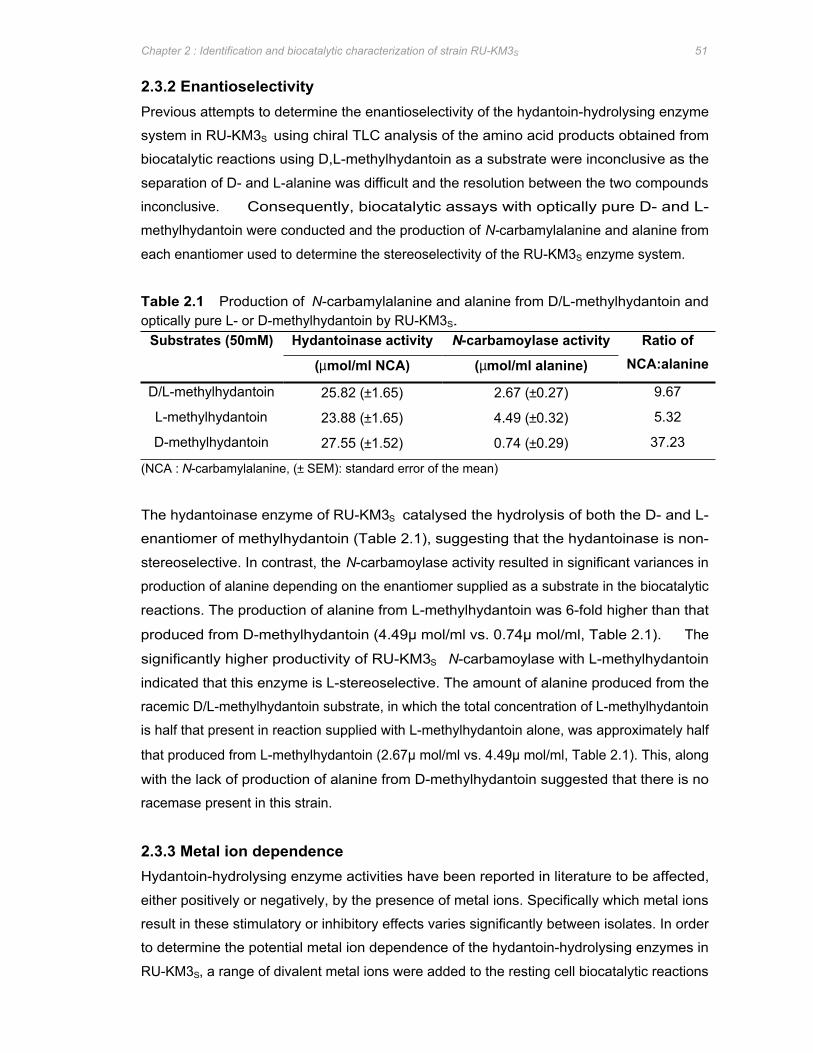
Chapter 2 : Identification and biocatalytic characterization of strain RU-KM3S 51
2.3.2 Enantioselectivity Previous attempts to determine the enantioselectivity of the hydantoin-hydrolysing enzyme
system in RU-KM3S using chiral TLC analysis of the amino acid products obtained from
biocatalytic reactions using D,L-methylhydantoin as a substrate were inconclusive as the
separation of D- and L-alanine was difficult and the resolution between the two compounds
inconclusive. Consequently, biocatalytic assays with optically pure D- and L-
methylhydantoin were conducted and the production of N-carbamylalanine and alanine from
each enantiomer used to determine the stereoselectivity of the RU-KM3S enzyme system.
Table 2.1 Production of N-carbamylalanine and alanine from D/L-methylhydantoin and optically pure L- or D-methylhydantoin by RU-KM3S.
Substrates (50mM) Hydantoinase activity N-carbamoylase activity Ratio of
(µ mol/ml NCA) (µ mol/ml alanine) NCA:alanine
D/L-methylhydantoin 25.82 (±1.65) 2.67 (±0.27) 9.67
L-methylhydantoin 23.88 (±1.65) 4.49 (±0.32) 5.32
D-methylhydantoin 27.55 (±1.52) 0.74 (±0.29) 37.23
(NCA : N-carbamylalanine, (± SEM): standard error of the mean)
The hydantoinase enzyme of RU-KM3S catalysed the hydrolysis of both the D- and L-
enantiomer of methylhydantoin (Table 2.1), suggesting that the hydantoinase is non-
stereoselective. In contrast, the N-carbamoylase activity resulted in significant variances in
production of alanine depending on the enantiomer supplied as a substrate in the biocatalytic
reactions. The production of alanine from L-methylhydantoin was 6-fold higher than that
produced from D-methylhydantoin (4.49μ mol/ml vs. 0.74μ mol/ml, Table 2.1). The
significantly higher productivity of RU-KM3S N-carbamoylase with L-methylhydantoin
indicated that this enzyme is L-stereoselective. The amount of alanine produced from the
racemic D/L-methylhydantoin substrate, in which the total concentration of L-methylhydantoin
is half that present in reaction supplied with L-methylhydantoin alone, was approximately half
that produced from L-methylhydantoin (2.67μ mol/ml vs. 4.49μ mol/ml, Table 2.1). This, along
with the lack of production of alanine from D-methylhydantoin suggested that there is no
racemase present in this strain.
2.3.3 Metal ion dependence Hydantoin-hydrolysing enzyme activities have been reported in literature to be affected,
either positively or negatively, by the presence of metal ions. Specifically which metal ions
result in these stimulatory or inhibitory effects varies significantly between isolates. In order
to determine the potential metal ion dependence of the hydantoin-hydrolysing enzymes in
RU-KM3S, a range of divalent metal ions were added to the resting cell biocatalytic reactions
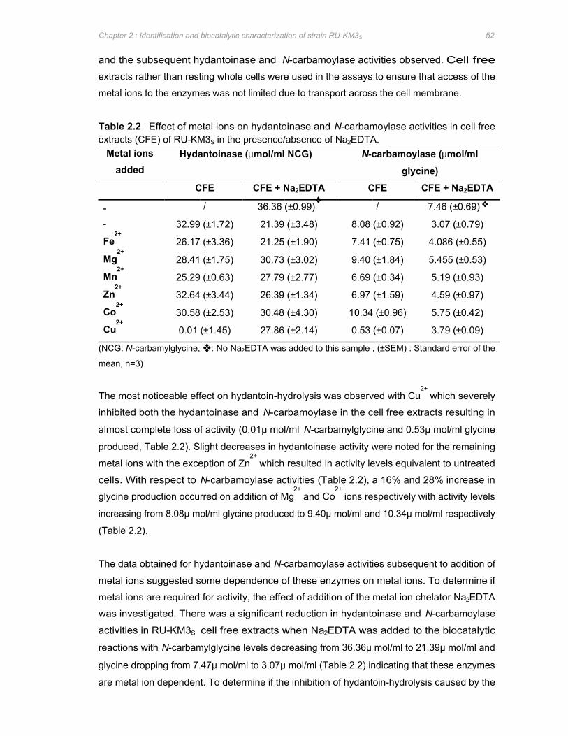
Chapter 2 : Identification and biocatalytic characterization of strain RU-KM3S 52
and the subsequent hydantoinase and N-carbamoylase activities observed. Cell free
extracts rather than resting whole cells were used in the assays to ensure that access of the
metal ions to the enzymes was not limited due to transport across the cell membrane.
Table 2.2 Effect of metal ions on hydantoinase and N-carbamoylase activities in cell free extracts (CFE) of RU-KM3S in the presence/absence of Na2EDTA.
Metal ions added
(µ mol/ml NCG)
-
Hydantoinase N-carbamoylase (µ mol/ml
glycine) CFE CFE + Na2EDTA CFE CFE + Na2EDTA
/ 36.36 (±0.99)❖
/ 7.46 (±0.69) ❖
- 32.99 (±1.72) 21.39 (±3.48) 8.08 (±0.92) 3.07 (±0.79)
Fe2+ 26.17 (±3.36) 21.25 (±1.90) 7.41 (±0.75) 4.086 (±0.55)
Mg2+ 28.41 (±1.75) 30.73 (±3.02) 9.40 (±1.84) 5.455 (±0.53)
Mn2+ 25.29 (±0.63) 27.79 (±2.77) 6.69 (±0.34) 5.19 (±0.93)
Zn2+ 32.64 (±3.44) 26.39 (±1.34) 6.97 (±1.59) 4.59 (±0.97)
Co2+ 30.58 (±2.53) 30.48 (±4.30) 10.34 (±0.96) 5.75 (±0.42)
Cu2+ 0.01 (±1.45) 27.86 (±2.14) 0.53 (±0.07) 3.79 (±0.09)
(NCG: N-carbamylglycine, ❖ : No Na2EDTA was added to this sample , (±SEM) : Standard error of the
mean, n=3)
The most noticeable effect on hydantoin-hydrolysis was observed with Cu2+
which severely
inhibited both the hydantoinase and N-carbamoylase in the cell free extracts resulting in
almost complete loss of activity (0.01μ mol/ml N-carbamylglycine and 0.53μ mol/ml glycine
produced, Table 2.2). Slight decreases in hydantoinase activity were noted for the remaining
metal ions with the exception of Zn2+
which resulted in activity levels equivalent to untreated
cells. With respect to N-carbamoylase activities (Table 2.2), a 16% and 28% increase in
glycine production occurred on addition of Mg2+
and Co2+
ions respectively with activity levels
increasing from 8.08μ mol/ml glycine produced to 9.40μ mol/ml and 10.34μ mol/ml respectively
(Table 2.2).
The data obtained for hydantoinase and N-carbamoylase activities subsequent to addition of
metal ions suggested some dependence of these enzymes on metal ions. To determine if
metal ions are required for activity, the effect of addition of the metal ion chelator Na2EDTA
was investigated. There was a significant reduction in hydantoinase and N-carbamoylase
activities in RU-KM3S cell free extracts when Na2EDTA was added to the biocatalytic
reactions with N-carbamylglycine levels decreasing from 36.36μ mol/ml to 21.39μ mol/ml and
glycine dropping from 7.47μ mol/ml to 3.07μ mol/ml (Table 2.2) indicating that these enzymes
are metal ion dependent. To determine if the inhibition of hydantoin-hydrolysis caused by the
Chapter 2 : Identification and biocatalytic characterization of strain RU-KM3S 53
addition of Na2EDTA is reversible, various metal ions were added to the biocatalytic reaction.
All of the tested metal ions, excepting Fe2+
with respect to hydantoinase activity, relieved the
negative effect of Na2EDTA to a certain degree but did not completely restore activity levels.
The increase in activity on addition of metal ions to the biocatalytic reactions containing
Na2EDTA could not, however, be directly attributed to the requirement of the respective
enzymes for a particular metal ion. Rather, the reduction in inhibition of enzyme activity was
most likely due to interaction of the respective metal ions with the Na2EDTA thus preventing
its interference with the hydantoinase and N-carbamoylase enzymes. To determine which
metal ions resulted in reactivation of the enzymes subsequent to inactivation due to metal ion
removal, dialysis of the cell free extract was done. Thus, when the cell free extract is utilized
in biocatalytic reactions to which the metal ions are added, the interference of the metal ion
chelating activity of Na2EDTA is absent. Dialysis also circumvents potential inhibitory effects
resulting from direct interaction between Na2EDTA and the enzymes.
Dialysis of cell free extracts against phosphate buffer alone resulted in a modest decrease in
enzyme activity (25.55μ mol/ml to 22.17μ mol/ml N-carbamylglycine and 6.9μ mol/ml to
5.34μ mol/ml glycine produced, Figure 2.3, panels A and B). This decrease was most likely
due to diffusion of metal ions into the metal ion free buffer as apposed to instability of the
enzymes under the conditions used for dialysis since a cell free extract incubated for an
equivalent time at 4oC showed only a small decrease in activity (Figure 2.3, panel A and B,
CFE vs. St). A significant reduction of 25% in hydantoinase (19.27μ mol/ml vs. 25.55μ mol/ml)
and 30% N-carbamoylase activity (4.8μ mol/ml vs. 6.91μ mol/ml), as compared with
undialysed cell-free extract was observed after dialysis against buffer containing Na2EDTA
(Figure 2.3, panels A and B, D+E vs. CFE). This data confirmed earlier results suggesting
the importance of divalent cations for hydantoinase and N-carbamoylase activities.
The addition of Cu2+
to dialyzed cell-free extracts resulted in dramatic loss of hydantoinase
and N-carbamoylase activity (Figure 2.3, panel A and B), as had been observed with
undialysed cell-free extracts (Table 2.2). The addition of Fe2+
resulted in a further reduction
in production of N-carbamylglycine (19.27μ mol/ml to 14.22μ mol/ml), and glycine (4.8μ mol/ml
to 2.83μ mol/ml) which accounted for an approximately 30% and 40% reduction in
hydantoinase and N-carbamoylase activity respectively. While Zn2+
increased hydantoinase
productivity slightly, it resulted in a 15% reduction in N-carbamoylase activity (Figure 2.3).
Metal ions Mg2+
, Mn2+
, and Co2+
were all stimulatory with respect to hydantoinase activity
with increases of 10%, 36% and 49% in N-carbamylglycine produced as compared with
dialysed cell-free extract (Figure 2.3, panel A). When considering N-carbamoylase activity,
Mg2+
inhibited the enzyme somewhat while Mn2+
and Co2+
significantly increased the activity
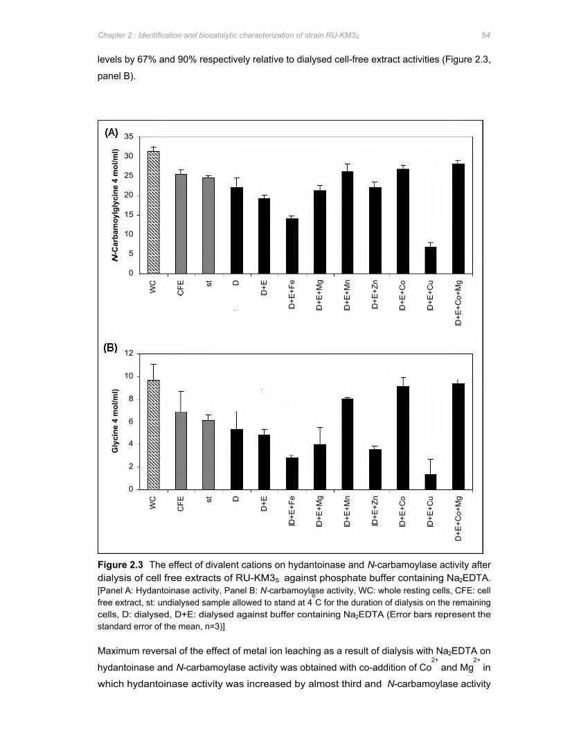
N -C
arba
moy
lgly
cine
4 m
ol/m
l) G
lyci
ne 4
mol
/ml)
35
30
25
20
15
10
12
10
4
5
0
8
6
2
0
WC
W
C
CFE
C
FE
st
st
D
D
D+E
D
+E
D+E
+Fe
D+E
+Fe
D+E
+Mg
D+E
+Mg
D+E
+Mn
D+E
+Mn
D+E
+Zn
D+E
+Zn
D+E
+Co
D+E
+Co
D+E
+Cu
D+E
+Cu
D+E
+Co+
Mg
D+E
+Co+
Mg
Chapter 2 : Identification and biocatalytic characterization of strain RU-KM3S 54
levels by 67% and 90% respectively relative to dialysed cell-free extract activities (Figure 2.3,
panel B).
Figure 2.3 The effect of divalent cations on hydantoinase and N-carbamoylase activity after dialysis of cell free extracts of RU-KM3S against phosphate buffer containing Na2EDTA. [Panel A: Hydantoinase activity, Panel B: N-carbamoylase activity, WC: whole resting cells, CFE: cell free extract, st: undialysed sample allowed to stand at 4
oC for the duration of dialysis on the remaining
cells, D: dialysed, D+E: dialysed against buffer containing Na2EDTA (Error bars represent the standard error of the mean, n=3)]
Maximum reversal of the effect of metal ion leaching as a result of dialysis with Na2EDTA on
hydantoinase and N-carbamoylase activity was obtained with co-addition of Co2+
and Mg2+
in
which hydantoinase activity was increased by almost third and N-carbamoylase activity
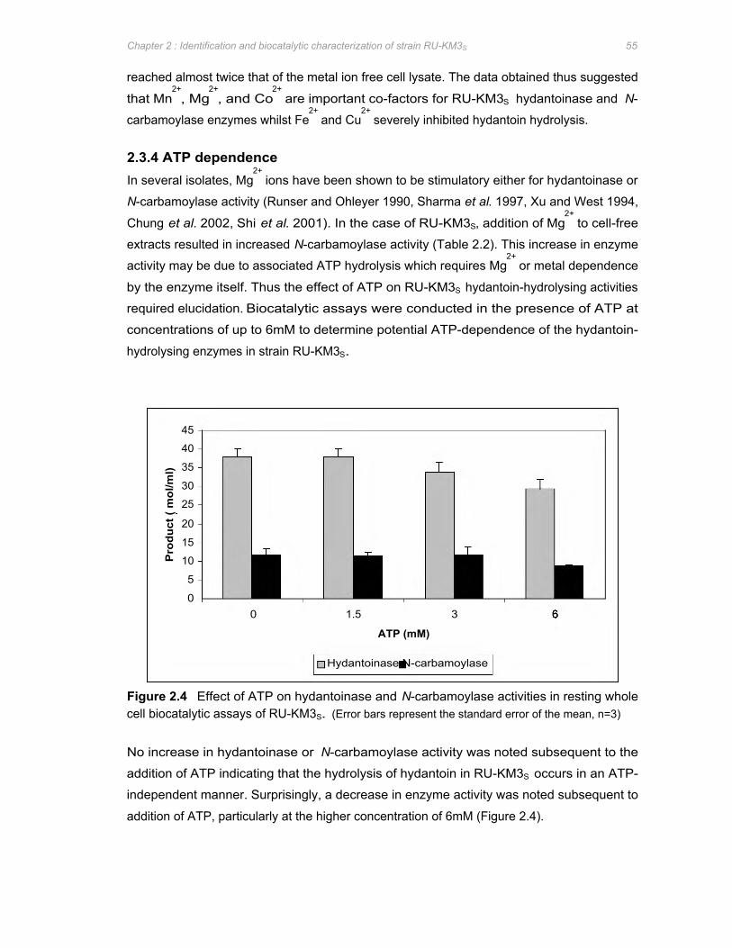
Pro
duct
( m
ol/m
l)
45
40
35
30
25
20
15
10
5
0 0 1.5 3
Hydantoinase N-carbamoylase
ATP (mM)
Chapter 2 : Identification and biocatalytic characterization of strain RU-KM3S 55
reached almost twice that of the metal ion free cell lysate. The data obtained thus suggested
that Mn2+
, Mg2+
, and Co2+
are important co-factors for RU-KM3S hydantoinase and N-
carbamoylase enzymes whilst Fe2+
and Cu2+
severely inhibited hydantoin hydrolysis.
2.3.4 ATP dependence In several isolates, Mg
2+ ions have been shown to be stimulatory either for hydantoinase or
N-carbamoylase activity (Runser and Ohleyer 1990, Sharma et al. 1997, Xu and West 1994,
Chung et al. 2002, Shi et al. 2001). In the case of RU-KM3S, addition of Mg2+
to cell-free
extracts resulted in increased N-carbamoylase activity (Table 2.2). This increase in enzyme
activity may be due to associated ATP hydrolysis which requires Mg2+
or metal dependence
by the enzyme itself. Thus the effect of ATP on RU-KM3S hydantoin-hydrolysing activities
required elucidation. Biocatalytic assays were conducted in the presence of ATP at
concentrations of up to 6mM to determine potential ATP-dependence of the hydantoin-
hydrolysing enzymes in strain RU-KM3S.
Figure 2.4 Effect of ATP on hydantoinase and N-carbamoylase activities in resting whole cell biocatalytic assays of RU-KM3S. (Error bars represent the standard error of the mean, n=3)
No increase in hydantoinase or N-carbamoylase activity was noted subsequent to the
addition of ATP indicating that the hydrolysis of hydantoin in RU-KM3S occurs in an ATP-
independent manner. Surprisingly, a decrease in enzyme activity was noted subsequent to
addition of ATP, particularly at the higher concentration of 6mM (Figure 2.4).
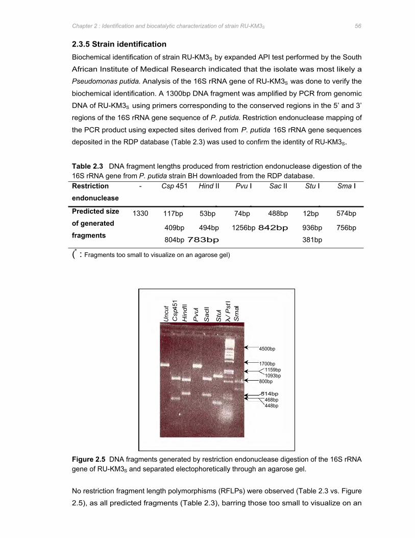
Unc
ut
Csp
451
Hin
d II
Pvu
I
Sa
c II
Stu
I λ
/ Pst
I S
ma I
514bp 468bp 448bp
4500bp
800bp
1700bp 1159bp 1093bp
Chapter 2 : Identification and biocatalytic characterization of strain RU-KM3S 56
2.3.5 Strain identification Biochemical identification of strain RU-KM3S by expanded API test performed by the South
African Institute of Medical Research indicated that the isolate was most likely a
Pseudomonas putida. Analysis of the 16S rRNA gene of RU-KM3S was done to verify the
biochemical identification. A 1300bp DNA fragment was amplified by PCR from genomic
DNA of RU-KM3S using primers corresponding to the conserved regions in the 5’ and 3’
regions of the 16S rRNA gene sequence of P. putida. Restriction endonuclease mapping of
the PCR product using expected sites derived from P. putida 16S rRNA gene sequences
deposited in the RDP database (Table 2.3) was used to confirm the identity of RU-KM3S.
Table 2.3 DNA fragment lengths produced from restriction endonuclease digestion of the 16S rRNA gene from P. putida strain BH downloaded from the RDP database. Restriction - Csp 451 Hind II Pvu I Sac II Stu I Sma I
endonuclease
Predicted size 1330 117bp∗
53bp∗
74bp∗
488bp 12bp∗
574bp of generated 409bp 494bp 1256bp 842bp 936bp 756bp fragments 804bp 783bp 381bp
(∗ : Fragments too small to visualize on an agarose gel)
Figure 2.5 DNA fragments generated by restriction endonuclease digestion of the 16S rRNA gene of RU-KM3S and separated electophoretically through an agarose gel.
No restriction fragment length polymorphisms (RFLPs) were observed (Table 2.3 vs. Figure
2.5), as all predicted fragments (Table 2.3), barring those too small to visualize on an
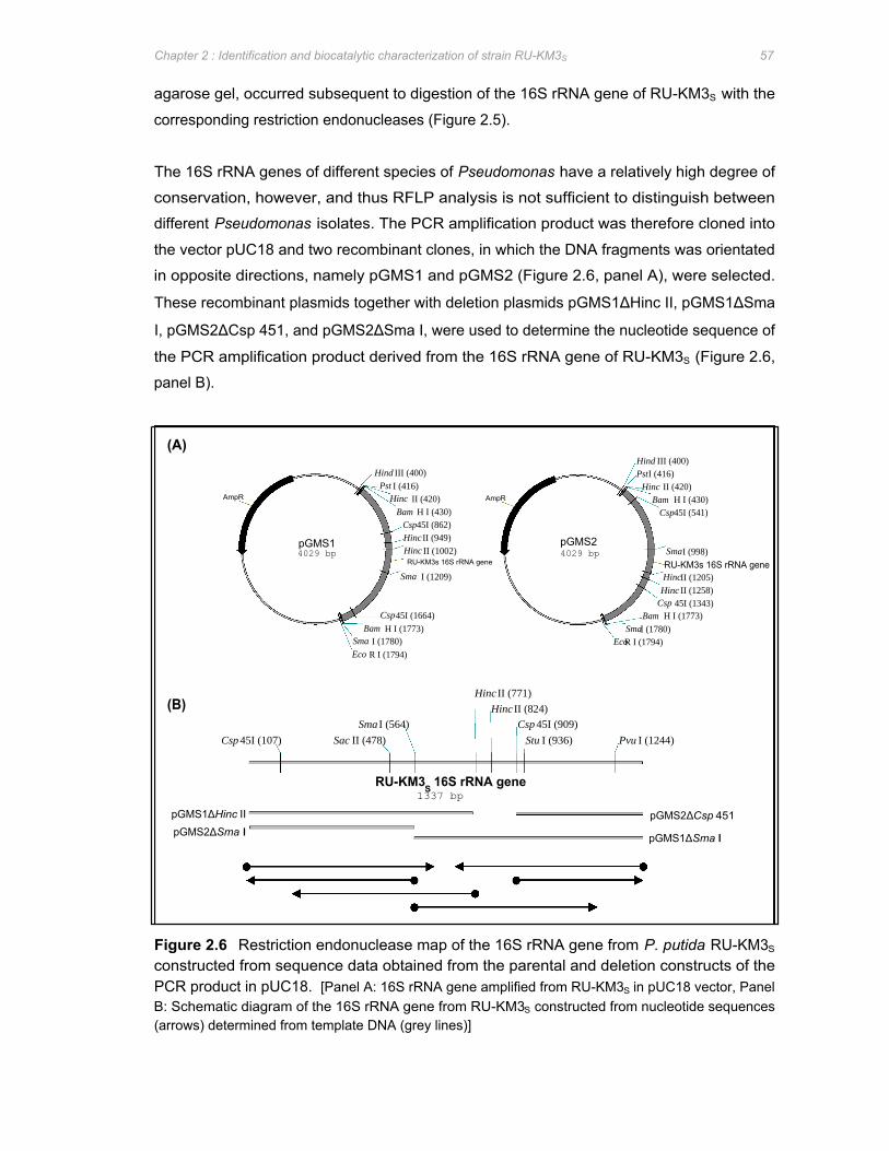
pGMS1ΔHinc II pGMS2ΔSma I
pGMS2ΔCsp 451
pGMS1ΔSma I
Hind III (400) PstI (416) Hind III (400)
Pst I (416) Hinc II (420) AmpR Bam H I (430)
Bam H I (430) Csp45I (541) Csp45I (862)
Hinc II (420) AmpR
Sma I (1209) HincII (1205) Hinc II (1258)
Csp 45I (1343) Bam H I (1773) Csp45I (1664)
SmaI (1780) Bam H I (1773) EcoR I (1794) Sma I (1780)
Eco R I (1794)
Sma I (564) Csp 45I (909)
pGMS1 4029 bp
Hinc II (949) pGMS2 Hinc II (1002) 4029 bp RU-KM3s 16S rRNA gene
SmaI (998)
RU-KM3s 16S rRNA gene
Hinc II (771)
Hinc II (824)
Csp 45I (107) Sac II (478) Stu I (936) Pvu I (1244)
(A)
(B)
RU-KM3S 16S rRNA gene 1337 bp
Chapter 2 : Identification and biocatalytic characterization of strain RU-KM3S 57
agarose gel, occurred subsequent to digestion of the 16S rRNA gene of RU-KM3S with the
corresponding restriction endonucleases (Figure 2.5).
The 16S rRNA genes of different species of Pseudomonas have a relatively high degree of
conservation, however, and thus RFLP analysis is not sufficient to distinguish between
different Pseudomonas isolates. The PCR amplification product was therefore cloned into
the vector pUC18 and two recombinant clones, in which the DNA fragments was orientated
in opposite directions, namely pGMS1 and pGMS2 (Figure 2.6, panel A), were selected.
These recombinant plasmids together with deletion plasmids pGMS1ΔHinc II, pGMS1ΔSma
I, pGMS2ΔCsp 451, and pGMS2ΔSma I, were used to determine the nucleotide sequence of
the PCR amplification product derived from the 16S rRNA gene of RU-KM3S (Figure 2.6,
panel B).
Figure 2.6 Restriction endonuclease map of the 16S rRNA gene from P. putida RU-KM3S
constructed from sequence data obtained from the parental and deletion constructs of the PCR product in pUC18. [Panel A: 16S rRNA gene amplified from RU-KM3S in pUC18 vector, Panel B: Schematic diagram of the 16S rRNA gene from RU-KM3S constructed from nucleotide sequences (arrows) determined from template DNA (grey lines)]

Chapter 2 : Identification and biocatalytic characterization of strain RU-KM3S 58
The nucleotide sequence obtained was entered into the Ribosomal Similarity Ranking facility
(Maidek et al. 2001) and aligned with the 10 most similar 16S rRNA sequences in the RDP
database. The Sequence Match Version 2.7 program quantifies the number of unique
oligomers in the submitted sequence which is common to each sequence in the database.
The result (S_ab) was expressed as the number of shared oligomers divided by either the
number of unique oligomers in the submitted sequence or the data base sequence,
depending on which is lower. This ensured that a good match between partial sequences, or
partial and complete sequences, was scored higher than a less efficient match between
complete sequences. High S_ab scores were obtained primarily with P. putida strains
although homology of the RU-KM3S 16S rRNA gene was observed with a P. plecoglossicida
and a P. monteilli strain (Table 2.4). Based on 16S rRNA gene sequence, RU-KM3S is most
closely related to Pseudomonas putida strain BH, thus verifying the biochemical identification
previously done.
Table 2.4 Similarity rank of the 16S rRNA gene from strain RU-KM3S with corresponding gene sequences submitted to the Ribosomal Database Project
S_ab Organism
0.964 Pseudomonas putida strain BH
0.964 Pseudomonas plecoglossicida strain FPC951
0.961 Pseudomonas putida strain A10L
0.956 Pseudomonas putida strain mt-2 JMC6156
0.955 Pseudomonas monteilli strain CFML 90-60 CIP 104883
0.953 Pseudomonas putida strain PB4
0.950 Pseudomonas putida strain K23-1
0.949 Pseudomonas putida strain F1
0.945 Pseudomonas putida strain mt-2
0.945 Pseudomonas putida strain IFO 14671
The nucleotide variances between P. putida strains RU-KM3S and BH can be seen in Figure
2.7 in which the sequence alignment of the 16S rRNA gene from these two isolates are
depicted in boxshade format. The nucleotide sequence of the terminal ends of the 16S rRNA
gene from RU-KM3S are not present in the alignment (Figure 2.7). This is due to the fact that
ion exchange columns, with a size exclusion of 75bp, were used to purify the extension
products of the cycle sequencing reaction before analysis in the ABI Prism 3100 Genetic
Analyser. Thus the first 75bp from the end of the primer utilized in the cycle sequencing
reaction will not be represented in the sequence data.
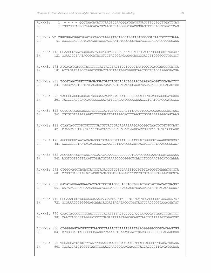
Chapter 2 : Identification and biocatalytic characterization of strain RU-KM3S 59
RU-KM3s 1 - - - - GCCTAACACATGCAAGTCGAACGGATGACGGGAGCTTGCTCCTTGATTCAG BH 1 TGGCGGCAGGCCTAACACATGCAAGTCGAGCGGATGACGGGAGCTTGCTCCTTGATTCAG
RU-KM3s 52 CGGCGGACGGGTGAGTAATGCCTAGGAATCTGCCTGGTAGTGGGGGACAACGTTTCGAAA BH 61 CGGCGGACGGGTGAGTAATGCCTAGGAATCTGCCTGGTAGTGGGGGACAACGTTTCGAAA
RU-KM3s 112 GGAACGCTAATACCGCATACGTCCTACGGGAGAAAGCAGGGGACCTTCGGGCCTTGCGCT BH 121 GGAACGCTAATACCGCATACGTCCTACGGGAGAAAGCAGGGGACCTTCGGGCCTTGCGCT
RU-KM3s 172 ATCAGATGAGCCTAGGTCGGATTAGCTAGTTGGTGGGGTAATGGCTCACCAAGGCGACGA BH 181 ATCAGATGAGCCTAGGTCGGATTAGCTAGTTGGTGGGGTAATGGCTCACCAAGGCGACGA
RU-KM3s 232 TCCGTAACTGGTCTGAGAGGATGATCAGTCACACTGGAACTGAGACACGGTCCAGACTCC BH 241 TCCGTAACTGGTCTGAGAGGATGATCAGTCACACTGGAACTGAGACACGGTCCAGACTCC
RU-KM3s 292 TACGGGAGGCAGCAGTGGGGAATATTGGACAATGGGCGAAAGCCTGATCCAGCCATGCCG BH 301 TACGGGAGGCAGCAGTGGGGAATATTGGACAATGGGCGAAAGCCTGATCCAGCCATGCCG
RU-KM3s 352 CGTGTGTGAAGAAGGTCTTCGGATTGTAAAGCACTTTAAGTTGGGAGGAAGGGCAGTAAG BH 361 CGTGTGTGAAGAAGGTCTTCGGATTGTAAAGCACTTTAAGTTGGGAGGAAGGGCAGTAAG
RU-KM3s 412 CTAATACCTTGCTGTTTTGACGTTACCGACAGAATAAGCACCGGCTAACTCTGTGCCAGC BH 421 CTAATACCTTGCTGTTTTGACGTTACCGACAGAATAAGCACCGGCTAACTCTGTGCCAGC
RU-KM3s 472 AGCCGCGGTAATACAGAGGGTGCAAGCGTTAATCGGAATTACTGGGCGTAAAGCGCGCGT BH 481 AGCCGCGGTAATACAGAGGGTGCAAGCGTTAATCGGAATTACTGGGCGTAAAGCGCGCGT
RU-KM3s 532 AGGTGGTTCGTTAAGTTGGATGTGAAAGCCCCGGGCTCAACCTGGGAACTGCATCCAAAA BH 541 AGGTGGTTCGTTAAGTTGGATGTGAAAGCCCCGGGCTCAACCTGGGAACTGCATCCAAAA
RU-KM3s 592 CTGGC-AGCTAGAGTACGGTAGAGGGTGGTGGAATTTCCTGTGTAGCGGTGAAATGCGTA BH 601 CTGGCGAGCTAGAGTACGGTAGAGGGTGGTGGAATTTCCTGTGTAGCGGTGAAATGCGTA
RU-KM3s 651 GATATAGGAAGGAACACCAGTGGCGAAGGC-ACCACCTGGACTGATACTGACACTGAGGT BH 661 GATATAGGAAGGAACACCAGTGGCGAAGGCGACCACCTGGACTGATACTGACACTGAGGT
RU-KM3s 710 GCGAAAGCGTGGGGAGCAAACAGGATTAGATACCCTGGTAGTCCACGCCGTAAACGATGT BH 721 GCGAAAGCGTGGGGAGCAAACAGGATTAGATACCCTGGTAGTCCACGCCGTAAACGATGT
RU-KM3s 770 CAACTAGCCGTTGGAATCCTTGAGATTTTAGTGGCGCAGCTAACGCATTAAGTTGACCGC BH 781 CAACTAGCCGTTGGAATCCTTGAGATTTTAGTGGCGCAGCTAACGCATTAAGTTGACCGC
RU-KM3s 830 CTGGGGAGTACGGCCGCAAGGTTAAAACTCAAATGAATTGACGGGGGCCCGCACAAGCGG BH 841 CTGGGGAGTACGGCCGCAAGGTTAAAACTCAAATGAATTGACGGGGGCCCGCACAAGCGG
RU-KM3s 890 TGGAGCATGTGGTTTAATTCGAAGCAACGCGAAGAACCTTACCAGGCCTTGACATGCAGA BH 901 TGGAGCATGTGGTTTAATTCGAAGCAACGCGAAGAACCTTACCAGGCCTTGACATGCAGA
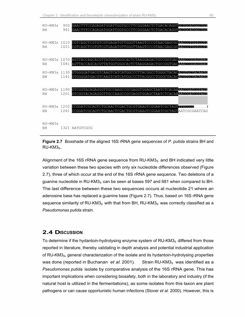
Chapter 2 : Identification and biocatalytic characterization of strain RU-KM3S 60
RU-KM3s BH
RU-KM3s BH
RU-KM3s BH
RU-KM3s BH
RU-KM3s BH
RU-KM3s BH
RU-KM3s BH
950 961
1010 1021
1070 1081
1130 1141
1190 1201
1250 1261
1321
GAACTTTCCAGAGATGGATTGGTGCCTTCGGGAACTCTGACACAGGTGCTGCATGGCTGT GAACTTTCCAGAGATGGATTGGTGCCTTCGGGAACTCTGACACAGGTGCTGCATGGCTGT
CGTCAGCTCGTGTCGTGAGATGTTGGGTTAAGTCCCGTAACGAGCGCAACCCTTGTCCTT CGTCAGCTCGTGTCGTGAGATGTTGGGTTAAGTCCCGTAACGAGCGCAACCCTTGTCCTT
AGTTACCAGCACGTTATGGTGGGCACTCTAAGGAGACTGCCGGTGACAAACCGGAGGAAG AGTTACCAGCACGTTATGGTGGGCACTCTAAGGAGACTGCCGGTGACAAACCGGAGGAAG
GTGGGGATGACGTCAAGTCATCATGGCCCTTACGGCCTGGGCTACACACGTGCTACAATG GTGGGGATGACGTCAAGTCATCATGGCCCTTACGGCCTGGGCTACACACGTGCTACAATG
GTCGGTACAGAGGGTTGCCAAGCCGCGAGGTGGAGCTAATCTCACAAAACCGATCGTAGT GTCGGTACAGAGGGTTGCCAAGCCGCGAGGTGGAGCTAATCTCACAAAACCGATCGTAGT
AATCGCGAATCAG CCGGATCGCAGTCTGCAACTCGACTGCGTGAAGTCGGAATCGCTAGTTTTCCCG - - CCGGATCGCAGTCTGCAACTCGACTGCGTGAAGTCGGAATCGCTAGT
AATGTCGCG
Figure 2.7 Boxshade of the aligned 16S rRNA gene sequences of P. putida strains BH and RU-KM3S.
Alignment of the 16S rRNA gene sequence from RU-KM3S and BH indicated very little
variation between these two species with only six nucleotide differences observed (Figure
2.7), three of which occur at the end of the 16S rRNA gene sequence. Two deletions of a
guanine nucleotide in RU-KM3S can be seen at bases 597 and 681 when compared to BH.
The last difference between these two sequences occurs at nucleotide 21 where an
adenosine base has replaced a guanine base (Figure 2.7). Thus, based on 16S rRNA gene
sequence similarity of RU-KM3S with that from BH, RU-KM3S was correctly classified as a
Pseudomonas putida strain.
2.4 DISCUSSION
To determine if the hydantoin-hydrolysing enzyme system of RU-KM3S differed from those
reported in literature, thereby validating in depth analysis and potential industrial application
of RU-KM3S, general characterization of the isolate and its hydantoin-hydrolysing properties
was done (reported in Buchanan et al. 2001). Strain RU-KM3S was identified as a
Pseudomonas putida isolate by comparative analysis of the 16S rRNA gene. This has
important implications when considering biosafety, both in the laboratory and industry (if the
natural host is utilized in the fermentations), as some isolates from this taxon are plant
pathogens or can cause opportunistic human infections (Stover et al. 2000). However, this is

Chapter 2 : Identification and biocatalytic characterization of strain RU-KM3S 61
not the case for P. putida. In addition, P. putida KT2440 has been certified as a biological
safety strain by the Recombinant DNA Advisory Committee (Nelson et al. 2002).
With respect to hydantoin-hydrolysing activities, initial characterization of RU-KM3S by
Buchanan (1996) revealed an optimum pH, temperature and incubation period for catalysis
of hydantoin by resting whole cells to be pH 8.0, 40oC, and 3 hours respectively after which
100% conversion of hydantoin to N-carbamylglycine and an average of 20% conversion to
glycine occurred. Induction of the hydantoinase and N-carbamoylase enzymes with
hydantoin, dihydrouracil, dimethylhydantoin, and thiouracil was investigated with hydantoin
being the best inducer (Buchanan, 1996). The substrate selectivity of the hydantoin
hydrolysing enzyme in RU-KM3S revealed high levels of hydantoin converting activity, with
the aliphatic but not necessarily short-chain 5-monosubstituted hydantoin derivatives as the
preferred substrates (Skepu, 2000). The hydantoinase enzyme appeared to be non-
stereoselective, whilst the N-carbamoylase is strictly L-stereoselective when supplied with
methylhydantoin as a substrate. The enzyme system from RU-KM3S appeared to be ATP-
independent but metal-ion dependent as significant reduction in the production of N-
carbamylglycine and glycine occurred after dialysis of crude cell extracts against buffer
containing the metal ion chelator Na2EDTA. Of the metal ions tested, Cu2+
severely inhibited
both enzymes whilst Co2+
, Mg2+
, and Mn2+
stimulated hydantoin-hydrolysing activities.
Expression of the hydantoinase and N-carbamoylase enzymes in RU-KM3S cells requires the
presence of an inducer and, when cultured in complete medium, optimal enzyme activities
were detected at early stationary growth phase. This observation corresponds to data in
literature where hydantoinase and N-carbamoylase enzymes are optimally produced after log
phase of growth (Gokhale et al. 1996, Sudge et al. 1998, Ishikawa et al. 1993, Hartley et al.
1998, Syldatk et al. 1990a, Lee et al. 1994, Hils et al. 2001). The increase in hydantoin-
hydrolysing enzyme activities late in the growth cycle of RU-KM3S suggests that the
hydantoinase and N-carbamoylase are subject to catabolite repression. Typically, a medium
with readily metabolizable nutrient sources will promote rapid biomass production but
simultaneously cause repression of enzymes involved in utilization of poorer sources of
nitrogen or carbon such as hydantoin. Both carbon and nitrogen catabolite repression have
been reported to regulate hydantoin-hydrolysing activity (Sudge et al. 1998, Hartley et al.
2001). The extent to which catabolite repression regulates hydantoinase and N-
carbamoylase activities in RU-KM3S cells will be examined in detail in Chapter 5.
Hydantoinase and N-carbamoylase enzymes have been reported in a wide diversity of
bacterial strains with the biocatalytic characteristics varying considerably between isolates.
The bulk of research with respect to hydantoin-hydrolysing bacterial strains has been carried
out on Agrobacterium, Arthrobacter, and Bacillus isolates. Of the Pseudomonas species with
Chapter 2 : Identification and biocatalytic characterization of strain RU-KM3S 62
hydantoin-hydrolysing abilities, only Pseudomonas sp. NS671 (Watabe et al. 1992a) is
reported to express a non-enantioselective hydantoinase. The hydantoinases from
remaining isolates all exhibit D-enantiospecificities (Ogawa and Shimizu 1994, LaPointe et
al. 1994, Gokhale et al.1996, Kim and Kim 1993, Sudge et al. 1998, Yokozeki et al. 1987c,
Chien et al. 1998, Shi et al. 2001). N-carbamoylase enzyme activities are not reported for
most of the Pseudomonas sp. listed above. Pseudomonas sp. KNK003A (Ikenaka et al.
1998a) and AJ-11220 (Yokozeki et al. 1987b) are reported to have a D-stereoselective
carbamoylases whilst Pseudomonas sp. NS671 expresses an L-enantioselective N-
carbamoylase (Watabe et al. 1992a) and Pseudomonas putida IFO 12996 expresses a β-
ureidopropionase with L-enantioselective N-carbamoylase activity (Ogawa and Shimizu
1994). Thus, with respect to enantioselectivities of the hydantoinase and N-carbamoylase
enzymes, RU-KM3S seems most similar to Pseudomonas sp. NS671 and P. putida IFO
12996 with a non-enantioselective hydantoinase and L-enantioselective N-carbamoylase.
However, the non-stereoselective hydantoinase from Pseudomonas sp. NS671 has been
shown to be ATP-dependant which is not the case for the hydantoinase from RU-KM3S. On
the other hand, the N-carbamoylase from this strain displays several similarities with the
corresponding enzyme in RU-KM3S with similar optimum pH and temperature profiles, L-
enantiospecificity, and inhibition by ATP at high concentrations (5mM-10mM) in crude
extracts (Ishikawa et al. 1993). The β-ureidopropionase from P. putida IFO 12996 (Ogawa
and Shimizu 1994) whilst sharing L-enantioselectivity with RU-KM3S, differs in terms of
optimal pH and temperature for biocatalytic activity. In addition, IFO 12996 β-
ureidopropionase does not hydrolyse N-carbamyl-L-leucine (Ogawa and Shimizu 1994)
which is readily hydrolysed by the N-carbamoylase from RU-KM3S (Buchanan et al. 2001).
However, characterization of the β-ureidopropionase from P. puitda IFO 12996 was done
using purified enzymes whilst characterization of the N-carbamoylase activity in RU-KM3S
was done with whole cells or cell-free extracts which may account for some of the differences
noted between these two enzymes.
The effect of metal ions, either positive or negative, on hydantoinase and N-carbamoylase
enzyme activities has been documented for almost all isolates reported to exhibit hydantoin-
hydrolysing activity. Which metal ions are stimulatory and which are inhibitory varies
between species and even between strains. However, many of the reported enzymes are
inhibited by Cu2+
as is the case with RU-KM3S (Morin et al. 1986b, Syldatk et al. 1987,
Ogawa and Shimizu 1994, Ogawa et al. 1993, Xu et al. 1994, Ishikawa et al. 1994, Louwrier
and Knowles 1996, 1997, Nanba et al. 1998). Exceptions to this are Ochobactrum anthropi
245 in which the addition of Cu2+
resulted in stimulation of the hydantoinase (Pozo et al.
2002), and B. stearothermophilus NS1122A hydantoinase which was unaffected by Cu2+
ions
(Mukohara et al. 1994).
Chapter 2 : Identification and biocatalytic characterization of strain RU-KM3S 63
Specific comparison of the effect of metal ions on hydantoinase (Shi et al. 2001), L-
carbamoylases (Ishikawa et al. 1994), and β-ureidopropionase (Ogawa and Shimizu 1994)
from Pseudomonas sp. with the corresponding enzymes in P. putida RU-KM3S demonstrates
some similarities and differences between these isolates. Both the L-carbamoylase and β-
ureidopropionase from Pseudomonas sp. NS671 and P. putida IFO 12996 respectively, were
stimulated by the addition of Mn2+
, Co2+
, and Ni2+
. Whilst Ni2+
was not tested for RU-KM3S,
Mn2+
and Co2+
were also stimulatory with respect to the N-carbamoylase activity in this strain.
However, P. putida IFO 12996 β-ureidopropionase activity was reported to be restored by
Fe2+
subsequent to treatment with EDTA (Ogawa and Shimizu 1994), whereas Fe2+
was
shown to significantly inhibit enzyme activity in RU-KM3S. The D-hydantoinase from
Pseudomonas sp. 2262 (Shi et al. 2001) was reported to be activated by addition of Mn2+
and Mg2+
which is consistent with what occurs in RU-KM3S. However, the Pseudomonas sp.
2262 hydantoinase enzyme is inhibited by Zn2+
which has a slight stimulatory effect on the
hydantoinase from RU-KM3S.
In conclusion, whilst some similarities exist between the hydantoin-hydrolysing enzymes
system in RU-KM3S and those characterized in other Pseudomonas isolates, sufficient
variability was observed for the enzymes from RU-KM3S to be considered different. The
most conclusive method to determine if an enzyme is unique or not is by comparison of the
nucleotide sequence of the encoding gene with those reported in literature. Thus isolation of
these genes from RU-KM3S and determination of their nucleotide sequence was
investigated.
Chapter 3 : Construction and screening of RU-KM3S genome library 64
3.1 INTRODUCTION
Isolation of the genes encoding the enzymes responsible for hydantoin hydrolysis in RU-
KM3S would be beneficial for a variety of reasons. Not only would it verify that the enzymes
are indeed different to those reported in literature but it would also provide insight into the
regulation of expression of the genes and, by comparison to crystallographic and mutational
analysis done on previously isolated enzymes, provide an opportunity to optimise the
catalytic function of the enzymes.
Several hydantoinase and N-carbamoylase enzymes have recently been cloned and
heterologously expressed in E. coli. The methodologies used to isolate the genes encoding
these enzymes include screening of genomic libraries or amplification of the relevant genes
from genomic DNA by PCR using primers designed from the N-terminal amino acid
sequence of the purified protein (Chen and Tsai 1998) or degenerate primers designed
based on sequences of related enzymes in literature (Chao et al. 1999b, Hils et al. 2001, Hu
et al. 2003). Genomic libraries created for the isolation of hydantoin-hydrolysing genes have
been constructed in plasmid vectors such as pUC18 (Mukohara et al. 1993, Mukohara et al.
1994, Ikenaka et al. 1998a, Nanba et al. 1998), pBR322 (Lee et al. 1996) and pBluescript
(Chien et al. 1998), or by construction of cosmid DNA libraries (Buson et al. 1996) and phage
libraries (Hils et al. 2001, Wiese et al. 2001). A critical step in the isolation of genes from
genomic libraries is the screening methodology utilized. With respect to hydantoinase and
N-carbamoylase encoding genes, this has been achieved by screening for the ability of cells
carrying recombinant plasmids to utilize hydantoins and/or N-carbamylamino acids as sole
sources of nitrogen for growth (Mukohara et al. 1993, Mukohara et al. 1994, Ikenaka et al.
1998a, Nanba et al. 1998), by detection of N-carbamoylase activity via a change in pH as a
result of N-carbamylamino acid production using phenol red indicator (Lee et al. 1996, Kim et
al. 1997), or by direct screening for hydantoin-hydrolysing activity using colorimetric assays
(LaPointe et al. 1994, Buson et al. 1996, Chien et al. 1998). In addition, screening of
genomic libraries by hybridisation to probes derived from the N-terminal amino acid
sequence of the purified protein (Wiese et al. 2001) or a gene involved in the same catalytic
pathway (Hils et al. 2001) has also been used.
With respect to Pseudomonas isolates, the genes encoding D-hydantoinases have been
isolated from P. putida strains 7711-2, CCRC 12857, and DSM 84 by screening of genomic
libraries by hybridisation with an oligonucleotide designed from the N-terminal amino acid
sequence of the purified protein or by detection of hydrolysis of dihydrouracil or hydantoin in
microtitre plates (Chen and Tsai 1998, Chien et al. 1998, LaPointe et al. 1994). A D-
enantioselective N-carbamoylase from Pseudomonas sp. KNK003A was isolated from a
genomic library with growth in minimal medium containing methlyhydantoin as the sole
Chapter 3 : Construction and screening of RU-KM3S genome library 65
nitrogen source as the selection method (Ikenaka et al. 1998a). The only L-enantiospecific
N-carbamoylase isolated from a Pseudomonas sp. was located on a native plasmid in
Pseudomonas sp. NS671 with a non-specific hydantoinase located downstream (Watabe et
al. 1992a).
Isolation of the genes encoding the hydantoinase and N-carbamoylase enzymes in RU-KM3S
was desirable for several reasons. Most importantly, the differences in the biocatalytic
properties of the RU-KM3S hydantoin-hydrolysing enzymes as compared to those reported in
literature (discussed in chapter 2) needed to be confirmed by characterization and
comparative analysis at the nucleotide and primary amino acid sequence level. In addition,
since only five L-enantioselective N-carbamoylases have been isolated to date (Wilms et al.
1999, Batisse et al. 1997, Mukohara et al. 1993, Watabe et al. 1992a, Hu et al. 2003),
characterization of the gene from RU-KM3S would contribute to our understanding of L-
stereoselective N-carbamoylase enzymes.
In some instances, the genes encoding the hydantoin-hydrolysing enzyme system have been
localized on large native plasmids in the wild type strains. Agrobacterium sp. IP I-671, A.
tumefaciens NRRL B11291 (Hils et al. 2001), and Pseudomonas sp. NS671 (Watabe et al.
1992a) were each shown to contain native plasmids of 190kb, 160kb, and 172kb in size
respectively, on which the genes of interest were encoded. Thus the potential existed that
the hydantoin-hydrolysing enzymes in strain RU-KM3S might also be located on a large
plasmid, which would obviate the necessity to generate a complete genomic library to isolate
the genes.
This chapter describes attempts to isolate the hydantoinase and N-carbamoylase encoding
genes from the RU-KM3S genome. While at least one large plasmid was identified in this
strain, it did not appear to be required for hydantoinase or N-carbamoylase activity. Thus,
construction of a genomic library of RU-KM3S and subsequent screening of the library for
genomic fragments encoding the hydantoinase and/or N-carbamoylase enzymes was done.
Screening methodologies including direct screening for hydantoinase and N-carbamoylase
activity using hydantoin or N-carbamylglycine as substrates as well as growth of recombinant
E. coli with hydantoin or N-carbamylglycine as sole sources of nitrogen.
Chapter 3 : Construction and screening of RU-KM3S genome library 66
3.2 METHODS AND MATERIALS
3.2.1 Large plasmid extraction and curing The extraction protocol recommended for the Bac-to-Bac Baculovirus Expression System
(Invitrogen), which is reputedly capable of isolating plasmids >100kb, was modified for
plasmid extraction from RU-KM3S. The cells were cultured overnight at 28oC in 5ml Luria
broth of which the cells in 1.5ml were pelleted by centrifugation in a Heraeus microfuge
(13,000rpm unless otherwise stated). The pellet was then resuspended in 0.3ml solution P1
(15mM Tris-HCl (pH 8), 10mM Na2EDTA, 100μ g/ml RNase A), to which 0.3ml solution P2
(0.2N NaOH, 1% SDS) was added and the mixture incubated at room temperature for 5
minutes such that the cells were lysed. In order to precipitate the protein and genomic DNA,
0.3ml solution P3 (3M potassium acetate, pH 5.5) was mixed gently with the cell lysate and
incubated for 5-10 minutes on ice. The resultant white precipitate was pelleted by
centrifugation for 10 minutes in a Heraeus microfuge and the supernatant transferred to a
fresh eppendorf tube containing 0.8ml isopropanol. The eppendorf was inverted a few times
to ensure complete mixing of the solutions and then placed on ice for a further 5 – 10
minutes followed by incubation at –20oC for 2 hours. After centrifugation for 15 minutes, the
supernatant was removed and the pellet washed twice with 0.5ml ice cold 70% ethanol with
5 minutes centrifugation in between. The resultant pellet was dried by vacuum centrifugation
with the Speedvac concentrator (Savant) and resuspended in 40μ l TE buffer of which 20μ l
was subjected to electrophoresis through a 0.7% agarose gel containing 0.5μ g/ml ethidium
bromide and visualized by UV light using the Kodak 120 Digital Imaging System.
Curing RU-KM3S of its native plasmid was accomplished by growth in nutrient broth
containing 150μ g/ml acridine orange overnight at 28oC. As antibiotic resistance is often
coded for on plasmids, an isolate of RU-KM3S cured of its plasmid was selected for based on
acquired sensitivity to ampicillin, and subjected to the plasmid extraction procedure
described above. An isolate, which no longer contained the native RU-KM3S plasmid, was
then tested for loss of hydantoinase and/or N-carbamoylase activity by whole cell biocatalytic
assay as described in Appendix 2.1.
3.2.2 Isolation and preparation of chromosomal DNA Total genomic DNA was extracted from RU-KM3S by the detergent lysis/CTAB and organic
solvent extraction method as described in Chapter 2, section 2.2.4 (Appendix 5). Partial
digestion of the genomic DNA was performed by digestion with restriction endonuclease Sau
3AI at varying concentrations such that suitably sized fragments of between 5 and 10kb were
obtained. The resultant fragments were then purified by addition of 1 volume of
phenol:chloroform:isoamyl alcohol (25:24:1) and centrifuged for 5 minutes in a Heraeus
Chapter 3 : Construction and screening of RU-KM3S genome library 67
microfuge. The aqueous phase was removed and the DNA fragments precipitated with 2
volumes ice cold 96% ethanol and 0.1 volumes sodium acetate (pH 5.2).
3.2.3 Construction of an RU-KM3S genomic library The genomic library of RU-KM3S was constructed using the ZAP Express
TM system
(Stratagene). The Sau 3AI-digested genomic fragments were ligated into ZAP express
phage vector, predigested with Bam HI and treated with Calf Intestinal Alkaline Phosphatase,
at a vector:insert ratio of 1:2. The ligated phage constructs were then packaged into
recombinant lambda phage using the Gigapack III Gold packaging extract in order to
optimise packaging efficiency and library representation. The packaged particles were
amplified to produce a high-titre phage library with an estimated titre of 108 pfu/ml. This was
then followed by in vivo excision of the pBK-CMV phagemid vector (Figure 3.4) from the ZAP
Express recombinant phages by co-infection with ExAssist helper phage. The mass-excision
supernatant was stored at –20oC until required.
The mass-excision supernatant containing the RU-KM3S genomic library was transformed
as described in Appendix 4 into E. coli XLOLR (Δ(mcrA)183∆(mcrCB-hsdSMR-mrr)173
endA1 thi-1 recA1 gyrA96 relA1 lac[F’proAB lacIqZΔM15 Tn10(Tet
r)] Su
- (nonsuppressing) λr
(Lambda resistant)) and plated onto Luria agar plates containing 50μ g/ml kanamycin,
40μ g/ml X-gal, and 50μ g/ml IPTG in order to isolate insert-containing recombinants by
selection for inactivated lacZ gene resulting in white colonies. A representative selection of
white colonies were then individually cultured in 5ml Luria broth with 50μ g/ml kanamycin to
confluence overnight at 37oC. The recombinant plasmids were isolated by the Easyprep
method (Berghammer and Auer 1993) and the size of the insert determined by
electrophoresis of diagnostic restriction endonuclease digests through a 1% agarose gel
stained with 0.5μ g/ml ethidium bromide and visualized under UV light. The average insert
size of these clones was utilized to estimate the number of clones, containing random
fragments of genomic DNA, required such that the entire genome of RU-KM3S would be
represented in the library. The probability of the representation of any given DNA sequence
in the genomic library was calculated using the following equation :
N = In (1 - P) / In [1 - (I / G)] Eq. 1
Where “N” is the number of independent clones which must be screened when the average
size of the cloned fragment, “I”, of a total genome size “G” to isolate a particular gene with
probability “P” (Ausubel et al. 1983).
Chapter 3 : Construction and screening of RU-KM3S genome library 68
3.2.4 Screening of the genomic library 3.2.4.1 Microtitre plate biocatalytic assays A set of master cultures were first generated in 96-well microtitre plates with each well
containing approximately 40 insert-containing recombinant clones. To do this, appropriate
dilutions of the mass excision supernatant transformed into E. coli XLOLR were aseptically
aliquoted into each well of a 96-well microtitre plate with Luria broth containing 50μ g/ml
kanamycin to a final volume of 200μ l. The microtitre plates were then incubated at 37oC
overnight before addition of 60μ l 50% glycerol (in Luria broth) and storage of the plates at
- 70oC. A total of ten master plates were generated to ensure complete representation of the
genomic library.
To screen the library, the master plates were thawed slightly and 20μ l of the culture from
each well was inoculated into the corresponding wells of four duplicate plates of which two
contained 180μ l Luria broth plus 50μ g/ml kanamycin and two contained 180μ l Luria broth
plus 50μ g/ml kanamycin and 1mM IPTG. These plates were incubated at 37oC with shaking
overnight until late log to early stationary growth phase had been reached. The cells were
pelleted by centrifugation at 5000rpm for 15 minutes using a Eppendorf A-2MTP rotor and
Eppendorf centrifuge. The supernatant was discarded by gently flicking the plates and the
pellets resuspended in 200μ l 0.1M phosphate buffer (pH 8.0) containing 50mM hydantoin or
25mM N-carbamylglycine and incubated at 40oC for 4-24 hours with shaking (100rpm). The
microtitre plates were then centrifuged as before to pellet the cells and the supernatant
analysed for the presence of N-carbamylglycine and glycine using a modified Ehrlich’s and
ninhydrin assay respectively (Appendix 2.2).
Wells in which production of N-carbamylglycine or glycine was detected in duplicated
biocatalytic assays, were selected for further screening. The master plate was again thawed
slightly and 20μ l from selected wells inoculated into 5ml Luria broth containing 50μ g/ml
kanamycin. The inoculated culture was grown at 37oC for approximately 5 hours before
being diluted and plated onto Luria agar plate containing 50μ g/ml kanamycin, 1mM IPTG,
and 40μ g/ml X-gal such that single colonies were obtained. White, insert-containing colonies
were isolated and grown to stationary phase in 5ml Luria broth plus 50μ g/ml kanamycin
with/without 1mM IPTG at 37oC. The confluent culture was then placed in 200μ l aliquots in 4
duplicate wells of a fresh microtitre plate and the cells pelleted as before. After removal of
the supernatant, 200μ l 0.1M phosphate buffer (pH 8.0) was added to one of the four
duplicate wells and 200μ l of either 50mM hydantoin or N-carbamylglycine in 0.1M phosphate
buffer was used to resuspend the cells in the remaining three wells. The biocatalytic
reactions were carried out as described above and the presence of N-carbamylglycine and
glycine detected as in Appendix 2.2.
Chapter 3 : Construction and screening of RU-KM3S genome library 69
3.2.4.2 Selective growth media Recombinant phagemid clones containing genomic fragments of RU-KM3S were screened in
E. coli XLOLR for growth on minimal medium agar plates (Appendix 1) with 1% hydantoin or
1% N-carbamylglycine as sole sources of nitrogen, supplemented with 0.01% thiamine
hydrochloride. In addition, growth of recombinant cells on nutrient agar plates containing
0.02% 5-fluorouracil (5-FU), a toxic structural analogue of hydantoin, was also investigated.
Colonies able to grow on these nutrient sources were selected and cultured overnight in 5ml
Luria broth, with 50μ g/ml kanamycin, at 37oC with shaking. Biocatalytic assays were
conducted on selected recombinant E. coli strains as follows: cells were cultured in 5ml Luria
broth with 50μ g/ml kanamycin and grown to confluence overnight at 37oC. The 5ml overnight
culture was then used to inoculate 100ml Luria broth, containing 50μ g/ml kanamycin, to a
final OD600 of 0.02, followed by incubation at 37oC overnight with and without the addition of
1mM IPTG after which biocatalytic assays on the harvested resting cells was done as
describe in Appendix 2.1. Alternatively, the cultures were grown to early log phase of growth
(OD600 ~ 0.5) before induction with 1mM IPTG for 15 minutes to 4 hours followed by
harvesting of the cells and analysis by biocatalytic reaction with resting cells.
3.2.5 E. coli plasmid isolation and manipulation Plasmid screening, isolation and manipulation was done as described in Appendix 5. Nested
deletions of recombinant clones pGMlib15 were created utilizing Exonuclease III from Erase-
a-Base system (Promega) which specifically digests 5’ protruding or blunt ended DNA
(Henikoff 1984). Protection of the sequencing primer binding site from digestion by the
exonuclease was achieved by digestion with Kpn I (USB) whilst an exonuclease sensitive
overhang was generated by restriction digestion with Xho I (Roche). In order to generate
nested deletions from the opposite end of the insert, restriction endonuclease Pst I (USB),
generating exonuclease-resistant 3’ overhangs, and Hind III (USB), to allow for exonuclease
digestion, were used. Ten colonies from each time interval were grown to confluence
overnight at 37oC in 5ml Luria broth containing 50μ g/ml kanamycin and the extracted
plasmids analysed by appropriate diagnostic digestion with restriction endonucleases as
described in Appendix 5.
3.2.6 DNA sequence determination and analysis Nucleotide sequences were determined from double stranded plasmid DNA by ABI Prism Big
Dye Terminator Cycle Sequencing as described in Appendix 5 using sequencing primers,
pUCR and pUCF (Appendix 3). Preliminary analysis of raw sequence data was carried out
using Vector NTI DeLuxe v4.0 or v5.0 software (Informax Inc.). Identification of known genes
to which acquired sequence data exhibits a high degree of similarity was performed using the
Chapter 3 : Construction and screening of RU-KM3S genome library 70
Basic Local Alignment Search Tool (BLASTX, BLASTP) (Altschul et al. 1990) hosted by the
National Centre for Biotechnology Information (NCBI) website (http://www.ncbi.nlm.nih.gov).
3.2.7 Southern hybridisation Chromosomal DNA, extracted from RU-KM3S as previously described, as well as selected
plasmid DNA, were digested with various combinations of Pst I, Sal I , and Hind III. The
resultant fragmented chromosomal and plasmid DNA were separated by electrophoresis on
0.7% and 1% agarose gels respectively and visualization achieved by UV illumination after
briefly staining the gel for 5-10 minutes in TAE buffer containing 50μ g/ml ethidium bromide.
The DNA was transferred to HybondTM
N+ nylon membrane (Amersham) by alkali capillary
transfer using 0.4M NaOH as per manufacturers specifications. A radioactive probe was
prepared by PCR amplification of the insert of pGMlib15 using pUCF and pUCR primers and
the Expand High Fidelity PCR system (Roche) under standard amplification parameters
(Appendix 5.3) with an annealing temperature of 57oC and 50μ Ci α32P dCTP added to the
reaction. The resultant radioactive PCR product was purified using High Pure PCR Product
Purification columns (Roche). Southern hybridisation of the probe to the immobilized DNA
on the prepared membrane was done as described by Ausubel et al. (1983) at 42oC in a
Hybaid Omnigene Hybridisation oven. High stringency washes were carried out as follows:
two 10 minute washes at room temperature with 2X SSPE (20X SSPE:3.6M NaCl, 0.2M
sodium phosphate, 0.02M Na2EDTA pH 7.7) and 0.1% SDS, one 15 minute wash at room
temperature with 0.5X SSPE and 0.1% SDS, one 30 minute wash at 42oC and one 10 minute
wash at 62oC with 0.5X SSPE and 0.1% SDS. To increase stringency, an additional wash
with 0.1X SSPE and 0.1% SDS at 62oC for 10 minutes was done for the plasmid DNA blot.
The presence of the membrane-bound radioactive probe was detected using BioMax light
film (Kodak), or Agfa medical X-ray film, incubated overnight at –70oC with an intensifying
screen. Development of the film was done using standard photographic developing
techniques with solutions obtained from Kodak.
3.3 RESULTS
3.3.1 Curing RU-KM3S of plasmids A large plasmid of approximately 30Kb in size was successfully isolated from RU-KM3S cells
subsequent to treatment with the modified Bac-to-Bac large plasmid isolation procedure
(Figure 3.1, panel A). Accurate estimation of the size of this native plasmid could not be
determined as large DNA molecules cannot be efficiently separated by standard agarose
electrophoresis and require techniques such as pulse field electrophoresis.
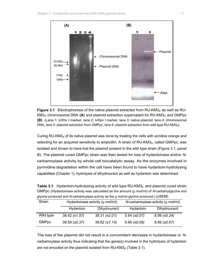
33.5Kb 29.9Kb
17Kb 15Kb
(A) (B)
1 2 3 4
Chromosomal DNA
Plasmid DNA
5 6
Plasmid
RNA
Chapter 3 : Construction and screening of RU-KM3S genome library 71
Figure 3.1 Electrophoresis of the native plasmid extracted from RU-KM3S as well as RU-KM3S chromosomal DNA (A) and plasmid extraction supernatant for RU-KM3S and GMPpc (B). (Lane 1: λ/Xho I marker, lane 2: λ/Kpn I marker, lane 3: native plasmid, lane 4: chromosomal DNA, lane 5: plasmid extraction from GMPpc, lane 6: plasmid extraction from wild type RU-KM3S).
Curing RU-KM3S of its native plasmid was done by treating the cells with acridine orange and
selecting for an acquired sensitivity to ampicillin. A strain of RU-KM3S, called GMPpc, was
isolated and shown to have lost the plasmid present in the wild type strain (Figure 3.1, panel
B). The plasmid cured GMPpc strain was then tested for loss of hydantoinase and/or N-
carbamoylase activity by whole cell biocatalytic assay. As the enzymes involved in
pyrimidine degradation within the cell have been found to have hydantoin-hydrolysing
capabilities (Chapter 1), hydrolysis of dihydrouracil as well as hydantoin was determined.
Table 3.1 Hydantoin-hydrolysing activity of wild type RU-KM3S and plasmid cured strain GMPpc. (Hydantoinase activity was calculated as the amount (μ mol/ml) of N-carbamylglycine and glycine produced and N-carbamoylase activity as the μ mol/ml glycine produced.) (±SEM) Strain Hydantoinase activity (μ mol/ml) N-carbamoylase activity (μ mol/ml)
Hydantoin Dihydrouracil Hydantoin Dihydrouracil
Wild type 38.42 (±1.57) 38.31 (±2.01) 5.64 (±0.07) 8.98 (±0.24)
GMPpc 39.59 (±0.37) 39.62 (±1.14) 6.66 (±0.09) 8.99 (±0.97)
The loss of the plasmid did not result in a concomitant decrease in hydantoinase or N-
carbamoylase activity thus indicating that the gene(s) involved in the hydrolysis of hydantoin
are not encoded on the plasmid isolated from RU-KM3S (Table 3.1).

23.13kb 9.42kb 6.68kb
2.32kb 2.02kb
1 2 3 4 5 6 7 8 9 10 11
Chapter 3 : Construction and screening of RU-KM3S genome library 72
3.3.2 Construction of RU-KM3S genomic library Chromosomal DNA extracted from RU-KM3S was subjected to partial digestion with varying
concentrations of restriction endonuclease Sau 3A1 in order to generate random
chromosomal DNA fragments of between 2 and 10kb in size. Thus 1μ g of chromosomal
DNA was digested with 0.8 to 0.002 units of Sau 3A1 and an aliquot of each digest was sized
by agarose gel electrophoresis (Figure 3.2).
Figure 3.2 Partial digestion of RU-KM3S chromosomal DNA by addition of varying concentrations of Sau 3A1. [Lane 1: Phage λ / Hind III marker, lanes 2-11: chromosomal DNA digested with 0.8, 0.08, 0.04, 0.02, 0.013, 0.01, 0.008, 0.006, 0.004, and 0.002 units of Sau 3A1 per μ g of genomic DNA respectively]
Chromosomal fragments generated by partial digestion with 0.06 units of Sau 3A1/1μ g
chromosomal DNA (Figure 3.2, lane 9) were used to produce the genomic library in pBK-
CMV using the ZAP Express procedure with a 60 to 80% insertion frequency after mass
excision. Recombinant plasmids contained an average insert size of approximately 5kb
(Figure 3.3).
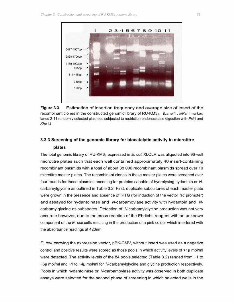
2838-1700bp
5077-4507bp
1159-1093bp 805bp
514-448bp
339bp
150bp
1 2 3 4 5 6 7 8 9 10 11
Chapter 3 : Construction and screening of RU-KM3S genome library 73
Figure 3.3 Estimation of insertion frequency and average size of insert of the recombinant clones in the constructed genomic library of RU-KM3S. (Lane 1 : λ/Pst I marker, lanes 2-11 randomly selected plasmids subjected to restriction endonuclease digestion with Pst I and Xho I.)
3.3.3 Screening of the genomic library for biocatalytic activity in microtitre plates
The total genomic library of RU-KM3S expressed in E. coli XLOLR was aliquoted into 96-well
microtitre plates such that each well contained approximately 40 insert-containing
recombinant plasmids with a total of about 38 000 recombinant plasmids spread over 10
microtitre master plates. The recombinant clones in these master plates were screened over
four rounds for those plasmids encoding for proteins capable of hydrolysing hydantoin or N-
carbamylglycine as outlined in Table 3.2. First, duplicate subcultures of each master plate
were grown in the presence and absence of IPTG (for induction of the vector lac promoter)
and assayed for hydantoinase and N-carbamoylase activity with hydantoin and N-
carbamylglycine as substrates. Detection of N-carbamylglycine production was not very
accurate however, due to the cross reaction of the Ehrlichs reagent with an unknown
component of the E. coli cells resulting in the production of a pink colour which interfered with
the absorbance readings at 420nm.
E. coli carrying the expression vector, pBK-CMV, without insert was used as a negative
control and positive results were scored as those pools in which activity levels of >1μ mol/ml
were detected. The activity levels of the 84 pools selected (Table 3.2) ranged from ~1 to
~6μ mol/ml and ~1 to ~4μ mol/ml for N-carbamylglycine and glycine production respectively.
Pools in which hydantoinase or N-carbamoylase activity was observed in both duplicate
assays were selected for the second phase of screening in which selected wells in the
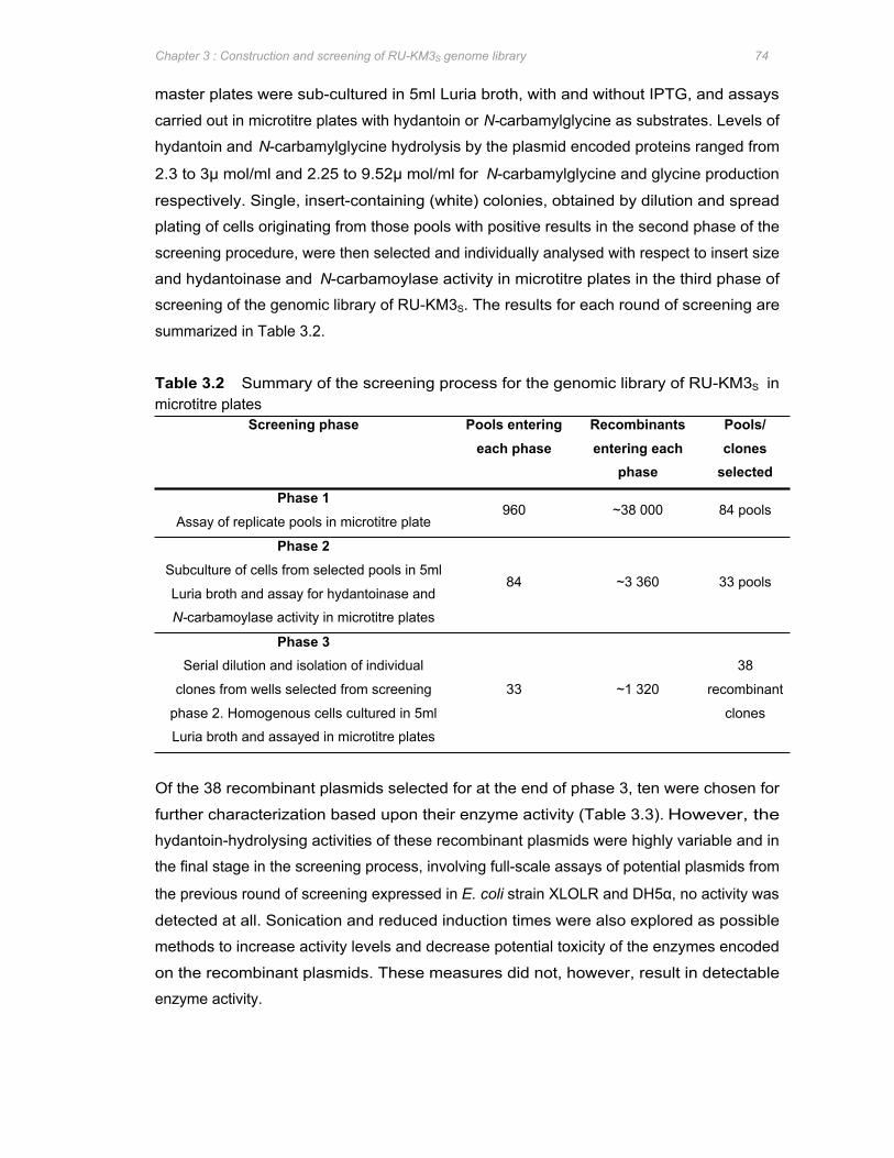
Chapter 3 : Construction and screening of RU-KM3S genome library 74
master plates were sub-cultured in 5ml Luria broth, with and without IPTG, and assays
carried out in microtitre plates with hydantoin or N-carbamylglycine as substrates. Levels of
hydantoin and N-carbamylglycine hydrolysis by the plasmid encoded proteins ranged from
2.3 to 3μ mol/ml and 2.25 to 9.52μ mol/ml for N-carbamylglycine and glycine production
respectively. Single, insert-containing (white) colonies, obtained by dilution and spread
plating of cells originating from those pools with positive results in the second phase of the
screening procedure, were then selected and individually analysed with respect to insert size
and hydantoinase and N-carbamoylase activity in microtitre plates in the third phase of
screening of the genomic library of RU-KM3S. The results for each round of screening are
summarized in Table 3.2.
Table 3.2 Summary of the screening process for the genomic library of RU-KM3S in microtitre plates
Screening phase Pools entering Recombinants Pools/ each phase entering each clones
phase selected
Phase 1 Assay of replicate pools in microtitre plate
960 ~38 000 84 pools
Phase 2 Subculture of cells from selected pools in 5ml
Luria broth and assay for hydantoinase and
N-carbamoylase activity in microtitre plates
84 ~3 360 33 pools
Phase 3 Serial dilution and isolation of individual
clones from wells selected from screening
phase 2. Homogenous cells cultured in 5ml
Luria broth and assayed in microtitre plates
33 ~1 320
38
recombinant
clones
Of the 38 recombinant plasmids selected for at the end of phase 3, ten were chosen for
further characterization based upon their enzyme activity (Table 3.3). However, the
hydantoin-hydrolysing activities of these recombinant plasmids were highly variable and in
the final stage in the screening process, involving full-scale assays of potential plasmids from
the previous round of screening expressed in E. coli strain XLOLR and DH5α, no activity was
detected at all. Sonication and reduced induction times were also explored as possible
methods to increase activity levels and decrease potential toxicity of the enzymes encoded
on the recombinant plasmids. These measures did not, however, result in detectable
enzyme activity.
Chapter 3 : Construction and screening of RU-KM3S genome library 75
Table 3.3 Biocatalytic activity of recombinant clones selected from the genomic library of RU-KM3S as determined in microtitre plates
Hydantoinase activity
(µ mol/ml)
N-carbamoylase activity
(µ mol/ml)
Insert size (kb)
+IPTG -IPTG +IPTG -IPTG
3.68 (0.50) - - - 7.3
1.85 (2.22) 4.6 (1.45) - - 6.4
2.93 (0.37) - 1.01 (0.28) - 4.9
1.77 (0.44) - 3.04 (0.11) 1.11 (0.12) 1.9
2.33 (0.23) - - - 4.3
- 2.27 (0.61) 2.196 (0.68) - 1.7
6.17 (1.40) 1.14 (1.74) 2.31 (0.75) 3.4
9.56 (0.74) - 2.47 (0.73)
-
- 6.3
2.32 (2.17) - 2.19 (1.31) - 4
- - - 3.44 (2.92) 2
Recombinant plasmid
pGMlib24
pGMlib23
pGMlib22
pGMlib21
pGMlib20
pGMlib19
pGMlib18
pGMlib17
pGMlib16
pGMlib15
(Standard error in italicised brackets, - : no detectable activity)
3.3.4 Southern hybridisation analysis As detection of hydantoinase and/or N-carbamoylase activity resulted in inconsistent results
and mapping of the restriction endonuclease sites of each of the recombinant plasmids did
not reveal any obvious common fragments (data not shown), Southern hybridisation was
done to assess whether the clones contained overlapping inserts. Recognition sites for the
restriction endonucleases Pst I, Sal I and Hind III are located in the cloning cassette of the
parental pBK-CMV vector (Figure 3.4) and digestion of the recombinant plasmids a using
combination of Pst I and Hind III or Sal I and Hind III resulted in excision of the insert from
the vector backbone.
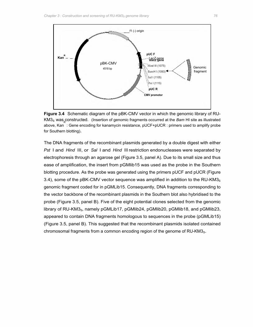
KanR
LacZ gene alacZ gene pBK-CMV
4518 bp
f1 (-) origin
pUC R
CMV promotor
pUC F
Hind III (1075)
Bam H I (1093)
Sal I (1105)
Pst I (1115)
Genomic fragment
Chapter 3 : Construction and screening of RU-KM3S genome library 76
Figure 3.4 Schematic diagram of the pBK-CMV vector in which the genomic library of RU-KM3S was constructed. (Insertion of genomic fragments occurred at the Bam HI site as illustrated above, Kan
R : Gene encoding for kanamycin resistance, pUCF+pUCR : primers used to amplify probe
for Southern blotting).
The DNA fragments of the recombinant plasmids generated by a double digest with either
Pst I and Hind III, or Sal I and Hind III restriction endonucleases were separated by
electrophoresis through an agarose gel (Figure 3.5, panel A). Due to its small size and thus
ease of amplification, the insert from pGMlib15 was used as the probe in the Southern
blotting procedure. As the probe was generated using the primers pUCF and pUCR (Figure
3.4), some of the pBK-CMV vector sequence was amplified in addition to the RU-KM3S
genomic fragment coded for in pGMLib15. Consequently, DNA fragments corresponding to
the vector backbone of the recombinant plasmids in the Southern blot also hybridised to the
probe (Figure 3.5, panel B). Five of the eight potential clones selected from the genomic
library of RU-KM3S, namely pGMLib17, pGMlib24, pGMlib20, pGMlib18, and pGMlib23,
appeared to contain DNA fragments homologous to sequences in the probe (pGMLib15)
(Figure 3.5, panel B). This suggested that the recombinant plasmids isolated contained
chromosomal fragments from a common encoding region of the genome of RU-KM3S.
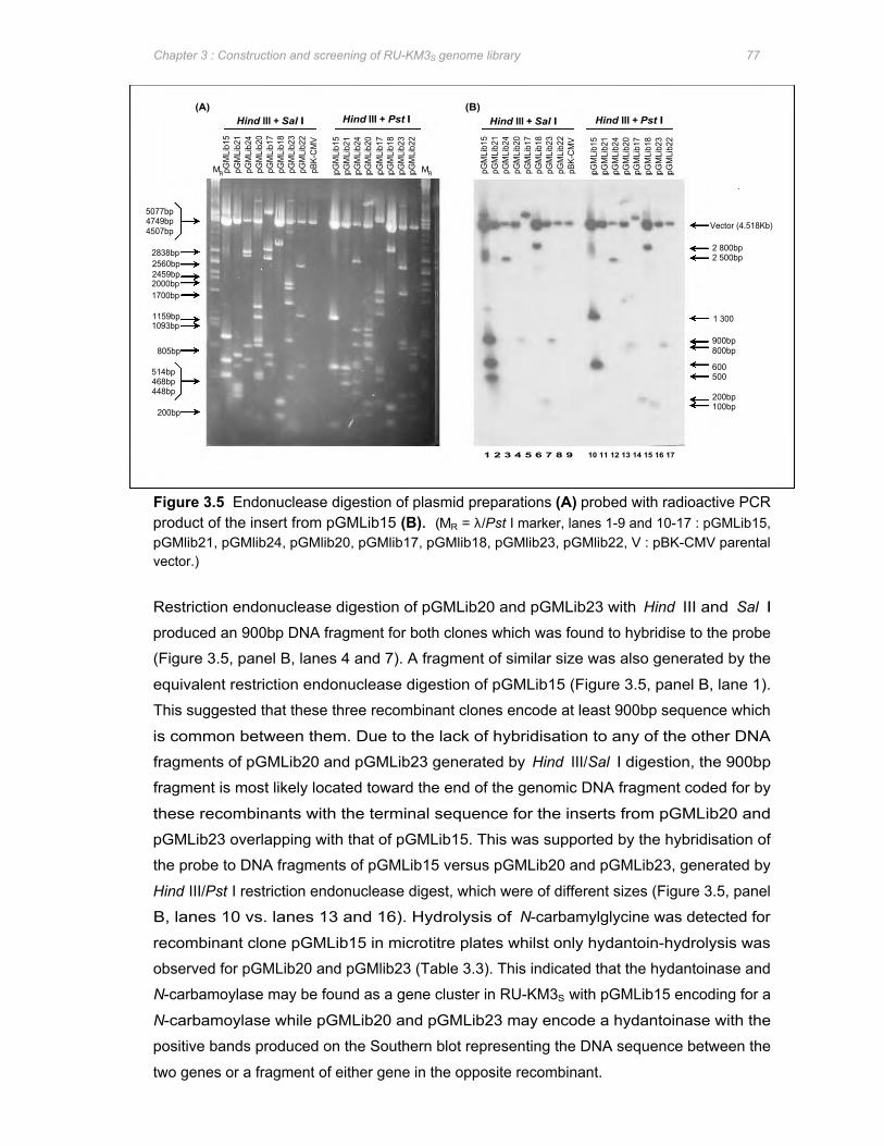
5077bp 4749bp 4507bp
2838bp 2560bp 2459bp 2000bp 1700bp
514bp 468bp 448bp
1093bp 1159bp
805bp
200bp
(A) (B)
MR pGM
Lib1
5 pG
MLi
b21
pGM
Lib2
4 pG
MLi
b20
pGM
Lib1
7 pG
MLi
b18
pGM
Lib2
3 pG
MLi
b22
pBK-
CM
V
pGM
Lib1
5 pG
MLi
b21
pGM
Lib2
4 pG
MLi
b20
pGM
Lib1
7 pG
MLi
b18
pGM
Lib2
3 pG
MLi
b22
Hind III + Sal I Hind III + Pst I
MR pGM
Lib1
5 pG
MLi
b21
pGM
Lib2
4 pG
MLi
b20
pGM
Lib1
7 pG
MLi
b18
pGM
Lib2
3 pG
MLi
b22
pBK-
CM
V
pGM
Lib1
5 pG
MLi
b21
pGM
Lib2
4 pG
MLi
b20
pGM
Lib1
7 pG
MLi
b18
pGM
Lib2
3 pG
MLi
b22
1 2 3 4 5 6 7 8 9 10 11 12 13 14 15 16 17
Hind III + Sal I Hind III + Pst I
Vector (4.518Kb)
2 800bp 2 500bp
900bp 800bp
600 500
200bp 100bp
1 300
Chapter 3 : Construction and screening of RU-KM3S genome library 77
Figure 3.5 Endonuclease digestion of plasmid preparations (A) probed with radioactive PCR product of the insert from pGMLib15 (B). (MR = λ/Pst I marker, lanes 1-9 and 10-17 : pGMLib15, pGMlib21, pGMlib24, pGMlib20, pGMlib17, pGMlib18, pGMlib23, pGMlib22, V : pBK-CMV parental vector.)
Restriction endonuclease digestion of pGMLib20 and pGMLib23 with Hind III and Sal I
produced an 900bp DNA fragment for both clones which was found to hybridise to the probe
(Figure 3.5, panel B, lanes 4 and 7). A fragment of similar size was also generated by the
equivalent restriction endonuclease digestion of pGMLib15 (Figure 3.5, panel B, lane 1).
This suggested that these three recombinant clones encode at least 900bp sequence which
is common between them. Due to the lack of hybridisation to any of the other DNA
fragments of pGMLib20 and pGMLib23 generated by Hind III/Sal I digestion, the 900bp
fragment is most likely located toward the end of the genomic DNA fragment coded for by
these recombinants with the terminal sequence for the inserts from pGMLib20 and
pGMLib23 overlapping with that of pGMLib15. This was supported by the hybridisation of
the probe to DNA fragments of pGMLib15 versus pGMLib20 and pGMLib23, generated by
Hind III/Pst I restriction endonuclease digest, which were of different sizes (Figure 3.5, panel
B, lanes 10 vs. lanes 13 and 16). Hydrolysis of N-carbamylglycine was detected for
recombinant clone pGMLib15 in microtitre plates whilst only hydantoin-hydrolysis was
observed for pGMLib20 and pGMlib23 (Table 3.3). This indicated that the hydantoinase and
N-carbamoylase may be found as a gene cluster in RU-KM3S with pGMLib15 encoding for a
N-carbamoylase while pGMLib20 and pGMLib23 may encode a hydantoinase with the
positive bands produced on the Southern blot representing the DNA sequence between the
two genes or a fragment of either gene in the opposite recombinant.
Chapter 3 : Construction and screening of RU-KM3S genome library 78
Hybridisation of the probe also occurred with the DNA fragments from recombinant clones
pGMLib24 and pGMlib18 (Figure 3.5, panel B, lanes 3, 6, 12, and 15). Despite positive
results for N-carbamylglycine hydrolysis by cells expressing both pGMLib15 and pGMlib18 in
microtitre plates (Table 3.3), no DNA fragments of equivalent sizes from these two
recombinants hybridised to one another (Figure 3.5, panel B, lanes 10 vs. 15). This
suggested that the sequence encoded within these two clones followed on from one another
with an overlap in the middle of the combined genomic sequence of the clones and that no
Pst I, Hind III, or Sal I recognition sites occurred within the overlapping region. Hydantoin-
hydrolysing activity in microtitre plates was observed for cells expressing both pGMLib18 and
pGMLib24 (Table 3.3). The insert encoding RU-KM3S genomic DNA in pGMLib18 was
calculated to be ~3.4Kb in size whilst that of pGMLib24 was ~7.3Kb in size (Figure 3.5, panel
A). The sizes of the fragments to which the probe hybridised were ~2.5Kb and ~2.8Kb in
size for pGMLib24 and pGMLib18 respectively (Figure 3.5, panel B, lanes 12 and 15). This
suggested that either the sequences coded for by these two fragments are identical and
represent genomic sequence running concurrent to one another with significant overlap, or
that the genomic fragments may be orientated on the chromosome with no overlap except for
that provided by the genomic DNA encoded by the probe (pGMLib15). However, hydantoin-
hydrolysing activity was observed in cells expressing both pGMLib24 and pGMLib18 (Table
3.3) suggesting that the former is true.
The inability to detect N-carbamoylase activity in cells expressing pGMLib24 in contrast to
pGMLib18 (Table 3.3) may be due to the truncation of at least 300bp of the pGMLib24 insert
closest to the potential N-carbamoylase encoding gene. In addition, hydantoinase activity in
pGMLib18 was detected in uninduced and induced cultures suggesting that expression of the
enzyme responsible for hydantoin-hydrolysis is under the control of a native, as opposed to
vector, promoter. Thus, either the gene was orientated in the opposite direction to that of the
vector promoter or sufficient DNA lies between the lacZ promoter and the encoded gene to
prevent expression under the control of the lacZ promoter. In contrast, hydantoin-
hydrolysing activity in cells expressing pGMLib24 was detected only in the presence of IPTG
(Table 3.3) suggesting that the encoded enzyme is located close to and in the same
orientation as the lacZ promoter in the vector.
Hybridisation of the probe to a ~200bp DNA fragment of pGMLib17, generated by Hind III/Pst
I digestion, was observed (Figure 3.5, panel B, lane 14). However, subsequent to digestion
of this plasmid with Hind III and Pst I, the DNA fragment corresponding to the vector was
larger than expected indicating that complete excision of the insert from the vector backbone
had not occurred (Figure 3.5, panel B, lane 14). Consequently, complete analysis of the
Southern blot of pGMLib17 was difficult.
(C) Hind III
Scale :
11497bp 11Kb
5077bp 4749bp 4507bp
2838bp 2560bp 2459bp 1986bp 1700bp
1159bp 1093bp
805bp
(A) (B)
III
III
Sal I
1kb
M R
Hin
d III
Pst
I +
Hin
d
Pst I Sal I Hind III Sal I Pst I Hind III
Pst
I
pGMLib15
Pst
I +
Sa
l I
Sa
l I
Sa
l I +
Hin
d
pGMLib18
Hin
d III
1 2 3 4 5 6
Pst
I +
Hin
d III
Pst
I
pGMLib20
pGMLib24
Pst
I +
Sa
l I
pGMLib23
Sa
l I
Sa
l I +
Hin
d III
4 000bp
2 500bp
3800bp
800bp
500bp
1 900bp 1800bp
1 400bp
Chapter 3 : Construction and screening of RU-KM3S genome library 79
Whilst sequence similarity between five of the selected clones had been established (Figure
3.5, panel B), restriction endonuclease mapping of the clones relative to one another
remained inconclusive (data not shown). In order to facilitate mapping of the recombinant
clones relative to one another, the chromosomal region on the genome from which the insert
encoded in pGMLib15 was derived was analysed by digestion of the chromosomal DNA with
Hind III, Sal I, and Pst I restriction endonucleases and generation of a Southern blot with the
pGMLib15 insert as the probe (Figure 3.6).
Figure 3.6 Southern analysis of the chromosomal DNA flanking the sequence encoded by
the insert in pGMLib15. Restriction digest of chromosomal DNA from RU-KM3S (A) probed with
radioactive PCR product of the insert from pGMlib15 (B) as well as mapping of the restriction
endonuclease recognition sites on the genomic DNA (coloured black) flanking pGMlib15 (coloured
grey) and suggested relative location of the genomic fragments contained in the recombinant clones
(C). (Lane 1: λ/Pst I marker, lane 2-7: genomic DNA digested with Hind III, Pst I+Hind III, Pst I, Pst
I+Sal I, and Sal I respectively.)
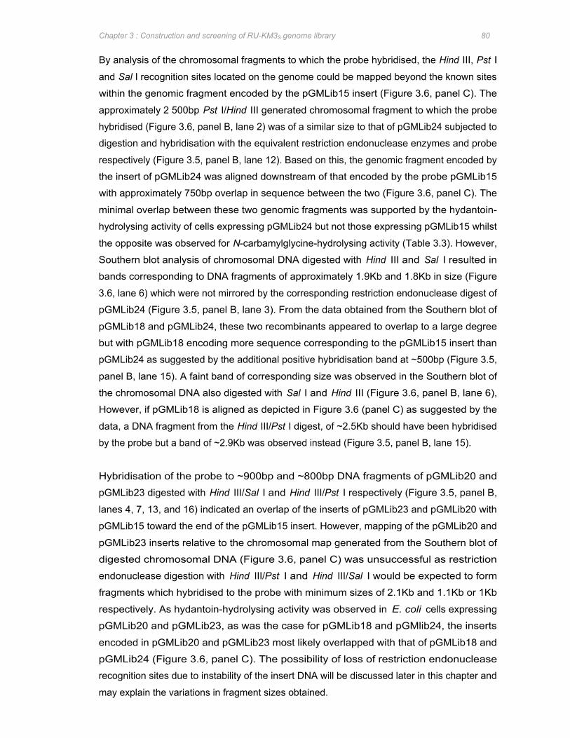
Chapter 3 : Construction and screening of RU-KM3S genome library 80
By analysis of the chromosomal fragments to which the probe hybridised, the Hind III, Pst I
and Sal I recognition sites located on the genome could be mapped beyond the known sites
within the genomic fragment encoded by the pGMLib15 insert (Figure 3.6, panel C). The
approximately 2 500bp Pst I/Hind III generated chromosomal fragment to which the probe
hybridised (Figure 3.6, panel B, lane 2) was of a similar size to that of pGMLib24 subjected to
digestion and hybridisation with the equivalent restriction endonuclease enzymes and probe
respectively (Figure 3.5, panel B, lane 12). Based on this, the genomic fragment encoded by
the insert of pGMLib24 was aligned downstream of that encoded by the probe pGMLib15
with approximately 750bp overlap in sequence between the two (Figure 3.6, panel C). The
minimal overlap between these two genomic fragments was supported by the hydantoin-
hydrolysing activity of cells expressing pGMLib24 but not those expressing pGMLib15 whilst
the opposite was observed for N-carbamylglycine-hydrolysing activity (Table 3.3). However,
Southern blot analysis of chromosomal DNA digested with Hind III and Sal I resulted in
bands corresponding to DNA fragments of approximately 1.9Kb and 1.8Kb in size (Figure
3.6, lane 6) which were not mirrored by the corresponding restriction endonuclease digest of
pGMLib24 (Figure 3.5, panel B, lane 3). From the data obtained from the Southern blot of
pGMLib18 and pGMLib24, these two recombinants appeared to overlap to a large degree
but with pGMLib18 encoding more sequence corresponding to the pGMLib15 insert than
pGMLib24 as suggested by the additional positive hybridisation band at ~500bp (Figure 3.5,
panel B, lane 15). A faint band of corresponding size was observed in the Southern blot of
the chromosomal DNA also digested with Sal I and Hind III (Figure 3.6, panel B, lane 6),
However, if pGMLib18 is aligned as depicted in Figure 3.6 (panel C) as suggested by the
data, a DNA fragment from the Hind III/Pst I digest, of ~2.5Kb should have been hybridised
by the probe but a band of ~2.9Kb was observed instead (Figure 3.5, panel B, lane 15).
Hybridisation of the probe to ~900bp and ~800bp DNA fragments of pGMLib20 and
pGMLib23 digested with Hind III/Sal I and Hind III/Pst I respectively (Figure 3.5, panel B,
lanes 4, 7, 13, and 16) indicated an overlap of the inserts of pGMLib23 and pGMLib20 with
pGMLib15 toward the end of the pGMLib15 insert. However, mapping of the pGMLib20 and
pGMLib23 inserts relative to the chromosomal map generated from the Southern blot of
digested chromosomal DNA (Figure 3.6, panel C) was unsuccessful as restriction
endonuclease digestion with Hind III/Pst I and Hind III/Sal I would be expected to form
fragments which hybridised to the probe with minimum sizes of 2.1Kb and 1.1Kb or 1Kb
respectively. As hydantoin-hydrolysing activity was observed in E. coli cells expressing
pGMLib20 and pGMLib23, as was the case for pGMLib18 and pGMlib24, the inserts
encoded in pGMLib20 and pGMLib23 most likely overlapped with that of pGMLib18 and
pGMLib24 (Figure 3.6, panel C). The possibility of loss of restriction endonuclease
recognition sites due to instability of the insert DNA will be discussed later in this chapter and
may explain the variations in fragment sizes obtained.
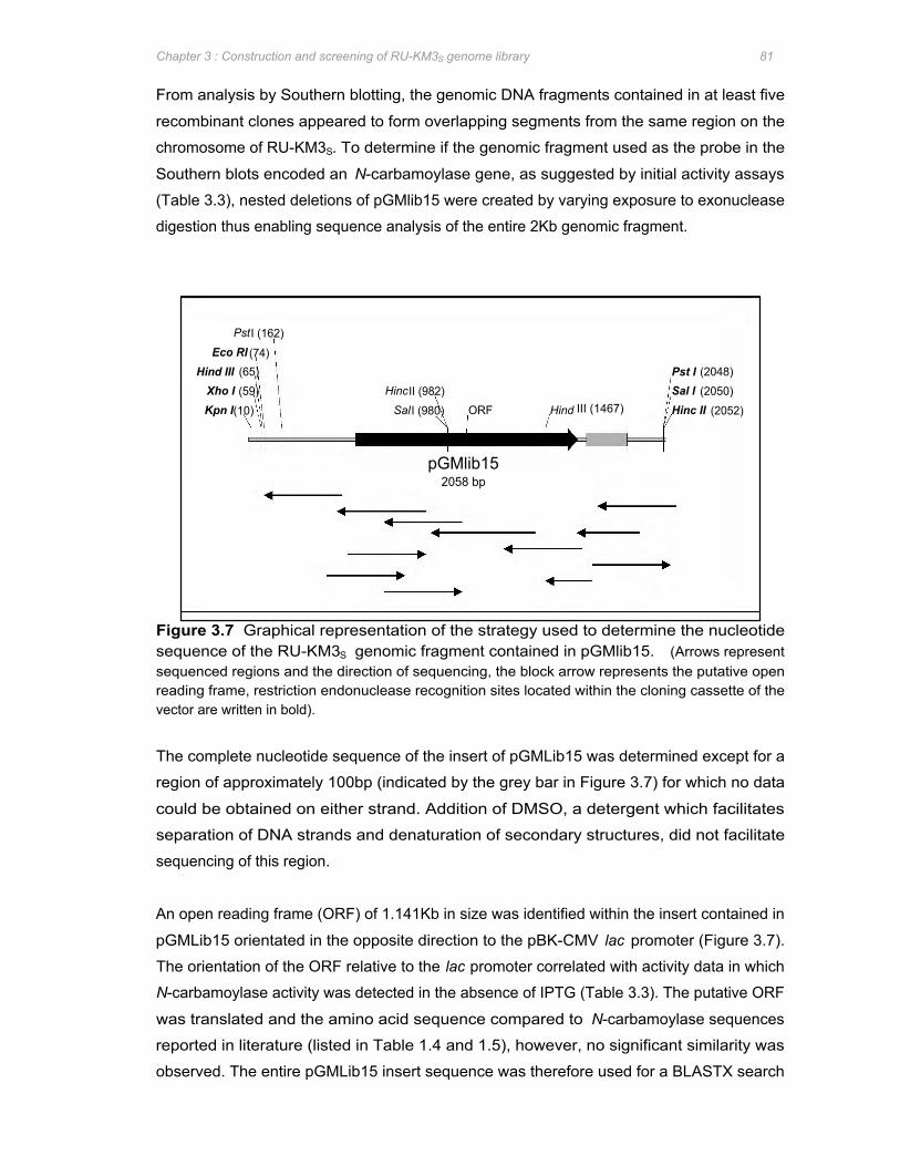
Xho I (59) HincII (982) Sal I (2050)
PstI (162) Eco RI (74)
Pst I (2048) Hind III (65)
Kpn I(10) SalI (980) Hind III (1467) Hinc II (2052) ORF
pGMlib15 2058 bp
Chapter 3 : Construction and screening of RU-KM3S genome library 81
From analysis by Southern blotting, the genomic DNA fragments contained in at least five
recombinant clones appeared to form overlapping segments from the same region on the
chromosome of RU-KM3S. To determine if the genomic fragment used as the probe in the
Southern blots encoded an N-carbamoylase gene, as suggested by initial activity assays
(Table 3.3), nested deletions of pGMlib15 were created by varying exposure to exonuclease
digestion thus enabling sequence analysis of the entire 2Kb genomic fragment.
Figure 3.7 Graphical representation of the strategy used to determine the nucleotide sequence of the RU-KM3S genomic fragment contained in pGMlib15. (Arrows represent sequenced regions and the direction of sequencing, the block arrow represents the putative open reading frame, restriction endonuclease recognition sites located within the cloning cassette of the vector are written in bold).
The complete nucleotide sequence of the insert of pGMLib15 was determined except for a
region of approximately 100bp (indicated by the grey bar in Figure 3.7) for which no data
could be obtained on either strand. Addition of DMSO, a detergent which facilitates
separation of DNA strands and denaturation of secondary structures, did not facilitate
sequencing of this region.
An open reading frame (ORF) of 1.141Kb in size was identified within the insert contained in
pGMLib15 orientated in the opposite direction to the pBK-CMV lac promoter (Figure 3.7).
The orientation of the ORF relative to the lac promoter correlated with activity data in which
N-carbamoylase activity was detected in the absence of IPTG (Table 3.3). The putative ORF
was translated and the amino acid sequence compared to N-carbamoylase sequences
reported in literature (listed in Table 1.4 and 1.5), however, no significant similarity was
observed. The entire pGMLib15 insert sequence was therefore used for a BLASTX search
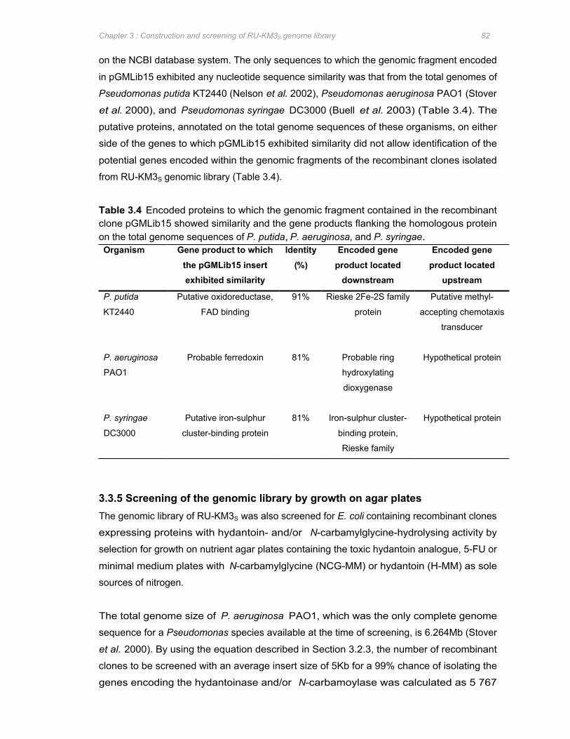
Chapter 3 : Construction and screening of RU-KM3S genome library 82
on the NCBI database system. The only sequences to which the genomic fragment encoded
in pGMLib15 exhibited any nucleotide sequence similarity was that from the total genomes of
Pseudomonas putida KT2440 (Nelson et al. 2002), Pseudomonas aeruginosa PAO1 (Stover
et al. 2000), and Pseudomonas syringae DC3000 (Buell et al. 2003) (Table 3.4). The
putative proteins, annotated on the total genome sequences of these organisms, on either
side of the genes to which pGMLib15 exhibited similarity did not allow identification of the
potential genes encoded within the genomic fragments of the recombinant clones isolated
from RU-KM3S genomic library (Table 3.4).
Table 3.4 Encoded proteins to which the genomic fragment contained in the recombinant clone pGMLib15 showed similarity and the gene products flanking the homologous protein on the total genome sequences of P. putida, P. aeruginosa, and P. syringae. Organism Gene product to which Identity Encoded gene Encoded gene
the pGMLib15 insert (%) product located product located exhibited similarity downstream upstream
P. putida
KT2440
Putative oxidoreductase,
FAD binding
91% Rieske 2Fe-2S family
protein
Putative methyl-
accepting chemotaxis
transducer
P. aeruginosa
PAO1
Probable ferredoxin 81% Probable ring
hydroxylating
dioxygenase
Hypothetical protein
P. syringae Putative iron-sulphur 81% Iron-sulphur cluster- Hypothetical protein
DC3000 cluster-binding protein binding protein,
Rieske family
3.3.5 Screening of the genomic library by growth on agar plates The genomic library of RU-KM3S was also screened for E. coli containing recombinant clones
expressing proteins with hydantoin- and/or N-carbamylglycine-hydrolysing activity by
selection for growth on nutrient agar plates containing the toxic hydantoin analogue, 5-FU or
minimal medium plates with N-carbamylglycine (NCG-MM) or hydantoin (H-MM) as sole
sources of nitrogen.
The total genome size of P. aeruginosa PAO1, which was the only complete genome
sequence for a Pseudomonas species available at the time of screening, is 6.264Mb (Stover
et al. 2000). By using the equation described in Section 3.2.3, the number of recombinant
clones to be screened with an average insert size of 5Kb for a 99% chance of isolating the
genes encoding the hydantoinase and/or N-carbamoylase was calculated as 5 767
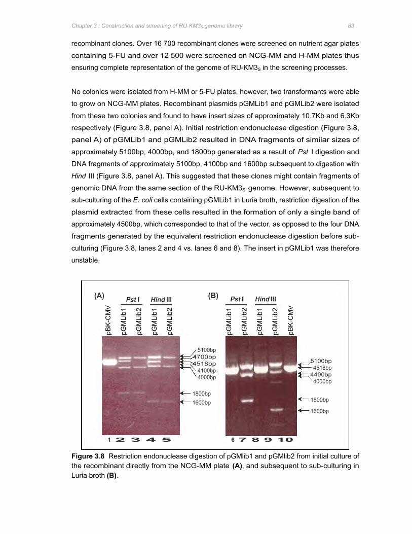
(A) (B) Pst I Hind III Pst I Hind III
pBK-
CM
V
pGM
Lib1
pGM
Lib2
pGM
Lib1
pGM
Lib2
1 2 3 4 5 6 7 8 9 10
5100bp 4700bp
4518bp 4100bp 4000bp
1800bp 1600bp
pGM
Lib1
pGM
Lib2
pGM
Lib1
pGM
Lib2
pBK-
CM
V
5100bp 4518bp
4400bp 4000bp
1800bp
1600bp
Chapter 3 : Construction and screening of RU-KM3S genome library 83
recombinant clones. Over 16 700 recombinant clones were screened on nutrient agar plates
containing 5-FU and over 12 500 were screened on NCG-MM and H-MM plates thus
ensuring complete representation of the genome of RU-KM3S in the screening processes.
No colonies were isolated from H-MM or 5-FU plates, however, two transformants were able
to grow on NCG-MM plates. Recombinant plasmids pGMLib1 and pGMLib2 were isolated
from these two colonies and found to have insert sizes of approximately 10.7Kb and 6.3Kb
respectively (Figure 3.8, panel A). Initial restriction endonuclease digestion (Figure 3.8,
panel A) of pGMLib1 and pGMLib2 resulted in DNA fragments of similar sizes of
approximately 5100bp, 4000bp, and 1800bp generated as a result of Pst I digestion and
DNA fragments of approximately 5100bp, 4100bp and 1600bp subsequent to digestion with
Hind III (Figure 3.8, panel A). This suggested that these clones might contain fragments of
genomic DNA from the same section of the RU-KM3S genome. However, subsequent to
sub-culturing of the E. coli cells containing pGMLib1 in Luria broth, restriction digestion of the
plasmid extracted from these cells resulted in the formation of only a single band of
approximately 4500bp, which corresponded to that of the vector, as opposed to the four DNA
fragments generated by the equivalent restriction endonuclease digestion before sub-
culturing (Figure 3.8, lanes 2 and 4 vs. lanes 6 and 8). The insert in pGMLib1 was therefore
unstable.
Figure 3.8 Restriction endonuclease digestion of pGMlib1 and pGMlib2 from initial culture of the recombinant directly from the NCG-MM plate (A), and subsequent to sub-culturing in Luria broth (B).
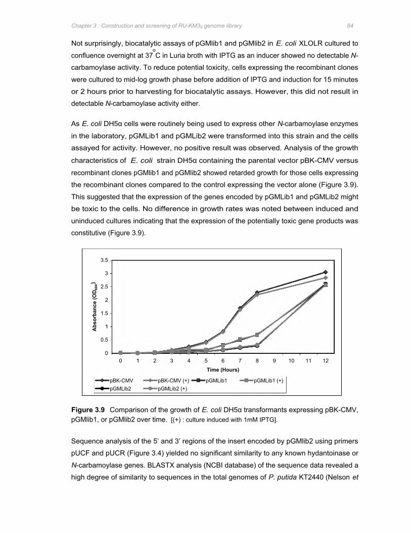
Abs
orba
nce
(OD 6
00)
3.5
2.5
0.5
1.5
3
2
0
1
pBK-CMV pBK-CMV (+) pGMLib1 pGMLib1 (+) pGMLib2 pGMLib2 (+)
0 1 2 3 4 5 6 7 8 9 10 11 12
Time (Hours)
Chapter 3 : Construction and screening of RU-KM3S genome library 84
Not surprisingly, biocatalytic assays of pGMlib1 and pGMlib2 in E. coli XLOLR cultured to
confluence overnight at 37oC in Luria broth with IPTG as an inducer showed no detectable N-
carbamoylase activity. To reduce potential toxicity, cells expressing the recombinant clones
were cultured to mid-log growth phase before addition of IPTG and induction for 15 minutes
or 2 hours prior to harvesting for biocatalytic assays. However, this did not result in
detectable N-carbamoylase activity either.
As E. coli DH5α cells were routinely being used to express other N-carbamoylase enzymes
in the laboratory, pGMLib1 and pGMLib2 were transformed into this strain and the cells
assayed for activity. However, no positive result was observed. Analysis of the growth
characteristics of E. coli strain DH5α containing the parental vector pBK-CMV versus
recombinant clones pGMlib1 and pGMlib2 showed retarded growth for those cells expressing
the recombinant clones compared to the control expressing the vector alone (Figure 3.9).
This suggested that the expression of the genes encoded by pGMLib1 and pGMLib2 might
be toxic to the cells. No difference in growth rates was noted between induced and
uninduced cultures indicating that the expression of the potentially toxic gene products was
constitutive (Figure 3.9).
Figure 3.9 Comparison of the growth of E. coli DH5α transformants expressing pBK-CMV, pGMlib1, or pGMlib2 over time. [(+) : culture induced with 1mM IPTG].
Sequence analysis of the 5’ and 3’ regions of the insert encoded by pGMlib2 using primers
pUCF and pUCR (Figure 3.4) yielded no significant similarity to any known hydantoinase or
N-carbamoylase genes. BLASTX analysis (NCBI database) of the sequence data revealed a
high degree of similarity to sequences in the total genomes of P. putida KT2440 (Nelson et
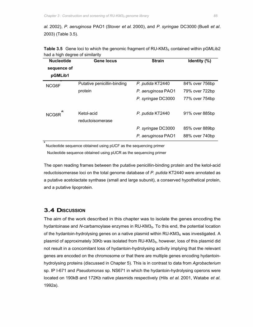
Chapter 3 : Construction and screening of RU-KM3S genome library 85
al. 2002), P. aeruginosa PAO1 (Stover et al. 2000), and P. syringae DC3000 (Buell et al.
2003) (Table 3.5).
Table 3.5 Gene loci to which the genomic fragment of RU-KM3S contained within pGMLib2 had a high degree of similarity
Nucleotide Gene locus Strain Identity (%) sequence of
pGMLib1
NCG6F∗ Putative penicillin-binding P. putida KT2440 84% over 756bp
protein P. aeruginosa PAO1 79% over 722bp
P. syringae DC3000 77% over 754bp
NCG6R'k Ketol-acid P. putida KT2440 91% over 885bp
reductoisomerase
P. syringae DC3000 85% over 889bp
P. aeruginosa PAO1 88% over 740bp ∗
Nucleotide sequence obtained using pUCF as the sequencing primer �
Nucleotide sequence obtained using pUCR as the sequencing primer
The open reading frames between the putative penicillin-binding protein and the ketol-acid
reductoisomerase loci on the total genome database of P. putida KT2440 were annotated as
a putative acetolactate synthase (small and large subunit), a conserved hypothetical protein,
and a putative lipoprotein.
3.4 DISCUSSION
The aim of the work described in this chapter was to isolate the genes encoding the
hydantoinase and N-carbamoylase enzymes in RU-KM3S. To this end, the potential location
of the hydantoin-hydrolysing genes on a native plasmid within RU-KM3S was investigated. A
plasmid of approximately 30Kb was isolated from RU-KM3S, however, loss of this plasmid did
not result in a concomitant loss of hydantoin-hydrolysing activity implying that the relevant
genes are encoded on the chromosome or that there are multiple genes encoding hydantoin-
hydrolysing proteins (discussed in Chapter 5). This is in contrast to data from Agrobacterium
sp. IP I-671 and Pseudomonas sp. NS671 in which the hydantoin-hydrolysing operons were
located on 190kB and 172Kb native plasmids respectively (Hils et al. 2001, Watabe et al.
1992a).
Chapter 3 : Construction and screening of RU-KM3S genome library 86
Amplification by PCR has been used to isolate the genes encoding D-enantioselective
hydantoinases and N-carbamoylases from P. putida 7711-2, A. radiobacter NRRL B11291
and Agrobacterium sp. IP I-671 (Chen and Tsai 1998, Chao et al. 1999b, Hils et al. 2001).
The primers used for the amplification procedure were either designed based on the N-
terminal amino acid sequence, determined by Edman degradation, of the purified protein
(Chen and Tsai 1998) or by alignment of related sequences reported in literature to produce
degenerate primers (Chao et al. 1999b, Hils et al. 2001). However, the only non-
enantioselective hydantoinases (from A. aurescens DSM 3747 and Pseudomonas sp.
NS671) for which the encoding genes have been isolated to date have no similarity to one
another (Wiese et al. 2001, Watabe et al. 1992a). With respect to L-enantioselective N-
carbamoylases, only four gene sequences were available at the time of this study (Watabe et
al. 1992a, Mukohara et al. 1993, Batisse et al. 1997, Wilms et al. 1999). While the amino
acid sequence of the L-carbamoylases from B. stearothermophilus NCIB8224 and NS1122A
had a high degree of similarity, the remaining enzymes did not (data not shown). Thus
design of degenerate primers for PCR amplification of the non-stereoselective hydantoinase
and L-stereoselective N-carbamoylase of RU-KM3S could not be attempted. Furthermore,
the genome sequence for P. aeruginosa PAO1 was the only complete genome available for
comparative analysis at the time of this study (Stover et al. 2000). Moreover, a search of the
annotated database of P. aeruginosa PAO1 did not reveal any genes annotated as a N-
carbamoylase (data not shown). Consequently, screening of a genomic library of RU-KM3S
was approached in order to isolate the genes encoding the hydantoin-hydrolysing enzymes
in this strain. A genomic library of RU-KM3S was successfully constructed, using the λ-
phage ZAP Express system, with an average insert size of 5Kb and 60 to 80% insertion
frequency. To ensure that the entire genome of RU-KM3S was represented in the screening
procedures, the number of recombinant clones screened was 2- to 4-fold in excess to that
required as calculated using equation 1 (Section 3.2.3).
Initial screening of recombinant E. coli, containing fragments of the RU-KM3S genome in the
pBK-CMV vector, was based on detection of N-carbamylglycine and glycine production from
hydantoin or N-carbamylglycine as substrates in microtitre plates. Whilst positive results
were obtained in the first three phases of the screening process, the hydantoin-hydrolysing
activity of the recombinant E. coli selected was highly variable and activity was completely
lost by screening phase 4 which entailed full-scale biocatalytic reactions. The variations
observed may in part be due to the inability to standardize recombinant cell concentration in
the microtitre plate biocatalytic reactions as growth of recombinants varied depending on the
metabolic burden placed on the cell by the expression of the foreign proteins encoded on the
genomic fragments. Also, low activities, obtained as a result of dilution in the mixed
population of clones (40 clones per well) in the early phases of screening, may have been
Chapter 3 : Construction and screening of RU-KM3S genome library 87
obscured by background readings. Furthermore, the pink colour resulting from cross-
reaction of the Ehrlichs reagent with components of the E. coli cell also affected absorbance
readings with respect to the detection of N-carbamylglycine. Loss of hydantoin- and N-
carbamylglycine-hydrolysing activity by the fourth phase of screening may be due to either
false positives in the microtitre plate assay or due to instability of the insert DNA. However,
Southern analysis of eight of the clones revealed at least five which contained apparently
homologous fragments of DNA suggesting that a common or overlapping region of genomic
DNA was encoded within the genomic fragment inserts. If the loss of activity was solely due
to false positive results, random fragments of genomic DNA as opposed to fragments from a
common region of the genome would have been isolated. The loss of activity was therefore
most likely due to instability of the insert DNA. Sequence analysis of the probe, generated
from the insert in pGMlib15, yielded no significant similarity to N-carbamoylase or
hydantoinase sequences reported in literature.
Screening of the genomic library by growth on minimal medium plates with hydantoin or N-
carbamylglycine as sole sources of nitrogen had advantages over screening for activity for
two reasons. Firstly, by culturing the recombinant E. coli on medium with hydantoin/N-
carbamylglycine as a sole source of nitrogen, a selection pressure was applied that would
promote insert stability. Secondly, the recombinant plasmid could be isolated directly after
transformation and culture of the recombinant plasmid without the necessity of sub-culturing
the E. coli through several rounds of screening during which the integrity of the plasmid could
not be monitored. Restriction analysis of the two recombinant clones isolated from NCG-MM
indicated the presence of common, overlapping sequences, suggesting that the inserts may
be from the same region on the RU-KM3S genome. However, as was the case with the
previously isolated clones, no hydantoinase or N-carbamoylase activity was observed in the
biocatalytic assays irrespective of the temperature at which the cells were cultured, induction
time, or sonication. The insert in pGMLib1 was found to be highly unstable as a single sub-
culture of the recombinant E. coli resulted in complete loss of the insert when cultured in E.
coli XLOLR. E. coli strain DH5α was therefore investigated as an alternative host for the
isolated recombinant clones. While the gross structural integrity of the recombinant plasmids
was maintained within this strain (data not shown), a significant reduction in growth was
noted when pGMlib1 and pGMlib2 were expressed suggesting that the expression of the
foreign proteins was either toxic to the cell or placed a high metabolic burden on the cell
resulting in slower growth.
The pBK-CMV vector utilized in the construction of the genomic library of RU-KM3S relies on
expression from the lacZ promoter. There are two negative feedback controls associated
with the lac operon. The first is carbon catabolite repression and the second involves the lac
Chapter 3 : Construction and screening of RU-KM3S genome library 88
repressor protein (LacI), encoded by the lacI gene, which controls transcription by binding to
the operator region and preventing RNA polymerase action. However, when present, lactose
or a structural analogue thereof (i.e. IPTG) binds to LacI resulting in a conformational change
thus preventing binding of the repressor to the operator region consequently resulting in
expression of lacZ (Snyder and Champness 2003). This repression of lacZ expression is
however incomplete and “leaky” (Freifelder 1987). “Leaky” lac promoters can be completely
repressed by expression of the plasmids in host strains carrying the lacI' allele. This
mutation of the chromosomal promoter region for lacI results in increased production of LacI
repressor molecules from 10-20 to over 100 molecules per cell (Baneyx 1999). E. coli strain
XLOLR contains this mutated allele whilst strain DH5α does not, thus biocatalytic assays of
β-galactosidase expressed from the lac promoter encoded within pBK-CMV results in
constitutive expression of the lacZ when in E. coli DH5α (Pers. Comm. M. Jiwaji). This
“leakiness” of the lac promoter when in E. coli DH5α due to an unequal supply of repressor
protein LacI, leads to instability of plasmids and/or low induction levels (Wilms et al. 2001a).
However, XLOLR, which contains the lacI' mutant allele, did not result in stable maintenance
of recombinant plasmids encoding genomic fragments of RU-KM3S either. Thus plasmid
instability may be attributed to the fact that pBK-CMV is derived from a high copy number
ColE1 based plasmid. High copy number plasmids burden the metabolic capacity of the host
cell resulting in a starvation-like condition. This in turn leads to the accumulation of
uncharged tRNAs which, due to sequence homologies, tend to interact with the plasmid-
replication mechanisms resulting in excessive replication of the plasmid up to the point where
the metabolic capacity of the cell is completely exhausted (Grabherr and Bayer 2002). In
addition, strong promoters, such as T7 found in pBK-CMV, also result in such high levels of
expression that the expressed protein is often unable to achieve its native conformation and
segregates in inclusion bodies (Baneyx 1999).
The toxic effect of heterologous expression of hydantoin-hydrolysing genes in E. coli has
been well documented as resulting in the formation of inclusion bodies (Chien et al. 1998,
Chao et al. 2000a , Hils et al. 2001, Wilms et al. 2001b), reduced growth rates (Mukohara et
al. 1994, Chien et al. 1998) and low levels of activity (Wilms et al. 2001a, Hils et al. 2001).
As hydantoinases and N-carbamoylases are associated with pyrimidine catabolism, the
unregulated degradation of pyrimidines in the cells by over-expression of the hydantoinase
may provide a possible explanation for the toxicity observed (Mukohara et al. 1994, Chien et
al. 1998). Furthermore, Pseudomonas proteins are well-known in literature for poor
expression in E. coli and further examples are continually being published (Schweizer 2001).
The shortcomings of screening for heterologous expression of the RU-KM3S hydantoinase
and N-carbamoylase encoding genes in E. coli are numerous thus necessitating an
alternative approach.
Chapter 4 : Mutagenesis of RU-KM3S 89
4.1 INTRODUCTION
Optimisation of hydantoin-hydrolysing enzyme systems in native organisms has been
accomplished by spontaneous mutation (Syldatk et al. 1990a) or via chemically induced
mutagenesis (Wagner et al. 1996, Hartley et al. 1998, 2001). Mutant strains thus generated
exhibit phenotypes such as increased hydantoin-hydrolysing activity (Syldatk et al. 1990a),
inducer-independent expression (Hartley et al. 1998, Wagner et al. 1996), as well as mutants
which are no longer subject to nitrogen catabolite repression (Hartley et al. 2001).
In the case of chemically induced mutagenesis, alklating agents ethylmethane sulfonate
(EMS) or N-methyl-N-nitro-N-nitrosoguanidine (MNNG) were utilized. The mutagenic action
of alklyating agents involve addition of ethyl groups to the nucleotide bases in DNA, with O6-
alkyl guanine being the prime cause of mutations in vivo, resulting in mispairing of thymine
with guanine. Consequently, G:C→A:T transitions are the primary point mutations which
occur (Miller 1992).
An alternative mutagenesis methodology involves the use of transposons. In contrast to
chemical mutagenesis, where subtle changes in the target gene may occur, complete
inactivation of the gene can result when utilizing transposons. Transposons are broadly
classified as segments of DNA that can move, termed transposition, from one location on a
genome or plasmid to another (Judson and Mekalanos 2000). While transposons may
contain several genes (Malay et al. 1994), the components essential for transposition are the
transposase gene and two inverted repeat sequences flanking the DNA encoding the
selectable phenotype (de Lorenzo and Timmis 1994). The transposase enzyme is
responsible for recognition of the inverted repeats and integration of the DNA located
between these repeats into the bacterial genome (Malay et al. 1994). Placement of the gene
encoding the transposase outside of the inverted repeats results in the formation, after the
insertional event, of a non-replicating DNA fragment containing the transposase gene which
is lost in subsequent cell replication (Dennis and Zylstra 1998). This ensures that
transposition occurs once only and that further movement or rearrangement of DNA due to
the transposase is negated resulting in a stable, inheritable insertional mutation which can
then be analysed (de Lorenzo and Timmis 1994). Furthermore, the presence of a conditional
origin of replication within the integrated DNA sequence allows for rapid identification of the
sequence flanking the transposon site of insertion. This is achieved by digestion of the total
genomic DNA of a selected insertional mutant strain with a restriction endonuclease non-
specific for the inserted foreign DNA, followed by circularisation of the resultant fragments by
self-ligation. Transformation of the ensuing circular DNA fragments into E. coli and selection
for the phenotype, such as an antibiotic resistance marker, inserted into the target genome
by the transposon allows for the isolation of the genomic fragment interrupted by the
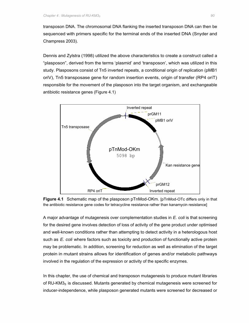
Tn5 transposase
RP4 oriT
pTnMod-OKm 5098 bp
Inverted repeat prGM11
Inverted repeat
pMB1 oriV
prGM12
Kan resistance gene
Chapter 4 : Mutagenesis of RU-KM3S 90
transposon DNA. The chromosomal DNA flanking the inserted transposon DNA can then be
sequenced with primers specific for the terminal ends of the inserted DNA (Snyder and
Champress 2003).
Dennis and Zylstra (1998) utilized the above characteristics to create a construct called a
“plasposon”, derived from the terms ‘plasmid’ and ‘transposon’, which was utilized in this
study. Plasposons consist of Tn5 inverted repeats, a conditional origin of replication (pMB1
oriV), Tn5 transposase gene for random insertion events, origin of transfer (RP4 oriT)
responsible for the movement of the plasposon into the target organism, and exchangeable
antibiotic resistance genes (Figure 4.1)
Figure 4.1 Schematic map of the plasposon pTnMod-OKm. [pTnMod-OTc differs only in that the antibiotic resistance gene codes for tetracycline resistance rather than kanamycin resistance]
A major advantage of mutagenesis over complementation studies in E. coli is that screening
for the desired gene involves detection of loss of activity of the gene product under optimised
and well-known conditions rather than attempting to detect activity in a heterologous host
such as E. coli where factors such as toxicity and production of functionally active protein
may be problematic. In addition, screening for reduction as well as elimination of the target
protein in mutant strains allows for identification of genes and/or metabolic pathways
involved in the regulation of the expression or activity of the specific enzymes.
In this chapter, the use of chemical and transposon mutagenesis to produce mutant libraries
of RU-KM3S is discussed. Mutants generated by chemical mutagenesis were screened for
inducer-independence, while plasposon generated mutants were screened for decreased or

Chapter 4 : Mutagenesis of RU-KM3S 91
loss of hydantoin-hydrolysing capabilities, indicating inactivation of genes involved in the
hydantoin-hydrolysing enzyme system of RU-KM3S.
4.2 METHODS AND MATERIALS
4.2.1 Chemical mutagenesis of RU-KM3S
RU-KM3S cells were subjected to random chemical mutagenesis with EMS (Sigma) as
described in Miller (1992). A confluent culture of RU-KM3S in 5ml nutrient broth, incubated
overnight at 28oC, was used to seed 30ml of nutrient broth which was incubated at 28
oC,
shaking at 200rpm, until an OD600 of 0.3 to 0.5 was achieved. The culture was then
centrifuged at 5 000rpm (Beckman centrifuge, JA20 rotor) for 10 minutes at 4oC. All
subsequent steps were carried out on ice. The pellet was washed in 30ml minimal buffer A
(per litre: 10.5g K2HPO4, 4.5g KH2PO4, 1g (NH4)2SO4, 0.5g sodium citrate.2H2O) and
centrifuged again as above. The pellet was then resuspended in 15ml minimal buffer A and
2ml removed as a control. EMS was added to the remaining cells at a concentration of
15μ l/ml and the flask placed at 28oC, 200rpm. At 10 minute intervals, 2ml aliquots of the
culture were removed, the cells pelleted by centrifugation in a Heraeus microfuge
(13,000rpm), washed with minimal buffer A, and resuspended in a final volume of 1ml of
minimal buffer A.
In order to calculate the percentage survival, a serial dilution in minimal buffer A was done for
each EMS-treated time interval, 10-5 and 10
-6 dilutions plated onto nutrient agar plates, and
incubated overnight at 28oC. A 1:20 dilution of the mutagenized cells in 5ml nutrient broth
was grown overnight at 28oC, 200rpm, as the outgrowth. Serial dilutions of the cells from the
outgrowth were plated onto nutrient agar plates with and without rifampicin in order to
determine the rate of mutation. The remaining outgrowth culture was stored by mixing 1ml of
the culture with 0.45ml 50% glycerol in Luria broth and placing at –70oC. Selection for
inducer-independent mutant strains was based on growth on nutrient agar supplemented
with 0.1% 5-flourouracil (5-FU) (Sigma) or minimal medium (Appendix 1) with 0.25%
ammonium sulphate as the sole nitrogen source and 0.1% 5-FU. Hydantoinase and N-
carbamoylase activities in the subsequently isolated mutants were assayed as described in
Appendix 2.1.
4.2.2 Tri-parental conjugal mating for transposon mutagenesis The suicide plasmid pTnMod-OKm, hosted in E. coli DH5α, was introduced into the target
strain RU-KM3S by conjugal transfer during tri-parental mating with E. coli HB101 (SupE44
hsdS20(rB-
mB-
)recA13 ara-14 proA2 lacY1 galK2 rspL20 xyl-5 mtl-1) hosting the mobilizing
plasmid pRK2013 (RK2-Tra+ RK2-Mob
+ Km
R ColE1) (Santos et al. 2001) as follows. Single
Chapter 4 : Mutagenesis of RU-KM3S 92
colonies of each strain were inoculated into 5ml Luria broth (50μ g/ml kanamycin was added
to the Luria broth to ensure maintenance of the plasmids), and incubated overnight at
optimum growth temperatures of 37oC, for the E. coli strains, and 28
oC, for RU-KM3S. These
confluent cultures were used to seed 40ml Luria broth cultures containing no antibiotic. The
strains were grown separately at their respective optimum growth temperatures to mid-log
phase as determined by their optical densities at OD600. 1ml aliquots of each of the cultures
were then combined in an eppendorf tube at a ratio of 1:1:1, 1:5:5, and 1:10:10 of P. putida
RU-KM3S, E. coli (pTnMod-OKm), and E. coli (pRK2013) respectively and the cells pelleted
by centrifugation (13 000rpm). The pellet was then resuspended in approximately 150μ l
Luria broth and the suspension pipetted onto Luria agar plates as a pool. The plates were
allowed to dry and then incubated overnight at 28oC. The mated cultures were scraped off
the agar plates using a sterile loop and resuspended in 1ml filter sterilized 10% glycerol (in
dddH2O), and plated onto Luria agar plates containing 25μ g/ml chloramphenicol (Boehringer
Mannheim), to select against the E. coli strains, and 50μ g/ml kanamycin (USB), to select
against untransposed RU-KM3S strains. In order to determine the operational transpositional
frequency, six 10-fold serial dilutions of the mated suspension were plated onto Luria broth
containing either 25μ g/ml chloramphenicol, or 25μ g/ml chloramphenicol and 50μ g/ml
kanamycin, and incubated at 28oC for one to two days to allow colonies to grow to a
workable size. All tri-parental matings entering the screening process used an optimal ratio
of 1:1:1 of RU-KM3S (wild type or mutant strain GMP1), E. coli (pTnMod-OKm) and E. coli
(pRK2013) respectively. (Plasposon vectors and helper plasmids were kindly donated by Dr
Dennis and Dr Zylstra)
4.2.3 Screening of GMP1 transposon mutants Transconjugants of RU-KM3S mutant strain GMP1 were spread plated to single colonies on
Luria agar plates containing 50μ g/ml kanamycin and 25μ g/ml chloramphenicol and grown at
28oC for 2 days. Approximately 12 000 transposon mutants were then aseptically transferred
into individual wells of sterile 96-well microtitre plates containing 150μ l Luria broth, 50μ g/ml
kanamycin, and 25μ g/ml chloramphenicol. The microtitre plates were then incubated at
28oC, gently shaking, for 2 days and the resultant cultures replica plated onto selective media
using an 96-pin microtitre plate aseptic replicator or “frog” (Omeg Scientific). Nutrient agar
plates containing 0.1% 5-FU were utilized as the selective media whilst growth on nutrient
agar plates without 5-FU provided the positive control. Transposon mutants which grew on
the control plates but not on the selective medium were assayed for hydantoinase and N-
carbamoylase activity in microtitre plates (Appendix 2.2). Full-scale biocatalytic assays of
hydantoinase and N-carbamoylase activities in the subsequently isolated mutants was done
as described in Appendix 2.1.

Chapter 4 : Mutagenesis of RU-KM3S 93
4.2.4 Enrichment and screening of RU-KM3S transposon mutants An enrichment for mutants unable to use hydantoin as a nitrogen source was performed by
resuspending 38 pools of plasposon mutants, generated by triparental mating with RU-KM3S,
and E. coli carrying plasmids pRK2013 or pTnMod-OKm, in minimal medium (Appendix 1)
with 1% hydantoin as a sole source of nitrogen. The culture was then grown at 28oC,
shaking, for 8 hours after which 1mg/ml ampicillin was added followed by incubation at 28oC
for a further 16 hours. The cells were harvested by low speed centrifugation (5 000rpm,
JA20 rotor in a Beckman centrifuge), washed with 10mM HEPES and the combined pellets
were resuspended in 1ml 10% filter-sterilized glycerol (in dddH2O) before being plated onto
minimal medium agar plates (Appendix 1) containing 0.25% (NH4)2SO4 as nitrogen source
supplemented with 50μ g/ml kanamycin and 25μ g/ml chloramphenicol.
Screening of transposon mutants of wild type RU-KM3S, after enrichment with hydantoin as a
sole source of nitrogen, was done as described for strain GMP1 but with different selective
media. Minimal medium (Appendix 1) with either 0.25% (NH4)2SO4 or 1% hydantoin as a
nitrogen source was used as the positive control and selective medium respectively.
Ultrapure agar (USB) was utilized to ensure no nutrient sources other than those supplied in
the minimal medium were available to the mutants. Approximately 6 600 mutants surviving
the enrichment process were replica plated and isolates unable to hydrolyse hydantoin as a
sole source of nitrogen were then patched onto minimal medium with either 1% hydantoin or
0.25% (NH4)2SO4 as sole sources of nitrogen in order to verify the phenotype noted. In
addition, these strains were also patched onto minimal medium plates with 1% uracil or 1%
glycine as sole sources of nitrogen and preliminary microtitre plate assays for hydantoinase
and N-carbamoylase activity were carried out (Appendix 2.2). Full-scale biocatalytic assays
of hydantoinase and N-carbamoylase activities in the subsequently isolated mutants were
done as described in Appendix 2.
4.3 RESULTS
4.3.1 Chemical mutagenesis of RU-KM3S
Before screening for inducer-independent mutants, the optimal period for which RU-KM3S
cells should be exposed to the chemical mutagen EMS had to be established. Optimal
mutagenic conditions are reflected by high mutational frequencies whilst maintaining survival
rates of at least 50%. Survival rates were determined by cell counts before and after
treatment with EMS whilst mutational frequencies were calculated as the frequency of a
single mutation of the gene encoding the β-subunit of the RNA polymerase. These
mutations were detected by selecting for acquired resistance of mutant isolates to rifampicin
(RifR). Rifampicin functions by inhibiting transcription by binding to the β-subunit of the RNA
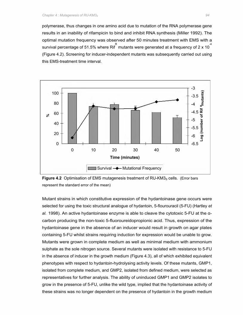
%
100
40
80
60
20
0 0 10 20 30 40 50
Survival Mutational Frequency
Time (minutes)
-3
-3.5
-4
-4.5
-5
-5.5
-6
-6.5
Log
(num
ber o
f Rif
R m
utan
ts)
Chapter 4 : Mutagenesis of RU-KM3S 94
polymerase, thus changes in one amino acid due to mutation of the RNA polymerase gene
results in an inability of rifampicin to bind and inhibit RNA synthesis (Miller 1992). The
optimal mutation frequency was observed after 50 minutes treatment with EMS with a
survival percentage of 51.5% where RifR mutants were generated at a frequency of 2 x 10
-4
(Figure 4.2). Screening for inducer-independent mutants was subsequently carried out using
this EMS-treatment time interval.
Figure 4.2 Optimisation of EMS mutagenesis treatment of RU-KM3S cells. (Error bars
represent the standard error of the mean)
Mutant strains in which constitutive expression of the hydantoinase gene occurs were
selected for using the toxic structural analogue of hydantoin, 5-flourouracil (5-FU) (Hartley et
al. 1998). An active hydantoinase enzyme is able to cleave the cytotoxic 5-FU at the α-
carbon producing the non-toxic 5-fluoroureidopropionic acid. Thus, expression of the
hydantoinase gene in the absence of an inducer would result in growth on agar plates
containing 5-FU whilst strains requiring induction for expression would be unable to grow.
Mutants were grown in complete medium as well as minimal medium with ammonium
sulphate as the sole nitrogen source. Several mutants were isolated with resistance to 5-FU
in the absence of inducer in the growth medium (Figure 4.3), all of which exhibited equivalent
phenotypes with respect to hydantoin-hydrolysing activity levels. Of these mutants, GMP1,
isolated from complete medium, and GMP2, isolated from defined medium, were selected as
representatives for further analysis. The ability of uninduced GMP1 and GMP2 isolates to
grow in the presence of 5-FU, unlike the wild type, implied that the hydantoinase activity of
these strains was no longer dependent on the presence of hydantoin in the growth medium
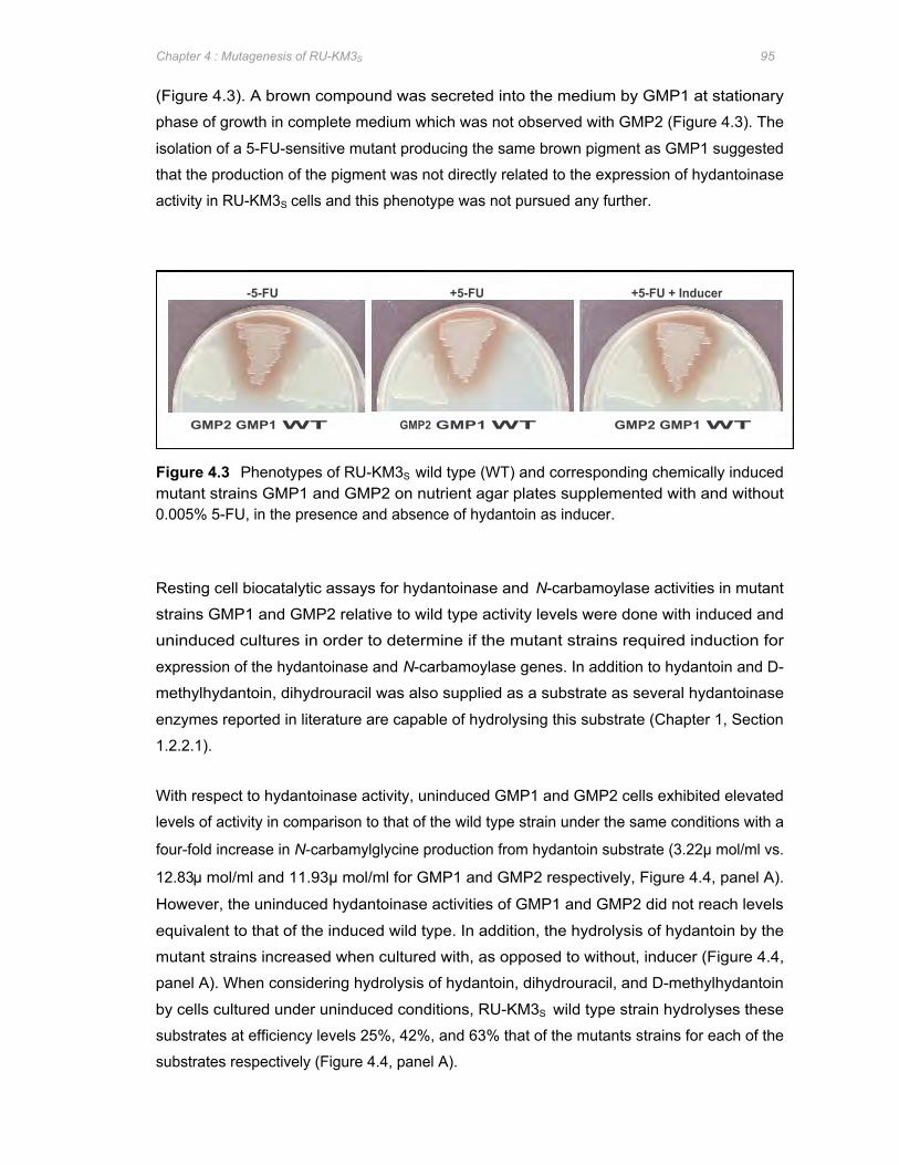
GMP2 GMP1 WT GMP2 GMP1 WT GMP2 GMP1 WT
-5-FU +5-FU +5-FU + Inducer
Chapter 4 : Mutagenesis of RU-KM3S 95
(Figure 4.3). A brown compound was secreted into the medium by GMP1 at stationary
phase of growth in complete medium which was not observed with GMP2 (Figure 4.3). The
isolation of a 5-FU-sensitive mutant producing the same brown pigment as GMP1 suggested
that the production of the pigment was not directly related to the expression of hydantoinase
activity in RU-KM3S cells and this phenotype was not pursued any further.
Figure 4.3 Phenotypes of RU-KM3S wild type (WT) and corresponding chemically induced mutant strains GMP1 and GMP2 on nutrient agar plates supplemented with and without 0.005% 5-FU, in the presence and absence of hydantoin as inducer.
Resting cell biocatalytic assays for hydantoinase and N-carbamoylase activities in mutant
strains GMP1 and GMP2 relative to wild type activity levels were done with induced and
uninduced cultures in order to determine if the mutant strains required induction for
expression of the hydantoinase and N-carbamoylase genes. In addition to hydantoin and D-
methylhydantoin, dihydrouracil was also supplied as a substrate as several hydantoinase
enzymes reported in literature are capable of hydrolysing this substrate (Chapter 1, Section
1.2.2.1).
With respect to hydantoinase activity, uninduced GMP1 and GMP2 cells exhibited elevated
levels of activity in comparison to that of the wild type strain under the same conditions with a
four-fold increase in N-carbamylglycine production from hydantoin substrate (3.22μ mol/ml vs.
12.83μ mol/ml and 11.93μ mol/ml for GMP1 and GMP2 respectively, Figure 4.4, panel A).
However, the uninduced hydantoinase activities of GMP1 and GMP2 did not reach levels
equivalent to that of the induced wild type. In addition, the hydrolysis of hydantoin by the
mutant strains increased when cultured with, as opposed to without, inducer (Figure 4.4,
panel A). When considering hydrolysis of hydantoin, dihydrouracil, and D-methylhydantoin
by cells cultured under uninduced conditions, RU-KM3S wild type strain hydrolyses these
substrates at efficiency levels 25%, 42%, and 63% that of the mutants strains for each of the
substrates respectively (Figure 4.4, panel A).

(A)
(B)
Pro
duct
( m
ol/m
l) P
rodu
ct (
mol
/ml)
16
14
12
10
4
8
6
2
0
40
60
50
30
20
10
0
Hydantoin (50mM) Dihydrouracil (50mM) D-Methylhydantoin (50mM)
WT (+) WT (-) GMP1 (+) GMP1 (-) GMP2 (+) GMP2 (-)
WT (+) WT (-) GMP1 (+) GMP1 (-) GMP2 (+) GMP2 (-)
Chapter 4 : Mutagenesis of RU-KM3S 96
Figure 4.4 Hydantoinase and N-carbamoylase activities, of induced and uninduced resting
cells, of wild type RU-KM3S and mutant strains GMP1 and GMP2. [Panel A: Hydantoinase
activity, Panel B: N-carbamoylase activity, (+): induced, (-): uninduced, WT: wild type. Whole cell
biocatalytic reactions were done with 50mM hydantoin, dihydrouracil, or D-methylhydantoin as
substrates. (Error bars represent the standard error of the mean, n=3)].
The N-carbamoylase in the wild type and mutant strains exhibited similar trends in activity
levels with negligible production of amino acids by uninduced cultures (Figure 4.4, panel B).
The low levels of D-alanine production in induced cells (an average of 0.46μ mol/ml D-alanine
produced vs. an average of 6.13μ mol/ml glycine and 13.75μ mol/ml β-alanine produced) was
expected as the N-carbamoylase of RU-KM3S is L-enantioselective (Figure 4.4, panel B).
Subsequent to induction during growth, slight increases in N-carbamoylase activity were
observed for GMP1 and GMP2 in comparison to the wild type strain (Figure 4.4, panel B).
Chapter 4 : Mutagenesis of RU-KM3S 97
4.3.2 Optimisation of RU-KM3S transposon mutagenesis Insertional mutation by transposons required that the target chromosome be accessible to
the transposable element which, in this study, was the plasposon pTnMod-OKm. This could
be achieved by electroporation or via conjugal transfer of the plasposon into the target
organism. Electroporation of DNA into RU-KM3S was tested but was found to be very
inefficient (data not shown) necessitating the development of a conjugal mating system
optimised for RU-KM3S cells. Plasposons, whilst encoding an oriT site, do not encode the tra
genes needed for pilus formation and plasmid mobilization which must therefore be supplied
by an alternate source. Some E. coli strains, such S17-1, encode these proteins on their
chromosomes. However, attempts to utilize E. coli strain S17-1 harbouring pTnMod-OTc in
bi-parental matings with strain RU-KM3S were unsuccessful. Consequently, tri-parental
mating with an E. coli strain containing a helper plasmid, such as pRK2013, encoding the
proteins required for conjugal transfer to occur was investigated. Tri-parental mating with P.
putida RU-KM3S, pTnMod-OKm in E. coli strain DH5α, and pRK2013 in E. coli strain HB101
was successful and is reflected by the operational transposition frequencies.
The operational transposition frequency, calculated as the ratio of exconjugants with non-
lethal insertions to the total number of recipient cells, was optimised by varying the ratios of
RU-KM3S to E. coli cells containing pTnMod-OKm, and pRK2013. The highest operational
transposition frequency was obtained with the ratio of 1:1:1 (RU-KM3S:pTnMod-
OKm:pRK2013) (Table 4.1) which was applied to all subsequent tri-parental mating
procedures.
Table 4.1 Optimisation of the operational transposition frequencies by varying the ratio of RU-KM3S cells to E. coli cells containing pTnMod-OKm and pRK2013.
Ratio Frequency
RU-KM3S E. coli E. coli (pTnMod-OKm) (pRK2013)
1 1 1 4.8 x 10⁻⁵
1 5 5 1.6 x 10⁻⁴
1 10 10 1.98 x 10⁻⁴
4.3.3 Isolation of 5-FU sensitive insertional mutants of GMP1 Mutant strain GMP1, isolated by chemical mutagenesis using EMS, was utilized as the
parental strain due to the high levels of dihydrouracil hydrolysis as well as increased levels of
hydantoinase activity exhibited by this strain in uninduced cultures resulting in resistance to
the toxic effect of 5-FU. Consequently, insertional mutant strains of GMP1 with sensitivity to
Chapter 4 : Mutagenesis of RU-KM3S 98
5-FU suggest that insertion of the plasposon DNA has interrupted either the hydantoinase
enzyme or the control thereof.
Two tri-parental matings with strain GMP1, pTnMod-OKm in E. coli DH5α, and pRK2013 in
E. coli HB101 were carried out with operational transpositional frequencies of 1.87 x 10⁻⁵
and
2.7 x 10⁻⁵
, respectively. Approximately 12 000 mutant strains from these tri-parental matings
were inoculated into 96-well microtitre plates containing Luria broth. Once grown to
confluence, the mutant strains were replica plated onto Luria agar plates supplemented with
(selection medium) and without (control) 0.1% 5-FU. Mutants which exhibited reduced or no
growth in the presence of 0.1% 5-FU were assayed for hydantoinase and N-carbamoylase
activity in microtitre plates. Of these, mutant strain GMP5 was found to have reduced levels
of hydantoin and dihydrouracil hydrolysis.
Initial full-scale resting cell biocatalytic assay of this mutant along with the parental strain
GMP1 and wild type strain of RU-KM3S showed that hydrolysis of dihydrouracil occurred
rapidly in the wild type and that the substrate quickly became a limiting factor in the
production of the intermediate, β-ureidopropionate. Thus in order to provide an accurate
reflection of the hydrolysis of dihydrouracil, the production of catalytic products was
determined after 1 and 2 hours incubation at 40oC as well as the standard 3 hours.
A significant reduction in the hydantoinase activity of mutant strain GMP5, particularly with
regard to hydrolysis of hydantoin (33.91μ mol/ml vs. 49.53μ mol/ml), when compared to that of
the parental strain GMP1 was observed (Figure 4.5, panel A). In addition, the reduction in
activity noted in uninduced cultures of GMP5 was not equivalent to, but rather much lower,
than that of uninduced wild type strain of RU-KM3S (Figure 4.5, panel A). This suggests that
the mutation in GMP5 is probably not an inactivation or nullification of the chemical mutation
in GMP1 which resulted in increased levels of activity. The reduction in N-carbamoylase
activity was even more pronounced than the hydantoinase activity levels with almost total
loss of production of glycine or alanine (0.58μ mol/ml glycine and 0.35μ mol/ml β-alanine vs.
9.53μ mol/ml and 15.81μ mol/ml glycine and β-alanine production in GMP1, Figure 4.5, panel
B). The presence, albeit reduced, of hydantoinase and N-carbamoylase activity in GMP5
indicated that the genes responsible for hydrolysis of hydantoin and dihydrouracil have not
been directly inactivated and that the insertional mutation in this strain is most likely in a
regulatory pathway affecting the expression or activity of these enzymes.
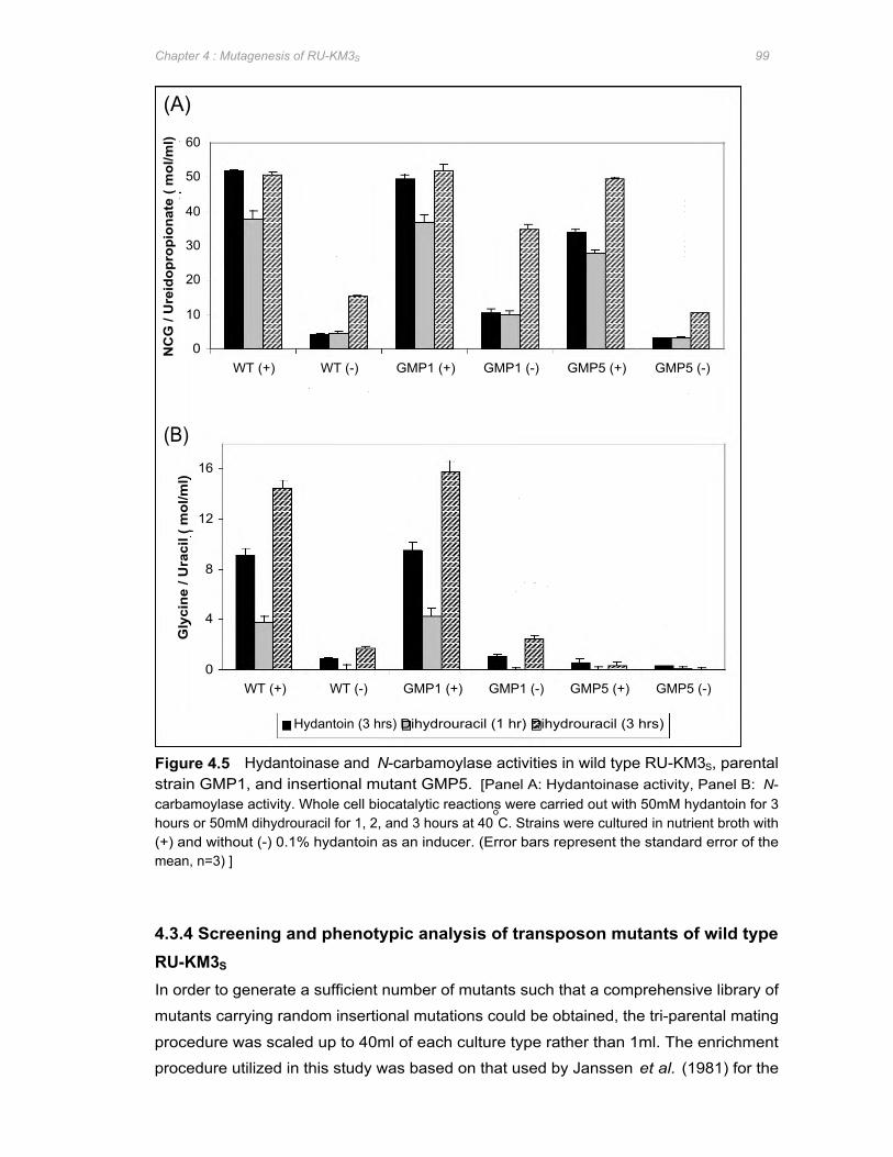
(A)
(B)
NC
G /
Ure
idop
ropi
onat
e ( m
ol/m
l) G
lyci
ne /
Ura
cil (
mol
/ml)
60
50
40
30
20
10
0
16
12
8
4
0
WT (+) WT (-) GMP1 (+) GMP1 (-) GMP5 (+) GMP5 (-)
WT (+) WT (-) GMP1 (+) GMP1 (-) GMP5 (+) GMP5 (-)
Hydantoin (3 hrs) Dihydrouracil (1 hr) Dihydrouracil (3 hrs)
Chapter 4 : Mutagenesis of RU-KM3S 99
Figure 4.5 Hydantoinase and N-carbamoylase activities in wild type RU-KM3S, parental strain GMP1, and insertional mutant GMP5. [Panel A: Hydantoinase activity, Panel B: N-carbamoylase activity. Whole cell biocatalytic reactions were carried out with 50mM hydantoin for 3 hours or 50mM dihydrouracil for 1, 2, and 3 hours at 40
oC. Strains were cultured in nutrient broth with
(+) and without (-) 0.1% hydantoin as an inducer. (Error bars represent the standard error of the mean, n=3) ]
4.3.4 Screening and phenotypic analysis of transposon mutants of wild type RU-KM3S
In order to generate a sufficient number of mutants such that a comprehensive library of
mutants carrying random insertional mutations could be obtained, the tri-parental mating
procedure was scaled up to 40ml of each culture type rather than 1ml. The enrichment
procedure utilized in this study was based on that used by Janssen et al. (1981) for the
Chapter 4 : Mutagenesis of RU-KM3S 100
isolation of glutamine-requiring mutants and modified to select for hydantoinase and/or N-
carbamoylase minus mutants of RU-KM3S as described in the methods and materials.
The bactericidal effect of ampicillin provides the basis on which the enrichment procedure
was developed. Ampicillin is a β-lactam antibiotic which severely inhibits bacterial cell wall
synthesis by inhibiting the transpeptidase enzyme responsible for cross-linkage of the
glycan-linked peptide chains essential for the formation of a strong cell wall in bacteria
(Madigan et al. 1984). In the absence of the transpeptidase activity, the cell wall continues to
be formed but gets progressively weaker until cell lysis occurs. If the cells are dormant,
however, no cell growth occurs thus no weak cell wall is synthesized and no cell lysis occurs
(Snyder and Champress 2003).
Enrichment of plasposon generated mutants in minimal medium with hydantoin as a sole
source of nitrogen therefore selected for those mutants which are unable to utilize hydantoin
as a source of nitrogen. Thus mutant strains encoding an inactivated hydantoinase and/or
N-carbamoylase gene were unable to grow and consequently survived treatment with
ampicillin, whilst the remaining mutants actively divided and eventually lysed due to weak cell
wall formation. In addition to hydantoinase and N-carbamoylase minus mutants, auxotrophic
mutants unable to grow in minimal medium, irrespective of the nitrogen source supplied, also
survived the enrichment. In order to eliminate these from the screening process, the mutant
strains surviving enrichment were plated onto minimal medium with ammonium sulphate as a
nitrogen source.
A large scale tri-parental mating with RU-KM3S, pTnMod-OKm in E. coli DH5α, and pRK2013
in E. coli HB101 was carried out and an operational transposon frequency of 7.1 x 10⁻⁶
achieved. Approximately 107 000 insertional mutants entered the enrichment process of
which approximately 7 000 survived indicating a survival rate of 6.5%. Not all insertional
mutants could be entered into the subsequent screening process as colonies which
overlapped one another could not be individually processed. Thus 6 624 mutants were
inoculated into 96-well microtitre plates and grown to confluence in Luria broth containing
chloramphenicol (resistance conferred by RU-KM3S) and kanamycin (resistance conferred by
inserted plasposon sequence). These isolated mutants were replica plated onto minimal
medium with hydantoin (HMM) or ammonium sulphate (AMM) as sole sources of nitrogen to
ensure that only mutants which grew well on ammonium sulphate, and are therefore not
auxotrophic for nutrients other than hydantoin, were analysed further. A total of 144 mutants
were isolated that were able to hydrolyse ammonium sulphate (amm+) but not hydantoin
(hyd-) as nitrogen sources.

Chapter 4 : Mutagenesis of RU-KM3S 101
Hydantoin is hydrolysed to N-carbamoylglycine and glycine by the hydantoinase and N-
carbamoylase enzymes respectively. Thus the inability to grow on HMM can be attributed to
either direct or indirect inactivation of the hydantoinase and/or N-carbamoylase enzymes or
an inability to utilize glycine as a nitrogen source. Furthermore, hydantoinase enzymes are
often associated with hydrolysis of pyrimidines (Syldatk et al. 1999), the end product of which
is uracil. Thus mutant strains unable to grow on HMM were scored for growth on minimal
medium with glycine (GMM) or uracil (UMM) as sole sources of nitrogen. In addition, the
hydantoin-hydrolysing activity of these mutants was evaluated using the modified microtitre
plate assay with hydantoin as a substrate. The 144 mutant strains previously isolated as
auxotrophic for hydantoin could thus be subdivided into 3 groupings (Table 4.2) based on
their growth with hydantoin, ammonium sulphate, uracil, or glycine as sole nitrogen sources
and the hydrolysis of hydantoin as determined by microtitre plate assays.
Table 4.2 Insertional mutants of RU-KM3S, auxotrophic with respect to hydantoin, grouped phenotypically based on the ability to utilize various sources of nitrogen, as well as the presence or absence of hydantoinase and N-carbamoylase activity.
Group Number of Representative Nitrogen utilization Enzyme activity
mutants isolates amm hyd gly ura HYDN NCAAH
WT - - + + + + + +
A 122 GMP6
GMP7
GMP9
GMP10
GMP15
GMP21
+ - + - + +
B 14 GMP8 + - - ± + / nd∗
+ / nd∗
C 8 GMP3
GMP4
GMP16
GMP17
GMP18
GMP19
GMP20
+ - + ± / - + -
(amm, hyd, gly, ura: minimal medium with 0.25% ammonium sulphate, 1% hydantoin, 1% glycine, or
1% uracil respectively as sole sources of nitrogen; WT : wild type RU-KM3S; HYDN: Hydantoinase;
NCAAH: N-carbamoylase; nd∗: The majority of mutants in phenotype group B exhibited retarded
growth rates and could therefore not be comparatively assayed)
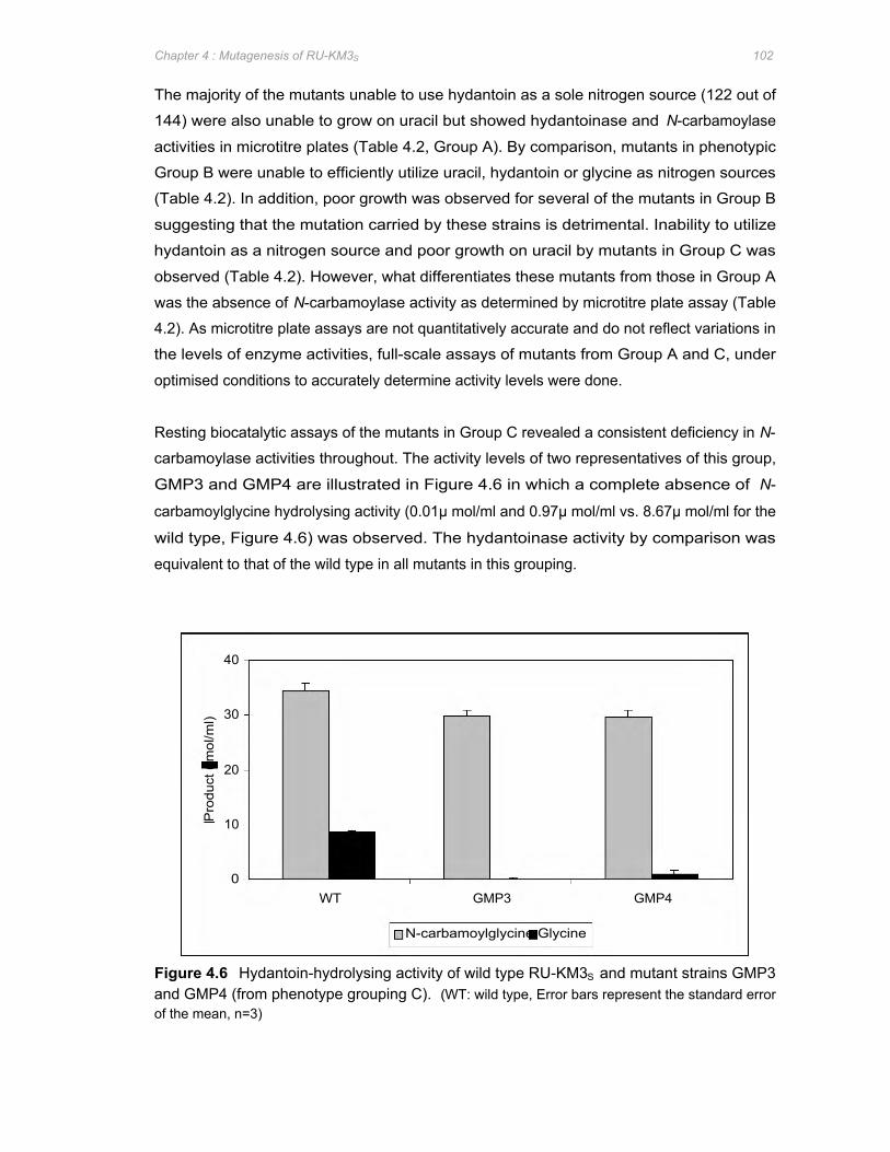
Pro
duct
( m
ol/m
l)
40
30
20
10
0 WT GMP3 GMP4
N-carbamoylglycine Glycine
Chapter 4 : Mutagenesis of RU-KM3S 102
The majority of the mutants unable to use hydantoin as a sole nitrogen source (122 out of
144) were also unable to grow on uracil but showed hydantoinase and N-carbamoylase
activities in microtitre plates (Table 4.2, Group A). By comparison, mutants in phenotypic
Group B were unable to efficiently utilize uracil, hydantoin or glycine as nitrogen sources
(Table 4.2). In addition, poor growth was observed for several of the mutants in Group B
suggesting that the mutation carried by these strains is detrimental. Inability to utilize
hydantoin as a nitrogen source and poor growth on uracil by mutants in Group C was
observed (Table 4.2). However, what differentiates these mutants from those in Group A
was the absence of N-carbamoylase activity as determined by microtitre plate assay (Table
4.2). As microtitre plate assays are not quantitatively accurate and do not reflect variations in
the levels of enzyme activities, full-scale assays of mutants from Group A and C, under
optimised conditions to accurately determine activity levels were done.
Resting biocatalytic assays of the mutants in Group C revealed a consistent deficiency in N-
carbamoylase activities throughout. The activity levels of two representatives of this group,
GMP3 and GMP4 are illustrated in Figure 4.6 in which a complete absence of N-
carbamoylglycine hydrolysing activity (0.01μ mol/ml and 0.97μ mol/ml vs. 8.67μ mol/ml for the
wild type, Figure 4.6) was observed. The hydantoinase activity by comparison was
equivalent to that of the wild type in all mutants in this grouping.
Figure 4.6 Hydantoin-hydrolysing activity of wild type RU-KM3S and mutant strains GMP3 and GMP4 (from phenotype grouping C). (WT: wild type, Error bars represent the standard error of the mean, n=3)
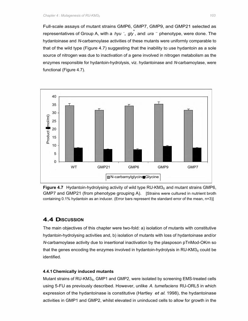
Pro
duct
( m
ol/m
l)
40
35
30
25
20
15
10
5
0 WT GMP21 GMP6 GMP9 GMP7
N-carbamylglycine Glycine
Chapter 4 : Mutagenesis of RU-KM3S 103
Full-scale assays of mutant strains GMP6, GMP7, GMP9, and GMP21 selected as
representatives of Group A, with a hyu -, gly+, and ura – phenotype, were done. The
hydantoinase and N-carbamoylase activities of these mutants were uniformly comparable to
that of the wild type (Figure 4.7) suggesting that the inability to use hydantoin as a sole
source of nitrogen was due to inactivation of a gene involved in nitrogen metabolism as the
enzymes responsible for hydantoin-hydrolysis, viz. hydantoinase and N-carbamoylase, were
functional (Figure 4.7).
Figure 4.7 Hydantoin-hydrolysing activity of wild type RU-KM3S and mutant strains GMP6, GMP7 and GMP21 (from phenotype grouping A). [Strains were cultured in nutrient broth containing 0.1% hydantoin as an inducer. (Error bars represent the standard error of the mean, n=3)]
4.4 DISCUSSION
The main objectives of this chapter were two-fold: a) isolation of mutants with constitutive
hydantoin-hydrolysing activities and, b) isolation of mutants with loss of hydantoinase and/or
N-carbamoylase activity due to insertional inactivation by the plasposon pTnMod-OKm so
that the genes encoding the enzymes involved in hydantoin-hydrolysis in RU-KM3S could be
identified.
4.4.1 Chemically induced mutants Mutant strains of RU-KM3S, GMP1 and GMP2, were isolated by screening EMS-treated cells
using 5-FU as previously described. However, unlike A. tumefaciens RU-ORL5 in which
expression of the hydantoinase is constitutive (Hartley et al. 1998), the hydantoinase
activities in GMP1 and GMP2, whilst elevated in uninduced cells to allow for growth in the
Chapter 4 : Mutagenesis of RU-KM3S 104
presence of 5-FU, were not equivalent to wild type induced activity levels. In addition to this,
an increase in hydantoin-hydrolysis from uninduced to induced cells of GMP1 and GMP2
was also observed.
In addition to the elevated but still inducible hydantoinase activity in GMP1 and GMP2, the
discrepancy between the conversion of dihydrouracil and D-methylhydantoin versus
hydantoin raised the possibility of the presence of a second hydantoinase in RU-KM3S cells
whose activity was not affected by the mutation in strain GMP1 and GMP2. The point
mutation generated by EMS may therefore have been in the promoter region of one of the
potential hydantoinase genes resulting in constitutive expression of one of the hydantoinases
whilst the remaining enzyme is inducible. Alternatively, the mutation may have occurred in
the gene itself such that increased levels of hydrolysing activity of one of the hydantoinases
resulted. The presence of two hydantoinases within a single bacterium has been reported
for Agrobacterium sp. IP I-671 (Hils et al. 2001). A third possibility is that the mutation has
occurred in a regulatory pathway which exerts repression with variable effects on hydrolysis
of different substrates. The latter is more likely as evidenced by the brown pigment produced
by GMP1 as regulatory pathways in bacteria often function on a global cellular scale thus
modification of a single pathway may have a pleiotrophic effect.
4.4.2 Transposon-generated mutants With respect to transposon generated mutants, the utilization of an enrichment procedure
proved to be successful. Where traditional direct screening was utilized, such as that used
for mutant strains of GMP1, over 12 000 plasposon-generated mutants had to be screened in
order to yield a single mutant strain (GMP5) of interest. In contrast, by processing mutant
strains through an enrichment step before entering the screening procedure, the number of
mutants exhibiting the desired phenotype (i.e. hyd -) was significantly increased (144 vs. 1)
whilst the number of mutants screened by replica plating was almost halved (6,600 vs.
12,000).
As identification of the genes encoding the hydantoin-hydrolysing enzymes in RU-KM3S by
complementation studies with a genomic library (Chapter 3) was unsuccessful, plasposons
were used as an alternative approach. Screening plasposon-generated mutants in which the
genes encoding the hydantoin-hydrolysing enzymes were inactivated was done based on an
inability to utilize hydantoin as a sole source of nitrogen. The plasposon-generated mutant
strains thus isolated could be subdivided, based on phenotypic characteristics, into three
groupings.
Chapter 4 : Mutagenesis of RU-KM3S 105
The inability of Group A mutant strains to utilize hydantoin as a sole nitrogen source cannot
be attributed to inactive hydantoinase or N-carbamoylase enzymes as biocatalytic assays of
these mutants showed hydrolytic activities equivalent to that of the wild type strain. Similarly,
an inactivation/blockage of the transport of hydantoin into the cells, which may have resulted
in a hyd − phenotype, can also be ruled out as the biocatalytic assays were done with intact
resting cells. The positive growth score with glycine minimal medium by mutant strains of
this grouping furthermore eliminates the possibility of the hyd − phenotype being due to an
inability to utilize the end-product of hydantoin hydrolysis as a nitrogen source. The absence
of growth on uracil minimal medium plates (ura −) suggest insertional mutations affecting
pyrimidine catabolism or the utilization of the end product of uracil degradation, β-alanine
(Michal 1999). As the hydantoinase and N-carbamoylase enzymes of RU-KM3S are able to
hydrolyse pyrimidines (i.e. dihydrouracil), the ura − phenotype observed is most likely due to
an failure to utilize alanine as a sole source of nitrogen.
Operons encoding genes responsible for utilizing specific amino acids as nitrogen sources
are not only regulated by individual induction pathways, but are often part of larger regulatory
mechanisms. Ntr regulation in particular, where expression of certain genes are repressed in
the presence of a more suitable nitrogen source such as NH3, affects expression of many
such operons (Snyder and Champness 2003). Thus a mutation in the Ntr regulatory
pathway would disrupt utilization of poorer nitrogen sources. This may provide an
explanation for the large number of mutants in phenotype Group A, which were found to be
hydantoin auxotrophs but do not have altered enzyme activities.
Resting cell biocatalytic assays of mutant strains belonging to phenotypic Group C indicated
a complete lack of N-carbamoylase activity within these mutants. The majority of hydantoin-
hydrolysing enzymes systems investigated to date contain a single enzyme responsible for
the conversion of N-carbamylglycine to glycine, with a few notable exceptions where two N-
carbamoylase enzymes have been isolated from a particular strain (Yamanaka et al. 1997,
Yokozeki et al. 1987c). The loss of N-carbamoylase activity by a single insertional event in
mutant strains in Group C suggests that only one N-carbamoylase enzyme is present in P.
putida strain RU-KM3S. The lack of N-carbamoylase activity would explain the absence of
growth on hydantoin minimal medium and possibly the poor growth on uracil (if hydantoin-
hydrolysis in RU-KM3S cells is synonymous with pyrimidine degradation).
The majority of the insertional mutants in the phenotypic Group B exhibited retarded growth
rates in complete medium indicating a detrimental though sub-lethal mutation. Thus their
inability to grow under nitrogen-limiting conditions was not surprising. The few mutant strains
with normal growth rates in complete medium, while displaying hydantoinase and N-
Chapter 4 : Mutagenesis of RU-KM3S 106
carbamoylase activity and growth in the presence of good nitrogen sources such as
ammonium sulphate, were unable to utilize poorer nitrogen sources suggesting the presence
of a mutation in the regulation of nitrogen assimilation by the cells under conditions of
nitrogen limitation.
No loss of hydantoinase activity due to insertional inactivation of the relevant genes was
observed for any of the mutants described above. Acquired sensitivity to 5-FU by
transposon-generated mutants with 5-FU resistant GMP1 as the parental strain, provided an
alternative approach to isolation of hydantoinase-inactivated mutants. Whilst a mutant in
which an absence of hydantoinase activity was observed was not isolated, mutant strain
GMP5 was isolated with significantly reduced levels of both hydantoinase and N-
carbamoylase enzyme activities. The reduction in both the hydantoinase and N-
carbamoylase enzyme activities in mutant strain GMP5 is most likely due to a mutation in a
regulatory mechanism responsible for the control of expression or activity of both these
genes. However, the mutation in GMP5 was probably not a repressor of the original
mutation in GMP1 as reversion from GMP1 to wild type RU-KM3S uniniduced activity levels
was not achieved.
In conclusion, mutant strains GMP1 and GMP2 were isolated and shown to have elevated,
but not constitutive, levels of hydantoin-hydrolysing activities. As discussed above, this may
be due to the presence of two hydantoinase enzymes or as a result of a mutation in a
regulatory pathway affecting hydantoin-hydrolysis in RU-KM3S. Inactivation of the
hydantoinase gene, once isolated, followed by biocatalytic assay to determine if any residual
hydantoin-hydrolysing activity remained would indicate if two hydantoinases are indeed
present in RU-KM3S or not. This is addressed in Chapter 5.
The loss of N-carbamoylase activity in mutant isolates from phenotype Group C suggested
that the gene interrupted may be that encoding for the N-carbamoylase enzyme itself. Group
A mutants on the other hand showed wild-type levels of hydantoin-hydrolysing activity but
were unable to grow on a poor nitrogen source. This implied that a regulatory pathway
involved in nitrogen metabolism but which does not affect expression of the hydantoinase
and N-carbamoylase genes was interrupted in these mutants. By contrast, the gene
inactivated in mutant strain GMP5 resulted in decreased, but not complete loss, of both
enzyme activities indicating that a regulatory pathway which regulates expression of the
hydantoinase and N-carbamoylase genes was affected. Identification of the genes
inactivated by transposon insertion, resulting in the phenotypes discussed above, would
provide greater insight into the enzymes responsible for hydantoin-hydrolysis in RU-KM3S
and the control thereof and is addressed in the following chapter.
Chapter 5 : Structural and regulatory components of the hydantoin-hydrolysing pathway of RU-KM3S 107
5.1 INTRODUCTION
In order to be competitive, bacteria employ complex regulatory mechanisms to control gene
expression to ensure that energy expensive production of catabolic enzymes occurs only in
the presence of the corresponding catabolite and absence of a more readily metabolizable
nutrient source. For maximum efficiency, many of these regulatory mechanisms function at
the level of transcription by elevating/inhibiting promoter activity. These mechanisms include
induction and feedback inhibition, as well as global regulatory pathways that reflect the
metabolic and energy status of the cell resulting in silencing of energetically less favourable
catabolic pathways (Sanchez and Demain 2002).
P. putida strain RU-KM3S was originally selected for based on the ability to utilize hydantoin
as a sole source of nitrogen in defined medium (Chapter 1, Section 1.7). As hydantoin is
catabolized in order to supply the nitrogen demand of the cells in defined medium, the
hydantoin-hydrolysing enzymes may well be controlled by the global nitrogen regulatory
system (Ntr) which is responsible for the coordinated expression of catabolic pathways
involved in nitrogen metabolism (Merrick and Edwards 1995, Hartley et al. 2001). Support
for this hypothesis includes the identification of putative σ54
-dependent promoter regions
located upstream of the genes encoding the hydantoin-hydrolysing enzymes in Arthrobacter
aurescens DSM 3747, Bacillus agri NCHU1002, and Pseudomonas sp. NS671, as well as
mutational analysis of the regulation of the hydantoin-hydrolysing enzyme system of
Agrobacterium tumefaciens RU-OR in which Ntr is implicated (Watabe et al. 1992a, Kao and
Hsu 2003, Wiese et al. 2001, Hartley et al. 2001).
Regulation of gene expression in response to the nitrogen status of the cell has been
extensively characterized in enteric bacteria (Reitzer 2003). In this system, the Ntr system
regulates gene expression at the level of transcription by interaction of the RNA polymerase
containing the sigma factor σ54
and the NtrC protein. In its phosphorylated form, NtrC acts as
a transcriptional activator of σ54
-dependent promoters. The phophorylation state of the NtrC
is in turn regulated by the bifunctional NtrB which can either phosphorylate or
dephosphorylate NtrC (Figure 5.1). The negative/positive phophorylation activity of NtrB is
dependent on the state of the regulatory protein PII which occurs in an uridylylated or
deuridylylated form catalysed by the UTase/UR (uridylyltransferase/uridylyl-removing)
enzyme. The ratio of glutamine to α-ketoglutarate reflects the nitrogen status of the cell.
Thus stimulation of UR by glutamine or UTase by α-ketoglutarate, affecting the uridylylation
status of PII and subsequent cascade reactions, result in transcriptional repression and
activation of the σ54
-dependent catabolic enzymes respectively (Merrick and Edwards 1995,
Eberl et al. 2000).
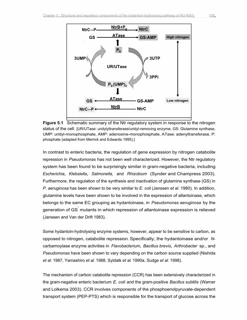
NtrB NtrC—P
ATase GS GS-AMP
High nitrogen
3UTP
GS-AMP ATase GS
NtrB+PII NtrC—P NtrC
3PPi
3UMP
PII
UR/UTase
PII (UMP)3
Low nitrogen
NtrC
Chapter 5 : Structural and regulatory components of the hydantoin-hydrolysing pathway of RU-KM3S 108
Figure 5.1 Schematic summary of the Ntr regulatory system in response to the nitrogen status of the cell. [UR/UTase: uridylyltransferase/uridyl-removing enzyme, GS: Glutamine synthase, UMP: uridyl-monophosphate, AMP: adenosine-monophosphate, ATase: adenyltransferase, P: phosphate (adapted from Merrick and Edwards 1995).]
In contrast to enteric bacteria, the regulation of gene expression by nitrogen catabolite
repression in Pseudomonas has not been well characterized. However, the Ntr regulatory
system has been found to be surprisingly similar in gram-negative bacteria, including
Escherichia, Klebsiella, Salmonella, and Rhizobium (Synder and Champress 2003).
Furthermore, the regulation of the synthesis and inactivation of glutamine synthase (GS) in
P. aeruginosa has been shown to be very similar to E. coli (Janssen et al. 1980). In addition,
glutamine levels have been shown to be involved in the expression of allantoinase, which
belongs to the same EC grouping as hydantoinase, in Pseudomonas aeruginosa by the
generation of GS- mutants in which repression of allantoinase expression is relieved
(Janssen and Van der Drift 1983).
Some hydantoin-hydrolysing enzyme systems, however, appear to be sensitive to carbon, as
opposed to nitrogen, catabolite repression. Specifically, the hydantoinase and/or N-
carbamoylase enzyme activities in Flavobacterium, Bacillus brevis, Arthrobacter sp., and
Pseudomonas have been shown to vary depending on the carbon source supplied (Nishida
et al. 1987, Yamashiro et al. 1988, Syldatk et al. 1990a, Sudge et al. 1998).
The mechanism of carbon catabolite repression (CCR) has been extensively characterized in
the gram-negative enteric bacterium E. coli and the gram-positive Bacillus subtilis (Warner
and Lolkema 2003). CCR involves components of the phosphoenolpyruvate-dependent
transport system (PEP-PTS) which is responsible for the transport of glucose across the
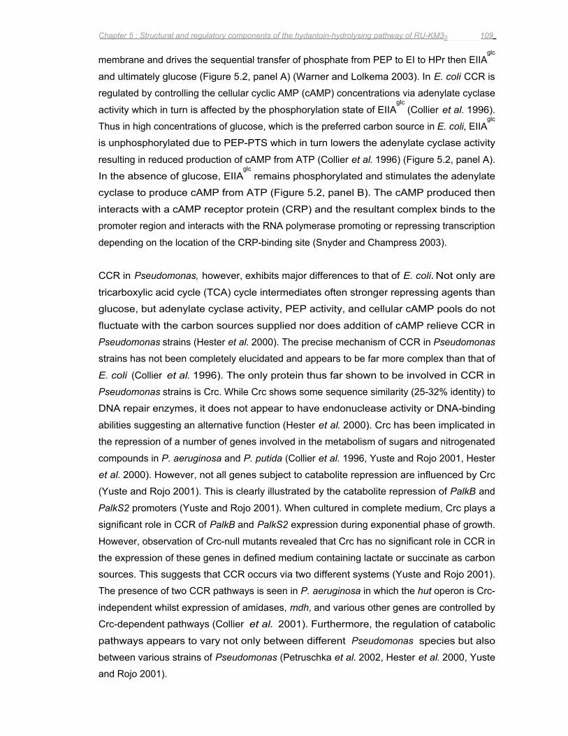
Chapter 5 : Structural and regulatory components of the hydantoin-hydrolysing pathway of RU-KM3S 109
membrane and drives the sequential transfer of phosphate from PEP to EI to HPr then EIIAglc
and ultimately glucose (Figure 5.2, panel A) (Warner and Lolkema 2003). In E. coli CCR is
regulated by controlling the cellular cyclic AMP (cAMP) concentrations via adenylate cyclase
activity which in turn is affected by the phosphorylation state of EIIAglc
(Collier et al. 1996).
Thus in high concentrations of glucose, which is the preferred carbon source in E. coli, EIIAglc
is unphosphorylated due to PEP-PTS which in turn lowers the adenylate cyclase activity
resulting in reduced production of cAMP from ATP (Collier et al. 1996) (Figure 5.2, panel A).
In the absence of glucose, EIIAglc
remains phosphorylated and stimulates the adenylate
cyclase to produce cAMP from ATP (Figure 5.2, panel B). The cAMP produced then
interacts with a cAMP receptor protein (CRP) and the resultant complex binds to the
promoter region and interacts with the RNA polymerase promoting or repressing transcription
depending on the location of the CRP-binding site (Snyder and Champress 2003).
CCR in Pseudomonas, however, exhibits major differences to that of E. coli. Not only are
tricarboxylic acid cycle (TCA) cycle intermediates often stronger repressing agents than
glucose, but adenylate cyclase activity, PEP activity, and cellular cAMP pools do not
fluctuate with the carbon sources supplied nor does addition of cAMP relieve CCR in
Pseudomonas strains (Hester et al. 2000). The precise mechanism of CCR in Pseudomonas
strains has not been completely elucidated and appears to be far more complex than that of
E. coli (Collier et al. 1996). The only protein thus far shown to be involved in CCR in
Pseudomonas strains is Crc. While Crc shows some sequence similarity (25-32% identity) to
DNA repair enzymes, it does not appear to have endonuclease activity or DNA-binding
abilities suggesting an alternative function (Hester et al. 2000). Crc has been implicated in
the repression of a number of genes involved in the metabolism of sugars and nitrogenated
compounds in P. aeruginosa and P. putida (Collier et al. 1996, Yuste and Rojo 2001, Hester
et al. 2000). However, not all genes subject to catabolite repression are influenced by Crc
(Yuste and Rojo 2001). This is clearly illustrated by the catabolite repression of PalkB and
PalkS2 promoters (Yuste and Rojo 2001). When cultured in complete medium, Crc plays a
significant role in CCR of PalkB and PalkS2 expression during exponential phase of growth.
However, observation of Crc-null mutants revealed that Crc has no significant role in CCR in
the expression of these genes in defined medium containing lactate or succinate as carbon
sources. This suggests that CCR occurs via two different systems (Yuste and Rojo 2001).
The presence of two CCR pathways is seen in P. aeruginosa in which the hut operon is Crc-
independent whilst expression of amidases, mdh, and various other genes are controlled by
Crc-dependent pathways (Collier et al. 2001). Furthermore, the regulation of catabolic
pathways appears to vary not only between different Pseudomonas species but also
between various strains of Pseudomonas (Petruschka et al. 2002, Hester et al. 2000, Yuste
and Rojo 2001).
(A)
Glucose
Glucose-6- P Glycolysis EIIc
EIIb
Out In
P
EIIAglc P ( )
AC
HPr P
EI
PEP Pyruvate
CRP cAMP
(±)
ATP
cAMP
Gene
(B)
PEP Pyruvate
Glucose
Out In
EIIc EIIb
EIIAglc P
HPr P
EI
(+) AC
Chapter 5 : Structural and regulatory components of the hydantoin-hydrolysing pathway of RU-KM3S 110
Figure 5.2 Schematic summary of the CCR regulatory system in E. coli. (AC: adenylate cyclase, PEP: phosphoenolypyruvate, P: phosphate) (adapted from Warner and Lolkema 2003).
As discussed in Chapter 4, several mutant strains of P. putida RU-KM3S with phenotypes
such as reduction or complete loss of hydantoinase and/or N-carbamoylase activity, as well
as mutant strains unable to utilize hydantoin as a nitrogen source but still exhibiting wild type
levels of hydantoin-hydrolysing activity, were isolated. Identification of the locus inactivated
by insertional mutation in each mutant strain would facilitate a) isolation of the structural
Chapter 5 : Structural and regulatory components of the hydantoin-hydrolysing pathway of RU-KM3S 111
genes involved in the hydrolysis of hydantoin in RU-KM3S cells, b) identification of genes
whose products are involved in the regulation of hydantoinase and N-carbamoylase
expression, and c) by use of the collective data, elucidation of the regulatory pathways that
control the hydantoin-hydrolysing operon in RU-KM3S cells. This chapter describes the
isolation and characterization of the genomic sequences flanking the transposon insertion in
14 selected mutants and the subsequent analysis of the structural and regulatory
components of hydantoin-hydrolysis in RU-KM3S cells.
5.2 METHODS AND MATERIALS
5.2.1 Identification of plasposon disrupted genes in RU-KM3S mutants Genomic DNA was extracted as described in Appendix 5.2 and approximately 1μ g of
genomic DNA was digested to completion with either Bam HI, Eco RI, Csp 451, or Pst I
restriction endonucleases, according to the manufacturers specifications, in a reaction
volume of 60μ l. After heat inactivation of the restriction endonuclease, 10μ l of the DNA
fragments were self-ligated using T4 DNA Ligase (Promega) in a total reaction volume of
50μ l, incubated at 4oC for 2 to 3 days. Transformation of 10μ l aliquots of the ligation with
150μ l competent E. coli DH5α cells was done (Appendix 4) and colonies exhibiting antibiotic
resistance conferred by the inserted plasposon DNA were selected by growth on Luria agar
plates containing 50μ g/ml kanamycin. Selected colonies were individually isolated and
screened for the presence of recombinant plasmids using the Easyprep method
(Berghammer and Auer 1993) and diagnostic restriction endonuclease digestion (Appendix
5.1). The High Pure Plasmid Isolation Kit (Roche) was used according to manufacturers
instructions to extract selected plasmids for subsequent sequence analysis. The nucleotide
sequence of the chromosomal DNA flanking the inserted plasposon DNA was determined
from double stranded plasmid DNA by the dideoxy-chain termination method (Sanger 1987)
using the ABI Prism Big Dye Terminator Cycle Sequencing Ready Reaction Kit (PE Applied
Biosystems) (Appendix 5.4). The primers utilized for the sequencing reactions, prGM11 and
prGM12 (Appendix 3), correspond to the terminal ends of the plasposon insertion sequence
(Figure 4.1).
5.2.2 DNA sequence analysis Preliminary analysis of raw sequence data was carried out using Vector NTI DeLuxe v4.0 or
v5.0 software (Informax Inc.). Identification of known genes to which acquired sequence
data exhibits a high degree of similarity was done using the Basic Local Alignment Search
Tool (BLASTX, BLASTP) (Altschul et al. 1990) hosted by the National Centre for
Biotechnology Information (NCBI) website (http://www.ncbi.nlm.nih.gov).
Chapter 5 : Structural and regulatory components of the hydantoin-hydrolysing pathway of RU-KM3S 112
5.2.3 Construction of dhp knockout mutants Maps of plasmids used in this chapter are contained in Appendix 3. The knockout vector,
pGMKdhp, utilized to generate a mutant strain of P. putida RU-KM3S encoding an inactivated
dihydropyrimidinase (dhp) gene, was constructed using the plasmid pTnMod-OTc and a 5’/3’
truncated fragment of the dhp gene. The transposase gene was removed from pTnMod-OTc
by restriction endonuclease digestion with Xho I and Pvu II as per manufacturers
specifications. The dhp fragment truncated at both the 3’ and 5’ end was obtained by
digestion of the dhp gene in pGMdhp.a with Sal I and Pvu II. The 4162bp backbone of
pTnMod-OTc and 712bp fragment of the dhp gene were purified from an agarose gel using
the GFXTM
PCR DNA and Gel Band Purification Kit (Amersham). These purified fragments
were then ligated with T4 DNA Ligase (Promega) as per manufacturers specifications,
incubated overnight at 4oC, and transformed into E. coli DH5α. The resultant tetracyline
resistant colonies were screened for the properly constructed pGMKdhp vector. The
pGMKdhp vector was then introduced into P. putida RU-KM3S by tri-parental mating with E.
coli DH5α containing the correct pGMKdhp vector, E. coli HB101 containing pRK2013, and
wild type RU-KM3S as described in Section 4.2.1 (Chapter 4). The cells were plated onto
Luria agar containing 25μ g.ml-1 chloramphenicol (to select against the E. coli strains) and
50μ g.ml-1 tetracyline (to select against RU-KM3S cells lacking the integrated vector) in order
to isolate mutant strains of RU-KM3S in which conjugal transfer and homologous
recombination of the pGMKdhp into the chromosome of RU-KM3S had occurred rendering
the dihydropyrimidinase gene inactive.
To confirm the disruption of the dhp gene, the recombinant genomic DNA was extracted as
described previously and subjected to PCR analysis using Expand High Fidelity PCR system
(Roche) or Taq polymerase (Bioline) using standard amplification parameters (Appendix 5.3)
with annealing temperatures of 60oC and 45
oC. Primers corresponding to internal sequences
of the dhp (prGM21, prGM28) and specific for pGMKdhp (prGM19, prGM20, prGM11)
(Appendix 3) were utilized. The products of the PCR reactions were then separated by
electrophoresis with 1% agarose gel containing 0.5μ g/ml ethidium bromide, and visualized
using the Kodak DC 120 gel imaging system.
5.2.4 Analysis of the promoter regions of bup , ORF1, and dhp The intergenic region between ORF1 and the dihydropyrimidinase gene was amplified from
genomic DNA from P. putida RU-KM3S and GMP1, by PCR using the Expand High Fidelity
PCR system (Roche) with primers prGM22, specific for the terminal end of the dhp gene, and
prGM30, specific for the start of ORF1 (Appendix 3) using standard amplification parameters
(Appendix 5.3) with an annealing temperatures of 55oC. The PCR product was then purified
(High Pure PCR Product Purification Kit, Roche) and ligated into the pGEM-T Easy vector
Chapter 5 : Structural and regulatory components of the hydantoin-hydrolysing pathway of RU-KM3S 113
(Promega) to form the constructs pGMIRwt and pGMIR1 containing the 2076bp PCR product
obtained with wild type and GMP1 genomic DNA as templates respectively. In order to
determine the nucleotide sequence of the entire intergenic region, a 1198bp fragment was
excised from pGMIRwt and pGMIR1 by restriction digestion with Sac II and recircularisation
by ligation to form pGMIRwtΔSac II and pGMIR1ΔSac II respectively. Using primers
prGM30, with pGMIRwt and pGMIR1 as templates, and pUCF with pGMIRwtΔSac II and
pGMIR1ΔSac II as templates the nucleotide sequence of the promoter region was
sequenced.
5.2.5 Biocatalytic assays Resting cell biocatalytic reactions were carried out with RU-KM3S as described in Appendix
2.1, except that the culture medium used was nutrient broth supplemented with and without
1% ammonium sulphate, 1% glucose, or 1% succinate. With respect to the biocatalytic
assay of pGMIRwt (Section 5.2.4) in E. coli strain DH5α, the cultures were grown to
confluence overnight, at 37oC, in Luria broth with 100μ g/ml ampicillin as well as Luria broth
supplemented with 100μ g/ml ampicillin and 1% ammonium sulphate, 1% glucose or 1%
succinate, and the assays carried out as described in Appendix 2.1. E. coli DH5α not
expressing the dhp gene was utilized as a negative control.
5.3 RESULTS
5.3.1 Sequence analysis of insertional mutants Chromosomal extractions from the selected mutant strains were digested to completion with
restriction endonucleases Pst I, Csp 451, or Bam HI. The recognition sites for these
restriction endonucleases are not present within the plasposon DNA sequence inserted into
the chromosomes of the mutant strains. This ensured that the plasposon origin of replication
and the gene conferring kanamycin resistance remained intact whilst the chromosomal DNA
was digested by the selected enzymes. Restriction endonuclease digestion with Pst I or Csp
451 resulted in small, easily manipulated chromosomal fragments (data not shown). Bam HI,
in contrast, cut less frequently resulting in larger fragments which, whilst more difficult to
ligate and transform into E. coli, would potentially provide additional chromosomal DNA
flanking the plasposon inserted DNA thereby allowing for inclusion of larger genomic
fragments upstream and downstream of the site of insertion.
Once digested, the chromosomal fragments were circularised by self-ligation and
transformed into E. coli DH5α. By selecting for kanamycin resistance, only the chromosomal
fragment containing the inserted plasposon DNA was isolated and primers specific for the
terminal ends of the inserted DNA were used to confirm the presence of the plasposon

Chapter 5 : Structural and regulatory components of the hydantoin-hydrolysing pathway of RU-KM3S 114
fragment and then to determine the nucleotide sequence of the chromosomal DNA flanking
the point of insertion. The nucleotide sequence data obtained was subjected to BLASTX
analysis using the NCBI database to facilitate in the identification of the interrupted genes
based on similarity to known genes on the database. The sequence data obtained from
each of these mutants showed significant similarity to the corresponding genes annotated in
the total genome sequence of Pseudomonas aeruginosa PAO1 and Pseudomonas putida
KT4420 with nucleotide sequence percentage identities of greater than 85% as indicated by
BLAST analysis (Section 5.3.2, Table 5.1 and Section 5.3.3, Table 5.2).
5.3.2 Identification of the genes encoding the structural proteins of the hydantoin-hydrolysing pathway in RU-KM3S cells
Of the seven mutants classified in Group C (amm+, hyd
−, gly
+, ura
±, wild type hydantoinase
but no N-carbamylglycine-hydrolysing activity), two (GMP3 and GMP16) arose as a result of
insertion of plasposon DNA into a region corresponding to a hypothetical ORF on the P.
aeruginosa PAO1 genome designated as a probable transporter. The remaining five
mutants (GMP4, GMP17, GMP18, GMP19, GMP20) all resulted from insertion of plasposon
DNA into sequence corresponding to a β-ureidopropionase (designated bup in this study) on
the P. aeruginosa PAO1 genome sequence. These sequences were also present in the P.
putida KT2440 genome sequence that was released subsequent to this work.
Table 5.1 Gene locus of insertional inactivation by pTnMod-OKm of mutant strains lacking N-carbamoylase activity
Mutant Parental Phenotypic TnMod Insertion site strain strain grouping ∗
Locus Gene product
GMP3 WT C ORF1 Probable transporter
GMP16 WT C
GMP4 WT C bup β-ureidopropionase
GMP17 WT C
GMP18 WT C
GMP19 WT C
GMP20 WT C
(∗: As described in Table 4.2)
The bup and ORF1 genes in the mutants described above (Table 5.1) were insertionally
inactivated at exactly the same location within the gene. This is due to the method by which
the cells underwent mutagenesis and enrichment as both procedures provided some
opportunity for the cells to divide and form identical daughter cells.
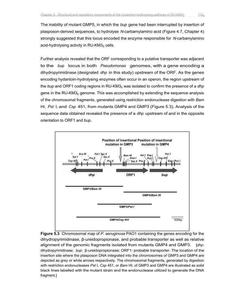
Position of insertional Position of insertional mutation in GMP3 mutation in GMP4
Csp 451
∗Sal 1 ∗
Pvu II Eco RI
Pst I
Pst I Sac II Sac II
Pvu II
Bam HI Sma I Sac II Pvu II
Sal 1 Pst 1 Eag I Pst I Csp 451
Eag I Pvu I
dhp ORF1 bup
GMP3/Bam HI
GMP4/Bam HI
GMP3/Pst I
GMP4/Csp 451
500bp
Chapter 5 : Structural and regulatory components of the hydantoin-hydrolysing pathway of RU-KM3S 115
The inability of mutant GMP5, in which the bup gene had been interrupted by insertion of
plasposon-derived sequences, to hydrolyse N-carbamylamino acid (Figure 4.7, Chapter 4)
strongly suggested that this locus encoded the enzyme responsible for N-carbamylamino
acid-hydrolysing activity in RU-KM3S cells.
Further analysis revealed that the ORF corresponding to a putative transporter was adjacent
to the bup locus in both Pseudomonas genomes, with a gene encoding a
dihydropyrimidinase (designated dhp in this study) upstream of the ORF. As the genes
encoding hydantoin-hydrolysing enzymes often occur in an operon, the region upstream of
the bup and ORF1 coding regions in RU-KM3S was isolated to confirm the presence of a dhp
gene in the RU-KM3S genome. This was accomplished by extending the sequence analysis
of the chromosomal fragments, generated using restriction endonuclease digestion with Bam
HI, Pst I, and Csp 451, from mutants GMP4 and GMP3 (Figure 5.3). Analysis of the
sequence data obtained revealed the presence of a dhp upstream of and in the opposite
orientation to ORF1 and bup.
Figure 5.3 Chromosomal map of P. aeruginosa PAO1 containing the genes encoding for the dihydropyrimidinase, β-ureidopropionase, and probable transporter as well as relative alignment of the genomic fragments isolated from mutants GMP4 and GMP3. [dhp: dihydropyrimidinase; bup: β-ureidopropionase; ORF1: probable transporter. The location of the insertion site where the plasposon DNA integrated into the chromosomes of GMP3 and GMP4 are depicted as grey or white arrows respectively. The chromosomal fragments, generated by digestion with restriction endonucleases Pst I, Csp 451, or Bam HI, of GMP3 and GMP4 are illustrated as solid black lines labelled with the mutant strain and the endonuclease utilized to generate the DNA fragment.]
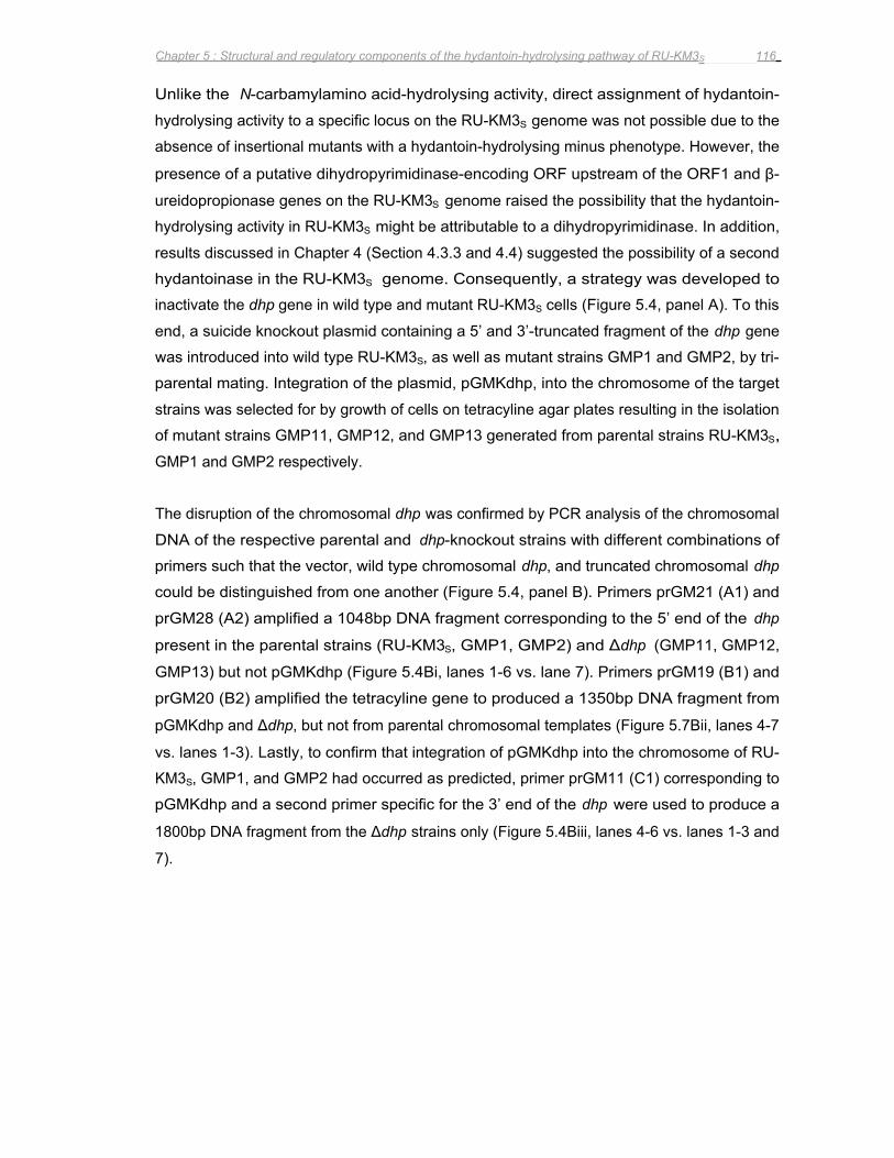
Chapter 5 : Structural and regulatory components of the hydantoin-hydrolysing pathway of RU-KM3S 116
Unlike the N-carbamylamino acid-hydrolysing activity, direct assignment of hydantoin-
hydrolysing activity to a specific locus on the RU-KM3S genome was not possible due to the
absence of insertional mutants with a hydantoin-hydrolysing minus phenotype. However, the
presence of a putative dihydropyrimidinase-encoding ORF upstream of the ORF1 and β-
ureidopropionase genes on the RU-KM3S genome raised the possibility that the hydantoin-
hydrolysing activity in RU-KM3S might be attributable to a dihydropyrimidinase. In addition,
results discussed in Chapter 4 (Section 4.3.3 and 4.4) suggested the possibility of a second
hydantoinase in the RU-KM3S genome. Consequently, a strategy was developed to
inactivate the dhp gene in wild type and mutant RU-KM3S cells (Figure 5.4, panel A). To this
end, a suicide knockout plasmid containing a 5’ and 3’-truncated fragment of the dhp gene
was introduced into wild type RU-KM3S, as well as mutant strains GMP1 and GMP2, by tri-
parental mating. Integration of the plasmid, pGMKdhp, into the chromosome of the target
strains was selected for by growth of cells on tetracyline agar plates resulting in the isolation
of mutant strains GMP11, GMP12, and GMP13 generated from parental strains RU-KM3S,
GMP1 and GMP2 respectively.
The disruption of the chromosomal dhp was confirmed by PCR analysis of the chromosomal
DNA of the respective parental and dhp-knockout strains with different combinations of
primers such that the vector, wild type chromosomal dhp, and truncated chromosomal dhp
could be distinguished from one another (Figure 5.4, panel B). Primers prGM21 (A1) and
prGM28 (A2) amplified a 1048bp DNA fragment corresponding to the 5’ end of the dhp
present in the parental strains (RU-KM3S, GMP1, GMP2) and Δdhp (GMP11, GMP12,
GMP13) but not pGMKdhp (Figure 5.4Bi, lanes 1-6 vs. lane 7). Primers prGM19 (B1) and
prGM20 (B2) amplified the tetracyline gene to produced a 1350bp DNA fragment from
pGMKdhp and Δdhp, but not from parental chromosomal templates (Figure 5.7Bii, lanes 4-7
vs. lanes 1-3). Lastly, to confirm that integration of pGMKdhp into the chromosome of RU-
KM3S, GMP1, and GMP2 had occurred as predicted, primer prGM11 (C1) corresponding to
pGMKdhp and a second primer specific for the 3’ end of the dhp were used to produce a
1800bp DNA fragment from the Δdhp strains only (Figure 5.4Biii, lanes 4-6 vs. lanes 1-3 and
7).
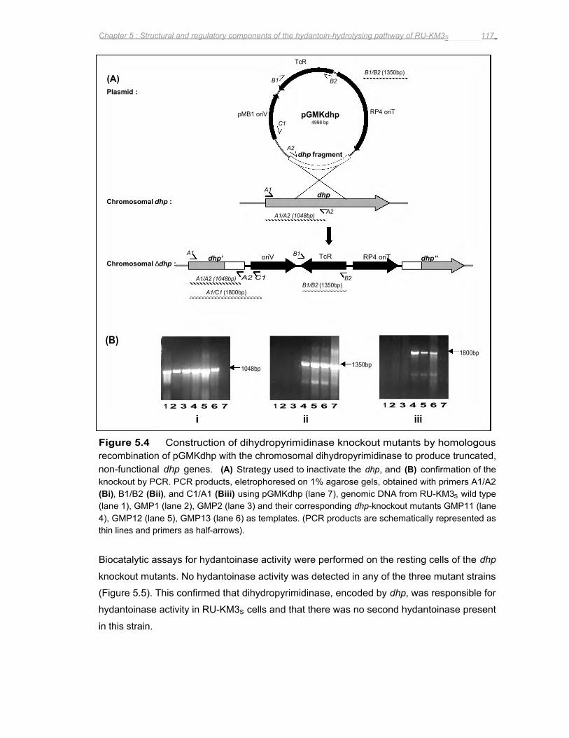
(B)
Chromosomal dhp :
Chromosomal ∆dhp :
(A) Plasmid :
1 2 3 4 5 6 7
i
A1 B1 dhp’ oriV TcR RP4 oriT
A1/A2 (1048bp)
A1/C1 (1800bp)
pMB1 oriV
A2 C1 B2
1048bp
A1
B1 B2
1 2 3 4 5 6 7
A1/A2 (1048bp)
pGMKdhp C1 4998 bp
A2
TcR
dhp fragment
B1/B2 (1350bp)
ii
dhp
A2
1350bp
B1/B2 (1350bp)
RP4 oriT
1 2 3 4 5 6 7
iii
dhp”
1800bp
Chapter 5 : Structural and regulatory components of the hydantoin-hydrolysing pathway of RU-KM3S 117
Figure 5.4 Construction of dihydropyrimidinase knockout mutants by homologous recombination of pGMKdhp with the chromosomal dihydropyrimidinase to produce truncated, non-functional dhp genes. (A) Strategy used to inactivate the dhp, and (B) confirmation of the knockout by PCR. PCR products, eletrophoresed on 1% agarose gels, obtained with primers A1/A2 (Bi), B1/B2 (Bii), and C1/A1 (Biii) using pGMKdhp (lane 7), genomic DNA from RU-KM3S wild type (lane 1), GMP1 (lane 2), GMP2 (lane 3) and their corresponding dhp-knockout mutants GMP11 (lane 4), GMP12 (lane 5), GMP13 (lane 6) as templates. (PCR products are schematically represented as thin lines and primers as half-arrows).
Biocatalytic assays for hydantoinase activity were performed on the resting cells of the dhp
knockout mutants. No hydantoinase activity was detected in any of the three mutant strains
(Figure 5.5). This confirmed that dihydropyrimidinase, encoded by dhp, was responsible for
hydantoinase activity in RU-KM3S cells and that there was no second hydantoinase present
in this strain.
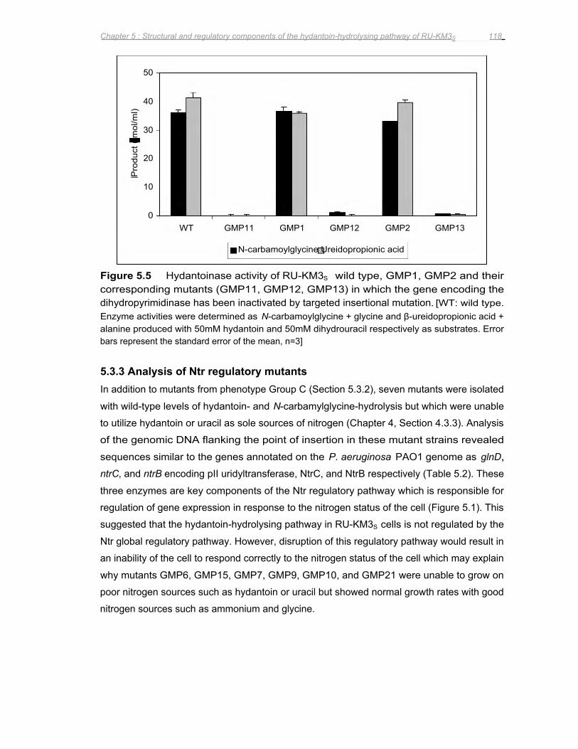
Pro
duct
( m
ol/m
l)
40
50
30
20
10
0 WT GMP11 GMP1 GMP12 GMP2 GMP13
N-carbamoylglycine Ureidopropionic acid
Chapter 5 : Structural and regulatory components of the hydantoin-hydrolysing pathway of RU-KM3S 118
Figure 5.5 Hydantoinase activity of RU-KM3S wild type, GMP1, GMP2 and their corresponding mutants (GMP11, GMP12, GMP13) in which the gene encoding the dihydropyrimidinase has been inactivated by targeted insertional mutation. [WT: wild type. Enzyme activities were determined as N-carbamoylglycine + glycine and β-ureidopropionic acid + alanine produced with 50mM hydantoin and 50mM dihydrouracil respectively as substrates. Error bars represent the standard error of the mean, n=3]
5.3.3 Analysis of Ntr regulatory mutants In addition to mutants from phenotype Group C (Section 5.3.2), seven mutants were isolated
with wild-type levels of hydantoin- and N-carbamylglycine-hydrolysis but which were unable
to utilize hydantoin or uracil as sole sources of nitrogen (Chapter 4, Section 4.3.3). Analysis
of the genomic DNA flanking the point of insertion in these mutant strains revealed
sequences similar to the genes annotated on the P. aeruginosa PAO1 genome as glnD,
ntrC, and ntrB encoding pII uridyltransferase, NtrC, and NtrB respectively (Table 5.2). These
three enzymes are key components of the Ntr regulatory pathway which is responsible for
regulation of gene expression in response to the nitrogen status of the cell (Figure 5.1). This
suggested that the hydantoin-hydrolysing pathway in RU-KM3S cells is not regulated by the
Ntr global regulatory pathway. However, disruption of this regulatory pathway would result in
an inability of the cell to respond correctly to the nitrogen status of the cell which may explain
why mutants GMP6, GMP15, GMP7, GMP9, GMP10, and GMP21 were unable to grow on
poor nitrogen sources such as hydantoin or uracil but showed normal growth rates with good
nitrogen sources such as ammonium and glycine.
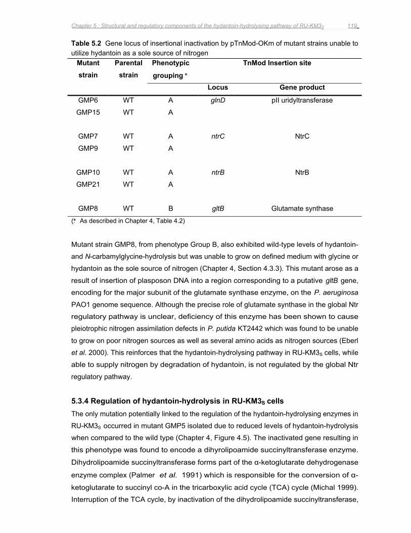
Chapter 5 : Structural and regulatory components of the hydantoin-hydrolysing pathway of RU-KM3S 119
Table 5.2 Gene locus of insertional inactivation by pTnMod-OKm of mutant strains unable to utilize hydantoin as a sole source of nitrogen
Mutant strain
Parental strain
Phenotypic
grouping ∗
TnMod Insertion site
Locus Gene product
GMP6 WT A glnD pII uridyltransferase
GMP15 WT A
GMP7 WT A ntrC NtrC
GMP9 WT A
GMP10 WT A ntrB NtrB
GMP21 WT A
GMP8 WT B gltB Glutamate synthase
(∗: As described in Chapter 4, Table 4.2)
Mutant strain GMP8, from phenotype Group B, also exhibited wild-type levels of hydantoin-
and N-carbamylglycine-hydrolysis but was unable to grow on defined medium with glycine or
hydantoin as the sole source of nitrogen (Chapter 4, Section 4.3.3). This mutant arose as a
result of insertion of plasposon DNA into a region corresponding to a putative gltB gene,
encoding for the major subunit of the glutamate synthase enzyme, on the P. aeruginosa
PAO1 genome sequence. Although the precise role of glutamate synthase in the global Ntr
regulatory pathway is unclear, deficiency of this enzyme has been shown to cause
pleiotrophic nitrogen assimilation defects in P. putida KT2442 which was found to be unable
to grow on poor nitrogen sources as well as several amino acids as nitrogen sources (Eberl
et al. 2000). This reinforces that the hydantoin-hydrolysing pathway in RU-KM3S cells, while
able to supply nitrogen by degradation of hydantoin, is not regulated by the global Ntr
regulatory pathway.
5.3.4 Regulation of hydantoin-hydrolysis in RU-KM3S cells The only mutation potentially linked to the regulation of the hydantoin-hydrolysing enzymes in
RU-KM3S occurred in mutant GMP5 isolated due to reduced levels of hydantoin-hydrolysis
when compared to the wild type (Chapter 4, Figure 4.5). The inactivated gene resulting in
this phenotype was found to encode a dihyrolipoamide succinyltransferase enzyme.
Dihydrolipoamide succinyltransferase forms part of the α-ketoglutarate dehydrogenase
enzyme complex (Palmer et al. 1991) which is responsible for the conversion of α-
ketoglutarate to succinyl co-A in the tricarboxylic acid cycle (TCA) cycle (Michal 1999).
Interruption of the TCA cycle, by inactivation of the dihydrolipoamide succinyltransferase,
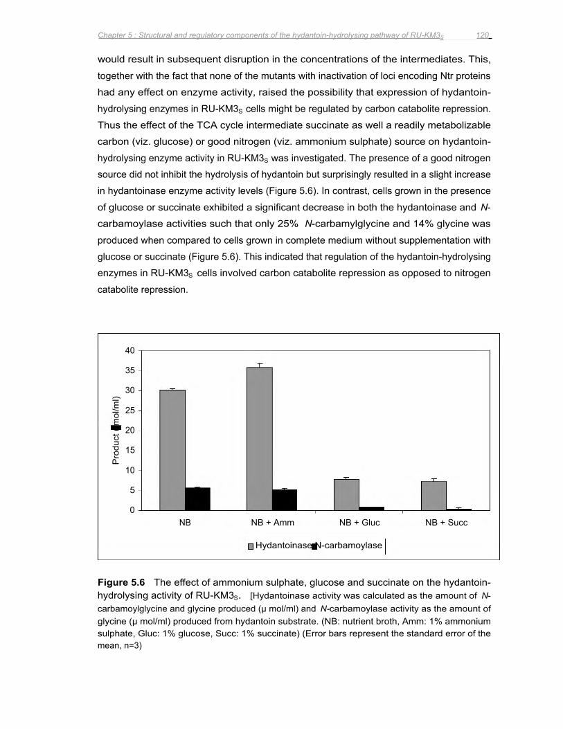
Pro
duct
( m
ol/m
l)
40
35
30
25
20
15
10
5
0 NB NB + Amm NB + Gluc NB + Succ
Hydantoinase N-carbamoylase
Chapter 5 : Structural and regulatory components of the hydantoin-hydrolysing pathway of RU-KM3S 120
would result in subsequent disruption in the concentrations of the intermediates. This,
together with the fact that none of the mutants with inactivation of loci encoding Ntr proteins
had any effect on enzyme activity, raised the possibility that expression of hydantoin-
hydrolysing enzymes in RU-KM3S cells might be regulated by carbon catabolite repression.
Thus the effect of the TCA cycle intermediate succinate as well a readily metabolizable
carbon (viz. glucose) or good nitrogen (viz. ammonium sulphate) source on hydantoin-
hydrolysing enzyme activity in RU-KM3S was investigated. The presence of a good nitrogen
source did not inhibit the hydrolysis of hydantoin but surprisingly resulted in a slight increase
in hydantoinase enzyme activity levels (Figure 5.6). In contrast, cells grown in the presence
of glucose or succinate exhibited a significant decrease in both the hydantoinase and N-
carbamoylase activities such that only 25% N-carbamylglycine and 14% glycine was
produced when compared to cells grown in complete medium without supplementation with
glucose or succinate (Figure 5.6). This indicated that regulation of the hydantoin-hydrolysing
enzymes in RU-KM3S cells involved carbon catabolite repression as opposed to nitrogen
catabolite repression.
Figure 5.6 The effect of ammonium sulphate, glucose and succinate on the hydantoin-hydrolysing activity of RU-KM3S. [Hydantoinase activity was calculated as the amount of N-carbamoylglycine and glycine produced (μ mol/ml) and N-carbamoylase activity as the amount of glycine (μ mol/ml) produced from hydantoin substrate. (NB: nutrient broth, Amm: 1% ammonium sulphate, Gluc: 1% glucose, Succ: 1% succinate) (Error bars represent the standard error of the mean, n=3)
Chapter 5 : Structural and regulatory components of the hydantoin-hydrolysing pathway of RU-KM3S 121
5.3.5 Analysis of the promoter regions of bup and dhp The genetic characterization and analysis of the dhp, bup, and ORF1 genes are discussed in
detail in Chapter 6, however, analysis of the intergenic region between dhp and ORF1 in RU-
KM3S will be discussed here. Insertional inactivation of the open reading frame upstream of
bup, designated ORF1, resulted in complete loss of β-ureidopropionase activity in mutant
strain GMP3 (Figure 4.7) suggesting that these two genes are transcribed as a polycistronic
mRNA. The short intergenic region that exists between the bup and ORF1 substantiates
this. Analysis of the 5’ region flanking ORF1 revealed a putative ribosome binding site
(Shine-Dalgarno) at nucleotides 8 to 13, a possible Pribnow box at nucleotides 25 to 29, and
a sequence similar to the prokaryotic –35 consensus sequence at nucleotides 66-72 (Figure
5.7). Analysis of the promoter region of dhp revealed a putative Shine-Dalgarno at
nucleotides 609 to 614 and sequences corresponding to the prokaryotic –10 and –35
consensus sequences at nucleotides 40-48 and 380-385 respectively (Figure 5.7).
Interestingly, a region with high similarity to an E. coli CRP-binding site was also identified at
nucleotides 392 to 422 (Figure 5.7).
Alignment of the corresponding intergenic regions from P. putida KT2440 and P. aeruginosa
PAO1 using ClustalW software was done to determine if similar promoter sequences existed
in these strains. In addition, the promoter region for the gene coding for the D-hydantoinase
from P. putida DSM 84 (LaPointe et al. 1994) was aligned with the promoter regions for the
dihydropyrimidinase encoding genes from RU-KM3S, P. putida KT2440, and P. aeruginosa
PAO1. The promoter region for the putative hydantoin-hydrolysing genes from P. aeruginosa
PAO1 was found to be similar to that of RU-KM3S and also contained the putative CRP-
binding site (Figure 5.7). However, the corresponding nucleotide sequence in P. putida
KT2440 was observed to be significantly shorter than that of P. putida RU-KM3S and P.
aeruginosa PAO1 and, in addition, did not exhibit the same degree of similarity with respect
to the putative CRP-binding site (Figure 5.7). The disparity between the locus encoding the
hydantoin-hydrolysing enzymes in RU-KM3S and that of P. putida KT2440 is further
demonstrated by the gene encoding the P. putida KT2440 dihydropyrimidinase containing an
authentic frameshift, which is not the result of a sequencing artefact, rendering the gene
inactive. Interestingly, the nucleotide sequence from P. putida DSM 84 which corresponded
to that of the putative CRP-binding site in the promoter region for RU-KM3S exhibited little
similarity (Figure 5.7). The fact that the levels of D-hydantoinase activity in P. putida DSM 84
decreased by a factor of 4 when the cells were cultured with glucose (Morin et al. 1986a)
suggested that although the enzymes from P. putida DSM 84 and RU-KM3S both appeared
to be subject to carbon catabolite repression, the mechanism by which the repression took
place is most likely different.
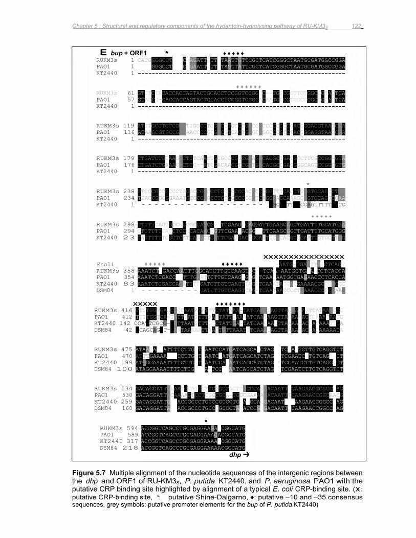
RUKM3s 416 TTTTGGTGAAC
KT2440 142 CCATTCGCG-C
RUKM3s 475 ATAA-ATTATTTTCTTGAT
KT2440 199 ATGGGAAAATTTTCTTGCT
RUKM3s 534 GACAGGATT
KT2440 259 GACAGGATT
DSM84 42 TCAGCAGGT-C
DSM84 100 ATAGGAAAATTTTCTTGAAGATTCCCAAATCAGCATCTAGCCTCGAATCTTGTCAGGTCT
DSM84 160 GACAGGATT
PAO1 412 T
PAO1 470 ATTT
PAO1 530 GACAGGATT
RUKM3s 1 CATGGGGCCT
KT2440 1
♦♦♦♦♦♦RUKM3s 61 GTCTCTACACCACCAGTACTGCACCTCCGGTCCGGCC--TGGCGGTTGTGGCCGTAC
KT2440 1
RUKM3s 119 ATTTGCGTGCCGGGTTGGCCTGGGCCTGCCCGACGGCCGCCGCCGACCGGAGGTAATCAA
KT2440 1
RUKM3s 179 CTGATCTGCAAGGCTTTCAACTGCGCCCGGCGAAGGCACGCTGATGCCTTCGCCGG-GT
KT2440 1
RUKM3s 238 TTCGCGCCGCCCTGGGCCGGCCCTGGCCCCCACACCCGGTTTCACCTTGGTGCAGCCTGG
KT2440 1 - - - - - - - - - - - - - - - - - - - GG
RUKM3s 298 TTTTTCAGTGGCGCGGCCAG
KT2440 23 TATTTTTTCGCTCATCAGGCTGATTCGATGACCGGAGTTCGCACGCGGAATTGTGCTTAG
xxxxxxxxxxxxxxxx Ecoli ♦♦♦♦♦ ♦♦♦♦♦ -AATG-TGAGTTAGCTCACT RUKM3s 358 AAATCTTGACCAATTTGGCATCTTGTCAAGTGCG-TCAA-AATGGTGAAACCTCACCA
KT2440 83 AAATCTCGACCAGATTTTCATCTTGTCAAGTTAG-TCAAATACCAGGAAAACCCCG DSM84 1 - - - - - - - - - CATCTTGTCAAGTCAG-TCAATAACCCCTGAAACCCCC
PAO1 1 ----GGGCCT
PAO1 57 GT
PAO1 116 AT
PAO1 176 CTGATCTGAAACACTTC--ATCGACAACGTCGGTGACACGCCGACGGGCAGTCCGGCGT
PAO1 234 T
PAO1 294 CTTTTTTCATCTCCCCACAACC
PAO1 354 AAATCTCGACCTT
∗∗∗∗∗∗∗ RUKM3s 594 ACCGGTCAGCCTGCGAGGAAGA-CGGCATG PAO1 589 ACCGGTCAGCCTGCGAGGAAAGACGGCATG KT2440 317 ACCGGTCAGCCTGCGAGGAAAA-CGGCATG DSM84 218 ACCGGTCAGCCTGCGAGGAAAAACGGCATG
dhp ➔
E bup + ORF1 ∗∗∗∗∗∗ ♦♦♦♦♦
xxxxx ♦♦♦♦♦♦♦
TTTGGTGA-T
CACCGGGGGAAATGCGCGAGCCTGTCGCCCCGGTCAGGAACCAAAGGAGTGCAGGCG
ACCG-CACCACCAGTACTGCACCTCCGGTCCGGACAATGCCGTGGCGGGCGGCAG
AAGCGTGCCGAAAACCCCGCGACGTCACTGGGCAGGCCGGTCTACTGGAGGTAACCGA
GAAAACAATCTTGCTCAATCGATGATCAGCATCTAGATTCGAATCCTGTCAGCGCT
ACAAAAGTCCTCGGCGCA-TGGCGAAAAAACAATTTCAAGAACCGGCAACG
A--ACCGCCCTGCCTGCCCTACACCCGAGACAATTCCAAGAACCGGCCCAG
GAAATCCATCCTCCAGCCACTGGCCCATAACAATTTCAAGAACCGGCCCAG
G--ACCGCCCTGCCCGCCCTGCATCCATGACAATGCAAAGAACCGGCCAAG
TCTCCAGATTTTTCTAATTGTTCGCTCATCGGGCTAATGCGATGGCCGGA CTCCAGAATTCTTTTAGTTATTCGCTCATCGGGCTAATGCGATGGCCGGA
ATTTATTTTTAATTATTTAAAATCAATAAGTTACAA-ACTCAAAC
AATAAAAATTAAGTGTTAAATTTCAAACAGTTAAAA-ACCACAAAAATA
GCCAATATATTTCTATAAA-TATCAGGTAGTTACGATAGTTATAAGCTC
GTTAATTTTTTCCTATTGA-TATCAACAACTTAAAA-ACCACAAATT
TTTGAAT
CAATCCATGATCAGCAACTAGATTCCATTCTTGTCAGGTCT
GAATCCAGAATCAGCATCTAGCCTCGAATCTTGTCAGGACT
CGCCTCGAAGCCAGGATTCAAGCAGCTGATTTTGCATGGA
TCTTGTCAAGCGCG-TCAA-AATGGTGAGAACCCTCACCA
GTTCGAACACGGCTTTCAAGCGGCTGATTTTGCATGGG
CCCTTCTCCAGTTTTTCTTC
∗∗∗∗∗∗
♦♦♦♦♦
AACC
GCGC GAAG
TCA TCA
TA
AA
A G
Chapter 5 : Structural and regulatory components of the hydantoin-hydrolysing pathway of RU-KM3S 122
Figure 5.7 Multiple alignment of the nucleotide sequences of the intergenic regions between the dhp and ORF1 of RU-KM3S, P. putida KT2440, and P. aeruginosa PAO1 with the putative CRP binding site highlighted by alignment of a typical E. coli CRP-binding site. (x : putative CRP-binding site, ∗ : putative Shine-Dalgarno, ♦: putative –10 and –35 consensus sequences, grey symbols: putative promoter elements for the bup of P. putida KT2440)
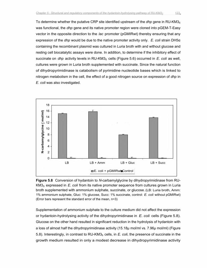
N-c
arba
myl
glyc
ine
( mol
/ml)
18
16
14
12
10
4
8
6
2
0 LB LB + Amm LB + Gluc LB + Succ
E. coli + pGMIRwt Control
Chapter 5 : Structural and regulatory components of the hydantoin-hydrolysing pathway of RU-KM3S 123
To determine whether the putative CRP site identified upstream of the dhp gene in RU-KM3S
was functional, the dhp gene and its native promoter region were cloned into pGEM-T-Easy
vector in the opposite direction to the lac promoter (pGMIRwt) thereby ensuring that any
expression of the dhp would be due to the native promoter activity only. E. coli strain DH5α
containing the recombinant plasmid was cultured in Luria broth with and without glucose and
resting cell biocatalytic assays were done. In addition, to determine if the inhibitory effect of
succinate on dhp activity levels in RU-KM3S cells (Figure 5.6) occurred in E. coli as well,
cultures were grown in Luria broth supplemented with succinate. Since the natural function
of dihydropyrimidinase is catabolism of pyrimidine nucleotide bases which is linked to
nitrogen metabolism in the cell, the effect of a good nitrogen source on expression of dhp in
E. coli was also investigated.
Figure 5.8 Conversion of hydantoin to N-carbamylglycine by dihydropyrimidinase from RU-KM3S expressed in E. coli from its native promoter sequence from cultures grown in Luria broth supplemented with ammonium sulphate, succinate, or glucose. (LB: Luria broth, Amm: 1% ammonium sulphate, Gluc: 1% glucose, Succ: 1% succinate, control: E. coli without pGMIRwt) (Error bars represent the standard error of the mean, n=3)
Supplementation of ammonium sulphate to the culture medium did not affect the expression
or hydantoin-hydrolysing activity of the dihydropyrimidinase in E. coli cells (Figure 5.8).
Glucose on the other hand resulted in significant reduction in the hydrolysis of hydantoin with
a loss of almost half the dihydropyrimidinase activity (15.18μ mol/ml vs. 7.96μ mol/ml) (Figure
5.8). Interestingly, in contrast to RU-KM3S cells, in E. coli, the presence of succinate in the
growth medium resulted in only a modest decrease in dihydropyrimidinase activity
Chapter 5 : Structural and regulatory components of the hydantoin-hydrolysing pathway of RU-KM3S 124
(15.18μ mol/ml vs. 13.78μ mol/ml) (Figure 5.8). This data suggested differences in the
mechanisms of carbon catabolite repression of this promoter in RU-KM3S and E. coli.
5.4 DISCUSSION
The major objective of this chapter was to identify the genes encoding for the structural
proteins responsible for hydantoin-hydrolysis in RU-KM3S cells by analysis of the genes
inactivated in mutant derivatives unable to utilize hydantoin as a sole source of nitrogen.
This approach also allowed for elucidation of the regulatory mechanisms affecting the
hydantoin-hydrolysing pathway in this strain.
The data presented in this chapter show that the enzymes responsible for the hydantoinase
and N-carbamoylase activities in P. putida RU-KM3S are a single dihydropyrimidinase,
encoded by dhp, and a single β-ureidopropionase, encoded by bup, respectively. The
presence of two hydantoinases, as occurs in Agrobacterium sp. IP I-671 (Hils et al. 2001), or
two carbamoylases, as occurs in Blastobacter sp. A17p-4 (Ogawa et al. 1994c) and
Pseudomonas sp. AJ-11220 (Yokozeki et al. 1987c) does not apply to RU-KM3S as complete
absence of hydantoin-hydrolysing activity is observed subsequent to inactivation of the bup
and dhp genes.
A third open reading frame, designated ORF1, was located between the bup and dhp genes.
It is tempting to propose that ORF1, which exhibits significant similarity to a putative transport
protein in the P. aeruginosa genome, could be involved in the transport of hydantoin.
However, the observation that insertional inactivation of ORF1 did not affect hydrolysis of
hydantoin to N-carbamylglycine in whole cell biocatalytic assays suggested otherwise. The
inability to convert N-carbamoylglycine to glycine when ORF1 is interrupted by insertion of
foreign plasposon DNA can be attributed to expression of ORF1 and bup from a common
promoter as a polycistronic mRNA. This is supported by the very short intergenic region
between the bup and ORF1 genes. The amino acid sequence and bioinformatic analysis of
the β-ureidopropionase, ORF1 and dihydropyrimidinase will be discussed in greater detail in
Chapter 6.
With respect to regulation of hydantoin-hydrolysis in RU-KM3S cells, the data presented
conclusively demonstrated that expression of RU-KM3S dhp and bup genes is regulated by
carbon catabolite repression and is not affected by the nitrogen status of the cell as has been
shown in studies of Agrobacterium tumefaciens RU-OR (Hartley et al. 2001). Firstly, growth
of RU-KM3S cells in complete medium supplemented with ammonium sulphate had no effect
on hydantoin-hydrolysis. Furthermore, mutant strains of RU-KM3S in which key components
Chapter 5 : Structural and regulatory components of the hydantoin-hydrolysing pathway of RU-KM3S 125
of the Ntr regulatory cascade, namely NtrC, NtrB, and PII uridyltransferase encoding genes
(Figure 5.1), were inactivated exhibited wild type levels of hydantoin-hydrolysing enzyme
activities. Thus expression of dhp and bup genes in RU-KM3S occurs independently of the
nitrogen catabolite repression control.
Support for the regulation of hydantoin-hydrolysis by CCR is provided by three observations:
first, addition of glucose or succinate to the growth medium resulted in significant reductions
in both N-carbamylglycine and glycine production. Succinate and other TCA cycle
intermediates have been shown to impose severe carbon catabolite repression on several
metabolic pathways in Pseudomonas (Collier et al. 1996), and the negative effect of glucose
on hydantoinase activities has been documented for Pseudomonas sp. NCIM 5109 and P.
putida DSM 84 (Sudge et al. 1998, Morin et al. 1986a). Second, the significant reduction in
mutant strain GMP5 enzyme activities in which an enzyme within the TCA cycle, namely
dihydrolipoamide succinyltransferase, was inactivated supports the proposed CCR regulation
of dhp and bup in RU-KM3S cells. And thirdly, the presence of a putative CRP-binding site in
the promoter region is further evidence for CCR.
As discussed in the introduction to this chapter (Section 5.1), carbon catabolite repression in
Pseudomonas species varies significantly from that of E. coli in which carbon catabolite
repression involves binding of the CRP protein to a CRP site in the promoter region thereby
stimulating/inhibiting transcription of the gene. To determine if the putative CRP-binding site
identified actually functions as one, the dhp gene, under control of its natural promoter
containing the CRP-binding site, was expressed in E. coli. In this strain, glucose functions as
a stimulus for CCR by resulting in elevated cAMP levels which complex with CRP and bind to
the CRP-binding site resulting in repression of gene expression. The reduction of hydantoin-
hydrolysis in the E. coli when cultured in the presence of glucose indicated that the cAMP-
CRP complex had successfully bound to the CRP-binding site thereby preventing
transcription of the dhp. The fact that succinate did not result in an equivalent decrease in
activity levels, as was the case in the wild type strain, is expected as TCA cycle
intermediates are not directly involved with the activity of the CRP protein. As cAMP levels in
Pseudomonas do not vary, regardless of the carbon source supplied, and CRP does not
function in Pseudomonas as it does in E. coli (Collier et al. 1996), the functional CRP-like site
upstream of dhp in RU-KM3S suggests the presence of an alternative protein which functions
in a manner similar to that of CRP (discussed in Chapter 7).
Surprisingly, the putative CRP-binding site upstream of the dhp in P. putida RU-KM3S is not
present in the corresponding nucleotide sequences of P. putida KT2440 or P. putida DSM
84. In contrast, the corresponding promoter region from P. aeruginosa PAO1 was very
similar to that of RU-KM3S. The hydantoin-hydrolysing activities in both P. putida DSM 84
Chapter 5 : Structural and regulatory components of the hydantoin-hydrolysing pathway of RU-KM3S 126
and RU-KM3S decreased when the cells were cultured in the presence of a readily
metabolizable carbon source suggesting that these enzymes are regulated by CCR,
however, the fact that the corresponding promoter regions differed implies separate
mechanisms of CCR in these two P. putida strains. Differences with respect to CCR of a
particular gene between P. putida isolates and between P. putida and P. aeruginosa isolates
have been observed for the bkd operon (encodes the multienzyme complex branched-chain
keto acid dehydrogenase), the zwf-pgl-eda operon (encoding glucose-6-phosphate
dehydrogenase, 6-phosphoglucolactonase, 2-keto-3-deoxy-6-phosphogluconate-aldolase),
as well as the expression of amidase (Hester et al. 2000, Petruschka et al. 2002). In
addition, while the Crc protein has been implicated in CCR for numerous enzymes in
Pseudomonas, several enzymes have been identified as sensitive to CCR but which are
unaffected by Crc-null mutations thus implying the presence of a second CCR mechanism in
Pseudomonas species (Yuste and Rojo 2001, Collier et al. 2001, Dinamarca et al. 2002).
In conclusion, the bup and dhp genes have been identified, by insertional inactivation, as
encoding proteins responsible for hydantoin-hydrolysis in RU-KM3S. Furthermore, regulation
of expression of these genes in RU-KM3S has been shown to be controlled by the carbon
catabolite repression pathway, as apposed to nitrogen catabolite repression which does not
affect the dihydropyrimidinase or β-ureidopropionase enzyme activities. This has important
implications when considering culture conditions for RU-KM3S such that maximal expression
of the dhp and bup genes can be achieved.
Chapter 6 : Analysis of the bup and dhp genes from RU-KM3S 127
6.1 INTRODUCTION
The amino acid sequence of a protein provides invaluable information and can be used to
predict secondary structure using protein modelling, to identify conserved domains with
potential structural or catalytic functions, as well as providing information about the molecular
evolution of the proteins investigated. In the previous chapter, the genes involved in the
hydrolysis of hydantoin in RU-KM3S cells were identified as a dihydropyrimidinase and a β-
ureidopropionase. An open reading frame, termed ORF1, was located between the bup and
dhp genes and proposed to be co-expressed with bup as a polycistronic mRNA. In this
chapter, elucidation of the complete nucleotide and amino acid sequence of the β-
ureidopropionase and dihydropyrimidinase enzymes from RU-KM3S as well as comparative
analysis of the amino acid sequences with those in literature are discussed. In addition, the
protein encoded by ORF1 was aligned with annotated amino acid sequences in order to
identify the potential function of this protein.
6.2 METHODS AND MATERIALS
6.2.1 DNA manipulation Genomic DNA was extracted by detergent lysis/CTAB and organic solvent extraction method
as described in Appendix 5. The nucleotide sequence of the bup and dhp genes was
determined from double stranded plasmid DNA by the dideoxy-chain termination method
(Sanger 1977) using the ABI Prism Big Dye Terminator Cycle Sequencing. The primers
used for sequencing are listed in Appendix 3.
6.2.2 Analysis of DNA sequence generated Preliminary analysis of raw sequence data was carried out using Vector NTI DeLuxe v4.0 or
v5.0 software (Informax Inc.). Identification of known genes to which the acquired sequence
data exhibits a high degree of similarity was performed using BLASTX and BLASTP (Altschul
et al. 1990) hosted by the NCBI (http://www.ncbi.nlm.nih.gov). Sequence alignments were
carried out using ClustalW software (Thompson et al. 1994) located at the BCM Search
Launcher website (http://searchlauncher.bcm.tmc.edu) and are presented in Boxshade
(version 3.21) format (http://www.ch.embnet.org/software/BOX form.html). The phylogenetic
trees were constructed with amino acid sequences aligned with ClustalW and, after being
manually revised, imported into Treeview software (Page 1996) and the tree generated using
the neighbour-joining algorithm.
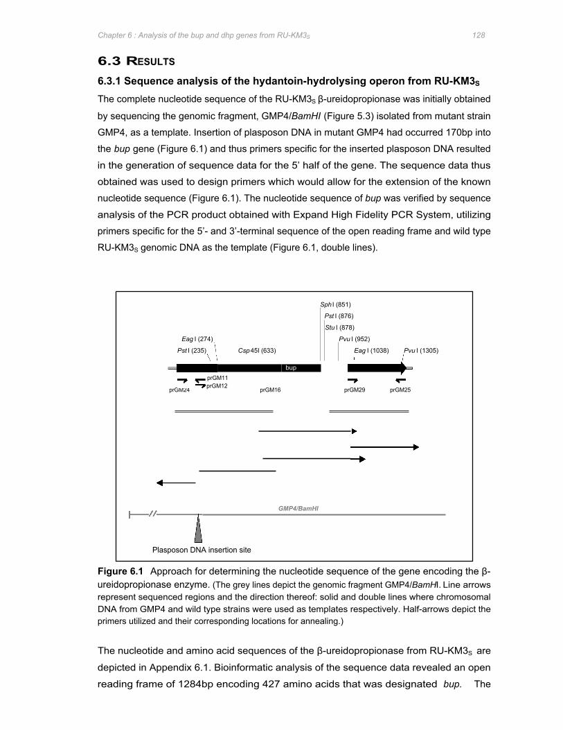
Csp 45I (633)
Sph I (851)
Pst I (876)
Stu I (878)
bup prGM11 prGM12
prGM24 prGM16 prGM29 prGM25
GMP4/BamHI
Eag I (274)
Pst I (235)
Pvu I (952)
Eag I (1038) Pvu I (1305)
Plasposon DNA insertion site
Chapter 6 : Analysis of the bup and dhp genes from RU-KM3S 128
6.3 RESULTS
6.3.1 Sequence analysis of the hydantoin-hydrolysing operon from RU-KM3S
The complete nucleotide sequence of the RU-KM3S β-ureidopropionase was initially obtained
by sequencing the genomic fragment, GMP4/BamHI (Figure 5.3) isolated from mutant strain
GMP4, as a template. Insertion of plasposon DNA in mutant GMP4 had occurred 170bp into
the bup gene (Figure 6.1) and thus primers specific for the inserted plasposon DNA resulted
in the generation of sequence data for the 5’ half of the gene. The sequence data thus
obtained was used to design primers which would allow for the extension of the known
nucleotide sequence (Figure 6.1). The nucleotide sequence of bup was verified by sequence
analysis of the PCR product obtained with Expand High Fidelity PCR System, utilizing
primers specific for the 5’- and 3’-terminal sequence of the open reading frame and wild type
RU-KM3S genomic DNA as the template (Figure 6.1, double lines).
Figure 6.1 Approach for determining the nucleotide sequence of the gene encoding the β-ureidopropionase enzyme. (The grey lines depict the genomic fragment GMP4/BamHI. Line arrows represent sequenced regions and the direction thereof: solid and double lines where chromosomal DNA from GMP4 and wild type strains were used as templates respectively. Half-arrows depict the primers utilized and their corresponding locations for annealing.)
The nucleotide and amino acid sequences of the β-ureidopropionase from RU-KM3S are
depicted in Appendix 6.1. Bioinformatic analysis of the sequence data revealed an open
reading frame of 1284bp encoding 427 amino acids that was designated bup. The
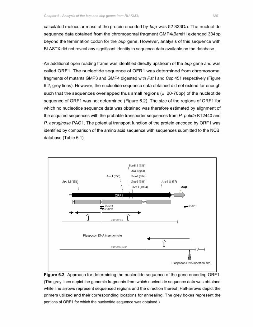
prGM11 prGM11 prGM12
GMP3/PstI
Plasposon DNA insertion site
GMP4/Csp45I
Ava I (850)
Apa LI (151)
ORF1
BamH I (951)
Ava I (984)
Xma I (984)
Sma I (986)
Nco I (1004)
Ava I (1457)
bup
Chapter 6 : Analysis of the bup and dhp genes from RU-KM3S 129
calculated molecular mass of the protein encoded by bup was 52 833Da. The nucleotide
sequence data obtained from the chromosomal fragment GMP4/BamHI extended 334bp
beyond the termination codon for the bup gene. However, analysis of this sequence with
BLASTX did not reveal any significant identity to sequence data available on the database.
An additional open reading frame was identified directly upstream of the bup gene and was
called ORF1. The nucleotide sequence of OFR1 was determined from chromosomal
fragments of mutants GMP3 and GMP4 digested with Pst I and Csp 451 respectively (Figure
6.2, grey lines). However, the nucleotide sequence data obtained did not extend far enough
such that the sequences overlapped thus small regions (± 20-70bp) of the nucleotide
sequence of ORF1 was not determined (Figure 6.2). The size of the regions of ORF1 for
which no nucleotide sequence data was obtained was therefore estimated by alignment of
the acquired sequences with the probable transporter sequences from P. putida KT2440 and
P. aeruginosa PAO1. The potential transport function of the protein encoded by ORF1 was
identified by comparison of the amino acid sequence with sequences submitted to the NCBI
database (Table 6.1).
Plasposon DNA insertion site
Figure 6.2 Approach for determining the nucleotide sequence of the gene encoding ORF1. (The grey lines depict the genomic fragments from which nucleotide sequence data was obtained
while line arrows represent sequenced regions and the direction thereof. Half-arrows depict the
primers utilized and their corresponding locations for annealing. The grey boxes represent the
portions of ORF1 for which the nucleotide sequence was obtained.)
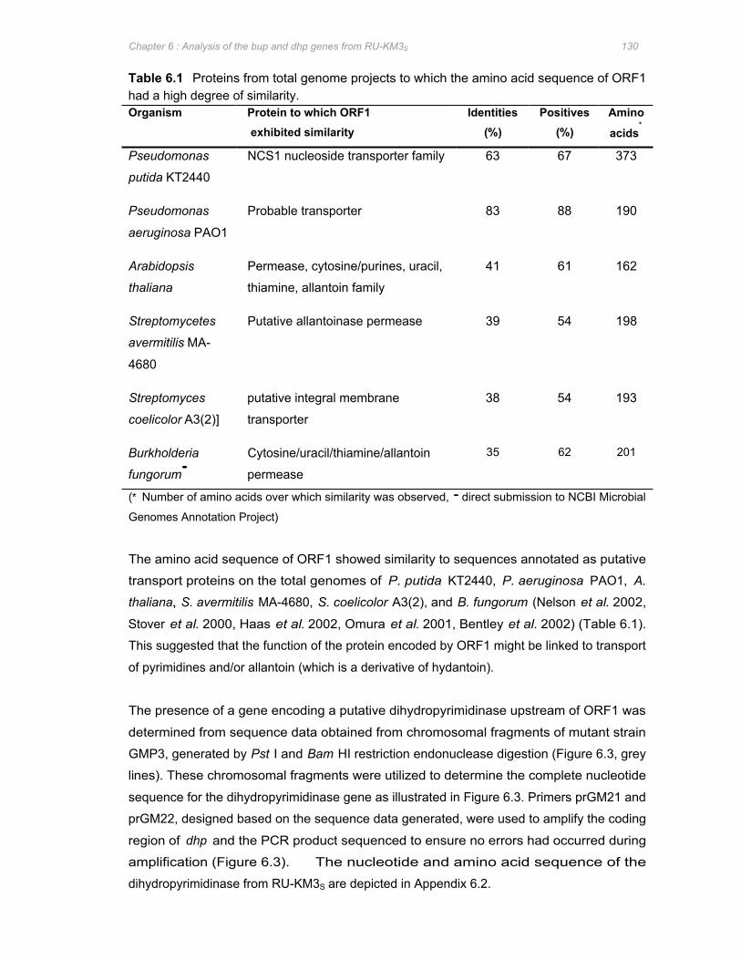
Chapter 6 : Analysis of the bup and dhp genes from RU-KM3S 130
Table 6.1 Proteins from total genome projects to which the amino acid sequence of ORF1 had a high degree of similarity. Organism Protein to which ORF1 Identities Positives Amino
exhibited similarity (%) (%) acids∗
Pseudomonas
putida KT2440
NCS1 nucleoside transporter family 63 67 373
Pseudomonas
aeruginosa PAO1
Probable transporter 83 88 190
Arabidopsis
thaliana
Permease, cytosine/purines, uracil,
thiamine, allantoin family
41 61 162
Streptomycetes
avermitilis MA-
Putative allantoinase permease 39 54 198
4680
Streptomyces
coelicolor A3(2)]
putative integral membrane
transporter
38 54 193
Burkholderia
fungorum•
Cytosine/uracil/thiamine/allantoin
permease
35 62 201
(∗ Number of amino acids over which similarity was observed, • direct submission to NCBI Microbial
Genomes Annotation Project)
The amino acid sequence of ORF1 showed similarity to sequences annotated as putative
transport proteins on the total genomes of P. putida KT2440, P. aeruginosa PAO1, A.
thaliana, S. avermitilis MA-4680, S. coelicolor A3(2), and B. fungorum (Nelson et al. 2002,
Stover et al. 2000, Haas et al. 2002, Omura et al. 2001, Bentley et al. 2002) (Table 6.1).
This suggested that the function of the protein encoded by ORF1 might be linked to transport
of pyrimidines and/or allantoin (which is a derivative of hydantoin).
The presence of a gene encoding a putative dihydropyrimidinase upstream of ORF1 was
determined from sequence data obtained from chromosomal fragments of mutant strain
GMP3, generated by Pst I and Bam HI restriction endonuclease digestion (Figure 6.3, grey
lines). These chromosomal fragments were utilized to determine the complete nucleotide
sequence for the dihydropyrimidinase gene as illustrated in Figure 6.3. Primers prGM21 and
prGM22, designed based on the sequence data generated, were used to amplify the coding
region of dhp and the PCR product sequenced to ensure no errors had occurred during
amplification (Figure 6.3). The nucleotide and amino acid sequence of the
dihydropyrimidinase from RU-KM3S are depicted in Appendix 6.2.

dhp
Pst I (996)
Eag I (1095)
Hind II (1141)
Hinc II (1141)
Sph I (629)
Pvu II (604)
Pst I (603)
Hinc II (517) Hind II (517)
Sac II (321)
prGM22 prGM27 prGM28 prGM21
EcoR I (1166
Bgl II (1200
Sal I (1316)
Hind II (1318)
Hinc II (1318)
Csp 45I (1332)
GMP3/PstI
Xba
I
Eco
RI
Xba
I
GMP3/BamHI
Chapter 6 : Analysis of the bup and dhp genes from RU-KM3S 131
Figure 6.3 Approach for determining the nucleotide sequence of the gene encoding the dihydropyrimidinase enzyme. (Grey lines depict genomic fragments of mutant strain GMP3 isolated by complete digestion of the chromosomal DNA of GMP3 with restriction endonuclease Bam HI or PstI. Arrows represent sequenced regions and the direction thereof: solid and double lines where chromosomal DNA from GMP3 and wild type strains were used as templates respectively. Hollow arrows indicate the chromosomal fragment of GMP3 from which the sequence was generated and half-arrows depict the primers utilized and their corresponding locations for annealing.)
Bioinformatic analysis of the sequence data revealed an open reading frame of 1140bp
encoding 479 amino acids that was designated dhp. The calculated molecular mass of the
protein encoded by dhp was 61 196Da. Analysis of the nucleotide sequence, of
approximately 400bp, flanking the terminal end of dhp revealed similarity to putative
oxidoreductases or dihydroorotate dehydrogenase proteins annotated on the total genome
databases of Pseudomonas putida KT2440 (Nelson et al. 2002), Pseudomonas aeruginosa
PAO1 (Stover et al. 2000), Mesorizobuim loti (Kaneko et al. 2000), Sinorhizobium meliloti
1021 (Capela et al. 2001), and Brucella suis 1330 (Paulsen et al. 2002) (Table 6.2).
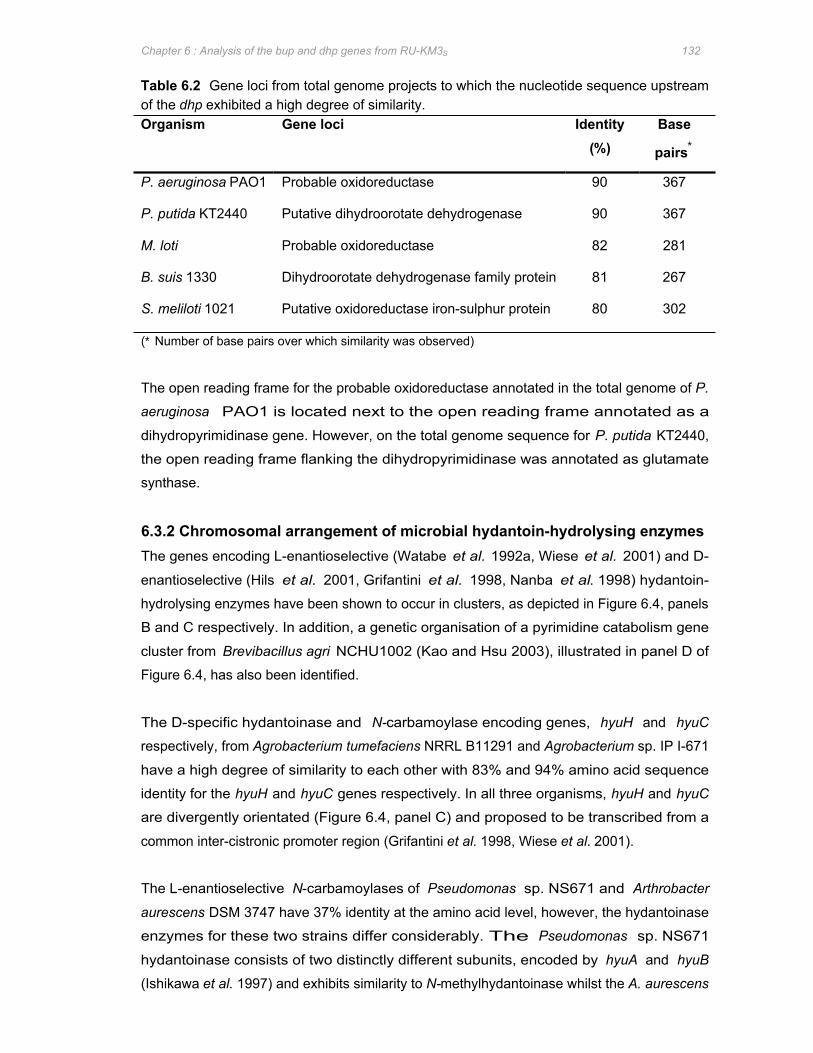
Chapter 6 : Analysis of the bup and dhp genes from RU-KM3S 132
Table 6.2 Gene loci from total genome projects to which the nucleotide sequence upstream of the dhp exhibited a high degree of similarity. Organism Gene loci Identity
(%) Base
pairs∗
P. aeruginosa PAO1 Probable oxidoreductase 90 367
P. putida KT2440 Putative dihydroorotate dehydrogenase 90 367
M. loti Probable oxidoreductase 82 281
B. suis 1330 Dihydroorotate dehydrogenase family protein 81 267
S. meliloti 1021 Putative oxidoreductase iron-sulphur protein 80 302
(∗ Number of base pairs over which similarity was observed)
The open reading frame for the probable oxidoreductase annotated in the total genome of P.
aeruginosa PAO1 is located next to the open reading frame annotated as a
dihydropyrimidinase gene. However, on the total genome sequence for P. putida KT2440,
the open reading frame flanking the dihydropyrimidinase was annotated as glutamate
synthase.
6.3.2 Chromosomal arrangement of microbial hydantoin-hydrolysing enzymes The genes encoding L-enantioselective (Watabe et al. 1992a, Wiese et al. 2001) and D-
enantioselective (Hils et al. 2001, Grifantini et al. 1998, Nanba et al. 1998) hydantoin-
hydrolysing enzymes have been shown to occur in clusters, as depicted in Figure 6.4, panels
B and C respectively. In addition, a genetic organisation of a pyrimidine catabolism gene
cluster from Brevibacillus agri NCHU1002 (Kao and Hsu 2003), illustrated in panel D of
Figure 6.4, has also been identified.
The D-specific hydantoinase and N-carbamoylase encoding genes, hyuH and hyuC
respectively, from Agrobacterium tumefaciens NRRL B11291 and Agrobacterium sp. IP I-671
have a high degree of similarity to each other with 83% and 94% amino acid sequence
identity for the hyuH and hyuC genes respectively. In all three organisms, hyuH and hyuC
are divergently orientated (Figure 6.4, panel C) and proposed to be transcribed from a
common inter-cistronic promoter region (Grifantini et al. 1998, Wiese et al. 2001).
The L-enantioselective N-carbamoylases of Pseudomonas sp. NS671 and Arthrobacter
aurescens DSM 3747 have 37% identity at the amino acid level, however, the hydantoinase
enzymes for these two strains differ considerably. The Pseudomonas sp. NS671
hydantoinase consists of two distinctly different subunits, encoded by hyuA and hyuB
(Ishikawa et al. 1997) and exhibits similarity to N-methylhydantoinase whilst the A. aurescens

A pydA dhp ORF1 bup
Pseudomonas putida RU-KM3S
B ORF5 hyuA hyuB hyuC ORF5 hyuE
76 65 46 50 32
Pseudomonas sp. NS671
ORF2 ORF3 hyuP hyuA hyuH hyuC ORF8
52 8 53 26 50 43 38
Arthrobacter aurescens DSM 3747
C ORF1 hyuN hyuH hyuC hyuD hyuA ORF7
25 34 34 45 26
Agrobacterium sp. IP I-671
hyuH hyuC 50 34
Agrobacterium tumefaciens NRRL B11291 dhp DCase
34
Agrobacterium sp. KNK712
D ORF3 gltB pydA pydB pydC ORF8
48 46 50 30
Brevibacillus agri NCHU1002
Key :
Hydantoinase
N-carbamoylase
Racemase
Permease
Miscellaneous
Chapter 6 : Analysis of the bup and dhp genes from RU-KM3S 133
DSM 3747 hydantoinase is encoded by a single gene (Watabe et al. 1992a) and shows very
little similarity to the hydantoinase from Pseudomonas sp. NS671. In contrast to the D-
specific organisms described above, the hydantoinase and L-specific N-carbamoylase genes
of Pseudomonas sp. NS671 and A. aurescens DSM 3747 are orientated in the same
direction and are suggested to be translated from a polycistronic mRNA (Figure 6.4, panel
B).
Figure 6.4 Organisation of the gene clusters encoding enzymes involved in hydantoin- and dihydropyrimidine-hydrolysis. [Gene cluster from P. putida RU-KM3S (panel A), L-enantiospecific hydantoin-hydrolysing (panel B), D-enantiospecific (panel C) gene clusters, and dihydropyrimidine catabolic gene cluster (panel D).]
The cluster of genes encoding the hydantoin-hydrolysing enzymes in P. putida RU-KM3S,
whilst exhibiting similarities to both the D-specific and L-specific gene clusters illustrated in
panel B and C, is conspicuously unique. While the dhp shows greater amino acid sequence
Chapter 6 : Analysis of the bup and dhp genes from RU-KM3S 134
identity (37%) with the D-hydantoinases of the Agrobacterium strains than with the hyuH of
A. aurescens DSM 3747 (28%), the bup exhibits no significant similarity to either of the D-
specific hyuC genes. In contrast, the bup from RU-KM3S has 24% identity at the amino acid
level with the L-carbamoylase of DSM 3747, however, the bup and dhp in RU-KM3S are
divergently orientated. Furthermore, the orientation of the dihydropyrimidinase and β-
ureidopropionase encoding gene from RU-KM3S differ significantly from the only other
bacterial pyrimidine catabolism gene cluster reported to date (Kao and Hsu 2003). Not only
are the dhp and bup divergently expressed in RU-KM3S as apposed to both being expressed
off a polycistronic mRNA as is the case for B. agri (Kao and Hsu 2003), but an additional
ORF is located between these two genes.
6.3.3 Molecular analysis of the gene encoding β-ureidopropionase in RU-KM3S
N-carbamoylase and β-ureidopropionase enzymes from several sources in literature were
subjected to comparative analysis at the amino acid level with the β-ureidopropionase
isolated from RU-KM3S using BLASTP.
Comparative amino acid sequence analysis of the β-ureidopropionase with L-
enantioselective N-carbamoylases from Bacillus, Arthrobacter, and Pseudomonas species
showed identities of between 34 and 39% (Table 6.3), whilst no similarity was noted between
the amino acid sequences of RU-KM3S β-ureidopropionase and D-carbamoylases. The β-
ureidopropionase from RU-KM3S was also compared to β-ureidopropionases from a wide
variety of organisms in order to determine if these enzymes are at all homologous. BLASTP
analysis showed no significant amino acid sequence similarity between the RU-KM3S β-
ureidopropionase and the β-ureidopropionases of Lycopersicon esculentum or Arabidopsis
thaliana (plants), Drosophila melanogaster (insect), Dictyostelium discoideum (slime mold),
Pyrococcus abyssi (Archea), Homo sapiens or Rattus norvegicus (animals), and bacterial
species Streptococcus pneumoniae R6, Brucella melitensis or Brevibacillus agri. However,
β-ureidopropionase amino acid sequences from yeast strain Saccharomyces kluyveri and
bacterial strains Agrobacterium tumefaciens, Mesorhizobium loti, Rhodopseudomonas
palustris, and Bradyrhizobium japonicum had amino acid sequence identities to that of RU-
KM3S ranging from 39 to 48% (Table 6.3). The amino acid sequence with the highest
percentage identity to that of the RU-KM3S β-ureidopropionase were the Pseudomonas
putida and Pseudomonas aeruginosa β-ureidopropionase amino acid sequences (Table 6.3).
The majority of the β-ureidopropionase sequences mentioned above were obtained from
total genome projects and as such are putative proteins assigned as β-ureidopropionases
based on structural similarities to purified and characterized β-ureidopropionases.
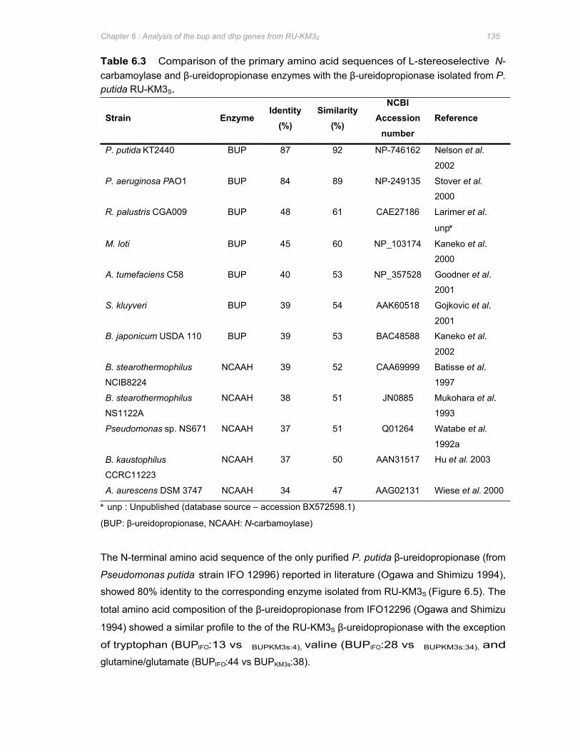
Chapter 6 : Analysis of the bup and dhp genes from RU-KM3S 135
Table 6.3 Comparison of the primary amino acid sequences of L-stereoselective N-carbamoylase and β-ureidopropionase enzymes with the β-ureidopropionase isolated from P. putida RU-KM3S.
Strain Enzyme Identity
(%) Similarity
(%)
NCBI Accession
number Reference
P. putida KT2440 BUP 87 92 NP-746162 Nelson et al.
2002
P. aeruginosa PAO1 BUP 84 89 NP-249135 Stover et al.
2000
R. palustris CGA009 BUP 48 61 CAE27186 Larimer et al.
unp∗
M. loti BUP 45 60 NP_103174 Kaneko et al.
2000
A. tumefaciens C58 BUP 40 53 NP_357528 Goodner et al.
2001
S. kluyveri BUP 39 54 AAK60518 Gojkovic et al.
2001
B. japonicum USDA 110 BUP 39 53 BAC48588 Kaneko et al.
2002
B. stearothermophilus NCAAH 39 52 CAA69999 Batisse et al.
NCIB8224 1997
B. stearothermophilus NCAAH 38 51 JN0885 Mukohara et al.
NS1122A 1993
Pseudomonas sp. NS671 NCAAH 37 51 Q01264 Watabe et al.
1992a
B. kaustophilus NCAAH 37 50 AAN31517 Hu et al. 2003
CCRC11223
A. aurescens DSM 3747 NCAAH 34 47 AAG02131 Wiese et al. 2000
∗ unp : Unpublished (database source – accession BX572598.1)
(BUP: β-ureidopropionase, NCAAH: N-carbamoylase)
The N-terminal amino acid sequence of the only purified P. putida β-ureidopropionase (from
Pseudomonas putida strain IFO 12996) reported in literature (Ogawa and Shimizu 1994),
showed 80% identity to the corresponding enzyme isolated from RU-KM3S (Figure 6.5). The
total amino acid composition of the β-ureidopropionase from IFO12296 (Ogawa and Shimizu
1994) showed a similar profile to the of the RU-KM3S β-ureidopropionase with the exception
of tryptophan (BUPIFO:13 vs BUPKM3s:4), valine (BUPIFO:28 vs BUPKM3s:34), and
glutamine/glutamate (BUPIFO:44 vs BUPKM3s:38).
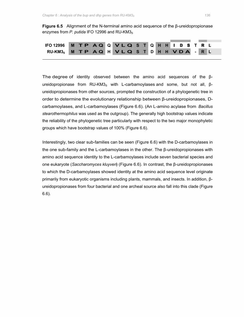
Chapter 6 : Analysis of the bup and dhp genes from RU-KM3S 136
Figure 6.5 Alignment of the N-terminal amino acid sequence of the β-ureidopropionase enzymes from P. putida IFO 12996 and RU-KM3S
IFO 12996 Q Q I D S T R L M T P A Q V L Q S T H H RU-KM3S M T P A Q H V L Q S T D H H V D A - R L
The degree of identity observed between the amino acid sequences of the β-
ureidopropionase from RU-KM3S with L-carbamoylases and some, but not all, β-
ureidopropionases from other sources, prompted the construction of a phylogenetic tree in
order to determine the evolutionary relationship between β-ureidopropionases, D-
carbamoylases, and L-carbamoylases (Figure 6.6). (An L-amino acylase from Bacillus
stearothermophilus was used as the outgroup). The generally high bootstrap values indicate
the reliability of the phylogenetic tree particularly with respect to the two major monophyletic
groups which have bootstrap values of 100% (Figure 6.6).
Interestingly, two clear sub-families can be seen (Figure 6.6) with the D-carbamoylases in
the one sub-family and the L-carbamoylases in the other. The β-ureidopropionases with
amino acid sequence identity to the L-carbamoylases include seven bacterial species and
one eukaryote (Saccharomyces kluyveri) (Figure 6.6). In contrast, the β-ureidopropionases
to which the D-carbamoylases showed identity at the amino acid sequence level originate
primarily from eukaryotic organisms including plants, mammals, and insects. In addition, β-
ureidopropionases from four bacterial and one archeal source also fall into this clade (Figure
6.6).
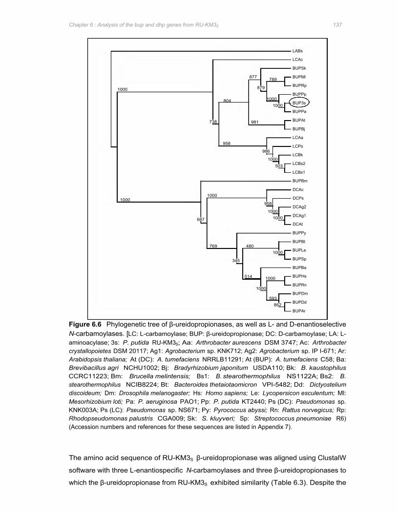
LABs
LCAc
BUPSk
BUPMl
BUPRp
BUPPp
BUP3s
789
1000 1000
877
879
804
1000
BUPPa
BUPAt
BUPBj
LCAa
LCPs
LCBk
LCBs2
LCBs1
BUPBm
DCAc
DCPs
DCAg2
DCAg1
DCAt
BUPPy
BUPBt
BUPLe
BUPSp
BUPBa
BUPHs
BUPRn
BUPDm
BUPDd
BUPAr
738 981
958
1000 1000
958
1000 1000 687
480 769 1000
345
514 1000
1000
593 862
986
1000 503
Chapter 6 : Analysis of the bup and dhp genes from RU-KM3S 137
Figure 6.6 Phylogenetic tree of β-ureidopropionases, as well as L- and D-enantioselective N-carbamoylases. [LC: L-carbamoylase; BUP: β-ureidopropionase; DC: D-carbamoylase; LA: L-aminoacylase; 3s: P. putida RU-KM3S; Aa: Arthrobacter aurescens DSM 3747; Ac: Arthrobacter crystallopoietes DSM 20117; Ag1: Agrobacterium sp. KNK712; Ag2: Agrobacterium sp. IP I-671; Ar: Arabidopsis thaliana; At (DC): A. tumefaciens NRRLB11291; At (BUP): A. tumefaciens C58; Ba: Brevibacillus agri NCHU1002; Bj: Bradyrhizobium japonitum USDA110; Bk: B. kaustophilus CCRC11223; Bm: Brucella melintensis; Bs1: B. stearothermophilus NS1122A; Bs2: B. stearothermophilus NCIB8224; Bt: Bacteroides thetaiotaomicron VPI-5482; Dd: Dictyostelium discoideum; Dm: Drosophila melanogaster; Hs: Homo sapiens; Le: Lycopersicon esculentum; Ml: Mesorhizobium loti; Pa: P. aeruginosa PAO1; Pp: P. putida KT2440; Ps (DC): Pseudomonas sp. KNK003A; Ps (LC): Pseudomonas sp. NS671; Py: Pyrococcus abyssi; Rn: Rattus norvegicus; Rp: Rhodopseudomonas palustris CGA009; Sk: S. kluyveri; Sp: Streptococcus pneumoniae R6) (Accession numbers and references for these sequences are listed in Appendix 7).
The amino acid sequence of RU-KM3S β-ureidopropionase was aligned using ClustalW
software with three L-enantiospecific N-carbamoylases and three β-ureidopropionases to
which the β-ureidopropionase from RU-KM3S exhibited similarity (Table 6.3). Despite the
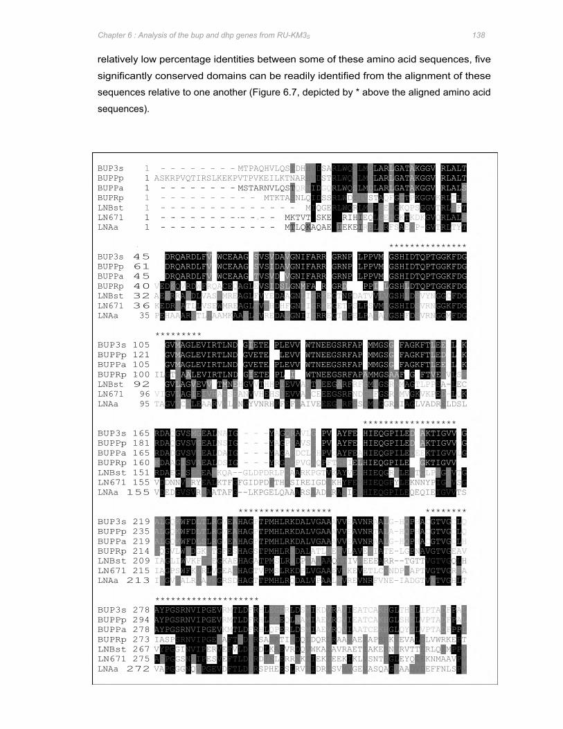
Chapter 6 : Analysis of the bup and dhp genes from RU-KM3S 138
relatively low percentage identities between some of these amino acid sequences, five
significantly conserved domains can be readily identified from the alignment of these
sequences relative to one another (Figure 6.7, depicted by * above the aligned amino acid
sequences).
BUP3s 1 - - - - - - - - MTPAQHVLQSTDHHVDSARLWQSLMD BUPPp 1 ASKRPVQTIRSLKEKPVTPVKEILKTNARHVDSTRLWQSLMD BUPPa 1 - - - - - - - - MSTARNVLQST BUPRp 1 - - - - - - - - - - MTKTASNLQIDSSRLWDTIVST
BUP3s 45 DLDRQARDLFVEWCEAAGCSVS BUPPp 61 DLDRQARDLFVQWCEAAGCSVS BUPPa 45 DLDRQARDLFVQWCEAAGCTVS BUPRp 40 VEDKQVRDWFRQACEQAGLE
BUP3s 105 CFGVMAGLEVIRTLNDLGIETEAPLEVVVWTNEEGSRFAPCMMGSGVFAGKFTLEETLAK BUPPp 121 CFGVMAGLEVIRTLNDLGVETEASLEVVVWTNEEGSRFAPCMMGSGVFAGKFTLEDTLAK BUPPa 105 CFGVMAGLEVIRTLNDLGVETEAPLEVVVWTNEEGSRFAPCMMGSGVFAGKFTLEETLAK BUPRp 100 ILGTL
BUP3s 165 RDAQGVSMGEALNAIG - - -YAG BUPPp 181 RDAQGVSVGEALNSIG - - -YAGP BUPPa 165 RDADGVSVGEALDAIG - - -YAGARDCLG BUPRp 160 KDAAGISVAEALDSIG - - -YR
BUP3s 219 ALGQKWFDLTLRGVEAHAGPTPMHLRKDALVGAAAVVEAVNRAALG-HQPHACGTVGCLQ BUPPp 235 ALGQKWFDLTLRGVEAHAGPTPMHLRKDALVGAAAVVEAVNRTALA-HQPHACGTVGCLQ BUPPa 219 ALGQKWFDLSLRGVEAHAGPTPMHLRKDALVGAAAVVEAVNRAALG-HQPHACGTVGCLH BUPRp 214 GQGVLWYDGKITGFESHAGSTPMHLRRDALATLSEIVLAVEKIATE-LGPNAVGTVGEAV
BUP3s 278 AYPGSRNVIPGEVRMTLDFRHLEGDRLDSMIKDVRAVIEATCAKHGLTHELIPTADFPAL BUPPp 294 AYPGSRNVIPGEVRMTLDFRHLEGEQLNAMIAEVRGVIEATCAKHGLSHELVPTADFPAL BUPPa 278 AYPGSRNVIPGEVKMTLDFRHLQPERLDSMIAEVRQVIAATCEKHGLQYELVPTADFPPL BUPRp 273 IASPSRNVIPGEIAFTIDMRSADATIMDQLDQRLRAAVAEIAPRRKVEVALDLVWRKEPT LNBst 267 VYPGGINVIPERVEFVLDLRDLKAEVRDQVWKAIAVRAETIAKERNVRVTTERLQEMPPV LN671 275 AFPGGSNIIPESVEFTLDIRDIELERRNKIIEKIEEKIKLVSNTRGLEYQIEKNMAAVPV LNAa 272 VAPGGGNQVPGEVDFTLDLRSPHEESLRVLIDRISVMVGEVASQAGVAADVDEFFNLSPV
LNBst 1 - - - - - - - - - - - - - - MIQGERLWQRLMEL LN671 1 - - - - - - - - - - - - MKTVTISKERLRIHI LNAa 1 - - - - - - - - - - - - MTLQKAQAERIEKEI
LNBst 32 AEERRAKDLVASYMREAGLF LN671 36 KEDREATLLVSEWMREAGL LNAa 35 PEHAAARETLIAAMKAAALS
LNBst 92 PLGVLAGVEVVQTMNE LN671 96 VIGVLAGIEIVHAISE LNAa 95 TAGVVCALEAARVMLE
LNBst 151 RDAEGISLAEAMKQA--GLDPDRLPQAARKPGTVKAYVELHIEQGRVLEETGLPVGIVTG LN671 155 VDDNNVTRYEALKTFGFGIDPDFTHQSIREIGDIKHYFEMHIEQGPYLEKNNYPIGIVSG LNAa 155 VDEDGVSVRQAATAFG--LKPGELQAAARSAADLRAFIELHIEQGPILEQEQIEIGVVTS
LNBst 209 IAGLIWVKFTIEGKAEHAGATPMSLRRD LN671 215 IAGPSWFKVRLVGEAGHAGTVPMSLRKDPLVGAAEVIKEVETLCMNDPNAPTVGTVGRIA LNAa 213 IVGVRALRVAVKGRSDHAGTTPMHLRQDALVPAALMVREVNRFVNE-IADGTVATVGHLT
*********
********************
AALEVIRTLNDAGIETELPLCITNWTNEEGSRFAPAMMGSAAFVGDFTVE
ANVVHEHSIEVVAFCEEEGSRFNDGLFGSRGMVGKVKP NGYVNRHPFEFIAIVEEEGARFSSGMLGGRAIAGLVADRELDSL
HGVVTHHPIEVVAFTDEEGARFRFGMIGSRAMAGTLPP
****************** ********
TVTHDHFGNLIGRKEGETPSLPSVMIGSHIDS
VSIDSLGNMFALRKGRDMSKPPIGLGSHLDTQPTGGKFDG VYEDAAGNLIGRKEGTNPDATVVLVGSHLD
VREDALGNIIGRREGTDPELPAIAVGSHFDS
VDAVGNIFARRPGRNPKLPPVMTGSHIDTQPTGGKFDG
VDRVGNIFARRPGRNPDLPPVMTGSHIDTQPTGGKFDG IDAVGNIFARRPGRNPKLPPVMTGSHIDTQPTGGKFDG
GNKPVGAQPFTGFI
QRAVLG RAVSG
QRHIDGQRLWQSLMD
PMAAAAQIIIVIEEE
HPVGAYFEAHIEQGPILEDQAKTIGVVL HPVGAYFEAHIEQGPILEDQAKTIGVVL HPVGAYFEAHIEQGPILEDEEKTIGVVLG
ELHIEQGPILEAEGKTIGVVDH
EQL REL
******************
LARLGAT LARLGAT LARLGAT AQFGGTPKGGVKRLTLS
ARR--TGTTVGTVGQLH
GEVGKQPSGGVTRLSFT GEIGKTKDKGVQRLALS SRFSAEGP-GVTRLTYT
***************
AKGGVCRLALT AKGGVCRLALT AKGGVCRLALS
SVYNGGCFDG VRNGGKFDG VRNGGMFDG
EA-LEC ED-LQK
DVLSR
G G
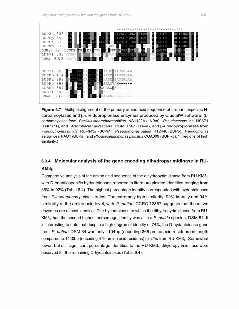
BUP3s 338 YFDRGCVDAVRDSARTLGLP BUPPp 354 YFDKGCVDAVRESAKALGLP BUPPa 338 YFDQGCVGAVREAAQALGMPQMDIVSGAGHDAIFLAELGPAGMIFVPCENGISHNEIENA BUPRp 333 HFD
BUP3s 398 TPQDLAAGCAVLLRAMLAASEAIASGRLAA BUPPp 414 TPDDLAAGCAVLLRAMLAASEAIANGRLAA BUPPa 398 SPDDLAAGCAVLLRAMLKASEAIAGGRLAA BUPRp 393 TQPDC
LNBst 327 LCSDEVKRAAEAACQKLGYPSFWLP LN671 335 KCSENLINSLKQSCKELEIDAPIIVSGAGHDAMFLAEITEIGMVFVRCRNGISHSPKEWA LNAa 332 QLAPTMVDAVREAASALQFTHRDISSGAGHDSMFIAQVTDVGMVFVPSRAGRSHVPEEWT
LNBst 387 TKEDC LN671 395 EIDDILTGTKVLYESIIKHI LNAa 392 DFDDLRKGTEVVLRVMKALDR
PALVGSVESAANALGYQNRRITSGAGHDACNLNTRI
AAGANVLLH AAGAEVLYH
TVLSLAGVAK TVWQLAQGE
YMDIVSGAGHDAIFLAE HMDIVSGAGHDAIFLAE
******************************
SGAAHDSVQLAPICPIGMIFVRSQD
MGPAGMIFVPCENGISHNEIENA LG PAGMIFVPCENGISHNE
PTAMIFVPCK DGISHNE GVSHSPAEWS
LEDA
IENA
Chapter 6 : Analysis of the bup and dhp genes from RU-KM3S 139
Figure 6.7 Multiple alignment of the primary amino acid sequence of L-enantiospecific N-carbamoylases and β-ureidopropionase enzymes produced by ClustalW software. (L-carbamoylases from Bacillus stearothermophilus NS1122A (LNBst), Pseudomonas sp. NS671 (LNP671), and Arthrobacter aurescens DSM 3747 (LNAa), and β-ureidopropionases from Pseudomonas putida RU-KM3S (BUKM), Pseudomonas putida KT2440 (BUPp), Pseudomonas aeruginosa PAO1 (BUPa), and Rhodopseudomonas palustris CGA009 (BUPRp). * : regions of high similarity.)
6.3.4 Molecular analysis of the gene encoding dihydropyrimidinase in RU-KM3S
Comparative analysis of the amino acid sequence of the dihydropyrimidinase from RU-KM3S
with D-enantiospecific hydantoinases reported in literature yielded identities ranging from
36% to 92% (Table 6.4). The highest percentage identity corresponded with hydantoinases
from Pseudomonas putida strains. The extremely high similarity, 92% identity and 94%
similarity at the amino acid level, with P. putida CCRC 12857 suggests that these two
enzymes are almost identical. The hydantoinase to which the dihydropyrimidinase from RU-
KM3S had the second highest percentage identity was also a P. putida species, DSM 84. It
is interesting to note that despite a high degree of identity of 74%, the D-hydantoinase gene
from P. putida DSM 84 was only 1104bp (encoding 368 amino acid residues) in length
compared to 1440bp (encoding 479 amino acid residues) for dhp from RU-KM3S. Somewhat
lower, but still significant percentage identities to the RU-KM3S dihydropyrimidinase were
observed for the remaining D-hydantoinases (Table 6.4).
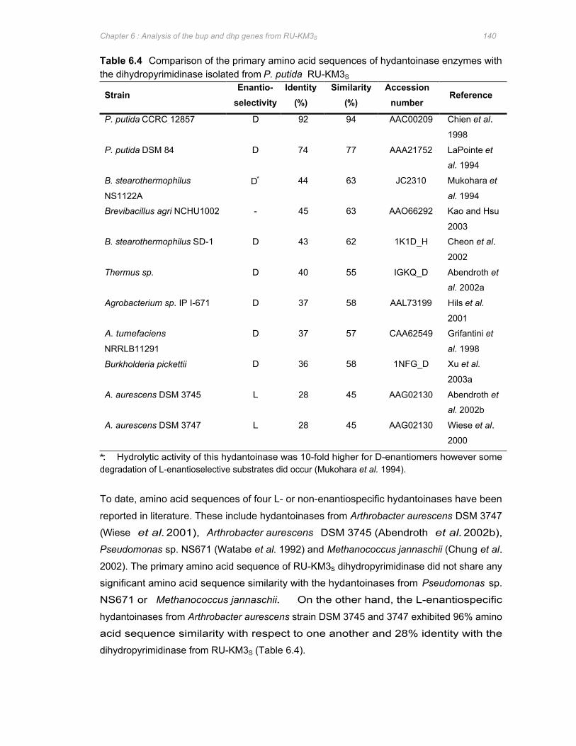
Chapter 6 : Analysis of the bup and dhp genes from RU-KM3S 140
Table 6.4 Comparison of the primary amino acid sequences of hydantoinase enzymes with the dihydropyrimidinase isolated from P. putida RU-KM3S
Strain Enantio-
selectivity Identity
(%) Similarity
(%) Accession
number Reference
P. putida CCRC 12857 D 92 94 AAC00209 Chien et al.
1998
P. putida DSM 84 D 74 77 AAA21752 LaPointe et
al. 1994
B. stearothermophilus
NS1122A D∗ 44 63 JC2310 Mukohara et
al. 1994
Brevibacillus agri NCHU1002 - 45 63 AAO66292 Kao and Hsu
2003
B. stearothermophilus SD-1 D 43 62 1K1D_H Cheon et al.
2002
Thermus sp. D 40 55 IGKQ_D Abendroth et
al. 2002a
Agrobacterium sp. IP I-671 D 37 58 AAL73199 Hils et al.
2001
A. tumefaciens
NRRLB11291
D 37 57 CAA62549 Grifantini et
al. 1998
Burkholderia pickettii D 36 58 1NFG_D Xu et al.
2003a
A. aurescens DSM 3745 L 28 45 AAG02130 Abendroth et
al. 2002b
A. aurescens DSM 3747 L 28 45 AAG02130 Wiese et al.
2000
∗ : Hydrolytic activity of this hydantoinase was 10-fold higher for D-enantiomers however some degradation of L-enantioselective substrates did occur (Mukohara et al. 1994).
To date, amino acid sequences of four L- or non-enantiospecific hydantoinases have been
reported in literature. These include hydantoinases from Arthrobacter aurescens DSM 3747
(Wiese et al. 2001), Arthrobacter aurescens DSM 3745 (Abendroth et al. 2002b),
Pseudomonas sp. NS671 (Watabe et al. 1992) and Methanococcus jannaschii (Chung et al.
2002). The primary amino acid sequence of RU-KM3S dihydropyrimidinase did not share any
significant amino acid sequence similarity with the hydantoinases from Pseudomonas sp.
NS671 or Methanococcus jannaschii. On the other hand, the L-enantiospecific
hydantoinases from Arthrobacter aurescens strain DSM 3745 and 3747 exhibited 96% amino
acid sequence similarity with respect to one another and 28% identity with the
dihydropyrimidinase from RU-KM3S (Table 6.4).

DHPBa 1 DhydBs 1 DhydBp 1 DHP3s 1 DhydPp 1 DhydTh 1 LhydAa 1
DHPBa 59 DhydBs 59 DhydBp 58 DHP3s 60 DhydPp 60 DhydTh 59 LhydAa 61
DHPBa 119 DhydBs 119 DhydBp 118 DHP3s 119 DhydPp 119 DhydTh 119 LhydAa 120
DHPBa 179 DhydBs 179 DhydBp 177 DHP3s 179 DhydPp 179 DhydTh 178 LhydAa 179
TLFKTLIRAKELGA TLYCTLLAAKELGA
LKTLDKAVKTGS TLVASFERCLQLGA TLVASFERCLQLGA
LVQVHAENGDVLDYLTKQALAEGNTDPIYHAYTRPPEAEGEATGRAIALTALADAQLYVV LVMVHAENGDVIDYLTKKALADGNTDPIYHALTRPPELEGEATGRACQLTELAGSQLYVV LVMVHAENGDAADYLRDKFVAEGKTAPIYHALSRPPRVEAEATARALALAEIVNAPIYIV VPTVHAENGELVYHLQQKLLAQGMTGPEAHPLSRPSQVEGEAASRAIRIAETIGTPLYVV VPTVHAENGELVYHLQKKLLAQGMTGPEAHPLSRPSQVEGEAASRAIRIAETIGTPLYVV IVTAHCENAELVGRLQQKLLSEGKTGPEWHEPSRPEAVEAEGTARFATFLETTGATGYVV VIVVHAENETIIQALQKQIKAAGGKDMAAYEASQPVFQENEAIQRALLLQKEAGCRLIVL
DYGFHLMVSDANDHVLEELESVVNNEGITSLKVFMAYKNVLMADDE DYGFHLMISEITDDVLEELPKVLEEEGITSLKVFMAYKNVFQADDG DYGYHIIVLDPTDSVIEELEVLP-DLGITSFKVFMAYRGMNMIDDVTL DYGFHVAITWWSEQVAEEMGELVTQHGVNSFKHFMAYKNAIMAADD DYGFHVAITWWSEQVAEEMGELVAKHGVNSFKHFMAYKNAIMAADD DYTFHMAVSKFDEKTEGQLREIVADG-ISSFKIFLSYKN DFALYGGGVPGNLPEIRKMHDAG-AVGFKSMMAASVPGMFDAVSDGELFEIFQEIAACGS
FFGVDDGEMYQTLRLAKELGV
MFDVIVKNCRLVSSDGITEADILVKDGKVAAISADTSDVEASRTIDAGGKFVMPGVVDEH
VHIYLPF VHIIDMDLKNRYGR-FELDSESAAVGGITTIIEMPITFPPTTTLDAFLEKKKQAGQRLKV
SGL3
THLDMPFGGTVTSDNFFTGTKAAAFGGTTSIVDFCLTSKGESLHSAIATWHEKARGKAVI THLDMPLGGTVTKDDFESGTIAAAFGGTTTIIDFCLTNKGEPLKKAIETWHNKANGKAVI THVETVSFNTQSADTFATATVAAACGGTTTIVDFCQQDRGHSLAEAVAKWDGMAGGKSAI THMQLPF THMQLPF
-MKKWIRNGTVVTASDT -MTKIIKNGTIVTATDT -MDIIIKNGTIVTADGISRADLGIKDGKITQIGGALG--PAERTIDAAGRYVFPGGIDVH -MSLLIRGATVVTHEESYPADVLCVDGQIRAIGKDLEPPTDCEILDGSGQYLMPGGIDPH -MSLLIRGATVVTHEESYPADVLCVDGLIRAIGPNLEPPTDCEILDGSGQYLMPGGIDPH --PLLIKNGEIITADSRYKADIYAEGETITRIGQNLEAPPGTEVIDATGKYVFPGFIDPH
SGL1 SGL2
MGTVASEDFFSGTAAGLAGGTTSIIDFVIPNPQQSLMEAFHTWRGWAQKS-AS MGTVASEDFFSGTAAGLAGGTTSIIDFVIPNPQQSLLEAFHTWRGWAQKS-AS MATFAKDTHETGSKAALMGGTTTYIEMCCPSRNDDALEGYQLWKSKAEGNSYC
YQADVLIDGEKVVAIGSDLQA-TDAEVIDATGYYLLPGGIDPH YEAHLLIKDGKIAMIGQNLEE-KGAEVIDAKGCYVFPGGIDPH
Chapter 6 : Analysis of the bup and dhp genes from RU-KM3S 141
In order to understand the structure, and consequently the function, of hydantoinases, the
crystal structures of D-hydantoinases from Thermus sp. (DhydTh) (Abendroth et al. 2002a), B.
stearothermophilus SD-1 (DhydBs) (Cheon et al. 2002) and Burkholderia pickettii (DhydBp)
(Xu et al. 2003a) in addition to an L-hydantoinase from Arthrobacter aurescens DSM 3745
(LhydAa) (Abendroth et al. 2002b) have been solved. Based on studies of these crystal
structures, amino acid residues involved in the stereospecific recognition, substrate binding,
and catalytic cleavage of 5-monosubstituted hydantoin derivatives have been identified
(summarized in Chapter 1). To determine if these residues are conserved in the
dihydropyrimidinase from RU-KM3S (DHPKM3s), a multiple alignment of the amino acid
sequences for the hydantoinases whose crystal structures have been solved was
constructed (Figure 6.8). A D-hydantoinase from Pseudomonas putida, exhibiting high
amino acid sequence similarity (94%) to the dhp of RU-KM3S, and a dihydropyrimidinase
from Brevibacillus agri were also included (Figure 6.8).
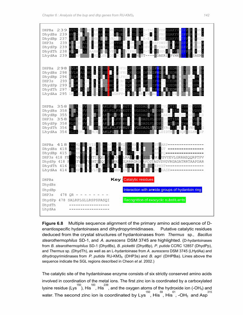
LhydAa 239 HVSNPDGVELIHQAQSEGQDVHCESGPQYL
LhydAa 295 IRLWEQLENGLI
LhydAa 354 RLSLERLVEVMCEK
LhydAa 414 FDGMPVTGAPVLTMVRGTVVAEKGEVLVEQGFGQFVTRRNYEASK
DhydTh LhydAa
DHPBa 239 HVSCADAVRRIAEAREKGWNVYGETCPQYLVLDI-TALEKPDFEGAKYVWSPPLREKWNQ DhydBs 239 HVTCAQAVEKIAEARNKGLDVWGETCP DhydBp 237 HVTCEESLEEVMRAKSRGVRALAETCTHYLYLTK-EDLERPDFEGAKYVFTPPARAKKDH DHP3s 239 HI DhydPp 239 HI DhydTh 238 HL
DHPBa 298 DhydBs 298 E DhydBp 296 D DHP3s 299 E DhydPp 299 E DhydTh 297 KVLWDALAQGFI
DHPBa 358 KISLNQFVDITSTKVAKLFGMFPQKGTIAVGSDADIVLFDPTVQRTISVETHHMNVDYNP DhydBs 358 RITLNQFVDIVSTRIAKLFGLFPKKGTIVVGSDADLVIFDPNIERVISAETHHMAVDYNA DhydBp 355 RISLTQFVELVATRPAKVFGMFPQKGTIAVGSDADIVLWDPEAEMVIEQTAMHNAMDYSS DHP3s 358 RLSMHEFVALTSTNTAKIFNLFPRKGAIRVGADADLVLWDPQGTRTISAQTHHQRVDFNI DhydPp 358 RLSMHEFVALTSTNTAKIFNLFPRKGAIRVGADADLVLWDPQGTRTLSAQTHHQRVDFNI DhydTh 356 RLDI
DHPBa 418 FEGMQVHGDVISVLSRGAFVVRNKQFVGHAGAGRYVKRSTFARP DhydBs 418 FEGMKVTGEPVSVLCRGEFVVRDKQFVGKPGYGQYLKRAKYGT DhydBp 415 YEGHKVKGVPKTVLLRG DHP3s 418 FEGRTVRGIPSHTISQGRVVWADGDLRAEPGAGRYVERPAYPSVYEVLGRRAEQQRPTPV DhydPp 418 FEGRTVRGVPSHTISQGKVLWADGDLRRRGRGGAVCGTAGVSVGVRGAGATRRTAAPDAR DhydTh 416 FEGFEIDGRPSVVTVRG
DHPBa Key : Catalytic residues DhydBs
DhydBp Interaction with am DHP3s 478 QR - - - - - - - -
DhydPp 478 SALRPLGLLRSPSPASQI
DVLWSALKNGILQTVGS
ALWRGLQSGNLHTTATDHCCFCA-EQKAMGRNDFSRIPNG ALWRGLQSGNLHTTATDHCCFCA-EQKAMGRDDFSRIPNGTAGIEDRMAVLWDAGVN
VLWNALKNGQLQTLGS VLWNALRNGVFETVSSDHCSWLFKGHKDRGRNDFRAIPNGAPGVEERLMMVYQG-VN
SSREALDEI SSREALDEI SCKPALDAAMAAKARGVPIYIES
HRFVDAASTKAAKLFGLFPRKGTIAVGSDADLVVYDPQ
TYARAKGQPVYGEVLPGHLLLDDSVYRDPDWATAAGYVMSPPFRPREH SYARAKGQPVYGEVLPGHLLIDDSVYRHPDWSTAAGYVMSPPFRPREH
DTVGTDHCPFDT-EQKLLGKEAFTAIPNG DTLGSDHGGHPV-EDKEPGWKDVWKAGNGALGLETS
PAKLFGIY
KVIVDEGS
KVAVRDGQFVGEK
DHCPFNFSGQKELGRRDF DQCSFDFKGQKELGRGDF
PQKGTLQVGSDADLLILDLDIDTKVDASQFRSLHKYSP
Recognition of exocyclic substituents
VIPHFLLDK-TYAERGGVEAMKYIMSPPLRDKRNQ
YVGEPTDGKFLKRRKYKQ
QYLVLDQ-SYL
NITT----DDAERIGPYMKVAPP
GWGKLLRREPMYF
TKIPNG TKIPNG
EKPNFEGAKYVWSPPLREKWHQ
ide groups of hydantoin ring
TAGIEDRMAVLWDAGVNN
GPIIEDRMTILFSEGVRK GPIIEDRVSILFSEGVKKG
IPAIEDRVNLLYTYGVSRG
YRGTISVKTQHVNNDYNG
LPMMLTNGVNKG
VRSAEMN
EG
SG
Q Q
G
G
Chapter 6 : Analysis of the bup and dhp genes from RU-KM3S 142
Figure 6.8 Multiple sequence alignment of the primary amino acid sequence of D- enantiospecific hydantoinases and dihydropyrimidinases. Putative catalytic residues deduced from the crystal structures of hydantoinases from Thermus sp., Bacillus stearothermophilus SD-1, and A. aurescens DSM 3745 are highlighted. (D-hydantoinases from B. stearothermophilus SD-1 (DhydBs), B. pickettii (DhydBp), P. putida CCRC 12857 (DhydPp), and Thermus sp. (DhydTh), as well as an L-hydantoinase from A. aurescens DSM 3745 (LHydAa) and dihydropyrimidinases from P. putida RU-KM3S (DHP3s) and B. agri (DHPBa). Lines above the sequence indicate the SGL regions described in Cheon et al. 2002.)
The catalytic site of the hydantoinase enzyme consists of six strictly conserved amino acids
involved in coordination of the metal ions. The first zinc ion is coordinated by a carboxylated
lysine residue (Lys150
), His183
, His239
, and the oxygen atoms of the hydroxide ion (-OH3) and
water. The second zinc ion is coordinated by Lys150
, His59
, His61
, -OH3 and Asp315
Chapter 6 : Analysis of the bup and dhp genes from RU-KM3S 143
(Abendroth et al. 2002a). These amino acids are strictly conserved, including in DHPKM3s,
regardless of enantiospecificity (Figure 6.8, red boxes) implying that the mechanism of
hydantoin hydrolysis is conserved regardless of the enantiospecificity of the enzyme (Cheon
et al. 2002).
There are two components to substrate interaction with the hydantoinase: recognition of the
functional amide groups thereby controlling the orientation of the substrate, whilst the
stereochemistry of the hydantoin substrates depends on the recognition of the exocyclic
substituents. The residues proposed to be involved in hydrogen-bond formation with the
hydantoin ring include Ser288
(adjacent to a hairpin loop supported by cis-Pro289
), Asn336
,
Tyr155
, and His183
(Abendroth et al. 2002a) and are highlighted in Figure 6.8 by blue boxes.
Again, these residues are highly conserved throughout the hydantoinases and
dihydropyrimidinases (Figure 6.8).
The recognition of the exocyclic side chain of the hydantoin substrate occurs in buried
hydrophobic pockets. The hydrophobic nature of this pocket in DhydTh and DhydBs implies
that this enzyme interacts only with hydrophobic amino acid residues on hydantoins, but not
charged residues. The residues forming this pocket vary between the hydantoinases
reported (Figure 6.8, green boxes). In DhydBS from B. stearothermophilus SD-1 (Cheon et al.
2002), the residues that form this pocket include Met63
, Leu65
, Phe152
, Phe159
and Tyr155
and
are located in three loops called stereochemistry gate loops or SGLs. These loops, SGL-1
(residues 62-72) SGL-2 (residues 93-100) and SGL-3 (residues 153-162) according to
Cheon et al. (2002) are highly conserved. Conversely, while DhydTh, DhydBs, DHPKM3s and
DhydPp are fairly homologous within these regions, LhydAa, DhydBp and DHPBa show
significant variability (Figure 6.8).
The amino acid residues attributed to the hydrophobic pocket in DHydTh include Leu64
, Cys95,
Phe152
, Tyr155
, Phe159
(Abendroth et al. 2002a). With respect to the exocyclic substituent
recognition residues in DHydBP, except for Phe150
and Tyr153
, the other residues differ. Both
DhydTh and DhydBs have hydrophobic residues at these positions while DhydBp has
hydrophilic residues (Thr62
, Ser64
, Gln93
, Asn157
) (Xu et al. 2003a) (Figure 6.8). The
interaction of these suggested amino acid residues with the exocyclic residue of hydantoin
substrates has been proposed to affect the substrate selectivity of the enzymes. DhydTh
preferentially hydrolyses phenylic substituted hydantoin while benzylic hydantoins are
hydrolysed with much lower efficiency. It is suggested that the hydrophobic pocket in DhydTh
can accommodate phenylic side chains whereas benzylic or polar side chains lead to less
productive binding (Abendroth et al. 2002a). DHPKM3s differs from DhydTh with an Ile159
instead of Phe159
and Ile95
instead of Cys95
. Replacement of the polar Cys with a non-polar
Ile may provide an explanation for the hydrolysis of polar hydroxyphenylhydantoin by this
Chapter 6 : Analysis of the bup and dhp genes from RU-KM3S 144
strain. This is supported by DHPBa and DhydBs which have leucine residues in these
positions and are also able to hydrolyse hydroxyphenylhydantoin (Kao and Hsu 2003, Lee et
al. 1995). Hydroxyphenylhydantoin specificities for the DhydPp and LhydAa, which also have
leucine or isoleucine residues at this position, have not been reported. Hydantoin substrates
with short side chains, such as methylhydantoin, are hydrolysed non-stereospecifically, as is
the case with DHPKM3s (Chapter 2, Table 2.1), because neither enantiomer causes any steric
clashes with the protein (Abendroth et al. 2002a).
6.4 DISCUSSION
A dihydropyrimidinase, encoded by dhp, and a β-ureidopropionase, encoded by bup,
catalyse the ring opening hydrolysis of hydantoin and the conversion of the resultant N-
carbamoyl amino acids to L-amino acids respectively in P. putida RU-KM3S. A third open
reading frame, ORF1, was identified between the bup and dhp gene and, based on
nucleotide sequence similarity, may be involved in transport of hydantoin derivatives across
the cell membrane. However, biocatalytic assays done with resting cells of a mutant strain,
in which ORF1 was insertionally inactivated, indicated no decrease in the activity of the
dihydropyrimidinase relative to the wild type. This implies one of two possibilities: either the
protein coded for by ORF1 is not involved in transport of hydantoin or there is more than one
permease pathway through which hydantoin can pass into the cell. As ORF1 and bup
appear to be expressed as a polycistronic mRNA, which implies linked functions of the
encoded proteins, the second theory is more likely. This is supported by the total genome of
P. putida KT2440 in which ~350 cytoplasmic membrane transport systems have been found
including a large repertoire of ABC amino acid transporters, 15% more than that found in P.
aeruginosa PAO1 (Nelson et al. 2002). While transport of hydantoin substrate into the cell
did occur when ORF1 was inactivated, it would be interesting to see if knockout of this
putative transport protein would affect transport of more complex hydantoin-derivatives
across the cell membrane. This would be of particularly import if the natural host is used in a
biotransformation process for the production of complex amino acids from hydantoin
substrates.
Excluding P. putida RU-KM3S, the only other bacterial pyrimidine catabolism gene cluster
reported to date is from Brevibacillus agri NCHU1002 (Kao and Hsu 2003). However, not
only does the gene arrangement between these two clusters differ considerably, but the
amino acid sequence of the β-ureidopropionases from these two strains do not have any
similarity and, from phylogenetic analysis, are evolutionarily divergent with the β-
ureidopropionase from RU-KM3S more closely related to the L-carbamoylases and the β-
ureidopropionase from B. agri to the D-carbamoylases. Furthermore, the substrate
Chapter 6 : Analysis of the bup and dhp genes from RU-KM3S 145
selectivity of the β-ureidopropionase from B. agri differs considerably from that of RU-KM3S
with an inability to hydrolyse N-carbamoyl-D,L-amino acids (Kao and Hsu 2003). Moreover,
despite the reasonably high levels of similarity between the dihydropyrimidinase of B. agri
and the dihydropyrimidinase from RU-KM3S, the dihydropyrimidinase enzyme from B. agri is
unable to hydrolyse N-butylhydantoin or N-methylhydantoin (Kao and Hsu 2003) which RU-
KM3S, in contrast, hydrolyses very effectively (Buchanan et al. 2001). However, no
significant differences were observed between the dihydropyrimidinases from RU-KM3S and
B. agri with respect to the amino acid residues proposed to be responsible for substrate
selectivity. This suggests there may be residues involved in interaction with the exocyclic
substituents of hydantoin derivatives which have not yet been identified.
The highest amino acid sequence identity to the β-ureidopropionase from RU-KM3S was
obtained with the β-ureidopropionases from P. putida KT2440 and P. aeruginosa PAO1
amino acid sequences which were both obtained from total genome projects. To date, only
two β-ureidopropionases have been isolated and characterized from aerobic bacteria, viz.
from P. putida IFO 12996 (Ogawa and Shimizu 1994) and B. agri NCHU1002 (Kao and Hsu
2003). As already established, the β-ureidopropionase from B. agri showed no amino acid
sequence similarity with the β-ureidopropionase from RU-KM3S. The primary amino acid
sequence of the RU-KM3S β-ureidopropionase enzyme also lacked identity to several β-
ureidopropionases from alternative sources. Whilst similarity to β-ureidopropionases from
other kingdoms such as plant, animal, and archea bacteria is perhaps understandable, the
lack of similarity to some bacterial β-ureidopropionases was somewhat surprising to
discover. The β-ureidopropionase from P. putida IFO 12996 differs significantly to that of B.
agri in terms of substrate selectivity as, unlike B. agri, it is able to hydrolyse N-carbamoyl-β, γ,
and α-amino acids (Ogawa and Shimizu 1994). The β-ureidopropionase from RU-KM3S has
been shown to hydrolyse a range of N-carbamoyl-α-amino acids (Buchanan et al. 2001) and
based on its ability to convert the intermediate of dihydrouracil degradation to alanine, is
capable of N-carbamoyl-β-amino acid hydrolysis as well. Hydrolysis of N-carbamoyl-α-amino
acids by the β-ureidopropionase from IFO12996 is strictly L-enantiospecific (Ogawa and
Shimizu 1994), a characteristic which the RU-KM3S β-ureidopropionase also exhibits with
regards to N-carbamoyl-α-alanine (Chapter 2). It would be interesting to see if the RU-KM3S
β-ureidopropionase further mimics the β-ureidopropionase from IFO12996 in L-
enantioselectivity regardless of substrate utilized and in its ability to hydrolyse N-carbamoyl-
γ-amino acids. Comparison of the N-terminal amino acid sequence of the β-
ureidopropionases from IFO12996 and RU-KM3S resulted in an 80% identity match.
However, the amino acid profiles of these two β-ureidopropionase enzymes do differ
Chapter 6 : Analysis of the bup and dhp genes from RU-KM3S 146
somewhat and the β-ureidopropionase from IFO 12996 appears to be 12 amino acids shorter
than the β-ureidopropionase from RU-KM3S.
D-enantioselective N-carbamoylases have been shown to have some amino acid sequence
identity, including strict conservation of residues proposed to be involved in the catalytic site,
with the β-ureidopropionase from Rattus norvegicus (rat) (Nakai et al. 2000). However, the
β-ureidopropionase from RU-KM3S showed no significant amino acid sequence identity to the
D-enantiospecific N-carbamoylases from Agrobacterium sp. IP I-671, NRRL B11291,
KNK712 (Hils et al. 2001, Grifantini et al. 1998, Nanba et al. 1998) or Pseudomonas sp.
KNK003A (Ikenaka et al. 1998a). Neither do the amino acid sequences of the N-terminal
region of the D-carbamoylases from Comamonas sp. EC22C (Ogawa et al. 1993) and
Blastobacter sp. A17p-4 (Ogawa et al. 1994b) exhibit any similarity to that of β-
ureidopropionase from RU-KM3S. This lack of identity between the amino acid sequences of
D-carbamoylases and the β-ureidopropionase from RU-KM3S is not entirely surprising as the
β-ureidopropionase from Pseudomonas sp. (Ogawa and Shimizu 1994) has been shown to
strictly L-enantiospecific, as opposed to D-enantiospecific. In addition, the inability of D-
carbamoylases to hydrolyse the natural substrate of β-ureidopropionase, namely β-
ureidopropionic acid (Nakai et al. 2000, Ogawa et al. 1994b) has been documented. It is
interesting to note that the L-N-carbamoylases from Pseudomonas sp. NS671, A. aurescens
DSM 3747, and Bacillus kaustophilus, to which β-ureidopropionase from RU-KM3S exhibits
≥34% identity, are unable to hydrolyse the natural substrate of this enzyme namely β-
ureidopropionic acid (Ishikawa et al. 1996, Hu et al. 2003, Wilms et al. 1999). The remaining
L-N-carbamoylases described in Table 6.3 were not analysed for the ability to hydrolyse β-
ureidopropionic acid.
Phylogenetic analysis of D-carbamoylases, L-carbamoylases, and β-ureidopropionases
revealed two distinct sub-families of D-carbamoylases and L-carbamoylases with β-
ureidopropionases co-grouping with both sub-families. The β-ureidopropionase from RU-
KM3S showed no significant amino acid sequence similarity to D-carbamoylases or the β-
ureidopropionases grouped in the same sub-family. By contrast, similarity to all the L-
carbamoylases, albeit low, and all the β-ureidopropionases grouped with L-carbamoylases to
the β-ureidopropionase from RU-KM3S was observed.
The dihydropyrimidinase from P. putida RU-KM3S (DHPKM3s) showed high identity with D-
enantiospecific hydantoinases, especially those from P. putida strains, and to a lesser
degree an L-enantiospecific hydantoinase from Arthrobacter aurescens DSM 3745. The
crystal structures of three D-hydantoinases and one L-hydantoinase to which DHPKM3s
Chapter 6 : Analysis of the bup and dhp genes from RU-KM3S 147
exhibits identity have been solved (Abendroth et al. 2002a, Cheon et al. 2002, Xu et al.
2003a, Abendroth et al. 2002b).
The amino acid residues involved in stereospecific recognition, binding and catalytic
cleavage of 5-monosubstituted hydantoin derivatives have been identified using the
determined crystal structures and homology to the catalytic action of a related enzyme,
dihydroorotase (Abendroth et al. 2002a, Cheon et al. 2002, Xu et al. 2003a, Abendroth et al.
2002b). The amino acids (Lys150
, His183
, His239
, His 59
, His61
, Asp315
) and the metal ions
involved in the catalytic site are strictly conserved irrespective of the stereo- or substrate-
specificity of the enzyme. The residues responsible for interaction with the hydantoin ring are
also highly conserved throughout. However, differences do occur in the hydrophobic pocket
proposed to be involved in recognition of the exocyclic side chain of the hydantoin substrate.
These discrepancies in amino acid residues may provide an explanation for differences in
substrate-selectivity between these enzymes, however, site-directed mutagenesis of these
residues needs to be done in order to conclusively identify these residues as responsible for
substrate-selectivity. It would be interesting to see if, by changing these selected amino acid
residues to the corresponding amino acids in other hydantoinase enzymes, substrate
selectivity would be consistent with that of the model enzyme.
Another potential target area for further analysis is the C-termini of the hydantoinase and
dihydropyrimidinase enzymes. The C-terminus of the dihydropyrimidinase from RU-KM3S
and the D-hydantoinase from P. putida CCRC12857 are significantly longer than the
remaining aligned sequences by 18 to 19 and 34 to 35 amino acid residues respectively.
Deletion analysis of the D-hydantoinase from P. putida CCRC 12857 indicated that the last
32 amino acid residues from the C-terminal end of the enzyme are essential for activity
(Chien et al. 1998). Furthermore, D-hydantoinases from B. stearothermophilus strains SD-1
and NS1122A were remarkably similar in amino acid sequence (88% amino acid similarity)
but differed significantly in the C-terminus (Kim et al. 1997). However, the hydantoinase from
B. stearothermophilus SD-1 is reported to be strictly D-enantiospecific whilst B.
stearothermophilus NS1122A hydantoinase is non-stereoselective implying that the C-
terminal region plays an important role in determining the biocatalytic properties of these
enzymes (Kim et al. 1997). The C-terminus is also implicated in the quaternary structure of
the hydantoinase enzyme as deletion of this region in the hydantoinase from B.
stearothermophilus GH2 resulted in the formation of a dimer instead of a tetramer as is the
case with the wild type enzyme (Kim and Kim 1998a). Cheon et al. (2002) reported that the
dimers of B. stearothermophilus SD-1 are further dimerized to form tetramers using the N-
terminal β-sheet which includes the N- and C-termini. It would be interesting to see what
Chapter 6 : Analysis of the bup and dhp genes from RU-KM3S 148
effect the deletion or alteration of the C-terminus of the dihydropyrimidinase from RU-KM3S
would have on the activity, specificity, and/or structure of the enzyme.
This is the first report of a dihydropyrimidinase and β-ureidopropionase enzyme working in
tandem to hydrolyse hydantoin derivatives to the corresponding amino acids. While the
amino acid sequence of the dihydropyrimidinase from RU-KM3S corresponded well with
those reported in literature, the biophysical characteristics of the respective enzymes
differed. Of greater interest is the β-ureidopropionase which is significantly different to β-
ureidopropionases and N-carbamoylases reported in literature and, due to its L-
enantioselectivity, is of considerable interest for the production of L-amino acids.
Chapter 7: General conclusions 149
The biocatalytic production of enantiomerically pure amino acids from hydantoin derivatives
is finding increasing application in industry requiring enzyme systems with improved or novel
biocatalytic activities. Thus, strain RU-KM3S, isolated from the environment and found to
express high levels of hydantoin- and N-carbamylglycine-hydrolysing activity, was selected
for further study. The principle objective was to determine the industrial potential of the
hydantoin-hydrolysing enzyme system of RU-KM3S in terms of the biocatalytic potential and
distinctiveness of the system. Thus the first step was to determine the broad biocatalytic
properties of the enzyme system (substrate selectivity, enantioselectivity, optimal reaction
parameters, etc.). P. putida strain RU-KM3S was selected for this study due to the apparent
L-selectivity and preference for aliphatic substrates of the enzyme system. The overriding
focus of the research described in this thesis was to confirm that the enzymes differed from
those reported in the literature through molecular determination of their genes.
7.1 Hydantoin-hydrolysing activities in Pseudomonas isolates To date, only three Pseudomonas isolates have been reported with both hydantoinase and
N-carbamoylase activities. Pseudomonas sp. AJ-11220 which expresses a D-hydantoinase
and D-carbamoylase (Yokozeki et al. 1987b,c), Pseudomonas sp. NS671 with a non-
enantioselective hydantoinase and L-enantioselective N-carbamoylase (Ishikawa et al.
1993), and Pseudomonas putida IFO 12996 from which a D-hydantoinase and a β-
ureidopropionase have been purified (Ogawa and Shimizu 1994, Ogawa et al. 1994c). While
some biochemical characteristics of these enzymes overlap with those of the hydantoin-
hydrolysing enzyme system in RU-KM3S, significant dissimilarities occur in all cases
particularly with respect to substrate selectivities. Furthermore, the genes coding for the
hydantoin-hydrolysing enzyme system have only been elucidated for Pseudomonas sp.
NS671 to which the dihydropyrimidinase from RU-KM3S exhibited no similarity and the β-
ureidopropionase showed minimal similarity (37% amino acid sequence identity). Thus, this
study is the first report of a complete gene cluster, containing a dihydropyrimidinase and β-
ureidopropionase, which are responsible for the hydrolysis of hydantoin to amino acids.
Analysis of the complete genome of P. aeruginosa PAO1 revealed a similar gene cluster to
that of RU-KM3S while the corresponding operon in P. putida KT2440 contains a confirmed
frameshift in the dhp gene (Stover et al. 2000, Nelson et al. 2002). In addition, the genes
flanking this operon in P. putida strain KT2440 and RU-KM3S differ. This reflects the
plasticity of bacterial genomes and that of P. putida KT2440 in particular which contains an
additional 36 putative transposable elements in comparison to that found in the genome of P.
aeruginosa PAO1 (Nelson et al. 2002). As dihydropyrimidinase and β-ureidopropionase are
involved in pyrimidine metabolism and are ubiquitous in bacteria, it may be surprising that
Chapter 7: General conclusions 150
these enzymes have not been isolated for the purpose of hydantoin-hydrolysis more
frequently. This may be due to the substrate-selectivity of these enzymes as, for example,
not all β-ureidopropionases are capable of hydrolysing N-carbamyl-α-amino acids (Kao and
Hsu 2003). Alternatively, as observed in RU-KM3S, the regulation of this operon may be
complex and, under conditions used to screen for hydantoin-hydrolysing organisms, may be
repressed.
7.1.1 Comparative analysis of the RU-KM3S dihydropyrimidinases Of the hydantoinase enzymes isolated from Pseudomonas species, all exhibit D-
enantioselectivity barring that of Pseudomonas sp. NS671 with a non-enantioselective
hydantoinase (Watabe et al. 1992a). However, this hydantoinase differs considerably from
the corresponding enzyme in RU-KM3S in that it is reported to consist of two subunits,
encoded on a large plasmid, and to be ATP-dependent (Ishikawa et al. 1993, Watabe et al.
1992a). Comparative analysis of the amino acid sequence of the dihydropyrimidinase
isolated from RU-KM3S with hydantoinases reported in literature revealed a high percentage
of identity with the D-hydantoinases from P. putida CCR 12857 (Chien et al. 1998) and P.
putida DSM 84 (LaPointe et al. 1994) with amino acid sequence identities of 92% and 74%
respectively. However, except for cloning the gene encoding the D-hydantoinase and
expression in E. coli, no extensive fundamental biochemical characterization of the D-
hydantoinase from P. putida CCRC 12857 has been reported in literature. With respect to
the D-hydantoinase from P. putida DSM 84, differences when compared to the
dihydropyrimidinase from RU-KM3S in terms of metal ions affecting enzyme activity, optimal
temperature and pH for hydrolytic activity, and substrate preferences were observed (Morin
et al. 1986b).
The enantioselectivity of the dihydropyrimidinase from RU-KM3S was determined based on
hydrolysis of D- or L-methylhydantoin. However, analysis of the crystal structure of the
dihydropyrimidinase (D-hydantoinase) from Thermus sp., to which the dihydropyrimidinase
from RU-KM3S exhibited 40% identity and 55% similarity in amino acid sequence, suggests
that methylhydantoin is hydrolysed non-stereoselecitively, due to the lack of steric hindrance,
while derivatives with larger exocyclic substituents are hydrolysed D-enantioselectively
(Abendroth et al. 2002a). This implies that for hydantoin substrates with large side chains,
such as hydroxyphenylhydantoin, hydrolysis occurs D-enantioselectively in RU-KM3S cells.
However, hydroxyphenylhydantoin is hydrolysed both to the intermediate as well as the
hydroxyphenylgycine end product by RU-KM3S (Buchanan et al. 2001). This suggests that
either the hydantoinase is indeed non-stereoselective or the enantioselectivity of the β-
ureidopropionase is substrate dependent allowing for hydrolysis of D-N-
carbamylhydroxyphenylglycine. The former option is most likely for two reasons: firstly, N-
Chapter 7: General conclusions 151
carbamoylases have been found to be strictly D- or L-enantioselective with no substrate-
dependent enantioselectivities reported, and secondly, the β-ureidopropionase from RU-
KM3S only exhibited amino acid sequence homology with L-carbamoylases and some β-
ureidopropionases whilst the dihydropyrimidinase exhibited similarity to both D-, L-, and non-
enantioselective hydantoinases. In addition, the only β-ureidopropionase for which
stereoselectivity in terms of N-carbamylamino acid-hydrolysis has been determined is that
from Pseudomonas putida IFO 12996 which was shown to have strict L-enantioselectivity
(Ogawa and Shimizu 1994).
The amino acid residues identified by Abendroth et al. (2002a,b), Cheon et al. (2002), and
Xu et al. (2003) as responsible for the catalytic activity of the enzymes, as well the orientation
of the substrate, are conserved in the dihydropyrimidinase from RU-KM3S. According to
Cheon et al. (2002), the residues which form the hydrophobic pocket attributed with
recognition and binding of the exocyclic side chain of the hydantoin derivatives, and thus
control substrate selectivity and enantioselectivity, are located in three stereochemistry gate
loops (SGL1, SGL2, SGL3). Analysis of the amino acid residues proposed to be responsible
for substrate selectivity of the hydantoinase enzymes revealed two residues within SGL2 and
SGL3, at positions 159 and 95, which differ between the enzymes from Thermus, B.
strearothermophilus SD-1, and RU-KM3S and which may account for the varying abilities of
these enzymes to hydrolyse hydroxyphenylhydantoin. Site-directed mutagenesis of these
residues would conclusively determine if this is true. Furthermore, by replacing the entire
SGL region from one enzyme by that belonging to another with alternative substrate
preferences, and determining if the substrate preference of the constructed enzyme would
mimic that of the enzyme from which the SGL regions originated, would also be informative.
7.1.2 Comparative analysis of the RU-KM3S β-ureidopropionase
The β-ureidopropionase isolated from RU-KM3S showed no primary amino acid structure
similarity to any of the D-carbamoylases reported in literature whilst varying degrees of
similarity was observed with L-carbamoylases. The highest similarity of the RU-KM3S β-
ureidopropionase was observed with β-ureidopropionases from other bacterial sources,
which were predominantly identified from total genome sequence data and were not
analysed for N-carbamylamino acid hydrolysing activity. Interestingly, unlike hydantoinases,
phylogenetic analysis of the β-ureidopropionases, D- and L-carbamoylases revealed a clear
subdivision of D-carbamoylases and L-carbamoylases. This formation of “sub-families” could
be useful for establishing the probable enantioselectivities of N-carbamoylase sequences
mined from genomic libraries.
Chapter 7: General conclusions 152
While the β-ureidopropionases from P. putida IFO 12996 and RU-KM3S do have significant
similarities, some differences can be observed when comparing their biochemical
characteristics suggesting that these two enzymes are distinct from one another. It should
be noted however that the β-ureidopropionase from P. putida IFO 12996 was characterised
as a purified protein whilst RU-KM3S β-ureidopropionase activity was analysed in the context
of whole cells. The differing environments under which these enzymes were tested may
have had some effect on the response of the respective enzymes to the biocatalytic
parameters analysed. However, analysis of the N-terminal amino acid sequence and the
total amino acid composition supports that the β-ureidopropionases from RU-KM3S and IFO
12996 are not identical.
The β-ureidopropionase purified from P. putida IFO 12996 was shown to have L-
enantioselective N-carbamylamino acid hydrolytic activity proving that β-ureidopropionase is
not synonymous with D-carbamoylase as was at first thought (Ogawa and Shimizu 1994).
Data obtained with the β-ureidopropionase from RU-KM3S supports this. However, it would
be interesting to see if the β-ureidopropionases which showed similarity with the D-
carbamoylases, based on phylogenetic analysis, would not perhaps be D-enantioselective
with respect to N-carbamylamino acid hydrolysis.
7.2 Potential industrial application Of the two enzymes isolated from RU-KM3S, the β-ureidopropionase is of greater interest
with respect to potential industrial application. Not only does it differ from reported enzymes
but, the literature does not report that β-ureidopropionases have been utilized in industry to
produce amino acids. As such, the β-ureidopropionase from RU-KM3S is of interest. In
addition, the β-ureidopropionase from RU-KM3S is capable of hydrolysing N-carbamyl-α-
amino acids and, as preliminary data suggests, N-carbamyl-β-amino acids as well. This
could be useful for the production of amino acids with chiral centres at the α- and β-carbon
(e.g. threonine and isoleucine) which occur as four stereoisomers and are therefore very
difficult to synthesize chemically and purify to an optically pure form (Ogawa et al. 1999).
The β-ureidopropionase from P. putida IFO 12996 was also observed to hydrolyse N-
carbamyl-γ-amino acids (Ogawa and Shimizu 1994). If this is true for the β-ureidopropionase
from RU-KM3S, it would further extend the range of applications of this enzyme.
In RU-KM3S cells, the β-ureidopropionase activity is limited as it is dependent on the N-
carbamylamino acid intermediate produced by the dihydropyrimidinase. By co-expression or
fusion of the β-ureidopropionase with an L-enantioselective hydantoinase the productivity of
the β-ureidopropionase may be greatly enhanced.
Chapter 7: General conclusions 153
The mutant strains of RU-KM3S in which the dhp or bup genes have been insertionally
inactivated were shown to exhibit complete loss of hydantoin-hydrolysing activity. These
mutants may therefore be valuable, as an alternative to E. coli, for the screening of genomic
libraries for hydantoin-hydrolysing genes. Using P. putida RU-KM3S as a heterologous host
for the expression of hydantoinase and/or N-carbamoylase genes is advantageous as the
strain is more robust than E. coli, and, as evidenced by the high levels of native expression
of dhp and bup, it is capable of surviving over-expression of pyrimidine-degrading enzymes
within the cell.
7.3 Limitations of traditional methods for the isolation of genes encoding hydantoin-hydrolysing enzymes Isolation of the genes encoding dhp and bup in RU-KM3S by complementation studies in E.
coli proved to be unsuccessful. This may be attributed to several factors including: a) over-
expression of enzymes responsible for nucleotide degradation could potentially be highly
toxic to the cell, b) screening criteria selected against metabolically burdened recombinant
cells which exhibited reduced growth rates, c) poor expression of Pseudomonas genes in E.
coli, d) instability of the insert DNA resulting in loss of genetic information. The instability of
genes heterologously expressed in E. coli was observed by loss of insert, or deletions of
DNA segments within the insert of the recombinant plasmid, not only in this study but in
similar studies done in A. tumefaciens RU-OR as well (Hartley 2001). Plasticity of the
genomic regions encoding hydantoin-hydrolysing enzymes is further illustrated by the
inactivation of the dhp in P. putida KT2440 due to a frameshift mutation (which was not a
sequencing artefact) (Nelson et al. 2002). Hils et al. (2001) also identified ORFs with
similarities to transpositional elements flanking the gene involved in hydantoin utilization in
Agrobacterium sp. IP I-671.
Transposon mutagenesis bypasses these negative drawbacks allowing for isolation of the
genes coding for the hydantoin-hydrolysing enzymes in RU-KM3S. However, at the time of
screening of the transposon generated mutants, the regulatory mechanism affecting
expression of the bup and dhp genes was unknown. Thus the enrichment and screening
was done in minimal medium with hydantoin and glucose as nitrogen and carbon sources
respectively. As a result, mutants which were isolated predominantly comprised of those
with mutations in the global Ntr pathway which resulted in an inability to utilize nitrogen
sources optimally. However, subsequent studies revealed that dhp and bup are regulated by
CCR. By screening in the presence of glucose, mutants in which the CCR pathway was
inactivated, resulting in derepression of the dhp and bup, would be selected against. This
would explain why no mutants in which the CCR pathway regulating expression of these
Chapter 7: General conclusions 154
genes was interrupted were isolated. In hindsight, glycerol would have served better as a
carbon source.
7.4 Regulation of catabolic enzymes in Pseudomonas In the process of isolating the dhp and bup genes in RU-KM3S, several mutants were isolated
which shed light on the mechanism by which the hydantoin-hydrolysing pathway is regulated
in this strain. In contrast to the regulation of the hydantoin-hydrolysing enzyme system in A.
tumefaciens RU-OR (Hartley et al. 2001), neither the expression or activity of the β-
ureidopropionase or dihydropyrimidinase was directly affected by the nitrogen status of the
cell as demonstrated by wild type activities in cells grown in good nitrogen sources and
mutants strains in which key elements of the global Ntr pathway had been inactivated (i.e.
NtrB, NtrC, pII uridylyltransferase).
Considering that the hydantoin-hydrolysing pathway is generally thought to be responsible
for hydrolysis of a potential nitrogen source for the production of amino acids, it is surprising
that the regulation of hydantoin-hydrolysing pathway in RU-KM3S was found to be directly
linked to carbon catabolite repression (CCR). This was evidenced by the decreased activity
levels of β-ureidopropionase and dihydropyrimidinase in medium supplemented with
succinate or glucose, in addition to decreased activities subsequent to inactivation of a
component of the TCA cycle. Analysis of the intergenic region between the dhp and
ORF1/bup genes revealed a putative CRP-binding site which was found to function as such
in E. coli. However, cAMP levels in Pseudomonas do not vary, regardless of the carbon
source supplied, and CRP does not function in Pseudomonas as it does in E. coli (Collier et
al. 1996). Thus an alternative protein which functions in a similar manner to CRP must be
present in RU-KM3S.
A homologue of the E. coli CRP protein, designated Vfr, has been isolated from P.
aeruginosa and is 67% identical and 91% similar at the amino acid level to the E. coli CRP
protein (West et al. 1994). Characterization of this protein by Suh et al. (2002) indicated that
the Vfr binds cAMP with a similar dissociation constant as that of CRP. In addition, Vfr-
cAMP complex resulted in similar repressive activities on the E. coli lac promoter as CRP-
cAMP signifying that, in E. coli, Vfr functions in a similar manner to CRP. However, while Vfr
functions as a global regulator of gene expression, it does not appear to be involved in CCR
in P. aeruginosa as succinate-repression of mannitol dehydrogenase, amidase, and
urocanase was not relieved by inactivation of the vfr gene (Suh et al. 2002). Conversely, the
presence of a functional CRP-binding site within the promoter region of the dhp in RU-KM3S
suggests that a protein such as Vfr, which has been shown to function in a similar manner to
CRP when expressed in E. coli, is involved in CCR of the hydantoin-hydrolysing enzyme
Chapter 7: General conclusions 155
system in P. putida RU-KM3S. It would be interesting to see if inactivation of the Vfr gene in
RU-KM3S would result in a concomitant release of the dhp and bup from CCR which would
also prove that, contrary to research to date, Vfr is involved in CCR. Also, while the CRP-
binding site is active in E. coli, deletion of the CRP-binding site within the promoter region of
dhp in wild type RU-KM3S would confirm that the site is functional in regulating expression.
To date, the only protein shown to be directly involved in CCR in Pseudomonas is Crc which
has been implicated in repression of genes involved in metabolism of both sugars and
nitrogenated compounds (Collier et al. 1996). However, CCR observed in Pseudomonas
cannot be completely attributed to the activity of Crc. This was demonstrated in P. putida
GPo1 containing an inactivated crc gene which resulted in repression relief of the alkane
degradation pathway observed in exponential phase of growth in rich medium. But the Crc-
phenotype had no effect on the repression caused when the cells were grown in defined
media with pyruvate or succinate as a carbon source (Yuste and Rojo 2001). Expression of
the genes coding for the proteins in this pathway may therefore by regulated by at least two
regulatory mechanisms, one of which is Crc-dependent (Yuste and Rojo 2001). A similar
result was obtained with the operon involved in histidine utilization where a crc- genotype did
not relieve CCR caused by succinate (Collier et al. 1996). Furthermore, while inactivation of
the crc gene reduced repression of the alkane degradation pathway in rich medium by 6-fold,
this relief was only partial (Yuste and Rojo 2001). When RU-KM3S cells were cultured in
complete medium, expression of the dihydropyrimidinase and β-ureidopropionase occurred
only once stationary phase of growth had been reached. As more than one mechanism can
be involved in the CCR of a particular pathway, as evidenced by the alkane degradation
pathway in P. putida, inactivation of the crc gene, in addition to the vfr gene, might shed light
on this regulatory pathway.
The questions that remain are: what activates CCR pathways and how is CCR linked to
nitrogen metabolism (i.e. hydantoin-hydrolysis)? Collier et al. (1996) proposed that CCR
may be controlled by a common effector which may be in the form of intermediates of a
metabolic pathway and/or a more general indicator of the metabolic state of the cell (e.g.
proton motive force, availability of high energy phosphodiester bonds, etc). Studies by
Dinamarca et al. (2002) suggest that regulation of CCR is linked to the energy status of the
cell as inactivation of cytochrome o ubiquinol oxidase, a component of the electron transport
chain, reduced the CCR observed with respect to the alkane degradation pathway in P.
putida. However, this inactivation reduced but did not eliminate CCR which indicates that
CCR in Pseudomonas is dependent on more than just the electron transport chain
(Dinamarca et al. 2002).
Chapter 7: General conclusions 156
Pseudomonads are renowned for their ability to colonise a wide diversity of ecological niches
due to their capacity to utilize a vast range of substrates, including some very complex
organic compounds and xenobiotics, for their growth (Stover et al. 2000, Nelson et al. 2002).
While such diversity of metabolic pathways is beneficial, in order to survive in the natural
environment and be competitive, expression and activity of these enzymes must be carefully
controlled. The intricacy of the regulation of metabolic pathways in Pseudomonads, as
compared to E. coli for example, is reflected by their genetic complexity. Analysis of the
complete genome of Pseudomonas aeruginosa revealed the highest percentage (8.4%) of
the total number of genes devoted to command and control systems observed in a bacterial
genome (Stover et al. 2000).
Not only is expression of the hydantoin-hydrolysing pathway in RU-KM3S regulated by CCR
but it is also dependent on induction by an inducer. In order to simplify culture conditions
and decrease production costs, an attempt was made to isolate a mutant strain that exhibited
inducer-independent hydantoin-hydrolysing activity. Isolated mutants GMP1 and GMP2
showed increased levels of dihydropyrimidinase activity but which were still inducible. The
increased activity in mutants strains GMP1 and GMP2 under uninduced conditions cannot be
ascribed to an increase in expression or activity of a second hydantoinase as dhp knockout
mutants GMP12 and GMP13 established the dihydropyrimidinase enzyme to be solely
responsible for hydantoin hydrolysis in these strains. Alternatively, the increase in activity
observed in GMP1 and GMP2 may be due to an indirect mutation in a regulatory pathway or
directly as a result of mutation of the dhp gene/promoter. However, the alignment of the
promoter region of GMP1 and wild type RU-KM3S resulted in 100% identity between these
two sequences. The dhp genes of GMP1 did contain a single mutation, Q143R, where a
polar glutamine was replaced by an positive arginine residue both of which are hydrophylic.
However, crystallographic analysis of dihydropyrimidinase enzymes from Thermus sp.
(Abendroth et al. 2002a) does not suggest that this amino acid residue is of significant
importance. This suggests that the mutation encoded in GMP1 and GMP2 resulting in the
observed increased hydantoin hydrolysis is most likely due to a mutation in a pathway
involved in regulation of the dihydropyrimidinase expression or activity. Screening for GMP1
and GMP2 was done in complete medium and minimal medium with glucose as a carbon
source respectively which may have skewed the selection of mutant strains toward those in
which CCR had been affected. As regulation of metabolic pathways in Pseudomonas
appears to occur via more than one pathway, it would be interesting to see if a similar
phenotype to GMP1/GMP2 could be achieved subsequent to inactivation of a single factor
regulating CCR in RU-KM3S (e.g. Vfr or Crc).
Chapter 7: General conclusions 157
7.5 Concluding remarks Transposon mutagenesis proved to be a versatile and effective way in which the structural
genes involved in hydantoin-hydrolysis could be elucidated. Of the genes isolated, bup is of
particular interest due to its substrate selectivity. In addition to the structural genes, the use
of transposon mutagenesis also facilitated in understanding the regulatory components of the
hydantoin-hydrolysing pathway in RU-KM3S. Application of this knowledge would allow for
deregulation, and thus increased production, of the dihydropyrimidinase and β-
ureidopropionase in the wild type strain which negates the difficulties encountered with
heterologous expression systems.
Not only does elucidation of the regulatory pathways in Pseudomonas directly apply to
hydantoin-hydrolysis but it is of interest with respect to other applications as well.
Pseudomonads are metabolically extremely versatile and due to their ability to degrade a
diverse range of substrates, including complex xenobiotics, have important application in
bioremediation (Stover et al. 2000). While rapid degradation of complex compounds by
Pseudomonas isolates can be observed in the laboratory, actual biodegradation rates of
xenobiotics in the natural environment may be significantly reduced due to the presence of a
more readily metabolizable substrate which results in repression of the required enzymes
systems for degradation of the pollutant. Catabolite repression in Pseudomonas isolates has
been reported for several hydrolytic pathways for hydrocarbons and aromatic compounds
(Yuste and Rojo 2001). Studies done by Duetz et al. (1996) revealed that the TOL pathway,
which is involved in the degradation of toluene and m- and p-xylene, in P. putida was not
significantly affected when phosphate or sulphate was limited. However, under conditions of
oxygen or carbon and energy limitation, high levels of expression occurred whereas
ammonium-limiting conditions resulted in intermediate levels of expression (Duetz et al.
1996).
In conclusion, CCR in Pseudomonas is complex and appears to be regulated by several
factors/pathways functioning independently of one another and at present is poorly
understood. Elucidation of the CCR of the hydantoin hydrolysing system in P. putida RU-
KM3S would contribute toward an improved understanding of this intricate regulatory
mechanism. The mutants already isolated in this study, as well as the identification of a
potential site for CCR in the bup/dhp promoter of RU-KM3S, provides a good foundation for
further analysis of CCR in this strain which can then be used as a model for other isolates.
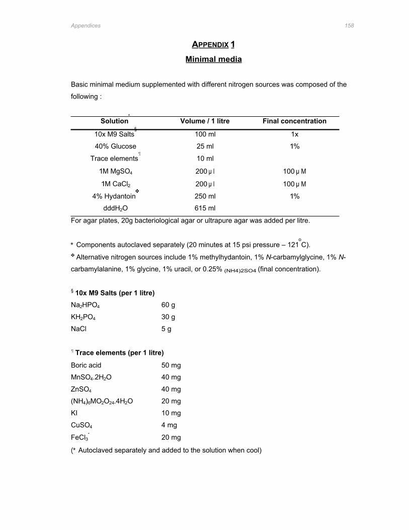
Appendices 158
APPENDIX 1 Minimal media
Basic minimal medium supplemented with different nitrogen sources was composed of the
following :
Solution∗
Volume / 1 litre Final concentration
10x M9 Salts§
100 ml 1x
40% Glucose 25 ml 1%
Trace elements¶
10 ml
1M MgSO4 200 μ l 100 μ M
1M CaCl2 200 μ l 100 μ M
4% Hydantoin✜
250 ml 1%
dddH2O 615 ml
For agar plates, 20g bacteriological agar or ultrapure agar was added per litre.
∗ Components autoclaved separately (20 minutes at 15 psi pressure – 121oC).
✜ Alternative nitrogen sources include 1% methylhydantoin, 1% N-carbamylglycine, 1% N-
carbamylalanine, 1% glycine, 1% uracil, or 0.25% (NH4)2SO4 (final concentration).
§ 10x M9 Salts (per 1 litre) Na2HPO4 60 g
KH2PO4 30 g
NaCl 5 g
¶ Trace elements (per 1 litre)
Boric acid 50 mg
MnSO4.2H2O 40 mg
ZnSO4 40 mg
(NH4)6MO2O24.4H2O 20 mg
KI 10 mg
CuSO4 4 mg
FeCl3∗
20 mg
(∗ Autoclaved separately and added to the solution when cool)
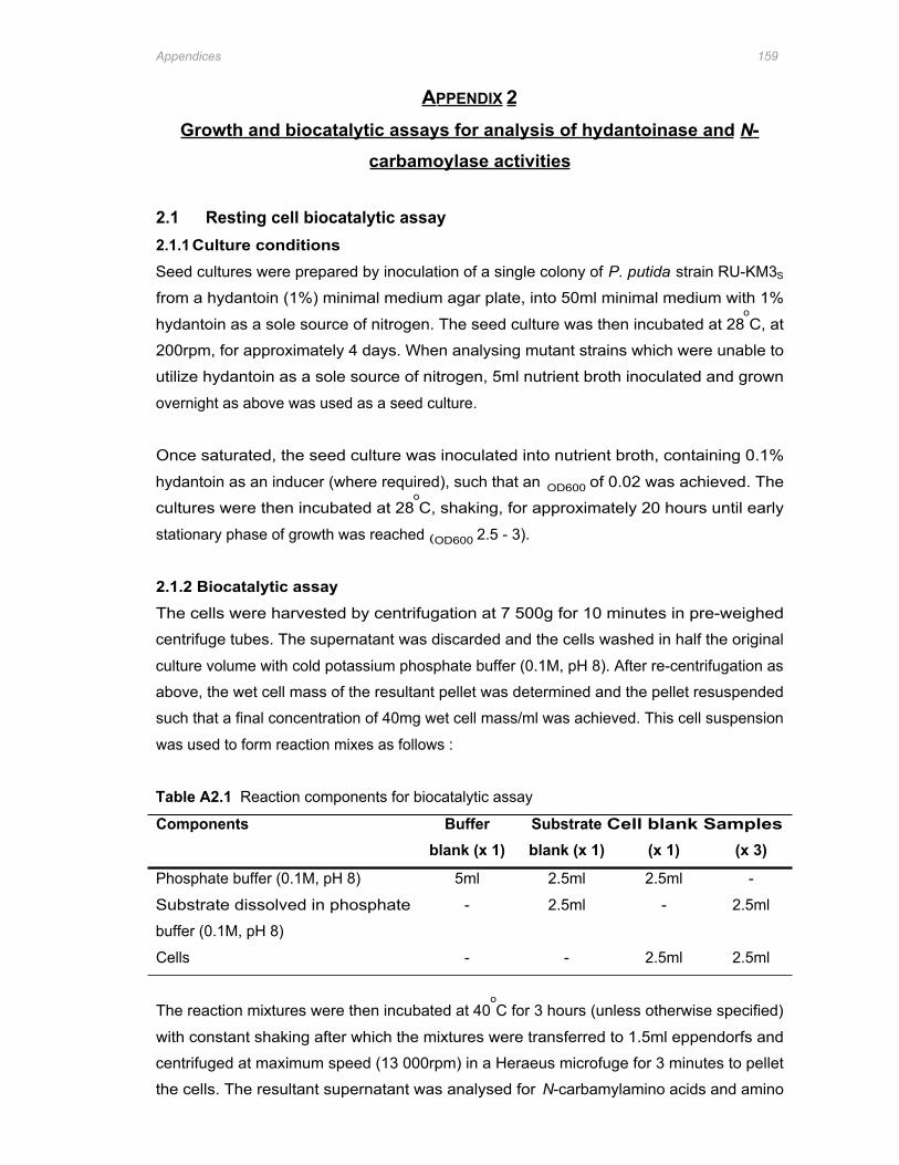
Appendices 159
APPENDIX 2 Growth and biocatalytic assays for analysis of hydantoinase and N-
carbamoylase activities
2.1 Resting cell biocatalytic assay 2.1.1 Culture conditions Seed cultures were prepared by inoculation of a single colony of P. putida strain RU-KM3S
from a hydantoin (1%) minimal medium agar plate, into 50ml minimal medium with 1%
hydantoin as a sole source of nitrogen. The seed culture was then incubated at 28oC, at
200rpm, for approximately 4 days. When analysing mutant strains which were unable to
utilize hydantoin as a sole source of nitrogen, 5ml nutrient broth inoculated and grown
overnight as above was used as a seed culture.
Once saturated, the seed culture was inoculated into nutrient broth, containing 0.1%
hydantoin as an inducer (where required), such that an OD600 of 0.02 was achieved. The
cultures were then incubated at 28oC, shaking, for approximately 20 hours until early
stationary phase of growth was reached (OD600 2.5 - 3).
2.1.2 Biocatalytic assay The cells were harvested by centrifugation at 7 500g for 10 minutes in pre-weighed
centrifuge tubes. The supernatant was discarded and the cells washed in half the original
culture volume with cold potassium phosphate buffer (0.1M, pH 8). After re-centrifugation as
above, the wet cell mass of the resultant pellet was determined and the pellet resuspended
such that a final concentration of 40mg wet cell mass/ml was achieved. This cell suspension
was used to form reaction mixes as follows :
Table A2.1 Reaction components for biocatalytic assay
Components Buffer Substrate Cell blank Samples blank (x 1) blank (x 1) (x 1) (x 3)
Phosphate buffer (0.1M, pH 8) 5ml 2.5ml 2.5ml -
Substrate dissolved in phosphate - 2.5ml - 2.5ml
buffer (0.1M, pH 8)
Cells - - 2.5ml 2.5ml
The reaction mixtures were then incubated at 40oC for 3 hours (unless otherwise specified)
with constant shaking after which the mixtures were transferred to 1.5ml eppendorfs and
centrifuged at maximum speed (13 000rpm) in a Heraeus microfuge for 3 minutes to pellet
the cells. The resultant supernatant was analysed for N-carbamylamino acids and amino
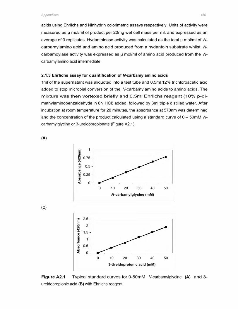
(C)
1
Abs
orba
nce
(420
nm)
0.75
0.5
0.25
0 0 10 20 30 40 50
N-carbamylglycine (mM)
2.5
Abs
orba
nce
(420
nm)
2
1.5
1
0.5
0 0 10 20 30 40 50
3-Ureidoproionic acid (mM)
Appendices 160
acids using Ehrlichs and Ninhydrin colorimetric assays respectively. Units of activity were
measured as μ mol/ml of product per 20mg wet cell mass per ml, and expressed as an
average of 3 replicates. Hydantoinase activity was calculated as the total μ mol/ml of N-
carbamylamino acid and amino acid produced from a hydantoin substrate whilst N-
carbamoylase activity was expressed as μ mol/ml of amino acid produced from the N-
carbamylamino acid intermediate.
2.1.3 Ehrlichs assay for quantification of N-carbamylamino acids 1ml of the supernatant was aliquoted into a test tube and 0.5ml 12% trichloroacetic acid
added to stop microbial conversion of the N-carbamylamino acids to amino acids. The
mixture was then vortexed briefly and 0.5ml Ehrlichs reagent (10% p-di-
methylaminobenzaldehyde in 6N HCl) added, followed by 3ml triple distilled water. After
incubation at room temperature for 20 minutes, the absorbance at 570nm was determined
and the concentration of the product calculated using a standard curve of 0 – 50mM N-
carbamylglycine or 3-ureidopropionate (Figure A2.1).
(A)
Figure A2.1 Typical standard curves for 0-50mM N-carbamylglycine (A) and 3-
ureidopropionic acid (B) with Ehrlichs reagent
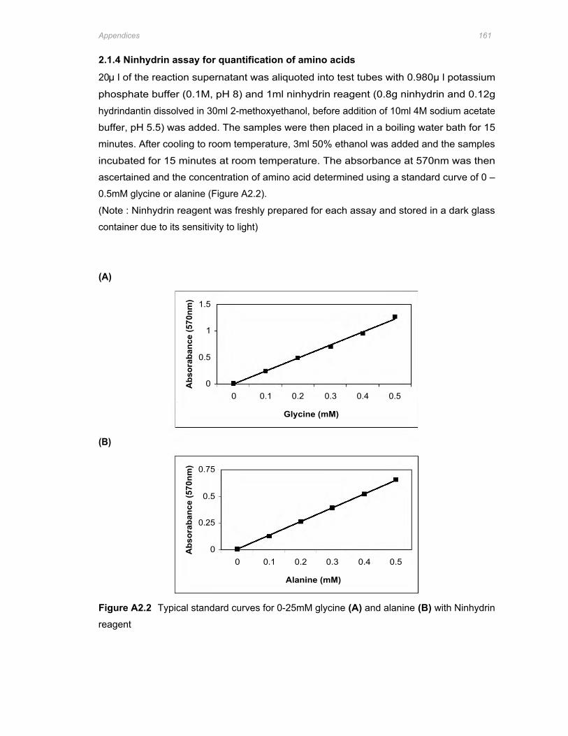
(B)
Abs
orab
ance
(570
nm) 1.5
1
0.5
0
Glycine (mM)
0 0.1 0.2 0.3 0.4 0.5
Abs
orab
ance
(570
nm) 0.75
0.5
0.25
0
Alanine (mM)
0 0.1 0.2 0.3 0.4 0.5
Appendices 161
2.1.4 Ninhydrin assay for quantification of amino acids
20μ l of the reaction supernatant was aliquoted into test tubes with 0.980μ l potassium
phosphate buffer (0.1M, pH 8) and 1ml ninhydrin reagent (0.8g ninhydrin and 0.12g
hydrindantin dissolved in 30ml 2-methoxyethanol, before addition of 10ml 4M sodium acetate
buffer, pH 5.5) was added. The samples were then placed in a boiling water bath for 15
minutes. After cooling to room temperature, 3ml 50% ethanol was added and the samples
incubated for 15 minutes at room temperature. The absorbance at 570nm was then
ascertained and the concentration of amino acid determined using a standard curve of 0 –
0.5mM glycine or alanine (Figure A2.2).
(Note : Ninhydrin reagent was freshly prepared for each assay and stored in a dark glass
container due to its sensitivity to light)
(A)
Figure A2.2 Typical standard curves for 0-25mM glycine (A) and alanine (B) with Ninhydrin
reagent
Appendices 162
2.2 Biocatalytic assays in microtitre plates Isolates to be assayed for hydantoinase or N-carbamoylase activity were inoculated directly
into microtitre plate wells containing 150μ l Luria broth supplemented with the appropriate
antibiotic and an inducer (1mM IPTG for heterologous expression in E. coli and 0.1%
hydantoin for P. putida strains) where necessary. The microtitre plates were then incubated
overnight at the optimum growth temperature of the strain. Alternatively, the strains were
cultured in 5ml Luria broth, overnight, at the optimal temperature for growth (37oC for E. coli
and 28oC for RU-KM3S) and then 200μ l aliquoted into the relevant wells in the microtitre
plates.
Harvesting the resting cells was accomplished by centrifugation of the microtitre plates at
4000rpm for 15 minutes in a Megafuge 1.0R (Heraeus) (rotor/bucket cat. #: 2705/2708). The
supernatant was then gently flicked out of the wells and the pellets resuspended in 200μ l
50mM hydantoin, dihydrouracil, or N-carbamylglycine in 0.1M phosphate buffer (pH 8) as
substrates (resuspension with 0.1M phosphate buffer alone functioned as a cell blank
control). The plates were then incubated at 40oC for 3 hours with shaking (100rpm) after
which the cells were pelleted once more by centrifugation as described above.
2.2.1 Ehrlich’s assay in microtitre plates
To determine the production of N-carbamylamino acid, 100μ l of the biocatalytic reaction
supernatant was transferred to a clean microtitre plate and 30μ l Ehrlich’s reagent (10% p-
dimethylaminobenzaldehyde in 6N HCl) added. After dilution with 100μ l distilled water, the
absorbance at a wavelength of 420nm was determined on the PowerwaveX Microtitre Plate
Reader (Bio-Tek Instruments, Inc.) and the N-carbamylamino acid concentration calculated
as μ mol/ml utilizing a standard curve of N-carbamylglycine (10 – 50 mM) prepared via same
method as the samples.
2.2.2 Ninhydrin assay in microtitre plates
Production of amino acids from the respective substrates was determined by aliquoting 10μ l
of the biocatalytic reaction supernatant from each well into a fresh microtitre plate to which
40μ l 0.1M phosphate buffer (pH 8) and 50μ l 4M sodium acetate buffer (pH 5.5) were added.
The plate was then incubated at 60oC for 5 minutes before adding 50μ l freshly prepared
ninhydrin solution (0.17g ninhydrin, 0.17g hydrindantin dissolved in 20ml 2-methoxyethanol).
Following incubation at 60oC for a further 20 minutes, 100μ l 50% ethanol was added to each
sample and the absorbance at a wavelength of 570nm was determined using the
PowerwaveX Microtitre Plate Reader. The amino acid concentration of each of the samples
was calculated as μ mol/ml utilizing a standard curve of glycine (0.1 – 0.5 mM) prepared via
the same method as the samples.
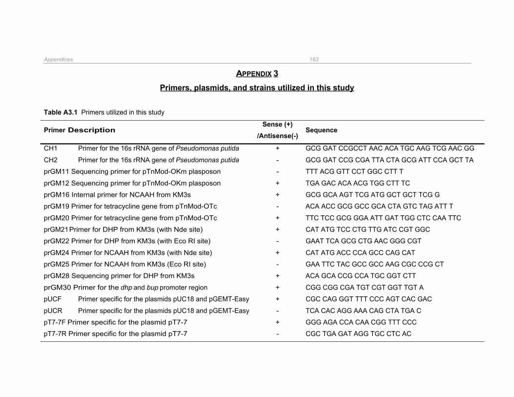
Appendices 163
APPENDIX 3 Primers, plasmids, and strains utilized in this study
Table A3.1 Primers utilized in this study
Primer Description Sense (+)
/Antisense(-) Sequence
CH1 Primer for the 16s rRNA gene of Pseudomonas putida + GCG GAT CCGCCT AAC ACA TGC AAG TCG AAC GG
CH2 Primer for the 16s rRNA gene of Pseudomonas putida - GCG GAT CCG CGA TTA CTA GCG ATT CCA GCT TA
prGM11 Sequencing primer for pTnMod-OKm plasposon - TTT ACG GTT CCT GGC CTT T
prGM12 Sequencing primer for pTnMod-OKm plasposon + TGA GAC ACA ACG TGG CTT TC
prGM16 Internal primer for NCAAH from KM3s + GCG GCA AGT TCG ATG GCT GCT TCG G
prGM19 Primer for tetracycline gene from pTnMod-OTc - ACA ACC GCG GCC GCA CTA GTC TAG ATT T
prGM20 Primer for tetracycline gene from pTnMod-OTc + TTC TCC GCG GGA ATT GAT TGG CTC CAA TTC
prGM21 Primer for DHP from KM3s (with Nde site) + CAT ATG TCC CTG TTG ATC CGT GGC
prGM22 Primer for DHP from KM3s (with Eco RI site) - GAAT TCA GCG CTG AAC GGG CGT
prGM24 Primer for NCAAH from KM3s (with Nde site) + CAT ATG ACC CCA GCC CAG CAT
prGM25 Primer for NCAAH from KM3s (Eco RI site) - GAA TTC TAC GCC GCC AAG CGC CCG CT
prGM28 Sequencing primer for DHP from KM3s + ACA GCA CCG CCA TGC GGT CTT
prGM30 Primer for the dhp and bup promoter region + CGG CGG CGA TGT CGT GGT TGT A
pUCF Primer specific for the plasmids pUC18 and pGEMT-Easy + CGC CAG GGT TTT CCC AGT CAC GAC
pUCR Primer specific for the plasmids pUC18 and pGEMT-Easy - TCA CAC AGG AAA CAG CTA TGA C
pT7-7F Primer specific for the plasmid pT7-7 + GGG AGA CCA CAA CGG TTT CCC
pT7-7R Primer specific for the plasmid pT7-7 - CGC TGA GAT AGG TGC CTC AC
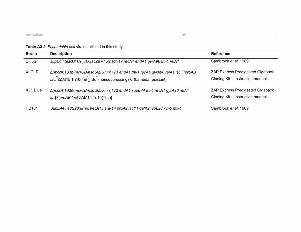
Appendices 164
Table A3.2 Escherichia coli strains utilized in this study
Strain Description Reference
DH5α supE44 ∆lacU169(∅80lacZΔM15)hsdR17 recA1 endA1 gyrA96 thi-1 relA1 Sambrook et al. 1989
XLOLR ∆(mcrA)183∆(mcrCB-hsdSMR-mrr)173 endA1 thi-1 recA1 gyrA96 relA1 lac[F’proAB ZAP Express Predigested Gigapack
lacIqZΔM15 Tn10(Tet
r)] Su
- (nonsuppressing) λ
r (Lambda resistant) Cloning Kit – Instruction manual
XL1 Blue ∆(mcrA)183∆(mcrCB-hsdSMR-mrr)173 endA1 supE44 thi-1 recA1 gyrA96 relA1 ZAP Express Predigested Gigapack
lac[F’proAB lacIqZΔM15 Tn10(Tet
r)] Cloning Kit – Instruction manual
HB101 SupE44 hsdS20(rB-
mB-
)recA13 ara-14 proA2 lacY1 galK2 rspL20 xyl-5 mtl-1 Sambrook et al. 1989
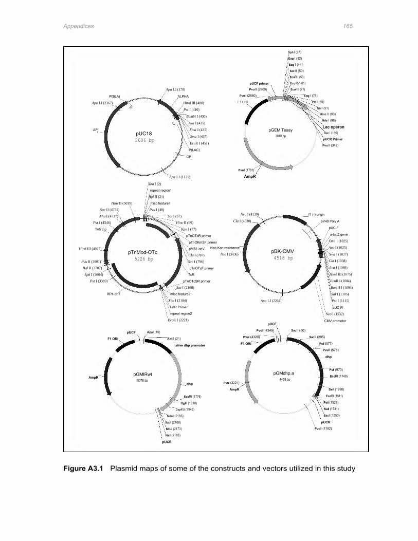
Hind III (4027)
Pvu II (3801)
Bgl II (3707)
Sph I (3604)
AmpR
Pst I (3389)
Apa LI (2367)
APr
Pst I (4546)
Tn5 tnp
Xho I (4737)
Sac II (4771)
RP4 oriT
F1 ORI
P(BLA)
Hinc II (5039)
pUCF
pTnMod-OTc 5226 bp
pGMIRwt 5076 bp
2686 bp pUC18
Apa I (15)
Xba I (2)
Bgl II (21)
Pvu I (48)
repeat region1
misc feature1
pUCR
SacI (2168)
NsiI (2186)
Apa LI (178)
MluI (2173)
AatII (21)
Nde I (2156)
Sal I (67)
EcoR I (2221)
Xba I (2184)
Apa LI (1121)
TetR Primer
misc feature2
repeat region2
native dhp promoter
Csp45I (1942)
Hinc II (69)
Sac I (2168)
ALPHA
BglII (1810)
Kpn I (77)
Hind III (400)
Pst I (416)
pTnOTcSR primer
EcoRI (1776)
BamH I (430)
pTnOTcR primer
P(LAC)
ORI
dhp
Sac I (796)
Ava I (435)
TcR
Cla I (787)
pTnOKmSF primer
pMB1 oriV
Xma I (435)
EcoR I (451)
pTnOTcF primer
Sma I (437)
Neo-Kan resistance
Nco I (3436)
PvuI (3221)
AmpR
Cla I (4030)
F1 ORI
Pvu I (1781)
Pvu I (2880)
Nco I (4139)
F1 ORI
PvuI (4320)
AmpR
Pvu II (2909)
pUCF primer
PvuII (4349)
Apa LI (2264)
pUCF
pGEM Teasy 3018 bp
pBK-CMV 4518 bp
pGMdhp.a 4458 bp
Sph I (27)
Eag I (32)
Eag I (44)
Sac II (50)
EcoR I (53)
Eco RV (61)
EcoR I (71)
Sac II (50)
Sac II (295)
Eag I (78)
f1 (-) origin
SV40 Poly A
pUC F
Pst I (89)
Sal I (91)
Hinc II (93)
Nde I (98)
Lac operon Sac I (110)
PvuII (1782)
dhp
PstI (577)
pUCR
Pvu II (342)
PvuII (578)
pUCR Primer
PstI (1529)
Sac I (1550)
SalI (1531)
CMV promotor
Nco I (1532)
EcoRI (1511)
Xma I (1025)
Ava I (1025)
Sal I (1290)
Sma I (1027)
Cla I (1038)
Ava I (1069)
Hind III (1075)
PstI (970)
EcoR I (1084)
a-lacZ gene
EcoRI (1140)
BamH I (1093)
Sal I (1105)
Pst I (1115)
pUC R
Appendices 165
Figure A3.1 Plasmid maps of some of the constructs and vectors utilized in this study
Appendices 166
APPENDIX 4 Preparation and transformation of competent E. coli cells
4.1 Preparation of competent Escherichia coli cells A test tube containing 5ml of Luria broth was inoculated with the appropriate strain of E. coli
and grown to confluence overnight at 37oC, shaking at 200rpm. Four Erlenmeyer flasks,
containing 100ml Luria broth each, were then inoculated with 1.5, 1.0, 0.7, and 0.3ml of the
overnight culture respectively and incubated at 37oC for approximately 2 hours until an OD600
of 0.6-0.8 was obtained with the culture pre-inoculated with 1.5ml overnight culture. The
flasks were then cooled for 5 to 10 minutes on ice, after which the flask contents were
processed separately until the final step. The flask contents were centrifuged in a Beckman
centrifuge (JA14 rotor) at 5000rpm, for 10 minutes, at 4oC. The supernatant was discarded
and the pellet resuspended in 50ml RF1 (100mM KCl, 50mM MnCl2, 30mM CH3COOK,
10mM CaCl2, 15% glycerol, pH5.8), followed by a further 20 minute incubation on ice. The
cells were once again pelleted by centrifugation as above and the supernatant discarded. All
four pellets were then resuspended together in a final volume of 4ml of RF2 (10mM MOPS,
10mM KCl, 75 mM CaCl2, 15% glycerol, pH 6.8). The resuspended cells were frozen and
stored at –80oC in 500μ l aliquots until required.
4.2 Transformation of competent E. coli cells Once thawed on ice, 150μ l competent E. coli cells were mixed with the plasmid DNA in a
sterile eppendorf and incubated on ice for 20 minutes. The cells were then subjected to
heat-shock by incubation at 42oC for 45 seconds immediately followed by placement on ice
for a further 5 minutes. 1ml cold Luria broth was then added and the cells incubated at 37oC
for 1 hour after which the cells were spread plated onto the appropriate selective medium.
Appendices 167
APPENDIX 5 DNA manipulation
5.1 Plasmid DNA extraction For initial screening purposes, selected recombinant E. coli colonies were cultured overnight
at 37oC, with shaking, in 5ml Luria broth supplemented with the appropriate antibiotic.
Plasmid DNA was extracted from these confluent cultures by the “Easyprep” method as
described in Berghammer and Auer (1993). Screening for the correct plasmid construct was
done by diagnostic restriction endonuclease digestion and electophoretic separation of the
resultant fragments through a 1% agarose gel containing 0.5μ g/ml ethidium bromide. The
buffer used for preparation and running of the agarose gel was 1x TAE (50x TAE (1L): 242g
Tris HCl, 57.1ml glacial acetic acid, 100ml 0.5M Na2EDTA, pH 8.0). Visualization of the DNA
was achieved by fluorescence of the DNA bound ethidium bromide under UV light and an
image captured using the Kodak DC 120 Gel Imaging System. Once a suitable culture
containing the correct plasmid construct was identified, high quality plasmid DNA was
extracted using the High Pure Plasmid Isolation Kit (Roche) or the Quantum Prep Plasmid
Midipreps (Qiagen) according to the manufacturers specifications. This pure DNA was then
used for in depth analysis and further manipulation.
5.2 Genomic DNA extraction Genomic DNA was extracted by the detergent lysis/CTAB and organic solvent extraction
method as described by Ausubel et al. (1983). The resultant extract was then treated with
10μ g/ml RNase A to degrade contaminating RNA and the remaining DNA precipitated by
addition of 0.1 volume 3M sodium acetate (pH 5.2) and 2 volumes ice cold 96% rectified
ethanol at –20oC for 18 hours. The precipitated DNA was pelleted by centrifugation
(13,000rpm in a Heraeus microfuge) for 10 minutes, washed two to three times with ice cold
(-20oC) 70% ethanol and the pellet dried by vacuum centrifugation (Speedvac concentrator,
Savant) before being resuspended in 50 to 100μ l TE buffer (pH 7). The extracted
chromosomal DNA concentration was estimated by visual comparison to phage Lambda
DNA standards after electrophoresis in a 1% agarose gel containing 0.5μ g/ml ethidium
bromide and visualization by UV light using the Kodak DC 120 Digital Imaging System.
Appendices 168
5.3 PCR thermal cycling paramerters
Initial denaturation: 94oC 90s
Denaturation: 94oC 45s
Annealing: ΧoC 45s
Extension: 72oC 60s
Final extension: 72oC 2min
ΧoC : Annealing temperatures – 63
oC (Chapter 2, Section 2.2.5)
57oC (Chapter 3, Section 3.2.6)
45oC (Chapter 5, Section 5.2.3)
(primers prGM24, prGM11, prGM28)
60oC (Chapter 5, Section 5.2.3)
(primers prGM19, prGM20)
55oC (Chapter 5, Section 5.2.4)
5.4 Determination of DNA sequence The nucleotide sequence of DNA was determined from double stranded plasmid DNA by the
dideoxy-chain termination method (Sanger, 1987) using the ABI Prism Big Dye Terminator
Cycle Sequencing Ready Reaction Kit v1.0, v2.0, or v3.1 (PE Applied Biosystems).
Sequencing primers utilized are listed in Appendix 3. The thermal cycling reaction
parameters were performed according to the manufacturers instruction for the GeneAmp
PCR System 9700 thermal cycler. Resultant extension products were purified by ion
exchange chromatography using the DNA Clean and Concentrator columns (Zymo), and the
sample dried by vacuum centrifugation (Speedvac concentrator, Savant). Pellets were
resuspended in Template Suppression Reagent and the nucleotide extension products
separated by capillary electrophoresis using an ABI Prism 3100 Genetic Analyser (Hitachi,
Applied Biosystems) and POP6 polymer in a 30cm or 50cm capillary.
X1
X30
X1
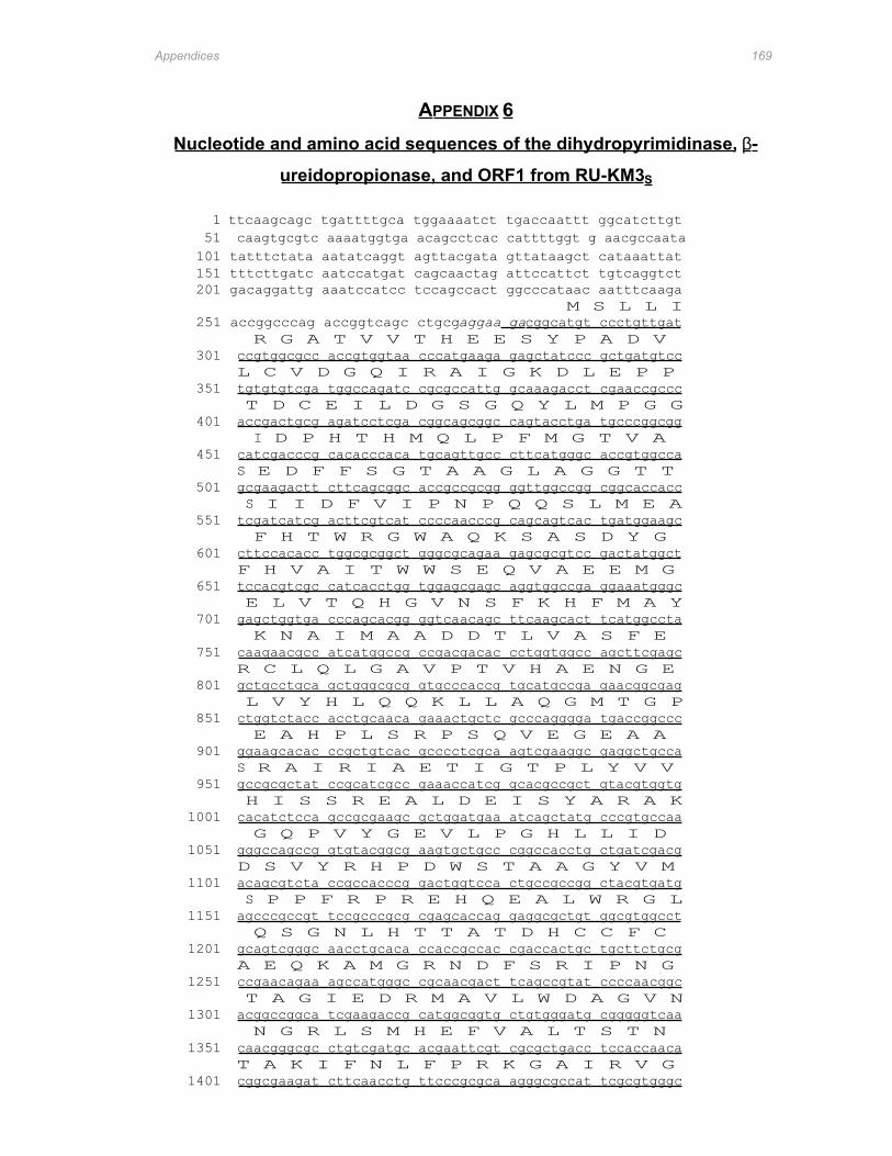
Appendices 169
APPENDIX 6
Nucleotide and amino acid sequences of the dihydropyrimidinase, β-
ureidopropionase, and ORF1 from RU-KM3 S
1 ttcaagcagc tgattttgca tggaaaatct tgaccaattt ggcatcttgt 51 caagtgcgtc aaaatggtga acagcctcac cattttggt g aacgccaata
101 tatttctata aatatcaggt agttacgata gttataagct cataaattat 151 tttcttgatc aatccatgat cagcaactag attccattct tgtcaggtct 201 gacaggattg aaatccatcc tccagccact ggcccataac aatttcaaga
M S L L I 251 accggcccag accggtcagc ctgcgaggaa gacggcatgt ccctgttgat
R G A T V V T H E E S Y P A D V 301 ccgtggcgcc accgtggtaa cccatgaaga gagctatccc gctgatgtcc
L C V D G Q I R A I G K D L E P P 351 tgtgtgtcga tggccagatc cgcgccattg gcaaagacct cgaaccgccc
T D C E I L D G S G Q Y L M P G G 401 accgactgcg agatcctcga cggcagcggc cagtacctga tgcccggcgg
I D P H T H M Q L P F M G T V A 451 catcgacccg cacacccaca tgcagttgcc cttcatgggc accgtggcca
S E D F F S G T A A G L A G G T T 501 gcgaagactt cttcagcggc accgccgcgg ggttggccgg cggcaccacc
S I I D F V I P N P Q Q S L M E A 551 tcgatcatcg acttcgtcat ccccaacccg cagcagtcac tgatggaagc
F H T W R G W A Q K S A S D Y G 601 cttccacacc tggcgcggct gggcgcagaa gagcgcgtcc gactatggct
F H V A I T W W S E Q V A E E M G 651 tccacgtcgc catcacctgg tggagcgagc aggtggccga ggaaatgggc
E L V T Q H G V N S F K H F M A Y 701 gagctggtga cccagcacgg ggtcaacagc ttcaagcact tcatggccta
K N A I M A A D D T L V A S F E 751 caagaacgcc atcatggccg ccgacgacac cctggtggcc agcttcgagc
R C L Q L G A V P T V H A E N G E 801 gctgcctgca gctgggcgcg gtgcccaccg tgcatgccga gaacggcgag
L V Y H L Q Q K L L A Q G M T G P 851 ctggtctacc acctgcaaca gaaactgctc gcccagggga tgaccggccc
E A H P L S R P S Q V E G E A A 901 ggaagcacac ccgctgtcac gcccctcgca agtcgaaggc gaggctgcca
S R A I R I A E T I G T P L Y V V 951 gccgcgctat ccgcatcgcc gaaaccatcg gcacgccgct gtacgtggtg
H I S S R E A L D E I S Y A R A K 1001 cacatctcca gccgcgaagc gctggatgaa atcagctatg cccgtgccaa
G Q P V Y G E V L P G H L L I D 1051 gggccagccg gtgtacggcg aagtgctgcc cggccacctg ctgatcgacg
D S V Y R H P D W S T A A G Y V M 1101 acagcgtcta ccgccacccg gactggtcca ctgccgccgg ctacgtgatg
S P P F R P R E H Q E A L W R G L 1151 agcccgccgt tccgcccgcg cgagcaccag gaggcgctgt ggcgtggcct
Q S G N L H T T A T D H C C F C 1201 gcagtcgggc aacctgcaca ccaccgccac cgaccactgc tgcttctgcg
A E Q K A M G R N D F S R I P N G 1251 ccgaacagaa agccatgggc cgcaacgact tcagccgtat ccccaacggc
T A G I E D R M A V L W D A G V N 1301 acggccggca tcgaagaccg catggcggtg ctgtgggatg cgggggtcaa
N G R L S M H E F V A L T S T N 1351 caacgggcgc ctgtcgatgc acgaattcgt cgcgctgacc tccaccaaca
T A K I F N L F P R K G A I R V G 1401 cggcgaagat cttcaacctg ttcccgcgca agggcgccat tcgcgtgggc
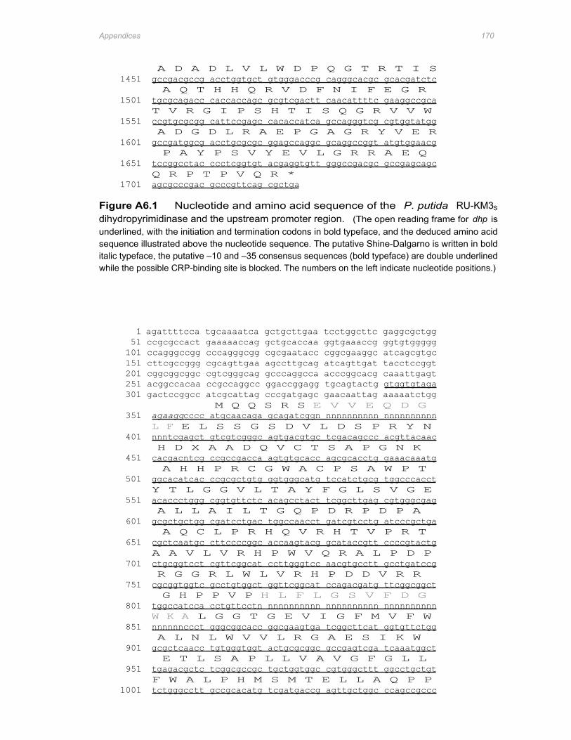
Appendices 170
A D A D L V L W D P Q G T R T I S 1451 gccgacgccg acctggtgct gtgggacccg cagggcacgc gcacgatctc
A Q T H H Q R V D F N I F E G R 1501 tgcgcagacc caccaccagc gcgtcgactt caacattttc gaaggccgca
T V R G I P S H T I S Q G R V V W 1551 ccgtgcgcgg cattccgagc cacaccatca gccagggtcg cgtggtatgg
A D G D L R A E P G A G R Y V E R 1601 gccgatggcg acctgcgcgc ggagccaggc gcaggccggt atgtggaacg
P A Y P S V Y E V L G R R A E Q 1651 tccggcctac ccctcggtgt acgaggtgtt gggccgacgc gccgagcagc
Q R P T P V Q R * 1701 agcgcccgac gcccgttcag cgctga
Figure A6.1 Nucleotide and amino acid sequence of the P. putida RU-KM3S
dihydropyrimidinase and the upstream promoter region. (The open reading frame for dhp is underlined, with the initiation and termination codons in bold typeface, and the deduced amino acid sequence illustrated above the nucleotide sequence. The putative Shine-Dalgarno is written in bold italic typeface, the putative –10 and –35 consensus sequences (bold typeface) are double underlined while the possible CRP-binding site is blocked. The numbers on the left indicate nucleotide positions.)
1 agattttcca tgcaaaatca gctgcttgaa tcctggcttc gaggcgctgg 51 ccgcgccact gaaaaaccag gctgcaccaa ggtgaaaccg ggtgtggggg 101 ccagggccgg cccagggcgg cgcgaatacc cggcgaaggc atcagcgtgc 151 cttcgccggg cgcagttgaa agccttgcag atcagttgat tacctccggt 201 cggcggcggc cgtcgggcag gcccaggcca acccggcacg caaattgagt 251 acggccacaa ccgccaggcc ggaccggagg tgcagtactg gtggtgtaga 301 gactccggcc atcgcattag cccgatgagc gaacaattag aaaaatctgg
M Q Q S R S E V V E Q D G 351 agaaggcccc atgcaacaga gcagatcggn nnnnnnnnnn nnnnnnnnnn
L F E L S S G S D V L D S P R Y N 401 nnntcgagct gtcgtcgggc agtgacgtgc tcgacagccc acgttacaac
H D X A A D Q V C T S A P G N K 451 cacgacntcg ccgccgacca agtgtgcacc agcgcacctg gaaacaaatg
A H H P R C G W A C P S A W P T 501 ggcacatcac ccgcgctgtg ggtgggcatg tccatctgcg tggcccacct
Y T L G G V L T A Y F G L S V G E 551 acaccctggg cggtgttctc acagcctact tcggcttgag cgtgggcgag
A L L A I L T G Q P D R P D P A 601 gcgctgctgg cgatcctgac tggccaacct gatcgtcctg atcccgctga
A Q C L P R H Q V R H T V P R T 651 cgctcaatgc cttccccggc accaagtacg gcataccgtt ccccgtactg
A A V L V R H P W V Q R A L P D P 701 ctgcggtcct cgttcggcat ccttgggtcc aacgtgcctt gcctgatccg
R G G R L W L V R H P D D V R R 751 cgcggtggtc gcctgtggct ggttcggcat ccagacgatg ttcggcggct
G H P P V P H L F L G S V F D G 801 tggccatcca cctgttcctn nnnnnnnnnn nnnnnnnnnn nnnnnnnnnn
W K A L G G T G E V I G F M V F W 851 nnnnnnccct gggcggcacc ggcgaagtga tcggcttcat ggtgttctgg
A L N L W V V L R G A E S I K W 901 gcgctcaacc tgtgggtggt actgcgcggc gccgagtcga tcaaatggct
E T L S A P L L V A V G F G L L 951 tgagacgctc tcggcgccgc tgctggtggc cgtgggcttt ggcctgctgt
F W A L P H M S M T E L L A Q P P 1001 tctgggcctt gccgcacatg tcgatgaccg agttgctggc ccagccgccc
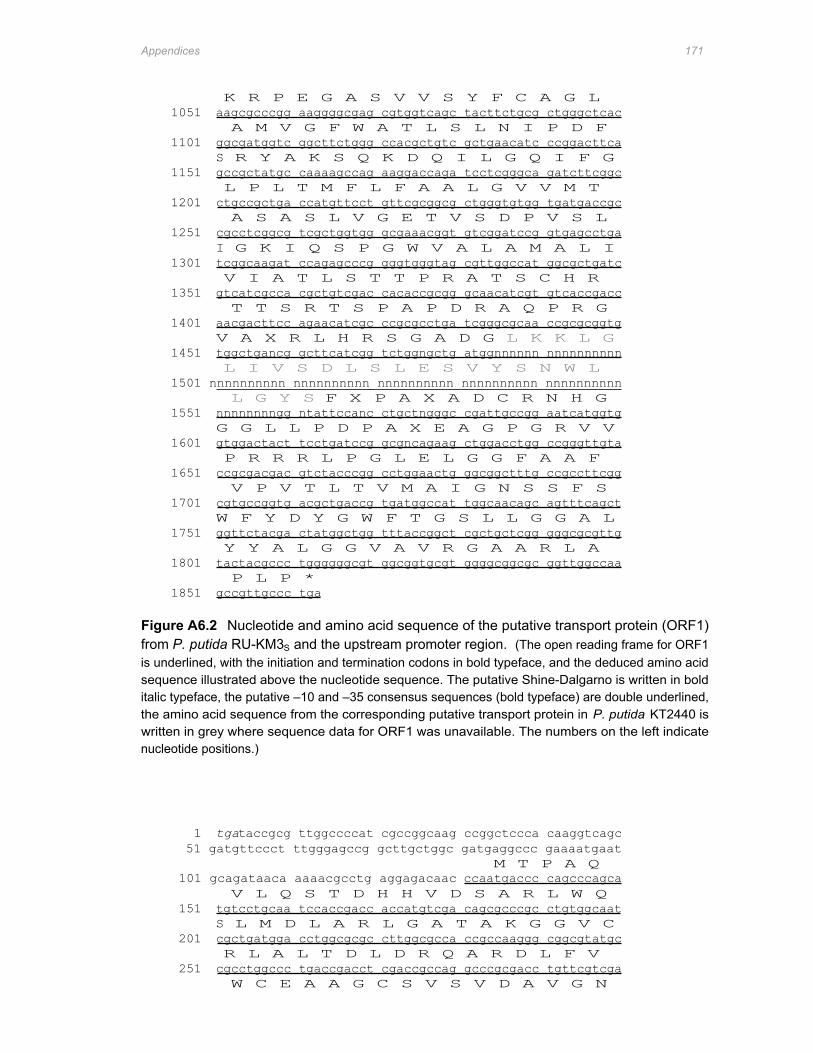
Appendices 171
K R P E G A S V V S Y F C A G L 1051 aagcgcccgg aaggggcgag cgtggtcagc tacttctgcg ctgggctcac
A M V G F W A T L S L N I P D F 1101 ggcgatggtc ggcttctggg ccacgctgtc gctgaacatc ccggacttca
S R Y A K S Q K D Q I L G Q I F G 1151 gccgctatgc caaaagccag aaggaccaga tcctcgggca gatcttcggc
L P L T M F L F A A L G V V M T 1201 ctgccgctga ccatgttcct gttcgcggcg ctgggtgtgg tgatgaccgc
A S A S L V G E T V S D P V S L 1251 cgcctcggcg tcgctggtgg gcgaaacggt gtcggatccg gtgagcctga
I G K I Q S P G W V A L A M A L I 1301 tcggcaagat ccagagcccg gggtgggtag cgttggccat ggcgctgatc
V I A T L S T T P R A T S C H R 1351 gtcatcgcca cgctgtcgac cacaccgcgg gcaacatcgt gtcaccgacc
T T S R T S P A P D R A Q P R G 1401 aacgacttcc agaacatcgc ccgcgcctga tcgggcgcaa ccgcgcggtg
V A X R L H R S G A D G L K K L G 1451 tggctgancg gcttcatcgg tctggngctg atggnnnnnn nnnnnnnnnn
L I V S D L S L E S V Y S N W L 1501 nnnnnnnnnn nnnnnnnnnn nnnnnnnnnn nnnnnnnnnn nnnnnnnnnn
L G Y S F X P A X A D C R N H G 1551 nnnnnnnngg ntattccanc ctgctngggc cgattgccgg aatcatggtg
G G L L P D P A X E A G P G R V V 1601 gtggactact tcctgatccg gcgncagaag ctggacctgg ccgggttgta
P R R R L P G L E L G G F A A F 1651 ccgcgacgac gtctacccgg cctggaactg ggcggctttg ccgccttcgg
V P V T L T V M A I G N S S F S 1701 cgtgccggtg acgctgaccg tgatggccat tggcaacagc agtttcagct
W F Y D Y G W F T G S L L G G A L 1751 ggttctacga ctatggctgg tttaccggct cgctgctcgg gggcgcgttg
Y Y A L G G V A V R G A A R L A 1801 tactacgccc tggggggcgt ggcggtgcgt ggggcggcgc ggttggccaa
P L P * 1851 gccgttgccc tga
Figure A6.2 Nucleotide and amino acid sequence of the putative transport protein (ORF1) from P. putida RU-KM3S and the upstream promoter region. (The open reading frame for ORF1 is underlined, with the initiation and termination codons in bold typeface, and the deduced amino acid sequence illustrated above the nucleotide sequence. The putative Shine-Dalgarno is written in bold italic typeface, the putative –10 and –35 consensus sequences (bold typeface) are double underlined, the amino acid sequence from the corresponding putative transport protein in P. putida KT2440 is written in grey where sequence data for ORF1 was unavailable. The numbers on the left indicate nucleotide positions.)
1 tgataccgcg ttggccccat cgccggcaag ccggctccca caaggtcagc 51 gatgttccct ttgggagccg gcttgctggc gatgaggccc gaaaatgaat
M T P A Q 101 gcagataaca aaaacgcctg aggagacaac ccaatgaccc cagcccagca
V L Q S T D H H V D S A R L W Q 151 tgtcctgcaa tccaccgacc accatgtcga cagcgcccgc ctgtggcaat
S L M D L A R L G A T A K G G V C 201 cgctgatgga cctggcgcgc cttggcgcca ccgccaaggg cggcgtatgc
R L A L T D L D R Q A R D L F V 251 cgcctggccc tgaccgacct cgaccgccag gcccgcgacc tgttcgtcga
W C E A A G C S V S V D A V G N
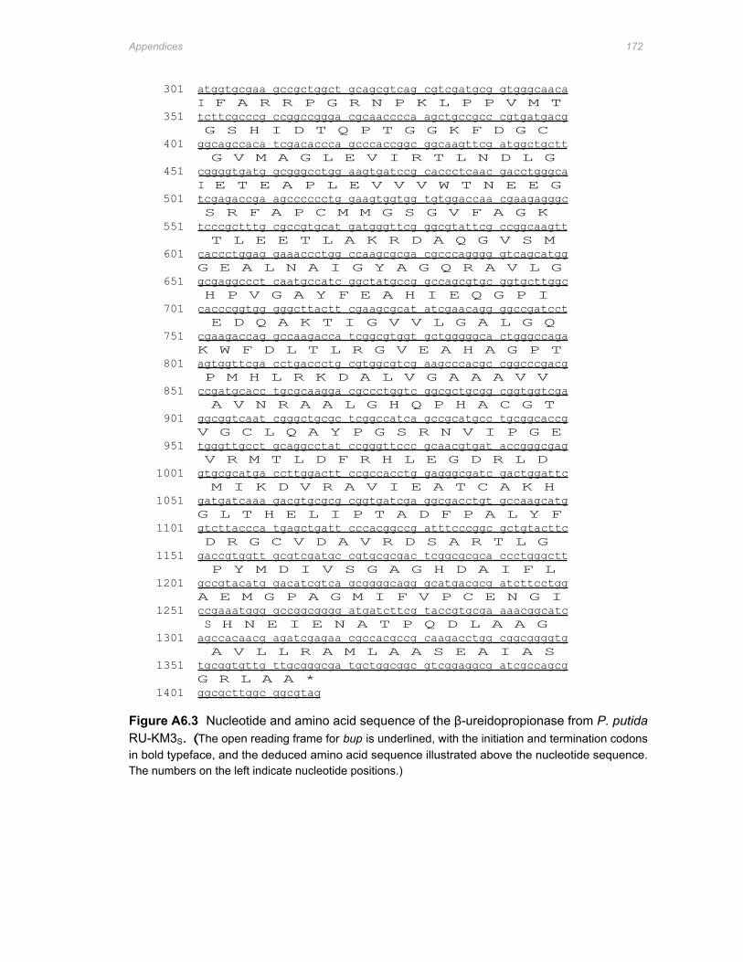
Appendices 172
301 atggtgcgaa gccgctggct gcagcgtcag cgtcgatgcg gtgggcaaca I F A R R P G R N P K L P P V M T
351 tcttcgcccg ccggccggga cgcaacccca agctgccgcc cgtgatgacg G S H I D T Q P T G G K F D G C
401 ggcagccaca tcgacaccca gcccaccggc ggcaagttcg atggctgctt G V M A G L E V I R T L N D L G
451 cggggtgatg gcgggcctgg aagtgatccg caccctcaac gacctgggca I E T E A P L E V V V W T N E E G
501 tcgagaccga agcccccctg gaagtggtgg tgtggaccaa cgaagagggc S R F A P C M M G S G V F A G K
551 tcccgctttg cgccgtgcat gatgggttcg ggcgtattcg ccggcaagtt T L E E T L A K R D A Q G V S M
601 caccctggag gaaaccctgg ccaagcgcga cgcccagggg gtcagcatgg G E A L N A I G Y A G Q R A V L G
651 gcgaggccct caatgccatc ggctatgccg gccagcgtgc ggtgcttggc H P V G A Y F E A H I E Q G P I
701 cacccggtgg gggcttactt cgaagcgcat atcgaacagg ggccgatcct E D Q A K T I G V V L G A L G Q
751 cgaagaccag gccaagacca tcggcgtggt gctgggggca ctgggccaga K W F D L T L R G V E A H A G P T
801 agtggttcga cctgaccctg cgtggcgtcg aagcccacgc cggcccgacg P M H L R K D A L V G A A A V V
851 ccgatgcacc tgcgcaagga cgccctggtc ggcgctgcgg cggtggtcga A V N R A A L G H Q P H A C G T
901 ggcggtcaat cgggctgcgc tcggccatca gccgcatgcc tgcggcaccg V G C L Q A Y P G S R N V I P G E
951 tgggttgcct gcaggcctat ccgggttccc gcaacgtgat accgggcgag V R M T L D F R H L E G D R L D
1001 gtgcgcatga ccttggactt ccgccacctg gagggcgatc gactggattc M I K D V R A V I E A T C A K H
1051 gatgatcaaa gacgtgcgcg cggtgatcga ggcgacctgt gccaagcatg G L T H E L I P T A D F P A L Y F
1101 gtcttaccca tgagctgatt cccacggccg atttcccggc gctgtacttc D R G C V D A V R D S A R T L G
1151 gaccgtggtt gcgtcgatgc cgtgcgcgac tcggcgcgca ccctgggctt P Y M D I V S G A G H D A I F L
1201 gccgtacatg gacatcgtca gcggggcagg gcatgacgcg atcttcctgg A E M G P A G M I F V P C E N G I
1251 ccgaaatggg gccggcgggg atgatcttcg taccgtgcga aaacggcatc S H N E I E N A T P Q D L A A G
1301 agccacaacg agatcgagaa cgccacgccg caagacctgg cggcggggtg A V L L R A M L A A S E A I A S
1351 tgcggtgttg ttgcgggcga tgctggcggc gtcggaggcg atcgccagcg G R L A A *
1401 ggcgcttggc ggcgtag
Figure A6.3 Nucleotide and amino acid sequence of the β-ureidopropionase from P. putida RU-KM3S. (The open reading frame for bup is underlined, with the initiation and termination codons in bold typeface, and the deduced amino acid sequence illustrated above the nucleotide sequence. The numbers on the left indicate nucleotide positions.)
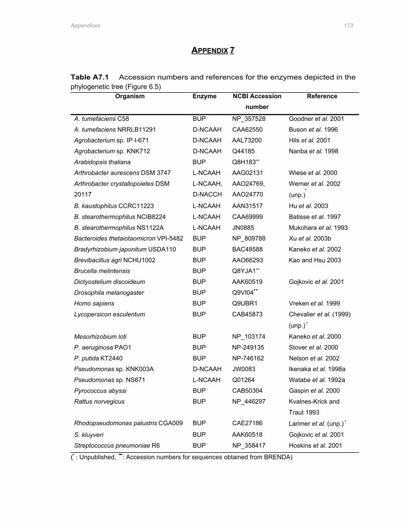
Appendices 173
APPENDIX 7
Table A7.1 Accession numbers and references for the enzymes depicted in the phylogenetic tree (Figure 6.5)
Organism Enzyme NCBI Accession number
Reference
A. tumefaciens C58 BUP NP_357528 Goodner et al. 2001
A. tumefaciens NRRLB11291 D-NCAAH CAA62550 Buson et al. 1996
Agrobacterium sp. IP I-671 D-NCAAH AAL73200 Hils et al. 2001
Agrobacterium sp. KNK712 D-NCAAH Q44185 Nanba et al. 1998
Arabidopsis thaliana BUP Q8H183~
Arthrobacter aurescens DSM 3747 L-NCAAH AAG02131 Wiese et al. 2000
Arthrobacter crystallopoietes DSM L-NCAAH, AAO24769, Werner et al. 2002
20117 D-NACCH AAO24770 (unp.)∗
B. kaustophilus CCRC11223 L-NCAAH AAN31517 Hu et al. 2003
B. stearothermophilus NCIB8224 L-NCAAH CAA69999 Batisse et al. 1997
B. stearothermophilus NS1122A L-NCAAH JN0885 Mukohara et al. 1993
Bacteroides thetaiotaomicron VPI-5482 BUP NP_809788 Xu et al. 2003b
Bradyrhizobium japonitum USDA110 BUP BAC48588 Kaneko et al. 2002
Brevibacillus agri NCHU1002 BUP AAO66293 Kao and Hsu 2003
Brucella melintensis BUP Q8YJA1~
Dictyostelium discoideum BUP AAK60519 Gojkovic et al. 2001
Drosophila melanogaster BUP Q9VI04''
Homo sapiens BUP Q9UBR1 Vreken et al. 1999
Lycopersicon esculentum BUP CAB45873 Chevalier et al. (1999)
(unp.) ∗
Mesorhizobium loti BUP NP_103174 Kaneko et al. 2000
P. aeruginosa PAO1 BUP NP-249135 Stover et al. 2000
P. putida KT2440 BUP NP-746162 Nelson et al. 2002
Pseudomonas sp. KNK003A D-NCAAH JW0083 Ikenaka et al. 1998a
Pseudomonas sp. NS671 L-NCAAH Q01264 Watabe et al. 1992a
Pyrococcus abyssi BUP CAB50304 Gaspin et al. 2000
Rattus norvegicus BUP NP_446297 Kvalnes-Krick and
Traut 1993
Rhodopseudomonas palustris CGA009 BUP CAE27186 Larimer et al. (unp.) ∗
S. kluyveri BUP AAK60518 Gojkovic et al. 2001
Streptococcus pneumoniae R6 BUP NP_358417 Hoskins et al. 2001
(∗ : Unpublished, ' : Accession numbers for sequences obtained from BRENDA)
References 174
REFERENCES
Abendroth J., Chatterjee S., & Schomburg D. (2000a) Purification of a D-hydantoinase using a laboratory-scale
streamline phenyl column as the initial step. Journal of Chromatography B 737, 187-194.
Abendroth J., Niefind K., Chatterjee S., & Schomburg D. (2000b) Crystallization, preliminary X-ray analysis of a
native and selenomethioneine D-hydantoinase from Thermus sp. Acta Crystallographica D56, 1166-1169.
Abendroth J., Niefind K., & Schomburg D. (2002a) X-ray structure of a dihydropyrimidinase from Thermus sp. at
1.3 A resolution. Journal of Molecular Biology 320, 143-156.
Abendroth J., Niefind K., May O., Siemann M., Syldatk C., & Schomburg D. (2002b) The structure of L-
hydantoinase from Arthrobacter aurescens leads to an understanding of dihydropyrimidinase substrate and
enantio selectivity. Biochemistry 41, 8589-8597
Achary A., Hariharan K.A., Bandhyopadhyaya S., Ramachandran R., & Jayaraman K. (1997) Application of
numerical modelling for the development of optimised complex medium for D-hydantoinase production from
Agrobacterium radiobacter NRRL B 11291. Biotechnology and Bioengineering 55, 148-154.
Altenbuchner J., Siemann-Herzberg M., & Syldatk C. (2001) Hydantoinases and related enzymes as biocatalysts
for the synthesis of unnatural chiral amino acids. Current Opinion in Biotechnology 12, 559-563.
Altschul S.F., Gish W., Miller W., Myers E.W. & Lipman D.J. (1990) Basic local alignment search tool. Journal of
Molecular Biology 215, 403-410
Arnold F.H. & Moore J.C. (1997) Optimisation industrial enzymes by directed evolution. Biochemical
Engineering/Biotechnology 58, 1-14.
Ausubel F.M., Brent R., Kingston R.E., Moore D.D., Seidman J.G., Smith J.A. & Struhl K. (1983) Current protocols
in molecular biology. Third Edition. Wiley-Interscience. New York.
Azerad R. (1995) Application of biocatalysis in organic synthesis. Bulletin de la Societe Chimique de France 132,
17-51
Baneyx F. (1999) Recombinant protein expression in Escherichia coli. Current Opinion in Biotechnology. 10, 411-
421
Batisse N., Weigel P., Lecocq M., & Sakanyan V. (1997) Two amino acid amidohydrolase genes encoding L-
stereospecific carbamoylase and aminoacylase are organised in a common operon in Bacillus
stearothermophilus. Applied and Environmental Microbiology 63, 763-766.
Bentley S.D., Chater K.F., Cerdeno-Tarraga A.M., Challis G.L., Thomson N.R., James K.D., Harris D.E., Quail
M.A., Kieser H., Harper D., Bateman A., Brown S., Chandra G., Chen C.W., Collins M., Cronin A., Fraser
A., Goble A., Hidalgo J., Hornsby T., Howarth S., Huang C.H., Kieser T., Larke L., Murphy L., Oliver K.,
O'Neil S., Rabbinowitsch E., Rajandream M.A., Rutherford K., Rutter S., Seeger K., Saunders D., Sharp S.,
Squares R., Squares S., Taylor K., Warren T., Wietzorrek A., Woodward J., Barrell B.G., Parkhill J., &
Hopwood D.A. (2002) Complete genome sequence of the model actinomycete Streptomyces coelicolor
A3(2). Nature 417, 141-147
References 175
Berghammer H. & Auer B. (1993) "Easypreps" : Fast and easy plasmid minipreparation for analysis of
recombinant clones in E. coli. Biotechniques 14, 527-528.
Bernheim F. & Bernheim M.L. (1946) The hydrolysis of hydantoin by various tissues. Journal of Biological
Chemistry 163, 683-685.
Bommarius A.S., Schwarm M., & Drauz K. (1998) Biocatalysis to amino acid-based chiralpharmacueticals -
examples and perspectives. Journal of Molecular Catalysis B: Enzymatic 5, 1-11.
Brunk C.F., Avaniss-Aghajani E., & Brunk C.A. (1996) A computer analysis of primer and probe hybridisation
potential with bacterial small-subunit rRNA sequences. Applied and Environmental Microbiology. 62, 872-
879.
Buchanan K. (1996) Optimisation of hydantoin biotransformations by RU-KM3S resting cells. Honours thesis,
Rhodes University
Buchanan K., Burton S., Dorrington R., Matcher G., Skepu Z. (2001) A novel Pseudomonas putida strain with high
levels of hydantoin-converting activity, producing L-amino acids. Journal of Molecular Catalysis B:
Enzymatic 11,397-406
Buell C.R., Joardar V., Lindeberg M., Selengut J., Paulsen I.T., Gwinn M.L., Dodson R.J., Deboy R.T., Durkin A.S.,
Kolonay J.F., Madupu R., Daugherty S., Brinkac L., Beanan M.J., Haft D.H., Nelson W.C., Davidsen T.,
Zafar N., Zhou L., Liu J., Yuan Q., Khouri H., Fedorova N., Tran B., Russell D., Berry K., Utterback T., Van
Aken S.E., Feldblyum T.V., D'Ascenzo M., Deng W.L., Ramos A.R., Alfano J.R., Cartinhour S., Chatterjee
A.K., Delaney T.P., Lazarowitz S.G., Martin G.B., Schneider D.J., Tang X., Bender C.L., White O., Fraser
C.M. & Collmer A. (2003) The complete genome sequence of the Arabidopsis and tomato pathogen
Pseudomonas syringae pv. tomato DC3000. Proceedings Of The National Academy Of Sciences Of The
United States Of America 100,10181-10186.
Burton S.G., Dorrington R.A., Hartley C., Kirchmann S., Matcher G., & Phehane V. (1998) Production of
enantiomerically pure amino acids: characterization of South African hydantoinases and hydantoinase-
producing bacteria. Journal of Molecular Catalysis B: Enzymatic 5, 301-305
Buson A., Negro A., Grassato L., Tagliaro M., Basaglia M., Grandi C., Fontana A., & Nuti M.P. (1996)
Identification, sequencing and mutagenesis of the gene for a D-carbamoylase from Agrobacterium
radiobacter. FEMS Microbiology Letters 145, 55-62.
Campbell L.L. (1958) Reductive degradation of pyrimidines. IV. Purification and properties of dihydrouracil
hydrase. Journal of Biological ChemistryI 233, 1236-1240
Capela D., Barloy-Hubler F., Gouzy J., Bothe G., Ampe F., Batut J., Boistard P., Becker A., Boutry M., Cadieu E.,
Dreano S., Gloux S., Godrie T., Goffeau A., Kahn D., Kiss E., Lelaure V., Masuy D., Pohl T., Portetelle D.,
Puehler A., Purnelle B., Ramsperger U., Renard C., Thebault P., Vandenbol M., Weidner S. & Galibert F.
(2001) Analysis of the chromosome sequence of the legume symbiont Sinorhizobium meliloti strain 1021.
Proceedings Of The National Academy Of Sciences Of The United States Of America. 98, 9877-9882
Cecere F., Galli G., & Morisi F. (1975) Substrate and steric specificity of hydropyrimidine hydrase. FEBS Letters
57 , 192-194.
References 176
Chao Y.P., Juang T., Chern J., & Lee C. (1999a) Production of D-p-hydroxyphenylglycine by N-carbamoyl-D-
amino acid amidohydrolase-overproducing Escherichia coli strains. Biotechnology Progress 15, 603-607.
Chao Y.P., Fu H., Lo T.E., Chen P.T., & Wang J. (1999b) One-step production of D-p-hydroxyphenylglycine by
recombinant Escherichia coli strains. Biotechnology Progress 15, 1039-1045.
Chao Y.P., Chiang C.J., Lo T.E., & Fu H. (2000a) Overproduction of D-hydantoinase and carbamoylase in a
soluble form in Escherichia coli. Applied Microbiology and Biotechnology 54, 348-353.
Chao Y.P., Chiang C.J., & Chen P.T. (2000b) Optimum ration of D-carbamoylase to D-hydantoinase for
maximizing D-p-hydroxyphenylglycine productivity. Biotechnology Letters 99-103.
Chen H. & Tsai H. (1998) Cloning, sequencing and expression in Escherichia coli of D-hydantoinase gene from
Pseudomonas putida. Annals of the New York Academy of Sciences 864, 234-237.
Chen C., Chiu W., Liu J., Hsu W., & Wang W. (2003) Structural basis for catalysis and substrate specificity of
Agrobacterium radiobacter N-carbamoyl-D-amino acid amidohydrolase. Journal of Biological Chemistry
278, 26194-26201.
Chevalier C., Joubes J., Petit J. & Raymond P. (1999) Isolation and characterization of a cDNA clone for a
putative beta-alanine synthase from tomato (Lycopersicon esculentum Mill.)developing fruits. Unpublished
Cheon Y.H., Kim H.S., Han K.H., Abendroth J., Niefind K., Schomburg D., Wang J., & Kim Y. (2002) Crystal
structure of D-hydantoinase from Bacillus stearothermophilus :Insight into the stereochemistry of
enantioselectivity. Biochemistry 41, 9410-9417.
Chien H.R., Jih Y., Yang W., & Hsu W. (1998) Identification of the open reading frame for the Pseudomonas putida
D-hydantoinase gene and expression of the gene in Escherichia coli. Biochimica et Biophysica Acta 1395,
68-77.
Chung J., Back J., Lim J., Park Y., & Han Y. (2002) Thermostable hydantoinase from a hyperthermophilic archeon,
Methanococcus jannaschii . Enzyme and Microbial Technology 30, 874.
Cole J, Chai B, Marsh T, Farris R, Wang Q, Kulam S, Chandra S, McGarrell D, Schmidt T, Garrity G, Tiedje J
(2003) The Ribosomal Database Project (RDP-II): Previewing a new autoaligner that allows regular
updates and the new prokaryotic taxonomy. Nucleic Acids Research 31:442-443
Collier D.N., Hager P.W. & Phibbs P.V. (1996) Catabolite repression control in the Pseudomonads. Research in
Microbiology 147:551-561
Collier D., Spence C., Cox M. & Phibbs P. (2001) Isolation and phenotypic characterization of Pseudomonas
aeruginosa pseudorevertants containing suppressors of the catabolite repression control-defective crc-10
allele. FEMS Microbiology Letters 196:87-92
Deepa S., Sivasankar B., & Jayaraman K. (1993) Enzymatic production and isolation of D-amino acids from the
corresponding 5-substituted hydantoins. Process Biochemistry 28, 447-452.
DeLorenzo V. & Timmis K. (1994) Analysis and construction of stable phenotypes in gram-negative bacteria with
Tn5- and Tn10-derived minitransposons in Methods in Enzymology, Eds. Clark V.L. and Bavoil P.M.
Academic Press, San Diego 235:386-405
References 177
Demain A. L. (2000) Small bugs, big business: The economic power of the microbe. Biotechnology Advances 18,
499-514
Dennis J. & Zylstra G. (1998) Plasposons: Modular self-cloning minitransposon derivatives for rapid genetic
analysis of gram-negative bacterial genomes. Applied and Environmental Microbiology 64:2710-2715
Dinamarca M.A., Ruiz-Manzano A., & Rojo F. (2002) Inactivation of cytochrome o ubiquinol oxidase relieves
catabolic repression of the Pseudomonas putida GPo1 Alkane degradation pathway. Journal of
Bacteriology 184, 3784-3793
Drauz K. (1997) Chiral amino acids: a versatile tool in the synthesis of pharmaceuticals and fine chemicals. Chimia
51, 310-314.
Duetz W.A., Marques S., Wind B., Ramos J.L., & van Andel J.G. (1996) Catabolite repression of the toluene
degradation pathway in Pseudomonas putida harbouring pWW0 under various conditions of nutrient
limitation in chemostat culture. Applied and Environmental Microbiology 62, 601-606
Durham D.R. & Weber J.E. (1995) Properties of D-hydantoinase from Agrobacterium tumefaciens and its use for
the preparation of N-carbamyl D-amino acids. Biochemical and Biophysical Research Communications
216, 1095-1100.
Eadie G.S., Bernheim F., & Bernheim M.L.C. (1949) The partial purification and properties of animal and plant
hydantoinases. Journal of Biological Chemistry 181, 449-458
Eberl L., Ammendola A., Rothballer M.H., Givskov M., Strenberg C., Kilstrup M., Schleifer K. & Molin S. (2000)
Inactivation of gltB abolishes expression of the assimilatory nitrate reductase gene (nasB) in Pseudomonas
putida KT2442. Journal of Bacteriology 182, 3368-3376
Freifelder D. (1987) Regulation of the activity of genes and gene production in prokaryotes. In: Molecular Biology,
Eds. Freifelder Jones and Bartlett, Boston
Gaspin C., Cavaille J., Erauso G. & Bachellerie J.P. (2000) Archaeal homologs of eukaryotic methylation guide
small nucleolar RNAs: lessons from the Pyrococcus genomes. Journal of Molecular Biology 297: 895-
906
Gojkovic Z., Sandrini M.P. & Piskur J. (2001) Eukaryotic beta-alanine synthases are functionally related but have
a high degree of structural diversity. Genetics 158: 999-1011
Gokhale D.V., Bastawde K.B., Patil S.G., Kalkote U.R., Joshi R.R., Joshi R.A., Ravindranathan T., Gaikwad B.G.,
Jogdand V.V., & Nene S. (1996) Chemoenzymatic synthesis of D-phenylglycine using hydantoinase of
Pseudomonas desmolyticum resting cells. Enzyme Microbial Technology 18, 353-357.
Goodner B., Hinkle G., Gattung S., Miller N., Blanchard M., Qurollo B., Goldman B.S., Cao Y., Askenazi M.,
Halling C., Mullin L., Houmiel K., Gordon J., Vaudin M., Iartchouk O., Epp A., Liu F., Wollam C., Allinger M.,
Doughty D., Scott C., Lappas C., Markelz B., Flanagan C., Crowell C., Gurson J., Lomo C., Sear C., Strub
G., Cielo C. & Slater S. (2001) Genome sequence of the plant pathogen and biotechnology agent
Agrobacterium tumefaciens C58. Science 294: 2323-2328
Grabherr R. & Bayer K. (2002) Impact of targeted vector design on ColE1 plasmid replication. Trends in
Biotechnology 20, 257-260
References 178
Grifantini R., Pratesi C., Galli G., & Grandi G. (1996) Topological mapping of the cysteine residues of N-carbamyl-
D-amino acid amidohydrolase and their role in enzymatic activity Journal of Biological Chemistry 16, 9326-
9331.
Grifantini R., Galli G., Carpani G., Pratesi C., Frascotti G., & Grandi G. (1998) Efficient conversion of 5-substituted
hydantoins to D-α-amino acids using recombinant Escherichia coli strains. Microbiology 144, 947-954.
Gross C., Syldatk C., Mackowiak V., & Wagner F. (1990) Production of L-tryptophan from D,L-5-
indolylmethylhydantoin by resting cells of a mutant of Arthrobacter species (DSM 3747). Journal of
Biotechnology 14, 363-376.
Haas B.J., Volfovsky N., Town C.D., Troukhan M., Alexandrov N., Feldmann K.A., Flavell R.B., White O., &
Salzberg,S.L. (2002) Full-length messenger RNA sequences greatly improve genome annotation. Genome
Biology 3, RESEARCH0029
Hartley C.J., Kirchmann S., Burton S.G., & Dorrington R.A. (1998) Production of D-amino acids from D,L-5-
substituted hydantoins by an Agrobacterium tumefaciens strain and isolation of a mutant with inducer-
independent expression of hydantoin-hydrolysing activity. Biotechnology Letters 20, 707-711.
Hartley C.J., Manford F., Burton S.G., & Dorrington R.A. (2001) Over-production of hydantoinase and N-
carbamylamino acid amidohydrolase enzymes by regulatory mutants of Agrobacterium tumefaciens.
Applied Microbiology and Biotechnology 57, 43-49.
Hartley C.J. (2001) Elucidation and manipulation of the hydantoin-hydrolysing enzyme system of Agrobacterium
tumefaciens RU-OR for the biocatalytic production of D-amino acids. PhD Thesis, Rhodes University
Henikoff S (1984) Unidirectional digestion with exonuclease III creates targeted breakpoints for DNA sequencing.
Gene 28:351
Hester K., Lehman J., Najar F., Song L., Roe B., MacGregor C., Hager P., Phibbs P., Sokatch J. (2000) Crc is
involved in catabolite repression control in the bkd operons of Pseudomonas putida and Pseudomonas
aeruginosa. Journal of Bacteriology 182:1144-1149.
Hils M., Munch P., Altenbuchner J., Syldatk C., & Mattes R. (2001) Cloning and characterization of genes from
Agrobacterium sp. IP I-671 involved in hydantoin degradation. Applied Microbiology and Biotechnology 57,
680-688.
Hockney R.C. (1994) Recent developments in heterologous protein production in Escherichia coli. TIBTECH 12,
456-463.
Hoskins J.A., Alborn W. Jr., Arnold J., Blaszczak L., Burgett S., DeHoff B.S., Estrem S., Fritz L., Fu D.J., Fuller W.,
Geringer C., Gilmour R., Glass J.S., Khoja H., Kraft A., LaGace R., LeBlanc D.J., Lee L.N., Lefkowitz E.J.,
Lu J., Matsushima P., McAhren S., McHenney M., McLeaster K., Mundy C., Nicas T.I., Norris F.H., O'Gara
M., Peery R., Robertson G.T., Rockey P., Sun P.-M., Winkler M.E., Yang Y., Young-Bellido M., Zhao G.,
Zook C., Baltz R.H., Jaskunas S.R., Rosteck P.R. Jr., Skatrud P.L. & Glass J.I. (2001) Genome of the
bacterium Streptococcus pneumoniae strain R6. Journal of Bacteriology 183: 5709-5717
References 179
Hsu W., Chien F., Hsu C., Wang T., Yaun H., & Wang W. (1999) Expression, crystallization and preliminary X-ray
diffraction studies of N-carbamyl-D-amino acid amidohydrolase from Agrobacterium radiobacter. Acta
Crystallographica D55, 694-695.
Hu H., Hsu W., & Chien H. (2003) Characterization and phylogenetic analysis of a thermostable N-carbamoyl-L-
amino acid amidohydrolase from Bacillus kaustophilus CCRC11223. Archives of Microbiology 179, 250-
257.
Ikenaka Y., Nanba H., Yamada Y., Yajima K., Takano M., & Takahashi S. (1998a) Screening, Characterization,
and cloning of the gene for N-carbamyl-D-amino acid amidohydrolase from thermotolerant soil bacteria.
Bioscience Biotechnology and Biochemistry 62, 882-886.
Ikenaka Y., Nanba H., Yajima K., Yamada Y., Takano M., & Takahashi S. (1999) Thermostability reinforcement
through a combination of thermostability-related mutations of N-carbamyl-D-amino acid amidohydrolase.
Bioscience Biotechnology and Biochemistry 63, 91-95.
Ishikawa T., Watabe K., Mukohara Y., Kobayashi S., & Nakamura H. (1993) Microbial conversion of DL-5-
substituted hydantoins to the corresponding L-amino acids by Pseudomonas sp. strain NS671. Bioscience
Biotechnology and Biochemistry 57, 982-986.
Ishikawa T., Mukohara Y., Watabe K., Kobayashi S., & Nakamura H. (1994) Microbial conversion of DL-5-
substituted hydantoins to the corresponding L-amino acids by Bacillus stearothermophilus NS1122A.
Bioscience Biotechnology and Biochemistry 58, 265-270.
Ishikawa T., Watabe K., Mukohara Y., & Nakamura H. (1996) N-carbamyl-L-amino acid amidohydrolase of
Pseudomonas sp. strain NS671 : Purification and some properties of the enzyme expressed in Escherichia
coli. Bioscience Biotechnology and Biochemistry 60, 612-615.
Ishikawa T., Watabe K., Mukohara Y., & Nakamura H. (1997) Mechanism of stereospecific conversion of DL-5-
substituted hydantoins to the corresponding L-amino acids by Pseudomonas sp. strain NS671. Bioscience
Biotechnology and Biochemistry 61, 185-187.
Janssen D.B., op den Camp H.J.M., Leene P.J.M., & van der Drift C. (1980) The enzymes of the ammonia
assimilation in Pseudomonas aeruginosa. Archives of Microbiology 124, 197-203
Janssen D.B., Herst P.M., Joosten H.M.L.J., & van der Drift C. (1981) Nitrogen control in Pseudomonas
aeruginosa: A role for glutamine in the regulation of the synthesis of NADP-dependent glutamate
dehydrogenase, urease and histidase. Archives of Microbiology 128, 398-402
Janssen D.B., & van der Drift C. (1983) Catabolite repression and nitrogen control of allantoin-degrading enzymes
in Pseudomonas aeruginosa. Antonie van Leeuwenhoek 49, 501-508
Judson N., & Mekalanos J. (2000) Transposon-based approaches to identify essential bacterial genes. Trends in
Microbiology 8, 521-526.
Kaneko T., Nakamura Y., Sato S., Asamizu E., Kato T., Sasamoto S., Watanabe A., Idesawa K., Ishikawa A.,
Kawashima K., Kimura T., Kishida Y., Kiyokawa C., Kohara M., Matsumoto M., Matsuno A., Mochizuki Y.,
Nakayama S., Nakazaki N., Shimpo S., Sugimoto M., Takeuchi C., Yamada M., & Tabata S. (2000)
Complete genome structure of the nitrogen-fixing symbiotic bacterium Mesorhizobium loti. DNA Research
7: 331-338
References 180
Kaneko T., Nakamura Y., Sato S., Minamisawa K., Uchiumi T., Sasamoto S., Watanabe A., Idesawa K., Iriguchi
M., Kawashima K., Kohara M., Matsumoto M., Shimpo S., Tsuruoka H., Wada T., Yamada M. & Tabata S.
(2002) Complete genomic sequence of nitrogen-fixing symbiotic bacterium Bradyrhizobium japonicum
USDA110. DNA Research 9: 189-197
Kao C. & Hsu W. (2003) A gene cluster involved in pyrimidine reductive catabolism from Brevibacillus agri
NCHU1002. Biochemical and Biophysical Research Communications. 303, 848-854
Kim D.M. & Kim H.S. (1993) Enzymatic synthesis of D-p-hydroxyphenylglycine from DL-p-hydroxyphenyl-
hydantoin in the presence of organic cosolvent. Enzyme and Microbial Technology 15, 530-534.
Kim G.J. & Kim H.S. (1995) Optimisation of the enzymatic synthesis of D-p-hydroxyphenylglycine from DL-5-
substituted hydantoin using D-hydantoinase and N-carbamoylase. Enzyme and Microbial Technology 17,
63-67.
Kim G.J., Park J.H., Lee D.C., Ro H.S., & Kim H.S. (1997) Primary structure, sequence analysis, and expression
of the thermostable D-hydantoinase from Bacillus stearothermophilus SD-1. Molecular and General
Genetics 255, 152-156.
Kim G.J. & Kim H.S. (1998) C-terminal regions of D-hydantoinase are nonessential for catalysis, but affect the
oligomeric structure. Biochemical and Biophysical Research Communications 243, 96-100.
Kim G.J., Cheon Y.H., & Kim H.S. (2000a) Directed evolution of a novel N-carbamoylase/D-hydantoinase fusion
enzyme for functional expression with enhanced stability. Biotechnology and Bioengineering 68, 211-217.
Kim G.J., Lee D.E., & Kim H.S. (2000b) Construction and evaluation of a novel bifunctionl N-carbamoylase-D-
hydantoinase fusion enzyme. Applied and Environmental Microbiology 66, 2133-2138.
Kim G.J., Lee D.E., & Kim H.S. (2000c) Functional expression and characterization of the two cyclic
amidohydrolase enzymes, allontoinase and a novel phenylhydantoinase, from Escherichia coli. Journal of
Bacteriology 182, 7021-7028.
Koeller K.M. & Wong C. (2001) Enzymes for chemical synthesis. Nature 409, 232-240.
Kraulis P. (1991) Molscript: a program to produce both detailed and schematic plots of protein structures. Journal
of Applied Crystallography 24, 946-950
Kvalnes-Krick K.L. & Traut T.W. (1993) Cloning, sequencing, and expression of a cDNA encoding beta-alanine
synthase from rat liver. Journal of Biological Chemistry 268: 5686-5693
Larimer F.W., Chain P., Hauser L., Lamerdin J., Malfatti S., Do L., Land M.L., Pelletier D.A., Beatty T.J., Lang
A.S., Tabita F.R., Gibson J.L., Hanson T.E., Torres y Torres J., Peres C., Harrison F.H., Gibson J. &
Harwood C.S. Complete genome sequence of the metabolically versatile photosynthetic bacterium
Rhodopseudomonas palustris. Unpublished
LaPointe G., Viau S., Leblanc D., Robert N., & Morin A. (1994) Cloning, sequencing, and expression in
Escherichia coli of the D-hydantoinase gene from Pseudomonas putida and distribution of homologous
genes in other microorganisms. Applied and Environmental Microbiology 60, 888-895.

References 181
Las Heras-Vazquez F., Martinez-Rodriguez S., Mingorance-Cazorla L., Clemente-Jimenez J., & Rodriguez-Vico F.
(2003) Overexpression and characterization of hydantoin racemase from Agrobacterium tumefaciens C58.
Biochemical and Biophysical Research Communications 303, 541-547.
Lee S.G., Lee D.C., Sung M.H., & Kim H.S. (1994) Isolation of thermostable D-hydantoinase-producing
thermophilic Bacillus sp. SD-1. Biotechnology Letters 16, 461-466.
Lee S.G., Lee D.C., Hong S.P., Sung M.H., & Kim H.S. (1995) Thermostable D-hydantoinase from thermophilc
Bacillus stearothermophilus SD-1 :characteristics of purified enzyme. Applied Microbiology and
Biotechnology 43, 270-276.
Lee D.C., Lee S.G., Hong S.P., Sung M.H., & Kim H.S. (1996) Cloning and overexpression of thermostable D-
hydantoinase from thermophile in E. coli and its application to the synthesis of optically active D-amino
acids. Annals of the New York Academy of Sciences 799, 401-405.
Liese A. & Fliho M.V. (1999) Production of fine chemicals using biocatalysis. Current Opinion in Biotechnology. 10,
595-603
Louwrier A. & Knowles C.J. (1996) The purification and characterization of a novel D(-)-specific carbamoylase
enzyme from an Agrobacterium sp. Enzyme and Microbial Technology 19, 562-571.
Louwrier A. & Knowles C.J. (1997) The aim of industrial enzymatic amoxycillin production: characterization of a
novel carbamoylase enzyme in the form of a crude, cell-free extract. Biotechnology and Applied
Biochemistry 25, 143-149.
Luksa V., Starkuviene V., Starkuviene B., & Dagys R. (1997) Purification and characterization of the D-
hydantoinase from Bacillus circulans. Applied Biochemistry and Biotechnology 62, 219-231.
Madigan M., Matinko J. & Parker J. (1984) Brock Biology of Microorganisms. 8th Ed. Prentice Hall International Inc,
New Jersey
Maidak B, Cole J, Lilburn T, Parker C, Saxman P, Farris R, Garrity G, Olsen G, Schmidt T, Tiedje J (2001) The
RDP-II (Ribosomal Database Project). NAR 29:173-174
Maier N.M., Franco P., Lindner W. (2001) Separation of enantiomers: needs, challenges, perspectives. Journal of
Chromatography A, 906, 3-33
Malay S.R., Cronan J.E., & Freifelder D. (1994) Transposable elements. Microbial Genetics 2nd edition, Jones and
Bartlett Publishers, Boston.
May O., Siemann M., Pietzsch M., Kiess M., Mattes R., & Syldatk C. (1998a) Substrate-dependent
enantioselectivity of a novel hydantoinase from Arthrobacter aurescens DSM 3745 : Purification and
characterization as new member of cyclic amidases. Journal of Biotechnology 61, 1-13.
May O., Siemann M., Siemann M.G., & Syldatk C. (1998b) Catalytic and structural function of zinc for the
hydantoinase from Arthrobacter aurescens DSM 3745. Journal of Molecular Catalysis B: Enzymatic 4, 211-
218.
May O., Siemann M., Siemann M.G., & Syldatk C. (1998c) The hydantoin amidohydrolase from Arthrobacter
aurescens DSM 3745 is a zinc metalloenzyme. Journal of Molecular Catalysis B: Enzymatic 5, 367-370.
References 182
May O., Habenicht A., Mattes R., Syldatk C., & Siemann M. (1998d) Molecular evolution of hydantoinases.
Biological Chemistry 379, 743-747.
May O., Siemann M., & Syldatk C. (1998e) A new method for the detection of hydantoinases with respect to their
enantioselectivity on acrylamide gels based on enzyme activity stain. Biotechnology Techniques 12, 309-
312.
May O., Nguyen P.T., & Arnold F.H. (2000) Inverting enantioselectivity by directed evolution of hydantoinase for
improved production of L-methionine. Nature Biotechnology 18, 317-320.
Merrick M.J. & Edwards R.A. (1995) Nitrogen control in bacteria. Microbiological Reviews 59, 604-622
Michal G. (1999) Biochemical pathways- an atlas of biochemistry and molecular biology. Wiley, New York
Miller J. (1992) A short course in bacterial genetics : A laboratory manual and handbook for Escherichia coli and
related bacteria. Cold Spring Harbor Laboratory Press, New York
Moller C., Syldatk C., Schulze M., & Wagner F. (1988) Stereo- and substrate-specificity of D-hydantoinase and a
D-N-carbamyl-amino acid amidohydrolase of Arthrobacter crystallopoietes AM 2. Enzyme and Microbial
Technology 10, 618-625.
Morett E., Segovia L. (1993) The σ54
bacterial enhancer-binding protein family: Mechanism of action and
phylogenetic relationship of their functional domains. Journal of Bacteriology 175:6067-6074
Morin A., Hummel W., & Kula M. (1986a) Production of hydantoinase from Pseudomonas fluorescens strain DSM
84. Applied Microbiology and Biotechnology 25, 91-96.
Morin A., Hummel W., Schutte H., & Kula M. (1986b) Characterization of hydantoinase from Pseudomonas
fluorescens strain DSM 84. Biotechnology and Applied Biochemistry 8, 564-574.
Morin A. (1993) Use of D-hydantoinase extracted from legumes to produce N-cabamyl D-amino acids. Enzyme
and Microbial Technology 15, 208-214.
Mukohara Y., Ishikawa T., Watabe K., & Nakamura H. (1993) Molecular cloning and sequencing of the gene for a
thermostable N-carbamyl-L-amino acid amidohydrolase from Bacillus stearothermophilus strain NS1122A.
Bioscience, Biotechnology, and Biochemistry 57, 1935-1937.
Mukohara Y., Ishikawa T., Watabe K., & Nakamura H. (1994) A thermostable hydantoinase of Bacillus
stearothermophilus NS1122A:Cloning, sequencing, and high expression of the enzyme gene, and some
properties of the expressed enzyme. Bioscience, Biotechnology, and Biochemistry 58, 1621-1626.
Nakai T., Hasegawa T., Yamashita E., Yamamoto M., Kumasaka T., Ueki T., Nanba H., Ikenaka Y., Takahashi S.,
Sato M., & Tsukihara T. (2000) Crystal structure of N-carbamyl-D-amino acid amidohydrolase with a novel
catalytic framework common to amidohydrolases. Structure 8, 729-737.
Nanba H., Ikenaka Y., Yamada Y., Yajima K., Takano M., & Takahashi S. (1998) Isolation of Agrobacterium sp.
strain KNK712 that produces N-carbamyl-D-amino amidohydrolase, cloning of the gene for this enzyme,
and properties of the enzyme. Bioscience Biotechnology and Biochemistry 62, 875-881.
References 183
Nelson K., Paulsen I., Weinel C., Dodson R., Hilbert H., Fouts D., Gill S., Pop M., Martins Dos Santos V., Holmes
M., Brinkac L., Beanan M., DeBoy R., Daugherty S., Kolonay J., Madupu R., Nelson W., White O.,
Peterson J., Khouri H., Hance I., Lee P., Holtzapple E., Scanlan D., Tran K., Moazzez A., Utterback T.,
Rizzo M., Lee K., Kosack D., Moestl D., Wedler H., Lauber J., Hoheisel J., Straetz M., Heim S., Kiewitz C.,
Eisen J., Timmis K., Duesterhoft A., Tummler B., Fraser C. (2002) Complete genome sequence and
comparative analysis of the metabolically versatile Pseudomonas putida KT2440. Environmental
Microbiology 4:799-808
Nishida Y., Nakamichi K., Nabe K., & Tosa T. (1987) Enzymatic production of L-tryptophan from DL-5-
indolylmethylhydantoin by Flavobacterium sp. Enzyme and Microbial Technology 9, 721-725.
Ogawa J., Shimizu S., & Yamada H. (1993) N-Carbamoyl-D-amino acid amidohydrolase from Comamonas sp.
E222c - Purification and characterization. European Journal of Biochemistry 212, 684-691.
Ogawa J. & Shimizu S. (1994) β-Ureidopropionase with N-carbamoyl-α-L-amino acid amidohydrolase activity from
an aerobic bacterium, Pseudomonas putida IFO 12996. European Journal of Biochemistry 223, 625-630.
Ogawa J., Chung M., Hida S., Yamada H., & Shimizu S. (1994b) Thermostable N-carbamyl-D-amino acid
amidohydrolase: screening, purification and characterization. Journal of Biotechnology 38, 11-19.
Ogawa J., Kaimura T., Yamada H., & Shimizu S. (1994c) Evaluation of pyrimidine- and hydantoin-degrading
enzyme activities in aerobic bacteria. FEMS Microbiology Letters 122, 55-60.
Ogawa J., Min Kim J., Nirdnoy W., Amano Y., Yamada H., & Shimizu S. (1995a) Purification and characterization
of an ATP-dependent amidohydrolase, N-methylhydantoin amidohydrolase, from Pseudomonas putida 77.
European Journal of Biochemistry 229, 284-290.
Ogawa J., Honda M., Soong C., & Shimizu S. (1995b) Diversity of cyclic uriede compound-,dihydropyrimidine-,
and hydantoin-hydrolysing enzymes in Blastobacter sp. A17p-4. Bioscience Biotechnology and
Biochemistry 59, 1960-1962.
Ogawa J., Miyake H., & Shimizu S. (1995c) Purification and characterization of N-carbamoyl-L-amino acid
amidohydrolase with broad substrate specificity from Alcaligenes xylosoxidans. Applied and Environmental
Microbiology 43, 1039-1043.
Ogawa J. & Shimizu S. (1997) Diversity and versatility of microbial hydantoin-transforming enzymes. Journal of
Molecular Catalysis B: Enzymatic 2, 163-176.
Ogawa J., Ryono A., Xie S., Vohra R., Indrati R., Miyakawa H., Ueno T., Ikenaka Y., Nanba H., Takahashi S., &
Shimizu S. (1999a) β-carbon stereoselectivity of N-carbamoyl-D-α-amino acid amidohydrolase for α,β-
diastereomeric amino acids. Applied Microbiology and Biotechnology 52, 801.
Ogawa J., Ryouno A., Xie S., Rakesh V., Indrati R., Miyakawa H., Ueno T., & Shimizu S. (1999b) Simultaneous
recognition of two chiral centres in α,β-diastereomeric amino acids by an N-carbamoyl-L-α-amino acid
amidohydrolase from Alcaligenes xylosoxidans. Biotechnology Letters 21, 711-713.
Ogawa J., Ryono A., Xie S., Vohra R., Indrati R., Akamatsu M., Miyagawa H., Ueno T., & Shimizu S. (2001)
Separative preparation of the four stereoisomers of β-methylphenylalanine with N-carbamoyl amino acid
amidohydrolases. Journal of Molecular Catalysis B: Enzymatic 12, 71-75.
References 184
Ogawa J. & Shimizu S. (2002) Industrial microbial enzymes: their discovery by screening and use in large-scale
production of useful chemicals in Japan. Current Opinion in Biotechnology 13, 367-375.
Oh K., Nam S., & Kim H.S. (2002a) Improvement of oxidative and thermostability of N-carbamyl-D-amino acid
amidohydrolase by directed evolution. Protein Engineering 15, 689-695.
Oh K., Nam S., & Kim H.S. (2002b) Directed evolution of N-carbamyl-D-amino acid amidohydrolase for
simultaneous improvement of oxidative and thermal stability. Biotechnology Progress 18, 413-417.
Olivieri R., Fascetti E., Angelini L., & Degan L. (1979) Enzymatic conversion of N-carbamoyl-D-amino acids to D-
amino acids. Enzyme and Microbial Technology 1, 201-204.
Olivieri R., Fascetti E., Angelini L., & Degan L. (1981) Microbial transformation of racemic hydantoins to D-amino
acids. Biotechnology and Bioengineering 23, 2173-2183.
Omura S., Ikeda H., Ishikawa J., Hanamoto A., Takahashi C., Shinose M., Takahashi Y., Horikawa H., Nakazawa
H., Osonoe T., Kikuchi H., Shiba T., Sakaki Y., & Hattori M. (2001) Genome sequence of an industrial
microorganism Streptomyces avermitilis: deducing the ability of producing secondary metabolites.
Proceedings Of The National Academy Of Sciences Of The United States Of America 98, 12215-12220
Page R.D.M (1996) Tree-view: An application to display phylogenetic trees on personal computers. Computer
Application in Biosciences 12:357-358
Palmer J.A., Hatter K. & Sokatch J.R. (1991) Cloning and sequence analysis of the LPD-glc structural gene of
Pseudomonas putida. Journal of Bacteriology 173, 3109-3116
Park J.H., Kim G.J., Lee S.G., & Kim H.S. (1998) Biochemical properties of thermostable D-hydantoinase from
Bacillus thermocatenulatus GH-2. Annals of the New York Academy of Sciences 864, 337-340.
Park J.H., Kim G.J., & Kim H.S. (2000) Production of D-amino acid using whole cells of recombinant Escherichia
coli with separately and coexpressed D-hydantoinase and N-carbamoylase. Biotechnology Progress 16,
564-570.
Paulsen I., Seshadri R., Nelson K.E., Eisen J.A., Heidelberg J.F., Read T.D., Dodson R.J., Umayam L.A., Brinkac
L.M., Beanan M.J., Daugherty S.C., Deboy R., Durkin A.S., Kolonay J.F., Madupu R., Nelson W.C., Ayodeji
B., Kraul M., Shetty J., Malek J.A., Van Aken S.E., Riedmuller S., Tettelin H., Gill S., White O., Salzberg
S.L., Hoover D.L., Lindler L., Halling S.M., Boyle S.M. & Fraser C.M. (2002) The Brucella suis genome
reveals fundamental similarities between animal and plant pathogens and symbionts. Proceedings Of The
National Academy Of Sciences Of The United States Of America. 99, 13148-13153
Petruschka L., Adolf K., Burchhardt G., Dernedde J., Jorgensen J. & Herrmann H. (2002) Analysis of the zwf-pgl-
eda-operon in Pseudomonas putida strains H and KT2440. FEMS Microbiology Letters 215:89-95
Pietzsch M., Wiese A., Ragnitz K., Wilms B., Altenbuchner J., Mattes R., & Syldatk C. (2000) Purification of
recombinant hydantoinase and L-N-carbamoylase from Arthrobacter aurescens expressed in Escherichia
coli : comparison of wild-type and genetically modified proteins. Journal of Chromatography B 737, 179-
186.
Pozo C., Rodelas B., de la Escalera S., & Gonzalez-Lopez J. (2002) D,L-hydantoinase activity of an Ochrobactrum
anthropi strain. Journal of Applied Microbiology 92, 1028-1034.
References 185
Ragnitz K., Syldatk C., & Pietzsch M. (2001) Optimisation of the immobilized parameters and operational stability
of immobilized hydantoinase and L-N-carbamoylase from Arthrobacter aurescens for the production of
optically pure L-amino acids. Enzyme and Microbial Technology 28, 713-720.
Reitzer L. (2003) Nitrogen assimilation and global regulation in Escherichia coli. Annual Review of Microbiology
57, 155-176
Rossello-Mora R. & Amann R. (2001) The species concept for prokaryotes. FEMS Microbiology Reviews 25:39-
67
Runser S. & Ohleyer E. (1990) Properties of the hydantoinase from Agrobacterium sp. IP I-671. Biotechnology
Letters 12, 259-264.
Runser S. & Meyer P.C. (1993) Purification and biochemical characterization of the hydantoin hydrolysing enzyme
system from Agrobacterium species. European Journal of Biochemistry 213, 1315-1324.
Sanchez S. & Demain A. (2002) Metabolic regulation of fermentation processes. Enzyme and Microbial
Technology 31, 895-906.
Sanger F., Nicklen S. & Coulson A.R. (1977) DNA sequencing with chain-terminating inhibitors. Proceedings Of
The National Academy Of Sciences Of The United States Of America 74, 5463–5467
Sano K., Yokozeki K., Eguchi C., Kagawa T., Noda I., & Mitsugi K. (1977) Enzymatic production of L-tryptophan
from L- and DL-5-indolylmethylhydantoin by newly isolated bacterium. Agricultural and Biological Chemistry
41, 819-825.
Santos P., Bartolo I., Blatny J., Zennaro E. & Valla S. (2001) New broad-host-range promoter probe vectors based
on the plasmid RK2 replicon. FEMS Microbiological Letters. 195:91-96
Sareen D., Sharma R., Nandanwar H., & Vohra R. (2001a) Two-step purification of D(-)-specific carbamoylase
from Agrobacterium tumefaciens AM 10. Protein Expression and Purification 21, 170-175.
Sareen D., Sharma R., & Vohra R. (2001b) Chaperone-assisted overexpression of an active D-carbamoylase from
Agrobacterium tumefaciens AM 10. Protein Expression and Purification 23, 374-379.
Schmid A., Dordick J.S., Hauer B., Kiener A., Wubbolts M., & Witholt B. (2001) Industrial biocatalysis today and
tomorrow. Nature 409, 258-268.
Schulze B. & Wubbolts M. (1999) Biocatalysis for industrial production of fine chemicals. Chemical Biotechnology
10, 609-615.
Schweizer H. (2001) Vectors to express foreign genes and techniques to monitor gene expression in
Pseudomonads. Current Opinion in Biotechnology 12, 439-445.
Sharma R. & Vohra R. (1997) A thermostable D-hydantoinase isolated from a mesophilic Bacillus sp. AR9.
Biochemical and Biophysical Research Communications 234, 485-488.
Sheibani N. (1999) Prokaryote gene fusion expression systems and their use in structural and functional studies.
Preparative biochemistry and biotechnology 29, 77-90
References 186
Shi Y., Li H., Yuan J., Qi Y., & Li J. (2001) Purification and some properties of D-hydantoinase produced by
Pseudomonas 2262. Wei Sheng Wu Xue Bao 41, 605-610.
Siemann M., Alvarado-Marin A., Pietzsch M., & Syldatk C. (1999) A D-specific hydantoin amidohydrolase :
properties of the metalloenzyme purified from Arthrobacter crystallopoietes. Journal of Molecular Catalysis
B: Enzymatic 6, 387-397.
Skepu Z. (2000) Characterization of amide bond hydrolysis in novel hydantoinase-producing bacteria. Masters
Thesis, Rhodes University
Smith R., Pietzsch M., Waniek T., Syldatk C., & Bienz S. (2001) Enzymatic synthesis of enantiomerically enriched
D- and L-3-silated alanines by deracemization of DL-5-silylmethylated hydantoins. Tetrahedron: Asymmetry
12, 157-165.
Snyder L, & Champness W (2003) Molecular Genetics of Bacteria. 2nd
Ed. ASM Press, Washington, D.C. pg 187-
207
Soong C., Ogawa J., Honda M., & Shimizu S. (1999) Cyclic-imide-hydrolysing activity of D-hydantoinase from
Blastobacter sp. strain A17p-4. Applied and Environmental Microbiology 65, 1459-1462.
Soong C., Ogawa J., & Shimizu S. (2001) Novel amidohydrolytic reactions in oxidative pyrimidine metabolism:
Analysis of the barbiturase reaction and discovery of a novel enzyme, ureidomalonase. Biochemical and
Biophysical Research Communications 286, 222-226.
Stover C., Pham X., Erwin A., Mizoguchi S., Warrener P., Hickey M., Brinkman F., Hufnagle W., Kowalik D.,
Lagrou M., Garber R., Tolentino E., Westbrook-Wadman S., Yuan Y., Brody L., Coulter S., Folger K., Kas
A., Larbig K., Lim R., Smith K., Spencer D., Wong G., Wu Z., Paulsen I., Saier M., Hancock R., Lory S.,
Olson M. (2000) Complete genome sequence of Pseudomonas aeruginosa PAO1, an opportunistic
pathogen. Nature 406:959-964
Sudge S.S., Bastawde K.B., Gokhale D.V., Kalkote U.R., & Ravindranathan T. (1998) Production of D-
hydantoinase by halophiilic Pseudomonas sp. NCIM 5109. Applied Microbiology and Biotechnology 49,
594-599.
Suh S., Runyen-Janecky L., Maleniak T., Hager P., MacGregor C., Zielinski-Mozny N., Phibbs P. & West S. (2002)
Effect of vfr mutation on global gene expression and catabolite repression control of Pseudomonas
aeruginosa. Microbiology. 148:1561-1569
Syldatk C., Cotoras D., Dombach G., Grob C., Kallwab H., & Wagner F. (1987) Substrate- and stereospecificity,
induction and metallo-dependence of a microbial hydantoinase. Biotechnology Letters 9, 25-30.
Syldatk C., Laufer A., Muller R., & Hoke H. (1990a) Production of optically pure D- and L−α-amino acids by
bioconversion of D,L-5-monosubstituted hydantoin derivatives. Adv.Biochem.Eng.Biotechnol. 41, 29-75.
Syldatk C., Mackowiak V., Hoke H., Gross C., Dombach G., & Wagner F. (1990b) Cell growth and enzyme
synthesis of a mutant of Arthrobacter sp. (DSM 3747)used for the production of L-amino acids from D,L-5-
monosubstituted hydantoins. Journal of Biotechnology 14, 345-362.
References 187
Syldatk C., Muller R., Siemann M., Krohn K., & Wagner F. (1992a) Microbial and enzymatic production of D-amino
acids from DL-5-monosubstituted hydantoins. Biocatalytic production of amino acids and derivatives
Hanser Publishers, Munich, 77-206.
Syldatk C., Muller R., Pietzsch M., & Wagner F. (1992b) Microbial and enzymatic production of L-amino acids from
DL-5-monosubstituted hydantoins. Biocatalytic production of amino acids and derivatives Hanser
Publishers, Munich, 131-175.
Syldatk C., May O., Altenbuchner J., Mattes R., & Siemann M. (1999) Microbial hydantoinases - industrial
enzymes from the origin of life? Applied Microbiology and Biotechnology 51, 293-309.
Syldatk C. & Pietzsch M. (1995) Hydrolysis and formation of hydantoins. in Enzyme catalysis in organic synthesis
– A comprehensive handbook. Vol. 1. Eds. Drauz K. & Waldmann H., VCH Publishers Inc. Weinheim, pg
409-431
Takahashi S., Kii Y., Kumagai H., & Yamada H. (1978) Purification, Crystallization and properties of hydantoinase
from Pseudomonas striata. Journal of Fermentation Technology 56, 492-498.
Tanner M.E., Gallo K.A., & Knowles J.R. (1993) Isotope effects and the identification of catalytic residues in the
reaction catalysed by glutamate racemase. Biochemistry 32, 3998-4006
Thompson J.D., Higgins D.G. & Gibson T.J. (1994) ClustalW: Improving the sensitivity of progressive multiple
sequence alignment through sequence weighting, position, specific gap penalties, and weight matrix
choice. Nucleic Acids Research 22, 4679-4680
Towner K.J. & Cockayne A. (1993) Molecular methods for microbial identification and typing. Chapman &Hall,
London
Volkel D. & Wagner F. (1995) Reaction mechanism for the conversion of 5-monosubstituted hydantoins to
enantiomerically pure L-amino acids. Annals of the New York Academy of Sciences 750, 1-9.
Vreken P., van Kuilenburg A.B., Hamajima N., Meinsma R., van Lenthe H., Gohlich-Ratmann G., Assmann B.E.,
Wevers R.A. & van Gennip A.H. (1999) cDNA cloning, genomic structure and chromosomal localization of
the human BUP-1 gene encoding beta-ureidopropionase. Biochimica Et Biophysica Acta 1447: 251-257
Wada M. (1934) Uber die bildung des harnstoffs aus uraminosauren. Hydantoinen und aus eiweisskorpern durch
einwirkung von enzymen (reduktasen) in neutraler losung. Proc. Imp. Acad. Jpn. 10, 17-20
Wagner T., Hantke B., & Wagner F. (1996) Production of L-methionine from D,L-5-(2-methylthioethyl)hydantoin by
resting cells of a new mutant strain of Arthrobacter species DSM 7330. Journal of Biotechnology 46, 63-68.
Wang W., Hsu W., Chien F., & Chen C. (2001) Crystal structure and site-directed mutagenesis studies of N-
carbamoyl-D-amino acid amidohydrolase from Agrobacterium radiobacter reveals a homotetramer and
insight into a catalytic cleft. Journal of Molecular Biology 306, 251-261.
Ware E. (1950) The chemistry of hydantoins. Chem Rev. 46, 403-470
Warner J.B. & Lolkema J.S. (2003) CcpA-dependent carbon catabolite repression in bacteria. Microbiology and
Molecular Biology Reviews 67, 475-490
References 188
Watabe K., Ishikawa T., Mukohara Y., & Nakamura H. (1992a) Cloning and sequencing of the genes involved in
the conversion of 5-substituted hydantoins to the corresponding L-amino acids from the native plasmid of
Pseudomonas sp. strain NS671. Journal of Bacteriology 174, 962-969.
Watabe K., Ishikawa T., Mukohara Y., & Nakamura H. (1992b) Identification and sequencing of a gene encoding a
hydantoin racemase from the native plasmid of Pseudomonas sp. strain NS671. Journal of Bacteriology
174, 3461-3466.
Watabe K., Ishikawa T., Mukohara Y., & Nakamura H. (1992c) Purification and characterization of the hydantoin
racemase of Pseudomonas sp. Strain NS671 expressed in Escherichia coli. Journal of Bacteriology 174,
7989-7995.
Weickert M.J., Doherty D.H., Best E.A., & Olins P.O. (1996) Optimisation of heterologous protein production in
Escherichia coli . Current Opinion in Biotechnology. 7, 494-499.
Werner M., Fritz C., Altenbuchner J., Siemann M. and Syldatk C. (2002) Hydantoin utilization genes of
Arthrobacter crystallopoietes DSM 20117. Unpublished
West S., Sample A. & Runyen-Janecky L. (1994) The vfr gene product, required for Pseudomonas aeruginosa
exotoxin A and protease production, belongs to the cyclic AMP receptor protein family. Journal of
Bacteriology 176:7532-7542
Wiese A., Pietzsch M., Syldatk C., Mattes R., & Altenbuchner J. (2000) Hydantoin racemase from Arthrobacter
aurescens DSM 3747 : Heterologous expression, purification and characterization. Journal of
Biotechnology 80, 217-230.
Wiese A., Syldatk C., Mattes R., & Altenbuchner J. (2001) Organization of genes responsible for the stereospecific
conversion of hydantoins to a-amino acids in Arthrobacter aurescens DSM 3747. Archives of Microbiology
176, 187-196.
Wilms B., Wiese A., Syldatk C., Mattes R., Altenbuchner J., & Pietzsch M. (1999) Cloning, nucleotide sequence
and expression of a new L-N-carbamoylase gene from Arthrobacter aurescens DSM 3747 in E. coli.
Journal of Biotechnology 68, 101-113.
Wilms B., Hauck A., Reuss M., Syldatk C., Mattes R., Siemann M., & Altenbuchner J. (2001a) High-cell-density
fermentation for production of L-N-carbamoylase using an expression system based on the Escherichia coli
rhaBAD promoter. Biotechnology and Bioengineering 73, 95-103.
Wilms B., Wiese A., Syldatk C., Mattes R., & Altenbuchner J. (2001b) Development of an Escherichia coli whole
cell biocatalyst for the production of L-amino acids. Journal of Biotechnology 86, 19-30.
Xu G. & West T.P. (1994) Characterization of dihydropyrimidinase from Pseudomonas stutzeri. Archives of
Microbiology 161, 70-74.
Xu Z., Liu Y., Yang Y., Jiang W., Arnold E., & Ding J. (2003a) Crystal structure of D-hydantoinase from
Burkholderia pickettii at a resolution of 2.7 angstroms: Insights into the molecular basis of enzyme
thermostability. Journal of Bacteriology 185, 4038-4049.
Xu J., Bjursell M.K., Himrod J., Deng S., Carmichael L.K., Chiang H.C., Hooper L.V. & Gordon J.I. (2003b) A
Genomic View of the Human-Bacteroides thetaiotaomicron Symbiosis. Science 299: 2074-2076
References 189
Yagasaki M., Iwata K., Ishino S., Azuma M, & Ozaki A. (1995) Cloning, purification, and properties of a co-factor-
independent glutamate racemase from Lactobacillus brevis ATCC 8287. Bioscience, Biotechnology, and
Biochemistry 59, 610-614
Yagasaki M. & Ozaki A. (1998) Industrial biotransformations for the production of D-amino acids. Journal of
Molecular Catalysis B: Enzymatic 4, 1-11.
Yamanaka H., Kawamoto T., & Tanaka A. (1997) Efficient preparation of optically active p-
trimethylsilylphenylalanine by using cell-free extract of Blastobacter sp. A17p-4. Journal of Fermentation
and Bioengineering 84, 181-184.
Yamashiro A., Yokozeki K., Kano H., & Kubota K. (1988) Enzymatic production of L-amino acids from the
corresponding 5-substituted hydantoins by a newly isolated bacterium, Bacillus brevis AJ-12299.
Agricultural and Biological Chemistry 52, 2851-2856.
Yokozeki K., Nakamori S., Eguchi C., Yajima K., & Mitsugi K. (1987a) Screening of microorganisms producing D-
p-hydroxyphenylglycine from DL-5-(p-hydroxyphenyl)hydantoin. Agricultural and Biological Chemistry 51,
355-362.
Yokozeki K., Nakamori S., Yamanaka S., Eguchi C., Mitsugi K., & Yoshinaga F. (1987b) Optimal conditions for the
enzymatic production of D-amino acids from the corresponding 5-substituted hydantoins. Agricultural and
Biological Chemistry 51, 715-719.
Yokozeki K. & Kubota K. (1987c) Mechanism of asymmetric production of D-amino acids from the corresponding
hydantoins by Pseudomonas sp. Agricultural and Biological Chemistry 51, 721-728.
Yokozeki K., Sano K., Eguchi C., Yamada K., & Mitsugi K. (1987d) Enzymatic production of L-tryptophan from DL-
5-indolylmethylhydantoin by mutants of Flavobacterium sp. T-523. Agricultural and Biological Chemistry
51, 363-369.
Yokozeki K., Hirose Y., & Kubota K. (1987e) Mechanism of assemetric production of L-aromatic amino acids from
the corresponding hydantoins by Flavobacterium sp. Agricultural and Biological Chemistry 51, 737-746
Yuste L. & Rojo F. (2001) Role of the crc gene in catabolic repression of the Pseudomonas putida Gpo1 alkane
degradation pathway. Journal of Bacteriology 183:6197-6206
Zaks A. & Dodds D.R. (1997) Application of biocatalysis and biotransformations to the synthesis of
pharmaceuticals. Drug Discovery Today 2, 513-531
Zhao H., Chockalingam K., & Chen Z. (2002) Directed evolution of enzymes and pathways for industrial
biocatalysis. Current Opinion in Biotechnology 13, 104-110