Hornblende etching and quartz/feldspar ratios as weathering ...
Upload
independentCategory
view
4download
0
Hybrid layer thickness and resin tag length of a self-etching adhesive bonded to sound dentin
Renato Herman Sundfelda,*, Thiago Assuncao Valentinob,Rodrigo Sversut de Alexandrec, Andre Luiz Fraga Brisoa,Maria Lucia Marcal Mazza Sundefeldd
aDiscipline of Restorative Dentistry, Aracatuba Dental School, UNESP, Brazil, Rua Jose Bonifacio 1193, CEP16015-050 Aracatuba, Sao Paulo, BrazilbAracatuba Dental School, UNESP, BrazilcDiscipline of Restorative Dentistry, Piracicaba Dental School, UNICAMP, BrazildDiscipline of Biostatistics, Aracatuba Dental School, UNESP, Brazil
Received 29 September 2004
03do
KEYWORDSDentin;Self-etching adhesive;Hybrid layer;Tags;Optical microscopy
00-5712/$ - see front matter Q 200i:10.1016/j.jdent.2005.01.011
* Corresponding author. Tel.: C55 1E-mail address: [email protected]
Summary Objectives: the purpose of this study is to employ optical microscopyto measure the thickness of the hybrid layer and the penetration (tags) of anaggressive self-etching adhesive system into sound dentin.
Methods: occlusal cavities were prepared in 40 extracted human posterior teeth.The prepared teeth were randomly assigned to four experimental groups with 10specimens each. The self-etching adhesive system Adper Prompt L-Pop was appliedto the dentin surface as follows: Group 1: cavosurface enamel was etched for 60 sand dentin for 20 s with 35% phosphoric acid gel, immediately followed by applicationof the self-etching adhesive with a brush to the entire cavity for 15 s; Groups 2, 3, and4: no pre-etching was performed, and the self-etching adhesive was applied to bothenamel and dentin for 15, 30 and 45 s, respectively. After curing, the cavities werefilled with composite resin Filtek Z250. Afterwards, the teeth were decalcified andthe restorations were carefully removed for later embedding in paraffin. Thespecimens were serially sectioned at 6 mm of thickness and sequentially mounted inglass slides. These sections were stained with Brown and Brenn staining for posterioranalysis and measurement of the hybrid layer and resin tags on a light microscopewith a micrometric ocular 40/075. The results were submitted to analysis of varianceat the 5% level. Results: whenever there was significance, the Tukey test was appliedat the 5% level. The specimens receiving application of acid etching before the self-etching adhesive displayed a larger thickness of the hybrid layer; on the other hand,specimens receiving only application of the self-etching adhesive on dentin for 15, 30and 45 s exhibited similar thickness of the hybrid layer. As regards the resin tags, nostatistically significant differences could be found between the study groups.
Journal of Dentistry (2005) 33, 675–681
www.intl.elsevierhealth.com/journals/jden
5 Elsevier Ltd. All rights reserved.
8 3636 3251; fax: C55 18 3636 3332.m.br (R.H. Sundfeld).

R.H. Sundfeld et al.676
Conclusions: it could be concluded that the increase in the time of application ofthe self-etching adhesive Adper Prompt L-Pop did not significantly influence theformation and thickness of hybrid layer, as well as its penetration into the sounddentin surface.Q 2005 Elsevier Ltd. All rights reserved.
Introduction
The recently introduced self-etching systems allowsimplification of restorative procedures, time saving,as well as elimination of the need of acid etchingbefore application. These may be available as two-bottle systems, with combination of the etchant andprimer and a separate bottle for the adhesive (self-etching primers), such as the Clearfil SE Bondadhesive system (KURARAY CO, LTD, Otemachi,Chiyoda-ku Tokyo, Japan), or as one-step systemswith combination of the etchant, primer andadhesive in a single container (all-in-one adhesive),such as the Adper Prompt L-Pop self-etching adhesivesystem (3 M ESPE Dental Products, St Paul, MN, USA).
Even though both systems are self-etching, thereare major differences in their ability to etch theunconditioned enamel and dentin. Self-etching pri-mers are less acidic and regarded as mild in theiraggressiveness when etching dental tissues.1,2 Thiscondition renders such systems susceptible to inter-ferences in their bonding capacity due to thecharacteristics of the substrates, such as thicknessof the smear layer and degree of mineralization.3–7
Conversely, self-etching adhesives are consideredvery aggressive, insensitive to surface characteristicsof the substrates and capable offorming hybrid layersthat are similar to those formed with conventional,total-etch adhesives.1,2 All this information, how-ever,wasobtained fromelectronmicroscopy studies,which are known to generate images from very smallregions of the interface. Because self-etchingadhesives may not properly and uniformly etchsome types of smear layers,4,6,7 some studiessuggested an increase in the time of application ofsuch adhesives8,9 in order to allow the adhesive toetch thick smear layers. There is no consistentinformation regarding the uniformity of etchingability of such systems on larger areas of the inter-face. The present study aimed at measuring thehybrid layer thickness and resin tag length byutilization of light microscopy.
Materials and methods
Forty intact posterior teeth, namely molars andpremolars extracted from patients aged 14–21 years
old were employed. After extraction, the teethwere cleaned and stored in distilled water untilutilization for the study. The study protocol wasapproved by the Human Subject Review Committee(Aracatuba—UNESP).
After prophylaxis with pumice and water appliedby a rotary brush and followed by rinsing and drying,wide preparations were performed involving almostthe entire occlusal aspect, with medium to deepdepth, non-retentive surrounding walls in occlusaldirection and occlusal margin in enamel. Cavitieswere prepared with a diamond bur n. 1092 (KGSorensen Ind and Com, Alphaville, Sao Paulo, SP,Brazil) at high speed under water spray cooling.Final cleaning of the cavities was accomplishedwith water jet and gentle air-drying. After cavitypreparation, the teeth were randomly divided intofour study groups, as follows:
G I: cavosurface enamel was etched for 60 s anddentin for 20 s, with 34% phosphoric acid gel(Dentsply Industria e Comercio Ltda, Petropolis,RJ, Brazil).10 The cavity was rinsed for 15 s andwhile enamel was air-dried, dentin was kept moistwith the aid of a moist cotton pellet placed in thecavity during air-drying. Two layers of the self-etching adhesive Adper Prompt L-Pop (3 M ESPEDental Products, St Paul, MN, USA) were gentlyrubbed with a brush Adper Prompt L-Pop (3 M ESPEDental Products, St Paul, MN, USA) on the entirecavity for 15 s, gently air-dried for 5 s and light-cured for 10 s. All light curing in this experimentwas carried out with an Ultralux Lens (Dabi Atlante,Ribeirao Preto, SP, Brazil) with an output of450 mW/cm2.
G II: no pre-etching was performed. Thereafter,the entire cavity was rinsed with water and air-dried, and two layers of the self-etching adhesiveAdper Prompt L-Pop (3 M ESPE Dental Products, StPaul, MN, USA) were gently rubbed on both enameland dentin for 15 s, air-dried for 5 s and light-curedfor 10 s.
G III and G IV: the procedures were the same asfor G II, but Adper Prompt L-Pop (3 M ESPE DentalProducts, St Paul, MN, USA) was gently rubbed forextended times of 30 and 45 s for G III and G IV,respectively.
After light-curing of the adhesive, compositeresin Filtek Z250 (3 M ESPE Dental Products, St Paul,

Infiltration of a self-etching adhesive into dentin 677
MN, USA) was inserted in the Class I cavities inlayers, beginning by insertion of the composite onthe buccal wall, followed by the lingual wall andfinally the last layer, until the entire cavity wasfilled. Each layer was cured for 40 s, with anUltralux Lens (Dabi Atlante, Ribeirao Preto, SP,Brazil) with an output of 450 mW/cm2, in thedirection of the wall on which the composite wasinserted.
Afterwards, the teeth were decalcified in asolution containing equal portions of 50% formicacid and 20% sodium citrate changed every 5 days;complete decalcification of each specimen wasradiographically checked.10 It should be remem-bered that this process completely removes thedental enamel, keeping just the demineralizeddentin tissue, which was the subject of this study.After decalcification, the restorations were care-fully removed for later embedding in paraffin.
The specimens were then serially sectioned inlongitudinal direction through the crown at 6 mm ofthickness and sequentially mounted in glass slides.Fifteen slides of each specimen containing approxi-mately six sections each were selected by systema-tic sampling, with an interval proportional to thenumber of sections achieved for each specimen.10
These sections were stained with Brown and Brennstaining;11 then, the better histological section ofeach slide was analyzed on a light microscopeAxiophot (ZEISS DSM-940 A, Oberkochen, Germany)at 400! magnification, with a micrometric ocular40/075.
Measurement of the hybrid layer and resin tags ofeach section was performed by careful analysis ofthe entire extension of the histological section by asingle calibrated examiner. Three measurementswere achieved for each section, for each factoranalyzed. Consequently, for each slide selected,the thickness of the hybrid layer and the length ofthe resin tags corresponded to the mean of thethree measurements performed.
Thus, 15 means, of the three measurementsperformed, were obtained for each specimen, forboth the hybrid layer and the resin tags. The meansof each factor corresponding to each specimenwere submitted to analysis of variance at the 5%level. Whenever there was significance, the Tukeytest was applied at the 5% level for comparison ofthe means.
Figure 1 Demineralized section stained with Brown andBrenn staining (1) and sections (2 and 3) of the specimens,when the direction of the serial sections of the specimenswas not favorable to the direction of the dentinal tubules,original magnification of 400! under light microscopeAXIOPHOT (ZEISS). A, resin; HL, hybrid layer; T, resin tagsand D, unaltered dentin.
Results
This research demonstrated a zone of alteredstaining in dentin tissue observed under light
microscopy, following the use of a self-etchingsystem adhesive. During the laboratory procedureof the specimens, all composite resin restorationswere carefully manually removed before they wereembedded in paraffin and submitted to the stainingprocedure.
A general view of the resin/dentin interface ispresented in Fig. 1, which shows the outer layer,which was stained light red, probably representedthe resin that did not penetrate into the deminer-alized dentin and was retained after removal of therestoration (A); the inner layer, the hybrid layer,was stained violet and could be detected as auniform surface layer demarcated from the under-lying unaltered dentin (HL); the resin tags withinthe dentin tissue usually stained violet (T); and theunderlying unaltered dentin (D).
As observed by Tay et al. in 1995,12 the presentstudy revealed that the resin tags had a funnel-shaped appearance, which were the result ofwidening of the tubules by removal of the miner-alized peritubular dentin. However, beyond thehybrid layer there was a progressive reduction inthe diameter of the resin tags, as less peritubulardentin was removed. It should also be mentionedthat the formation of resin tags in some histological
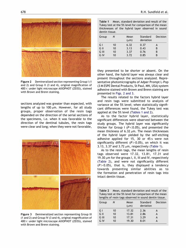
Figure 2 Demineralized section representing Group I (1and 2) and Group II (3 and 4), original magnification of400! under light microscope AXIOPHOT (ZEISS), stainedwith Brown and Brenn staining.
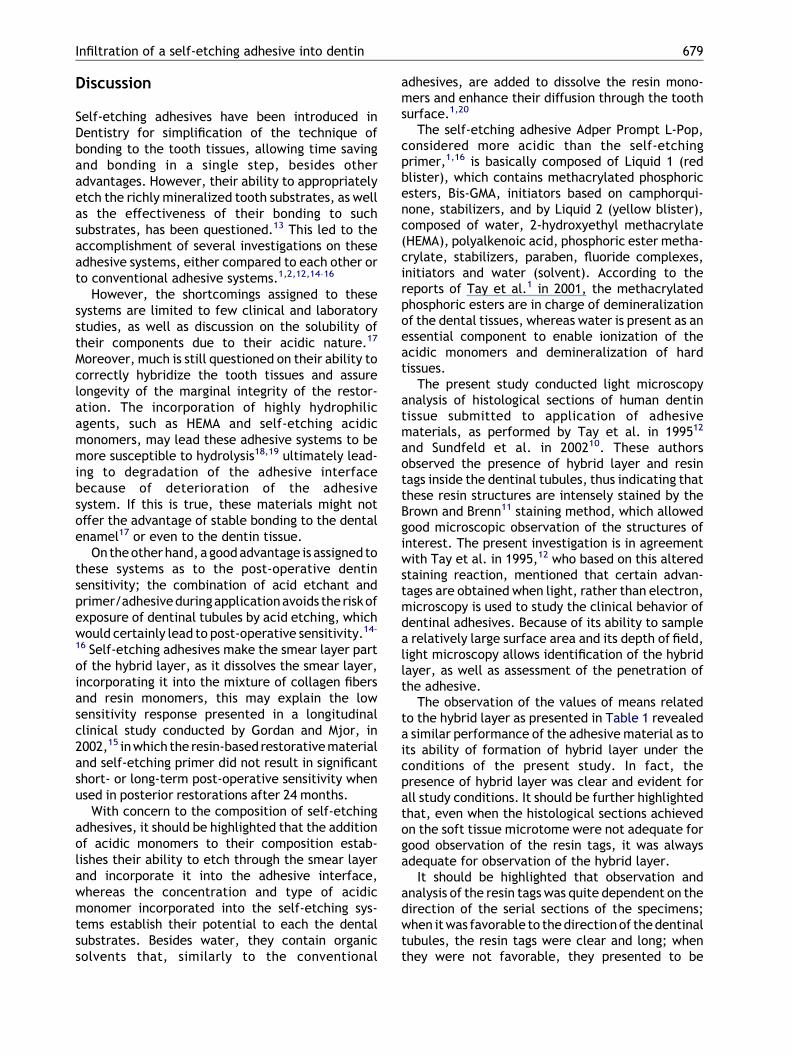
Table 1 Mean, standard deviation and result of theTukey test at the 5% level for comparison of the meanthicknesses of the hybrid layer observed in sounddentin tissue.
Group N Mean(mm)
Standarddeviation
Decision
G I 10 6.32 0.37 AG II 10 3.13 0.43 BG III 10 3.37 0.76 BG IV 10 3.72 0.85 B
R.H. Sundfeld et al.678
sections analyzed was greater than expected, withlengths of up to 100 mm. However, for all studygroups, proper observation of the resin tagsdepended on the direction of the serial sections ofthe specimens, i.e. when it was favorable to thedirection of the dentinal tubules, the resin tagswere clear and long; when they were not favorable,
Figure 3 Demineralized section representing Group III(1 and 2) and Group IV (3 and 4), original magnification of400! under light microscope AXIOPHOT (ZEISS), stainedwith Brown and Brenn staining.
they presented to be shorter or absent. On theother hand, the hybrid layer was always clear andpresent throughout the sections analyzed. Repre-sentative photomicrographs of Adper Prompt L-Pop(3 M ESPE Dental Products, St Paul, MN, USA) systemadhesive stained with Brown and Brenn staining arepresented in Figs. 2 and 3.
The results related to the factors hybrid layerand resin tags were submitted to analysis ofvariance at the 5% level; when statistically signifi-cant differences were found, the Tukey test wasapplied at the 5% level (Tables 1 and 2).
As to the factor hybrid layer, statisticallysignificant differences were observed between thestudy groups. The hybrid layer was significantlythicker for Group I (P!0.05), and presented themean thickness of 6.32 mm. The mean thicknessesof the hybrid layer yielded by the self-etchingadhesive applied for 15, 30 or 45 s were notsignificantly different (PO0.05), on which it was3.13, 3.37 and 3.72 mm, respectively (Table 1).
As to the resin tags, the mean lengths of resintags observed were 17.32, 13.81, 17.31 and19.30 mm for the groups I, II, III and IV, respectively(Table 2), and were not significantly different(PO0.05), that is, they displayed a tendencytowards presenting similar abilities as tothe formation and penetration of resin tags intointact dentin tissue.
Table 2 Mean, standard deviation and result of theTukey test at the 5% level for comparison of the meanlengths of resin tags observed in sound dentin tissue.
Group N Mean(mm)
Standarddeviation
Decision
G I 10 17.32 2.36 AG II 10 13.81 2.86 AG III 10 17.31 3.29 AG IV 10 19.30 8.01 A
Infiltration of a self-etching adhesive into dentin 679
Discussion
Self-etching adhesives have been introduced inDentistry for simplification of the technique ofbonding to the tooth tissues, allowing time savingand bonding in a single step, besides otheradvantages. However, their ability to appropriatelyetch the richly mineralized tooth substrates, as wellas the effectiveness of their bonding to suchsubstrates, has been questioned.13 This led to theaccomplishment of several investigations on theseadhesive systems, either compared to each other orto conventional adhesive systems.1,2,12,14–16
However, the shortcomings assigned to thesesystems are limited to few clinical and laboratorystudies, as well as discussion on the solubility oftheir components due to their acidic nature.17
Moreover, much is still questioned on their ability tocorrectly hybridize the tooth tissues and assurelongevity of the marginal integrity of the restor-ation. The incorporation of highly hydrophilicagents, such as HEMA and self-etching acidicmonomers, may lead these adhesive systems to bemore susceptible to hydrolysis18,19 ultimately lead-ing to degradation of the adhesive interfacebecause of deterioration of the adhesivesystem. If this is true, these materials might notoffer the advantage of stable bonding to the dentalenamel17 or even to the dentin tissue.
On the other hand, agood advantage is assigned tothese systems as to the post-operative dentinsensitivity; the combination of acid etchant andprimer/adhesiveduringapplicationavoids the riskofexposure of dentinal tubules by acid etching, whichwould certainly lead to post-operative sensitivity.14–
16 Self-etching adhesives make the smear layer partof the hybrid layer, as it dissolves the smear layer,incorporating it into the mixture of collagen fibersand resin monomers, this may explain the lowsensitivity response presented in a longitudinalclinical study conducted by Gordan and Mjor, in2002,15 in which the resin-based restorative materialand self-etching primer did not result in significantshort- or long-term post-operative sensitivity whenused in posterior restorations after 24 months.
With concern to the composition of self-etchingadhesives, it should be highlighted that the additionof acidic monomers to their composition estab-lishes their ability to etch through the smear layerand incorporate it into the adhesive interface,whereas the concentration and type of acidicmonomer incorporated into the self-etching sys-tems establish their potential to each the dentalsubstrates. Besides water, they contain organicsolvents that, similarly to the conventional
adhesives, are added to dissolve the resin mono-mers and enhance their diffusion through the toothsurface.1,20
The self-etching adhesive Adper Prompt L-Pop,considered more acidic than the self-etchingprimer,1,16 is basically composed of Liquid 1 (redblister), which contains methacrylated phosphoricesters, Bis-GMA, initiators based on camphorqui-none, stabilizers, and by Liquid 2 (yellow blister),composed of water, 2-hydroxyethyl methacrylate(HEMA), polyalkenoic acid, phosphoric ester metha-crylate, stabilizers, paraben, fluoride complexes,initiators and water (solvent). According to thereports of Tay et al.1 in 2001, the methacrylatedphosphoric esters are in charge of demineralizationof the dental tissues, whereas water is present as anessential component to enable ionization of theacidic monomers and demineralization of hardtissues.
The present study conducted light microscopyanalysis of histological sections of human dentintissue submitted to application of adhesivematerials, as performed by Tay et al. in 199512
and Sundfeld et al. in 200210. These authorsobserved the presence of hybrid layer and resintags inside the dentinal tubules, thus indicating thatthese resin structures are intensely stained by theBrown and Brenn11 staining method, which allowedgood microscopic observation of the structures ofinterest. The present investigation is in agreementwith Tay et al. in 1995,12 who based on this alteredstaining reaction, mentioned that certain advan-tages are obtained when light, rather than electron,microscopy is used to study the clinical behavior ofdentinal adhesives. Because of its ability to samplea relatively large surface area and its depth of field,light microscopy allows identification of the hybridlayer, as well as assessment of the penetration ofthe adhesive.
The observation of the values of means relatedto the hybrid layer as presented in Table 1 revealeda similar performance of the adhesive material as toits ability of formation of hybrid layer under theconditions of the present study. In fact, thepresence of hybrid layer was clear and evident forall study conditions. It should be further highlightedthat, even when the histological sections achievedon the soft tissue microtome were not adequate forgood observation of the resin tags, it was alwaysadequate for observation of the hybrid layer.
It should be highlighted that observation andanalysis of the resin tags was quite dependent on thedirection of the serial sections of the specimens;when it was favorable to thedirectionof thedentinaltubules, the resin tags were clear and long; whenthey were not favorable, they presented to be

R.H. Sundfeld et al.680
shorter or even absent. These outcomes are corro-borated by previous findings,21,22 which revealedthat the orientation of tubules had a profound effecton the formation of resin structures analyzed.
According to the aforementioned studies andconsidering the present methodology, it was men-tioned that some sections presented areas with tagsmeasuring up to 100 mm in length. However, thelaboratory nature of the study should also beconsidered, in that the penetration of self-etchingadhesive material may have been enhanced by theabsence of factors as internal pulp pressure anddentin moisture. In fact, Sundfeld et al.10 in 2002,conducted a clinical and light microscopy study andobserved that the internal pressure existing in thedentinal tubule may have negatively influenced thegreater penetration of conventional adhesive sys-tems into the etched dentin tissue.
However, it should be mentioned that, in thepresent study, the mean length of the resin tags ofeach section corresponded to the mean of the threemeasurements performed on each slab. Thisexplains the values of the means in micrometersobtained for the length of the resin tags corre-sponding to the study groups, which ranged from13.81–19.30 mm.
The specimens in Group I, submitted to acidetching before application of the self-etchingadhesive, displayed thicker hybrid layers withclear tags within it. The hybrid layer in Group Iwas thicker than in the other Groups, with a meanof 6.32 mm. This was probably caused by the pre-etching effect that dissolves the smear layer, thusallowing easy access of the self-etching adhesive tothe dentin underneath. Since there was already ademineralized layer on the top dentin surface, theself-etching action of the adhesive furtherdemineralized the dentin and resulted in thickerhybrid layer.
The mean thickness of the hybrid layer andlength of the resin tags formed by the self-etching adhesive when applied for 15, 30 or 45 swere not significantly different, thereforesuggesting that the increase in the time ofapplication of the self-etching adhesive did notsignificantly interfere with their thickness andlength achieved, respectively.
It should be highlighted that application of self-etching adhesive Adper Prompt L-Pop on the intactdentin tissue showed that this aggressive self-etching was able to demineralize the subsurfacedentin with a similar ability of formation of anauthentic hybrid layer that was around 3.13–3.72 mm, for all times of application employed inthis study. These adhesive interfaces were mor-phologically similar to those produced by
conventional adhesives in the dentin tissue,10 asrevealed by the light microscopy analysis. Inagreement with these findings, Tay et al.1,2 in2001, stated that the more aggressive systemcompletely solubilized the smear layer and smearplugs, even with thick smear layers, and formedhybrid layers with a thickness approaching those ofphosphoric acid-etched dentin.
The present findings are corroborated by thereport of Carvalho et al., in 2004,20 whichrevealed that deeper penetration of the adhesiveis less relevant, since there is neutralization ofthe acidic monomers some seconds after appli-cation and limitation of the extension of demi-neralization, regardless of the time of applicationof the material on the tooth surface, as alsomentioned by other investigators1,2,22 who sta-ted, besides other observations, that if self-etching adhesive materials eliminate the needof rinsing the tooth structure, the byproducts ofenamel and dentin yielded by the low pH of theadhesive system may lead to a limited deminer-alizing action, restricting penetration of theadhesive system to the most superficial enameland dentin layers. Moreover, the mineral com-ponents from the smear layer may neutralize theacidity of these self-etching systems.1
In this work, it could be concluded that theincrease in the time of application of the self-etching adhesive Adper Prompt L-Pop (3 M ESPEDental Products) did not significantly influencethe formation and thickness of hybrid layer, aswell as its penetration into the sound dentinsurface. However, since there are doubts on thereal effectiveness of self-etching adhesive sys-tems, the longevity of their bonding to the toothtissues should be investigated to allow betterunderstanding on their adhesive mechanisms,degree of acidity and curing, so that they maybe regarded as substitutes of conventionaladhesive systems.
Conclusions
The following could be concluded:
1.
The hybrid layer was significantly thicker whenthe adhesive system was applied on dentinpreviously etched with phosphoric acid.2.
The increase in the time of application of theself-etching adhesive Adper Prompt L-Pop didnot influence the formation and thickness ofhybrid layer, as well as its penetration (tags) intothe sound dentin surface.
Infiltration of a self-etching adhesive into dentin 681
Acknowledgements
Financial support provided by Fapesp, Sao PauloState Agency for Research Funding, Sao Paulo,Brazil, Process #02/07962-8.
References
1. Tay FR, Pashley DH. Aggressiveness of contemporary self-etching systems I: depth of penetration beyond dentin smearlayers. Dental Materials 2001;17:296–308.
2. Pashley DH, Tay FR. Aggressiveness of contemporary self-etching adhesives part II: etching effects on ungroundenamel. Dental Materials 2001;17:430–44.
3. Ogata M, Harada N, Yamaguchi S, Nakajima M, Pereira PNR,Tagami J. Effects of different burs on dentin bond strengthsof self-etching primer bonding systems. Operative Dentistry2001;26:375–82.
4. Inoue H, Inoue S, Uno S, Takahashi A, Koase K, Sano H.Microtensile bond strength fo two single-step adhesivesystems to bur-prepared dentin. Journal of AdhesiveDentistry 2001;3:129–36.
5. Ogata M, Harada N, Yamaguchi S, Nakajima M, Tagami J.Effect of self-etching primer vs phosphoric acid etchant onbonding to bur-prepared dentin. Operative Dentistry 2002;27:447–54.
6. Tani C, Finger WL. Effect of smear layer thickness on bondstrength mediated by three all-in-one self-etching primingadhesives. Journal of Adhesive Dentistry 2002;4:283–9.
7. Chan KM, Tay FR, King NM, Imazato S, Pashley DH. Bonding ofmild self-etching primers/adhesives to dentin with thicksmear layers. American Journal of Dentistry 2003;16:340–6.
8. Miyazaki M, Hirohata N, Takagaki K, Onose H, Moore BK.Influence of self-etching primer drying time on enamel bondstrength of resin composites. Journal of Dentistry 1999;27:203–7.
9. Marquezini Jr L. Efeito do tempo de aplicacao de adesivosautocondicionantes na durabilidade da uniao ao esmalte.Bauru, 90p. PhD Thesis, School of Dentistry, Sao PauloUniversity; 2003.
10. Sundfeld RH, Mauro SJ, Sundefeld MLMM, Briso ALF. Avalia-cao clınico/microscopica da camada hıbrida de adesao e dosprolongamentos resinosos (tags), em tecido dentinario
condicionado. Efeitos de materiais, tecnicas de aplicacao ede analise. Jornal Brasileiro de Dentıstica and Estetetica2002;1:315–31.
11. Brown JH, Brenn L. A method for differencial staining ofGram positive and Gram negative bacteria in tissuereactions. Bulletin Johns Hopkins Hospital 1931;48:69–73.
12. Tay FR, Gwinnett AJ, Pang KM, Wei SHY. Micromorphologicrelationship of the resin-dentin interface following a total-etch technique in vivo using a dentinal bonding system.Quintessence International 1995;26:63–70.
13. Kaaden C, Powers JM, Friedl KH, Schmalz G. Bond strength ofself-etching adhesives to dental hard tissues. Clinical OralInvestigations 2002;6:155–60.
14. Opdam NJM, Roeters FJM, Feilzer AJ, Verdonschot EH.Marginal integrity and postoperative sensitivity in Class 2resin composite restorations in vivo. Journal of Dentistry1998;26:555–62.
15. Gordan VV, Mjor IA. Short- and long-term clinical evaluationof post-operative sensitivity of a new resin-based restorativematerial and self-etching primer. Operative Dentistry 2002;27:543–8.
16. Kiremitci A, Yalcin F, Gokalp S. Bonding to enamel anddentin using self-etching adhesive systems. QuintessenceInternational 2004;35:367–70.
17. Tay FR, Pashley DH, Yoshiyama M. Two modes of nanoleak-age expression in single-step adhesives. Journal of DentalResearch 2002;81:472–6.
18. Hashimoto M, Ohno H, Sano H, Tay FR, Kaga M, Kudou Y,Oguchi H, Araki Y, Kubota M. Micromorphological changes inresin-dentin bonds after 1 year of water storage. Journal ofBiomedical Materials Research 2002;63:306–11.
19. Tay FR, Pashley DH. Water-treeing. A potential mechanismfor degradation of dentin adhesives. American Journal ofDentistry 2003;16:6–12.
20. Carvalho RM, Carrilho MRO, Pereira LCG, Garcia FCP,Marquezini Jr L, Silva SMA, Kussmaul APM. Sistemasadesivos: fundamentos para aplicacao clınica. BiodontoPublicacoes Cientıficas 2004;2:1–89.
21. Schupbach P, Krejci I, Lutz F. Dentin bonding: effect oftubule orientation on hybrid-layer formation. EuropeanJournal of Oral Sciences 1997;105:344–52.
22. Phrukkanon S, Burrow MF, Tyas MJ. The effect of dentinelocation and tubule orientation on the bond strengthsbetween resin and dentine. Journal of Dentistry 1999;27:265–74.
Copyright © 2022 FDOKUMEN