Humoral immunity in tuberculin skin test anergy and its role in high-risk persons exposed to active...
19
Humoral immunity in tuberculin skin test anergy and its role in high-risk persons exposed to active tuberculosis Liliana Encinales 1 , Joaquin Zuñiga 2 , Maria Yunis 9 , Julio Granados-Montiel 1 , Julio Granados 4 , Ingrid Almeciga 1 , Olga Clavijo 1 , Carlos Awad 7 , Vilma Collazos 8 , María Inés Vargas-Rojas 2 , José Luis Bañales-Mendez 2,3 , Lilia Vazquez-Castañeda 2 , Joel N. Stern 1 , Viviana Romero 1 , Masha Frindkis-Hareli 1 , Daniel Terreros 5 , Marcelo Fernandez-Viña 6 , and Edmond J. Yunis 1 1 Department of Cancer Immunology and AIDS, Dana-Farber Cancer Institute, Boston, MA 02115, USA 2 Laboratory of Immunobiology and Genetics, Instituto Nacional de Enfermedades Respiratorias, Mexico City, Mexico. 3 Laboratory of Molecular Biology, Instituto Nacional de Cardiología Ignacio Chavez, Mexico City, Mexico. 4 Immunology and Rheumatology Department, Instituto Nacional de Ciencias Medicas y Nutricion México City, México. 5 Biomedical Sciences Department, Texas Tech Health Sciences Center, Paul L. Foster SOM, El Paso TX 79905 USA 6 Department of Laboratory Medicine, University of Texas M. D. Anderson Cancer Center, Houston, TX, USA 2 Division of Immunology and Organ Transplantation, University of Texas Medical School, Houston, TX, USA 7 Hospital Santa Clara, Bogotá Colombia. 8 Hospital de la Tebaida Armenia, Colombia 9 Maria Yunis Design, Arlington, MA USA. Abstract The most common test to identify latent tuberculosis is the Tuberculin skin test that detects T cell responses of delayed type hypersensitivity type IV. Since it produces false negative reactions in active tuberculosis or in high-risk persons exposed to tuberculosis patients as shown in this report, we studied antibody profiles to explain the anergy of such responses in high-risk individuals without active infection. Address correspondence to: Edmond J Yunis, Dana-Farber Cancer Institute, Department of Cancer Immunology and AIDS, 44 Binney Street. Boston, Massachusetts 02115-6084, USA. Phone +1 (617), 632-3347. Fax +1 (617) 632-4466. [email protected] or Marcelo Fernandez-Viña, Department of Laboratory Medicine, University of Texas M. D. Anderson Cancer Center, Houston, TX, USA 2 Division of Immunology and Organ Transplantation, University of Texas Medical School, Houston, 77030 TX, USA. Phone +1 (713) 792-2658. Fax +1 (713) 794-4773. [email protected]. Authorship note: Liliana Encinales, Joaquin Zuñiga, Maria Yunis and Julio Granados-Montiel contributed equally to this work. Publisher's Disclaimer: This is a PDF file of an unedited manuscript that has been accepted for publication. As a service to our customers we are providing this early version of the manuscript. The manuscript will undergo copyediting, typesetting, and review of the resulting proof before it is published in its final citable form. Please note that during the production process errors may be discovered which could affect the content, and all legal disclaimers that apply to the journal pertain. NIH Public Access Author Manuscript Mol Immunol. Author manuscript; available in PMC 2011 February 1. Published in final edited form as: Mol Immunol. 2010 February ; 47(5): 1066. doi:10.1016/j.molimm.2009.11.005. NIH-PA Author Manuscript NIH-PA Author Manuscript NIH-PA Author Manuscript
Transcript of Humoral immunity in tuberculin skin test anergy and its role in high-risk persons exposed to active...
Humoral immunity in tuberculin skin test anergy and its role inhigh-risk persons exposed to active tuberculosis
Liliana Encinales1, Joaquin Zuñiga2, Maria Yunis9, Julio Granados-Montiel1, JulioGranados4, Ingrid Almeciga1, Olga Clavijo1, Carlos Awad7, Vilma Collazos8, María InésVargas-Rojas2, José Luis Bañales-Mendez2,3, Lilia Vazquez-Castañeda2, Joel N. Stern1,Viviana Romero1, Masha Frindkis-Hareli1, Daniel Terreros5, Marcelo Fernandez-Viña6, andEdmond J. Yunis11Department of Cancer Immunology and AIDS, Dana-Farber Cancer Institute, Boston, MA 02115,USA2Laboratory of Immunobiology and Genetics, Instituto Nacional de Enfermedades Respiratorias,Mexico City, Mexico.3Laboratory of Molecular Biology, Instituto Nacional de Cardiología Ignacio Chavez, Mexico City,Mexico.4Immunology and Rheumatology Department, Instituto Nacional de Ciencias Medicas y NutricionMéxico City, México.5Biomedical Sciences Department, Texas Tech Health Sciences Center, Paul L. Foster SOM, ElPaso TX 79905 USA6Department of Laboratory Medicine, University of Texas M. D. Anderson Cancer Center, Houston,TX, USA 2 Division of Immunology and Organ Transplantation, University of Texas Medical School,Houston, TX, USA7Hospital Santa Clara, Bogotá Colombia.8Hospital de la Tebaida Armenia, Colombia9Maria Yunis Design, Arlington, MA USA.
AbstractThe most common test to identify latent tuberculosis is the Tuberculin skin test that detects T cellresponses of delayed type hypersensitivity type IV. Since it produces false negative reactions in activetuberculosis or in high-risk persons exposed to tuberculosis patients as shown in this report, westudied antibody profiles to explain the anergy of such responses in high-risk individuals withoutactive infection.
Address correspondence to: Edmond J Yunis, Dana-Farber Cancer Institute, Department of Cancer Immunology and AIDS, 44 BinneyStreet. Boston, Massachusetts 02115-6084, USA. Phone +1 (617), 632-3347. Fax +1 (617) 632-4466. [email protected] Marcelo Fernandez-Viña, Department of Laboratory Medicine, University of Texas M. D. Anderson Cancer Center, Houston, TX,USA 2 Division of Immunology and Organ Transplantation, University of Texas Medical School, Houston, 77030 TX, USA. Phone +1(713) 792-2658. Fax +1 (713) 794-4773. [email protected] note: Liliana Encinales, Joaquin Zuñiga, Maria Yunis and Julio Granados-Montiel contributed equally to this work.Publisher's Disclaimer: This is a PDF file of an unedited manuscript that has been accepted for publication. As a service to our customerswe are providing this early version of the manuscript. The manuscript will undergo copyediting, typesetting, and review of the resultingproof before it is published in its final citable form. Please note that during the production process errors may be discovered which couldaffect the content, and all legal disclaimers that apply to the journal pertain.
NIH Public AccessAuthor ManuscriptMol Immunol. Author manuscript; available in PMC 2011 February 1.
Published in final edited form as:Mol Immunol. 2010 February ; 47(5): 1066. doi:10.1016/j.molimm.2009.11.005.
NIH
-PA Author Manuscript
NIH
-PA Author Manuscript
NIH
-PA Author Manuscript

Our results showed that humoral immunity against Tuberculin, regardless of the result of theTuberculin skin test is important for protection from active tuberculosis and that the presence of highantibody titers is a more reliable indicator of infection latency suggesting that latency can be basedon the levels of antibodies together with in vitro proliferation of peripheral blood mononuclear cellsin the presence of the purified protein derivative. Importantly, anti-Tuberculin IgG antibody levelsmediate the anergy described herein, which could also prevent reactivation of disease in high-riskindividuals with high antibody titers. Such IgG Tuberculin antibodies were also found associatedwith blocking and/or stimulation of in vitro cultures of PBMC with Tuberculin. In this regard, futurestudies need to establish if immune responses to Mycobacterium tuberculosis can generate a broadspectrum of reactions either toward Th1 responses favoring stimulation by cytokines or by antibodiesand those toward diminished responses by Th2 cytokines or blocking by antibodies; possiblyinvolving mechanisms of antibody dependent protection from Mtb by different subclasses of IgG.
IntroductionMycobacterium tuberculosis (Mtb) infection is a major world public health problem; over 2.0million people die every year from this common infection. One third of the world’s populationis thought to have latent tuberculosis (LTBI) [Smith. 2003], a condition where individuals areinfected by the intracellular bacteria without exhibiting the active disease but are at risk forreactivation, if their immune system fails. The infection by Mtb is accompanied by non-specificinflammatory responses regulated by cytokines and chemokines produced by macrophageswhich are activated by toll-like receptors and dendritic cells [Gehring et al, 2003, Lin. 2005].Also, interferon (IFNγ), an inflammatory cytokine, stimulates the antimicrobial activity ofmacrophages and regulates their antigen presentation through the MHC class II molecules byup-regulating their mRNA and protein expression [Pier, 2004]. As well, IFNγ can induceautophagy, a mechanism that plays an important role in the innate immunity againstintracellular microorganisms [Harris et al, 2007 and Vergne et al, 2006]; MHC type II restrictedCD4+T cells, MHC class I CD8+T cells and macrophages are important in the protectiveimmunity against Mtb where a decrease of the number or function of these cells results in thereactivation of the infection [Tully et al, 2005]. And, γ/δ T cells play an important role in theprotective immune response to tuberculosis (TB) [Szereday et al, 2003].
The most common screening for Mtb infection in asymptomatic patients (LTBI) are theTuberculin skin test (TST) and chest × rays to detect the evidence of the Ghon complex (agranuloma that contains an organized collection of immune cells, predominantlymacrophages). The TST is performed by intradermal injection in the anterior forearm of 5 units(0.1 ml) of Tuberculin. Reaction in the skin to Mtb, purified protein derivative (PPD) alsonamed Tuberculin begins when T cells, sensitized by vaccination or infection, are recruited tothe intradermal site and lymphokines are locally secreted. These lymphokines causevasodilatation and edema plus recruitment of additional inflammatory cells. A positive reactionusually begins 5–6 hours after injection, reaching a maximum point at 48–72 hours andcontinues over a few days [Pier, 2004]. The results of the TST are based on the immune statusof the individual and three cut off points have been recommended for a positive reaction toTuberculin based on the size of the indurations seen after injection of the antigen: 1) 5 mm ormore: individuals with HIV infection, recent contacts of TB patients, LTBI in patients withorgan transplants, and other immuno-suppressed patients receiving corticosteroids (i.e.,prednisone) for at least one month, 2) 10 mm or more: recent immigrants (within 5 years) fromcountries with high TB prevalence, recent infection with Mtb, immuno-compromisedindividuals other than HIV positive individuals, intravenous drug users, and health careworkers with exposure to TB, and 3) 15 mm and greater: people with no risk to TB [AmericanThoracic Society, 2000]. Unfortunately in the absence of chest X-rays, which unequivocallyshow the absence of Ghon complexes the TST, is not reliable to detect LTBI, to predict disease
Encinales et al. Page 2
Mol Immunol. Author manuscript; available in PMC 2011 February 1.
NIH
-PA Author Manuscript
NIH
-PA Author Manuscript
NIH
-PA Author Manuscript

progression, nor to determine the risk of disease reactivation [Chee et al, 2007]. Chest X-raysmay not unveil the Ghon complexes that help to contain the spread of Mtb [Pier, 2004] andmore sensitive radiological techniques are frequently unavailable in areas were Mtb infectionsare common. In 2001, the IFNγ release assays (IGRAs) [Chee et al, 2007] were developed withthe advantage of no false positives secondary to BCG or exposure to non-tuberculosismycobacterial strains.
The absence of a positive TST has also been described in immunocompetent anergic patientsdiagnosed with active TB; inability to mount an antigen specific DTH response to PPD wasshown to be associated with a defective T cell response including an antigen-specific impairedinability to produce IL-2 and to proliferate in response to the PPD [Delgado et al, 2002 andSousa et al, 1997] regardless of prior BCG vaccination status [Jasmer, 2002]. These findingscould be due to a defective phosphorylation of T cell receptor (TCR) leading to a defectiveactivation of ZAP-70 and MAPK proteins related to IL2 gene transcription and proteinproduction after antigen stimulation of T cells [Boussiotis et al, 2000]. Or, decreases on the γ/δ T cells subset [Szereday, 2003], decreases of IFN-γ production, increase of IL-10 in responseto PPD [Boussiotis et al, 2000], and a failure of the autophagic control by macrophages havebeen associated with anergy in ATBI [Harris et al, 2007]. However, studies of anergy in LTBIhave not been performed before. In this regard, we studied individuals without ATBI, withdifferent degrees of exposure to patients with ATBI from a tuberculosis specialized hospitalin Bogota, Colombia. In a first cohort we observed a high incidence of negative TST in high-risk contacts. Based on these preliminary results (Cohort I) we investigated a second cohortfor TST, in-vitro proliferation of peripheral blood leukocytes (PBMC) with Tuberculin toestablish anergy more frequently in high-risk contacts. Also, we studied the role antituberculinand anti-HLA antibodies in anergic and non-anergic contacts or hospital employees sincehumoral immune responses had been described in anergic individuals diagnosed with ATBI(Sousa et al, 1997).
MethodsBaseline characteristics of subjects studied
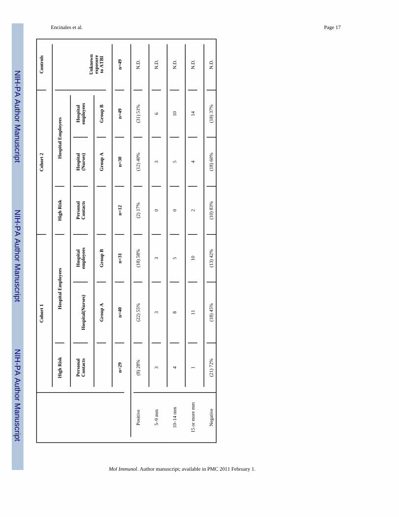
The two cohorts of study subjects are shown in Table A. Cohort I was recruited between theyears of 1999 and 2001 from a community hospital specialized in the treatment of tuberculosisin Bogotá, Colombia. The majority of these individuals had received BCG vaccination shortlyafter birth. All subjects were studied by the TST. We did not include immunocompromisedindividuals such as HIV positive patients, patients using steroids, or patients with neoplasiasthat could affect the TST [Chee et al, 2007]. The subjects of cohort I included 29 high-riskpersonal contacts (spouses and significant others) with patients with established diagnosis ofactive tuberculosis infection (ATBI) and compared them with 71 employees with a range ofservice of 5 to 23 years (nurses, administrators, general services, and security). Based on theassumption that nurses have higher risk due to high patient contact, we analyzed the 40 nursesas a separate group A, and the 31 other employees as group B. All three groups showed a higherpercentage of women and the mean age was 40 years. Cohort 2 was recruited from 2002 to2006 from the same hospital and consisted of 91 individuals, 12 high-risk, 30 nurses, and 49hospital employees. The gender distribution and mean age was comparable to Cohort 1. Wealso tested 6 healthy Colombians and 43 Mexicans as controls. All subjects signed a consentform and the institutional review board at Dana-Farber Cancer Institute approved the protocolfor the studies. After consent was obtained, 0.1 ml of Tubersol (5 Tuberculin units (TU) PPD;Aventis-Pasteur, Swiftwater, PA) was injected intradermally in the forearms of the subjectswho were then evaluated for induration 48 hours later and scored as positive if the diameter ofinduration was 5 mm or greater.
Encinales et al. Page 3
Mol Immunol. Author manuscript; available in PMC 2011 February 1.
NIH
-PA Author Manuscript
NIH
-PA Author Manuscript
NIH
-PA Author Manuscript
HLA typingsHLA-A, B, and C alleles were typed using polymerase chain reaction and sequence-specificoligonucleotide probes hybridization techniques (PCR-SSOP) as described previously [Cao,1999]. Briefly extracted DNA was amplified using intronic locus-specific and exonic group-specific primers. The PCR products were immobilized onto nylon membranes that werehybridized with sets of probes matching polymorphic sequences of HLA-A, B, and C alleles.The hybridization signal was detected with a chemiluminescent substrate. Alleles wereassigned based on the hybridization patterns with the sets of oligonucleotide probes.
Serology testingThe serum from each subject was screened for anti-HLA antibodies using beads coated withsoluble HLA class I and II antigens and was analyzed with luminex (an instrument based onindirect immunofluorescence) using pre-screened positive sera for HLA class I or II antibodiesas a positive control and normal human serum as a negative control. We also screened forantibodies anti IgM and IgG against the PPD antigen. Sera from the 4 panels were tested forIgM and IgG antibodies against PPD antigen by enzyme-linked immuno-absorbent assay(ELISA) as follows: 96 wells microtiter plates were coated with 25 ng/well of PPD antigen(Aventis-Pasteur, Swiftwater, PA) overnight at 4°C. Excess protein binding sites were blockedby incubation with 0.5% BSA in PBS-Tween 20 0.5% at 37°C for 1 hour. The plates werewashed 4 times with PBS containing 0.5% Tween 20. Sera samples were diluted 1:50 in PBScontaining 0.5% BSA and added to the plates for 1 hour of incubation at 37°C. The plates werewashed 4 times and then incubated with a second antibody (peroxidase-conjugated monoclonalantibody anti-alpha chain IgM diluted 1:5000 or IgG diluted 1:10000 in blocking solution for1 hour at 37°C) and washed 4 times (Sigma-Aldrich, US). A substrate solution containing 10µl of 30% H2O2 and 25 mg o-phenylenediamine (OPD) dihydrochloride (Sigma-Aldrich) in25 ml citrate buffer at pH 5 was added and incubated for 15 min at room temperature. Fiftymicroliters of 1N H2SO4 solution were used to stop the reaction. Color development wasmeasured in an ELISA reader at 492 nm [Araujo et al, 2004].
Antigen presentation assaysSubjects from cohort 2 were tested by the TST twice during a period of 3 months and thesamples for in vitro proliferation assays from this group were obtained 7–10 days followingthe last TST. Samples from individuals were processed in Boston, MA 12 hours after collection.(The blood samples were collected in EDTA and an investigator carried them by air fromColombia). Peripheral blood mononuclear cells (PBMC) and plasma were separated fromwhole blood samples from each subject. In this experiment we measured the ability ofleucocytes to proliferate in vitro after stimulation with antigens. Triplicates of individual’sPBMC (1 × 10⁵cells) were incubated with three different antigens: 1) Phytoheamagglutinin(PHA, 2ug/ml, Sigma-Aldrich, St. Louis, MO), a mitogen that stimulates T lymphocytesproliferation used as a control test; 2) Candida albicans (20ug/ml, Green laboratories, Lenoir,NC), used to test each individual’s antigen-specific T cell proliferation and to evaluate thepossibility of anergy; and 3) Tuberculin (10ug/ml, Mycos Research LLC, Loveland, CO). Thecells were incubated with human group AB or with autologous sera to demonstrate the cellularproliferation in these subjects for 3 days with PHA and 5 days with Candida or PPD at 37°C,5% CO2. The cells were labeled with [H3] thymidine (1 µCi/well) 6 hours prior to harvesting.Plates were harvested and the radioactivity was monitored by the Wallac liquid scintillationcounter (Perkin-Elmer, Boston, MA). PBMC from each subject was isolated and cultured withPHA which served to determine the cell viability, and with Tuberculin (PPD) or Candidaalbicans to test antigen-specific T cell proliferation measured by the intake of thymidine(H3) as counts per minute (cpm) of the mean of triplicate cultures and expressed by stimulationindex (SI) = cpm in cultures with each antigen divided by cpm in culture without antigen.
Encinales et al. Page 4
Mol Immunol. Author manuscript; available in PMC 2011 February 1.
NIH
-PA Author Manuscript
NIH
-PA Author Manuscript
NIH
-PA Author Manuscript

Statistical methodsDifferences between groups were determined using the Mann-Whitney U test and the chi-square or Fischer exact method when needed with STATA version 9.0. The statisticaldifference was considered significant when P values were less or equal to 0.05.
ResultsFrequency distribution of TST of contacts exposed to patients with ATBI
Table A showed the two cohorts of individuals exposed to ATBI. Cohort I was used todetermine the frequency of negative results from the TST in the high-risk group and showedan increased proportion of negative TST in personal contacts, compared to those of group B:72% versus 42 %, p= .01. Cohort I produced the hypothesis that personal contacts to patientswith ATBI had a high incidence of false negative TSTs. We confirmed these results in a secondcohort, the group of high-risk showed higher frequency of negative TST (83%) compared toGroup B (37%), p=. 01. Of interest, the group A composed of nurses showed intermediatefrequency of TST negativity between the high risk group and the group B of hospitalemployees, but not significantly different than that of group B. The number of years employeeswere exposed to patients were, 12 to 20 for physicians, 15 to 23 for microbiologists, 5 to 22for nurses, 12 for social workers, 6 to 27 for ambulance drivers, 1 to 10 for security, 3 to 19for X-ray techs, 1 to 23 for administration and 6 to 20 for general services. Furthermore, theexposure contacts for spouses, significant others and siblings that were included had aminimum of 6 months since diagnosis of ATBI.
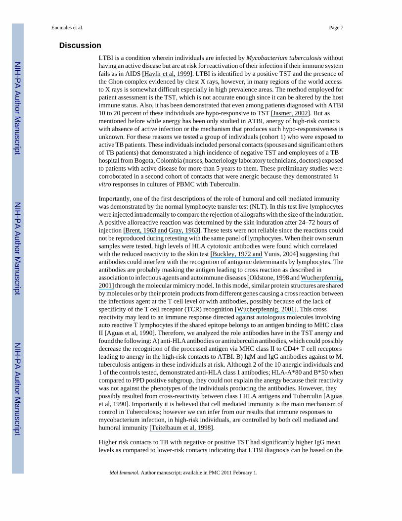
Antituberculin (IgG and IgM) studies of contacts to patients with ATBI86 out of 104 individuals of the second cohort from Bogotá, Colombia and 49 controls fromMexico City and from Bogotá were tested for IgG and IgM antibodies to Tuberculin antigenthat was used for TST. All individuals were tested for TST at least twice before their serumsamples were taken. Sera separated 7–10 days after the last phlebotomy were analyzed forspecific antibodies reactive to Tuberculin antigen in order to investigate the role of IgG and/or IgM antibodies against Tuberculin. The results were expressed as mean optical density(O.D.). Data from 10 of the high risk group, 26 of group A, 43 of group B, that included 8friends of patients with ATBI were analyzed for IgG and IgM antibodies against Tuberculin,they are shown in Figure 1 and Figure 2. Fig 1 shows the means and S.D. (standard deviations)of IgG levels as follows: high risk as .39 +/− .42, group A as .18 +/− .03, group B as .18+/− .03. The mean of the controls was .08 +/− .037. The mean values for IgM were comparable inthe four groups.
The high-risk group (10) had titers of more than .21 O.D. which is more than three standarddeviations higher than the mean for IgG compared to the controls. The figure also shows thesignificant p values when comparing the high-risk group versus Groups A and B and thecontrols. Also, the combined comparison between Groups A and B had higher titers than thecontrol groups p=.0001.
In vitro proliferation studies from contacts to patients with ATBI; studies of anergy inindividuals with a negative TST and positive in vitro proliferation with Tuberculin
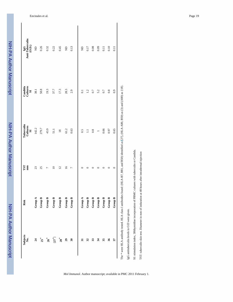
Table B showed 9 high-risk, 5 group A and 4 group B of hospital employees with anergy and3 high risk, 3 group A and 6 group B TST positives without anergy. The PBMC culturesincubated with Tuberculin (PPD) antigen showing significant stimulation indexes (SI) in 18anergic [negative TST individuals showing significant proliferation (S.I. ranging from 7.9 to561.1) in the presence of Tuberculin] and 7 TST negative non-anergic, p=.0001.
Encinales et al. Page 5
Mol Immunol. Author manuscript; available in PMC 2011 February 1.
NIH
-PA Author Manuscript
NIH
-PA Author Manuscript
NIH
-PA Author Manuscript
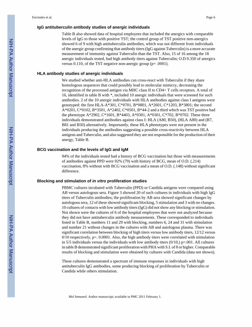
IgG antituberculin antibody studies of anergic individualsTable B also showed data of hospital employees that included the anergics with comparablelevels of IgG to those with positive TST; the control group of TST positive non-anergicsshowed 6 of 9 with high antituberculin antibodies, which was not different from individualsof the anergic group confirming that antibody titers (IgG against Tuberculin) is a more accuratemeasurement of immunity against Tuberculin than the TST. Also, 15 of 16 among the 18anergic individuals tested, had high antibody titers against Tuberculin; O.D 0.350 of anergicsversus 0.110, of the TST negative non-anergic group (p= .0001).
HLA antibody studies of anergic individualsWe studied whether anti-HLA antibodies can cross-react with Tuberculin if they sharehomologous sequences that could possibly lead to molecular mimicry, decreasing therecognition of the processed antigen via MHC class II to CD4+ T cells receptors. A total of16, identified in table B with *, included 10 anergic individuals that were screened for suchantibodies. 2 of the 10 anergic individuals with HLA antibodies against class I antigens weregenotyped: the first HLA-A*301, C*0701, B*0801, A*2601, C*1203, B*3801; the secondA*0201, C*0102, B*3501, A*2402, C*0501, B*44-2 and a third which was TST positive hadthe phenotype A*2902, C*1601, B*4403, A*0301, A*0301, C*702, B*0702. These threeindividuals demonstrated antibodies against class I: HLA (A80, B50), (HLA A80) and (B7,B81 and B50) alternatively. Importantly, these HLA phenotypes were not present in theindividuals producing the antibodies suggesting a possible cross-reactivity between HLAantigens and Tuberculin, and also suggested they are not responsible for the production of theiranergy, Table B.
BCG vaccination and the levels of IgG and IgM94% of the individuals tested had a history of BCG vaccination but those with measurementsof antibodies against PPD were 92% (79) with history of BCG, mean of O.D. (.214)vaccination, 8% without with BCG vaccination and a mean of O.D. (.148) without significantdifference.
Blocking and stimulation of in vitro proliferation studiesPBMC cultures incubated with Tuberculin (PPD) or Candida antigens were compared usingAB versus autologous sera. Figure 3 showed 20 of such cultures in individuals with high IgGtiters of Tuberculin antibodies; the proliferation by AB sera showed significant changes byautologous sera, 12 of these showed significant blocking, 5 stimulation and 3 with no changes.10 cultures of contacts with low antibody titers (IgG) did not show any blocking or stimulation.Not shown were the cultures of 6 of the hospital employees that were not analyzed becausethey did not have antituberculin antibody measurements. These corresponded to individualslisted in Table B, numbers 11 and 29 with blocking, numbers 6, 24 and 31 with stimulationand number 21 without changes in the cultures with AB and autologous plasma. There wassignificant correlation between blocking of high titers versus low antibody titers, 12/12 versus0/10 respectively, p=. 0.0001. Also, the high antibody titers were correlated with stimulationin 5/5 individuals versus the individuals with low antibody titers (0/10,) p=.001. All culturesin table B demonstrated significant proliferation with PHA with S.I. of 8 or higher. Comparableresults of blocking and stimulation were obtained by cultures with Candida (data not shown).
These cultures demonstrated a spectrum of immune responses in individuals with highantituberculin IgG antibodies, some producing blocking of proliferation by Tuberculin orCandida while others stimulation.
Encinales et al. Page 6
Mol Immunol. Author manuscript; available in PMC 2011 February 1.
NIH
-PA Author Manuscript
NIH
-PA Author Manuscript
NIH
-PA Author Manuscript
DiscussionLTBI is a condition wherein individuals are infected by Mycobacterium tuberculosis withouthaving an active disease but are at risk for reactivation of their infection if their immune systemfails as in AIDS [Havlir et al, 1999]. LTBI is identified by a positive TST and the presence ofthe Ghon complex evidenced by chest X rays, however, in many regions of the world accessto X rays is somewhat difficult especially in high prevalence areas. The method employed forpatient assessment is the TST, which is not accurate enough since it can be altered by the hostimmune status. Also, it has been demonstrated that even among patients diagnosed with ATBI10 to 20 percent of these individuals are hypo-responsive to TST [Jasmer, 2002]. But asmentioned before while anergy has been only studied in ATBI, anergy of high-risk contactswith absence of active infection or the mechanism that produces such hypo-responsiveness isunknown. For these reasons we tested a group of individuals (cohort 1) who were exposed toactive TB patients. These individuals included personal contacts (spouses and significant othersof TB patients) that demonstrated a high incidence of negative TST and employees of a TBhospital from Bogota, Colombia (nurses, bacteriology laboratory technicians, doctors) exposedto patients with active disease for more than 5 years to them. These preliminary studies werecorroborated in a second cohort of contacts that were anergic because they demonstrated invitro responses in cultures of PBMC with Tuberculin.
Importantly, one of the first descriptions of the role of humoral and cell mediated immunitywas demonstrated by the normal lymphocyte transfer test (NLT). In this test live lymphocyteswere injected intradermally to compare the rejection of allografts with the size of the induration.A positive alloreactive reaction was determined by the skin induration after 24–72 hours ofinjection [Brent, 1963 and Gray, 1963]. These tests were not reliable since the reactions couldnot be reproduced during retesting with the same panel of lymphocytes. When their own serumsamples were tested, high levels of HLA cytotoxic antibodies were found which correlatedwith the reduced reactivity to the skin test [Buckley, 1972 and Yunis, 2004] suggesting thatantibodies could interfere with the recognition of antigenic determinants by lymphocytes. Theantibodies are probably masking the antigen leading to cross reaction as described inassociation to infectious agents and autoimmune diseases [Oldstone, 1998 and Wucherpfennig,2001] through the molecular mimicry model. In this model, similar protein structures are sharedby molecules or by their protein products from different genes causing a cross reaction betweenthe infectious agent at the T cell level or with antibodies, possibly because of the lack ofspecificity of the T cell receptor (TCR) recognition [Wucherpfennig, 2001]. This crossreactivity may lead to an immune response directed against autologous molecules involvingauto reactive T lymphocytes if the shared epitope belongs to an antigen binding to MHC classII [Aguas et al, 1990]. Therefore, we analyzed the role antibodies have in the TST anergy andfound the following: A) anti-HLA antibodies or antituberculin antibodies, which could possiblydecrease the recognition of the processed antigen via MHC class II to CD4+ T cell receptorsleading to anergy in the high-risk contacts to ATBI. B) IgM and IgG antibodies against to M.tuberculosis antigens in these individuals at risk. Although 2 of the 10 anergic individuals and1 of the controls tested, demonstrated anti-HLA class 1 antibodies; HLA-A*80 and B*50 whencompared to PPD positive subgroup, they could not explain the anergy because their reactivitywas not against the phenotypes of the individuals producing the antibodies. However, theypossibly resulted from cross-reactivity between class I HLA antigens and Tuberculin [Aguaset al, 1990]. Importantly it is believed that cell mediated immunity is the main mechanism ofcontrol in Tuberculosis; however we can infer from our results that immune responses tomycobacterium infection, in high-risk individuals, are controlled by both cell mediated andhumoral immunity [Teitelbaum et al, 1998].
Higher risk contacts to TB with negative or positive TST had significantly higher IgG meanlevels as compared to lower-risk contacts indicating that LTBI diagnosis can be based on the
Encinales et al. Page 7
Mol Immunol. Author manuscript; available in PMC 2011 February 1.
NIH
-PA Author Manuscript
NIH
-PA Author Manuscript
NIH
-PA Author Manuscript

levels of antibodies (IgG) against Tuberculin in the absence of X rays regardless of their TSTstatus, together with the in vitro proliferation of PBMC in the presence of PPD. IgG antiTuberculin levels mediate a new form of anergy in which antibody to Tuberculin impairs thedelayed type IV hypersensitivity in skin via macrophages and T cells (Fig 4).
Our experiments of in-vitro cultures of PBMC with Tuberculin comparing the role ofautologous sera with that of AB sera in ATBI contacts, demonstrated that the levels of IgGantituberculin antibodies correlated with blocking of the proliferation responses, although therewas a spectrum of responses from blocking to stimulation (Fig 3). Such results suggested thepolarity responses that could be produced by either Th1 to Th2 responses. In this regard, twodifferent forms of leprosy correlate with Th1 or Th2 polarity of the immune response [Pier,2004]. In tuberculoid leprosy, patients mount a strong Th1 type cell-mediated immune responseproducing interferon γ and interleukin 2 in lesions and localize the infection, restricting thegrowth of the bacteria. While in lepromatous leprosy, a widely disseminated form of infectionis associated with a Th2 cytokine response with diminished cell-mediated immunity [Modlinet al, 1986]. It remains to be determined if the same polarity exists in contacts with ATBI thatproduce high levels of antituberculin IgG and can generate a broad spectrum of immunereactions with those toward Th1 responses favoring stimulation by autologous sera [Clay,2008] and those toward decreased responses produced either blocking by antibodies [Aguaset al, 1990] or by Th2 cytokines [Delgado, et a 2,002] (figure 4). In this regard, our blockingor stimulation studies of in vitro proliferation by autologous sera could be due to differentsubclass of IgG that mediate reactions against Mtb that need to be investigated by: 1) increasedkilling by phagocytosis by the process of opsonization, 2) Type II hypersensitivity reaction byactivation of complement possibly by IgG3, and 3) involvement of NK cells by ADCC, whichsuggests a possible interaction between antibodies and cell mediated immunity [Glatman-Freedman, 1998].
One interesting aspect of our studies was the possibility that the individuals studied haveimmunity to both Candida and Mycobacterium and that most individuals studied have a pro-inflammatory state. In the future it will be important to investigate the role of immunity toCandida and other antigens such as bacterial lipopolysaccharide (LPS) in relation to possiblecross-reactivity or cross-stimulation by Tuberculin or Candida antigens. Previously, we hadshown that up-regulation of the same pro-inflammatory genes in cultures with PPD or Candidain ATBI [Stern et al, 2008], which, is consistent with a report that immunity to Tuberculin isinfluenced by LPS antigens particularly in relation to protection of latency in HIV infection[Brenchley et al, 2006].
ConclusionsOur results represent an important advance in the detection of previous TB infection in anergichighly exposed individuals. They suggest screening for anti Tuberculin antibodies (IgG) todetermine previous TB infection together with TST, chest x-rays and in vitro tests of cellularproliferation against Tuberculin. Furthermore investigations should particularly focus on Th1and Th2 cytokine responses in high-risk individuals with low IgG titers against PPD, in orderto develop novel immunization protocols that will increase humoral immunity against TB inindividuals with or without BCG vaccination. This will in turn could produce protection againstnew infection or from reactivation of the disease [Havlir, 1999], in either immunocompromisedpatients or patients treated with immunosuppressors in order to avoid reactivation of the diseasefor example in patients with rheumatoid arthritis treated with anti TNF alpha [Hamdi et al,2006].
Encinales et al. Page 8
Mol Immunol. Author manuscript; available in PMC 2011 February 1.
NIH
-PA Author Manuscript
NIH
-PA Author Manuscript
NIH
-PA Author Manuscript

AcknowledgmentsThis work was supported by NIH grants AI49213, HL29583 and HL59838
References1. American Thoracic Society. Targeted Tuberculin Testing and Treatment of Latent Tuberculosis
Infection. American J Respir Crit Care Med 2000;161:S221–S247.2. Aguas A, Esaguy N, Sunkel CE, Silva MT. Cross-Reactivity and sequence homology between the 65-
Kilodalton mycobacterial heat shock protein and human lactoferrin, transferrin, and DR beta Subsetsof major histocompatibility complex class II molecules. Infection and Immunity 1990;58:1461–1470.[PubMed: 2323824]
3. Araujo Z, Waard JH, Fernández de Larrea C, López D, Fandiño C, Maldonado A, Hernández E, OcañaY, Ortega R, Singh M, Ottenhoff TH, Arend SM, Convit J. Study of the antibody response againstMycobacterium tuberculosis antigens in Warao Amerindian children in Venezuela. Mem Inst OswaldoCruz 2004;99:517–524. [PubMed: 15543417]
4. Boussiotis VA, Tsai EY, Yunis EJ, Thim S, Delgado JC, Dascher CC, Berezovskaya A, Rousset D,Reynes JM, Goldfeld AE. IL-10 producing T cells suppress immune responses in anergic tuberculosispatients. Journal of Clinical Investigation 2000;105:1317–1325. [PubMed: 10792007]
5. Brenchley JM, Price DA, Schacker TW, Asher TE, Silvestri G, Rao S, Kazzaz Z, Bornstein E, LambotteO, Altmann D, Blazar BR, Rodriguez B, Teixeira-Johnson L, Landay A, Martin JN, Hecht FM, PickerLJ, Lederman MM, Deeks SG, Douek D. Microbial translocation is a cause of systemic immuneactivation in chronic HIV infection. Nat Medicine 2006;12:1365–1371.
6. Brent L, Medawar PB. Tissue transplantation: a new approach to the “typing” problem. Br Med J1963;2:269–272. [PubMed: 14015250]
7. Buckley RH, Schiff RI, Amos DB. Blocking of autologous and homologous leukocyte responses byhuman alloimmune plasmas: A possible in vitro correlate of enhancement. J Immunol 1972;108:34–44. [PubMed: 5010395]
8. Cao K, Chopek M, Fernandez-Vina M. High and intermediate resolution DNA typing systems for classI HLA-A, B, C genes by hybridization with sequence-specific oligonucleotide probes (SSOP). RevImmunogenet 1999;1:177–208. [PubMed: 11253946]
9. Chee CB, KhinMar KW, Gan SH, Barkham TM, Pushparani M, Wang YT. Latent tuberculosis infectiontreatment and T-cell responses to Mycobacterium tuberculosis-specific antigens. Am J Respir CritCare Med 2007;175:282–287. [PubMed: 17082492]
10. Clay H, Volkman HE, Ramakrishnan L. Tumor necrosis factor signaling mediates resistance tomycobacteria by inhibiting bacterial growth and macrophage death. Immunity 2008;29:283–294.[PubMed: 18691913]
11. Delgado JC, Tsai EY, Thim S, Baena A, Boussiotis VA, Reynes JM, Sath S, Grosjean P, Yunis EJ,Goldfeld AE. Antigen-specific and persistent tuberculin anergy in a cohort of pulmonary tuberculosispatients from rural Cambodia. Proc Natl Acad Sci 2002;99:7576–7581. [PubMed: 12032325]
12. Gehring AJ, Rojas RE, Canaday DH, Lakey DL, Harding CV, Boom WH. The Mycobacteriumtuberculosis 19-kilodalton Lipoprotein Inhibits Gamma Interferon-Regulated HLA-DR and FCgamma R1 on human macrophages through Toll-Like Receptor. Infection and Immunity2003;71:4487–4497. [PubMed: 12874328]
13. Glatman-Freedman A, Cassadevall A. Serum therapy for tuberculosis revisited: reappraisal of therole of antibodies-mediated immunity against Mycobacterium tuberculosis. Clinical microbiologyreviews 1998;11:514–532. [PubMed: 9665981]
14. Gray JG, Russell PS. Donor selection in human organ transplantation. A possible screening test.Lancet 1963;2:863–865. [PubMed: 14056013]
15. Hamdi H, Marriette X, Godot V, Weldingh K, Hamid A, Prejean M-V, Baron G, Emilie. Inhibitionof anti-tuberculosis T-lymphocyte function with tumour necrosis factor antagonists. ArthritisResearch & Therapy 2006;8:R114. [PubMed: 16859506]
Encinales et al. Page 9
Mol Immunol. Author manuscript; available in PMC 2011 February 1.
NIH
-PA Author Manuscript
NIH
-PA Author Manuscript
NIH
-PA Author Manuscript

16. Harris J, De Haro SA, Master SS, Keane J, Roberts EA, Delgado M, Deretic V. T helper 2 cytokinesinhibit autophagic control of intracellular Mycobacterium tuberculosis. Immunity 2007;27:505–517.[PubMed: 17892853]
17. Havlir DV, Barnes PF. Tuberculosis in patients with human immunodeficiency virus infection. NewEngland J Med 1999;340:367–373. [PubMed: 9929528]
18. Jasmer RM, Nahid P, Hopewell PC. Clinical practice. Latent tuberculosis infection. N Engl J Med2002;347:1860–1866. [PubMed: 12466511]
19. Lin FC, Chen YC, Chen FJ, Chang SC. Cytokines and fibrinolytic enzymes in tuberculous andparapneumonic effusions. Clin Immunol 2005;116:166–173. [PubMed: 15897010]
20. Modlin RI, Mehra V, Wong L, Fujimiya Y, Chang WC, Horwitz DA, Bloom BR, Rea TH, PattengalePK. Suppressor T lymphocytes from lepromatous leprosy skin lesion. J Immunology 1986;137:2831–2834. [PubMed: 2944966]
21. Oldstone MB. Molecular mimicry and immune-mediated diseases. FASEB J 1998;12:1255–1265.[PubMed: 9761770]
22. Pier, G.; Lyczak, J.; Wetzler, L. Cell mediated immunity in Blackwell publishing. Immunology,Infection, and Immunity. Washington, D.C.: ASM Press; 2004.
23. Smith I. Mycobacterium tuberculosis Pathogenesis and Molecular Determinants of Virulence. ClinMicrobiol Rev 2003;16:463–496. [PubMed: 12857778]
24. Sousa AO, Salem JI, Lee FK, Verçosa MC, Cruaud P, Bloom BR, Lagrange PH, David HL. Anepidemic of tuberculosis with a high rate of tuberculin anergy among a population previouslyunexposed to tuberculosis, the Yanomami Indians of the Brazilian Amazon. Proc Natl Acad Sci1997;94:13227–12232. [PubMed: 9371828]
25. Stern J, Keskin DB, Romero V, Zuniga J, Encinales L, Li C, Awad C, Yunis EJ. Molecular signaturesdistinguishing active from latent tuberculosis in peripheral blood mononuclear cells, after in vitroantigenic stimulation with purified protein derivative of tuberculin (PPD) or Candida: a preliminaryreport. Immunol Res. 2008 0257-277X(print).
26. Szereday L, Baliko Z, Szekeres-Bartho J. Gamma/delta T cell subsets in patients with activeMycobacterium tuberculosis infection and tuberculin anergy. Clin Exp Immunol 2003;131:287–291.[PubMed: 12562390]
27. Teitelbaum R, Glatman-Freedman A, Chen B, Robbins JB, Unanue E, Casadevall A, Bloom BR. AmAB recognizing a surface antigen of Mycobacterium tuberculosis enhances host survival. Pro. Natl.Acad. Sic U S A 1998;95:15688–15693.
28. Tully G, Kortsik C, Höhn H, Zehbe I, Hitzler WE, Neukirch C, Freitag K, Kayser K, Maeurer MJ.Highly focused T cell responses in Latent human pulmonary Mycobacterium tuberculosis infection.Journal of Immunology 2005;174:2174–2184.
29. Vergne I, Singh S, Roberts E, Kyei G, Master S, Harris J, de Haro S, Naylor J, Davis A, Delgado M,Deretic V. Autophagy in immune defense against Mycobacterium tuberculosis. Autophagy2006;2:175–178. [PubMed: 16874111]
30. Wucherpfennig KW. Structural basis of molecular mimicry. J Autoimmun 2001;16:293–302.[PubMed: 11334495]
31. Yunis E. Bernard Amos A biographical memoir. Biographical memoirs. National academy of sciences2004:287.
Encinales et al. Page 10
Mol Immunol. Author manuscript; available in PMC 2011 February 1.
NIH
-PA Author Manuscript
NIH
-PA Author Manuscript
NIH
-PA Author Manuscript
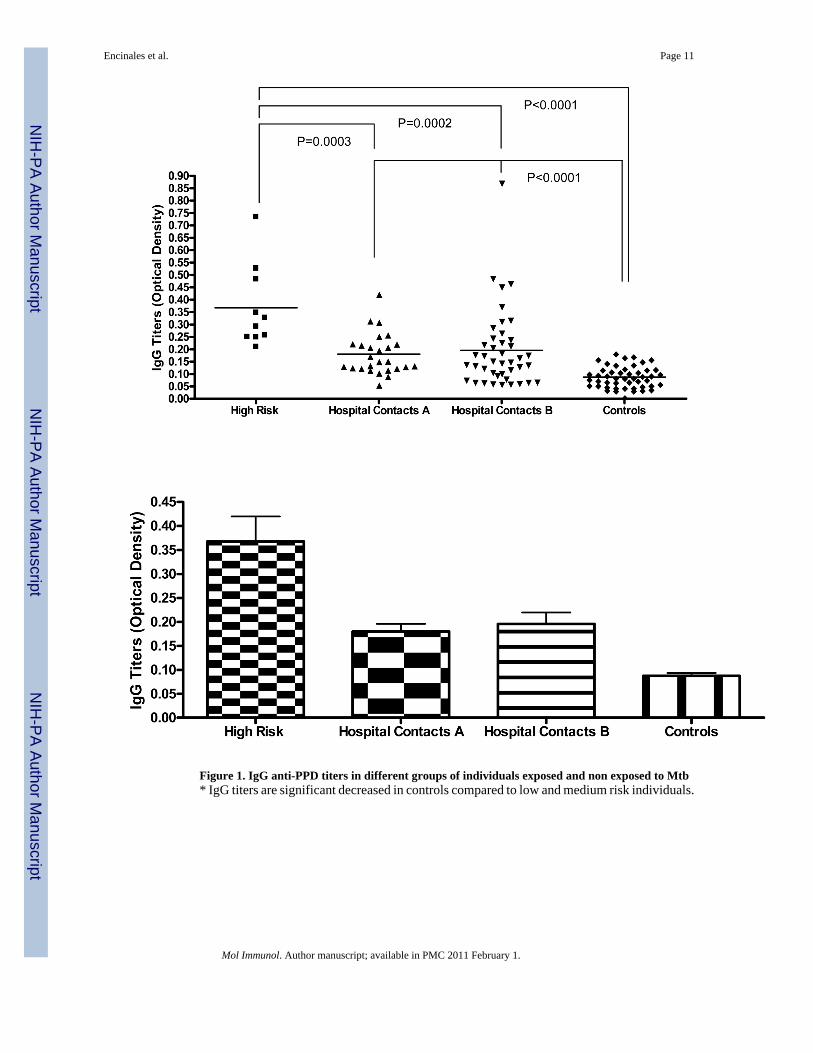
Figure 1. IgG anti-PPD titers in different groups of individuals exposed and non exposed to Mtb* IgG titers are significant decreased in controls compared to low and medium risk individuals.
Encinales et al. Page 11
Mol Immunol. Author manuscript; available in PMC 2011 February 1.
NIH
-PA Author Manuscript
NIH
-PA Author Manuscript
NIH
-PA Author Manuscript

Figure 2. IgM anti-PPD titers in different groups of individuals exposed and non-exposed to Mtb
Encinales et al. Page 12
Mol Immunol. Author manuscript; available in PMC 2011 February 1.
NIH
-PA Author Manuscript
NIH
-PA Author Manuscript
NIH
-PA Author Manuscript
Figure 3. Comparison of PBMC cultures with tuberculin; AB versus autologous seraThe columns are expressed in stimulation indexes (SI) demonstrating that 12 cultures showedblocking (graph A) ranging from .18 to .76 fold decreases and 5 showed stimulation (graph B)from .52 to 7.45 fold increases. There were 10 cultures in low antibody titers against tuberculin(IgG) (graph C) without blocking or stimulation. The levels of IgG antituberculin antibodiesare given in O.D. Numbers 1–5,7–13,15–18 and 32–37 were TST (−). Numbers 19, 20, 22, 23,25, 26, 27, 28 and 30 were TST (+).
Encinales et al. Page 13
Mol Immunol. Author manuscript; available in PMC 2011 February 1.
NIH
-PA Author Manuscript
NIH
-PA Author Manuscript
NIH
-PA Author Manuscript
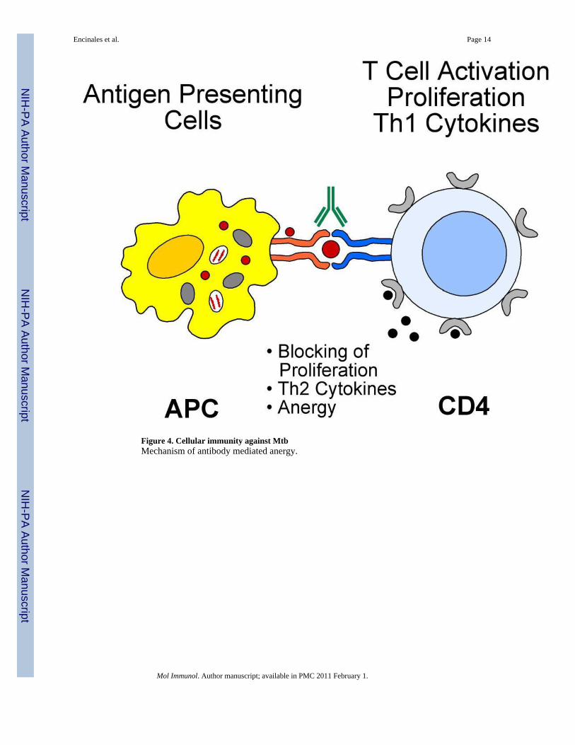
Figure 4. Cellular immunity against MtbMechanism of antibody mediated anergy.
Encinales et al. Page 14
Mol Immunol. Author manuscript; available in PMC 2011 February 1.
NIH
-PA Author Manuscript
NIH
-PA Author Manuscript
NIH
-PA Author Manuscript

Figure 5.
Encinales et al. Page 15
Mol Immunol. Author manuscript; available in PMC 2011 February 1.
NIH
-PA Author Manuscript
NIH
-PA Author Manuscript
NIH
-PA Author Manuscript

NIH
-PA Author Manuscript
NIH
-PA Author Manuscript
NIH
-PA Author Manuscript
Encinales et al. Page 16
Tabl
e A
Bas
elin
e ch
arac
teri
stic
s of i
ndiv
idua
ls e
xpos
ed to
pat
ient
s with
act
ive
tube
rcul
osis
(AT
BI)
Skin
test
reac
tions
wer
e mea
sure
d in
all i
ndiv
idua
ls an
d re
spon
sive
ness
was
asse
ssed
bas
ed o
n th
e dia
met
er (m
m) o
f ind
urat
ion
afte
r PPD
antig
en in
trade
rmal
adm
inis
tratio
n. C
ohor
t 1 sh
own
in th
e le
ft w
as u
sed
as e
xplo
rato
ry to
det
erm
ine
the
freq
uenc
y of
neg
ativ
ity o
f TST
in th
e hi
gh-r
isk
grou
p. C
ohor
t 2 sh
own
in th
e rig
ht ri
sk c
onfir
med
the
high
freq
uenc
y of
TST
neg
ativ
es in
the
high
-ris
k gr
oup,
com
pare
d w
ith th
e gr
oup
B o
f the
hos
pita
l em
ploy
ees.
Coh
ort 1
Coh
ort 2
Con
trol
s
Hig
h R
isk
Hos
pita
l Em
ploy
ees
Hig
h R
isk
Hos
pita
l Em
ploy
ees
Unk
now
nex
posu
reto
AT
BI
Pers
onal
Con
tact
sH
ospi
tal(N
urse
s)H
ospi
tal
empl
oyee
sPe
rson
alC
onta
cts
Hos
pita
l(N
urse
s)H
ospi
tal
empl
oyee
s
Gro
up A
Gro
up B
Gro
up A
Gro
up B
n=29
n=40
n=31
n=12
n=30
n=49
n=49
Age
(mea
n, +
/− S
D)
43, +
/− 1
6.4
40, +
/− 7
.943
, +/−
9.9
47 +
/− 1
7.6
37 +
/− 1
1.8
38 +
/− 8
.133
+/−
10
Gen
der
(n, %
)
Mal
e(7
) 24
%(4
) 7 %
(14)
45%
(7) 5
8%(6
) 20%
(16)
33%
(22)
45%
Fem
ale
(22)
76
%(3
7) 9
3%(1
7) 5
5%(5
) 42%
(24)
80%
(33)
67%
(27)
55%
BC
G V
acci
ne (n
,%
)
Yes
(12)
75%
(34)
95%
(29)
94%
(10)
83%
(29)
97%
(43)
82%
(49)
100
%
No
(4) 2
5%(2
) 5%
(2) 6
%(2
) 17%
(1) 3
%(6
) 12%
(0) 0
%
TST
(n, %
)
Mol Immunol. Author manuscript; available in PMC 2011 February 1.
NIH
-PA Author Manuscript
NIH
-PA Author Manuscript
NIH
-PA Author Manuscript
Encinales et al. Page 17
Coh
ort 1
Coh
ort 2
Con
trol
s
Hig
h R
isk
Hos
pita
l Em
ploy
ees
Hig
h R
isk
Hos
pita
l Em
ploy
ees
Unk
now
nex
posu
reto
AT
BI
Pers
onal
Con
tact
sH
ospi
tal(N
urse
s)H
ospi
tal
empl
oyee
sPe
rson
alC
onta
cts
Hos
pita
l(N
urse
s)H
ospi
tal
empl
oyee
s
Gro
up A
Gro
up B
Gro
up A
Gro
up B
n=29
n=40
n=31
n=12
n=30
n=49
n=49
Posi
tive
(8) 2
8%(2
2) 5
5%(1
8) 5
8%(2
) 17%
(12)
40%
(31)
51%
N.D
.
5–9
mm
33
30
36
N.D
.
10–1
4 m
m4
85
05
10N
.D.
15 o
r mor
e m
m1
1110
24
14N
.D.
Neg
ativ
e(2
1) 7
2%(1
8) 4
5%(1
3) 4
2%(1
0) 8
3%(1
8) 6
0%(1
8) 3
7%N
.D.
Mol Immunol. Author manuscript; available in PMC 2011 February 1.

NIH
-PA Author Manuscript
NIH
-PA Author Manuscript
NIH
-PA Author Manuscript
Encinales et al. Page 18
Tabl
e B
PBM
C p
rolif
erat
ion
and
tube
rcul
in a
ntib
ody
stud
ies i
n 37
indi
vidu
als w
ith d
iffer
ent d
egre
es o
f exp
osur
e to
AT
BI p
atie
nts
Ther
e w
ere
18 a
nerg
ic (T
ST n
egat
ive,
pos
itive
pro
lifer
atio
n in
tube
rcul
in c
ultu
res)
, 12
cont
rols
(pos
itive
TST
, pos
itive
pro
lifer
atio
n in
tube
rcul
in c
ultu
res)
and
7 no
n an
ergi
c (N
egat
ive
TST,
and
neg
ativ
e pr
olife
ratio
n).
Subj
ects
No.
Ris
kT
ST mm
Tub
ercu
linC
ultu
res
SI
Can
dida
Cul
ture
sSI
IgG
Ant
i-Tub
ercu
lin(O
.D.)
1*H
igh
014
411
5.8
0.25
2*H
igh
030
1.2
89.7
0.29
(3* )
Hig
h0
31.7
42.5
0.35
4*H
igh
078
.849
.90.
73
5H
igh
078
550.
21
6H
igh
020
.112
.8N
D
7H
igh
017
.139
.80.
25
8H
igh
07.
91
0.33
9*H
igh
056
1.1
37.4
0.35
10*
Gro
up A
020
0.4
120.
10.
22
11G
roup
A0
140.
737
.9N
D
12G
roup
A0
19.5
410.
15
13G
roup
A0
45.7
43.9
0.42
14G
roup
A0
1548
0.21
15*
Gro
up B
030
9.6
30.1
0.21
16*
Gro
up B
053
.956
.20.
37
17*
Gro
up B
019
.342
.10.
31
{18*
}G
roup
B0
27.7
17.9
0.89
19*
Hig
h13
42.2
20.4
0.53
20*
Hig
h25
44.4
16.8
0.25
21H
igh
2521
818
.2N
D
22G
roup
A14
8065
0.11
23G
roup
A7
3.6
1.2
0.15
Mol Immunol. Author manuscript; available in PMC 2011 February 1.
NIH
-PA Author Manuscript
NIH
-PA Author Manuscript
NIH
-PA Author Manuscript
Encinales et al. Page 19
Subj
ects
No.
Ris
kT
ST mm
Tub
ercu
linC
ultu
res
SI
Can
dida
Cul
ture
sSI
IgG
Ant
i-Tub
ercu
lin(O
.D.)
24G
roup
A23
142.
238
.1N
D
25*
Gro
up B
2527
9.7
58.8
0.26
26*
Gro
up B
743
.919
.30.
32
[27*
]G
roup
B10
51.1
37.7
0.22
28*
Gro
up B
1218
17.3
0.45
29G
roup
B16
61.2
28.3
ND
30G
roup
B7
0.63
2.9
0.13
31G
roup
A0
0.5
0.1
ND
32G
roup
B0
1.1
1.2
0.17
33G
roup
B0
0.8
0.7
0.08
34G
roup
B0
15.
20.
09
35G
roup
B0
0.66
0.7
0.11
36G
roup
B0
0.97
0.8
0.10
37G
roup
B0
0.65
0.9
0.11
The
* w
ere
HLA
ant
ibod
y te
sted
. HLA
cla
ss I
antib
odie
s fou
nd: (
HLA
-B7,
B81
, and
B50
) ide
ntifi
ed a
s [27
], (H
LA-A
80, B
50) a
s (3)
and
(A80
) as {
18}.
IgG
ant
itube
rcul
in le
vels
in O
.D w
ere
give
n.
SI: s
timul
atio
n in
dex,
3H
thym
idin
e in
corp
orat
ion
of P
BM
C c
ultu
res w
ith tu
berc
ulin
or C
andi
da.
TST:
tube
rcul
in sk
in te
st. D
iam
eter
in m
m o
f ind
urat
ion
at 4
8 ho
urs a
fter i
ntra
derm
al in
ject
ion
Mol Immunol. Author manuscript; available in PMC 2011 February 1.