Heavy load carrying and symptoms of pelvic organ prolapse ...

Oppenheimer S.Institute of Cognitive and Evolutionary Anthropology, School of Anthropology and Museum Ethnography, University of Oxford, UK.E-mail: [email protected]
Human’s Association with Water Bodies: The ‘Exaggerated Diving Reflex’ and its Relationship with the Evolutionary Allometry of Human Pelvic and Brain Sizes
Key words: anatomically modern humans, water bodies, Aquatic Ape Hypothesis, coastal migratory routes, exaggerated mammalian diving reflex, evolutionary allometry of human pelvic and brain sizes.
Vol. 28 n.3-4 (137-170) - 2013HUMAN EVOLUTION
As Darwin suggested, humans first evolved in Africa, then quickly spread out. Occupation sites from the Palaeolithic to the present show a niche preference for proximity to water bod-ies, rivers, lakes and sea coasts. From 165 Kya there is clear evidence for shellfish use, with characteristic shell middens in littoral zones. Genetic phylo-geography for Anatomically Modern Humans (AMH) indicates there was a single definitive exit from near the Horn Africa about 70 Kya, moving, with ex-traordinary rapidity, along coastlines around the Indian Ocean to Bali, later to China and the Americas. They crossed the sea to New Guinea and Australia by 50 KYA. Although AMH were not the first humans to cross Wallace’s Line, they were the first to make open-sea voyages of over 100 miles. Why are anatomi-cally modern humans associated with water bodies? Is it to do with their daily need for fresh drinking water? Is there any spe-cial significance to their exploitation of the food they find there, or is it just making the best of their niche? What aspect of their past evolution explains their ‘exaggerated mammalian diving reflex’? How is it connected with water?The pan-vertebrate reflex cardio-vascular response to extreme hypoxia has long been acknowledged as essential protection in terrestrial vertebrates against the risk of perinatal asphyxia as their milieu changes from aqueous to air. In spite of this role, 28% of annual worldwide human perinatal mortality still re-sults from perinatal asphyxia. In spite of obstetric intervention, obstructed labour is still responsible for 11% of all maternal deaths. Such figures represent a critical evolutionary bottleneck. Homo brains have grown over 3 times in size over 2.5 MYA resulting in dangerous cephalo-pelvic disproportion (CPD). In response, the human fœtus shows several adaptations, includ-ing likely an ‘exaggerated diving reflex’. Maternal adaptations include exaggerated pelvic sexual dimorphism (PSD). How has the recent decline in AMH stature affected CPD? Recent evidence suggests this may have paradoxically enhanced PSD, whilst maintaining our high Encephalisation Quotient (EQ) constant.

138 OPPENHEIMER138
Introduction
An astronaut’s view of the world at night shows modern human habitation as clusters of light of varying brightness, in a distinct geographic distribution that reveals our ten-dency to avoid deserts and savannahs in preference to living near water bodies (NASA, 2007). Less obvious are traces of the two parallel ancient ‘Silk roads’, one north and one south of Mongolia, with trails linking key water sources, used from Palaeolithic times (Oppenheimer, 2003, Fig 5.5) until present, to cross arid Central Asia.
Our cherished water bodies include rivers, lakes and salt sea coastlines. Importantly, the latter are much more sparsely inhabited, when the immediate hinterland is desert or savannah. Obvious examples of such contrasting coastal and riverine use, on view-ing the world at night, are the bright Nile and the Mediterranean coastline versus the darker coasts of Africa, southern Arabia and in the north-western three quarters of Aus-tralia. Palaeolithic sites with evidence of human activity show similar distributions to the modern ones along rivers and coastlines. These ancient/modern parallels likely have a similar, common sense rationale: we did and do still use water bodies as convenient high roads and sources of sustenance, whatever their salt content; but we also depend on reliable sources of daily fresh water to live, settle and, from agricultural times, to farm.
These heuristic rationalisations and others like them need to be examined against certain parallel rationalisations used to support the Aquatic Ape Hypothesis (AAH), which argue that our evolutionary relationship with water goes deeper than convenience, with anatomical and physiological adaptations to a former semi-aquatic existence which have left permanent inherited traces in our bodies, un-explained by other models. Such comparisons cannot of course ignore the Savannah Hypothesis, with its similar heuristic explanations for physical and physiological adaptations, such as bipedalism and lack of hair, being associated with a change from semi-arboreal habitat to savannah-based hunting.
As in my introductory talk at the Human Evolution conference, I will not make an exhaustive list of comparisons, others (e.g., Langon) have done that in their talks and in this volume. My choices of relevant behavioural, physiological and anatomical topics (e.g., occupation of and migration along water margins, the need for daily fresh water, beach-combing, fishing, wading, swimming and duck-diving, the exaggerated mamma-lian dive reflex and its evolutionary causes and effects) arise from my present and past experience and research-knowledge as a tropical paediatrician (1972-1997) and latterly from research into human evolution, migrations and genetics (1982- present). I focus particularly on the possible reason for the so-called exaggerated mammalian diving re-flex, which may not necessarily depend on external water bodies, but does importantly have measurable potential evolutionary significance.

139HUMAN’S ASSOCIATION WITH WATER BODIES 139
Apes to Humans
Chimps and bonobos, our closest living relatives, walk fast both on their hind-limbs and their knuckles and inhabit both the land and trees of African woodland, ranging from near-savannah to tropical rainforest. In the wetter habitats, they regularly wade on two legs through water, as do our more distant cousins, orang utan and gorillas, not to mention ourselves (see also Kuliukas et al., 2009). Like our graminivorous sibling genus Paranthropus, the Homo genus appears to have evolved first in Africa around 2.5 MYA, from Australopithecine precursors, apes who habitually walked on two feet from over 4 MYA. Before them, fragmentary fossil candidates for bipedal apes, possible an-cestral links to chimps, (e.g., Ardipithecus ramidus, Sahelanthropus tchadensis, Orrorin tugenensis. See Oppenheimer, 2003, Figure 0.1), ultimately stretch back up to 6-7 MYA. This common-ancestor date range fits with mitochondrial and autosomal estimates of 6.5 MYA (Soares et al., 2009) & 6.3 MYA (Patterson et al., 2006) respectively. The relative abundance of Hominin fossils found in modern savannah contexts, compared to modern forest habitats in Africa does not necessarily reconstruct their original evolutionary nurs-ery, nor can it confirm the perceived and simplistic dichotomy between bipeds trotting across the savannah after game, versus quadrupeds living exclusively in forest, given the difficulty of finding any fossils in the rain forests of Central and West Africa.
Human migration out of Africa: who, when, which way - and by sea?
Presuming Homo floresiensis was Homo not Australopithecus, then the genus Homo were the only members of the tribe Hominini to leave Africa; and they started from there around 1.8 MYA. Judging from the distribution and dating of the few known fossils, two alternative route directions were taken from the start. H. georgicus went north through the Levant towards West Eurasia, likely in common with other local wandering Pleis-tocene African mammals during brief Interglacials (Oppenheimer, 2003, Chapter 1), and was followed much later by larger-brained H. heidelbergensis. The alternative, East Eurasian route was taken earliest by H. erectus, and represented a dramatic range exten-sion mainly to China and Southeast Asia, with little fossil evidence along the intervening trail from Africa. The lack of fossil evidence en route might suggest, but not confirm, the rapid coastal route for archaic Homo, but genetic evidence strongly suggests that it took our own ancestors only a few thousand years to beachcomb from Aden to Bali before taking the sea route towards Australia and Melanesia over 50KYA (Figure 1); see below and Oppenheimer, 2003, Chapter 1; Oppenheimer, 2012).

140 OPPENHEIMER
Figure 1. Single southern route out of Africa. The full beachcomber route from the Red Sea along the In-dian and Pacific Ocean coasts to Australia and beyond, including likely extensions to China, Japan and New Guinea. Vegetation and sea-level shown as at 65–85,000 years ago. Note that the extent of desert until, c. 50 KYA, effectively prevented access to Northern Eurasia. (Figure from Oppenheimer, 2003).
On the other hand, fossil evidence for H. floresiensis in Flores east of the Wallace Line (Brown et al., 2004) confirms their ancestors must also have crossed the interven-ing sea. Dating the earliest stone tools found on Flores (Morwood, 1998) provides evi-dence that some Homo species, most likely H. erectus, must have crossed the several, permanent, intervening stretches of sea no less than 0.8-0.9 MYA. Swimming to Flores is out of the question for us, and was unlikely possible for archaic humans either, due to distance and strong currents crossing the archipelago transversely; so deliberate migra-tion and some sort of buoyancy devices (possibly bamboo rafts) have to be assumed as the most likely vehicles. There is genetic evidence for so-called Denisovan admixture in all populations tested east of Wallace’s Line (Reich et al., 2011), but very little to the west of his Line in Island Southeast Asia (ISEA), suggesting that admixture with AMH occurred at the least offshore in Wallacea (e.g., Flores & Oppenheimer, 2014, in press). This Wallacean archaic admixture was most likely with H. erectus (Oppenheimer, 2012) but, by exclusion on present evidence, could only have been with another Homo sp. All this is consistent with the earliest Homo species moving not just along water bodies, but across them by some kind of craft.
Rainforest
Woodland
Scrub
Grassland
Steppe tundra
Desert
Ice
Beachcomber route
Mountain
Lake
Ice
Ice
Polar desert
Desert
Desert
Steppe tundra
Grassland
Desert
Himalayas
Glacial lake
Glacial lake
Steppe tundra
Abdur
Grassland
Gate of Grief
ArabianDesert

141HUMAN’S ASSOCIATION WITH WATER BODIES
In summary, archaic humans may have achieved most of the same Palaeolithic con-tinental exploratory achievements as AMH, bar the Sahul and the New World 1-2 MYA before ourselves, not long after the emergence of Homo as a genus, and without neces-sarily having to swim. Clearly the most parsimonious evolutionary roles of water bodies in this context are as convenient highways and sources of drinking water. As for desert salt-sea coasts such as in Yemen and Oman, that we may have followed, fresh-water seepage from the South Arabian aquifers would, paradoxically, have been even more abundant at lower sea levels than they are now (Sauer, 1963; Faure 2002; Oppenheimer, 2012).
Modern Human migration out of Africa: ways & means
During the Late Pleistocene, anatomically modern humans (AMH) dispersed out of Africa. They first spread north with game, across the Sahara and, like H. georgicus before them, reached the Levant, arriving during the Eemian interglacial (c.125 ka). So far, there is no evidence of surviving non-African-AMH DNA lineages, dating from anywhere near the Eemian, to suggest this first group persisted into the modern gene-pool. Genetic evidence indicates that AMH definitively left Africa much later, as a single group, by the southern route to India around 70KYA or possibly less. Since all non-African uniparental lineages date to the later exit, this appears to have been the only ultimately successful AMH exit (Oppenheimer, 2003, 2012).
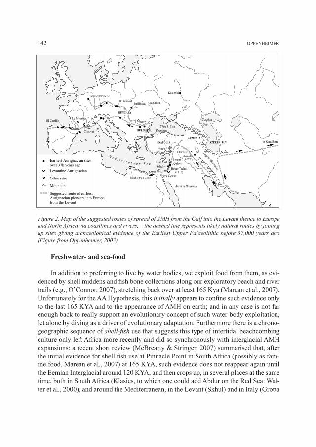
AMH reached the isolated Sahul continent across the seas at least by 48 ka and pos-sibly by 60-50 ka. AMH only finally arrived in Europe from South Asia around 46 KYA, probably linked to climatic amelioration during MIS-3 (Oppenheimer, 2009). Use of water bodies is exemplified by the earliest Upper Palaeolithic colonisation of Europe and North Africa from the Gulf region in South Asia, via the Levant, from around 45KYA, which then followed both coasts of the Mediterranean and the European rivers such as the Don and the longest, most-central European river, the Danube (Figure 2).
AMH extended water bodies as highways somewhat farther than archaics. Apart from moving to the Sahul by at least 50 KYA (Oppenheimer, 2012), we spread onward to the North Solomons by 30 KYA and the tiny Admiralty Islands 200 miles north of New Guinea by 20 KYA (Oppenheimer, 2009), the rest of the Solomons by 6 KYA and the Far Pacific by c.3,100 years ago (Oppenheimer, 2004, Figure 2). As for the New World, while Beringia, a major former landbridge, allowed the potential for an inland walk to Alaska, of the four commonly suggested routes of New World exploration three involved the sea (Figure 3).
142 OPPENHEIMER
Figure 2. Map of the suggested routes of spread of AMH from the Gulf into the Levant thence to Europe and North Africa via coastlines and rivers, – the dashed line represents likely natural routes by joining up sites giving archaeological evidence of the Earliest Upper Palaeolithic before 37,000 years ago (Figure from Oppenheimer, 2003).
Freshwater- and sea-food
In addition to preferring to live by water bodies, we exploit food from them, as evi-denced by shell middens and fish bone collections along our exploratory beach and river trails (e.g., O’Connor, 2007), stretching back over at least 165 Kya (Marean et al., 2007). Unfortunately for the AA Hypothesis, this initially appears to confine such evidence only to the last 165 KYA and to the appearance of AMH on earth; and in any case is not far enough back to really support an evolutionary concept of such water-body exploitation, let alone by diving as a driver of evolutionary adaptation. Furthermore there is a chrono-geographic sequence of shell-fish use that suggests this type of intertidal beachcombing culture only left Africa more recently and did so synchronously with interglacial AMH expansions: a recent short review (McBrearty & Stringer, 2007) summarised that, after the initial evidence for shell fish use at Pinnacle Point in South Africa (possibly as fam-ine food, Marean et al., 2007) at 165 KYA, such evidence does not reappear again until the Eemian Interglacial around 120 KYA, and then crops up, in several places at the same time, both in South Africa (Klasies, to which one could add Abdur on the Red Sea: Wal-ter et al., 2000), and around the Mediterranean, in the Levant (Skhul) and in Italy (Grotta
ITALY
ANATOLIA
ARMENIAAZERBAIJAN
KURDISTAN
HUNGARY
BULGARIA
UKRAINE
Levant
B l a ck S e a
Zagros Mountains
Taurus Mts.
Caucasus
M e d i t e r r a n e a n S e a
Arabian Peninsula
Caspian Sea
Bosporus
Negev Desert
Don
Danube
Levantine Aurignacian
Other sites
Mountain
Suggested route of earliestAurignacian pioneers into Europefrom the Levant
Earliest Aurignacian sitesover 37k years ago
3.1
to Kara Bom
ShanidarCave
Hauah Fteah Cave
Ksar Akil Qafzeh
Boker Tachtit(EUP)
Skhul
Kostenki
Le Moustier
Chauvet
El Castillo
Geissenklösterle
WillendorfIstállósko

143HUMAN’S ASSOCIATION WITH WATER BODIES
Laurentide Ice Sheet
HudsonBay
Cordilleran Ice Sheet
Bering Strait
Vancouver Island
Prince of Wales Island
Queen Charlotte Islands
Santa Rosa Island
7.1
Palaeo-Indian sites
Other archaeological sites mentioned in text
Ice sheet
Beringian continent
Theories of entry to the Americas
Ice corridor route from Beringian land bridge
Coastal route theory
“Solutrean Hypothesis”: from Spain
Pacific crossing theory
Meadowcroft Rockshelter
Monte Verde
Clovis Cactus Hill
Old Crow
Kennewick
Spirit Cave Man
Pelican Rapids
Browns Valley ManWizards Beach Man
Buhl Woman
Luzia
Olmec culture
Quebrada TacahuayQuebrada Jaquay
Daisy CaveKenosha
Topper
Taima Taima
Pedra Furada
Bluefish Caves
Tiérra del Fuego
GREENLAND
ALASKA
BERINGIA
dei Moscerini). While the others could imply involvement of AMH, Italy would imply that Neanderthals might have learnt this culture at that time, perhaps from AMH. There-after, from around 80 KYA (McBrearty & Stringer, 2007, Figure 2) examples of shell-use appear in quick succession in Morocco (Taforalt) and Africa (Blombos and Enkapune Ya muto - Kenya). Later a variety of bones of other fish, fish-hooks and harpoons appear in the record and now factory ships hoover the seas.
While the above narrative could argue for a late AMH acquisition of beachcomb-ing culture, well in time for them to take it everywhere, only after their definitive exit, it does not preclude earlier water-body exploitation by Hominins. Absence of evidence is not evidence of absence, and invisibility of evidence of shell use on ancient sea coasts is very much determined by ancient sea levels relative to today’s sea level. The brief in-terglacial high levels, such as the Eemian were a few metres higher than today and their
Figure 3. Four commonly suggested routes of initial entry to the Ameri-cas from Asia and Europe, three of which presume movement along coast (an ice coast in the case of the Solutrean Hypothesis) or across sea. For clarity, present-day coast-line shown (Figure from Oppenhe-imer, 2003).

144 OPPENHEIMER
shell middens should be visible today. By comparison, for most of the Pleistocene era, sea levels were far lower than they are today, which would have hidden such evidence today (Oppenheimer, 2003, p. 77).
Evidence of exploitation of fresh-water molluscs and crustaceans, not to mention vegetation, by hominins, Erectus (in SE Asia and Africa), or Paranthropus and Australo-pithecines (in Africa) is difficult to exclude and is the subject of several papers (Verhae-gen & Puech, 2000; Verhaegen, this issue; Zohar & Biton, 2011; Joordens et al., 2009; Rabenold & Pearson, 2011).
Exploitation of new sources of food, including scavenging in the intertidal zone and around and wading (Review: Niemitz, 2010) into the shallow edges of lakes is not, in itself, evidence of innate adaptation; rather it could denote behavioural flexibility and cultural inheritance. Dietary opportunism in varied habitats is characteristic of hunter-gatherer humans and extends particularly to our closest cousins, chimps, who have a far wider range of diet than, for instance, gorillas. So, it is important to try and distinguish the nature, strength and reasons for our inclination or niche attachment to water bodies and explore evidence for any putative physical or physiological adaptations to that habi-tat. For the former I will discuss our daily fluid requirements as well as human parasites that hold evidence for prolonged water contact during our evolution. For the latter, I will examine several putative physical and physiological adaptations for indications of the degree of water contact between ourselves compared to other mammals. Again my choices will be eclectic rather than exhaustive.
Fluid balance among Hominins
Compared with desert-adapted mammals in our size range, humans are not even physiologically well-adapted to savannah. Unlike Arabian gazelles, Oryx and camels, the first of which need not drink at all, and the latter only periodically, humans have poor tolerance of dehydration and a profligate daily water-requirement (3-4 litres in temper-ate zones). Our obligatory daily fluid loss, 1,600ml or 2% of body weight (BW) causes noticeable dehydration with thirst, without frequent replenishment. At 5% dehydration noticeable symptoms and physical signs occur, while at 10–15% death supervenes. In the tropics and sub-tropics the recommended minimum daily requirement of a 70 kg man is 5 litres, or 7% of BW. Additionally, a 70 kg man in the heat can sweat 4–6 litres daily (total thus averaging 14% BW) without additional activity or food intake (Gleick, 1996). Even this is not an upper limit of water loss, but the point is evident, that daily water intake is required in all environments and, while walking in tropical heat across deserts, frequent drinks are essential to prevent rapid demise. Inland arid routes with insecure water supplies are not conducive to voluntary wandering, or survival.
Our BW-based daily fluid requirements are, in fact, very similar to those recom-mended by veterinarians for our cousins: chimps, gorillas and orangs living in zoos. One

145HUMAN’S ASSOCIATION WITH WATER BODIES
could even make the comparison that as far as such physiological requirements are con-cerned our bodies and kidneys are still working on jungle settings. However, it may actu-ally be worse than that, since our extraordinary capacity for sweating (above), argued by some to be an adaptation to keep our body temperature stable when following wounded game, puts even greater demands on the availability of fresh water in the tropical zone.
Whatever our other possible reasons for staying near water bodies, the imperative need for fresh water trumps all and may have obliged our ancestors to make a virtue of ‘niche necessity’ or at least make the best job of an inevitable limit to wandering when faced with arid environments. It thus becomes more important in the evolution-ary/aquatic discourse to assess other evidence as to whether early hominins had a closer relationship with water bodies than just opportunistic beachcombing.
Parasites, humans and water
A surprising source of evidence for intense water-contact and a wet human home-land comes from Hominid parasites, which offer a postscript or ‘parascipt’ view on our evolutionary past (Ashford, 2000). In a perceptive review, parasitologist Richard Ash-ford (2000) pointed out:
“Three Schistosoma species and Dracunculus medinensis depend for their transmis-sion on our entering water…Elaine Morgan [1972] suggested an aquatic phase in our evolution… in support of this theory, as far as we know, no other primate has indications of such long-standing association with water.”
Indeed a whole section of research into the transmission of Schistosomiasis in Af-rica, generating multiple PhDs, is devoted to measuring the duration and frequency of water contact, mainly among women and children in shallow, Schistosomiasis-snail-vector infested waters. Such exclusive human distribution of a parasite, that requires regular water contact for transmission, implies an unrelated species jump, coupled with a systematically new human water-contact behaviour, acquired somewhere between the most common recent ancestor of chimps and humans (6.5 MYA); then allowing suf-ficient time for three unique new Schistosome species to evolve worldwide. (Ashford, 2000). One could of course add to this requirement for direct human contact with water, the indirect requirements of the four human-specific malaria species, which depend for their life-cycles on human groups living continuously within a 300 metres radius and whose mosquito vectors are dependent on water bodies to breed.
Ashford’s well-informed insights do not stop there. In a reconstructed scenario, quoted here, which offers an unique parasitic view on the evolutionary homeland of Homo and/or their Australopithecine ancestors, he points out first:
“…of the 400 or so species of parasite that infect man…only about 10% specialise in life in man [i.e., are human-specific]… consistent with the idea that we are a recent species to which not many parasites have become adapted”

146 OPPENHEIMER
Of these very few ‘human specialists’ even fewer are geographically restricted and these very few are unsurprisingly found in sub-Saharan Africa. But Ashford speculates further:
“However, the ‘human specialists’ are not concentrated, as might be expected, in the East African savannahs, but in the rain forests of West and Central Africa: the cradle of humanity, according to our parasites, was not Olduvai or Danakil, but somewhere in Gabon or Cameroon.”
While the parasitic lens Ashford uses might be regarded by some as exotic, even fanciful, the logic is impeccable and the woodland/waterside (rather than dry savannah) inference seems to fit the same chrono-geography which, in terms of evolutionary time, could explain why modern humans still share the same extravagant fresh water require-ments as chimps and our other great ape relatives in the rainforest 6.5 MYA after their final evolutionary split.
Other parasitic clues to our past habitats and habits come from the phylogeny of body lice, ectoparasites, which shadow Hominin evolution closely, at the same time confirming some of the dating nodes (Review: Weiss, 2009). Chimp and human body lice (respectively Pediculus schaeffi & Pediculus humanus) have an estimated and an un-surprising mitochondrial split date of 5.6 MYA, while we apparently acquired our pubic lice (Pthirus pubis. which are separated genetically from Pediculus sp. by 13 MYA) as an opportunistic species-jump from gorillas (Pthirus gorillae) only 3.3 MYA ago (Weiss, 2009), inferring that Hominin partial body hairlessness may go back at least that far (Weiss, 2009) and significantly, that as naked apes, we would have shared the same habitat (presumably woodland) as gorillas as late as then, and thus with our common ancestor to chimps somewhat earlier (Weiss, 2009).
Finally, on this uncomfortable topic, salt water apparently kills both human head and body lice (Pediculus capitis & Pediculus corporis) (although not their nits), suggest-ing that if the ancestors of modern Eurasians started out beachcombing along the Indian Ocean (Oppenheimer, 2003), they didn’t spend much of their time in the sea; otherwise the salt would have quickly rid them of these unwanted guests, rather than carrying them way out of Africa, again, to meet and mix, as they did, with their pre-existing genetic cousins living on archaic Homo in Eurasia (Kittler et al., 2003).
Waders, but not divers?
The above suggestion of a West and Central African human evolutionary nursery in the jungle (see also below), might also explain an anatomical anomaly which, on the face of it, denies the AAH perspective that Homo has appropriate anatomical adaptations for prolonged immersive water contact. This anomaly is the lack of convincing facial adaptations for swimming or duck-diving amongst any of the great apes, while at the same time they all share the same predilection for bipedal wading.

147HUMAN’S ASSOCIATION WITH WATER BODIES
Modern humans can swim, but it is generally observed that, as children or adults, they do not initially take to water safely or naturally without some degree of assistance or training; and airway protection is a constant issue for parents. This contrasts with a number of mammalian quadrupeds, which choose to swim on occasion and need, natu-rally, safely and without such hesitancy - from the start. These occasionally-aquatic vol-untary swimmers include a variety of ungulates (horses, antelopes, reindeer and even sheep), carnivores (dogs and tigers), pigs and elephants; and all these quadrupeds appear to swim efficiently and with ease. Two naturally shared structural anatomical features amongst all these groups give them a ‘head-start’ in comparison to humans. The first is that they are quadrupeds, allowing the same trunk orientation and streamlining as when they are walking on dry land. The second is the relative position of their nostrils, which in the case of the quadrupedal ungulates and carnivores are naturally proud of the water and nearly on the same level as the eye-line, allowing continuous breathing and forward vision, while swimming. Specialised rodents like beavers and capybara (Hydrochoerus hydrochaeris) have further adapted to a semi-aquatic existence by repositioning their eyes, ears and nostrils further to the top of the head, and developing the ability to close their nostrils and ears under water (neither of which we can do). Elephants of course have their own muscular and extensible snorkel.
In contrast, humans (and other primates if they ever try to swim) have to rotate their trunk forward to a prone position in order to progress through deep water, thus obliging the head and neck to extend backwards by up to 90° in order to see and to breath. Breast-stroke and the easier, more-natural quadrupedal doggy paddle allows easier breathing, both at the cost of horizontal streamlining. Experienced swimmers, using the invented streamlined crawl style, have to breathe sideways intermittently. Furthermore, as a re-sult of the upright stance and the enlargement of the brain in hominids, the cranium has rotated above and over the face, leaving the nostrils well below the eyes and pointing downwards and a heavy brain pushing the nose even further down in the water, thus increasing the risk of water inhalation. The same general principle holds not just for the great apes, but other primates as well. The proboscis monkey (Nasalis larvatus) famously wades around swamps, with even more familiarity than the great apes. But on occasion, when the water is too deep to wade, Nasalis swims. It is not helped in this latter pursuit by the high position of its eyes and the lower position of its long proboscis (but see Morgan, 1972); so, when forced to swim, it uses simple doggy-paddle with its head in a rather more vertical position than ungulates and holds its head and proboscis to one side to avoid waves (personal observation).
In addition to the physical handicaps for swimming listed above, the inability to close our nostrils and ears underwater, would be even more maladaptive for duck-diving or more adventurous dives past or present (Rhys-Evans, this issue, part 2; Erica Schaga-tay, this volume). Clearly, experienced free-divers and other traditional-habitual divers, such as Japanese pearl divers and certain sea-gypsy communities (Erica Schagatay, this issue, part 2) have overcome these disadvantages, with voluntary breath and larynx con-

148 OPPENHEIMER
trol and the usual human inventiveness and perspicacity. But such examples seem rare cultural curiosities which, while being remarkable behavioural adaptations, offer little direct evidence for a prior, mainstream, human-evolutionary route.
There is, however, one set of progressive hominin adaptations, which might have favoured those, who were obliged to wade through forest swamps or even seek refuge there from tree-climbing predators. Those changes are habitual bipedalism, increased height and upright posture (Niemitz, 2010). This would be consistent with the third of the scenarios of Hominin evolution apart from AAH and savannah bipedal ape, namely a woodland, bipedal, wading ape (Niemitz, 2010; Kuliukas, 2009, and this issue). The latter habitat fits with our extravagant chimp-like water requirements, the dates of our ac-quisition of Pediculus and Pthirus (above) and the geographic homeland of our uniquely “human specialist” parasites. It might also help the unresolved issue of a human-genetic and -fossil African-homeland currently still undecided among Palaeoanthropologists be-tween East and South Africa.
West and Central African homeland?
Maybe our ultimate homeland was neither East nor South Africa, rather in West and Central Africa as suggested (above) by our parasites. Recent genetic findings may support these wetter north-westerly African AMH origins. For instance complete-se-quencing of African Y-chromosomes have found that: “…contrary to previous phylog-eny-based conclusions, the deepest clades of the revised MSY phylogeny are currently found in central and northwest Africa. MSY lineages from these regions coalesce at an older time … than do those from east and South Africa ... A scenario of a Y chromosome ‘‘Adam’’ living in central-northwest Africa … would provide a good fit to the present data.” (Cruciani et al., 2011). A more recent report finds an even deeper MSY coales-cence using the complete sequence of an African American Y chromosome. Multiple close African-based genetic-relations to this lineage are found among the Mbo language group in the Cameroon (Mendez et al., 2013; for this new view of the MSYtree in Africa, see also Batini et al., 2011).
This new, wet, West African Y perspective is consistent with features of the re-vised African mitochondrial complete-sequence lineage tree. This tree (e.g., Behar et al., 2008) has a deep split c. 200 KYA, at its root, into two branches, L0 aged c. 150 KYA and the larger, slightly older L1’2’3’4’5’6 branch, aged c. 180 KYA. The L0 branch contains, among others, deep lineages characteristic to the Khoisan of South Africa (L0d & L0k: Barbieri et al., 2013) and other lineages (e.g., L0f &L0b), characteristic to East Africa while L0a is found both in Central and East Africa. The older L1’2’3’4’5’6 branch lacks ancient lineages characteristic of the Khoisan, and South & East Africa, but gave rise to all the rest of worldwide human mitochondrial diversity. Its deepest branch (c. 180 KYA) gave rise to L1 (c. 145 KYA) with its primary branch L1b, being characteristic to

149HUMAN’S ASSOCIATION WITH WATER BODIES
Central/West Africa. Phylogeographically, the new mtDNA tree does not provide good support either for
an East or South African AMH homeland, with ancient spread elsewhere, but rather is consistent, with a Central/West African homeland c. 200 KYA, with early spread to East and South Africa followed by local isolation and drift in the latter until the out of Africa ‘L3’ expansions c. 70 KYA (Soares et al., 2012).
Aquatic ancestry: past diving in humans and a possible evolutionary reason for our “exaggerated mammalian dive reflex”
Moving on to the main topic in my review, I wish to discuss the “exaggerated mam-malian dive reflex” in humans, what it is, why it is exaggerated in ourselves and what the evolutionary evidence is for this, and how the underlying reason for it may have fed back into our recent physical evolution. Of all the anatomical, physiological and biochemical pieces of evidence put forward in support for the AAH, the claim that we have an “exag-gerated mammalian dive reflex”, appeals perhaps most to medics (e.g., like myself). It can be defined, observed and repeated and, on the face of it, does not have any other bio-logical function apart from prolonging one’s period of voluntary hypoxia under water. Few if any of these presumptions are, however, what they seem or are even true.
The very phrase “exaggerated mammalian dive reflex” acknowledges that other mammals also have a ‘dive reflex’, begging the question, if most of them do not dive, as to why it should be called that? Indeed it is stated in the experimental literature, that all vertebrates tested have “diving responses” differing only in intensity and tempo, with the most pronounced being in diving mammals and diving birds (Lin, 1982). So, as with much jargon, the labels “diving…” and “exaggerated diving…” evidently loosely reflect those enhancements, rather than arguing that diving underlies the evolutionary origin of this ancient class of cardio-respiratory reflexes, or that all vertebrates with such en-hanced reflexes are natural divers.
Given the vertebrate universality of these ancient and most powerful of autonomic reflexes (Burgh Daly et al., 1979; Gorini et al., 2009; Schaller, 2004; Scholander, 1963), since they over-ride normal reflex control of arterial blood pressure and blood gases, it is very unlikely they evolved primarily for diving mammals, but more as a protection against some other cause(s) of hypoxia, and have been co-opted for diving purposes by diving mammals and birds.
Rather than get involved in polemic as to whether, and to what degree, these vagal-associated reflexes (for vagus nerve and vagal reflexes see below) are in fact enhanced in humans, I accept, for the purpose of discussion, that they are in fact exaggerated, although this is not stressed in experimental physiological literature, and I would defer to Erica Schagatay and others (this issue, part 2) for the evidence. The question then rephrases to: Why do humans have exaggerated vaso-vagal-associated reflexes to certain stimuli, including water on the face and hypoxia? Have they evolved to help them dive or

150 OPPENHEIMER
is there another evolutionary mechanism, e.g., shared with other terrestrial mammals and even with birds, but especially important to humans? Answering such questions requires closer examination of the literature on these reflexes.
Definition: There is no single adequate definition of the mammalian dive reflex. Lin’s useful (1982) paper summarises the physiology of experimental breath-hold (BH) diving in terrestrial mammals and is worth quoting here. He strangely refers to BH as “self-imposed” rather than voluntary or imposed, presumably since few mammals would continue to breathe normally immediately after immersion, which was only voluntary among the human experimental subjects:
“Prominent responses to BH as bradycardia [= slowing of the heart rate], peripheral vasoconstriction, redistribution of blood flow [i.e., reduced blood flow to everything except brain and heart], and decreased respiratory gas exchange ratio.”
While Lin mentions additional protective mechanisms in diving mammals: “High oxygen-carrying capacity of the blood and relative insensitivity to carbon
dioxide,” not to mention high muscle myoglobin and others, he goes on, “Studies of the superior ability of the diving species to stay under water revealed that the key to surviv-ing prolonged asphyxia is maintenance of the oxygen supply to the brain and heart at the expense of other organs. A similar relationship also holds in human BH but is subject to modifications by neural, chemical, and mechanical factors as the BH progresses. Div-ing bradycardia is mediated by the vagus nerves… confirmed in all species tested… the afferent limb [i.e., the sensory source of stimulus resulting in vagal bradycardia] has multiple origins … In general, low PO2 …and young age potentiate apnea-induced bradycardia.”
Bar some subsequent insights, those statements, echoing Scholander (1963), still hold and in addition indicate the potential reflex complexity.
The other main problem with the definition of the “diving reflex” is that there is no one simple “vaso-vagal reflex” involved. There are in fact several reflexes, which interact with each other, both as far as afferent & efferent pathways are concerned, but also with the respiratory and cardiovascular centres of the brain, in a concerto that is not just focussed on avoiding the effects of extreme hypoxia, but is also about determining whether or not to breathe when hypoxic.
This interacting complex is particularly well understood from work on young in-fants and new-born mammals. Although the bulk of the world observational electronic data on cardio-respiratory responses to perinatal hypoxia comes from millions of routine clinical observations of human mothers and babies both during labour and afterwards (Beard, 1974), the main controlled experimental studies have been carried out in gravid sheep and their newborn lambs, as well as rabbits (Tchobroutsky et al., 1969), all of whose “diving reflexes” are vigorous. Similar work has been carried out on avian em-bryos within eggs. The latter also have problems of severe pre- and peri-natal hypoxic episodes, associated with reflex protective bradycardia (Mortola, 2009), i.e., “dive re-flexes” and are much more accessible for experimental study than mammalian embryos.

151HUMAN’S ASSOCIATION WITH WATER BODIES
As stated, there are several reflex elements which are involved and commonly con-flated in the term “mammalian diving reflex”. The common efferent (or motor) side of two of these involves, among others, motor parasympathetic fibres of the vagus nerve causing marked bradycardia, often followed by hypotension (low blood pressure), and then peripheral vascular constriction caused by sympathetic nerves, reducing blood flow to nearly everything except the heart and brain. Although common to all mammals, this apparently protective autonomic response, being powerful, potentially-dangerous, and occasionally lethal in unusual or inappropriate circumstances, is rarely invoked in nature except in very young infants and the perinatal period, when sensitivity to its several trig-gers is at its highest.
The carotid-body-cardiac reflex (CBCR)
The afferent (sensory) signal for one of these two reflexes, the CBCR, is triggered by sudden severe hypoxia and comes from the carotid body (in the neck) and aortic body sensors, which are excited by a reduction in blood oxygen tension and also by a rise in blood CO2 tension and hydrogen-ion concentration (Burgh Daly, 1979). The carotid body’s every-day role is as an essential sensor, signalling minute changes in blood gas status to the central respiratory centre which finely controls respiration.
The relevance of the more powerful CBCR as a protective response at birth has long been realised (Beard, 1974). Birth asphyxia (more strictly perinatal asphyxia, since severe hypoxic events occur before birth but may continue after birth) is a major cause of morbidity and mortality in the perinatal period (see sections below) for all mammals and also other terrestrial vertebrates. Part of the hypoxia results from the profound car-diovascular and respiratory effects resulting from change of environment at birth. Addi-tionally in placental mammals, during the immediate prenatal period and during labour, uterine contractions and the passage through the birth canal also frequently compromise placental oxygen supply to the fœtus leading to prenatal hypoxic events. Many of these events are extreme and associated with severe fœtal bradycardias observed frequently on external fœtal cardiotocography. These bradycardias (known in obstetric jargon, when I did my neonatal training, as “dips”) are well recognised as responses to hypoxia (“fœtal distress”) and are of potentially serious import to the survival of the fœtus unless careful observation and appropriate active intervention are instituted (Beard, 1974).
The trigemino-cardiac-reflex (TCR)
The other afferent sensory source for the “mammalian diving reflex” comes from the head and neck region, particularly the face, nose and larynx, and is thus sometimes called the trigemino-cardiac-reflex (Schaller, 2004) and is particularly associated with

152 OPPENHEIMER
the immediate prenatal period. Most terrestrial vertebrates avoid inhaling water when immersed, but the TCR takes this simple reflex much further. A variety of stimuli in the face and neck (Burgh Daly et al., 1979; Tchobroutsky, et al., 1969; Gorini et al., 2009), particularly warm water (>34°C) or irritants (Tchobroutsky et al., 1969) in the nasal/laryngeal region, can elicit the reflex. While the bradycardia, hypotension and periph-eral vascular shutdown responses of the CBCR are co-opted centrally, another powerful negative effect is added, central reflex apnœa persisting in the presence of hypoxia.
Reflex mediated respiratory depression overriding the normal effect of hypoxia on the carotid bodies in the prenatal period was recognised from over 50 years ago (e.g., Barcroft, 1946):
“We suggest that an inhibitory mechanism may be responsible for the apneic state of the fetus in utero: the immersion of the head, and particularly the contact of fluid with the glottis … inhibits the respiratory movements as it does during diving in adult and newborn animals …We think that in some respect the fetal state can be compared to a dive.” (Tchobroutsky et al., 1969) “Hypoxic stimulation of the carotid bodies normally produces hyperventilation and bradycardia. When apnœa occurs centrally or reflexly, carotid chemoreceptor excitation resulting from asphyxia now causes a much enhanced bradycardia and even cardiac arrest, but paradoxically does not usually affect respira-tion.” (Burgh Daly, 1979)
And: “In the case of birth asphyxia, short-term responses to acute oxygen lack: … in-
clude reduction of heart rate and redistribution of circulation as in diving mammals, & reduction of respiration rate” (Singer, 1999)
This means that when the TCR is activated, the respiratory centre in the brain in-hibits breathing as well, by over-riding normal breathing reflexes. The central neurop-harmacology of this combined reflex, which is enhanced in the perinatal period and in early infancy, is now quite well-understood (Gorini et al., 2009), although this does not directly illuminate the purpose or role of the apnœa.
The addition of over-riding central apnœa to over-riding bradycardia, hypotension, peripheral shutdown and ongoing severe hypoxia obviously makes the TCR especial-ly dangerous immediately after birth, when the need to take the first breath is urgent. The reflex also later contributes to rare deaths associated with intubation, laryngoscopy, bronchoscopy and accidents involving underwater swimming, and it has repeatedly been implicated, on neuropharmacological evidence, as an underlying cause of Sudden Infant Death Syndrome, which is confined to early infancy (Gorini et al., 2009; Schaller, 2004).
Fortunately for the newborn, nature has other reflex mechanisms to reverse the pre-natal inhibition of breathing, using a combination of sensory and chemical stimuli, one of which: cold water (<24°C) on the face has clearly been demonstrated in lambs experi-mentally, even in utero (Tchobroutsky et al., 1969).

153HUMAN’S ASSOCIATION WITH WATER BODIES
Evolutionary selective purpose of prenatal reflex apnœa?
Such a potentially fatal, powerful and rare combined autonomic reflex (TCR) can hardly be random, and a uniquely appropriate protective role for adding apnœa to the hy-poxic vaso-vagal response in the immediate prenatal period has been suggested several times (Tchobroutsky et al., 1969; Schaller, 2004).
It might be asked why apnœa should be of value to a human fœtus (or any other spe-cies) and especially in association with reflex bradycardia? There is a simple answer to that: meconium aspiration, though there may be others, such as keeping the lungs empty of fluid before passage through the narrow birth canal.
A serious and common consequence of severe hypoxic fœtal distress in late preg-nancy and of post-term delivery is evacuation of the bowel contents, resulting in so-called meconium (a dark brown viscid substance from the fœtal gut) -staining of the amniotic fluid. Prolonged extreme hypoxia ultimately results in pre-terminal gasping of the fœtus in the uterus and then, inevitably, inhalation of the meconium occurs, causing a severe and frequently fatal meconium pneumonitis, that is, if the hypoxic infant survives delivery, which a surprising number do.
This dangerous sequence of events is sufficiently common in human deliveries that an evolutionary argument has to be made for the survival value of a reflex inhibiting respiration in the presence, not just of submersion in fluid (which is obvious and occurs in most mammals and when a newborn baby is deliberately placed or delivered under water), but especially in combination with the vagal-reflex response to severe hypoxia in utero. The combined reflex situation clearly only occurs commonly among terrestrial mammals in utero, or when aquatic mammals (or even highly trained free-divers) make prolonged dives.
Can the evolutionary benefit of the ‘diving’ reflexes (CBCR & TCR) to as-phyxiated newborns be measured?
Outside Africa, 43%-47% of under-5 deaths occur in the neonatal period in all re-gions (Bryce et al., 2005). 77% of neonatal deaths occur in the first week (Early Neonatal Mortality ENM) after birth (WHO, 2006, Table A5.2). Stillbirths (SB) account for more deaths than ENM (WHO, 2006, Table A6.1) and up to 45% of intrapartum SBs are likely to be due to asphyxia (Lawn, 2009).
Severe hypoxia with patent carotid arteries results in damage to the brain, the gut, the kidneys and results in death in adults within minutes. In adults, the heart and brain are only relatively protected as a result of the carotid-body-cardiac reflex (see above). Objective evidence of multiple bradycardias during a significant proportion of human deliveries indicates the potential size of the threat. However, the full-term fœtus and newborn seem to be especially protected by their reflex responses to hypoxia. From my

154 OPPENHEIMER
own experience as a paediatrician working both in developed and tropical environments and from reading of the literature (e.g., review Singer, 1999), I have never ceased to be amazed at the extraordinary physiological resilience of severely asphyxiated newborns, to these dire outcomes, after prompt & adequate resuscitation, when compared with other times of life. But obviously there are limits. Could it be worse?
That newborn mammals do often manage to survive birth asphyxia, and avoid most of those permanent morbidities, owes itself apparently to their adaptive reflexes, in par-ticular to their vertebrate diving reflexes. Their specific relevance in perinatal defense to hypoxia was recognised by Scholander (1963) who described the reflex as the ‘mas-ter switch of life’. However, such protective reflexes cannot be directly measured for their special adaptive value in the human perinatal period, against their ‘absence’ since they are universal and very ancient, being elicited even in fish out-of-water (Scholander, 1963).
There is, however, an indirect approach to illuminate the potential cost of perinatal hypoxic risks in humans, since not all of the credit for modern perinatal survival goes to nature. Obstetric care and timely intervention for the distressed fœtus, in slow or ob-structed labour and resuscitation for the terminally asphyxiated newborn, has made an enormous difference both to perinatal and maternal mortality over the past 150 years, though obstetricians are often unjustly criticised from outside the profession for being too ‘interventionist’. Their contributions can be assessed through regional and historic statistics.
Asphyxia neonatorum, and its association with neonatal death, convulsions and/or later cerebral complications and spasticity (Little’s disease) was recognised in the 19th Century (Little, 1861). When Little presented these associations to the Obstetric Society of London, several colleagues objected, pointing to numerous examples of infants (such as Samuel Johnson) who, in their knowledge or experience, had survived the insult un-blemished by palsies or mental retardation, a remarkable paradox still present today, and acknowledged then by Little in his recorded response.
My own personal experience allows a comparison between rapid access to first class perinatal facilities in Oxford, England (1976) and the available use of electronic fœtal monitoring during labour in contrast to the situation in Papua New Guinea (PNG), where I worked between 1976-1983 and access to hospital was difficult and electronic monitor-ing of fœtal heart rate was not available. In one hospital I worked in, perinatal mortality (PM=combined ENM and SB) was 8.3%-8.5% of term deliveries in 1980 and had only fallen to 5.1% in the 1995-1999 period (Kemiki & Vince, 2003).
Pre-modern obstetric figures for perinatal mortality in Europe give a stark picture of natural perinatal wastage, much of it due to asphyxia. However, while asphyxia neonato-rum was recorded over 150 years ago, the consistency of the earlier statistics are too poor to compare accurately with 20th-21st century figures. Comparable perinatal figures are available for Norway for 1905 when the stillbirth rate (SBR) was 23/1000 births. 37% of these deaths would have been intrapartum (i.e., likely due to asphyxia and/or difficulty

155HUMAN’S ASSOCIATION WITH WATER BODIES
of birth: WHO, 2006, Table A7.1). Early (first week) neonatal mortality rate (ENMR) was 18/1000 live births, giving a perinatal mortality (PM) of 41/1000 births (i.e., 4%), of which around 40% would have been likely due to asphyxia (WHO, 2006). In other words about 2% of all Norwegian births died of birth asphyxia in 1905. The comparable Norwegian figures in 2000 were SBR: 4, ENMR: 2 and PM of 6/1000 i.e., a decrease to 1/7th of the 1905 figure, in large part due to active management of perinatal asphyxia (WHO, 2006).
By comparison equivalent PM figures for the whole of Africa were still (similar to PNG) 80/1000 (8%) in 1983 falling to 60 (6%) in 2000, although many individual African country SB figures were much higher (e.g., Mauritania PM=111/1000 or 11.1% of births in 2000; WHO, 2006). The latter figure could imply as high as 4.4% African births dying due to perinatal asphyxia. Worldwide, such figures translate to recent total estimates (Lawn, 2008) of 4 million annual neonatal deaths and 3.2 million stillbirths, 99% in low- and middle income countries and half at home (Lawn, 2008). 2.02 million of these perinatal deaths (0.92 million neonatal deaths and 1.1 million stillbirths) are directly related to intrapartum hypoxia (i.e., still 28% of total PM) each year (Lawn, 2007, 2008). Today’s perinatal asphyxial wastage (estimated 2%-4% of all newborns worldwide), would clearly have been even higher pre-modern medicine and such a con-stant cull in every generation would be expected to select powerfully for an exaggerated protective ‘diving reflex’ in the human perinatal period. It is difficult to see how the sup-posed evolutionary benefits of adult free-diving in the scattered traditional communities that practice it, could ever have matched such a powerful selective effect which is still a universal risk in our perinatal period, added to the selective risks to mothers, which will be discussed below.
Why is labour so dangerous for the human fœtus; is it adapted? Birth from egg or womb is physiologically stressful for every terrestrial or avian
vertebrate conceptus, with elaborate cardio-vascular reflexes coordinating the transition to air-breathing. With the possible exception of marsupials, exit from the mother’s body is also mechanically stressful, thus dangerous, to both mother and offspring, delay being risky and failure of delivery being lethal to both. A delicate evolutionary balance is thus struck between maturity (precocity) favouring the survival of conceptus and immaturity (altriciality) favouring the survival of the mother at delivery, the limiting balance being fought between the largest transverse diameter of the fœtus, or the diameter of the egg, and the relevant smallest diameter of the birth passage (Kurki, 2011). Evidence for such evolutionary conflicts and compromises are apparent in many mammals, earlier primates being no exception (Tague, 2005; Rosenberg & Trevathan, 2002). Before discussing evolutionary aspects of this conflict on adult human female dimensions it is worth exam-ining the fœtal evidence for adaptation to a limiting birth canal.

156 OPPENHEIMER
In human newborns, whilst shoulder dystocia is a significant cause of obstruction, the key limiting dimension is the relative size of the fœtal head. Humans are altricial (helpless) compared with other primates (Rosenberg & Trevathan, 2002) and rapid brain growth continues long after birth, unlike chimps, their nearest relatives, whose brain growth is already declining markedly at birth and continues to decline thereafter. Curi-ously, two publications I was involved in, just before leaving for PNG in 1976, found evidence that even this altricial adaptation was insufficient in humans. In the last 8 weeks before delivery, human head circumference growth rate, which is at its maximum be-tween 29-31 weeks gestation, decelerates dramatically until delivery by which time the head has stopped growing. Then within a day or so of birth, head growth again achieves its former maximum growth rate of over 1mm/day (Fujimura & Seryu, 1977).
The timing of this reversal from zero growth to maximum head-circumference growth in the newborn is determined by the event of delivery and gestation (Fujimura, 1977), indicating the evolutionary mechanical importance of the third trimester decelera-tion in head growth as a compromise to accommodate limiting maternal pelvic dimen-sions.
It has been long known that there is a high correlation between measures of mater-nal size (in particular pre-pregnancy weight and to a lesser extent also of paternal size) and fœtal size at birth (Miletic et al., 2007; Thame et al., 1997; Neggers et al., 1995; Rice & Thapar, 2010; Knight et al., 2005), i.e., small mothers and fathers have smaller babies (and vice versa). However, it is not always clear how much of the correlative effects and timing are pre-determined by parental genetic influence and how much is determined by the fœtus and/or the interactive environment of the individual pregnancy (also referred to as the intra-uterine environment).
To answer this question, a careful recent study has found evidence for a specific interactive environmental relationship between maternal skeletal height, newborn head circumference and timing of delivery (Rice & Thapar, 2010). The modern phenomenon of in vitro fertilization was used with multiple regression analysis to separate passive inherited maternal (and paternal) genetic influence from active, environmentally deter-mined interactions during pregnancy. Three comparable groups were studied: 1) homol-ogous (i.e., the default with both parents genetically related to the conceptus), 2) sperm donation (only the mother genetically related) and 3) egg donation (only the biological father related).
While there was exclusive evidence from all three respective groups that both bio-logical parents individually had clear genetic influence on the size of the newborn, the third group was key in the context of active maternal-fœtal interactions, since in group 3), individual maternal interactions/correlations with fœtal dimensions could only be environmentally determined, and could not be confounded/contributed by shared genes. In this group, maternal height was strongly and positively associated with birth head circumference. The effect on cranial size was specific for maternal height and absent for maternal weight.

157HUMAN’S ASSOCIATION WITH WATER BODIES
Maternal height was also associated positively with birth weight in this unrelated ‘environmental’ group, but to a much lesser extent. When these correlations were cor-rected for gestational age of the newborn “…the correlation of maternal height in the egg donation group was no longer significant illustrating that maternal height in part affects infant birth weight through influences on gestational age in addition to effects on foetal growth.” (Rice & Thapar, 2010).
Another study, but in homologous pregnancies, supports this environmental infer-ence and has “…found a gradual increase in pregnancy length by increasing maternal height, and the association was essentially unchanged after adjustment for maternal car-diovascular risk factors, parental age, offspring sex, parity, and socioeconomic meas-ures… Paternal height was neither associated with pregnancy length nor with the risk of pre- and post-term birth.” (Myklestad et al., 2013). Looked at the other way, babies of smaller mothers tend to be born at an earlier gestation, i.e., when they are smaller, with less risk of obstruction (see also below).
However, Rice & Thapar’s phrasing “maternal height …affects…gestational age” implies, by default, that the mother determines delivery date through her skeletal stat-ure, whereas the literature (Turnbull & Anderson, 1969) tends to suggest the opposite, namely that the fœtus hormonally controls the date of delivery by affecting uterine con-tractility. How an unrelated fœtus could assess maternal skeletal dimensions, in order to make an appropriate determination, except by tightness of fit after its head has engaged (descended into) the pelvis during the third trimester, is another matter. Possibly the de-tector mechanism could be shared with the mechanism for the slowing of head growth reported above.
Maternal mortality resulting from cephalo-pelvic disproportion (CPD) and ob-structed labour
Turning to modern evidence for maternal factors associated with difficult deliver-ies, one may enquire if there is continuing evidence for potential selective influence of the size of the birth canal. While it might be imagined that enlargement of the birth canal to accommodate larger human newborn heads would be a trivial evolutionary adapta-tion, this cannot be assumed, since that would tend to affect the stability and integrity of the pelvis for bipedal gait. In any case, the slowing of the fœtal head growth, discussed above, indicates that an evolutionary compromise must have occurred in the fœtus. So, what evidence is there that the female pelvis is a continuing evolutionary bottleneck?
Worldwide, the mother dies in 0.4% of all livebirths, with a lifetime mortality risk of 1.35%; these figures rise to 0.92% of all livebirths in Sub-Saharan Africa, with a life-time mortality risk of 6.25%. Obstructed labour (OL) occurs in over 6 million deliveries (4.6% of all livebirths in 2000:AbouZahr, 2003) annually, and is the third common-est major obstetric complication after severe haemorrhage and abortion accounting for

158 OPPENHEIMER
12.3% of cases. OL is a potent annual cause of maternal mortality, accounting for 42,000 maternal deaths and 11% of all maternal deaths (case fatality rate 0.67%). The world figure of 42,000 deaths would of course be much closer to 6 million i.e., 4.6% of births, without any modern obstetric intervention, incidentally losing the baby as well as the mother (AbouZahr, 2003).
In the PNG Highlands hospital I worked in from 1976-77 (Goroka Base Hospital), the Maternal Mortality Rate (MMR) over the period 1964-73 was 2.16% of all deliver-ies, far higher even than the African figures. 54% of this loss was due to obstructed labour and its complications, many cases of which often arrived from far afield, already with uterine rupture and maternal shock (Cambell, 1974).
These horrific figures for 20th Century MMR in obstructed labour indicate that, in recent-modern human communities, the size of the birth canal was an even more potent evolutionary bottleneck than it is now. Such wastage would be expected to select rap-idly both on the relative dimensions of our birth canal and on term head circumference, since the survivorship of mother and fœtus are closely connected. There is evidence for such human evolutionary adaptation both in terms of head/brain size (over and above the active fœto-maternal control discussed above) and in pelvic size; the former will be addressed first.
Brain and head size in hominin and human evolution
Can one predict any other, independent, evolutionary evidence that the size of the birth canal is still a potent maternal evolutionary bottleneck in humans and that prehis-toric human heads were getting too big for their mothers’ pelves? The simple answer is yes: due to high maternal wastage from CPD, our heads might have stopped their evo-lutionary trend to enlargement - or even got smaller. Judging from the evidence of adult human fossil skulls (Ruff, et al., 1997) and modern anatomical studies (Miller & Penke, 2007), this is just what happened (Figure 4) (Ruff, et al., 1997; Oppenheimer, 2003, pp. 16-17 & Figure 0.2). A reduction in brain size to adapt to high maternal and fœtal wastage is a tempting hypothesis, though the evolutionary allometric argument for that mechanism is anything but straightforward.
OppOsite page:Figure 4. Three phases of brain growth in Homo and Paranthropus over the last 2.5 million years, sep-arated chrono-geographically by vertical dashed lines. The last of these phases, brain size reduction in H. sapiens, occurred during our most rapid cultural acceleration (important cultural milestones given equal weighting). Log-log regression lines 1–6 relate to six closely related contemporary regional hu-man types as shown by symbols in the key. Data from Ruff et al., 1997; Elton et al., 2001. Ages as in McBrearty & Brooks, 2000. (Figure from Oppenheimer, 2003).

159HUMAN’S ASSOCIATION WITH WATER BODIES
6
5
4
3
2
1
Asian erectus sp.
Anatomically modern humans/H.sapiens
African archaic H.sapiens/H.helmei
1700cc
1800cc
1900cc
2000cc
400cc
500cc
600cc
700cc
800cc
900cc
1000cc
1100cc
1200cc
1300cc
1400cc
1500cc
1600cc
100k250k500k1M2.5M 2M 1.5M
Brain size reduction inHomo sapiens line
Steady brain growth only outside Africa– in lines not ancestral to Homo sapiens
Rapid proportionalbrain growth in Africa
Long-distance exchange
Shellfishing Microliths
Beads
Barbed pointsBone toolsFishingIncised pieces
Gravettian arte.g. High-qualitypainting
Mesolithic
NeolithicMetal Age
Writing
Musical notation
Acheulian
Stone toolsOldowan
Oldowan
Start of MiddlePalaeolithic
Developed
LowerPalaeolithic
Start ofUpper
Palaeolithic
Bra
in s
ize
evol
utio
n
T E C H N I C A L C U L T U R A L
I N
NO V
AT
I ON
Years before present
0.2
African: H.erectus/H.ergaster/ H.rhodesiense
H.habilis/ H.rudolfensis
Paranthropus sp.
European: H.heidelbergensis& H.neanderthalensis

160 OPPENHEIMER
As already mentioned, humans are more altricial than other primates, and they adapt to slow their intrauterine head growth and also get delivered earlier, if their mothers are small. Additionally the fœtal head moulds to the shape of the birth canal during passage; and during pregnancy, maternal pelvic ligaments loosen under the influence of Relaxin to allow the pelvis more squeeze room (see also Tague, 2000, p.390; Russell 1969). In spite of all these adaptive responses we still have the most difficult delivery and easily the tightest cephalo-pelvic fit among the great apes (Tague & Lovejoy, 1986). Bipedal Australopithecus afarensis (AKA Lucy) had a pelvis estimated at 13.5% more spacious to accommodate the term fœtal head (assumed the same size as Pan) than modern hu-mans, while Lucy was estimated at 5.9% more constricted compared with chimps (Tague & Lovejoy, 1986), with a similar-sized brain.
Modern humans have an average brain volume nearly 3.5 times chimp size at 1350 cc (Ruff, et al., 1997). Nearly all the increase in cranial capacity (Figure 4) occurred before H. neandertalensis and H. sapiens (Oppenheimer, 2003, Figure 0.2 & pp. 16-17; Ruff, et al., 1997). Indeed, the modern adult mean brain volume of 1,349 c.c. was already achieved in the early Late Pleistocene (100-150 KYA), among a mixed archaic H.sapiens/Neanderthal sample: 1,354 c.c. - the presence of Neanderthals in the pooled sample made no difference to the trends (Ruff et al., 1997). Brain volumes then peaked at mean 1,501 c.c. around c. 100 KYA (AMH/Shkul-Qafzeh) and plateaued there for 80 Kyr (Ruff, et al., 1997), next estimated in Neanderthals: 1,498 cc., 72-40 KYA; and then Early Upper Palaeolithic (EUP) AMH :1,517 cc., 35-21 KYA.
From the EUP, however, AMH brain volume decreased progressively with mean Late Upper Palaeolithic (21-10 KYA) volumes at 1,466 c.c. and Pueblo Indians: 1,308 c.c.; 1,000 YA; and worldwide modern values at 1,349 c.c. (Ruff, et al., 1997, Table 1). The stepped decline in grouped cranial capacity estimates was statistically signifi-cant from the EUP group onwards to the Pueblo group, with a total fall in adult cranial volume of 13.8% (Ruff, et al., 1997). Prior to this, every other Homo group both within and outside Africa, including Neanderthals, and even the Paranthropus genus, had pro-gressively and dramatically increased their brain volume (Figure 4). AMH is the only Homo group that failed to increase their cranial capacity further, with a prolonged 80kyr plateau, then reducing brain volume progressively over 35 Kyr; (Ruff, et al., 1997, Table 1 & Figure 2b; Oppenheimer, 2003, pp. 16-17 & Figure 0.2) The sharp decline in AMH cranial capacity occurred over the most dramatic period of cultural change in the Homo genus, from the Early Upper Palaeolithic onwards. It is difficult to see this decline as an adaptation to culture.
Clearly the plateau and then decline in AMH cranial capacity might give support to the inference of adaptation to a selective cephalo-pelvic bottleneck, but there are poten-tial problems with this simplistic view. Firstly, body mass in AMH went through parallel, though not identical changes over the same period. This meant that the AMH Encephali-sation Quotient (EQ - which uses body-mass 0.76 in the denominator) in the same fossils remained nearly constant throughout the last 100 Kyr, neither improving nor declining

161HUMAN’S ASSOCIATION WITH WATER BODIES
(Ruff, et al., 1997, Table 1). This EQ plateau could simply be interpreted as neutral, as far as both intelligence and relative difficulty in delivery were concerned. However, neither interpretation would actually explain the clear negative reversal of anthropometric size trends in Homo over the past 50-100 Kyr. Importantly, in Ruff’s analysis, EQ was clearly higher in AMH than in Neanderthals (by 7.7%), offering the possible interpretation that increased CPD was a major problem from the earliest time in AMH.
Finally, it is relevant that body mass in fossil humans was estimated by Ruff (1997), using a variable combination of three skeletal measures, two of which are connected with the pelvis: Bi-iliac breadth and femoral head. In addition, the formula for estimating fossil female body mass differed from that for males, and was not always applied, since sex was often not known in the fossils. These issues could potentially confound evolu-tionary interpretation of a constant adult EQ over time in AMH (e.g., underestimating EQ as a possible surrogate for relative AMH fœtal head-size in relation to CPD, in the presence of increased female pelvic sexual dimorphism: see below).
Is brain size still being selected for? Before going on to discuss evidence on recent human pelvic evolution, it is impor-
tant to look at evidence for evolutionary selection on brain size in present-day human populations. Miller has promoted the hypothesis of sexual selection as driver of selection for intelligence and, by correlative association, with brain size (Miller, 2000; Miller & Penke, 2007). In evaluating this hypothesis, Miller has carried out studies and careful meta-analyses of other computerised brain volume studies, to explore the evolutionary evidence for and against. The main results of analysis in favour of his hypothesis are that adult human intelligence remains highly heritable, is genetically correlated with brain size and has remained sexually and socially attractive as a fitness indicator, with apparent directional selection for higher intelligence, even continuing throughout recent historical time.
The main problematic outcome for his own hypothesis, which Miller acknowledges in his analysis, is that brain size, the main physical correlate of intelligence, differs in variance from other physical attributes that clearly are under selection. For instance pe-nis and breast size, which are under continuing sexual selection, are typically highly variable in size, showing high coefficients of phenotypic and additive genetic variance (CPV & CVA). By contrast, in spite of having apparently strong potential for positive selection, brain size in modern humans, is apparently under severe constraint, showing very low variance (both CPV & CVA), thus indicating strong stabilising selection. In that respect our brain appears to behave more like the size of the eye, i.e., maintaining the status quo. Miller’s reasonable suggestion for the paradox is: “…obstetric constraint could have imposed much of the stabilizing selection on brain size, severely limiting the brain’s potential CVA compared to that of other organ volumes” (Miller & Penke, 2007).

162 OPPENHEIMER
The modern evidence for strong stabilising selection is also consistent with the plateau and decline of adult brain volume in AMH, but still begs the question of whether the parallel decline in body mass and stature of AMH, while EQ remained constant, could have nullified any benefit on incidence of CPD.
Apart from the scaling issues referred to above, the main linked objection, to the suggestion of obstetric benefit by body-size reduction, is the widespread obstetric per-ception that small women (<5th percentile) have difficult labours. This is reflected in evolutionary arguments suggesting that tall women have easier labour (reviewed in Tague, 2000 & 2005) implying that increase in Homo body and pelvic size over 2.5 MYr accommodated the increase in brain volume. There is a considerable literature to support the obstetric perception (reviewed in Tague, 2005; but see Fields & Frisancho, 1993, re-viewed in Tague, 2000), confounded by most reports using Caesarean section rate as the measure of cephalo-pelvic disproportion (e.g., Dujardin et al., 1996). The latter approach introduces circularity of outcome, since small maternal size is an obstetric indication for more active management of labour, which includes a lower decision threshold for Caesar. Where populations of different average adult female height have been studied, including meta-analysis, it is clear that absolute height limits used in Europeans are not predictive of Caesarean risk in shorter populations, whereas <5th percentile (i.e., less than the normal range for the local population) is more predictive (Dujardin et al., 1996; Toh-Adam et al., 2012). In other words extremes of size in any population are not neces-sarily representative of the rest of the population, let alone of other world populations. Ethnic grouping affects both body size and gestation in the same direction. Gestation is systematically shorter in African and Asian populations than in Europeans (Patel et al., 2003). The same ethnic division occurs in body size (e.g., in the Ruff living AMH data), with Europeans in high latitudes being larger than the other two groups living at low latitudes.
Given that maternal weight and fœtal size are highly correlated (as discussed in the previous sections), it is important to try and obtain more direct measures of relative benefit that take this relationship into account, or are less confounded by it. Fields and Frisancho (1993) reported that, with respect to neonatal survivorship, there is negative allometric selection between neonatal body weight and maternal stature and pre-preg-nancy weight; i.e., neonates with relatively smaller mothers have higher survivorship than those with relatively larger mothers; and this selection allows for an increase in neo-natal body weight without necessarily increasing the stature and pre-pregnancy weight of the mother (see also discussion of this observation in Tague, 2000). This inference would be consistent with the overall fall in size of AMH perhaps being adaptive, to the final 7.7% increase in EQ from archaic to modern humans.

163HUMAN’S ASSOCIATION WITH WATER BODIES
Pelvic Sexual dimorphism in hominin and human evolution
Another approach to relative risks and benefits of absolute maternal size, is to look at evolutionary allometric determinants of pelvic sexual dimorphism. There is consid-erable literature on this subject and on the similarities and differences between pon-gids, and hominins with regard to pelvic sexual dimorphism (Schultz, 1949; Tague, 1989,1991,1992,1995, 2000, 2005; Tague & Lovejoy, 1986; Kurki, 2011). Tague (2005) has demonstrated that in living anthropoid primates, the degrees of body size dimor-phism (sexual size dimorphism: SSD) and pelvic SSD are positively related (in respect of size of effect), albeit in the converse gender direction. He suggested that this is re-lated to overall testosterone sensitivity, in non-human anthropoid primates in general and consequently that “Obstetrical difficulty does not explain this relationship.” Of course that careful statement on mechanism of variation does not mean that pelvic SSD has no obstetric significance. Concomitantly with this evolutionary model, increased non-pelvic size would be associated with increased pelvic SSD, by implication giving a positive obstetric advantage to large overall adult size in non-human anthropoid primates.
In modern humans however, Kurki (2011) has shown, unlike other living anthro-poid primates, “… an overall pattern of no association between pelvic and body size dimorphism … among human skeletal samples that represent populations of varying body sizes and proportions.” Rather, Kurki’s study shows that for the two most critical (narrowest) dimensions of the true pelvis (midplane and outlet Antero-Posterior (A-P) lengths) sexual dimorphism is in the opposite direction, i.e., higher in smaller-bodied modern human populations compared with larger-bodied populations, showing negative correlation between female femoral size and both midplane (r2=0.539) and outlet A-P (r2=0.450) SSD. For non-pelvic size dimorphism, however, modern humans follow non-human living anthropoid primates with a positive correlation (r2=0.581) between male femoral size and femoral size SSD. As Kurki infers with caution, this critical AMH pelvic anomaly is consistent with recent obstetric selection for small skeletal size, con-fined to humans.
Summary and Conclusions
Modern humans show increasing rather than decreasing use of and dependence on water, for reasons that are self-evident. During humans’ clear association with both wa-ter and shellfish over the past 165 KYA (and possibly longer amongst our forebears), diversity of water uses has increased, but the underlying reasons of drink, food and trail-blazing have not. Like other great apes, we require daily fresh water and live near water bodies, adaptively wading into and across shallower parts. Again, for self-evident reasons of fluid balance requirements, following our migration out of Africa, our trails hugged the edges of water bodies, whether streams, swamps, lakes, seas or rivers, the

164 OPPENHEIMER
last two still being progressively more dangerous for swimmers and those minorities who use duck-diving to harvest food. While lacking the ability to close our nostrils, we are still better able, than chimps, to control our larynx and soft palate to minimise water inhalation – failing that, we use nose clips!
Does this evidence of our special relationship with water offer good reasons or hard evidence for past evolutionary adaptation? In my opinion, the evidence remains soft and, personally, I am still unclear of the fossil identity of our putative semi-aquatic ancestor. Rather than rehearse four decades of locked debate, in this review I have chosen to con-centrate on a limited set of problems and habitats that we may share, to a certain extent, with our nearest living relatives, orang-utan, gorillas, chimps and bonobos. Incidentally the latter two are, after perhaps 0.86-0.89 Myr of separation (Won & Hey, 2005), still largely divided by the impassable Congo and Kasai Rivers.
Wading through swamps, a facility which we share with other great apes, perhaps even excel at, owing to our bi-pedalism, upright gait, height and smooth skin, could only be possibly relevant in wet swampy regions. The parasitological evidence of a wet West/Central African distribution of uniquely human parasites and the need for frequent fresh-water contact for some of their life-cycles, not to mention the phylogeny of ecto-parasites (lice), that we share closely with other great apes in addition to our jungle kid-neys, also tends to place our hominin homeland amongst chimps, gorillas and bonobos, and not in East or South Africa. To date, Palaeontological orthodoxy has placed modern human origins either in the savannahs of East, or of South Africa, where most hominin fossils are found (and are obviously easier to find). Geneticists have characteristically followed this lead rather than testing it. Recent high-resolution uniparental genetic phy-logeography now tends to challenge that orthodoxy, as discussed above, and may also be pointing to a wet West/Central African homeland for AMH.
Apart from habitats, we share large brains with all other hominins but, as discussed above, we do not share our extreme risk of obstructed labour, as far as CPD is concerned. Tague’s (2005) analysis of pelvic sexual dimorphism among living great apes inciden-tally also shows that, with the possible exception of the bonobo, the other living great apes have relatively ample pelvic size and absolutely lower pelvic SSD (Tague, 2005, Table 3), when compared with much smaller primates such as Presbytis rubicunda. Even Australopithecus afarensis (AKA Lucy) had a pelvis estimated to be 13.5% more spa-cious than AMH. So the hominin pelvic bottleneck appears to be confined at least to Homo. Further, the ceiling effect of increasing brain size causing severe CPD amongst Homo appears to be very recent, perhaps within the past 100 KYA, if the plateau, fall in brain size (Ruff et al., 1997) and stabilizing selection solely among AMH (Miller & Penke, 2007), is any indication of adaptation.
The concept of an adaptive human “exaggerated diving reflex” in search of an ex-planation is tempting to medics and human biologists. However, for any adaptation to be maintained in a species, especially an enhanced primitive reflex that is potentially dangerous, even when not exaggerated, it would help to have evidence of universal ac-

165HUMAN’S ASSOCIATION WITH WATER BODIES
tive benefit of that adaptation in all habitats and amongst dispersed descendants of that species.
The first hypothesis offered here, by no means the first time it has been suggested, is that the high risk of CPD in all modern humans occurring during the perinatal period of-fers adequate, universal risk of hypoxia, more severe in AMH, which would be mitigated by an exaggerated hypoxic reflex (i.e., the carotid-body-cardiac reflex). What would seem to anchor this enhanced hypoxia protection in the perinatal period, not just for humans but for most terrestrial mammals is its association with the trigemino-cardiac-reflex. The latter adds a delicate control on the perinatal respiratory drive, inhibiting it when the head and neck are under warm water and disinhibiting it when they are exposed to cold.
Undoubtedly, some minority populations such as the Bajau (orang laut/peoples of the sea), pearl divers and sport free-divers may utilise our exaggerated ‘diving reflex’ to subsist on or earn their income, using practiced skills of breath-control and management of hypoxia while under water. However, that would not even start to satisfy universality of selection for the whole species, unlike the situation in aquatic diving mammals.
The second hypothesis, to my knowledge new in this paper, but based on an inter-pretation of relevant published evidence (Ruff et al., 1997; Kurki, 2011; Miller & Penke, 2007) is that the plateau and fall of body and brain size, along with stabilising selection on the latter, over the past 100 KYA, is a response to the limiting selective effect of cephalo-pelvic disproportion in AMH. Maintenance of the high AMH EQ is consistent with such a mechanism. Clearly the weakest link in this hypothesis is the inference, based on fairly slender anthropometric trends (carefully discussed in Kurki, 2011), that smaller modern human populations may have greater pelvic sexual dimorphism (F>M), in two critical midplane human pelvic dimensions, than larger-sized human populations. Kurki’s study, however clearly shows no such a correlation for other pelvic dimensions In both these respects the pattern in AMH differs from the pattern found in other living anthropoid primates, among whom large overall size is systematically associated both with increasing non-pelvic size dimorphism (M>F) and with increasing pelvic dimor-phism (F>M).
If the different strands of evidence for strong positive selection on brain size bal-anced by recent strong balancing obstetric selection limiting modern human head size hang together, testable predictions may be made: populations with adequate access to obstetric intervention will benefit (or will have benefited) from that intervention and will now be selecting, without constraint, for larger brains and will possibly also be select-ing for larger bodies. In demonstration of the former prediction, obstetrically-protected populations should show secular trends to increasing coefficients of variation (CPV & CVA) in adult brain size, though average population Encephalisation Quotients may remain constant.
Comparing coefficients of variation rather than absolute brain and body size be-tween populations ought to reduce the confounding environmental effects of nutrition

166 OPPENHEIMER
and infection (and possible systematic genetic differences) on size. Since critical obstet-ric intervention has been available for over 100 years, with differential availability in different populations, and with marked differentials in maternal and perinatal mortality still evident in the world today, such predictions might be testable right now.
References
AbouZahr, C., World Health Organization, Geneva, Switzerland. (2003). Global Burden of Maternal Death and Disability. British Medical Bulletin, 67: 1–11. DOI: 10.1093/bmb/ldg015
Ashford, R. (2000). Parasites as Indicators of Human Biology and Evolution. Journal of Medical Mi-crobiology, 49, 771-772.
Barbieri, C., Vicente, M., Rocha, J., Mpoloka, S.W., Stoneking, M., & Pakendorf, B. (2013). Ancient Substructure in Early mtDNA Lineages of Southern Africa. The American Journal of Human Genet-ics, 92, 285-292.
Barcroft, J. (1946). Researches on Pre-Natal Life. Blackwell: Oxford.Batini, C., Ferri, G., Destro-Bisol, G., Brisighelli, F., Luiselli, D., Sánchez-Diz, P., Rocha, J., et al.
(2011). Signatures of the Preagricultural Peopling Processes in Sub-Saharan Africa as Revealed by the Phylogeography of Early Y Chromosome Lineages. Molecular Biology and Evolution, 28, 2603-2613.
Beard R.W. (1974). The Detection of Fetal Asphyxia in Labor. Pediatrics, 53;157Behar, D.M., Villems, R., Soodyall, H., Blue-Smith, J., Pereira, L., Metspalu, E., Scozzari, R. et al.
(2008). The Dawn of Human Matrilineal Diversity. American Journal of Human Genetics, 82, 1130-1140.
Brown, P., Sutikna, T., Morwood, M.J., Soejono, R.P., Jatmiko, Saptomo, E.W., & Due, R.A. (2004). A New Small-Bodied Hominin from the Late Pleistocene of Flores, Indonesia. Nature, 431: 1055-1061.
Bryce, J., Boschi-Pinto, C., Shibuya, K., Black, R.E., & WHO Child Health Epidemiology Reference Group. (2005). WHO Estimates of the Causes of Death in Children. Lancet, ii 365: 1147–52
Cambell, G.R. (1974). Maternal Mortality at Goroka Hospital. Papua New Guinea Medical Journal, 17(4):335-41
Cruciani, F., Trombetta, B., Massaia, A., Destro-Bisol, G., Sellitto, D, & Scozzari R. (2011). A Revised Root for the Human Y Chromosomal Phylogenetic Tree: The Origin of Patrilineal Diversity in Af-rica, The American Journal of Human Genetics, 88, 1–5, doi:10.1016/j.ajhg.2011.05.002
De Burgh Daly, M., & Angell-James, J.E. (1979). Role of Carotid-Body Chemoreceptors and their Reflex Interactions in Bradycardia and Cardiac Arrest. Lancet, i, 764-767.
Dujardin, B., Van Cutsem, R., & Lambrechts T. (1996). The Value of Maternal Height as a Risk Factor of Dystocia: A Meta-Analysis. Tropical Medicine and International Health, 1(4): 510-521.
Elton, S., Bishop, L.C., & Wood, B. (2001). Comparative Context of Plio-Pleistocene Hominid Brain Evolution. Journal of Human Evolution, 41: 1–27.
Faure, H., Walter, R.C., & Grant, D.R. (2002). The Coastal Oasis: Ice Age Springs on Emerged Conti-nental Shelves. Glob. Planet. Change, 33, 47–56. (doi:10.1016/S0921-8181(02)00060-7)
Fields, S.J., & Frisancho, R. (1993). Selection on Maternal and Neonate Size at Birth. Human Biology, 65:579–591.
Fujimura, M., & Seryu, J-I. (1977). Velocity of Head Growth During the Perinatal Period. Archives of Disease in Childhood, 52: 105-112.
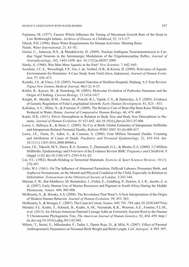
167HUMAN’S ASSOCIATION WITH WATER BODIES
Fujimura, M. (1977). Factors Which Influence the Timing of Maximum Growth Rate of the Head in Low Birthweight Infants. Archives of Disease in Childhood, 52: 113-117
Gleick, P.H. (1996). Basic Water Requirements for Human Activities: Meeting BasicNeeds. Water International, 21, 83–92.Gorini, C., Jameson, H.S., & Mendelowitz, D. (2009). Nucleus Ambiguus Neurotransmission to Car-
diac Vagal Neurons in the Serotonergic Modulation of the Trigeminocardiac Reflex. Journal of Neurophysiology, 102, 1443-1450. doi: 10.1152/jn.00287.2009
Hardy, A. (1960). Was Man More Aquatic in the Past? New Scientist, 7, 642–645. Joordens, J.C.A., Wesselingh, F.P., Vos, J. de, Vonhof, H.B., & Kroon, D. (2009). Relevance of Aquatic
Environments for Hominins: A Case Study from Trinil (Java, Indonesia). Journal of Human Evolu-tion, 57, 656–671.
Kemiki, J.S., & Vince, J.D. (2003). Neonatal Outcome at Modilon Hospital, Madang: A 5-Year Review. Papua New Guinea Medical Journal, 46(1-2): 8-15.
Kittler, R., Kayser, M., & Stoneking, M. (2003). Molecular Evolution of Pediculus Humanus and the Origin of Clothing. Current Biology, 13:1414-1417
Knight, B., Shields, B.M., Turner, M., Powell, R.J., Yajnik, C.S., & Hattersley, A.T. (2005). Evidence of Genetic Regulation of Fetal Longitudinal Growth. Early Human Development, 81, 823—831.
Kuliukas, A.V., Milne, N., & Fournier, P. (2009). The Relative Cost of Bent-Hip Bent-Knee Walking is Reduced in Water. Homo. Journal of Comparative Human Biology, 60, 479–488.
Kurki, H.K. (2011). Pelvic Dimorphism in Relation to Body Size and Body Size Dimorphism in Hu-mans. Journal of Human Evolution, 61 631-643. doi:10.1016/j.jhevol.2011.07.006
Lawn, J., Shibuya, K., & Stein, C. (2005). No Cry at Birth: Global Estimates of Intrapartum Stillbirths and Intrapartum-Related Neonatal Deaths. Bulletin WHO 2005, 83 (6):409-417.
Lawn, J.E., Osrin, D., Adler, A., & Cousens, S. (2008). Four Million Neonatal Deaths: Counting and Attribution of Cause of Death. Paediatric and Perinatal Epidemiology, 22, 410–416. doi: 10.1111/j.1365-3016.2008.00960.x
Lawn, J.E., Yakoob, M.Y., Haws, R.A. Soomro, T., Darmstadt, G.L., & Bhutta, Z.A. (2009). 3.2 Million Stillbirths: Epidemiology and Overview of the Evidence Review BMC Pregnancy and Childbirth, 9 (Suppl 1):S2 doi:10.1186/1471-2393-9-S1-S2
Lin, Y.C. (1982). Breath-Holding in Terrestrial Mammals. Exercise & Sport Sciences Reviews, 10 (1): 270-307.
Little, W.J. (1861). On The Influence of Abnormal Parturition, Difficult Labours, Premature Birth, and Asphyxia Neonatorum, on the Mental and Physical Condition of the Child, Especially in Relation to Deformities. Transactions of the Obstetrical Society of London, 3:243-344,
Marean, C.W., Bar-Matthews, M. Bernatchez, J., Fisher, E., Goldberg, P., Herries, A. I. R., Jacobs, Z. et al. (2007). Early Human Use of Marine Resources and Pigment in South Africa During the Middle Pleistocene. Nature, 449, 905-908.
McBrearty, S., & Brooks, A.S. (2000). The Revolution That Wasn’t: A New Interpretation of the Origin of Modern Human Behavior. Journal of Human Evolution, 39: 453–563.
McBrearty, S., & Stringer, C. (2007). The Coast in Colour. Nature, 449, 793–794. (doi:10.1038/449793a)Mendez, F.L. Krahn, T., Schrack, B., Krahn, A.-M., Veeramah, K.R., Woerner, A.E., Fomine, F.L.M.,
et al. (2013). An African American Paternal Lineage Adds an Extremely Ancient Root to the Human Y Chromosome Phylogenetic Tree, The American Journal of Human Genetics, 92, 454–459. http://dx.doi.org/10.1016/j.ajhg.2013.02.002.
Miletic, T., Stoini, E., Mikulandra, F., Tadin, I., Damir Roje, D., & Milic N. (2007). Effect of Parental Anthropometric Parameters on Neonatal Birth Weight and Birth Length. Coll. Antropol., 4: 993–997.

168 OPPENHEIMER
Miller, G. F. (2000). The Mating Mind: How Sexual Choice Shaped the Evolution of Human Nature. Heinemann: London.
Miller, G.F., & Penke, L. (2007). The Evolution of Human Intelligence and the Coefficient of Additive Genetic Variance in Human Brain Size. Intelligence, 35: 97–114.
Morgan, E. (1972). The Descent of Woman. New York: Bantam Books.Morgan, E. (1982). The Aquatic Ape: A Theory of Human Evolution. New York: Stein and Day.Mortola, J.P. (2009). Gas Exchange in Avian Embryos and Hatchlings, Comparative Biochemistry and
Physiology, Part A, 153: 359–377.Morwood, M.J., O’Sullivan, P. B. Aziz, F., & Raza, A. (1998). Fission-Track ages of Stone Tools and
Fossils on the East Indonesian Island of Flores. Nature, 392, 173-176. Myklestad, K., Vatten, L.J., Magnussen, E.B., Salvesen, K.A., & Romundstad, P.R. (2013). Do Paren-
tal Heights Influence Pregnancy Length?: A Population-Based Prospective Study, HUNT 2. BMC Pregnancy and Childbirth, 13:33.
NASA. (2007). Earth’s Nighlights, Looking at Earth, Goddard Space Flight Center, http://www.nasa.gov/vision/earth/lookingatearth/NIGHTLIGHTS.html Page last viewed 21/9/2013
Neggers, Y., Goldenberg, R.L., Cliver, S.P., Hoffman, H.J., & Cutter, G.R. (1995). The Relationship Between Maternal and Neonatal Anthropometric Measurements in Term Newborns. Obstetrics & Gynecology, 85 (2): 192-196.
Niemitz, C. (2010). The Evolution of Upright Posture and Gait: A Review and a New Synthesis. Natur-wissenschaften, 97, 241-263.
O’Connor, S. (2007). New Evidence from East Timor Contributes to our Understanding of Earliest Modern Human Colonisation East of the Sunda Shelf. Antiquity, 81, 523–535.
Oppenheimer, S.J. (2003). Out of Eden: The Peopling of the World. London: Constable Robinson. Oppenheimer, S.J (2004). Austronesian Spread into Southeast Asia and Oceania: Where From and
When. In Pacific Archaeology: Assessments and Prospects: Proceedings of the International Con-ference for the 50th Anniversary of the first Lapita excavation - Kone, Noumea 2002 (Ed Christophe Sand) Les Cahiers de l’Archeologie en Nouvelle Caledonie. Pp.54-70.
Oppenheimer, S.J. (2009). The Great Arc of Dispersal of Modern Humans: Africa to Australia. Quater-nary International, 202, 2–13. (doi:10.1016/j.quaint.2008.05.015).
Oppenheimer, S. J. (2012). Out-of-Africa, The Peopling of Continents & Islands: Tracing Uniparental Gene Trees Across the Map. Philosophical Transactions of the Royal Society B: Biological Sciences, 2012, 367, 770-784. doi: 10.1098/rstb.2011.0306.
Oppenheimer, S.J. (2014). Modern Humans Spread from Aden to the Antipodes: With Passengers and When? In: Robin Dennell and Martin Porr eds, East of Africa: Southern Asia, Australia and Modern Human Origins in press New York: Cambridge University Press.
Patel, R.R., Steer, P., Doyle, P., Little, M.P., & Elliott, P. (2003). Does Gestation Vary by Ethnic Group? A London-Based Study of Over 122 000 Pregnancies with Spontaneous Onset of Labour. Interna-tional Journal of Epidemiology, 33:107–113. DOI: 10.1093/ije/dyg238.
Patterson, N., Richter, D.J., Gnerre, S., Lander, E., & Reich, D. (2006). Genetic Evidence for Complex Speciation of Humans and Chimpanzees. Nature, 441, 1103–1108.
Rabenold, D., Pearson, O.M. (2011). Abrasive, Silica Phytoliths and the Evolution of Thick Molar Enamel in Primates, with Implications for the Diet of Paranthropus boisei. PLoS ONE, 6(12): e28379. doi:10.1371/journal.pone.0028379.
Reich, D., Patterson, N., Kircher, M., Delfin, F., Nandineni, M.R., Pugach, I., Ko, A., et al. (2011). Denisova Admixture and the First Modern Human Dispersals into Southeast Asia and Oceania. The American Journal of Human Genetics, 89, 1–13.

169HUMAN’S ASSOCIATION WITH WATER BODIES
Rice, F., & Thapar, A. (2010). Estimating the Relative Contributions of Maternal Genetic, Paternal Genetic and Intrauterine Factors to Offspring Birth Weight and Head Circumference. Early Human Development, 86: 425–432.
Rosenberg, K., & Trevathan, W. (2002). BJOG: An International Journal of Obstetrics and Gynaecol-ogy, 109: 1199–1206.
Ruff, C.B., Trinkaus, E., & Holliday, T.W. (1997). Body Mass and Encephalization in Pleistocene Homo. Nature, 387: 173–6.
Russell, J.G.B. (1969). Moulding of the Pelvic Outlet. Journal of Obstetrics & Gynaecology, British Commonwealth, 76:817–820.
Sauer, C. O. (1963). Seashore–Primitive Home of Man? In Land and Life: A Selection From the Writ-ings of Carl Ortwin Sauer (ed. L. John), pp. 312–330. Berkeley, CA: University of California Press.
Schaller, B. (2004). Trigeminocardiac Reflex: A Clinical Phenomenon or a New Physiological Entity? Journal of Neurology, 251, 658–665. DOI 10.1007/s00415-004-0458-4.
Scholander, P.F. (1963). The Master Switch of Life. Scientific American, 209: 92-104.Schultz, A.H. (1949). Sex Differences in the Pelves of Primates. American Journal of Physical Anthro-
pology, 7:401–424.Singer, D. (1999). Neonatal Tolerance to Hypoxia: A Comparative-Physiological Approach. Compara-
tive Biochemistry and Physiology Part A., 123 (1999) 221–234.Soares, P., Ermini, L., Thomson, N., Mormina, M., Rito, T., Röhl, A., Salas, A., Oppenheimer, S. et al.
(2009). Correcting for Purifying Selection: An Improved Human Mitochondrial MolecularClock. American Journal of Human Genetics, 84, 1–20.
Soares, P., Alshamali, F., Pereira, J.B., Fernandes, V., Silva, N.M., Afonso, C., Costa, M.D. et al. (2012). The Expansion of mtDNA Haplogroup L3 Within and Out of Africa. Molecular Biology and Evolu-tion 29,915-927.
Tague, R.G., & Lovejoy, C.O. (1986). The Obstetric Pelvis of A.L. 288-1 (Lucy). Journal of Human Evolution, 15:237–255
Tague, R.G. (1989). Variation in Pelvic Size Between Males and Females. American Journal of Physi-cal Anthropology, 80:59–71.
Tague, R.G. (1991). Commonalities in Dimorphism and Variability in the Anthropoid Pelvis, with Im-plications for the Fossil Record. Journal of Human Evolution, 21:153–176.
Tague, R.G. (1992). Sexual Dimorphism in the Human Bony Pelvis, with a Consideration of the Ne-andertal Pelvis from Kebara Cave, Israel. American Journal of Physical Anthropology, 88:1–21.
Tague, R.G. (1995). Variation in Pelvic Size Between Males and Females in Nonhuman Anthropoids. American Journal of Physical Anthropology, 97, 213-233.
Tague, R.G. (2000). Do Big Females Have Big Pelves? American Journal of Physical Anthropology, 112:377–393.
Tague, R.G. (2005). Big-Bodied Males Help Us Recognize That Females Have Big Pelves. American Journal of Physical Anthropology, 127:392–405, 2005.127:392–405. DOI 10.1002/ajpa.20226
Tchobroutsky, C., Merleta, C., & Rey, P. (1969). The Diving Reflex in Rabbit, Sheep and Newborn Lamb and its Afferent Pathways. Respiration Physiology, 8, 108-117
Thame, M., Wilks, R.J., McFarlane-Anderson, N., Bennett, F.I., & Forrester, T.E. (1997). Relationship between Maternal Nutritional Status and Infant’s Weight and Body Proportions at Birth. European Journal of Clinical Nutrition, 51, 134-138.
Toh-Adam, R., Srisupundit, K., & Tongsong, T. (2012). Short Stature as an Independent Risk Factor for Cephalopelvic Disproportion in a Country of Relatively Small-Sized Mothers. Archives of Gynecol-ogy and Obstetrics, 285: 1513–1516. DOI 10.1007/s00404-011-2168-3.

170 OPPENHEIMER
Turnbull, A.C., & Anderson, A.B.M. (1969). Evidence of a Foetal Role in Determining the Length of Gestation. Postgraduate Medical Journal, 45, 65-67.
Verhaegen, M., & Puech, P.-F. (2000). Hominid Lifestyle and Diet Reconsidered: Paleo-Environmental and Comparative Data, Human Evolution, 15: 175-186.
Walter, R.C., Buffler, R.T., Bruggemann, J.H., Guillaume, M.M., Berhe, S.M., Negassi, B., Libsekal, Y., Cheng, H., Edwards, R.L., von Cosel, R., Neraudeau, D., & Gagnon, M., (2000). Early Human Occupation of the Red Sea Coast of Eritrea During the Last Interglacial. Nature, 405, 65–69.
Won, Y-J., & Hey, J. (2005). Divergence Population Genetics of Chimpanzees Molecular Biology and Evolution, 22 (2):297–307. doi:10.1093/molbev/msi017
World Health Organization. (2006). Neonatal and Perinatal Mortality, Country, Regional and Global Estimates. ISBN 92 4 156320 6.
Zohar, I., & Biton, R. (2011). Land, Lake, and Fish: Investigation of Fish Remains from Gesher Benot Ya‘Aqov (Paleo-Lake Hula). Journal of Human Evolution, 60, 343-356.
Copyright © 2022 FDOKUMEN