Pelvic Pain in Reproductive Age: US Findings - MDPI
19
Citation: Di Serafino, M.; Iacobellis, F.; Schillirò, M.L.; Verde, F.; Grimaldi, D.; Dell’Aversano Orabona, G.; Caruso, M.; Sabatino, V.; Rinaldo, C.; Cantisani, V.; et al. Pelvic Pain in Reproductive Age: US Findings. Diagnostics 2022, 12, 939. https:// doi.org/10.3390/diagnostics12040939 Academic Editor: Antonio Farina Received: 21 February 2022 Accepted: 7 April 2022 Published: 9 April 2022 Publisher’s Note: MDPI stays neutral with regard to jurisdictional claims in published maps and institutional affil- iations. Copyright: © 2022 by the authors. Licensee MDPI, Basel, Switzerland. This article is an open access article distributed under the terms and conditions of the Creative Commons Attribution (CC BY) license (https:// creativecommons.org/licenses/by/ 4.0/). diagnostics Review Pelvic Pain in Reproductive Age: US Findings Marco Di Serafino 1, * ,† , Francesca Iacobellis 1,† , Maria Laura Schillirò 1 , Francesco Verde 1 , Dario Grimaldi 1 , Giuseppina Dell’Aversano Orabona 1 , Martina Caruso 1 , Vittorio Sabatino 1 , Chiara Rinaldo 1 , Vito Cantisani 2 , Gianfranco Vallone 3 and Luigia Romano 1 1 Department of General and Emergency Radiology, “Antonio Cardarelli” Hospital, 80131 Naples, Italy; [email protected] (F.I.); [email protected] (M.L.S.); [email protected] (F.V.); [email protected] (D.G.); [email protected] (G.D.O.); [email protected] (M.C.); [email protected] (V.S.); [email protected] (C.R.); [email protected] (L.R.) 2 Department of Radiology, Sapienza Rome University, Policlinico Umberto I, 00185 Rome, Italy; [email protected] 3 Department of Life and Health, University of Molise “V. Tiberio”, 86100 Campobasso, Italy; [email protected] * Correspondence: marco.diserafi[email protected] † These authors contributed equally to this work. Abstract: Pelvic pain in reproductive age often represents a diagnostic challenge due to the variety of potential causes characterized by overlapping clinical symptoms, including gynecological and other disorders (e.g., entero-colic or urological). It is also necessary to determine if there is a possibility of pregnancy to rule out any related complications, such as ectopic pregnancy. Although ultrasound (US), computed tomography (CT), and magnetic resonance imaging (MRI) are strongly integrated, the choice of which is the ideal diagnostic tool should be guided both by clinical suspicion (gynecological vs. non-gynecological cause) and by the risk ratio–benefit (ionizing radiation and instrumental costs), too. The didactic objective proposed by this review consists in the diagnosis of the cause and differential of pelvic pain in reproductive age by describing and critically analyzing the US diagnostic clues of the most frequent adnexal, uterine, and vascular causes. Keywords: pelvic pain; reproductive age; ultrasound; differential diagnosis 1. Introduction Pelvic pain in reproductive age often represents a diagnostic challenge due to the variety of potential causes characterized by overlapping clinical symptoms, including gynecological and other disorders (e.g., entero-colic or urological). It is also necessary to determine if there is a possibility of pregnancy to rule out any related complications, such as ectopic pregnancy [1]. Several imaging techniques can be adopted to image the female genital tract. The choice of the most suitable imaging approach and the related imaging protocols varies depending on the clinical indications and the patient conditions. Although ultrasound (US), computed tomography (CT), and magnetic resonance imaging (MRI) are strongly integrated, the choice of which is the ideal diagnostic tool should be guided both by clinical suspicion (gynecological vs non-gynecological cause) and by the risk–benefit ratio (ionizing radiation and instrumental costs), too [2]. Pelvic US is generally the first diagnostic tool used in this clinical setting and may require a transabdominal (TSA–US) and, if possible, transvaginal (TSV–US) approach to improve diagnostic sensitivity. CT and MRI may offer an added diagnostic value in pelvic pain assessment when US findings are unreliable or when pre-surgical evaluation is required [1]. The didactic objective proposed by this review consists in the diagnosis of the cause and differential of pelvic pain in reproductive age by describing and critically analyzing the US diagnostic clues of the most frequent adnexal, uterine, and vascular causes. Diagnostics 2022, 12, 939. https://doi.org/10.3390/diagnostics12040939 https://www.mdpi.com/journal/diagnostics
-
Upload
khangminh22 -
Category
Documents
-
view
6 -
download
0
Transcript of Pelvic Pain in Reproductive Age: US Findings - MDPI

�����������������
Citation: Di Serafino, M.; Iacobellis,
F.; Schillirò, M.L.; Verde, F.; Grimaldi,
D.; Dell’Aversano Orabona, G.;
Caruso, M.; Sabatino, V.; Rinaldo, C.;
Cantisani, V.; et al. Pelvic Pain in
Reproductive Age: US Findings.
Diagnostics 2022, 12, 939. https://
doi.org/10.3390/diagnostics12040939
Academic Editor: Antonio Farina
Received: 21 February 2022
Accepted: 7 April 2022
Published: 9 April 2022
Publisher’s Note: MDPI stays neutral
with regard to jurisdictional claims in
published maps and institutional affil-
iations.
Copyright: © 2022 by the authors.
Licensee MDPI, Basel, Switzerland.
This article is an open access article
distributed under the terms and
conditions of the Creative Commons
Attribution (CC BY) license (https://
creativecommons.org/licenses/by/
4.0/).
diagnostics
Review
Pelvic Pain in Reproductive Age: US FindingsMarco Di Serafino 1,*,†, Francesca Iacobellis 1,† , Maria Laura Schillirò 1 , Francesco Verde 1 , Dario Grimaldi 1,Giuseppina Dell’Aversano Orabona 1 , Martina Caruso 1 , Vittorio Sabatino 1 , Chiara Rinaldo 1,Vito Cantisani 2, Gianfranco Vallone 3 and Luigia Romano 1
1 Department of General and Emergency Radiology, “Antonio Cardarelli” Hospital, 80131 Naples, Italy;[email protected] (F.I.); [email protected] (M.L.S.); [email protected] (F.V.);[email protected] (D.G.); [email protected] (G.D.O.);[email protected] (M.C.); [email protected] (V.S.); [email protected] (C.R.);[email protected] (L.R.)
2 Department of Radiology, Sapienza Rome University, Policlinico Umberto I, 00185 Rome, Italy;[email protected]
3 Department of Life and Health, University of Molise “V. Tiberio”, 86100 Campobasso, Italy;[email protected]
* Correspondence: [email protected]† These authors contributed equally to this work.
Abstract: Pelvic pain in reproductive age often represents a diagnostic challenge due to the variety ofpotential causes characterized by overlapping clinical symptoms, including gynecological and otherdisorders (e.g., entero-colic or urological). It is also necessary to determine if there is a possibility ofpregnancy to rule out any related complications, such as ectopic pregnancy. Although ultrasound(US), computed tomography (CT), and magnetic resonance imaging (MRI) are strongly integrated, thechoice of which is the ideal diagnostic tool should be guided both by clinical suspicion (gynecologicalvs. non-gynecological cause) and by the risk ratio–benefit (ionizing radiation and instrumentalcosts), too. The didactic objective proposed by this review consists in the diagnosis of the cause anddifferential of pelvic pain in reproductive age by describing and critically analyzing the US diagnosticclues of the most frequent adnexal, uterine, and vascular causes.
Keywords: pelvic pain; reproductive age; ultrasound; differential diagnosis
1. Introduction
Pelvic pain in reproductive age often represents a diagnostic challenge due to thevariety of potential causes characterized by overlapping clinical symptoms, includinggynecological and other disorders (e.g., entero-colic or urological). It is also necessary todetermine if there is a possibility of pregnancy to rule out any related complications, suchas ectopic pregnancy [1]. Several imaging techniques can be adopted to image the femalegenital tract. The choice of the most suitable imaging approach and the related imagingprotocols varies depending on the clinical indications and the patient conditions. Althoughultrasound (US), computed tomography (CT), and magnetic resonance imaging (MRI) arestrongly integrated, the choice of which is the ideal diagnostic tool should be guided bothby clinical suspicion (gynecological vs non-gynecological cause) and by the risk–benefitratio (ionizing radiation and instrumental costs), too [2]. Pelvic US is generally the firstdiagnostic tool used in this clinical setting and may require a transabdominal (TSA–US)and, if possible, transvaginal (TSV–US) approach to improve diagnostic sensitivity. CT andMRI may offer an added diagnostic value in pelvic pain assessment when US findings areunreliable or when pre-surgical evaluation is required [1]. The didactic objective proposedby this review consists in the diagnosis of the cause and differential of pelvic pain inreproductive age by describing and critically analyzing the US diagnostic clues of the mostfrequent adnexal, uterine, and vascular causes.
Diagnostics 2022, 12, 939. https://doi.org/10.3390/diagnostics12040939 https://www.mdpi.com/journal/diagnostics

Diagnostics 2022, 12, 939 2 of 19
2. Examination Technique
Pelvic US is considered the ideal investigative tool to use at the beginning of the diag-nostic and evaluation process for suspected gynaecological disorders in patients of all ages.US has the advantages of being widely available, low cost, and free from ionising radiation.Additionally, US is often sufficient to diagnose uterine, ovarian, and adnexal pathologies [1].US is also extremely useful in evaluating pathological changes that can affect the pelvicportions of the urinary tract, the gastrointestinal tract, and in musculoskeletal structures,which can mimic the clinical picture of gynaecological pathology [1]. TSA–US and TSV–USare complementary techniques; both are used extensively in the evaluation of the femalepelvis Table 1 [1].
Table 1. TSA–US and TSV–US in comparison.
US Methods Probe Protocol Utility Limits
Transabdominalsonography(TSA–US)
Low-frequencyprobe (convex
probe 3–5 MHz).The standard protocol for examiningthe female pelvis involves an initial
TAS with the urinary bladdercompletely full, so it can act as an
acoustic window. Following bladderemptying, the patient assumes alithotomy position, and TVS isperformed. The two imaging
techniques are complementary andoften provide different diagnostic
information.The protocol often also calls for the
execution of Doppler, power Doppler,and pulsed wave Doppler flowmetry
depending on the clinical situation andpathology that emerge from grayscale
imaging.
TAS offers a wider field ofview than TVS and allowsbetter visualisation of the
superficial and distalstructures of the vagina bybringing the probe closer
to the target organs.
• Empty bladder.• Obese patients.• Retroverse uterus where the
fundus is located beyond thefocal zone of the transducer.
• Less effective forcharacterisation of adnexalmasses.
Transvaginalsonography(TVS–US)
High-frequencyprobe
(endocavitaryprobe > 7 MHz).
TVS approach requires agreater penetration depthto avoid the attenuating
soft tissues that cover thepelvic organs. Therefore, it
requires the use of ahigher frequency probe,which, in turn, providesgreater resolution of theanatomical details of the
uterus, ovary, and adnexalstructures.
• Limited field of view.• Should not be performed on
patients who are unable orunwilling to consent to theprocedure, as well as onmost virgin patients and forthose in which the insertionof the probe producesmarked discomfort.
• It is contraindicated in someobstetric patients in the 2ndand 3rd trimester ofpregnancy due to the risk ofactive bleeding or membranerupture.
However, the characteristics of pelvic pain must be strongly considered before anyinstrumental diagnostic approach. In this regard, Table 2 shows the most frequent adnexal,uterine, and vascular causes of pelvic pain in reproductive age where US has often aconclusive diagnostic role, also highlighting their clinical presentation characteristics.
Table 2. Pelvic pain in reproductive age: causes, symptoms, and occurrence.
Causes of Pelvic Pain Occurrence Pain Characteristics
• Adnexal
Adnexal torsion 3% of gynecologic emergenciesAcute persistent (complete torsion) or
intermittent (intermittent torsion)right/left pelvic pain
Ruptured or bleeding ovarian cysts
The incidence is difficult to estimate. Abroader estimate calculates about 7% of
women worldwide experience asymptomatic cyst during their lifetime
Acute right/left pelvic pain
Pelvic inflammatory disease
No specific international data areavailable for PID incidence worldwide. A
study reports a prevalence ofself-reported lifetime PID of 4.4%
Chronic pelvic pain with reacutization

Diagnostics 2022, 12, 939 3 of 19
Table 2. Cont.
Causes of Pelvic Pain Occurrence Pain Characteristics
Endometritis
Pregnancy-related endometritis with anincidence of 1–3% after a vaginal delivery
and of 13–90% following cesareandelivery, and endometritis unrelated to
pregnancy that may occur in up to70–90% of documented cases of PID
Chronic pelvic pain with reacutization
Endometriosis It affects up to 10% of women ofreproductive age
Asymptomatic/poorlysymptomatic/chronic pelvic pain with
reacutization during menses
Peritoneal inclusion cystsApproximately 3–5% occur in women of
childbearing age following invasivepelvic surgery, infection, or cancer
Asymptomatic/poorlysymptomatic/chronic pelvic pain withreacutization (especially if complicated)
Ectopic pregnancy 1–2% of all pregnancies Acute pelvic pain
• Uterine
Fibroids: degeneration, rupture, andtorsion
A study examining the incidence ofdegeneration of leiomyoma in patients
referred for uterine fibroid embolisationunderwent MRI found an incidence of
5.1%.Torsion and rupture are a rare entity(reported incidence for torsion of less
than 0.25%)
Acute pelvic pain
Post-embolisation syndrome Occurs in about 40% of womenundergoing uterine artery embolisation Pelvic pain of variable entity
• Vascular
Pelvic congestion syndromeIn patients with presenting complaints of
chronic pelvic pain, the prevalence ofPCS is nearly 30%
Chronic pelvic pain
Thrombosis of the gonadal veins Referred incidence of about 0.18% of thegeneral population Acute pelvic pain of variable entity
3. Adnexal Causes of Pelvic Pain3.1. Adnexal Torsion
Adenxal torsion is the partial or complete rotation of the adnexum on its vascularpeduncle resulting in congestion and oedema, due to compromised venous and lymphaticdrainage, and subsequent ischaemia and necrosis, due to compromise of the arterial bloodsupply [3,4]. Although frequently reported in the scientific literature, it is an uncommoncause of pelvic pain, accounting for only 3% of gynecologic emergencies [3].
It can affect the adnexum in its entirety involving both the ovary and the fallopiantube [4]. The torsion is often unilateral, and is more common on the right, presumablydue to the reduced mobility of the left gonad due to the presence of the sigmoid colon.Torsion has a higher incidence in women of childbearing age and can be secondary toa space-occupying lesion, either cystic (large cystic lesions) or neoplastic, acting as alead point [1,4,5]. Other potential causes include pregnancy, polycystic ovary syndrome,previous surgery to the pelvis, and hyperlaxity of the mesenteries and ovarian ligaments [1].

Diagnostics 2022, 12, 939 4 of 19
Since it is defined as a surgical emergency, an early differential diagnosis includingother genitourinary and gastrointestinal conditions is essential to preserve the ovaries. Theclinical symptoms are characterized by acute and sudden pain, sometimes associated withnausea and vomiting with the finding of a palpable pelvic mass and signs of peritonitis [1,3].The initial assessment of a suspected adnexal torsion requires an US examination, whichallows an increase in the volume of the ovary to be observed, which is generally greaterthan 4 cm, with or without an associated mass (Figure 1).
Diagnostics 2022, 12, x FOR PEER REVIEW 4 of 20
space-occupying lesion, either cystic (large cystic lesions) or neoplastic, acting as a lead point [1,4,5]. Other potential causes include pregnancy, polycystic ovary syndrome, pre-vious surgery to the pelvis, and hyperlaxity of the mesenteries and ovarian ligaments [1].
Since it is defined as a surgical emergency, an early differential diagnosis including other genitourinary and gastrointestinal conditions is essential to preserve the ovaries. The clinical symptoms are characterized by acute and sudden pain, sometimes associated with nausea and vomiting with the finding of a palpable pelvic mass and signs of peri-tonitis [1,3]. The initial assessment of a suspected adnexal torsion requires an US exami-nation, which allows an increase in the volume of the ovary to be observed, which is generally greater than 4 cm, with or without an associated mass (Figure 1).
Figure 1. Adnexal torsion of giant mature cystic right teratoma. Axial TSA-US image (a) shows a mostly echogenic mass (caliper) with some sound attenuation. On power Doppler (b) no adnexal vascularization is detected.
The ovary may be found in an abnormal position, close to the midline of the uterus, higher or lower in the pouch of Douglas, or displaced to the contralateral ovarian space [1,4,5]. The ovarian echotexture shows a hyperechoic central stroma due to the vascular congestion, associated with small peripheral follicles, with a “pearl necklace” appearance and, in the most advanced stages of vascular impairment, a diffusely uneven stroma due to the presence of focal haemorrhages and necrosis. The pathognomonic finding, how-ever, remains the identification of the twisted vascular peduncle (“whirl” sign or “vas-cular vortex”), obtained by transversal scans along the central axis of the pedicle (Figure 2) [1–5].
Figure 1. Adnexal torsion of giant mature cystic right teratoma. Axial TSA-US image (a) shows amostly echogenic mass (caliper) with some sound attenuation. On power Doppler (b) no adnexalvascularization is detected.
The ovary may be found in an abnormal position, close to the midline of the uterus,higher or lower in the pouch of Douglas, or displaced to the contralateral ovarian space [1,4,5].The ovarian echotexture shows a hyperechoic central stroma due to the vascular congestion,associated with small peripheral follicles, with a “pearl necklace” appearance and, in themost advanced stages of vascular impairment, a diffusely uneven stroma due to the presenceof focal haemorrhages and necrosis. The pathognomonic finding, however, remains theidentification of the twisted vascular peduncle (“whirl” sign or “vascular vortex”), obtainedby transversal scans along the central axis of the pedicle (Figure 2) [1–5].
The twisted and congested peduncle may appear as an indistinct adnexal mass adja-cent to the twisted ovary. The colour Doppler examination allows for the assessment ofthe degree of torsion, the time elapsed from the beginning of the disease and therefore thedegree of vascular compromise [1–5]. Vascular colour Doppler signals in the gonadal tissueare generally absent and this allows for a confident diagnosis with a positive predictivevalue of 94% (Figure 1).
However, the presence of flow does not exclude torsion, both due to the two-foldperfusion of the ovary by the ovarian and uterine arteries, despite the initial loss of venousflow, and because the torsion may be intermittent or incomplete. However, Dopplerspectral waveform analysis can increase the sensitivity of the torsion diagnosis because anarterial waveform with diastolic flow inversion, indicating high resistance, may suggestthe presence of torsion (Table 3) [1–5].

Diagnostics 2022, 12, 939 5 of 19Diagnostics 2022, 12, x FOR PEER REVIEW 5 of 20
Figure 2. Same patient of Figure 1. Axial TSA–US image with high frequency probe (a) shows a twisted vascular peduncle (“whirl” sign) from the side of the right adnexal mass of above Figure 1. The twisted adnexal peduncle is better seen at colour-Doppler (b) and power-Doppler evaluation (c) and confirmed also at enhanced CT ((d) arrow), related with the presence of a large mature cystic teratoma ((e) arrow). At the operative specimen, ovarian torsion is confirmed, and the right ovary with the cystic teratoma with its mixed content of hairs ((f) arrowheads) and teeth ((f) curved arrow) is shown. Figure 2 is reprinted with permission from Iacobellis, F., Di Serafino, M., Danzi, R., Ponticiello, G., Schillirò, M.L., Barbuto, L., Nicola, R., Scaglione, M., Romano, L. Gynecological and obstetric causes of acute right iliac fossa pain: imaging approach and related imaging findings. EPOS ECR 2021 doi: 10.26044/ecr2021/C-12876.
The twisted and congested peduncle may appear as an indistinct adnexal mass ad-jacent to the twisted ovary. The colour Doppler examination allows for the assessment of the degree of torsion, the time elapsed from the beginning of the disease and therefore the degree of vascular compromise [1–5]. Vascular colour Doppler signals in the gonadal tissue are generally absent and this allows for a confident diagnosis with a positive pre-dictive value of 94% (Figure 1).
However, the presence of flow does not exclude torsion, both due to the two-fold perfusion of the ovary by the ovarian and uterine arteries, despite the initial loss of ve-nous flow, and because the torsion may be intermittent or incomplete. However, Doppler spectral waveform analysis can increase the sensitivity of the torsion diagnosis because
Figure 2. Same patient of Figure 1. Axial TSA–US image with high frequency probe (a) shows atwisted vascular peduncle (“whirl” sign) from the side of the right adnexal mass of above Figure 1.The twisted adnexal peduncle is better seen at colour-Doppler (b) and power-Doppler evaluation (c)and confirmed also at enhanced CT ((d) arrow), related with the presence of a large mature cysticteratoma ((e) arrow). At the operative specimen, ovarian torsion is confirmed, and the right ovarywith the cystic teratoma with its mixed content of hairs ((f) arrowheads) and teeth ((f) curved arrow)is shown. Reprinted with permission from [2].
Table 3. Adnexal torsion US diagnostic clue.
Adnexal Causes of Pelvic Pain US Diagnostic Clue US Limits
Adnexal torsionTwisted vascular peduncle (whirl sign)with absent flows or with increase in
resistance indices if represented
Transabdominal US may be limited in obesepatients or when the ovaries are masked by
intestinal meteorism.Endovaginal US may be limited in cases of
large ovarian masses causing cranialdisplacement of the ovary, hindering the
exploration of the ovarian vessels.

Diagnostics 2022, 12, 939 6 of 19
3.2. Ruptured or Bleeding Ovarian Cysts
Ovarian cysts are growths that develop in the ovaries during follicular maturation andare defined as functional cysts as they represent a physiological phenomenon related toovarian function [1,6]. Functional ovarian cysts are caused by the overgrowth of a follicledue to the accumulation of fluid inside it, which is usually spontaneously reabsorbedwithout causing pain [6]. In some cases, they can rupture, releasing fluid into the peritonealcavity, causing intense pain and bleeding complications. Haemorrhagic or ruptured ovariancysts are common in women of reproductive age; however, the actual incidence is difficultto estimate, as many ruptured cysts are asymptomatic or found incidentally [7]. A broaderestimate calculates about 7% of women worldwide experience a symptomatic cyst duringtheir lifetime and in this event, US is the primary investigative tool [6–8]. The cyst maynot be detectable if it has ruptured completely and decompressed; however, free fluid willgenerally be present in the pelvis. Therefore, rupture of ovarian cysts is often a diagnosis ofexclusion when no other potential cause of the pain is identified [1,6–8]. The appearance ofa haemorrhagic cyst detected by US is also variable and depends on the stage at which thepatient has the scan. In the acute phase, it is possible to observe widespread homogeneousechoes with a fluid level and a dependent echogenic component represented by the bloodsediment; over time, with the progressive degradation of haemoglobin, a reticular structurewith internal echoes is observed (also described as a fishing net, spider web, or lace-likestructure) (Figure 3) [1].
Diagnostics 2022, 12, x FOR PEER REVIEW 6 of 20
an arterial waveform with diastolic flow inversion, indicating high resistance, may sug-gest the presence of torsion (Table 3) [1–5].
Table 3. Adnexal torsion US diagnostic clue.
Adnexal Causes of Pelvic Pain US Diagnostic Clue US Limits
Adnexal torsion
Twisted vascular peduncle (whirl sign) with absent flows or with increase in resistance
indices if represented
Transabdominal US may be limited in obese patients or when the ovaries are masked by intestinal meteorism. Endovaginal US may be limited in cases of large ovar-ian masses causing cranial displacement of the ovary,
hindering the exploration of the ovarian vessels.
3.2. Ruptured or Bleeding Ovarian Cysts Ovarian cysts are growths that develop in the ovaries during follicular maturation
and are defined as functional cysts as they represent a physiological phenomenon related to ovarian function [1,6]. Functional ovarian cysts are caused by the overgrowth of a fol-licle due to the accumulation of fluid inside it, which is usually spontaneously reab-sorbed without causing pain [6]. In some cases, they can rupture, releasing fluid into the peritoneal cavity, causing intense pain and bleeding complications. Haemorrhagic or ruptured ovarian cysts are common in women of reproductive age; however, the actual incidence is difficult to estimate, as many ruptured cysts are asymptomatic or found in-cidentally [7]. A broader estimate calculates about 7% of women worldwide experience a symptomatic cyst during their lifetime and in this event, US is the primary investigative tool [6–8]. The cyst may not be detectable if it has ruptured completely and decom-pressed; however, free fluid will generally be present in the pelvis. Therefore, rupture of ovarian cysts is often a diagnosis of exclusion when no other potential cause of the pain is identified [1,6–8]. The appearance of a haemorrhagic cyst detected by US is also variable and depends on the stage at which the patient has the scan. In the acute phase, it is pos-sible to observe widespread homogeneous echoes with a fluid level and a dependent echogenic component represented by the blood sediment; over time, with the progressive degradation of haemoglobin, a reticular structure with internal echoes is observed (also described as a fishing net, spider web, or lace-like structure) (Figure 3) [1].
Figure 3. Haemorrhagic left ovarian cyst. Axial TSA-US image (a) shows a hemorrhagic corpus luteum ((a) caliper) with blood deposits ((a) arrows). On colour-Doppler imaging (b) circumferen-tial blood flow is also shown.
The fibrin bundles produce a network of precise linear echoes and are distinguisha-ble from the real septa because they are numerous, thin, and dispersed, with an irregular structure and are not vascularised on colour Doppler US or contrast-enhanced US (CEUS) (Figure 4) [1].
Figure 3. Haemorrhagic left ovarian cyst. Axial TSA-US image (a) shows a hemorrhagic corpusluteum ((a) caliper) with blood deposits ((a) arrows). On colour-Doppler imaging (b) circumferentialblood flow is also shown.
The fibrin bundles produce a network of precise linear echoes and are distinguishablefrom the real septa because they are numerous, thin, and dispersed, with an irregularstructure and are not vascularised on colour Doppler US or contrast-enhanced US (CEUS)(Figure 4) [1].
Diagnostics 2022, 12, x FOR PEER REVIEW 7 of 20
Figure 4. Haemorrhagic left ovarian cyst. Axial US image (a) shows a hemorrhagic cyst ((a) arrow) with reticular structure. On colour-Doppler imaging (b) there is no blood flow of the reticular structure. At follow-up (c) a complete resorption of the cyst is observed ((c) arrow).
In addition, unlike a true septum, they do not extend from one wall of the cyst to the other. The lack of flow detected by the colour Doppler imaging is an important feature, suggesting the presence of a mural thrombus rather than a tumour nodule. However, low levels of flow in solid tissue may not be detected by colour Doppler US, whereas contrast CEUS may optimise the diagnosis when clinical suspicion is present (Table 4) [1,6–8].
Table 4. Ruptured or bleeding ovarian cysts US diagnostic clue.
Adnexal Causes Of Pelvic Pain US Diagnostic Clue US Limits
Ruptured or bleeding ovarian cysts
Cystic mass with an inhomogeneous echo structure in relation to hemoglobin degradation often with evidence of haematic sediment or in an advanced phase with relief of thin internal echoes arranged
in a “fishing net” or fibrin bundles not vascularized by col-our-Doppler or CEUS (differential diagnosis with tumor mass).
US cannot detect and quantify the active bleeding
3.3. Pelvic Inflammatory Disease Pelvic inflammatory disease (PID) is an infection of the female genital tract caused
by the ascent of microorganisms from the vagina to the uterus, fallopian tubes, and ova-ries. Common causes are Neisseria gonorrhoeae and Chlamydia trachomatis, which are sexually transmitted [1,9]. Other aerobic and anaerobic bacteria can cause bacterial vaginosis with retrograde spread of vaginal microorganisms [1]. The continuum of in-fection begins with cervicitis causing mucopurulent discharge and progresses to endo-metritis and salpingitis. Pus can collect in the tube (pyosalpinx) and form a tubo-ovarian abscess.
No specific international data are available for PID incidence worldwide. However, among 1171 sexually experienced reproductive-aged women in the 2013–2014 National Health and Nutrition Education Survey (NHANES), the prevalence of self-reported life-time PID was 4.4% [10].
PID can be acute, chronic, or subclinical and is often underdiagnosed. Symptoms include lower abdominal pain, fever, mucopurulent discharge, and abnormal uterine bleeding during or after menstruation. Untreated PID can lead to chronic pelvic pain, infertility, ectopic pregnancy, and intra-abdominal infections. The diagnosis is made primarily on clinical suspicion [11]. In this setting, TSA–US of the abdomen and pelvis is frequently performed as the initial diagnostic imaging examination, to first rule out a broad spectrum of pathologic conditions, such as appendicitis, diverticulitis, adnexal torsion, and bowel obstruction [9–11].
On US, infected fallopian tubes may show thickened and hyperaemic walls, which are dilated in the presence of pyosalpinx with occluded ovarian fimbria, echogenic in-traluminal sediment, and stratified echoes that signal the presence of exudate (Figure 5) [9].
Figure 4. Haemorrhagic left ovarian cyst. Axial US image (a) shows a hemorrhagic cyst ((a) arrow)with reticular structure. On colour-Doppler imaging (b) there is no blood flow of the reticularstructure. At follow-up (c) a complete resorption of the cyst is observed ((c) arrow).

Diagnostics 2022, 12, 939 7 of 19
In addition, unlike a true septum, they do not extend from one wall of the cyst to theother. The lack of flow detected by the colour Doppler imaging is an important feature,suggesting the presence of a mural thrombus rather than a tumour nodule. However, lowlevels of flow in solid tissue may not be detected by colour Doppler US, whereas contrastCEUS may optimise the diagnosis when clinical suspicion is present (Table 4) [1,6–8].
Table 4. Ruptured or bleeding ovarian cysts US diagnostic clue.
Adnexal Causes of Pelvic Pain US Diagnostic Clue US Limits
Ruptured or bleeding ovarian cysts
Cystic mass with an inhomogeneous echo structurein relation to hemoglobin degradation often withevidence of haematic sediment or in an advanced
phase with relief of thin internal echoes arranged ina “fishing net” or fibrin bundles not vascularized bycolour-Doppler or CEUS (differential diagnosis with
tumor mass).
US cannot detect and quantify theactive bleeding
3.3. Pelvic Inflammatory Disease
Pelvic inflammatory disease (PID) is an infection of the female genital tract caused bythe ascent of microorganisms from the vagina to the uterus, fallopian tubes, and ovaries.Common causes are Neisseria gonorrhoeae and Chlamydia trachomatis, which are sexuallytransmitted [1,9]. Other aerobic and anaerobic bacteria can cause bacterial vaginosis withretrograde spread of vaginal microorganisms [1]. The continuum of infection begins withcervicitis causing mucopurulent discharge and progresses to endometritis and salpingitis.Pus can collect in the tube (pyosalpinx) and form a tubo-ovarian abscess.
No specific international data are available for PID incidence worldwide. However,among 1171 sexually experienced reproductive-aged women in the 2013–2014 NationalHealth and Nutrition Education Survey (NHANES), the prevalence of self-reported lifetimePID was 4.4% [10].
PID can be acute, chronic, or subclinical and is often underdiagnosed. Symptomsinclude lower abdominal pain, fever, mucopurulent discharge, and abnormal uterinebleeding during or after menstruation. Untreated PID can lead to chronic pelvic pain,infertility, ectopic pregnancy, and intra-abdominal infections. The diagnosis is madeprimarily on clinical suspicion [11]. In this setting, TSA–US of the abdomen and pelvis isfrequently performed as the initial diagnostic imaging examination, to first rule out a broadspectrum of pathologic conditions, such as appendicitis, diverticulitis, adnexal torsion, andbowel obstruction [9–11].
On US, infected fallopian tubes may show thickened and hyperaemic walls, which aredilated in the presence of pyosalpinx with occluded ovarian fimbria, echogenic intraluminalsediment, and stratified echoes that signal the presence of exudate (Figure 5) [9].
The inflamed fallopian tube appears adjacent or adherent to the ovary with the for-mation, in the most advanced cases, of an ovarian abscess represented on US by an in-flammatory mass which engulfs the ovary and the tube, making the ovary no longerdistinguishable (Figure 6) [1,9].
Rupture of tubo-ovarian abscess can result in septic shock (Table 5) [1,9].

Diagnostics 2022, 12, 939 8 of 19Diagnostics 2022, 12, x FOR PEER REVIEW 8 of 20
Figure 5. Pelvic inflammatory disease. US scan of the right adnexa (a) shows an anechoic oblong structure ((a), caliper). Contrast-enhanced axial CT image (b) and maximum intensity projection (MIP) coronal-oblique reconstruction (c) show tortuous and tubular fluid filled structure seen in the right adnexa ((b,c) arrows).
The inflamed fallopian tube appears adjacent or adherent to the ovary with the formation, in the most advanced cases, of an ovarian abscess represented on US by an inflammatory mass which engulfs the ovary and the tube, making the ovary no longer distinguishable (Figure 6) [1,9].
Figure 6. Tubo-ovarian complex. Axial colour-Doppler TSA–US scan of the left adnexa (a) shows a complex cystic mass with peripheral signal colour Doppler ((a) arrow). Contrast-enhanced axial CT image (b) shows a thick enhancing left adnexa abscess wall ((b) arrows).
Rupture of tubo-ovarian abscess can result in septic shock (Table 5) [1,9].
Figure 5. Pelvic inflammatory disease. US scan of the right adnexa (a) shows an anechoic oblongstructure ((a), caliper). Contrast-enhanced axial CT image (b) and maximum intensity projection(MIP) coronal-oblique reconstruction (c) show tortuous and tubular fluid filled structure seen in theright adnexa ((b,c) arrows).
Diagnostics 2022, 12, x FOR PEER REVIEW 8 of 20
Figure 5. Pelvic inflammatory disease. US scan of the right adnexa (a) shows an anechoic oblong structure ((a), caliper). Contrast-enhanced axial CT image (b) and maximum intensity projection (MIP) coronal-oblique reconstruction (c) show tortuous and tubular fluid filled structure seen in the right adnexa ((b,c) arrows).
The inflamed fallopian tube appears adjacent or adherent to the ovary with the formation, in the most advanced cases, of an ovarian abscess represented on US by an inflammatory mass which engulfs the ovary and the tube, making the ovary no longer distinguishable (Figure 6) [1,9].
Figure 6. Tubo-ovarian complex. Axial colour-Doppler TSA–US scan of the left adnexa (a) shows a complex cystic mass with peripheral signal colour Doppler ((a) arrow). Contrast-enhanced axial CT image (b) shows a thick enhancing left adnexa abscess wall ((b) arrows).
Rupture of tubo-ovarian abscess can result in septic shock (Table 5) [1,9].
Figure 6. Tubo-ovarian complex. Axial colour-Doppler TSA–US scan of the left adnexa (a) shows acomplex cystic mass with peripheral signal colour Doppler ((a) arrow). Contrast-enhanced axial CTimage (b) shows a thick enhancing left adnexa abscess wall ((b) arrows).
Table 5. Inflammatory disease US diagnostic clue.
Adnexal Causes of Pelvic Pain US Diagnostic Clue US Limits
Pelvic inflammatory disease
The fallopian tubes with thickened and hyperemic walls,dilated in the presence of pyosalpinx with occluded ovarian
fimbria, echogenic intraluminal sediments, and echoesstratified by exudate. The inflamed fallopian tube appearsadjacent to or adhering to the ovary with the formation, in
more advanced cases, of an ovarian tube abscessrepresented on US by an inflammatory mass that engulfsthe ovary and the fallopian tube, no longer making the
ovary distinguishable.
US may suffer from limitedpanoramicity and, in cases ofextensive adhesions, can bedifficult to discriminate each
anatomical structure.

Diagnostics 2022, 12, 939 9 of 19
3.4. Endometritis
Endometritis is inflammation of the endometrial lining of the uterus that could alsoinvolve the myometrium and, occasionally, the parametrium too, and clinically manifestwith fever, chills, lower abdominal pain, and foul-smelling lochia or PID related symp-toms [12,13]. It can be divided into pregnancy-related endometritis with an incidence of1–3% after a vaginal delivery and of 13–90% following cesarean delivery, and endometritisunrelated to pregnancy that may occur in up to 70–90% of documented cases of PID [12,13].
On US, inflammation of the endometrium can show a thickened endometrium withan irregular profile and the presence of more or less echogenic fluid or pus in the uterinecavity (pyometra) (Table 6) (Figure 7) [14].
Table 6. Endometritis US diagnostic clue.
Adnexal Causes of Pelvic Pain US Diagnostic Clue US Limits
Endometritis
Thickened endometrium with an irregularprofile and the presence of more or less
echogenic fluid or pus in the uterine cavity(pyometra)
At US, may be difficult to distinguishsevere endometritis from cancer
Diagnostics 2022, 12, x FOR PEER REVIEW 9 of 20
Table 5. Inflammatory disease US diagnostic clue.
Adnexal Causes of Pelvic Pain US Diagnostic Clue US Limits
Pelvic inflammatory disease
The fallopian tubes with thickened and hyperemic walls, di-lated in the presence of pyosalpinx with occluded ovarian
fimbria, echogenic intraluminal sediments, and echoes strat-ified by exudate. The inflamed fallopian tube appears adja-cent to or adhering to the ovary with the formation, in more advanced cases, of an ovarian tube abscess represented on
US by an inflammatory mass that engulfs the ovary and the fallopian tube, no longer making the ovary distinguishable.
US may suffer from lim-ited panoramicity and, in cases of extensive adhe-sions, can be difficult to discriminate each ana-
tomical structure.
3.4. Endometritis Endometritis is inflammation of the endometrial lining of the uterus that could also
involve the myometrium and, occasionally, the parametrium too, and clinically manifest with fever, chills, lower abdominal pain, and foul-smelling lochia or PID related symp-toms [12,13]. It can be divided into pregnancy-related endometritis with an incidence of 1–3% after a vaginal delivery and of 13–90% following cesarean delivery, and endome-tritis unrelated to pregnancy that may occur in up to 70–90% of documented cases of PID [12,13].
On US, inflammation of the endometrium can show a thickened endometrium with an irregular profile and the presence of more or less echogenic fluid or pus in the uterine cavity (pyometra) (Table 6) (Figure 7) [14].
Table 6. Endometritis US diagnostic clue.
Adnexal Causes of Pelvic Pain US Diagnostic Clue US Limits
Endometritis Thickened endometrium with an irregular pro-file and the presence of more or less echogenic
fluid or pus in the uterine cavity (pyometra)
At US, may be difficult to distinguish severe endometritis from cancer
Figure 7. Pyometra. TSA-US scan of the uterus (a) shows a distended uterine cavity containing complex fluid with echogenic foci (short arrows). Axial T2w MRI imaging (b) confirms the diag-nosis showing pus collection into uterine cavity.
3.5. Endometriosis Endometriosis is a chronic inflammatory condition caused by the abnormal presence
of endometrial tissue at sites other than the physiological endometrium. It affects up to 10% of women of reproductive age, is often asymptomatic and, when symptomatic, it manifests with chronic pelvic pain, dysmenorrhea, dyspareunia, and abnormal uterine bleeding [1,15–18]. Symptoms are often cyclical in nature as endometriosis is a hor-mone-responsive disease. Infertility is an important consequence of endometriosis, due
Figure 7. Pyometra. TSA-US scan of the uterus (a) shows a distended uterine cavity containingcomplex fluid with echogenic foci (short arrows). Axial T2w MRI imaging (b) confirms the diagnosisshowing pus collection into uterine cavity.
3.5. Endometriosis
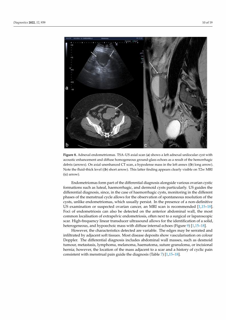
Endometriosis is a chronic inflammatory condition caused by the abnormal presenceof endometrial tissue at sites other than the physiological endometrium. It affects up to10% of women of reproductive age, is often asymptomatic and, when symptomatic, itmanifests with chronic pelvic pain, dysmenorrhea, dyspareunia, and abnormal uterinebleeding [1,15–18]. Symptoms are often cyclical in nature as endometriosis is a hormone-responsive disease. Infertility is an important consequence of endometriosis, due to theanatomical deformation of the pelvic structures and occlusion of the fallopian tubes. Ectopicsites of endometriosis implantation include the ovarian surface, the suspensory ligamentsof the uterus, the uterus itself, the peritoneal surfaces of the pouch of Douglas, and thefallopian tubes [1,15–18]. When the endometriosis tissue reaches structures deeper than5 mm from the peritoneal surface causing fibrosis and muscle hyperplasia, it is defined asdeep pelvic endometriosis. On US, endometriomas appear as unilocular swellings, whichare often bilateral and multiple, with a thick capsule, regular margins, and homogeneouslyechogenic content, with fine internal echoes, due to the blood cells flaking off the walls,resulting in a “ground glass” appearance (Figure 8).
Diagnostics 2022, 12, 939 10 of 19
Diagnostics 2022, 12, x FOR PEER REVIEW 10 of 20
to the anatomical deformation of the pelvic structures and occlusion of the fallopian tubes. Ectopic sites of endometriosis implantation include the ovarian surface, the sus-pensory ligaments of the uterus, the uterus itself, the peritoneal surfaces of the pouch of Douglas, and the fallopian tubes [1,15–18]. When the endometriosis tissue reaches structures deeper than 5 mm from the peritoneal surface causing fibrosis and muscle hyperplasia, it is defined as deep pelvic endometriosis. On US, endometriomas appear as unilocular swellings, which are often bilateral and multiple, with a thick capsule, regular margins, and homogeneously echogenic content, with fine internal echoes, due to the blood cells flaking off the walls, resulting in a “ground glass” appearance (Figure 8).
Figure 8. Adnexal endometriomas. TSA–US axial scan (a) shows a left adnexal unilocular cyst with acoustic enhancement and diffuse homogeneous ground-glass echoes as a result of the hemor-rhagic debris (arrows). On axial unenhanced CT scan, a hypodense mass in the left annex ((b) long arrow). Note the fluid-thick level ((b) short arrow). This latter finding appears clearly visible on T2w MRI ((c) arrow).
Endometriomas form part of the differential diagnosis alongside various ovarian cystic formations such as luteal, haemorrhagic, and dermoid cysts particularly. US guides the differential diagnosis, since, in the case of haemorrhagic cysts, monitoring in the dif-ferent phases of the menstrual cycle allows for the observation of spontaneous resolution of the cysts, unlike endometriomas, which usually persist. In the presence of a non-definitive US examination or suspected ovarian cancer, an MRI scan is recom-mended [1,15–18]. Foci of endometriosis can also be detected on the anterior abdominal wall, the most common localisation of extrapelvic endometriosis, often next to a surgical or laparoscopic scar. High-frequency linear transducer ultrasound allows for the identi-fication of a solid, heterogeneous, and hypoechoic mass with diffuse internal echoes (Figure 9) [1,15–18].
Figure 8. Adnexal endometriomas. TSA–US axial scan (a) shows a left adnexal unilocular cyst withacoustic enhancement and diffuse homogeneous ground-glass echoes as a result of the hemorrhagicdebris (arrows). On axial unenhanced CT scan, a hypodense mass in the left annex ((b) long arrow).Note the fluid-thick level ((b) short arrow). This latter finding appears clearly visible on T2w MRI((c) arrow).
Endometriomas form part of the differential diagnosis alongside various ovarian cysticformations such as luteal, haemorrhagic, and dermoid cysts particularly. US guides thedifferential diagnosis, since, in the case of haemorrhagic cysts, monitoring in the differentphases of the menstrual cycle allows for the observation of spontaneous resolution of thecysts, unlike endometriomas, which usually persist. In the presence of a non-definitiveUS examination or suspected ovarian cancer, an MRI scan is recommended [1,15–18].Foci of endometriosis can also be detected on the anterior abdominal wall, the mostcommon localisation of extrapelvic endometriosis, often next to a surgical or laparoscopicscar. High-frequency linear transducer ultrasound allows for the identification of a solid,heterogeneous, and hypoechoic mass with diffuse internal echoes (Figure 9) [1,15–18].
However, the characteristics detected are variable. The edges may be serrated andinfiltrated by adjacent soft tissues. Most disease deposits show vascularisation on colourDoppler. The differential diagnosis includes abdominal wall masses, such as desmoidtumour, metastasis, lymphoma, melanoma, haematoma, suture granuloma, or incisionalhernia; however, the location of the mass adjacent to a scar and a history of cyclic painconsistent with menstrual pain guide the diagnosis (Table 7) [1,15–18].

Diagnostics 2022, 12, 939 11 of 19Diagnostics 2022, 12, x FOR PEER REVIEW 11 of 20
Figure 9. Abdominal wall endometriosis nodule. TSA–US axial scan (a) of the abdominal wall shows a 35 mm abdominal wall endometriosis nodule with hypoechoic content and well-defined margins ((a), arrows). The nodule is enclosed in the muscular fascia along the right rectal abdomen. Contrast-enhanced axial CT image (b) shows heterogeneous enhanced mass in the right rectus sheath. The mass was subsequently proved to be abdominal endometriosis.
However, the characteristics detected are variable. The edges may be serrated and infiltrated by adjacent soft tissues. Most disease deposits show vascularisation on colour Doppler. The differential diagnosis includes abdominal wall masses, such as desmoid tumour, metastasis, lymphoma, melanoma, haematoma, suture granuloma, or incisional hernia; however, the location of the mass adjacent to a scar and a history of cyclic pain consistent with menstrual pain guide the diagnosis (Table 7) [1,15–18].
Table 7. Endometriosis US diagnostic clue.
Adnexal Causes of Pelvic Pain US Diagnostic Clue US Limits
Endometriosis
Unilocular swellings, which are often bilateral and multi-ple, with a thick capsule, regular margins, and homoge-
neously echogenic content, with fine internal echoes, due tothe blood cells flaking off the walls, resulting in a “ground glass” appearance. Useful monitoring for differential di-
agnosis with hemorrhagic ovarian cyst (persistence to fol-low-up of the endometriotic cyst addresses the diagnosis)
At US, may be difficult to detect millimetric foci of
ovarian endometriosis and to detect retrocervical or ligaments thickening, as
well as possible intestinal or nerve involvement.
3.6. Peritoneal Inclusion Cyst Peritoneal inclusion cysts (PICs) are generally benign mesothelial lesions with an
estimate incidence of approximately 3–5% of the peritoneal mesotheliomas containing peritoneal fluid that occur in women of childbearing age, often following invasive pelvic surgery, infection, or cancer [19].
This condition is most commonly described in women of reproductive age with a ratio of 4–5:1 female to male, with very few cases reported in females over 30 years of age [19].
Intra-abdominal inflammation with reactive mesothelial proliferation and impaired absorption of peritoneal fluid can lead to its formation [20,21].
PICs are irregularly shaped, multiloculated adnexal cystic masses with angled mar-gins, lacking a wall of their own and moulding themselves around the surrounding pel-vic organs within the peritoneal cavity. A common clinical presentation is a progressive lower-abdominal and pelvic pain or a palpable mass [20,21]. Due to their rarity and non-specific clinical signs, the pre-operative diagnosis of PICs is challenging [20,21].
The imaging features of peritoneal inclusion cysts reflect their pathogenesis and al-low for their differential diagnosis from an ovarian cystic mass [20,21]. Indeed, the typi-cal US finding is an ovary trapped inside a cyst, surrounded by septa and fluid. The fluid
Figure 9. Abdominal wall endometriosis nodule. TSA–US axial scan (a) of the abdominal wallshows a 35 mm abdominal wall endometriosis nodule with hypoechoic content and well-definedmargins ((a), arrows). The nodule is enclosed in the muscular fascia along the right rectal abdomen.Contrast-enhanced axial CT image (b) shows heterogeneous enhanced mass in the right rectus sheath.The mass was subsequently proved to be abdominal endometriosis.
Table 7. Endometriosis US diagnostic clue.
Adnexal Causes of Pelvic Pain US Diagnostic Clue US Limits
Endometriosis
Unilocular swellings, which are often bilateraland multiple, with a thick capsule, regularmargins, and homogeneously echogenic
content, with fine internal echoes, due to theblood cells flaking off the walls, resulting in a
“ground glass” appearance. Useful monitoringfor differential diagnosis with hemorrhagicovarian cyst (persistence to follow-up of theendometriotic cyst addresses the diagnosis)
At US, may be difficult to detectmillimetric foci of ovarian endometriosisand to detect retrocervical or ligamentsthickening, as well as possible intestinal
or nerve involvement.
3.6. Peritoneal Inclusion Cyst
Peritoneal inclusion cysts (PICs) are generally benign mesothelial lesions with anestimate incidence of approximately 3–5% of the peritoneal mesotheliomas containingperitoneal fluid that occur in women of childbearing age, often following invasive pelvicsurgery, infection, or cancer [19].
This condition is most commonly described in women of reproductive age with a ratioof 4–5:1 female to male, with very few cases reported in females over 30 years of age [19].
Intra-abdominal inflammation with reactive mesothelial proliferation and impairedabsorption of peritoneal fluid can lead to its formation [20,21].
PICs are irregularly shaped, multiloculated adnexal cystic masses with angled margins,lacking a wall of their own and moulding themselves around the surrounding pelvicorgans within the peritoneal cavity. A common clinical presentation is a progressive lower-abdominal and pelvic pain or a palpable mass [20,21]. Due to their rarity and non-specificclinical signs, the pre-operative diagnosis of PICs is challenging [20,21].
The imaging features of peritoneal inclusion cysts reflect their pathogenesis and allowfor their differential diagnosis from an ovarian cystic mass [20,21]. Indeed, the typicalUS finding is an ovary trapped inside a cyst, surrounded by septa and fluid. The fluid isusually anechoic but may contain echoes in some compartments due to haemorrhage orprotein-rich fluid (Figure 10).

Diagnostics 2022, 12, 939 12 of 19
Diagnostics 2022, 12, x FOR PEER REVIEW 12 of 20
is usually anechoic but may contain echoes in some compartments due to haemorrhage or protein-rich fluid (Figure 10).
Figure 10. Peritoneal inclusion cyst. TSA–US axial scan (a) shows an irregular, anechoic pa-ra-adnexal right fluid collection ((a) arrows). Axial T2w MRI (b) shows a large, well-defined mul-tilocular cystic pelvic mass (arrows). Note several thin internal septa, radiating from the ovary, representing pelvic adhesions. The septa radiate from the ovary (star; (b)). U: uterus.
Peritoneal inclusion cysts must also be differentiated from hydrosalpinx, which appears as a tubular or ovoid cystic formation, with visible folds, with no ovary inside of it (Table 8) [1,19–21].
Table 8. Peritoneal inclusion cyst US diagnostic clue.
Adnexal Causes of Pelvic Pain US Diagnostic Clue US Limits
Peritoneal inclusion cyst Trapped ovary inside a cyst, surrounded by septa and fluid. The fluid is usually anechoic but may contain echoes in some
compartments due to haemorrhage or protein-rich fluid
At US, can be difficult to differentiate the
cystic origin.
3.7. Ectopic Pregnancy The definition of ectopic pregnancy (EP) is that of a pregnancy that has implanted in
a location other than the uterine cavity occurring in 1–2% of all pregnancies [22]. The most common abnormal implantation site for ectopic pregnancy is in the fallo-
pian tubes, which occurs in up to 97% of cases. Of these, 75% to 80% are found in the ampullary region, 10% in the isthmic portion, 5% in the fimbrial portion, and 2 to 4% in the interstitial portion (Figure 11) [1,23–26].
Figure 11. Abnormal implantation sites for ectopic pregnancy.
Figure 10. Peritoneal inclusion cyst. TSA–US axial scan (a) shows an irregular, anechoic para-adnexalright fluid collection ((a) arrows). Axial T2w MRI (b) shows a large, well-defined multilocular cysticpelvic mass (arrows). Note several thin internal septa, radiating from the ovary, representing pelvicadhesions. The septa radiate from the ovary (star; (b)). U: uterus.
Peritoneal inclusion cysts must also be differentiated from hydrosalpinx, which ap-pears as a tubular or ovoid cystic formation, with visible folds, with no ovary inside of it(Table 8) [1,19–21].
Table 8. Peritoneal inclusion cyst US diagnostic clue.
Adnexal Causes of Pelvic Pain US Diagnostic Clue US Limits
Peritoneal inclusion cyst
Trapped ovary inside a cyst, surrounded bysepta and fluid. The fluid is usually anechoic
but may contain echoes in some compartmentsdue to haemorrhage or protein-rich fluid
At US, can be difficult to differentiate thecystic origin.
3.7. Ectopic Pregnancy
The definition of ectopic pregnancy (EP) is that of a pregnancy that has implanted in alocation other than the uterine cavity occurring in 1–2% of all pregnancies [22].
The most common abnormal implantation site for ectopic pregnancy is in the fallopiantubes, which occurs in up to 97% of cases. Of these, 75% to 80% are found in the ampullaryregion, 10% in the isthmic portion, 5% in the fimbrial portion, and 2 to 4% in the interstitialportion (Figure 11) [1,23–26].
Diagnostics 2022, 12, x FOR PEER REVIEW 12 of 20
is usually anechoic but may contain echoes in some compartments due to haemorrhage or protein-rich fluid (Figure 10).
Figure 10. Peritoneal inclusion cyst. TSA–US axial scan (a) shows an irregular, anechoic pa-ra-adnexal right fluid collection ((a) arrows). Axial T2w MRI (b) shows a large, well-defined mul-tilocular cystic pelvic mass (arrows). Note several thin internal septa, radiating from the ovary, representing pelvic adhesions. The septa radiate from the ovary (star; (b)). U: uterus.
Peritoneal inclusion cysts must also be differentiated from hydrosalpinx, which appears as a tubular or ovoid cystic formation, with visible folds, with no ovary inside of it (Table 8) [1,19–21].
Table 8. Peritoneal inclusion cyst US diagnostic clue.
Adnexal Causes of Pelvic Pain US Diagnostic Clue US Limits
Peritoneal inclusion cyst Trapped ovary inside a cyst, surrounded by septa and fluid. The fluid is usually anechoic but may contain echoes in some
compartments due to haemorrhage or protein-rich fluid
At US, can be difficult to differentiate the
cystic origin.
3.7. Ectopic Pregnancy The definition of ectopic pregnancy (EP) is that of a pregnancy that has implanted in
a location other than the uterine cavity occurring in 1–2% of all pregnancies [22]. The most common abnormal implantation site for ectopic pregnancy is in the fallo-
pian tubes, which occurs in up to 97% of cases. Of these, 75% to 80% are found in the ampullary region, 10% in the isthmic portion, 5% in the fimbrial portion, and 2 to 4% in the interstitial portion (Figure 11) [1,23–26].
Figure 11. Abnormal implantation sites for ectopic pregnancy.
Figure 11. Abnormal implantation sites for ectopic pregnancy.
Other less common sites include scarring of the ovary, abdomen, cervix, and uterus.The most common predisposing risk factor for EP is impaired tubal function, usually caused

Diagnostics 2022, 12, 939 13 of 19
by tubal scarring, secondary to salpingitis associated with pelvic inflammatory diseasetriggered by sexually transmitted infections [23–26]. Additionally, the incidence of EPincreases in women who have conceived through assisted reproduction techniques or in thepresence of an intrauterine device. The joint detection of human chorionic gonadotropin(β-hCG) in the serum and an ultrasound scan, preferentially transvaginal, allows thephysician to differentiate between intrauterine pregnancy and EP. Ectopic pregnancy shouldbe suspected in patients with vaginal bleeding or lower abdominal pain and a positivepregnancy test in the absence of an intrauterine gestational sac visible on US [27].
In general, an intrauterine gestational sac is expected to be visualised when the b-hCGis 1000 mIU/mL (according to the international standard) or 2000 mIU/mL (according tothe International Reference Preparation, IRP). When the b-hCG value is below the cut-offvalue of 2000 mIU/mL (IRP) and there is no intrauterine gestational sac, the diagnosiscould be an early intrauterine pregnancy, a miscarriage, or an ectopic pregnancy, andtherefore follow-up is indicated [23–26]. On the other hand, when the b-hCG value is abovethe cut-off value, you can expect to observe an intrauterine gestational sac which, if notvisualised, will raise the suspicion of an indeterminate pregnancy [1,23–26]. However, USfindings relating to the uterus are not specific and include the absence of an intrauterinegestational sac but with the finding of fluid in the endometrial canal. The latter, oftencontaining low-level echoes, centrally located within the uterine cavity, and surroundedby a single echogenic decidual layer, is called a “pseudogestational sac” or “pseudosac”.It differs from a genuine intrauterine gestational sac, which can be either in an eccentricposition or incorporated in a single layer of the endometrium (intradecidual sac) or at leastpartially surrounded by two echogenic tissue furrows with an interposed hypoechoic layer(double decidual sac). However, the most specific ultrasound finding for the diagnosis ofEP is the presence of an ectopic gestational sac containing the yolk sac or embryo (with orwithout cardiac activity), while the most common consists of an echogenic tubal ring in theadnexa (Figure 12) (Table 9) [23–26].
Diagnostics 2022, 12, x FOR PEER REVIEW 13 of 20
Other less common sites include scarring of the ovary, abdomen, cervix, and uterus. The most common predisposing risk factor for EP is impaired tubal function, usually caused by tubal scarring, secondary to salpingitis associated with pelvic inflammatory disease triggered by sexually transmitted infections [23–26]. Additionally, the incidence of EP increases in women who have conceived through assisted reproduction techniques or in the presence of an intrauterine device. The joint detection of human chorionic gonadotropin (β-hCG) in the serum and an ultrasound scan, preferentially transvaginal, allows the physician to differentiate between intrauterine pregnancy and EP. Ectopic pregnancy should be suspected in patients with vaginal bleeding or lower abdominal pain and a positive pregnancy test in the absence of an intrauterine gestational sac visible on US [27].
In general, an intrauterine gestational sac is expected to be visualised when the b-hCG is 1000 mIU/mL (according to the international standard) or 2000 mIU/mL (ac-cording to the International Reference Preparation, IRP). When the b-hCG value is below the cut-off value of 2000 mIU/mL (IRP) and there is no intrauterine gestational sac, the diagnosis could be an early intrauterine pregnancy, a miscarriage, or an ectopic preg-nancy, and therefore follow-up is indicated [23–26]. On the other hand, when the b-hCG value is above the cut-off value, you can expect to observe an intrauterine gestational sac which, if not visualised, will raise the suspicion of an indeterminate pregnancy [1,23–26]. However, US findings relating to the uterus are not specific and include the absence of an intrauterine gestational sac but with the finding of fluid in the endometrial canal. The latter, often containing low-level echoes, centrally located within the uterine cavity, and surrounded by a single echogenic decidual layer, is called a “pseudogestational sac” or “pseudosac”. It differs from a genuine intrauterine gestational sac, which can be either in an eccentric position or incorporated in a single layer of the endometrium (intradecidual sac) or at least partially surrounded by two echogenic tissue furrows with an interposed hypoechoic layer (double decidual sac). However, the most specific ultrasound finding for the diagnosis of EP is the presence of an ectopic gestational sac containing the yolk sac or embryo (with or without cardiac activity), while the most common consists of an echogenic tubal ring in the adnexa (Figure 12) (Table 9) [23–26].
Figure 12. Ectopic pregnancy. Axial (a) and longitudinal (b) TSA–US scans reveal an extrauterine adnexal gestational sac with a fetal body ((a,b), arrow). On pulsed Doppler (c) a heartbeat is also detected.
Figure 12. Ectopic pregnancy. Axial (a) and longitudinal (b) TSA–US scans reveal an extrauterineadnexal gestational sac with a fetal body ((a,b), arrow). On pulsed Doppler (c) a heartbeat isalso detected.
Table 9. Ectopic pregnancy US diagnostic clue.
Adnexal Causes of Pelvic Pain US Diagnostic Clue US Limits
Ectopic pregnancy
When the b-hCG value is below the cut-off value of 2000mIU/mL (IRP) and there is no intrauterine gestationalsac, the diagnosis could be an early intrauterinepregnancy, a miscarriage, or an ectopic pregnancy, andtherefore follow-up is indicated
• Uterine findings: gestational pseudosac(differential diagnosis with gestational sac: doubleechogenic wall versus single echogenic wall of thepseudosac).
• Adnexal findings: echogenic tubal ring or ectopicgestational sac containing the yolk sac or theembryo (with or without cardiac activity)
Related with difficulties in exploring theadnexa and in detecting early pregnancy as
well as active bleeding in case of rupture
Diagnostics 2022, 12, 939 14 of 19
4. Uterine Causes of Pelvic Pain4.1. Fibroids: Degeneration, Rupture and Torsion
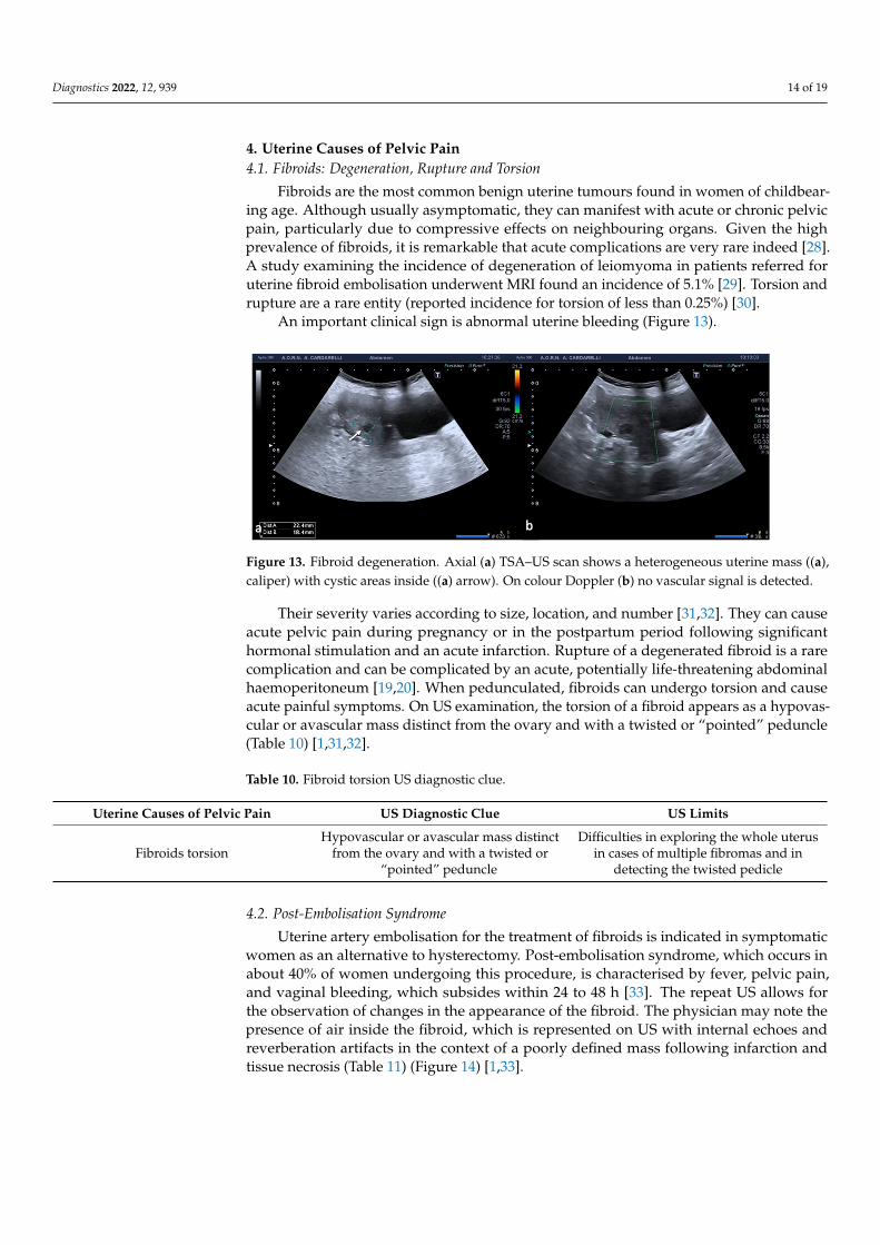
Fibroids are the most common benign uterine tumours found in women of childbear-ing age. Although usually asymptomatic, they can manifest with acute or chronic pelvicpain, particularly due to compressive effects on neighbouring organs. Given the highprevalence of fibroids, it is remarkable that acute complications are very rare indeed [28].A study examining the incidence of degeneration of leiomyoma in patients referred foruterine fibroid embolisation underwent MRI found an incidence of 5.1% [29]. Torsion andrupture are a rare entity (reported incidence for torsion of less than 0.25%) [30].
An important clinical sign is abnormal uterine bleeding (Figure 13).
Diagnostics 2022, 12, x FOR PEER REVIEW 14 of 20
Table 9. Ectopic pregnancy US diagnostic clue.
Adnexal Causes of Pelvic Pain US Diagnostic Clue US Limits
Ectopic pregnancy
When the b-hCG value is below the cut-off value of 2000 mIU/mL (IRP) and there is no intrauterine gestational sac, the diagnosis could be an early intrauterine pregnancy, a miscarriage, or an ectopic pregnancy, and therefore follow-up is indicated • Uterine findings: gestational pseudosac (differential diagnosis
with gestational sac: double echogenic wall versus single echo-genic wall of the pseudosac).
• Adnexal findings: echogenic tubal ring or ectopic gestational saccontaining the yolk sac or the embryo (with or without cardiac activity)
Related with dif-ficulties in ex-ploring the ad-nexa and in de-
tecting early pregnancy as well as active
bleeding in case of rupture
4. Uterine Causes of Pelvic Pain 4.1. Fibroids: Degeneration, Rupture and Torsion
Fibroids are the most common benign uterine tumours found in women of childbearing age. Although usually asymptomatic, they can manifest with acute or chronic pelvic pain, particularly due to compressive effects on neighbouring organs. Given the high prevalence of fibroids, it is remarkable that acute complications are very rare indeed [28]. A study examining the incidence of degeneration of leiomyoma in pa-tients referred for uterine fibroid embolisation underwent MRI found an incidence of 5.1% [29]. Torsion and rupture are a rare entity (reported incidence for torsion of less than 0.25%) [30].
An important clinical sign is abnormal uterine bleeding (Figure 13).
Figure 13. Fibroid degeneration. Axial (a) TSA–US scan shows a heterogeneous uterine mass ((a), caliper) with cystic areas inside ((a) arrow). On colour Doppler (b) no vascular signal is detected.
Their severity varies according to size, location, and number [31,32]. They can cause acute pelvic pain during pregnancy or in the postpartum period following significant hormonal stimulation and an acute infarction. Rupture of a degenerated fibroid is a rare complication and can be complicated by an acute, potentially life-threatening abdominal haemoperitoneum [19,20]. When pedunculated, fibroids can undergo torsion and cause acute painful symptoms. On US examination, the torsion of a fibroid appears as a hypo-vascular or avascular mass distinct from the ovary and with a twisted or “pointed” pe-duncle (Table 10) [1,31,32].
Figure 13. Fibroid degeneration. Axial (a) TSA–US scan shows a heterogeneous uterine mass ((a),caliper) with cystic areas inside ((a) arrow). On colour Doppler (b) no vascular signal is detected.
Their severity varies according to size, location, and number [31,32]. They can causeacute pelvic pain during pregnancy or in the postpartum period following significanthormonal stimulation and an acute infarction. Rupture of a degenerated fibroid is a rarecomplication and can be complicated by an acute, potentially life-threatening abdominalhaemoperitoneum [19,20]. When pedunculated, fibroids can undergo torsion and causeacute painful symptoms. On US examination, the torsion of a fibroid appears as a hypovas-cular or avascular mass distinct from the ovary and with a twisted or “pointed” peduncle(Table 10) [1,31,32].
Table 10. Fibroid torsion US diagnostic clue.
Uterine Causes of Pelvic Pain US Diagnostic Clue US Limits
Fibroids torsionHypovascular or avascular mass distinct
from the ovary and with a twisted or“pointed” peduncle
Difficulties in exploring the whole uterusin cases of multiple fibromas and in
detecting the twisted pedicle
4.2. Post-Embolisation Syndrome
Uterine artery embolisation for the treatment of fibroids is indicated in symptomaticwomen as an alternative to hysterectomy. Post-embolisation syndrome, which occurs inabout 40% of women undergoing this procedure, is characterised by fever, pelvic pain,and vaginal bleeding, which subsides within 24 to 48 h [33]. The repeat US allows forthe observation of changes in the appearance of the fibroid. The physician may note thepresence of air inside the fibroid, which is represented on US with internal echoes andreverberation artifacts in the context of a poorly defined mass following infarction andtissue necrosis (Table 11) (Figure 14) [1,33].

Diagnostics 2022, 12, 939 15 of 19
Table 11. Post-embolisation syndrome US diagnostic clue.
Uterine Causes of Pelvic Pain US Diagnostic Clue US Limits
Post-embolisation syndromeFibroid with internal echoes and reverberationartifacts in the context of a poorly defined mass
following infarction and tissue necrosis
Difficulties in exploring the whole uterusin cases of multiple fibromas
Diagnostics 2022, 12, x FOR PEER REVIEW 15 of 20
Table 10. Fibroid torsion US diagnostic clue.
Uterine Causes of Pelvic Pain US Diagnostic Clue US Limits
Fibroids torsion Hypovascular or avascular mass dis-
tinct from the ovary and with a twisted or “pointed” peduncle
Difficulties in exploring the whole uterus in cases of multiple fibromas and in detecting the twisted pedicle
4.2. Post-Embolisation Syndrome Uterine artery embolisation for the treatment of fibroids is indicated in symptomatic
women as an alternative to hysterectomy. Post-embolisation syndrome, which occurs in about 40% of women undergoing this procedure, is characterised by fever, pelvic pain, and vaginal bleeding, which subsides within 24 to 48 h [33]. The repeat US allows for the observation of changes in the appearance of the fibroid. The physician may note the presence of air inside the fibroid, which is represented on US with internal echoes and reverberation artifacts in the context of a poorly defined mass following infarction and tissue necrosis (Table 11) (Figure 14) [1,33].
Table 11. Post-embolisation syndrome US diagnostic clue.
Uterine Causes of Pelvic Pain US Diagnostic Clue US Limits
Post-embolisation syndrome Fibroid with internal echoes and reverberation
artifacts in the context of a poorly defined mass following infarction and tissue necrosis
Difficulties in exploring the whole uterus in cases of multiple fibromas
Figure 14. Post-embolization fibroid necrosis. Axial TSA-US (a) scan shows a heterogeneous fibroid ((a) arrow) in necrotic evolution. Axial T2w MRI (b) confirmed the diagnosis (arrow).
This finding, if isolated, is not indicative of infection. However, the correlation be-tween the patient’s symptoms and clinical findings is essential to promptly diagnose superinfection and, consequently, to assess the possible development of a pyomyoma (suppurative fibroid) with a high risk of septic shock [33]. CT may be performed as a complement to diagnosis to evaluate the possible presence of associated complications, such as pelvic abscess, rupture of the uterus, or septic thrombophlebitis of the ovarian vein [1,33].
5. Vascular Causes of Pelvic Pain 5.1. Pelvic Congestion Syndrome
Pelvic congestion syndrome (PCS) is characterized by chronic symptoms that may include pelvic pain, perineal heaviness, urinary urgency, and postcoital pain, caused by valvular insufficiency of the ovarian veins, resulting in reflux to the pelvic veins and vulvar, perineal, and lower limb varices. In patients with presenting complaints of chronic pelvic pain, the prevalence of PCS is nearly 30% [34,35].
It is estimated that not all patients with pelvic varicose veins have pain and that approximately 40–60% of women with pelvic varicose veins and reflux develop pelvic
Figure 14. Post-embolization fibroid necrosis. Axial TSA-US (a) scan shows a heterogeneous fibroid((a) arrow) in necrotic evolution. Axial T2w MRI (b) confirmed the diagnosis (arrow).
This finding, if isolated, is not indicative of infection. However, the correlation betweenthe patient’s symptoms and clinical findings is essential to promptly diagnose superinfec-tion and, consequently, to assess the possible development of a pyomyoma (suppurativefibroid) with a high risk of septic shock [33]. CT may be performed as a complement todiagnosis to evaluate the possible presence of associated complications, such as pelvicabscess, rupture of the uterus, or septic thrombophlebitis of the ovarian vein [1,33].
5. Vascular Causes of Pelvic Pain5.1. Pelvic Congestion Syndrome
Pelvic congestion syndrome (PCS) is characterized by chronic symptoms that mayinclude pelvic pain, perineal heaviness, urinary urgency, and postcoital pain, caused byvalvular insufficiency of the ovarian veins, resulting in reflux to the pelvic veins and vulvar,perineal, and lower limb varices. In patients with presenting complaints of chronic pelvicpain, the prevalence of PCS is nearly 30% [34,35].
It is estimated that not all patients with pelvic varicose veins have pain and thatapproximately 40–60% of women with pelvic varicose veins and reflux develop pelviccongestion syndrome [1,36]. According to the International Union of Phlebology consensusdocument, the dilation of pelvic veins is defined as an increase in their diameter of greaterthan 5 mm, and the pelvic venous reflux is considered pathological if it lasts for greaterthan 1 s [37].
Predisposing factors for the disease are age between 20 and 40 years, retroverted uterus,multiparity and pelvic surgery [1]. Affected women complain of pain after prolongedstanding, lifting weights, coitus, or during the premenstrual period. Low BMI is a riskfactor for pelvic congestion syndrome [38].
US examination permits to exclude pelvic masses, cystic changes in the ovaries, anduterine pathologies as potential causes of pain and represents the first line diagnostic testto evaluate pelvic congestion syndrome [37,39]. Indeed, on US examination, it is possibleto observe multiple veins with a diameter greater than 5 mm adjacent to the ovary anduterus and enlarged arcuate veins also with a diameter greater than 5 mm, which can crossthe myometrium and connect to varicosities (Figure 15) [1,36].

Diagnostics 2022, 12, 939 16 of 19
Diagnostics 2022, 12, x FOR PEER REVIEW 16 of 20
congestion syndrome [1,36]. According to the International Union of Phlebology con-sensus document, the dilation of pelvic veins is defined as an increase in their diameter of greater than 5 mm, and the pelvic venous reflux is considered pathological if it lasts for greater than 1 s [37].
Predisposing factors for the disease are age between 20 and 40 years, retroverted uterus, multiparity and pelvic surgery [1]. Affected women complain of pain after pro-longed standing, lifting weights, coitus, or during the premenstrual period. Low BMI is a risk factor for pelvic congestion syndrome [38].
US examination permits to exclude pelvic masses, cystic changes in the ovaries, and uterine pathologies as potential causes of pain and represents the first line diagnostic test to evaluate pelvic congestion syndrome [37,39]. Indeed, on US examination, it is possible to observe multiple veins with a diameter greater than 5 mm adjacent to the ovary and uterus and enlarged arcuate veins also with a diameter greater than 5 mm, which can cross the myometrium and connect to varicosities (Figure 15) [1,36].
Figure 15. Pelvic congestion syndrome. US axial scan with colour-Doppler mode shows dilated veins in the right adnexa with reversed venous flow after Valsalva maneuver.
However, the diameter of pelvic veins is not a diagnostic criterion to differentiate symptomatic and asymptomatic PCS, how much more the duration of pelvic venous re-flux is greater than 1 second, its prevalence in the pelvic veins, and blood deposition in the pelvic venous plexuses, including the uterine and parametrial veins and not only the ovarian ones, are usually the leading factors in the development of symptomatic forms of pelvic congestion (Table 12) [40,41].
Table 12. Pelvic congestion syndrome US diagnostic clue.
Vascular Causes of Pelvic Pain US Diagnostic Clue US Limits
Pelvic congestion syndrome Multiple pelvic veins with a diam-eter greater than 5 mm and a ve-
nous reflux greater than 1 s
Possible difficulties in sampling the vessel and correctly evaluate the blood flow, and in detect-
ing eventual complications such as thrombosis.
In this regard, it appears critical to have a rigorous methodological US approach of pelvic and retroperitoneal veins combining TSA–US and TVS–US with dynamic col-our-Doppler Valsalva maneuvers performed in the patient’s supine, half-sitting (with trunk raised to 45°), and half-standing positions [40,41]. TVS–US with colour-Doppler is considered as the gold standard investigation for the hemodynamic assessment of pelvic veins reflux in women since it offers better visualization of the pelvic venous plexus compared to TSA–US and is not hampered by patient habitués or undisplaceable bowel
Figure 15. Pelvic congestion syndrome. US axial scan with colour-Doppler mode shows dilated veinsin the right adnexa with reversed venous flow after Valsalva maneuver.
However, the diameter of pelvic veins is not a diagnostic criterion to differentiatesymptomatic and asymptomatic PCS, how much more the duration of pelvic venous refluxis greater than 1 second, its prevalence in the pelvic veins, and blood deposition in thepelvic venous plexuses, including the uterine and parametrial veins and not only theovarian ones, are usually the leading factors in the development of symptomatic forms ofpelvic congestion (Table 12) [40,41].
Table 12. Pelvic congestion syndrome US diagnostic clue.
Vascular Causes of Pelvic Pain US Diagnostic Clue US Limits
Pelvic congestion syndrome Multiple pelvic veins with a diameter greaterthan 5 mm and a venous reflux greater than 1 s
Possible difficulties in sampling thevessel and correctly evaluate the blood
flow, and in detecting eventualcomplications such as thrombosis.
In this regard, it appears critical to have a rigorous methodological US approach ofpelvic and retroperitoneal veins combining TSA–US and TVS–US with dynamic colour-Doppler Valsalva maneuvers performed in the patient’s supine, half-sitting (with trunkraised to 45◦), and half-standing positions [40,41]. TVS–US with colour-Doppler is consid-ered as the gold standard investigation for the hemodynamic assessment of pelvic veinsreflux in women since it offers better visualization of the pelvic venous plexus compared toTSA–US and is not hampered by patient habitués or undisplaceable bowel gas [37,40–42].TSA–US with colour Doppler of the iliac veins, inferior vena cava, renal, and gonadal isuseful in searching functional causes of pelvic reflux such as incompetent gonadal veinvalves or structural causes of pelvic reflux such as renal vein, iliac vein, and/or inferiorvena cava compression and/or abnormalities, too [40–42].
Finally, duplex-ultrasonography of the veins of lower extremities is also a necessarypart of the imaging protocol for improved evaluation of pelvic congestion syndrome,especially in the presence of atypical varicose veins [37].
5.2. Thrombosis of the Gonadal Veins
Gonadal vein thrombosis is a condition that can occur in postpartum women or thoseundergoing pelvic surgery, with a referred incidence of about 0.18% of the general popula-tion [43,44]. Affected patients present with acute pain, often with fever and leucocytosison laboratory examination. Thrombosis is observed more frequently on the right, proba-

Diagnostics 2022, 12, 939 17 of 19
bly due to the greater pressure present in the right gonadal vein than on the left, wherethe pressure would be protected by the retrograde flow of the left renal vein; the rightovary, the ipsilateral iliopsoas muscle, and the inferior vena cava are therefore frequentlyaffected [1,43]. Uterine venous plexus thrombosis is also an unusual site of thrombosisoften asymptomatic and incidental detected by TSV–US, which is decisive in the diagnosisand subsequent therapeutic choices [45]. US approach of gonadic vein thrombosis com-bines TSA–US and TSV–US. The latter is referred to the US method to explore the interegonadic vein decourse and it is performed through transverse and longitudinal scans of theretroperitoneum showing an avascular structure with a tortuous tubular appearance, withadjacent anechoic or hypoechoic areas without any flow detection on the colour Dopplerevaluation (Table 13) [46].
Table 13. Thrombosis of the gonadal veins US diagnostic clue.
Vascular Causes of Pelvic Pain US Diagnostic Clue US Limits
Thrombosis of the gonadal veins
Avascular structure with a tortuous tubularappearance, with adjacent anechoic or
hypoechoic areas without any flow detectionon the colour Doppler evaluation.
Possible limits in panoramicity thathinder the detection of thrombosed
vessels. Difficulties in Dopplerevaluations.
CT or MRI, both with contrast medium, should be carried out in the event of anuncertain diagnosis and to assess the extent of thrombosis (Figure 16). Furthermore,Time-of-flight (TOF) sequence is a non-invasive MRI technique that can be used to visu-alize thrombus as filling defects within gonadal veins, without the need to administercontrast [47].
Diagnostics 2022, 12, x FOR PEER REVIEW 17 of 20
gas [37,40–42]. TSA–US with colour Doppler of the iliac veins, inferior vena cava, renal, and gonadal is useful in searching functional causes of pelvic reflux such as incompetent gonadal vein valves or structural causes of pelvic reflux such as renal vein, iliac vein, and/or inferior vena cava compression and/or abnormalities, too [40–42].
Finally, duplex-ultrasonography of the veins of lower extremities is also a necessary part of the imaging protocol for improved evaluation of pelvic congestion syndrome, especially in the presence of atypical varicose veins [37].
5.2. Thrombosis of the Gonadal Veins Gonadal vein thrombosis is a condition that can occur in postpartum women or
those undergoing pelvic surgery, with a referred incidence of about 0.18% of the general population [43,44]. Affected patients present with acute pain, often with fever and leu-cocytosis on laboratory examination. Thrombosis is observed more frequently on the right, probably due to the greater pressure present in the right gonadal vein than on the left, where the pressure would be protected by the retrograde flow of the left renal vein; the right ovary, the ipsilateral iliopsoas muscle, and the inferior vena cava are therefore frequently affected [1,43]. Uterine venous plexus thrombosis is also an unusual site of thrombosis often asymptomatic and incidental detected by TSV–US, which is decisive in the diagnosis and subsequent therapeutic choices [45]. US approach of gonadic vein thrombosis combines TSA–US and TSV–US. The latter is referred to the US method to explore the intere gonadic vein decourse and it is performed through transverse and longitudinal scans of the retroperitoneum showing an avascular structure with a tortu-ous tubular appearance, with adjacent anechoic or hypoechoic areas without any flow detection on the colour Doppler evaluation (Table 13) [46].
Table 13. Thrombosis of the gonadal veins US diagnostic clue.
Vascular Causes Of Pelvic Pain US Diagnostic Clue US Limits
Thrombosis of the gonadal veins
Avascular structure with a tortuous tubular ap-pearance, with adjacent anechoic or hypoechoic areas without any flow detection on the colour
Doppler evaluation.
Possible limits in panoramicity that hinder the detection of
thrombosed vessels. Difficulties in Doppler evaluations.
CT or MRI, both with contrast medium, should be carried out in the event of an uncertain diagnosis and to assess the extent of thrombosis (Figure 16). Furthermore, Time-of-flight (TOF) sequence is a non-invasive MRI technique that can be used to visu-alize thrombus as filling defects within gonadal veins, without the need to administer contrast [47].
Figure 16. Thrombosis of the right gonadal vein. TSA–US scan of the left ovarian vein in axial (A) and longitudinal (B) view before delivery show the ovarian vein as a tubular structure with het-erogeneous hypoechoic echotexture ((B) arrows), located superiorly to the ovary and anteriorly to the psoas muscle. Contrast-enhanced CT coronal image (C) was performed after delivery and con-firmed the left gonadic vein thrombosis (red circle). Figure 16 is reprinted with permission from Caruso, M.; Dell’Aversano Orabona, G.; Di Serafino, M.; Iacobellis, F.; Verde, F.; Grimaldi, D.; Sa-
Figure 16. Thrombosis of the right gonadal vein. TSA–US scan of the left ovarian vein in axial(A) and longitudinal (B) view before delivery show the ovarian vein as a tubular structure withheterogeneous hypoechoic echotexture ((B) arrows), located superiorly to the ovary and anteriorlyto the psoas muscle. Contrast-enhanced CT coronal image (C) was performed after delivery andconfirmed the left gonadic vein thrombosis (red circle). Reprinted with permission from [46].
6. Conclusions
Pelvic US is the ideal investigative tool to adopt as first line examination for suspectedgynaecological disorders in patients of all ages. As pictured, US allows for an accuratediagnosis of ovarian torsion, hemorrhagic ovarian cyst, endometriosis, pelvic inflammatorydisease, complications related to leiomyomas, and vascular anomalies first by placing themin the differential diagnosis and between other non-gynecological causes of pelvic painsuch as gastrointestinal and genitourinary disorders. The recognition of such disorders canmake timely diagnosis possible and avoid further imaging tests.

Diagnostics 2022, 12, 939 18 of 19
Author Contributions: Conceptualization, M.D.S.; methodology, M.D.S.; validation, V.C., G.V. andL.R.; formal analysis, F.V. and F.I. investigation, M.D.S. and D.G.; resources, M.D.S., F.I. and V.S.;data curation, C.R., G.D.O. and M.C.; outlining the drafts D.G.; writing—original draft preparation,M.D.S.; writing—review and editing, M.D.S., F.I. and M.L.S.; supervision, L.R. All authors have readand agreed to the published version of the manuscript.
Funding: This research received no external funding.
Institutional Review Board Statement: Not applicable.
Informed Consent Statement: Written informed consent has been obtained from the patients topublish this paper.
Data Availability Statement: Data sharing is not applicable.
Conflicts of Interest: The authors declare no conflict of interest.
References1. Norton, M.E.; Scoutt, L.M.; Feldstein, V.A.; Callen, P.W. Callen’s Ultrasonography in Obstetrics and Gynecology; Elsevier: Philadelphia,
PA, USA, 2017.2. Iacobellis, F.; Di Serafino, M.; Danzi, R.; Ponticiello, G.; Schillirò, M.L.; Barbuto, L.; Nicola, R.; Scaglione, M.; Romano, L.
Gynecological and obstetric causes of acute right iliac fossa pain: Imaging approach and related imaging findings. EPOS ECR2021. [CrossRef]
3. Dewey, K.; Wittrock, C. Acute Pelvic Pain. Emerg. Med. Clin. N. Am. 2019, 37, 207–218. [CrossRef] [PubMed]4. Dawood, M.T.; Naik, M.; Bharwani, N.; Sudderuddin, S.A.; Rockall, A.G.; Stewart, V.R. Adnexal Torsion: Review of Radiologic
Appearances. Radiographics 2021, 41, 609–624. [CrossRef]5. Moro, F.; Bolomini, G.; Sibal, M.; Vijayaraghavan, S.B.; Venkatesh, P.; Nardelli, T.; Pasciuto, T.; Mascilini, F.; Pozzati, F.; Leone,
F.P.G.; et al. Imaging in gynecological disease (20): Clinical and ultrasound characteristics of adnexal torsion. Ultrasound Obstet.Gynecol. 2020, 56, 934–943. [CrossRef]
6. Bottomley, C.; Bourne, T. Diagnosis and management of ovarian cyst accidents. Best Pract. Res. Clin. Obstet. Gynaecol. 2009, 23,711–724. [CrossRef] [PubMed]
7. Mobeen, S.; Apostol, R. Ovarian Cyst; StatPearls Publishing: Treasure Island, FL, USA, 2022.8. Caprio, M.G.; Di Serafino, M.; De Feo, A.; Guerriero, E.; Perillo, T.; Barbuto, L.; Vezzali, N.; Rossi, E.; Ferro, F.; Vallone, G.; et al.
Ultrasonographic and multimodal imaging of pediatric genital female diseases. J. Ultrasound. 2019, 22, 273–289. [CrossRef]9. Charvériat, A.; Fritel, X. Diagnostic d’une infection génitale haute: Critères cliniques, paracliniques, imagerie, et cœlioscopie.
RPC infections génitales hautes CNGOF et SPILF [Diagnosis of pelvic inflammatory disease: Clinical, paraclinical, imagingand laparoscopy criteria. CNGOF and SPILF Pelvic Inflammatory Diseases Guidelines]. Gynecol. Obstet. Fertil. Senol. 2019, 47,404–408. (In French) [CrossRef]
10. Ness, R.B.; Hillier, S.L.; Kip, K.E.; Soper, D.E.; Stamm, C.A.; McGregor, J.A.; Bass, D.C.; Sweet, R.L.; Rice, P.; Richter, H.E. Bacterialvaginosis and risk of pelvic inflammatory disease. Obstet. Gynecol. 2004, 104, 761–769. [CrossRef] [PubMed]
11. Curry, A.; Williams, T.; Penny, M.L. Pelvic Inflammatory Disease: Diagnosis, Management, and Prevention. Am. Fam. Physician2019, 100, 357–364.
12. Faure, K.; Dessein, R.; Vanderstichele, S.; Subtil, D. Endométrites du post-partum. RPC infections génitales hautes CNGOF etSPILF [Postpartum endometritis: CNGOF and SPILF Pelvic Inflammatory Diseases Guidelines]. Gynecol. Obstet. Fertil. Senol.2019, 47, 442–450. (In French) [CrossRef]
13. Workowski, K.A.; Bolan, G.A.; Centers for Disease Control and Prevention. Sexually transmitted diseases treatment guidelines,2015. MMWR Recomm. Rep. 2015, 64, 1–137. [PubMed]
14. Kitaya, K.; Takeuchi, T.; Mizuta, S.; Matsubayashi, H.; Ishikawa, T. Endometritis: New time, new concepts. Fertil. Steril. 2018, 110,344–350. [CrossRef] [PubMed]
15. Van den Bosch, T.; Van Schoubroeck, D. Ultrasound diagnosis of endometriosis and adenomyosis: State of the art. Best Pract. Res.Clin. Obstet. Gynaecol. 2018, 51, 16–24. [CrossRef] [PubMed]
16. Piessens, S.; Edwards, A. Sonographic Evaluation for Endometriosis in Routine Pelvic Ultrasound. J. Minim Invasive Gynecol.2020, 27, 265–266. [CrossRef] [PubMed]
17. Mehedintu, C.; Plotogea, M.N.; Ionescu, S.; Antonovici, M. Endometriosis still a challenge. J. Med. Life 2014, 7, 349–357.18. Woodward, P.J.; Sohaey, R.; Mezzetti, T.P., Jr. Endometriosis: Radiologic-pathologic correlation. Radiographics 2001, 21, 193–216,
questionnaire 288–294. [CrossRef]19. Rapisarda, A.M.C.; Cianci, A.; Caruso, S.; Vitale, S.G.; Valenti, G.; Piombino, E.; Cianci, S. Benign multicystic mesothelioma and
peritoneal inclusion cysts: Are they the same clinical and histopathological entities? A systematic review to find an evidence-basedmanagement. Arch. Gynecol. Obstet. 2018, 297, 1353–1375. [CrossRef]
20. Ho-Fung, V.; Jaimes, C.E.; Pollock, A.N. Peritoneal inclusion cyst. Pediatr. Emerg. Care 2011, 27, 430–431. [CrossRef]

Diagnostics 2022, 12, 939 19 of 19
21. Di Serafino, M.; Pezzullo, F.; Iacobellis, F.; Angelini, V.; Barbuto, L.; Romano, L. Multilocular Peritoneal Inclusion Cyst: ADiagnostic Challenge. Available online: https://www.eurorad.org/case/16913 (accessed on 6 April 2022).
22. Panelli, D.M.; Phillips, C.H.; Brady, P.C. Incidence, diagnosis and management of tubal and nontubal ectopic pregnancies: Areview. Fertil. Res. Pract. 2015, 1, 15. [CrossRef]
23. Nielsen, S.K.; Møller, C.; Glavind-Kristensen, M. Abdominal ectopic pregnancy. Ugeskr Laeger 2020, 182, V08190467. (In Danish)24. Baker, M.; dela Cruz, J. Ectopic Pregnancy, Ultrasound; StatPearls Publishing: Treasure Island, FL, USA, 2022.25. Jiang, F.; Nong, W.; Su, H.; Yu, S. Intrauterine ectopic pregnancy—Ultrasound typing and treatment. Ginekol. Pol. 2020, 91,
111–116. [CrossRef] [PubMed]26. Lin, E.P.; Bhatt, S.; Dogra, V.S. Diagnostic clues to ectopic pregnancy. Radiographics 2008, 28, 1661–1671. [CrossRef] [PubMed]27. Hendriks, E.; Rosenberg, R.; Prine, L. Ectopic Pregnancy: Diagnosis and Management. Am. Fam. Physician 2020, 101, 599–606.28. Gupta, S.; Manyonda, I.T. Acute complications of fibroids. Best Pract. Res. Clin. Obstet. Gynaecol. 2009, 23, 609–617. [CrossRef]
[PubMed]29. Han, S.C.; Kim, M.D.; Jung, D.C.; Lee, M.; Lee, M.S.; Park, S.I.; Won, J.Y.; Lee, D.Y.; Lee, K.H. Degeneration of leiomyoma in
patients referred for uterine fibroid embolization: Incidence, imaging features and clinical characteristics. Yonsei Med. J. 2013, 54,215–219. [CrossRef] [PubMed]
30. Le, D.; Dey, C.B.; Byun, K. Imaging findings of a torsed pedunculated uterine leiomyoma: A case report. Radiol. Case Rep. 2019,15, 144–149. [CrossRef] [PubMed]
31. Lotterman, S. Massive hemoperitoneum resulting from spontaneous rupture of uterine leiomyoma. Am. J. Emerg. Med. 2008, 26,974.e1–2. [CrossRef]
32. Tavernaraki, E.; Athanasiou, S.; Ampatzis, P. Spontaneous Uterine Leiomyoma Torsion: A Challenging Differential Diagnosis forRadiologists. Eur. J. Case Rep. Intern. Med. 2020, 7, 001586. [CrossRef]
33. Leonhardt, H.; Aziz, A.; Lönn, L. Post-embolization syndrome and complete expulsion of a leiomyoma after uterine arteryembolization. Acta Obstet. Gynecol. Scand. 2005, 84, 303–305. [CrossRef]
34. O’Brien, M.T.; Gillespie, D.L. Diagnosis and treatment of the pelvic congestion syndrome. J. Vasc. Surg. Venous Lymphat Disord.2015, 3, 96–106. [CrossRef]
35. Saadat Cheema, O.; Singh, P. Pelvic Congestion Syndrome. In StatPearls; StatPearls Publishing: Treasure Island, FL, USA, 2022.36. Ney, B.; Diserens, C.; Vial, Y.; Mazzolai, L. Syndrome de congestion pelvienne: Une cause fréquente de douleur chronique [Pelvic
congestion syndrome]. Rev. Med. Suisse 2020, 16, 2042–2045. [PubMed]37. Antignani, P.L.; Lazarashvili, Z.; Monedero, J.L.; Ezpeleta, S.Z.; Whiteley, M.S.; Khilnani, N.M.; Meissner, M.H.; Wittens, C.H.;
Kurstjens, R.L.; Belova, L.; et al. Diagnosis and treatment of pelvic congestion syndrome: UIP consensus document. Int. Angiol.2019, 38, 265–283. [CrossRef] [PubMed]
38. Nanavati, R.; Jasinski, P.; Adrahtas, D.; Gasparis, A.; Labropoulos, N. Correlation between pelvic congestion syndrome and bodymass index. J. Vasc. Surg. 2018, 67, 536–541. [CrossRef] [PubMed]
39. Borghi, C.; Dell’Atti, L. Pelvic congestion syndrome: The current state of the literature. Arch. Gynecol. Obstet. 2016, 293, 291–301.[CrossRef] [PubMed]
40. Gavrilov, S.; Karalkin, A.; Mishakina, N.; Efremova, O.; Grishenkova, A. Relationships of Pelvic Vein Diameter and Reflux withClinical Manifestations of Pelvic Venous Disorder. Diagnostics 2022, 12, 145. [CrossRef]
41. Gavrilov, S.; Moskalenko, Y.P.; Mishakina, N.Y.; Efremova, O.I.; Kulikov, V.M.; Grishenkova, A.S. Stratification of pelvic venousreflux in patients with pelvic varicose veins. J. Vasc. Surg. Venous Lymphat. Disord. 2021, 9, 1417–1424. [CrossRef]
42. Whiteley, M.S.; Dos Santos, S.J.; Harrison, C.C.; Holdstock, J.M.; Lopez, A.J. Transvaginal duplex ultrasonography appears to bethe gold standard investigation for the haemodynamic evaluation of pelvic venous reflux in the ovarian and internal iliac veins inwomen. Phlebology 2015, 30, 706–713. [CrossRef]
43. Labropoulos, N.; Malgor, R.D.; Comito, M.; Gasparis, A.P.; Pappas, P.J.; Tassiopoulos, A.K. The natural history and treatmentoutcomes of symptomatic ovarian vein thrombosis. J. Vasc. Surg. Venous Lymphat. Disord. 2015, 3, 42–47. [CrossRef]
44. Brown, C.E.L.; Cunningham, F.G. Ovarian Vein Thrombosis: Incidence of Recurrent Venous Thromboembolism and Survival.Obstet. Gynecol. 2018, 131, 740–741. [CrossRef]
45. Amin, T.N.; Cohen, H.; Wong, M.; Pointer, S.L.; Aslam, N.; Jurkovic, D. The Natural History of Uterine Venous Plexus Thrombosis.Diagnostics 2021, 11, 1338. [CrossRef]
46. Caruso, M.; Dell’Aversano Orabona, G.; Di Serafino, M.; Iacobellis, F.; Verde, F.; Grimaldi, D.; Sabatino, V.; Rinaldo, C.; Schillirò,M.L.; Romano, L. Role of Ultrasound in the Assessment and Differential Diagnosis of Pelvic Pain in Pregnancy. Diagnostics 2022,12, 640. [CrossRef] [PubMed]
47. Virmani, V.; Kaza, R.; Sadaf, A.; Fasih, N.; Fraser-Hill, M. Ultrasound, computed tomography, and magnetic resonance imaging ofovarian vein thrombosis in obstetrical and nonobstetrical patients. Can. Assoc. Radiol. J. 2012, 63, 109–118. [CrossRef] [PubMed]