Human β nerve growth factor obtained from a baculovirus expression system has potent in vitro and...
14
EXPERIMENTAL NEUROLOGY 110, 11-24 (19%)) Human ,8 Nerve Growth Factor Obtained from a Baculovirus Expression System Has Potent in Vitro and in Viva Neurotrophic Activity JIMBARNETT,* PRESTONBAECKER,* CAROLROUTLEDGE-WARD,~HELABURSZTYN-PETTEGREW,* JOANCHOW,* BINHNGUYEN,* CHINHBACH,* HARDY CHAN,* MARK H. TLJSZYNSKI,$ KAZUNARI YOSHIDA,$ RAFAEL RUBALCAVA,$ AND FRED H. GAGE$? *Institute of Bio-Organic Chemistry and tznstitute of Experimental Pharmacology, Syntex Research, 3401 Hillview Avenue, Palo Alto, California 94303; and SDepartment of Neuroscience M-024, University of California-San Diego, La Jolla, California 92093 A baculovirus expression vector, which contains the coding sequences for human prepro (0) nerve growth factor under control of the viral polyhedrin promoter, was constructed. Upon infection of insect cells with the recombinant virus, mature human fi nerve growth fac- tor (rhNGF) was released into the culture fluid. The mature rhNGF was biologically active since rat pheochromocytoma (PC 12) and human neuroblastoma (SH-SYBY) cells were induced to extend neurites upon treatment with this material. This activity was abol- ished by treating with antiserum prepared against ma- ture mouse @ NGF (mNGF). When compared with mNGF, rhNGF more rapidly elicited the differentiation response in both PC12 and SH-SYBY cells. In an in vivo assay of cholinergic cell survival, rhNGF was nearly as potent as mNGF in protecting choline&c neurons from degeneration following a fimbria-fornix lesion. These results show that the baculovirus expression system provides quantities of biologically potent human @NGF suitable for a comprehensive program of research to ascertain /3 NGF’s potential as a therapeutic agent for the treatment of Alzheimer’s disease. o lmo Academic Press, Inc. INTRODUCTION Nerve growth factor (NGF) has long been recognized as having a crucial function in development and mainte- nance of the peripheral nervous system (9, 10, 12, 33). More recently, the importance of NGF in the central nervous system (CNS) was demonstrated. Expression of the NGF gene in the CNS has been detected at the levels of transcription (3, 54) and translation (2, 23). The highest levels of NGF exist in target regions of ba- sal forebrain cholinergic neurons (32,46). Radiolabeled NGF injected into these target regions is taken up into axons and retrogradely transported to cell bodies in the 1 To whom correspondence should be addressed. basal forebrain (43,45). NGF administered to the brain increases choline acetyltransferase (ChAT) activity and ChAT mRNA (27,28,37). In the adult rat and monkey, the majority of NGF receptors in the forebrain are found on cholinergic neurons (5, 42). Further, chronic infusion of NGF in the rat brain results in an increase in NGF receptor immunoreactivity and mRNA for NGF receptor, suggesting that NGF autoregulates its own re- ceptors (21). Transection of the fimbria-fornix (FF) pathway in adult rats and primates results in a rapid and consistent retrograde degeneration of many cholinergic and non- cholinergic septal/diagonal band neurons that contrib- ute axons through this pathway (22). Transection may interrupt retrograde transport of NGF from target cells and thereby deprive cholinergic neurons of a critical supply of NGF needed for cell survival. Indeed, chronic infusions of NGF in rats after FF lesions promote sur- vival of basal forebrain cholinergic neurons (26, 55). NGF may also stimulate the regrowth and regenerative capacity of transected cholinergic axons (20,50, 55). While many transmitter systems are affected in Alz- heimer’s disease (AD), decreases in cholinergic neurons are most consistent and correlate best with both the severity of memory loss and the presence of neuropath- ological markers of the disease (i.e., senile plaque den- sity) (19, 31, 39, 53). In addition, both animal and hu- man studies have shown the importance of the choliner- gic system in memory function. Interruption of cholinergic functions impairs memory but augmenta- tion of these functions restores memory (4,ll). In fact, mild improvement in symptoms of cognitive dysfunc- tion in AD has resulted from treatment with cholines- terase inhibitors (38,49). However, cholinesterase inhib- itor therapy would presumably affect neither the under- lying defect leading to cell dysfunction or death in AD nor the rate of pathological decline. Given the severe damage to cholinergic neurons in AD and the apparent normal dependence of adult basal forebrain cholinergic neurons on adequate supplies of target-derived NGF, the use of NGF as cholinergic neurotrophic factor ther- 11 0014-4886/90 $3.00 Copyright 0 1990 by Academic Press, Inc. All rights of reproduction in any form reserved.
-
Upload
independent -
Category
Documents
-
view
0 -
download
0
Transcript of Human β nerve growth factor obtained from a baculovirus expression system has potent in vitro and...

EXPERIMENTAL NEUROLOGY 110, 11-24 (19%))
Human ,8 Nerve Growth Factor Obtained from a Baculovirus Expression System Has Potent in Vitro and in Viva Neurotrophic Activity
JIMBARNETT,* PRESTONBAECKER,* CAROLROUTLEDGE-WARD,~HELABURSZTYN-PETTEGREW,* JOANCHOW,* BINHNGUYEN,* CHINHBACH,* HARDY CHAN,* MARK H. TLJSZYNSKI,$
KAZUNARI YOSHIDA,$ RAFAEL RUBALCAVA,$ AND FRED H. GAGE$?
*Institute of Bio-Organic Chemistry and tznstitute of Experimental Pharmacology, Syntex Research, 3401 Hillview Avenue, Palo Alto, California 94303; and SDepartment of Neuroscience M-024, University of California-San Diego, La Jolla, California 92093
A baculovirus expression vector, which contains the coding sequences for human prepro (0) nerve growth factor under control of the viral polyhedrin promoter, was constructed. Upon infection of insect cells with the recombinant virus, mature human fi nerve growth fac- tor (rhNGF) was released into the culture fluid. The mature rhNGF was biologically active since rat pheochromocytoma (PC 12) and human neuroblastoma (SH-SYBY) cells were induced to extend neurites upon treatment with this material. This activity was abol- ished by treating with antiserum prepared against ma- ture mouse @ NGF (mNGF). When compared with mNGF, rhNGF more rapidly elicited the differentiation response in both PC12 and SH-SYBY cells. In an in vivo assay of cholinergic cell survival, rhNGF was nearly as potent as mNGF in protecting choline&c neurons from degeneration following a fimbria-fornix lesion. These results show that the baculovirus expression system provides quantities of biologically potent human @ NGF suitable for a comprehensive program of research to ascertain /3 NGF’s potential as a therapeutic agent for the treatment of Alzheimer’s disease. o lmo Academic
Press, Inc.
INTRODUCTION
Nerve growth factor (NGF) has long been recognized as having a crucial function in development and mainte- nance of the peripheral nervous system (9, 10, 12, 33). More recently, the importance of NGF in the central nervous system (CNS) was demonstrated. Expression of the NGF gene in the CNS has been detected at the levels of transcription (3, 54) and translation (2, 23). The highest levels of NGF exist in target regions of ba- sal forebrain cholinergic neurons (32,46). Radiolabeled NGF injected into these target regions is taken up into axons and retrogradely transported to cell bodies in the
1 To whom correspondence should be addressed.
basal forebrain (43,45). NGF administered to the brain increases choline acetyltransferase (ChAT) activity and ChAT mRNA (27,28,37). In the adult rat and monkey, the majority of NGF receptors in the forebrain are found on cholinergic neurons (5, 42). Further, chronic infusion of NGF in the rat brain results in an increase in NGF receptor immunoreactivity and mRNA for NGF receptor, suggesting that NGF autoregulates its own re- ceptors (21).
Transection of the fimbria-fornix (FF) pathway in adult rats and primates results in a rapid and consistent retrograde degeneration of many cholinergic and non- cholinergic septal/diagonal band neurons that contrib- ute axons through this pathway (22). Transection may interrupt retrograde transport of NGF from target cells and thereby deprive cholinergic neurons of a critical supply of NGF needed for cell survival. Indeed, chronic infusions of NGF in rats after FF lesions promote sur- vival of basal forebrain cholinergic neurons (26, 55). NGF may also stimulate the regrowth and regenerative capacity of transected cholinergic axons (20,50, 55).
While many transmitter systems are affected in Alz- heimer’s disease (AD), decreases in cholinergic neurons are most consistent and correlate best with both the severity of memory loss and the presence of neuropath- ological markers of the disease (i.e., senile plaque den- sity) (19, 31, 39, 53). In addition, both animal and hu- man studies have shown the importance of the choliner- gic system in memory function. Interruption of cholinergic functions impairs memory but augmenta- tion of these functions restores memory (4,ll). In fact, mild improvement in symptoms of cognitive dysfunc- tion in AD has resulted from treatment with cholines- terase inhibitors (38,49). However, cholinesterase inhib- itor therapy would presumably affect neither the under- lying defect leading to cell dysfunction or death in AD nor the rate of pathological decline. Given the severe damage to cholinergic neurons in AD and the apparent normal dependence of adult basal forebrain cholinergic neurons on adequate supplies of target-derived NGF, the use of NGF as cholinergic neurotrophic factor ther-
11 0014-4886/90 $3.00
Copyright 0 1990 by Academic Press, Inc. All rights of reproduction in any form reserved.

12 BARNETT ET AL.
apy for AD has been suggested (26, 41). NGF replace- ment therapy may prevent the dysfunction or death of cholinergic neurons and result in memory improve- ment. Even if NGF failed to alter the fate of dysfunc- tional cholinergic neurons, it might still result in cogni- tive improvement by augmenting the function of re- maining, intact cholinergic neurons.
A recent meeting sponsored by the National Institute on Aging sought to assess the potential of neurotrophic factor therapy in AD and recommended that several pre- requisites be met before trials of NGF were initiated in AD patients (41). One was a “critical need for a reliable source of well-characterized human NGF with known activity in sufficient quantities for a comprehensive pro- gram of research.” Since only low levels of NGF can readily be isolated from natural tissues and fluids, these are unsuitable as sources of NGF for a comprehensive program. Larger quantities could potentially be ob- tained by overexpression of NGF in heterologous cells as a result of genetic manipulations. A number of inves- tigators have reported expression of NGF in several types of cells. Rat, chicken, and human fi NGF have been transiently expressed in COS cells (8, 25), rat @ NGF has been stably expressed in mouse cells (16), mouse /? NGF (mNGF) has been expressed in a variety of mammalian cell types using a vaccinia virus vector (14,15), and human fi NGF (hNGF) has been expressed in yeast (30). In addition, mouse preproNGF and hNGF have been expressed in bacteria as fusion proteins (13). None of these systems resulted in high levels of expres- sion of biologically active NGF. Much larger quantities are necessary to fully explore the complex biological ac- tivities of this growth factor, particularly in the context of evaluating its potential in treatment of neurological diseases, such as AD (41).
High levels of expression of numerous mammalian genes in insect cells using baculovirus vectors have been reported (34, 36). The expressed proteins are typically processed and folded in much the same way as in mam- malian cells, resulting in retention of good biological activity. We report here the expression, to an apprecia- ble level, of biologically active rhNGF in the baculovirus expression system. In addition, the large quantity ob- tained from this system permitted us to characterize both the in vitro and the in uiuo neurotrophic activities of rhNGF. The baculovirus system, therefore, appears to be a suitable source of hNGF to support a compre- hensive program to evaluate its potential as a therapeu- tic agent for the treatment of AD.
MATERIALS AND METHODS
Tissue culture and virus propagation. A Spodoptera frugiperda cell line, Sf-9 (ATCC No. CRL 1711), was propagated at 28°C in Grace’s media (J. R. Scientific, Inc., Woodland, CA) which was supplemented with 10%
fetal calf serum. Autographa californica nuclear polyhe- drosis virus (AcNPV), strain E, (from Max Summers, Texas A&M University), was propagated as described elsewhere (47).
The rat pheochromocytoma cell line, PC12 (24), was obtained from Eric Shooter (Stanford University). These cells were cultured in 150-cm2 tissue culture flasks (Corning Glassworks, Inc., Corning, NY) con- taining Dulbecco’s modified Eagle’s medium (DMEM) supplemented with 5% fetal calf serum (J. R. Scientific, Inc.) and 5% horse serum (Flow Laboratories, Inc., McLean, Virginia and HyClone Laboratory, Inc., Lo- gan, UT). The cells were propagated at 37°C in a humid atmosphere containing 5% CO,. When confluent, the cells were split at a 1:4 ratio.
The human neuroblastoma cell line, SH-SY5Y (6,7), was obtained from Eric Shooter. The cells were main- tained at 37’C in 75-cm2 flask (Corning Glassworks, Inc.) in a 1:l mixture of DMEM and Ham’s F-12 me- dium (J. R. Scientific, Inc.) supplemented with 10% fe- tal bovine serum (J. R. Scientific, Inc.). Cells were de- tached for subculturing by adding 4 ml of 0.02% EDTA in DMEM to each flask and shaking vigorously. The split ratio was 1:5. No antibiotics were used at any time during propagation of this cell line.
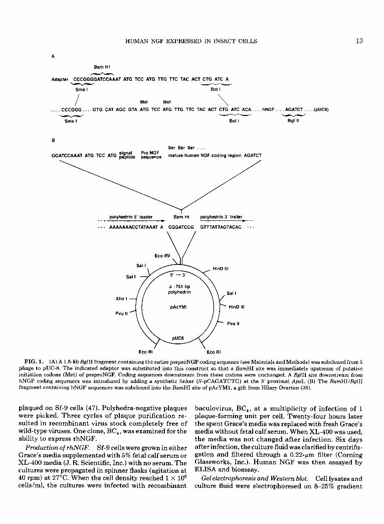
Cloning of human NGF gene and construction of the transplacement plasmid. A human leukocyte genomic library (constructed using cloning vector, X EMBL-3) was purchased from Clontech Laboratories, Inc. (Palo Alto, CA). Phage-containing sequences homologous to mNGF cDNA (a gift from W. F. Rutter, University of California, San Francisco) (44) were selected by three cycles of hybridization and subcloning. During the third cycle, an end-labeled synthetic oligonucleotide (5’- GAGGTGAACATTAACAACAGTGTATTCA-3’) com- plementary to the hNGF gene was also used as a hybrid- ization probe. A 1.8-kb BgZII fragment that hybridized with this probe was sequenced and found to contain the entire preproNGF coding sequences. This was modified and inserted into the baculovirus transfer vector pA- cYM1 (a gift from Hilary Overton) (35) as described in the legend to Fig. 1 to make the transplacement plasmid pYM12MhNGF.
Lipofection. “DOTMA” lipid-DNA complexes were prepared as described (17). One microgram of AcNPV DNA and a lo-fold molar excess of transplacement plasmid pYM12MhNGF DNA were used to transfect Sf-9 cells that had been seeded 1 h previously in 25-cm2 flasks in the absence of serum (3 X lo6 cells per flask). Ninety minutes after addition of the lipid-DNA com- plex, an equal volume of Grace’s media supplemented with 10% fetal calf serum and gentamycin (50 pug/ml) was added. Four hours later, the lipofection solution was decanted and fresh Grace’s media with 10% fetal calf serum was added. Three days later, when polyhedra were abundant, the culture fluid was harvested and
HUMAN NGF EXPRESSED IN INSECT CELLS 13
A
Barn Hl
Adapter CGATCCAAAT ATG TCC ATG TTG TTC TAC ACT CTG ATC A
Sma I Bcl I
I Met Met \ . CCCGGG.. GTG CAT AGC GTA ATG TCC ATG TTG TTC TAC ACT CTG ATC ACA.. hNGF.. AGATCT.. (PUCE)
Sma I
Ser Ser Ser .
GGATCCAAAT ATG TCC ATG “,$;;, Pro NGF sequence mature human NGF coding region AGATCT
polyhedrin 5’ leader Ba& HI polyhedrin 3’ trailer --- e --_
- - - AAAAAAACCTATAAAT A CGGATCCG GTTTATTAGTACAC - - -
Xho
Pvu
Eco RV
Sal I HinD Ill
I
Sal I 5’ - 3’
A 751 bp polyhedrin Sal I
pAcYMl HinD Ill
II
Pvu II
Eco RI. Eco RI
FIG. 1. (A) A 1.8kb BglII fragment containing the entire preproNGF coding sequence (see Materials and Methods) was subcloned from X phage to pUC-8. The indicated adaptor was substituted into this construct so that a BumHI site was immediately upstream of putative initiation codons (Met) of preproNGF. Coding sequences downstream from these codons were unchanged. A BglII site downstream from hNGF coding sequences was introduced by adding a synthetic linker (5’-pCAGATCTG) at the 3’ proximal ApaI. (B) The BamHI/BglII fragment containing hNGF sequences was subcloned into the BamHI site of pAcYM1, a gift from Hilary Overton (35).
plaqued on Sf-9 cells (47). Polyhedra-negative plaques were picked. Three cycles of plaque purification re- sulted in recombinant virus stock completely free of wild-type viruses. One clone, BC,, was examined for the ability to express rhNGF.
Production of rhNGF. Sf-9 cells were grown in either Grace’s media supplemented with 5% fetal calf serum or XL-400 media (J. R. Scientific, Inc.) with no serum. The cultures were propagated in spinner flasks (agitation at 40 rpm) at 27°C. When the cell density reached 1 X lo6 cells/ml, the cultures were infected with recombinant
baculovirus, BC,, at a multiplicity of infection of 1 plaque-forming unit per cell. Twenty-four hours later the spent Grace’s media was replaced with fresh Grace’s media without fetal calf serum. When XL-400 was used, the media was not changed after infection. Six days after infection, the culture fluid was clarified by centrifu- gation and filtered through a 0.22~pm filter (Corning Glassworks, Inc.). Human NGF was then assayed by ELISA and bioassay.
Gel electrophoresis and Western blot. Cell lysates and culture fluid were electrophoresed on 8-25% gradient

14 BARNETT ET AL.
A 6 C D
- 95 ‘=I
33
1;:
-18.4 - -1i.4
FIG. 2. Sf-9 cells were infected with wild-type NPV or recombi- nant BC, at a multiplicity of infection of 20 plaque-forming units per cell. Five days later culture fluid and cells were separated by centrifu- gation and both were analyzed by Western blot. Lane A, cell lysate from wild-type-infected cells; lane B, cell lysate from BC,-infected cells; lane C, culture fluid from wild-type-infected cells; lane D, cul- ture fluid from BC,-infected cells. The lysate samples loaded were from 5 X lo4 cells and the culture fluid samples were the amount taken from 1 X lo6 cells. Position of mNGF is shown on the left. Positions of molecular weight standards (Diversified Biotech pre- stained, midrange standards) are indicated on the right.
SDS-polyacrylamide gels (18). After transfer onto ni- trocellulose filters (48), rhNGF-related proteins were detected with an antiserum (polyclonal) prepared against mNGF (a gift from Eric Shooter) and 1251-la- beled protein A (8 &i/pg, New England Nuclear, Wil- mington, DE).
NGFprotein measurement. NGF protein levels were measured by two-site enzyme immunoassay. (i) Polysty- rene microtiter plates (PRO-BIND assay plate, Falcon) were incubated with 100 pi/well of 50 mM sodium car- bonate/sodium bicarbonate buffer, pH 9.6, containing 0.1 gg of mouse monoclonal anti-mouse p NGF (Boehringer-Mannheim, Indianapolis, IN) for 2 h at 37°C. (ii) Each well was incubated for 16 h at 4°C with 50 ~1 of the samples or with a standard solution (5-640 pg of mouse /3 NGF/ml of sample buffer: Tris-HCl, 50 mM; NaCl, 200 m&f; CaCl,, 10 mM; bovine serum albu- min (BSA), 1% (w/v); Triton X-100, 0.1% (w/v); pH 7.0). An additional three blank wells were incubated with sample buffer but without NGF: P-galactosi- dase-conjugated anti-mNGF monoclonal antibody (Boehringer-Mannheim) was used at 0.4 U/ml. (iii) Each well was incubated with this solution for 4 h at 37”C, and each step was follouied by three washes con- taining sample buffer but not BSA. (iv) P-Galactosidase activity was determined by incubation with 100 ~1 of the freshly prepared substrate solution (0.3 mg of O-nitro-
phenyl galactopyranoside in the substrate buffer: Hepes, 100 m&f; NaCl, 150 mM, MgCl,, 2 n&f; BSA, 1% (w/v); pH 7.0) for 2 h in the dark at 37°C. The optical density of the generated colored product was measured at 405 nm by using ELISA-reader (Titertek Multiskan Plus, Flow Laboratories, Inc.).
Relative potencies of rhNGF and mNGF were com- pared in vitro to determine the concentrations of rhNGF required to match concentrations of mNGF used in the in vivo portion of this experiment. A stan- dard curve of mNGF protein activity (absorbance) on ELISA was plotted. In turn, the rhNGF was diluted by factors of 1:l X 106, 1:2 X 10⁶, 1:4 X 10⁶, and 1:16 X 106, and activity (absorbance) was plotted against concen- tration.
Bioassay. PC12 cells were plated at a density of 5 X lo4 cells/ml in 24-well tissue culture plates (Falcon, Oxnard, CA). Media, sera, and incubation conditions were the same as those described for cell propagation. Spent culture fluid from Sf-9 cells infected with recom- binant baculovirus, BC,, was filtered through a 0.22-pm filter (Corning Glassworks) of which 25 ~1 containing 100 ng rhNGF was added to the PC12 cell cultures. Mouse NGF (Sigma Chemical Co., St. Louis, MO) was used as a positive control and spent culture fluid from Sf-9 cells infected with wild-type baculovirus was a nega- tive control. Media were not changed and no additional NGF was added after the initial treatment. PC12 cells were monitored daily by microscopic examination (3 fields per well, 4 wells per data point) and the percent- age of cells that had differentiated was determined. A cell was considered differentiated when the length of its neurite(s) was equal to the cell body diameter.
Neutralization of rhNGF-induced differentiation was analyzed by treating PC12 cells with 1:500 or 1:lOOO di- lutions of anti-mNGF serum (polyclonal, Collaborative Research, Inc., Waltham, MA). Ten minutes after treatment with the antisera, 25 ~1 of culture fluid con- taining 100 ng insect cell produced rhNGF or 100 ng mNGF (Sigma Chemical Co.) was added to the PC12 cells and the percentage of cells undergoing differentia- tion after 14 days was determined by microscopic exami- nation.
The bioassay on human SH-SY5Y cells was similar to the protocol for PC12 cells except fresh media contain- ing rhNGF was added to the SH-SY5Y cells every 2 days (29). In addition, aphidicolin was added to the culture during the second week of treatment at a concentration of 10 pug/ml.
Purification. The rhNGF released into insect cell culture fluid was purified by ion-exchange and reverse- phase chromatography. These procedures were modifi- cations of the purification scheme previously described by Petrides and Shooter (40) for purification of NGF from mouse submaxillary glands. The purity of the ob-

HUMAN NGF EXPRESSED IN INSECT CELLS 15
I ?IG. 3. Rat pheochromocytoma (PC12) cells (A, B) or human neurohlastoma (SH-SY5Y) cells (C, D) were treated with 25 pl of cult flui .d from wild-type baculovirus-infected Sf-9 cells (A, C) or 25 ~1 of culture fluid from BC,-infected Sf-9 cells (B, D). Seven days later the c we1 te microscopically examined (200x magnification) and photographed.
;ure ells

16 BARNETT ET AL.
0123456769 10 11 12 13 14 15 16 Time (days)
FIG. 4. PC12 cells were treated with 100 ng rhNGF in culture fluid from insect cells infected with recombinant baculovirus (m), the same material concentrated U-fold (A), culture fluid from insect cells infected with wild-type baculovirus (O), the wild-type culture fluid concentrated X-fold (V), or 100 ng mouse NGF (0). Differentiation was monitored by daily microscopic examinations. The bars repre- sent the standard deviation of the 12 microscopic fields counted for each data point.
tained rhNGF was greater than 98% as judged by a silver-stained SDS-PAGE gel. The details of this purifi- cation and characterization of rhNGF will be presented elsewhere (Barnett et al., manuscript in preparation).
Surgery. Female Sprague-Dawley rats (200-250 g) were used throughout this study and were housed in standard laboratory cages with free access to food and water. All animals received unilateral FF lesions fol- lowed by implantation of cannulae into the intracere- broventricular space through which one of five solutions was infused continuously for a 2-week period: control animals (n = 13) received artificial CSF (ACSF) only; a low-dose mNGF group (n = 7) received 25 pg/ml of mNGF in ACSF; a high-dose mNGF group (n = 7) re- c&ved 50 pg/ml of mNGF in ACSF; a low-dose hNGF group (n = 8) received 25 pg/ml of hNGF in ACSF; and a high-dose hNGF group (n = 7) received 50 pg/ml of hNGF in ACSF.
Animals were anesthetized and placed in the stereo- taxic apparatus. A 3-cm incision in the sagittal plane was made along the scalp to expose the skull. Perios- teum was scraped away with the knife blade, exposing surface landmarks of the skull. Using bregma as a zero reference point, a unilateral hole on the right side of the brain was drilled at stereotaxic coordinates A/P +0.8 mm and M/L +1.2 mm to receive the cannula for intra- ventricular infusion. Next, a 2-mm’portion of skull was removed immediately caudal to the first hole to provide access for aspiration of the superficial cingulate cortex, deep white matter, and corpus callosum, thus exposing the ventricular system and permitting aspiration of the FF.
An osmotic pump, Alzet Model 2002, was dipped into molten paraffin to cover one-half of its surface area to reduce its flow rate from 0.5 to 0.25 pi/h. The pumps were then filled with artificial CSF containing 0.1% rat serum (to supply a protein carrier for NGF), 50 pg/ml gentamycin, and NGF solutions as noted above, de- pending on the experimental group. Mouse NGF was obtained from a commercial source (Bioproducts for Science). The pump was attached to a platform (possess- ing a cannula that constituted the intracranial portion of the infusion apparatus) with a 3-cm length of vinyl tubing. The pump assembly was vertically oriented in all planes and then lowered through the smaller, rostra1 craniotomy opening until it contacted the surface of the skull. The proper ventral/dorsal distance was predeter- mined by the 5-mm length of the intraventricular can- nula. A drop of superglue was placed around the base of the pump to promote stable adhesion to the skull, with a more substantial base of support created using cyanoac- rylate. A subcutaneous pocket was formed in the ani-
h 0.4
1
n Mouse (Sub. MAX)
y I 0.109 + 4.4+4x R2 = 0.997
0.0-l n 8 0 1 0 200 400 600 800
Mouse [NGFI pg
B 0.41 /
3 0.3 -
s
g 9 oz- . P k
p” 0 Human (Recomb.)
c 0.1-
y E 0.10 + 1se-2x R = = 0.996
0.0’ - 16 4 2 1
Dilutions in (X) 10 6
FIG. 5. (A) Standard curve for mouse NGF and (B) dilution curve for human recombinant NGF showing high correlation coeffi- cients for both and equivalent slopes.

HUMAN NGF EXPRESSED IN INSECT CELLS 17
FIG. 6. Acetylcholinesterase-stained sections in animals receiving rhNGF. (A, B) Septum of rat infused with rhNGF, showing increased density of cholinergic fibers and cell bodies in the medial septum and of cholinergic fibers in the lateral septum. A shows minimal to moderate damage in region of tip of infusion apparatus (c), while B shows minimal if any damage (c). (C) Level of FF lesion, showing complete unilateral aspiration of FF. (D) Dorsal hippocampus, showing depletion of cholinergic fibers ipsilateral to FF lesion. Scale bars = 1000 pm for A, B, D; 2000 am for C. Star indicates lesioned side; dg, dentate gyrus; ca,, subfield of hippocampus.
mal’s neck and infrascapular space by blunt dissection to receive the osmotic pump. The incision was then closed with wound clips.
Histology. All animals survived for 2 weeks prior to sacrifice. Each animal was perfused with 100 cc cold 0.1 M phosphate-buffered saline (PBS) followed by 300 cc 4% formalin in PBS. The brains were removed, post- fixed overnight in the same fixative, and then left for 3 days in phosphate buffer containing 30% sucrose at 4°C. The brains were frozen with dry ice and coronal sections were cut at 40 pm with a sliding microtome. Sections were collected in 24-well tissue culture plates (Falcon) containing cryoprotectant (glycerol/ethylene glycol/phosphate buffer). Sections were taken rostrally
from the medial septum/vertical limb diagonal band (MS/VDB) complex and caudally through the extent of the dorsal hippocampus. Every fourth section was alter- nately processed for acetylcholinesterase, cresyl violet, and ChAT or NGFr immunocytochemistry.
ChAT immunocytochemistry was conducted as re- ported previously (1). Briefly, the procedure consisted of (i) overnight incubation of a polyclonal antibody against ChAT (courtesy of L. Hersh, UTHSC) of veri- fied specificity after 1:1,500 dilution with 0.1 M Tris-sa- line containing 1% goat serum and 0.25% Triton X-100; (ii) incubation for 1 h with biotinylated goat anti-rabbit IgG (Vector Laboratories) diluted l:200 with Tris-sa- line containing 1% goat serum; (iii) 1 h of incubation

18 BARNETT ET AL.
A 120
110
100 a g 90
80 B b
70 8 60
v 50 % 40
# 30 20 10
0 GROUPS
B 120: 110 -
100 -
4 90- ;ij so-
z 70- 3 8 60 - V 50-
8 40-
GROUPS
n c0ntr01 n mNGFIow
i
q hNGFlow
m mNGFhiih
q hNGFhigh
FIG. 7. Histogram showing percentage of cell savings in medial septum following FF lesions and in control animals. (A) ChAT-im- munoreactivity; (B) NGF receptor immunoreactivity. mNGF low = 25 rg/ml mouse NGF; rhNGF low = 25 rglml recombinant human NGF; mNGF high = 50 pglml mouse NGF; rhNGF high = 50 pglml recombinant human NGF. Double asterisk indicates significant (P < 0.05) difference of control animals from all other groups; single asterisk indicates significant difference of low-dose rhNGF group from other NGF-treated groups.
with avidin-biotinylated peroxidase complex (Vector Laboratories) diluted 1:lOOO with Tris-saline contain- ing 1% goat serum; and (iv) treatment for 15 min with 0.05% solution of 3,3’-diaminobenzidine, 0.01% H,O,, and 0.04% nickel chloride in 0.1 M Tris buffer. Immu- nolabeled tissue sections were mounted onto gelatin- coated glass slides, air-dried, and covered with Per- mount and glass coverslips.
Nerve growth factor receptor (NGFr) immunocyto- chemistry was conducted similarly to ChAT immunocy- tochemistry. Primary NGFr monoclonal antibody (192-IgG) (courtesy E. Johnson, Washington Univer- sity) specific for NGFr was obtained from a hybridoma cell culture supernatant. Primary NGFr antibody was used at 1:lOO dilution in a 24-h incubation, diluted in horse serum rather than in goat serum.
Counting of ChAT or NGFr-positive cells in the me- dial septum was performed under light microscopy em-
ploying three 40-pm-thick sections located approxi- mately 160, 320, and 480 pm rostra1 to the decussation of the anterior commissure. The sections were analyzed with a 10X objective using a 0.5 X 0.5-mm counting grid. Cells labeled positively with peroxidase reaction prod- uct and possessing either (i) a cell body with emerging fiber or (ii) a cell body with well-defined nucleus were counted. Results were expressed as a percentage of the unlesioned side of the medial septum of each animal.
StutzkticuE anatysis. Group differences were assessed by a one-way ANOVA. Dunnett’s was used to assess individual group differences.
RESULTS
Expression of biologically active human NGF in insect cells using a buculovirus expression vector. The coding sequence for the prepro precursor of human NGF was inserted into the BumHI site of the baculovirus cloning vector pAcYM1 (35) as described under Materials and Methods (Fig. 1). Following cotransfection with AcNPV DNA using a lipofection procedure, polyhedrin negative recombinant baculoviruses were obtained and plaque- purified. A high titer virus stock was prepared from one of the viral isolates, designated as BC,.
S. frugiperdu cells propagated in Grace’s media con- taining fetal calf serum were infected with baculovirus BC,. Release of rhNGF-related molecules into the cul- ture fluid was monitored by ELISA using a NGF-speci- fit monoclonal antibody. Immunoreactive material was detected as early as 2 days postinfection and continued to increase, reaching 2 pg/ml7 days after infection. No immunoreactive material was detected in culture fluid harvested from Sf-9 cells infected with the wild-type baculovirus. In subsequent experiments, when Sf-9 cells were propagated and infected with BC, in XL-400 serum-free media, a higher level of rhNGF (up to 7 pg/ ml) was produced by 3 days postinfection.
Nerve growth factor-related molecules released into BC,-infected insect cell culture fluid were further char- acterized by Western blot analysis (Fig. 2). Two immu- noreactive bands having molecular weights of approxi- mately 12.5 and 34 kDa were detected. The former co- migrated with the mNGF marker. The 34-kDa immunoreactive protein had a molecular weight similar to that described for mouse glycosylated pro-NGF pre- cursor (14, 15). Again, neither of these molecules was detected in culture fluid harvested from Sf-9 cells in- fected with the wild-type baculovirus (Fig. 2). Western blot analysis of lysates prepared from pelleted, BC,-in- fected cells indicated that an appreciable amount of the NGF-related material was sequestered within the cells and not released into the culture media. While hardly any of the 12.5-kDa mature rhNGF was detected in the cell pellet, two immunoreactive bands having molecular weights of 36 and 34 kDa were quite dominant (Fig. 2).

HUMAN NGF EXPRESSED IN INSECT CELLS 19
FIG. 8. ChAT-immunoreactive sections through medial septum following intraventricular infusions of (A) control, (B) rhNGF, and (C) mNGF. Upper case letters are low power (scale bar = 1000 pm); lower case letters are high power (scale bar = 75 pm).
On the basis of their sizes and intracellular location, the two molecular species could be the prepro (with signal peptide) and the pro-NGF (signal peptide removed) precursors. Evidently, once the 12.5kDa mature rhNGF was synthesized, it was efficiently secreted into the culture fluid.
Biological activity of baculovirus expressed human nerve growth factor. To ascertain if the recombinant baculovirus-produced rhNGF was indeed biologically active, rat PC12 pheochromocytoma cells were treated with culture fluid harvested from insect cells infected
with recombinant baculovirus BC,. Within 12 h of treatment, PC12 cells flattened and extended neuritic projections. By 36 h, 100% of the test cells had under- gone differentiation. The differentiated morphology was still evident at 7 days post-treatment (Fig. 3B). Un- treated controls showed no evidence of differentiation (Fig. 3A). In a like manner, human SH-SY5Y differen- tiated upon exposure to the baculovirus-produced rhNGF (Fig. 3D).
To establish more rigorously that the differentiation described so far was indeed due to rhNGF instead of

20 BARNETT ET AL.
FIG. 9. NGF receptor-immunoreactive sections through medial septum following intraventricular infusions of (A) control, (B) rhNGF, and (C) mNGF. Upper case letters are low power (scale bar = 1000 pm); lower case letters are high power (scale bar = 75 am).
other adventitious agents present in the culture fluid harvested from BC,-infected Sf-9 cells, the effect of NGF-specific antibody on PC12 cell differentiation was determined. When treated with antiserum, only 5% of PC12 cells differentiated in response to rhNGF com- pared to 100% of untreated control cells (see Materials and Methods). Similarly, differentiation elicited by mNGF was reduced from 100 to 10% by the antiserum. These results show that differentiation of PC12 cells elicited by spent culture fluid was readily neutralized by a polyclonal antiserum that was specifically prepared
against mouse NGF, which is highly homologous with human NGF. This confirms that the rhNGF was indeed responsible for the observed PC12 cell differentiation.
To compare the biological activities of rhNGF and mNGF, PC12 cells were treated with equal amounts of the two proteins. The extent of differentiation was monitored on a daily basis by microscopic examination. When treated with culture fluid containing 100 ng rhNGF, PC12 cell differentiation reached 100% within 2 days (Fig. 4). In contrast, when 100 ng mouse NGF was added to PC12 cells, the culture was not fully differen-
HUMAN NGF EXPRESSED IN INSECT CELLS 21
tiated until 4 days after treatment (Fig. 4). Even at 300 ng, mNGF did not elicit as rapid an onset of differentia- tion as rhNGF at 100 ng. In addition, mixing mNGF in culture fluid derived from wild-type baculovirus-in- fected insect cells did not augment the rate at which mouse NGF elicited PC12 cell differentiation. The in- duction of a rapid onset of differentiation, therefore, appears to be an intrinsic property of insect cell-pro- duced rhNGF.
The more rapid onset of differentiation elicited by rhNGF compared to mNGF was also not due to a gross underestimation of human NGF present in the culture media due to inaccuracy of our ELISA assays. Twofold dilutions of equal amounts of the mouse and human NGFs, as determined by ELISA, were electrophoresed on a polyacrylamide gel which was silver-stained. The intensities of the resulting bands confirmed that the concentrations of rhNGF corresponded well with that estimated by the ELISA.
A substantial fraction of the PC12 cells treated with 100 ng of rhNGF dedifferentiated by 5 days after treat- ment (Fig. 4). On the other hand, cells treated with equivalent amounts of mNGF did not dedifferentiate for the duration of the experiment (16 days). When a higher amount of rhNGF (12-fold) was applied, the cells also remained differentiated throughout the experi- ment. The reason for early dedifferentiation of cells treated with the lower concentration of rhNGF is un- clear.
When the human neuroblastoma cell line, SH-SY5Y, was treated with either mNGF or rhNGF present in the BC,-infected Sf-9 culture fluid, an even more marked onset of differentiation was observed. The rhNGF in- duced rapid onset of differentiation (within 24 h), as evidenced by the extension of neurites. In comparison, mNGF required at least 5 days to elicit a similar re- sponse.
rhNGF activity in vivo. The rhNGF secreted into in- sect cell culture fluid was purified (see Materials and Methods). This preparation appeared to be >98% pure by SDS-PAGE and retained potent biological activity (manuscript in preparation). The standard curve for mNGF (Fig. 5A) and the activity curve for purified rhNGF (Figure 5b) were both linear, with correlation coefficients of greater than 0.99 and equivalent slopes. From these curves equivalent concentrations of purified rhNGF and mNGF were determined and loaded into Alzet pumps. Control experiments showed that after 2 weeks in vivo, 63.2% of mNGF and 60.3% of rhNGF antigenic activity could be recovered from the pumps (determined by ELISA). Therefore, both rhNGF and mNGF were stable under the in vivo conditions of ad- ministration.
All rats tolerated the infusion period well and without infections. No pump blockage occurred as indicated by
residual pump fluid volumes at the end of the infusion period. Microscopic inspection of the brain in the area of the cannula tip revealed mild to moderate tissue dam- age attributable to the infusion apparatus. Figure 6B shows a case with minimal if any damage, while Figure 6A shows a case with slightly more extensive damage. There was no apparent difference in the degree of par- enchymal damage; however, that could be ascribed to varying concentrations of NGF, sources of NGF (mouse vs human), or NGF-treated vs control animals.
Complete FF lesions were verified histologically in all animals by visualizing the extent of resection of the for- nix, fimbria, corpus callosum, and overlying cingulate cortex (Fig. 6C) and by total loss of AChE fibers in the dorsal hippocampus including the CA3, CAl, dentate gyrus, and stria fornicatus (Fig. 6D).
Evaluation of ChAT immunoreactivity revealed a 57% decrease in the number of labeled neurons in the medial septum of control animals compared to the con- tralateral, unlesioned side (Figs. 7A, 8A, and 8a). The low-dose and high-dose mNGF groups showed only a 3% and 0% cell loss, respectively (Figs. 8C and 8c), while the low-dose rhNGF group showed a 21% cell loss that was significantly different from both of the mNGF groups (Fig. 8B and 8b). The high-dose rhNGF (7% cell loss) group was not different from any of the NGF- treated groups. In general, cholinergic cells of the me- dial septum that were ipsilateral to the side of the NGF infusion appeared darker in all the NGF-treated groups, particularly in the region of the cannula tip. Further, there was a marked increase in immunoreactive fiber- like processes that accumulated in the dorsal lateral quadrant of the septum, as previously reported (20,55).
NGFr immunoreactivity paralleled the pattern ob- served for ChAT immunoreactivity. There was a 55% decrease in NGFr-immunolabeled cells in the control group (Figs. 7B, 9A, and 9a), while the low-dose and high-dose mNGF groups had decreases of 4 and 8%, respectively (Figs. 9C and 9c). The low-dose and high- dose rhNGF groups showed 15 and 10% decreases, re- spectively (Figs. 9B and 9b). Thus 2 weeks postlesion, all NGF-infused groups were significantly different from control groups but were not different from each other. Once again, as noted with the ChAT-immunore- active label, there was a marked increase in immunore- active fiber-like processes in the dorsal lateral quadrant of the septum on the side of the infusion and an increase in the size and intensity of NGFr-immunoreactive label in cells near the cannula tip, regardless of the type or dose of NGF.
DISCUSSION
Mammalian proteins expressed in insect cells using the baculovirus expression vector are typically pro- cessed and folded so that biological activity is retained.

22 BARNETT ET AL.
We have expressed rhNGF that is processed to give the biologically active mature form by the following criteria: (i) conditioned media from recombinant baculovirus- infected Sf-9 cells induces differentiation of PC12 and SH-SY5Y cells, whereas conditioned media from wild- type baculovirus-infected cells does not; (ii) antibody against mNGF neutralizes the factor responsible for this differentiation; and (iii) immunoblots detect a pro- tein that has the molecular weight expected of mature rhNGF.
Human NGF produced with the baculovirus system appears to be more potent than mNGF on rat and hu- man cells in d-o. The rhNGF induced a much more rapid onset of differentiation in rat PC12 and human SH-SY5Y cells than did mNGF. The reason for this ob- servation is not clear. However, this property of rhNGF was not the result of other components in the insect cell culture fluid since culture fluid from insect cells infected with wild-type baculovirus did not enhance the rate of differentiation elicited by mNGF. Also it is unlikely that the difference between mNGF and rhNGF was due to unequal concentrations. In fact, mouse NGF at a con- centration threefold greater than the rhNGF concen- tration failed to elicit a rapid onset.
Whereas in vivo results show that 50 pglml of mNGF and 50 pg/ml of rhNGF are equally potent on degener- ating rat choline& neurons, at lower doses mNGF is slightly more potent. This is not surprising given that mouse NGF possesses higher sequence homology to rat than human NGF (44, 52). We are currently testing rhNGF in nonhuman primates, and it will be of interest to determine whether an inverse relationship occurs, that is, whether rhNGF is more potent than mNGF in a primate model of cholinergic degeneration (51).
As noted in the Introduction, an intriguing relation- ship may exist between cholinergic degeneration in hu- man pathology and levels of NGF. However, chronic infusion of NGF in patients with AD must await several important experimental steps. Some of these steps in- clude (41) (i) a reliable source of well-characterized hNGF, (ii) a reliable delivery system to function over long periods, (iii) dose-response characterization of hNGF to be used in potential trials, (iv) effects of hNGF on nonhuman primate cholinergic neurons, and (v) acute and chronic studies of toxicity and side effects of hNGF in at least two animal species. The last four steps have awaited completion of the first.
The levels of biologically active rhNGF obtained from the baculovirus/insect cell system described in this com- munication are substantially greater than the levels ob- served in previously reported systems (30). Further op- timization of the insect cell system may result in even higher yields. Furthermore, the insect cell-produced rhNGF possesses biological activity that is comparable to mNGF from a natural source. This system is, there-
quired to pursue additional steps in a comprehensive program to test the efficacy of NGF for AD.
ACKNOWLEDGMENTS
We are grateful to Dr. Eric Shooter for his helpful suggestions and reagents. We also thank Linda Fish, Mary Jane McRoberts, Carole Kurahara, D. Chen, and S. Forbes for their technical assistance. This work was conducted in part with funds from the NIA (AGO 0353A and AGO 5131), the Pew Foundation, and the Herbert and Margaret Hoover Jr,. Foundation.
1.
2.
3.
4.
5.
6.
7.
8.
9.
10.
Il.
12.
13.
fore, suitable for producing the amounts of rhNGF re- 14.
REFERENCES
ARMSTRONG, D. M., R. D. TERRY, R. M. DETERESA, G. BRUCE, L. B. HERSH, AND F. H. GAGE. 1987. Response of septal choliner- gic neurons to axotomy. J. Comp. Neural. 264: 421-436. AYER LELIEVRE, C. S., T. EBENDAL, L. OLSEN, AND A. SEIGER. 1983. Localization of NGF-like immunoreactivity in rat neurons tissue. Med. Biol. 61: 296-304. AYER-LELIEVRE, C., L. OLSON, T. EBENDAHL, A. SEIGER, AND H. PERSSON. 1988. Expression of the p-nerve growth factor gene in hippocampal neurons. Science 240: 1339-1341. BARTUS, R., R. L. DEAN, C. BEER, AND A. S. LIPPA. 1982. The choline+ hypothesis of geriatric memory dysfunction. Science 217:408-417. BATCHELOR, P. E., D. M. ARMSTRONG, S. M. BLAKER, AND F. H. GAGE. 1989. Nerve growth factor receptor and choline acetyl- transferase colocalization in neurons within the rat forebrain: Response to fimbria-fornix transection. J. Comp. Neural. 284: 187-204. BIEDLER, J. L., L. HELSON, AND B. A. SPENGLER. 1973. Morphol- ogy and growth, tumorigenicity and cytogenetics of human neu- roblastoma cells in continuous culture. Cancer Res. 33: 2643- 2652. BIEDLER, J. L., S. ROFFLER-TARLOV, M. SCHACHNER, AND L. S. FREEDMAN. 1978. Multiple neurotransmitter synthesis by hu- man neuroblastoma cell lines and clones. Cancer Res. 38: 3751- 3757. BRUCE, G., AND G. HEINRICH. 1989. Production and characteriza- tion of biologically active recombinant human nerve growth fac- tor. Neurobiol. Aging 10: 89-94. CHUN, L. L. Y., AND P. H. PATTERSON. 1977. Role of nerve growth factor in the development of rat sympathetic neurons in vitro. I. Survival, growth, and differentiation of catecholamine production. J. Cell Biol. 75: 694-704.
CHUN, L. L. Y., AND P. H. PA~RSON. 1977. Role of nerve growth factor in the development of rat sympathetic neurons in vitro. II. Developmental study. J. Cell Biol. 75: 705-711. COYLE, J. T., P. H. PRICE, AND M. R. DELONG. 1983. Alzheimer’s disease: A disorder of cortical choline@ innervation. Science 219: 1184-1189. GRAIN, S. M., AND E. R. PETERSON. 1974. Enhanced afferent synaptic functions in fetal mouse spinal cord-sensory ganglion explants following NGF-induced ganglion hypertrophy. Bruin Res. 79: 145-152. DICOU, E., R. HOULGA?TE, J. LEE, AND B. YON WILCKEN-BERG- MANN. 1989. Synthesis of chimeric mouse nerve growth factor precursor and human o-nerve growth factor in Escherichia coli: Immunological properties. J. Neurosci. Res. 22: 13-19. EDWARDS, R. H., M. J. SELBY, P. D. GARCIA, AND W. J. R~R.

HUMAN NGF EXPRESSED IN INSECT CELLS 23
15. EDWARDS, R. H., M. J. SELBY, W. C. MOBLEY, S. L. WEINRICH, D. E. HRUBY, AND W. J. RUTTER. 1988. Processing and secretion of nerve growth factor: Expression in mammalian cells with a vaccinia virus vector. Mol. Cell. Biol. 8: 2456-2464.
16. ERNFORS, P., T. EBENDAL, L. OLSON, P. MOUTON, I. STROMBERG, AND H. PERSSON. 1989. A cell line producing recom- binant nerve growth factor evokes growth responses in intrinsic and grafted central choline@ neurons. Proc. Natl. Acad. Sci. USA 86: 4756-4760.
17. FELGNER, P. L., T. R. GADEK, M. HOLM, R. ROMAN, H. W. CHAN, M. WENZ, J. P. NORTHROP, G. M. RINGOLD, AND M. DAN- IELSEN. 1987. Lipofection: A highly efficient, lipid-mediated DNA-transfection procedure. Proc. Natl. Acad. Sci. USA 84: 7413-7417.
18. FLING, S. P., AND D. S. GREGERSON. 1986. Peptide and protein molecular weight determination by electrophoresis using a high- molarity tris buffer system without urea. Anal. Biochem. 155: 83-88.
19. FRANCIS, P. T., A. M. PALMER, N. R. SIMS, D. M. BOWEN, A. N. DAVISON, M. M. ESIRI, D. NEARY, J. S. SNOWDEN, AND G. K. WILCOCK. 1985. Neurochemical studies of early-onset Alz- heimer’s disease: Possible influence on treatment. N. Engl. J. Med. 313: 7-11.
20.
21.
GAGE, F. H., D. M. ARMSTRONG, L. R. WILLIAMS, AND S. VARON. 1988. Morphologic response of axotomized septal neurons to nerve growth factor. J. Comp. Neurol. 269: 147-155.
GAGE, F. H., P. BATCHELOR, K. S. CHEN, D. CHIN, G. A. HIG- GINS, S. KOH, S. DEPUTY, M. B. ROSENBERG, W. FISCHER, AND A. BJORKLUND. 1989. NGF receptor reexpression and NGF me- diated cholinergic neuronal hypertrophy in the damaged adult neostriatum. Neuron 2: 1177-1184.
22.
23.
GAGE, F. H., K. WICTORIN, W. FICHER, L. R. WILLIAMS, S. VARON, AND A. BJORKLUND. 1986. Life and death of cholinergic neurons: In the septal and diagonal band region following com- plete fimbria-fornix transection. Neuroscience 19: 241-255.
GREENE, L., AND E. M. SHOOTER. 1980. The nerve growth factor: Biochemistry, synthesis, and mechanism of action. Annu. Reu. Neurosci. 3: 353-402.
24. GREENE, L. A., AND A. S. TISCHLER. 1976. Establishment of a noradrenergic clonal line of rat adrenal phenochromocytoma cells which respond to nerve growth factor. Proc. Natl. Acad. Sci. USA 73: 2424-2428.
25. HALLB~%K, F., T. EBENDAL, AND H. PERSSON. 1988. Production and characterization of biologically active recombinant beta nerve growth factor. Mol. Cell. Biol. 8: 452-456.
26. HEFTI, F. 1986. Nerve growth factor (NGF) promotes survival of septal cholinergic neurons after fimbrial transection. J. Neuro- xi. 6: 2155-2162.
27.
28.
29.
30.
HEFTI, F., A. DRAVID, AND J. J. HARTIKKA. 1984. Chronic intra- ventricular injections of nerve growth factor elevate hippocam- pal choline acetyltransferase activity in adult rats with partial septohippocampal lesions. J. Bruin Res. 293: 305-311.
HIGGINS, G. A., S. KOH, K. S. CHEN, AND F. H. GAGE. 1989. Nerve growth factor induces NGF-receptor gene expression in basal forebrain neurons of adult rat brain. Neuron 3: 247-256.
JENSEN, L. M. 1987. Phenotype differentiation of aphidicolin- selected human neuroblastoma cultures after long-term expo- sure to nerve growth factor. Dev. Biol. 120: 56-64. KANAYA, E., T. HIGASHIZAKI, F. OZAWA, K. HIRAI, M. NISHI- ZAWA, M. TOKUNAGA, H. TSUKUI, H. HATANAKA, AND F. HISHI-
1988. Processing of the native nerve growth factor precursor to form biologically active nerve growth factor. J. Biol. Chem. 263: 6810-6815.
43.
44.
31.
32.
33.
34.
35.
36.
37.
38.
39.
40.
41.
42.
45.
46.
47. SUMMERS, M. D., AND G. E. SMITH. 1987. A Manual of Methods
NUMA. 1989. Synthesis and secretion of human nerve growth factor by Sacchnromyces cerevisiae. Gene 83: 65-74.
KATZMAN, R., T. BROWN, P. FULD, L. THAL, P. DAVIES, AND R. D. TERRY. 1986. Significance of neurotransmitter abnormali- ties in Alzhiemer’s disease. In Neuropeptides in Neurologic and Psychiatric Disease (J. B. Martin and J. Barchas, Eds.), pp. 279- 286. Raven Press, New York.
LARGE, T. H., S. C. BODARY, D. 0. CLEGG, G. WESKAMP, U. OTTEN, AND L. F. REICHARDT. 1986. Nerve growth factor gene expression in the developing rat brain. Science 234: 352-355.
LEVI-M• NTALCINI, R., AND P. U. ANGELE~I. 1963. Essential role of the nerve growth factor on the survival and maintenance of dissociated sensory and sympathetic embryonic nerve cells in vitro. Dev. Biol. 7: 653-659.
LUCKOW, V. A., AND M. D. SUMMERS. 1988. Trends in the devel- opment of baculovirus expression vectors. Biotechnology 6: 47- 55.
MATSWRA, Y., R. D. POSSEE, H. A. OVERTON, AND D. H. L. BISHOP. 1987. Baculovirus expression vectors: The require- ments for high level expression of proteins, including glycopro- teins. J. Gen. Virol. 68: 1233-1250.
MILLER, L. K. 1988. Baculoviruses as gene expression vectors. Annu. Rev. Microbial. 42: 177-199.
MOBLEY, W. C., J. L. RUTKOWSKI, G. I. TENNEKOON, J. GEMSKI, K. BUCHANAN, AND M. V. JOHNSTON. 1986. Nerve growth factor increases choline acetyltransferase activity in developing basal forebrain neurons. Mol Bruin Res. 1: 53-62. MOHS, R. C., B. M. DAVIS, AND C. A. JOHNS. 1985. Oral physo- stigmine treatment of patients with Alzheimer’s disease. Am. J. Psychiatry 142:28-33. PERRY, E. K., B. E. TOMLINSON, G. BLESSED, K. BERGMANN, P. H. GIBSON, AND R. H. PERRY. 1978. Correlation of choline@ abnormalities with senile plaques and mental test scores in se- nile dementia. Br. Med. J. 2: 1457-1459.
PETRIDES, P. E., AND E. M. SHOOTER. 1986. Rapid isolation of the 7S-nerve growth factor complex and its subunits from mu- rine submaxillary glands and saliva. J. Neurochcm. 46: 721-725.
PHELPS, C. H., F. H. GAGE, J. H. GROWDON, S. HEFTI, R. HAR- BAUGH, M. V. JOHNSTON, Z. S. KHACHATURIAN, W. C. MOBLEY, D. L. PRINCE, M. RASKIND, J. SIMPKINS, L. J. THAL, AND J. WOODCOCK. 1989. Potential use of NGF to treat Alzheimer’s disease. Neurobiol. Aging 10: 205-207. SCHATTEMAN, G. C., L. GIBBS, A. A. LANAHAN, P. CLAUDE, AND M. BOTHWELL. 1988. Expression of NGF receptor in the develop- ing and adult primate central nervous system. J. Neurosci. 8: 860-873.
SCHWAB, M. E., AND H. THOENEN. 1983. Retrograde axonal transport. In Handbook of Neurochemistry (A. Lajtha, Ed.), pp. 381-404. Plenum, New York.
SCOTT, J., M. SELBY, M. UFZDEA, M. QUIROGA, G. I. BELL, AND W. J. RUTTER. 1983. Isolation and nucleotide sequence of a cDNA encoding the precursor of mouse nerve growth factor. Nature (London) 302: 538-540.
SEILER, M., AND M. E. SCHWAB. 1984. Specific retrograde trans- port of nerve growth factor (NGF) from cortex to nucleus basalis in the rat. Brain Res. 300: 33-39.
SHELDON, D. L., AND L. F. REICHARDT. 1986. Studies on the expression of the p-nerve growth factor (NGF) gene in the cen- tral nervous system: Level and regional distribution of NGF mRNA suggest that NGF functions as a trophic factor for sev- eral distinct populations of neurons. Proc. Natl. Acad. Sci. USA 83: 2714-2718.

24 BARNETT ET AL.
for Baculovirus Vectors and Insect Cell Culture Procedures 16-l 8. Texas A&M University, College Station, TX.
48. SZEWCZYK, B., AND L. M. KOZLOFF. 1985. A method for the effi- cient blotting of strongly basic proteins from sodium dodecyl sulfate-polyacrylamide gels to nitrocellulose. Anal. Biochem. 160:403-407.
49. THAL, L. J., P. A. BULD, D. M. MASUR, AND N. S. SHARPLESS. 1983. Oral physostigmine and lecithin improve memory in Alz- heimer’s disease. Ann. Neural. 13: 491-496.
50. TUSZYNSKI, M. H., G. BUZSAKI, AND F. H. GAGE. 1990. NGF infusions combined with fetal hippocampal grafts enhance re- construction of the lesioned septo-hippocampal projection. Neuroscience, in press.
51. TUSZYNSKI, M. H., H. S. U., D. G. AMARAL, AND F. H. GAGE. 1989. Nerve growth factor infusion in primate brain reduces le- sion-induced choline@ neuronal degeneration. Sot. Neurosci. Abstr. 15: 707.
52. ULLRICH, A., A. GRAY, C. BERMAN, AND T. J. DULL. 1983. Hu- man B-nerve growth factor gene sequence highly homologous to that of mouse. Nature (London) 303: 821-825.
53. WHITEHOUSE, P. J., D. L. PRICE, R. G. STRUBLE, A. W. CLAR, J. T. COYLE, AND M. R. DELONG. 1982. Alzheimer’s disease and senile dementia: Loss of neurons in the basal forebrain. Science 216: 1237-1239.
54. WHI?TMORE, S. R., T. EBENDAL, L. LARKFORS, L. OLSON, A. SEIGER, I. STROMBERG, AND H. PERSSON. 1986. Developmental and regional expression of fi nerve growth factor messenger RNA and protein in the rat central nervous system. Proc. Natl. Acad. Sci. USA 83: 817-821.
55. WILLIAMS, L. R., S. VARON, G. M. PETERSON, K. WICTORJZN, W. FISHER, A. BJORKLUND, AND F. H. GAGE. 1986. Continuous infu- sion of nerve growth factor prevents basal forebrain neuronal death after fimbria-fornix transection. Proc. Natl. Acad. Sci. USA 83: 9231-9235.