Human Cytomegalovirus Infection Induces Specific Hyperphosphorylation of the Carboxyl-Terminal...
18
10.1128/JVI.79.24.15477-15493.2005. 2005, 79(24):15477. DOI: J. Virol. Sama Tamrakar, Anokhi J. Kapasi and Deborah H. Spector cdk9 and cdk7 Abundance, Activity, and Localization of Associated with Changes in the Subunit of RNA Polymerase II That Is Carboxyl-Terminal Domain of the Large Specific Hyperphosphorylation of the Human Cytomegalovirus Infection Induces http://jvi.asm.org/content/79/24/15477 Updated information and services can be found at: These include: REFERENCES http://jvi.asm.org/content/79/24/15477#ref-list-1 at: This article cites 51 articles, 29 of which can be accessed free CONTENT ALERTS more» articles cite this article), Receive: RSS Feeds, eTOCs, free email alerts (when new http://journals.asm.org/site/misc/reprints.xhtml Information about commercial reprint orders: http://journals.asm.org/site/subscriptions/ To subscribe to to another ASM Journal go to: on August 7, 2014 by guest http://jvi.asm.org/ Downloaded from on August 7, 2014 by guest http://jvi.asm.org/ Downloaded from
Transcript of Human Cytomegalovirus Infection Induces Specific Hyperphosphorylation of the Carboxyl-Terminal...

10.1128/JVI.79.24.15477-15493.2005.
2005, 79(24):15477. DOI:J. Virol. Sama Tamrakar, Anokhi J. Kapasi and Deborah H. Spector cdk9 and cdk7Abundance, Activity, and Localization of Associated with Changes in theSubunit of RNA Polymerase II That Is Carboxyl-Terminal Domain of the LargeSpecific Hyperphosphorylation of the Human Cytomegalovirus Infection Induces
http://jvi.asm.org/content/79/24/15477Updated information and services can be found at:
These include:
REFERENCEShttp://jvi.asm.org/content/79/24/15477#ref-list-1at:
This article cites 51 articles, 29 of which can be accessed free
CONTENT ALERTS more»articles cite this article),
Receive: RSS Feeds, eTOCs, free email alerts (when new
http://journals.asm.org/site/misc/reprints.xhtmlInformation about commercial reprint orders: http://journals.asm.org/site/subscriptions/To subscribe to to another ASM Journal go to:
on August 7, 2014 by guest
http://jvi.asm.org/
Dow
nloaded from
on August 7, 2014 by guest
http://jvi.asm.org/
Dow
nloaded from

JOURNAL OF VIROLOGY, Dec. 2005, p. 15477–15493 Vol. 79, No. 240022-538X/05/$08.00�0 doi:10.1128/JVI.79.12.15477–15493.2005Copyright © 2005, American Society for Microbiology. All Rights Reserved.
Human Cytomegalovirus Infection Induces Specific Hyperphosphorylationof the Carboxyl-Terminal Domain of the Large Subunit of RNA
Polymerase II That Is Associated with Changes in theAbundance, Activity, and Localization of cdk9 and cdk7
Sama Tamrakar, Anokhi J. Kapasi, and Deborah H. Spector*Department of Cellular and Molecular Medicine, School of Pharmacy and Pharmaceutical Sciences, and
Center for Molecular Genetics, University of California, San Diego, La Jolla, California 92093-0712
Received 17 August 2005/Accepted 26 September 2005
Human cytomegalovirus infection in the presence of the cyclin-dependent kinase (cdk) inhibitor roscovitineleads to changes in differential splicing and the polyadenylation of immediate early IE1/IE2 and UL37transcripts (V. Sanchez, A. K. McElroy, J. Yen, S. Tamrakar, C. L. Clark, R. A. Schwartz, and D. H. Spector,J. Virol. 78:11219-11232, 2004). To determine if this was associated with specific phosphorylation of theC-terminal domain (CTD) of the RNA polymerase II (RNAP II) large subunit by cdk7/cyclin H and cdk9/cyclinT1, we examined the expression and localization of these kinases and the various phosphorylated forms ofRNAP II. Infection resulted in increased RNAP II CTD phosphorylated on serines 2 and 5 and increased levelsof activity of cdk7 and cdk9. At early times, cdk9 localizes with input viral DNA, and aggregates of cdk9 andcdk7 and a subset of Ser2-phosphorylated RNAP II colocalize with IE1/IE2 proteins adjacent to promyelocyticleukemia protein oncogenic domains. Later, cdk9 and Ser2-phosphorylated RNAP II form a nuclear punctatepattern; cdk7 resides in replication centers, and Ser5-phosphorylated RNAP II clusters at the peripheries ofreplication centers. Roscovitine treatment leads to decreased levels of hyperphosphorylated RNAP II (RNAPIIo) in infected cells and of hypophosphorylated RNAP II in mock-infected and infected cells. The RNAP IIodecrease does not occur if roscovitine is added 8 h postinfection, as was previously observed for processing ofIE transcripts. These results suggest that accurate IE gene expression requires specific phosphorylation of theRNAP II CTD early in infection.
Human cytomegalovirus (HCMV), a herpesvirus, is an ob-ligate parasite whose life cycle requires an intricate set ofinteractions between the virus and the host that optimize theenvironment for viral replication and assembly (for a review,see reference 14). Intertwined with this subversion of the hostcell is a defined temporal order of viral gene expression thathas been loosely divided into three phases—immediate early(IE), early, and late. The IE gene products are synthesizedsoon after infection and rely primarily on host factors for theirexpression, although proteins carried in the viral particleclearly contribute to the process. Several of the viral IE pro-teins serve as essential transactivators of the next class of geneproducts, the early genes. Included in the early class are thoseviral proteins required to “activate” the cell to a metabolicstate most conducive for viral DNA synthesis and those pro-teins involved in the actual replication process itself. Lategenes, which constitute the majority of the viral genome andencode primarily structural and maturation proteins, are tran-scribed in abundance only after the onset of viral DNA repli-cation.
Upon infection, the viral DNA enters the nucleus and asubset of HCMV genomes are deposited at nuclear structuresreferred to variously as nuclear domain 10 (ND10) structures
or promyelocytic leukemia protein (PML) oncogenic domains(PODs), where viral RNA synthesis begins (21, 22). TheHCMV tegument protein pp71 interacts with POD-associatedDaxx, which may contribute to the initiation of transcription atPOD sites (9, 20, 23, 28). The proximity of the PODs to thespliceosome assembly factor SC35 domains may aid in rapidexpression of IE genes, which are often multiply spliced (22). Amajor region of viral IE transcription includes two geneticunits, IE1 and IE2 (for reviews, see references 15 and 31). Thepredominant IE RNA (IE1) consists of four exons; a single openreading frame (ORF) (UL123) initiates in exon 2 and specifies a72-kDa nuclear protein designated IE1-72. The IE2 gene prod-uct, IE2-86 (ORF UL122), is an 86-kDa protein that is en-coded by an alternatively spliced RNA that contains the firstthree exons of IE1 and a different terminal exon. Anotherregion of IE gene expression is UL36-38, which includes atleast five transcripts directed by three promoters (1, 26, 49, 50).One of the promoters directs the synthesis of several spliced3.2- to 3.4-kb RNAs (UL37 and UL37M ORFs) that arepresent in small amounts only at IE times as well as an abun-dant 1.7-kb unspliced RNA that encodes the UL37 exon 1(UL37X1) gene product.
Newly synthesized IE1-72 and IE2-86 localize to the PODs.While the punctate pattern of the IE2-86 protein persists, at 3to 6 h postinfection (p.i.), both IE1-72 and POD-associatedproteins become dispersed throughout the nucleus (5, 22, 24,25). Several studies have shown that IE2-86 is able to localizeto the PODs in the absence of IE1-72 but is not able to disrupt
* Corresponding author. Mailing address: Cellular and MolecularMedicine East, Room 2059, Mail Code 0712, 9500 Gilman Drive,University of California, San Diego, La Jolla, California 92093-0712.E-mail: [email protected].
15477
on August 7, 2014 by guest
http://jvi.asm.org/
Dow
nloaded from

them (4, 5, 22). IE1-72 is required for disruption of the PODs,but since an IE1 deletion mutant virus (CR208) replicates wellat high multiplicity (18, 19, 32), this event is not essential. Itappears that even after IE1-72 has caused the dispersal of thePODs, these locations remain important for viral replication.The UL112-113 early gene proteins appear to colocalize withIE2-86 at the peripheries of the original POD sites beginningat about 6 h p.i., and these go on to form sites of viral DNAreplication (3, 35). By 48 h p.i., there is a high level of viralDNA synthesis in the replication centers, and the majority ofthe viral genome is being transcribed.
Viral transcription is directed by the cellular RNA polymer-ase II (RNAP II), a multisubunit complex. The largest subunitof RNAP II contains a C-terminal domain (CTD), which inhuman cells consists of 52 repeats of the consensus heptapep-tide sequence Tyr-Ser-Pro-Thr-Ser-Pro-Ser. The CTD is dif-ferentially phosphorylated primarily at the serine 2 and serine5 positions, and the level of phosphorylation varies consider-ably during the transcription cycle (for reviews, see references27, 30, and 37). Hypophosphorylated RNAP II (RNAP IIa) isrecruited to the initiation complex, and the CTD is then phos-phorylated to a hyperphosphorylated form (RNAP IIo). Thephosphorylation of the serines in position 5 by cdk7/MAT-1/cyclinH (a component of the basal transcription factor complex TFIIH)is followed by the phosphorylation of the serines in position 2 bycdk9/cyclin T (also referred to as positive transcription elongationfactor B [P-TEFb]), which is associated with the commitment ofthe RNAP II complex to elongation. RNAP IIa and IIo forms arein a dynamic equilibrium such that RNAP IIo needs to be de-phosphorylated to the IIa form in order to engage in anotherround of transcription. The primary phosphatase is Fcp1, whichis highly conserved and can remove phosphates from the CTDof RNAP IIo when it is engaged in transcription or free. Re-cently, other phosphatases have been discovered, including thesmall CTD phosphatases and Ssu72.
The prevailing hypothesis is that the CTD plays a regulatoryrole in all steps of transcription by serving as the bindingdomain and transporter of factors involved in RNA synthesisinitiation, elongation, 5� capping, splicing, and cleavage/poly-adenylation (for reviews, see references 8, 30, 34, and 38). It isbelieved that the differential recruitment and binding of specificfactors that are involved in these processes are significantly influ-enced by the pattern of phosphorylation of the various Ser2 andSer5 residues (and possibly by the ubiquitylation, glycosylation,and phosphorylation of other residues) within the 52 repeats. Forexample, the phosphorylation of the CTD at Ser5 is associatedwith the recruitment of the mRNA capping enzymes, and thephosphorylation of the CTD at Ser2 is linked to the recruit-ment of 3� RNA processing factors.
In a recent study, we used the drug roscovitine, which is aspecific inhibitor of the cyclin-dependent kinases (cdk’s) 1, 2, 5,7, and 9 (7, 10, 13, 29, 45, 51), to examine the role of thesekinases in viral replication (44). We found that addition of thedrug at the beginning of the infection resulted in changes in theaccumulation and processing of IE transcripts and inhibition ofthe expression of selected viral early gene products, viral DNAreplication, and late gene expression. Roscovitine specificallyaffected the differential splicing and polyadenylation of theRNAs from both the IE1/IE2 and UL37 genes. The relativeposition of the sequences used for processing the HCMV un-
spliced UL37X1 RNA and spliced UL37 IE RNAs (47) is verysimilar to the position of the region between UL123 exon 4 andUL122 exon 5, which are used for the alternative cleavage/polyadenylation and splicing that generates the IE1-72 andIE2-86 RNAs, respectively. For both regions, the signals on theRNA for cleavage/polyadenylation overlap those for the down-stream 3� splice acceptor site, with the cleavage/polyadenyla-tion site being preferentially used to generate either IE1-72 orUL37X1 RNAs at IE times. However, in the presence of ros-covitine, there was greater utilization of the downstream 3�splice acceptor site, yielding higher levels of the IE2-86 andspliced UL37 RNAs and corresponding lower levels of theIE1-72 or UL37X1 RNAs. We also showed that when ros-covitine was added after the first 4 h of infection, the effects onIE gene expression were no longer observed. When it wasadded after 6 h, viral replication proceeded through the latephase but the viral titer remained low.
One possible explanation for the altered pattern of viral RNAprocessing was that the effects of the cdk inhibitor were related tothe phosphorylation of the CTD of the large subunit of RNAP IIby cdk7/MAT-1/cyclin H and cdk9/cyclin T. A recent paper show-ing that HCMV induces an intermediate form of phosphorylatedRNAP II supports this idea (6). Those authors proposed that theCTD might be phosphorylated by the HCMV-encoded kinaseUL97. However, their data showed that although the CTD canserve as a substrate for UL97 in vitro, RNAP II does not appearto be phosphorylated by this kinase in vivo.
In this study, we examine the effect of HCMV infection on theexpression, activity, and localization of cdk7/MAT-1/cyclin H,cdk9/cyclin T1, and several forms of the large subunit of RNAP IIat both early and late times during the infection. We show thatduring the course of the infection, there is an increase in cdk7 andcdk9 protein levels and kinase activity and in the amount ofRNAP II that is phosphorylated on serine 2 and serine 5 withinthe CTD. At 48 h p.i., cdk7 and hypophosphorylated RNAP IIlocalize to replication centers, cdk9 and Ser2-phosphorylatedRNAP II are distributed in a punctate pattern throughout thenuclei, and Ser5-phosphorylated RNAP II appears in clusters atthe peripheries of the viral replication centers. At early times,cdk9 localizes with input viral DNA. In addition, aggregates ofcdk9 and cdk7 and a subset of Ser2-phosphorylated RNAP IIcolocalize with IE1/IE2 proteins adjacent to the PODs. Additionof the cdk inhibitor roscovitine at the time of infection results indecreased CTD phosphorylation in the infected cells and a de-crease in the level of the hypophosphorylated RNAP II in bothinfected and mock-infected cells. In accord with our previ-ous results regarding the effect of the cdk inhibitors on theprocessing and accumulation of the HCMV IE1/IE2 andUL37 IE transcripts, the decrease in CTD phosphorylationdoes not occur if the drug is added after 8 h p.i. Theseresults suggest that the phosphorylation of the CTD is es-sential at early time points of the infection and that therequired level of CTD phosphorylation for IE gene expres-sion is established within 8 h.
MATERIALS AND METHODS
Cell culture and virus. Human foreskin fibroblasts (HFF) were obtained fromthe University of California, San Diego, Medical Center and cultured in Earle’sminimal essential medium supplemented with 10% heat inactivated fetal bovineserum, 1.5 �g/ml amphotericin B, 2 mM L-glutamine, 100 U/ml penicillin, and
15478 TAMRAKAR ET AL. J. VIROL.
on August 7, 2014 by guest
http://jvi.asm.org/
Dow
nloaded from

100 �g/ml streptomycin. All reagents were from Invitrogen (Carlsbad, CA). Cellswere kept in incubators maintained at 37°C with 7% CO2. The Towne strain ofHCMV was obtained from the American Type Culture Collection (VR 977) andpropagated as previously described (48).
Cell synchronization and infections. HFF (passage numbers 15 to 20) weresynchronized in G0 phase by allowing them to grow to confluence as previouslydescribed (42). Three days after confluence, the cells were trypsinized, replatedat a lower density to allow progression into the cell cycle, and infected at amultiplicity of infection (MOI) of 3 to 5 with HCMV Towne or mock infectedwith tissue culture supernatants. At 6 h postplating/postinfection, the inoculumwas replaced with fresh medium. At designated times p.i., 20 �M roscovitine(Sigma Aldrich, St. Louis, MO) was added to the medium. For experimentsextending beyond 24 h, medium was replaced with fresh drug-containing mediumat 24 h p.i. and 48 h p.i. The roscovitine stock solution was in dimethyl sulfoxide.Control samples were treated with appropriate volumes of dimethyl sulfoxide. Atvarious times p.i., cells were washed with phosphate-buffered saline (PBS),scraped or trypsinized, and processed as described below.
Antibodies. We used cdk7 monoclonal antibody (MAb) MO-1 (BD Pharmin-gen, La Jolla, CA); ARNA3 (Chemicon, Temecula, CA); CH16.0, UL44, andUL57 MAbs (Goodwin Institute, Plantation, FL); cdk7 polyclonal Ab sc-529,cdk9 MAb sc-13130, cdk9 polyclonal Ab sc-484, MAT-1 polyclonal Ab sc-6234,MAT-1 MAb sc-13142, cyclin H MAb sc-1662, cyclin T1 polyclonal Ab sc-10750,�-catenin MAb sc-7963, and PML MAb sc-966 (Santa Cruz Biotechnology, SantaCruz, CA); H5, H14, and 8WG16 MAbs (Covance, Berkeley, CA); �-actin MAb(Sigma Aldrich); cyclin T1 MAb (Novocastra, Newcastle upon Tyne, UnitedKingdom); rat anti-BrdU (Accurate Chemicals & Scientific Corp., Westbury,NY); goat anti-mouse immunoglobulin G (IgG)-horseradish peroxidase (HRP)and goat anti-rabbit IgG-HRP (Calbiochem, San Diego, CA); goat anti-mouseIgM-HRP, donkey anti-rat IgG-fluorescein isothiocyanate (FITC), goat anti-rabbit IgG-Cy3, and normal rat IgG (Jackson ImmunoResearch Laboratories,West Grove, PA); normal mouse IgM, IgG, IgG1, and IgG2b (Zymed, SanFrancisco, CA); and FITC- or tetramethyl rhodamine isothiocyanate (TRITC)-conjugated goat anti-mouse IgG1, IgG2a, and IgG2b (Southern Biotech, Bir-mingham, AL).
Western blots and immunoprecipitations. For Western blot analyses, cellswere lysed in Laemmli reducing sample buffer (50 mM Tris, pH 6.8, 0.2% sodiumdodecyl sulfate [SDS], 10% glycerol, 5% 2-mercaptoethanol, 50 mM leupeptin,100 mM pepstatin, 50 mM sodium fluoride, 1 mM phenylmethylsulfonyl fluoride,0.5 mM sodium orthovanadate, 5 mM �-glycerophosphate). The lysates werethen sonicated briefly to shear the DNA, and protein content was determined byBradford assay. Proteins from an equivalent number of cells were separated bySDS-polyacrylamide gel electrophoresis and transferred to nitrocellulose. Mem-branes were stained with amido black to assess protein loading in each lane. Theblots were blocked with 5% nonfat dried milk in TBST (10 mM Tris, pH 8.0, 150mM NaCl, and 0.1% Tween 20) and incubated with primary antibodies dilutedin blocking solution as follows: cdk7 MAb, 1:500, cdk9 MAb, 1:250; MAT-1MAb, 1:500; cyclin T1 MAb, 1:250; CH16.0 MAb, 1:14,000; RNAP II MAbARNA3, 1:300; CTD Ser2 MAb H5, 1:500; CTD Ser5 MAb H14, 1:1,000; CTDMAb 8WG16, 1:500; �-actin MAb, 1:10,000; and �-catenin MAb, 1:500. The�-actin MAb and �-catenin MAb were used as controls for protein loading.Following three washes with TSBT, blots were incubated with horseradish per-oxidase-conjugated immunoglobulin-specific antibodies and diluted 1:2,000(anti-IgG) or 1:10,000 (anti-IgM). After the washing in TBST, proteins weredetected using SuperSignal chemiluminescent substrate (Pierce, Rockford, IL)according to the manufacturer’s instructions.
For immunoprecipitations (IP), 0.8 to 1.2 �g of polyclonal antibody was coupledper �l of protein A-Sepharose beads with dimethyl pimelimidate dihydrochloride.Cells were lysed in immunoprecipitation buffer (100 mM NaCl, 20 mM Tris-HCl[pH 8], 1 mM EDTA, 0.5% IGEPAL CA-630 (Sigma-Aldrich, St. Louis, MO), 1�protease inhibitor cocktail [Roche Chemicals, Indianapolis, IN], and the phospha-tase inhibitors 50 mM sodium fluoride, 0.5 mM sodium orthovanadate, and 5 mM�-glycerophosphate). Empirically determined amounts of antibody-coupled beads (2to 8 �g antibodies per IP) were used for immunoprecipitation of the various pro-teins. Lysates and antibody-coupled beads were incubated for 4 h or overnight at4°C. Immunocomplexes were washed in IP buffer and used in kinase assays or boiledin reducing sample buffer prior to polyacrylamide gel electrophoresis and Westernblotting.
Bacterial production of GST-CTD. Bacteria harboring a glutathione S-trans-ferase (GST)-CTD expression vector were a gift from William Dynan, and theprotein was produced as previously described with minor modifications (36).Briefly, bacterial cultures were grown in 5 liters of LB-ampicillin. At an opticaldensity at 600 nm of 0.4 to 0.6, GST-CTD expression was induced with 0.5 mM
IPTG (isopropyl-�-D-thiogalactopyranoside) at 30°C for 16 h. The cell pellet wascollected by centrifugation at 4,000 � g. The cells were lysed in 20 ml lysis buffer (50mM Tris-HCl, pH 7.9, 12.5 mM MgCl2, 1 mM EDTA, 100 mM KCl, 1% TritonX-100, 20 �g/ml phenylmethylsulfonyl fluoride, 1 �g/ml leupeptin, 1 �g/ml pepstatinA, 100 �g/ml lysozyme) and incubated on ice for 30 min; DNA was sheared bysonication for 2 min in 30-second pulses. The lysate was centrifuged at 6,800 � g, andthe supernatant was incubated with glutathione Sepharose beads (Amersham Phar-macia, Uppsala, Sweden) for 6 h at 4°C. The beads were washed three times withwash buffer (100 mM NaCl, 20 mM Tris-HCl, pH 8, 1 mM EDTA, 0.5% IGEPALCA-630, and 1� protease inhibitor cocktail). The final wash was with kinase reactionbuffer (25 mM HEPES, pH 7.4, 150 mM NaCl, 10 mM MgCl2, 0.5 mM dithiothre-itol). GST-CTD was eluted with 50 mM glutathione solution in kinase reactionbuffer (pH 8.0). The purified protein was quantified by silver staining of a polyacryl-amide gel and was stored in aliquots at �80°C.
Kinase assays. Kinase reactions were carried out with 0.1 �g of GST-CTD perreaction. The kinase complexes were immunoprecipitated by incubating celllysates with 4 to 6 �g of polyclonal antibody for 4 h at 4°C. The reaction mix (130�l), consisting of kinase reaction buffer, GST-CTD, 50 �M ATP, and 12 �Ci of[�-32P]ATP, was added to the Sepharose beads with the immunocomplexes.Reactions were carried out at 37°C for 30 min with mixing of the beads atfrequent intervals to prevent settling. The reactions were stopped by the additionof 4� Laemmli buffer and boiled for 5 min. The entire contents of the reactionsexcept the beads were loaded onto an 8% polyacrylamide gel and separated byelectrophoresis. The gel was fixed for 5 min, dried, and exposed to X-ray film.
Immunofluorescence. Cells were infected as described above with HCMVTowne or the IE1 deletion virus CR208 (a gift from Edward Mocarski) ormock infected and plated on sterile glass coverslips. At specified time points,the coverslips were washed in PBS and fixed either with ice-cold methanol for10 min or with ambient-temperature 2% paraformaldehyde solution in PBSfor 20 min. Cells were processed as described before (43). Paraformaldehyde-fixed cells were permeabilized with 0.1% Triton X-100 in PBS for 5 min ortreated with �20°C acetone for 2 min. Following the washes, the cells wereblocked in 10% normal goat serum in PBS for 30 min. Specific antibodiesdiluted in blocking solution were used to stain the cells. The dilutions were asfollows: cdk7 MAb, 1:50; cdk9 MAb, 1:50; cdk9 polyclonal Ab, 1:50; MAT-1MAb, 1:50; PML MAb, 1:25; CH16.0 MAb, 1:1,000; RNAP II MAb ARNA3,1:50; CTD phospho-Ser2 MAb H5, 1:50; CTD phospho-Ser5 MAb H14,1:100; CTD MAb 8WG16, 1:50; UL44 MAb, 1:1,000; and UL57 MAb, 1:500.As controls, the slides were dually stained with each specific antibody and apurified normal mouse IgG, mouse IgM, or rabbit IgG. After three washeswith PBS, the coverslips were incubated with an FITC- or TRITC-conjugated,goat anti-mouse isotype-specific secondary antibody diluted 1:50 or a goatanti-rabbit IgG-Cy3 secondary antibody diluted 1:600 and Hoechst dye. Cov-erslips were washed three times and mounted with SlowFade mounting so-lution (Molecular Probes, Eugene, OR) onto glass slides. Images were cap-tured using a Nikon Eclipse E800 microscope and Photometrics CoolSnap fxcharge-coupled-device camera with Metamorph software. Confocal imageswere captured at the UCSD Cancer Center Digital Imaging Shared Resourceusing a Deltavision microscope and SoftWorx software.
BrdU-labeled virus production, infection, and immunofluorescence. Produc-tion of BrdU-labeled HCMV Towne virus and infections were performed aspreviously described (41). Briefly, confluent G0-phase-synchronized HFFs weretrypsinized and infected at an MOI of 0.05 with HCMV Towne. The medium wasreplaced the next day. At day 6 (�90% cytopathic effect), the medium wasreplaced with fresh medium containing 10 �M BrdU (Sigma), and the cells wereprotected from light thereafter. On day 8, additional BrdU was added to themedium, and the viral supernatant was harvested the following day. The titer ofthe virus was determined by a standard plaque assay.
Two hours prior to infection, confluent G0-phase-synchronized HFFs weretrypsinized and seeded onto coverslips. The cells were incubated on ice 30 minprior to being infected at an MOI of 3 with the BrdU-labeled Towne HCMV ortissue culture supernatant. The cells were incubated on ice for an additional 30min before the medium was replaced and cells were transferred to 37°C. Immu-nofluorescence was performed as described above with the following modifica-tions. After completion of the staining for cdk9, cells were denatured with 4 NHCl for 10 min, followed by three 5-min washes in PBS. The cells were blockedwith 10% normal donkey serum in PBS for 30 min. The BrdU-specific antibodywas diluted in blocking solution at 1:50. A nonspecific rat IgG was used as acontrol. The FITC-conjugated donkey anti-rat secondary antibody was diluted1:200 in blocking solution that contained Hoechst dye.
VOL. 79, 2005 HCMV EFFECTS ON RNA POLYMERASE II PHOSPHORYLATION 15479
on August 7, 2014 by guest
http://jvi.asm.org/
Dow
nloaded from

RESULTS
HCMV infection is associated with increased RNAP II CTDphosphorylation. To assess the effect of the HCMV infectionon the pattern of phosphorylation of the CTD of the largestsubunit of RNAP II, we used Western blot analysis with mono-clonal antibodies that recognize different phosphorylated forms ofthe CTD (Fig. 1). The hyperphosphorylated form (IIo) with themajority of the phosphorylation on serines 2 and 5 within theCTD repeats migrates on gels at 240 kDa, while the hypophos-phorylated form (IIa) migrates at approximately 220 kDa. Withthe antibody ARNA3, which detects both forms of RNAP II, wedetected a modest increase in the amount of the hyperphosphor-ylated protein at the early time points in the infected cells (8 and12 h p.i.). In the viral samples, there was also a diffuse signalbetween the two major forms of RNAP II that likely correspondsto the intermediate form of phosphorylated RNAP II reported byBaek et al. (6). At 48 h p.i., an increase in the accumulation of allforms was observed in the infected cells. The difference is some-what greater than that represented in the figure, as the �-actincontrol shows that the 48-h viral sample is slightly underloaded onthe gel. Using the antibody 8WG16, which detects the hypophos-phorylated RNAP IIa and the protein with phosphorylation ofSer5 (but not Ser2) within the CTD, the pattern of accumulationof RNAP IIa in the infected cells was comparable to that ob-served with the ARNA3 antibody. With this antibody, the inter-mediate forms were readily seen at 8 h p.i. in the infected cells,although a low level could also be detected in the mock samples.
To further define the nature of the hyperphosphorylatedRNAP IIo, we analyzed the cell lysates by immunoblotting withthe antibody H14, which detects primarily phosphorylation ofSer5 within the CTD repeats, and with the antibody H5, whichhas greater specificity for the CTD that is phosphorylated on
Ser2. In accord with the above results, there was an increase inthe amount of the Ser5-phosphorylated RNAP IIo and thefaster-migrating intermediate species in the infected cells at 8 hp.i. In the case of the Ser2-phosphorylated RNAP IIo, a sig-nificant increase in the viral sample was most apparent at 48 hp.i. Taken together, these results suggest that there were in-creases in both the hypophosphorylated and hyperphosphory-lated RNAP II forms as well as in the intermediate forms in theinfected cells.
Localization of differentially phosphorylated forms RNAP IIin infected and mock-infected cells. RNAP II assembles in thetranscription initiation complex in its hypophosphorylated form.The phosphorylation of the serines at position 5 within the CTDis associated with the recruitment of the enzymes involved in theaddition of the 7-methyl G cap to the RNA and initiation oftranscription. Commitment of the RNAP II complex to elonga-tion follows phosphorylation of the serines at position 2 within theCTD, which is also associated with the recruitment of factorsinvolved in 3� RNA processing. Since the alteration of host pro-tein localization is one of several means by which HCMV furthersits replication at the expense of the host cell, we examined thedistribution of the various phosphorylated forms of RNAP II inthe infected cells. We were particularly interested in the localiza-tion of RNAP II relative to the viral replication centers since highlevels of viral RNA are synthesized after the onset of viral DNAsynthesis. In addition, the above results showed that the levels ofphosphorylated RNAP II were significantly higher in the infectedcells at 48 h p.i. than in the mock-infected cells.
To visualize both the viral DNA replication centers andRNAP II, the cells were dually stained at 48 h p.i. with anti-bodies specific for the different forms of RNAP II as well aswith antibodies to the viral DNA replication proteins UL44and UL57. With the antibody ARNA3, which detects all spe-cies of RNAP II, staining in the infected cells was concentratedin the replication center, with a small amount visible in thenucleus outside of the replication center (Fig. 2, panels 1 to3), while in the mock-infected cells, the staining was diffusethroughout the nucleus (panel 4). In contrast, the confocalimages revealed that the RNAP II recognized by the antibody8WG16 (hypophosphorylated RNAP II and RNAP II withonly Ser5 phosphorylated) was localized in the infected cells atthe periphery of the replication center in small aggregates(panels 5 to 7) and more diffusely distributed with a punctateappearance in the mock-infected cells (panel 8). The pattern ofstaining with the H14 antibody (specific for the Ser5-phosphor-ylated CTD) resembled the 8WG16 staining in that the local-ization was at the periphery of the replication center in theinfected cells (panels 9 to 11) and more diffusely distributedand punctate in the mock-infected cells (panel 12). This wasexpected since 8WG16 recognizes both hypophosphorylatedCTD and a subset of Ser5-phosphorylated CTD that is notphosphorylated at Ser2. However, with H14, some staining inthe region of the nucleus distant from the replication centerwas also visible in the infected cells, which may representRNAP II that has the CTD phosphorylated at both Ser5 andSer2. The H5 antibody showed that the Ser2-phosphorylatedform of RNAP II was distributed throughout the nucleus, witha punctate appearance in both the infected (panels 13 to 15)and mock-infected (panel 16) cells. Panels 17 to 23 are controlsin which the infected cells were stained with a specific antibody
FIG. 1. Mobility of RNAP II changes during the infection alongwith the increases in Ser2 and Ser5 phosphorylation of the CTD.G0-synchronized cells were released into G1, infected with HCMVTowne at an MOI of 5 or mock infected, and harvested at the timeintervals indicated. Total cell lysates from an equal number of cellswere loaded on 6% polyacrylamide gels and transferred to nitrocellu-lose membranes in buffer containing 0.1% SDS. Western blotting wascarried out using different antibodies against RNAP II. ARNA3 re-cognizes the region of RNAP II outside of the CTD, 8WG16 is specificfor unphosphorylated CTD and the CTD that is phosphorylated onlyon Ser5, and H5 and H14 are specific for the CTD phosphorylated onSer2 and Ser5, respectively. �-Actin was used as a loading control.
15480 TAMRAKAR ET AL. J. VIROL.
on August 7, 2014 by guest
http://jvi.asm.org/
Dow
nloaded from
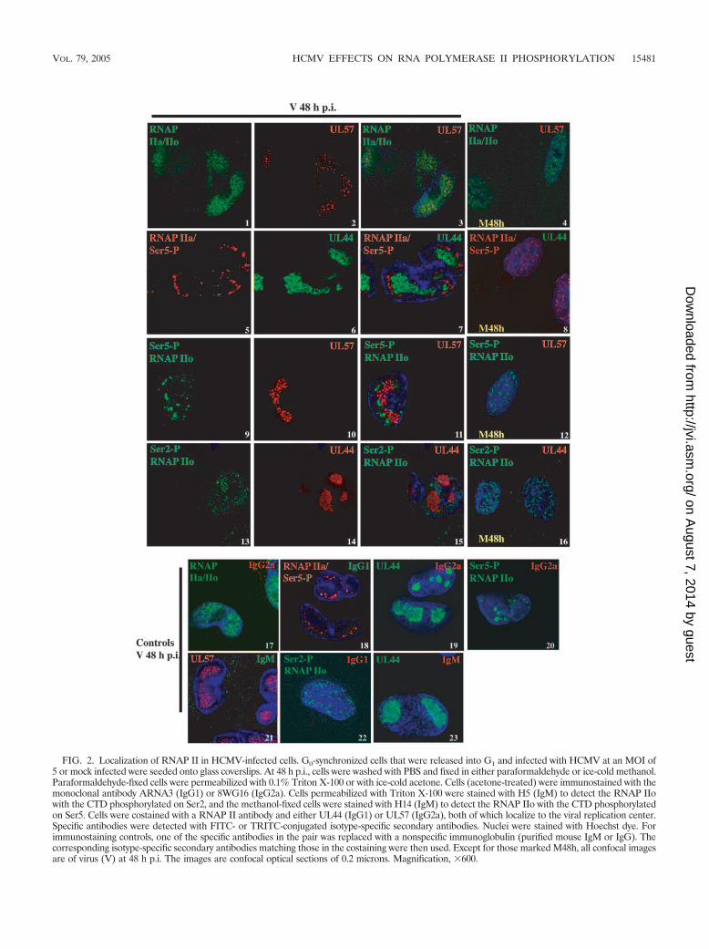
FIG. 2. Localization of RNAP II in HCMV-infected cells. G0-synchronized cells that were released into G1 and infected with HCMV at an MOI of5 or mock infected were seeded onto glass coverslips. At 48 h p.i., cells were washed with PBS and fixed in either paraformaldehyde or ice-cold methanol.Paraformaldehyde-fixed cells were permeabilized with 0.1% Triton X-100 or with ice-cold acetone. Cells (acetone-treated) were immunostained with themonoclonal antibody ARNA3 (IgG1) or 8WG16 (IgG2a). Cells permeabilized with Triton X-100 were stained with H5 (IgM) to detect the RNAP IIowith the CTD phosphorylated on Ser2, and the methanol-fixed cells were stained with H14 (IgM) to detect the RNAP IIo with the CTD phosphorylatedon Ser5. Cells were costained with a RNAP II antibody and either UL44 (IgG1) or UL57 (IgG2a), both of which localize to the viral replication center.Specific antibodies were detected with FITC- or TRITC-conjugated isotype-specific secondary antibodies. Nuclei were stained with Hoechst dye. Forimmunostaining controls, one of the specific antibodies in the pair was replaced with a nonspecific immunoglobulin (purified mouse IgM or IgG). Thecorresponding isotype-specific secondary antibodies matching those in the costaining were then used. Except for those marked M48h, all confocal imagesare of virus (V) at 48 h p.i. The images are confocal optical sections of 0.2 microns. Magnification, �600.
VOL. 79, 2005 HCMV EFFECTS ON RNA POLYMERASE II PHOSPHORYLATION 15481
on August 7, 2014 by guest
http://jvi.asm.org/
Dow
nloaded from
along with a nonspecific IgG or IgM antibody that matched thesecond antibody used in the dual staining. Taken together,these results suggest that in the infected cells, the initiation ofviral (and possibly cellular) transcription, as indicated by Ser5phosphorylation of RNAP II, may occur in clusters at theperipheries of the replication centers and be physically sepa-rate from the major region of viral DNA synthesis.
Treatment of cells with the cdk inhibitor roscovitine leads toa loss of the phosphorylated forms of RNAP II in infectedcells. The above results showed that there was an increase in thephosphorylation of the RNAP II CTD as the infection pro-gressed. Using the cdk inhibitor roscovitine at a concentration (20�M) that inhibits cdk7 and cdk9, the two primary kinases involvedin phosphorylating the CTD, we looked for any changes in thepattern of CTD phosphorylation. Western blot analysis with theantibody ARNA3, which detects all forms of RNAP II, revealedthat in the presence of roscovitine, there was a marked loss of thehypophosphorylated IIa form of RNAP in the mock-infected
lysates at 8 h p.i. (Fig. 3). In contrast, in the infected cells, the IIaform showed little change in the presence of roscovitine, whilethere was a detectable, albeit modest, loss of the hyperphosphor-ylated RNAP IIo. At 12 h p.i, the effect of roscovitine on RNAPII can be clearly seen in the infected cells, as evidenced by thedecreased levels of both the IIa and IIo forms.
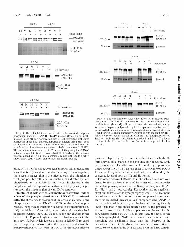
The observed loss of RNAP IIo in the infected cells was con-firmed by Western blot analysis of the lysates with the antibodiesthat detect primarily either Ser5- or Ser2-phosphorylated RNAPIIo (Fig. 4 and 5, respectively). Roscovitine had no significanteffect on the levels of the hyperphosphorylated RNAP IIo in themock-infected cells. In contrast, roscovitine primarily preventedthe virus-associated increase in Ser5-phosphorylated RNAP IIothat was observed by 8 h p.i., but the level was not significantlylower than that in the mock-infected cells in the presence orabsence of roscovitine. A different pattern was observed for theSer2-phosphorylated RNAP IIo. In this case, the level of theSer2-phosphorylated RNAP IIo in the infected cells treated withroscovitine was significantly lower than that observed in themock-infected cells in the absence or presence of roscovitine. Itshould be noted that at the 24-h p.i. time point the lanes contain-
FIG. 3. The cdk inhibitor roscovitine affects the virus-induced phos-phorylation state of RNAP II. HCMV-infected (lanes V) or mock-infected (lanes M) cells were treated with 20 �M roscovitine at the timeof infection or at 8 h p.i. and were harvested at different time points. Totalcell lysates from an equal number of cells were run on 6% gels andtransferred to nitrocellulose membranes in buffer containing 0.1% SDS.The membranes were subjected to Western blotting using the ARNA3antibody, which detects all forms of RNAP II. �* indicates that roscovi-tine was added at 8 h p.i. The membrane stained with amido black isshown below each Western blot to show the protein loading.
FIG. 4. The cdk inhibitor roscovitine affects virus-induced phos-phorylation of Ser5 within the RNAP II CTD. Infected (lanes V) andmock-infected (lanes M) cells were treated with roscovitine, and ly-sates were prepared, subjected to gel electrophoresis, and transferredto nitrocellulose membranes for Western blotting as described in thelegend for Fig. 3. The membranes were probed with the antibody H14,which is directed against RNAP IIo with the CTD phosphorylated onSer5. �* indicates that roscovitine was added at 8 h p.i. The lowerportion of the blot was probed for �-catenin as a protein loadingcontrol.
15482 TAMRAKAR ET AL. J. VIROL.
on August 7, 2014 by guest
http://jvi.asm.org/
Dow
nloaded from

ing the viral samples in Fig. 4 and 5 are underloaded relative tothe mock-infected samples.
In our previous study (44), we found that the effect of ros-covitine on viral gene expression depends on the time of ad-dition. When added at the time of the infection, there weremarked changes in the accumulation and processing of the IEtranscripts and inhibition of early gene expression. However,by delaying the addition of the drug until 8 h p.i., these effectson viral gene expression were abrogated and the infectionproceeded normally until late times. We reasoned that if the
phosphorylation of the RNAP II CTD and the effects of ros-covitine on the phosphorylation were related to the effects onviral transcription, then a delay in the addition of roscovitineshould prevent the loss of the hyperphosphorylated RNAP IIo.Figures 3 to 5 show that this is what occurs. Notably, at 24 hp.i., the levels of the hyperphosphorylated RNAP IIo in theinfected cells that were treated with roscovitine at 8 h p.i. werecomparable to those in the untreated cells. Interestingly, at24 h p.i., there were still lower levels of the hypophosphory-lated RNAP IIa in both the infected and mock-infected cellsregardless of the time of addition of roscovitine, although theeffect was greater in the cells that had been in roscovitine fora longer period of time.
The kinase activity and amount of cdk7, cdk9, and associ-ated proteins are upregulated in infected cells. The changes inthe phosphorylation of RNAP II and the effect of roscovitinein the infected cells suggested that the viral infection mightalso affect the levels, activity, or localization of cdk7 and cdk9.cdk7 is found in association with cyclin H or in a complex withcyclin H and MAT-1 to form the cdk-activating kinase. Thesethree proteins are also part of the multisubunit complexTFIIH, which is an RNAP II transcription initiation factor. Toassess the effect of the infection on cdk7 and the proteins thatassociate with this kinase, cells that were synchronized in G0
were released into G1 at the time of infection and then har-vested at various times p.i. By Western blot analysis, we foundthat an increase in the amount of cdk7 could be detectedbetween 8 and 24 h p.i. in the infected cells, and this levelcontinued to increase as the infection progressed (Fig. 6).MAT-1 and cyclin H displayed a similar trend, although theincrease in MAT-1 was greater than that of cyclin H.
FIG. 5. The cdk inhibitor roscovitine affects the virus-induced phos-phorylation of Ser2 within the RNAP II CTD. Infected (lanes V) andmock-infected (lanes M) cells were treated with roscovitine, and ly-sates were prepared, subjected to gel electrophoresis and transferredto nitrocellulose membranes for Western blotting as described in thelegend for Fig. 3. The membranes were probed with the antibody H5,which is directed against RNAP IIo with the CTD phosphorylated onSer2. �* indicates that roscovitine was added at 8 h p.i. The lowerportion of the blot was probed for �-catenin as a protein loadingcontrol.
FIG. 6. Levels of cdk-activating kinase (CAK) components andP-TEFb increase during HCMV infection. Upon release from G0,synchronized HFFs were infected with HCMV Towne (lanes V) at anMOI of 5 or mock infected (lanes M). Cells were harvested at theindicated time points. Lysates from an equal number of cells wereloaded onto gels, electrophoresed, and subjected to Western blottingwith antibodies against cdk7, MAT-1, cyclin H, cyclin T1, and cdk9.�-Actin was used as a protein loading control.
VOL. 79, 2005 HCMV EFFECTS ON RNA POLYMERASE II PHOSPHORYLATION 15483
on August 7, 2014 by guest
http://jvi.asm.org/
Dow
nloaded from
Western blot analysis of the lysates revealed that the patternof cdk9 accumulation in the infected cells was similar to that ofcdk7. An increased amount of cdk9 was visible in the infectedcells at 24 h p.i., and the level continued to rise throughout theinfection. We also noted that the cdk9 antibody detected aband of 55 kDa at later times p.i., which likely corresponds tothe isoform of cdk9 described by Shore et al. (46). The level ofcyclin T1, another component of P-TEFb, was also greater invirus-infected cells at 24 h p.i. and continued to increase as the
infection progressed. In accord with previously published stud-ies (16, 17), cdk9 and cyclin T1 did not cycle in the mock-infected cells, and the level of cyclin T1 remained low in themock-infected cells throughout the time course. The antibodyto cyclin T1 also detected a faster-migrating protein of 62 kDathat disappeared from infected cells by 24 h p.i. It is possiblethat this smaller form is cyclin T2a (11).
To assess whether the increased cdk7 and MAT-1 remainedin association with each other during the infection, we per-
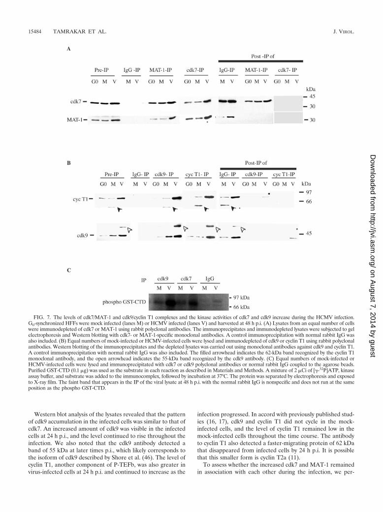
FIG. 7. The levels of cdk7/MAT-1 and cdk9/cyclin T1 complexes and the kinase activities of cdk7 and cdk9 increase during the HCMV infection.G0-synchronized HFFs were mock infected (lanes M) or HCMV infected (lanes V) and harvested at 48 h p.i. (A) Lysates from an equal number of cellswere immunodepleted of cdk7 or MAT-1 using rabbit polyclonal antibodies. The immunoprecipitates and immunodepleted lysates were subjected to gelelectrophoresis and Western blotting with cdk7- or MAT-1-specific monoclonal antibodies. A control immunoprecipitation with normal rabbit IgG wasalso included. (B) Equal numbers of mock-infected or HCMV-infected cells were lysed and immunodepleted of cdk9 or cyclin T1 using rabbit polyclonalantibodies. Western blotting of the immunoprecipitates and the depleted lysates was carried out using monoclonal antibodies against cdk9 and cyclin T1.A control immunoprecipitation with normal rabbit IgG was also included. The filled arrowhead indicates the 62-kDa band recognized by the cyclin T1monoclonal antibody, and the open arrowhead indicates the 55-kDa band recognized by the cdk9 antibody. (C) Equal numbers of mock-infected orHCMV-infected cells were lysed and immunoprecipitated with cdk7 or cdk9 polyclonal antibodies or normal rabbit IgG coupled to the agarose beads.Purified GST-CTD (0.1 �g) was used as the substrate in each reaction as described in Materials and Methods. A mixture of 2 �Ci of [�-32P]ATP, kinaseassay buffer, and substrate was added to the immunocomplex, followed by incubation at 37°C. The protein was separated by electrophoresis and exposedto X-ray film. The faint band that appears in the IP of the viral lysate at 48 h p.i. with the normal rabbit IgG is nonspecific and does not run at the sameposition as the phospho GST-CTD.
15484 TAMRAKAR ET AL. J. VIROL.
on August 7, 2014 by guest
http://jvi.asm.org/
Dow
nloaded from

formed IP experiments with antibodies to the individual pro-teins. Lysates of the infected and mock-infected cells wereprepared at the 48-h p.i. time point and immunoprecipitatedwith cdk7 antibody or with control IgG coupled to proteinA-Sepharose beads. The immunoprecipitates were then sub-jected to SDS-polyacrylamide gel electrophoresis followed byWestern blotting with antibodies to both cdk7 and MAT-1.The Western blot of the cdk7 immunoprecipitate (Fig. 7A)shows that both MAT-1 and cdk7 are in a complex, and thereappears to be no free MAT-1 in the post-IP supernatant. Asexpected, neither protein was immunoprecipitated with thecontrol IgG. In a reciprocal experiment, the immunoprecipi-tation of MAT-1 also brings down cdk7 from the lysates ofboth mock- and virus-infected cells that were obtained at 48 hp.i. However, cdk7 is still present in the MAT-1-depleted ly-sate, suggesting that cdk7 is present in excess. Taken together,these data suggest that MAT-1 is not found in a free form ineither infected or mock-infected cells. In these experiments, wecould not assess whether cyclin H was present in the com-plexes, as the antibody has low affinity in immunoprecipitationexperiments, and there is a high level of background staining ofthe Western blot following immunoprecipitation with eitherMAT-1 or cdk7.
In a similar manner, we also examined whether cdk9 andcyclin T1 were in a complex. Immunoprecipitation of lysatesobtained at 48 h p.i. with an antibody to cdk9 (Fig. 7B) dem-onstrated that cdk9 is associated with cyclin T1 in both mock-and virus-infected cells. Likewise, antibody to cyclin T1 copre-cipitated cdk9 in lysates from both mock-infected and infectedcells. The data also showed that the increased levels of theproteins in the infected cells coincided with an increase in theamount of the complex. However, the presence of a smallamount of cyclin T1 and cdk9 in the post-IP lysates of infectedcells indicated that a minor population of each protein was notpresent in the complex. This may be due to the presence of freecdk9 or cdk9 that associates with cyclins T2a, T2b, and K. Theunidentified protein of 55 kDa that is recognized by cdk9antibody was also coprecipitated by cyclin T1. Therefore, thisprotein is likely to be a higher-molecular-weight form of cdk9.Similarly, cdk9 coprecipitated the smaller protein (62 kDa)that was recognized by anti-cyclin T1 antibody by Westernblotting. This form disappeared in the infected cells as early as24 h p.i. and therefore was not seen in the cdk9 IP of virallysates.
To determine whether the increase in the protein level of theCTD kinases that began at 24 h p.i. corresponds to their ac-tivity, bacterially expressed GST-CTD was used as a substratefor in vitro kinase assays with lysates obtained at 48 h p.i.Figure 7C shows that in accord with the increased levels ofcdk7 and cdk9 in the infected cells at 48 h p.i., there was alsoa significant increase in the kinase activities.
Cdk7 and MAT-1 are localized primarily in replication cen-ters, and cdk9 is distributed throughout the nuclei of infectedcells. Since the above results showed that there was an increase inboth the number of complexes containing cdk7 and MAT-1 andthe associated kinase activity, we wanted to determine if theirlocalization was also altered by the HCMV infection. Visualiza-tion of the immunofluorescence by confocal microscopy of verti-cal optical sections documented that cdk7 and MAT-1 were uni-formly distributed in the nuclei of mock-infected cells (Fig. 8A,
panels 1 to 3), while at 48 h p.i., they accumulated primarily in theviral replication centers of infected cells (panels 5 to 7). Theincreased intensity of the cdk7 and MAT-1 signals in the infectedcells correlated with the Western blot results. The presence ofcdk7 and MAT-1 in the replication centers was confirmed by dualstaining of the slides with antibodies to cdk7 or MAT-1 and theviral replication protein UL44 or UL57, respectively (panels 8 to10 and 11 to 13, respectively). However, the cdk7 and MAT-1staining was not limited to the replication centers, as both pro-teins were also observed in the nuclear region at the peripheriesof the replication centers.
The distribution of cdk9 in the infected cells differed fromthat of cdk7. Costaining with UL44 showed that the majority ofthe protein was distributed uniformly throughout the nucleusand punctate in appearance, although some of the protein didlocalize to the replication centers (panels 14 to 16). This wassimilar to the distribution of RNAP II with Ser2 phosphoryla-tion of the CTD (Fig. 2, panels 13 to 15). Although the stainingfor cdk9 in the mock-infected cells was fainter, it was similar tothat seen in the infected cells (for visualization, the photo inpanel 4 is overexposed). As controls, the slides were duallystained with each specific antibody and a nonspecific antibodythat matched the isotype of the second antibody used in thedual staining (Fig. 8B, panels 1 to 6).
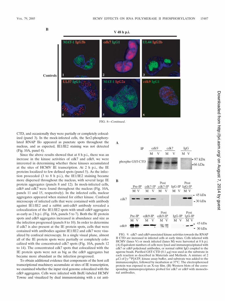
Cdk7 and cdk9 kinase activities are elevated at early timesin the infection. As shown above, the level of phosphorylatedRNAP II was greater in the infected cells at 8 h p.i. than in themock-infected cells. However, at this time point the amount ofcdk7 and cdk9 in the infected cells was comparable to that inthe mock-infected cells. To determine whether the kinase ac-tivity was higher in the infected cells at this early time point, weimmunoprecipitated the lysates with antibody to cdk7 or cdk9and assayed the phosphorylation of bacterially expressed GST-CTD in an in vitro kinase assay. Figure 9A shows that the kinaseactivities of both cdk7 and cdk9 were higher in the infected cells.The Western blot of the immunoprecipitates confirmed that thelevels of cdk7 and cdk9 were equivalent in the mock-infected andinfected cells at this time (Fig. 9B).
Ser2-phosphorylated RNAP II, cdk9, and cdk7 colocalizewith the IE1/IE2 proteins at the beginning of the infection.Ishov et al. (22) previously showed that some HCMV inputgenome is deposited at a subset of the PODs and that HCMVIE transcription originates at these sites. Their work also dem-onstrated that there is a differential localization of the IE1 andIE2 proteins, with the IE1 protein initially localizing to mostPODs and IE2 juxtaposed to only a subset of the PODs. Alldomains that had IE2 protein also had IE transcripts. Al-though IE1 then disrupted the PODs at approximately 3 to 4 hp.i., IE2 protein remained at these sites.
In order to determine if transcriptionally active RNAP II isbeing recruited to and accumulates at the immediate early sitesof viral gene transcription, we examined the distribution ofRNAP II with the CTD phosphorylated on Ser2 (H5 Ab)relative to the IE proteins (Fig. 10). CH16.0, an antibody thatdetects both IE1 and IE2, was used to track IE protein local-ization. Confocal microscopy revealed that a subset of Ser2-phosphorylated RNAP IIo localized near the IE protein ag-gregates (Fig. 10A, panels 1 to 3). Within a single visual plane,the majority of the IE aggregates were localized adjacent to thepunctate spots containing RNAP II with the Ser2-phosphorylated
VOL. 79, 2005 HCMV EFFECTS ON RNA POLYMERASE II PHOSPHORYLATION 15485
on August 7, 2014 by guest
http://jvi.asm.org/
Dow
nloaded from

FIG. 8. Localization of cdk7, MAT-1 and cdk9 in HCMV-infected cells. (A) G0-synchronized cells were released in G1, infected with HCMVat an MOI of 5 or mock infected, and seeded onto the glass coverslips. At 48 h p.i., cells were washed with PBS, fixed in paraformaldehyde,permeabilized, and immunostained with monoclonal antibodies against cdk7 (IgG2b), MAT-1 (IgG1), or cdk9 (IgG2b) and a viral replicationcenter protein UL44 (IgG1) or UL57 (IgG2a) as described in Materials and Methods. Specific antibody staining was detected with FITC- orTRITC-conjugated isotype-specific secondary antibodies. (B) For controls, one of the specific antibodies of the pair was replaced with purifiednormal mouse IgG, and the corresponding pair of isotype-specific secondary antibodies was used. Nuclei were stained with Hoechst dye. Exceptfor those marked M48h, all of the images are of infected cells at 48 h p.i. All of the images except image 4 in panel A are confocal optical sectionsof 0.2 �m. Magnification, �600.
15486 TAMRAKAR ET AL. J. VIROL.
on August 7, 2014 by guest
http://jvi.asm.org/
Dow
nloaded from
CTD, and occasionally they were partially or completely colocal-ized (panel 3). In the mock-infected cells, the Ser2-phosphory-lated RNAP IIo appeared as punctate spots throughout thenucleus, and as expected, IE1/IE2 staining was not detected(Fig. 10A, panel 4).
Since the above results showed that at 8 h p.i., there was anincrease in the kinase activities of cdk7 and cdk9, we wereinterested in determining whether these kinases accumulatedat the sites of HCMV IE transcription. At 2 h p.i., the IEproteins localized to few defined spots (panel 5). As the infec-tion proceeded (3 to 8 h p.i.), the IE1/IE2 staining becamemore dispersed throughout the nucleus, with several large IEprotein aggregates (panels 8 and 12). In mock-infected cells,cdk9 and cdk7 were found throughout the nucleus (Fig. 10A,panels 11 and 15, respectively). In the infected cells, nuclearaggregates appeared when stained for either kinase. Confocalmicroscopy of infected cells that were costained with antibodyagainst IE1/IE2 and a rabbit anti-cdk9 antibody revealed acolocalization of the IE1/IE2 spots with small cdk9 aggregatesas early as 2 h p.i. (Fig. 10A, panels 5 to 7). Both the IE proteinspots and cdk9 aggregates increased in abundance and size asthe infection progressed (panels 8 to 10). In order to determineif cdk7 is also present at the IE protein spots, cells that werecostained with antibodies against IE1/IE2 and cdk7 were visu-alized by confocal microscopy. In a single visual plane, almostall of the IE protein spots were partially or completely colo-calized with the concentrated cdk7 spots (Fig. 10A, panels 12to 14). The concentrated cdk7 spots that colocalized with theIE protein spots were not as big as the cdk9 aggregates butbecame more abundant as the infection progressed.
To obtain additional evidence that components of the host celltranscriptional machinery accumulate at sites of IE transcription,we examined whether the input viral genome colocalized with thecdk9 aggregates. Cells were infected with BrdU-labeled HCMVTowne and visualized by dual immunostaining with a rat anti-
FIG. 8—Continued.
FIG. 9. cdk7- and cdk9-associated kinase activities towards the RNAPII CTD are increased in infected cells at early times. Cells infected withHCMV (lanes V) or mock infected (lanes M) were harvested at 8 h p.i.(A) Equivalent numbers of cells were lysed and immunoprecipitated withcdk7 or cdk9 polyclonal antibodies, or normal rabbit IgG coupled to theagarose beads. Purified GST-CTD (0.1 �g) was used as the substrate ineach reaction as described in Materials and Methods. A mixture of 2�Ci of [�-32P]ATP, kinase assay buffer, and substrate was added to theimmunocomplex, followed by incubation at 37°C. The electrophoresedprotein was exposed to an X-ray film. (B) Western blots of the corre-sponding immunoprecipitates probed for cdk7 or cdk9 with monoclo-nal antibodies.
VOL. 79, 2005 HCMV EFFECTS ON RNA POLYMERASE II PHOSPHORYLATION 15487
on August 7, 2014 by guest
http://jvi.asm.org/
Dow
nloaded from

15488 TAMRAKAR ET AL. J. VIROL.
on August 7, 2014 by guest
http://jvi.asm.org/
Dow
nloaded from

BrdU antibody and a cdk9 antibody. By confocal imaging, a clearcolocalization between the viral genome and the cdk9 aggregateswas observed (Fig. 10A, panels 16 to 18). In a single visual plane,there were some viral genomes that did not colocalize with thecdk9 aggregates, but these were usually outside of the nucleus. NoBrdU signal or cdk9 aggregates were detected in mock-infectedcells (panel 19). It should be noted that the diffuse backgroundnuclear staining for cdk9 and the Hoechst dye seen in panels 17to 19 are the result of an added HCl denaturation step duringthe immunostaining. Figure 10B shows immunostaining controlswhere one of the specific antibodies in the dually stained panelsshown in Fig. 10A was replaced with purified mouse IgM, rat IgG,rabbit IgG, or isotype-specific mouse IgG.
cdk9 localizes adjacent to sites that correspond to the PODsin infected cells prior to dispersal. To confirm that the aboveresults were consistent with the previous studies showing thatthe PODs are the sites of viral IE transcription, the localizationof the cdk9 aggregates in relation to the PODs was examinedby confocal microscopy (Fig. 11). Cells were costained with therabbit anti-cdk9 antibody and an anti-PML MAb, a standardmarker for the PODs. In mock-infected cells, the PML stainingdisplayed a prominent speckled pattern in the nucleus, andcostaining with cdk9 showed that cdk9 spots were seeminglyrandomly oriented with respect to the PODs (panels 1 to 4).The trend in the infected cells was that the cdk9 aggregationand the maintenance of PML at the PODs are mutuallyexclusive (panels 5 to 7). The cell on the left in panels 5 to 7shows the dispersal of PML and the presence of cdk9 aggre-gates, and the cell on the right shows the typical PML specklesmarking the position of the PODs and the absence of cdk9aggregates. In the rare case that an infected cell exhibitedsome cdk9 aggregation before dispersal of the PML, themajority of the cdk9 aggregates were not colocalized at theremaining PODs, although some cdk9 did appear to beadjacent to the PODs. A representative example of this isshown in panels 8 to 11.
One explanation for the above observations was that thecdk9 aggregates are located at PODs, but those PODs arepreferentially disrupted in the infected cell. Another possibilitywas that cdk9 aggregation cannot occur at sites where thePODs are still intact. To distinguish between these possibili-ties, cells were infected with HCMV CR208, a mutant virusthat does not express the IE1-72 protein and hence does notdisrupt the PODs. The CR208-infected cells maintained thePODs with a distribution similar to that in mock-infected cells,and dual staining showed that each cdk9 aggregate localizeddirectly adjacent to one or more PODs (panels 12 to 15).Antibody staining controls are shown in panels 16 and 17. The
fact that the cdk9 aggregates still formed in the CR208-infected cells implies that POD dispersal is not a requirementfor cdk9 aggregation during the infection, and the localizationpattern suggests that the cdk9 aggregates form at sites wherePOD structures previously existed.
DISCUSSION
The current model of transcription is that synthesis andprocessing of pre-mRNA are temporally and functionally cou-pled (for reviews, see references 8, 27, 30, 34, 37, and 38). TheCTD on the large subunit of RNAP II plays an important rolein providing access to the nascent RNA for different factorsinvolved in the processing of mRNA, and there is increasingevidence that specificity is associated with differential phos-phorylation on serines 2 and 5 of its heptapeptide repeats.Phosphorylation of Ser5 on the CTD occurs near the promoterand is involved in the recruitment of capping enzymes. Incontrast, phosphorylation of Ser2 appears to occur at a sitedownstream of the promoter after the initiation of transcrip-tion and is associated with the binding of the 3� RNA process-ing machinery. In this study, we examined the effect of HCMVinfection on CTD phosphorylation and the kinases involved atboth early and late times. We found that the levels of cdk7 andcdk9, as well as other components of the kinase complexes,MAT-1/cyclin H and cyclin T1, respectively, are upregulatedduring the infection. In addition, there was a correspondingincrease in the kinase activities of cdk7 and cdk9, as measuredby the phosphorylation of GST-CTD in vitro. The relativeamount of phosphorylated CTD was also elevated in the in-fected cells.
Interestingly, the infected cells do not show a noticeableincrease in the amount of cdk7 or cdk9 until 24 h p.i. However,their in vitro activities, as well as the level of the hyperphos-phorylated form of the RNAP II CTD, increase at earliertimes. This suggests that the initial increased kinase activitymay be due to other virus-induced changes. It is possible thata viral protein functions as a regulatory subunit for either ofthese kinases or that the virus induces an association of thekinases with another cellular protein to facilitate the phosphor-ylation of the RNAP II CTD during the infection. This isconsistent with the recent observation that the RNAP II CTDis phosphorylated in herpes simplex virus 1-infected cells by acomplex that contains cdk9 and ICP22 (12). Alternatively, thevirus may induce relocalization and/or recruitment of cdk7 andcdk9 to RNAP II. Our data showing that concentrated aggre-gates of these kinases form in infected cells at the site of IEtranscription support this hypothesis.
FIG. 10. Ser2-phosphorylated RNAP IIo, cdk7, and cdk9 localize with IE protein domains. (A) G0-synchronized cells were released into G1 andinfected at an MOI of 3 or 5 or mock infected and seeded onto coverslips. The cells were washed with PBS and fixed with either 2% paraformaldehydeor ice-cold methanol between 2 to 8 h p.i. Cells were coimmunostained with the appropriate combinations of CH16.0 MAb (IgG1) to detect IE1/IE2,H5 MAb (IgM) to detect Ser2-phosphorylated RNAP IIo, and cdk9 rabbit polyclonal antibody, cdk7 MAb (IgG2b), and BrdU rat polyclonal antibody.Specific antibodies were detected with FITC- or TRITC-conjugated isotype-specific anti-mouse Ab, goat anti-rat FITC-IgG, or goat anti-rabbit Cy3-IgGsecondary antibodies. Nuclei were stained with Hoechst dye. (B) For immunostaining controls, one of the specific antibodies in the pair was replaced witha nonspecific immunoglobulin (purified mouse IgM, isotype-specific mouse IgG, rabbit IgG, or rat IgG). The corresponding isotype-specific secondaryantibodies matching those in the costaining were then used. Arrows denote examples exhibiting some degree of colocalization: a, adjacent; pc, partialcolocalization; c, completely colocalized. All the images are 0.2-�m sections that were obtained by confocal microscopy. Magnification is �1,000 underoil immersion.
VOL. 79, 2005 HCMV EFFECTS ON RNA POLYMERASE II PHOSPHORYLATION 15489
on August 7, 2014 by guest
http://jvi.asm.org/
Dow
nloaded from

Previously, we found that infection of cells in the presence ofthe cdk inhibitor roscovitine altered the pattern of processingfor both the UL122-123 and UL37 transcripts (44). Normally,the first cleavage/polyadenylation site is preferentially used to
generate IE1-72 or UL37X1 RNAs, but in the presence ofroscovitine, there appeared to be suppression of the first cleav-age/polyadenylation site and enhanced utilization of the adja-cent downstream 3� splice acceptor site. As a result, there were
FIG. 11. Cdk9 localizes adjacent to sites that correspond to the PODS in infected cells prior to their dispersal. G0-synchronized cells werereleased into G1 and infected with HCMV Towne or HCMV CR208 virus at an MOI of 5 or mock infected and seeded onto coverslips. The cellswere washed with PBS and fixed with 2% paraformaldehyde at 8 h p.i. Cells were coimmunostained with the appropriate combinations of PMLMAb (IgG1) and cdk9 rabbit polyclonal antibody. Specific antibodies were detected with goat anti-mouse isotype-specific FITC-conjugated or goatanti-rabbit Cy3 secondary antibodies. Nuclei were stained with Hoechst dye. For immunostaining controls, one of the specific antibodies in the pairwas replaced with a normal isotype-specific mouse IgG1 or rabbit IgG. The corresponding isotype-specific secondary antibodies matching thosein the costaining were then used. Panels 4, 11, and 15 are insets depicting an enlarged view of the region within the white box from the previouspanel. All of the images are 0.2-�m sections that were obtained by confocal microscopy. Panels 5 to 7 and 8 to 11 are different cells from the sameexperiment. Magnification is �600 under oil immersion for panels 5 to 7 and �1,000 under oil immersion for all other panels. Inset panels 4, 11,and 15 were magnified 200% using Adobe Photoshop v. 7.0.
15490 TAMRAKAR ET AL. J. VIROL.
on August 7, 2014 by guest
http://jvi.asm.org/
Dow
nloaded from

lower levels of the IE1-72 or UL37X1 RNAs and higher levelsof the IE2-86 and spliced UL37 RNAs. We proposed thatthese changes were related to the phosphorylation of theRNAP II CTD, primarily by cdk7/cyclin H and cdk9/cyclin T1(44). In support of this hypothesis, we show here that whenroscovitine is present at the beginning of the infection, thelevel of CTD phosphorylation is significantly decreased in theinfected cells, most notably the phosphorylation of Ser2; thereis no change in the mock-infected cells. For the Ser5-phos-phorylated form, it appears that inhibition of cdk primarilyprevents the increase observed in the infected cells, at leastduring the first 12 h. In the case of the Ser2-phosphorylatedform, the levels are actually lower in the infected cells than inthe mock-infected cells. One possibility for this result is that thevirus either brings into the cell or activates a phosphatase thatpreferentially removes the phosphate from Ser2 within the CTD.In the absence of roscovitine, the action of cdk9/cyclin T1 maycounter this dephosphorylation. It is tempting to speculate thatthe reduction in Ser2 phosphorylation of the CTD is associatedwith decreased recruitment of the cleavage/polyadenylation fac-tors, but further experiments will be required to determine if thisis the case.
The effects of roscovitine on the hypophosphorylated RNAP IIalso differ in the infected and mock-infected cells. Here, there isa more rapid decrease in the mock-infected cells than in theinfected cells during the first 8 h, although by 12 h, the levels arecomparable. This loss of the hypophosphorylated RNAP IIa inthe presence of roscovitine may be related to some inhibitoryeffect on transcription, as others have observed a similar loss ofRNAP IIa when cells are exposed to UV light, -amanitin, oractinomycin D (33, 39, 40). It is possible that the rate of degra-dation of the hypophosphorylated RNAP II in the infected cells iscomparable to that in the mock-infected cells, but the dephos-phorylation of the Ser2-phosphorylated form by a viral-infection-associated phosphatase initially compensates for this. Alterna-tively, inhibition of the cdk’s may lead to differential degradationof the hypophosphorylated and hyperphosphorylated forms ofRNAP II in the infected and mock-infected cells. These twopossibilities are not mutually exclusive, and there may be somecombination of enhanced phosphatase activity and altered stabil-ity. In-depth studies of protein stability and assays of phosphataseactivity may help resolve these possibilities, and these experimentsare in progress.
In accord with our previous study of the effect of roscovitineon IE gene expression (44), the decrease in CTD phosphory-lation does not occur if the drug is added after 8 h p.i. Thereare several possibilities for this result. One explanation is thatthe phosphorylation of RNAP II necessary for programmedviral IE gene expression is complete by 8 h p.i., and thusinhibitors of cdk7 and cdk9 no longer have an effect. Alterna-tively, the kinases may no longer be accessible to the inhibitor.In addition, if there is a virus-associated phosphatase thatmakes the infected cells more sensitive to cdk inhibition, it maybe present for only a limited period of time at the beginning ofthe infection. All of these possibilities are currently being in-vestigated.
Taken together, the studies presented here suggest that thein vivo increase of CTD phosphorylation is due to the upregu-lated cdk7 and cdk9 kinase activities. However, although thesetwo kinases are believed to be responsible for most of the CTD
phosphorylation, it remains possible that other kinases are alsoinvolved in CTD phosphorylation in the infected cell. Theseresults also do not preclude the possibility that there is cdk-dependent modification of other proteins that have an impacton the posttranscriptional processing of the IE transcripts.Multiple factors are involved in the splicing and cleavage/polyadenylation of RNA transcripts, and a change in the abun-dance, activity, or localization of any of the factors due toalteration of their phosphorylation state could affect the bal-ance of transcripts produced. For example, it has recently beenshown that the splicing factor ASF/SF2 is hypophosphorylatedand presumably less active early in the infection (2).
In addition to identifying specific components of the hosttranscriptional machinery that are upregulated during the in-fection, we also determined the nuclear localization of thesecomponents at times of viral IE and late transcription. Ishovet al. (22) provided a model for the IE transcription environ-ment based on their results showing that the viral input ge-nome, IE transcripts, and IE proteins all localize at the PODsand that the IE transcripts move towards the spliceosomeassembly factor domains. They hypothesized that either theincoming viral genome is required to localize at preexistingtranscriptional environments in the host cell or essential tran-scription factors must be recruited to the input viral genome.Our data suggest that both events take place. Figure 12 sum-marizes the findings from Ishov et al. (22) with respect to theIE transcription environment and extends their model basedon the results of our studies. We find that that the transcriptionelongation factor cdk9 forms aggregates that colocalize withthe IE proteins as early as 2 h p.i. As the infection proceedsduring the next few hours, the aggregates of cdk7 and cdk9increase in size, indicating that both CTD kinases are furtherrecruited to these transcription sites. Since continued IE tran-
FIG. 12. Model for an IE transcription site in HCMV-infectedcells. The input genome that is deposited at the POD structure func-tions as a template for IE RNA synthesis. Cellular hypophosphory-lated RNAP IIa is recruited to the site and is phosphorylated by cdk7and cdk9 to the hyperphosphorylated and transcriptionally activeRNAP IIo. The IE transcripts move towards the SC35 domains, prob-ably for further processing. The newly synthesized major IE proteinsalso localize to this region. IE1-72 eventually causes POD dispersal,but the IE2-86 protein remains at the established transcription site.The color key to structures is shown at the right.
VOL. 79, 2005 HCMV EFFECTS ON RNA POLYMERASE II PHOSPHORYLATION 15491
on August 7, 2014 by guest
http://jvi.asm.org/
Dow
nloaded from

scription requires recruitment of the CTD kinases to the sitesof viral transcription within the first hours of infection, it isreasonable to assume that the required phosphorylation ofRNAP II for synthesis of the IE RNAs may be complete by 8 hp.i. This would also be consistent with the finding that the IEprotein spots are localized near the Ser2-phosphorylatedRNAP II.
At later times in the infection, multiple copies of the viralDNA are synthesized and there is abundant late gene tran-scription. It was therefore of interest to determine the relativelocations of the viral DNA replication proteins, host cell CTDkinases, and RNA polymerase. We find that cdk7 is localizedprimarily within the replication center, although some kinaseappears at the periphery of the replication center. Interest-ingly, with the antibody (ARNA3) that is directed against thebody of the large subunit of RNAP II outside of the CTD (andthus detects both the hypophosphorylated and hyperphosphor-ylated forms of the CTD), RNAP II shows a distribution sim-ilar to that of cdk7. Yet, the antibody (8WG16) that is directedagainst the hypophosphorylated CTD, as well as the forms ofthe RNAP II CTD that are phosphorylated on Ser5 but notSer2, detects primarily RNAP II in aggregates at the peripheryof the replication center. This may be due to the relativeaffinity of the antibodies in the immunofluorescence assay. Wealso cannot rule out the possibility that ARNA3 detects RNAPII that is heavily phosphorylated on a residue different thanSer2 or Ser5 (or modified in other ways) and that this formpreferentially localizes at the replication centers. However, it islikely that the antibody 8WG16 detects Ser5-phosphorylatedCTD, as a similar pattern in the infected cell was observed withthe antibody (H14) that detects the Ser5-phosphorylated CTD,irrespective of the phosphorylation of Ser2. In contrast, theRNAP II with the CTD phosphorylated on Ser2 was distrib-uted throughout the replication center and nucleus. One inter-pretation of these results is that the initiation of the high level oflate gene transcription on the viral DNA, as indicated by theclusters of Ser5-phosphorylated RNAP II, occurs at a site that isphysically separate from the replication center where the viralDNA is being synthesized. These may also be sites of initiation ofhost cell RNA transcription. Commitment to elongation thenoccurs at domains distal to these aggregates.
The studies presented here provide another example of theingenious ways that CMV subverts the host cell for its ownneeds. Deciphering the mechanisms governing the coupling ofRNA transcription and processing and the role of differentialCTD phosphorylation in the regulation of the various stepsinvolved in mRNA synthesis is an active area of research. Anunderstanding of how the virus commandeers the transcrip-tional machinery not only may advance our knowledge of viralpathogenesis but also may help elucidate the mechanisms un-derlying regulated gene expression.
ACKNOWLEDGMENTS
We are grateful to Veronica Sanchez and Elizabeth White for com-ments on the manuscript and to members of the laboratory for helpfulsuggestions.
This work was supported by NIH grants CA73490 and CA34729.
REFERENCES
1. Adair, R., G. W. Liebisch, and A. M. Colberg-Poley. 2003. Complex alterna-tive processing of human cytomegalovirus UL37 pre-mRNA. J. Gen. Virol.84:3353–3358.
2. Adair, R., G. W. Liebisch, Y. Su, and A. M. Colberg-Poley. 2004. Alterationof cellular RNA splicing and polyadenylation machineries during productivehuman cytomegalovirus infection. J. Gen. Virol. 85:3542–3553.
3. Ahn, J.-H., W.-J. Jang, and G. S. Hayward. 1999. The human cytomegalo-virus IE2 and UL112-113 proteins accumulate in viral DNA replicationcompartments that initiate from the periphery of promyelocytic leukemiaprotein-associated nuclear bodies (PODs or ND10). J. Virol. 73:10458–10471.
4. Ahn, J. H., E. R. Brignole, and G. S. Hayward. 1998. Disruption of PMLsubnuclear domains by the acidic IE1 protein of human cytomegalovirus ismediated through interaction with PML and may modulate a RING finger-dependent cryptic transactivator function of PML. Mol. Cell. Biol. 18:4899–4913.
5. Ahn, J. H., and G. S. Hayward. 1997. The major immediate-early proteinsIE1 and IE2 of human cytomegalovirus colocalize with and disrupt PML-associated nuclear bodies at very early times in infected permissive cells.J. Virol. 71:4599–4613.
6. Baek, M.-C., P. M. Krosky, A. Pearson, and D. M. Coen. 2004. Phosphory-lation of the RNA polymerase II carboxyl-terminal domain in human cyto-megalovirus-infected cells and in vitro by the viral UL97 protein kinase.Virology 324:184–193.
7. Bain, J., H. McLauchlan, M. Elliott, and P. Cohen. 2003. The specificities ofprotein kinase inhibitors: an update. Biochem. J. 371:199–204.
8. Bentley, D. 2002. The mRNA assembly line: transcription and processingmachines in the same factory. Curr. Opin. Cell Biol. 14:336–342.
9. Cantrell, S. R., and W. A. Bresnahan. 2005. Interaction between the humancytomegalovirus UL82 gene product (pp71) and hDaxx regulates immediate-early gene expression and viral replication. J. Virol. 79:7792–7802.
10. De Azevedo, W. F., S. Leclerc, L. Meijer, L. Havlicek, M. Strnad, and S. H.Kim. 1997. Inhibition of cyclin-dependent kinases by purine analogues: crys-tal structure of human cdk2 complexed with roscovitine. Eur. J. Biochem.243:518–526.
11. De Luca, A., M. De Falco, A. Baldi, and M. G. Paggi. 2003. Cyclin T: threeforms for different roles in physiological and pathological functions. J. Cell.Physiol. 194:101–107.
12. Durand, L. O., S. J. Advani, A. P. Poon, and B. Roizman. 2005. The carboxyl-terminal domain of RNA polymerase II is phosphorylated by a complexcontaining cdk9 and infected-cell protein 22 of herpes simplex virus 1. J. Vi-rol. 79:6757–6762.
13. Edamatsu, H., C. L. Gau, T. Nemoto, L. Guo, and F. Tamanoi. 2000. Cdkinhibitors, roscovitine and olomoucine, synergize with farnesyltransferaseinhibitor (FTI) to induce efficient apoptosis of human cancer cell lines.Oncogene 19:3059–3068.
14. Fortunato, E. A., A. K. McElroy, V. Sanchez, and D. H. Spector. 2000.Exploitation of cellular signaling and regulatory pathways by human cyto-megalovirus. Trends Microbiol. 8:111–119.
15. Fortunato, E. A., and D. H. Spector. 1999. Regulation of human cytomega-lovirus gene expression. Adv. Virus Res. 54:61–128.
16. Garriga, J., A. Limon, X. Mayol, S. G. Rane, J. H. Albrecht, E. P. Reddy, V.Andres, and X. Grana. 1998. Differential regulation of the retinoblastomafamily of proteins during cell proliferation and differentiation. Biochem. J.333:645–654.
17. Garriga, J., J. Peng, M. Parreno, D. H. Price, and E. E. Henderson. 1998.Upregulation of cyclin T1/CDK9 complexes during T cell activation. Onco-gene 17:3093–3102.
18. Gawn, J. M., and R. F. Greaves. 2002. Absence of IE1 p72 protein functionduring low-multiplicity infection by human cytomegalovirus results in a broadblock to viral delayed-early gene expression. J. Virol. 76:4441–4455.
19. Greaves, R. F., and E. S. Mocarski. 1998. Defective growth correlates withreduced accumulation of a viral DNA replication protein after low-multiplicityinfection by a human cytomegalovirus ie1 mutant. J. Virol. 72:366–379.
20. Hofmann, H., H. Sindre, and T. Stamminger. 2002. Functional interactionbetween the pp71 protein of human cytomegalovirus and the PML-interactingprotein human Daxx. J. Virol. 76:5769–5783.
21. Ishov, A. M., and G. G. Maul. 1996. The periphery of nuclear domain 10(ND10) as site of DNA virus deposition. J. Cell Biol. 134:815–826.
22. Ishov, A. M., R. M. Stenberg, and G. G. Maul. 1997. Human cytomegalovirusimmediate early interaction with host nuclear structures: definition of animmediate transcript environment. J. Cell Biol. 138:5–16.
23. Ishov, A. M., O. V. Vladimirova, and G. G. Maul. 2002. Daxx-mediatedaccumulation of human cytomegalovirus tegument protein pp71 at ND10facilitates initiation of viral infection at these nuclear domains. J. Virol.76:7705–7712.
24. Kelly, C., R. V. Driel, and G. W. Wilkinson. 1995. Disruption of PML-associated nuclear bodies during human cytomegalovirus infection. J. Gen.Virol. 76:2887–2893.
25. Korioth, F., G. G. Maul, B. Plachter, T. Stamminger, and J. Frey. 1996. Thenuclear domain 10 (ND10) is disrupted by the human cytomegalovirus geneproduct IE1. Exp. Cell Res. 229:155–158.
26. Kouzarides, T., A. T. Bankier, A. C. Satchwell, E. Preddy, and B. G. Barrell.1988. An immediate early gene of human cytomegalovirus encodes a poten-tial membrane glycoprotein. Virology 165:151–164.
15492 TAMRAKAR ET AL. J. VIROL.
on August 7, 2014 by guest
http://jvi.asm.org/
Dow
nloaded from

27. Majello, B., and G. Napolitano. 2001. Control of RNA polymerase II activityby dedicated CTD kinases and phosphatases. Front. Biosci. 6:D1358–D1368.
28. Marshall, K. R., K. V. Rowley, A. Rinaldi, I. P. Nicholson, A. M. Ishov, G. G.Maul, and C. M. Preston. 2002. Activity and intracellular localization of thehuman cytomegalovirus protein pp71. J. Gen. Virol. 83:1601–1612.
29. Meijer, L., A. Borgne, O. Mulner, J. P. J. Chong, J. J. Blow, N. Inagaki, M.Inagaki, J. G. Delcros, and J. P. Molinoux. 1997. Biochemical and cellulareffects of roscovitine, a potent and selective inhibitor of the cyclin-dependentkinases cdc2, cdk2 and cdk5. Eur. J. Biochem. 243:527–536.
30. Meinhart, A., T. Kamenski, S. Hoeppner, S. Baumli, and P. Cramer. 2005.A structural perspective of CTD function. Genes Dev. 15:1401–1415.
31. Mocarski, E. S., and C. T. Courcelle. 2001. Cytomegaloviruses and theirreplication, p. 2629–2673. In D. M. Knipe and P. M. Howley (ed.), Fieldsvirology, 4th ed., vol. 2. Lippincott Williams & Wilkins, Philadelphia, Pa.
32. Mocarski, E. S., G. W. Kemble, J. M. Lyle, and R. F. Greaves. 1996. Adeletion mutant in the human cytomegalovirus gene encoding ie1 (491aa) isreplication defective due to a failure in autoregulation. Proc. Natl. Acad. Sci.USA 93:11321–11326.
33. Nguyen, V. T., F. Giannoni, M. F. Dubois, S. J. Seo, M. Vigneron, C.Kedinger, and O. Bensaude. 1996. In vivo degradation of RNA polymeraseII largest subunit triggered by alpha-amanitin. Nucleic Acids Res. 24:2924–2929.
34. Orphanides, G., and D. Reinberg. 2002. A unified theory of gene expression.Cell 108:439–451.
35. Penfold, M. E., and E. S. Mocarski. 1997. Formation of cytomegalovirusDNA replication compartments defined by localization of viral proteins andDNA synthesis. Virology 239:46–61.
36. Peterson, S. R., A. Dvir, C. W. Anderson, and W. S. Dynan. 1992. DNAbinding provides a signal for phosphorylation of RNA polymerase II hepta-peptide repeats. Genes Dev. 6:426–438.
37. Prelich, G. 2002. RNA polymerase II carboxy-terminal domain kinases:emerging clues to their function. Eukaryot. Cell 1:153–162.
38. Proudfoot, N. J., A. Furger, and M. J. Dye. 2002. Integrating mRNA pro-cessing with transcription. Cell 108:501–512.
39. Ratner, J. N., B. Balasubramanian, J. Corden, S. L. Warren, and D. B.Bregman. 1998. Ultraviolet radiation-induced ubiquitination and proteaso-mal degradation of the large subunit of RNA polymerase II. Implications fortranscription-coupled DNA repair. J. Biol. Chem. 273:5184–5189.
40. Rockx, D. A., R. Mason, A. van Hoffen, M. C. Barton, E. Citterio, D. B.Bregman, A. A. van Zeeland, H. Vrieling, and L. H. Mullenders. 2000.UV-induced inhibition of transcription involves repression of transcription
initiation and phosphorylation of RNA polymerase II. Proc. Natl. Acad. Sci.USA 97:10503–10508.
41. Rosenke, K., and E. A. Fortunato. 2004. Bromodeoxyuridine-labeled viralparticles as a tool for visualization of the immediate-early events of humancytomegalovirus infection. J. Virol. 78:7818–7822.
42. Salvant, B. S., E. A. Fortunato, and D. H. Spector. 1998. Cell cycle dysregu-lation by human cytomegalovirus: influence of the cell cycle phase at the timeof infection and effects on cyclin transcription. J. Virol. 72:3729–3741.
43. Sanchez, V., C. L. Clark, J. Y. Yen, R. Dwarakanath, and D. H. Spector.2002. Viable human cytomegalovirus recombinant virus with an internaldeletion of the IE2 86 gene affects late stages of viral replication. J. Virol.76:2973–2989.
44. Sanchez, V., A. K. McElroy, J. Yen, S. Tamrakar, C. L. Clark, R. A.Schwartz, and D. H. Spector. 2004. Cyclin-dependent kinase activity is re-quired at early times for accurate processing and accumulation of the humancytomegalovirus UL122-123 and UL37 immediate-early transcripts and atlater times for virus production. J. Virol. 78:11219–11232.
45. Schang, L. M., A. Bantly, M. Knockaert, F. Shaheen, L. Meijer, M. H. Malim,N. S. Gray, and P. A. Schaffer. 2002. Pharmacological cyclin-dependent kinaseinhibitors inhibit replication of wild-type and drug-resistant strains of herpessimplex virus and human immunodeficiency virus type 1 by targeting cellular,not viral, proteins. J. Virol. 76:7874–7882.
46. Shore, S. M., S. A. Byers, W. Maury, and D. H. Price. 2003. Identification ofa novel isoform of cdk9. Gene 307:175–182.
47. Su, Y., R. Adair, C. D. Davis, N. L. DiFronzo, and A. M. Colberg-Poley. 2003.Convergence of RNA cis elements and cellular polyadenylation factors in theregulation of human cytomegalovirus UL37 exon 1 unspliced RNA produc-tion. J. Virol. 77:12729–12741.
48. Tamashiro, J. C., L. J. Hock, and D. H. Spector. 1982. Construction of acloned library of the EcoRI fragments from the human cytomegalovirusgenome (strain AD169). J. Virol. 42:547–557.
49. Tenney, D. J., and A. M. Colberg-Poley. 1991. Expression of the humancytomegalovirus UL36-38 immediate early region during permissive infec-tion. Virology 182:199–210.
50. Tenney, D. J., and A. M. Colberg-Poley. 1991. Human cytomegalovirusUL36-38 and US3 immediate-early genes: temporally regulated expressionof nuclear, cytoplasmic, and polysome-associated transcripts during infec-tion. J. Virol. 65:6724–6734.
51. Wang, D., C. de la Fuente, L. Deng, L. Wang, I. Zilberman, C. Eadie, M.Healey, D. Stein, T. Denny, L. E. Harrison, L. Meijer, and F. Kashanchi.2001. Inhibition of human immunodeficiency virus type 1 transcription bychemical cyclin-dependent kinase inhibitors. J. Virol. 75:7266–7279.
VOL. 79, 2005 HCMV EFFECTS ON RNA POLYMERASE II PHOSPHORYLATION 15493
on August 7, 2014 by guest
http://jvi.asm.org/
Dow
nloaded from