HUMÁN ABC TRANSZPORTEREK FUNKCIONÁLIS ... - REAL-d
315
Akadémiai doktori értekezés HUMÁN ABC TRANSZPORTEREK FUNKCIONÁLIS VIZSGÁLATA Homolya László MTA-SE Membránbiológiai Kutatócsoport Budapest, 2010. dc_46_10
-
Upload
khangminh22 -
Category
Documents
-
view
0 -
download
0
Transcript of HUMÁN ABC TRANSZPORTEREK FUNKCIONÁLIS ... - REAL-d

Akadémiai doktori értekezés
HUMÁN ABC TRANSZPORTEREK
FUNKCIONÁLIS VIZSGÁLATA
Homolya László
MTA-SE Membránbiológiai Kutatócsoport
Budapest, 2010.
dc_46_10

"Az ember minden jel szerint arra van teremtve, hogy gondolkozzék; ebben rejlik minden
méltósága; egyetlen kötelessége az, hogy helyesen gondolkozzék."
(Blaise Pascal: Gondolatok)
dc_46_10

Tartalomjegyzék
Rövidítések jegyzéke ........................................................................................... 4 1. Bevezetés........................................................................................................... 6
1.1. Az ABC fehérjecsalád áttekintése.............................................................. 6 1.2. Az ABC fehérjék működése és élettani szerepe ........................................ 9 1.3. Multidrog ABC transzporterek .................................................................11 1.4. A multidrog transzporterek funkcionális detektálása (calcein assay).......13 1.5. A lipidanyagcserében szerepet játszó ABC transzporterek ......................16
2. Célkitűzések ....................................................................................................20 3. Alkalmazott módszerek .................................................................................21 4. Eredmények és diszkusszió............................................................................24
4.1. A calcein assay kiterjesztése és alkalmazásai ...........................................24 4.1.1. Az MRP1 transzport tulajdonságainak vizsgálata ................................................24
4.1.2. Az MDR1 és MRP1 párhuzamos detektálása ........................................................28
4.1.3. A calcein assay diagnosztikai alkalmazása ...........................................................31
4.2. Az ABCG2 multidrog transzporter funkcionális vizsgálata .....................34 4.2.1. Az ABCG2 fehérje Iressával való kölcsönhatása ..................................................34
4.2.2. A membrán koleszterintartalmának hatása az ABCG2 aktivitására .....................40
4.2.3. Az ABCG2 multidrog transzporter GFP-vel való címkézése ................................45
4.2.4. Az ABCG2 transzportmechanizmusának vizsgálata..............................................50
4.3. A lipidanyagcserében szerepet játszó ABC transzporterek vizsgálata .....57 4.3.1. Az ABCG1 fehérje funkcionális jellemzése............................................................57
4.3.2. Az ABCG1 fehérje apoptotikus hatása ..................................................................61
4.3.3. Az ABCA1 fehérje sejtfelszíni expresszió változásainak vizsgálata ......................65
4.4. Az ABC transzporterek védőhálója a szervezetben..................................70 5. Az új eredmények összefoglalása ..................................................................77 6. Saját közlemények jegyzéke ..........................................................................79
6.1. Az értekezés alapjául szolgáló közlemények jegyzéke ............................79 6.2. Egyéb saját közlemények jegyzéke...........................................................81
7. Köszönetnyilvánítás .......................................................................................84 8. Irodalomjegyzék .............................................................................................85 9. Függelék ..........................................................................................................99
dc_46_10

Homolya László, akadémiai doktori értekezés, 2010.
4
Rövidítések jegyzéke
ABC ATP-kötő kazetta ALL akut limfoid leukémia ALLN acetil-L-leucil-L-leucil-L-norleucinál AM acetoxi-metilészter AMD kor-függő makula degeneráció AML akut mieloid leukémia apoA-I apolipoprotein A-I apoE apolipoprotein E BCRP mellrák rezisztencia protein (ABCG2) BFA brefeldin A BSA marhaszérum albumin BSEP epesó efflux pumpa (ABCB11) BSO butionin-sulfoximin CAM calcein acetoxi-metilészter CAR konstitutív androsztán receptor CCCP klorokarboxi-cianid fenilhidrazon CD ciklodextrin CD/Kol koleszterinnel töltött ciklodextrin CFTR cisztikus fibrózis transzmembrán konduktancia regulátor CYP citokróm CsA ciklosporin A DIC differenciál interferencia kontraszt DPBS Dulbecco módosított foszfát puffer ECL erősített kemilumineszcens eljárás (enhanced chemiluminescence) EGF epidermális növekedési faktor EGFR epidermális növekedési faktor receptor EKI EKI-785 tirozinkináz inhibitor EZ ezetimib FBS borjúszérum FTC ABCG2-specifikus gátlószer GFP zöld fluoreszcens protein GSH glutation GSSG oxidált glutation HA hemagglutinin HDL nagy sűrűségű lipoprotein IL-1β, IL-6 interleukin 1β, illetve interleukin 6 KM lizin-metionin csere a Walker A motívumban KO Ko143, ABCG2-specifikus gátlószer Ko143 ABCG2-specifikus gátlószer LDL alacsony sűrűségű lipoprotein LDLR LDL receptor LTC4 leukotrién C4
dc_46_10

Homolya László, akadémiai doktori értekezés, 2010.
5
LXR máj X receptor MAF MDR aktivitásfaktor MDR multidrog rezisztencia MDR1 multidrog rezisztencia protein 1 (P-glikoprotein) MK571 MRP1 gátlószer (leukotrién D4-receptor antagonista) MRP1 multidrog rezisztencia-asszociált protein 1 MX mitoxantron MXR mitoxantron-rezisztencia protein (ABCG2) NBD nukleotid-kötő domén NEM N-etil-maleimid NPM N-pirén-maleimid OATP organikus anion transzport protein PAPS 3′-foszfoadenilszulfát PC foszfatidil-kolin PE foszfatidil-etanolamin PFA paraformadehid PFIC progresszív familiáris intrahepatikus kolesztázis Pgp P-glikoprotein (MDR1) feoA feoforbid A PI propidium-jodid PM plazmamembrán PPARγ peroxiszóma proliferátor-aktivált receptor γ PS foszfatidil-szerin PXR pregnán X receptor RMSE empirikus szórás (root mean square error) ROI kijelölt régió (region of interest) RXR retinoid X receptor SEM standard hiba Sf9 Spodoptera frugiperda rovar sejt SLC multispecifikus solute carrier STS staurosporin SUR1 szulfonurea receptor 1 TGF-β transzformáló növekedési faktor β TKRI tirozinkináz-receptor inhibitor TMD transzmembrán domén TMH transzmembrán hélix TNF-α tumor nekrózis faktor α UDPGlcUA uridin 5′-difoszfo-glukoronsav VBL vinblasztin Verap verapamil VLCFA nagyon hosszú szénláncú zsírsav WGA búzacsíra agglutinin VT vad típus YFP sárga fluoreszcens protein
dc_46_10

Homolya László, akadémiai doktori értekezés, 2010.
6
1. Bevezetés
1.1. Az ABC fehérjecsalád áttekintése
Az ATP-Binding Cassette (ABC) transzporterek a fehérjék egyik legnépesebb családját
alkotják. Az ABC szuperfehérjecsalád tagjai a legtöbb élő szervezetben megtalálhatóak a
baktériumoktól kezdve egészen az emberig [1]. Az egyes fajokban mintegy 30-100 ABC
fehérje szekvenciát azonosítottak. Definíciójuk szerint három konzervatív szekvencia
motívum megléte alapján soroljuk a fehérjéket ehhez a családhoz. Ezek az ATP-kötő
fehérjékre jellemző Walker A (GXXGXGKS/T), Walker B (hhhhD), valamint az ABC
transzportereket megkülönböztető „ABC signature” (LSGGQQ/R/KQR) motívum (1a ábra).
1. ábra: a) Az ABC transzportereket meghatározó konzervált szekvencia motívumok (az ABCB1 és
ABCC2 példáján bemutatva): Walker A, Walker B és ABC signature szekvenciák. b) Az ABC transzporterek
jellegzetes doménszerkezete és membrántopológiája. Az ABCB alcsalád tagjaira jellemző két transzmembrán
doménból (TMD), valamint két nukleotid-kötő doménból (NBD) álló kanonikus szerkezet az ABCC alcsalád
tagjainál kiegészül egy további transzmembrán doménnal (TMD0) és egy linker (L0) régióval. A
féltranszporterek esetében a szekvenciában csak egy TMD és egy NBD található. [2]
dc_46_10

Homolya László, akadémiai doktori értekezés, 2010.
7
A szekvencia homológián túl az ABC transzporterek nagyfokú szerkezeti hasonlóságot
is mutatnak, jellemzően kétféle, funkcionálisan és szerkezetileg elkülönülő egységből épülnek
fel. A fent említett konzervált szekvenciákat a citoplazmatikusan elhelyezkedő, 200-250
aminosavból álló globuláris szerkezet, az ún. ABC-egység vagy másképpen nukleotid-kötő
domén (NBD) tartalmazza. A fehérjecsalád erről az „ATP-kötő kazettáról” nyerte az ABC
nevet. Ez a domén képezi a fehérje katalitikus centrumát, itt történik a fehérje működéséhez
szükséges ATP-kötés és hidrolízis.
Az ABC transzporterek másik jellegzetes szerkezeti egysége a transzmembrán domén
(TMD), amely tipikusan 6 membránon átívelő hélixből áll. Általánosan elfogadottá vált, hogy
egy működőképes ABC transzporter legalább 2 nukleotid-kötő doménből és 2 transzmembrán
doménből áll. Számos bakteriális ABC transzporter szerkezeti egységeit egy operonon belül
különálló gének kódolják, azonban arra is találunk példát, hogy egyetlen gén kódolja az
összes domént. A humán ABC transzporterek esetében kizárólag az utóbbi a felépítéssel
találkozunk.
A működéshez szükséges minimális (core) szerkezet jellemzi például a MDR1
(P-glikoprotein, Pgp, ABCB1) fehérjét, ahol TMD1-NBD1-TMD2-NBD2 sorrendben követik
egymást a domének a polipeptidláncon belül (1b ábra). Ez a kanonikus szerkezet egészül ki
sok esetben egy extra, N-terminálisan elhelyezkedő transzmembrán doménnal (TMD0),
amelyet tipikusan egy amfipatikus hélixet tartalmazó linker régió (L0) köt össze a kanonikus
szerkezettel. Ez a felépítés jellemző az ABCC alcsalád tagjaira (az alcsaládok felosztását ld.
később). Több olyan humán ABC transzporterrel is találkozunk, amelyek molekulasúlyukat
tekintve nagyjából fele akkorák, mint az eddig tárgyalt ún. „teljes transzporterek”, és egyetlen
NBD-ből, illetve egyetlen TMD-ből állnak (pl. TAP1/TAP2). Ezeknek a „féltransz-
portereknek” – a mai álláspont szerint – dimerizálódniuk kell ahhoz, hogy működőképes
egységet alkossanak. A féltranszportereken belül egy érdekes csoportot képeznek az ABCG
alcsalád tagjai, amelyek esetében a domén sorrend fordított a szokásoshoz képest, azaz az
NBD N-termiálisan helyezkedik el a TMD-hez viszonyítva (1b ábra).
A 2 NBD és 2 TMD meglétének szükségességét indokolja az a mára már általánosan
elfogadott nézet, miszerint a két L-alakú NBD fej-láb orientációban helyezkedik el, és a két
ATP-kötőhelyet két külön NBD-ben lévő szekvenciák közösen alakítják ki. Azaz az egyik
NBD-ben lévő Walker A motívum a másik NBD-ben lévő ABC signature szekvenciával
együtt vesz részt a kompozit katalitikus centrum kialakításában. Hasonló szoros funkcionális
együttműködést mutattak ki a transzmembrán domének között. Úgy tűnik minimálisan 12
hélix szükséges a transzportált szubsztráttal létrejövő komplex reakcióhoz.
dc_46_10

Homolya László, akadémiai doktori értekezés, 2010.
8
Az emberi fajban 48 ABC fehérjét azonosítottak. Az egymás után felfedezett ABC
transzportereket különféle, legtöbbször a funkcióra utaló névvel láttak el. Sokszor egyetlen
fehérjét több névvel is illetnek. Hosszú évek zavaros nevezéktana után a közelmúltban
bevezettek egy egységes, a szekvencia homológián alapuló szisztematikus nomenklatúrát,
amely alapján a humán ABC fehérjéket hét alcsaládba soroljuk. Az alcsaládokat ABCA-tól
ABCG-ig betűkóddal jelöljük, az egyes fehérjéket az alcsaládon belül pedig egy további
számmal azonosítjuk (http://nutrigene.4t.com/humanabc.htm). A 2. ábrán látható a humán
ABC transzporterek filogenetikai fája [12. sz. közlemény]. Sok ABC transzporter esetében
továbbra is használatos a hagyományos elnevezés, ezért az MDR1 (ABCB1), az MRP1
(ABCC1), valamint a CFTR (ABCC7) megnevezésére az értekezésben a triviális nevet
használom.
2. ábra: A humán ABC fehérjék rokonsági fája. A szekvencia homológia alapján készült a filogenetikai fa a
48 humán ABC fehérje közül annak a 42 fehérjének a rokonsági viszonyait mutatja be, amelyek transzporterként
működnek. Így az ábrán csak az ABCA, ABCB, ABCC, ABCD, valamint az ABCG alcsalád szerepel.
Az ABCE és ABCF alcsalád tagjaiként azonosított szekvenciák nem tartalmaznak transzmembrán domént.
[12. sz. közlemény]
dc_46_10

Homolya László, akadémiai doktori értekezés, 2010.
9
1.2. Az ABC fehérjék működése és élettani szerepe
Az ABC transzporterek integráns membránfehérjék, melyek az ATP kötés és hidrolízis
energiáját hasznosítva látják el funkciójukat. A legtöbb humán ABC fehérje aktív transzporter,
azaz az elektrokémiai hajtóerővel szemben végzi az anyagok transzlokációját. Találunk
azonban arra is példát, amikor egy ABC transzporter ioncsatornaként funkcionál. A CFTR
(ABCC7) - a cisztikus fibrózis kialakulásáért felelős transzporter - klorid csatornaként
működik [3]. Hasonlóan a szulfonurea receptor 1 (SUR1/ABCC8) sem aktív transzporter,
hanem más transzportereket – nevezetesen egy ATP-függő K+-csatornát – szabályoz a
hasnyálmirigyben, fontos szerepet töltve be az inzulin szekrécióban [4, 5]. Ez utóbbi
esetekben az ATP kötése és hidrolízise nem energetikai szempontból fontos, hanem az ABC
transzporter funkciójának szabályozásához szükséges.
A bakteriális ABC transzporterek körében uptake transzportereket és efflux pumpákat
egyaránt találunk. A humán ABC transzporterek nukleotid-kötő doménja a citoplazmában
helyezkedik el, a transzport legtöbbször a sejtből kifelé irányul, de számos esetben belső
celluláris kompartmentek lumenjébe történik. Olyanra is találunk példát, ahol mind kifelé,
mind az organellumokba irányuló transzport megtörténik, pl. magas expresszió esetén az ún.
multidrog transzporterek nemcsak eltávolítják a drogot a sejtből, hanem belső
kompartmentekbe szekvesztrálják [6, 7]. Uptake transzporterek az eukarióta ABC
transzporterek körében ritka kivételnek számítanak.
Az aktív transzporterként működő ABC fehérjék sajátossága, hogy szemben a P-típusú
ATPázokkal, esetükben a transzport ciklus során nem jön létre foszforilált komplex, a
hidrolízis eredményeképpen egy feszült állapotú intermedier jön létre, ami kulcsszerepet
játszik a transzportált szubsztrát transzlokációjában és a molekulán belüli affinitási viszonyok
megváltozásában. Szintén fontos jellemzője ezeknek a transzportereknek, hogy mind az
átmeneti komplex kialakulását, mind az ATP hidrolízis sebességét sok esetben fokozza a
transzportált szubsztrátok jelenléte [8-10].
Az ABC transzporterek számos élettani folyamatban kulcsfontosságú szerepet játszanak.
Sokan közülük részt vesznek a szervezet szintű detoxifikálásban (MDR1), az endo- és
xenobiotikum elleni védelemben, az oxidatív stressz mérséklésében (MRP1), valamint
különböző abszorpciós és szekréciós folyamatok szabályozásában (pl. SUR1/ABCC8). Más
ABC fehérjék a lipidanyagcserében töltenek be fontos szerepet (ABCA1, ABCB4, ABCB11),
de találunk példát arra is, hogy az MHC I-típusú antigénprezentációban (TAP1/TAP2) vagy a
hámfelületek ionháztartásának szabályozásában (CFTR) játszanak meghatározó szerepet.
dc_46_10

Homolya László, akadémiai doktori értekezés, 2010.
10
A számos fontos élettani funkcióból következően több ismert örökletes betegség
köthető az ABC transzporterekben bekövetkező mutációkhoz. Példaként hozhatjuk a
korábban említett CFTR-t a cisztikus fibrózissal összefüggésben, amely a kaukázusi
populációban az egyik leggyakoribb öröklődő betegség. Ritkábban előforduló betegségek,
mint a Stargardt-betegség kialakulásáért az ABCA4-ben, a Dubin-Johnson szindrómáért az
ABCC2-ben, a pseudoxanthoma elasticum kórképért az ABCC6 transzporter génjében
bekövetkező mutációk tehetők felelőssé. Az 1. táblázat foglalja össze az ABC transzporterek
mutációja következtében kialakuló örökletes betegségeket. Fontos orvosi jelentőséggel bírnak
ezeken kívül az ún. multidrog transzporterek, de mivel részben ehhez a csoporthoz tartoznak
az értekezés tárgyát képező transzporterek, ezek részletesebb ismertetésével és a multidrog
rezisztencia bemutatásával külön alfejezetben foglalkozom.
I. táblázat. ABC fehérjék szerepe különböző betegségekben szisztema-tikus név
triviális név
feltételezett funkció kapcsolódó betegség
ABCA1 ABC1 foszfolipid- és koleszterin-transzport
Tangier-betegség, familiáris HDL-hiányos kórképek
ABCA4 ABCR N-retinil-PE transzportja a retinában
Stargardt-betegség, csap-pálcika disztrófia, makula degeneráció
ABCB2/ ABCB3
TAP1/ TAP2
MHC I-típusú antigénprezentáció
immundeficiencia, inzulin-függő diabétesz
ABCB4 MDR3 kanalikuláris foszfatidil-kolin szekréció
progresszív familiáris intra-hepatikus kolesztázis 3
ABCB6 mitokondriális porfirin transzport
letális újszülöttkori metabolikus szindróma (?)
ABCB7 mitokondriális porfirin transzport
X-kromoszómához kötött szideroblasztos anémia
ABCB11 BSEP, sister Pgp
kanalikuláris epesó szekréció
progresszív familiáris intra-hepatikus kolesztázis 2 (PFIC2)
ABCC2 MRP2 organikus anionok szekréciója az epébe
Dubin-Johnson szindróma
ABCC6 MRP6 ismeretlen pseudoxanthoma elasticum ABCC7 CFTR kloridcsatorna (regulátor) cisztikus fibrózis ABCC8 SUR1 inzulin szekréció
szabályozása gyermekkori hiperinzulinémiás hipoglikémia
ABCD1 ALD peroxiszómális zsírsav transzport
adrenoleukodisztrófia
ABCD2 ALDL1 peroxiszómális zsírsav transzport
adrenoleukodisztrófia, Zellweger-szindróma
ABCG5/ ABCG8
szterolok szelektív transzportja
szitoszterolémia
dc_46_10

Homolya László, akadémiai doktori értekezés, 2010.
11
1.3. Multidrog ABC transzporterek
Az ABC transzporterek egyik legkülönösebb csoportját alkotják a korábban már
említett multidrog transzporterek. Nevüket onnan nyerték, hogy ezek a transzporterek humán
tumorokban és in vitro sejtes modellekben kereszt-, azaz multidrog-rezisztenciát (MDR)
okoznak kémiailag és szerkezetileg a legkülönfélébb citotoxikus drogokkal szemben. Közös
jellemzőjük, hogy nagyon széles szubsztrát-felismerő képességgel rendelkeznek, így a
klasszikus értelemben vett szubsztrát-specifitásról nem is beszélhetünk ezen transzporterek
esetében. Mára már általánosan elfogadottá vált, hogy a multidrog transzporterek a
tumorsejtek plazmamembránjában elhelyezkedve a sejtekből kipumpálják a citotoxikus
anyagokat, a hatásos „sejtölő” szint alatt tartva az intracelluláris drog koncentrációt [11]. A 48
humán ABC fehérje közül elsősorban az MDR1 (P-glikoprotein, ABCB1) transzportert, az ún.
multidrog rezisztencia-asszociált fehérjét (MRP1, ABCC1), valamint az ABCG2 (MXR,
BCRP) féltranszportert hozzák összefüggésbe a multidrog rezisztenciával. Érdemes azonban
megemlíteni, hogy leírtak olyan eseteket is, ahol más ABC transzporterek, pl. az MRP2
(ABCC2), az MDR3 (ABCB4), vagy a BSEP (ABCB11) okoztak keresztrezisztenciát
különböző citotoxikus szerekkel szemben [12].
3. ábra: Az MDR-ABC transzporterek tumorellenes drog szubsztrátjai. Jelölések: ACT-D: aktinomicin D;
VBL: vinblasztin, VCR: vinkrisztin, COLCH: kolhicin; TAM: tamoxifen, DNR: daunorubicin; ETOP: etopozid,
IMAT: imatinib, CPT: kamptotecin, MTX: methotrexát, MITO: mitoxantrone, BIS: bizantrén,
FLAVO: flavopiridol, TOPO: topotekán; CPHAM: ciklofoszfamid, CHLB: klorambucil; HUR, hidroxiurea. [2]
dc_46_10

Homolya László, akadémiai doktori értekezés, 2010.
12
Az említett 3 fő MDR-ABC transzporter különböző, mégis sok esetben átfedő
szubsztrát-felismerő képességgel rendelkezik. Az MDR1 elsősorban hidrofób karakterű, töltés
nélküli vagy gyenge pozitív töltésű anyagok transzportját végzi. Az MRP1 fehérje a hidrofób
anyagokon felül képes organikus anionokat, elsősorban glutation-, glukoronát- és szulfát-
konjugátumokat is kipumpálni a sejtekből. Az eredetileg mitoxantron-rezisztencia proteinként
azonosított ABCG2 szubsztrátjai közt szintén legkülönbözőbb szerkezetű és karakterű
vegyületeket találunk: amfipatikus drogokat, szulfát-konjugátumokat, bioflavonidokat,
porfirin-származékokat, stb. A 3 főbb MDR-ABC transzporter részben átfedő szubsztrát-
felismerő képességét a 3. ábrán a különböző tumorellenes szerek példáján mutatom be [2].
Bár az in vitro modellek vitathatatlanul megmutatták, hogy a multidrog transzporterek
védelmet nyújtanak a sejtek számára a citotoxikus drogokkal szemben, szerepük, jelentőségük
a klinikai drog-rezisztenciában mégis vitatott. Egyedül az MDR1 esetében sikerült egyértelmű
összefüggést kimutatni a klinikai kimenetel és a transzporter jelenléte között bizonyos
tumoroknál (mellrák, szarkóma, bizonyos típusú leukémiák). Mind az MRP1, mind az
ABCG2 esetében a klinikai drogrezisztenciában betöltött szerepük nem egyértelműen
tisztázott. A vitatott eredmények hátterében legtöbbször a nem kielégítő módszerekkel és nem
egységes módon végzett klinikai vizsgálatok állnak. Az MDR fehérjék diagnosztikája a mai
napig nem megoldott a klinikumban. A problémát bonyolítja egyrészt, hogy potenciálisan
többféle transzporter jöhet szóba, másrészt polimorfizmusok megváltoztathatják az egyes
transzporterek szubsztrát-specifitását [13, 14].
Élettani szerepüket tekintve az eredetileg multidrog transzporterként azonosított ABC
fehérjék a különböző fiziológiás barrierekben (pl. vér-agy gátban, placentában, bélhámban)
expresszálódva szabályozzák az anyagok szelektív ki- és bejutását. Az MDR-ABC
transzporterek fontos szerepet játszanak a szervezet szintű detoxifikálásban is. Egyrészt az
első vonalban eleve megakadályozzák a toxikus anyagok bejutását, másrészt elősegítik a
különböző oxidált és konjugált endo- és xenobiotikumok epébe történő kiválasztását.
Az abszorpciós és szekréciós folyamatokban betöltött szerepükön felül a multidrog
transzporterek hatékony védelmet nyújtanak toxinok ellen bizonyos érzékeny, általában
magas proliferatív állapotú sejtekben, szövetekben. Erre példa az ABCG2 magas expressziója
az őssejtekben [15-17] vagy az MRP1 jelenléte a bélhám kripta (Paneth) sejtjeiben és a here
Sertoli sejtjeiben [15, 16]. Az MRP1 sajátos szerepet tölt be ezen felül a leukotrién metaboliz-
musban, valamint az oxidatív stressz elleni védelemben, mivel az LTC4 ennek a
transzporternek az egyik legnagyobb affinitású szubsztrátja, a redukált glutationt (GSSG-t)
pedig az MRP1 nagy transzportkapacitással képes a sejtekből eltávolítani [17, 18].
dc_46_10

Homolya László, akadémiai doktori értekezés, 2010.
13
1.4. A multidrog transzporterek funkcionális detektálása (calcein assay)
A fluoreszcens indikátorok élő sejtekbe juttatásának egy szokásos, nem-invazív módja,
hogy a festéket észteresítve, sejtpermeábilis formában adjuk a sejtekhez. A citoplazmatikus
aspecifikus észteráz aktivitás következtében a sejten belül a festékészter hidrolízisére kerül
sor. Mivel a szabad sav forma már nem permeábilis, így a festék becsapdázódik a sejtbe, és
felhalmozódik a citoplazmában. A többféle észterforma közül a sejtek nem-invazív töltésére
leginkább az acetoxi-metilészter (AM) használata terjedt el.
A korábbi évek kutatómunkája során kimutattuk, hogy az MDR1 fehérje szubsztrátként
ismeri fel számos fluoreszcens indikátor AM formáját, és hatékonyan képes kipumpálni a
sejtekből. Megfigyelésünk alapja, hogy az MDR1 fehérjét expresszáló sejtekben a festék
citoplazmatikus felhalmozódása – a transzportert nem-expresszáló sejtekhez képest - csak
csekély mértékben következik be. Amennyiben MDR1 gátlószert vagy nagy-affinitású
szubsztrátot adunk feleslegben a sejtekhez, felfüggeszthetjük a transzporter festékeltávolító
aktivitását, és a kontroll sejtekhez hasonló mértékű festékfelhalmozódást érhetünk el (4a ábra).
A jelenséget részletesen tanulmányozva megmutattuk, hogy sokféle fluoreszcens indikátor
AM formájával juthatunk hasonló eredményre (4b ábra) [19].
4. ábra: Az MDR1 fehérje transzportaktivitásának fluoreszcens-alapú detektálása. a) Az assay sémája. [11.
sz. közlemény] Jelölések: CAM – a festék nem-fluoreszcens acetoxi-metilészter (AM) formája; C – a festék
fluoreszcens, szabadsav formája; I – a festékeltávolítást gátló anyag (inhibitor vagy szubsztrátfelesleg).
b) Különböző fluoreszcens indikátorok felhalmozódása 3T3 MDR sejtekben DMSO (oldószerkontroll, -ver)
vagy 100 μM verapamil jelenlétében (+ver). A festékeket AM formában adtuk a sejtekhez. [19]
A fent leírt jelenség egy olyan fluoreszcens alapú módszer kidolgozását tette lehetővé,
amely segítségével az MDR1 aktivitása könnyen, megbízhatóan és reprodukálható módon
detektálható. Erre a célra legalkalmasabb fluoreszcens festéknek egy viabilitási indikátor, a
calcein tűnt. Ez a festék azzal az előnnyel rendelkezik, hogy - szemben más fluoreszcens
indikátorokkal – fluoreszcenciáját nem befolyásolják a sejten belüli különböző paraméterek
dc_46_10

Homolya László, akadémiai doktori értekezés, 2010.
14
(pl. pH, Ca2+-szint, stb.). Ráadásul a vizsgált fluoreszcens indikátorok közül az MDR1 egyik
legnagyobb affinitású szubsztrátjának a calcein AM bizonyult [20].
Vizsgálataink során megmutattuk azt is, hogy az MDR1 expressziójának emelkedésével
a fluoreszcencia felhalmozódása fokozatosan csökken a sejtekben (5a ábra) [21]. Ez
lehetőséget ad arra, hogy módszerünkkel ne csak detektáljuk a fehérje funkcionális jelenlétét,
hanem mennyiségileg is meghatározzuk az aktivitását. Mivel a festék felhalmozódása a
sejtekben több olyan paramétertől függ, mint a sejtek mérete, citoplazmatikus észteráz
aktivitása, stb., az MDR1 mennyiségi meghatározására egy olyan normalizált mérőszámot
vezettünk be, amely egyrészt egyenes összefüggést mutat a transzporter aktivitással, másrészt
független az említett paraméterektől. Ezt a mérőszámot MDR aktivitásfaktornak (MAF)
neveztük el, és a következőképpen számoltuk:
v
v
FFF
MAF 0−= (1)
ahol Fv a transzporter teljes gátlása mellett mért festékfelvételi sebesség, F0 pedig a gátlószer
nélküli kapott érték [21]. Gyakorlati szempontból az előbbit leggyakrabban 50-100 μM
verapamil jelenlétében határozzuk meg, és gyakran a festékfelvételi sebesség helyett az
azonos inkubálási időkkel kapott fluoreszcencia értékekkel számolunk. Demonstráltuk, hogy a
módszer alkalmas kevert küvettás fluoriméterben, áramlási citométerben, vagy akár egy-
sejtszintű, mikroszkópos képalkotórendszerben történő mérésekre is. Megmutattuk, hogy az
MDR aktivitás faktor széles határok között független értéket ad az alkalmazott calcein AM
koncentrációjától, az inkubálási időtől, a pH-tól és egyéb kísérleti paraméterektől.
5. ábra: A calcein assay kvantitatív módszerré fejlesztése. a) A festék felhalmozódása a sejtekben az MDR1
expressziójával és aktivitásával fordított viszonyban áll. Jelölések: K – kontroll, drog-szenzitív, A – alacsony, M
– magas expressziójú drog-rezisztens sejt b) A verapamil jelenlétében, illetve távollétében mért festékfelvételi
sebességekből számolt MDR aktivitás faktor (ld. szöveg) érzékenyen tükrözi az MDR1 aktivitását, különösen az
alacsony expressziós tartományban [21].
dc_46_10

Homolya László, akadémiai doktori értekezés, 2010.
15
Mint azt az 5a ábrán bemutattuk, a sejteket calcein AM-mel inkubálva annál
alacsonyabb festékfelhalmozódást tapasztalunk, minél magasabb az MDR1 expressziója. Az
aktivitás faktor bevezetésének az volt az elsődleges célja, hogy olyan mérőszámot kapjunk,
ami az MDR1 aktivitását nem fordított módon, hanem egyenes összefüggésben tükrözi.
Annak igazolására, hogy a MAF teljesíti ezt a kívánalmat, különböző fajokból és szövetekből
származó, drog-szenzitív és különböző mértékben drog-rezisztens sejteket analizáltunk
calcein assay-vel. Az így kapott MAF értékeket korreláltattuk a kvantitatív immunoblottal
meghatározott MDR1 expressziós szintekkel (5b ábra). A legkülönbözőbb sejtekben mért
MAF értékek egyetlen görbe mentén helyezkedtek el, amin az is jól látszik, hogy az MDR1
mennyiségével az aktivitás faktor monoton emelkedik. Az alacsony expressziós szinteknél
ráadásul arányosság áll fenn a két paraméter között (ld. mellékábra), míg a magasabbaknál a
görbe telítésbe hajlik. Ennek oka valószínűleg az, hogy ebben a tartományban a transzporter
már gyakorlatilag az összes festékészter molekulát eltávolítja a sejtből, így az F0 érték közel
0-nak adódik, és ekkor már hiába növeljük tovább az MDR1 mennyiségét. Az alacsony
expressziós tartományban a MAF és MDR1 mennyisége közötti arányosságot sejtkeveréses
módszerrel is igazoltuk (ábrán nem mutatott eredmény).
A calcein assay-t, amely szabadalommal védett eljárás, széles körben használják az
MDR1 és a MRP1 (ld. 4.1.1. fejezet) multidrog transzporterek vizsgálatára. A módszer
egyrészt alkalmas arra, hogy segítségével meghatározzuk a multidrog transzporter aktivitást
különböző sejtmintákban, ezért daganatos mintákon alkalmazva diagnosztikai jelentőséggel
bír (ld. 4.1.3. fejezet). Másik oldalról farmakológiai tesztelésekre is fel lehet használni ezt a
technikát, amennyiben ismert, stabil MDR1 (vagy MRP1) expressziót mutató sejteken
különböző tesztanyagokat vizsgálunk a transzporterrel való kölcsönhatás szempontjából. A
vizsgált anyag és a multidrog transzporter közötti interakciót a festékeltávolítás gátlása
mutatja, melynek mértékét következőképpen fejezzük ki:
100(%)0
0 ⋅−−
=FFFF
Iv
x (2)
ahol Fx a tesztvegyület jelenlétében mért festékfelvételi sebességet jelenti [22]. Ugyan a
módszer nagy érzékenységű és könnyen kivitelezhető, azonban hátrányként meg kell említeni,
hogy a festéktranszport gátlása alapján nem tudjuk megkülönböztetni a szubsztrátokat
(kompetitív gátlás) és a direkt inhibitorokat. A diagnosztikai és farmakológiai felhasználáson
túl a módszer segítséget nyújthat a multidrog rezisztencia fehérjék működési mechanizmusára
irányuló kutatásokhoz is (ld. 4.1.1. fejezet). Az MDR1 fehérje és hidrofób peptidek
kölcsönhatását is ezzel a módszerrel sikerült korábban kimutatnunk [22].
dc_46_10

Homolya László, akadémiai doktori értekezés, 2010.
16
1.5. A lipidanyagcserében szerepet játszó ABC transzporterek
Az ABC fehérjecsalád több tagja fontos szerepet játszik a sejtek, illetve a szervezet
lipidháztartásában. Különleges és speciális funkciót látnak el a hepatociták kanalikuláris
membránjában elhelyezkedő ABC transzporterek. Ezek közül az ABCB11 (BSEP, sister Pgp)
fehérje végzi az epesók kanalikuláris kiválasztását [23, 24], míg az ABCB4 (MDR3) fehérje a
foszfatidil-kolin (PC) szekréciójáért felelős [25, 26]. Általánosan elfogadott, hogy az ABCB4
transzporter floppázként működik, azaz a transzportált szubsztrátját a sejtmembrán belső
rétegéből a külsőbe fordítja, ami végeredményben nettó kiáramláshoz vezet. A foszfatidil-
kolinban feldúsult, feszült állapotú membránrészek egyrészt lefűződnek, PC-vezikulákat
hozva létre, másrészt a foszfatidil-kolin az epekanalikulusba szekretált epesókkal vegyes
micellákat képez [25, 26] (6. ábra).
6. ábra: ABC transzporterek szerepe az epeszekrécióban. Az ABCB4 (BSEP) felelős az epesók kanalikuláris
szekréciójáért, az ABCB4 (MDR3) a foszfatidil-kolint fordítja ki a membrán belső feléből a külsőbe, nettó
kiáramlást idézve ezzel elő, az ABCG5/ABCG8 heterodimer pedig a koleszterin epébe juttatását végzi. Részletes
magyarázatot lásd a szövegben. [11. sz. közlemény]
dc_46_10

Homolya László, akadémiai doktori értekezés, 2010.
17
Némileg vitatott a jelenlegi szakirodalomban az ABCG5/ABCG8 transzporterpár
szerepe az epekiválasztásban. Eredetileg a bélhámban a növényi eredetű szteroloknak a bél
lumenjébe történő szelektív visszajutatásáért felelős obligát heterodimerként írták le az
ABCG5/ABCG8 fehérjét [27-29]. Feltételezések szerint szintén ez a transzporterpár tehető
felelőssé a koleszterin epébe történő kiválasztásáért [28, 29], azonban ezt az elképzelést
újabban többen megkérdőjelezik [30, 31]. Kétféle transzportmechanizmust is javasoltak az
ABCG5/ABCG8 kanalikuláris koleszterin transzport leírására: az egyik értelmében - az
ABCB4 analógiájára alapozva - az ABCG5/ABCG8 heterodimer is floppázként működik [27].
A másik, ún. „aktiváció-ütközés” modell energetikai megfontolásokat is figyelembe véve azt
javasolja, hogy a transzporterpár flip-flop transzlokáció nélkül csak kismértékben változtatja
meg a koleszterin elhelyezkedését a membránban, de ezzel már elérhetővé válik a molekula
az epekanalikulusban lévő lipidakceptorok, PC-vezikulák és vegyes micellák számára [32].
Az epeszekrécióban részt vevő ABC transzporterekben bekövetkező mutációkhoz
szintén köthetők örökletes betegségek: az ABCB4 gén mutációi a 3-as típusú progresszív
familiáris intrahepatikus kolesztázisért (PFIC3) [33], míg az ABCB11 génben lévő mutációk a
2-es típusú PFIC kialakulásáért tehetők felelőssé [34]. Akár az ABCG5-ben akár az ABCG8-
ban bekövetkező mutációk a szitoszterolémia kórképhez vezetnek, melyet elsősorban a
növényi eredetű szterolok felhalmozódása és korai ateroszklerózis jellemez.
A lipidháztartásában szerepet játszó ABC transzporterek között meg kell említeni a
peroxiszómában expresszálódó ABCD féltranszportereket, melyek homodimereket és
heterodimereket is képeznek. Az ABCD fehérjék a zsírsavak – elsősorban a nagyon hosszú
szénláncú zsírsavak (VLCFA) – peroxiszómába történő felvételéért felelősek [34]. Az
ABCD1 (ALD) és ABCD2 (ALDL1) transzporterek génjeiben lévő mutációk okozzák az
adrenoleukodisztrófia betegséget, amely egy X-kromoszómához kötött öröklődésű,
demielinizációval járó idegrendszeri kórkép [35].
Kiemelt szereppel bír a lipidanyagcserében az ABCA1 transzporter, amely a reverz
koleszterin transzport egyik meghatározó eleme. Ez a transzporter felelős a koleszterinnek és
a foszfolipideknek a lipid-szegény apolipoproteinekre (pl. apoA-I-re) történő transzportjáért
mind a periférián, mind a májban és a vékonybélben, - fontos szerepet töltve be a felesleges
celluláris koleszterin eltávolításában, illetve a nascens HDL partikulumok előállításában [36-
38]. Bár az ABCA1 egy igen széles körben kutatott transzporter, működési mechanizmusa a
mai napig vitatott. Egyrészt kétséges, hogy maga az ABCA1 fehérje transzporter-e vagy csak
elősegíti a foszfolipidek és a koleszterin kiáramlását a sejtekből, másrészt megkérdőjelezik,
dc_46_10

Homolya László, akadémiai doktori értekezés, 2010.
18
hogy a transzportfolyamathoz szükséges-e a közvetlen ABCA1-apoA-I kapcsolat, harmad-
részt vitatják, hogy mindez a sejtfelszínen vagy endocitotikus vezikulákban zajlik [39].
Az ABCA1 fehérje mutációi húzódnak meg a Tangier-betegség hátterében, amelyet
nagyon alacsony HDL-szint, a különböző szövetekben (pl. makrofágokban) történő nagyfokú
koleszterin felhalmozódás és korai megjelenésű ateroszklerózis jellemez [40]. Újabban több
más familiáris HDL-hiányos betegség, valamint egy igen ritka, öröklődő vérzékenység, a
Scott betegség genetikai okaként írtak le az ABCA1 génben lévő mutációkat [41].
Az ABCA alcsalád néhány további tagja is kapcsolatban áll a lipidanyagcserével. Az
ABCA4 (RmP, ABCR) kizárólag a retinában expresszálódik, és a retinoid felvételben játszik
kulcsszerepet. Szubsztrátját, az N-retinil-foszfatidil-etanolamint flippáz mechanizmussal
transzportálja a fotoreceptor-sejtek külső lemezeibe. Több örökletes szembetegség hátterében
az ABCA4 fehérje génjében lévő mutációk húzódnak meg. Ezek közé tartozik a fiatalkori
makula degenerációval járó Stargardt-betegség, a csap-pálcika disztrófia, és a kor-függő
makula degeneráció (AMD) [42, 43]. Az ABCA7 fehérje a mielo-limfatikus szövetekben
expresszálódik, és a koleszterin, illetve a foszfolipidek transzportjával hozzák összefüggésbe.
Funkciója nem teljesen tisztázott, de az immunsejtek lipidmetabolizmusában betöltött
szerepét valószínűsítik az ABCA7-tel foglalkozó tanulmányok [45-47].
Egyre növekvő figyelem irányul az ABCG1 féltranszporterre, amely fehérjéről azt
feltételezik, hogy a HDL partikulumok további lipidálásáért, koleszterinnel való feltöltéséért
tehető felelőssé [44]. Az ABCG4, amely az ABCG1 legközelebbi rokona, az utóbbival - az
egész fehérjét tekintve - 72 %-os szekvenciaazonosságot, bizonyos transzmembrán hélixek
vonatkozásában (TMH2 és TMH5) pedig 100 %-os azonosságot mutat. Az ABCG1 alacsony
szintű, ubikviter szöveti eloszlást mutat, azonban bizonyos szövetekben, mint az agy, a lép és
a tüdő, viszonylag magas expressziót ér el [45]. Hepatikus kifejeződése a szöveti
makrofágokra (Kupffer-sejtekre) korlátozódik [46]. Makrofágokban, dendritikus sejtekben,
endotél sejtekben és a máj parenchima sejtjeiben expressziója jelentős mértékben fokozható a
lipidanyagcseréhez köthető transzkripciós szabályozás révén (LXR, RXR, PPARγ
útvonalakon) [47-51]. Az ABCG4 expressziója – az ABCG1-gyel szemben – csak bizonyos
szövetekre korlátozódik, így az agyban és a szemben viszonylag magas ABCG4 szintet
találtak, míg a makrofágokban kismértékű expressziót mutattak ki [52, 53]. Az ABCG1-hez
hasonlóan az ABCG4 expressziója is indukálható oxiszterolokkal és retiniodokkal, LXR- és
RXR-agonistákkal [52]. Mindezek megerősítik azt a feltételezést, hogy ezek a fehérjék
funkciója a lipidanyagcseréhez köthető.
dc_46_10

Homolya László, akadémiai doktori értekezés, 2010.
19
Mind az ABCG1, mind az ABCG4 féltranszporter, azaz - ahogy azt korábban
kifejtettem - működésükhöz dimerizálódniuk kell. Az ABCG alcsalád egyéb tagjait tekintve
az ABCG2-ről kimutatták, hogy homodimerként működik, míg az ABCG5 és ABCG8
esetében azt valószínűsítik, hogy heterodimerként működnek. A magas fokú szekvencia-
azonosságuk, az ABCG alcsaládra jellemző fordított doménszerkezetük, illetve hasonló
transzkripciós szabályozásuk alapján feltételezhető volt, hogy az ABCG1 és ABCG4 is
heterodimert képez [54]. Megfelelő kísérleti eszközök hiánya miatt azonban ilyen irányú
vizsgálatok munkánk megkezdéséig nem folytak, a dimerizáció kérdése nyitott maradt.
A humán ABCG1 fehérje a „White” nevű Drosophila ABC transzporter humán
ortológjának tekinthető, amely fehérje egy triptofán transzporter. Erre az analógiára építve
feltételezték, hogy az ABCG1 fehérje funkciója a triptofán-szerotonin anyagcseréhez köthető.
Korábbi munkák összefüggésbe is hozták az ABCG1 génben található mutációkat a pánik
betegség, illetve az öngyilkos hajlam halmozódásával [55, 56], azonban ezeket az elméleteket
későbbi tanulmányok nem erősítették meg. Jelenleg nem ismeretes olyan betegség, amely
egyértelmű összefüggésbe hozható az ABCG1 vagy az ABCG4 génjében lévő mutációkkal.
Az ABCG1 és ABCG4 fiziológiás szubsztrátjait nem ismerjük, így élettani szerepük is
egyelőre nyitott kérdés marad. Leginkább az az álláspont terjedt el, hogy az ABCG1 a
makrofágokban az ABCA1-gyel együttműködve részt vesz a HDL partikulumok
előállításában: míg az utóbbi a nascens HDL-t hozza létre az apoA-I és koleszterin
kölcsönhatása révén, addig az ABCG1 szerepe a HDL részecske „hizlalása”, azaz
koleszterinnel való további töltése [57]. Ezt az elképzelést mind over-expressziós, mind
knock-down kísérletekkel alátámasztják [46, 58, 59], azonban azt, hogy a transzporter
közvetlenül részt venne a koleszterin transzportjában, a mai napig nem bizonyították.
Érdemes megjegyezni, hogy ezekben a kísérletekben a sejtekből kikerülő koleszterin
akceptoraként nemcsak HDL szerepel, hanem az apoA-I kivételével mindenféle más
lipidakceptor: LDL, apoE-tartalmú lipoproteinek, BSA, ciklodextrin stb. Az ABCG1-
deficiens egerekkel végzett tanulmányok sem támasztották alá teljesen az ABCG1 fent vázolt
élettani szerepét, ugyanis a knockout és kontroll egerek makrofágjából a HDL-re történő
koleszterin-kiáramlás alaphelyzetben nem különbözött [60]. Különbség csak akkor volt
tapasztalható, ha a makrofágokat LXR-agonistával kezelték. Hasonlóképpen a knockout
egerekkel végzett kísérletek sem igazolták vissza az ABCG1 ateroprotektív szerepét, hiszen a
diétától függően egymásnak ellentmondó eredmények születtek [61-64]. Az ABCG4 élettani
funkcióját illetően még kevesebb ismerettel rendelkezünk, azt feltételezik, hogy az idegsejtek
lipidhomeosztázisában lehet szerepe [65, 66].
dc_46_10

Homolya László, akadémiai doktori értekezés, 2010.
20
2. Célkitűzések
Munkánk alapvető célja az ABC transzporterek transzporttulajdonságainak megismerése,
működési mechanizmusának, funkciójának és élettani szerepének megértése. Ezen belül
ebben a disszertációban bemutatott munkában a következő konkrét célok megvalósítását
tűztük ki feladatul:
1.) Az MRP1 multidrog transzporter funkcionális detektálására alkalmas fluoreszcens
módszert kívántunk kidolgozni, hogy annak segítségével részletesen jellemezzük a
fehérje transzporttulajdonságait. Célul tűztük ki azt is, hogy az MDR1 és MRP1
fehérjék egymás melletti kimutatására alkalmas módszert fejlesszünk ki.
2.) Igazolni kívántuk, hogy a korábban kidolgozott módszer, a calcein assay alkalmas a
multidrog transzporterek funkcionális jelenlétének mérésére klinikai mintákban.
3.) Célul tűztük ki az ABCG2 multidrog fehérje transzporttulajdonságainak vizsgálatát
többféle szempontból. Egyrészt fel kívántuk tárni az ABCG2 kölcsönhatását az Iressa
(Gefitinib) nevű tumorellenes szerrel, másrészt vizsgálni akartuk a membrán
koleszterintartalmának az ABCG2 transzportaktivitására gyakorolt hatását.
4.) Megfelelő módszer kidolgozásával igazolni kívántuk azt a feltételezést, hogy a
multidrog transzporterek nem a citoplazmából szállítják a transzportált szubsztrátot a
külső térbe, hanem közvetlenül a membránból pumpálják ki ezeket a molekulákat.
5.) Célul tűztük ki a két kevéssé ismert ABC féltranszporter, az ABCG1 és az ABCG4
alapvető karakterizálását: szubsztrátjaiknak feltérképezését, dimerizációs partnerük,
szubcelluláris lokalizációjuk és szerepük megismerését.
6.) Létre akartunk hozni egy olyan kísérleti eszközt, amely lehetővé teszi az ABCA1
sejtfelszíni expressziójának szenzitív követését, és vizsgálni kívántuk a különböző
koleszterinszint-csökkentő szerek az ABCA1 plazmamembrán expressziójára gyakorolt
hatását.
7.) Végül célul tűztük ki a multidrog transzporterek szerepének megértését a szervezet
szintjén, az általuk alkotott védelmi hálózat működésének megismerését.
dc_46_10

Homolya László, akadémiai doktori értekezés, 2010.
21
3. Alkalmazott módszerek
Mivel a disszertációnak a elsődleges célja a tudományos teljesítmény bemutatása, és
nem a kísérleti munkának a reprodukálható közlése, ezért ebben a következő fejezetben csak a
legalapvetőbb metodikák vázlatos leírását adom. A több év munkája során olyan sokféle és
kisebb-nagyobb részletekben különböző módszert használtunk, hogy ezeknek a részletes
leírása feleslegesen növelné a dolgozat terjedelmét. Az alkalmazott metodikák részletes
leírását a csatolt közlemények ide vonatkozó fejezetei tartalmazzák.
Sejttenyésztés
Minden sejtkultúrát standard körülmények között az adott sejtnek megfelelő tenyésztő
médiumban tartottuk az ajánlott kiegészítésekkel. Az Sf9 (Spodoptera frugiperda) rovar
sejteket 27°C-on, 10% borjúszérummal (FCS) és penicillin/streptomicin keverékkel
kiegészített TNM-FH (Sigma) médiumban tenyésztettük. Az emlős sejtek esetében
legtöbbször 10% FCS-sel, 1% L-glutaminnal és penicillin/streptomicin keverékkel
kiegészített RPMI vagy D-MEM médiumot (Gibco) alkalmaztunk. Az emlős sejtkultúrákat
5% CO2 koncentráció mellett, 37°C-on tenyésztettük.
ABC fehérjék expressziója
Az Sf9 sejtekben a különböző ABC fehérjéket bakulovírus segítségével expresszáltuk,
amihez a vírusfelülúszókat BaculoGold (Pharmingen) transzfekciós kit felhasználásával
állítottuk elő általában pAcUW21-L bakulovektor használatával. Az Sf9 sejtekből az ABC
transzportert tartalmazó membránok preparálására vonatkozó metodikai leírást a [8] sz.
közlemény tartalmazza. Az emlős sejtekbe tranziens transzfekcióval vagy retrovírus
transzdukcióval vittük be a vizsgálni kívánt ABC fehérjét. A transzfekcióhoz Fugene 6
(Roche) transzfekciós kittet és leggyakrabban pEGFP-N1 (Clontech) vektort használtunk. Az
emlős sejtek transzdukciójához pedig két-lépéses retrovírus fertőzést alkalmaztunk, amihez a
vírusfelülúszót Phoenix eco, illetve PG13 pakolósejtekkel állítottuk elő.
Western blot
A mintákban lévő ABC transzporterek kimutatására általában teljes sejtlizátumokkal
SDS-PAGE gélelektroforézist végeztünk, majd a fehérjéket PVDF (Bio-Rad) membránra
blottoltuk át. A membránokat megfelelő blokkolás után a vizsgálni kívánt ABC fehérjére
specifikus elsődleges ellenanyaggal festettük, majd tormaperoxidáz-konjugált másodlagos
antitesttel (Jackson Immunoresearch) hívtuk elő, végül „enhanced chemiluminescence”
technikával (ECL, Amersham) tettük láthatóvá.
dc_46_10

Homolya László, akadémiai doktori értekezés, 2010.
22
ATPáz mérés
Az Sf9 membránpreparátumok ATPáz aktivitását a felszabaduló inorganikus foszfát
kolorimetriás detektálásával határoztuk meg 1 mM Na-ortovanadát jelenlétében, illetve
távollétében [8] 2 mM DTT-t, 500 µM EGTA-Tris-t, 5 mM Na-azidot és 1 mM ouabaint
tartalmazó 40 mM MOPS-Tris-ből és 50 mM KCl-ból álló reakcióelegyben (pH 7,0). A 20 µg
fehérjét tartalmazó membránokat a vizsgálandó anyagokkal különböző koncentrációban 5
percig 37°C-on előinkubáltuk. A reakciót 3,3 mM MgATP hozzáadásával indítottuk el, és 20
perc elteltével 5% SDS hozzáadásával állítottunk le. ABC fehérjékre jellemző vanadát-
szenzitív ATPáz aktivitást a vanadát jelenlétében mért aktivitás levonásával kaptuk meg.
Immunfluoreszcens jelölés
Az immunfluoreszcens festéshez a sejteket 8 lyukas LabTek II kamrán (Nunc Nalgene)
vagy polarizált tenyésztés esetén Transwell Col (Corning) membránon növesztettük.
A tenyészeteket általában először 4 % paraformaldehiddel, majd előhűtött metanollal fixáltuk.
A blokkolást 2mg/ml BSA-t, 1% halzselatint, 0,1 % Triton X-100-t és 5% kecske szérumot
tartalmazó Dulbecco módosított PBS (DPBS) oldattal végeztük. A megfelelő mosási lépések
közbeiktatásával a vizsgálni kívánt ABC transzporterre specifikus elsődleges ellenanyaggal
való inkubálást legtöbbször AlexaFluor fluorofórral konjugált másodlagos ellenanyaggal
történő festés követte. A mintákat Olympus FV500-IX konfokális mikroszkóppal általában
PLAPO 60× (1.4) olaj immerziós objektívvel vizsgáltuk.
A sejtfelszíni expresszió mérése
Az immunfestés előtt a sejteket tripszineztük, festettük 2 percig 10 mg/ml propidium-
jodiddal (PI), majd kíméletesen fixáltuk 1% PFA-val. Az immunfestés további lépéseit 4 ºC-
on végeztük. A mintákat 2 mg/ml BSA-t tartalmazó PBS oldatban 10 percig blokkoltuk.
Ezután a megfelelő elsődleges, majd általában AlexaFluor fluorofórral konjugált másodlagos
antitesttel inkubáltuk 30-30 percig. A jelölést áramlási citométerrel detektáltuk (Becton
Dickinson FACS Calibur). Amennyiben valamilyen tesztanyag hatását vizsgáltuk, akkor a
sejteket az egész eljárás előtt még a tenyésztőedényben 4 órán keresztül inkubáltuk a
vizsgálandó anyaggal. A relatív sejtfelszíni expressziót a drog kezelt és az oldószer kezelt
minták mértani átlag fluoreszcencia értékeinek a hányadosával fejeztük ki.
Fluoreszcens festékfelvétel mérése
A sejteket megfelelő festékkel (0,25 μM calcein AM-mel vagy 2 μM Hoechst 33342)
inkubáltuk és kevert küvettás fluoriméterrel (Hitachi) vagy konfokális mikroszkóppal
követtük a fluoreszcencia intenzitás változását. A multidrog ABC transzporter festékeltávolító
dc_46_10

Homolya László, akadémiai doktori értekezés, 2010.
23
aktivitását 50-100 μM verapamil, 10 μM MK571, 50 μM benzbromaron vagy 1 μM Ko143
hozzáadásával függesztettük fel. Az MDR ABC transzporter festék-eltávolító tevékenységét
az MDR aktivitás faktorral (MAF) jellemeztük, amelyet a festékfelvételi sebességekből a
bevezetésben leírt módon számoltunk ki (1. egyenlet). A fluoreszcens festékfelvétel áramlási
citometriás méréséhez a sejteket külön csövekben azonos ideig (általában 10 percig)
inkubáltuk a festékkel (0,25 μM calcein AM-mel) a gátlószer jelenlétében és távollétében. Az
aktivitás faktort ekkor az azonos idők alatt felhalmozódott fluoreszcenciák alapján számoltuk.
Az áramlási citométeres méréseknél az elpusztult sejteket a PI festődés alapján kizártuk.
A festék kiáramlásának méréséhez (az efflux mérésekhez) a sejteket a festékkel
inkubáltuk a megfelelő gátlószer (verapamil, Ko143) jelenlétében, majd alapos mosás után
időben követtük a fluoreszcencia csökkenését a fent leírt módokon.
Némelyik vizsgálathoz módosítottuk a membránok koleszterintartalmát. A koleszterin
deplécióhoz a sejteket 2,5-4 mM koncentrációjú ciklodextrinnel (CD) inkubáltuk 37 ˚C-on
20-30 percig. A koleszterinnel való feltöltéshez ugyanilyen körülmények között 4,4 %
koleszterin-tartalmú CD-t használtunk.
Apoptotikus sejtek detektálása
A 8 lyukas LabTek II kamrán növesztett sejteket Alexa Fluor fluorofórral konjugált
annexin V-tel (Invitrogen) inkubáltuk pár percig a megfelelő kötőpufferben (10 mM Hepes,
140 mM NaCl, 2,5 mM CaCl2, pH: 7,4). Amennyiben szükséges volt, a sejteket különböző
tesztanyagokkal (1 μM Iressa, 100 μM L-tiroxin, 50 μM benzamil) előkezeltük 24 órával a
vizsgálat előtt. Kontroll kísérletekben az apoptózist 2 nM staurosporinnal indukáltuk 5 órával
a vizsgálat előtt. A kaszpáz 3 aktivitás kimutatásához az annexin V festést megelőzően 10 μM
PhiPhiLuxG2D2 (Calbiochem) + 10% FCS adtunk a sejtekhez 20 percen keresztül. A minták
festődését a fent specifikált konfokális mikroszkóppal végeztük. A kvantitatív kiértékeléshez
a látómezőben lévő összes sejt számát DIC felvétel vagy magfestés alapján határoztuk meg.
Az eredményt az Annexin pozitív sejtek az össz-sejtszámhoz viszonyított arányában fejeztük
ki legalább 6-8 látómezőt vizsgálva legalább 3 független kísérletben.
Statisztikai módszerek
Az eredményeket általában átlag ± standard hiba (SEM) formában tüntettük fel. A
statisztikailag szignifikáns különbségek megítéléséhez Student t-tesztet használtunk, p < 0,05
küszöbértéket meghatározva. Az illesztéseket a legkisebb négyzetek módszerével végeztük.
dc_46_10

Homolya László, akadémiai doktori értekezés, 2010.
24
4. Eredmények és diszkusszió
Az ABC transzporterekkel kapcsolatban a 90-es évek közepén felmerült annak
lehetősége, hogy néhány közülük (pl. MDR1, CFTR) részt vehet a bizonyos sejtekből
tapasztalható, nem vezikuláris úton történő ATP kiáramlásban. Ezért posztdoktori munkám
során az extracelluláris nukleotidok jelátviteli szerepével és annak az ABC transzporterekkel
való kapcsolatával foglalkoztam. Mivel a jelen disszertáció témájába ez a kutatási terület nem
illeszthető be szervesen, itt csak röviden összefoglalom a purinerg jelátvitelben született
eredményeimet.
Létrehoztunk egy olyan knockout egeret, amely az egyik legfontosabb purinerg receptor,
a P2Y2 (P2U) tekintetében deficiens. Ezt az egeret, illetve annak légúti hámsejtjeit részletesen
jellemeztük a Ca2+-jelátvitel, illetve a Cl--szekréció tekintetében [67, 68]. Kimutattuk, hogy a
sejtekből mechanikai stimulus hatására – a sejtek sérülése nélkül - nemcsak ATP kerül ki az
extracelluláris térbe, hanem UTP is [69]. Sikerült megmutatnunk, hogy a sejtek felszínén
jelentős nukleozid-difoszfokináz aktivitás tapasztalható, amely alapvetően befolyásolja az
extracelluláris nukleotidok féléletidejét és az általuk képviselt szignál hatótávolságát [70].
Végül kimutattuk, hogy a hámsejtekben leírt, mechanikai stimulus által kiváltott Ca2+-hullám
hátterében nem gap junction-okon keresztül történő kommunikáció áll, hanem mindkét
epiteliális felszínen zajló (bilaterális) nukleotid jelátvitel [71]. Erőfeszítéseink ellenére sem
sikerült azonban direkt összefüggést kimutatnunk több megvizsgált ABC transzporter (MDR1,
MRP1, MRP2 és CFTR), valamint a nukleotid kiáramlás között (csak konferencia előadás
formájában bemutatott eredmény).
4.1. A calcein assay kiterjesztése és alkalmazásai 4.1.1. Az MRP1 transzport tulajdonságainak vizsgálata
[1. sz. közlemény]
A bevezetőben részletesen szó esett arról, hogy korábbi munkánk során sikerült
kimutatnunk, hogy az MDR1 transzporter hatékonyan képes kipumpálni a sejtekből különféle
fluoreszcens indikátorok hidrofób karakterű, acetoxi-metilészter (AM) formáját. Ez a jelenség
és a calcein kedvező tulajdonságai lehetőséget adtak egy olyan kvantitatív módszer
kidolgozására, amellyel az MDR1 transzport aktivitását egyszerűen és megbízható módon
detektálni tudjuk. Az MRP1 fehérje klónozását követően, az alapvető karakterizálások
megmutatták, hogy az említett két multidrog transzporter részben átfedő szubsztrát
felismeréssel rendelkezik, - leginkább a hidrofób karakterű drogokat tekintve (ld. 3. ábra).
dc_46_10

Homolya László, akadémiai doktori értekezés, 2010.
25
Ez a megfigyelés volt az alapja annak a munkánknak, melynek elsődleges célja az volt,
hogy feltérképezzük az újonnan azonosított multidrog transzporter, az MRP1 és a calcein AM
kölcsönhatását. Vizsgálatainkhoz egy olyan tüdőkarcinóma sejtvonalat használtunk, amely
transzfekciós, re-szelekciós és klónozási eljárás eredményeképpen stabilan expresszálja az
MRP1 fehérjét (SW1573/S1/MRP). Összehasonlítva a kiindulási (parentális) sejtvonalat (S1)
az MRP-transzfekált sejtekkel (S1MRP1), azt tapasztaltuk, hogy a calcein AM gyorsan bejut a
parentális sejtekbe, és a szabad festék akkumulálódik azokban. Ezzel szemben az MRP1-t
expresszáló sejtekben csak csekély mértékű festékfelhalmozódást lehetett megfigyelni (7a
ábra). Vinblasztin, amelyről ismert volt, hogy az MRP1-nek is szubsztrátja, a fluoreszcencia
gyors növekedését okozza a transzfektált sejtekben, de a kontroll sejtekben mérhető festék
akkumulációt a VBL nem befolyásolta. Hasonlót tapasztaltunk verapamil hozzáadásával is.
Mindez arra utalt, hogy az MRP1 transzporter, az MDR1-hez hasonlóan, a calcein AM-et
kipumpálja a sejtekből. Az MRP1-transzfektált sejtek ATP-depléciójával megmutattuk, hogy
az alacsony festékfelvétel ATP-függő, tovább erősítve azt a feltevést, hogy a jelenség
hátterében az MRP1 transzportaktivitása húzódik meg.
Mivel az MRP1 feltérképezett szubsztrátjai között anionok is szerepeltek, megvizs-
gáltuk, hogy vajon ez a transzporter a szabad sav formájú calceint is képes-e kipumpálni a
sejtekből. Calceinnel előre feltöltött sejtekből mértük a festék kiáramlását, és azt tapasztaltuk,
hogy a transzfektált sejtekben valóban jelentős mértékű, vinblasztinnal vagy verapamillal
gátolható calcein efflux tapasztalható (ábrán nem bemutatott eredmény). Ez a megfigyelés
arra utalt, hogy szemben az MDR1-gyel, az MRP1 nemcsak a calcein AM formáját, hanem a
szabad sav formáját is képes transzportálni. Érdemes megjegyezni, hogy a két szubsztrát
transzportkinetikája jelentősen különbözik, az acetoxi-metilészter eltávolítása sokkal gyor-
sabb, mint a szabad sav transzportja. Figyelembe kell tehát venni, hogy az MRP1-et
expresszáló sejtekben tapasztalható alacsony calcein akkumuláció a két jelenség eredője.
A fent ismertetett megfigyelések alapján a calcein assay az MRP1 vizsgálatára is
kiterjeszthető. Ahogy korábban említettem, ez a módszer lehetőséget ad egyrészt a transzpor-
ter aktivitásának mérésére, diagnosztikájára, másrészt felhasználható a fehérje transzport-
tulajdonságainak vizsgálatára, szubsztrátjainak és gátlószereinek feltérképezésére. Ez utóbbi
vonalat követve farmakológiailag aktív anyagokat vizsgáltunk, hogy vajon befolyásolják-e az
MDR1-, illetve az MRP1-mediált festék eltávolítást. Ezzel a megközelítéssel számos olyan
vegyületet azonosítottunk, amely mindkét multidrog transzporterrel kölcsönhatásba lép, de
sikerült olyat is találnunk, amely csak az MRP1 fehérje hatott (ld. 1. közlemény 1. táblázat).
dc_46_10

Homolya László, akadémiai doktori értekezés, 2010.
26
7. ábra: Az MPR1 transzporttulajdonságainak vizsgálata calcein assay-vel. Drog-szenzitív (S1) és drog-
rezisztens (S1MRP1 és K562 MDR1) sejteket 0,25 μM calcein AM (CAM) festékkel inkubáltunk, majd a
nyilakkal jelölt időpontban 10 μM vinblasztint (VBL), 1 μM N-pirén-maleimidet (NPM) vagy 100 μM Verapa-
milt (Verap) adtuk a médiumhoz. A sejtek fluoreszcenciáját fluoriméterben mértük. a) Gyors calcein
felhalmozódást látunk S1 kontroll sejtekben, amit nem befolyásol a VBL hozzáadása. Ezzel szemben S1MRP1
sejtekben alacsony festék akkumulációt tapasztalunk, amit revertál a VBL. A folyamat ATP-függését ATP-
depletált S1MRP1 sejtekkel mutattuk meg. b) Az NPM nem volt hatással a calcein AM kipumpálásra K562
MDR1 sejtekben, viszont az MRP1-et expresszáló sejtekben meggátolta a CAM transzportját. c) A glutation
(GSH) depléció VBL hatását nem befolyásolta, viszont az NPM gátlóhatását felfüggesztette S1MRP1 sejtekben.
dc_46_10

Homolya László, akadémiai doktori értekezés, 2010.
27
A mindkét transzporterre ható anyagok között leginkább hidrofób karakterű molekulák
szerepelnek, mint a vinblasztin, verapamil, tamoxifen, ciklosporin A, kinin, stb. Kizárólag az
MRP1-gyel kölcsönhatásba lépő szerek között azonban vannak hidrofób anionok (pl. CCCP),
gyenge savak (pl. probenicid, benzbromaron, indometacin) is, de ugyanígy maleimid-szárma-
zékok (pl. N-etil-maleimid - NEM, N-pirén-maleimid - NPM) vagy a prosztaglandin A1.
Példaként a 7b ábrán az NPM-mel kapott eredményeket mutatom be.
Mivel az MRP1 transzporterrel kapcsolatban felvetették, hogy a drogokat glutation
(GSH) konjugátum formájában pumpálja ki a sejtekből, ezért megvizsgáltuk a GSH szerepét a
calcein assay-ben, illetve az MRP1-mediált calcein eltávolítást gátló anyagok vonatkozásában.
Az MRP1-et expresszáló sejteket többféle módszerrel (BSO, illetve diamin előkezeléssel)
GSH-depletáltuk. Azt tapasztaltuk, hogy a GSH depléció érdemben nem befolyásolja a
calcein AM eltávolítását, mivel ezekben a sejtekben is alacsony festékakkumulációt
figyelhettünk meg (7c ábra). A vinblasztin ugyanúgy felfüggesztette a festék eltávolítását,
mint a nem kezelt sejtekben (vö. 7a ábra középső panel). Mindez arra utal, hogy a glutation
nem szükséges sem a calcein, sem a vinblasztin transzportjához. Ezzel szemben a hidrofób
SH-reaktív ágensek (NEM vagy NPM) MRP1-gyel való kölcsönhatása glutation-függőnek
bizonyult. A GSH-depletált sejtekben ezek a maleimid-származékok nem gátolták a
festékeltávolítást (7c ábra jobb oldali panel, vö. 7b ábra jobb oldali panel). Ezek alapján az
valószínűsíthető, hogy az MRP1 többféle mechanizmussal, GSH-függő és attól független
utakon pumpálja ki a szubsztrátokat. A későbbiekben, itt nem bemutatott kísérletekben,
kimutattuk az NPM-glutation-konjugátum kiáramlását is a sejtekből. Ez a folyamat egyrészt
függött az MRP1 expressziójától, másrészt gátolható volt calcein AM-mel, illetve más MRP1-
gyel kölcsönhatásba lépő szerekkel (1. közlemény 3 ábra).
Összefoglalva, ebben a munkában megmutattuk, hogy az MRP1 képes a calcein
acetoxi-metilészter- és szabad sav formáját is kipumpálni a sejtekből. Ezek alapján a calcein
assay megfelelő sejtek alkalmazásával az MRP1 transzporterre is alkalmazható. A
farmakológiai tesztelés során azonosítottunk olyan vegyületeket, amelyek kizárólag az
MRP1-gyel lépnek kölcsönhatásba, valamint olyanokat is, melyek mind az MDR1, mind az
MRP1 fehérjékre hatnak. Demonstráltuk, hogy bizonyos anyagokat az MRP1 glutation-
konjugátumként pumpál ki a sejtekből, míg vannak olyanok, melyek transzportja a
glutationtól független.
dc_46_10

Homolya László, akadémiai doktori értekezés, 2010.
28
4.1.2. Az MDR1 és MRP1 párhuzamos detektálása
[2. sz. közlemény]
Az a megfigyelés, hogy a calcein AM-et mind az MDR1, mind az MRP1 hatékonyan
képes kipumpálni a sejtekből, azt a lehetőséget kínálta, hogy a calcein assay diagnosztikai
célú alkalmazásánál képesek lehetünk mindkét multidrog transzporter funkcionális jelenlétét
meghatározni. A módszer kidolgozásánál az alapvető elképzelés az volt, hogy szelektív
gátlószer alkalmazásával először az egyik transzporter festékeltávolító aktivitását függesztjük
fel, majd a másikét, így szétválasztva a két transzportaktivitást.
Mivel az MRP1 még az MDR1-nél is szélesebb szubsztrát-felismerő képességgel
rendelkezik, elsősorban olyan inhibitort kerestünk, amely szelektíven az MRP1-et gátolja.
Többféle vegyület is alkalmasnak bizonyult a két transzpert megkülönböztetésére, azonban
kritikus volt a megfelelő inhibitor koncentráció meghatározása. Hármas kritérium rendszert
állítottunk fel: egyrészt hogy az inhibitor teljes mértékben gátolja az MRP1-et, másrészt hogy
legkevésbé se gátolja az MDR1-et, harmadrészt hogy ne befolyásolja a sejtek állapotát, illetve
az észteráz aktivitást. Ezek a tulajdonságok és ennek következtében az ideális küszöb-
koncentráció azonban némiképpen függött a mintáktól és az alkalmazott technikától (kevert
küvettás fluoriméter vagy áramlási citométer, stb.)
A módszer beállításához és finomhangolásához MRP1-et és MDR1-et expresszáló
sejteket (HL60 MRP, 3T3 MDR) használtunk. Mivel a verapamil 50-100 μM tartományban -
az észteráz aktivitás befolyása nélkül - teljes mértékben gátolja mindkét transzporter
festékeltávolító aktivitását, pozitív kontrollként ezt a vegyületet használtuk. A 8. ábrán
bemutatott áramlási citométerrel végzett kísérletben az MRP1 gátlására 10 μM MK571-et
alkalmaztunk, amit eredetileg leukotrién D4 receptor antagonistaként hoztak forgalomba, de
az MRP1-nek is hatékony és szelektív gátlószerének bizonyult. A HL60 MRP sejtekben ez a
gátlószer koncentráció teljes mértékben gátolta az MRP1 festékeltávolító aktivitását (8a ábra),
az MDR1 aktivitására azonban csak csekély mértékben hatott (8b ábra). Érdemes felhívni a
figyelmet arra, hogy az ábrán logaritmikus skála szerepel, ezért az MDR1 gátlása mindössze
kb. 5 %-ra tehető.
Ezt követően különböző arányokban (50-50 %, illetve 5-95 %) összekevertük az MRP1-
et és MDR1-et expresszáló két sejttípust, és megmutattuk, hogy az MK571 és a verapamil
alkalmazásával a calcein assay segítségével a két multidrog transzporter aktivitása egymástól
függetlenül is detektálható - még akár olyan kevert sejtkultúrákban is, ahol az egyik
transzportert expresszáló sejtek aránya mindössze 5 %-ot tesz ki (8c-d ábra).
dc_46_10

Homolya László, akadémiai doktori értekezés, 2010.
29
8. ábra: Az MDR1 és MRP1 funkcionális elkülönítése áramlási citométer használatával. a-d) HL60 MRP
és 3T3 MDR sejteket különböző arányban kevertük és 0,25 µM calcein AM-mel inkubáltuk DMSO (oldószer-
kontroll, zöld), 10 µM MK571 (MRP1 gátlószer, kék) vagy 60 µM verapamil (MDR1-MRP1 inhibitor, piros)
jelenlétében, majd a mintákat áramlási citométerben vizsgáltuk. a) 100% HL60 MRP b) 100 % 3T3 MDR c) 50-
50 % HL60 MRP és 3T3 MDR d) 5% HL60 MRP and 95 % 3T3 MDR sejt. e) MRP1 aktivitás detektálása akut
mieloid leukémiában szenvedő beteg perifériális véréből izolált mononukleáris sejtekben. A kísérletet az
előzőekhez hasonlóan végeztük annyival kiegészítve, hogy a betegmintát propidium-jodiddal (PI) is inkubáltuk,
és az elpusztult sejteket a PI-festődés alapján kizártuk.
A calcein assay diagnosztikai célra történő alkalmazását a 4.1.3. fejezetben mutatom be
részletesen leukémiás betegekből nyert mintákon. Azokat a vizsgálatokat még úgy indítottuk,
hogy nem volt a kezünkben az MDR1 és az MRP1 aktivitását egymástól függetlenül
meghatározó módszer, ezért az ott bemutatott adatok csak az általános (verapamil gátláson
alapuló) MDR aktivitás faktor értékekre alapulnak. Viszont annak demonstrálására, hogy a
módszer alkalmas arra, hogy akár klinikai mintákban is detektáljuk az MRP1 aktivitást, a 8e
ábrán egy esettanulmányt mutatok be. Egy akut mieloid leukémiában szenvedő beteg
perifériás véréből mononukleáris sejteket izoláltunk, majd a sejtmintán szelektív (MK571-t és
verapamilt alkalmazó) calcein asssay-t végeztünk. Mivel ebben a bemutatott esetben az
MK571 ugyanolyan mértékben gátolta a festékeltávolítást, mint a verapamil, az valószínű-
síthető, hogy az adott beteg sejtjeiben MRP1 expresszálódik, de MDR1 nem.
dc_46_10

Homolya László, akadémiai doktori értekezés, 2010.
30
9. ábra: Az MDR1-et, illetve MRP1-et expresszáló sejtek elkülönítése egy-sejt szinten. HL60 MRP és 3T3
MDR sejteket tartalmazó kevert tenyészetet 0,25 µM calcein AM-mel inkubáltuk, majd a jelzett időpontban
50 µM benzbromaront, majd 50 µM verapamilt adtunk a médiumhoz. A sejtekben a calcein felhalmozódást
fluoreszcens képalkotó rendszerrel vizsgáltuk. a) A DIC felvételen kiválasztottunk néhány sejtet. b) Az egyes
sejtekre helyezett régiókkal (ROI: region of interest) meghatároztuk a festékfelvételi kinetikát. A benzbromaron
gátlóhatása alapján az MRP1-et kifejező sejtek megkülönböztethetőek az MDR1-et expresszálóktól.
Az MDR1 és MRP1 aktivitásának az előzőekben bemutatott, calcein assay-vel történő
megkülönböztetése nemcsak áramlási citométerrel lehetséges, hanem fluoreszcens
mikroszkóp és ahhoz csatlakozó képalkotó rendszer segítségével akár az egyes sejtek szintjén
is alkalmazható a módszer. Ennek bemutatására összekevertünk HL60 MRP és 3T3 MDR
sejteket, és a calcein AM hozzáadását követően mikroszkópos rendszerrel követtük a
fluoreszcencia felhalmozódását az egyes sejtekben (9. ábra). Ebben az esetben benzbromaront
használtunk MRP1-szelektív gátlószerként. A 9b ábrán bemutatott festékfelvételi kinetikákból
látható, hogy a tetszőlegesen kiválasztott 5 sejt közül kettőben növekedett meg a festék-
felvétel sebessége, a másik háromban csak a verapamil hozzáadása után tapasztaltuk ezt, ami
arra utal, hogy ezekben a sejtekben az MDR1 expresszálódik. A látómezőt rögzítve és a
sejteket fixálva, immunfestéssel igazoltuk abban a három sejtben az MDR1 jelenlétét (ábrán
nem bemutatott eredmény, 2. közlemény 4. ábra).
A fent ismertetett munkában megmutattuk, hogy a calcein assay alkalmas mind az
MDR1, mind az MRP1 aktivitásának detektálására. Szelektív gátlószer alkalmazásával
heterogén kultúrákban pedig megkülönböztethetők az egyik vagy másik multidrog
transzportert expresszáló sejtpopulációk vagy akár egyes sejtek. Bár az itt bemutatott
példákban ilyen eset nem fordult elő, de olyan is előfordulhat, hogy egy sejt expresszálja
mindkét transzportert, módszerünk azonban alkalmas ennek detektálására is.
dc_46_10

Homolya László, akadémiai doktori értekezés, 2010.
31
4.1.3. A calcein assay diagnosztikai alkalmazása
[3. sz. közlemény]
A calcein assay egyik alkalmazási területe a multidrog fehérjék (MDR1, MRP1)
transzporttulajdonságainak vizsgálata. Ahogy ezt a 4.1.1. fejezetben az MRP1 példáján
bemutattuk, a módszer segítségével fel lehet térképezni a transzporterekkel kölcsönhatásba
lépő anyagokat. Az ilyen jellegű farmakológiai felhasználásán túl a calcein assay alkalmas
arra is, hogy különböző mintákban kvantitatívan meghatározzuk a multidrog transzporterek
(MDR1, MRP1) aktivitását. A technikának az ilyen irányú felhasználása szintén fontos lehet
különböző kutatási területeken, azonban még jelentősebb szerepet kaphat a klinikai
diagnosztikában. A következőkben azt a munkánkat foglalom össze, mely során a calcein
assay segítségével rákos betegektől kapott mintákban határoztuk meg a multidrog
transzporterek aktivitását, és összevetettük a klinikai paramétereikkel.
A multidrog rezisztencia diagnosztikájának elsősorban azokban a tumorokban van
jelentősége, ahol az MDR fenotípus nem kötelező módon, de gyakorta megjelenik. Az akut
leukémiák ehhez a csoporthoz tartoznak, ráadásul gyakorlati szempontból egy olyan előnnyel
is bírnak a szolid tumorokkal szemben, hogy a minta könnyen hozzáférhető, és a rákos sejtek
izolálása nem igényel különösebb előkészítést.
A bemutatott munkában 93 de novo akut leukémiával diagnosztizált beteg mintáit
vizsgáltuk meg két nagy hematológiai központban, a Debreceni Egyetemen, illetve az
Országos Hematológiai Intézetben, Budapesten. A betegek közül 65-nél akut mieloid
leukémiát (AML), míg 28-nál akut limfoid leukémiát (ALL) diagnosztizáltak. A betegek
perifériális véréből a mononukleáris sejteket izolálva, calcein assay-vel áramlási citométerben
mértük a multidrog rezisztencia transzporterek aktivitását. A vizsgálatoknál csak az élő sejt-
frakciót vettük figyelembe, amit propidium-jodid (PI) festéssel, és a PI-pozitív sejtek
kizárásával hajtottunk végre. Az eredményeket a bevezetésben említett MDR aktivitás
faktorral fejeztük ki az (1) képlet alapján. A budapesti centrumban vizsgált betegmintákban
meghatározott MAF 17,6-nak adódott az AML-es esetekben (n = 43), míg 7,8-nak az ALL-es
esetekben (n = 14) (10a ábra). Az utóbbi nem különbözött szignifikánsan a nem-beteg
mononukleáris sejtekben mérhető MAF értéktől (11,9 ± 6,4), míg az AML esetek szignifikáns
eltérést mutattak (p < 0,05). A debreceni központban is hasonló különbséget mutattak ki az
AML-es és ALL-es betegek mintái között (ábrán nem bemutatott eredmény, 3. közlemény 1a
ábra). Ezek alapján a következőkben csak az AML-lel diagnosztizált betegcsoport
elemzésével foglalkoztunk.
dc_46_10

Homolya László, akadémiai doktori értekezés, 2010.
32
10. ábra: A calcein assay alkalmazása klinikai mintákon. a) Akut limfoid leukámiában (ALL) és akut mieloid
leukémiában (AML) szenvedő betegek perifériális véréből izolált mononukleáris sejtekben calcein assay-vel
(0,25 µM calcein AM, 100 µM verapamil) áramlási citométer segítségével meghatároztuk az MDR aktivitás
faktort. Az oszlopokon az átlag ± SEM értékek szerepelnek (n = 14, illetve 43) b) A calcein assay prediktív
értéke a terápiára adott válasz tekintetében 65 AML-es beteget vizsgálva. MDR+: aktivitás faktor > 20 (n = 26);
MDR-: aktivitás faktor < 20/25 (n = 39); R (responder): az első vagy legalább a második kezelés hatására teljes
remisszióba kerülő betegek; NR (non-responder): a terápiára nem válaszoló betegek. A calcein assay-vel
meghatározott MDR fenotípus pozitív prediktív érték 69 %-nak adódott, ami szignifikáns összefüggést mutatott
a terápiára adott válasszal (p < 0,005) c) Az MDR fenotípus alapján két csoportba sorolt 65 AML-es beteg
Kaplan-Meier túlélési görbéje (n = 26, illetve 39). Csak a 8 hónapnál hosszabb túlélési időket vettük figyelembe.
A két csoport között szignifikáns különbség mutatkozott a túlélés tekintetében is (p < 0,05).
Mindkét központban az AML-es betegeket standard, 7 + 3 remisszió indukciós
kemoterápiás protokoll alapján kezelték a megfelelő kiegészítésekkel - egy vagy szükség
esetén két ciklust alkalmazva. 4 hét elteltével a betegek kezelésre adott válaszát a csontvelői
és perifériális vérminta morfológiája alapján értékelték. Teljes remissziónak – az ajánlások
alapján - azt tekintették, ha a csontvelőben a blasztok aránya nem haladta meg az 5 %-ot, a
periférián pedig egyáltalán nem volt blasztos sejt [72]. A kezelést követően meghatározva az
MDR aktivitás faktort a betegek vérmintáiban, azt tapasztaltuk, hogy a terápiára reagálók
(responder-ek) esetében szignifikánsan alacsonyabb értéket kaptunk, mint a nem-reagálók
(non responder-ek) mintáiban. A kapott MAF értékek eloszlási görbéje alapján mindkét
centrumban beállítottunk egy-egy küszöbértéket. A kisebb metodikai eltérések következtében
ez az érték a budapesti központban 20-nak, míg a debreceni centrumban 25-nek adódott.
Ezek után a 65 betegmintát két csoportra (MDR-pozitív, illetve MDR-negatív) osztottuk
az alapján, hogy az aktivitás faktor a megállapított küszöbértéket meghaladta vagy alatta
maradt, és kiszámoltuk az egyes csoportokban a terápiára reagálók (R) és nem-reagálók (NR)
dc_46_10

Homolya László, akadémiai doktori értekezés, 2010.
33
arányát. A calcein assay alapján meghatározott MDR-negativitás magas pozitív prediktív
értéket adott a terápiára adott válasz tekintetében (72 %), míg az MDR-pozitív fenotípus
69 %-os eséllyel a kezelés negatív kimenetelét vetítette előre. Az esélyhányados az MDR-
pozitivitás és a sikertelen terápia viszonylatában 5,7-nek adódott (95 %-os konfidencia
intervallum: 1,7-19, p = 0,004). Érdekes módon a vizsgált betegcsoportban az MDR
fenotípuson kívül csak a beteg kora bizonyult prognosztikai jelentőségű faktornak a
kemoterápiára adott válasz tekintetében.
Megvizsgáltuk az említett két betegcsoportnál az MDR fenotípus és a hosszú távú
klinikai kimenetel összefüggését. A 10c ábrán bemutatott Kaplan-Meier túlélési görbén csak
azokat a betegeket vettük figyelembe, akik a diagnózist követően 8 hónapnál tovább éltek.
Azt tapasztaltuk, hogy az MDR-negatív csoportba tartozó betegek 50 %-os túlélési iedeje kb.
háromszorosa az MDR-pozitív betegekénél. Azonban statisztikailag ez a különbség nem
bizonyult szignifikánsnak (p = 0,07).
Összefoglalva, ebben a munkában bemutattuk, hogy a calcein módszer alkalmas klinikai
minták kvantitatív elemzésére is. Kimutattuk továbbá, hogy az ilyen módon meghatározott
MDR fenotípus a de novo akut mieloid leukémiában szenvedő betegeknél prognosztikai
értékkel bír a kemoterápiás kezelésre adott válasz tekintetében. A magas MDR aktivitás faktor
kedvezőtlen terápiás válaszképességet vetít előre.
A jelen ismertetett munkában a calcein assay diagnosztikai célú felhasználását
leukémiás mintákon mutattuk be. Érdemes azonban megemlíteni, hogy arra is erőfeszítéseket
tettünk, hogy a módszert kiterjesszük a szolid tumorok MDR diagnosztikájára is. Ebből a
munkából egy szabadalom született [73], azonban ennek ismertetésére ebben a disszertáció-
ban nem térek ki.
dc_46_10

Homolya László, akadémiai doktori értekezés, 2010.
34
4.2. Az ABCG2 multidrog transzporter funkcionális vizsgálata
Az ABCG2 fehérje az ABCG alcsaládhoz tartozó féltranszporter. Az alcsalád mind az
öt tagjára jellemző, hogy a nukleotid-kötő domén N-terminálisan helyezkedik el a polipeptid-
láncon belül (ld. 1b ábra). Ahogy a bevezetőben arról szó esett, az ABCG1 és ABCG4 élettani
szerepe nem teljesen tisztázott, de expressziós mintázatuk és transzkripciós szabályozásuk
alapján leginkább a lipidanyagcserével hozhatók összefüggésbe. Az ABCG5 és ABCG8
fehérjékről viszont ismeretes, hogy heterodimert képezve az intesztinális és hepatikus szterol-
transzportban töltenek be fontos szerepet. Bár az ABCG5/ABCG8 transzporterpár aktivitása
révén növényi szterolokkal szembeni rezisztenciát biztosít a sejtek számára, az ABCG2 az
ABCG alcsaládon belül az egyetlen klasszikus értelemben vett multidrog transzporter.
Az ABCG2 fehérjét eredetileg szelektált multidrog-rezisztens sejtvonalakból klónozták
[74-76], később igazolták, hogy ez a fehérje atipikus multidrog rezisztenciát képes okozni
[77]. Endogén expressziója számos szövetben kimutatható, ezek között meg kell említeni a
petefészket, a vesét, az emlő hámsejtjeit, a vékonybelet, a vér-agy gátat és a placentát [78].
Ahogy az szintén szóba került, az ABCG2 több őssejt-típusban is megtalálható, és
elsődlegesen felelős az ún. „side population” fenotípusért, melyet az őssejtek diagnosztikus
markereként is szoktak használni [79]. Az ABCG2 szubsztrátjai között számos tumorellenes
szer szerepel, mint például a mitoxantron, topotekán, flavopiridol, methotrexát. Bár
fiziológiás szubsztrátjai a mai napig nincsenek feltérképezve, az Abcg2-deficiens egerekkel
kapott eredmények arra utalnak, hogy ez a transzporter fontos szerepet játszhat a különböző
toxikus anyagok elleni védelemben, valamint a sejt porfirin-háztartásában [80, 81].
A következő fejezetekben azokat a munkáinkat fogalom össze, amelyekben az ABCG2
multidrog fehérje transzport tulajdonságainak, működési mechanizmusának jobb megértésé-
hez vittek közelebb.
4.2.1. Az ABCG2 fehérje Iressával való kölcsönhatása
[4. sz. közlemény]
Számos rosszindulatú daganat esetében aktiválódik az EGFR (epidermális növekedési
faktor receptor) szignálútvonal. E tumorok közé tartozik az egyik legsúlyosabb tüdőkarcinó-
ma, a kissejtes tüdőrák is. A tumorellenes szerek egyik fejlesztési iránya, hogy az EGF-
receptor gátlása révén próbálják a rákos sejtburjánzást megakadályozni. Ezen belül az egyik
megközelítés monoklonális ellenanyagok alkalmazásával valósítja ezt meg, másrészt nagy
erőkkel fejlesztenek olyan kis molekulákat is, amelyek az EGF-receptor tirozinkináz enzim-
dc_46_10

Homolya László, akadémiai doktori értekezés, 2010.
35
aktivitását közvetlenül gátolják. Ezek közé a tirozinkináz-receptor inhibitor (TKRI) drogok
közé tartozik az Iressa (Gefitinib, ZD1839) nevű tumorellenes szer is, amely szelektíven és
reverzibilisen képes gátolni az EGF-receptort, és a hozzá kapcsolódó szignálutat [82, 83]. Az
ígéretes eredmények után a kissejtes tüdőrák kezelésénél azonban az Iressa a klinikai
vizsgálatok 3. fázisában elbukott [84, 85]. Valószínűleg ennek hátterében az áll, hogy az
EGF-receptorban bekövetkező mutációk, melyek gyakoriak a kissejtes tüdőrákokban,
nagymértékben befolyásolják a tumor Iressával szembeni érzékenységét [86, 87].
Rendkívül fontos kérdés azonban a tumorellenes szerek fejlesztésénél, hogy az adott
molekula kölcsönhat-e a multidrog transzporterekkel, illetve, hogy milyen jellegű ez a
kölcsönhatás. Azok a drogok, amelyeket az MDR-ABC fehérjék szubsztrátként ismernek fel,
hatástalanok maradhatnak olyan tumorokban, melyekben a multidrog transzporterek
expresszálódnak. Másrészt pedig a gátló hatású drogok elősegíthetik a terápiát, ha
kombinációban alkalmazzák más tumorellenes szerrel. Ugyanakkor figyelembe kell venni,
hogy az ilyen hatású szerek károsan befolyásolhatnak fontos élettani folyamatokat és
nagymértékben megváltoztathatják más drogok biohasznosulását is. A kutatócsoportunkban
zajló korábbi munkák során sikerült kimutatni számos tirozinkináz-receptor inhibitor és a
multidrog transzporterek (MDR1, illetve ABCG2) közötti kölcsönhatást [88, 89]. Ezek közül
figyelemre méltó volt az Iressa és az ABCG2 közötti nagy affinitású kapcsolat. Nyitva maradt
azonban az a kérdés, hogy az ABCG2 valóban transzportálja-e ezt a drogot.
Az ABCG2 és az Iressa közötti funkcionális kölcsönhatást többféle megközelítéssel
igazoltuk. Egyrészt közvetett módon, a korábban az MDR1, illetve az MRP1 esetében sikerrel
alkalmazott módszer, a drog-stimulált ATPáz aktivitás mérés segítségével kimutattuk a drog
és transzporter közötti kölcsönhatást (11. ábra). Az Iressa tizedmikromólos tartományban
stimulálta az ABCG2-t tartalmazó membránpreparátumok vanadát-szenzitív ATPáz
aktivitását, ami arra utal, hogy az Iressa az ABCG2-nek szubsztrátja. Magasabb
koncentrációban a drog viszont gátolta az ABCG2-függő ATPáz aktivitást, amely más MDR-
ABC transzperek, illetve más drogok esetében is gyakran tapasztalt jelenség. Ha ugyanezt a
kísérletet az ABCG2 specifikus gátlószere, a Ko143 jelenlétében végeztük, az Iressa stimuláló
hatása elmaradt, ami igazolja, hogy a jelenség az ABCG2 transzportaktivitásától függő
folyamat. Hasonlóképpen nem tapasztaltunk ATPáz aktivitás fokozást olyan membrán-
preparátumokon, melyek kontroll (ABCG2-t nem tartalmazó) sejtekből, illetve az inaktív
mutáns variánst (ABCGK86M-t) expresszáló sejtekből készültek (ábrán nem bemutatott
eredmény). Ez utóbbi ABCG2 változat, amely a kritikus Walker A motívumban egy lizin-
metionin cserét tartalmaz, szolgál kontrollként a legtöbb funkcionális vizsgálathoz.
dc_46_10

Homolya László, akadémiai doktori értekezés, 2010.
36
11. ábra: Az ABCG2 drog-stimulált, vanadát-szenzitív ATPáz aktivitása. Az ABCG2-t tartalmazó
(MCF7/MX) membránpreparátumokban mérhető vanadát-szenzitív ATPáz aktivitást az Iressa koncentráció-
függő módon fokozta ( ). Magasabb koncentrációkban azonban gátló hatás volt tapasztalható. Az ABCG2
specifikus gátlószere (1 μM Ko143) jelenétében az Iressának ez a stimuláló hatása nem volt megfigyelhető ( ).
Az ábrán szereplő adatok átlag + SD értékeket mutatnak (n>3).
Az Iressa és a transzporter közötti funkcionális kölcsönhatás közvetlen kimutatására egy
újszerű kísérleti megközelítést alkalmaztunk. Egy olyan pikkelysejtes karcinóma sejtvonalat
használtunk (A431) kísérleteinkben, melynek a túlélése az EGFR-szignálúttól függ. Ezért az
Iressa, amely specifikusan gátolja az EGF-receptort, ezekben a sejtekben sejtpusztulást okoz.
Ebbe a sejtvonalba retrovírus segítségével expresszáltattuk a vad típusú ABCG2-t (G2),
illetve az inaktív mutáns variánst (G2K86M). Mitoxantronnal (MX) történt szelekciós eljárással
a transzporter expresszióját fokoztuk a vad típusú ABCG2-vel transzdukált sejtekben (G2Mx).
Western analízissel igazoltuk, hogy az ABCG2 egyforma magas szinten expresszálódik a G2,
illetve G2K86M jelű A431 sejtvonalakban. Ennél is magasabb expressziós szintet tapasztalunk
az MX-szelekcióval létrehozott G2Mx sejtekben (12a ábra).
A calcein assay-hez hasonló, festék kipumpáláson alapuló módszer segítségével
meghatároztuk a transzdukált A431-es sejtekben az ABCG2 transzportaktivitását is. Az
alkalmazott fluoreszcens festék ebben az esetben a Hoechst 33342 volt, amely az ABCG2-nek
nagy affinitású szubsztrátja, és a sejtben a DNS-hez kötődve válik fluoreszcenssé. Specifikus
gátlószerként pedig itt is Ko143-at alkalmaztunk. Az eredményeket a korábban ismertetett
aktivitás faktorral fejeztük ki. Jelentős ABCG2-függő transzportaktivitást tapasztaltunk a G2
jelű A431 sejtekben, amelyet felülmúlt a G2Mx sejtekben mért érték. Ezzel szemben a G2K86M
mutáns variánst expresszáló sejtekben nem volt mérhető transzportaktivitás (12b ábra).
dc_46_10

Homolya László, akadémiai doktori értekezés, 2010.
37
12. ábra: Az ABCG2 multidrog transzportert
expresszáló A431 sejtek jellemzése. a) Az
ABCG2 expresszióját a retrovírus transzdukcióval
(G2, G2K86M), illetve a G2-es sejtek további sze-
lekciójával létrehozott (G2Mx) A431 sejtek teljes
sejtlizátumából (50 μg) Western blot technikával
detektáltuk BXP-21, ABCG2-specifikus monoklo-
nális ellenanyagot használva. Kontrollként a kiin-
dulási sejtvonalat (Ctrl A431) használtuk. b) Az
ABCG2 transzportaktivitását a Hoechst 33342
fluoreszcens festék kipumpálásán alapuló módszer
segítségével határoztuk meg. Az ABCG2-függő
festék eltávolítást 1 μM Ko143 gátlószerrel
függesztettük fel, majd a kapott festékfelvételi
sebességekből meghatároztuk az aktivitás faktor
értékeket (részletesebb magyarázat az „Alkalma-
zott módszerek” fejezetben található). Az oszlop-
diagramon átlag + SD értékek szerepelnek (n > 3).
Ezt követően citotoxicitási assay-vel meghatároztuk a létrehozott A431-es sejtvonalak
Iressával szembeni rezisztenciáját. A sejteket az 50-500 nM koncentrációtartományba eső
Iressával kezeltük 48 órán keresztül. Míg a kontroll sejtekben tapasztalt IC50 érték 26,7 nM-
nak adódott, addig a vad típusú ABCG2-t expresszáló sejtekben (G2, G2Mx) ez az érték 45,8
illetve 68,4 nM-nak bizonyult. Az inaktív mutánst kifejező sejtek (G2K86M) esetében a kontroll
sejtekhez hasonló 24,5 nM-os IC50 értéket kaptunk. Az ABCG2-gátlószer (1 μM Ko143) és az
Iressa együttes alkalmazásakor mindegyik sejtvonalban - a kontroll sejtekkel kapott értékkel
megegyező -, alacsony IC50 értékeket tapasztaltunk (ábrán nem bemutatott eredmény).
Mindez egyértelműen megmutatja, hogy az ABCG2 transzporter képes megvédeni az őt
expresszáló sejteket az Iressa sejtölő hatásával szemben. Az is kiderült, hogy ez a
védőfunkció egyértelműen az ABCG2 transzportaktivitásához köthető, hiszen az inaktív
mutáns esetében, illetve az ABCG2 gátlószer jelenlétében ez a védőhatás nem érvényesült.
Mivel az Iressa hatása az EGF-receptor gátlásán keresztül valósul meg, azt is kimutattuk,
hogy a vad típusú ABCG2-t expresszáló sejtekben az Iressa kevésbé gátolta az EGFR
foszforilációját, mint a parentális, illetve az inaktív mutánst expresszáló sejtvonalakban (ábrán
nem bemutatott eredmény) [4. sz. közlemény 4. ábra].
dc_46_10

Homolya László, akadémiai doktori értekezés, 2010.
38
13. ábra: Iressával kiváltott sejthalál kontroll és ABCG2-t expresszáló A431 sejtekben. Kontroll sejteket
(a-b panelek), valamint vad típusú (G2 – c-d panelek), illetve inaktív mutáns (G2K86M – e-f panelek) ABCG2-t
expresszáló sejteket szérum mentes körülmények között tenyésztettünk 24 órán át. Míg az a) c) e) paneleken
bemutatott kísérletekben a sejtek nem kaptak Iressa kezelést, addig a b) d) f) esetekben 1 μM Iressával
egészítettük ki a tenyésztő médiumot. Konfokális mikroszkóp segítségével az apoptotizáló sejteket Annexin V
kötés (zöld) alapján mutattuk ki, az elpusztult sejteket propidium-jodid festéssel (kék) jelöltük meg, míg a
mitokondriális aktivitást MitoTracker Deep Red 633 festék (piros) alkalmazásával tettük láthatóvá.
Korábbi tanulmányok kimutatták, hogy az Iressa kezelés programozott sejthalált,
apoptózist okoz a sejtekben [90]. Az ABCG2 Iressával szembeni védőhatását kimutattuk
konfokális mikroszkópos elemzéssel is. Amennyiben kontroll A431 sejteket kezeltünk 1 μM
Iressával 24 órán át, a tenyészetben jelentős mennyiségű apoptotikus sejtet figyelhettünk meg
(13. ábra a, b), amit Annexin V kötéssel tettünk láthatóvá. Ez a fehérje specifikusan kötődik
dc_46_10

Homolya László, akadémiai doktori értekezés, 2010.
39
foszfatidil-szerinhez, amely csak apoptotizáló sejtek külső membránfelszínén jelenik meg a
programozott sejthalál korai szakaszában. Az Annexin V-tel festődő sejteken jól
megfigyelhető az apoptotizáló sejtekre jellemző morfológiai változás is, a membrán
„blebbing”. A kísérlet során a látómezőben jelenlévő sejteket egy mitokondriális festék, a
Mito Tracker Deep Red használatával tettük láthatóvá, valamint megfestettük a membrán-
integritásukat elvesztett, gyakorlatilag elpusztult sejteket propidium-jodid segítségével. Az
apoptotizáló sejtekben csökkent mitokondriális aktivitást tapasztaltunk, és sok sejt a
propidium-jodidra permeábilissé vált. A kontroll sejtekkel ellentétben az ABCG2-t
expresszáló sejtekben ilyen körülmények között az Iressa hatástalan maradt (13. ábra c, d). Az
inaktív mutáns ABCG2 variánst expresszáló sejtekben az ABCG2-nek ez a protektív hatása
nem érvénysült (13. ábra e, f), ami egyértelműen megmutatja, hogy a tapasztalt rezisztenciáért
a fehérje transzportaktivitása a felelős.
Az itt bemutatott kísérleteink arra utalnak, hogy az ABCG2 Iressával szembeni védő
hatása az EGF-receptor szintjén érvényesül. Mindez összhangban áll azzal az elképzeléssel,
hogy a multidrog transzporterek a hidrofób szubsztrátokat azelőtt kipumpálják a sejtből,
mielőtt azok a citoplazmát elérnék (hidrofób porszívó modell). Ezzel a modellel, illetve az
ABCG2 transzportmechanizmusával részletesen foglalkozom a 4.3.4. fejezetben. Az A431
sejtekkel végzett kísérleteink egyértelműen megmutatták, hogy az ABCG2 aktívan
kipumpálja az Iressát a sejtekből, így biztosítva számunkra a droggal szembeni védelmet. Két
megközelítést alkalmaztunk ennek bemutatására: egyrészt használtunk egy - mind az ATPáz
aktivitás, mind a transzport tekintetében - inaktív mutáns variánst (G2K86M), másrészt
alkalmaztunk egy specifikus ABCG2 inhibitort (Ko143). Mindkét módszerrel felfüggeszthető
volt az ABCG2 védő hatása, bizonyítva a fehérje transzportaktivitásának szükségességét.
Érdekes megemlíteni, hogy egy másik fontos tirozinkináz-receptor inhibitor, a Glivec
(Imatinib) az ABCG2 hatásos gátlószerének bizonyult, viszont ezt a drogot nem transzportálja
a fehérje [91]. Megfigyelésünk, hogy az ABCG2 aktívan részt vesz egy tumoros sejt drog-
rezisztenciájában, több fontos klinikai vonatkozással bír. Egyrészt az Iressa hatástalan
maradhat olyan tumorokban, amelyekben ez a multidrog transzporter jelen van, másrészt az
ABCG2 nagymértékben befolyásolhatja ennek a drognak az ADME-Tox tulajdonságait. Az
ABCG2 polimorfizmusai és a polimorf változatok sokszor eltérő transzporttulajdonságai
tovább bonyolíthatják az Iressa és az ABCG2-t kifejező tumorsejt kölcsönhatását.
Megfigyeléseink tehát aara hívják fel a figyelmet, hogy mindezek a tényezők nagymértékben
befolyásolhatják és bizonytalanná is tehetik az Iressával végzett klinikai vizsgálatok
eredményét, ezért az ABCG2 figyelembevétele a következőkben elkerülhetetlen.
dc_46_10

Homolya László, akadémiai doktori értekezés, 2010.
40
4.2.2. A membrán koleszterintartalmának hatása az ABCG2 aktivitására
[5. sz. közlemény]
Az ABCG2 funkcionális szabályozásáról ma még viszonylag keveset tudunk.
Valamennyi ismerettel rendelkezünk az ABCG2 transzkripcióját befolyásoló tényezőkről.
Ismeretes, hogy citotoxikus szerek, a hipoxia és bizonyos hormonok befolyásolják az ABCG2
átíródását [81, 92, 93]. Arról is beszámoltak már, hogy az öszrogén poszt-transzlációs
módosítás révén csökkenti az érett ABCG2 celluláris mennyiségét [94]. A membránok
koleszterintartalma számos membránfehérje – többek között ABC transzporterek - funkcióját
képes befolyásolni. Különös szereppel bírnak ebben a tekintetben a membránok magas
koleszterin-tartalmú mikrodoménjei. Egy korábbi tanulmány beszámolt arról, hogy az
ABCG2 ATPáz aktivitását stimulálja a koleszterin [95]. Mivel a transzportált szubsztrátok
legtöbbször fokozzák az ABC transzporterek ATPáz aktivitását [8], a szerzők azt a
következtetést vonták le, hogy az ABCG2 közvetlenül részt vehet a koleszterintranszportban.
Ennek közvetlen igazolására azonban nem került sor. A következőkben bemutatott
munkánkban a koleszterin ABCG2 aktivitására gyakorolt hatását vizsgáltuk meg.
A korábbiakban sikerrel alkalmazott Sf9 rovarsejtes heterológ expressziós rendszer
segítségével az ABCG2 fehérjét tartalmazó membránpreparátumokon azt tapasztaltuk , hogy a
drog-stimulált ATPáz aktivitást a membránok koleszterinnel való feltöltése jelentősen fokozza
– még olyan vegyületek esetében is, amelyek máskülönben csak csekély mértékben
stimulálják az ABCG2 ATPáz aktivitását (ábrán nem bemutatott eredmény). Fontos
megjegyezni, hogy a rovarsejtek koleszterintartalma messze elmarad az emlős sejtek plazma-
membránjának koleszterinszintjétől. Ezeknél a méréseknél a membránokhoz a koleszterin
hozzáadását a preparálásnál alkalmazott koleszterinnel töltött random metilezett β-
ciklodextrin (CD/Kol) segítségével végeztük. Érdemes megjegyezni azt is, hogy az
alapaktivitást, azaz a kívülről hozzá adott szubsztrát nélkül tapasztalt aktivitást önmagában a
koleszterin nem fokozta. Az ABCG2 viszonylag magas alap ATPáz aktivitással rendelkezik,
amivel kapcsolatban két elképzelés él. Egyrészt szóba jöhet, hogy a fehérje ebben az
expressziós rendszerben részben szétkapcsolt, azaz az ATPáz aktivitás és a transzport nincs
teljesen összhangban, másrészt – többek által elfogadott nézet szerint – a preparátumban jelen
lehet egy nem ismert endogén szubsztrát molekula. Ezért némileg meglepő, hogy a
koleszterin nem fokozta az ABCG2 alap ATPáz aktivitását.
A következőkben arra kerestünk választ, hogy a koleszterin csak az ABCG2 által
képviselt ATPáz aktvitást fokozza-e, vagy képes befolyásolni a transzportot is. Ennek a
dc_46_10

Homolya László, akadémiai doktori értekezés, 2010.
41
kérdésnek a megválaszolásához első sorban emlős sejteket használtunk, és egy fordított
megközelítést alkalmaztunk. Ebben az esetben nem növeltük a membránok koleszterin-
tartalmát, hanem az emlős sejtek membránjának viszonylag magas koleszterinszintjét
csökkentettük üres ciklodextrinnel (CD) történő előkezeléssel (depléció). Emellett persze
koleszterin-tartalmú CD (CD/Kol) előkezeléssel megkíséreltük az emlős sejtekben tovább
fokozni (töltés), illetve a koleszterin-depletált sejtekben helyreállítani a koleszterinszintet
(repléció: CD+CD/Kol). Ezekhez a kísérletekhez az ABCG2-t stabilan expresszáló HEK293
sejteket (HEK/G2), illetve az előző fejezetben többször alkalmazott pikkelysejtes karcinóma
sejteket (A431/G2) használtuk. Az ABCG2 transzportaktivitását a calcein assay-vel hasonló
elvekre alapuló, a 12b ábra kapcsán már ismertetett Hoechst transzport méréssel határoztuk
meg. Ennek lényege röviden, hogy a Hoechst 33342 fluoreszcens DNS festéket az ABCG2
hatékonyan kipumpálja a sejtekből, a transzportot specifikus ABCG2 gátlószerrel (Ko143-
mal) felfüggesztve, a két kapott festékfelvételi sebességből meghatározhatjuk az ABCG2
transzportaktivitását (ld. 1. képlet). Vizsgálatainkat mind sejtszuszpenzióban (14. ábra), mind
részletes konfokális mikroszkópos elemzéssel (15. ábra) elvégeztük.
14. ábra: A koleszterin depléció és koleszterin töltés hatása az ABCG2 transzportaktivitására ép sejtekben.
Az ABCG2-t expresszáló A431 (A431/G2) és HEK293 (HEK/G2) sejtekben, valamint az MDR1-et kifejező
HEK293 sejtekben (HEK/MDR1) mértük a Hoechst 33342 fluoreszcens festék felvételét, majd az ABCG2-függő
festék eltávolítást ABCG2-specifikus gátlószerrel (1 μM Ko143) függesztettük fel. Az eredményeket a
festékfelvételi sebességekből számított aktivitás faktor értékkel (átlag + SD, n > 3) fejeztük ki (ld. „Alkalmazott
módszerek”). A koleszterin depléciót közvetlenül a festékfelvételi kísérlet előtt végzett ciklodextrin (CD)
kezeléssel (4 mM, 37 ˚C, 30 perc), míg a sejtek koleszterinnel való töltését ugyanilyen körülmények között
végzett koleszterint tartalmazó CD (CD/Kol) előkezeléssel hajtottuk végre. A csíkozott oszlopok azt mutatják,
amikor a CD-vel történt depléciót követően visszatöltöttük a sejteket koleszterinnel CD/Kol kezeléssel.
dc_46_10

Homolya László, akadémiai doktori értekezés, 2010.
42
15. ábra: Az ABCG2-függő festéktranszport koleszterin-függésének konfokális mikroszkópos elemzése.
ABCG2-t expresszáló HEK293 sejteket ciklodextrinnel (CD), illetve koleszterin-töltött ciklodextrinnel (CD/Kol)
előkezeltünk (2,5 mM, 37 ˚C, 20 perc), majd követtük a Hoechst 33342 (2 μM) akkumulációt. A bal oldali
(a, c, e) paneleken kb. 2 perccel a festék hozzáadása után készült, reprezentatív konfokális képek láthatók, míg a
jobb oldaliak (b, d, f) az ABCG2-gátlószer (Ko143) addíciója után 4 perccel készült felvételeket mutatnak.
g) A korábbi paneleken bemutatott kísérlet alapján készült, 12 sejt átlagából meghatározott festékfelvételi görbék.
h) Az előzőekhez hasonló, több független kísérlet eredményeként kapott ABCG2-függő aktivitás faktor értékek
(átlag + SD, n > 3). A csillagok a kezeletlen sejtekkel kapott értékhez viszonyított szignifikáns eltérést jelölik.
Azt tapasztaltuk, hogy az emlős sejtek koleszterin depléciója nagymértékben gátolta az
ABCG2 transzportaktivitását mindkét ABCG2-t expresszáló sejtvonalban, a koleszterintöltés
viszont csak az A431/G2 sejtekben volt képes tovább fokozni a festék kipumpálását. A
jelenség reverzibilitását igazolta, hogy a koleszterin-depletált sejtekben a repléció helyre-
állította az eredeti transzportaktivitást. Összehasonlításképpen MDR1-et expresszáló HEK293
sejtekben (HEK/MDR1) is mértük a koleszterin-tartalom transzportra gyakorolt hatását. Ezt
az összehasonlítást az teszi lehetővé, hogy a Hoechst 33342 az MDR1-nek is jó szubsztrátja,
ebben az esetben azonban inhibitorként verapamilt használtunk. Eredményeink azt mutatták,
hogy a sejtek koleszterintartalma nem befolyásolja az MDR1 transzportaktivitását.
dc_46_10

Homolya László, akadémiai doktori értekezés, 2010.
43
Fontos megjegyezni, hogy az ABCG2-t nem expresszáló kontroll (HEK293 és A431)
sejtekben mért aktivitás faktort nem befolyásolta sem a koleszterin depléció, sem a
koleszterinnel való töltés, valamint hogy hasonló eredményekre jutottunk más fluoreszcens
szubsztrát molekulák (mitoxantron, feoforbid A) alkalmazásával is. Enzimatikus koleszterin
méréssel megállapítottuk, hogy a HEK293 sejtekben lévő eredeti koleszterin szint 7,7 ± 1,1
μg/teljes fehérje értékről a CD kezelés hatására (4 mM, 37 ˚C, 30 perc) kb. 25 %-kal (5,7 ±
0,9 μg/teljes fehérje értékre) csökkent. A koleszterinnel való töltés (CD/Kol) kb. 50 %-os
emelkedést okozott (12,0 ± 1,2 μg/teljes fehérje). A fenti körülmények között végrehajtott
ciklodextrin-kezelések érdemben nem csökkentették a sejtek életképességét, amit propidium-
jodid (PI) festéssel állapítottunk meg (a PI negatív sejtek aránya: > 85 %).
16. ábra: A koleszterin depléció hatása az ABCG2 sejten belüli elhelyezkedésére. ABCG2-t expresszáló
HEK293 sejteket - a 15. ábrán bemutatott kísérlethez hasonló módon - ciklodextrinnel (CD) kezeltük, majd
immunfluoreszcens festést végeztünk extracelluláris epitópot felismerő ellenanyag (5D3) segítségével nem-
permeabilizált sejteken (a, b), illetve intracelluláris epitóp ellen készült ellenanyag (BXP21) felhasználásával
permeabilizált sejteken (c, d). Az immunfluoreszcens festés eredményét konfokális mikroszkóppal vizsgáltuk.
Fontos volt tisztázni, hogy a koleszterin depléció következtében tapasztalt transzport-
aktivitás csökkenés vajon a fehérje aktivitásának közvetlen modulálásából ered vagy pedig az
ABCG2 sejtfelszíni expresszióját befolyásolja a membránok koleszterin tartalma. Ezért
immunfluoreszcens festéssel ellenőriztük a CD-kezelés hatását az ABCG2 sejten belüli
elhelyezkedésére HEK/G2 sejteken (16. ábra). A sejtfelszíni jelölést nem permeabilizált
dc_46_10

Homolya László, akadémiai doktori értekezés, 2010.
44
sejteken végeztük extracelluláris epitópot felismerő, direkt jelölt ellenanyaggal (5D3), míg az
ABCG2 lokalizációját az egész sejtben permeabilizált mintákon BXP21 ellenanyagot
használva indirekt immunfestéssel vizsgáltuk. Megállapítottuk, hogy az ABCG2 elsődlegesen
a sejtek plazmamembránjában helyezkedik el, és hogy ez a rövid idejű koleszterin depléció a
fehérje lokalizációját sem a sejtfelszínen, sem a sejt egészét tekintve érdemben nem
befolyásolja.
Az ép sejteken végzett kísérletek eredményét a későbbiekben megerősítettük közvetlen
transzportmérésekkel is ABCG2-t tartalmazó Sf9 kifordított (inside out) membránvezikulákon.
Mind a metotrexát, mind az ösztradiol-glükoronid ATP-függő felvételét jelentős mértékben
(kb. 20-szorosára) fokozta a membránvezikulák előzetes feltöltése koleszterinnel (ábrán nem
bemutatott eredmények). A transzport specifikusságát a Ko143 gátlószer alkalmazásával iga-
zoltuk. Ezekben a mérésekben vizsgáltuk más szterolok hatását is, azonban sem az ergoszterol,
sem a szitoszterol, sem a hidrokortizon nem fokozta az ABCG2-függő drog transzportot.
Összefoglalva, sikerült bemutatnunk, hogy a membrán koleszterin-tartalma jelentős
mértékben, szelektíven és reverzibilisen képes modulálni az ABCG2 transzportaktivitását. Ez
a megfigyelésünk felhívja a figyelmet a plazmamembrán magas koleszterintartalmú mikro-
domén struktúráinak és az ABCG2 kölcsönhatásának jelentőségére, ugyanis ezek az
interakciók az ABCG2 működésének finom szabályozását teszik lehetővé. Hasonlóképpen az
intracelluláris kompartmentekben kifejeződő ABCG2 transzportaktivitását is befolyásolhatja a
mikrokörnyezet koleszterin tartalma. Gyakorlati szempontból pedig eredményeink jelentős
mértékben növelhetik az ABCG2 fiziológiai és farmakológiai szubsztrátjainak feltérképezését
célzó vizsgálatok hatékonyságát, amennyiben az alkalmazott tesztrendszerek kihasználják a
koleszterinnek ezt a moduláló hatását.
dc_46_10

Homolya László, akadémiai doktori értekezés, 2010.
45
4.2.3. Az ABCG2 multidrog transzporter GFP-vel való címkézése
[6. sz. közlemény]
A fehérjék funkcionális tanulmányozására egyre szélesebb körben elterjedt módszer egy
fluoreszcens protein (GFP, YFP, mCherry, stb.) kovalens kapcsolása a vizsgálandó objektum-
hoz. Ezek a fúziós fehérjék lehetőséget nyújtanak a sejtben zajló dinamikus folyamatok
közvetlen tanulmányozására, mint például a sejten belüli trafficking vagy a szubcelluláris
lokalizáció megváltozása. Az ABC transzporterek sejtbiológiai vizsgálatára is kiváló
lehetőséget nyújtana egy ilyen konstrukció, azonban az ABC fehérjék speciális feltekeredése,
a doménok szigorú elrendeződése és funkcionális együttműködése rendkívül érzékennyé teszi
ezeket a transzportereket az ilyenfajta beavatkozásokra, amelyet sok más fehérjénél rutin-
szerűen alkalmaznak. Sokszor egy kisebb epitóp tag bevitele is jelentősen befolyásolja az
ABC transzporter aktivitását, érését, sejten belüli elhelyezkedését. A következőkben arról a
munkánkról számolok be, aminek eredményeképpen – számos sikertelen próbálkozás után –
létrehoztunk egy olyan zöld fluoreszcens proteinnel (GFP-vel) fuzionáltatott ABCG2 fehérjét,
amely teljes mértékben megőrizte minden fontos tulajdonságát, további lehetőségeket kínálva
az ABCG2 részletes funkcionális vizsgálatára.
Az ABCG2 GFP-vel való címkézésére többféle konstrukciót készítettünk. Kritikusnak
bizonyult, hogy a fehérje C-terminálisa érintetlen maradjon. Amennyiben a transzportert ezen
a végén fuzionáltattuk a GFP-vel, akkor az instabil, intracelluláris kompartmentekben maradó
és funkció-vesztett fehérjét eredményezett. Az N-terminálisan címkézett konstrukciók
esetében is fontos volt a megfelelő linker régió közbeiktatása (17. ábra). A korábban használt
inaktív mutáns (ABCG2K86M) GFP-vel fuzionáltatott változatát is elkészítettük (GFP-G2KM).
17. ábra: A GFP-vel címkézett ABCG2 szerkezete. NBD: nukleotid-kötő domén, TMD: transzmembrán do-
mén. Az ábra alján a GFP és a fehérje közötti linker régió aminosav-sorrendje kiemelve látható. A KM és a nyíl
a funkcionális vizsgálatok kontrolljaként használt mutáns változatban lévő lizin-metionin csere pozícióját jelöli.
dc_46_10

Homolya László, akadémiai doktori értekezés, 2010.
46
Elvégeztük a GFP címkével ellátott ABCG2 (GFP-G2) részletes elemzését, a kapott
eredményeket minden esetben a címke nélküli, vad típusú fehérjével (G2) hasonlítva össze.
Western analízis egyértelműen megmutatta a két fehérje molekulatömege közötti különbséget,
és demonstrálta a GFP-G2 fehérje stabilitását is (18a ábra). A vanadát-szenzitív ATPáz
aktivitást mérve, a fúziós fehérje az eredeti transzporterrel egyenrangúnak bizonyult mind az
alap-, mind drog-stimulált aktivitás tekintetében (18b ábra). A mérés specifikusságát a drog-
szubsztrát mellé adott Ko143 inhibitor alkalmazásával igazoltuk.
18. ábra: A GFP-G2 expressziója és funkcionális jellemzése. a) ABCG2-specifikus monoklonális ellen-
anyaggal (BXP21) készült Western analízis vad típusú (G2), illetve GFP-vel címkézett (GFP-G2) ABCG2-t
tartalmazó Sf9 membránpreparátumokon. b-c) Szubsztrát-stimulált APTáz aktivitás, illetve metotrexát
vezikuláris transzport mérés ugyanezeken a mintákon. Jelölések: +C: koleszterintöltés, EKI: 1 μM EKI-785
(tirozinkináz inhibitor) KO: 1 μM EKI-785 + 1 μM Ko143 (ABCG2-specifikus gátlószer). Az oszlopdiagramok
átlag ± SEM (n = 3) értékeket tüntetnek fel. A transzportmérésnél az eredményeket a vad típusú ABCG2-vel,
koleszterintöltés nélkül kapott aktivitásra normalizáltuk. d-e) A glikoziláció és dimerképződés vizsgálata
Western analízissel BXP21 ellenanyaggal G2-t, illetve GFPG2-t tartalmazó Sf9 membránokon, illetve HEK293
sejtlizátumokon. A dimerizáció vizsgálatához szintén HEK293 sejtekből preparáltunk mintákat redukáló (R),
illetve nem-redukáló (NR) körülmények között.
dc_46_10

Homolya László, akadémiai doktori értekezés, 2010.
47
Az előző fejezetben részletesen bemutattam, hogy az ABCG2 funkcióját jelentősen
befolyásolja a membránok koleszterin tartalma. Azokat az eredményeket viszont a
disszertációban nem ismertettem, amelyek azt demonstrálják, hogy az ABCG2 egy
természetes előfordulású pontmutációt hordozó (R482G) változata nem mutat koleszterin-
függést [5. sz. közlemény]. Ezek alapján indokolt volt a GFP-vel címkézett változat
aktivitásának a koleszterintartalomtól való függését is megvizsgálni. Azt tapasztaltuk, hogy a
koleszterin a vad típusú fehérjéhez teljesen hasonló módon fokozza GFP-címkézett ABCG2
aktivitását (18b ábra). Hasonlóképpen egyformának mutatkozott a címkézett és címkézetlen
ABCG2 a kifordított vezikulákon mért drogtranszport (metotrexát felvétel) tekintetében is
(18c ábra). A drogtranszportok koleszterin-függése is megegyezett.
Ismeretes, hogy az ABCG2 a legutolsó, a C-terminálisához legközelebb eső külső
hurkon glikozilálódik [96]. Ha emlős glikoproteineket Sf9 rovarsejtes heterológ expressziós
rendszerben termeltetünk, a fehérje glikozilációja csak részben megy végbe (ún. core
glikoziláció). Ezért a glikoziláció ellenőrzésének egyik legegyszerűbb módja, ha a vizsgált
fehérjét mind rovarsejtes, mind emlős expressziós rendszerben kifejeztetjük, és a két minta
elektroforetikus migrációs tulajdonságait összehasonlítjuk. Eredményeink megmutatták, hogy
a GFP-G2 a vad típusú fehérjéhez teljesen hasonlóan glikozilálódik (18d ábra). Bár ez a
módosítás a fehérje normális lokalizációjához és funkciójához nem feltétlenül szükséges [97],
de tükrözi a fehérje megfelelő érését és a plazmamembránba történő célbajuttatását (tageting-
jét).
Korábban szó esett arról, hogy az ABCG2 homodimerként működik. Kritikus kérdés
volt, hogy a GFP-vel való címkézés érinti-e a dimerképzést. Ezért redukáló és nem-redukáló
körülmények között is preparáltunk mintákat GFP-G2-t, illetve vad típusú G2-t expresszáló
HEK293 sejtekből. Western blot analízissel sikerült kimutatnunk, hogy a GFP-G2 a
címkézetlen fehérjéhez hasonlóan dimerizálódik (18e ábra). Hasonlóképpen immun-
fluoreszcens festéssel és konfokális mikroszkópos vizsgálattal ellenőriztük a GFP-G2,
valamint a GFP-G2KM sejten belüli elhelyezkedését, és azt tapasztaltuk, hogy a címkézett
változatok ugyanúgy, mint a címkézetlenek elsősorban a plazmamembránban helyezkednek el
(ábrán nem bemutatott eredmény, 6. közlemény, 2. ábra). Az ABCG2-nek egy jellegzetessége,
hogy konformáció-függő módon jelölhető az 5D3 felszíni antitesttel (5D3-shift) [98]. Szintén
kimutattuk, hogy a GFP-vel ellátott ABCG2 az 5D3 ellenanyaggal történt jelölését- a
címkézetlen fehérjéhez hasonló mértékben - fokozza az enyhe paraformaldehiddel történő
fixálás vagy az ABCG2 inhibitorok (Ko143, FTC) alkalmazása [6. közlemény, 3. ábra].
dc_46_10

Homolya László, akadémiai doktori értekezés, 2010.
48
A GFP-vel ellátott ABCG2 alkalmazásának legnagyobb előnyét az jelenti, hogy a GFP
címke lehetővé teszi az ABCG2 expressziójának követését és funkciójának közvetlen
vizsgálatát akár kevert sejtkultúrákban is. Ez különösen kedvező lehet nehezen transz-
fektálható, a klónozást nehezen toleráló vagy a drog-szelekcióra érzékeny sejttípusok tanul-
mányozásakor. További előnyt jelent az is, hogy a transzfekciót követő rövid időn belül, a
hosszadalmas klónozási és szelekciós eljárások elhagyásával végezhetők el ezek a vizsgálatok.
A következőkben a GFP-G2 ilyen irányú felhasználásának lehetőségét mutatom be.
Az előző két fejezetben már ismertettem a festék kipumpáláson alapuló fluoreszcens
módszer, a transzport assay lényegét. A 19. ábrán bemutatott kísérletben is ezt a DNS festéket
használtuk. Konfokális mikroszkóp segítségével követtük a Hoechst 33342 celluláris
akkumulációját a GFP-címkézett ABCG2-vel tranziensen transzfektált HEK293 sejtekben 48
órával a transzfekciót követően. A festék ABCG2-függő kipumpálását - a szokásos módon -,
ABCG2-specifikus gátlószer (1 μM Ko143) hozzáadásával függesztettük fel. A GFP
fluoreszcenciája lehetőséget adott a sejtkultúrában a transzfektált sejtek azonosítására, így két
külön csoportot képezve határoztuk meg a GFP-pozitív és a GFP-negatív sejtekben a
festékfelvétel kinetikáját (19b ábra). A GFP-G2-t expresszáló sejtekben a transzporter
hatékonyan megakadályozta a Hoechst bejutását, míg a kontroll (nem-transzfektált) sejtekben
a festék gyors akkumulációját figyelhettük meg (19a ábra, felső, középső panel). A gátlószer
hozzáadása után a transzfektált sejtekbe is bejutott a DNS-festék, fluoreszcenssé téve azok
sejtmagját is (jobb panel). A 19b ábra mutatja a GFP-pozitív és -negatív sejtek nukleáris
régiójában mért festékfelvétel kinetikáját. Hasonló kísérletet végeztünk a GFP-címkézett
inaktív mutáns ABCG2 variánssal (GFP-G2KM-mel) transzfektált sejtekkel is (19a-b ábra, alsó
panelek). Ebben az esetben mind a transzfektált, mind a nem-transzfektált sejtekben hasonló
kinetikájú, gyors festékakkumulációt figyelhettünk meg, megerősítve azt, hogy az ABCG2
transzportaktivitása felelős az előzőekben tapasztalt Hoechst-felvétel megakadályozásáért.
Ezt a kísérleti összeállítást használva, vad típusú GFP-G2-t expresszáló sejteken
vizsgáltuk a koleszterin tartalom hatását az ABCG2 festékeltávolító aktivitására. Az előző
fejezetben részletesen ismertetett, korábbi eredményeinkkel összhangban a ciklodextrin (CD)
kezeléssel történt koleszterin depléció csökkentette, míg a koleszterinnel való töltés (CD/Kol)
növelte a transzporter aktivitását (19c ábra). Érdekes megfigyelés, hogy a koleszterinszint
emelkedésével a festék penetrációja is csökkent. Ahogy azt a calcein assay kapcsán
részletesen tárgyaltuk, az aktivitás faktor képes kiküszöbölni ezeket az aspecifikus hatásokat.
Így szoros összefüggést találtunk az enzimatikus úton meghatározott koleszterintartalom és a
GFP-G2 aktivitás faktorral kifejezett transzportaktivitása között (19c ábra, jobb panel).
dc_46_10

Homolya László, akadémiai doktori értekezés, 2010.
49
19. ábra: Fluoreszcens festékfelvétel mérése GFP-G2-t expresszáló ép sejteken. Tranziens transzfekcióval
kapott HEK293 sejteken vizsgáltuk a GFP-G2, illetve annak mutáns változatának (GFP-G2KM) transzport-
aktivitását Hoechst 33342 festéket (2 μM) használva. Az ABCG2 aktivitását kb. 3 perccel a Hoechst hozzáadása
után 1 μM Ko143 alkalmazásával függesztettük fel (KO). a) Konfokális felvételek, melyek a festék addíciója
előtt (bal oldali), közvetlenül a KO hozzáadását megelőzően (középső), illetve 2-3 perccel azután (jobb oldali
panelek) készültek. b) Festékfelvételi kinetikai görbék, melyeket az a) panelen bemutatott kísérletek alapján 6-6
transzfektált (nem szaggatott), illetve nem-transzfektált (szaggatott) sejtmagi régiójában mért fluoreszcencia
értékek átlagából határoztuk meg. c) Hoechst akkumuláció mérése kezeletlen ( ), 2,5 mM ( ), illetve 5 mM
( ) ciklodextrinnel (CD), valamint 2,5 mM koleszterin-töltött ciklodextrinnel (CD/Kol, ) kezelt, GFP-G2-t
expresszáló HEK293 sejtekben. A bal oldali panel a festékfelvételi görbéket mutatja, míg a jobb oldalon az
azokból számolt aktivitás faktor és az enzimatikus úton meghatározott koleszterinszint korrelációja látható. Az
ábrán feltüntetett eredmények átlag ± SEM értékeket tükröznek (n > 3).
Hasonló kísérletet végeztünk áramlási citométerrel is mitoxantron-transzportot mérve.
Ebben az esetben a transzfektált sejteket a GFP fluoreszcencia alapján végzett kapuzással
különítettük el a nem-transzfektálódott sejtektől, így ezzel a módszerrel is megbízhatóan
tudtuk mérni az ABCG2-függő drogtranszportot [6. közlemény, 3. ábra].
Összefoglalva, ebben a fejezetben bemutattuk, hogy az ABCG2 multidrog transzporter
GFP-vel való címkézésével a fehérje legfőbb jellemvonásai teljes mértékben megőrződtek,
valamint demonstráltuk a GFP-címkézett ABCG2 alkalmazásában rejlő lehetőségeket.
dc_46_10

Homolya László, akadémiai doktori értekezés, 2010.
50
4.2.4. Az ABCG2 transzportmechanizmusának vizsgálata
[7. sz. közlemény]
A membrántranszporterek többsége – az enzimekhez hasonlóan - egyfajta szubsztrát-
specifitással rendelkezik, azaz egy vagy néhány molekulát képesek megkötni egy jól
meghatározott szubsztrát-kötőhelyen. A szubsztrát-felismerést követően a transzporterek a
megkötött vegyületet transzlokálják a membrán egyik oldaláról a másikra, amit egy elenge-
dési lépés követ. Ez a klasszikus mechanizmus több tekintetben nehezen hozható összhangba
a multidrog transzporterek aktivitásával. Ahogy arról korábban szó esett, az MDR-ABC
fehérjék egyik legjellegzetesebb vonása a promiszkuitás, vagyis hogy rendkívül széles
szubsztrát-felismerő képességgel rendelkeznek. A szubsztrátmolekulák sem kémiailag, sem
szerkezetileg, de még méretben sem mutatnak hasonlóságot. Jellemző azonban, hogy sok
hidrofób karakterű, illetve amfipatikus vegyületet találunk közöttük.
Mivel a hidrofób molekulák a lipid fázisban jól oldódnak, és megoszlás következtében
magas koncentrációt érnek el a membránban, a fenti „klasszikus pumpa” mechanizmussal
szemben az MDR-ABC fehérjék működésének magyarázatára alternatív modelleket
javasoltak (20. ábra). Mindkettő lényege, hogy a szubsztrátfelismerés a lipid fázisban történik.
A „hidrofób porszívó” modell értelmében a multidrog transzporter közvetlenül a membránból
pumpálja ki a transzportált szubsztrátot az extracelluláris térbe [99, 100]. A másik elképzelés
szerint a transzporter floppázként működik, azaz a transzportált szubsztrátot átfordítja a
sejtmembrán belső lipid rétegéből a külsőbe [101, 102]. A membránban létrehozott és
folyamatosan fenntartott egyenlőtlen eloszlás - a vizes fázisokkal történő megoszlásokkal
együtt a molekula nettó kiáramlását eredményezi.
20. ábra: A multidrog transzporterek működési mechanizmusára javasolt modellek. A klasszikus pumpa a
citoplazmából a külső térbe helyezi át a transzportált szubsztrátot, ezzel szemben a hidrofób porszívó mecha-
nizmussal működő transzporter közvetlenül a lipid kettősrétegből pumpálja ki a hidrofób karakterű szubsztrátot.
A floppáz a belső membránrétegből a külsőbe fordítja át a molekulákat, ezzel idézve elő azok nettó kiáramlását.
dc_46_10

Homolya László, akadémiai doktori értekezés, 2010.
51
Ezek az alternatív modellek a multidrog transzporterek promiszkuitását is a lipid
fázisban történő hidrofób kölcsönhatásra vezetik vissza, amely kevésbé specifikus, mint egy
vizes fázisban létrejövő, hidrogénhidakon és egyéb erős molekuláris kölcsönhatásokon
alapuló kapcsolat. Bár számos erőfeszítést tettek az alternatív transzportmodellek kísérletes
igazolására, eddig csak közvetett eredmények születtek. A transzportált szubsztrátokkal
történt affinitás-jelöléssel a transzporter membránba ágyazott felülete rajzolódik ki [103-106].
Jól definiált drog-kötőhely nem látszik ezekből az eredményekből, ami összhangban áll a
gyenge hidrofób kölcsönhatásokra épülő transzporter-szubsztrát kapcsolat hipotézisével.
Néhány, jelölt molekulákkal membránvezikulákon végzett funkcionális vizsgálat is arra utal,
hogy a transzporter a lipid fázisból pumpálja ki a szubsztrát molekulát [107, 108].
Érdekes módon egy, a témával foglalkozó átfogó közlemény a calcein assay alapját
képező korábbi felfedezésünket is az alternatív modelleket megerősítő kísérletes adatok közé
sorolja [101]. A fluoreszcens acetoxi-metilészterekkel végzett munkánk megmutatta, hogy
megfelelően magas MDR1 expresszió mellett csak minimális észterhasítás történik a
sejtekben. Ez – az említett közlemény szerint – azt mutatja, hogy a transzporter kipumpálja a
sejtből a szubsztrátot mielőtt az elérné a citoplazmát. Véleményem szerint eredményeinkből
ez a következtetés nem vonható le, ugyanis ha a transzporter - az észterázok affinitásához és
aktivitásához képest - kellően nagy affinitással és transzportkapacitással rendelkezik, akkor is
tapasztalhatunk ilyen csekély festékfelhalmozódást a sejtekben.
A következőkben ismertetett munkánkban az ABCG2 multidrog transzporter működési
mechanizmusát vizsgáltuk a fehérje GFP-vel címkézett változatát (GFP-G2) alkalmazva.
Ezekben a tanulmányokban olyan ABCG2 szubsztrátokat használtunk, mint a mitoxantron
(MX) vagy a feoforbid A (feoA), melyek azzal a különleges tulajdonsággal bírnak, hogy a
lipid környezetben a fluoreszcenciájuk nagyságrendekkel megnő [109, 110]. Kimutattuk,
hogy a 19. ábrán bemutatott kísérletekhez hasonlóan on-line konfokális elemzéssel az
ABCG2 transzportaktivitása megbízhatóan meghatározható. A Hoechst transzport assay-vel
ellentétben ezekben a tanulmányokban nem a sejtmagokban mértük a festék celluláris felhal-
mozódását, hanem az intracelluláris membránokban, ahol a MX és a feoA fluoreszcenssé
válik (ábrán nem bemutatott eredmény, 7. közlemény 1. ábra). Azt is demonstráltuk, hogy a
GFP-G2 drog-eltávolító aktivitása az expressziós szinttől függ. Hasonló összefüggést
mutattunk ki a MX akkumulációval meghatározott aktivitás faktor és a GFP fluoreszcencia
alapján becsült expressziós szint között, mint amit korábbi munkáinkban az MDR1
expressziója és a calcein assay-vel meghatározott aktivitásfaktorral kaptunk (ábrán nem
bemutatott eredmény, 7. közlemény 2. ábra).
dc_46_10

Homolya László, akadémiai doktori értekezés, 2010.
52
21. ábra: A mitoxantron plazmamembrán akkumulációjának mennyiségi meghatározása. a-f) A GFP-G2-t
expresszáló HEK293 sejtekkel készült konfokális mikroszkópos felvételsorozaton a felső panelek a GFP
fluoreszcenciát, míg az alsó panelek a mitoxantron (MX) távoli vörös fluoreszcenciáját mutatják. A képek fölötti
számok a 2 μM MX addíciójától számított időt mutatják másodpercben kifejezve. A jobb oldali panelek az
ABCG2-specifikus gátlószer (KO: 1 μM Ko143) hozzáadása után készültek. A skála 5 μm-t jelöl. g) A plazma-
membrán pozícióját a GFP fluoreszcencia alapján követtük egy régióval (ROI-val) ( , GFP). Ugyanazt a ROI-t
használva meghatároztuk a mitoxantron fluoreszcenciáját a plazmamembránban ( , MXmem), illetve közvetlenül
a membrán alatti régióban ( , MXintra). h-i) GFP-G2-t, illetve GFP-G2KM-et expresszáló sejtekben a
mitoxantron akkumulációja a plazmamembránban (mem), illetve a membrán alatti régióban (intra). A teli
szimbólumok a transzfektált sejteket (tr), míg az üresek a kontroll, azaz nem-transzfektált (non-tr) sejteket jelölik.
Az utóbbi két panelen legalább 4 független kísérletből kapott átlag ± SEM értékek vannak feltüntetve.
Ezekben a kísérletekben megfigyeltük, hogy a MX-t vagy a feoA-t a sejtekhez adva, a
fluoreszcencia növekedése nemcsak az intracelluláris membránokban tapasztalható, hanem a
sejtmembránok is világosan kirajzolódnak (21a-f ábrák). Az ABCG2 fehérjéhez kapcsolt
GFP-címke lehetőséget adott arra, hogy nyomon kövessük a plazmamembrán változó
pozícióját, és így meghatározzuk a drog plazmamembránban történő akkumulációjának
kinetikáját (21g ábra). Ezzel párhuzamosan közvetlenül a sejtmembrán alatti régióban
követtük a drog intracelluláris felhalmozódását is. Azt tapasztaltuk, hogy a plazma-
membránban a drog koncentráció hamar telítésbe hajlik, míg a sejten belül csak lassan, de
fokozatosan emelkedik. Az ABCG2-specifikus gátlószer (Ko143) hozzáadása után mindkét
dc_46_10

Homolya László, akadémiai doktori értekezés, 2010.
53
kompartmentben meredek fluoreszcencia emelkedést figyelhettünk meg (21f-g ábra), ami arra
utal, hogy a tapasztalt jelenség az ABCG2 transzportaktivitásának következménye.
A tranziens transzfekciós rendszer, valamint a GFP-címkézett transzporter lehetőséget
adott arra, hogy egy látómezőben párhuzamosan kövessük nyomon a drogfelvételt a
transzgént expresszáló és a kontroll (nem-transzfektált) sejtekben. Összevetettük a
drogfelvétel kinetikáját kontroll sejtekben, GFP-címkézett ABCG2 vad típusú (GFP-G2),
illetve annak inaktív mutáns variánsát (GFP-G2KM) expresszáló sejtekben - mind a
plazmamembránt, mind a szubmembrán régiót tekintve. Azt tapasztaltuk, hogy a plazma-
membrán drog koncentrációjának gyors telítésbe hajlása sem a kontroll, sem a GFP-G2KM-t
expresszáló sejtekre nem jellemző (21h-g ábra), tovább megerősítve azt a feltételezést, hogy a
vad típusú sejteknél tapasztalt kinetika az ABCG2 aktivitásának következménye.
22. ábra: A drog celluláris felvételét és kipumpálást leíró különböző kinetikai modellek. a) A drogfelvétel
nem-transzfektált, kontroll sejtekbe (0 modell) b) A klasszikus pumpa mechanizmusnak megfelelő séma (A
modell), melyben a transzportált szubsztrát a citoplazmából a külső térbe transzlokálódik. c) Egy alternatív
modell, melyben a szubsztrátot közvetlenül a plazmamembránból pumpálja ki a transzporter. Jelölések:
c – koncentráció, k – sebességi állandó, V – térfogat; indexek: e – külső, m – plazmamembrán, i – intracelluláris.
d-f) A kinetikai modellek zárt alakú parametrikus megoldásai. Szaggatott vonal: nem transzfektált (non-tr),
folytonos vonal: transzfektált (tr) sejtek (tr); vastag vonal: plazmamembrán (mem); vékony vonal: intracelluláris
(intra) koncentráció. Az inhibitor hozzáadását úgy modelleztük, hogy 300 másodpercnél a k3 paramétert nullával
tettük egyenlővé.
dc_46_10

Homolya László, akadémiai doktori értekezés, 2010.
54
A drog celluláris felvételének, illetve kipumpálásának leírására különböző transzport-
kinetikai modelleket alkottunk (22. ábra). A transzporter nélküli helyzetet, azaz a kontroll
sejtekbe történő drog akkumulációt a „0 modell” segítségével, míg a transzportert expresszáló
sejtekben a drog eloszlását két különböző, „A”, illetve „B” modellel írtuk le. Az előbbi a
„klasszikus pumpa” mechanizmusnak feleltethető meg, míg az utóbbiban a transzporter a
szubsztrátot közvetlenül a membránból pumpálja ki, nem meghatározva a szubsztrát-
felismerés pontos helyét. A „B” modell ezért tulajdonképpen egyesíti magában a „hidrofób
porszívó” és a „floppáz” modelleket.
A transzportfolyamatok ilyenfajta leírása számos egyszerűsítést és feltételezést foglal
magában. Egyrészt első közelítésben a membránt egy egységes kompartmentnek tekintettük,
ezzel áttételesen feltételeztük azt, hogy a membránon belüli diffúzió és/vagy flip-flop mozgás
nem sebesség-meghatározó lépés. Ezt a mitoxantron fizikai-kémiai tulajdonságai alapján
tehettük meg, hiszen a lipid/víz megoszlási hányadosa 230 000. További egyszerűsítés, hogy
a pumpa által képviselt transzportlépést – a többi lépéshez hasonlóan - elsőrendű kinetikával
írtuk le, arra alapozva, hogy az adott drogkoncentráció a Km alatti tartományba esik. A 22a-c
ábrákon bemutatott sémákat egy-egy differenciál-egyenletrendszerrel írtuk le. Ezekben éltünk
még egy feltételezéssel, nevezetesen, hogy a külső drog koncentráció az adott idő keretben
állandó. Ezt az alapján tehettük meg, hogy a külső tér nagyságrendileg nagyobb a sejtek és a
sejtmembránok térfogatánál. A differenciál-egyenletrendszerek zártalakú megoldásakor azt is
feltételeztük, hogy k2 = k-2 azzal a megfontolással, hogy két lipid fázis közötti átmenetet írunk
le ezekkel a lépésekkel, hiszen a kísérletekben a plazmamembránban és a belső membránok-
ban határozzuk meg a drogkoncentrációt.
A differenciál-egyenletrendszerek parametrikus megoldása alapján több kvalitatív
megállapítást tehettünk a modellek viselkedésére vonatkozóan. 1.) Az „A” modellben a drog
plazmamembrán koncentrációja (cm) csak kevéssé tér el a kontroll sejtekben mért értékektől,
csak lassan éri el az egyensúlyi koncentrációt, ezzel szemben a „B” modell alapján cm
hamarosan egyensúlyba jut. 2.) A membrán (cm) és az intracelluláris (ci) drogkoncentráció az
„A” modellben különböző értéket vesz fel egyensúlyban, míg a „B” modellben azonos érték
felé tartanak. 3.) A gátlószer hozzáadása után az „A” modellben ci növekedése indul meg
gyorsan, amit a cm emelkedése lassan követ, míg a „B” modellben éppen fordított a helyzet.
4.) Az is megállapítható – akár egyszerű logikai megfontolás alapján is -, hogy ha a droggal
előzetesen megtöltött sejtekből a drogkiáramlást mérjük, akkor abból a kompartmentben
csökken a koncentráció hamarább, ahonnan a transzporter kipumpálja a vegyületet. Ezeket a
megállapításokat matematikailag is igazoltuk (7. sz. közlemény, függelék).
dc_46_10

Homolya László, akadémiai doktori értekezés, 2010.
55
23. ábra: A drogfelvétel kísérleti adatainak és a modell predikciók összevetése. a) GFP-G2-t expresszáló (tr),
illetve nem-transzfektált (non-tr) sejtekben a mitoxantron plazmamembránba ( , , mem) és a membrán alatti
régióba ( , , intra) történő akkumulációjának kísérletileg meghatározott kinetikája (n > 4). A szaggatott
vonalak a kontroll sejtekben mért cm és ci értékeihez a „0 modell” alapján illesztett görbéket jelölik, míg a
folytonos vonalak ugyanezt reprezentálják a GFP-G2-t expresszáló sejtekben a „B” modellt alapul véve. Vastag
vonal: plazmamembrán (mem); vékony vonal: intracelluláris (intra) koncentráció. b) A drogfelvétel egyensúlyi
értékeinek meghatározása GFP-G2-t expresszáló sejtek plazmamembránjában ( , trmem), illetve a szubmembrán
régióban ( ,trintra) mérve. c) A drogkiáramlás kinetikája mitoxantronnal előzetesen feltöltött sejtekből.
A jelölések ugyanazok, mint az előző panelen.
Kísérleti adataink minden tekintetben a „B” modellt igazolták vissza, mivel cm – mint
ahogy azt korábban megállapítottuk - gyorsan telítődik (23a ábra), cm és ci azonos egyensúlyi
érték felé konvergálnak (23b ábra), a Ko143 addícióját követően cm növekedése indul meg
gyorsan (23a ábra), és végül az efflux kísérletben a plazmamembránból indul meg a a drog
kiáramlása először (23c ábra). Az is belátható, hogy a „B” modell esetében 1/cm egyensúlyi
értéke lineáris összefüggést mutat k3-mal, ami arányos az expressziós szinttel. Kísérleti
adataink visszaigazolták a lineáris kapcsolatot az egyensúlyi 1/cm értékek és a GFP
fluoreszcencia között (ábrán nem bemutatott eredmény, 7. közlemény 5f ábra).
Egy további megközelítésben a kísérleteinkben meghatározott cm és ci értékekre kineti-
kai görbéket illesztettük a különböző modellek alapján. Először a kontroll sejtekbe történő
passzív drogfelvétel időgörbéjével végeztünk paraméterillesztést a legkisebb négyzetek
módszerét használva (23a ábra, szaggatott vonalak). Érdemes megjegyezni, hogy ez a numeri-
kus megközelítés is visszaigazolta a zártalakú megoldáskor tett feltételezésünket, miszerint
k2 = k-2. Ezt követően a kontroll sejtek adataival kapott illesztés paramétereit rögzítve, a GFP-
G2-t expresszáló sejtekkel kapott kísérleti értékekhez illesztettünk görbét k3–mal, mint egyet-
len szabad paraméterrel, mind az „A”, mind a „B” modellt alapul véve. Az előbbi esetében
nem kaptunk elfogadható illeszkedést (RMSE = 652,1), a „B” modellt használva viszont jó
volt a kísérleti adatokhoz való illeszkedés (RMSE = 38,1) (23a ábra, folytonos vonalak).
dc_46_10

Homolya László, akadémiai doktori értekezés, 2010.
56
Ezen felül alkottunk olyan transzportsémákat is, melyekben a membrán külső és belső
rétege külön kompartmentet képez (7. közlemény 6. ábra). Ez a megközelítés egyrészt nem él
azzal a feltételezéssel, hogy a membránon belüli tranzíció gyors folyamat, másrészt
lehetőséget teremt a „hidrofób porszívó” és a „floppáz” modell megkülönböztetésére. Ezeket
a sémákat leíró differenciál-egyenletrendszerek numerikus megoldása megmutatta, hogy az
előbb említett feltételezés megalapozott volt. Másrészt szintén azt eredményezte, hogy a
„klasszikus pumpa” modellhez való illesztés elfogadhatatlan. A másik két transzportmodellel
viszonylag jó illeszkedést kaptunk, azonban azok közül a „floppáz” mechanizmust feltételező
modell bizonyult jobbnak.
Összefoglalva, eredményeink megmutatták, hogy egy kulcsfontosságú multidrog
transzporter, az ABCG2 az egyik legfontosabb drog-szubsztrátját, a mitoxantront nem a
klasszikus mechanizmus révén transzportálja, hanem közvetlenül a membránból pumpálja ki a
sejtből. Eredményeink ugyan valamelyest a „floppáz” modellt részesítik előnyben, azonban a
kísérleteinkből nem vonható le olyan következtetés, amely a „hidrofób porszívó” modellt
elvetné, viszont azt egyértelműen valószínűsítik, hogy a szubsztrátfelismerés a lipid fázisban
történik. Meg kell jegyezni azonban azt is, hogy nem szabad általánosítani ezekből az
eredményekből, hiszen ismeretes, hogy például az MRP1 többféle transzportmechanizmussal
képes a különböző szubsztrátmolekulákat kezelni. Elképzelhető, hogy az ABCG2 is a kevésbé
hidrofób szubsztrátjait (pl. a topotekánt) a klasszikus mechanizmussal transzportálja.
Mindenestre a mostani eredményeink fontos adalékot jelenthetnek egy hosszú évekre
visszanyúló tudományos vitához, és egyedülálló módon, ép sejteken végzett transzport-
kísérletekkel igazolják az elméleti megfontolásokkal felállított alternatív transzportmodellek
érvényességét.
dc_46_10

Homolya László, akadémiai doktori értekezés, 2010.
57
4.3. A lipidanyagcserében szerepet játszó ABC transzporterek vizsgálata
4.3.1. Az ABCG1 fehérje funkcionális jellemzése
[8. sz. közlemény]
Ahogy a bevezetőben szó esett róla, az ABCG1 és különösen az ABCG4 két kevéssé
ismert ABC transzporter. Mindkettő az ABCG alcsaládba tartozó féltranszporter, és rendkívül
nagyfokú szekvencia-azonosságot mutatnak. Az ABCG1 és ABCG4 transzporterek tanulmá-
nyozásánál nemcsak a kevés rendelkezésre álló információ, hanem a megfelelő ellenanyag
hiánya is nehézséget jelentett. Az ABCG1 és ABCG4 vizsgálatát tehát specifikus
ellenanyagok előállításával kezdtük. A fehérjék N-terminális citoplazmatikus részével
immunizáltunk állatokat, és differenciális szűréssel teszteltük a kapott szérumokat. A
megfelelő érzékenységű és szelektivitású poliklonális ellenanyagot termelő állatokat
felhasználva, később több lépésben specifikus monoklonális ellenanyagot állítottunk elő.
A legtöbb ABC fehérje az ATP kötéséből és hidrolíziséből biztosítja a transzport-
funkciójához szükséges energiát. Több ABC transzporter biokémiai vizsgálatához használtuk
korábban az Sf9 rovarsejtes heterológ expressziós rendszert, melyben a fehérje nagy
mennyiségben előállítható. A transzporterek jellemzésére, szubsztrátjainak, illetve gátló-
szereinek feltérképezésére az Sf9 membránpreparátumokon végzett vanadát-szenzitív ATPáz
aktivitás mérést használtuk, mivel jellemzően a transzportált szubsztrát fokozza, a
transzportot gátló anyag pedig gátolja a transzporter ATPáz aktivitását is [8]. A korábban
szerzett tapasztalatainkra építve, az ABCG1 és az ABCG4 fehérjéket is kifejeztettük Sf9
sejtekben. Összehasonlítás végett elkészítettük és kifejeztettük a transzporterek katalitikus
hely mutáns variánsait, melyek a Walker A motívumban lévő kritikus pozícióban egy lizin-
metionin cserét tartalmaznak (KM mutánsok: ABCG1K124M, ABCG4K108M). Kontrollként
a β-galaktozidázt (βgal), valamint egy ABCG2 variánst, az ABCG2R482G-t, illetve annak
inaktív mutáns változatát (ABCG2R482G;K86M) tartalmazó membránokat használtunk. Az
ABCG2-nek azért ezt a variánsát választottuk, mert erről kimutatták, hogy a rodamin123
festékkel kölcsönhatásba lép, amit az ABCG1 esetében is megfigyeltünk (ld. később).
A membránokban mérve a vanadát-szenzitív ATPáz aktivitást, azt tapasztaltuk, hogy
mind a vad típusú ABCG1, mind a vad típusú ABCG4 - a KM mutánsokkal kapott értékeknél
szignifikánsan magasabb alapaktivitást mutat (24a ábra). A mutánsok ATPáz aktivitása nem
különbözött a háttértől (βgal). A vad típusú ABCG1, de különösen az ABCG4 által képviselt
alapaktivitás nem volt igazán magas, messze elmaradt az ABCG2-vel kapott értéktől.
dc_46_10

Homolya László, akadémiai doktori értekezés, 2010.
58
24. ábra: Az ABCG1 és ABCG4 ATPáz aktivitása. a) Az ABCG1 (G1), ABCG4 (G4), és ABCG2R482G
(G2) transzportert tartalmazó Sf9 membránpreparátumokon mértük a vanadát-szenzitív ATPáz aktivitást (világos
oszlopok). Negatív kontrollként egyrészt a katalitikus hely mutánsokat használtuk (KM indexek):
ABCG1K124M, ABCG4K108M, és ABCG2R482G;K86M (sötét oszlopok), másrészt β-galaktozidázt (βgal). Ez
utóbbi aktivitást vonallal is jelöltük az ábrán. Az ábrán átlag ± SEM értékek szerepelnek (n > 3). A csillag a βgal
aktivitásához viszonyított szignifikáns eltérést jelez az ABCG4 esetében (p < 0,001). b) G1-t és G4-t tartalmazó
membránok ATPáz aktivitása rodamin123 jelenlétében. A βgal által képviselt aktivitást szaggatott vonal jelöli.
Ahogy arról szó volt az ABCG2 kapcsán, a tapasztalt alapaktivitás lehet a transzporter
szétkapcsoltságának következménye vagy utalhat arra, hogy endogén szubsztrát vagy
szubsztrátok vannak jelen a preparátumban. Az ABCG1 és ABCG4 szubsztrátjainak és
gátlószereinek feltérképezésére megvizsgáltunk egy kb. 100 vegyületből álló molekula-
könyvtárat az ATPáz mérést használva. Ezek között szerepeltek tumorellenes szerek (pl.
mitoxantron, doxorubicin), fluoreszcens vegyületek (pl. calcein AM, rodamin123), különböző
ismert gátlószerek (pl. verapamil, Ko143), prosztaglandinok, hormonok, neurotranszmitterek,
peptidek, konjugált molekulák, szterolok, stb.
Érdekes módon a nagyszámú vizsgálat ellenére csak két olyan vegyületet találtunk,
amely az ABCG1 ATPáz aktivitását fokozta: a rodamin123 és a rodamin6G fluoreszcens
molekulákat. Az előbbivel kapott koncentráció-függő stimuláló hatás látható a 24b ábrán,
összehasonlítva az ABCG4 fehérjével kapott eredménnyel. Ez utóbbi esetében egyetlen
vegyületet sem találtunk, amely fokozta volna az ATPáz aktivitását. Hasonló módon, ATPáz
méréssel inhibitormolekulákat is próbáltunk azonosítani. Így az ABCG1-et tartalmazó
membránokban a benzamil, a ciklosporin A és az L-tiroxin viszonylag alacsony
koncentrációban gátolta mind az alap-, mind a rodamin123-stimulált ATPáz aktivitást (ábrán
nem bemutatott eredmény, 8. közlemény 2d-f ábrák). Az ABCG4 esetében azonban gátló
hatású vegyületeket sem sikerült azonosítanunk.
dc_46_10

Homolya László, akadémiai doktori értekezés, 2010.
59
25. ábra: Ko-expresszált transzporterek ATPáz
aktivitása. a) Sf9 rovarsejteket ko-infektáltunk
ABCG1-gyel és azon felül β-galaktozidázzal (G1 +
βgal), ABCG4K108M-mel (G1 + G4KM), valamint
ABCG2R482G;K86M-mel (G1 + G2KM). Mértük a
membránok vanadát-szenzitív alap- (világos oszlo-
pok) és 100 μM rodamin123-mal stimulált (sötét
oszlopok) ATPáz aktivitását. A háttéraktivitást
(βgal) vonal jelöli az ábrán. Az értékek átlag ±
SEM jelentenek (n > 3). b) A membránprepará-
tumokban lévő fehérjék jelenlétét és az egyenlő
ABCG1 expressziós szintet Western blot analízissel
specifikus ellenanyagokat használva mutattuk ki.
Ismeretes, hogy az ABC féltranszportereknek dimerizálódniuk kell ahhoz, hogy
működőképes egységet alkossanak. Az ABCG1 esetében mért alap- és stimulálható ATPáz
aktivitás arra utal, hogy az ABCG2 transzporterhez hasonlóan, ez a fehérje homodimerként
funkcionál. Az ABCG4 esetében a csekély mértékű alapaktivitásból nem célszerű bármiféle
következtetést levonni. Felmerült annak lehetősége, hogy az ABCG1 és ABCG4 dimerizációs
partnerek lehetnek, hiszen nagyon nagyfokú közöttük a szekvencia-azonosság, és a
Drosophila ortológoknál (White, Scarlet, Brow) leírták a heterodimerizációt [111]. Az
ABCG1 által képviselt ATPáz aktivitás lehetőséget adott ennek a kérdésnek a megvizsgá-
lására is. Ennek érdekében Sf9 membránokban együtt expresszáltunk vad típusú ABCG1-et és
ABCG4KM-et, és mértük mind az alap-, mind a rodamin123-stimulált ATPáz aktivitást. Azt
tapasztaltuk, hogy az inaktív mutáns ABCG4 variáns gátolja az ABCG1 aktivitását (25a ábra),
ami arra utal, hogy a két fehérje heterodimert képez. Kontrollként olyan membránokat
használtunk, amelyben az ABCG1 mellett β-galaktozidázt (G1 + βgal), illetve az ABCG2
inaktív mutáns variánsát (G1 + G2KM) expresszáltuk. Egyik esetben sem tapasztaltuk a G4KM-
mel látott domináns negatív hatást, bizonyítva a kölcsönhatás specifikusságát. Kritikus
ezekben a kísérletekben, hogy azonos legyen az ABCG1 expressziós szintje a különböző
membránpreparátumokban. Ezt Western analízissel igazoltuk (25b ábra).
dc_46_10

Homolya László, akadémiai doktori értekezés, 2010.
60
Összefoglalva, sikeresen expresszáltuk a kevéssé jellemzett ABCG1 és ABCG4
féltranszportereket. Mindkét fehérje ellen sikerült specifikus monoklonális ellenanyagot
előállítanunk, és mérhető ATPáz aktivitást mutattunk ki mindkettő esetében. Az ABCG1
ATPáz aktivitását a rodamin123 és a rodamin6G fluoreszcens molekulák stimulálták, ami arra
utal, hogy ezek a vegyületek az ABCG1 szubsztrátjai. Ezt a későbbiekben közvetlen
transzportmérésekkel nem sikerült igazolnunk. Továbbra is nyitott marad a kérdés, hogy
ennek a transzporternek mi a fiziolóigás szubsztrátja. A vizsgált vegyületek között szerepeltek
bizonyos szterolok is, de ezeknél nem tapasztaltunk ATPáz aktivitást fokozó hatást. Legújabb,
még nem publikált eredményeink megmutatták, hogy a koleszterin és a szitoszterin
koncentráció-függő módon képes fokozni az ABCG1 ATPáz aktivitását. Annak
megválaszolása, hogy vajon ez a stimuláció közvetlen transzportra utal, vagy hogy az
ABCG2-nél tapasztalt moduláló hatást tükröz, további vizsgálatokat igényel.
Az ABCG1 és ABCG4 szöveti eloszlása csak részben fed át. A most ismertetett
eredményeink arra utalnak, hogy ezek az ABC fehérjék mind homo-, mind heterodimerként
képesek funkcionálni, ami összhangban áll a részben átfedő expressziós mintázattal. A
legújabb eredményeinkben közvetlen módon, ko-immunprecipitációval is megmutattuk az
ABCG1 és ABCG4 között létrejövő kölcsönhatást (publikálás alatt lévő, csak konferencián
bemutatott eredmények). Ez a fajta felemás kapcsolat a két fehérje között fiziológiás
szempontból finom szabályozásra ad lehetőséget, amennyiben a homodimer és a heterodimer
szubsztrát-specifitása, affinitási és transzportkapacitási viszonyai eltérőek. Azonban ennek
felderítése, különös tekintettel a heterodimer részletes jellemzésére, további vizsgálatokat
igényel.
dc_46_10

Homolya László, akadémiai doktori értekezés, 2010.
61
4.3.2. Az ABCG1 fehérje apoptotikus hatása
[9. sz. közlemény]
Az előző fejezetben bemutatott munkánkban az ABCG1 fehérjét heterológ, bakulovírus-
Sf9 rendszer segítségével fejeztettük ki, és biokémiai szempontból jellemeztük. Ebben a
tranziens expressziós rendszerben a vírusfertőzést követően a rovarsejtek egy idő után
elpusztulnak. Az ABCG1 fehérjével végzett kísérleteinkben azonban azt tapasztaltuk, hogy a
vad típusú fehérjét expresszáló kultúrákban sokkal gyorsabban következik be a sejtpusztulás,
mint a kontrollként használt β-galaktozidáz, vagy az egyéb ABC fehérjével (pl. ABCG2-vel)
vagy akár az ABCG1 inaktív mutáns változatával (ABCG1K124M-mel) transzdukált Sf9
tenyészetekben - annak ellenére, hogy a sejtek fertőzése teljesen azonos körülmények között
történt, és hogy az expressziós szintek is egyformának bizonyultak (ábrán nem bemutatott
eredmény, 9. sz. közlemény 1. ábra). A féléletidőket összevetve azt találtuk, hogy a vad típusú
ABCG1 esetében ez a mutató 40,5± 1,2 óra, míg az összes többinél 60 óra körüli értékeket
kaptunk.
Az ABCG1 fehérjével végzett tanulmányainkat kiterjesztettük emlős expressziós rend-
szerre is. Különféle emlős sejtvonalakat (HEK293, HepG2, COS-7 és MDCKII) használva azt
figyeltük meg, hogy a vad típusú ABCG1 (G1) fehérjével transzfektált sejtek morfológiája
jelentősen megváltozik. Ezekben a tenyészetekben sok lekerekedett és felúszott sejt figyelhető
meg, valamint jelentős mennyiségű, immunfestéssel pozitívnak mutatkozó sejttörmeléket
lehet találni. Ezzel szemben az ABCG1KM (G1KM) variánst expresszáló sejtek a megszokott
morfológiát mutatták (ábrán nem bemutatott eredmény, 9. sz. közlemény 2. ábra).
Mivel a megváltozott sejtmorfológia az apoptotikus sejtekre jellemző jegyeket mutatott,
a következőkben megvizsgáltuk ezeket a sejttenyészeteket különböző apoptózis markerekkel.
Fluoreszcensen jelölt annexin V kötődésével mutattuk ki a foszfatidil-szerin (PS) sejtfelszíni
megjelenését, amely egy korai apoptotikus esemény. A kaszpáz 3 aktiválódását, ami az
apoptotikus folyamatok egy központi szerepű enzime, hasonlóképpen fluoreszcens módszerrel,
PhiPhiLuxG2D2 alkalmazásával detektáltuk. Ez a vegyület egy sejtpermeábilis kaszpáz 3
szubsztrát, ami a proteáz hasítása után válik fluoreszcenssé. Azt találtuk, hogy a vad típusú
ABCG1-gyel transzfektált kultúrákban nagy számban lehetett látni annexin V-pozitív és
magas kaszpáz 3 aktivitást mutató sejteket (26a-c ábra). Ezzel szemben a KM mutánssal
transzfektált tenyészetben alig lehetett ilyen sejtet találni (26d-g ábra). Azt is kimutattuk,
hogy a vad típusú G1-gyel transzfektált kultúrákban a transzgént expresszáló sejtekben lehet
annexin V-pozitivitást látni (ábrán nem bemutatott eredmény, 9. sz. közlemény 4. ábra).
dc_46_10

Homolya László, akadémiai doktori értekezés, 2010.
62
26. ábra: Apoptotikus markerek az ABCG1 és ABCG1KM fehérjékkel transzfektált sejtkultúrákban.
(a-c) Annexin V kötéssel detektált foszfatidil-szerin transzlokáció (zöld), illetve PhiPhiLuxG2D2-vel láthatóvá
tett kaszpáz 3 aktivitás (piros) az ABCG1-gyel (G1) transzfektált HEK293 tenyészetben. A jobb oldali panel az
előző kettő kombinációja. (d-g) Hasonló kísérlet ABCG1K124M konstrukcióval (G1KM) transzfektált
sejtkultúrában. A jobb szélen lévő panel az előző paneleken feltüntetett látómezőről készült DIC felvétel, ami azt
demonstrálja, hogy a negatív festődés ellenére vannak jelen sejtek a vizsgált területen. A skálák 30 μm-t jelölnek.
Az előbb ismertetett reprezentatív kísérleteket számszerűen is kiértékeltük. A
kvantitatív analízis megmutatta, hogy az annexin V jelölést tekintve statisztikailag
szignifikáns különbség adódik a vad típusú ABCG1-et expresszáló HEK293 sejtek és a
vektor-kontroll között, mely utóbbitól az ABCG1KM-et expresszáló sejtek nem különböztek
(27a ábra). Mindezek a megfigyelések arra utalnak, hogy a tapasztalt apoptotikus események
az ABCG1 fehérje aktivitásával állnak összefüggésben. Tovább vizsgálva ezt a kérdést, a
sejteket előkezeltük olyan anyagokkal, melyekről korábbi vizsgálataink megmutatták, hogy
gátolják az ABCG1 ATPáz aktivitását. Ezek a gátlószerek (L-tiroxin és benzamil) képesek
voltak az ABCG1-által indukált PS-kifordulást is meggátolni. Hogy kizárjuk ezeknek a
vegyületeknek az esetleges aspecifikus gátló hatását, kimutattuk, hogy egyikük sem
befolyásolta a staurosporin által indukált PS-expozíciót (27ábra inzert). Mindezeket a
kísérleteket nemcsak HEK293 sejteken, hanem HepG2 sejttenyészeteken is elvégeztük, és
hasonló eredményre jutottunk (ábrán nem bemutatott eredmény).
Ezt követően megvizsgáltuk, hogy a tapasztalt jelenség, melyet transzfektált, a
transzgént over-expresszáló sejtekben figyeltünk meg, reprodukálható-e endogén rendszerek-
ben is. Az ABCG1 fehérjéről ismert, hogy expresszálódik makrofágokban, és hogy a transz-
kripciós szintje jelentősen megemelkedik az LXR magreceptor útvonal aktiválódása révén.
dc_46_10

Homolya László, akadémiai doktori értekezés, 2010.
63
Ezért ezekhez a vizsgálatokhoz különböző makrofág modelleket használtunk: egyrészt forbol-
észterrel előkezelt Thp-1 sejteket, másrészt primér monocita-eredetű makorfágokat. Mivel
mindkét esetben hasonló eredményre jutottunk, értekezésemben csak az utóbbival kapott
eredményeket mutatom be. Az LXR-agonista jelentős mértékben fokozta az ABCG1
expresszióját a makrofágokban, melyet sem a tiroxinnal, sem a benzamillal történő előkezelés
nem befolyásolt (ábrán nem bemutatott eredmény, 9. sz. közlemény 7a ábra). Ezzel szemben a
foszfatidil-szerin sejtfelszíni megjelenését jelentősen fokozta az LXR-agonista, mely hatást az
ABCG1 inhibitorai, a tiroxin és a benzamil meggátolták (27b ábra). Itt szintén megmutattuk,
hogy az említett két ABCG1 inhibitor nem gátolja a staurosporinnal kiváltott apoptózist,
kizárva az aspecifikus hatás lehetőségét. Mindezek az eredmények azt támasztják alá, hogy az
ABCG1 aktivitása révén képes apoptózist indukálni a különféle sejtekben.
27. ábra: Az ABCG1 által indukált foszfatidil-szerin (PS) transzlokáció kvantitatív értékelése
transzfektált HEK293 sejtekben és indukált makrofágokban. a) Annexin V-pozitív sejtek százalékos arányát
határoztuk meg vad típusú ABCG1-et (G1) vagy inaktív mutáns ABCG1-et (G1KM) tartalmazó konstrukcióval
transzfektált HEK293 sejtekben (fekete oszlopok). Negatív kontrollként üres vektorral transzfektált sejteket
használtunk. Az ABCG1-et expresszáló sejtekben tapasztalt PS-kifordulást teljes mértékben gátolták az ABCG1
ATPáz aktivitásának inhibitorai, az L-tiroxin (100 μM) és a benzamil (50 μM). A gátlás specifikus voltát azzal
igazoltuk, hogy a staurosporin (STS) által indukált apoptózist ezek a vegyületek nem gátolták (mellékábra). Az
oszlopok átlag ± SEM értékeket tüntetnek fel (1000-3000 sejt legalább 3 független kísérletben). A csillagok a
szignifikáns különbségeket jelölik a vektor-kontrollhoz viszonyítva (*), illetve a vad típusú ABCG1-et
expresszáló sejtekhez képest (**) (p < 0,001). b) Annexin V kötés humán monocita-eredetű makrofágokban. Az
ABCG1 expressziót LXR-agonistával (1 μM T0901317) történt előkezeléssel indukáltuk (LXR). Negatív
kontrollként a nem-indukált, csak az oldószerrel kezelt makrofágokat használtuk. A többi jelölés megegyezik az
előző panelen feltüntetett jelölésekkel. A gátlószerek aspecifikus hatását a makrofágokon is STS-indukált
annexin V méréssel zártuk ki.
dc_46_10

Homolya László, akadémiai doktori értekezés, 2010.
64
Ezek a megfigyeléseink érdekes új kontextusba helyezik az ABCG1 fehérjét. Korábbi
tanulmányok arról számoltak be, hogy ez a fehérje koleszterin kiáramlást okoz a sejtekből.
Mivel kimutatták, hogy az ABCG1 jelenléte elősegíti a HDL partikulumok koleszterinnel
való töltődését, ezért ennek a fehérjének érelmeszesedést gátló, ateroprotektív szerepet
tulajdonítottak. A modell elképzelés szerint az ABCG1 - az ABCA1 által előállított nascens
HDL-t alakítja tovább nagyobb koleszterintartalmú HDL partikulummá [57]. Az alaposabb
vizsgálatok azonban arra is rávilágítottak, hogy a folyamat nem igazán specifikus, ugyanis az
ABCG1 által kiváltott koleszterin kiáramlásban nemcsak a HDL szerepelhet lipid
akceptorként, hanem az apoA-I-et kivéve sokféle más molekula, illetve molekula komplex is.
Ezek között szerepel az LDL, apoE-tartalmú lipoproteinek, foszfatidil-kolin vezikulák, de
még a BSA vagy a ciklodextrin is. A folyamat aspecifikus volta összhangban áll az általunk
megfigyelt ABCG1-hez kapcsolható apoptózissal, mivel a programozott sejthalál könnyen
magyarázhatja a sejtekből történő nem-specifikus koleszterin kiáramlást.
A knockout egerekkel végzett in vivo kísérletek sem erősítették meg egyértelműen az
ABCG1 ateroprotektív szerepét. Ellentmondó eredményekre vezettek az Abcg1-deficiens
egerekből származó csontvelővel átültetett ateroszklerózis modell egerekkel végzett
tanulmányok, ugyanis hol az ateroszklerotikus lézió méretének növekedését, hol csökkenését
figyelték meg [61-63]. Ldlr-deficiens egereket ABCG1 −/− csontvelővel transzplantálva a
koleszterinszint csökkenését lehetett tapasztalni – meglepő módon - a nem-HDL
lipoproteinekben [112]. Kimutatták, hogy az ABCG1 ateroszklerózist elősegítő, illetve gátló
hatása erősen függött a diétától [63]. Ezeket az ellentmondásokat is fel tudja oldani az
általunk megfigyelt ABCG1-hez kapcsolódó programozott sejthalál, hiszen ismeretes, hogy a
makrofágok apoptózisa a korai léziókban ateroprotektív, míg az előrehaladott állapotban a
folyamatok kedvezőtlen progresszióját segíti elő [113]. Feltételezésünket tovább erősíti az az
in vivo megfigyelés, hogy az ABCG1 éppen a korai léziókban képes gátolni a plakk
növekedését [64].
Ugyan kísérleteink egyelőre nem adnak magyarázatot az ABCG1 által kiváltott
apoptózis mechanizmusára, de egy új megvilágításba helyezik ennek a fehérjének a szerepét.
Megfigyeléseink alapján feloldható az ABCG1 - in vivo kísérletekben tapasztalt, az
ateroszklerózis patomechanizmusában betöltött ellentmondásos szerepe. Az ABCG1 fehérjé-
hez kapcsolódó makrofág apoptózis magyarázatot adhat az ABCG1 hol aterogenikus, hol
ateroprotektív szerepére.
dc_46_10

Homolya László, akadémiai doktori értekezés, 2010.
65
4.3.3. Az ABCA1 fehérje sejtfelszíni expresszió változásainak vizsgálata
[10. sz. közlemény]
Az ABCA1 fehérje a koleszterin-anyagcsere egy kulcsfontosságú szereplője, mivel a
reverz koleszterin transzport kezdeti lépésében, a koleszterin és az apoA-I kölcsönhatásában,
a nascens HDL előállításában játszik meghatározó szerepet. A rendelkezésre álló adatok
alapján nem teljesen világos, hogy maga az ABCA1 a koleszterin transzporter vagy más
fehérjéket szabályozva segíti elő a transzportfolyamatot. Szintén vitatott kérdés, hogy az
apoA-I koleszterinnel való feltöltése a sejtfelszínen történik vagy a sejten belül reciklizáló
endoszómákban. Mindenesetre az apoA-I megkötése biztosan a sejtfelszínen zajlik, mely
folyamatban az ABCA1 kulcsszerepet játszik, hiszen funkcióvesztéssel járó mutációi (pl. a
Tangier betegségben) HDL-deficienciához és a koleszterin-anyagcsere zavaraihoz vezetnek.
Az ABC transzportereket általában tekintve, csak kevés olyan ellenanyag létezik, amely
a fehérje külső felszínéhez köt. A legtöbb ezek közül ráadásul több epitópot keresztkötő,
konformáció-szenzitív antitest (pl. 5D3 anti-ABCG2 ellenanyag). Még az olyan nagy külső
hurkot tartalmazó ABC fehérjék ellen, mint az ABCA alcsalád tagjai, sem áll rendelkezésre
megfelelően szenzitív és szelektív külső felismerésű ellenanyag. Mivel az ABCA1 működésé-
nek megértéséhez kulcsfontosságú a fehérje sejtfelszíni detektálása, a következőkben
ismertetett munkánk célja az volt, hogy egy olyan kísérleti eszközt hozzunk létre, amely
lehetővé teszi az ABCA1 funkcionális expressziójának szenzitív követését a sejtfelszínen.
28. ábra: Az ABCA1 címkézése extracelluláris hemagglutinin (HA) epitóppal és a Walker A mutációk
elhelyezkedése a polipeptidláncon belül. Az ABCA1 feltételezett membrántopológiáján feltüntettük a HA-
epitóp pozícióját, amely az első külső hurok elején helyezkedik el (207-es pozíció). Mindkét nukleotid-kötő
domén (NBD) Walker A motívumában kicseréltünk egy kritikus lizint metioninra: K939M, illetve K1952M.
Jelölések: MK - a fehérje N-terminális felében egyetlen mutációt tartalmazó variáns; KM – az ABCA1 C-
terminális felében mutáns változat; MM – dupla mutáns.
dc_46_10

Homolya László, akadémiai doktori értekezés, 2010.
66
Ennek érdekében egy hemagglutinin (HA) epitópot helyeztünk be az ABCA1 fehérje
első, külső hurkába, a 207-es pozícióba (28. ábra). A funkcionális vizsgálatokhoz létrehoztunk
olyan mutáns variánsokat is, amelyeket korábbi munkáinkban sikeresen alkalmaztunk más
ABC fehérjéknél. Így a nukleotid-kötő doménekben lévő Walker A motívumban a kritikus
lizint metioninra cseréltük, létrehozva két egyes mutánst, a K939M (MK) és K1952M (KM)
variánst, illetve egy dupla mutánst (MM). Ezeket a mutánsokat is HA-címkével láttuk el.
Retrovírus expressziós rendszer segítségével többféle emlős sejtvonalat (HEK293,
MDCKII, HeLa) transzdukáltunk a különböző ABCA1 variánsokkal. Majd különböző
szelekciós, szétválogatós (sorting) és klónozási eljárásokkal erőfeszítéseket tettünk arra, hogy
a transzgént stabilan expresszáló sejtvonalakat hozzunk létre. Az irodalomból ismert volt,
hogy az ABCA1 expresszió hamar lecseng a transzfektált/transzdukált sejtekben.
Törekedtünk arra, hogy közepes, az endogén rendszerekkel összevethető expressziós szinteket
állítsunk be az egyes sejtvonalaknál. Kvantitatív RT-PCR-rel ellenőriztük, hogy a HA-
címkézett ABCA1 variánsok mRNS szintje hasonló az LXR-indukált makrofágokban
tapasztalható értékekkel (ábrán nem bemutatott eredmény, 10. sz. közlemény 2a ábra).
Sikerült létrehoznunk olyan HEK293, illetve MDCKII sejtvonalakat, amelyek stabilan,
20, illetve 50 passzázson keresztül expresszálták a HA-címkézett ABCA1 variánsokat. A
teljes fehérje expressziót Western blot analízissel követtük (29a ábra), az ABCA1 variánsok
sejtfelszíni megjelenését pedig nem-permeabilizált sejteken végzett immunfestést követően
áramlási citométerrel határoztuk meg (ábrán nem bemutatott eredmény, 10. sz. közlemény
3. ábra). Mindkét módszer esetében kereskedelmi forgalomban kapható, a HA-epitópot
specifikusan felismerő, nagy érzékenységű ellenanyagot használtunk a bevitt fehérje-
variánsok detektálására.
Megvizsgáltuk, szintén anti-HA ellenanyagot alkalmazva, a HA-címkézett ABCA1
variánsok sejten belüli elhelyezkedését. Különböző szubcelluláris markereket használva,
HEK293 sejteken megmutattuk, hogy a bevitt fehérje mind a vad típusú, mind a mutáns
változataiban elsősorban a plazmamembránban lokalizálódik (29b ábra). Valamennyi
festődést láttunk a Golgi apparátusban és csekély mértékűt az endoszómákban (ábrán nem
bemutatott eredmény, 10. sz. közlemény 4. ábra). MDCKII sejteken azt is demonstráltuk,
hogy polarizáltan növesztve a sejteket, a HA-címkézett ABCA1 variánsok a bazolaterális
membránban helyezkednek el (ábrán nem bemutatott eredmény, 10. sz. közlemény 5. ábra),
ami megegyezik azzal, amit korábban leírtak a címke nélküli ABCA1 szubcelluláris
lokalizációjáról. Összességében a fehérje expresszió és lokalizáció az általunk létrehozott
sejtvonalakban minden tekintetben megfelelt az elvárásoknak.
dc_46_10

Homolya László, akadémiai doktori értekezés, 2010.
67
29. ábra: A HA-ABCA1 variánsok stabil expressziója és jellemzése. a) HA-címkézett vad típusú (HA-VT),
K939M mutáns (HA-MK), K1952M mutáns (HA-KM), valamint dupla mutáns (HA-MM) ABCA1-gyel
transzdukált HEK293 és MDCKII sejtek teljes sejtlizátumából készült mintákon Western blot analízissel
ellenőriztük a fehérje expressziót 20-50 passzázson keresztül, anti-HA ellenanyagot használva. Az ábra az 5.
passzázs utáni állapotot mutatja. Negatív kontrollként az üres vektorral transzdukált sejteket (vektor), felviteli
kontrollként a Na+/K+-ATPázt használtuk. b) Anti-HA ellenanyaggal vizsgáltuk a címkézett fehérje sejten belüli
lokalizációját transzdukált HEK293 sejteken, konfokális mikroszkópot használva (zöld). Plazmamembrán
markerként jelölt búzacsíra agglutinint (WGA) használtunk a sejtek enyhe fixálása után, de a sejtek
permeabilizálása nélkül (piros). c) ApoA-I-függő koleszterin kiáramlást mértünk a HA-címkézett, illetve címke
nélküli, vad típusú ABCA1-et expresszáló HEK293 és MDCKII sejteken. Negatív kontrollként a kiindulási
(parentális) sejtvonalakat használtuk. n.d. - nem detektálható. Az alsó panelek mutatják a fehérje expressziókat
anti-ABCA1 ellenanyaggal készült Western blotokon.
A következő kérdés az volt, hogy vajon a bevitt HA-epitóp a fehérje funkcióját
befolyásolja-e. Ennek felderítése érdekében megmértük az általunk létrehozott sejtvonalakban
a sejtfelszíni apoA-I kötést (ábrán nem bemutatott eredmény, 10. sz. közlemény 6b ábra),
valamint a sejtekből történő apoA-I-függő koleszterinkiáramlást (29c ábra), és összevetettük a
címke nélküli ABCA1-gyel kapott értékekkel. Eredményeink azt mutatták, hogy az ABCA1
ezen funkcióit a HA-epitóp bevitele nem érintette.
Mivel munkánk célja az ABCA1 sejtfelszíni expressziójának detektálására alkalmas
kísérleti eszköz létrehozása volt, a következőkben megmutattuk, hogy a létrehozott
sejtvonalak – elsősorban a HA-ABCA1 variánsokat expresszáló HEK293 sejtek – alkalmasak
az ilyen jellegű vizsgálatokra. Rendszerünk validálására olyan anyagokat használtunk,
amelyek irodalmi adatok alapján mind pozitív, mind negatív irányba képesek befolyásolni az
ABCA1 sejtfelszíni expresszióját.
dc_46_10

Homolya László, akadémiai doktori értekezés, 2010.
68
30. ábra: Különböző anyagok az ABCA1 sejtfelszíni expressziójára gyakorolt hatása. A HA-címkézett vad
típusú ABCA1-gyel (HA-VT) (a), illetve a K939M/K1952M dupla mutánssal (HA-MM) (b) transzdukált
HEK293 sejteket különböző anyagokkal 4 órán át előkezeltünk, majd anti-HA ellenanyaggal, nem-permabilizált
sejteken végzett immunfestést követően áramlási citométerrel határoztuk meg a sejtfelszíni expressziókat. Az
elpusztult sejteket propidium-jodid festés alapján zártuk ki. A kapott eredményeket az oldószerrel előkezelt
sejtekhez (kontroll) viszonyítottuk. Jelölések: ALLN – 50 μM acetil-L-leucil-L-leucil-L-norleucinál; BFA –
5 mg/ml brefeldin A; apoA-I - 10 μg/ml apoA-I; CsA – 10 μM ciklosporin A; EZ – 50 μM ezetimib. Az
oszlopok átlag ± SEM értékeket mutatnak (n > 3), a csillagok a kontrollhoz viszonyított szignifikáns különb-
ségeket jelölik (p < 0,005), n.s. – nem szignifikáns.
Mivel ismert, hogy az ABCA1 lebontásában közreműködik a kalpain cisztein-proteáz,
ezért ennek gátlásával várhatóan növelni lehet a fehérje sejtfelszíni expresszióját. Ennek
megfelelően az acetil-L-leucil-L-leucil-L-norleucinál (ALLN), ami egy kalpain inhibitor, a mi
rendszerünkben is emelte a HA-ABCA1 szintet a plazmamembránban (30. ábra). A brefeldin
A (BFA) pedig azáltal, hogy gátolja a membránfehérjék transz-Golgiból való továbbjutását, a
várakozásnak megfelelően csökkentette az ABCA1 sejtfelszíni megjelenését. Ennek a két
anyagnak a hatása az ABCA1 funkciójától független, és ezzel összhangban kísérleti
rendszerünkben mind a vad típusú, mind a mutáns változat expresszióját megváltoztatta.
Ezzel szemben az apoA-I csak a vad típusú ABCA1 sejtfelszíni expresszióját növelte, mivel a
kölcsönhatásuk ismerten funkció-függő [114].
Miután ismert hatású anyagokkal meggyőződtünk tesztrendszerünk használhatóságáról,
a továbbiakban megvizsgáltuk számos olyan anyag - az ABCA1 variánsok sejtfelszíni
expressziójára gyakorolt hatását, melyekről azt tartják, hogy egymagukban vagy
kombinációban alkalmazva in vivo csökkentik a koleszterinszintet. Ezek között szerepelt az
atorvasztatin, ezetimib, niacin, a kálcium-csatorna blokkoló nifedipin és a verapamil.
dc_46_10

Homolya László, akadémiai doktori értekezés, 2010.
69
Vizsgálatunk célja az volt, hogy kiderítsük, hogy esetleg ezek a szerek részben vagy teljesen
az ABCA1 expressziójának módosítása révén fejtik-e ki hatásukat. Emellett tanulmányoztunk
olyan anyagokat is, amelyekről más kísérleti rendszerekben kimutatták, hogy befolyásolják az
ABCA1 fehérje plazmamembrán szintjét és működését. Ilyenek pl. a ciklosporin A (CsA),
amelyről azt írták le, hogy „becsapdázza” a fehérjét a sejt felszínen [115], vagy a gliburid,
amely több ABC fehérjével együtt képes gátolni az ABCA1 működést [116, 117]. Az ABCG2
koleszterin-függő működését látva (ld. 4.2.2. fejezet), megvizsgáltuk a koleszterin-
depléciónak, illetve a membrán koleszterinnel való töltésének az ABCA1 sejtfelszíni
megjelenésére gyakorolt hatását is.
Mivel a legtöbb vizsgált anyag érdemben nem befolyásolta az ABCA1 variánsok
sejtfelszíni expresszióját, ezek részletes ismertetésére a jelen disszertációban nem térek ki (ld.
10. közlemény 1. táblázat). Két vegyületet azonban érdemes itt is megemlíteni. Azt
tapasztaltuk, hogy a CsA mind a vad típusú, mind a mutáns HA-ABCA1 expressziós szintjét
megemeli a plazmamembránban (30. ábra), ami egyrészt összhangban áll korábbi irodalmi
adatokkal, másrészt újdonságként megmutatja, hogy ez az effektus nem a fehérje
funkciójához kapcsolódik. A másik új és meglepő eredmény, hogy az ezetimib, a koleszterin
bélből való felszívódását gátló gyógyszer, csökkentette a vad típusú HA-ABCA1 sejtfelszíni
expresszióját, de nem befolyásolta a mutáns variáns plazmamembrán szintjét. Jelen
vizsgálataink ugyan nem adnak magyarázatot az ABCA1 és az ezetimib kölcsönhatásának
mechanizmusára, de felhívják a figyelmet arra, hogy ennek a farmakonnak az eredeti
támadáspontján kívül más hatása is tapasztalható. Ráadásul a két effektus, a koleszterin-
felszívódás gátlása és az ABCA1 szintjének csökkentése, a szervezet szintjén ellentétes hatást
eredményez. Ezért ez a megfigyelésünk – és hasonló jövőbeli vizsgálatok eredménye -
felhasználható lehet a tudatos gyógyszermolekula tervezéseknél.
Összefoglalva, a jelen bemutatott munkánk során sikerült létrehozni egy olyan kísérleti
rendszert, amely segítségével érzékenyen, megbízhatóan és reprodukálható módon lehet
vizsgálni az ABCA1 funkcionális jelenlétét a plazmamembránban. Vizsgálataink az ezetimib
példáján keresztül arra is rávilágítottak, hogy bizonyos gyógyszermolekulák – esetleg az
eredeti támadásponttól függetlenül – befolyásolhatják az ABCA1 sejtfelszíni megjelenését, és
ezzel a koleszterinháztartásban betöltött szerepét.
dc_46_10

Homolya László, akadémiai doktori értekezés, 2010.
70
4.4. Az ABC transzporterek védőhálója a szervezetben [11-13. sz. közlemények]
Az élő szervezet egy olyan dinamikusan működő rendszer, aminek egyik legalapvetőbb
funkciója integritásának biztosítása egy folyamatosan változó környezetben. A különböző
mechanikai, kémiai és biológiai behatások állandó kihívást jelentenek az élő számára. A
„hasznos” és „káros” anyagok, vagy másképpen a „saját” és idegen” alkotók megkülön-
böztetése és megfelelő kezelése a legfontosabb biológiai funkciók közé tartozik. A sejt
szintjén ezt az integritást és a szelektív anyag-, energia- és információ-áramlást a sejtmembrán
biztosítja a kettős lipidréteggel és a benne található membránfehérjékkel (transzporterekkel,
receptorokkal, membrán-kötött enzimekkel, stb.). A szervezet szintjén a különböző védelmi
vonalak (fiziológiai barrierek), a kiválasztó és detoxifikáló szervek és az immunrendszer
látják el ezt az rendkívüli mértékben összetett feladatot. Különös kihívást jelent a szervezet
számára a hidrofób karakterű anyagok kezelése, hiszen ezek szabadon átjutnak a lipid-alapú
biológiai membránokon. Az MDR-ABC transzporterek ez utóbbi védekezésben kitüntetett
szereppel bírnak mind a sejt, mind a szervezet szintjén.
A multidrog transzporterek a toxin metabolizmusban résztvevő enzimekkel és egyéb
transzporterekkel együtt egy olyan összefüggő, koordinált működésű hálózatot alkotnak a
szervezetben, amelynek feladata a különböző toxikus hatású anyagok: endo- és
xenobiotikumok hatásának semlegesítése és azok eltávolítása a szervezetből. Mivel ez a
védőhálózat számos tekintetben hasonlít a klasszikus immunrendszerre, egy nemrégiben
megjelent átfogó közleményben javasoltuk a „kemoimmunitási hálózat” kifejezés bevezetését
[11. sz. közlemény]. A következőkben azt fejtem ki, hogy mik a legfőbb jellemzői ennek a
védőhálónak és milyen vonatkozásban állítható párhuzamba az immunrendszerrel.
Az endo- és xenobiotikumok elleni védekezésben mindhárom fő multidrog transzporter
(MDR1, MRP1 és ABCG2) részt vesz, kiegészülve még az ABCC alcsalád számos tagjával
(pl. MRP2/ABCC2, MRP4/ABCC4), illetve néhány további ABC transzporterrel (pl. ABCB4,
ABCG5/ABCG8). Ezeknek a transzportereknek a legjellemzőbb tulajdonsága a promiszkuitás,
vagyis hogy – szemben a klasszikus membrán transzporterekkel - ezek a fehérjék rendkívül
széles szubsztrát-felismerő képességgel rendelkeznek. A sokféle szubsztrát molekula között
nagyon sok hidrofób karakterű vegyületet találunk, de számos példa van amfipatikus vagy
éppen töltéssel rendelkező szubsztrátmolekulákra is. Részben átfedő szubsztrát felismerésük
és specifikus elhelyezkedésük a különböző védelmi vonalakban biztosítja a szervezet számára
a széleskörű és hatékonyan működő védelmet.
dc_46_10

Homolya László, akadémiai doktori értekezés, 2010.
71
Az MDR-ABC fehérjék szöveti eloszlását tekintve szembeötlő, hogy ezek a
transzporterek magas expressziót mutatnak az említett védelmi vonalakban. A védekezés első
helyszíne a bélhám, de legalább ilyen fontos a máj és a vese, ahol a toxikus anyagok
kiválasztása zajlik. Mindezekben a szervekben, szövetekben az MDR-ABC transzporterek
nemcsak jelen vannak, hanem jelentős mértékben hozzájárulnak az adott szerv/szövet élettani
funkcióihoz [16, 78, 118, 119]. Hasonlóan magas expressziót és specifikus elhelyezkedést
látunk az olyan fiziológiás barrierekben, amelyek különleges és érzékeny tereket választanak
el a szervezetben. Ilyen határolók például a vér-agy gát, a vér-here gát vagy a placenta [78,
119-127]. Ezeken a határfelületeken a multidrog transzporterek a szorosan záró sejttípusokban
expresszálódnak: az agyi kapillárisokban pl. az endotél, míg a choroid plexusban az epitél
sejtekben, ráadásul úgy helyezkednek el ezekben a polarizált sejtekben, hogy a transzportjuk a
védett térrész felől a kevésbé érzékeny szövet felé irányul (31. ábra). Sokszor megtaláljuk az
MDR-ABC transzportereket olyan sejttípusokban is, amelyek különös védelmet igénylenek,
így pl. az ABCG2 magas szinten expresszálódik az őssejtekben [79], az MRP1 a bélhám
kriptasejtjeiben [16], vagy az MDR1 és MRP1 a here Leydig sejtjeiben [127].
31. ábra: Multidrog transzporterek expressziója és elhelyezkedése néhány fiziológiai barrierben. a) Az
agyi kapillárisokban az MDR1, MRP1, MRP2 és ABCG2 ipszilaterálisan helyezkedik el az endotél sejtek
apikális oldalán. (b-d) A choroid plexus epitél sejtjeiben, a placenta szinciciotrofoblasztjaiban és a here Sertoli
sejtjeiben az MDR1 apikálisan, míg az MRP1 bazolaterálisan lokalizálódik. Az ABCG2 és MRP2 a szincicio-
trofoblasztokban szintén apikálisan helyezkedik el. A fötális kapillárisokban MRP1, míg a here kapillárisában
MDR1 expresszálódik. [13. sz. közlemény]
dc_46_10

Homolya László, akadémiai doktori értekezés, 2010.
72
A klasszikus immunrendszer elsődleges feladata az idegen anyagok: vízoldékony
toxinok, mikroorganizmusok, és más nem kívánatos biológiai komponensek (pl. tumorsejtek)
felismerése és eliminálása. Ennek a védekező mechanizmusnak az egyik kulcsfontosságú
kezdeti lépése a hihetetlen nagyszámú ágens közül az „idegen” és a „saját” megkülön-
böztetése, vagyis az immunfelismerés. Az MDR-ABC transzporterek hasonló feladatot látnak
el a kemoimmunitási hálózatban, hiszen transzportaktivitásuk és promiszkuitásuk révén
szintén rendkívül nagyszámú vegyület közül válogatják szét a „hasznos” és „káros” anyagokat,
és elősegítik a károsnak ítélt komponensek eltávolítását. Jellegzetes példaként lehet említeni
az ABCG5/ABCG8 szerepét a bélhámban, – bár klasszikus értelemben ezt a transzporterpárt
nem soroljuk a multidrog fehérjék közé. Az enterociták kefeszegély membránjában zajlik a
szterolok felvétele – széles körben elfogadott nézet szerint - a Niemann-Pick C1 like 1 fehérje
transzportaktivitása révén. Az ABCG5/ABCG8 heterodimer pedig ugyanitt elhelyezkedve
szelektíven visszapumpálja a növényi eredetű szitoszterolokat a bél lumenébe, elvégezve a
„hasznos” és „káros” anyagok leválogatását. Hasonlóan több más MDR-ABC transzporter
(MDR1, MRP2, ABCG2) szintén alapvető befolyással van a különböző anyagok bélben
történő szelektív felszívódására. Mivel a hasznos anyagok felvételre kerülnek és beépülnek a
szervezetbe, a káros anyagok pedig kikerülnek a szervezetből, a szelektív transzport-folyamat
teljes joggal tekinthető egyfajta „saját-idegen” megkülönböztetésnek.
A vesében és a májban az ABC transzporterek leválogató és elimináló szerepe
kismértékben eltér az előbbiekben leírtaktól, mivel ez esetben az ABC fehérjék egy összetett
detoxifikáló gépezet részeként töltik be funkciójukat. A tankönyvek a toxin metabolizmusban
általában két fázist különböztetnek meg: a toxikus anyagok citokróm p-450 (CYP)
enzimekkel történő oxidálását (1. fázis), valamint az oxidált toxinok konjugálását glutationnal,
glukoroniddal vagy egyéb kisebb hidrofil molekulával (2. fázis). A detoxifikálás folyamata
azonban kiegészíthető, illetve kiegészítendő két további lépéssel (32. ábra).
A toxin metabolizmus 0. fázisának lehet tekinteni azt a lépést, amikor a sejt szintjén
döntés születik arról, hogy az anyag egyáltalán bejut-e a sejtbe vagy sem. Ebben a fázisban
kulcsfontosságú szerepe van a multidrog transzportereknek – különösképpen hidrofób
karakterű anyagok esetében. A 4.3.4. fejezetben ismertettem az MDR-ABC transzporterek
működési mechanizmusára vonatkozó modelleket. Kísérleti eredményeink megerősítették azt
az elképzelést, hogy a multidrog transzporterek a hidrofób karakterű szubsztrátokat
közvetlenül a lipid fázisból pumpálják ki a sejtből [7. közlemény]. Tehát az MDR-ABC
transzporterek képesek eleve megakadályozni bizonyos anyagok bejutását a sejtbe. A másik
oldalon ehhez a folyamathoz hozzájárulnak az ún. „uptake transzporterek” vagy multi-
dc_46_10

Homolya László, akadémiai doktori értekezés, 2010.
73
specifikus solute carrier-ek (SLC-k, pl. OATP-k), melyek számos olyan anyag celluláris
felvételét segítik elő, amelyek további sorsát a toxin-metabolizáló rendszer határozza meg.
A detoxifikálás teljes folyamatát tekintve, a toxikus anyag oxidálását és konjugálását
követően történik egy további fontos lépés: a konjugátumok eltávolítása a sejtekből (3. fázis).
Ebben a lépésben is kulcsszerepet játszanak az MDR-ABC transzporterek. Elsősorban az
ABCC alcsaládhoz tartozó MRP fehérjék felelősek a különféle konjugátumok sejtekből
történő, ATP-függő kipumpálásáért, de hasonló szerephez jut az ABCG2 multidrog
transzporter is. Különösképpen az MRP-típusú ABC transzporterek idomultak ehhez a
speciális feladathoz. Bár a transzport kapacitásuk viszonylag kicsi – pl. az MDR1-hez mérve -,
de a különböző membrán kompartmentekben való specifikus elhelyezkedésük, összehangolt
működésük és a metabolikus kondíciók által szigorúan szabályozott aktivitásuk által gyorsan
és hatékonyan képesek a konjugátumokat eltávolítani a sejtből. A konjugált endo- és
xenobiotikumok ezáltal a transzportlépéssel kikerülnek a megfelelő extracelluláris térrészbe
(az epekanalikulusokba, a vesetubulusokba), és végeredményben az epével, illetve a vizelettel
ürülnek a szervezetből.
32. ábra: A multidrog-ABC transzporterek szerepe a detoxifikálás különböző fázisaiban. A toxin
metabolizmus 0. fázisának tekinthető a toxikus anyag felvélte vagy ennek megakadályozása, mely utóbbiban az
MDR-ABC transzporterek kulcsszerepet játszanak. A felvételt követően a toxint oxidálják (1. fázis), majd
konjugálják (2. fázis) a megfelelő enzimek (citokrómok, transzferázok). A folyamat végén történik a
konjugátumok sejtből történő, ATP-függő eltávolítása (3. fázis), melyet szintén MDR-ABC transzporterek
végeznek. Jelölések: X: toxikus anyag, X-OH: oxidált toxikus anyag, GS-X: konjugált toxikus anyag,
OATP-k: organikus anion transzporterek, CYP-ek: citokrómok, GSH: glutation, UDPGlcUA: uridin 5′-difoszfo-
glukoronsav, PAPS : 3′-foszfoadenilszulfát. [12. sz. közlemény]
dc_46_10

Homolya László, akadémiai doktori értekezés, 2010.
74
A klasszikus immunrendszert tekintve szokás megkülönböztetni a természetes és az
adaptív immunitást. Az előző egy kevésbé specifikus, akut választ jelent az idegennel
szemben, míg az utóbbi egy specifikus, hatékony és hosszú távú reakciót takar. Hasonló
kettősség megfigyelhető a kemoimmunitási rendszerben is. A multidrog transzporterek – a
természetes immunitás szereplőihez hasonlóan - képesek fellépni igen nagyszámú, olyan
ágenssel szemben, amelyekkel a szervezet korábban nem találkozott. A gyógyszer
fejlesztések állandó kihívása, hogy ezek a transzportrendszerek és a metabolikus utak hogyan
reagálnak egy-egy új hatóanyag-molekulára. Az igen széles spektrumú, mégis szelektív, és
azonnali reakciónak alapvető feltétele a korábban már említett promiszkuitás, amely a kemo-
immunitási rendszer több szereplőjére is jellemző. A CYP fehérjéket tekintve szembeötlő,
hogy az igen kiterjedt enzimcsalád nagyszámú tagja mind valamelyest eltérő feladattal és
némileg különböző szubsztrát-specifitással rendelkezik [128, 129]. Emellett fontos
jellemzőjük, hogy a CYP fehérjék egyenként is viszonylag kis specifitású enzimek, számos
molekulát képesek szubsztrátként kezelni. Amennyire a mai ismereteink érteni engedik a
konjugáló rendszer működését, a transzferázok a CYP enzimek által megjelölt molekulákat
képesek módosítani, és szintén széles szubsztrát-felismerő képességgel rendelkeznek [130].
Ez utóbbi tulajdonság – ahogy már arról korábban szó volt - a multidrog transzporterek-
re is jellemző. Érdekesség azonban velük kapcsolatban, hogy esetükben viszonylag kevés
számú fehérje látja el ezt a feladatot. Az MDR1, az MRP1 és az ABCG2 multidrog
transzportereket tekintve, az emberi genom csak egy-egy gént tartalmaz. Sőt különböző
izoformáik sem ismeretesek, amelyek alternatív splicing vagy más mechanizmus révén
jönnének létre. Emiatt az egyes multidrog transzporterekkel szemben követelmény, hogy
képesek legyenek akár több ezer molekulát szubsztrátként kezelni. Gyakran ezt a feladatot
úgy biztosítják az MDR-ABC fehérjék, hogy többféle mechanizmussal képesek a transzportált
szubsztrátmolekulát a külső térbe juttatni. Legjellemzőbb példaként lehet említeni az MRP1
transzportert [13. sz. közlemény], amely elsődlegesen glutation-, glukoronid- és szulfát-
konjugátumokat szállít, de ismeretesek hidrofób, nem konjugált transzportált szubsztrátjai is,
melyek kipumpálása - szemben az MDR1 fehérje hidrofób szubsztrátjainak transzportjával -
glutation-függő [131-133]. Ráadásul sokszor kereszt-stimulációt mutat a drog a GSH-
transzporttal (pl. vinkrisztin) [134]. Régebben ennek hátterében a GSH-val történő ko-
transzportot sejtettek, a mai álláspont szerint kölcsönös heterotropikus kooperativitással
magyarázható a jelenség [135]. Olyanra is találunk példát, amikor kereszt-stimuláció nélkül a
GSH fokozza az MRP1 által mediált drog-transzportot (pl. daunorubicin) [132], vagy a drog
stimulálja a GSH transzportot (pl. verapamil, bioflavonoidok) [136, 137].
dc_46_10

Homolya László, akadémiai doktori értekezés, 2010.
75
Bár a multidrog transzporterek által nyújtott védelem jellegében leginkább a természetes
immunitásra emlékeztet, de sok tekintetben adaptív jegyeket is felfedezhetünk az MDR-ABC
transzporterekre épülő kemoimmunitási rendszerben. Számos adat bizonyítja, hogy multidrog
transzporterek expressziója fokozódik a tumorokban a citotoxikus szerek hatására. Hasonló
jelenség nemcsak patológiás esetekben, hanem fiziológiás körülmények között is bekövetkez-
het. Számos transzkripciós és poszt-transzlációs szabályozó mechanizmus képes az MDR-
ABC transzporterek expresszióját és aktivitását befolyásolni. Legfontosabb ezek közül
megemlíteni a különböző stressz-faktorokat, mint a szteroid hormonok, a hősokk vagy a
hipoxia [81, 138]. Leginkább azt feltételezik, hogy ennek a hátterében PXR (pregnán X
receptor) és CAR (konstitutív androsztán receptor) szabályozás áll [139], így egy
összehangolt, általános válasz részeként emelkedik meg az MDR-ABC transzporterek
expressziója, számos más toxin-metabolizmushoz tartozó enzim, transzporter (pl. CYP-ek,
SLC-k) szintjével együtt. Érdekes példaként lehet említeni, hogy az orbáncfű tartós
alkalmazása megemeli mind az MDR1, mind a CYP3 expresszióját [140], alapvetően
befolyásolva más anyagok, gyógyszerek felszívódását és metabolizmusát. Hasonlóan
példázza az MDR-ABC fehérjék stresszre adott válaszkészségét az a megfigyelés, hogy az
MDR1 expressziós szintje mérhetően megemelkedik limbikus görcsroham után [141].
Fontos megemlíteni, hogy az általános stresszválaszon kívül vagy éppen ahhoz
kapcsolódóan, számos hormon is befolyásolja az MDR-ABC transzporterek expressziós
szintjét. Ezek között találunk olyanokat, amelyek fokozzák (pl. tiroid hormonok, tesztoszteron,
glutamát), illetve olyanokat, melyek csökkentik az expressziót (endotelin, ösztradiol,
szerotonin) [94, 142-145]. A hormonokhoz hasonlóan az immunmediátorok között is vannak
MDR-ABC transzporter szintet növelő anyagok (pl. IFγ, IL-6), illetve az expressziót
csökkentő mediátorok (TGF-β, IL-1β, TNF-α) [146-149]. Érdekes további kapcsolódást jelent
a klasszikus immunrendszer és az MDR-ABC fehérjék között az, hogy az MRP1 transzporter
fontos szerepet tölt be a leukotrién-mediált gyulladásos válaszokban azáltal, hogy több
gyulladásos citokint (LTC4, LTD4, LTE4) képes szubsztrátként kezelni és kipumpálni a
sejtekből. Knockout egereken végzett kísérletekkel bizonyították, hogy a különböző
leukotrién-produkáló sejtekben (hízósejtekben, makrofágokban, dendritikus sejtekben) az
MRP1 ténylegesen részt vesz a celluláris LTC4 kiáramlásban [150, 151].
Érdekes módon az MDR-ABC fehérjék egy nem ismert szabályozó mechanizmus révén
egymás expresszióját is képesek befolyásolni. Például a Dubin-Johnson betegségben, aminek
molekuláris alapja az MRP2 diszfunkciója, illetve a betegség állatmodelljében (EHBR)
megfigyelték az MRP3 expressziójának növekedését [152, 153], amely kompenzálja az
dc_46_10

Homolya László, akadémiai doktori értekezés, 2010.
76
elégtelen kanalikuláris konjugátum-transzportot. Hasonlóképpen leírták az MDR1 expresszió
növekedését az Abcg2-deficiens egerekben, különösképpen a vér-agy gátat tekintve [154].
Az MDR-ABC transzporterek, toxin-metabolizáló enzimek és uptake transzporterek
fent leírt, koordinált és specifikus szabályozása egy olyan a környezeti behatásokra adott
válasz, amely hosszabb távon hat és az adott körülményekhez igazítja a kemoimmunitási
hálózat szereplőit, elősegítve ezzel a rendszer hatékonyabb működését. Ilyen tekintetben ez a
fajta szabályozó mechanizmus az adaptív immunrendszer kézenfekvő analógiája.
A klasszikus immunrendszer és a kemoimmunitási hálózat párhuzamba állításakor
érdemes szót ejteni arról, hogy túlműködéssel nemcsak az előbbi esetében találkozhatunk,
hanem egyfajta hiperszenzitivitás a kemoimmunitási rendszerben is megfigyelhető. Számos
tanulmány beszámol a „kollaterális szenzitivitás” jelenségről, vagyis, hogy az MDR-ABC
transzporterek megnövekedett expressziója bizonyos drogokkal szembeni érzékenységet okoz.
Példaként lehet említeni a gemcitabint, a citozin-arabinozidot, az arzenitet vagy az NSC73306
jelű vegyületet az MDR1 vonatkozásában [155-158], illetve a verapamilt és annak NMeOHI2
nevű derivátumát, a melfalánt, az apigenint, valamint a butionine-szulfoximint az MRP1
esetében [159-162]. Bár a jelenség mögött meghúzódó mechanizmusra még nem született
magyarázat, legalább az MRP1 vonatkozásában létezik egy hipotézis. Ennek értelmében az
MRP1 fehérjét magas szinten expresszáló sejtekben csökkent a glutation-szint, ami azzal a
következménnyel jár, hogy a sejt érzékennyé válik azokra a drogokra, amelyek a GSH
koncentráció további csökkenését okozzák. Elképzelhető, hogy a 4.4.2. fejezetben leírt
jelenség, az ABCG1 által okozott apoptózis mögött is hasonló mechanizmus, azaz valamiféle
endogén vegyülethez kapcsolódó kollaterális szenzitivitás húzódik meg [9. sz. közlemény].
A fentieket összefoglalva elmondható, hogy a kemoimmunitási hálózat számos
tekintetben hasonlít a klasszikus immunrendszerre. Mindkét rendszer egy sok szereplőből
felépülő, koordinált működésű, összefüggő hálózat, amelynek feladata a káros ágensek
felismerése és eliminálása a szervezetből. Mindkettő kiindulópontja pontja az „idegen”
felismerése és a „sajáttól” való megkülönböztetése. A kemoimmunitási rendszer is képes egy
nagyon széleskörű, kevéssé specifikus akut válaszra, ami a természetes immunitásra jellemző.
Ugyanakkor a körülmények hatására gyakran megtörténik a rendszer áthangolása, amely
hosszabb távú hatással bír és hatékonyabb működést tesz lehetővé. Ez a sajátosság jellemzi az
adaptív immunrendszert is. Ezen felül mindkét rendszer esetében találkozhatunk túlműködés-
sel, azaz a rendszer hiperszenzitivitásával. A szervezet védekezését tehát - a klasszikus
immunrendszer vízoldékony világát kiegészítve - a kemoimmunitási rendszer teszi teljessé,
biztosítva a hidrofób és amfipatikus toxikus anyagokkal szembeni védelmet.
dc_46_10

Homolya László, akadémiai doktori értekezés, 2010.
77
5. Az új eredmények összefoglalása
A disszertációban bemutatott munkákban néhány kiválasztott ABC transzporter
jellemzésével foglalkoztam. Ezek között szerepeltek olyanok, amelyek a tumorok multidrog-
rezisztenciájáért tehetők felelőssé (MDR1, MRP1 és ABCG2), illetve olyanok, amelyek
esetlegesen a lipidanyagcserében játszanak szerepet (ABCG1, ABCG4 és ABCA1). Alapvető
célunk volt ezen fehérjék működésének megértése, ezért a vizsgálatok nagy részében vagy
kidolgoztunk olyan módszert, vagy céljainknak megfelelően módosítottunk meglévő olyan
mérési technikát, amellyel specifikusan és megfelelő érzékenységgel tudjuk az adott ABC
transzporter aktivitását követni. Ezekkel a funkcionális mérésekkel erőfeszítéseket tettünk a
felsorolt ABC fehérjék transzporttulajdonságainak, működési mechanizmusának és alapvető
funkciójának megismerésére. Konkrétan a következő eredményeket sikerült vizsgálataink
során elérni:
1.) Megmutattuk, hogy az MRP1 transzporter képes a calcein acetoxi-metilésztert
kipumpálni a sejtekből, így a calcein assay segítségével nemcsak az MDR1, hanem az
MRP1 is vizsgálható. Kifejlesztettünk egy olyan módszert, amely alkalmas a két
multidrog transzporter egymás melletti funkcionális detektálására. A kiterjesztett calcein
assay segítségével kimutattuk, hogy bizonyos drogokat az MRP1 glutation-
konjugátumként pumpál ki a sejtekből, míg vannak olyanok, melyek transzportja a
glutationtól független.
2.) Sikeresen alkalmaztuk a calcein módszert tumoros betegekből származó klinikai
mintákon. Megmutattuk, hogy akut mieloid leukémiában a kemoterápiára adott válasz
tekintetében a calcein assay-vel meghatározott MDR fenotípus prognosztikai jelentő-
ségű faktor.
3.) Egyértelműen megmutattuk, hogy az ABCG2 multidrog transzportert expresszáló
sejtekben tapasztalható Iressa-val szembeni rezisztencia az ABCG2 transzport-
aktivitásának a következménye. Kimutattuk továbbá, hogy a membrán koleszterin-
tartalma jelentős mértékben, szelektíven és reverzibilisen modulálja az ABCG2
transzportaktivitását.
dc_46_10

Homolya László, akadémiai doktori értekezés, 2010.
78
4.) Az ABCG2 multidrog transzportert GFP-címkével láttuk el oly módon, hogy a
fehérje funkciója és legfőbb jellemvonásai teljes mértékben megőrződtek. A GFP-
címkézett variánsok felhasználásával kimutattuk, hogy az ABCG2 az egyik legfontosabb
drog-szubsztrátját, a mitoxantront nem a klasszikus mechanizmus révén transzportálja,
hanem közvetlenül a membránból pumpálja ki a sejtekből.
5.) Heterológ expressziós rendszerben sikeresen kifejeztettük az ABCG1 és ABCG4
fehérjéket, és mindkettő ellen létrehoztunk megfelelően érzékeny, specifikus
monoklonális ellenanyagot. Megmutattuk, hogy mindkét fehérje rendelkezik ATPáz
aktivitással, és az ABCG1 esetében azonosítottunk 2 szubsztrátot (rodamin123 és a
rodamin6G) és 3 gátlószert (benzamil, ciklosporin A, L-tiroxin). Eredményeink arra is
rávilágítottak, hogy ezek a féltranszporterek mind homo-, mind heterodimerként
képesek működni.
6.) Az ABCG1 szerepének vizsgálata során kimutattuk, hogy ez a fehérje apoptózist
indukál makrofágokban és egyéb sejttípusokban, amely jelenség egyértelműen a fehérje
aktivitásának a következménye. Mivel a makrofágok apoptózisa rendkívül fontos az
ateroszklerotikus léziók fejlődésében, megfigyeléseink lényeges, új szerepkörbe helyezik
az ABCG1 fehérjét.
7.) Az ABCA1 transzporterrel végzett munkánk eredményeképpen létrehoztunk egy olyan
kísérleti eszközt, amely alkalmas az ABCA1 sejtfelszíni expressziójának érzékeny és
megbízható detektálására. Ennek az assay-rendszernek a segítségével kimutattuk, hogy
az ezetimib nevű gyógyszer az ABCA1 sejtfelszíni megjelenését funkció-függő módon
befolyásolja.
8.) Végül megmutattuk, hogy a multidrog transzporterek a toxin metabolizmusban
résztvevő enzimekkel és egyéb transzporterekkel együtt egy olyan összefüggő,
koordinált működésű hálózatot alkotnak a szervezetben, amelynek feladata a
különböző toxikus hatású anyagok: endo- és xenobiotikumok hatásának semlegesítése és
azok eltávolítása a szervezetből.
dc_46_10

Homolya László, akadémiai doktori értekezés, 2010.
79
6. Saját közlemények jegyzéke
6.1. Az értekezés alapjául szolgáló közlemények jegyzéke
1. Hollo, Z., Homolya, L., Hegedus, T., and Sarkadi, B.: Transport properties of the
multidrug resistance-associated protein (MRP) in human tumour cells. FEBS Lett, 1996.
383(1-2): 99-104.
2. Hollo, Z., Homolya, L., Hegedus, T., Muller, M., Szakacs, G., Jakab, K., Antal, F., and
Sarkadi, B.: Parallel functional and immunological detection of human multidrug
resistance proteins, P-glycoprotein and MRP1. Anticancer Res, 1998. 18(4C): 2981-7.
3. Karaszi, E., Jakab, K., Homolya, L., Szakacs, G., Hollo, Z., Telek, B., Kiss, A., Rejto,
L., Nahajevszky, S., Sarkadi, B., and Kappelmayer, J.: Calcein assay for multidrug
resistance reliably predicts therapy response and survival rate in acute myeloid
leukaemia. Br J Haematol, 2001. 112(2): 308-14.
4. Elkind, N.B., Szentpetery, Z., Apati, A., Ozvegy-Laczka, C., Varady, G., Ujhelly, O.,
Szabo, K., Homolya, L., Varadi, A., Buday, L., Keri, G., Nemet, K., and Sarkadi, B.:
Multidrug transporter ABCG2 prevents tumor cell death induced by the epidermal
growth factor receptor inhibitor Iressa (ZD1839, Gefitinib). Cancer Res, 2005. 65(5):
1770-7.
5. Telbisz, A., Muller, M., Ozvegy-Laczka, C., Homolya, L., Szente, L., Varadi, A., and
Sarkadi, B.: Membrane cholesterol selectively modulates the activity of the human
ABCG2 multidrug transporter. Biochim Biophys Acta, 2007. 1768(11): 2698-713.
6. Orban, T.I., Seres, L., Ozvegy-Laczka, C., Elkind, N.B., Sarkadi, B., and Homolya, L.:
Combined localization and real-time functional studies using a GFP-tagged ABCG2
multidrug transporter. Biochem Biophys Res Commun, 2008. 367(3): 667-73.
7. Homolya, L., Orban, T.I., Csanady, L., and Sarkadi, B.: Mitoxantrone expelled by the
ABCG2 multidrug transporter directly from the plasma membrane. Biochim Biophys
Acta, 2010. in press.
dc_46_10

Homolya László, akadémiai doktori értekezés, 2010.
80
8. Cserepes, J., Szentpetery, Z., Seres, L., Ozvegy-Laczka, C., Langmann, T., Schmitz, G.,
Glavinas, H., Klein, I., Homolya, L., Varadi, A., Sarkadi, B., and Elkind, N.B.:
Functional expression and characterization of the human ABCG1 and ABCG4 proteins:
indications for heterodimerization. Biochem Biophys Res Commun, 2004. 320(3): 860-7.
9. Seres, L., Cserepes, J., Elkind, N.B., Torocsik, D., Nagy, L., Sarkadi, B., and Homolya,
L.: Functional ABCG1 expression induces apoptosis in macrophages and other cell
types. Biochim Biophys Acta, 2008. 1778(10): 2378-87.
10. Kasza, I., Hegyi, Z., Szabo, K., Andrikovics, H., Nemet, K., Varadi, A., Sarkadi, B., and
Homolya, L.: Model system for the analysis of cell surface expression of human
ABCA1. BMC Cell Biol, 2009. 10: 93.
11. Sarkadi, B., Homolya, L., Szakacs, G., and Varadi, A.: Human multidrug resistance
ABCB and ABCG transporters: participation in a chemoimmunity defense system.
Physiol Rev, 2006. 86(4): 1179-236.
12. Homolya, L., Varadi, A., and Sarkadi, B.: Multidrug resistance-associated proteins:
Export pumps for conjugates with glutathione, glucuronate or sulfate. Biofactors, 2003.
17(1-4): 103-14.
13. Bakos, E., and Homolya, L.: Portrait of multifaceted transporter, the multidrug
resistance-associated protein 1 (MRP1/ABCC1). Pflugers Arch, 2007. 453(5): 621-41.
dc_46_10

Homolya László, akadémiai doktori értekezés, 2010.
81
6.2. Egyéb saját közlemények jegyzéke
1. Sarkadi, B., Tordai, A., Homolya, L., Scharff, O., and Gardos, G.: Calcium influx and
intracellular calcium release in anti-CD3 antibody-stimulated and thapsigargin-treated
human T lymphoblasts. J Membr Biol, 1991. 123(1): 9-21.
2. Homolya, L., Hollo, Z., Germann, U.A., Pastan, I., Gottesman, M.M., and Sarkadi, B.:
Fluorescent cellular indicators are extruded by the multidrug resistance protein. J Biol
Chem, 1993. 268(29): 21493-6.
3. Hollo, Z., Homolya, L., Davis, C.W., and Sarkadi, B.: Calcein accumulation as a
fluorometric functional assay of the multidrug transporter. Biochim Biophys Acta, 1994.
1191(2): 384-8.
4. Sarkadi, B., Muller, M., Homolya, L., Hollo, Z., Seprodi, J., Germann, U.A., Gottesman,
M.M., Price, E.M., and Boucher, R.C.: Interaction of bioactive hydrophobic peptides
with the human multidrug transporter. Faseb J, 1994. 8(10): 766-70.
5. Homolya, L., Hollo, M., Muller, M., Mechetner, E.B., and Sarkadi, B.: A new method
for a quantitative assessment of P-glycoprotein-related multidrug resistance in tumour
cells. Br J Cancer, 1996. 73(7): 849-55.
6. Lazarowski, E.R., Homolya, L., Boucher, R.C., and Harden, T.K.: Direct demonstration
of mechanically induced release of cellular UTP and its implication for uridine
nucleotide receptor activation. J Biol Chem, 1997. 272(39): 24348-54.
7. Lazarowski, E.R., Homolya, L., Boucher, R.C., and Harden, T.K.: Identification of an
ecto-nucleoside diphosphokinase and its contribution to interconversion of P2 receptor
agonists. J Biol Chem, 1997. 272(33): 20402-7.
8. Cressman, V.L., Lazarowski, E., Homolya, L., Boucher, R.C., Koller, B.H., and Grubb,
B.R.: Effect of loss of P2Y(2) receptor gene expression on nucleotide regulation of
murine epithelial Cl(-) transport. J Biol Chem, 1999. 274(37): 26461-8.
9. Homolya, L., Watt, W.C., Lazarowski, E.R., Koller, B.H., and Boucher, R.C.:
Nucleotide-regulated calcium signaling in lung fibroblasts and epithelial cells from
normal and P2Y(2) receptor (-/-) mice. J Biol Chem, 1999. 274(37): 26454-60.
dc_46_10

Homolya László, akadémiai doktori értekezés, 2010.
82
10. Homolya, L., Steinberg, T.H., and Boucher, R.C.: Cell to cell communication in
response to mechanical stress via bilateral release of ATP and UTP in polarized
epithelia. J Cell Biol, 2000. 150(6): 1349-60.
11. Hegedus, T., Sessler, T., Scott, R., Thelin, W., Bakos, E., Varadi, A., Szabo, K.,
Homolya, L., Milgram, S.L., and Sarkadi, B.: C-terminal phosphorylation of MRP2
modulates its interaction with PDZ proteins. Biochem Biophys Res Commun, 2003.
302(3): 454-61.
12. Sinko, E., Ilias, A., Ujhelly, O., Homolya, L., Scheffer, G.L., Bergen, A.A., Sarkadi, B.,
and Varadi, A.: Subcellular localization and N-glycosylation of human ABCC6,
expressed in MDCKII cells. Biochem Biophys Res Commun, 2003. 308(2): 263-9.
13. Ujhelly, O., Ozvegy, C., Varady, G., Cervenak, J., Homolya, L., Grez, M., Scheffer, G.,
Roos, D., Bates, S.E., Varadi, A., Sarkadi, B., and Nemet, K.: Application of a human
multidrug transporter (ABCG2) variant as selectable marker in gene transfer to
progenitor cells. Hum Gene Ther, 2003. 14(4): 403-12.
14. Paszty, K., Antalffy, G., Penheiter, A.R., Homolya, L., Padanyi, R., Ilias, A., Filoteo,
A.G., Penniston, J.T., and Enyedi, A.: The caspase-3 cleavage product of the plasma
membrane Ca2+-ATPase 4b is activated and appropriately targeted. Biochem J, 2005.
391(Pt 3): 687-92.
15. Apati, A., Orban, T.I., Varga, N., Nemeth, A., Schamberger, A., Krizsik, V., Erdelyi-
Belle, B., Homolya, L., Varady, G., Padanyi, R., Karaszi, E., Kemna, E.W., Nemet, K.,
and Sarkadi, B.: High level functional expression of the ABCG2 multidrug transporter
in undifferentiated human embryonic stem cells. Biochim Biophys Acta, 2008.
1778(12): 2700-9.
16. Hegedus, C., Szakacs, G., Homolya, L., Orban, T.I., Telbisz, A., Jani, M., and Sarkadi,
B.: Ins and outs of the ABCG2 multidrug transporter: an update on in vitro functional
assays. Adv Drug Deliv Rev, 2009. 61(1): 47-56.
17. Hou, Y.X., Li, C.Z., Palaniyandi, K., Magtibay, P.M., Homolya, L., Sarkadi, B., and
Chang, X.B.: Effects of putative catalytic base mutation E211Q on ABCG2-mediated
methotrexate transport. Biochemistry, 2009. 48(38): 9122-31.
dc_46_10

Homolya László, akadémiai doktori értekezés, 2010.
83
18. Orban, T.I., Apati, A., Nemeth, A., Varga, N., Krizsik, V., Schamberger, A., Szebenyi,
K., Erdei, Z., Varady, G., Karaszi, E., Homolya, L., Nemet, K., Gocza, E., Miskey, C.,
Mates, L., Ivics, Z., Izsvak, Z., and Sarkadi, B.: Applying a "double-feature" promoter
to identify cardiomyocytes differentiated from human embryonic stem cells following
transposon-based gene delivery. Stem Cells, 2009. 27(5): 1077-87.
19. Torocsik, D., Barath, M., Benko, S., Szeles, L., Dezso, B., Poliska, S., Hegyi, Z.,
Homolya, L., Szatmari, I., Lanyi, A., and Nagy, L.: Activation of liver X receptor
sensitizes human dendritic cells to inflammatory stimuli. J Immunol, 2010. 184(10):
5456-65.
20. Sarkadi, B., Orban, T.I., Szakacs, G., Varady, G., Schamberger, A., Erdei, Z., Szebenyi,
K., Homolya, L., and Apati, A.: Evaluation of ABCG2 expression in human embryonic
stem cells: crossing the same river twice? Stem Cells, 2010. 28(1): 174-6.
dc_46_10

Homolya László, akadémiai doktori értekezés, 2010.
84
7. Köszönetnyilvánítás
Mindenekelőtt köszönetet mondok mentoromnak, Prof. Sarkadi Balázsnak a tudományos gondolkodásom formálásáért, a folyamatos támogatásért és az önállóságra való biztatásáért. Meleg hálával emlékezem meg Prof. Gárdos Györgyről, aki lehetővé tette számomra a tudományos pályán való elindulást.
Köszönettel tartozom közvetlen munkatársaimnak, dr. Hegedűs Tamásnak, dr. Cserepes Juditnak, dr. Kasza Ildikónak, Seres Lászlónak, Hegyi Zoltánnak, Erdélyi-Belle Boglárkának és Török Györgynek, hogy munkájukkal hozzásegítettek ahhoz, hogy a bemutatott eredmények megszülethessenek. Külön köszönet illeti Bézsenyi Gyöngyit, aki mindig precíz és megbízható munkájával stabil hátteret biztosított a munkánkhoz.
Kiemelten köszönöm dr. Enyedi Ágnesnek és dr. Váradi Andrásnak, hogy szakmai és emberi támogatásukkal folyamatosan segítettek munkámban. Megköszönöm Prof. Richard C. Boucher-nak, hogy a kutatócsoportjában eltöltött évek alatt szélesíthettem látókörömet. Hálás vagyok szerzőtársaimnak, dr. Bakos Évának, dr. N. Barry Elkind-nek, dr. Holló Zsoltnak, dr. Karászi Évának, dr. Orbán Tamásnak, dr. Özvegy-Laczka Csillának, dr. Szabó Katalin-nak, dr. Szakács Gergelynek, dr. Szentpétery Zsófiának, dr. Telbisz Ágnesnek, hogy együtt dolgozhattunk érdekes és izgalmas problémák megoldásán. Köszönöm dr. Német Katalinnak és dr. Tordai Attilának, hogy tapasztalataikkal és tanácsaikkal támogatták munkámat, valamint osztályunk és kutatócsoportunk valamennyi munkatársának, hogy egymást segítve, támogató közösségként tudtunk előrehaladni a tudomány útján.
Köszönöm a különböző szponzoráló szervezeteknek, hogy anyagi támogatásukkal lehetővé tették a kutatómunkámat. Nagyra értékelem az Észak-amerikai Cisztikus Fibrózis Alapítvány (CFF), a Howard Hughes Medical Institute (HHMI), az European Molecular Biology Organization (EMBO), az Országos Tudományos Kutatási Alap (OTKA), a Nemzeti Kutatási és Technológia Hivatal (NKTH), Egészségügyi Minisztérium Egészségügyi Tudományos Tanácsa (ETT), a Felsőoktatási Kutatási és Fejlesztési Pályázat (FKFP), a Nemzeti Kutatási és Fejlesztési Program (NKFP) kutatási támogatását, valamint az Alapítvány a Magyar Felsőoktatásért és Kutatásért Magyary Zoltán posztdoktori ösztöndíját és az MTA Bolyai János kutatási ösztöndíját.
Végezetül köszönetet mondok családomnak, feleségemnek, Verának, szüleimnek és gyermekeimnek támogató szeretetükért, végtelen türelmükért és azért, hogy mindvégig biztosították számomra a stabil és harmonikus családi hátteret.
dc_46_10

Homolya László, akadémiai doktori értekezés, 2010.
85
8. Irodalomjegyzék
1. Higgins, C.F.: ABC transporters: from microorganisms to man. Annu Rev Cell Biol, 1992. 8: 67-113.
2. Szakács, G., Homolya, L., Sarkadi, B., Váradi, A.: MDR-ABC Transporters. in Encyclopedia of Molecular Pharmacology, R. Offermanns S., W., Editor. 2008, Springer-Verlag: Berlin, Heidelberg, New York. ISBN: 978-3-540-38916-3 p. 749-52.
3. Kunzelmann, K., and Schreiber, R.: CFTR, a regulator of channels. J Membr Biol, 1999. 168(1): 1-8.
4. Aguilar-Bryan, L., Clement, J.P.t., Gonzalez, G., Kunjilwar, K., Babenko, A., and Bryan, J.: Toward understanding the assembly and structure of KATP channels. Physiol Rev, 1998. 78(1): 227-45.
5. Bryan, J., Munoz, A., Zhang, X., Dufer, M., Drews, G., Krippeit-Drews, P., and Aguilar-Bryan, L.: ABCC8 and ABCC9: ABC transporters that regulate K+ channels. Pflugers Arch, 2007. 453(5): 703-18.
6. Van Luyn, M.J., Muller, M., Renes, J., Meijer, C., Scheper, R.J., Nienhuis, E.F., Mulder, N.H., Jansen, P.L., and De Vries, E.G.: Transport of glutathione conjugates into secretory vesicles is mediated by the multidrug-resistance protein 1. Int J Cancer, 1998. 76(1): 55-62.
7. Rajagopal, A., and Simon, S.M.: Subcellular localization and activity of multidrug resistance proteins. Mol Biol Cell, 2003. 14(8): 3389-99.
8. Sarkadi, B., Price, E.M., Boucher, R.C., Germann, U.A., and Scarborough, G.A.: Expression of the human multidrug resistance cDNA in insect cells generates a high activity drug-stimulated membrane ATPase. J Biol Chem, 1992. 267(7): 4854-8.
9. Szabo, K., Welker, E., Bakos, Muller, M., Roninson, I., Varadi, A., and Sarkadi, B.: Drug-stimulated nucleotide trapping in the human multidrug transporter MDR1. Cooperation of the nucleotide binding domains. J Biol Chem, 1998. 273(17): 10132-8.
10. Ozvegy, C., Varadi, A., and Sarkadi, B.: Characterization of drug transport, ATP hydrolysis, and nucleotide trapping by the human ABCG2 multidrug transporter. Modulation of substrate specificity by a point mutation. J Biol Chem, 2002. 277(50): 47980-90.
11. Gottesman, M.M., Fojo, T., and Bates, S.E.: Multidrug resistance in cancer: role of ATP-dependent transporters. Nat Rev Cancer, 2002. 2(1): 48-58.
12. Borst, P., Evers, R., Kool, M., and Wijnholds, J.: A family of drug transporters: the multidrug resistance-associated proteins. J Natl Cancer Inst, 2000. 92(16): 1295-302.
13. Wada, M.: Single nucleotide polymorphisms in ABCC2 and ABCB1 genes and their clinical impact in physiology and drug response. Cancer Lett, 2006. 234(1): 40-50.
dc_46_10

Homolya László, akadémiai doktori értekezés, 2010.
86
14. Yanase, K., Tsukahara, S., Mitsuhashi, J., and Sugimoto, Y.: Functional SNPs of the breast cancer resistance protein-therapeutic effects and inhibitor development. Cancer Lett, 2006. 234(1): 73-80.
15. Flens, M.J., Zaman, G.J., van der Valk, P., Izquierdo, M.A., Schroeijers, A.B., Scheffer, G.L., van der Groep, P., de Haas, M., Meijer, C.J., and Scheper, R.J.: Tissue distribution of the multidrug resistance protein. Am J Pathol, 1996. 148(4): 1237-47.
16. Peng, K.C., Cluzeaud, F., Bens, M., Duong Van Huyen, J.P., Wioland, M.A., Lacave, R., and Vandewalle, A.: Tissue and cell distribution of the multidrug resistance-associated protein (MRP) in mouse intestine and kidney. J Histochem Cytochem, 1999. 47(6): 757-68.
17. Leier, I., Jedlitschky, G., Buchholz, U., Center, M., Cole, S.P., Deeley, R.G., and Keppler, D.: ATP-dependent glutathione disulphide transport mediated by the MRP gene-encoded conjugate export pump. Biochem J, 1996. 314 ( Pt 2): 433-7.
18. Leier, I., Jedlitschky, G., Buchholz, U., Cole, S.P., Deeley, R.G., and Keppler, D.: The MRP gene encodes an ATP-dependent export pump for leukotriene C4 and structurally related conjugates. J Biol Chem, 1994. 269(45): 27807-10.
19. Homolya, L., Hollo, Z., Germann, U.A., Pastan, I., Gottesman, M.M., and Sarkadi, B.: Fluorescent cellular indicators are extruded by the multidrug resistance protein. J Biol Chem, 1993. 268(29): 21493-6.
20. Hollo, Z., Homolya, L., Davis, C.W., and Sarkadi, B.: Calcein accumulation as a fluorometric functional assay of the multidrug transporter. Biochim Biophys Acta, 1994. 1191(2): 384-8.
21. Homolya, L., Hollo, M., Muller, M., Mechetner, E.B., and Sarkadi, B.: A new method for a quantitative assessment of P-glycoprotein-related multidrug resistance in tumour cells. Br J Cancer, 1996. 73(7): 849-55.
22. Sarkadi, B., Muller, M., Homolya, L., Hollo, Z., Seprodi, J., Germann, U.A., Gottesman, M.M., Price, E.M., and Boucher, R.C.: Interaction of bioactive hydrophobic peptides with the human multidrug transporter. Faseb J, 1994. 8(10): 766-70.
23. Arrese, M., and Ananthanarayanan, M.: The bile salt export pump: molecular properties, function and regulation. Pflugers Arch, 2004. 449(2): 123-31.
24. Trauner, M., and Boyer, J.L.: Bile salt transporters: molecular characterization, function, and regulation. Physiol Rev, 2003. 83(2): 633-71.
25. Elferink, R.P., Tytgat, G.N., and Groen, A.K.: Hepatic canalicular membrane 1: The role of mdr2 P-glycoprotein in hepatobiliary lipid transport. Faseb J, 1997. 11(1): 19-28.
26. Borst, P., Zelcer, N., and van Helvoort, A.: ABC transporters in lipid transport. Biochim Biophys Acta, 2000. 1486(1): 128-44.
27. Wittenburg, H., and Carey, M.C.: Biliary cholesterol secretion by the twinned sterol half-transporters ABCG5 and ABCG8. J Clin Invest, 2002. 110(5): 605-9.
dc_46_10

Homolya László, akadémiai doktori értekezés, 2010.
87
28. Yu, L., Hammer, R.E., Li-Hawkins, J., Von Bergmann, K., Lutjohann, D., Cohen, J.C., and Hobbs, H.H.: Disruption of Abcg5 and Abcg8 in mice reveals their crucial role in biliary cholesterol secretion. Proc Natl Acad Sci U S A, 2002. 99(25): 16237-42.
29. Yu, L., Li-Hawkins, J., Hammer, R.E., Berge, K.E., Horton, J.D., Cohen, J.C., and Hobbs, H.H.: Overexpression of ABCG5 and ABCG8 promotes biliary cholesterol secretion and reduces fractional absorption of dietary cholesterol. J Clin Invest, 2002. 110(5): 671-80.
30. Klett, E.L., Lu, K., Kosters, A., Vink, E., Lee, M.H., Altenburg, M., Shefer, S., Batta, A.K., Yu, H., Chen, J., Klein, R., Looije, N., Oude-Elferink, R., Groen, A.K., Maeda, N., Salen, G., and Patel, S.B.: A mouse model of sitosterolemia: absence of Abcg8/sterolin-2 results in failure to secrete biliary cholesterol. BMC Med, 2004. 2: 5.
31. Paulusma, C.C., and Oude Elferink, R.P.: The type 4 subfamily of P-type ATPases, putative aminophospholipid translocases with a role in human disease. Biochim Biophys Acta, 2005. 1741(1-2): 11-24.
32. Small, D.M.: Role of ABC transporters in secretion of cholesterol from liver into bile. Proc Natl Acad Sci U S A, 2003. 100(1): 4-6.
33. de Vree, J.M., Jacquemin, E., Sturm, E., Cresteil, D., Bosma, P.J., Aten, J., Deleuze, J.F., Desrochers, M., Burdelski, M., Bernard, O., Oude Elferink, R.P., and Hadchouel, M.: Mutations in the MDR3 gene cause progressive familial intrahepatic cholestasis. Proc Natl Acad Sci U S A, 1998. 95(1): 282-7.
34. Wanders, R.J., Visser, W.F., van Roermund, C.W., Kemp, S., and Waterham, H.R.: The peroxisomal ABC transporter family. Pflugers Arch, 2007. 453(5): 719-34.
35. McGuinness, M.C., Zhang, H.P., and Smith, K.D.: Evaluation of pharmacological induction of fatty acid beta-oxidation in X-linked adrenoleukodystrophy. Mol Genet Metab, 2001. 74(1-2): 256-63.
36. Lee, J.Y., and Parks, J.S.: ATP-binding cassette transporter AI and its role in HDL formation. Curr Opin Lipidol, 2005. 16(1): 19-25.
37. Yokoyama, S.: Assembly of high density lipoprotein by the ABCA1/apolipoprotein pathway. Curr Opin Lipidol, 2005. 16(3): 269-79.
38. Brunham, L.R., Kruit, J.K., Iqbal, J., Fievet, C., Timmins, J.M., Pape, T.D., Coburn, B.A., Bissada, N., Staels, B., Groen, A.K., Hussain, M.M., Parks, J.S., Kuipers, F., and Hayden, M.R.: Intestinal ABCA1 directly contributes to HDL biogenesis in vivo. J Clin Invest, 2006. 116(4): 1052-62.
39. Zarubica, A., Trompier, D., and Chimini, G.: ABCA1, from pathology to membrane function. Pflugers Arch, 2007. 453(5): 569-79.
dc_46_10

Homolya László, akadémiai doktori értekezés, 2010.
88
40. Rust, S., Rosier, M., Funke, H., Real, J., Amoura, Z., Piette, J.C., Deleuze, J.F., Brewer, H.B., Duverger, N., Denefle, P., and Assmann, G.: Tangier disease is caused by mutations in the gene encoding ATP-binding cassette transporter 1. Nat Genet, 1999. 22(4): 352-5.
41. Albrecht, C., McVey, J.H., Elliott, J.I., Sardini, A., Kasza, I., Mumford, A.D., Naoumova, R.P., Tuddenham, E.G., Szabo, K., and Higgins, C.F.: A novel missense mutation in ABCA1 results in altered protein trafficking and reduced phosphatidylserine translocation in a patient with Scott syndrome. Blood, 2005. 106(2): 542-9.
42. Sun, H., Molday, R.S., and Nathans, J.: Retinal stimulates ATP hydrolysis by purified and reconstituted ABCR, the photoreceptor-specific ATP-binding cassette transporter responsible for Stargardt disease. J Biol Chem, 1999. 274(12): 8269-81.
43. Mata, N.L., Weng, J., and Travis, G.H.: Biosynthesis of a major lipofuscin fluorophore in mice and humans with ABCR-mediated retinal and macular degeneration. Proc Natl Acad Sci U S A, 2000. 97(13): 7154-9.
44. Nicholls, S.J., Rye, K.A., and Barter, P.J.: High-density lipoproteins as therapeutic targets. Curr Opin Lipidol, 2005. 16(3): 345-9.
45. Chen, H., Rossier, C., Lalioti, M.D., Lynn, A., Chakravarti, A., Perrin, G., and Antonarakis, S.E.: Cloning of the cDNA for a human homologue of the Drosophila white gene and mapping to chromosome 21q22.3. Am J Hum Genet, 1996. 59(1): 66-75.
46. Hoekstra, M., Kruijt, J.K., Van Eck, M., and Van Berkel, T.J.: Specific gene expression of ATP-binding cassette transporters and nuclear hormone receptors in rat liver parenchymal, endothelial, and Kupffer cells. J Biol Chem, 2003. 278(28): 25448-53.
47. Laffitte, B.A., Repa, J.J., Joseph, S.B., Wilpitz, D.C., Kast, H.R., Mangelsdorf, D.J., and Tontonoz, P.: LXRs control lipid-inducible expression of the apolipoprotein E gene in macrophages and adipocytes. Proc Natl Acad Sci U S A, 2001. 98(2): 507-12.
48. Venkateswaran, A., Repa, J.J., Lobaccaro, J.M., Bronson, A., Mangelsdorf, D.J., and Edwards, P.A.: Human white/murine ABC8 mRNA levels are highly induced in lipid-loaded macrophages. A transcriptional role for specific oxysterols. J Biol Chem, 2000. 275(19): 14700-7.
49. Tangirala, R.K., Bischoff, E.D., Joseph, S.B., Wagner, B.L., Walczak, R., Laffitte, B.A., Daige, C.L., Thomas, D., Heyman, R.A., Mangelsdorf, D.J., Wang, X., Lusis, A.J., Tontonoz, P., and Schulman, I.G.: Identification of macrophage liver X receptors as inhibitors of atherosclerosis. Proc Natl Acad Sci U S A, 2002. 99(18): 11896-901.
50. Li, A.C., Binder, C.J., Gutierrez, A., Brown, K.K., Plotkin, C.R., Pattison, J.W., Valledor, A.F., Davis, R.A., Willson, T.M., Witztum, J.L., Palinski, W., and Glass, C.K.: Differential inhibition of macrophage foam-cell formation and atherosclerosis in mice by PPARalpha, beta/delta, and gamma. J Clin Invest, 2004. 114(11): 1564-76.
dc_46_10

Homolya László, akadémiai doktori értekezés, 2010.
89
51. Torocsik, D., Barath, M., Benko, S., Szeles, L., Dezso, B., Poliska, S., Hegyi, Z., Homolya, L., Szatmari, I., Lanyi, A., and Nagy, L.: Activation of liver X receptor sensitizes human dendritic cells to inflammatory stimuli. J Immunol, 2010. 184(10): 5456-65.
52. Engel, T., Lorkowski, S., Lueken, A., Rust, S., Schluter, B., Berger, G., Cullen, P., and Assmann, G.: The human ABCG4 gene is regulated by oxysterols and retinoids in monocyte-derived macrophages. Biochem Biophys Res Commun, 2001. 288(2): 483-8.
53. Lorkowski, S., Rust, S., Engel, T., Jung, E., Tegelkamp, K., Galinski, E.A., Assmann, G., and Cullen, P.: Genomic sequence and structure of the human ABCG1 (ABC8) gene. Biochem Biophys Res Commun, 2001. 280(1): 121-31.
54. Annilo, T., Tammur, J., Hutchinson, A., Rzhetsky, A., Dean, M., and Allikmets, R.: Human and mouse orthologs of a new ATP-binding cassette gene, ABCG4. Cytogenet Cell Genet, 2001. 94(3-4): 196-201.
55. Nakamura, M., Ueno, S., Sano, A., and Tanabe, H.: Polymorphisms of the human homologue of the Drosophila white gene are associated with mood and panic disorders. Mol Psychiatry, 1999. 4(2): 155-62.
56. Rujescu, D., Giegling, I., Dahmen, N., Szegedi, A., Anghelescu, I., Gietl, A., Schafer, M., Muller-Siecheneder, F., Bondy, B., and Moller, H.J.: Association study of suicidal behavior and affective disorders with a genetic polymorphism in ABCG1, a positional candidate on chromosome 21q22.3. Neuropsychobiology, 2000. 42 Suppl 1: 22-5.
57. Vaughan, A.M., and Oram, J.F.: ABCA1 and ABCG1 or ABCG4 act sequentially to remove cellular cholesterol and generate cholesterol-rich HDL. J Lipid Res, 2006. 47(11): 2433-43.
58. Klucken, J., Buchler, C., Orso, E., Kaminski, W.E., Porsch-Ozcurumez, M., Liebisch, G., Kapinsky, M., Diederich, W., Drobnik, W., Dean, M., Allikmets, R., and Schmitz, G.: ABCG1 (ABC8), the human homolog of the Drosophila white gene, is a regulator of macrophage cholesterol and phospholipid transport. Proc Natl Acad Sci U S A, 2000. 97(2): 817-22.
59. Wang, N., Lan, D., Chen, W., Matsuura, F., and Tall, A.R.: ATP-binding cassette transporters G1 and G4 mediate cellular cholesterol efflux to high-density lipoproteins. Proc Natl Acad Sci U S A, 2004. 101(26): 9774-9.
60. Kennedy, M.A., Barrera, G.C., Nakamura, K., Baldan, A., Tarr, P., Fishbein, M.C., Frank, J., Francone, O.L., and Edwards, P.A.: ABCG1 has a critical role in mediating cholesterol efflux to HDL and preventing cellular lipid accumulation. Cell Metab, 2005. 1(2): 121-31.
dc_46_10
Homolya László, akadémiai doktori értekezés, 2010.
90
61. Baldan, A., Pei, L., Lee, R., Tarr, P., Tangirala, R.K., Weinstein, M.M., Frank, J., Li, A.C., Tontonoz, P., and Edwards, P.A.: Impaired development of atherosclerosis in hyperlipidemic Ldlr-/- and ApoE-/- mice transplanted with Abcg1-/- bone marrow. Arterioscler Thromb Vasc Biol, 2006. 26(10): 2301-7.
62. Ranalletta, M., Wang, N., Han, S., Yvan-Charvet, L., Welch, C., and Tall, A.R.: Decreased atherosclerosis in low-density lipoprotein receptor knockout mice transplanted with Abcg1-/- bone marrow. Arterioscler Thromb Vasc Biol, 2006. 26(10): 2308-15.
63. Out, R., Hoekstra, M., Hildebrand, R.B., Kruit, J.K., Meurs, I., Li, Z., Kuipers, F., Van Berkel, T.J., and Van Eck, M.: Macrophage ABCG1 deletion disrupts lipid homeostasis in alveolar macrophages and moderately influences atherosclerotic lesion development in LDL receptor-deficient mice. Arterioscler Thromb Vasc Biol, 2006. 26(10): 2295-300.
64. Out, R., Hoekstra, M., Meurs, I., de Vos, P., Kuiper, J., Van Eck, M., and Van Berkel, T.J.: Total body ABCG1 expression protects against early atherosclerotic lesion development in mice. Arterioscler Thromb Vasc Biol, 2007. 27(3): 594-9.
65. Wang, N., Yvan-Charvet, L., Lutjohann, D., Mulder, M., Vanmierlo, T., Kim, T.W., and Tall, A.R.: ATP-binding cassette transporters G1 and G4 mediate cholesterol and desmosterol efflux to HDL and regulate sterol accumulation in the brain. Faseb J, 2008. 22(4): 1073-82.
66. Uehara, Y., Yamada, T., Baba, Y., Miura, S., Abe, S., Kitajima, K., Higuchi, M.A., Iwamoto, T., and Saku, K.: ATP-binding cassette transporter G4 is highly expressed in microglia in Alzheimer's brain. Brain Res, 2008. 1217: 239-46.
67. Cressman, V.L., Lazarowski, E., Homolya, L., Boucher, R.C., Koller, B.H., and Grubb, B.R.: Effect of loss of P2Y(2) receptor gene expression on nucleotide regulation of murine epithelial Cl(-) transport. J Biol Chem, 1999. 274(37): 26461-8.
68. Homolya, L., Watt, W.C., Lazarowski, E.R., Koller, B.H., and Boucher, R.C.: Nucleotide-regulated calcium signaling in lung fibroblasts and epithelial cells from normal and P2Y(2) receptor (-/-) mice. J Biol Chem, 1999. 274(37): 26454-60.
69. Lazarowski, E.R., Homolya, L., Boucher, R.C., and Harden, T.K.: Direct demonstration of mechanically induced release of cellular UTP and its implication for uridine nucleotide receptor activation. J Biol Chem, 1997. 272(39): 24348-54.
70. Lazarowski, E.R., Homolya, L., Boucher, R.C., and Harden, T.K.: Identification of an ecto-nucleoside diphosphokinase and its contribution to interconversion of P2 receptor agonists. J Biol Chem, 1997. 272(33): 20402-7.
71. Homolya, L., Steinberg, T.H., and Boucher, R.C.: Cell to cell communication in response to mechanical stress via bilateral release of ATP and UTP in polarized epithelia. J Cell Biol, 2000. 150(6): 1349-60.
dc_46_10

Homolya László, akadémiai doktori értekezés, 2010.
91
72. Cheson, B.D., Cassileth, P.A., Head, D.R., Schiffer, C.A., Bennett, J.M., Bloomfield, C.D., Brunning, R., Gale, R.P., Grever, M.R., Keating, M.J., and et al.: Report of the National Cancer Institute-sponsored workshop on definitions of diagnosis and response in acute myeloid leukemia. J Clin Oncol, 1990. 8(5): 813-9.
73. Schwab, R., Petak, I., Sarkadi, B., Kopper, L., Pap, A., Szakacs, G., Homolya, L., Jakab, F., Moldvay, J., Feher, A.: Method and Reagent Kit For Evaluation of Multidrug-Resistance Activity. Patent num.: WO2005116648, 2005.
74. Allikmets, R., Schriml, L.M., Hutchinson, A., Romano-Spica, V., and Dean, M.: A human placenta-specific ATP-binding cassette gene (ABCP) on chromosome 4q22 that is involved in multidrug resistance. Cancer Res, 1998. 58(23): 5337-9.
75. Doyle, L.A., Yang, W., Abruzzo, L.V., Krogmann, T., Gao, Y., Rishi, A.K., and Ross, D.D.: A multidrug resistance transporter from human MCF-7 breast cancer cells. Proc Natl Acad Sci U S A, 1998. 95(26): 15665-70.
76. Lage, H., and Dietel, M.: Effect of the breast-cancer resistance protein on atypical multidrug resistance. Lancet Oncol, 2000. 1: 169-75.
77. Miyake, K., Mickley, L., Litman, T., Zhan, Z., Robey, R., Cristensen, B., Brangi, M., Greenberger, L., Dean, M., Fojo, T., and Bates, S.E.: Molecular cloning of cDNAs which are highly overexpressed in mitoxantrone-resistant cells: demonstration of homology to ABC transport genes. Cancer Res, 1999. 59(1): 8-13.
78. Maliepaard, M., Scheffer, G.L., Faneyte, I.F., van Gastelen, M.A., Pijnenborg, A.C., Schinkel, A.H., van De Vijver, M.J., Scheper, R.J., and Schellens, J.H.: Subcellular localization and distribution of the breast cancer resistance protein transporter in normal human tissues. Cancer Res, 2001. 61(8): 3458-64.
79. Zhou, S., Schuetz, J.D., Bunting, K.D., Colapietro, A.M., Sampath, J., Morris, J.J., Lagutina, I., Grosveld, G.C., Osawa, M., Nakauchi, H., and Sorrentino, B.P.: The ABC transporter Bcrp1/ABCG2 is expressed in a wide variety of stem cells and is a molecular determinant of the side-population phenotype. Nat Med, 2001. 7(9): 1028-34.
80. Jonker, J.W., Buitelaar, M., Wagenaar, E., Van Der Valk, M.A., Scheffer, G.L., Scheper, R.J., Plosch, T., Kuipers, F., Elferink, R.P., Rosing, H., Beijnen, J.H., and Schinkel, A.H.: The breast cancer resistance protein protects against a major chlorophyll-derived dietary phototoxin and protoporphyria. Proc Natl Acad Sci U S A, 2002. 99(24): 15649-54.
81. Krishnamurthy, P., Ross, D.D., Nakanishi, T., Bailey-Dell, K., Zhou, S., Mercer, K.E., Sarkadi, B., Sorrentino, B.P., and Schuetz, J.D.: The stem cell marker Bcrp/ABCG2 enhances hypoxic cell survival through interactions with heme. J Biol Chem, 2004. 279(23): 24218-25.
82. Arteaga, C.L., and Johnson, D.H.: Tyrosine kinase inhibitors-ZD1839 (Iressa). Curr Opin Oncol, 2001. 13(6): 491-8.
dc_46_10

Homolya László, akadémiai doktori értekezés, 2010.
92
83. Barker, A.J., Gibson, K.H., Grundy, W., Godfrey, A.A., Barlow, J.J., Healy, M.P., Woodburn, J.R., Ashton, S.E., Curry, B.J., Scarlett, L., Henthorn, L., and Richards, L.: Studies leading to the identification of ZD1839 (IRESSA): an orally active, selective epidermal growth factor receptor tyrosine kinase inhibitor targeted to the treatment of cancer. Bioorg Med Chem Lett, 2001. 11(14): 1911-4.
84. Cohen, M.H., Williams, G.A., Sridhara, R., Chen, G., McGuinn, W.D., Jr., Morse, D., Abraham, S., Rahman, A., Liang, C., Lostritto, R., Baird, A., and Pazdur, R.: United States Food and Drug Administration Drug Approval summary: Gefitinib (ZD1839; Iressa) tablets. Clin Cancer Res, 2004. 10(4): 1212-8.
85. Giaccone, G., Herbst, R.S., Manegold, C., Scagliotti, G., Rosell, R., Miller, V., Natale, R.B., Schiller, J.H., Von Pawel, J., Pluzanska, A., Gatzemeier, U., Grous, J., Ochs, J.S., Averbuch, S.D., Wolf, M.K., Rennie, P., Fandi, A., and Johnson, D.H.: Gefitinib in combination with gemcitabine and cisplatin in advanced non-small-cell lung cancer: a phase III trial--INTACT 1. J Clin Oncol, 2004. 22(5): 777-84.
86. Lynch, T.J., Bell, D.W., Sordella, R., Gurubhagavatula, S., Okimoto, R.A., Brannigan, B.W., Harris, P.L., Haserlat, S.M., Supko, J.G., Haluska, F.G., Louis, D.N., Christiani, D.C., Settleman, J., and Haber, D.A.: Activating mutations in the epidermal growth factor receptor underlying responsiveness of non-small-cell lung cancer to gefitinib. N Engl J Med, 2004. 350(21): 2129-39.
87. Paez, J.G., Janne, P.A., Lee, J.C., Tracy, S., Greulich, H., Gabriel, S., Herman, P., Kaye, F.J., Lindeman, N., Boggon, T.J., Naoki, K., Sasaki, H., Fujii, Y., Eck, M.J., Sellers, W.R., Johnson, B.E., and Meyerson, M.: EGFR mutations in lung cancer: correlation with clinical response to gefitinib therapy. Science, 2004. 304(5676): 1497-500.
88. Hegedus, T., Orfi, L., Seprodi, A., Varadi, A., Sarkadi, B., and Keri, G.: Interaction of tyrosine kinase inhibitors with the human multidrug transporter proteins, MDR1 and MRP1. Biochim Biophys Acta, 2002. 1587(2-3): 318-25.
89. Ozvegy-Laczka, C., Cserepes, J., Elkind, N.B., and Sarkadi, B.: Tyrosine kinase inhibitor resistance in cancer: role of ABC multidrug transporters. Drug Resist Updat, 2005. 8(1-2): 15-26.
90. Janmaat, M.L., and Giaccone, G.: The epidermal growth factor receptor pathway and its inhibition as anticancer therapy. Drugs Today (Barc), 2003. 39 Suppl C: 61-80.
91. Houghton, P.J., Germain, G.S., Harwood, F.C., Schuetz, J.D., Stewart, C.F., Buchdunger, E., and Traxler, P.: Imatinib mesylate is a potent inhibitor of the ABCG2 (BCRP) transporter and reverses resistance to topotecan and SN-38 in vitro. Cancer Res, 2004. 64(7): 2333-7.
92. Ee, P.L., Kamalakaran, S., Tonetti, D., He, X., Ross, D.D., and Beck, W.T.: Identification of a novel estrogen response element in the breast cancer resistance protein (ABCG2) gene. Cancer Res, 2004. 64(4): 1247-51.
dc_46_10

Homolya László, akadémiai doktori értekezés, 2010.
93
93. Szatmari, I., Vamosi, G., Brazda, P., Balint, B.L., Benko, S., Szeles, L., Jeney, V., Ozvegy-Laczka, C., Szanto, A., Barta, E., Balla, J., Sarkadi, B., and Nagy, L.: Peroxisome proliferator-activated receptor gamma-regulated ABCG2 expression confers cytoprotection to human dendritic cells. J Biol Chem, 2006. 281(33): 23812-23.
94. Imai, Y., Ishikawa, E., Asada, S., and Sugimoto, Y.: Estrogen-mediated post transcriptional down-regulation of breast cancer resistance protein/ABCG2. Cancer Res, 2005. 65(2): 596-604.
95. Janvilisri, T., Venter, H., Shahi, S., Reuter, G., Balakrishnan, L., and van Veen, H.W.: Sterol transport by the human breast cancer resistance protein (ABCG2) expressed in Lactococcus lactis. J Biol Chem, 2003. 278(23): 20645-51.
96. Diop, N.K., and Hrycyna, C.A.: N-Linked glycosylation of the human ABC transporter ABCG2 on asparagine 596 is not essential for expression, transport activity, or trafficking to the plasma membrane. Biochemistry, 2005. 44(14): 5420-9.
97. Takada, T., Suzuki, H., and Sugiyama, Y.: Characterization of polarized expression of point- or deletion-mutated human BCRP/ABCG2 in LLC-PK1 cells. Pharm Res, 2005. 22(3): 458-64.
98. Ozvegy-Laczka, C., Varady, G., Koblos, G., Ujhelly, O., Cervenak, J., Schuetz, J.D., Sorrentino, B.P., Koomen, G.J., Varadi, A., Nemet, K., and Sarkadi, B.: Function-dependent conformational changes of the ABCG2 multidrug transporter modify its interaction with a monoclonal antibody on the cell surface. J Biol Chem, 2005. 280(6): 4219-27.
99. Gottesman, M.M., and Pastan, I.: Biochemistry of multidrug resistance mediated by the multidrug transporter. Annu Rev Biochem, 1993. 62: 385-427.
100. Raviv, Y., Pollard, H.B., Bruggemann, E.P., Pastan, I., and Gottesman, M.M.: Photosensitized labeling of a functional multidrug transporter in living drug-resistant tumor cells. J Biol Chem, 1990. 265(7): 3975-80.
101. Higgins, C.F.: Flip-flop: the transmembrane translocation of lipids. Cell, 1994. 79(3): 393-5.
102. Higgins, C.F., and Gottesman, M.M.: Is the multidrug transporter a flippase? Trends Biochem Sci, 1992. 17(1): 18-21.
103. Shapiro, A.B., and Ling, V.: Extraction of Hoechst 33342 from the cytoplasmic leaflet of the plasma membrane by P-glycoprotein. Eur J Biochem, 1997. 250(1): 122-9.
104. Shapiro, A.B., and Ling, V.: Stoichiometry of coupling of rhodamine 123 transport to ATP hydrolysis by P-glycoprotein. Eur J Biochem, 1998. 254(1): 189-93.
105. Lugo, M.R., and Sharom, F.J.: Interaction of LDS-751 with P-glycoprotein and mapping of the location of the R drug binding site. Biochemistry, 2005. 44(2): 643-55.
dc_46_10

Homolya László, akadémiai doktori értekezés, 2010.
94
106. Qu, Q., and Sharom, F.J.: Proximity of bound Hoechst 33342 to the ATPase catalytic sites places the drug binding site of P-glycoprotein within the cytoplasmic membrane leaflet. Biochemistry, 2002. 41(14): 4744-52.
107. Omote, H., and Al-Shawi, M.K.: A novel electron paramagnetic resonance approach to determine the mechanism of drug transport by P-glycoprotein. J Biol Chem, 2002. 277(47): 45688-94.
108. Shapiro, A.B., Corder, A.B., and Ling, V.: P-glycoprotein-mediated Hoechst 33342 transport out of the lipid bilayer. Eur J Biochem, 1997. 250(1): 115-21.
109. Feofanov, A., Sharonov, S., Kudelina, I., Fleury, F., and Nabiev, I.: Localization and molecular interactions of mitoxantrone within living K562 cells as probed by confocal spectral imaging analysis. Biophys J, 1997. 73(6): 3317-27.
110. Kaestner, L.: Red blood cell ghosts and intact red blood cells as complementary models in photodynamic cell research. Bioelectrochemistry, 2004. 62(2): 123-6.
111. Ewart, G.D., Cannell, D., Cox, G.B., and Howells, A.J.: Mutational analysis of the traffic ATPase (ABC) transporters involved in uptake of eye pigment precursors in Drosophila melanogaster. Implications for structure-function relationships. J Biol Chem, 1994. 269(14): 10370-7.
112. Terasaka, N., Wang, N., Yvan-Charvet, L., and Tall, A.R.: High-density lipoprotein protects macrophages from oxidized low-density lipoprotein-induced apoptosis by promoting efflux of 7-ketocholesterol via ABCG1. Proc Natl Acad Sci U S A, 2007. 104(38): 15093-8.
113. Tabas, I.: Consequences and therapeutic implications of macrophage apoptosis in atherosclerosis: the importance of lesion stage and phagocytic efficiency. Arterioscler Thromb Vasc Biol, 2005. 25(11): 2255-64.
114. Chambenoit, O., Hamon, Y., Marguet, D., Rigneault, H., Rosseneu, M., and Chimini, G.: Specific docking of apolipoprotein A-I at the cell surface requires a functional ABCA1 transporter. J Biol Chem, 2001. 276(13): 9955-60.
115. Le Goff, W., Peng, D.Q., Settle, M., Brubaker, G., Morton, R.E., and Smith, J.D.: Cyclosporin A traps ABCA1 at the plasma membrane and inhibits ABCA1-mediated lipid efflux to apolipoprotein A-I. Arterioscler Thromb Vasc Biol, 2004. 24(11): 2155-61.
116. Le Goff, W., Zheng, P., Brubaker, G., and Smith, J.D.: Identification of the cAMP-responsive enhancer of the murine ABCA1 gene: requirement for CREB1 and STAT3/4 elements. Arterioscler Thromb Vasc Biol, 2006. 26(3): 527-33.
117. Gedeon, C., Behravan, J., Koren, G., and Piquette-Miller, M.: Transport of glyburide by placental ABC transporters: implications in fetal drug exposure. Placenta, 2006. 27(11-12): 1096-102.
dc_46_10

Homolya László, akadémiai doktori értekezés, 2010.
95
118. Chandra, P., and Brouwer, K.L.: The complexities of hepatic drug transport: current knowledge and emerging concepts. Pharm Res, 2004. 21(5): 719-35.
119. Thiebaut, F., Tsuruo, T., Hamada, H., Gottesman, M.M., Pastan, I., and Willingham, M.C.: Cellular localization of the multidrug-resistance gene product P-glycoprotein in normal human tissues. Proc Natl Acad Sci U S A, 1987. 84(21): 7735-8.
120. Atkinson, D.E., Greenwood, S.L., Sibley, C.P., Glazier, J.D., and Fairbairn, L.J.: Role of MDR1 and MRP1 in trophoblast cells, elucidated using retroviral gene transfer. Am J Physiol Cell Physiol, 2003. 285(3): C584-91.
121. Tribull, T.E., Bruner, R.H., and Bain, L.J.: The multidrug resistance-associated protein 1 transports methoxychlor and protects the seminiferous epithelium from injury. Toxicol Lett, 2003. 142(1-2): 61-70.
122. Melaine, N., Lienard, M.O., Dorval, I., Le Goascogne, C., Lejeune, H., and Jegou, B.: Multidrug resistance genes and p-glycoprotein in the testis of the rat, mouse, Guinea pig, and human. Biol Reprod, 2002. 67(6): 1699-707.
123. Cooray, H.C., Blackmore, C.G., Maskell, L., and Barrand, M.A.: Localisation of breast cancer resistance protein in microvessel endothelium of human brain. Neuroreport, 2002. 13(16): 2059-63.
124. Rao, V.V., Dahlheimer, J.L., Bardgett, M.E., Snyder, A.Z., Finch, R.A., Sartorelli, A.C., and Piwnica-Worms, D.: Choroid plexus epithelial expression of MDR1 P glycoprotein and multidrug resistance-associated protein contribute to the blood-cerebrospinal-fluid drug-permeability barrier. Proc Natl Acad Sci U S A, 1999. 96(7): 3900-5.
125. Sugiyama, D., Kusuhara, H., Lee, Y.J., and Sugiyama, Y.: Involvement of multidrug resistance associated protein 1 (Mrp1) in the efflux transport of 17beta estradiol-D-17beta-glucuronide (E217betaG) across the blood-brain barrier. Pharm Res, 2003. 20(9): 1394-400.
126. Zhang, Y., Schuetz, J.D., Elmquist, W.F., and Miller, D.W.: Plasma membrane localization of multidrug resistance-associated protein homologs in brain capillary endothelial cells. J Pharmacol Exp Ther, 2004. 311(2): 449-55.
127. Bart, J., Hollema, H., Groen, H.J., de Vries, E.G., Hendrikse, N.H., Sleijfer, D.T., Wegman, T.D., Vaalburg, W., and van der Graaf, W.T.: The distribution of drug-efflux pumps, P-gp, BCRP, MRP1 and MRP2, in the normal blood-testis barrier and in primary testicular tumours. Eur J Cancer, 2004. 40(14): 2064-70.
128. Choudhary, D., Jansson, I., Stoilov, I., Sarfarazi, M., and Schenkman, J.B.: Metabolism of retinoids and arachidonic acid by human and mouse cytochrome P450 1b1. Drug Metab Dispos, 2004. 32(8): 840-7.
129. Lewis, D.F.: 57 varieties: the human cytochromes P450. Pharmacogenomics, 2004. 5(3): 305-18.
dc_46_10

Homolya László, akadémiai doktori értekezés, 2010.
96
130. Xu, C., Li, C.Y., and Kong, A.N.: Induction of phase I, II and III drug metabolism/transport by xenobiotics. Arch Pharm Res, 2005. 28(3): 249-68.
131. Cole, S.P., Sparks, K.E., Fraser, K., Loe, D.W., Grant, C.E., Wilson, G.M., and Deeley, R.G.: Pharmacological characterization of multidrug resistant MRP-transfected human tumor cells. Cancer Res, 1994. 54(22): 5902-10.
132. Renes, J., de Vries, E.G., Nienhuis, E.F., Jansen, P.L., and Muller, M.: ATP- and glutathione-dependent transport of chemotherapeutic drugs by the multidrug resistance protein MRP1. Br J Pharmacol, 1999. 126(3): 681-8.
133. Qian, Y.M., Song, W.C., Cui, H., Cole, S.P., and Deeley, R.G.: Glutathione stimulates sulfated estrogen transport by multidrug resistance protein 1. J Biol Chem, 2001. 276(9): 6404-11.
134. Loe, D.W., Deeley, R.G., and Cole, S.P.: Characterization of vincristine transport by the M(r) 190,000 multidrug resistance protein (MRP): evidence for cotransport with reduced glutathione. Cancer Res, 1998. 58(22): 5130-6.
135. Borst, P., Zelcer, N., van de Wetering, K., and Poolman, B.: On the putative co-transport of drugs by multidrug resistance proteins. FEBS Lett, 2006. 580(4): 1085-93.
136. Leslie, E.M., Deeley, R.G., and Cole, S.P.: Bioflavonoid stimulation of glutathione transport by the 190-kDa multidrug resistance protein 1 (MRP1). Drug Metab Dispos, 2003. 31(1): 11-5.
137. Loe, D.W., Deeley, R.G., and Cole, S.P.: Verapamil stimulates glutathione transport by the 190-kDa multidrug resistance protein 1 (MRP1). J Pharmacol Exp Ther, 2000. 293(2): 530-8.
138. Comerford, K.M., Cummins, E.P., and Taylor, C.T.: c-Jun NH2-terminal kinase activation contributes to hypoxia-inducible factor 1alpha-dependent P-glycoprotein expression in hypoxia. Cancer Res, 2004. 64(24): 9057-61.
139. Staudinger, J.L., Madan, A., Carol, K.M., and Parkinson, A.: Regulation of drug trans-porter gene expression by nuclear receptors. Drug Metab Dispos, 2003. 31(5): 523-7.
140. Perloff, M.D., von Moltke, L.L., Stormer, E., Shader, R.I., and Greenblatt, D.J.: Saint John's wort: an in vitro analysis of P-glycoprotein induction due to extended exposure. Br J Pharmacol, 2001. 134(8): 1601-8.
141. Volk, H.A., Potschka, H., and Loscher, W.: Increased expression of the multidrug transporter P-glycoprotein in limbic brain regions after amygdala-kindled seizures in rats. Epilepsy Res, 2004. 58(1): 67-79.
142. Hiraoka, H., Kimura, N., Furukawa, Y., Ogawara, K., Kimura, T., and Higaki, K.: Up-regulation of P-glycoprotein expression in small intestine under chronic serotonin-depleted conditions in rats. J Pharmacol Exp Ther, 2005. 312(1): 248-55.
dc_46_10

Homolya László, akadémiai doktori értekezés, 2010.
97
143. Mitin, T., Von Moltke, L.L., Court, M.H., and Greenblatt, D.J.: Levothyroxine up-regulates P-glycoprotein independent of the pregnane X receptor. Drug Metab Dispos, 2004. 32(8): 779-82.
144. Tanaka, Y., Slitt, A.L., Leazer, T.M., Maher, J.M., and Klaassen, C.D.: Tissue distribution and hormonal regulation of the breast cancer resistance protein (Bcrp/Abcg2) in rats and mice. Biochem Biophys Res Commun, 2005. 326(1): 181-7.
145. Zhu, H.J., and Liu, G.Q.: Glutamate up-regulates P-glycoprotein expression in rat brain microvessel endothelial cells by an NMDA receptor-mediated mechanism. Life Sci, 2004. 75(11): 1313-22.
146. Belliard, A.M., Lacour, B., Farinotti, R., and Leroy, C.: Effect of tumor necrosis factor-alpha and interferon-gamma on intestinal P-glycoprotein expression, activity, and localization in Caco-2 cells. J Pharm Sci, 2004. 93(6): 1524-36.
147. Dreuw, A., Hermanns, H.M., Heise, R., Joussen, S., Rodriguez, F., Marquardt, Y., Jugert, F., Merk, H.F., Heinrich, P.C., and Baron, J.M.: Interleukin-6-type cytokines upregulate expression of multidrug resistance-associated proteins in NHEK and dermal fibroblasts. J Invest Dermatol, 2005. 124(1): 28-37.
148. Dohgu, S., Yamauchi, A., Takata, F., Naito, M., Tsuruo, T., Higuchi, S., Sawada, Y., and Kataoka, Y.: Transforming growth factor-beta1 upregulates the tight junction and P-glycoprotein of brain microvascular endothelial cells. Cell Mol Neurobiol, 2004. 24(3): 491-7.
149. Hisaeda, K., Inokuchi, A., Nakamura, T., Iwamoto, Y., Kohno, K., Kuwano, M., and Uchiumi, T.: Interleukin-1beta represses MRP2 gene expression through inactivation of interferon regulatory factor 3 in HepG2 cells. Hepatology, 2004. 39(6): 1574-82.
150. Robbiani, D.F., Finch, R.A., Jager, D., Muller, W.A., Sartorelli, A.C., and Randolph, G.J.: The leukotriene C(4) transporter MRP1 regulates CCL19 (MIP-3beta, ELC)-dependent mobilization of dendritic cells to lymph nodes. Cell, 2000. 103(5): 757-68.
151. Wijnholds, J., Evers, R., van Leusden, M.R., Mol, C.A., Zaman, G.J., Mayer, U., Beijnen, J.H., van der Valk, M., Krimpenfort, P., and Borst, P.: Increased sensitivity to anticancer drugs and decreased inflammatory response in mice lacking the multidrug resistance-associated protein. Nat Med, 1997. 3(11): 1275-9.
152. Corpechot, C., Ping, C., Wendum, D., Matsuda, F., Barbu, V., and Poupon, R.: Identification of a novel 974C-->G nonsense mutation of the MRP2/ABCC2 gene in a patient with Dubin-Johnson syndrome and analysis of the effects of rifampicin and ursodeoxycholic acid on serum bilirubin and bile acids. Am J Gastroenterol, 2006. 101(10): 2427-32.
153. Kuroda, M., Kobayashi, Y., Tanaka, Y., Itani, T., Mifuji, R., Araki, J., Kaito, M., and Adachi, Y.: Increased hepatic and renal expressions of multidrug resistance-associated protein 3 in Eisai hyperbilirubinuria rats. J Gastroenterol Hepatol, 2004. 19(2): 146-53.
dc_46_10

Homolya László, akadémiai doktori értekezés, 2010.
98
154. Cisternino, S., Mercier, C., Bourasset, F., Roux, F., and Scherrmann, J.M.: Expression, up-regulation, and transport activity of the multidrug-resistance protein Abcg2 at the mouse blood-brain barrier. Cancer Res, 2004. 64(9): 3296-301.
155. Bergman, A.M., Munch-Petersen, B., Jensen, P.B., Sehested, M., Veerman, G., Voorn, D.A., Smid, K., Pinedo, H.M., and Peters, G.J.: Collateral sensitivity to gemcitabine (2',2'-difluorodeoxycytidine) and cytosine arabinoside of daunorubicin- and VM-26-resistant variants of human small cell lung cancer cell lines. Biochem Pharmacol, 2001. 61(11): 1401-8.
156. Martin-Aragon, S., Mukherjee, S.K., Taylor, B.J., Ivy, S.P., Fu, C.H., Ardi, V.C., and Avramis, V.I.: Cytosine arabinoside (ara-C) resistance confers cross-resistance or collateral sensitivity to other classes of anti-leukemic drugs. Anticancer Res, 2000. 20(1A): 139-50.
157. Seo, T., Urasaki, Y., Takemura, H., and Ueda, T.: Arsenic trioxide circumvents multidrug resistance based on different mechanisms in human leukemia cell lines. Anticancer Res, 2005. 25(2A): 991-8.
158. Szakacs, G., Annereau, J.P., Lababidi, S., Shankavaram, U., Arciello, A., Bussey, K.J., Reinhold, W., Guo, Y., Kruh, G.D., Reimers, M., Weinstein, J.N., and Gottesman, M.M.: Predicting drug sensitivity and resistance: profiling ABC transporter genes in cancer cells. Cancer Cell, 2004. 6(2): 129-37.
159. Bergman, A.M., Pinedo, H.M., Talianidis, I., Veerman, G., Loves, W.J., van der Wilt, C.L., and Peters, G.J.: Increased sensitivity to gemcitabine of P-glycoprotein and multidrug resistance-associated protein-overexpressing human cancer cell lines. Br J Cancer, 2003. 88(12): 1963-70.
160. Jonsson-Videsater, K., Andersson, G., Bergh, J., and Paul, C.: Doxorubicin-resistant, MRP1-expressing U-1285 cells are sensitive to idarubicin. Ther Drug Monit, 2003. 25(3): 331-9.
161. Laberge, R.M., Karwatsky, J., Lincoln, M.C., Leimanis, M.L., and Georges, E.: Modulation of GSH levels in ABCC1 expressing tumor cells triggers apoptosis through oxidative stress. Biochem Pharmacol, 2007. 73(11): 1727-37.
162. Trompier, D., Chang, X.B., Barattin, R., du Moulinet D'Hardemare, A., Di Pietro, A., and Baubichon-Cortay, H.: Verapamil and its derivative trigger apoptosis through glutathione extrusion by multidrug resistance protein MRP1. Cancer Res, 2004. 64(14): 4950-6.
dc_46_10

Homolya László, akadémiai doktori értekezés, 2010.
99
9. Függelék
A dolgozat alapjául szolgáló 13 közlemény teljes terjedelemben.
1. sz. közlemény
dc_46_10

dc_46_10

FEBS 16851 FEBS Letters 383 (1996) 99-104
Transport properties of the multidrug resistance-associated protein (MRP) in human tumour cells
Zsolt Holl6, L~szl6 Homolya, TamS.s Hegedfis, Bal~tzs Sarkadi* National Institute o f Haematology and Immunology, Dar6czi u. 24, H-1113 Budapest, Hungary
Received 3 November 1995; revised version received 19 February 1996
Abstract In this paper we demonstrate that the expression of the multidrug resistance-associated protein (MRP) in a variety of intact human tumour cells results in the ATP-dependent, mutually exclusive extrusion of both the acetoxymethyl ester and the free anion forms of the fluorescent dye calcein, as well as that of a fluorescent pyrenemaleimide-glutathione conjugate. The MRP-dependent transport of all these three model compounds closely correlates with the expression level of MRP and is cross-inhibited by hydrophobic anticancer drugs, by reversing agents for MDR1, and also by compounds not influencing MDR1, such as hydrophobic anions, alkylating agents, and inhibitors of organic anion transporters. Cellular glutathione depletion affects neither the MRP-dependent extru- sion of calcein AM or free calcein, nor its modulation by most hydrophobic or anionic compounds, although eliminating the cross-inhibitory effect of glutathione conjugates. These results suggest that the outward pumping of both hydrophobic uncharged and water-soluble anionic compounds, including glutathione conjugates, is an inherent property of MRP, and offer sensitive methods for the functional diagnostics of this transport protein as well as for the rapid screening of drug- resistance modulating agents.
K, T words." Multidrug resistance-associated protein; Multidrug-resistance protein; P-glycoprotein; Drug resistance; Calcein; Fluorometry; Immunoblotting; T~ ansport assay
1. Introduction
A large number of clinically observed resistance of cancers t ( chemically unrelated cytotoxic compounds is caused by the o~ erexpression of multidrug transporter proteins in the tu- mour cell membranes. The two major multidrug transporter gone products identified are MDR1 (P-glycoprotein, P-170), arid MRP (multidrug resistance-associated protein, P-190), x~ rich are both members of the ATP binding cassette (ABC) tr msporter family, and perform an ATP-dependent extrusion ot various cytotoxic agents. Many of the transported com- p, mnds are of natural origin, and in the case of MDR1 they aJ e relatively large, hydrophobic, either uncharged or weakly b~sic molecules [1~1].
*t '.orresponding author. Fax: (36) (1) 185 2234.
Ae,breviations. ADR, adriamycin (doxorubicin); AM, acetoxymethy- letter; CCCP, chlorocarbonyl cyanide phenylhydrazone; 2,4-DNP, 2,4-dinitrophenol; DMEM, Dulbecco's modified Eagle's medium; FCS, fetal calf serum; GSH, reduced glutathione; MDR1, multidrug- resistance protein; MRP, multidrug resistance-associated protein; NEM, N-ethylmaleimide; NPM, N-pyrenemaleimide; PGA1, prosta- glandin A1 ; PVDF, polyvinylidene difluoride; VBL, vinblastine.
MRP has recently been cloned [4] and its function as a drug-extrusion pump established [5,6], but the nature of the transported drugs and other chemicals is still controversial. According to several reports, MRP effectively pumps out most substrates of MDR1, but may also interact with a wider range of chemicals, and seems to be specifically modulated by genistein and probenecid [6-14]. MRP was shown to bind and transport glutathione S (GS) conjugates, such as leukotriene C4 [8-10], and other large anionic substances ([15]; P. Twenty- man, personal communication), thus raising the possible simi- larity or identity of MRP and the multispecific organic anion transporter (MOAT; [16]). Moreover, a recent communica- tion [17] suggested that the key mechanism of the cytotoxic drug resistance induced by MRP is the extrusion of intracel- lularly formed drug-GS conjugates.
Earlier we have shown [18] that hydrophobic acetoxymethyl ester (AM) derivatives, but not the free acid forms of various fluorescent indicators, are actively extruded by the P-glyco- protein (MDR1), and that the measurement of the cellular accumulation of such an indicator, e.g. calcein, can be effec- tively utilised for the diagnostics and the transport character- ization of MDR1 [19,20]. Moreover, the rapid extrusion of calcein AM before its cleavage by cellular esterases suggested a drug-transport mechanism by MDR1 directly from the membrane phase. The experiments described in this paper demonstrate that calcein AM is also extruded by the human MRP, although in this case the hydrophilic free calcein anion, and an intracellularly formed fluorescent GS-pyrenemaleimide complex can also be exported. The studies presented here on the cross-inhibition of the transported compounds, as well as the effects of different drugs and of cellular glutathione deple- tion on the MRP-dependent transport, may significantly help our understanding of the molecular mechanism of this clini- cally important transport pathway.
2. Materials and methods
2.1. Cell culturing Cell culturing was performed under standard conditions in RPMI
or DMEM media, containing 10% fetal calf serum, 5 mM glutamine, 100 U/ml penicillin, and 100 ktg/ml streptomycin. S1 (SW1573 cells), and S1MRP (MRP-transfected S1 cells re-selected and cloned in ADR-containing media), were originally prepared as described in [6]. GLC4, and GLC4 ADR (GLC4 cells selected in ADR and reg- ularly re-selected with 1 p.M ADR containing media) were described in [21]. These cell lines were kind gifts of Drs. P. Borst, A. Schinkel and G. Zaman, while HL60 ADR (ADR-selected HL60 cells) were gifts of Dr. M. Center (see [22]). K562 MDR1 (selected in ADR and regularly re-selected in 50 ng/ml ADR), were prepared in our labora- tory. KB3 and KB-VI (containing MDR1 and grown in media con- taining 50-500 ng/ml vincristine) human epidermoid carcinoma cells, NIH 3T3 fibroblasts and their human MDRl-transfected counterpart (NIH 3T3 MDR1 G185, see [23]) were kind gifts of Dr. M. M. Gottesman. Drug resistance was determined by cell counting after a
S~014-5793/96/$12.00 © 1996 Federation of European Biochemical Societies. All rights reserved. S . ; D I S 0 0 1 4 - 5 7 9 3 ( 9 6 ) 0 0 2 3 7 - 2
dc_46_10

100
72 h incubation of the cells in 24-well plates in the respective culture media, supplemented with various concentrations of adriamycin. Ad- herent cells (NIH 3T3, S1, GLC4, KB, and KBV1 control and drug- resistant counterparts) were detached by mild trypsinization (5 min, 1 mg/ml trypsin), then washed and resuspended in HPMI medium.
2.2. Calcein fluorescence Calcein fluorescence was measured by incubating 2 5 × 10 ~ cells/ml
in HPMI medium [18,24] containing 0.25 ktM calcein AM (Molecular Probes, Eugene, OR, USA), at 37°C with gentle stirring in a Hitachi F-4000 fluorescence spectrophotometer (excitation and emission wave- lengths for calcein were 493 and 515 nm, respectively, with a band width of 5 nm). No self-quenching of free calcein was observed under the experimental conditions used in the present experiments (short incubation periods and low concentrations of calcein AM in the in- cubation medium). The multidrug-resistance activity factor (MAF, see [20]) was calculated according to the equation:
MAF = (F*-F)/F* (1)
where F" and F designate the dye accumulation rate in the presence and absence of an inhibitor of the multidrug transporter, respectively.
2.3. Inhibition of MRP-, or MDRl-dependent calcein A M extrusion Inhibition of MRP-, or MDRl-dependent calcein AM extrusion by
various compounds was determined by measuring the rate of increase in fluorescence of free calcein for 5 min periods, and the ICs0 values were obtained by estimating the drug concentrations resulting in 50% of the maximum transport inhibition. Maximum inhibition was achieved in each experiment by the addition of 100 ~tM verapamil or 100 ~tM vinblastine, and the 100% range was calculated by sub- tracting the rate of fluorescence increase without any inhibitor from that at maximum inhibition. In each case the values represent the range obtained in at least 3 independent experiments with each cell line.
2.4. A TP depletion ATP depletion was achieved by 1 h incubation of the cells at 37°C
in HPMI media containing no glucose but 5 mM 2-deoxyglucose and 5 mM NAN3, producing cellular ATP levels below 10% of the original [15].
2.5. GSH depletion GSH depletion was achieved by 24 h preincubation of the cells in
the culture media supplemented with 50 txM butathione sulfoximine (BSO), or with a 5 min preincubation with 50 ktM diamide, followed by a washing in HPMI medium. Cellular GSH levels were examined by NPM fluorescence measurements (see below), and in both methods the non-protein SH levels were reduced to less than 10% of the ori- ginal (see also [12-14]).
2.6. Quantitative immunoblotting Quantitative immunoblotting was performed by washing the cul-
tured cells twice in a protein-free (HPMI) medium followed by dis- solution and sonication in a disaggregation buffer [24]. Detection of human MRP with the R1 (rat) monoclonal anti-MRP antibody, kindly provided by Dr. R. Schaper, was carried out as described in [25], while detection of MDR1 was performed with the 4077 anti- MDR1 polyclonal antibody on the same blots [18]. The second anti- bodies were obtained from Boehringer (anti-rat, peroxidase conju- gated IgG, 1000x dilution), and from Jackson Immunoresearch (anti-rabbit, peroxidase conjugated goat IgG, 20000× dilution). HRP-dependent luminescence on the blots (ECL, Amersham) was determined by autoradiography and by excising the respective bands from the PVDF membrane and measuring their luminescence in a liquid scintillation counter (Beckman LS 6000, Single Photon Monitor mode).
2.7. N P M labelling NPM labelling of the cells was performed by incubating 5× 10⁶
cells in 1 ml HPMI medium in the presence of 0.5 ktM NPM for 5 min at 4°C. The cell suspension was spun in an Eppendorf micro- centrifuge (20 s, 10000×g) and the pellet rapidly diluted in 5 ml of HPMI, prewarmed to 37°C. The concentration of unconjugated NPM
Z Holl6 et al./FEBS Letters 383 (1996) 99-104
(the fluorescence of which is less than 10% of the S-conjugated mole- cule) after the loading procedure was determined by the addition of free glutathione to the loading medium, and was found to be negli- gible. Non-protein SH was determined by NPM titration of the super- natant of trichloroacetic acid-precipitated cells.
2.8. NPM-S conjugate efftux measurements In the NPM-S conjugate efflux measurements, samples of the cell
suspension, taken at the times indicated, were rapidly spun as above, and the supernatants were collected. The amount of total releasable NPM-S conjugate was determined by the addition of 1 mg/ml digito- nin to the cell suspension and by preparing a cell-free supernatant by rapid centrifugation, as described above (this was not significantly different from the amount of non-protein NPM-S conjugate, deter- mined after trichloroacetic acid precipitation of the cells). The fluores- cence of NPM-S conjugate was determined at excitation and emission wavelengths of 337 and 375 nm, respectively, with a band width of 20 nm, in samples diluted 5 times with distilled water. The above fluor- escence measurement procedure minimised the possible effects of en- ergy transfer due to the changing environment of the fluorophore.
3. Results and discussion
Calcein A M is non-fluorescent, and the fluorescence of free calcein, which is produced by esterases and then t rapped in the cytoplasm of mos t living cells, is essentially insensitive to changes of pH, calcium, or magnes ium concent ra t ions [26]. As shown in Fig. 1A, when t u m o u r cells were incubated in the presence of low concent ra t ions (e.g. 0.25 ~tM) of calcein AM, and the increase of free calcein concen t ra t ion was followed by f luorometry in a stirred cuvette at 37°C, this increase was much slower in MRP- t rans fec ted SW1573/S1 lung cancer cells
Table 1 Inhibitors of the MRP-and/or MDRl-mediated calcein AM extru- sion
Compound IC5o (~tM)
MRP MDR1
Vinblastine 2-5 2 3 Tamoxifen 3-6 2 5 Cyclosporine A 2 4 0.5-1 Verapamil 4~8 2-5 Econazole 3 10 1-3 Quinine 50-100 20-30 Oligomycin 1 2 1-2 CCCP 10-30 >200 a 2,4-DNP 30-50 >200 a Probenecid 500-800 >2000 a Sulfinpyrazone 300-500 >2000 a Benzbromarone 5-10 > 500 ~,b Indomethacin 1 0-20 > 800 a,b Na-cholate 200-300 > 1000 b Bromosulfophthalein 100-150 > 1000 b Ethacrynic acid 2(~30 >800 b Merthiolate 1 0-20 >200 b NEM 0.24).5 >50 b NPM 0.1~.2 >20 b Genistein 150-200 b > 1000 ~,b Chlorambucil 30-50 > 500 b Prostaglandin Aa 3 5 > 100 ~
Drug effects on MRP were estimated from calcein accumulation measurements in S1MRP and in HL60 ADR cells, while the effects on MDR1 were deduced from similar measurements in 3T3 MDR and K562 MDR cells. In each case the values represent the range obtained in at least 3 independent experiments with each cell line. > indicates that 50% inhibition could not be achieved in the mea- surable concentration range, and either a solubility problem (a), or a strong esterase inhibition (b) was experienced at higher concentra- tions.
dc_46_10

Z. Holl6 et al./FEBS Letters 383 (1996) 99-104
than in untransfected control cells. Since in the MRP-trans- fected cells the rate of calcein production was greatly in- creased (up to the rates seen in the control cells) by ATP depletion, or by the addition of vinblastine or verapamil, the slower calcein production was not due to reduced cellular eslerase activity, but to active, MRP-dependent elimination of
A
?:
S~ ceils SIMRP cells ATP-depleted S1MRP cells
t t t t t t CaAM VBL CaAM VBL CaAM VBL
2 rain
B K562 MDR1 cells S1MRP cells
t t t t t t CaAM NPld Verapamil CaAM NPM Verapamil
2 min
C GSH depleted S1MRP cells (BSO pretreatment)
t t t t t t CaAM NPM VBL CaAM PGA 1 VBL
D
o ,T
GSH depleted S1MRP cells (diamide pretreatment)
t t t t t t CaAM VBL NPM CaAM NPM VBL
101
calcein AM from tumour cells. A similar strong reduction of free calcein production, and its reversal by ATP depletion, verapamil or vinblastine was observed in HL60 ADR and in GLC4 ADR multidrug-resistant tumour cells, all of which express MRP but not MDR1 (see below).
In these fluorometry experiments the conversion of calcein AM to free calcein was measured, thus MRP most probably extrudes the hydrophobic calcein AM before its esterase cleav- age. These findings indicate a similar drug transport mechan- ism to that seen in the case of MDR1 [18]. In order to clarify the possible role of MDR1 in the observed phenomena, the cell extracts were subjected to immunoblotting with specific monoclonal antibodies. As shown in Fig. 2, MRP-expressing cell lines had no detectable levels of MDR1, and MDRI-ex- pressing cells did not show increased MRP expression (a very low level of MRP could be detected even in most of the parental cell lines as well). Moreover, the differences in calcein accumulation rates in the absence and presence of vinblastine, respectively (shown as the multidrug-resistance activity factor, MAF), as well as the drug resistance of these different cell lines, closely correlated with the relative amounts of the re- spective multidrug-resistance proteins (Fig. 2).
In the following experiments we have compared the mod- ulation of the MRP-dependent and MDRl-dependent calcein AM extrusion, respectively, by several pharmacologically ac- tive compounds. Acceleration of cellular calcein production to the control level, i.e. inhibition of calcein AM extrusion, was produced in the S1MRP and HL60 ADR cells by several unrelated compounds, acting with the respective apparent IC50 values as listed in Table 1. The inhibitory effects of all these compounds on calcein extrusion were found to be simi- lar in GLC4 ADR cells as well, although in this case the respective IC50 values were generally about 50% higher than in the two other MRP-expressing cell types. As shown in Table 1, some of these compounds (e.g. vinblastine, tamoxi- fen, verapamil, cyclosporin A, quinine, or econazole) inhibited calcein AM extrusion from MRP or MDRl-expressing cells with almost the same efficiency. However, several molecules, such as the hydrophobic anions CCCP, 2,4-DNP; weak acids modulating uric acid or bile acid transport (e.g. probenecid, sulfinpyrazone, benzbromarone, bromosulfophthalein, etha- crynic acid, indomethacin, cholic acid); alkylating agents (e.g. chlorambucil), cell-permeant mercury or maleimide derivatives (e.g. merthiolate, NEM and NPM; see Fig. 1B), as well as prostaglandin A1, but not E1 or F2a, selectively inhibited MRP-dependent dye extrusion. Although some of these compounds during longer incubations may induce cel- lular ATP depletion or a non-specific increase in membrane permeability, during the measurement periods of 10-15 min the applied concentrations had no such effects, as shown by
<.__
Fig. 1. Production of free calcein in drug-sensitive and drug-resist- ant cells: effects of glutathione depletion. Drug-sensitive (S1) and drug-resistant (S1MRP and K562 MDR1) cells were incubated in the presence of 0.25 ~tM calcein-AM, and at the times indicated by the arrows the respective agents were added to the media. The plots show calcein fluorescence (in arbitrary units) against time. (A) S1, S1MRP and ATP-depleted S1MRP cells; (B) K562 MDR1 and S1MRP cells; (C) S1MRP cells depleted from GSH by 24 h BSO pretreatment; (D) S1MRP cells depleted from GSH by pretreatment with diamide. Additions: CaAM = 0.25 ~M calcein AM; VBL = 10 ktM vinblastine, verapamil = 100 laM; NPM = 1 ~tM N-pyrenemalei- mide.
dc_46_10

102
antJ.MRP 1A 1B lC 2A 2B 3A mAb (R1)
Mr 2OO
07
45
3" 4A 4a
iiiiiii!ii',iiiiil iiii:~i!iiii:iiiiii;!iii:il)~!i:i!iiiilili
(" MRP
antkMDR1 1A 1B lc Ab (4077)
......
9 7 I ili[~N %
45
ADR IC50 (riM)
/ 2B 3A 3B 4A 4B
Ill 0.01 0.15 0.60 0.08 0.78 0.02 0.05 0.02 0.65
<2 8 45 <2 200 <2 300 <2 200
(- MDRI
Fig. 2. Estimation of MRP and MDR1 expression by immunoblot- ting in various cell lines, and characteristics of their calcein extru- sion rate and ADR resistance. (A) Immunoblot detection of human MRP by the R1 anti-MRP monoclonal antibody; (B) detection of MDR1 protein by the polyclonal antibody, 4077. Each lane con- tained 20 rtg cellular protein. Lanes: 1A, S1 control cells; 1B, S1MRP cells; 1C, S1MRP cells with ADR selection; 2A, HL60 control cells; 2B, HL60 ADR cells; 3A, GLC4 control cells; 3B, GLC4 ADR cells; 4A, K562 control cells; 4B, K562 MDR1 cells. For the calculation of the multidrug-transporter activity factor (MAF), and the measurement of adriamycin resistance see section 2.
the lack of modulation of calcein AM extrusion m MDR1- expressing cells (Fig. 1B).
Since the formation of drug-GS conjugates was suggested as the key mechanism for MRP-dependent drug extrusion [11-15,27], in the following experiments we have examined the effects of cellular glutathione (GSH) depletion on MRP- dependent calcein AM efflux. In S1MRP or HL60 ADR cells GSH depletion, achieved either in a 24 h preincubation with 50 ~tM BSO (a specific inhibitor of glutathione synthetase, see [11-15,27]), as shown in Fig. 1C, or in a 5 min preincubation with 50 ~tM diamide (a selective glutathione oxidising agent; see [28]), followed by washing of the cells in a medium with- out amino acids (Fig. 1D), did not significantly affect calcein AM extrusion, or its inhibition by vinblastine. This was true for many other agents, e.g. verapamil, cyclosporine A, PGA1 (see Fig. 1C), or probenecid. In contrast, the inhibition of the MRP-dependent calcein AM extrusion by micromolar con- centrations of the hydrophobic SH-reactive agents, N-ethyl- maleimide (NEM) and N-pyrenemaleimide (NPM; see Table 1) was eliminated by GSH depletion (Fig. 1C,D). It should be mentioned that the addition of 50 ~tM BSO or diamide to the calcein AM uptake assay induced a slight increase in free calcein production, thus these compounds may (partially) di- rectly inhibit MRP-dependent drug transport.
In the calcein AM transport experiments we noted that NEM or NPM inhibition of free calcein formation was fully reversible upon 5 10 min incubation of the cells at 37°C, in spite of the irreversible reaction of these agents with SH groups. The S-conjugates of NPM show a characteristic fluor-
z. Holl6 et al./FEBS Letters 383 (1996) 99-104
escence emission, thus the complex formation and transport of this molecule in the tumour cells could be directly exam- ined. The NPM fluorescence measurements (see section 2) showed that this maleimide derivative, when added in submi- cromolar concentrations to the medium, rapidly diffused into the cells and formed predominantly non-protein S-, thus mostly GS-conjugates. As shown in Fig. 3 for HL60 cells, in contrast to the parental non-resistant cells, MRP-containing tumour cells exhibited rapid extrusion of the intracellularly formed, water-soluble GS-NPM complex, and this extrusion was inhibited by low concentrations of calcein AM, vinblas- tine, or PGA1. The same results were obtained with S1 and S1MRP cells, respectively, i.e. rapid GS-NPM efflux was only observed in the MRP-expressing cell, while the MDRl-expres- sing K562 cells did not show such GS-NPM extrusion. In both HL60 MRP and S1MRP cells the rapid phase of GS- NPM efflux was inhibited by verapamil, quinine, or probene- cid, and also eliminated by ATP depletion (data not shown).
The above experiments indicate that intracellular GS con- jugates are transported substrates of MRP, although, as shown above, the interaction of MRP with several trans- ported molecules does not seem to involve GS-conjugate for- mation. Still, GS conjugates inhibit calcein AM extrusion and calcein AM inhibits GS-NPM conjugate transport, while both of these transports are similarly affected by all the different MRP-modulating compounds.
In most living cells, including MDRl-expressing tumour cells, free calcein is trapped and is not significantly released [18,26]. However, a recent report indicated relatively rapid, ATP-dependent effiux of the accumulated free calcein from MRP-expressing cells [15]. Indeed, when measuring the effiux of free calcein from the S1 MRP and HL60 ADR cells, in contrast to that seen in the MDRl-expressing fibroblasts or K562 tumour cells, we also observed increased calcein efflux, blocked by low concentrations of verapamil, vinblastine, pros- taglandin A1, NEM, NPM, and by all the agents inhibiting calcein AM extrusion (as listed in Table 1). In accordance with the results in [15], in our experiments GSH depletion by BSO did not modify the MRP-dependent free calcein efflux either. The rates of free calcein extrusion in the different cell types correlated well with the expressed amounts of MRP: the time required for a 50% decrease of cellular free calcein (at an 901 l e
80
.~" 70
~ 60
50- 03 (.:3 ~ 40. O.
z 30-
201
10 • --4,'-- HL60 MRP + CaAM
g 1'0 l's 2'0 Time (rain)
Fig. 3. Ettlux of water-soluble NPM-S conjugate from HL60 tumour cells. (©) HL60 control cells; (e) HL60 ADR cells; ( - ) HL60 ADR cells+100 ~tM vinblastine; (v) HL60 ADR cells + 10 ~tM PGA1; (0) HL60 ADR cells + 0.5 ~tM calcein AM.
dc_46_10

Z. Holl6 et aI./FEBS Letters 383 (1996) 99-104
initial value of about 1 mM) was more than 60 min in S1MRP cells, 25 min in ADR-selected S1MRP cells and 20 min in HL 60 ADR cells. An important point in these experiments is that relatively high intracellular free calcein concentrations (0.5-2 mM, achieved by preincubating the cells for 10 min in media containing 1-5 ~tM calcein AM) were required for a full stim- ulzttion of the free dye efflux by MRP, while submicromolar concentrations of calcein AM already produced efficient MRP-dependent extrusion (see Fig. 1). These observations re~der it unlikely that the MRP-dependent transport of any nc~ n-fluorescent, partially hydrolysed (thus negatively charged) fo m of calcein AM may significantly contribute to the ob- se~ ved calcein AM extrusion.
Based on the above experimental data, we suggest that M RP has an inherent transport activity for a variety of chem- ic~dly and pharmacologically unrelated compounds, e.g. un- charged hydrophobic molecules, lipid-soluble anions, and hy- dr,)philic anions, including glutathione conjugates with maltiple and fully dissociating negative charges. The hydro- pt~obic substrates most probably do not have to be converted i n o GS conjugates, but seem to be recognised by MRP near to or in the hydrophobic membrane phase, as suggested for M DR1. This versatility makes MRP a highly efficient xeno- bi,)tic efflux pump, although the large differences in the ap- p~rent IC50 values for the different chemicals still indicate a liraited substrate acceptance. The specificity of the transporter is clearly shown by the relatively small transport effect of the in~ racellularly formed GS-SG complex (obtained in millimolar le"els during GSH depletion by diamide), or the selective in- hi!~ition of the MRP-dependent calcein movements by PGA1, al~d not by several other closely related prostaglandins. An earlier report [29], by studying multidrug resistance in L1210 m,)use leukaemia cell line, demonstrated a pattern of shared st, bstrates and inhibitors closely resembling that shown in the plesent paper (including verapamil, quinidine, cholate, and pJ ostaglandin A1).
An interesting observation in these experiments is the close ct,rrelation of the MRP-inhibitory effects with the pharmaco- logical effectivity of several agents (probenecid, sulfinpyrazone al:d benzbromarone) in blocking the active reabsorption of u~ ic acid in the human kidney [30]. This finding and the effects o1 other agents, e.g. indomethacin and bromosulfophthalein, o l the MRP-dependent drug transport further support the p,,ssible molecular similarity or identity of MRP with the maltispecific organic anion transporter (MOAT; see [16]), p~ esent in various secretory epithelia.
In this report we demonstrate that the measurement of the f( rmation of free calcein from calcein AM in the tumour cells is especially suitable for characterizing the transport activity o~ MRP (in flow cytometry the additive effects of calcein AM a l d free calcein extrusion may occur), and by using appro- priate inhibitors, the function of MDR1 and MRP can be differentiated. This method has several advantages over other fl~lorescent dye (e.g. rhodamine 123 [31], or free calcein [15]) el:iux measurements, due to the steady-state conditions for czdcein AM transport, and the high-level accumulation of fr,ze calcein (see [20]). Both the calcein accumulation and flqorescent GS-NPM conjugate transport measurements are ai~o applicable for screening the MRP-inhibitory effects of wtrious compounds, as well as for determining the involve- ment of intracellular glutathione conjugation in the MRP-de- pendent transport of a given molecule.
103
Acknowledgements: This work has been supported by research grants from COST, ACCORD, OMFB, OTKA (F13178, T17602) and ETT Hungary. B.S. is a recipient of a Howard Hughes International Fel- lowship. We thank Drs. G.J.R. Zaman, A. Schinkel and P. Borst for helpful discussions and for providing the S1 and S1MRP cells, and Dr. E.G. de Vries for providing the GLC4 and GLC4/ADR cells. We are grateful to Drs. G. G~trdos, Andrfis Vfi.radi, and J. Mandl for consultations, to Dr. R.J. Scheper for providing the anti-MRP anti- bodies, to Dr. M.M. Gottesman for providing the NIH 3T3 MDRI G185 cell line and the anti-MDR1 antibodies, and to Dr. Melvin S. Center for the HL60 ADR cell line. The technical help of Andrea Siposs and Ilona Zombori is gratefully acknowledged.
References
[l] Endicott, J.A. and Ling, V. (1989) Annu. Rev. Biochem. 58, 137- 171.
[2] Higgins, C.F. (1992) Annu. Rev. Cell Biol. 8, 67-113. [3] Gottesman, M.M. and Pastan, I. (1993) Annu. Rev. Biochem 62,
385-427. [4] Cole, S.P.C., Bhardway, G., Gerlach, J.H., Mackie, J.E., Grant,
C.E., Almquist, K.C., Stewart, A.J., Kurtz, E.U., Duncan, A.M.V. and Deeley, R.G. (1992) Science 258, 1650-1654.
[5] Cole, S.P.C., Sparks, K.E., Fraser, K., Loe, D.W., Grant, C.E., Wilson, G.M. and Deeley, R.G. (1994) Cancer Res. 54, 590~ 5910.
[6] Zaman, G.J.R., Flens, M.J., Van Leusden, M.R., De Haas, M., Miilder, H.S., Lankelma, J., Pinedo, H.M., Scheper, R.J., Baas, F., Broxterman, H.J. and Borst, P. (1994) Proc. Natl. Acad. Sci. USA 91, 8822-8826.
[7] Almquist, K.C., Loe, D.W., Hipfner, D.R., Mackie, J.E., Cole, S.P.C. and Deeley, R.G. (1995) Cancer Res. 55, 102-110.
[8] Jedlitschky, G., Leier, I., Buchholz, U., Center, M.S. and Kep- pier, D. (1994) Cancer Res. 54, 4833-4836.
[9] Leier, I., Jedlitschky, G., Buchholz, U., Cole, S.P.C., Deeley, R.G. and Keppler, D. (1994) J. Biol. Chem. 269, 27807-27810.
[10] Gekeler, V., Ise., W., Sanders, K.H., Ulrich, W.R. and Beck, J. (1995) Biochem. Biophys. Res. Commun. 208, 345-352.
[11] Barrand, M.A., Rhodes, T., Center, M.S. and Twentyman P.R. (1993) Eur. J. Cancer 29A, 408-415.
[12] Versantvoort, C.H.M., Broxterman, H.J., Pinedo, H.M., De Vries E.G., Feller, N., Kuiper, C.M. and Lankelma, J. (1992) Cancer Res. 52, 17-23.
[13] Versantvort, C.H.M., Schuurhuis, G.J., Pinedo, H.M., Eekman, C.A., Kuiper, C.M., Lankelma, J. and Broxterman, H.J. (1993) Br. J. Cancer 68, 939-946.
[14] Versantvoort, C.H.M., Broxterman, H.J., Lankelma, J., Feller, N. and Oiendo, H.M. (1994) Biochem. Pharmacol. 48, 1129- 1136.
[15] Feller, N., Broxterman, H.J., W/ihrer, D.C.R. and Pinedo, H.M. (1995) FEBS Lett. 368, 385-388.
[16] Oude-Elferink, R.P. and Jansen, P.L.M. (1994) Pharmacol. Ther. 64, 77-97.
[17] Zaman, G.J.R., Lankelma, J., Van Tellingen, O., Beijnen, J., Dekker, H., Paulusma, C., Oude-Elferink, R.P.J., Baas, F. and Borst, P. (1995) Proc. Natl. Acad. Sci. USA 92, 7690-7694.
[18] Homolya, L., Hol16, Zs., Germann, U.A., Pastan, I., Gottesman, M.M. and Sarkadi, B. (1993) J. Biol. Chem. 268, 21493-21496.
[19] Hol16, Zs., Homolya, L., Davis, C.W. and Sarkadi, B. (1994) Biochim. Biophys. Acta 1191, 384-388.
[20] Homolya L., Hol16, Zs., Miiller, M., Mechetner, E.B., Sarkadi, B. (1996) Br. J. Cancer (in press).
[21] Zaman, G.J.R., Versantvoort, C.H.M., Smit, J.J.M., Eijdems, E.W.H.M., De Haas, M., Smith, A.J., Broxterman, H.J., Mulder, N.H., De Vries, E.G.E., Baas, F. and Borst, P. (1993) Cancer Res. 53, 1747-1750.
[22] McGrath, T., Latoud, C., Arnold, S.T., Safa, A.R., Felsted, E.R. and Center, M.S. (1990) Biochem. Pharmacol. 38, 3611 3619.
[23] Bruggemann, E.P., Currier, S.J., Gottesman, M.M. and Pastan, I. (1992) J. Biol. Chem. 267, 21020-21026.
[24] Sarkadi, B., Price, E.M., Boucher, R.C., Germann, U.A. and Scarborough, G.A. (1992) J. Biol. Chem. 267, 4854~4858.
[25] Flens, M.J., Izquierido, M.A., Scheffer, G.L., Fritz, J.M., Meijer, C.J.L.M., Scheper, R.J. and Zaman, G.J.R. (1994) Cancer Res. 54, 4557-4563.
dc_46_10

104
[26] Haughland, R.P. and Larison K.D. (1992) Handbook of Fluor- escent Probes and Research Chemicals, Molecular Probes, Inc., Eugene, OR.
[27] Versantvoort, C.H.M., Broxterman, H.J., Bagjir, T., Scheper, R.J. and Twentyman, P.R. (1995) Br. J. Cancer 72, 82-89.
[28] Plummer, J.L., Smith, B.R., Sies, H. and Bend, J.R. (1981) Meth- ods. Enzymol. 77, 50-63.
Z. Hol16 et al./FEBS Letters 383 (1996) 99-104
[29] Henderson, G.B. and Tsuji, J.M. (1990) Biochim. Biophys. Acta 1051, 60-70.
[30] Ullrich, K.J. and Rumrich, G. (1993) Clin. Invest. 71, 843-848. [31] Twentyman, P.R., Rhodes, T. and Rayner, S. (1994) Eur. J.
Cancer 30A, 1360-1369.
dc_46_10

2. sz. közlemény
dc_46_10

dc_46_10

dc_46_10

dc_46_10

dc_46_10

dc_46_10

dc_46_10

dc_46_10

dc_46_10

dc_46_10

3. sz. közlemény
dc_46_10

dc_46_10

308 q 2001 Blackwell Science Ltd
Calcein assay for multidrug resistance reliably predicts therapy
response and survival rate in acute myeloid leukaemia
E va Kara szi,1 Katalin Jakab,2 La szlo Homolya,3 Gergely Szaka cs,3 Zsolt Hollo ,2
BeÂla Telek,4 Attila Kiss,4 La szlo Rejtoà , 4 Sarolta Nahajevszky,2 Bala zs Sarkadi,2,3 and
Ja nos Kappelmayer1 1Department of Clinical Biochemistry and Molecular Pathology,
Medical and Health Sciences Centre, University of Debrecen, 2National Institute of Haematology and Immunology,
Budapest, 3Membrane Research Group of the Hungarian Academy of Sciences, Budapest, and4Second Department of Medicine, Medical and Health Sciences Centre, University of Debrecen, Hungary
Received 24 July 2000; accepted for publication 2 October 2000
Summary. In this study, we evaluated the suitability of thecalcein assay as a routine clinical laboratory method for theidentification of multidrug-resistant phenotype in acuteleukaemia. This study presents the results of the calceintests obtained in two large haematological centres inHungary. Assays were performed with blast cells of 93 denovo acute leukaemia patients, including 65 patients withacute myeloid leukaemia (AML). Results were expressed asmultidrug resistance activity factor (MAF) values. AMLpatients were divided into responders and non-respondersand MAF values were calculated for each group. In bothcentres, responder patients displayed significantly lowerMAF values than non-responders (P � 0´0045 andP � 0´0454). Cut-off values were established between theMAFR 1 SEM and MAFNR 2 SEM values. On the basis of
these cut-off levels, multidrug resistance (MDR) negativityshowed a 72% predictive value for the response tochemotherapy, whereas MDR positivity was found to havean average predictive value of 69% for therapy failure. MDRactivity was a prognostic factor for survival rate and the testwas suitable for detecting patients at relapse. The calceinassay can be used as a quantitative, standardized, inexpen-sive screening test in a routine clinical laboratory setting.The assay detects both P-glycoprotein and multidrugresistance-associated protein activities, and identifies AMLpatients with unfavourable therapy responses.
Keywords: P-glycoprotein, calcein-AM, acute myeloidleukaemia, verapamil.
Several types of drug resistance mechanisms have beenidentified in acute leukaemias, including the presence of themultidrug resistance proteins; MDR1 or P-glycoprotein(MDR1-Pgp) and multidrug resistance-associated protein(MRP1) seem to be the most frequent and best-characterizedcauses of therapy failure (Marie et al, 1996; Nussler et al,1996; Dalton, 1997; Hunault et al, 1997; Bradshaw &Arceci, 1998). Both MDR1-Pgp and MRP1 are transmem-brane glycoproteins and work as ATP-dependent effluxpumps that are able to remove hydrophobic molecules,including cytotoxic drugs and dyes, from cells. Theexpression of the multidrug resistance (MDR) proteinsconfers resistance by maintaining the level of a widerange of currently used anti-neoplastic drugs below a
cell-killing threshold in the tumour cells. Proper evaluationof clinical MDR should lead to a more efficient treatment ofmalignant diseases. However, the routine laboratory assess-ment of the level of such a resistance in clinical samplesawaits standardization.
There are at least three different approaches to detectclinical MDR: (i) functional tests measuring drug or dyeefflux/accumulation; (ii) detection of the expression level bymonoclonal antibodies; and (iii) detection of the mRNA levelby Northern blot analysis or quantitative reverse transcrip-tion polymerase chain reaction (RT-PCR). Several investiga-tions have compared and evaluated these approaches(Legrand et al, 1998). Some studies found a good correlationbetween the results of the functional test and antigen ormRNA detection (Leith et al, 1995; Broxterman et al, 1996;Homolya et al, 1996), whereas others failed to confirm thesedata (Brophy et al, 1994). The inconsistency and variabilityare largely the result of technical differences and theabsence of a standardized methodology. Another possible
British Journal of Haematology, 2001, 112, 308±314
Correspondence: JaÂnos Kappelmayer MD PhD, Department of
Clinical Biochemistry and Molecular Pathology, Medical and HealthSciences Centre, University of Debrecen, PO Box 40, H-4012,
Hungary. E-mail:[email protected]
dc_46_10

explanation is that functional tests measure transportactivity maintained by different drug transporters. Recentstudies indicated a significant contribution of MRP1 to themultidrug resistance phenotype observed in leukaemia(Nooter et al, 1995, 1996; Hollo et al, 1996; Bradshaw &Arceci, 1998; Legrand et al, 1998). However, the immuno-logical detection of MRP1 imposes a special problem as noantibodies against external epitopes of MRP1 are currentlyavailable (Den Boer et al, 1997; Boutonnat et al, 1998).Furthermore, MDR positivity may depend on differences insample preparation, the type and binding site of antibodyused, possible cross-reactions between MDR1 and MDR3proteins, and on the probes used for reporting MDR activity.In addition, the results of molecular biological methodsmay be compromised by the presence of normal cells, asmolecular genetic methods can not distinguish normal andtumour cells (Hegewisch-Becker & Hossfeld, 1996).
From the clinical laboratory point of view, the importantquestion is how effectively the proteins function as drugtransporters and, indeed, the clinical outcome shows thestrongest correlation with functional measurements, asopposed to antigen or mRNA detection (Lamy et al, 1995;Del Poeta et al, 1996; Martinez et al, 1997).
Many types of functional MDR tests have been described:some use drugs (daunorubicin, doxorubicin) and others usedyes (rhodamin 123, calcein, DiOC2) as fluorescent probes(Maslak et al, 1994; Feller et al, 1995; Minderman et al,1996; Tiberghien & Loor, 1996; Szakacs et al, 1998; Sieverset al, 1999). A multiparameter analysis ± by simultaneouslyapplying two independent tests ± can be helpful in definingMDR-positive phenotype, as recommended in a previousconsensus document (Beck et al, 1996). However, as with allother types of laboratory diagnostics, a single quantitativefunctional assay, if performed in a standardized formwith appropriate sensitivity and specificity, may serve as afront-line test for the identification of drug resistance.
Members of our group have previously shown thatcalcein-acetoxymethyl ester (calcein-AM), the hydrophobicderivative of calcein, is actively extruded from resistant cellsby both MDR1-Pgp and MRP1 (Hollo et al, 1998). Theconsequent reduction in the cellular accumulation ofthe fluorescence-free calcein, relative to that seen whenthe transport is blocked by an inhibitor, e.g. verapamil,provides a quantitative measure of transport activity (Holloet al, 1994, 1998; Homolya et al, 1996; Szakacs et al,1998). Here, we demonstrate that the use of the calceinassay can be assembled to provide sufficient precision,accuracy and stability of the components as a routinediagnostic procedure. We show that MDR1-P-gp- and/orMRP1-mediated drug resistance, as determined using thisassay, predicts therapy response and survival rate in AMLpatients.
PATIENTS AND METHODS
Patients and samples. The study was performed in twolarge haematological centres in Hungary, at the Universityof Debrecen and at the National Institute of Haematology,Budapest, between December 1997 and December 1999.
The results of calcein assays performed with the samplesfrom 93 untreated de novo acute leukaemia patients arepresented [65 acute myeloid leukaemia (AML) and 28 acutelymphoblastic leukaemia (ALL) patients]. The mean age ofthe 65 AML patients was 50´5 years and the sex distribu-tion was 40 male and 25 female patients. Classification ofAML patients was established using FAB criteria, andimmunophenotyping was carried out in all cases usingthree-colour analysis to confirm diagnosis. Distribution ofAML subtypes was as follows: M1:9, M2:11, M3:6, M4:25,M5:7, M6:1, M7:3 and biphenotypic:3. In Debrecen, thepatients were uniformly treated with the 7 1 3 remissioninduction protocol, consisting of cytosine arabinoside200 mg/m2 for 7 d and anthracycline for 3 d (adriamycin45 mg/m2 or idarubicin 12 mg/m2), supplemented with100 mg/m2 etoposide in the case of AML M4 and M5. InBudapest, the patients were treated with the 7 1 3remission induction protocol, consisting of cytosine arabino-side 200 mg/m2 for 7 d and anthracycline for 3 d(idarubicin 12 mg/m2 or daunoblastin 60 mg/m2), supple-mented with 100 mg/m2 etoposide for 4 d in the case ofAML M4 and M5. Patients were given one or two cycles ofthe above protocol depending on the response to therapy. Toevaluate treatment efficiency, bone marrow and peripheralblood were examined morphologically at 4 weeks. Completeremission was defined as less than 5% blasts in the bonemarrow and no blasts in the peripheral blood (Cheson et al,1990).
Clinical laboratory procedures. Immunophenotypic analysisof surface and intracellular markers was performed usingstandard three-colour methodology as described previously(Rothe et al, 1996). Prognostic factors such as leucocytecount, percentage of CD34-positive cells and lactatedehydrogenase activity were determined using standardlaboratory procedures.
Calcein assay. Measurements were performed at the timeof diagnosis as described previously (Homolya et al, 1993;Hollo et al, 1994), with slight modification and smalldifferences in the two centres. Briefly, mononuclear cellsfrom peripheral blood and bone marrow samples wereseparated on Ficoll gradient (Histopaque-1077, Sigma, StLouis, MO, USA) and washed twice in phosphate-bufferedsaline (PBS). In Debrecen, 5 � 105 cells were preincubatedfor 5 min at room temperature in Hanks' balanced saltsolution (HBSS, Sigma) containing 50 mmol/l verapamil(Sigma) or its solvent HBSS. In Budapest, 5 � 105 cellssolely from bone marrow samples were preincubated for5 min at room temperature in HPMI medium (pH 7´4)containing 100 mmol/l verapamil or 0´5% dimethyl sulph-oxide (DMSO). The cells were then incubated with0´25 mmol/l calcein-AM (Molecular Probes, Eugene, OR,USA) for 10 min at 378C in both centres. After a fastcentrifugation (15 s, 14 000 r.p.m.), the cells were resus-pended and 5 mg/ml propidium-iodide (PI) was added toinvestigate cell viability. Samples were stored at 48C andmeasured within 4 h on a FACScan or FacsCalibur flowcytometer (Becton Dickinson Biosciences, San Jose, CA,USA) in Debrecen and Budapest respectively. Duringanalysis, three regions (from R1 to R3) were defined. Blast
q 2001 Blackwell Science Ltd, British Journal of Haematology 112: 308±314
Calcein Assay is a Predictive Test in Acute Myeloid Leukaemia 309 dc_46_10

cells were selected on forward scatter-side scatter (FS-SS) dotplot (R1), cells with low fluorescence intensity wereexcluded on FL1 histogram (R2), and dead cells wereexcluded on the basis of their high PI fluorescence (R3). Agate was defined as R1 1 R2 1 R3 for the green fluores-cence; thus, only live, leukaemic blast cells were examined.List mode data of 10 000 PI-negative cells were collectedusing either cellquest or lysis II software. The activity ofthe multidrug transporters was expressed as a dimensionlessvalue using the mean fluorescence intensity (MFI) valuesmeasured in the presence and absence of verapamil (MFIV
and MFI0 respectively). The MDR activity factor (MAF)was determined using the formula: MAF � 100 � (MFIV±MFI0)/MFIV as previously described (Hollo et al, 1994).
Quality control measurements for calcein assay. Humanepidermoid carcinoma cell line and peripheral blood samplesfrom normal subjects and patients with AML were used asquality control for this method. KBV1 (human MDR1-positive) cell line was cultured in Dulbecco's modified Eaglemedium supplemented with 10% fetal bovine serum and0´2 mg/ml vinblastine at 378C and 5% CO2 content. MAFvalues were determined for accuracy.
The stability of assay components (stock solutions ofverapamil in HBSS and calcein-AM in DMSO) stored at 48Cand 2208C was monitored for up to 12 months. MAFvalues of the resistant cell line were determined using thestored reagents at 0, 1, 3, 6 and 12 months. At the sametime, points fluorescence intensity of stored calcein-AM wasmeasured using a Perkin Elmer fluorescence spectro-photometer at an excitation wavelength of 488 nm andan emission wavelength of 525 nm, with a 12-nm slit tomonitor potential hydrolysis. We found that the stocksolution of modulator verapamil (8 mmol/l in HBSS) couldbe stored for up to 1 year at both 48C and 2208C withoutany effect on its inhibitory activity. The stock solution ofcalcein-AM (100 mmol/l in DMSO) stored at 48C showedconsiderable hydrolysis after 12 months of storage, but canreliably be used for up to 12 months when stored at 2208C.The procedure was patented as Assay and reagent kit for thequantitative in vitro determination in biological specimens ofthe activity of proteins causing multidrug resistance intumors' United States Patent no. 5 872 014, EuropeanPatent no. PCT/Hu95/0042.
Statistical analysis. Differences for significance betweengroups were analysed using the unpaired Student's t-test.Multiple logistic regression models were used for evaluatingtherapy response and Cox regression was used for analysisof survival data. Kaplan±Meier survival curves wereevaluated statistically using the log-rank test. P-valuesbelow 0´05 were reported as significant.
RESULTS
Reproducibility of the testThe calcein assay was studied for precision in an MDR-positive cell line, as well as in peripheral blood samples fromnormal subjects and patients with AML. Although the testwas found to be the most reproducible in the cell line, whichdemonstrated extremely high levels of drug resistance, it
was also reproducible in a wide range of MAF values inclinical samples (Table I).
Comparison of MDR activity of AML and ALL casesWe have investigated the MAF values in 93 de novo acuteleukaemia cases (65 AML and 28 ALL). The results obtainedare presented separately for each centre because, inDebrecen, mostly peripheral blood samples were assayedwith 50 mmol/l verapamil as a transport inhibitor, whereasthe samples in Budapest, were obtained from bone marrowand the final concentration of verapamil was 100 mmol/l.Otherwise, all steps of the assay and the data analysis werethe same.
In both centres, the mean MAF values in ALL were notdifferent from that found in normal mononuclear cells(n � 40, mean ^ SEM � 11´9 ^ 6´4). In contrast, the
Table I. Reproducibility of the calcein assay.
Sample Mean MAF CV (%)
KBV1 cell line 97´9 0´3AML sample 45´8 11
Normal sample 16 13
To examine the reproducibility of the test, we checked within-run
precision with different samples in a wide range of MAF: (1) KBV1
(human MDR-positive) cell line; (2) peripheral blood sample from anAML patient; (3) peripheral blood from a healthy individual. With
each sample, 10 consecutive measurements were carried out using
50 mmol/l verapamil as the modulator. Mean MAF values and the
coefficient of variation (CV) are shown. These results also confirmthe accuracy of the test, showing high MAF values with the P-gp-
expressing cell line and low values with the normal peripheral blood
sample.
Fig 1. Comparison of MAF values obtained in AML and ALL cases
in both centres. The mean MAF ^ SEM values obtained in theDepartment of Clinical Biochemistry and Molecular Pathology at
the University of Debrecen (A), and the National Institute of
Haematology and Immunology in Budapest (B) are plotted. Themean MAF values in AML patients were found to be significantly
higher than those in ALL cases in both centres. A. The values were
26´5 and 16´6 in AML (n � 22), and ALL (n � 14) respectively
(P � 0´029). B. The values were 17´6 and 7´8 in AML (n � 43)and ALL (n � 14) respectively (P � 0´021).
310 EÂ . KaraÂszi et al
q 2001 Blackwell Science Ltd, British Journal of Haematology 112: 308±314
dc_46_10

AML cases gave significantly higher MAF values than ALLcases (Fig 1). The differences in the absolute MAF values inthe Debrecen and Budapest centres can be explained, eitherby the different origin of the samples or by the smallvariance in methodology, i.e. by the difference in verapamilconcentration.
Determination of the predictive value of the test for inductiontherapyIn order to analyse the clinical relevance of the MAF valuesdetermined, the 65 patients with AML were divided into twogroups on the basis of their response to remission inductionchemotherapy. In the group of responders (R), completeremission was achieved during the first or second course ofchemotherapy, whereas in non-responders (NR), blastcounts remained above 5% in bone marrow and/or blastswere still detectable in the peripheral blood. Mean values ofMAF and SEM for the two centres were calculated in bothgroups. MAF values of 17´1 and 14´1 were obtained forresponders, whereas non-responder patients displayed sig-nificantly higher values of MDR activity (MAF values were32´9 and 23´3 respectively).
Based on the distribution of MAF values in respondersand non-responders we established cut-off values betweenthe (MAFR 1 SEM) and (MAFNR 2 SEM) values. These cut-off limits were 25 and 20 in the Debrecen and Budapestcentres respectively. The validity of these values wasconfirmed statistically by area under the curve (AUC)analysis of receiver operator characteristic (ROC) curvesand we found the highest sensitivity and specificity of thetest at these cut-off limits. By using the respective cut-offvalues, patients were divided into MDR-positive and MDR-negative groups, and the percentage of responders and non-responders in both groups was calculated by pooling AMLsamples from both centres to determine the positive andnegative predictive value of the test (Fig 2). As documented,MDR negativity showed a high predictive value for therapyresponse (72%), whereas the predictivity of MDR positivityfor therapy failure was 69%. If the terms MDR1 and MDR±patients are defined as binary variables, the presence ofMDR positivity resulted in a 5´7 odds ratio for therapyfailure (95% confidence interval 1´7±19, P � 0´004). Inaddition to MDR activity, in this study, only patient ageproved to be significant as a prognostic factor. Each 10-yearincrement in patient age resulted in an odds ratio of 1´8(95% confidence interval 1´2±2´6, P � 0´005).
Relationship between test results and survival ratesIn order to assess the effect of the presence or absence ofMDR-positive phenotype on the long-term clinical outcome± as expressed by the MAF values ± survival times wereinvestigated in both MDR-positive and MDR-negative cases.Only patients with 8 months or longer follow-up periodswere included. As shown on Kaplan±Meier curves in Fig 3,MDR-negative cases displayed a threefold increase in 50%survival rate compared to MDR-positive cases; however, thedifference between curves did not reach a statisticallysignificant level in the log rank test (P � 0´07).
Results of follow-up examinationsIn selected cases, repeated measurements were alsoperformed, i.e. the MAF values were determined with thecalcein assay at the onset of disease and later, at the time ofrelapse or in the refractory state. Two different patternswere seen. Either the previously MDR-negative cases werefound to turn to MDR-positive or the relatively high MAFvalues remained basically unchanged. No case of an MDR-positive to an MDR-negative switch was detected uponrelapse (Fig 4).
Fig 2. Positive- and negative-predictive value of the test. Using thecut-off limits determined previously, patients were divided into
MDR-positive and MDR-negative groups in both centres. This figure
shows the pooled data of both centres. The percentage of responder
patients (resp.) in the MDR-negative group (n � 39) was 72%(negative predictive value), whereas the percentage of non-
responder (non-resp.) patients in the MDR-positive group (n � 26)
was 69% (positive predictive value). The relationship between theMDR phenotype determined by calcein assay and the response to
the induction therapy (responder/non-responder type) was highly
significant (P � 0´004).
Fig 3. Kaplan±Meier overall survival curves of 26 MDR-positive
and 39 MDR-negative patients. The mean follow-up period was
11´7^11´1 months and only patients with 8 months or longerfollow-up periods were included. We found a remarkable difference
between the survival function of MDR-positive and MDR-negative
cases established by long-term follow-up, but using the previously
determined cut-off limits, the difference between the two groups wasnot statistically significant (P � 0´07).
q 2001 Blackwell Science Ltd, British Journal of Haematology 112: 308±314
Calcein Assay is a Predictive Test in Acute Myeloid Leukaemia 311 dc_46_10

DISCUSSION
There is a definite clinical need for accurate detection ofMDR-positive cells in haematological malignancies. Chemo-therapy is the key choice of treatment in these diseases andtherapy outcome is significantly affected by primary orsecondary drug resistance, observed in many cases. More-over, the recent clinical trials, involving the co-application ofdrug resistance-modulating agents (Kaye, 1998; Merlin et al,1998; Advani et al, 1999), should be planned andperformed on the basis of a proper diagnosis of the actualpresence and form of MDR.
Laboratory methods detecting MDR comprise molecularbiology- (Marie et al, 1991) and immunology-based assays(Filipits et al, 1997; Huet et al, 1998) or various functionaltests (Feller et al, 1995; Leith et al, 1995; Den Boer et al,1997; Legrand et al, 1998). In this paper, we present theresults of a two-centre study that was performed withsamples derived from 95 de novo acute leukaemia patients byusing a quantitative functional MDR assay, the calcein test.
Calcein-AM was chosen as the efflux indicator becausethis compound is an excellent substrate for both MDR1-Pgpand MRP1, and can therefore ideally screen for thefunctional presence of the two major proteins causingdrug resistance. In contrast to other MDR substrate dyes,e.g. Rhodamin 123 or Fluo-3, the fluorescence of calcein isindependent of intracellular environment or binding, andthe assay provides a quantitative estimation of the drugpumping function. It has also been established (Hollo et al,1994, 1998; Homolya et al, 1996) that results of the calceinassay, expressed as multidrug resistance activity factor, i.e.MAF values, reliably and quantitatively differentiate MDR-positive and MDR-negative cells. As a further advantage, theuse of selective inhibitors in the calcein assay system enablesdiscrimination between drug resistance caused by theMDR1-Pgp or the MRP1 efflux pumps (Feller et al, 1995;Hollo et al, 1998; Legrand et al, 1998).
In the current study, we focused on establishing theefficient clinical laboratory application of the basic calceinassay, that is, a combined determination of MDR1-Pgp andMRP1 activities. By using cell lines and clinical samples,both the accuracy and the reproducibility of the method hasbeen examined, and the applicability of the stored chemicalcomponents has also been monitored. This allowed us toassemble a `calcein assay kit', which proved to be stable forup to 1 year. To minimize preanalytical variables, all cellularsamples were assayed within 4 h after sample preparation.
In the present study, by using the calcein test, we couldconfirm previous results when MDR activity was found to besignificantly higher in AML patients than in ALL patients.In fact, the MAF values determined in mononuclear cellsfrom 40 normal donors were not significantly different fromthose observed in ALL patients.
Because no `normal control' values could be establishedfor AML cases with normal myeloblast cells, we determinedcut-off values for clinical relevance. To achieve this goal, themean MAF values in the chemotherapy responder and non-responder AML patients were calculated, and a cut-offvalue between the (MAFR 1 SEM) and (MAFNR 2 SEM)values was established. Using this cut-off value to classifythe patients into MDR-negative and MDR-positive groups,we found that MDR negativity was highly predictive for asuccessful clinical response to chemotherapy and had aprognostic value for better survival in AML patients (seeFigs 3 and 4). It should be noted, however, that MAF valuesin de novo AML patients have to be interpreted in relation tothe patient's age. Older patients, even those with low MAFvalues, may exhibit an unfavourable therapy response, ashas been described previously.
The data obtained in the current study using the calceintest are in agreement with some other clinical studies(Martinez et al, 1997; Legrand et al, 1998), but contradictseveral results obtained using immunological assays (Inoet al, 1994; Wattel et al, 1995). It should be noted that, inimmunophenotyping studies, the methods used for theestimation of MDR1-Pgp expression are not uniform; theestablished reference ranges are variable and there is nogenerally used method for MRP1 detection. The onlyprevious report on a large cohort of AML patients usingcalcein-AM (Legrand et al, 1998) found that functionaltesting by selective inhibitors for MDR1-Pgp and MRP1 is ofprognostic value. This study investigated MRP1 activity onthe basis of calcein efflux. As verapamil blocks both effluxpumps, our test screens for both MDR1-Pgp and MRP1activities, and the measured values correlate with survivaland clinical response. This approach permits a reliableidentification of patients prone to resist chemotherapy orrelapse during treatment. Therefore, clinically relevantmultidrug resistance is measured better by this functionalassay than by antigenic tests. In fact, in our own studies,several MDR1-Pgp negative cases ± determined by UIC2monoclonal antibody staining ± were identified as drugresistant, both by the calcein assay and by their clinicalresponse (data not shown). It should be noted that otherdrug resistance mechanisms, such as the expression ofbreast cancer resistance protein (BCRP), may have an
Fig 4. Follow-up examinations in selected cases. Determination of
MDR activity was accomplished at the time of the diagnosis (diag)
and was repeated later in the course of the disease in relapsedpatients or in refractory state (refr/rel). Two different patterns were
found: MDR-negative cases turned to MDR-positive at the time of
relapse/refractory state (cases 1 and 2), indicating the possibility of
drug induced resistance mechanism, or MDR status remainedunchanged (cases 3±5). In relapse, no MDR-positive !MDR-
negative switch was found.
312 EÂ . KaraÂszi et al
q 2001 Blackwell Science Ltd, British Journal of Haematology 112: 308±314
dc_46_10

influence on disease outcome in AML (Ross et al, 2000).However, in our AML samples, the presence of BCRPprobably did not influence the results, as recently it has beendescribed that neither calcein-AM nor calcein is a substratefor BCRP (Litman et al, 2000).
The overall percentage of the MDR-positive cases reportedin this study was in a similar range to that found previouslyusing a functional assay (Lamy et al, 1995). The highestMAF values were found in cases transformed to AML frompreleukaemic states such as myelodysplasia, myelofibrosis orchronic granulocytic leukaemia.
Our data present a standardized form of the calcein assay:that is, a reproducible, inexpensive, feasible and quantitativefunctional test for MDR detection that can be easily used inroutine clinical laboratories. These investigations verifiedthe correlation between the clinical outcome and the testresults and estimated a high predictive value of the assay fortherapy response. Therefore, it should be considered aclinically relevant method for examining MDR activity.
ACKNOWLEDGMENTS
The technical assistance of ValeÂria Kiss and KatalinPetrovics is gratefully acknowledged. We would like tothank Dr LaÂszlo Kardos for statistical evaluation of the data,and the Department of Biophysics and Cell Biology at theMedical and Health Sciences Centre in Debrecen forproviding the KBV1 cell line. This study was supported inpart by a grant from the Hungarian National ScienceFoundation OTKA-T030389. Gergely SzakaÂcs was supportedby Soros predoctoral fellowship. B. Sarkadi is a recipient of aHoward Hughes International Scholarship.
REFERENCES
Advani, R., Visani, G., Milligan, D., Saba, H., Tallman, M., Rowe,
J.M., Wiernik, P.H., Ramek, J., Dugan, K., Lum, B., Villena, J.,Davis, E., Paietta, E., Lichtman, M., Covelli, A., Sikic, B. &
Greenberg, P. (1999) Treatment of poor prognosis AML patients
using PSC833 (valspodar) plus mitoxantron, etoposide, andcytarabine (PSC-MEC). Advances in Experimental Medicine and
Biology, 456, 47±56.
Beck, W.T., Grogan, T.M., Willman, C.L., Cordon-cardo, C., Parham,
D.M., Kuttesh, J.F., Andreff, M., Bates, S.E., Berard, C.W., Boyett,
J.M., Brophy, N.A., Broxterman, H.J., Chan, H.S.L., Dalton, W.S.,
Dietel, M., Fojo, A.T., Gascoyne, R.D., Head, D., Houghton, P.J.,Srivastava, D.K., Lehnert, M., Leith, C.P., Paietta, E., Pavelic, Z.P.,
Rimsza, L., Roninson, I.B., Sikic, B.I., Twentyman, P.R., Warnke,
R. & Weinstein, R. (1996) Methods to detect P-glycoprotein-
associated multidrug resistance in patients' tumors: consensusrecommendations. Cancer Research, 56, 3010±3020.
Boutonnat, J., Bonnefoix, M., Seigneurin, D. & Ronot, X. (1998)Coexpression of multidrug resistance involve proteins: a flow
cytometric analysis. Anticancer Research, 18, 2993±2999.
Bradshaw, D.M. & Arceci, R.J. (1998) Clinical relevance of
transmembrane drug efflux as mechanism of multidrug resis-
tance. Journal of Clinical Oncology, 16, 3674±3690.
Brophy, N.A., Marie, J.P., Rojas, V.A., Warnke, R.A., McFall, P.J.,
Smith, S.D. & Sikic, B.I. (1994) Mdr1 gene expression inchildhood acute lymphoblastic leukemias and lymphomas: a
critical evaluation by four techniques. Leukemia, 8, 327±335.
Broxterman, H.J., Sonneveld, P., Feller, N., Ossenkoppele, G.J.,
Wahrer, D.C.R., Eekman, C.A., Schoester, M., Lankelma, J.,
Pineda, H.M., Lowenberg, B. & Schuurhuis, G.J. (1996) Qualitycontrol of multidrug resistance assays in adult acute leukemia:
correlation between assays for P-glycoprotein expression and
activity. Blood, 87, 4809±4816.
Cheson, B.D., Cassileth, P.A., Head, D.R., Schiffer, C.A., Bennett,
J.M., Bloomfield, C.D., Brunning, R., Gale, R.P., Grever, M.R.,
Keating, M.J., Sawitsky, A., Stass, S., Weinstein, H. & Woods, W.G.
(1990) Report of the National Cancer Institute-sponsored work-shop on definitions of diagnosis and response in acute myeloid
leukaemia. Journal of Clinical Oncology, 8, 813±819.
Dalton, W.S. (1997) Mechanism of drug resistance in hematologic
malignancies. Seminars in Hematology, 34, 3±8.
Del Poeta, G., Stasi, R., Aronica, G., Venditti, A., Cox, M.C., Bruno,
A., Buccisano, F., Masi, M., Tribalto, M., Amadori, S. & Papa, G.
(1996) Clinical relevance of P-glycoprotein expression in de novoacute myeloid leukaemia. Blood, 87, 1997±2004.
Den Boer, M.L., Zwaan, C.M., Pieters, R., Kazemier, K.M., Rottier,
M.M., Flens, M.J., Scheper, R.J. & Veerman, A.J. (1997) Optimal
immunocytochemical and flow cytometric detection of P-gp, MRPand LRP in childhood acute lymphoblastic leukemia. Leukemia,
11, 1078±1085.
Feller, N., Kuiper, C.M., Lankelma, J., Ruhdal, J.K., Scheper, R.J.,
Pinedo, H.M. & Broxterman, H.J. (1995) Functional detection ofMDR1/P170 and MRP/P190-mediated multidrug resistance in
tumour cells by flow cytometry. British Journal of Cancer, 72,
543±549.
Filipits, M., Suchomel, R.W., Lechner, K. & Pirker, R. (1997)Immunocytochemical detection of the multidrug resistance-
associated protein and P-glycoprotein in acute myeloid leukae-
mia: impact of antibodies, sample source and disease status.Leukemia, 11, 1073±1077.
Hegewisch-Becker, S. & Hossfeld, D.K. (1996) The MDR phenotype
in hematologic malignancies: prognostic relevance and future
perspectives. Annals of Hematology, 72, 105±117.
Hollo, Z., Homolya, L., Davis, C.W. & Sarkadi, B. (1994) Calcein
accumulation as a fluorometric functional assay of the multidrug
transporter. Biochimica et Biophysica Acta, 1191, 384±388.
Hollo, Z., Homolya, L., Hegedus, T. & Sarkadi, B. (1996) Transportproperties of the multidrug resistance-associated protein (MRP)
in human tumour cells. FEBS Lettters, 383, 99±104.
Hollo, Z., Homolya, L., Hegedus, T., Muller, M., Szakacs, G., Jakab,K., Antal, F. & Sarkadi, B. (1998) Parallel functional and
immunological detection of human multidrug resistance
proteins, P-glycoprotein and MRP1. Anticancer Research, 18,
2981±2987.
Homolya, L., Hollo, Z., Germann, U.A., Pastan, I., Gottesman, M.M.
& Sarkadi, B. (1993) Fluorescent cellular indicators are extruded
by the multidrug resistance protein. Journal of Biological
Chemistry, 268, 21493±21496.
Homolya, L., Hollo, Zs., Muller, M., Mechetner, E.B. & Sarkadi, B.
(1996) A new method for a quantitative assessment of P-
glycoprotein-related multidrug resistance in tumour cells. British
Journal of Cancer, 73, 849±855.
Huet, S., Marie, J.P., Gualde, N. & Robert, J. (1998) Reference
method for detection of Pgp mediated multidrug resistance in
human hematological malignancies: a method validated by thelaboratories of the French drug resistance network. Cytometry,
34, 248±256.
Hunault, M., Zhou, D., Delmer, A., Ramond, S., Viguie, F., Cadiou,
M., Perrot, J.Y., Levy, V., Rio, B., Cymbalista, F., Zittoun, R. &Marie, J.P. (1997) Multidrug resistance gene expression in
acute myeloid leukemia: major prognosis significance for in vivo
q 2001 Blackwell Science Ltd, British Journal of Haematology 112: 308±314
Calcein Assay is a Predictive Test in Acute Myeloid Leukaemia 313 dc_46_10

drug resistance to induction treatment. Annals of Hematology, 74,
65±71.
Ino, T., Miyazaki, H., Isogai, M., Nomura, T., Tsuzuki, M., Tsuruo, T.,Ezaki, K. & Hirano, M. (1994) Expression of P-glycoprotein in de
novo acute myelogenous leukemia at initial diagnosis: results of
molecular and functional assays, and correlation with treatmentoutcome. Leukemia, 8, 1492±1497.
Kaye, S.B. (1998) Multidrug resistance: clinical relevance in solid
tumours and strategies for circumvention. Current Opinion in
Oncology, 10, 15±19.Lamy, T., Drenou, B., Grulois, I., Fardel, O., Jacquelinet, C.,
Goasguen, J., Dauriac, C., Amiot, I., Bernard, M., Fauchet, R. &
Le Prise, P.-Y. (1995) Multi-drug resistance (MDR) activity in
acute leukemia determined by rhodamin 123 efflux assay.Leukemia, 9, 1549±1555.
Legrand, O., Simonin, G., Perrot, J.Y., Zittoun, R. & Marie, J.P.
(1998) Pgp and MRP activities using calcein-AM are prognosticfactors in adult acute myeloid leukemia patients. Blood, 91,
4480±4488.
Leith, C.P., Chen, I.M., Kopecky, K.J., Appelbaum, F.R., Head, D.R.,
Godwin, J.E., Weick, J.K. & Willman, C.L. (1995) Correlation ofmultidrug resistance (MDR1) protein expression with functional
dye/drug efflux in acute myeloid leukemia by multiparameter
flow cytometry: identification of discordant MDR-/efflux1 and
MDR11/efflux± cases. Blood, 86, 2329±2342.Litman, T., Brangi, M., Hudson, E., Fetsch, P., Abati, A., Ross, D.D.,
Miyake, K., Resau, J.H. & Bates, S.E. (2000) The multidrug-
resistant phenotype associated with overexpression of the new
ABC half-transporter, MXR. Journal of Cell Science, 113, 2011±2021.
Marie, J.P., Zittoun, R. & Sikic, B.I. (1991) Multidrug resistance
(mdr1) gene expression in adult acute leukemias: correlationswith treatment outcome and in vitro drug sensitivity. Blood, 78,
586±592.
Marie, J.P., Zhou, D.C., Gurbuxani, S., Legrand, O. & Zittoun, R.
(1996) MDR/P-glycoprotein in hematological neoplasms.European Journal of Cancer, 32, 1034±1038.
Martinez, A., San Miguel, J.F., Valverde, B., Barez, A., Moro, M.J.,
Garcia-Marcos, M.A., Perez-Simon, J.A., Vidriales, B. & Orfao, A.
(1997) Functional expression of MDR-1 in acute myeloidleukemia: correlation with clinical-biological, immunophenoty-
pical and prognostic disease characteristic. Annals of Hematology,
75, 81±86.Maslak, P., Hegewish-Becker, S., Godfrey, L. & Andreeff, M. (1994)
Flow cytometric determination of the multidrug-resistant
phenotype in acute leukemia. Cytometry, 17, 84±93.
Merlin, J.L., Guerci, A.P., Marchal, S., Bour, C., Colosetti, P., Kataki,
A. & Guerci, O. (1998) Influence of SDZ-PSC833 on daunorubicin
intracellular accumulation in bone marrow specimens from
patients with acute myeloid leukaemia. British Journal ofHaematology, 103, 480±487.
Minderman, H., Vanhoefer, U., Toth, K., Yin, M.B., Minderman,M.D., Wrzosek, C., Slovak, M.L. & Rustum, Y.M. (1996) DiOC2 is
not a substrate for multidrug resistance protein (MRP)-mediated
drug efflux. Cytometry, 25, 14±20.
Nooter, K., Westerman, A.M., Flens, M.J., Zaman, G.J., Scheper, R.J.,
van Windenger, K.E., Burger, H., Oostrum, R., Boersma, T.,
Sonneveld, P., Gratama, J.W., Kok, T., Eggermont, A.M.M.,Bosman, F.T. & Stoter, G. (1995) Expression of the multidrug
resistance-associated protein (MRP) gene in human cancers.
Clinical Cancer Research, 1, 1301±1310.
Nooter, K., Burger, H. & Stoter, G. (1996) Multidrug resistance-
associated protein (MRP) in hematological malignancies. Leuke-mia and Lymphoma, 20, 381±387.
Nussler, V., Pelka-Fleischer, R., Zwierzina, H., Nerl, C., Beckert, F.,
Diem, H., Ledderose, G., Gullis, E., Sauer, H. & Willmans, W.(1996) P-glycoprotein expression in patients with acute leukemia
± clinical relevance. Leukemia, 10, S23±S31.
Ross, D.D., Karp, J.E., Chen, T.T. & Doyle, L.A. (2000) Expression of
breast cancer resistance protein in blast cells from patients with
acute leukemia. Blood, 96, 365±368.
Rothe, G., Schmitz, G., Adorf, D., Barlage, S., Gramatzki, M.,
Hanenberg, H., HoÈffkes, H.G., Janossy, G., KnuÈ chel, R., Ludwig,W.D., Nebe, T., Nerl, C., Orfao, A., Serke, S., Sonnen, R., Tichelli,
A. & WoÈrmann, B. (1996) Consensus protocol for the flow
cytometric immunophenotyping of hemopoietic malignancies.
Leukemia, 10, 877±895.
Sievers, E.L., Appelbaum, F.R., Spielberger, R.T., Forman, S.J.,
Flowers, D., Smith, F.O., Shannon-Dorcy, K., Berger, M.S. &Bernstein, I.D. (1999) Selective ablation of acute myeloid
leukemia using antibody±targeted chemotherapy: a phase I
study of an anti-CD33 calicheamicin immunoconjugate. Blood,
93, 3678±3684.
Szakacs, G., Jakab, K., Antal, F. & Sarkadi, B. (1998) Diagnostics of
multidrug resistance in cancer. Pathology Oncology Research, 4,251±257.
Tiberghien, F. & Loor, F. (1996) Ranking of P-glycoproteinsubstrates and inhibitors by a calcein-AM fluorometry screening
assay. Anticancer Drugs, 7, 568±578.
Wattel, E., Lepelley, P., Merlat, A., Sartiaux, F., Jouet, J.P. & Fenaux,
P. (1995) Expression of multidrug resistance P glycoprotein in
newly diagnosed adult acute lymphoblastic leukaemia: absence of
correlation with response to treatment. Leukemia, 9, 1870±1874.
314 EÂ . KaraÂszi et al
q 2001 Blackwell Science Ltd, British Journal of Haematology 112: 308±314
dc_46_10

dc_46_10

4. sz. közlemény
dc_46_10

dc_46_10

Multidrug Transporter ABCG2 Prevents Tumor Cell Death Induced
by the Epidermal Growth Factor Receptor Inhibitor Iressa
(ZD1839, Gefitinib)
N. Barry Elkind,1Zsofia Szentpetery,
2Agota Apati,
1Csilla Ozvegy-Laczka,
1,2Gyorgy Varady,
1
Olga Ujhelly,1Katalin Szabo,
2Laszlo Homolya,
1Andras Varadi,
2Laszlo Buday,
3
Gyorgy Keri,4Katalin Nemet,
1and Balazs Sarkadi
1
1National Medical Center, Institute of Haematology and Immunology, Membrane Research Group of the Hungarian Academy of Sciences;2Institute of Enzymology, Biological Research Center of the Hungarian Academy of Sciences; and Departments of 3Medical Chemistry,Pathobiochemistry, and Molecular Biology and 4Medical Chemistry, Peptide Biochemistry Research Group of the Hungarian Academy ofSciences, Semmelweis University, Budapest, Hungary
Abstract
Iressa (ZD1839, Gefitinib), used in clinics to treat non–smallcell lung cancer patients, is a tyrosine kinase receptorinhibitor that leads to specific decoupling of epidermalgrowth factor receptor (EGFR) signaling. Recent data indicatethat Iressa is especially effective in tumors with certain EGFRmutations; however, a subset of these tumors does notrespond to Iressa. In addition, certain populations have anelevated risk of side effects during Iressa treatment. Thehuman ABCG2 (BCRP/MXR/ABCP) transporter causes cancerdrug resistance by actively extruding a variety of cytotoxicdrugs, and it functions physiologically to protect our tissuesfrom xenobiotics. Importantly, ABCG2 modifies absorption,distribution, and toxicity of several pharmacologic agents.Previously, we showed that ABCG2 displays a high-affinityinteraction with several tyrosine kinase receptor inhibitors,including Iressa. Here, we show that the expression of ABCG2,but not its nonfunctional mutant, protects the EGFR signaling-dependent A431 tumor cells from death on exposure to Iressa.This protection is reversed by the ABCG2-specific inhibitor,Ko143. These data, reinforced with cell biology andbiochemical experiments, strongly suggest that ABCG2 canactively pump Iressa. Therefore, variable expression andpolymorphisms of ABCG2 may significantly modify theantitumor effect as well as the absorption and tissuedistribution of Iressa. (Cancer Res 2005; 65(5): 1770-7)
Introduction
The epidermal growth factor receptor (EGFR) signalingpathway regulates diverse physiologic responses, includingproliferation, differentiation, cell motility, and survival (1).However, this pathway is also used to promote tumor growthin a variety of epithelial tumors, including the most malignantform of lung carcinoma, non–small cell lung cancer (2, 3). EGFRbelongs to the ErbB/HER family of tyrosine kinase receptors,which homodimerize or heterodimerize on ligand activation,resulting in transphosphorylation on tyrosine residues in thetyrosine kinase receptor cytoplasmic domain. The subsequent
recruitment and activation of signaling effectors transmit theEGFR signal within the cell.
Much effort has been taken to develop specific methods ofinhibiting EGFR to prevent cancerous processes. The two mostpromising therapeutic approaches make use of monoclonalantibodies and small molecule inhibitors that specifically targetthe EGFR tyrosine kinase enzymatic activity. Iressa (ZD1839,Gefitinib, AstraZeneca Pharmaceuticals, London, United Kingdom)is a small, orally active molecule that is a selective and reversibleinhibitor of the EGFR tyrosine kinase activity, blocking EGFR signaltransduction pathways (4, 5). As an antitumor agent, after passingphase I and II of clinical trials, Iressa failed phase III trials for thetreatment of non–small cell lung cancer patients (6–8). Despitephase III failure, Iressa was the first drug of its kind to receiveapproval in Japan, the United States, and in Australia, and Iressahas shown promising efficacy in other solid tumors that rely onEGFR-related mechanisms for growth and survival (9).
Data explaining why Iressa works well in some cancer patientsand not in others were discovered recently (10, 11). Somaticmutations clustered around the active site of the kinase domain ofEGFR, present in the majority of non–small cell lung cancertumors, correlate with hypersensitivity to Iressa. This importantobservation will allow enhanced success in the treatment ofpatients in the future. However, there is a subset of cancer patientswith EGFR mutations that do not respond well to Iressa treatment.This could be explained by inadequate dosing and delivery of drugto the tumor, which may have resulted in suboptimal receptormodulation.
In addition, inappropriate dosing could also be the cause forthe elevated risk of toxicity (compared with worldwide statistics)experienced by Japanese patients (12, 13). In fact, the search forfactors that may cause severe side effects from Iressa treatmentare now under way in Japan, where hundreds of patients havedied from treatment (14). Factors affecting the absorption,distribution, metabolism, excretion, and toxicity of Iressa are ofinterest for study in order that a phase III clinical trial mightappropriately choose patients whose tumors are likely to respondto treatment.
A group of ATP binding cassette proteins, including P-glycoprotein, several multidrug resistance proteins, and theABCG2 protein (also known as BCRP/MXR/ABCP), causemultidrug resistance in tumors, as they actively extrude a widevariety of anticancer drugs ( for reviews, see refs. 15–17). Theseproteins are regarded as potential clinical targets for regulationto inhibit cancer multidrug resistance as well as to alter the
Note: N.B. Elkind, Z. Szentpetery, and A. Apati contributed equally to this work.Requests for reprints: Balazs Sarkadi, National Medical Center, Institute of
Haematology and Immunology, Membrane Research Group of the HungarianAcademy of Sciences, Dioszegi u 64, H-1113 Budapest, Hungary. Phone: 36-1-372-4316; Fax: 36-1-372-4353; E-mail: [email protected].
I2005 American Association for Cancer Research.
Cancer Res 2005; 65: (5). March 1, 2005 1770 www.aacrjournals.org
Research Article dc_46_10

absorption, distribution, metabolism, excretion, and toxicityvariables for various chemotherapeutic drugs (18, 19). One ofthe key multidrug transporters, ABCG2, is a primary activetransporter for mitoxantrone, topotecan, or flavopiridol, and itsoverexpression was documented in several drug-resistant celllines and tumors (15, 20–23). ABCG2 is present in the plasmamembrane in stem cells, placenta, liver, small intestine, colon,lung, and kidney, suggesting its role in the protection/detoxification of xenobiotics (15, 24–26). Importantly, singlepolymorphisms, which result in amino acid substitutions, havebeen identified in relatively large percentage of populations(e.g., in Japan; refs. 27, 28). These naturally occurring mutationscould alter drug absorption and metabolism as well as rendersome cancer patients differently susceptible to chemotherapy (29).Recent work in our laboratory showed that ABCG2 has a high-
affinity (nanomolar range) interaction with several tyrosinekinase receptor inhibitors (TKRI), including Iressa. AlthoughABCG2 possesses a partially overlapping substrate profile withboth P-glycoprotein and multidrug resistance protein-1 (15–17,26, 30), and although all three interacted with Iressa, onlyABCG2 had a high affinity to the drug (31).In the present study, we used the A431 squamous cell
carcinoma line as a model system for studying the functionalinteraction of ABCG2 and Iressa. This tumor cell line is dependenton EGFR signaling for survival. In fact, EGFR is amplified in thesecells, allowing them to grow in serum-free medium (32–35). Incontrast, drug-induced inactivation of the EGFR tyrosine kinaseenzyme by Iressa or other EGFR inhibitors causes the cessation ofgrowth and induction of mostly apoptotic cell death in A431 cells(2, 36, 37).For our studies, we have generated retrovirally transduced A431
cells, expressing various levels of the wild-type ABCG2, or afunctionally inactive mutant (K86M) ABCG2 variant. We analyzedif functional ABCG2 provided a selective growth advantage duringIressa treatment and followed the changes in the phosphorylationstate of the EGFR in these cells. We also examined if the Iressa-induced cell death was modulated by ABCG2 by using Annexin Vbinding and confocal microscopy to detect apoptosis. Our cellbiology experiments were extended by direct biochemical studiesfor studying the effect of Iressa on the ABCG2-ATPase activity inisolated mammalian cell membranes. The results obtainedstrongly suggest that the function of ABCG2 results in an activeextrusion of Iressa, preventing the apoptotic effect of thismolecule at the level of receptor tyrosine kinase inhibition intumor cells.
Materials and Methods
Materials. Ko143 was a generous gift from Drs. J. Allen and G. Koomen(University of Amsterdam, Amsterdam, the Netherlands). Mitoxantrone and
propidium iodide (PI) were purchased from Sigma-Aldrich (St. Louis, MO).
Hoechst 33342, Alexa Fluor 488 Annexin V, and MitoTracker Deep Red 633
were purchased from Molecular Probes (Eugene, OR).Retroviral DNAExpression Construction. The retroviral vector, SPsLdS
(38), was modified previously by us to contain a neomycin resistance
cassette (39). cDNA encoding wild-type (R482) ABCG2 or ABCG2K86M was
inserted by blunt-end ligation into the EcoRI sites of the retroviral vector,thus removing the original gp91 cDNA. Expression plasmids were purified
from endotoxins with Endo-free Qiagen (Hilden, Germany) kits and used to
transfect Phoenix-Eco packaging cells.Cell Growth and Propagation. The human skin–derived, epidermoid
carcinoma cells, A431, were maintained in a-MEM (Life Technologies,
Grand Island, NY) supplemented with 10% FCS, 50 units/mL penicillin,50 units/mL streptomycin, and 5 mmol/L glutamine at 37jC in 5% CO2.
MCF-7/MX cells (40) were a generous gift from Dr. Susan Bates (NIH,
Bethesda, MD). These were maintained in DMEM supplemented with 10%
FCS, 50 units/mL penicillin, 50 units/mL streptomycin, and 5 mmol/Lglutamine at 37jC in 5% CO2.
Retroviral Transduction of A431 Cells with ABCG2 Constructs. Theexpression of ABCG2, its nonfunctional mutant variant, ABCG2K86M, and
vector control in A431 cells was achieved by a method describedpreviously by us (41). Briefly, Phoenix-Eco packaging cells were
transiently transfected with retroviral DNA (see above). Viral super-
natants were collected and used to transduce A431 cells. Cells expressing
retroviral DNA were then selected with 10 mg/mL G418. Mitoxantroneselection of the transduced A431 cells was achieved by culturing the cells
in 500 nmol/L drug.
Detection of ABCG2 and Its Variant in A431 Cells by WesternAnalysis and Hoechst 33342 Dye Accumulation Assay. ABCG2 and its
catalytically inactive variant, ABCG2K86M, were detected in A431 lysates by
Western analysis as described previously (42) using the BXP-21 anti-ABCG2
monoclonal antibody. BXP-21 was a kind gift from Drs. George Scheffer andRik Scheper (Department of Pathology, VU University Medical Center,
Amsterdam, the Netherlands).
Accumulation of Hoechst 33342 dye was done by using intact A431 cells
without (control) or with the overexpression of wild-type ABCG2 or itsinactive K86M mutant in a fluorescence spectrophotometer (Perkin-Elmer
LS 50B, Perkin-Elmer/Applied Biosystems, Foster City, CA) at 350 nm
(excitation)/460 nm (emission) as described (31). The increase in cellularfluorescence due to Hoechst 33342 accumulation was determined in the
absence (F0) or presence of 1 Amol/L Ko143 (F100, giving 100% inhibition of
ABCG2 transport activity). The value reflecting the transport activity was
calculated as [(F100 � F0) / F100] � 100.A431/Iressa Cytotoxicity Assay. Parental A431 cells and those ex-
pressing ABCG2, ABCG2MX, and ABCG2K86M were grown to subconfluency,
trypsinized, and diluted in an Eppendorf tube to 105 cells/mL serum-free
medium. Cells were treated for 15 minutes at room temperature in the
absence or presence of different concentrations of Ko143 and Iressa. Cells
(4 � 104 per 0.4 mL) were then dispersed into a well of a 24-well plate in
triplicates and incubated for 48 hours in the same Iressa- and/or Ko143-
containing medium at 37jC, 5% CO2. Cells were then harvested with trypsin
and collected in complete medium. Subsequently, PI (2 Ag/mL) was added
and the samples were counted in a FACSCalibur cytometer (Becton
Dickinson, San Jose, CA) for live cells.
Iressa-Mediated EGFR Dephosphorylation. Parental A431 cells and
those expressing ABCG2, ABCG2MX, and ABCG2K86M were stimulated withepidermal growth factor (20 ng/mL) for 15 minutes and subsequently
incubated for an additional 15 minutes in the presence or absence of Iressa
as described in A431/Iressa Cytotoxicity Assay. Cell lysates were produced
as described previously (43), and proteins (30 Ag) were separated by SDS-PAGE and transferred to polyvinylidene difluoride membrane using the
Mini-Protean II System (Bio-Rad, Hertfordshire, United Kingdom). Western
blots were probed with mouse anti-phosphotyrosine (clone 4G-10)
monoclonal antibody, which was used to detect the phosphorylation stateof the EGFR. Subsequently, membranes stained with the primary antibody
were incubated in TBS/0.5% Tween 20 containing 0.5% H2O2 at room
temperature for 30 minutes and then washed thrice with TBS/0.5% Tween20 to deactivate residual horseradish peroxidase activity. Then, detection by
anti-EGFR rabbit polyclonal IgG (Upstate, Lake Placid, NY) was used to
ensure that equal levels of EGFR were present in the lysates. Secondary
antibodies were mouse and rabbit IgG horseradish peroxidase conjugate(Jackson Immunoresearch, West Grove, PA). The enhanced chemilumines-
cence system was used for chemiluminescence detection (Amersham, Little
Chalfont, United Kingdom). For quantitative analysis of Western blots,
image analysis of X-ray films was done by Bioscan version 1.0 softwarefollowing digitalization with Hewlett Packard (Palo Alto, CA) 5100 C
scanner.
Annexin V Binding and Confocal Microscopy. The cells (4 � 104 perwell) were seeded onto eight-well Nunc Lab-Tek Chambered Coverglass
Prevention of the Cytotoxic Effect of Iressa by ABCG2
www.aacrjournals.org 1771 Cancer Res 2005; 65: (5). March 1, 2005
dc_46_10

(Nalge Nunc International, Rochester, NY) coated previously with 0.03mg/mLVitrogene (Cohesion Technology, Palo Alto, CA) and grown overnight in
a-MEM containing 10% FCS. The cells were then cultured in serum free
a-MEM for an additional 24 hours. The medium contained 1 Amol/L Iressa
where indicated. For microscopy studies, the cultures were subjectedto Annexin V binding buffer containing 10 mmol/L HEPES (pH 7.4),
140 mmol/L NaCl, 2.5 mmol/L CaCl2, Alexa Fluor 488 Annexin V (in 1:80
dilution), 10 Ag/mL PI, and 100 nmol/L MitoTracker Deep Red 633 and
studied after 5 minutes of incubation time. The samples were observedusing an Olympus FV500-IX confocal laser scanning microscope using an
Olympus PLAPO 60� (1.4) oil immersion objective (Olympus Europa
GmbH, Hamburg, Germany). For green, red, and deep red fluorescence
acquisitions, the samples were illuminated with 488, 543, and 633 nm laserlines, respectively, and confocal images were taken at 505 to 545, 560 to 600,
and >660 nm, respectively.
Membrane ATPase Measurements. To produce human ABCG2-con-taining membranes, MCF-7/MX (ABCG2/R482) and A431 cells were
harvested and their membranes were isolated and stored at �80jC (44).
ATPase activity was measured basically as described previously by
determining the liberation of inorganic phosphate from ATP with acolorimetric reaction (44). The incubation medium contained 40 mmol/L
MOPS-Tris (pH 7.0), 50 mmol/L KCl, 2 mmol/L DTT, 0.5 mmol/L EDTA, 5
mmol/L sodium azide, 1 mmol/L ouabain, 10 Ag membrane, and 3.3 mmol/L
Mg ATP. Data represent means of at least three independent experimentswith duplicates.
Results
Recently, we documented (31) that ABCG2 displays a high-affinity, submicromolar interaction with the tyrosine kinaseinhibitor Iressa. To extend these results and show that ABCG2-mediated transport activity can protect cells from the toxiceffects of Iressa in cells, we generated an appropriate cellularmodel system. Because Iressa efficiently acts as an anticanceragent in EGFR-dependent tumor cells, we used the human skin–derived, epidermoid carcinoma cells, A431, for studying ABCG2-Iressa interactions. This cell line, which is dependent on EGFRsignaling for survival, was retrovirally transduced by ABCG2 andalso by an inactive, catalytic site mutant, ABCG2K86M. Weobtained several clones of these cells, expressing variable levelsof ABCG2, and for comparative experiments, we further selectedsome of the ABCG2-transduced A431 cells in mitoxantrone,resulting in high levels of the ABCG2 protein expression(ABCG2MX).In Fig. 1, we show the steady-state ABCG2 protein expression
levels in the cells used in this study by Western analysis (Fig. 1A).Western blot analysis, using the specific anti-ABCG2 monoclonalantibody, BXP-21, revealed well measurable levels of ABCG2 proteinin the transduced A431 cells overexpressing ABCG2 (G2) andconsiderably higher levels in cells further selected with the ABCG2substrate, mitoxantrone (G2MX; ref. 42). ABCG2K86M (G2K86M)represents cells expressing the inactive, catalytic site mutant ofABCG2 at comparable levels with the cells transduced with theactive, wild-type ABCG2 (G2). As a negative control, parental A431cells were also used (control A431). As we will present below, A431cells express low levels of endogenous ABCG2, which was notdetected by Western analysis.To detect functionality of the expressed ABCG2 protein in the
transduced A431 cell lines, we used the Hoechst 33342 dyeaccumulation assay (Fig. 1B). Hoechst 33342 is a cell-permeablehydrophobic dye, which is a characterized substrate of ABCG2(26, 45). Whereas drug-sensitive cells rapidly take up this dye,which becomes fluorescent on binding to DNA, cells expressing
functional ABCG2 show only a low rate of increase influorescence. By applying the specific ABCG2 inhibitor, Ko143,the Hoechst 33342 dye extrusion activity of ABCG2 can bequantified (46).This assay is very sensitive and allowed the detection of
functional endogenous ABCG2 even in control A431 cells (Fig. 1B ,Ctrl A431). However, in the ABCG2 (G2)–expressing cells, a muchgreater Hoechst 33342 extrusion activity was seen, which wasfurther augmented after mitoxantrone selection of the transducedA431 (G2MX) cells. In contrast, the catalytic site mutant,ABCG2K86M, possessed even less Hoechst 33342 transport activitythan the A431 control cells. This is likely due to the overexpressedmutant protein, which may form a dominant-negative hetero-dimeric complex with endogenous ABCG2. In conclusion, we havecreated A431 cell lines expressing differing levels of transport-competent ABCG2 as well as its inactive mutant form,ABCG2K86M.In the following experiments, we studied if the overexpression of
ABCG2 conferred a survival advantage to A431 cells exposed toIressa. As shown in Fig. 2A , we compared the effects of Iressa oncell survival of A431 cells over a 48-hour period of exposure tovarious concentrations of Iressa (50-500 nmol/L). Cells weretreated with Iressa and then plated for following cell growth in thesurface-attached cell populations. As shown, low concentrations
Figure 1. Expression and function of ABCG2 in A431 cells. A, detection ofexpression of ABCG2 in A431 cells. Total cell lysates (50 Ag) of A431 cells stablyexpressing ABCG2 (G2) or ABCG2K86M (G2K86M) by retroviral transduction weredetected in Western analysis using the anti-ABCG2 monoclonal antibody,BXP-21. G2MX are G2 cells that were selected for high ABCG2 expression with500 nmol/L mitoxantrone. Parental A431 cells (Ctrl A431 ) were used as acontrol. kDa represents the protein size markers and show that ABCG2 in itsglycosylated form is f70 kDa. B, ABCG2 function detected by Hoechst 33342dye accumulation assay. A431 cells expressing ABCG2 (as described above)had a significantly slower Hoechst 33342 fluorescent dye accumulation (andhigher dye extrusion) than the control cells. The increase in cellular fluorescencedue to Hoechst 33342 accumulation was determined in the absence or presenceof 1 Amol/L Ko143 as described in Materials and Methods. Columns, mean offour different experiments; bars, SD.
Cancer Research
Cancer Res 2005; 65: (5). March 1, 2005 1772 www.aacrjournals.org
dc_46_10

(50-200 nmol/L) of Iressa caused significant cell death in thecontrol A431 cells, although there was statistically significantprotection against these concentrations of Iressa in the ABCG2-expressing A431 cells. This inhibition of Iressa effect was evenstronger in the mitoxantrone-selected, ABCG2-overexpressing cells(ABCG2MX), whereas the expression of the inactive mutant ofABCG2 (ABCG2K86M) did not provide such a protection. Thecalculated IC50 values are 26.7 Amol/L for control A431 cells, 45.8nmol/L for ABCG2-expressing cells, 68.4 nmol/L for ABCG2MX
cells, and 24.5 nmol/L Iressa for ABCG2K86M-expressing A431 cells.Under these experimental conditions, at higher Iressa concen-trations (500 nmol/L-1 Amol/L), cell death was maximal, andABCG2 expression could no longer protect A431 cells from Iressa-mediated death.
To further characterize this protective effect in terms of itsspecific dependence on the activity of the ABCG2 protein, wecarried out a detailed study using 50 nmol/L exposure of Iressa inA431 cells expressing various levels of ABCG2 protein or itsnonfunctional mutant with and without the addition of the specific,high-affinity ABCG2 inhibitor, Ko143 (47). As documented in Fig. 2Aand B , ABCG2 expression protected A431 cells from Iressa-induceddeath, observed in the parental A431 cells or those expressingABCG2K86M. In contrast, when the ABCG2 inhibitor Ko143 wasincluded in the Iressa treatment (Fig. 2B , white columns), theprotective effect of ABCG2 expression was removed (i.e., cell deathwas not significantly different from that found in the parental orABCG2K86M-expressing A431 cells). We also observed a smallincrease in death rate in the parental A431 cells when Iressa wascombined with Ko143, but this effect was not statisticallysignificant. Ko143 applied alone did not cause any cell death (datanot shown).To analyze the molecular basis of these cellular effects, we did
direct ABCG2-ATPase measurements in isolated membranes ofABCG2-expressing mammalian MCF-7/MX and A431 cells. Wemeasured the vanadate-sensitive ATPase activity (44), reflecting theABCG2 transport activity in these membranes, and studied theeffects of various concentrations of Iressa as well as that of Ko143on this ABCG2-ATPase activity.As shown in Fig. 3, both MCF-7-ABCG2 and A431-ABCG2
membranes had a significant basal ATPase activity, which wasabsent in the control, MCF-7, or A431 cell membranes (data notshown) and strongly inhibited by the addition of Ko143. Lowconcentrations of Iressa significantly activated the ABCG2-ATPasein these membrane preparations, whereas higher Iressa concen-trations (>1 Amol/L) had a decreasing stimulatory effect. The sametendency was observed in ABCG2-expressing A431 cells, althoughFigure 2. ABCG2-mediated protection of A431 cytotoxicity caused by exposure
to Iressa. A, A431 cells expressing ABCG2 are protected from the cytotoxiceffects mediated by exposure to Iressa. Cells expressing ABCG2 (o, doubledot-dash line ), ABCG2MX (., solid line ), ABCG2K86M (E, dotted line ), andparental A431 cells (5, dashed line ) were exposed to 0, 50, 100, and 500 nmol/LIressa in serum-free medium for 48 hours on which cells were collected, stainedwith PI, and counted by fluorescence-activated cell sorting. Mean percentage oflive cells (seven independent experiments, each done in triplicate) is plottedagainst the log of Iressa concentration, and exponential fit was applied. Bars,SE. B, A431 cell protection from Iressa cytotoxicity is ABCG2 specific.Experiments were done as in A , except that cells were incubated with 50 nmol/LIressa (solid columns ) or 50 nmol/L Iressa plus 100 nmol/L of the specificABCG2 inhibitor, Ko143 (white columns). Columns, mean percentage of ABCG2(G2), ABCG2MX (G2MX), ABCG2K86M (G2K86M), and parental A431 (controlA431) live cells from three independent experiments each done in triplicate;bars , SE. These data were found to be statistically significant using Student’s ttest (P = 0.05). Parental A431 cell (control A431) survival data are significantlydifferent from ABCG2 (G2) and ABCG2MX (G2MX) (P = 0.00329 and 0.00003,respectively). In contrast, parental A431 cell (control A431) survival data are thesame as for the nonfunctional ABCG2-expressing cells (P = 0.30764).
Figure 3. Iressa stimulates ABCG2-ATPase activity in isolated mammalian cellmembranes ABCG2-containing MCF-7/MX and A431-ABCG2 membranes(10 Ag) were assayed for vanadate-sensitive ATPase activity (nmol Pi/min/mgmembrane protein) in the presence of Iressa and with or without the ABCG2inhibitor, Ko143. Various Iressa concentrations in MCF-7/MX membranes with(n) or without (5) Ko143. Various Iressa concentrations used in A431-ABCG2membranes with (E) or without (4) Ko143. Membranes of MCF-7 and A431control cells had a low level of vanadate-sensitive ATPase activity comparablewith that in the ABCG2-containing membranes in the presence of Ko143(data not shown). Points, means of at least three independent experiments withduplicates; bars, SD.
Prevention of the Cytotoxic Effect of Iressa by ABCG2
www.aacrjournals.org 1773 Cancer Res 2005; 65: (5). March 1, 2005
dc_46_10

the lower expression levels of ABCG2 in these membranes yieldedlower maximum ATPase activities. The maximum stimulationobtained by Iressa was f2.6 to 3 in both membranes, and the Km
for ATPase activation was also similar (f100 nmol/L in bothcases). The Vmax of the vanadate-sensitive ATPase activity was73.97 F 6.0 nmol Pi/mg membrane protein/min in the MCF-7/MXmembranes and 38.9 F 2.0 nmol Pi/mg membrane protein/min inthe A431-ABCG2 membranes. When we compared the effect ofIressa on this ATPase activity with that by other drug substrates,we found that in the A431-ABCG2 membranes prazosin gave asimilar (2.5-fold) activation, whereas mitoxantrone activated theATPase with somewhat lower effectivity (1.9-fold stimulation; datanot shown).The present data are in line with our earlier experiments carried
out using ABCG2-expressing isolated Sf9 cell membranes (31) but,due to the lower basal ABCG2-ATPase activity in the mammaliancell membranes, the stimulatory effect of Iressa is much betterappreciated here. It should be noted that because we usedrelatively high membrane concentrations in these ATPase experi-ments, most probably due to the high lipid absorption of thiscompound, the effective Iressa concentrations were shifted tosomewhat higher levels than found in the whole cell studies.In the following experiments, we analyzed the cellular mode of
action of ABCG2 in protecting A431 tumor cells from Iressa.According to unequivocal evidence presented in the literature,Iressa exposure leads to rapid dephosphorylation of the EGFR, andthis loss of specific phosphorylated tyrosine in the membrane-bound receptor is the basis of cell death induction in the EGFR-dependent A431 cells. We were interested in exploring if thepresence of the functional ABCG2 transporter could protect thecells from this Iressa effect directly at the membrane receptor level.To this end, parental A431 cells and those expressing ABCG2
(G2), ABCG2MX (G2MX), and ABCG2K86M (G2K86M) were exposed toIressa concentrations between 25 and 100 nmol/L, and thephosphorylation status of the EGFR was directly assayed byWestern blotting using anti-phosphotyrosine antibodies. As docu-mented in Fig. 4A , EGFR in parental A431 cells (control A431)became f80% dephosphorylated on treatment with 25 nmol/LIressa. In ABCG2K86M (G2K86M) cells, the receptor was dephos-phorylated to a greater extent (i.e., less ABCG2 protection) than theparental cells, probably owing to the dominant-negative effect ofABCG2K86M (G2K86M) on endogenous ABCG2 in A431 cells asdiscussed above. In contrast, ABCG2, and to a greater extentABCG2 mitoxantrone-selected cells (ABCG2MX; G2MX), wereprotected significantly, and phosphorylated EGFR was stillobserved at 25 and 50 nmol/L Iressa treatment levels (Fig. 4A).As in the cytotoxicity experiments, ABCG2 could not protect fromhigher levels of Iressa treatment (Fig. 4B , 100 nmol/L Iressa).Figure 4B shows the quantitation of the Western data shownin Fig. 4A .These data suggest that ABCG2 acts at the EGFR level by
preventing the action of Iressa on tyrosine kinase activity. Thisaction is significantly inhibited by a functional ABCG2, most likelyacting through the active transport of Iressa from the plasmamembrane before it reaches the plasma membrane residentEGFR.It has been established that Iressa leads to cell death partly
through apoptosis in A431 cells (2, 36, 37). To examine if ABCG2protected against the early apoptotic steps of cell death, we used anAnnexin V binding assay in conjunction with confocal microscopy.We followed the apoptotic process by staining the cells with
fluorescent Annexin V, which reports the appearance of extracel-lular phosphatidylserine in the early apoptotic phase. Cellmembrane destruction, leading to the influx and nuclear stainingby PI, was also followed in the same cell samples. Theseexperiments were carried out directly in tissue culture well–platedA431 cells, and under these conditions, cell death was prevented byABCG2 up to Iressa concentrations of 1 Amol/L (see below).According to our experience with similar, hydrophobic drugs, dueto drug absorption, higher drug concentrations are required intissue culture plates than in cell suspensions to obtain similarcellular effects (see Materials and Methods).As shown in Fig. 5, in the parental control A431 cells, the
addition of 1 Amol/L Iressa (Fig. 5B , D , and F ) produced anearly appearance of significant Annexin V staining (Fig. 5B )compared with the nontreated parental A431 cells. Inapoptotic cell membranes, blebbing was observed as AnnexinV–stained (green) membrane vesicles coming off the plasmamembrane. Mitochondrial activity of the cells (red) wasvisualized by MitoTracker Deep Red 633 to demarcate cellspresent in the field. In apoptotic cells, mitochondria typicallylose the ability to bind MitoTracker Deep Red 633 as was thecase here (48).As documented in Fig. 5, on exposure to Iressa, in the control
A431 cells, both Annexin V–positive/PI-negative (apoptotic) cells
Figure 4. ABCG2 protects cells from Iressa-mediated EGFR dephosphorylation.A, parental A431 cells (control A431) and those expressing ABCG2 (G2),ABCG2MX (G2MX), and ABCG2K86M (G2K86M) were incubated first in epidermalgrowth factor and subsequently in the absence or presence of 25, 50, and 100nmol/L Iressa as described in Materials and Methods. EGFR phosphorylationstatus in cell lysates (30 Ag) was probed by Western analysis using theanti-phosphotyrosine monoclonal antibodies. B, quantitation of EGFRdephosphorylation study. Experiments conducted above (A) were repeated andquantified using Bioscan version 1.0 software following digitalization with aHewlett Packard 5100C scanner. Black columns, nontreated cells; white, gray,and hatched columns, cells treated with increasing concentrations of Iressa.Columns, normalized mean ratio of band signal (n = 2 independent experiments);bars, SE.
Cancer Research
Cancer Res 2005; 65: (5). March 1, 2005 1774 www.aacrjournals.org
dc_46_10

and Annexin V–positive/PI-positive (in late phase of death) cellscould be observed. Both of these phases were absent in theABCG2-expressing A431 cells (Fig. 5D), whereas cells expressingABCG2K86M displayed a similar cell death pattern as the controlA431 cells (Fig. 5F). These microscopic data indicate that theprotective effect of ABCG2 against Iressa at the EGFR level indeedremoves all the following steps of the apoptotic and celldestruction process.In conclusion, the data presented here strongly suggest that the
expression of functional ABCG2 provides significant protectionagainst low, therapeutically relevant concentrations of Iressa in
causing apoptosis-related tumor cell death. This protectioncorrelates with the active transport capacity of ABCG2 and isdirectly correlated with the prevention of Iressa effect at the EGFRlevel.
Discussion
In this study we have examined the protective effects of thehuman multidrug resistance transporter, ABCG2, on A431 tumorcells when exposed to the TKRI, Iressa. Iressa is a potent inhibitorof the EGFR, and when cells, such as A431, which depend on
Figure 5. Morphology of Iressa-inducedcell death in control and ABCG2-expressingA431 cells. Control (A and B ), ABCG2(C and D ), and ABCG2K86M (E and F )transduced A431 cells were cultured inserum-free medium for 24 hours in theabsence (A , C , and E ) and presence(B , D , and F ) of 1 Amol/L Iressa. Confocalimages show Annexin V binding (green ),nuclear DNA of dead cells stained with PI(blue ), and mitochondrial activity of cells(red) visualized by MitoTracker DeepRed 633.
Prevention of the Cytotoxic Effect of Iressa by ABCG2
www.aacrjournals.org 1775 Cancer Res 2005; 65: (5). March 1, 2005
dc_46_10

epidermal growth factor signaling for survival, are exposed toIressa, they die due to disruption of the essential epidermal growthfactor signal. ABCG2 is an active transporter of small hydrophobicdrugs and was found recently to interact with several TKRIs withhigh affinity (31).Our results presented here strongly suggest that ABCG2 actively
transports Iressa from A431 cells and thus protects the cells by atransport-based mechanism. ABCG2 expressed in A431 cells byretroviral transduction (Fig. 1A) was functional for Hoechst 33342transport (Fig. 1B), which is a well-established method fordetermining ABCG2 transport function (31). In experiments notshown in detail, we found that 1 Amol/L Iressa interfered withHoechst 33342 transport in A431-ABCG2 cells, inhibiting thistransport by >95%. Although this effect shows only an interactionbetween Iressa and ABCG2, the ABCG2-associated ATPase activityin isolated membranes was significantly stimulated by Iressa,indicating a direct transport activity for this compound (Fig. 3).Most importantly, abolishment of ABCG2-mediated protection ofA431 cells to Iressa was shown in two ways: (a) using an ATPase- andtransport-defective catalytic center mutant (ABCG2K86M) expressedat similar levels as wild-type ABCG2 (see Fig. 1) and (b) using a well-established, specific, potent inhibitor of ABCG2 activity, Ko143.Using the above-mentioned controls, we have shown ABCG2-
mediated protection at several levels. In cytotoxicity experiments(Fig. 2A), we found that parental A431 cells or those expressingcatalytically inactive ABCG2 (G2K86M) die when exposed to lownanomolar concentrations, whereas cells expressing lower (G2) orhigher (G2MX) amounts of transporter are protected. The greatestlevel of protection was seen at 50 nmol/L Iressa treatment. At thisconcentration, we show that the ABCG2 inhibitor, Ko143, cancompletely reverse ABCG2-mediated protection from Iressa inthese cells (Fig. 2B). An important point to note is that ABCG2prevented the cytotoxic effects at Iressa concentrations likely to bepresent in tumor tissues after the oral administration of this TKRI(the pharmacologically relevant maximum plasma Iressa concen-trations are between 500 nmol/L and 1 Amol/L; see ref. 8). Inaddition, the ABCG2 expression level in the transduced cells,especially without mitoxantrone selection, is in the range of thatobserved in drug-resistant tumor samples. Interestingly, at higherIressa concentrations, a decreasing protection was seen by ABCG2.One explanation for the difficulty of ABCG2 in overcoming theseexcessive Iressa concentrations (>500 nmol/L) could be that thedrug off-rate from the transporter is becoming slower, thus leadingto a decreased transport rate. This phenomenon most probablydoes not affect the pharmacologic function of ABCG2 in the tumorcells with respect to Iressa because, as mentioned, there may berather low local concentrations of Iressa during cancer therapy.We have also explored the biochemical mechanism of the ABCG2-
mediated cytotoxic protection by probing the EGFR phosphoryla-tion state. On epidermal growth factor treatment, the EGFR wasfound highly phosphorylated (see Fig. 4A). Low concentrations ofIressa caused the rapid dephosphorylation of the receptor, whichwas reversed by functional ABCG2 (G2, G2MX) but not by anonfunctional transporter (G2K86M). These data suggest that ABCG2acts at the EGFR level, preventing the action of Iressa. ABCG2 mostlikely acts through the active transport of Iressa from the plasmamembrane before it reaches plasma membrane resident EGFR. Thisfinding is in line with earlier results showing that the multidrugtransporters (e.g., P-glycoprotein) remove their drug substrates fromthe lipid phase or from the vicinity of the plasma membrane, thuspreventing the cellular entry of cytotoxic agents (49).
We also did direct ABCG2-ATPase measurements in isolatedmembranes of ABCG2-expressing mammalian MCF-7/MX andA431 cells at various concentrations of Iressa and Ko143concentrations (Fig. 3). We found that the ABCG2 ATPase activitywas activated by low concentrations of Iressa and stronglyinhibited by Ko143. These direct enzymatic studies indicate thatIressa is a transported substrate of ABCG2. In addition, higherIressa concentrations were inhibitory for the maximum ATPase(and transport) activity of this protein. The data are also inaccordance with the less effective protective effect of ABCG2 in theA431 cells at higher Iressa concentrations.Collectively, based on all these cellular and biochemical data, we
suggest that ABCG2 is actively transporting Iressa out from thecells, and this is the basis of ABCG2 protecting the tumor cells fromthe cytotoxic effects of this TKRI.These results, indicating direct extrusion of Iressa by ABCG2 are
somewhat unexpected based on recent studies indicating thatanother TKRI, STI-571 (Imatinib, Glivec), showing high-affinityinteraction with ABCG2 (31), was found to be only an inhibitor andnot a transported substrate of this multidrug resistance protein (50).In another study, however, the specific cytotoxic effect of STI-571was also shown to be inhibited by ABCG2 expression (51). A possiblesource of this controversy is that STI-571 may have an evennarrower window in the low concentration ranges, where its activetransport by ABCG2 is efficient, although higher concentrations areinhibitory (31).The findings that ABCG2 can actively protect the relevant tumor
cells from Iressa have important implications regarding the clinicaluse of this tyrosine kinase inhibitor. First, a subset of tumors,bearing a sensitizing EGFR mutation but still unresponsive toIressa, may be protected by an elevated level ABCG2. Second,because ABCG2 is expressed in many tissues, it could play animportant role in the absorption, distribution, metabolism,excretion, and toxicity of Iressa in the patients. This wouldcertainly affect the local concentrations of Iressa in the tumors aswell as other, potentially sensitive tissues. Further, detailed clinicalstudies are required to examine the functional expression and thedirect in vivo relevance of ABCG2 in this regard.Importantly, inappropriate dosing could also be the cause of
overall elevated risk of toxicity to Japanese patients and the hundredsof deaths that have occurred (12, 13). In fact, naturally occurringsingle nucleotide polymorphisms of ABCG2 in the Japanesepopulation significantly alter the expression and/or the activity ofthis protein (27, 28), and these polymorphisms may be importantmodulators of individual therapeutic potential and toxicity (29).In conclusion, our data strongly suggest that ABCG2 can actively
extrude the clinically effective TKRI, Iressa, at low, therapeuticallyrelevant concentrations of this compound. Our results also indicatethat specific ABCG2 transporter modulation (such shown here byKo143) may greatly increase the antitumor efficacy of Iressa.
Acknowledgments
Received 9/13/2004; revised 11/22/2004; accepted 12/15/2004.Grant support: Orszagos Tudomanyos Kutatasi Alapprogramok and Oktatasi
Miniszterium grants T31952, T35926, T038337, T38337, D45957, ETT, F034247, andNKFP; Hungarian Academy of Sciences Young Investigator Fellowship (N.B.Elkind); Janos Bolyai Scholarship (C. Ozvegy-Laczka); and Howard HughesInternational Scholarship (B. Sarkadi).
The costs of publication of this article were defrayed in part by the payment of pagecharges. This article must therefore be hereby marked advertisement in accordancewith 18 U.S.C. Section 1734 solely to indicate this fact.
We thank Sui Li, Zsuzsanna Andrasi, Judit Kis, and Gyorgyi Demeter fortechnical help.
Cancer Research
Cancer Res 2005; 65: (5). March 1, 2005 1776 www.aacrjournals.org
dc_46_10

References1. Marmor MD, Kochupurakkal BS, Yarden Y. Signaltransduction and oncogenesis by ErbB/HER receptors.Int J Radiat Oncol Biol Phys 2004;58:903–13.
2. Salomon DS, Brandt R, Ciardiello F, Normanno N.Epidermal growth factor-related peptides and theirreceptors in human malignancies. Crit Rev OncolHematol 1995;19:183–232.
3. Bunn PA Jr, Franklin W. Epidermal growth factorreceptor expression, signal pathway, and inhibitors innon-small cell lung cancer. Semin Oncol 2002;29:38–44.
4. Arteaga CL, Johnson DH. Tyrosine kinase inhibitors—ZD1839 (Iressa). Curr Opin Oncol 2001;13:491–8.
5. Barker AJ, Gibson KH, Grundy W, et al. Studies leadingto the identification of ZD1839 (IRESSA): an orallyactive, selective epidermal growth factor receptortyrosine kinase inhibitor targeted to the treatment ofcancer. Bioorg Med Chem Lett 2001;11:1911–4.
6. Herbst RS, Giaccone G, Schiller JH, et al. Gefitinib incombination with paclitaxel and carboplatin inadvanced non-small-cell lung cancer: a phase IIItrial–INTACT 2. J Clin Oncol 2004;22:785–94.
7. Giaccone G, Herbst RS, Manegold C, et al. Gefitinibin combination with gemcitabine and cisplatin inadvanced non-small-cell lung cancer: a phase III trial–INTACT 1. J Clin Oncol 2004;22:777–84.
8. Cohen MH, Williams GA, Sridhara R, et al. UnitedStates Food and Drug Administration drug approvalsummary: gefitinib (ZD1839; Iressa) tablets. Clin CancerRes 2004;10:1212–8.
9. Vansteenkiste J. Gefitinib (Iressa(R)): a novel treat-ment for non-small cell lung cancer. Expert RevAnticancer Ther 2004;4:5–17.
10. Lynch TJ, Bell DW, Sordella R, et al. Activatingmutations in the epidermal growth factor receptorunderlying responsiveness of non-small-cell lung cancerto gefitinib. N Engl J Med 2004;350:2129–39.
11. Paez JG, Janne PA, Lee JC, et al. EGFR mutations inlung cancer: correlation with clinical response togefitinib therapy. Science 2004;304:1497–500.
12. Inoue A, Saijo Y, Maemondo M, et al. Severe acuteinterstitial pneumonia and gefitinib. Lancet 2003;361:137–9.
13. Dancey JE, Freidlin B. Targeting epidermal growthfactor receptor—are we missing the mark? Lancet2003;362:62–4.
14. Cyranoski D. Biologists take tentative steps towardsbespoke cancer drugs. Nature 2003;423:209.
15. Litman T, Druley TE, Stein WD, Bates SE. From MDRto MXR: new understanding of multidrug resistancesystems, their properties and clinical significance. CellMol Life Sci 2001;58:931–59.
16. Haimeur A, Conseil G, Deeley RG, Cole SP. The MRP-related and BCRP/ABCG2 multidrug resistance pro-teins: biology, substrate specificity and regulation. CurrDrug Metab 2004;5:21–53.
17. Sarkadi B, Ozvegy-Laczka C, Nemet K, Varadi A.ABCG2—a transporter for all seasons. FEBS Lett 2004;567:116–20.
18. Fisher GA, Lum BL, Hausdorff J, Sikic BI. Pharmaco-logical considerations in the modulation of multidrugresistance. Eur J Cancer 1996;32A:1082–8.
19. Bakos E, Evers R, Sinko E, et al. Interactions of thehuman multidrug resistance proteins MRP1 and MRP2with organic anions. Mol Pharmacol 2000;57:760–8.
20. Ross DD, Karp JE, Chen TT, Doyle LA. Expression ofbreast cancer resistance protein in blast cells frompatients with acute leukemia. Blood 2000;96:365–8.
21. Brangi M, Litman T, Ciotti M, et al. Camptothecinresistance: role of the ATP-binding cassette (ABC),mitoxantrone-resistance half-transporter (MXR), andpotential for glucuronidation in MXR-expressing cells.Cancer Res 1999;59:5938–46.
22. Diestra JE, Scheffer GL, Catala I, et al. Frequentexpression of the multi-drug resistance-associatedprotein BCRP/MXR/ABCP/ABCG2 in human tumoursdetected by the BXP-21 monoclonal antibody inparaffin-embedded material. J Pathol 2002;198:213–9.
23. Doyle LA, Yang W, Abruzzo LV, et al. A multidrugresistance transporter from human MCF-7 breast cancercells. Proc Natl Acad Sci U S A 1998;95:15665–70.
24. Cooray HC, Blackmore CG, Maskell L, Barrand MA.Localisation of breast cancer resistance protein inmicrovessel endothelium of human brain. Neuroreport2002;13:2059–63.
25. Maliepaard M, van Gastelen MA, Tohgo A, et al.Circumvention of breast cancer resistance protein(BCRP)-mediated resistance to camptothecins in vitrousing non-substrate drugs or the BCRP inhibitorGF120918. Clin Cancer Res 2001;7:935–41.
26. Zhou S, Schuetz JD, Bunting KD, et al. The ABCtransporter Bcrp1/ABCG2 is expressed in a widevariety of stem cells and is a molecular determinantof the side-population phenotype. Nat Med 2001;7:1028–34.
27. Mizuarai S, Aozasa N, Kotani H. Single nucleotidepolymorphisms result in impaired membrane localiza-tion and reduced ATPase activity in multidrug trans-porter ABCG2. Int J Cancer 2004;109:238–46.
28. Imai Y, Nakane M, Kage K, et al. C421A polymor-phism in the human breast cancer resistance proteingene is associated with low expression of Q141K proteinand low-level drug resistance. Mol Cancer Ther2002;1:611–6.
29. Sparreboom A, Gelderblom H, Marsh S, et al.Diflomotecan pharmacokinetics in relation to ABCG2421C>A genotype. Clin Pharmacol Ther 2004;76:38–44.
30. Volk EL, Farley KM, Wu Y, et al. Overexpression ofwild-type breast cancer resistance protein mediatesmethotrexate resistance. Cancer Res 2002;62:5035–40.
31. Ozvegy-Laczka C, Hegedus T, Varady G, et al. High-affinity interaction of tyrosine kinase inhibitors with theABCG2 multidrug transporter. Mol Pharmacol2004;65:1485–95.
32. Merlino GT, Xu YH, Ishii S, et al. Amplification andenhanced expression of the epidermal growth factorreceptor gene in A431 human carcinoma cells. Science1984;224:417–9.
33. Ullrich A, Coussens L, Hayflick JS, et al. Humanepidermal growth factor receptor cDNA sequence andaberrant expression of the amplified gene in A431epidermoid carcinoma cells. Nature 1984;309:418–25.
34. Lin CR, Chen WS, Kruiger W, et al. Expressioncloning of human EGF receptor complementary DNA:gene amplification and three related messenger RNAproducts in A431 cells. Science 1984;224:843–8.
35. Barnes DW. Epidermal growth factor inhibits growthof A431 human epidermoid carcinoma in serum-freecell culture. J Cell Biol 1982;93:1–4.
36. Bianco R, Shin I, Ritter CA, et al. Loss of PTEN/MMAC1/TEP in EGF receptor-expressing tumor cells
counteracts the antitumor action of EGFR tyrosinekinase inhibitors. Oncogene 2003;22:2812–22.
37. Janmaat ML, Giaccone G. The epidermal growthfactor receptor pathway and its inhibition as anticancertherapy. Drugs Today (Barc) 2003;39 Suppl C:61–80.
38. Becker S, Wasser S, Hauses M, et al. Correction ofrespiratory burst activity in X-linked chronic granulo-matous cells to therapeutically relevant levels after genetransfer into bone marrow CD34+ cells. Hum Gene Ther1998;9:1561–70.
39. Sinko E, Ilias A, Ujhelly O, et al. Subcellularlocalization and N -glycosylation of human ABCC6,expressed in MDCKII cells. Biochem Biophys ResCommun 2003;308:263–9.
40. Ross DD, Yang W, Abruzzo LV, et al. Atypicalmultidrug resistance: breast cancer resistance proteinmessenger RNA expression in mitoxantrone-selectedcell lines. J Natl Cancer Inst 1999;91:429–33.
41. Ujhelly O, Ozvegy C, Varady G, et al. Application of ahuman multidrug transporter (ABCG2) variant asselectable marker in gene transfer to progenitor cells.Hum Gene Ther 2003;14:403–12.
42. Ozvegy C, Litman T, Szakacs G, et al. Functionalcharacterization of the human multidrug transporter,ABCG2, expressed in insect cells. Biochem Biophys ResCommun 2001;285:111–7.
43. Apati A, Janossy J, Brozik A, Bauer PI, Magocsi M.Calcium induces cell survival and proliferation throughthe activation of the MAPK pathway in a humanhormone-dependent leukemia cell line, TF-1. J BiolChem 2003;278:9235–43.
44. Sarkadi B, Price EM, Boucher RC, Germann UA,Scarborough GA. Expression of the human multidrugresistance cDNA in insect cells generates a high activitydrug-stimulated membrane ATPase. J Biol Chem1992;267:4854–8.
45. Kim M, Turnquist H, Jackson J, et al. The multidrugresistance transporter ABCG2 (breast cancer resistanceprotein 1) effluxes Hoechst 33342 and is overexpressedin hematopoietic stem cells. Clin Cancer Res 2002;8:22–8.
46. Ozvegy C, Varadi A, Sarkadi B. Characterization ofdrug transport, ATP hydrolysis, and nucleotide trappingby the human ABCG2 multidrug transporter. Modula-tion of substrate specificity by a point mutation. J BiolChem 2002;277:47980–90.
47. Allen JD, van Loevezijn A, Lakhai JM, et al. Potentand specific inhibition of the breast cancer resistanceprotein multidrug transporter in vitro and in mouseintestine by a novel analogue of fumitremorgin C. MolCancer Ther 2002;1:417–25.
48. Poot M, Gibson LL, Singer VL. Detection of apoptosisin live cells by MitoTracker Red CMXRos and SYTO dyeflow cytometry. Cytometry 1997;27:358–64.
49. Higgins CF, Gottesman MM. Is the multidrugtransporter a flippase? Trends Biochem Sci 1992;17:18–21.
50. Houghton PJ, Germain GS, Harwood FC, et al.Imatinib mesylate is a potent inhibitor of theABCG2 (BCRP) transporter and reverses resistanceto topotecan and SN-38 in vitro . Cancer Res 2004;64:2333–7.
51. Burger H, Van Tol H, Boersma AW, et al. Imatinibmesylate (STI571) is a substrate for the breast cancerresistance protein (BCRP)/ABCG2 drug pump. Blood2004;104:2940–2.
Prevention of the Cytotoxic Effect of Iressa by ABCG2
www.aacrjournals.org 1777 Cancer Res 2005; 65: (5). March 1, 2005
dc_46_10

5. sz. közlemény
dc_46_10

dc_46_10

Available online at www.sciencedirect.com
1768 (2007) 2698–2713www.elsevier.com/locate/bbamem
dc_46_10
Biochimica et Biophysica Acta
Membrane cholesterol selectively modulates the activityof the human ABCG2 multidrug transporter
Ágnes Telbisz a, Marianna Müller a, Csilla Özvegy-Laczka a, László Homolya a,Lajos Szente b, András Váradi c, Balázs Sarkadi a,⁎
a National Medical Center, Institute of Hematology and Immunology, Membrane Research Group of Hung. Acad. Sci, 1113 Budapest, Diószegi u. 64, Hungaryb CycloLab Ltd. H-1525 Budapest, P.O. Box 435, Hungary
c Institute of Enzymology, Hungarian Academy of Sciences, 1113 Budapest, Hungary
Received 17 April 2007; received in revised form 13 June 2007; accepted 25 June 2007Available online 10 July 2007
Abstract
The human ABCG2 multidrug transporter provides protection against numerous toxic compounds and causes multidrug resistance in cancer.Here we examined the effects of changes in membrane cholesterol on the function of this protein. Human ABCG2 was expressed in mammalianand in Sf9 insect cells, and membrane cholesterol depletion or enrichment was achieved by preincubation with beta cyclodextrin or its cholesterol-loaded form. We found that mild cholesterol depletion of intact mammalian cells inhibited ABCG2-dependent dye and drug extrusion in areversible fashion, while the membrane localization of the transporter protein was unchanged. Cholesterol enrichment of cholesterol-poor Sf9 cellmembrane vesicles greatly increased ABCG2-driven substrate uptake, substrate-stimulated ATPase activity, as well as the formation of a catalyticcycle intermediate (nucleotide trapping). Interestingly, modulation of membrane cholesterol did not significantly affect the function of the R482Gor R482T substrate mutant ABCG2 variants, or that of the MDR1 transporter. The selective, major effect of membrane cholesterol on the wild-typeABCG2 suggests a regulation of the activity of this multidrug transporter during processing or in membrane micro-domain interactions. Theexperimental recognition of physiological and pharmacological substrates of ABCG2, as well as the fight against cancer multidrug resistance maybe facilitated by demonstrating the key role of membrane cholesterol in this transport activity.© 2007 Elsevier B.V. All rights reserved.
Keywords: ABC transporters; ABCG2 transport activity; Multidrug resistance; Membrane cholesterol; ATPase activity
1. Introduction
ABCG2 is a member of the ABC multidrug resistanceprotein family, a plasma membrane glycoprotein, present inmany human tissues, including the placenta, liver, kidney, andintestine. The tissue distribution of ABCG2 suggests that itsmain physiological role is the regulation of intestinal absorptionand biliary secretion of potentially toxic xenobiotics. ABCG2may also be a protective element in the maternal–fetus and in
Abbreviations: ABC transporters, ATP binding cassette transporters; CD,cyclodextrin; C-CD, cyclodextrin loaded with cholesterol; S-CD, cyclodextrinloaded with sitosterol; EKI, EKI-785 tyrosine kinase inhibitor; ESG, estradiol17-beta glucuronide; E3S, estrone 3-sulfate; MDR1, multidrug resistanceprotein 1; MRP1, multidrug resistance associated protein 1; MTX, methotrexate;PheA, Pheophorbide A; R123, Rhodamine 123⁎ Corresponding author. Tel./fax: +36 1 372 4353.E-mail address: [email protected] (B. Sarkadi).
0005-2736/$ - see front matter © 2007 Elsevier B.V. All rights reserved.doi:10.1016/j.bbamem.2007.06.026
the blood–brain barrier, and the protein is present at high levelsin stem cells, currently with an unknown physiological function[1–5].
ABCG2 is also used by tumor cells to fight anticancermedicines, as ABCG2 overexpression provides an active pro-tection against a number of chemotherapeutic drugs. ABCG2expression was found in various leukemias, in the tumors of theovary, lung, breast, colon and gastric cancer. The anticancercytotoxic compounds extruded by ABCG2 include mitoxan-trone, topotecan, camptothecin, irinotecan, flavopiridol, as wellas methotrexate and its polyglutamated metabolites [1,3,6–8].
ABCG2 is an ABC “half-transporter”, which requires homo-dimerization for its transport function [9]. Similarly to all ABCmultidrug transporters, drug extrusion by ABCG2 is closelycoupled to a drug-stimulated, vanadate-sensitive ATPase acti-vity, which requires the presence of Mg2+ ions [9,10]. During itssubstrate transport and ATP hydrolytic cycle ABCG2 occludes

2699Á. Telbisz et al. / Biochimica et Biophysica Acta 1768 (2007) 2698–2713 dc_46_10
ADP, and this catalytic intermediate can be “trapped” in thepresence of vanadate. The direct, ATP-dependent transport ofseveral substrates of ABCG2, including methotrexate, glucur-onidated or sulfated estrogens and xenobiotics, has beendemonstrated [11–14].
The R482G and R482T mutant variants of ABCG2, foundonly in drug-selected tumor cells, show a significantly altereddrug resistance pattern, as compared to the wild-type protein.These mutants efficiently transport various anthracyclinederivatives or Rhodamine 123, while the wild-type ABCG2shows very little transport activity for these compounds. Incontrast, the R482G and R482T variants practically do nottransport methotrexate or drug conjugates [14–17]. There areseveral polymorphic variants of ABCG2 present in largepercentage in the human population (e.g. V12M, Q141K), andthe possible alterations in the transport capacity and substratehandling of these variants have been examined in numerousexperimental systems [18–24].
The functional regulation of the ABCG2 transport activity isrelatively unexplored as yet. There are several data for themodulation of ABCG2 transcription during cell differentiation,cytotoxic exposure, or hypoxia, as well as after exposure tohormones or hormone derivatives [25–27]. It has also beenproposed that estrogen may have a post-translational regulatoryeffect by altering the biosynthesis and maturation of ABCG2[28]. Membrane cholesterol has been implicated in theregulation of many membrane proteins, including several ABCtransporters, and membrane micro-domains with increasedcholesterol content have been suggested to play a role in sucha regulation [29,30]. In the case of ABCG2 one study found asignificant cholesterol stimulation of its ATPase activity, whenthe protein was expressed in Lactococcus bacteria [31]. Theauthors suggested that ABCG2 may be directly involved incholesterol transport. However, no detailed studies are availableas yet for the transport and ATPase modulation of ABCG2activity by cholesterol in eukaryote model systems.
In the present experiments we have examined the effects ofmembrane cholesterol modulation on the function of the humanABCG2 protein. We followed the function and localization ofthis transporter in intact mammalian cells, depleted from, orloaded with cholesterol. We also studied the direct transport andATPase activity, as well as the formation of a catalyticintermediate of the human ABCG2, expressed in recombinantbaculovirus-infected Sf9 cells. Sf9 cell membranes containsignificantly lower cholesterol than mammalian cell membranes[32] and proteins sensitive to membrane cholesterol havedifferent activities when expressed in this system [33,34].Thus Sf9 cell membrane preparations provide an excellentopportunity to directly explore the effects of increasingmembrane cholesterol on the characteristics of a given mam-malian membrane protein.
Our experiments show a major effect of membranecholesterol on the activity of ABCG2. We found that cholesteroldepletion strongly inhibited ABCG2-dependent drug/dye extru-sion in intact mammalian cells in a reversible fashion, whilecellular cholesterol loading increased this transport activity.There was no effect of cholesterol loading or depletion on the
general membrane localization of ABCG2 in these short-termexperiments. In isolated Sf9 cell membranes cholesterol loadinggreatly and specifically increased the maximum transportcapacity and ATPase activity of ABCG2, and increased therate of the formation of a catalytic intermediate. Interestingly,membrane cholesterol modulation under the same conditionshad only a negligible effect on the activity of ABCG2–R482Gand ABCG2–R482T mutant variants, or that of the MDR1multidrug transporter. These data indicate a significant,selective, and reversible modulation of ABCG2 activity bymembrane cholesterol.
2. Materials and methods
2.1. Cell and membrane preparations
Mammalian HEK and A431 cells, selectively overexpressing the humanABCG2 or its mutant variants, were prepared either by transfection and selectionas described in [23], or by retroviral transduction as described in [35]. For Sf9 cellexpression, cDNAs of human ABCG2 and its mutant variants were cloned intorecombinant baculovirus transfer vectors, the insect cells were cultured, andinfectedwith the baculoviruses as described in [14]. Virus-infected Sf9 cells wereharvested, cell membranes were isolated, and membrane protein concentrationswere determined as described previously [9,14,36]. The membrane preparationwas optimized to yield high amount of inside-out vesicles in these preparations,and the constant amount of these vesicles was assured by measuring endogenouscalcium transport activity, as described in refs. [14,17].
The level of ABCG2 expression was detected by immunoblotting, using theBXP-21 monoclonal antibody and the enhanced chemiluminescence technique(ECL, Amersham Biosciences). Quantitation of ABCG2 expression wasachieved by densitometry of the immunoblots.
2.2. Cholesterol loading and depletion
For the modulation of membrane cholesterol content in mammalian cells, weused cyclodextrin pretreatments. In case of intact cells (see Figure legends) 106–107 suspended cells were preincubated at 37 °C for 20–30 min in 1 ml serum-free media, containing 2–4 mM of random methylated beta (RAMEB)cyclodextrin (CD, CycloLab), either without lipids or loaded with cholesterol(C-CD, cholesterol content 4.4%). The cells were washed to remove CD or freecholesterol and used for further transport experiments within 1 h. Cell viabilitywas tested by trypan-blue exclusion or propidium-iodide staining, indicatingthat CD or C-CD treatments did not significantly increase the number of deadcells. Cholesterol content of the cells was measured by the enzymatic (MolecularProbes, Amplex Red) cholesterol assay kit.
For cholesterol loading of Sf9 cell membranes, we used two differenttechniques. In experiments shown in Fig. 3A, isolated membranes were preparedas described in [17], and then preincubated for 10 min at 4 °C with the indicatedconcentrations of CD–lipid complexes, including cholesterol-CD (cholesterolcontent 4.4%), sitosterol-CD (sitosterol content 1.9%), ergosterol-CD (ergos-terol content 2%), or hydrocortisone–CD (hydrocortisone content 10.2%),prepared by CycloLab technology. The following transport or ATPaseexperiments were then performed by directly using these membrane prepara-tions. In most experiments, during the course of the membrane preparation,before the final centrifugation step, the isolated membranes were incubated for20 min at 4 °C with 2–4 mM of various RAMEB cyclodextrin preparations, andthen cyclodextrin complexes were eliminated by a 20× dilution and a followinghigh speed (100,000×g) centrifugation. Membrane preparations were stored at−80 °C in aliquots and cholesterol content was estimated by the Amplex red kit,described above.
2.3. ABCG2 activity measurements
In intact mammalian cells ABCG2 activity was measured in various assaysystems. Hoechst 33342 dye uptake in a fluorescence spectrophotometer was

Fig. 1. Effect of cholesterol depletion or cholesterol loading on the activity ofABCG2 in intact HEK and A431 cells. Panel A. Effect of cyclodextrin (CD) orcholesterol-cyclodextrin (Chol-CD) pre-treatment on the Hoechst dye uptakeactivity factor in HEK cells, as measured in a fluorescence spectrophotometryassay. HEK cells, expressing the humanwild-type ABCG2, the ABCG2–R482Gvariant, or the MDR1 protein, were pretreated in HPMI medium by 4 mMCD or4 mM C-CD for 30 min at 37 °C, then washed twice to eliminate cyclodextrin.Hoechst dye (2 μM) accumulation was continuously measured at 37 °C in HPMImedium in a fluorescence spectrophotometer for 8 min, then 1 μMKo143 (to theABCG2 expressing cells) or 30 μM verapamil (to the MDR1 expressing cells)was added, and Hoechst fluorescence was measured for another 8 min. Dyeextrusion activity factor was calculated based on the difference between thecontrol uptake rate and that in the presence of inhibitor, as described in Materialsand methods. The mean values±SD are presented. Empty columns: untreatedcells, light gray columns: cyclodextrin (CD) pretreated cells, dark gray columns:cholesterol-cyclodextrin (C-CD) pretreated cells, striped columns: cyclodextrin(CD) pretreated, and then cholesterol repleated cells by cholesterol-cyclodextrin(C-CD) treatment. Panel B. Effect of cyclodextrin (CD) or cholesterol-cyclodextrin (Chol-CD) pre-treatment on the Hoechst dye uptake activity factorin A431 cells, as measured in a fluorescence spectrophotometry assay. The A431cells were treated and dye accumulation measured as described for panel A.Empty columns: untreated cells, light gray columns: cyclodextrin (CD)pretreated cells, dark gray columns: cholesterol-cyclodextrin (C-CD) pretreatedcells, striped columns: cyclodextrin (CD) pretreated, and then cholesterolrepleated cells by cholesterol-cyclodextrin (C-CD) treatment.
2700 Á. Telbisz et al. / Biochimica et Biophysica Acta 1768 (2007) 2698–2713 dc_46_10
followed as described in [17]. Dye transport activity factor was calculated asdescribed [17,37], that is the initial uptake rate (without inhibitor) was subtractedfrom the rate measured in the presence of a specific inhibitor, and divided by theuptake rate measured with the inhibitor. Mitoxantrone (MX) or Pheophorbide A(PheA) uptake was measured by flow cytometry, essentially as described in [38].PheA uptake was followed for 12 min after the addition of 0.5 μM PheA, eitherin the absence or in the presence of 1 μM Ko143. Cellular PheA fluorescencewas determined at excitation and emission wavelengths of 635 and 661 nm,respectively, in a FACSCalibur cytometer. Dead cells were excluded on the basisof propidium iodide staining.
In isolated Sf9 cell membranes vanadate-sensitive ATPase activity wasmeasured as described in [39], using 20 min incubation times at 37 °C. MgATP-dependent uptake of 3H-labeled methotrexate (MTX) and 3H-labeled estradiol-17-beta glucuronide (ESG) in inside-out membrane vesicles was measured by arapid filtration method as described [14,40], using 5-min incubations at 37 °C.We ensured that during this time period the rate of vesicular substrate uptake waslinear, allowing to estimate the kinetic values. ATP-dependent transport activityof ABCG2 was determined by subtracting the uptake measured in the presenceof MgAMP. MgATP-dependent transport by ABCG2 was fully inhibited byKo143 (see Figures). MgATP-dependent Rhodamine 123 uptake by Sf9membrane vesicles was measured by flow cytometry, by using the parametersdescribed for Rhodamine fluorescence [14], and selecting the vesicle populationbased on FSC/SSC parameters. R123 uptake was followed in the presence of1 μM R123, by taking 30 second time points for 5 min either at 37 °C or 22 °C.Again, the addition of MgAMP and Ko143 served as negative controls.
Formation of the catalytic intermediate (“nucleotide trapping”) by theABCG2 protein was measured as described in [17]. In brief, control orcholesterol-loaded Sf9 membranes (150 μg/assay) were incubated with 2.5 μMCo-8-azido-ATP (containing alpha-32P-8-azido-ATP) for 2 or 5 min, thenwashed with ATP-containing media and UV-irradiated on ice. After gel-electrophoresis and electroblotting the labeled bands were visualized byPhospho-Imager and identified by immunoblotting, using the BXP-21 anti-ABCG2 antibody, as well as by Coomassie staining.
2.4. Confocal microscopy
HEK cells stably transfected with ABCG2 (wt) or ABCG2–R482G wereseeded onto eight-well Nunc Lab-Tek II Chambered Coverglass (Nalge NuncInternational, Rochester, NY) at 3×10⁴ per well cell density, and grown for 48 hin D-MEM containing 10% FCS. Prior to the microscopy studies, the cultureswere incubated with serum-free D-MEM containing 2.5 mM CD or 2.5 mMcholesterol-CD, for 20 min at 37 °C. For cholesterol repletion, the cells were firstsubjected to 2.5 mM CD for 20 min, then to 2.5 mM C-CD for an additional20 min at 37 °C. Following the preincubation, the cells were gently washed,placed into serum free D-MEM, and studied by an Olympus FV500-IX confocallaser scanning microscope using an Olympus PLAPO 60× (1.4) oil immersionobjective (Olympus Europa GmbH, Hamburg, Germany) at room temperature.The blue fluorescence was acquired at 405 nm excitation. The samples weresubjected to 2 μM Hoechst dye, and after a 120 sec incubation time, 1 μMKo143 was added to the medium. The activity factor was determined from thesteady state fluorescence accumulation rates before and after Ko143 addition(see Fig. 1B).
For immunofluorescence studies the cells were seeded, cultured andpreincubated with CD or C-CD as described above. For cell surface labeling,the cells were gently washed with Dulbecco's modified PBS (DPBS), and fixedwith 1% paraformaldehyde in DPBS for 15 min at room temperature, and thenblocked for 1 hr at room temperature in DPBS containing 0.5% bovine serumalbumin. The samples were then incubated for 1 h at room temperature with themonoclonal anti-ABCG2 antibody, 5D3, conjugated with allophycocyanin(APC), diluted 5× in DPBS containing 0.5% BSA, finally washed with DPBS.For immunostaining of permeabilized cells, the CD- or C-CD pretreated sampleswere gently washed, and fixed with 4% paraformaldehyde in DPBS for 15 minat room temperature. After few washes with DPBS, the cells were further fixedand permeabilized in pre-chilled methanol for 5 min at −20 °C. Followingfurther washing steps, the cells were blocked for 1 hr at room temperature inDPBS containing 2% bovine serum albumin, 1% fish gelatin, 0.1% Triton-X100, and 5% goat serum (blocking buffer). The samples were then incubated for1 hr at room temperature with the monoclonal anti-ABCG2, BXP-21 diluted
100× in blocking buffer. After washing with DPBS, the cells were incubated for1 hr at room temperature with Alexa Fluor 488-conjugated goat anti-mouse IgG(H+L) diluted 250× in blocking buffer. As isotype controls, APC-conjugatedmouse IgG2b (5 μg/ml) and mouse IgG2a (2.5 μg/ml) plus Alexa Fluor 488-conjugated goat anti-mouse IgG (1:250) were used. The APC-conjugated 5D3,the BXP-21, and the anti-mouse secondary antibodies were obtained from R&D

2701Á. Telbisz et al. / Biochimica et Biophysica Acta 1768 (2007) 2698–2713 dc_46_10
Systems (Minneapolis, USA), Axxora/Alexis (Lausen, Switzerland), andMolecular Probes (Eugene, USA), respectively. The APC-conjugated mouseIgG2b, and the mouse IgG2a isotype controls were purchased from eBioscience(San Diego, USA) and DAKO Cytomation (Glostrup, Denmark), respectively.The stained samples were studied with confocal microscope specified above.The green and deep red fluorescence were acquired above 505 nm and 650 nm,using 488 nm and 633 nm excitations, respectively.
All documented measurements were carried out in at least two independentmembrane preparations, at least in triplicates.
3. Results
3.1. Experiments with intact mammalian cells
For modulation of membrane cholesterol in intact mamma-lian cells, we applied mild treatments with beta cyclodextrin(CD), or with its cholesterol-loaded form (C-CD). Cyclodextrincomplexes have been shown to be rapidly and effectivelytransferring cholesterol to and from cells and biological mem-branes [41,42].
In the experiments documented in Fig. 1A, we examined theeffect of cholesterol depletion or cholesterol loading, respec-tively, on fluorescent dye extrusion from ABCG2-expressingintact HEK cells. The Hoechst 33342 dye is a good substrate ofboth the wild-type and the R482G or R482T mutant variants ofABCG2 [1,3], as well as of the MDR1 transporter [4]. This dyebecomes fluorescent only inside the cells, when bound to DNA,thus increase in fluorescence directly reflects dye accumulation.In control cells this uptake is fast and not influenced by theaddition of transporter inhibitors. The expression of ABCG2, itsR482G mutant form, as well as MDR1 strongly decreaseHoechst dye accumulation. In the case of the addition of aspecific inhibitor there is a major increase in rate of dyeaccumulation, reaching the level seen without transporteroverexpression. As specific inhibitors, in the case of ABCG2we used 1 μM Ko143, while in the case of MDR1 we applied30 μM verapamil. The activity factor calculations (see Methodsand 17) provide a quantitative estimate of the transporteractivity in intact cells. As shown below in the isolated mem-brane experiments (see Fig. 3), the applied Ko143 concentrationresulted in a full inhibition of ABCG2, independent of themembrane cholesterol concentrations.
Fig. 1 shows the effect of cholesterol depletion or loading onthe activity factors calculated for Hoechst 33342 dye extrusionat 37 °C in ABCG2-expressing HEK (Panel A) and A431 cells(Panel B), respectively, measured in a fluorescence spectro-photometer. For demonstrating the effect of cholesterol wechose here cells expressing medium levels of the transporters,thus having a Hoechst transport activity factor between 0.4 and0.7.
As documented in Fig. 1A, cholesterol depletion of HEKcells by 4 mM CD for 30 min at 37 °C practically eliminatedHoechst dye extrusion by the wild-type ABCG2 (the activityfactor decreased from 0.54 to 0.05), while this treatment had noeffect on dye extrusion in ABCG2–R482G expressing, orMDR1 expressing HEK cells. The effect of additionalcholesterol loading by C-CD was not significant in theseexperiments in any of the above cell types.
It is also important to note, that the applied cholesteroldepletion or cholesterol loading did not significantly affect therate of Hoechst 33342 dye accumulation in the control HEK orA431 (see below) cells. Also, as measured by trypan-blue orpropidium iodide exclusion, this mild pre-treatment did notincrease the number of dead cells in the preparation (cellviability was over 85% in each experiment), and preserved thefunction of the ABCG2 mutant variants or other ABC-multidrug transporters (see also below).
Based on the enzymatic cholesterol measurements (seeMethods), the original total cholesterol content in the HEK cellsused in this study was 7.71±1.1 μg/mg total protein, and thischolesterol content could be decreased by about 25%, to 5.71±0.9 μg/mg total protein by 4 mM CD preincubation, orincreased to 12±1.2 μg/mg total protein, that is to about 150%,by 4 mM C-CD incubation.
When examining the possible reversibility of cholesteroldepletion, we depleted HEK-ABCG2 cells from cholesterol for30 min, as described above, and then after one washing stepfurther incubated these cells for 20 min in a serum-free medium,or in a medium containing 4 mM Cholesterol-CD. Whenmeasuring Hoechst dye uptake in these cells we found that theoriginal activity factor of 0.54±0.06 decreased to 0.05±0.04during CD treatment, and the activity factor was increased to0.58±0.07 after cholesterol reloading. Thus, the modulation ofABCG2 transport activity by cholesterol was fully reversible.
As shown in Fig 1B, we have also performed Hoechst dyeuptake experiments in ABCG2-expressing A431 tumor cells,overexpressing ABCG2 after a retroviral transduction [35]. Inthese cells the results were essentially similar to those obtainedin HEK cells. The activity factor in the ABCG2 expressingA431 cells decreased to very low levels after 30 min cholesteroldepletion, and increased to slightly above the control valuesafter cholesterol loading. Cholesterol repletion of cholesterol-depleted cells restored the activity factor to the level of thecontrol cells. Thus cholesterol modulation of ABCG2 transportactivity in both HEK and A431 cells was also fully reversibleand selective for the wild-type protein.
In experiments not documented here in detail, we haverepeated these cellular transport experiments by measuring theuptake of mitoxantrone (MX) and Pheophorbide A (PheA), bothtransported compounds for both ABCG2 and its R482G variant,by using flow cytometry. We found that after cholesteroldepletion the HEK cells expressing the wild-type ABCG2 had asignificantly lower extrusion capacity, while cholesterol deple-tion had no effect on MX or PheA extrusion in HEK cellsexpressing the R482 mutant variant.
In order to demonstrate the major effect of membranecholesterol on the ABCG2-dependent Hoechst dye transport inintact cells, we performed similar experiments in a real-timemicroscopic transport assay system, by using confocal micro-scopy. Cellular Hoechst dye uptake was directly, in situfollowed under the microscope, and the changes in the rate ofdye uptake were analyzed by determining the mean fluores-cence in a selected number [10–12] of cell nuclei.
As shown in Fig. 2A, left panel, after the Hoechst dyeaddition, ABCG2-expressing HEK cells showed a low level of

2702 Á. Telbisz et al. / Biochimica et Biophysica Acta 1768 (2007) 2698–2713 dc_46_10
nuclear staining, which was greatly increased upon the additionof the ABCG2 inhibitor, Ko143. CD-pretreatment of theattached HEK cells significantly increased Hoechst dye uptake,while C-CD pretreatment rather decreased this accumulation.The actual dye accumulation rate was determined in a 60- to120-s steady state uptake period, before and after the addition ofKo143 (see Fig. 2A, upper right panel), and the activity factorswere calculated. The lower right panel shows that the activityfactor was significantly decreased in the CD-treated, whileincreased in the C-CD treated cells. Moreover, the decrease inactivity factor caused by CD treatment was fully reversible upona following C-CD incubation. Again, dye extrusion activity bythe ABCG2–R482G variant was practically unaffected by CDor C-CD treatment. These experiments confirm that Hoechst33342 dye extrusion in HEK cells, expressing the wild-typeABCG2, is strongly modulated by membrane cholesterol, whileHoechst transport by the ABCG2–R482G variant is not affectedby this membrane lipid. Cell viability was unchanged in thesestudies—when measured by propidium iodide exclusion,viability was above 85% in all samples examined.
In order to examine the possible changes in ABCG2 local-ization during cholesterol depletion, we have performedimmunostaining of the HEK cells by using confocal microscopy.We used both a cell-surface reactive anti-ABCG2 monoclonalantibody (5D3) [43], and another monoclonal antibody (BXP21)[44], recognizing an intracellular epitope of ABCG2. Asdocumented in Fig. 2B, both antibodies detected exclusiveplasma membrane staining, both in the control, and in the CD-treated, cholesterol-depleted cells. A similar picture wasobserved in the cholesterol repleted HEK cells as well (notshown).
These experiments strongly suggested that in intact cellsmembrane cholesterol has a major effect on the wild-typeABCG2 protein, while the morphology or the basic functions ofthe cells were unaltered under the mild cholesterol depletionapplied. Moreover, the ATP-dependent transport by ABCG2-R482G or MDR1 was also not significantly affected under theseconditions.
3.2. Experiments with isolated Sf9 cell membranes
In order to explore the molecular details of the cholesteroleffects observed in intact cells, ABCG2 and its R482G, R482Tmutant variants were expressed in Sf9 cells. Sf9 cells, ascompared to mammalian cells, contain low level of endogenousmembrane cholesterol, thus in this system a direct effect ofcholesterol loading can be better examined [32–34]. Sf9 cellswere engineered to express high amounts of the human ABCG2variants, at about equal transporter protein levels [14]. More-over, in isolated membranes of Sf9 cells both direct vesiculartransport, drug-stimulated ATPase activity and catalytic inter-mediate formation (nucleotide trapping) could be examined[17].
For cholesterol loading of Sf9 cell membranes we applied apreincubation of the membranes at 4 °C with sterol-containingbeta cyclodextrin, followed by a removal of this agent during thefurther centrifugation steps. The original membrane cholesterol
content in our Sf9 cell membrane preparations was between 5and 8 μg/mg membrane protein, and with 1–5 mM cholesterol-cyclodextrin (C-CD) preincubation this cholesterol contentcould be gradually increased up to 60–80 μg cholesterol/mgmembrane protein. As a comparison, we found that thecholesterol content of the HEK or MCF7 human cell membranepreparations was between 30 and 40 μg/mg membrane protein,irrespective of the presence or absence of ABCG2 expression,while isolated red cell membranes contain up to 100 μgcholesterol/mg protein [45].
3.3. Vesicular transport studies
We carried out direct, ATP-dependent substrate transportmeasurements by using isolated inside-out membrane vesiclesof ABCG2-expressing Sf9 cells [14,17]. In the first set ofexperiments we studied the effects of various sterol–CDcomplexes directly added to the membrane vesicles in a shortpreincubation period at 4 °C (see Materials and methods).Methotrexate (MTX) transport activity of the wild-type ABCG2was measured at 100 μMMTX concentration, by the addition of5 mM ATP in a 5 min incubation period at 37 °C. To correct forany endogenous MTX transport activity, ABCG2-dependentactive MTX uptake was calculated by subtracting MTX uptakemeasured in the presence of 1 μM Ko143, fully inhibitingABCG2.
As documented in Fig. 3A, the addition of 1–4 mM CD didnot have any significant effect of MTX transport activity.However, Cholesterol-CD greatly increased ABCG2-dependentMTX uptake in a concentration-dependent manner (we found amaximum effect at 4 mMC-CD, data not shown in detail). Therewas no such transport increase in the presence of 1-4 mMsitosterol-CD or hydrocortisone-CD, while ergosterol-CDactually slightly decreased ABCG2-dependent MTX uptake. Ithas been shown earlier that all these sterol-CD complexes allowa rapid exchange of sterol molecules with hydrophobic bindingmaterials [41].
In order to further investigate the effect of cholesterol loadingof Sf9 cell membranes, the following experiments wereperformed by C-CD pretreated, cholesterol preloaded isolatedmembrane preparations. In these experiments the preloadedmembranes, with stable membrane sterol content, could becharacterized in detail. As shown in Fig. 3B, preloading of themembranes with cholesterol (C-CD) greatly increased the MTXtransport activity of the wild-type ABCG2, to yield a 15–20 foldincrease, when the initial membrane cholesterol was elevated to65 μg cholesterol/mgmembrane protein.We found a similar largeincrease in the ABCG2-dependent uptake of estradiol beta-17-glucuronide (ESG—see below), and of estrone 3-sulfate (E3S—not shown) in the Sf9 membrane vesicles after cholesterolloading. We found that pretreatment with free cyclodextrin (CD)did not significantly affect either the MTX (see Fig. 3), ESG orE3G transport activity of the human ABCG2 protein. Also, whenwe applied sitosterol (S) loaded CD (Fig. 3B—part 4) we foundno effect on the ABCG2-dependent transport activity. Ko143, aspecific inhibitor of ABCG2, abolished substrate transport both inthe control and cholesterol-loaded membrane vesicles.

2703Á. Telbisz et al. / Biochimica et Biophysica Acta 1768 (2007) 2698–2713 dc_46_10
The R482G or R482T variants of ABCG2 have significantlydifferent substrate handling properties than the wild-typeprotein. These mutant variants transport certain negatively
Fig. 2. Confocal microscopy studies on the effect of membrane cholesterol on Ho(A) Effect of cyclodextrin (CD) or cholesterol-cyclodextrin (Chol-CD) pre-treatmentassay. Cholesterol depletion or repletion by CD or C-CD was carried out in the surfaserum free D-MEMmedium, as described in Materials and methods. Hoechst dye accABCG2–R482G variant at 37 °C, and cellular dye content was estimated based ondifference between the control uptake rate and that in the presence of 1 μM Ko14microscopy picture of HEK cells after 2 min of the Hoechst dye addition, and after anright panel: nuclear Hoechst dye accumulation averaged in 12 cells during the experimexpressing the wild-type ABCG2 transporter,•—Hoechst dye uptake in cyclodextriHoechst dye uptake in cholesterol-cyclodextrin (C-CD) pretreated HEK cells, expressactivity factors based on the average values obtained in at least 3 independent exper(CD) pretreated HEK cells, dark gray columns: cholesterol-cyclodextrin (C-CD) pretresecond treatment with cholesterol-cyclodextrin (C-CD—see Materials and methods).Effect of cyclodextrin (CD) pre-treatment on the immunolocalization of ABCG2 protwas carried out as described for panel A. Untreated control, or cholesterol-depleted (CMaterials and methods, by the 5D3 monoclonal antibody, reacting with an extracellu
charged compounds, e.g. MTX, ESG or E3S only with a verylow activity [17–21]. As shown in Fig. 3B, MTX transport bythe ABCG2–R482G variant was very low both in the control
echst dye uptake and ABCG2 localization in ABCG2-expressing HEK cells.on the Hoechst dye uptake in HEK cells, as followed in a confocal microscopyce attached HEK cells by 2.5 mM CD or 2.5 mM C-CD for 20 min at 37 °C, inumulation was measured in cells expressing the human wild-type ABCG2 or thethe fluorescence in selected regions of interests. Activity factor, based on the3, was calculated as described in Materials and methods. Left panel: confocaladditional 4 min, following the addition of the ABCG2 inhibitor, Ko143. Upperent described above.▪—Hoechst dye uptake in the control, untreated HEK cells,n (CD) pretreated HEK cells, expressing the wild-type ABCG2 transporter,▴—ing the wild-type ABCG2 transporter. Lower right panel: the calculated ABCG2iments. Empty columns: untreated HEK cells, light gray columns: cyclodextrinated HEK cells, striped columns: cyclodextrin (CD) pretreated HEK cells, after aThe mean values±SD are presented, the stars indicate significant differences. (B)ein in HEK cells, followed by confocal microscopy. Cholesterol depletion by CDD) ABCG2-expressing HEK cells were fixed and immunostained as described inlar epitope, or by BXP21, reacting with an intracellular epitope of ABCG2.

Fig. 2 (continued).
2704 Á. Telbisz et al. / Biochimica et Biophysica Acta 1768 (2007) 2698–2713 dc_46_10
and in the cholesterol-loaded Sf9 membrane vesicles. Similarly,the MTX transport rate by the ABCG2–R482T variant wasbelow 50 pmol/mg membrane protein/min, irrespective of themembrane cholesterol content. We found a similar lack ofsignificant ESG and E3S transport by the R482G and R482Tvariants, irrespective of the cholesterol content of the membranevesicles (not shown).
It is important to note that we found little effect of sterolloading or CD treatment on the endogenous ATP-dependentcalcium transport activity, used to estimate the vesicle content ofthe Sf9 membrane preparations [17,40]. In the transportexperiments documented in this manuscript, CD or sterol-CDtreatment of the vesicles before the actual transport experimentdid not affect, while preloading with CD-cholesterol duringmembrane preparation decreased the relative membranevesicular content by 20–30%, as measured by endogenouscalcium transport activity. Thus in these experiments theincrease in the ABCG2 transport activity is probably under-estimated, as we did not perform a correction of the data basedon the calcium transport activity.
All these experiments suggest that the cholesterol effect wasspecific for the wild-type ABCG2, and depletion of other lipidconstituents of the Sf9 cell membrane (e.g. by unloaded CD), anincrease in other related membrane sterols, or non-specificpermeability changes caused by various CD complexes couldnot be accounted for the observed effects.
In the following experiments we examined how cholesterolloading affected the kinetic parameters of MTX and ESGtransport by ABCG2. Fig. 4A shows the MTX concentrationdependence of the ABCG2-dependent MTX uptake in control,and cholesterol-loaded Sf9 membrane vesicles, respectively. In
the original, untreated Sf9 cell membrane vesicles (containing6–8 μg cholesterol/mg membrane protein) the rate of MTXuptake was low, with an apparent Vmax of about 0.5 nmolMTX/mg membrane protein/min. In contrast, in the C-CDpretreated vesicles (in this experiment containing 55 μgcholesterol/mg membrane protein), MTX uptake had anestimated Vmax of about 10 nmol MTX/mg membraneprotein/min. The apparent Km of MTX uptake was about0.5 mM in both cases, but the proper determination of the Kmand Vmax values in these experiments was hindered by the lowsolubility of MTX at higher than 3 mM concentrations. Fig. 4Aalso documents that the R482G variant of ABCG2 had a verylow MTX transport activity, irrespective of the MTX concen-trations examined.
When we examined the effect of membrane cholesterol onthe ATP concentration dependence of the MTX uptake in theABCG2-containing Sf9 membrane vesicles we found that,irrespective of the membrane cholesterol content, MTXtransport had a saturable ATP-dependence, with an apparentKm of 0.6–0.8 mM ATP and a maximum transport rate at about5 mM ATP. These values are in agreement with the data in theliterature for the ATP-dependence of vesicular transport byABCG2 [17,21–23]. Again, neither the R482G, nor the R482Tvariants showed any MTX transport activity, irrespective of theATP concentration or the cholesterol content of the Sf9 cellmembrane vesicles.
In the following experiments we examined the ESGconcentration dependence of ATP-dependent ESG uptake inSf9 membrane vesicles (Fig. 4B). Similarly to that seen forMTX, in the cholesterol-loaded vesicles this transport showedsimple saturation kinetics, with an apparent Vmax of 700 pmol

Fig. 3. (A) Effect of CD and sterol-CD complexes on the ATP-dependent transport of MTX in inside-out Sf9 membrane vesicles. Isolated membranes werepreincubated for 10 min at 4 °C with CD or with the indicated concentrations of CD–lipid complexes, including cholesterol-CD, sitosterol-CD, ergosterol-CD, orhydrocortisone-CD. MTX uptake was measured at 100 μM MTX concentration for 5 min at 37 °C in membrane vesicles containing the human wild-type ABCG2(WT), or R482G–ABCG2 (G) transporter. ABCG2-dependent transport was calculated by subtracting MTX uptake measured in the presence of 1 μM Ko143. Lightgray columns: 1 mM CD or sterol-CD, dark gray columns: 4 mM CD or sterol-CD. The respective structures of the sterols applied are shown in the right panel.(B) Effect of cholesterol and sitosterol loading on ATP-dependent MTX uptake in Sf9 membrane vesicles preloaded with sterol-CD complexes. MTX uptake wasmeasured at 100 μMMTX concentration for 5 min at 37 °C in membrane vesicles containing the human wild-type ABCG2 (WT), or R482G–ABCG2 (G) transporter.Dark columns: vesicular MTX transport without inhibitor, striped columns: MTX transport in the presence of the specific ABCG2 inhibitor, Ko143 (1 μM). Part 1demonstrates MTX uptake in the unloaded, control vesicles (containing 8 μg cholesterol/mg membrane protein), Part 2 shows MTX uptake in cholesterol-cyclodextrin(C-CD) pre-treated vesicles (containing 56 μg cholesterol/mg membrane protein), and Part 3 shows MTX uptake in empty cyclodextrin (CD) pre-treated vesicles(containing 5 μg cholesterol/mg membrane protein). Part 4 shows the effect of sitosterol loading on ATP-dependent MTX uptake in Sf9 membrane vesicles, when thevesicles were pre-treated with sitosterol-beta-cyclodextrin (S-CD), similarly to the CD or C-CD pre-treatments. The mean values±SD are presented. Significantdifferences are indicated by stars.
2705Á. Telbisz et al. / Biochimica et Biophysica Acta 1768 (2007) 2698–2713 dc_46_10
ESG/mg membrane protein/min, while the apparent Km forESG was about 45 μM. In the control Sf9 membrane vesiclesESG uptake was too low for a proper estimation of Km. Anapparent saturation of this uptake was also achieved above100 μM ESG, with an apparent Vmax of less than 50 pmolESG/mg membrane protein/min.
These experiments suggest that modulation of the MTX andESG transport by membrane cholesterol has a predominanteffect on the Vmax, that is increasing the substrate transportcapacity of this active transporter. Cholesterol may also slightlymodulate the substrate affinity and the transporter/substrateinteractions, but we need further studies in this respect.

2706 Á. Telbisz et al. / Biochimica et Biophysica Acta 1768 (2007) 2698–2713 dc_46_10
Fig. 4C demonstrates the stimulation of the MTX and ESGtransport in inside-out Sf9 cell membrane vesicles by differentmembrane cholesterol levels. The effect of cholesterol on thevesicular transport was measured at MTX and ESG concentra-tions (50 μM MTX and 25 μM ESG, respectively) belowsaturating values. In these experiments pooled membranepreparations, containing the same amount of human ABCG2,but loaded to different cholesterol levels, were applied.Although a slight difference in these activation curves may beobserved, the effect of cholesterol on both ABCG2-dependentMTX and ESG transport was maximum above 55 μgcholesterol/mg membrane protein. Thus cholesterol modulationof ABCG2 transport activity was the most pronounced in therange of physiological cholesterol levels in various mammaliancellular membranes (see Discussion).
The experiments shown in Fig. 4 demonstrate that increasedmembrane cholesterol did not convert the ABCG2–R482Gmutant into an efficient MTX or ESG transporter. However, inorder to further explore the effect of cholesterol on the substratespecificity of the mutant and wild-type ABCG2, we have alsoexamined Rhodamine 123 (R123) uptake by the Sf9 membranevesicles. R123 is a transported substrate of the ABCG2–R482Gvariant, while this compound is practically not transported bythe wild-type protein. As shown in Fig. 5, MgATP-dependent,rapid vesicular R123 uptake was well measurable by flowcytometry in Sf9 membrane vesicles, expressing the humanR482G–ABCG2 protein. This transport was fully inhibited by1 μM Ko143. Under the same conditions membrane vesiclescontaining the wild-type ABCG2 showed no measurable R123uptake. Also, no such ATP-dependent Rhodamine uptake wasobserved in the control, beta-galactosidase expressing Sf9membrane vesicles.
These data indicate that cholesterol loading of the vesicles(in these experiments to 55–62 μg cholesterol/mg membraneprotein) did not evoke R123 uptake in the vesicles containingthe wild-type ABCG2, while slightly increased (as an averageby 25–30%) both the initial rate and the maximum level of theR123 uptake in the R482G–ABCG2 vesicles. This effect wasnegligible as compared to the cholesterol effect seen on theMTX or ESG transport activity of the wild-type ABCG2transporter. We have performed these experiments with similarresults with three independent membrane preparations, both at22 °C and 37 °C, and in Fig. 5 we show a representativeexperiment, performed in triplicates, at 22 °C.
Fig. 4. Effect of cholesterol loading on ATP-dependent MTX and ESG uptake inSf9 membrane vesicles. MTX and ESG uptake was measured for 5 min at 37 °Cat 5 mM ATP in membrane vesicles containing the human wild-type ABCG2(WT), or R482G–ABCG2 (G) transporter. During this time period drug uptakewas linear. The mean values±SD are presented. Panel A MTX concentrationdependence of MTX uptake. ABCG2-specific uptake at each substrateconcentration was calculated by subtracting the rate obtained in the presenceof 1 μM Ko143 ABCG2 inhibitor. ▪—MTX uptake in the control vesicles(8 μg cholesterol/mg membrane protein), containing the human wild-typeABCG2 (WT) transporter, •—MTX uptake in cholesterol-loaded vesicles(56 μg cholesterol/mg membrane protein), containing the human wild-typeABCG2 (WT) transporter, ▴—MTX uptake in the control vesicles (8 μgcholesterol/mg membrane protein), containing the human R482G ABCG2 (G)transporter, ▵—MTX uptake in cholesterol-loaded vesicles (62 μg cholesterol/mg membrane protein), containing the human R482G ABCG2 (G) transporter.Panel B ESG concentration dependence of ESG uptake. ▪—ESG uptake in thecontrol vesicles (8 μg cholesterol/mg membrane protein), containing the humanwild-type ABCG2 (WT) transporter, □—ESG uptake in the control vesicles(8 μg cholesterol/mg membrane protein), containing the human wild-typeABCG2 (WT) transporter, in the presence of 1 μM Ko143,•—ESG uptake incholesterol-loaded vesicles (56 μg cholesterol/mg membrane protein), contain-ing the human wild-type ABCG2 (WT) transporter, ○—ESG uptake incholesterol-loaded vesicles (56 μg cholesterol/mg membrane protein), contain-ing the human wild-type ABCG2 (WT) transporter, in the presence of 1 μMKo143. Panel C Relative stimulation of MTX and ESG transport by cholesterolloading in Sf9 membrane vesicles. MTX uptake (•) was measured at 50 μMMTX concentration, while ESG transport (□) was measured at 25 μM ESG, for5 min at 37 °C, in Sf9 membrane vesicles containing the human wild-typeABCG2. Membranes from the same cell preparation, containing identicalamount of ABCG2, were pre-loaded by variable C-CD concentrations tocontain different levels of cholesterol. On the figure for each data point themean values±SD are presented.

Fig. 5. Effect of cholesterol loading on ATP-dependent Rhodamine 123 (R123)uptake in Sf9 membrane vesicles. R123 uptake was measured by flow cytometry(see Materials and methods) at 1 μM R123 concentration, by taking 30-s timepoints for 5 min at 22 °C in membrane vesicles containing the human wild-typeABCG2 (WT), or R482G–ABCG2 (G) transporter. The figure shows a repre-sentative experiment, depicting the mean values of a triplicate measurement.The SD values for these data points were within the size of the symbols. ▪—R123 uptake in the control vesicles (8 μg cholesterol/mg membrane protein),containing the human wild-type ABCG2 (WT) transporter,•—R123 uptake incholesterol-loaded vesicles (56 μg cholesterol/mg membrane protein), contain-ing the human wild-type ABCG2 (WT) transporter, ▴—R123 uptake in thecontrol vesicles (8 μg cholesterol/mg membrane protein), containing the humanR482G ABCG2 (G) transporter,▾—R123 uptake in cholesterol-loaded vesicles(62 μg cholesterol/mg membrane protein), containing the human R482GABCG2 (G) transporter. ×—R123 uptake in cholesterol-loaded vesicles (62 μgcholesterol/mg membrane protein), containing the human R482G ABCG2 (G)transporter, in the presence of 1 μM Ko143.
2707Á. Telbisz et al. / Biochimica et Biophysica Acta 1768 (2007) 2698–2713 dc_46_10
As a summary, membrane cholesterol greatly and selectivelyincreased substrate transport by the wild-type ABCG2, whilewe found no major changes by cholesterol in the substratehandling properties of the mutant ABCG2 protein variants.
3.4. Membrane ATPase measurements
In the following experiments we examined the effect ofcholesterol loading on the membrane ATPase activity of theABCG2 and the MDR1 proteins. Vanadate-sensitive membraneATPase activity, selectively blocked by a specific inhibitor,reflects the transport activity of a number of the ABC multidrugtransporters [1,5,39]. As reported earlier, the ABCG2-ATPaseactivity can be specifically inhibited by Fumitremorgin C or itsanalog, Ko143. This “basal”ABCG2-ATPase activity is relativelyhigh in isolated Sf9 cell membrane vesicles, but a significantsubstrate-activation could not be detected in the case of the wild-type protein in the Sf9 cell membrane preparations. In contrast,many substrates caused a strong activation for the ABCG2-ATPase of the R482G or R482T variants [9,14]. The significantsubstrate-stimulation of the humanMDR1-ATPase activity in Sf9membranes has also been extensively documented [4,39].
Fig. 6A shows the vanadate-sensitive ATPase activity of thewild-type ABCG2 as well as the R482G and the R482Tvariants, both in the absence and presence of two potentialtransported substrates. In these studies we selected prazosin,and the EKI-785 tyrosine kinase inhibitor (EKI), as these
compounds were shown to be substrates both for the wild-type,as well as the R482G or R482T variants of ABCG2 [14,35].
In Fig. 6A we document the respective ATPase activitiesmeasured at two different membrane cholesterol levels, that is inthe control (6–8 μg cholesterol/mg membrane protein) andcholesterol-loaded (50–65 μg cholesterol/mg membrane pro-tein) Sf9 cell membranes, respectively. Ko143 was shown tofully and selectively inhibit the transport activities in all theseABCG2 variants [14], and the level of the endogenousvanadate-sensitive ATPase activity in the control Sf9 cellmembranes was in the range of that measured in the presence ofKo143 in the ABCG2 expressing membranes (about 8–10 nmol/mg membrane protein/min). These data indicate thatthe Ko143 sensitive fraction of the membrane ATPase activityclosely correlates with the activity of the ABCG2 protein.
As documented in Fig. 6A, cholesterol loading of the Sf9 cellmembranes did not significantly affect, or only slightly increasedthe basal ATPase activity of all the three ABCG2 variants, anddid not affect the low background ATPase activity measured inthe presence of Ko143. However, cholesterol loading greatlyincreased the drug-stimulated ATPase activity of the wild-typeABCG2 in the presence of both substrates, while it had no sucheffect in the case of the R482G or R482T mutant variants. Thus,the effect of cholesterol on the drug-stimulated ATPase activity isin close correlation with the observed alterations in the directvesicular substrate transport, while the basal ATPase activity maynot be directly related to this transport.
In the following experiments we have examined the effectsof several transported substrates on the ABCG2-ATPase activity(the Ko143-sensitive fraction) in isolated Sf9 cell membranes ina concentration range of 0.1–50 micromolar. According to ourearlier studies [14,35], in this system we did not observe, orobtained only a minor stimulation of the ABCG2-ATPaseactivity by the compounds examined. As shown in Fig. 6B, thecompounds already indicated to be transported substrates ofABCG2, that is the tyrosine kinase inhibitor Iressa (Gefitinib),the widely applied topotecan, the irinotecan metabolite SN38,the experimental anticancer agent flavopiridol, and theflavonoid compound quercetin, all produced a major stimula-tion of the ABCG2-ATPase activity in the cholesterol-loadedSf9 cell membranes (Panel II), while there was only a smallstimulation in the control membranes (Panel I). In the case ofquercetin and some tyrosine kinase inhibitors this stimulationwas observed already in sub-micromolar concentrations andproduced very high maximum ATPase activity levels. Thesedata show that in cholesterol-loaded Sf9 membranes ABCG2substrate screening, based on measuring Ko143-sensitiveATPase activity, can be efficiently and reliably performed.This is an important practical point in drug development studies,as the Sf9 membrane system is a powerful method formeasuring substrate modulation or inhibition of ABC transpor-ter ATPase activity, due to the much higher and well-controlledexpression level of membrane proteins than achievable inmammalian cell membranes.
Fig. 6C shows the membrane cholesterol content dependenceof the percent drug-stimulation of the ABCG2-ATPase activityby prazosin and EKI. In the case of the wild-type ABCG2, drug-

2708 Á. Telbisz et al. / Biochimica et Biophysica Acta 1768 (2007) 2698–2713 dc_46_10
stimulation was greatly increased by increasing membranecholesterol levels. Although the cholesterol dependence of thismodulation was somewhat different for the two substrate
molecules applied, a maximum effect of membrane cholesterolabove 40 μg cholesterol/mg membrane protein was observed inboth cases. Fig. 6C also depicts the effect of membrane

Fig. 7. Effect of cholesterol loading on the formation of the catalytic intermediate(nucleotide trapping) of human ABCG2 in isolated Sf9 membrane preparations.8-azido-nucleotide trapping by the human, wild-type ABCG2 protein, expressedin Sf9 cell membranes, was measured as described in Materials and methods, inthe presence of 2.5 μM Co-8-azido-ATP (containing alpha-32P-8-azido-ATP) at37 °C for 2 min. Following UV-irradiation, gel-electrophoresis and electroblot-ting, the labeled bands were visualized by Phospho-Imager. The arrow indicatesthe position of the ABCG2 protein identified by immunoblotting. Isolated Sf9cell membranes, containing the same amount of wild-type ABCG2 protein wereloaded with cholesterol by preincubation with 4 mM C-CD, as described in theMaterials and methods. Cholesterol-loaded membrane vesicles contained 50 μgcholesterol/mg membrane protein, while the control membranes contained 8 μgcholesterol/mg membrane protein. The concentration of EKI or Ko143 was1 μM, the concentration of prazosin was 20 μM in the assay. The figure shows arepresentative experiment.
2709Á. Telbisz et al. / Biochimica et Biophysica Acta 1768 (2007) 2698–2713 dc_46_10
cholesterol on the prazosin and EKI stimulation of the ATPaseactivity of the ABCG2–R482G variant. In the case of this mutanttransporter, increasing membrane cholesterol levels caused onlya slight decrease in the relative substrate stimulation.
In experiments not documented here in detail, we haverepeated these experiments with human MDR1-containing Sf9membrane vesicles, and analyzed the verapamil-stimulation ofthe MDR1-ATPase under various cholesterol loading condi-tions. We found that the basal activity of the MDR1-ATPasewas slightly increased, while the verapamil-stimulated max-imum ATPase activity, measured at 50 μM verapamil, waspractically unchanged in cholesterol-loaded Sf9 membranes. Adetailed study on the cholesterol modulation of MDR1 andMRP1 activities is underway in our laboratory.
3.5. Nucleotide trapping measurements
In order to explore the molecular mechanism of the cholesteroleffect on human ABCG2, we examined the vanadate-sensitivenucleotide trapping in the control and the cholesterol-loadedisolated Sf9 cell membranes, respectively. Most active ABCtransporters form a catalytic intermediate, stabilized by thepresence of vanadate, which can be visualized through UV-dependent photo-cross-linking and covalent labeling by alpha32P-8-azido-ATP [1,3,4]. As we documented earlier [17], inthe case of ABCG2 this experiment requires the use of Co-alpha 32P-8-azido-ATP, and nucleotide trapping is entirelyvanadate-dependent.
In our earlier nucleotide trapping experiments, carried out inhuman ABCG2-containing Sf9 cell membranes, we found thatin the case of wild-type ABCG2 the addition of drugs, e.g.prazosin, did not increase, but rather slightly decreased theformation of this intermediate (see 17). In contrast, nucleotidetrapping in the ABCG2–R482G variant was significantlyincreased by transported substrates.
In the current study we repeated these experiments in Sf9 cellmembranes, expressing ABCG2, either without or withcholesterol preloading. As shown in Fig. 7, in the control Sf9
Fig. 6. Effect of cholesterol loading on the ATPase activity of ABCG2 in isolated Sf9sensitive ATPase activity in isolated Sf9 membrane preparations. ATPase activity in thwild-type ABCG2 (WT), the R482G–ABCG2 (R482G), or the R482T–ABCG2 (Rsubstrates of the ABCG2 transporter were examined. Prazosin was applied in 20 μMspecific ABCG2 inhibitor was measured at 1 μM concentration. The mean values±Sprotein, while cholesterol-loaded membranes contained 56 μg cholesterol/mg membrain the case of the ABCG2–R482G, and 65 μg cholesterol/mg membrane protein in tblack columns: 1 μMEKI, light gray columns: 20 μM prazosin, striped columns: 1 μMdrugs on the ABCG2ATPase activity in control (Panel I) and in cholesterol loaded (Paat 37 °C in membranes containing the human wild-type ABCG2, either without cholespecific ATPase activity was determined as the Ko143-sensitive fraction of the activimethods. Each drug was tested in at least two independent membrane preparations, tmeasurements. The control membranes contained 8 μg cholesterol/mg membrane protmembrane protein. □—Quercetin, ●—EKI-785, ▵—Iressa, ▾—Prazosin, ⋄—Tocontent on the substrate stimulation of the ABCG2 ATPase activity in Sf9 membraneshuman wild-type ABCG2 (WT), or the R482G–ABCG2 (R482G) transporter. The speCholesterol loading by C-CD was achieved as described in Materials and methods.
•—Per cent stimulation of the Ko143-sensitive ATPase activity by 20 μM PrazosinPer cent stimulation of the Ko143-sensitive ATPase activity by 1 μM EKI in membstimulation of the Ko143-sensitive ATPase activity by 20 μM Prazosin in memstimulation of the Ko143-sensitive ATPase activity by 1 μM EKI in membranes c
cell membranes vanadate-dependent nucleotide trapping waswell measurable, but the addition of prazosin or EKI onlyslightly decreased the formation of this intermediate. Whencalculating the average values in three independent experi-ments, corrected by the immunoblot loading control of ABCG2,the relative ABCG2 labeling was decreased to 45% by prazosinand to 55% by EKI. Cholesterol loading did not alter the basiclevel of ABCG2-nucleotide trapping (the mean relative value ofABCG2 labeling was 105%). However, the addition of prazosin(a relative increase to 125%), or even more significantly, of EKI(an increase to 180%), stimulated nucleotide trapping in thecholesterol-loaded membranes, in contrast to the strong
membrane preparations. Panel A Effect of cholesterol loading on the vanadate-e vesicles was measured for 20 min at 37 °C in membranes containing the human482T) transporter. The basal ATPase activity and the effects of two potentialconcentration, EKI was used in 1 μM concentration. The effect of Ko143, a
D are presented. Control membranes contained 8 μg cholesterol/mg membranene protein in the case of the wt ABCG2, 62 μg cholesterol/mg membrane proteinhe case of the ABCG2–R482 T variant. Empty columns: basal ATPase activity,Ko143. Panel B Concentration dependence of the stimulatory effect of different
nel II) Sf9 cell membrane preparations. ATPase activity was measured for 20 minsterol-loading (left panel) or loaded with cholesterol (right panel). The ABCG2-ty. Cholesterol loading by C-CD was achieved as described in the Materials andhe mean±SD values are presented in a representative experiment with triplicateein, while cholesterol-loaded membrane vesicles contained 42 μg cholesterol/mgpotecan, ♦ Flavopiridol, ×—SN-38. Panel C Effect of membrane cholesterol. ATPase activity was measured for 20 min at 37 °C in membranes containing thecific ATPase activity for ABCG2was calculated as the Ko143-sensitive fraction.The mean values obtained in three independent experiments±SD are presented.in membranes containing the human wild-type ABCG2 (WT) transporter, ▪—ranes containing the human wild-type ABCG2 (WT) transporter, ○—Per centbranes containing the human R482G ABCG2 (G) transporter, □—Per centontaining the human R482G ABCG2 (G) transporter.

2710 Á. Telbisz et al. / Biochimica et Biophysica Acta 1768 (2007) 2698–2713 dc_46_10
decrease produced by these drugs in the unloaded controlmembranes. Similar results were obtained at two different time-points (2 min and 5 min) in these nucleotide trapping studies. Inall cases the addition of Ko143 eliminated ABCG2-dependentnucleotide trapping.
In the case of the ABCG2–R482G mutant variant, prazosinand EKI stimulation of nucleotide trapping was already presentin the control membranes [17], and in this case we did not findany significant difference by cholesterol enrichment of the Sf9cell membranes in the present study (data not shown).
These experiments indicate a significant effect of membranecholesterol on the rate of nucleotide trapping by the humanABCG2 protein. In correlation with the observed acceleration ofdirect vesicular substrate transport, and the appearance of drug-stimulated ATPase activity, cholesterol loading was found topromote the drug stimulation of the formation of the catalyticintermediate, indicating a cholesterol stimulation of the turnoverof the wild-type transporter.
4. Discussion
Our present experiments document a major effect ofmembrane cholesterol on the activity of the human ABCG2protein. We found that in intact mammalian cells a rapid andmild cholesterol depletion, without causing a significant cellulardamage, strongly reduced ABCG2-dependent transport activity.This effect was fully reversible, and selective for the wild-typeABCG2, while the function of the highly active mutant variantABCG2–R482G, found in drug-selected tumor cells, was notinfluenced by a similar cholesterol depletion. It is important tonote that cell viability or membrane localization of ABCG2 wasunchanged in the course of these cholesterol modulatingexperiments. Moreover, under similar conditions we found nomeasurable effect of cholesterol depletion or cholesterol loadingon the activity of another ABC multidrug transporter, theMDR1 (ABCB1) protein.
In intact cells several non-specific effects may occur, includingthe removal of various hydrophobic membrane components bycyclodextrin. Moreover, long-term cholesterol depletion signifi-cantly reduces survival, thus drug-resistance experiments couldnot be performed under these conditions. Therefore, themoleculardetails of cholesterol effects could be much better examined inisolated membrane preparations. In order to perform such studieswe used Sf9 cell membrane vesicles, obtained from insect cellsexpressing high levels of the human ABCG2 or its variants. Sf9cell membranes contain relatively low levels of endogenouscholesterol, thus variable levels of increased membrane choles-terol, approaching or even exceeding those in mammalian cellmembranes, could be achieved.
Direct vesicular transport studies performed in such prepara-tions indicated that the maximum transport rate of ABCG2 canbe increased up to 20 fold by increasing membrane cholesterollevels. By using various sterol-cyclodextrin complexes wefound that this effect on ABCG2 was selective for alterations inmembrane cholesterol. Although the structurally similar ergos-terol or sitosterol also have the capacity to increase membraneorder in artificial membranes, and they have a stabilizing role in
drosophila (ergosterol) or plant (sitosterol) membranes [see refs.[34,42,50], none of them were able to mimic the effect ofcholesterol in our experiments. Similarly to that seen in intactcells, this effect of membrane cholesterol was absent in the caseof the R482G mutant variant of the transporter.
Vanadate- and inhibitor-sensitive membrane ATPase activityin ABC transporter expressing cell membrane preparations hasbeen shown to correlate with the transport activity of theseproteins [5]. However, according to our earlier experiments, inthe case of the wild-type ABCG2 transporter, in spite of apronounced basal ATPase activity, the substrate drug stimula-tion in the Sf9 cell membranes was relatively small [14]. Incontrast, in isolated mammalian cell (e.g. MCF7) membranes,ABCG2-ATPase activity could be significantly stimulated byvarious drug substrates. It has been suggested for MDR1 and forABCG2 as well, that the basal ATPase activity, measured in theabsence of added substrates, may be due to activation byendogenous substrates, or may reflect a partially uncoupledATPase activity of the transporter [1,4]. Interestingly, theATPase activity of the R482G or R482T mutant variants ofABCG2 could be significantly enhanced by the respectivesubstrate drugs both in the Sf9 and the mammalian cellmembrane preparations [10,14].
Since membrane proteins, when expressed in Sf9 cells, areunderglycosylated, we first speculated that the loss ofglycosylation may result in these alterations of the ABCG2-ATPase activity. However, it has been demonstrated thatglycosylation of ABCG2 has no effect either on its activity,processing, or membrane localization [46–48]. The presentexperiments strongly suggest that alterations in ABCG2-ATPase are mostly due to the different membrane cholesterollevels, that is cholesterol enrichment of the Sf9 cell membranesenables a substrate-stimulation of the wild-type ABCG2-ATPase activity. An important result of these experiments isthat by using cholesterol preloaded Sf9 cell membranes thistechnology allows the screening of a wide variety of ABCG2-specific substrate molecules by measuring ABCG2-ATPaseactivity. This is clearly shown by the large effects ofsubmicromolar concentrations of e.g. Gefitinib (Iressa), orquercetin in this system (see Fig. 6B-II).
Earlier we examined the vanadate-dependent formation of atrapped nucleotide by ABCG2, reflecting the catalytic inter-mediate in ABC transporters in isolated Sf9 cell membranepreparations [17]. In these experiments we found that in the caseof the wild-type ABCG2 protein, the transported substrates didnot increase, rather slightly decreased the formation of thisintermediate. In contrast, transported substrates significantlyincreased nucleotide trapping by the ABCG2-R482G variant[17]. In our present experiments we found that in cholesterolenriched Sf9 cell membranes transported substrates produced anincreased rate of catalytic intermediate formation also in thewild-type ABCG2. This finding indicates that cholesterolenhances the drug substrate-dependent turnover of ABCG2.
In this study we observed that the effect of membrane cho-lesterol was specific for the wild-type ABCG2 protein, containingan Arg (R) at the proposed intracellular membrane region ofthe third transmembrane loop of the protein. Replacement of this

2711Á. Telbisz et al. / Biochimica et Biophysica Acta 1768 (2007) 2698–2713 dc_46_10
Arg by Gly or Thr significantly alters the substrate specificity ofABCG2, and seems to removes its cholesterol modulation. Thisfinding excludes a non-specific stabilization or modulation of thisprotein by cholesterol, and suggests a special role of this proteinregion both in the substrate recognition and cholesterol modula-tion of ABCG2. A recent study [48,49] concluded that mutationsat position 482 in ABCG2, while alter the transport and ATPaseprofile of this protein, do not affect drug binding. Thus cholesterolmay interact with a region involved in the catalytic/transportregion of ABCG2. An exploration of the exact molecular orconformational specificity of this cholesterol effect needs furtherexperiments.
Based on Sf9 membrane ATPase measurements, earlier weproposed that the R482G and R482T variants of ABCG2 mayhave “gain-of-function” properties [14]. Our present experi-ments indicate that these mutant ABCG2 variants may show ahigher ATPase and transport activity only in the cholesterol-poor membranes, and at increasing membrane cholesterol levelsin the Sf9 cell membranes the wild-type protein may achieve anATPase and transport capacity approaching that in the mutantvariants. We speculate that in spite of the reduced xenobioticrecognition profile of the wild-type ABCG2, its cholesterol-modulation may explain the evolutionary conservation of Arg atposition 482.
In these experiments we observed gradual effects of the Sf9cell membrane cholesterol on the transport and ATPase activityof ABCG2. The range where membrane cholesterol had themost significant effect, in mammalian cells corresponds to thevalues observed between the low-cholesterol intracellular (e.g.ER) membranes, and the high-cholesterol plasma membranes[50]. Certain plasma membrane microdomains (e.g. “membranerafts”) were shown to have especially high cholesterol levels[34,50]. Thus in mammalian cells a complex, reversiblecholesterol modulation of the ABCG2-dependent xenobioticextrusion or cancer drug resistance may be expected. Therouting of the ABCG2 protein to specific plasma membraneregions, or the internalization of the protein to intracellularmembrane compartments, may alter ABCG2 transport activityby more than an order of magnitude.
Several members of the ABCG family are involved in activecholesterol transport [51–53], and this may be true for ABCG2as well. However, direct vesicular cholesterol transportmeasurements could not be performed in our system. Still, thepresent study does not suggest that cholesterol itself is atransported substrate for ABCG2. We did not observe anycholesterol competition in the transport assays, and found nomajor stimulation of the ABCG2-ATPase activity or nucleotidetrapping by cholesterol in the absence of added substrates.However, a possible co-transport of cholesterol with specificABCG2 substrates cannot be excluded at present.
Cholesterol modulation of multidrug transporters, especiallythat of MDR1 (ABCB1) has been reported in the literature[29,30]. In our present study, under similar conditions asexamined for ABCG2, we found no major effect of the mildcholesterol depletion of mammalian cell membranes on theMDR1 transport activity (see Fig. 1A). In experiments to bereported in detail elsewhere, we have examined the effects of
membrane cholesterol both on the human MDR1 and the MRP1proteins, expressed in Sf9 cell membrane preparations. How-ever, under conditions used in the present study, the effect ofmembrane cholesterol on these transporters was almostnegligible, as compared to the wild-type human ABCG2. Inearlier studies with isolated MDR1 protein it has been shown[54] that cholesterol increased the basal ATPase activity but didnot significantly modulate the drug-stimulated MDR1-ATPasein proteoliposomes. In a similar system, recent experimentsdocumented that cholesterol directly and variably modulated thedrug-affinity of the isolated MDR1 protein, while did not affectits maximum turnover rate in the case of most substrates, havinghigh stimulatory activity. Paclitaxel, a substrate with a lowstimulatory effect, was found to be an exception [55].
As a summary, here we found that membrane cholesteroldepletion inhibits, while cholesterol enrichment greatly aug-ments the active transport and ATPase activity, as well as thecatalytic intermediate formation of the ABCG2 protein.Detailed studies on these cholesterol effects, regarding varioustoxin and drug substrates, as well as the polymorphic variants ofABCG2, are underway in our laboratory. These combinedstudies may allow the exploration of a functional, complexregulation of this medically important ABC transporter protein.
5. Note added in proof
While this manuscript was under reviewing, a communica-tion (Ref. [56]) described a stimulating effect of cholesterolloading on the ABCG2 ATPase and transport activity in isolatedmembrane preparations.
Acknowledgments
This work has been supported by OTKA (AT48986,AT48729), and NKFP. Csilla Özvegy-Laczka is a PostdoctoralFellow (OTKA D45957), and recipient of the János BolyaiScholarship. We thank for the valuable advice of Peter Krajcsi,György Báthori and Gergely Szakács. The experimental help byDr. Attila Iliás, Judit Kis, Éva Krizsán, Zsuzsanna Andrási andGyörgyi Demeter is greatly appreciated.
References
[1] M.M. Gottesman, T. Fojo, S.E. Bates, Multidrug resistance in cancer: roleof ATP-dependent transporters, Nat. Rev., Cancer 2 (2002) 48–58.
[2] B. Sarkadi, C. Ozvegy-Laczka, K. Nemet, A. Varadi, ABCG2—Atransporter for all seasons, FEBS Lett. 567 (2004) 116–120.
[3] P. Krishnamurthy, J.D. Schuetz, Role of ABCG2/BCRP in biology andmedicine, Annu. Rev. Pharmacol. Toxicol. 46 (2006) 381–410.
[4] G. Szakacs, J.K. Paterson, J.A. Ludwig, C. Booth-Genthe, M.M.Gottesman, Targeting multidrug resistance in cancer, Nat. Rev., DrugDiscov. 5 (2006) 219–234.
[5] B. Sarkadi, L. Homolya, G. Szakacs, A. Varadi, Human multidrugresistance ABCB and ABCG transporters: participation in a chemoimmu-nity defense system, Physiol. Rev. 86 (2006) 1179–1236.
[6] B.L. Abbott, ABCG2 (BCRP): a cytoprotectant in normal and malignantstem cells, Clin. Adv. Hematol. Oncol. 4 (2006) 63–72.
[7] S. Choudhuri, C.D. Klaassen, Structure, function, expression, genomicorganization, and single nucleotide polymorphisms of human ABCB1

2712 Á. Telbisz et al. / Biochimica et Biophysica Acta 1768 (2007) 2698–2713 dc_46_10
(MDR1), ABCC (MRP), and ABCG2 (BCRP) efflux transporters, Int. J.Toxicol. 25 (2006) 231–259.
[8] A.E. van Herwaarden, A.H. Schinkel, The function of breast cancerresistance protein in epithelial barriers, stem cells and milk secretion ofdrugs and xenotoxins, Trends Pharmacol. Sci. 27 (2006) 10–16.
[9] C. Ozvegy, T. Litman, G. Szakacs, Z. Nagy, S. Bates, A. Varadi,B. Sarkadi, Functional characterization of the human multidrugtransporter, ABCG2, expressed in insect cells, Biochem. Biophys. Res.Commun. 285 (2001) 111–117.
[10] T. Litman, M. Brangi, E. Hudson, P. Fetsch, A. Abati, D.D. Ross,K. Miyake, J.H. Resau, S.E. Bates, The multidrug-resistant phenotypeassociated with overexpression of the new ABC half-transporter, MXR(ABCG2), J. Cell. Sci. 113 (Pt 11) (2000) 2011–2021.
[11] E.L. Volk, K.M. Farley, Y. Wu, F. Li, R.W. Robey, E. Schneider,Overexpression of wild-type breast cancer resistance protein mediatesmethotrexate resistance, Cancer Res. 62 (2002) 5035–5040.
[12] Z.S. Chen, R.W. Robey, M.G. Belinsky, I. Shchaveleva, X.Q. Ren,Y. Sugimoto, D.D. Ross, S.E. Bates, G.D. Kruh, Transport ofmethotrexate, methotrexate polyglutamates, and 17beta-estradiol 17-(beta-D-glucuronide) by ABCG2: effects of acquired mutations at R482on methotrexate transport, Cancer Res. 63 (2003) 4048–4054.
[13] Y. Imai, S. Asada, S. Tsukahara, E. Ishikawa, T. Tsuruo, Y. Sugimoto,Breast cancer resistance protein exports sulfated estrogens but not freeestrogens, Mol. Pharmacol. 64 (2003) 610–618.
[14] C. Ozvegy-Laczka, G. Koblos, B. Sarkadi, A. Varadi, Single amino acid(482) variants of the ABCG2 multidrug transporter: major differences intransport capacity and substrate recognition, Biochim. Biophys. Acta 1668(2005) 53–63.
[15] Y. Honjo, C.A. Hrycyna, Q.W. Yan, W.Y. Medina-Perez, R.W. Robey,A. van de Laar, T. Litman, M. Dean, S.E. Bates, Acquired mutations inthe MXR/BCRP/ABCP gene alter substrate specificity in MXR/BCRP/ABCP-overexpressing cells, Cancer Res. 61 (2001) 6635–6639.
[16] E.L. Volk, E. Schneider, Wild-type breast cancer resistance protein (BCRP/ABCG2) is a methotrexate polyglutamate transporter, Cancer Res. 63(2003) 5538–5543.
[17] C. Ozvegy, A. Varadi, B. Sarkadi, Characterization of drug transport, ATPhydrolysis, and nucleotide trapping by the human ABCG2 multidrugtransporter. Modulation of substrate specificity by a point mutation, J. Biol.Chem. 277 (2002) 47980–47990.
[18] Y. Honjo, K. Morisaki, L.M. Huff, R.W. Robey, J. Hung, M. Dean, S.E.Bates, Single-nucleotide polymorphism (SNP) analysis in the ABC half-transporter ABCG2 (MXR/BCRP/ABCP1), Cancer Biol. Ther. 1 (2002)696–702.
[19] H. Mitomo, R. Kato, A. Ito, S. Kasamatsu, Y. Ikegami, I. Kii, A. Kudo,E. Kobatake, Y. Sumino, T. Ishikawa, A functional study onpolymorphism of the ATP-binding cassette transporter ABCG2: criticalrole of arginine-482 in methotrexate transport, Biochem. J. 373 (2003)767–774.
[20] C.P. Zamber, J.K. Lamba, K. Yasuda, J. Farnum, K. Thummel, J.D.Schuetz, E.G. Schuetz, Natural allelic variants of breast cancer resistanceprotein (BCRP) and their relationship to BCRP expression in humanintestine, Pharmacogenetics 13 (2003) 19–28.
[21] M. Miwa, S. Tsukahara, E. Ishikawa, S. Asada, Y. Imai, Y. Sugimoto,Single amino acid substitutions in the transmembrane domains of breastcancer resistance protein (BCRP) alter cross resistance patterns intransfectants, Int. J. Cancer 107 (2003) 757–763.
[22] C. Kondo, H. Suzuki, M. Itoda, S. Ozawa, J. Sawada, D. Kobayashi,I. Ieiri, K. Mine, K. Ohtsubo, Y. Sugiyama, Functional analysis ofSNPs variants of BCRP/ABCG2, Pharm. Res. 21 (2004) 1895–1903.
[23] K. Morisaki, R.W. Robey, C. Ozvegy-Laczka, Y. Honjo, O. Polgar,K. Steadman, B. Sarkadi, S.E. Bates, Single nucleotide polymorphismsmodify the transporter activity of ABCG2, Cancer Chemother. Pharmacol.56 (2005) 161–172.
[24] J. Cervenak, H. Andrikovics, C. Ozvegy-Laczka, A. Tordai, K. Nemet,A. Varadi, B. Sarkadi, The role of the human ABCG2 multidrugtransporter and its variants in cancer therapy and toxicology, CancerLett. 234 (2006) 62–72.
[25] P.L. Ee, S. Kamalakaran, D. Tonetti, X. He, D.D. Ross, W.T. Beck,
Identification of a novel estrogen response element in the breast cancerresistance protein (ABCG2) gene, Cancer Res. 64 (2004) 1247–1251.
[26] P. Krishnamurthy, D.D. Ross, T. Nakanishi, K. Bailey-Dell, S. Zhou, K.E.Mercer, B. Sarkadi, B.P. Sorrentino, J.D. Schuetz, The stem cell markerBcrp/ABCG2 enhances hypoxic cell survival through interactions withheme, J. Biol. Chem. 279 (2004) 24218–24225.
[27] I. Szatmari, G. Vamosi, P. Brazda, B.L. Balint, S. Benko, L. Szeles,V. Jeney, C. Ozvegy-Laczka, A. Szanto, E. Barta, J. Balla, B. Sarkadi,L. Nagy, Peroxisome proliferator-activated receptor gamma-regulatedABCG2 expression confers cytoprotection to human dendritic cells,J. Biol. Chem. 281 (2006) 23812–23823.
[28] Y. Imai, E. Ishikawa, S. Asada, Y. Sugimoto, Estrogen-mediated posttranscriptional down-regulation of breast cancer resistance protein/ABCG2, Cancer Res. 65 (2005) 596–604.
[29] Z. Bacso, H. Nagy, K. Goda, L. Bene, F. Fenyvesi, J. Matko, G. Szabo,Raft and cytoskeleton associations of an ABC transporter: P-glycoprotein,Cytometry, Part A 61 (2004) 105–116.
[30] S. Orlowski, S. Martin, A. Escargueil, P-glycoprotein and ‘lipid rafts’:some ambiguous mutual relationships (floating on them, building them ormeeting them by chance?), Cell. Mol. Life Sci. 63 (2006) 1038–1059.
[31] T. Janvilisri, H. Venter, S. Shahi, G. Reuter, L. Balakrishnan, H.W. vanVeen, Sterol transport by the human breast cancer resistance protein(ABCG2) expressed in Lactococcus lactis, J. Biol. Chem. 278 (2003)20645–20651.
[32] K. Marheineke, S. Grunewald, W. Christie, H. Reilander, Lipidcomposition of Spodoptera frugiperda (Sf9) and Trichoplusia in (Tn)insect cells used for baculovirus infection, FEBS Lett. 441 (1998) 49–52.
[33] G. Gimpl, U. Klein, H. Reilander, F. Fahrenholz, Expression of the humanoxytocin receptor in baculovirus-infected insect cells: high-affinity bindingis induced by a cholesterol-cyclodextrin complex, Biochemistry 34 (1995)13794–13801.
[34] K. Burger, G. Gimpl, F. Fahrenholz, Regulation of receptor function bycholesterol, Cell Mol. Life Sci. 57 (2000) 1577–1592.
[35] N.B. Elkind, Z. Szentpetery, A. Apati, C. Ozvegy-Laczka, G. Varady,O. Ujhelly, K. Szabo, L. Homolya, A. Varadi, L. Buday, G. Keri,K. Nemet, B. Sarkadi, Multidrug transporter ABCG2 prevents tumor celldeath induced by the epidermal growth factor receptor inhibitor Iressa(ZD1839, Gefitinib), Cancer Res. 65 (2005) 1770–1777.
[36] M. Muller, E. Bakos, E. Welker, A. Varadi, U.A. Germann, M.M.Gottesman, B.S. Morse, I.B. Roninson, B. Sarkadi, Altered drug-stimulated ATPase activity in mutants of the human multidrug resistanceprotein, J. Biol. Chem. 271 (1996) 1877–1883.
[37] L. Homolya, M. Hollo, M. Muller, E.B. Mechetner, B. Sarkadi, A newmethod for a quantitative assessment of P-glycoprotein-related multidrugresistance in tumour cells, Br. J. Cancer 73 (1996) 849–855.
[38] R.W. Robey, K. Steadman, O. Polgar, K. Morisaki, M. Blayney, P. Mistry,S.E. Bates, Pheophorbide a is a specific probe for ABCG2 function andinhibition, Cancer Res. 64 (2004) 1242–1246.
[39] B. Sarkadi, E.M. Price, R.C. Boucher, U.A. Germann, G.A. Scarborough,Expression of the human multidrug resistance cDNA in insect cellsgenerates a high activity drug-stimulated membrane ATPase, J. Biol.Chem. 267 (1992) 4854–4858.
[40] A. Bodo, E. Bakos, F. Szeri, A. Varadi, B. Sarkadi, Differential modulationof the human liver conjugate transporters MRP2 and MRP3 by bile acidsand organic anions, J. Biol. Chem. 278 (2003) 23529–23537.
[41] L. Szente, J. Szejtli, Highly soluble cyclodextrin derivatives: chemistry,properties, and trends in development, Adv. Drug Deliv. Rev. 36 (1999)17–28.
[42] G.H. Rothblat, M. de la Llera-Moya, V. Atger, G. Kellner-Weibel, D.L.Williams, M.C. Phillips, Cell cholesterol efflux: integration of old and newobservations provides new insights, J. Lipid Res. 40 (1999) 781–796.
[43] S. Zhou, J.D. Schuetz, K.D. Bunting, A.M. Colapietro, J. Sampath, J.J.Morris, I. Lagutina, G.C. Grosveld, M. Osawa, H. Nakauchi, B.P.Sorrentino, The ABC transporter Bcrp1/ABCG2 is expressed in a widevariety of stem cells and is a molecular determinant of the side-populationphenotype, Nat. Med. 7 (2001) 1028–1034.
[44] M. Maliepaard, G.L. Scheffer, I.F. Faneyte, M.A. van Gastelen, A.C.Pijnenborg, A.H. Schinkel, M.J. van De Vijver, R.J. Scheper, J.H.

2713Á. Telbisz et al. / Biochimica et Biophysica Acta 1768 (2007) 2698–2713 dc_46_10
Schellens, Subcellular localization and distribution of the breast cancerresistance protein transporter in normal human tissues, Cancer Res. 61(2001) 3458–3464.
[45] M. Parmahamsa, K.R. Reddy, N. Varadacharyulu, Changes in compositionand properties of erythrocyte membrane in chronic alcoholics, AlcoholAlcohol. 39 (2004) 110–112.
[46] K. Mohrmann, M.A. van Eijndhoven, A.H. Schinkel, J.H. Schellens,Absence of N-linked glycosylation does not affect plasma membranelocalization of breast cancer resistance protein (BCRP/ABCG2), CancerChemother. Pharmacol. 56 (2005) 344–350.
[47] T. Takada, H. Suzuki, Y. Sugiyama, Characterization of polarizedexpression of point- or deletion-mutated human BCRP/ABCG2 in LLC-PK1 cells, Pharm. Res. 22 (2005) 458–464.
[48] N.K. Diop, C.A. Hrycyna, N-Linked glycosylation of the human ABCtransporter ABCG2 on asparagine 596 is not essential for expression,transport activity, or trafficking to the plasma membrane, Biochemistry 44(2005) 5420–5429.
[49] K.F. Ejendal, N.K. Diop, L.C. Schweiger, C.A. Hrycyna, The natureof amino acid 482 of human ABCG2 affects substrate transport andATP hydrolysis but not substrate binding, Protein Sci. 15 (2006)1597–1607.
[50] G. van Meer, D. Halter, H. Sprong, P. Somerharju, M.R. Egmond, ABC
lipid transporters: extruders, flippases, or flopless activators? FEBS Lett.580 (2006) 1171–1177.
[51] M. Dean, The genetics of ATP-binding cassette transporters, MethodsEnzymol. 400 (2005) 409–429.
[52] G. Schmitz, T. Langmann, High-density lipoproteins and ATP-bindingcassette transporters as targets for cardiovascular drug therapy, Curr. Opin.Investig. Drugs 6 (2005) 907–919.
[53] K. Takahashi, Y. Kimura, K. Nagata, A. Yamamoto, M. Matsuo, K. Ueda,ABC proteins: key molecules for lipid homeostasis, Med. Mol. Morphol.38 (2005) 2–12.
[54] A. Rothnie, D. Theron, L. Soceneantu, C. Martin, M. Traikia, G. Berridge,C.F. Higgins, P.F. Devaux, R. Callaghan, The importance of cholesterol inmaintenance of P-glycoprotein activity and its membrane perturbinginfluence, Eur. Biophys. J. 30 (2001) 430–442.
[55] Y. Kimura, N. Kioka, H. Kato, M. Matsuo, K. Ueda, Modulation of drug-stimulated ATPase activity of human MDR1/P-glycoprotein by choles-terol, Biochem. J. 401 (2007) 597–605.
[56] A. Pal, D. Mehn, E. Molnar, S. Gedey, P. Meszaros, T. Nagy, H. Glavinas,T. Janaky, O. von Richter, G. Bathori, L. Szente, P. Krajcsi, Cholesterolpotentiates ABCG2 activity in a heterologous expression system:improved in vitro model to study function of human ABCG2,J. Pharmacol. Exp. Ther. 321 (2007) 1085–1094.

6. sz. közlemény
dc_46_10

dc_46_10

Available online at www.sciencedirect.com dc_46_10
www.elsevier.com/locate/ybbrc
Biochemical and Biophysical Research Communications 367 (2008) 667–673
Combined localization and real-time functional studies usinga GFP-tagged ABCG2 multidrug transporter
Tamas I. Orban, Laszlo Seres, Csilla Ozvegy-Laczka, N. Barry Elkind,Balazs Sarkadi, Laszlo Homolya *
Research Group for Membrane Biology, Hungarian Academy of Sciences, Dioszegi u. 64, 1113 Budapest, Hungary
Received 23 December 2007Available online 7 January 2008
Abstract
ABCG2 is a half-transporter which causes multidrug resistance when overexpressed in tumor cells. Availability of combined locali-zation and functional assays would greatly improve cell biology and drug modulation studies for this transporter. Here we demonstratethat an N-terminally GFP-tagged version of the protein (GFP-G2) can be used to directly monitor ABCG2 expression, dimerization,localization and function in living cells. GFP-G2 is fully functional when tested for drug-stimulated ATPase activity, vesicular transportassay, subcellular localization or cell surface epitope conformational changes. By measuring both GFP and Hoechst 33342 dye fluores-cence in HEK-293 cells, we provide evidence that a real-time transport assay can be reliably applied to identify ABCG2 substrates, trans-port modulators, as well as to monitor the cellular functions of this multidrug transporter protein. This approach also avoids the need ofcloning, drug selection or other further separation or characterization of the transgene-expressing cells.� 2008 Elsevier Inc. All rights reserved.
Keywords: Multidrug resistance; ABCG2; Green fluorescent protein; Hoechst 33342; Dye transport; Functional assay
The ABCG2 protein is a key multidrug transporter inthe human body. Originally cloned from cells selected formultidrug resistance [1–3], it was later confirmed to elicitan ‘‘atypical” multidrug resistance phenotype [4]. This655 amino acid long half-transporter is endogenouslyexpressed in various tissues, including the ovary, the kid-ney, breast epithelial cells, the small intestine, the blood–brain barrier, and the placenta [5,6]. It is also present ina variety of stem cells, conferring a ‘‘side population” phe-notype determined by dye efflux properties [7]. ABCG2participates in protecting the cells from endogenous andenvironmental toxins but its physiological substrates havestill not been identified [5,6]. Molecular tools improvingstudies on its function would unambiguously facilitateour understanding of the exact role of ABCG2 in the
0006-291X/$ - see front matter � 2008 Elsevier Inc. All rights reserved.
doi:10.1016/j.bbrc.2007.12.172
* Corresponding author. Fax: +36 1 372 4353.E-mail address: [email protected] (L. Homolya).
human body, as well as to identify its bona fide cellularsubstrate(s).
To study the cellular function of a protein, it is widelyaccepted to generate a fusion species with a fluorescenttag, since it provides an opportunity to directly monitordynamic processes in living cells such as subcellular traf-ficking or change in localization. The most convenient ofsuch molecules is GFP because it represents an indepen-dent folding unit operating without cofactors, it is activein a variety of organisms, and its detection is easy andrapid [8]. Such a fusion protein for an ABC transporteropens up new opportunities for cell-based, fluorescencetransport assay systems applicable for studying transporterfunction, drug-transporter interactions, as well as substrateor inhibitor identification [9]. GFP-tagging of an ABC-transporter makes possible to carry out these assays rapidlywithout the usual laboratory practice of establishing atransporter-expressing cell line with laborious cloning andselection procedures, even in short-term transient transfec-

668 T.I. Orban et al. / Biochemical and Biophysical Research Communications 367 (2008) 667–673 dc_46_10
tion systems. In addition, such a system is well-suited forkinetic analysis of a given drug transport or study the cel-lular function of ABCG2 by the use of fluorescent substratemolecules.
However, the tagged versions of membrane transporterproteins often become inactive or mislocalized, whichgreatly inhibits their use in further functional or localiza-tion studies. Due to these technical difficulties, up to nowvery limited number of studies demonstrated functionalABC transporters tagged with a fluorescent protein. Suchtagging of the ABCG2 half-transporter has not beenreported yet. Therefore, in order to generate a useful toolfor the elucidation of ABCG2 function, in the present workwe examined in detail both N- and C-terminally GFP-tagged ABCG2 fusion proteins. We demonstrated theapplicability and practical advantage of an N-terminallyGFP-tagged ABCG2 transporter in functional assays usingboth membrane-based and whole-cell assay systems,including flow cytometry and confocal microscopy.
Materials and methods
Expression vectors and cell lines. For expression in mammalian cells, N-or C-terminally tagged ABCG2 proteins were generated using pEGFP-C1or pEGFP-N1 vectors (Clontech), respectively. As a control, K86M cat-alytic site mutant was also created for the tagged proteins [10]. HEK-293,HepG2, N2a and COS-7 cells were transfected using the FuGENE� 6(Roche). For Sf9 expression system, the tagged proteins were cloned into apAcUW21-L vector; Sf9 cells were cultured and infected as previouslydescribed [11].
ABCG2 activity measurements. Sf9 membrane fractions were isolatedas described earlier [11,12]. For cholesterol loading, the membranes werepreincubated with 1–2.5 mM randomly methylated-b-cyclodextrin (CD,CycloLab) containing 4.4% cholesterol [13]. Western analysis was carriedout by standard methodology using BXP-21 monoclonal antibody. Van-adate-sensitive ATPase activity was measured as described earlier [12,14].MgATP-dependent uptake of 3H-methotrexate in inside-out vesicles wasdetermined by a rapid filtration after 5 min incubation at 37 �C; Ca2+-transport was used for vesicle integrity control as described previously[11].
Flow cytometry. Cell surface expression was measured by the ABCG2-specific, conformation-sensitive 5D3 antibody directly labeled withAlexaFluor-647 dye. For 5D3-shift detection, cells were treated withABCG2-specific inhibitors (Ko143, FTC), or fixed with 0.5% parafor-maldehyde (PFA) [15]. The ABCG2-mediated drug extrusion was evalu-ated by the modified mitoxantrone (MX) uptake assay as previouslydescribed [15]. Fluorescence was detected by a FACSCalibur flowcytometer (BD Biosciences).
Confocal microscopy. For immunofluorescence staining, GFP-G2-transfected cells were permeabilized, then labeled with BXP-21 andAlexaFluor-647-conjugated secondary antibody as detailed in [13]. Forreal-time fluorescent dye transport studies, GFP-G2-transfected cellswere subjected to 2 lM Hoechst 33342 (Hst), whilst the blue and greenfluorescence was simultaneously acquired by a confocal microscopespecified in [13]. After a certain time 1 lM Ko143 was added to themedium. For cholesterol loading or depletion, the cells were preincu-bated with 2.5 mM cholesterol-CD, 2.5 or 5 mM bare CD as previouslydescribed [13]. For kinetic analysis, the cells were classified into twogroups (non-transfected and transfected cells) on basis of the greenfluorescence, and time course of nuclear Hst accumulation was deter-mined for both. The activity factors were calculated from the steadystate uptake rates before and after Ko143 addition [16]. Cholesterolcontent of the cells was determined by Amplex Red assay kit (Invit-rogen) [13].
Results and discussion
Examination of GFP-tagged ABCG2 proteins in isolated
membrane preparations
As a pre-screening approach, we tested the N- and C-terminally GFP-fused ABCG2 proteins in Sf9 insect cellsby using the baculovirus expression system, and in HEK-293 mammalian cells using a lipid-based transfectionmethod. Although the C-terminally tagged ABCG2 wasexpressed at high level in Sf9 cells, it showed no transportor ATPase activity in isolated membrane vesicle prepara-tions. Also, this fusion protein was not stable in mamma-lian cells and could not be detected in the plasmamembrane, but was retained in intracellular compartments(data not shown). These data are consistent with the find-ing of another group [17] and thus we carried out our fur-ther experiments with the N-terminally tagged ABCG2protein (GFP-G2, see Supplementary Fig. 1).
To test and compare the function of GFP-G2 to that ofthe wild-type transporter, we first expressed the corre-sponding protein variants in Sf9 insect cells. In isolatedSf9 cell membranes, the vanadate-sensitive ATPase assayis a reliable method for testing the function of ABC trans-porters [14]. Measurements were carried out in Sf9 mem-brane preparations where the normal and the GFP-tagged ABCG2 proteins were expressed at comparable lev-els (Fig. 1A and B). As described earlier, the ATPase activ-ity of ABCG2 can be slightly stimulated by a knownsubstrate, EKI-785, and specifically inhibited by the com-pound Ko143 [11,18]. GFP-G2 shows a comparable basalATPase activity to the wild-type protein. In addition, thestimulatory or the specific inhibitory compounds resultedin similar effects on the activity of the fusion protein to thatof the wild-type transporter.
It has recently been demonstrated that membrane cho-lesterol acts as an activator of the wild-type ABCG2, andcholesterol loading of isolated Sf9 cell membranes signifi-cantly increases its drug-stimulated ATPase activity[13,19]. We therefore measured the ATPase activity ofABCG2 in cholesterol loaded Sf9 membranes (Fig. 1B),and observed a similar increase in the EKI-stimulatedactivity both in the wild-type and the GFP-tagged proteins.In addition, we examined the direct, ATP-dependent meth-otrexate transport of GFP-G2, which was not differentfrom that of the wild-type ABCG2 (Fig. 1C). The effectof cholesterol loading of the membranes was also foundto increase the methotrexate transport activity of thewild-type and the GFP-G2 in a similar manner (Fig. 1C).These results support the notion that the function ofGFP-G2 is comparable to the wild-type counterpart.
The ABCG2 protein was shown to undergo glycosyla-tion and to appear in the plasma membrane as an S–Sbridge stabilized homodimer [5,6], thus we tested the gly-cosylation and the dimerization of the GFP-G2 protein.Western blot analysis was carried out on total cell lysatesof HEK-293 cells transiently transfected with ABCG2 or

Fig. 1. Expression and functional examination of GFP-G2. Western analysis using the BXP-21 antibody (A), ATPase activity measurements (B), andmethotrexate transport studies (C) were performed with Sf9 membranes containing wild-type or GFP-tagged ABCG2. The basal ATPase activity (emptycolumns) was stimulated by 1 lM EKI-785 (gray columns) or blocked by 1 lM Ko143 (black columns). Cholesterol loading (+C) elevated the EKI-stimulated ATPase activity and methotrexate transport of both variants. Results are given as mean ± S.E.M. (n = 3). Glycosylation (D) and dimerformation (E) of ABCG2 and GFP-G2 was investigated by Western analysis using BXP-21. A difference in the apparent molecular weights of the proteinsexpressed in Sf9 or HEK-293 is indicative for glycosylation. Monomeric ABCG2 and the dimers are visualized by using total HEK-293 cell lysatesprepared under reducing (R) or non-reducing (NR) conditions, respectively.
T.I. Orban et al. / Biochemical and Biophysical Research Communications 367 (2008) 667–673 669 dc_46_10
GFP-G2, as well as on isolated Sf9 cell membranes con-taining the same protein species. In insect cells, theexpressed membrane proteins undergo only core glycosyl-ation, therefore glycosylated proteins appear with alower molecular weight as compared to those expressedin mammalian cells. Fig. 1D shows that both ABCG2variants expressed in Sf9 cells result in a faster migratingprotein than those expressed in mammalian cells.Although glycosylation does not seem to be crucial forABCG2 function [10,12,20], the finding that this charac-teristic remained unchanged further suggests a properfolding of GFP-G2 and is indicative for plasma mem-brane targeting.
Due to the presence of extracellular S–S bridges,ABCG2 homodimers are covalently cross-linked whenpresent in the cell surface membranes. This dimer forma-tion can be assessed by SDS gel-electrophoresis carriedout under non-reducing conditions. As shown in Fig. 1E,when ABCG2 or GFP-G2 was expressed in HEK-293 cells,and the cell lysates were prepared under non-reducing con-ditions, the immunoblot indicated dimer formation in thecase of both variants.
Taken together, all the above data indicate that N-ter-minal GFP-tagging did not disturb the expression, glyco-sylation, dimerization, or the ATPase activity andtransport characteristics of the ABCG2 protein.
Localization and function of GFP-G2 in mammalian cells
The mature ABCG2 protein has been shown to localizeto the plasma membrane [21]. To determine the subcellularlocalization of GFP-G2, we studied transiently transfectedHEK-293, HepG2, N2a and COS-7 cells by using confocalmicroscopy. By detecting both GFP fluorescence andimmunostaining with an anti-ABCG2 monoclonal anti-body, we observed co-localization of GFP and the anti-body, and both signals were predominantly found in theplasma membrane (Fig. 2). Similar experiments with theGFP-tagged ABCG2 catalytic site mutant (GFP-G2K86M)also showed predominant plasma membrane localization(Fig. 2, lower panels). As proven earlier, this mutant isnonfunctional in terms of transport activity, though stillproperly localized in mammalian cells [15]. These studiesindicate that processing of the N-terminally tagged protein

Fig. 2. Subcellular localization of GFP-tagged ABCG2. GFP-G2 and GFP-G2K86M-transfected HEK-293 cells were immunostained with BXP21, andstudied by confocal microscopy. See text for details. Bars: 10 lm.
670 T.I. Orban et al. / Biochemical and Biophysical Research Communications 367 (2008) 667–673 dc_46_10
is not disturbed, and GFP-G2 localizes properly in mam-malian cells.
The monoclonal antibody 5D3 recognizes the ABCG2protein on the cell surface [7,21]. However, the interactionof this antibody with an extracellular ABCG2 epitope issensitive to the actual conformation of the transporter pro-tein. As shown earlier, 5D3 antibody labeling of plasmamembrane ABCG2 is greatly increased by the addition ofthe ABCG2 inhibitors Ko143 or FTC, as well as by mildfixation with PFA [15]. Based on these results, we per-formed cell surface labeling with 5D3 antibody on HEK-293 cells transiently expressing the GFP-G2 protein,assessed by flow cytometry in the GFP-positive cell popu-lation. As a positive control, HEK-293 cells stably and uni-formly expressing ABCG2 were used [15]. As documentedin Fig. 3, a significant cell surface expression of ABCG2was detected in both cell types. Moreover, increased 5D3labeling (5D3-shift) was seen both in the wild-type ABCG2and the GFP-G2-expressing cells upon fixation with PFA(Fig. 3A, lower panels). We observed a similar 5D3-shiftafter the addition of Ko143 or FTC (data not shown).These experiments further document the cell surfaceexpression of the GFP-G2 protein and indicate the typicalconformational changes in this N-terminally taggedABCG2.
The most prominent advantage deriving from the GFP-tagged ABCG2 is its application in functional assays within48 h after transfection. As shown in Fig. 3B, MX uptakecould be determined selectively in the GFP-positive cellsby flow cytometry in the presence or absence of theABCG2-specific inhibitor Ko143. The reduced MX uptakein the absence of Ko143 reflects the transporter dye extru-sion activity (lower left panel). Similar transport activity
was seen in cells expressing the wild-type protein [15].The lack of transport activity of the inactive GFP-G2K86M mutant (lower right panel) provided evidence thatthe MX extrusion is fully attributed to GFP-G2 function.This experiment exemplifies that the GFP-tagged multi-drug transporter can be easily applied in functional assays.
The major advantage of this assay system is that thedrug transport characteristics can be rapidly assessed onthe cell population gated for GFP-positivity, without theneed of the laborious establishment of a cell line stablyexpressing the transporter. It also becomes very practicalfor applications on hard-to-transfect or difficult-to-clonecell lines. The system allows to identify compounds inter-acting with the transporter (either substrates or inhibitors),or modulators of the transporter protein in a short periodof time. Such experiments are especially informativetogether with the use of the inactive mutant version ofthe GFP-tagged transporter (see Fig. 3B).
Real-time direct Hoechst 33342 transport assay in cells
expressing GFP-G2
As the above described experiments indicate the fullyfunctional expression of GFP-G2, we attempted to moni-tor fluorescent dye uptake in cells transiently expressingthis fusion protein by microscopy analysis, and tested theeffects of transport activators and inhibitors. After 48 hof transfection, HEK-293 cells were subjected to Hoechst33342 (Hst) fluorescent dye, a known transported substrateof ABCG2 [7,22,23]. The Hst becomes fluorescent onlyafter interaction with cellular DNA, thus a fluorescenceincrease reflects the intracellular, nuclear accumulation of

Fig. 3. Conformational changes (5D3-shift) and drug extrusion of GFP-G2. (A) 5D3 antibody labeling of GFP-G2-transfected HEK-293 cells pretreatedwith or without 0.5% paraformaldehyde (+PFA/�PFA) were studied by flow cytometry. Transfectants were gated by the indicated marker (M1). Forcomparison, HEK-293 cells stably expressing wild-type ABCG2 were used (left panels); no gating for green fluorescence was applied. (B) MX uptake wasdetermined in GFP-G2- or GFP-G2K86M-expressing HEK-293 cells in the presence and absence of Ko143. M1 gate was applied to identify thetransfectants.
T.I. Orban et al. / Biochemical and Biophysical Research Communications 367 (2008) 667–673 671 dc_46_10
Hst, which was monitored in parallel with GFP fluores-cence by confocal microscopy.
As shown in Fig. 4, non-transfected (GFP-negative)HEK cells showed a rapid accumulation of Hst, while inthe cells expressing GFP-G2, hardly any dye uptake wasobserved (Fig. 4A, upper panels). After a certain time per-iod, the ABCG2 inhibitor Ko143 was added to the cells,resulting in a rapid accumulation of the Hst in the GFP-G2-expressing cells, while the Ko143 addition had nodetectable effect in the non-transfected cells (see Supple-mentary Movie 1).
In order to demonstrate the specificity of Hst dye extru-sion, similar experiments were carried out in cells transfec-ted with the non-functional GFP-G2K86M. In this case, Hstaccumulation was similar in both the control and GFP-positive cells, indicating that the mutant transporter couldnot protect the cells from dye accumulation (Fig. 4A, lowerpanels). Also, no noticeable effect of Ko143 was observedon dye uptake in either GFP-G2K86M-transfected or non-transfected cells (Supplementary Movie 2).
The kinetics of cellular accumulation of Hst can easilybe derived from acquired image sequences (Fig. 4B). It isimportant to note that in the case of GFP-G2-transfectedcells, dye uptake rate after Ko143 addition became similarto that in the non-transfected cells, indicating that theABCG2 transporter function was rapidly and completelyblocked by this inhibitor. The previously defined activityfactors [16] were calculated and found to be 0.905 and0.0 for GFP-G2-expressing and non-transfected cells,respectively. On the other hand, no differences betweenthe dye uptake rate of transfected and non-transfected cellscould be observed in ABCG2K86M-transfected cultures(activity factors were 0.0 for both).
In mammalian cells, plasma membrane cholesterol isabundant, but cholesterol depletion strongly reducesABCG2 activity [13]. In order to further analyze theapplicability of the GFP-G2 monitoring system, wetested the effect of membrane cholesterol content onthe transporter activity. HEK-293 cells transfected withGFP-G2 were treated with CD or with cholesterol-CDcomplex (to reduce or increase membrane cholesterolcontent, respectively). ABCG2 function was monitoredby the Hst extrusion assay described above. Time coursesof the dye accumulation were plotted and activity factorsof the transporter after CD or cholesterol-CD treatmentswere calculated. As shown in Fig. 4C, cholesterol-deple-tion strongly decreased, while cholesterol loadingincreased the activity of the GFP-G2 in the Hst extru-sion assay as previously shown with non-tagged ABCG2[13].
We have provided evidence that using a fluorescentsubstrate, the transport properties and kinetics, as wellas the effects of modulators of a transporter protein,ABCG2, can be reliably investigated when applying aGFP-tagged version of the transporter. ABCG2-negative(non-transfected) and ABCG2-expressing (transfected)cells can be examined ‘‘real-time” next to each other,thereby providing juxtaposed controls for the measure-ment. Graphical representation of the data allows theinvestigator to carefully evaluate the effect of an inhibitoror an enhancer on the transport reaction and potentiallyfine tune the applicable concentration of a specific mod-ulator. A proof of concept for such experiments is pro-vided by the use of cholesterol, a known stimulator ofthe ABCG2 transport activity. In addition, this methodcan be further refined to provide high-throughput assays.

Fig. 4. Fluorescent dye uptake in cells expressing GFP-G2. (A) The intracellular accumulation of Hst in HEK-293 cells transfected with GFP-G2 (upperpanels) or GFP-G2K86M (lower panels) was monitored by confocal microscopy. Approx. 3 min after Hst addition, ABCG2-mediated dye extrusion wasblocked by 1 lm Ko143. Left panels depict the cells before Hst addition; the middle and right panels show the same filed right before and 2–3 min afterKo143 addition, respectively. The blue fluorescence is pseudo-colored; bars: 10 lm. (B) Kinetics of nuclear accumulation of Hst in transfected (solid lines)and non-transfected cells (dashed lines). The upper panels: GFP-G2-transfectants; lower panels: GFP-G2K86M-transfectants. (C) The effect of membranecholesterol content on the GFP-G2-mediated dye extrusion. Hst accumulation was followed in GFP-G2-expressing cells pretreated with 2.5 or 5 mM CD,or 2.5 mM chol-CD (left panel). The activity factors calculated from the kinetic curves were correlated with the cholesterol content (right panel). Resultsare presented as mean ± SEM (n > 3).
672 T.I. Orban et al. / Biochemical and Biophysical Research Communications 367 (2008) 667–673 dc_46_10
The availability of modern microtiter plate-based fluores-cence image devices, e.g., FLIPR makes such cell-basedkinetic assays feasible for automated, fast version offunctional drug testing in various multi-well formats.
In summary, we demonstrated the applicability andpractical advantages of a GFP-tagged ABC transporter,in this case GFP-G2, in functional assays using flow cytom-etry and confocal microscopy.
Acknowledgments
We appreciate the technical help from Gyongyi Bezse-nyi, Zsuzsanna Sebestyen and Krisztina Lor, and the grant
supports from OTKA-K68936, OTKA-T48986, OTKA-D45957, ETT-557/2006, and NKFP-1A/005/2004. L.H.was an EMBO-HHMI Young Scientist; Cs.O.L. is a recipi-ent of the Janos Bolyai Scholarship. CD-derivatives ob-tained from Lajos Szente, and the directly labeled 5D3antibody obtained from Katalin Goda are greatlyappreciated.
Appendix A. Supplementary data
Supplementary data associated with this article can befound, in the online version, at doi:10.1016/j.bbrc.2007.12.172.

T.I. Orban et al. / Biochemical and Biophysical Research Communications 367 (2008) 667–673 673 dc_46_10
References
[1] R. Allikmets, L.M. Schriml, A. Hutchinson, V. Romano-Spica, M.Dean, A human placenta-specific ATP-binding cassette gene (ABCP)on chromosome 4q22 that is involved in multidrug resistance, CancerRes. 58 (1998) 5337–5339.
[2] L.A. Doyle, W. Yang, L.V. Abruzzo, T. Krogmann, Y. Gao, A.K.Rishi, D.D. Ross, A multidrug resistance transporter from humanMCF-7 breast cancer cells, Proc. Natl. Acad. Sci. USA 95 (1998)15665–15670.
[3] K. Miyake, L. Mickley, T. Litman, Z. Zhan, R. Robey, B. Cristensen,M. Brangi, L. Greenberger, M. Dean, T. Fojo, S.E. Bates, Molecularcloning of cDNAs which are highly overexpressed in mitoxantrone-resistant cells: demonstration of homology to ABC transport genes,Cancer Res. 59 (1999) 8–13.
[4] H. Lage, M. Dietel, Effect of the breast-cancer resistance protein onatypical multidrug resistance, Lancet. Oncol. 1 (2000) 169–175.
[5] P. Krishnamurthy, J.D. Schuetz, Role of ABCG2/BCRP in biologyand medicine, Annu. Rev. Pharmacol. Toxicol. 46 (2006) 381–410.
[6] B. Sarkadi, L. Homolya, G. Szakacs, A. Varadi, Human multidrugresistance ABCB and ABCG transporters: participation in a che-moimmunity defense system, Physiol. Rev. 86 (2006) 1179–1236.
[7] S. Zhou, J.D. Schuetz, K.D. Bunting, A.M. Colapietro, J. Sampath,J.J. Morris, I. Lagutina, G.C. Grosveld, M. Osawa, H. Nakauchi,B.P. Sorrentino, The ABC transporter Bcrp1/ABCG2 is expressed ina wide variety of stem cells and is a molecular determinant of the side-population phenotype, Nat. Med. 7 (2001) 1028–1034.
[8] R.H. Stauber, K. Horie, P. Carney, E.A. Hudson, N.I. Tarasova,G.A. Gaitanaris, G.N. Pavlakis, Development and applications ofenhanced green fluorescent protein mutants, Biotechniques 24 (1998)462–466, 468-471.
[9] C.Q. Xia, M.N. Milton, L.S. Gan, Evaluation of drug-transporterinteractions using in vitro and in vivo models, Curr. Drug Metab. 8(2007) 341–363.
[10] C. Ozvegy, A. Varadi, B. Sarkadi, Characterization of drug transport,ATP hydrolysis, and nucleotide trapping by the human ABCG2multidrug transporter. Modulation of substrate specificity by a pointmutation, J. Biol. Chem. 277 (2002) 47980–47990.
[11] C. Ozvegy-Laczka, G. Koblos, B. Sarkadi, A. Varadi, Single aminoacid (482) variants of the ABCG2 multidrug transporter: majordifferences in transport capacity and substrate recognition, Biochim.Biophys. Acta. 1668 (2005) 53–63.
[12] C. Ozvegy, T. Litman, G. Szakacs, Z. Nagy, S. Bates, A. Varadi, B.Sarkadi, Functional characterization of the human multidrug trans-porter, ABCG2, expressed in insect cells, Biochem. Biophys. Res.Commun. 285 (2001) 111–117.
[13] A. Telbisz, M. Muller, C. Ozvegy-Laczka, L. Homolya, L. Szente, A.Varadi, B. Sarkadi, Membrane cholesterol selectively modulates theactivity of the human ABCG2 multidrug transporter, Biochim.Biophys. Acta. 1768 (2007) 2698–2713.
[14] B. Sarkadi, E.M. Price, R.C. Boucher, U.A. Germann, G.A.Scarborough, Expression of the human multidrug resistance cDNAin insect cells generates a high activity drug-stimulated membraneATPase, J. Biol. Chem. 267 (1992) 4854–4858.
[15] C. Ozvegy-Laczka, G. Varady, G. Koblos, O. Ujhelly, J. Cervenak,J.D. Schuetz, B.P. Sorrentino, G.J. Koomen, A. Varadi, K. Nemet, B.Sarkadi, Function-dependent conformational changes of the ABCG2multidrug transporter modify its interaction with a monoclonalantibody on the cell surface, J. Biol. Chem. 280 (2005) 4219–4227.
[16] L. Homolya, M. Hollo, M. Muller, E.B. Mechetner, B. Sarkadi, Anew method for a quantitative assessment of P-glycoprotein-relatedmultidrug resistance in tumour cells, Br. J. Cancer 73 (1996) 849–855.
[17] T. Takada, H. Suzuki, Y. Sugiyama, Characterization of polarizedexpression of point- or deletion-mutated human BCRP/ABCG2 inLLC-PK1 cells, Pharm. Res. 22 (2005) 458–464.
[18] J.D. Allen, A. van Loevezijn, J.M. Lakhai, M. van der Valk, O.van Tellingen, G. Reid, J.H. Schellens, G.J. Koomen, A.H.Schinkel, Potent and specific inhibition of the breast cancerresistance protein multidrug transporter in vitro and in mouseintestine by a novel analogue of fumitremorgin C, Mol. CancerTher. 1 (2002) 417–425.
[19] A. Pal, D. Mehn, E. Molnar, S. Gedey, P. Meszaros, T. Nagy, H.Glavinas, T. Janaky, O. von Richter, G. Bathori, L. Szente, P.Krajcsi, Cholesterol potentiates ABCG2 activity in a heterologousexpression system: improved in vitro model to study function ofhuman ABCG2, J. Pharmacol. Exp. Ther. 321 (2007) 1085–1094.
[20] T. Janvilisri, H. Venter, S. Shahi, G. Reuter, L. Balakrishnan, H.W.van Veen, Sterol transport by the human breast cancer resistanceprotein (ABCG2) expressed in Lactococcus lactis, J. Biol. Chem. 278(2003) 20645–20651.
[21] B.L. Abbott, A.M. Colapietro, Y. Barnes, F. Marini, M. Andreeff,B.P. Sorrentino, Low levels of ABCG2 expression in adult AML blastsamples, Blood 100 (2002) 4594–4601.
[22] C.W. Scharenberg, M.A. Harkey, B. Torok-Storb, The ABCG2transporter is an efficient Hoechst 33342 efflux pump and ispreferentially expressed by immature human hematopoietic progen-itors, Blood 99 (2002) 507–512.
[23] M. Kim, H. Turnquist, J. Jackson, M. Sgagias, Y. Yan, M. Gong, M.Dean, J.G. Sharp, K. Cowan, The multidrug resistance transporterABCG2 (breast cancer resistance protein 1) effluxes Hoechst 33342and is overexpressed in hematopoietic stem cells, Clin. Cancer Res. 8(2002) 22–28.

dc_46_10

7. sz. közlemény
dc_46_10

dc_46_10

Biochimica et Biophysica Acta xxx (2010) xxx–xxx
BBAMEM-80346; No. of pages: 10; 4C: 3
Contents lists available at ScienceDirect
Biochimica et Biophysica Acta
j ourna l homepage: www.e lsev ie r.com/ locate /bbamem
dc_46_10
Mitoxantrone is expelled by the ABCG2 multidrug transporter directly from theplasma membrane
László Homolya a,⁎, Tamás I. Orbán a, László Csanády b, Balázs Sarkadi a
a Membrane Biology Research Group, Semmelweis University, Hungarian Academy of Sciences, Diószegi u. 64, H-1113 Budapest, Hungaryb Department of Medical Biochemistry, Semmelweis University, Tűzoltó utca 37-47, H-1094 Budapest, Hungary
Abbreviations: ABC transporter, ATP-binding cassmultidrug transporter (breast cancer resistance progreen fluorescent protein; GFP-G2, ABCG2 protein tagspecific inhibitor of ABCG2; MDR1, multidrug resistanP-glycoprotein);MRP1,multidrug resistance-associatedprotPheo, Pheophorbide A; RMSE, Root Mean Square Error; ROI⁎ Corresponding author. Tel.: +36 1 372 4317; fax: +
E-mail addresses: [email protected] (L. [email protected] (T.I. Orbán), laszlo.csanady@[email protected] (B. Sarkadi).
0005-2736/$ – see front matter © 2010 Elsevier B.V. Aldoi:10.1016/j.bbamem.2010.07.031
Please cite this article as: L. Homolya, etmembrane, Biochim. Biophys. Acta (2010),
a b s t r a c t
a r t i c l e i n f oArticle history:Received 28 April 2010Received in revised form 24 July 2010Accepted 27 July 2010Available online xxxx
Keywords:ABCG2mitoxantroneABC transportermultidrug resistancetransport kinetic modelhydrophobic vacuum cleaner
ABC multidrug transporter proteins expel a wide variety of structurally unrelated, mostly hydrophobiccompounds from cells. The special role of these transporters both at the physiological barriers and in cancercells is based on their extremely broad substrate recognition. Since hydrophobic compounds are known topartition into the lipid bilayer and accumulate in membranes, the “classical pump” model for the mechanismof multidrug transporter proteins has been challenged, and alternative models suggesting substraterecognition within the lipid bilayer have been proposed. Although much effort has been made to validate thisconcept, unambiguous evidence for direct drug extrusion from the plasma membrane has not been providedyet. Here we show a detailed on-line microscopic analysis of cellular extrusion of fluorescent anti-cancerdrugs, mitoxantrone and pheophorbide A, by a key human multidrug transporter, ABCG2. Using the fullyactive GFP-tagged ABCG2 and exploiting the special character of mitoxantrone that gains fluorescence in thelipid environment, we were able to determine transporter-modulated drug concentrations separately in theplasma membrane and the intracellular compartments. Different kinetic models describing the varioustransport mechanisms were generated and the experimental data were analyzed using these models. On thebasis of the kinetic analysis, drug extrusion from the cytoplasm can be excluded, thus, our results indicatethat ABCG2 extrudes mitoxantrone directly from the plasma membrane.
ette transporter; ABCG2, G2tein, MXR/BCRP/ABCP); GFP,ged with GFP; Ko143 or KO,ce transporter 1 (ABCB1, Pgp,ein (ABCC1);MX,Mitoxantrone;, Region of Interest36 1 372 4353.molya),ok.sote.hu (L. Csanády),
l rights reserved.
al., Mitoxantrone is expelled by the ABCG2doi:10.1016/j.bbamem.2010.07.031
© 2010 Elsevier B.V. All rights reserved.
1. Introduction
Human multidrug transporters, including MDR1, MRP1, andABCG2, play crucial role in absorption, distribution, metabolism,excretion and toxicity of pharmacologically relevant drugs [1–5]. Themedical importance of these transporters is even more evident incancer therapy, since they significantly contribute to the clinical drugresistance, hindering the effectivity of chemotherapeutical regimens[6–10]. Both the physiological and pathophysiological roles of thesetransporters are based on their ability to expel an astonishingly widevariety of drugs from cells. Explaining this extremely broad substraterecognition remained a major challenge to the scientists for decades,even though a better understanding of the molecular mechanism of
these transporters is crucial for the generation of structure-basedspecific drugs and modulators.
Similar to enzymes, most membrane transporter proteins specif-ically bind one or a limited number of substrates in a well-definedbinding pocket. Following substrate recognition, transporter proteinstranslocate the transported substrate from one side to the other sideof the membrane. However, this classical mechanism cannot bedirectly applied to the so-called multidrug transporters, whichrecognize an exceptionally large number of chemically unrelatedcompounds as substrates. In the early 1990s, special transportmechanisms were proposed to explain the “promiscuity” of thesetransporters. Since the transported substrates of the multidrugtransporters are mostly lipophilic, the hypothetical models suggestedless specific, hydrophobic substrate-transporter interaction withinthe lipid bilayer of the membrane. The “hydrophobic vacuum cleaner”model suggested direct extrusion of the drugs from the membranelipid bilayer [11,12], whereas the “floppase” model proposedtranslocation of the substrate from the inner leaflet to the outerleaflet of the membrane resulting in a net cellular efflux [13].Although the concept of hydrophobic substrate recognition seemsto be rational, these proposals launched a long-lasting scientificdebate, since only indirect experimental evidences supported thealternative models. Some experimental data indicated that ABCpumps are capable of extruding their substrates before they reach
multidrug transporter directly from the plasma

2 L. Homolya et al. / Biochimica et Biophysica Acta xxx (2010) xxx–xxx dc_46_10
the cytosole [14–16]. Also, mapping the drug-binding sites tomembrane-embedded part of the transporter [17–21] and a limitednumber of functional studies using reconstituted MDR1/Pgp [17,22,23]supported the idea of substrate recognition within the membrane.Recently, we generated and expressed an N-terminally GFP-taggedversion of ABCG2 (GFP-G2), and proved that this fusion protein is fullyfunctional [24]. The transport activity of GFP-tagged ABCG2 has beendemonstrated in various transport assays using radiolabeled drugsubstrate, methotrexate and the fluorescent dye, Hoechst 33342. Thespectral separation in fluorescence techniques allows identifyingsubpopulation of cells expressing the transporter, and in parallel,monitoring cellular dye accumulation. This combined fluorescencetransport assay was proven to be particularly useful in inhomoge-neous cultures, such as transiently transfected cells [24].
In the present work, we employed the GFP-tagged ABCG2 inconfocal microscopy studies to determine the kinetics of drugdistribution in the plasma membrane and other cellular compart-ments using the fluorescent anti-cancer agent, mitoxantrone. Toinvestigate whether ABCG2 acts as a classical pump or extrudes itstransported substrate directly from the plasma membrane, wegenerated kinetic models describing the transport processes accord-ing to both the classical pump model and the alternative models, andcompared the characteristic features of these models with ourexperimental observations. We found full agreement betweenexperimental data and the models, which indicates substraterecognition by the pump within the plasma membrane.
2. Materials and methods
2.1. Fluorescent drug uptake studies
Cell culturing and expression of GFP-tagged ABCG2 variants inHEK-293 cells was performed as described in [24]. Briefly, the cellswere maintained in D-MEM containing 10 % FCS, seeded onto eight-well Lab-Tek II Chambered Coverglass (Thermo Scientific Nunc) at5×104 per well cell density. 24 h after seeding the cells weretransfected with GFP-ABCG2 plasmid using the FuGENE® 6 (Roche)in accordance with the manufacturer's instruction. The medium waschanged 24 h after transfection. Fluorescent drug transport studieswere carried out usually 48 h after transfection, but when cells withvarious GFP-G2 expression levels were examined, the cultures werestudied 24 h after transfection.
To study drug uptake kinetics, the transfected cultures weresubjected to 5 μM mitoxantrone (MX) (Sigma-Aldrich) or 2 μmpheophorbide A (Pheo) (Frontier Scientific) in serum free culturingmedium (pH 7.4) at room temperature. To block the dye extrusionactivity of the transporter, 1 μM Ko143 (gift of Dr. G.J. Kooman) wasused approximately 5 min after the addition of the drug. The green(505–525 nm) and far red (N 650 nm) fluorescence were monitoredby an Olympus FV500-IX confocal laser scanning microscope using aPLAPO 60× (1.4) oil immersion objective (Olympus) at 488 and633 nm excitations, respectively. For acquisition and image analysisFluoView (Olympus) software was used.
For efflux studies, transfected cells were incubated with 5 μM MXin the presence of 1 μm Ko143 for 15 min, and washed twice withmedium prior to the experiment. Thereafter the fluorescence wasmonitored as described for the drug uptake experiments.
2.2. Analysis of cellular drug uptake
For kinetic analysis, the cells were classified into two groups (non-transfected and transfected cells) on the basis of green fluorescence.To determine the kinetics of intracellular drug accumulation, themean far red fluorescence was monitored in various regions ofinterest (ROIs) positioned in the cell interior apart from the nuclearregion. The drug extrusion activity of ABCG2 was expressed by the
Please cite this article as: L. Homolya, et al., Mitoxantrone is expellemembrane, Biochim. Biophys. Acta (2010), doi:10.1016/j.bbamem.2010
activity factor calculated from the steady state uptake rates before (Fo)and after (Fi) Ko143 addition by using the formula (Fi−Fo) /Fi asdescribed previously [25]. To correlate activity factor with proteinexpression at a single cell level, the mean GFP fluorescence wasdetermined in the plasmamembrane of the individual cells by placingROIs over their cell membrane, and the activity factors were plottedagainst the corresponding GFP fluorescence values.
To study the kinetics of MX accumulation within the plasmamembrane, the mean far red fluorescence was monitored in ROIspositioned at the plasma membrane of the cells (cm). The exactlocation of the plasma membrane was determined on the basis of GFPfluorescence. Whenever the plasma membrane changed position, theROI was moved accordingly. The proper tracking of the plasmamembrane was verified by the steady green fluorescence values overtime (see Fig. 3G). In parallel with monitoring GFP and MXfluorescence in this ROI, the mean far red fluorescence was followedin another ROI with the exact same shape and size, positioned in thecell interior adjacent to the ROI at the plasma membrane. The GFPfluorescence in the second ROI was not different from the backgroundlevel. The mean far red fluorescence in this ROI was designated asinternal or submembrane MX fluorescence (ci).
2.3. Transport kinetic models
To describe cellular drug uptake and extrusion, various transportkinetic models have been generated. In the first set of models themembrane leaflets are not distinguished, the plasma membrane isconsidered as a “black box.” In these models the drug distribution hasbeen analyzed in three compartments, i.e., the extracellular space, theplasma membrane, and the intracellular submembrane region (seeFig. 4). Model 0 represents the non-transfected (control) cells, inwhich no ABCG2 is expressed. Model A corresponds to the ”classicalpump” mechanism, in which the transported substrate is expelledfrom cell interior, whereas model B describes a situation, where thesubstrate is recognized within the plasma membrane and exportedfrom this compartment. The latter model includes both the “hydro-phobic vacuum cleaner” and “floppase” models. To make our modelsmanageable, some reasonable assumptions have been introduced. (i)All transport steps, including ABCG2-mediated transport, are taken asfirst order kinetic reactions, assuming that the transport capacity ofABCG2 is much larger than the actual transport rate under the givenconditions (cbKm). (ii) We have considered the external drugconcentration as a constant parameter, based on the fact that theexternal volume is orders of magnitude larger than the cell membranevolume. Thus, in these three-compartment models there are only twovariables: the drug concentration in the membrane (cm) and in thesubmembrane region (ci). The differential equations for the threeabove mentioned models are as follows.
model 0:
dcmdt
=1Vm
k1ce−k2cm−k−1cm + k−2cið Þ ð1Þ
dcidt
=1Vi
k2cm−k−2cið Þ ð2Þ
model A:
dcmdt
=1Vm
k1ce−k2cm−k−1cm + k−2cið Þ ð3Þ
dcidt
=1Vi
k2cm−k−2ci−k3cið Þ ð4Þ
d by the ABCG2 multidrug transporter directly from the plasma.07.031

3L. Homolya et al. / Biochimica et Biophysica Acta xxx (2010) xxx–xxx dc_46_10
model B:dcmdt
=1Vm
k1ce−k2cm−k−1cm−k3cm + k−2cið Þ ð5Þ
dcidt
=1Vi
k2cm−k−2cið Þ ð6Þ
where the designations are as follows: c—concentration, k—rate con-stant, V—volume; indexes: e—external,m—membrane, i—intracellular(submembrane).
Although our experimental approach is not suitable for distin-guishing the individual membrane leaflets, to attempt to discriminatebetween the “hydrophobic vacuum cleaner” and the “floppase”models, we have generated another set of transport kinetic models,which comprise four compartments (see Fig. 6). In these models, theplasma membrane is divided into two separate compartments, andthree variables are considered: the drug concentration in the outerleaflet (cm,e), in the inner leaflet (cm,i), and in the submembraneregion (ci). There are additional assumptions in these models: (i) thevolumes of the inner and outer leaflets are taken as equal; (ii) the rate
Fig. 1. GFP-tagged ABCG2 prevents cells from intracellular accumulation of fluorescent drugssubjected to mitoxantrone (MX) (A–F) or pheophorbide A (Pheo) (G–L). Confocal images(pseudo-colored) images depict cellular accumulation of the drugs before (B, E, H, K) andrepresent 10 μm. The elapsed times are shown in brackets above the images. Time coursedetermined and shown by solid lines (tr). Non-transfected cells are represented by dashed
Please cite this article as: L. Homolya, et al., Mitoxantrone is expellemembrane, Biochim. Biophys. Acta (2010), doi:10.1016/j.bbamem.2010
constants for the passive translocation from the outer leaflet to theinner leaflet (”flip”) and for the reverse process (”flop”) areconsidered as an identical parameter (km). The differential equationsfor the four-compartment models are as follows
model 0:
dcme
dt=
1Vme
k1ce−kmcme−k−1cme + kmcmið Þ ð7Þ
dcmi
dt=
1Vmi
kmcme−kmcmi−k2cmi + k−2cið Þ ð8Þ
dcidt
=1Vi
k2cmi−k−2cið Þ ð9Þ
In model A, the differential equations are the same as in model 0,with the exception of Eq. (9), which has been modified as follows:
dcidt
=1Vi
k2cmi−k−2ci−k3cið Þ ð10Þ
. HEK-293 cells transfected with GFP-G2 or its inactive mutant variant, GFP-G2KM wereof GFP fluorescence (A, D, G, J) identify transfected cells, whereas far red fluorescence3 min after the addition of Ko143 (KO), a specific inhibitor of ABCG2 (C, F, I, L). Barsof drug uptake in cells transfected with GFP-G2 (M, O) or GFP-G2KM (N, P) were alsolines (non-tr).
d by the ABCG2 multidrug transporter directly from the plasma.07.031

Fig. 2. Analysis of mitoxantrone uptake in cells expressing different levels of GFP-G2.Confocal images of green (A) and far red fluorescence (B) are shown 5 min after MXaddition. Bar represents 10 μm. (C) Kinetic curves of intracellular accumulation of MXin four selected cells indicated by arrows in the images. The numbers represent themean fluorescence intensities of GFP in the plasma membrane. (D) Correlation of theexpression level with the ABCG2-mediated drug extrusion activity factor calculated asdescribed previously [25]. (For interpretation of the references to colour in this figurelegend, the reader is referred to the web version of this article.)
4 L. Homolya et al. / Biochimica et Biophysica Acta xxx (2010) xxx–xxx dc_46_10
In model B, both Eqs. (7) and (8) are altered, but Eq. (9) remainsthe same as it is in model 0:
dcme
dt=
1Vme
k1ce−kmcme−k−1cme−k3cme + kmcmiÞð ð11Þ
dcmi
dt=
1Vmi
kmcme−kmcmi−k2cmi−k3cmi + k−2cið Þ ð12Þ
Similarly, in model C, Eq. (9) remains the same, but Eqs. (7) and(8) are modified as follows:
dcme
dt=
1Vme
k1ce−kmcme−k−1cme + kmcmi + k3cmiÞð ð13Þ
dcmi
dt=
1Vmi
kmcme−kmcmi−k2cmi−k3cmi + k−2ciÞð ð14Þ
For numerical analysis of the kinetic models, first the kineticparameters of passive drug uptake were determined in both thethree-compartment or the four-compartment models. Kinetic curves,based on model 0, were fitted to the pairs of experimental timecourses of cm and ci valuesmeasured in non-transfected (control) cellsusing the least squares method. For fitting the four-compartmentmodel to the experimental values, cm was considered as the mean ofcm,e and cm,i. Once the parameters of passive drug uptake had beendetermined, predicted kinetic curves based on either model A, modelB, ormodel Cwere fitted to the pair of experimental time courses of cmand ci values of GFP-G2-expressing cells, with k3 as a single freeparameter.
3. Results
3.1. Cellular drug uptake in GFP-G2-transfected cells
In our previous work, the applicability of GFP-G2 in fluorescencetransport assay system was demonstrated by using Hoechst 33342,which becomes fluorescent after interaction with cellular DNA [24].To study the transport mechanism of the ABCG2 multidrug trans-porter, we chose such known ABCG2 substrates for the transportassay, which gain fluorescence in the lipid environment. The uptake ofmitoxantrone (MX) and pheophorbide A (Pheo) were measured inGFP-G2-expressing HEK-293 cells by confocal microscopy, 48 h aftertransfection (Fig. 1). Transfected and non-transfected cells wereidentified on the basis of GFP fluorescence, whereas cellular druguptake was monitored by far red fluorescence. As documented inFig. 1B, intracellular accumulation of MXwas completely prevented inGFP-G2-expressing cells, while rapid drug uptake was observed innon-transfected cells. The addition of Ko143, a specific inhibitor ofABCG2 [26], resulted in a fast accumulation of MX even in the GFP-G2-transfectants without affecting drug uptake in the non-transfectedcells (Fig. 1C), demonstrating that the protection from drug uptake isdue to the transport activity of GFP-G2. The specificity of drugextrusion was further supported by the observation that a catalyticsite mutant variant of GFP-G2 (GFP-G2K86M) did not prevent MXuptake (Fig. 1D–F). Accordingly, for this construct Ko143 addition hadno effect on intracellular drug uptake in either transfected or non-transfected cells. Similar results were obtainedwhen Pheo uptakewasmeasured in GFP-G2- and GFP-G2KM-transfected cells (Fig. 1G–L). Thekinetic curves of intracellular drug accumulation were also obtainedfrom the image sequences (Fig. 1M–P). Activity factors werecalculated from the drug uptake rates before (Fo) and after (Fi)inhibitor addition by the formula (Fi−Fo)/Fi as described previously[25]. In GFP-G2-expressing cells these activity factors were 0.89 and0.86 for MX and Pheo, respectively, whereas these values were 0 forboth dyes in either non-transfected or GFP-G2KM-expressing cells.
Please cite this article as: L. Homolya, et al., Mitoxantrone is expellemembrane, Biochim. Biophys. Acta (2010), doi:10.1016/j.bbamem.2010
24 h after transfection, the expression levels vary from cell to cell.When dye uptake kinetics and activity factors were determined inindividual cells with different expression levels of GFP-G2 (Fig. 2A–C),a close correlation between the activity factor and plasma membraneGFP-fluorescence was found (Fig. 2D). The characteristics of thisrelationship are similar to those reported previously for MDR1-expressing cells [25].
3.2. Determination of drug uptake kinetics in the plasma membrane
As mentioned above, in contrast to Hoechst 33342, MX and Pheobecome fluorescent in non-polar environment [27,28]. We observedthat some accumulation of MX also takes place in the plasmamembrane of GFP-G2-expressing cells (Fig. 3D–E), whereas druguptake into the internal membranes can be seen only after theinhibition of the transporter (Fig. 3F). Tracking the position of theplasma membrane in the GFP image allows determining the kineticsof MX accumulation separately in the plasma membrane and in thesubmembrane regions (Fig. 3G). Drug concentration in the plasmamembrane rapidly saturates in GFP-G2-expressing cells, whereas innon-transfected cells it shows a continuous increase within thestudied time frame (Fig. 3H). Uptake into the internal membranesmonotonically increased in both cell types, although the uptake ratewas much higher in control cells than in transfectants. GFP-G2KM-expressing cells exhibited drug accumulation kinetics similar to thatseen in control cells (Fig. 3I), demonstrating that the observed
d by the ABCG2 multidrug transporter directly from the plasma.07.031

Fig. 3. Quantitative determination of mitoxantrone accumulation in the plasma membrane. (A–F) Image sequence of a representative mitoxantrone uptake experiment performedwith GFP-G2-transfected HEK-293 cells (for details see the legend of Fig. 1). (A–C) GFP fluorescence; (D–F) far red fluorescence; numbers on the top indicate the elapsed time inseconds; bars represent 5 μm. The ABCG2 inhibitor KO was added at around 300 s. (G) Kinetic analysis of mitoxantrone uptake experiment shown in Panels A–F. The plasmamembrane was tracked by a region of interest (ROI) on the basis of GFP fluorescence (GFP, triangles). MX accumulation into the plasma membrane was determined by using thesame ROI in the far red fluorescence images (MXmem, squares) while the submembrane dye concentration was measured by another ROI situated next to the first one, inside the cell(MXintra, circles). (H, I) Comparison of mitoxantrone uptake into the plasma membrane (mem, squares) and the submembrane region (intra, circles) of GFP-G2- and GFP-G2KM-transfected cells (tr, filled symbols) to that of non-transfected (non-tr, empty symbols) cells. Values are means±S.E.M. of at least four independent experiments. (For interpretationof the references to colour in this figure legend, the reader is referred to the web version of this article.)
5L. Homolya et al. / Biochimica et Biophysica Acta xxx (2010) xxx–xxx dc_46_10
saturation of the drug level in the plasma membrane of GFP-G2-expressing cells is due to the activity of the transporter. This notion isfurther supported by the observation that addition of Ko143 to GFP-G2-expressing cells resulted in rapid and continuous elevation in theplasma membrane drug concentration (Fig. 3G).
3.3. Transport kinetic models of cellular drug uptake
To evaluate the kinetics of drug accumulation in the plasmamembrane and the internal membranes, we generated varioustransport kinetic models, which describe cellular drug uptake andextrusion. Since the experimentally measured parameter is the integralaverage of MX fluorescence across the entire plasma membrane, as afirst approach a three-compartment model was used, in which theplasma membrane was considered as a “black box”, the membraneleaflets are not distinguished. Using the three-compartment scheme,three different models were generated: model 0 describes the drugmovement in the non-transfected (control) cells, whereas model A andmodel B illustrate the situationwith theGFP-G2-transfected cells.ModelA corresponds to the “classical pump” mechanism, in which thetransported substrate is expelled from cell interior. In model B thesubstrate is recognized within the plasma membrane, thus, this modelcombines the “hydrophobic vacuum cleaner” and the “floppase”models(see Fig. 4A–C).
These three-compartment models implicitly suggest that thepassive transition rate between the membrane leaflets is not a ratelimiting step, thus, it is larger than the diffusion rates to any other
Please cite this article as: L. Homolya, et al., Mitoxantrone is expellemembrane, Biochim. Biophys. Acta (2010), doi:10.1016/j.bbamem.2010
compartments. This assumption is reasonable, considering thechemical properties of mitoxantrone at pH 7.4, the pH value of theexperiments. This drug is a very weak base, has four amines with twodifferent dissociation constants (pKa 5.99 and 8.13 in pairs), thus, it isslightly positively charged at physiological pH. Mitoxantrone is ahydrophobic molecule, sparingly soluble in water even in itshydrochloride form [29]. It is more soluble in octanol, but its solubilityis extremely good in lipids as demonstrated in Ref. [30]. The lipid/water partitioning coefficient for mitoxantrone was found to be230,000. Taking together these characteristics of mitoxantrone, it isreasonable to assume that it crosses the plasma membrane at a highrate. This notion is in accordance with our experimental observationthat MX rapidly accumulates within the non-transfected cells.
To describe our models mathematically, we employed some otherassumptions. The transport activity of ABCG2 was described with afirst order kinetics, on the basis of the fact that the transport capacitysubstantially exceeds the particular transport rate under the givenconditions (cbKm). Similarly, we assumed that the external drugconcentration (ce) is constant. Although the lipid/water partitioningcoefficient for mitoxantrone is relatively high (230,000) [30] asmentioned earlier, the several orders of magnitude differencebetween the volume of the external space and the cell membranesalong with the short time frame of the study makes this assumptionreasonable. The differential equations for the three different modelsare given in Section 2.3 (Eqs. (1)–(6)).
For closed-form solution of the differential equations, we alsoassumed that the rate constants for spontaneous drug equilibration
d by the ABCG2 multidrug transporter directly from the plasma.07.031

Fig. 4. Kinetic models describing drug uptake and extrusion by a multidrug transporter. (A) Drug uptake into non-transfected, control cells (model 0); (B) Classicalpump mechanism, where the substrate transported from the cytosole (model A); (C) an alternative model, in which the transporter expels its substrate directly from themembrane (model B). The variables are as follows: c—concentration, k—rate constant, V—volume; indexes: e—external,m—membrane, i—intracellular. The differential equations forthesemodels are given in Section 2.3 (Eqs. (1)–(6)). (D–E) Closed-form solutions of the kinetic models. Dashed lines: non-transfected cells (non-tr), solid lines: transfected cells (tr);thick lines: plasma membrane concentrations (mem); thin lines: intracellular concentrations (intra). At the time point 300 s k3 is made equal to 0, mimicking the addition of theinhibitor.
6 L. Homolya et al. / Biochimica et Biophysica Acta xxx (2010) xxx–xxx dc_46_10
between the plasma membrane and intracellular compartmentsare identical both ways (k2=k−2), based on the consideration thatthe measured intracellular drug concentration (ci) reflects themitoxantrone level in internal membranes, thus, in another lipidenvironment.
Parametric solution of the differential equations elucidated variousqualitative distinguishing features of the different models. (i) Inmodel A, the plasma membrane concentration of the drug (cm) intransfectants differs only slightly from the cm value in control cells,whereas in model B, cm rapidly saturates at a greatly reduced level(Fig. 4E–F). (ii) It is also easy to show that the equilibrium values forcm and ci are different in model A, whereas they approach the sameequilibrium value in model B. For mathematical verification seeStatement 1 in the Appendix. iii) Upon addition of an inhibitor to thesystem, i.e., when k3 is suddenly made 0, model A predicts a promptincrease in ci, followed with a delay by an elevation in cm. In contrast,in model B cm elevates first and ci lags behind. In other words, ifan inhibitor is added when the system has already reachedequilibrium, then model A predicts a positive value for the initialslope of ci and a zero value for that of cm, whereas for model B theinitial slope of cm is positive and that of ci is zero. The detailedmathematical demonstrations of this statement (Statement 2) can befound in the Appendix.
3.4. Comparison of experimental data with the kinetic models
The experimental time courses shown in Fig. 5A are congruentwith the features of model B, i.e. (i) cm in transfected cells rapidlysaturates, (ii) cm and ci seem to tend towards the same equilibrium(see below), and (iii) cm promptly increases after the addition of theinhibitor (Fig. 5A). To study the equilibrium of cm and ci values further,we performed an extended uptake experiment without addinginhibitor to the cells. This experiment also confirmed the validity ofmodel B, since cm and ci approach the same equilibrium value(Fig. 5B).
Please cite this article as: L. Homolya, et al., Mitoxantrone is expellemembrane, Biochim. Biophys. Acta (2010), doi:10.1016/j.bbamem.2010
To determine the kinetic parameters of passive drug uptake(k1, k−1, k2, k−2) in our system, we fitted kinetic curves based onmodel 0 to the pairs of experimental cm and ci time courses of non-transfected cells, using the least squares method (Fig. 5A, dashedlines, RMSE=75.3). Interestingly, the fitting returned k2 equal to k−2,as was previously assumed based on theoretical considerations forobtaining closed-form solutions (see above). Using these parameterswe then fitted kinetic curves, based on either model A or model B, tothe pairs of experimental cm and ci time courses of transfected cells,with k3 as the single free parameter. While the fit was unacceptablewhen model A was used (RMSE=655.7, not shown), we obtained areasonable fit using model B (Fig. 5A, solid lines, RMSE=158.7). It isnoteworthy that the obtained value for k3 was 108-fold larger than k2or k−2 when model A was fitted, whereas this ratio proved to be only35 when model B was used. In other words, overcoming drug influxwould require extremely high transport rates if the drug wereextruded from the cytoplasm, whereas a reasonable transport rate canefficiently compensate for the influx if the drug is expelled directlyfrom the membrane.
3.5. Kinetics of drug efflux and equilibrium drug concentrations
In addition to drug uptake, we also studied efflux of mitoxantronefrom preloaded cells. It is easily conceivable that the drug concentra-tion should decrease first in the intracellular compartment, and itsplasma membrane concentration should fall with a delay, if the drugwere extruded from the cell's interior (model A). However, the effluxexperiment shown in Fig. 5C demonstrates the reverse situation, i.e.the drug concentration drops first in the membrane followed with adelay by a decrease in ci,—a scenario which is in accordance withmodel B.
Finally, we determined the equilibrium concentrations of mitox-antrone in the plasma membrane (cm,eq) in cells expressing differentamounts of GFP-G2 as demonstrated in Fig. 5D–E. The inverses ofthese values were correlated with GFP-fluorescence, which is
d by the ABCG2 multidrug transporter directly from the plasma.07.031

Fig. 5. Experimental evaluations of model predictions on cellular drug accumulation. (A) Experimental values of mitoxantrone uptake into the plasma membrane (mem, squares)and the submembrane region (intra, circles) of GFP-G2-transfected (tr, filled symbols) and non-transfected cells (non-tr, empty symbols). Values are means of at least fourindependent experiments, error bars are not shown to avoid confusion. Dashed lines represent fitted kinetic curves based on model 0 to the pairs of experimental cm and ci timecourses of non-transfected cells. Similar fitted curves based on model B to the experimental values of GFP-G2-expressing cells are shown by solid lines. Thick lines: plasmamembrane concentrations; thin lines: intracellular concentrations. (B) Determination of equilibrium values of mitoxantrone uptake in GFP-G2-transfected cells. Kinetics of druguptake into the plasma membrane (trmem, squares) and the intracellular space (trintra, circles) are shown in a representative extended experiment. Symbols: squares—plasmamembrane; circles—intracellular concentrations. (C) Time course of drug efflux from cells preloaded with mitoxantrone. Symbols are the same as in Panel B. (D) Representativeexperiment demonstrating mitoxantrone uptake in cells expressing various levels of GFP-G2. (E) Kinetics of mitoxantrone uptake into the plasma membrane of the cells shown inPanel D. (F) Correlation of the inverses of the equilibrium concentrations of mitoxantrone in the plasma membrane (1/cm,eq) in different GFP-G2-expressing cells with thecorresponding GFP fluorescence. Each symbol represents a value pair (1/cm,eq–GFP fluorescence) for an individual cell; data derived from four independent experiments. Inset showsmodel predictions: dependency of 1/cm,eq value on k3 is indicated for both model A and B (for detailed mathematical explanation see Statement 3 in the Appendix).
7L. Homolya et al. / Biochimica et Biophysica Acta xxx (2010) xxx–xxx dc_46_10
proportional to the expression level of the transporter, i.e., to the rateconstant k3. Model A suggests a saturating curve when 1/cm,eq isplotted against k3, whereas model B predicts a linear relationship(inset in Fig. 5F) (for explanation see Statement 3 in the Appendix).The experimental values exhibit a linear correlation between 1/cm,eq
and GFP fluorescence, providing further support for the validity ofmodel B (Fig. 5F).
3.6. Four-compartment kinetic models
As discussed above, the three-compartment models imply that thepassive transition across the membrane is not a rate limiting step.Although this assumption seemed reasonable on the basis of thechemical properties of mitoxantrone, we considered the possibilitythat the membrane translocation rate is comparable with the rates ofthe other steps, and generated another set of models, in which themembrane leaflets were considered as separate compartments(Fig. 6A–D). Similar to the designation used earlier, model 0 describesthe drug movement in the control cells; model A illustrates the“classical pump” mechanism. Nevertheless, this four-compartmentmodel scheme allowed us to distinguish between the “hydrophobicvacuumcleaner” (model B) and the “floppase”mechanisms (model C).To keep the number of parameters at a reasonable level, equal volumesfor the inner and outer leaflets were assumed, as well as the rateconstants for passive inward and outward translocation in themembrane were described with a single parameter (km). Thedifferential equations for the four-compartment models are given inSection 2.3 (Eqs. (7)–(14)).
Due to resolution limits, our experimental approach does not allowdiscriminating between the individual membrane leaflets. Therefore,only two experimental parameters were measured: the mean MXfluorescence across the entire plasma membrane (cm) and in the
Please cite this article as: L. Homolya, et al., Mitoxantrone is expellemembrane, Biochim. Biophys. Acta (2010), doi:10.1016/j.bbamem.2010
submembrane region (ci). When fitting the four-compartmentmodels, this measured cm value was compared with the arithmeticmean of the predicted cm,i and cm,e values. Similar to the method usedfor the three-compartment models, first the kinetic parameters ofpassive drug uptake (k1, k−1, k2, k−2, km) were determined by fittingthe four-compartment model 0 to the pair of the kinetic curves (cmand ci) measured in the non-transfected cells (Fig. 6A). This resultedin a reasonable fit (RMSE=75.7, Fig. 6E), and again returned k2 equalto k−2. In addition, the membrane translocation rate (km) wasobtained to be 16-fold larger than k−1 and 60-fold larger than k2 ork−2, supporting our previous assumption that the passive transitionacross the membrane is not a rate limiting step.
Using the parameters obtained for the control cells and model 0,the experimental cm and ci time courses of GFP-G2-expressing cellswere fitted either with model A, model B, or model C, with k3 as asingle free parameter (Fig. 6F–H). Similar to that seen in the case ofthe three-compartmentmodels, the fit was unacceptable whenmodelA was used (Fig. 6F, RMSE=652.1). A substantially better fit wasobtained on the basis of model B (Fig. 6G, RMSE=155.2), neverthe-less, the best fit was obtained when model C, corresponding to the“floppase” mechanism, was used (Fig. 6H, RMSE=38.1).
In addition to separately fitting the time courses of control andGFP-G2-expressing cells, we also performed ensemble fits of all 8 freeparameters to all four kinetic curves using either model A, model B, ormodel C. A reasonable fit was obtained only when model C was used(RMSE=63.4), returning the same kinetic parameters obtained bythe separate fitting approach. In contrast, 8-parametric fitting withmodels A and B resulted in unacceptable fits and meaninglessparameters, e.g., negative rate constants.
In the four-compartment models, we assumed that the passive“flip” and “flop” rate constants are identical (km). To examinewhetherthis assumption is acceptable, we also analyzed a model in which
d by the ABCG2 multidrug transporter directly from the plasma.07.031

Fig. 6. Numerical analysis of kinetic models comprising four compartments. In these model schemes the membrane leaflets are considered as separate compartments. (A) Model 0describes drug distribution in non-transfected (control cells); (B–D) drug movement in GFP-G2-espressing cells is shown in accordance with different transport models: the“classical pump” (model A), the “hydrophobic vacuum cleaner” (model B) and the “floppase” (model C)mechanisms. Designations are similar used in Fig. 4: c—concentration, k—rateconstant, V—volume; indexes: e—external,m—membrane, i—intracellular;m,i—innermembrane leaflet;m,e—outer membrane leaflet. The differential equations for these models aregiven in Section 2.3 (Eqs. (7)–(14)). (E–H) The four-compartment models were fitted to the experimental time courses of mitoxantrone uptake into the plasma membrane (mem,squares) and the submembrane region (intra, circles). The solid lines represent the fitted kinetic curves tomembraneMX fluorescence time course, whereas the dashed lines indicatethe intracellular (submembrane) values. The fittings were based on the corresponding models shown above. The experimental values of control cells were fitted with 7 freeparameter using model 0. These parameters were then used for fitting the other models to the time courses of GFP-G2-expressing cells, with k3 as a single free parameter. The qualityof the fits is represented by the root mean square error (RMSE) values. Arrows mark the addition of the drug.
8 L. Homolya et al. / Biochimica et Biophysica Acta xxx (2010) xxx–xxx dc_46_10
these rate constants were independent variables (km,i and km,e).Interestingly, the fit using this model scheme returned a km,i/km,e ratioclose to 1, verifying the applicability of our original assumption.Finally, we also examined a modified model B. In our “hydrophobicvacuum cleaner” model, drug extrusion was allowed from bothleaflets (see Fig. 6C). We analyzed a model scheme in which thetransported substrate is exported exclusively from the inner leaflet.This modification of model B had little effect on the fit (data notshown, RMSE=152.1).
Taken together, the four compartment models provide furthersupport to the notion that the drug is transported by ABCG2 from theplasma membrane, rather than from the cell interior. Moreover, thisapproach prefers the “floppase” mechanisms over the “hydrophobicvacuum cleaner” model.
4. Discussion
To investigate the substrate-transporter interaction, we employedthe fully functional fluorescently labeled multidrug transporter, GFP-tagged ABCG2 [24] in an on-line confocal microscopic assay system.The GFP-tag allowed us to identify the ABCG2-expressing cells and totrack down the position of the plasma membrane in living cells. Westudied the cellular uptake of well-known fluorescent substrates ofABCG2, mitoxantrone and pheophorbide A. The peculiarity of thisexperimental setup is that these compounds become fluorescent inthe lipid environment, which feature made possible to determine the
Please cite this article as: L. Homolya, et al., Mitoxantrone is expellemembrane, Biochim. Biophys. Acta (2010), doi:10.1016/j.bbamem.2010
drug concentration in the plasma membrane. We performed adetailed kinetic analysis of drug uptake and efflux, and found that(i) the plasmamembrane drug concentration (cm) rapidly saturates inthe AGCG2-expressing cells as compared to control cells; (ii) theplasma membrane and intracellular drug concentrations approach tothe same equilibrium value (cm,eq=ci,eq); (iii) the drug concentrationincreases first in the plasma membrane, when ABCG2 is inhibited;(iv) the drug concentration drops first in the plasmamembrane in theefflux experiment; (v) there is a linear relationship between 1/cm,eq
values and GFP fluorescence. Numerical analysis of both the three-compartment and four-compartment models resulted in reasonablefits only when transport from plasma membrane was assumed(model B and model C). Acceptable fit was never obtained withmodel A.
Taken together, all observed characteristics of mitoxantroneuptake and efflux experiments in concert disprove model A, whichcorresponds to the “classical pump” mechanism. Classical pumpstranslocate the transported substrate from the cytosole to the externalspace through a pore. In contrast, in the “hydrophobic vacuumcleaner” model, drugs partition into the membrane and are expelledby the transporter directly from the lipid bilayer. In the “floppasemodel”, drugs are translocated by the transporter from the cytoplas-mic leaflet to the external leaflet of the lipid bilayer. This redistribu-tion of the drug along with the portioning between the membraneleaflets and the aqueous phases results in a net flux of the drug. Boththe hydrophobic vacuum cleaner and floppase models suggest
d by the ABCG2 multidrug transporter directly from the plasma.07.031

9L. Homolya et al. / Biochimica et Biophysica Acta xxx (2010) xxx–xxx dc_46_10
substrate recognitionwithin the lipid bilayer. Our three-compartmentmodel B, considering the membrane as a “black box”, also based onsubstrate recognition within the membrane, and combines these twomechanisms. The four compartment model scheme, however, wherethe membrane leaflets are distinguished, may allow discriminatingbetween the “hydrophobic vacuum cleaner” and the “floppase”models. Numerical analysis of these models showed a slightpreference for the “floppase” mechanism, although we do not feelconfident to exclude the “hydrophobic vacuum cleaner”model on thebasis of our results. Nevertheless, all approaches clearly invalidatedthe “classical pump” mechanism.Asmentioned earlier, ci is in fact not the cytoplasmic concentrationof MX, but reflects the drug level in the internal membranes adjacentto the plasma membrane. Thus, disapproval of model A at firstexcludes only the mechanism that the drug is expelled directly fromthe internal membrane. Nevertheless, if we assume drug extrusionfrom an aqueous layer (quasi cytoplasm) located between the plasmamembrane and the internal membrane compartments, at least amixed behavior should be observed, since the drug diffuses equally toboth neighboring compartments. It is easy to see, for instance, thatthere would always be a difference in the equilibrium cm and ci values(cm,eq, ci,eq), or that cm and ci would decrease with the same delay inthe efflux experiment, or that there would be no linear relationshipbetween 1/cm,eq and the expression level, if the changes in themembrane drug concentrations are indirect consequence of drugextrusion from the “cytoplasmic” compartment and redistribution ofdrug by passive diffusion. Since all the arguments, listed here andabove, unambiguously support the validity of model B in the three-compartment model, we can excluded drug extrusion from theaqueous phase.
Some ABC transporters, e.g. MRP1, are known to transport theirsubstrates by multiple transport mechanisms [4]. Thus, it is plausibleto raise the question whether ABCG2 might extrude drugs from boththe cytoplasm and the plasma membrane. Our observations excludethe possibility of such a mixed transport mechanism, since cm and ciapproach the same equilibrium (Fig. 5B), and 1/cm,eq exhibited clearlinear relationship with k3 (Fig. 5F). However, it should be mentionedthat our studies were limited to the transport mechanism of oneparticular drug, mitoxantrone. It is still possible that other, lesshydrophobic substrates of ABCG2 are transported via the classicalmechanism. It is especially plausible to assume this for water solubleABCG2 substrates, such as topotecan, although most ABCG2 sub-strates, such as flavopiridol, methotrexate, Imatinib mesylate, orpheophorbide A are strongly hydrophobic.
5. Conclusions
Our study clearly indicates that the anti-cancer drug, mitoxantroneis expelled by ABCG2 directly from the plasma membrane, providingunambiguous experimental evidence for the non-canonical way ofaction of a multidrug protein. In general, this mechanism canexplain the extremely broad substrate recognition of the multidrugtransporters, which feature is indispensable for fulfilling their specialphysiological role. In addition, our novel experimental approach,using substrate molecules, which are fluorescent in the lipidenvironment, combined with transport kinetic analysis, offers a newtool for studying the mechanism of membrane transporters.
Acknowledgements
This work has been supported by research grants from OTKA(K68936, CK80283, T48986), ETT (211-09), NKFP-1A-060/2004, andKMOP-1.1.2-07/1-2008-0003. T. I. Orbán and L. Csanády have beenrecipients of the János Bolyai Scholarship of the Hungarian Academyof Sciences. We appreciate the technical help of Gyöngyi Bézsenyi. Wethank for the valuable advice of Zoltán Noszticiusz.
Please cite this article as: L. Homolya, et al., Mitoxantrone is expellemembrane, Biochim. Biophys. Acta (2010), doi:10.1016/j.bbamem.2010
Appendix
For comparison of the kinetic models with experimental data,various features of the models were used. These features, given asstatements, can be mathematically verified as follows.
Statement 1: the equilibrium values for cm and ci are different inmodel A, whereas they approach the same equilibrium value inmodel B. In equilibrium dci/dt is equal to 0, the relation between theequilibrium values of cm and ci can be expressed from Eqs. (4) and (6)as follows
model A:
cm;eq =k−2 + k3
k2ci;eq ðA:1Þ
model B:
cm;eq =k−2
k2ci;eq ðA:2Þ
With the assumption that k2= k−2 (for explanation seeSection 3.3),
model A:
cm;eq = 1 +k3k2
� �ci;eq ðA:3Þ
model B:
cm;eq = ci;eq ðA:4Þ
Since k3 and k2 are positive values, cm,eq is always greater than ci,eqin model A, whereas they are equal in model B.
Statement 2: if an inhibitor is added when the system has alreadyreached equilibrium, then model A predicts a positive value for theinitial slope of ci and a zero value for that of cm, whereas for model Bthe initial slope of cm is positive and that of ci is zero. To prove thisstatement Eqs. (3)–(6) were made equal to zero, and the equilibriumvalues of cm and ci were expressed as follows
model A:
cm;eq =k1 k−2 + k3ð Þ
k2k3 + k−1 k−2 + k3ð Þ ce ðA:5Þ
ci;eq =k1k2
k2k3 + k−1 k−2 + k3ð Þ ce ðA:6Þ
model B:
cm;eq =k1
k−1 + k3ce ðA:7Þ
ci;eq =k1k2
k−2 k−1 + k3ð Þ ce ðA:8Þ
Eqs. (A.5) and (A.6) were introduced into Eq. (1) or Eq. (2),mimicking complete inhibition by the inhibitor by model 0
model A:
dcmdt
= 0 ðA:9Þ
dcidt
=k3ci;eqVi
ðA:10Þ
d by the ABCG2 multidrug transporter directly from the plasma.07.031

10 L. Homolya et al. / Biochimica et Biophysica Acta xxx (2010) xxx–xxx dc_46_10
The latter is always greater then zero, since k3, ci,eq and Vi arepositive values. Similarly, introducing Eqs. (A.7) and (A.8) into Eq. (1)or Eq. (2) yields
model B:
dcmdt
=k3cm;eq
VmðA:11Þ
dcidt
= 0 ðA:12Þ
The former is always greater then zero, since k3, cm,eq and Vm arepositive values.
Statement 3: model A suggests a saturating curve when 1/cm,eq isplotted against k3, whereas model B predicts a linear relationship.Rearrangement of Eqs. (A.5) and (A.7) yields
model A:
1cm;eq
=k−1
k1ce+
1k1ce
⋅ k2k3k−2 + k3
ðA:13Þ
model B:
1cm;eq
=k−1
k1ce+
k3k1ce
ðA:14Þ
Since the volume of the external space is several orders ofmagnitude larger than that of the cells, ce is considered as constant,thus, Eq. (A.14) shows linear relationship between 1/cm,eq and k3. Incontrast, on the basis of Eq. (A.13), 1/cm,eq approaches (k−1+k2)/(k1ce) as k3 becomes extremely large.
References
[1] M.M. Gottesman, T. Fojo, S.E. Bates, Multidrug resistance in cancer: role of ATP-dependent transporters, Nat. Rev. Cancer 2 (2002) 48–58.
[2] B. Sarkadi, L. Homolya, G. Szakacs, A. Varadi, Human multidrug resistance ABCBand ABCG transporters: participation in a chemoimmunity defense system,Physiol. Rev. 86 (2006) 1179–1236.
[3] R.G. Deeley, C. Westlake, S.P. Cole, Transmembrane transport of endo- andxenobiotics by mammalian ATP-binding cassette multidrug resistance proteins,Physiol. Rev. 86 (2006) 849–899.
[4] E. Bakos, L. Homolya, Portrait of multifaceted transporter, the multidrug resistance-associated protein 1 (MRP1/ABCC1), Pflugers Arch. 453 (2007) 621–641.
[5] G. Szakacs, A. Varadi, C. Ozvegy-Laczka, B. Sarkadi, The role of ABC transporters indrug absorption, distribution, metabolism, excretion and toxicity (ADME-Tox),Drug Discov. Today 13 (2008) 379–393.
[6] P. Borst, J. Jonkers, S. Rottenberg, What makes tumors multidrug resistant? CellCycle 6 (2007) 2782–2787.
[7] G. Szakacs, J.K. Paterson, J.A. Ludwig, C. Booth-Genthe, M.M. Gottesman, Targetingmultidrug resistance in cancer, Nat. Rev. Drug Discov. 5 (2006) 219–234.
Please cite this article as: L. Homolya, et al., Mitoxantrone is expellemembrane, Biochim. Biophys. Acta (2010), doi:10.1016/j.bbamem.2010
[8] R.W. Robey, O. Polgar, J. Deeken, K.W. To, S.E. Bates, ABCG2: determining itsrelevance in clinical drug resistance, Cancer Metastasis Rev. 26 (2007) 39–57.
[9] F.J. Sharom, ABC multidrug transporters: structure, function and role inchemoresistance, Pharmacogenomics 9 (2008) 105–127.
[10] O. Polgar, S.E. Bates, ABC transporters in the balance: is there a role in multidrugresistance? Biochem. Soc. Trans. 33 (2005) 241–245.
[11] Y. Raviv, H.B. Pollard, E.P. Bruggemann, I. Pastan, M.M. Gottesman, Photosensi-tized labeling of a functional multidrug transporter in living drug-resistant tumorcells, J. Biol. Chem. 265 (1990) 3975–3980.
[12] M.M. Gottesman, I. Pastan, Biochemistry of multidrug resistance mediated by themultidrug transporter, Annu. Rev. Biochem. 62 (1993) 385–427.
[13] C.F. Higgins, M.M. Gottesman, Is the multidrug transporter a flippase? TrendsBiochem. Sci. 17 (1992) 18–21.
[14] L. Homolya, Z. Hollo, U.A. Germann, I. Pastan, M.M. Gottesman, B. Sarkadi,Fluorescent cellular indicators are extruded by the multidrug resistance protein,J. Biol. Chem. 268 (1993) 21493–21496.
[15] H. Bolhuis, H.W. van Veen, D. Molenaar, B. Poolman, A.J. Driessen, W.N. Konings,Multidrug resistance in Lactococcus lactis: evidence for ATP-dependent drugextrusion from the inner leaflet of the cytoplasmic membrane, EMBO J. 15 (1996)4239–4245.
[16] W.D. Stein, Kinetics of the P-glycoprotein, the multidrug transporter, Exp. Physiol.83 (1998) 221–232.
[17] A.B. Shapiro, V. Ling, Extraction of Hoechst 33342 from the cytoplasmic leaflet ofthe plasma membrane by P-glycoprotein, Eur. J. Biochem. 250 (1997) 122–129.
[18] A.B. Shapiro, V. Ling, Stoichiometry of coupling of rhodamine 123 transport to ATPhydrolysis by P-glycoprotein, Eur. J. Biochem. 254 (1998) 189–193.
[19] D. Ferry, R. Boer, R. Callaghan, W.R. Ulrich, Localization of the 1, 4-dihydropyridinedrug acceptor of P-glycoprotein to a cytoplasmic domain using a permanentlycharged derivative N-methyl dexniguldipine, Int. J. Clin. Pharmacol. Ther. 38(2000) 130–140.
[20] Q. Qu, F.J. Sharom, Proximity of bound Hoechst 33342 to the ATPase catalytic sitesplaces the drug binding site of P-glycoprotein within the cytoplasmic membraneleaflet, Biochemistry 41 (2002) 4744–4752.
[21] M.R. Lugo, F.J. Sharom, Interaction of LDS-751 with P-glycoprotein andmapping ofthe location of the R drug binding site, Biochemistry 44 (2005) 643–655.
[22] A.B. Shapiro, A.B. Corder, V. Ling, P-glycoprotein-mediated Hoechst 33342transport out of the lipid bilayer, Eur. J. Biochem. 250 (1997) 115–121.
[23] H. Omote, M.K. Al-Shawi, A novel electron paramagnetic resonance approach todetermine the mechanism of drug transport by P-glycoprotein, J. Biol. Chem. 277(2002) 45688–45694.
[24] T.I. Orban, L. Seres, C. Ozvegy-Laczka, N.B. Elkind, B. Sarkadi, L. Homolya,Combined localization and real-time functional studies using a GFP-taggedABCG2 multidrug transporter, Biochem. Biophys. Res. Commun. 367 (2008)667–673.
[25] L. Homolya, Z. Hollo, M. Muller, E.B. Mechetner, B. Sarkadi, A new method for aquantitative assessment of P-glycoprotein-related multidrug resistance in tumourcells, Br. J. Cancer 73 (1996) 849–855.
[26] J.D. Allen, A. van Loevezijn, J.M. Lakhai, M. van der Valk, O. van Tellingen, G. Reid,J.H. Schellens, G.J. Koomen, A.H. Schinkel, Potent and specific inhibition of thebreast cancer resistance protein multidrug transporter in vitro and in mouseintestine by a novel analogue of fumitremorgin C, Mol. Cancer Ther. 1 (2002)417–425.
[27] A. Feofanov, S. Sharonov, I. Kudelina, F. Fleury, I. Nabiev, Localization andmolecular interactions of mitoxantrone within living K562 cells as probed byconfocal spectral imaging analysis, Biophys. J. 73 (1997) 3317–3327.
[28] L. Kaestner, Red blood cell ghosts and intact red blood cells as complementarymodels in photodynamic cell research, Bioelectrochemistry 62 (2004) 123–126.
[29] The Merck Index: an encyclopedia of chemicals, drugs, and biologicals, 14thedMerck & Co., Inc, Whitehouse Station, 2006.
[30] R. Regev, D. Yeheskely-Hayon, H. Katzir, G.D. Eytan, Transport of anthracyclinesand mitoxantrone across membranes by a flip-flop mechanism, Biochem.Pharmacol. 70 (2005) 161–169.
d by the ABCG2 multidrug transporter directly from the plasma.07.031

8. sz. közlemény
dc_46_10

dc_46_10

dc_46_10
Biochemical and Biophysical Research Communications 320 (2004) 860–867
BBRCwww.elsevier.com/locate/ybbrc
Functional expression and characterization of the human ABCG1and ABCG4 proteins: indications for heterodimerizationq
Judit Cserepes,a,d,1 Zs�ofia Szentp�etery,a,1 L�aszl�o Seres,d Csilla €Ozvegy-Laczka,a,d
Thomas Langmann,b Gerd Schmitz,b Hristos Glavinas,c Izabella Klein,a L�aszl�o Homolya,d
Andr�as V�aradi,a Bal�azs Sarkadi,d,* and N. Barry Elkindd
a Institute of Enzymology, Biological Research Center, Hungarian Academy of Sciences, Hungaryb Institute of Clinical Chemistry and Laboratory Medicine, University of Regensburg, Regensburg, Germany
c Solvo Biotechnology, B�eke u. 5/a, H-6721 Szeged, Hungaryd National Medical Center, Institute of Haematology and Immunology, Membrane Research Group of the Hungarian Academy of Sciences,
H-1113 Budapest, Hungary
Received 5 June 2004
Available online
Abstract
The closely related human ABC half-transporters, ABCG1 and ABCG4, have been suggested to play an important role in
cellular lipid/sterol regulation but no experimental data for their expression or function are available. We expressed ABCG1 and
ABCG4 and their catalytic site mutant variants in insect cells, generated specific antibodies, and analyzed their function in isolated
membrane preparations. ABCG1 had a high basal ATPase activity, further stimulated by lipophilic cations and significantly in-
hibited by cyclosporin A, thyroxine or benzamil. ABCG4 had a lower basal ATPase activity which was not modulated by any of the
tested compounds. The catalytic site (K–M) mutants had no ATPase activity. Since dimerization is a requirement for half-trans-
porters, we suggest that both ABCG1 and ABCG4 function as homodimers. Importantly, we also found that co-expression of the
ABCG4-KM mutant selectively abolished the ATPase activity of the ABCG1 and therefore they most probably also heterodimerize.
The heterologous expression, specific recognition, and functional characterization of these transporters should help to delineate their
physiological role and mechanism of action.
� 2004 Elsevier Inc. All rights reserved.
Keywords: ABC half-transporter; ABCG1; ABCG4; Heterodimer; Sf9 cells; Drug-stimulated ATPase activity; Dominant negative
The five members of the human ATP-binding cassette
(ABC) G subfamily of transporters (ABCG1, ABCG2,
ABCG4, ABCG5, and ABCG8) have a unique domain
structure consisting of one single nucleotide-bindingdomain (NBD) located N-terminally of the six pass
transmembrane domain (TMD) (for review [1], Fig. 1).
These half-transporters have to homo- or heterodimer-
ize in order to form functionally active transporters.
ABCG2 is thought to act as homodimer [2,3] while
qAbbreviations: ABC, ATP-binding cassette; wt, wildtype; Sf9,
Spodoptera frugiperda.* Corresponding author. Fax: +36-1-372-4353.
E-mail address: [email protected] (B. Sarkadi).1 These authors contributed equally.
0006-291X/$ - see front matter � 2004 Elsevier Inc. All rights reserved.
doi:10.1016/j.bbrc.2004.06.037
ABCG5 and G8 function as an obligatory heterodimeric
complex [4].
Human ABCG2 (BCRP/MXR/ABCP) is a well-char-
acterized member of the ABCG family. The overexpres-sion of ABCG2 in drug-resistant cell lines and tumors, as
well as its demonstrated transport activity for a number of
clinically applied antitumor agents, suggests an important
role for this protein in cancer multidrug resistance. In
addition, ABCG2 is expressed in stem cells, placenta, li-
ver, small intestine, colon, lung, kidney, adrenal and
sweat glands, and in the endothelia, suggesting its im-
portant role in protection against xenobiotics. Homodi-merization, the ATP-dependent active transport function
and the molecular mechanism of ABCG2, as well as of its
mutant and polymorphic variants have been analyzed in

Fig. 1. (A) Membrane topology and phylogenetic tree model for the
half-transporter ABCG family. The NBD is located N-terminally
(H2N) to the TMD (proximal to the COOH end). The six membrane-
spanning helices (grey gradient) of the TMD are shown as cylinders
passing through the lipid bilayer. (A,B,C) The ATPase catalytic
Walker A, Walker B, and the signature motifs, respectively. The KM
arrow marks the catalytic site mutation (KM) engineered into the
Walker A motif. The phylogenetic tree (bottom), comparing human
ABCG family members, shows that ABCG1 and ABCG4 are more
closely related to each other than to ABCG2, the next most related
member. (B) Western blot analysis of Sf9 expressed ABCG family
members used in this study. Membrane fractions (20 lg membrane
protein), dissolved in disaggregation buffer, were separated on a 7.5%
Laemmli-type gel and blotted onto PVDF. Filters were probed with
anti-G1, anti-G4 polyclonal antisera or anti-G2 monoclonal antibody.
ABCG1 (G1), ABCG1K124M (G1KM), ABCG4 (G4), ABCG4K108M
(G4KM), co-expressed ABCG1 and ABCG4 (G1/G4), ABCG2 (G2),
and control membranes from b-Gal virus-infected cells.
J. Cserepes et al. / Biochemical and Biophysical Research Communications 320 (2004) 860–867 861 dc_46_10
several experimental studies in various expression systems
(for review see [5,6]).
It is well documented that ABCG5 and ABCG8
function as heterodimeric active transporters for sitos-
terols and probably also for cholesterol and cholesterol
derivatives. The inherited disease, sitosterolemia, iscaused by a mutation in either one of these proteins, and
the proper plasma membrane localization and function
of ABCG5 and ABCG8 is only achieved when they form
heterodimers and co-processed by the cellular expres-
sion machinery [4].
There is much less information known as yet about
the function, localization, and the mechanism of action
of ABCG1 and ABCG4. The ABCG1 gene and its pu-
tative gene product were independently recognized bytwo groups as the Drosophila white gene homologue
[7,8]. The human ABCG1 mRNA was found to be ex-
pressed primarily in the heart, spleen, brain, liver, lung,
skeletal muscle, kidney, and placenta [7–10]. In human
macrophages elevated expression of ABCG1 mRNA
was identified subsequent to cholesterol loading
[9,11,12], oxidized LDL treatment, or upon the addition
of LXR and RXR agonists [13]. Thus, a growing bodyof evidence indicates ABCG1 involvement in lipid/sterol
regulation (for review see [14]). According to initial
studies in human cells, endogenous ABCG1 was found
to localize to both plasma membrane and to internal
membranes [9,15].
Human ABCG4 was identified independently in two
laboratories based on its homology and close similarity
to ABCG1, and its cDNA was cloned from testes li-braries [10,16]. ABCG4 gene expression was also found
to be regulated by oxysterols and retinoids in a similar
manner to ABCG1 [13]. There are no protein expression
and localization data available for ABCG4 as yet.
ABCG1 and ABCG4 share 72% identity at the amino
acid level. Based on high sequence similarity (refer to
Fig. 1A phylogenetic tree) and the observation that
both transporter mRNAs are upregulated upon stimu-lation of sterol pathways, ABCG1 and ABCG4 make
good candidates for heterodimer partners, as was pro-
posed [16].
In the present study we expressed ABCG1, ABCG4,
and their catalytic site mutants alone, and in various
combinations, utilizing the Sf9 insect cell expression
system. We prepared specific polyclonal antibodies that
distinguish ABCG1 and ABCG4 in Western blot anal-ysis; these antibodies allowed us to monitor the ex-
pression levels of both transporters in Sf9 membranes.
In isolated membrane preparations we studied the van-
adate-sensitive ATPase activity and screened over 100
compounds to stimulate or inhibit their ATPase activity.
By using the catalytic site mutants we could analyze the
specificity of the observed ATPase activities and their
stimulation by putative transported substrate com-pounds. Moreover, by combined expression of the
ABCG1, ABCG4, and their mutant variants we ana-
lyzed the possible effects of the co-expressed proteins.
Our data suggest that both ABCG1 and ABCG4 are
active and they may function both as homo- and hete-
rodimers in membranes. This first functional expression
and characterization of ABCG1 and ABCG4 as inter-
acting proteins may “fuel the fire” in the hunt for thephysiological function of these proteins.
Materials and methods
Material. Rhodamine123, Rhodamine6G, Na-orthovanadate, 3-
OH-kynurenine, cyclosporin A, benzamil, LL-thyroxin, and ATP were

862 J. Cserepes et al. / Biochemical and Biophysical Research Communications 320 (2004) 860–867 dc_46_10
from Sigma. Ko143 was a generous gift from Drs. J. Allen and G.
Koomen (University of Amsterdam, Amsterdam, The Netherlands).
Generation of baculovirus vectors expressing the cDNAs of human
ABCG1 and ABCG4. To construct a human ABCG1 expression vec-
tor, a 2038 nucleotide cDNA fragment of the long isoform of ABCG1
was amplified with primers ABCG1F (50-caccatggcctgtctgatggccgc-30)and ABCG1R (50-tcctctctgcccggattttgtac-30) by RT-PCR from mac-
rophage cDNA and inserted into the pcDNA3.1/CT-GFP-TOPO
vector (Invitrogen) by TA-cloning. Subsequent PCR subcloning
placed the cDNA in the baculovirus expression vector, pAcUW21-L,
and added a stop coding. ABCG4 cDNA was purchased from the
I.M.A.G.E. consortium (clone ID 1537140). It was PCR cloned to add
an A to the Start ATG at the 50-end of the gene and cloned into
pAcUW21-L as described elsewhere [17]. ABCG1 and ABCG4
cDNAs were sequenced to confirm no errors existed. Catalytic site
mutants were prepared using PCR mutagenic primers and subcloning
as described above.
Generation of polyclonal antibodies against ABCG1 and ABCG4 and
Western blot analysis. Polyclonal antibodies were prepared by fusing
the N-terminal soluble domain of each transporter, which contain the
ATP-binding domains (amino acids 1–418 for ABCG1 and 1–386 for
ABCG4), to the C-terminus of GST. We expressed these proteins in
bacteria, inclusion bodies were purified, and proteins were resolved on
7.5% protein gels. Protein bands were cut from the gel, pulverized,
dried, mixed with adjuvant, and injected into mice. Mice were boosted
and small amounts of sera were recovered for use in this study.
Western blot analysis was performed as previously described [18].
Secondary antibody was anti-mouse, peroxidase-conjugated, goat IgG
(Jackson Labs), used in 10,000� dilutions.
Generation of recombinant baculoviruses, expression in Sf9 cells, and
ATPase activity measurements. Recombinant baculoviruses carrying
transporter cDNA were generated with BaculoGold Transfection Kit
(Pharmingen), in accordance with the manufacturer’s protocol. The
infection and culturing of Sf9 (Spodoptera frugiperda ovarian) cells as
well as the membrane preparations were carried out as described [18].
Membrane ATPase activity was measured by colorimetric detection of
inorganic phosphate liberation as described [17,18].
Results
Expression and detection of ABCG1 and ABCG4
Human ABCG1 and ABCG4 are ABC half-trans-
porters with similar length (ABCG1 and ABCG4 con-
tain 678 and 646 amino acids, respectively) and share
72% amino acid sequence identity. The membrane to-
pology of ABCG family transporters is assumed to besimilar (the phylogenetic tree and the ABCG family
structure are shown schematically in Fig. 1A). We
modeled this structure using the online software pro-
gram HMMTOP (http://www.enzim.hu/hmmtop/ [19]).
The NBD (or ABC) is N-terminal to the TMD
(Fig. 1A). Based on the topology model and computer
predictions, neither ABCG1 nor ABCG4 has N-glyco-
sylation receptor sites.In order to biochemically characterize ABCG1 and
ABCG4, we utilized the baculovirus-infected Sf9 cell
system which had been used to successfully express bi-
ologically active ABCG2 at high levels in Sf9 cell
membranes [17]. We also generated specific antisera that
distinguished ABCG1 and ABCG4 in Western blot
analysis. We generated recombinant baculoviruses con-taining the cDNAs of the ABCG family members.
Viruses were propagated in Sf9 cells and high-titer vi-
ruses were produced and used to infect new cultures of
Sf9 cells. Two days after transfection, the cells were
harvested and membranes containing the transporter of
interest were prepared.
As a control of activity, we generated catalytic site
mutants by replacing the conserved lysine residue in theWalker A region of ABCG1 and ABCG4 with
methionine in both transporters (ABCG1K124M and
ABCG4K108M—see Fig. 1A, KM and arrow pointing to
the Walker A motif mutation) which we expected would
abrogate ATPase activity as we also observed for
ABCG2 [2]. To determine the background ATPase
activity in the Sf9 cell membranes we produced b-ga-
lactosidase (b-Gal) virus-infected Sf9 membranes.For comparison, we chose to express the glycine
variant, ABCG2R482G (and its catalytic site mutant
ABCG2R482G; K86M), which is well characterized and its
ATPase activity is stimulated by Rhodamine123 [17] as
we found for ABCG1 (see below [2]). All transporters
were expressed at high, similar, levels as observed by
Coomassie staining (data not shown).
In order to follow the expression of ABCG1 andABCG4 and to distinguish these two closely related
transporters, we produced polyclonal antibodies against
the N-terminal soluble domain of each transporter (see
Materials and methods). These antibodies proved to be
specific in distinguishing ABCG1 and ABCG4 in Wes-
tern blots (Fig. 1B) while, due to the high sequence
similarity of the two proteins, several earlier attempts
failed to produce specific antibodies (data not shown).We used these antibodies to follow the expression of
ABCG1, ABCG1K124M, ABCG4, ABCG4K108M,
ABCG2, b-Gal, and the co-expressed ABCG1 and
ABCG4, in isolated Sf9 cell membranes, by Western
blot analysis. As documented in Fig. 1B, the anti-
G1 antibody specifically recognized ABCG1 and
ABCG1K124M but not ABCG4, ABCG4K108M, ABCG2
or any other Sf9 protein bands (anti-G1 panel). ABCG1and ABCG1K124M were expressed at high levels in the
membrane; when ABCG1 was co-expressed with
ABCG4, the ABCG1 level was reduced but still ob-
served as a single band migrating at approximately
60 kDa. ABCG4 and ABCG4K108M were specifically
recognized migrating slightly faster than ABCG1, also
at approximately 60 kDa, by the anti-G4 antibody
(Fig. 1B, anti-G4 panel). Neither antisera recognizedABCG2 or other nonspecific bands in the control b-Gal
lane. The anti-G2 monoclonal antibody, BXP-21, was
specific for ABCG2 (Fig. 1B, anti-G2 panel). Two bands
for ABCG2 were observed; the higher one may be the
core glycosylated form of ABCG2 [17]. These specific
antisera allowed us to fine-tune the levels of transporter
expression in Sf9 cells.

J. Cserepes et al. / Biochemical and Biophysical Research Communications 320 (2004) 860–867 863 dc_46_10
ATPase activity of ABCG1 and ABCG4 and its modu-
lation in Sf9 cell membranes
Most ABC transporters bind and hydrolyze ATP,
which provides the energy for transport. When ex-
pressed in Sf9 membranes, the function of several ABC
transporters has been successfully examined by investi-
gating the sodium orthovanadate-sensitive and sub-
strate-modified phosphate liberation in isolatedmembranes [2,17,18].
In order to characterize the function of ABCG1 and
ABCG4 we subjected isolated Sf9 cell membranes con-
Fig. 2. ATPase activities measured for ABCG1 and ABCG4 in Sf9 membr
measuring vanadate-sensitive inorganic phosphate liberation, using 3.3 mM
sensitive ATPase activity in nmol Pi/min/mg membrane protein and are referr
(G1), ABCG4 (G4), and ABCG2R482G (G2G) are shown as black bars, the cor
and ABCG2R482G; K86M (G2GKM) are represented by white bars, whereas the
corresponding horizontal line. The asterisks denote ATPase activities statist
stimulation Sf9 membranes containing ABCG1 and ABCG4 (B) ATP-depen
sporin A (E), and LL-thyroxin (F). The ATPase activity of ABCG1 in the prese
lines and open circles/dashed lines. Open squares and solid line are ABCG4,
dotted, line is b-Gal.
taining these transporters to ATPase activity measure-ments. Fig. 2A shows that ABCG1 has a relatively high
vanadate-sensitive basal ATPase activity of 25� 1.57
(SEM, n ¼ 15) nmol Pi/mg membrane protein/min
(defined as units), compared to background activity.
The background ATPase activity for Sf9 membranes not
expressing a heterologous transporter is low, as found in
membranes of Sf9 cells expressing the b-Gal protein
(6.8� 0.56, SEM, n ¼ 15, units). The ABCG1K124M
catalytic site mutant has an activity of 6.1� 0.83 (SEM,
n ¼ 8) units which is similar to that of the background
and therefore is considered inactive.
anes. ATPase activity of isolated Sf9 membranes was determined by
MgATP. All measurements represent means�SEM of the vanadate-
ed to as units. (A) ATPase activities of membranes containing ABCG1
responding KM mutants, ABCG1K124M (G1KM), ABCG4K108M (G4KM),
background ATPase activity of b-Gal is shown as hatched bar and
ically different from b-Gal activity (p < 0:001). (B–F) Rhodamine123
dence (C) and inhibition of ABCG1 activity by benzamil (D), cyclo-
nce and absence of rhodamine123 is plotted as black up-triangles/solid
solid squares and dotted line represent ABCG1K124M whereas straight,

864 J. Cserepes et al. / Biochemical and Biophysical Research Communications 320 (2004) 860–867 dc_46_10
We measured the basal ATPase activity for ABCG4and observed 11.1� 0.95 (SEM, n ¼ 15) unit activity.
This ABCG4 activity was shown to be statistically dif-
ferent from that of the background (p < 0:001, Fig. 2A,
labeled by an asterisk). Although this basal activity is
relatively small, it is similar to that found for some
functional, transport competent ABC transporters, e.g.,
MRP6 and MRP3 [20,21]. As expected, the
ABCG4K108M catalytic site mutant is inactive (5.8� 0.51,SEM, n ¼ 3, units).
For comparison, we plotted ABCG2R482G (G2G;
90.84� 2.49, SD, units). ABCG2R482G, K86M activity was
similar to the background ATPase activity. These results
are consistent with those published for ABCG2 and
small differences could result from varying amounts of
protein expressed in the Sf9 cells [2].
The ATPase activity measurements for ABCG1 andABCG4 in isolated Sf9 cell membranes allowed us to
search for substrates and inhibitors which could stim-
ulate or inhibit this ATPase activity. To this end, we
screened about 100 compounds for their ability to alter
the ATPase activity of these transporters. These com-
pounds fell into several categories: anticancer agents
(e.g., mitoxantrone, doxorubicin), receptor/channel
modifiers (e.g., benzamil), prostaglandins, kynurenine(e.g., 3-OH-kynurenine), hormones and neurotrans-
mitters (e.g., LL-thyroxine), conjugated bile-acids, glu-
tathione conjugates, ionophores, peptides, sterols,
fluorescent (e.g., Rhodamine123, calcein-AM), and
other small molecules (e.g., cyclosporin A, Ko134, and
verapamil). Surprisingly, we found that Rhodamine123
(and to a lesser extent Rhodamine6G, data not shown)
could substantially stimulate the ATPase activity ofABCG1 in a concentration-dependent manner (Fig. 2).
In the presence of 20 lM Rhodamine123, the ATPase
activity of ABCG1 was increased almost twofold
and we calculated a Kact for Rhodamine123 of
about 10 lM. The compound had no effect on ABCG4
ATPase activity (Fig. 2B). The effect of Rhodamine123
on ABCG1K124M was negligible (data not shown). It
should be mentioned that Rhodamine123 also stim-ulates the ABCG2R482G mutant ATPase activity about
1.4-fold [17].
Basal ATPase activity could reflect a partial uncou-
pling of the ATPase function in unfolded transporter.
To explore this, and to determine the correct working
concentration of ATP in experiments, we measured the
MgATP dependence on ABCG1 in the presence and
absence of substrate. Fig. 2C shows the ABCG1 ATPasedependence on ATP with or without 100 lM Rhoda-
mine123, over a range of ATP concentrations. The cal-
culated Km for ATP was found to be approximately
0.5 mM in both cases. The ATP dependence of the
ABCG1K124M ATPase activity is also shown (dotted
line). These data are consistent with those published for
ABCG2R482G, showing a Km for ATP of about 0.6 mM
[17]. Since the Km ATP for ABCG1 in the presence orabsence of the stimulating compound, Rhodamine123,
are similar, we conclude that the majority of ABCG1 in
Sf9 membranes is intact and that the high vanadate-
sensitive basal ATPase activity observed for ABCG1 is
brought about by molecules, possibly lipid or lipid de-
rivatives, present in the membrane as previously pro-
posed for ABCG2 [17]. ABCG4 activity was maximal at
the ATP concentrations used in these experiments (datanot shown).
The search for compounds that interact with ABCG1
led to the identification of several inhibitors of ABCG1
function (Figs. 2D–F). These drugs inhibited both the
basal and the Rhodamine123 activated ATPase activity
of ABCG1 but not the KM mutant (data not shown).
Benzamil, a substrate of ABCG2, decreased the ABCG1
ATPase activity to the control level with a Ki about0.5 lM. Cyclosporin A, an inhibitor of ABCG2, also
inhibited ABCG1 ATPase activity at low concentra-
tions. Additionally, the thyroid hormone LL-thyroxine
also inhibited ABCG1. It is worth mentioning that the
specific ABCG2 inhibitor, Ko143, had no effect on the
activity of ABCG1 (data not shown).
ABCG1 and ABCG4 may act as a heterodimer when
co-expressed in Sf9 membranes
ABCG half-transporters function either as homodi-
mers (ABCG2) [3,17] or heterodimers (ABCG5 and
ABCG8) [4]. Since Drosophila homologues of ABCG1and ABCG4 appear to function as a heterodimer [22] it
has been proposed their human counterparts may do
the same [16]. In order to investigate this question we
co-expressed ABCG1 in various combinations and as-
sayed the basal- and Rhodamine123-stimulated ATPase
activity in these membranes. We reasoned that the co-
expression of ABCG1 with the non-functional catalytic
site mutant, ABCG4K108M, could result in loweredATPase activity if the two proteins interacted.
In order to study the interaction between ABCG1
and ABCG4, ABCG1 was co-expressed with different
viral quantities of b-Gal baculovirus, which allowed the
normalization of ABCG1 expression per milligram
membrane protein. ATPase activity was measured for
membranes expressing certain levels of ABCG1, as as-
sayed by using the anti-G1 specific antibody. In similarexperiments, ABCG1 was also co-expressed with
ABCG4 or the ABCG4K108M mutant protein, and the
same enzymatic assays were performed, in membranes
containing the same levels of ABCG1, as detected by
Western blotting and subsequent signal densitometry
analysis (Fig. 3B).
As shown in Fig. 3A, when co-expressed with b-Gal,
ABCG1 had a basal- and Rhodamine123-stimulatedactivity of 19.2� 1.24 (SEM, n ¼ 3) and 33.6� 0.65
(SEM, n ¼ 3) units (Fig. 3A, G1 + b-Gal). The non-

Fig. 3. ABCG1/ABCG4 co-expression ATPase activity in Sf9 mem-
branes. Sf9 cells were co-infected with ABCG1 plus b-Gal (G1 + b-
Gal), ABCG1 plus ABCG4K108M (G1 + G4KM), and ABCG1 plus
ABCG2R482G; K86M (G1+G2KM) viruses. Membranes were isolated and
ATPase assays were performed (A). Expression levels of ABCG1,
ABCG4, and ABCG2 were determined with specific antibodies (B).
(A) Membranes for each group, expressing similar levels of ABCG1,
were used in ATPase assays. Black bars show basal activity and white
bars show the Rhodamine123-stimulated (100 lM) activities. The
solid, horizontal, line represents the b-Gal basal activity. Units are
defined in Fig. 2A legend. Values are means�SEM for one experiment
done in triplicate. (B) To ensure that a similar level of ABCG1 was
expressed in each co-expression experiment, equal amounts of mem-
branes (5 lg) were loaded onto 10% SDS–PA gels, electro-blotted, and
analyzed as described for Fig. 1B, using the same antibodies.
J. Cserepes et al. / Biochemical and Biophysical Research Communications 320 (2004) 860–867 865 dc_46_10
functional ABCG4K108M mutant, when co-expressed in
several fold excess over ABCG1, severely abrogated the
ABCG1 activity over background (Fig. 3A,
G1 + G4KM). The horizontal line through the bar graph
represents the background (b-Gal) ATPase activity level
observed for the experiment. This experiment, per-formed with similar levels of ABCG1 expression (see
Fig. 3B), strongly suggests that ABCG4K108M can in-
teract with ABCG1 in membranes and the mutant
ABCG4 induces a dominant-negative effect on ABCG1
ATPase activity.
In order to examine the specificity of the ABCG1–
ABCG4 interaction, the catalytic site mutant
ABCG2R482G,K86M was also co-expressed with ABCG1.As documented, the ABCG2R482G; K86M mutant did not
interfere with ABCG1 activity (Fig. 3A, G1 + G2KM).
As a further control, the ABCG4K108M mutant was co-
expressed with the functional ABCG2R482G; however,
no dominant-negative effect of ABCG4K108M on the
ATPase activity was observed (data not shown). Wes-tern blot analysis confirmed the presence of
ABCG4K108M and ABCG2R482G; K86M in these membrane
preparations (Fig. 3B, anti-G4 and anti-G2).
In similar experiments, various ratios of ABCG1 and
ABCG4 were also co-expressed but no appreciable dif-
ferences in the ABCG1 ATPase activity (as compared to
b-Gal controls) were detected (data not shown). It will
be important to test the effects of co-expression ofABCG1 and ABCG4 in other systems and using (still
unknown) physiological substrates of these proteins.
Discussion
In this study we utilized a baculovirus Sf9 insect cell
system to express and biochemically characterize thehuman ABCG1 and ABCG4 transporters. Heterologous
baculovirus expression allows high level and “fine-tun-
ing” of the transporter protein(s) expressed, without the
possible interference of endogenous mammalian type
ABC transporters. This methodology has been used
earlier in our laboratory to characterize several trans-
porters of the MDR, MRP, and ABCG family. For this
work we also developed specific polyclonal antisera thatrecognize and distinguish the highly similar (72% iden-
tity at the amino acid level) ABCG1 and ABCG4 pro-
teins, allowing detection of their expression in Sf9
membranes.
We found that these closely related human ABC half-
transporters functioned as vanadate-sensitive membrane
ATPases. Since dimerization is a requirement for the
function of the G type ABC half-transporters, we sug-gest that both ABCG1 and ABCG4 can work as ho-
modimers. Moreover, as we found a functional
interaction between the two proteins, they most proba-
bly can also form heterodimeric complexes.
The vanadate-sensitive basal ATPase activity of
ABCG1 was relatively high and, similar to ABCG2 [17],
it appears that a yet unidentified compound in Sf9
membranes can stimulate ABCG1 activity. In fact,ABCG2 has been found to transport sterols in bacteria
[23] and overexpression leads to the extracellular expo-
sure of phosphatidylserine in cancer cells [24]. Also,
ABCG1 has been implicated in sterol transport (see
Introduction).
We also identified Rhodamine123 as an ATPase ac-
tivator and thus potential substrate for ABCG1.
Moreover, by screening a large compound library, wefound several agents which strongly inhibited ABCG1
ATPase activity at relatively low concentrations.
ABCG4 had a low, but statistically significant, level
of ATPase activity, which could not be stimulated or
inhibited by more than 100 potential substrate com-
pounds tested. It is possible that no drugs affect the
basal activity of ABCG4 because the protein is not

866 J. Cserepes et al. / Biochemical and Biophysical Research Communications 320 (2004) 860–867 dc_46_10
properly folded in Sf9 cells and therefore can bind andturn over ATP at a low rate and cannot be stimulated by
substrate. Another possibility is that ABCG4 is fully
functional but we did not find a substrate that can fur-
ther stimulate its ATPase activity above its basal level.
In fact, the relatively low ABCG4 basal activity ob-
served here is reminiscent of the activity found for
functional, transport competent, MRP6 and MRP3
[20,21].When co-expressing the catalytic site mutant
ABCG4K108M with functional ABCG1, we observed a
dominant-negative effect of the mutant ABCG4 on
ABCG1 activity (Fig. 3). In contrast, the ABCG2 cat-
alytic site mutant, when co-expressed with ABCG1, had
no effect. We conclude that this is due to a specific in-
teraction of ABCG4 with ABCG1 in a heterodimeric
complex. Catalytic site mutations (KM) in the Walker Amotif of ABCG1, ABCG4 or ABCG2 rendered the
homodimeric transporters inactive. We previously re-
ported that mutating just one Walker A motif in a full
transporter, like MDR1, is enough to inactivate the
transporter [25]. In line with this, we now show that one
functional ABCG1 subunit interacting with a catalyti-
cally inactive ABCG4K108M subunit is not active as a
whole. In summary, our data indicate that ABCG1 andABCG4 can form both homo- and heterodimers.
The suggestion that human ABCG1 and ABCG4
may function both as homodimers and heterodimers is
consistent with the mRNA expression data from several
laboratories, which show that this transporter expres-
sion overlaps in some but not all tissues assayed [7–
10,16,26]. Our newly developed specific antibodies can
specifically recognize ABCG1 and ABCG4 protein ex-pression, thus these reagents should help establish tissue
expression, cellular localization, and distribution for
these proteins.
The physiological functions of mammalian ABCG1
and ABCG4 are not known, yet there is a growing body
of evidence that they are involved in lipid/sterol regu-
lation (for review see [14]). Elevated expression of
ABCG1 [9,11,12] and ABCG4 mRNAs [13] was identi-fied subsequent to cholesterol loading of macrophages.
Acetylated or oxidized LDL strongly induced ABCG1
expression as does their treatment with LXR and RXR
agonists, oxysterols and retinoids [13]. Also, cholesterol
efflux mediated by the cholesterol acceptor HDL3
completely suppressed ABCG1 expression [9,12,15].
Inhibition of ABCG1 expression by antisense oligonu-
cleotides decreased HDL3 cholesterol efflux from cells[9]. The physiological substrates for ABCG1, ABCG4,
and the potential heterodimer are unknown. We are
currently investigating whether these transporters act
individually or in complex as lipid and/or sterol trans-
porters, as previously proposed. The Drosophila homo-
logues (White, Scarlet, and Brown) of the human
ABCG1 and ABCG4 were proposed to work as hete-
rodimers in various combinations: the White and Scarletheterodimers are thought to transport 3-OH kynurenine
from the cytoplasm into the pigment granules in the eye
of the fly [27]. Since ABCG4 is expressed in the human
eye [10], we tested if this compound could stimulate the
ATPase activity of ABCG1, ABCG4, and ABCG1–
ABCG4, co-expressed in Sf9 membranes. However, we
could not detect a change in activity when 3-OH kynu-
renine was added at various concentrations (data notshown).
Based on the present data, future ABCG1 or ABCG4
expression studies in mammalian cells should take note
of the endogenous levels of both transporters. The
overexpression of one transporter or the other may
dramatically influence the dimeric state of the trans-
porters and thus influence function. We generated spe-
cific mouse polyclonal antisera that can recognizeABCG1 and ABCG4 on Western blot. Monoclonal
antibody production is well underway and the encour-
aging specificity of the polyclonal antisera suggests that
we will be able to perform studies that consider endog-
enous transporter levels.
Acknowledgments
The technical help by Zsuzsanna Andr�asi, Judit Kis, and
Gy€orgyi Demeter is gratefully acknowledged. The authors are also
grateful for G�abor E. Tusn�ady for useful discussion on the topol-
ogy/structure of ABCG. We appreciate the kind gift of the BXP-21
antibody obtained from Drs. George Scheffer and Rik Scheper. This
work has been supported by research grants from OTKA and OM,
Hungary (T-31952, T35926, T38337, D 45957, ETT, F034247, and
NKFP). N. Barry Elkind was a recipient of a young investigator
fellowship of the Hung. Acad. Sci., Bal�azs Sarkadi is a recipient of
a Howard Hughes International Scholarship. L�aszl�o Homolya is an
HHMI/EMBO scientist.
References
[1] I. Klein, B. Sarkadi, A. Varadi, An inventory of the human ABC
proteins, Biochim. Biophys. Acta 1461 (1999) 237–262.
[2] C. Ozvegy, A. Varadi, B. Sarkadi, Characterization of drug
transport, ATP hydrolysis, and nucleotide trapping by the human
ABCG2 multidrug transporter. Modulation of substrate specific-
ity by a point mutation, J. Biol. Chem. 277 (2002) 47980–47990.
[3] H. Mitomo, R. Kato, A. Ito, S. Kasamatsu, Y. Ikegami, I. Kii, A.
Kudo, E. Kobatake, Y. Sumino, T. Ishikawa, A functional study
on polymorphism of the ATP-binding cassette transporter
ABCG2: critical role of arginine-482 in methotrexate transport,
Biochem. J. 373 (2003) 767–774.
[4] G.A. Graf, L. Yu, W.P. Li, R. Gerard, P.L. Tuma, J.C. Cohen,
H.H. Hobbs, ABCG5 and ABCG8 are obligate heterodimers for
protein trafficking and biliary cholesterol excretion, J. Biol. Chem.
278 (2003) 48275–48282.
[5] A. Haimeur, G. Conseil, R.G. Deeley, S.P. Cole, The MRP-
related and BCRP/ABCG2 multidrug resistance proteins: biology,
substrate specificity and regulation, Curr. Drug Metab. 5 (2004)
21–53.

J. Cserepes et al. / Biochemical and Biophysical Research Communications 320 (2004) 860–867 867 dc_46_10
[6] B. Sarkadi, C. €Ozvegy-Laczka, K. N�emet, A. V�aradi, ABCG2—a
transporter for all seasons, FEBS Lett. 567 (2004) 116–120.
[7] J.M. Croop, G.E. Tiller, J.A. Fletcher, M.L. Lux, E. Raab, D.
Goldenson, D. Son, S. Arciniegas, R.L. Wu, Isolation and
characterization of a mammalian homolog of the Drosophila
white gene, Gene 185 (1997) 77–85.
[8] H. Chen, C. Rossier, M.D. Lalioti, A. Lynn, A. Chakravarti, G.
Perrin, S.E. Antonarakis, Cloning of the cDNA for a human
homologue of the Drosophila white gene and mapping to
chromosome 21q22.3, Am. J. Hum. Genet. 59 (1996) 66–75.
[9] J. Klucken, C. Buchler, E. Orso, W.E. Kaminski, M. Porsch-
Ozcurumez, G. Liebisch, M. Kapinsky, W. Diederich, W. Drob-
nik, M. Dean, R. Allikmets, G. Schmitz, ABCG1 (ABC8), the
human homolog of the Drosophila white gene, is a regulator of
macrophage cholesterol and phospholipid transport, Proc. Natl.
Acad. Sci. USA 97 (2000) 817–822.
[10] S. Oldfield, C. Lowry, J. Ruddick, S. Lightman, ABCG4: a novel
human white family ABC-transporter expressed in the brain and
eye, Biochim. Biophys. Acta 1591 (2002) 175–179.
[11] A. Venkateswaran, J.J. Repa, J.M. Lobaccaro, A. Bronson, D.J.
Mangelsdorf, P.A. Edwards, Human white/murine ABC8 mRNA
levels are highly induced in lipid-loaded macrophages. A tran-
scriptional role for specific oxysterols, J. Biol. Chem. 275 (2000)
14700–14707.
[12] B.A. Laffitte, J.J. Repa, S.B. Joseph, D.C. Wilpitz, H.R. Kast,
D.J. Mangelsdorf, P. Tontonoz, LXRs control lipid-inducible
expression of the apolipoprotein E gene in macrophages and
adipocytes, Proc. Natl. Acad. Sci. USA 98 (2001) 507–512.
[13] T. Engel, S. Lorkowski, A. Lueken, S. Rust, B. Schluter, G.
Berger, P. Cullen, G. Assmann, The human ABCG4 gene is
regulated by oxysterols and retinoids in monocyte-derived mac-
rophages, Biochem. Biophys. Res. Commun. 288 (2001) 483–488.
[14] G. Schmitz, T. Langmann, S. Heimerl, Role of ABCG1 and other
ABCG family members in lipid metabolism, J. Lipid Res. 42
(2001) 1513–1520.
[15] S. Lorkowski, M. Kratz, C. Wenner, R. Schmidt, B. Weitkamp,
M. Fobker, J. Reinhardt, J. Rauterberg, E.A. Galinski, P. Cullen,
Expression of the ATP-binding cassette transporter gene ABCG1
(ABC8) in Tangier disease, Biochem. Biophys. Res. Commun. 283
(2001) 821–830.
[16] T. Annilo, J. Tammur, A. Hutchinson, A. Rzhetsky, M. Dean,
R. Allikmets, Human and mouse orthologs of a new ATP-
binding cassette gene, ABCG4, Cytogenet. Cell. Genet. 94
(2001) 196–201.
[17] C. Ozvegy, T. Litman, G. Szakacs, Z. Nagy, S. Bates, A. Varadi,
B. Sarkadi, Functional characterization of the human multidrug
transporter, ABCG2, expressed in insect cells, Biochem. Biophys.
Res. Commun. 285 (2001) 111–117.
[18] B. Sarkadi, E.M. Price, R.C. Boucher, U.A. Germann, G.A.
Scarborough, Expression of the human multidrug resistance
cDNA in insect cells generates a high activity drug-stimulated
membrane ATPase, J. Biol. Chem. 267 (1992) 4854–4858.
[19] G.E. Tusnady, I. Simon, Principles governing amino acid com-
position of integral membrane proteins: application to topology
prediction, J. Mol. Biol. 283 (1998) 489–506.
[20] A. Ilias, Z. Urban, T.L. Seidl, O. Le Saux, E. Sinko, C.D. Boyd,
B. Sarkadi, A. Varadi, Loss of ATP-dependent transport activity
in pseudoxanthoma elasticum- associated mutants of human
ABCC6 (MRP6), J. Biol. Chem. 277 (2002) 16860–16867.
[21] A. Bodo, E. Bakos, F. Szeri, A. Varadi, B. Sarkadi, Differential
modulation of the human liver conjugate transporters MRP2 and
MRP3 by bile acids and organic anions, J. Biol. Chem. 278 (2003)
23529–23537.
[22] G.D. Ewart, D. Cannell, G.B. Cox, A.J. Howells, Mutational
analysis of the traffic ATPase (ABC) transporters involved in
uptake of eye pigment precursors in Drosophila melanogaster.
Implications for structure–function relationships, J. Biol. Chem.
269 (1994) 10370–10377.
[23] T. Janvilisri, H. Venter, S. Shahi, G. Reuter, L. Balakrishnan,
H.W. van Veen, Sterol transport by the human breast cancer
resistance protein (ABCG2) expressed in Lactococcus lactis, J.
Biol. Chem. 278 (2003) 20645–20651.
[24] H. Woehlecke, A. Pohl, N. Alder-Baerens, H. Lage, A. Herrmann,
Enhanced exposure of phosphatidylserine in human gastric
carcinoma cells overexpressing the half-size ABC transporter
BCRP (ABCG2), Biochem. J. 376 (2003) 489–495.
[25] M. Muller, E. Bakos, E. Welker, A. Varadi, U.A. Germann,
M.M. Gottesman, B.S. Morse, I.B. Roninson, B. Sarkadi, Altered
drug-stimulated ATPase activity in mutants of the human
multidrug resistance protein, J. Biol. Chem. 271 (1996) 1877–1883.
[26] M. Yoshikawa, H. Yabuuchi, A. Kuroiwa, Y. Ikegami, Y. Sai, I.
Tamai, A. Tsuji, Y. Matsuda, H. Yoshida, T. Ishikawa, Molecular
and cytogenetic characterization of the mouse ATP-binding
cassette transporter Abcg4, Gene 293 (2002) 67–75.
[27] S.M. Mackenzie, A.J. Howells, G.B. Cox, G.D. Ewart, Sub-
cellular localisation of the white/scarlet ABC transporter to
pigment granule membranes within the compound eye of Dro-
sophila melanogaster, Genetica 108 (2000) 239–252.

9. sz. közlemény
dc_46_10

dc_46_10

Biochimica et Biophysica Acta 1778 (2008) 2378–2387
Contents lists available at ScienceDirect
Biochimica et Biophysica Acta
j ourna l homepage: www.e lsev ie r.com/ locate /bbamem
dc_46_10
Functional ABCG1 expression induces apoptosis in macrophages and other cell types
László Seres a, Judit Cserepes a, N. Barry Elkind a, Dániel Törőcsik b, László Nagy b,Balázs Sarkadi a, László Homolya a,⁎a Research Group for Membrane Biology, Semmelweis University, Hungarian Academy of Sciences, Diószegi u. 64, H-1113 Budapest, Hungaryb Department of Biochemistry and Molecular Biology, Medical and Health Science Center, University of Debrecen, Hungary
⁎ Corresponding author. Fax: +36 1 372 4353.E-mail address: [email protected] (L. Hom
0005-2736/$ – see front matter © 2008 Elsevier B.V. Adoi:10.1016/j.bbamem.2008.06.010
a b s t r a c t
a r t i c l e i n f oArticle history:
The expression of the ATP Received 13 February 2008Received in revised form 9 June 2008Accepted 9 June 2008Available online 19 June 2008Keywords:ABC transporterMacrophageLXR-ligandAnnexin V bindingAtherosclerosis
-binding cassette transporter ABCG1 is greatly increased in macrophages bycholesterol loading via the activation of the nuclear receptor LXR. Several recent studies demonstrated thatABCG1 expression is associated with increased cholesterol efflux from macrophages to high-densitylipoprotein, suggesting an atheroprotective role for this protein. Our present study uncovers an as yet notdescribed cellular function of ABCG1. Here we demonstrate that elevated expression of human ABCG1 isassociated with apoptotic cell death in macrophages and also in other cell types. We found thatoverexpression of the wild type protein results in phosphatidyl serine (PS) translocation, caspase 3activation, and subsequent cell death, whereas neither the inactive mutant variant of ABCG1 (ABCG1K124M)nor the ABCG2 multidrug transporter had such effect. Induction of ABCG1 expression by LXR activation inThp1 cells and in human monocyte-derived macrophages was accompanied by a significant increase in thenumber of apoptotic cells. Thyroxin and benzamil, previously identified inhibitors of ABCG1 function,selectively prevented ABCG1-promoted apoptosis in transfected cells as well as in LXR-induced macrophages.Collectively, our results suggest a causative relationship between ABCG1 function and apoptotic cell death,and may offer new insights into the role of ABCG1 in atherogenesis.
© 2008 Elsevier B.V. All rights reserved.
1. Introduction
Since the concept of apoptosis was introduced in the early 70s byKerr et al. [1], considerable knowledge on the physiological relevanceof this phenomenon has accumulated. In a number of physiologicalprocesses the proper balance between cell proliferation and con-trolled cell death regulates organ development. The progression ofatherosclerotic lesions is also determined by the balance of a series ofpro- and antiatherogenic events. In recent years, apoptosis has beenimplicated as one of the key factors influencing the cellularity,stability, and thrombogenicity of the atherosclerotic lesions (forreviews see [2–4]).
Apoptosis in the atherosclerotic plaque can be either beneficial orharmful, depending on the cell type, the stage of the lesion, and thecontext. Apoptosis of endothelial cells and smooth muscle cells favorsplaque instability [3–7], whereas macrophage apoptosis seems to be adouble edged sword. In the early lesions, decrease in the number ofmacrophages is antiatherogenic as macrophage apoptosis results in alower level of metalloproteinase activity and reduction in collagenbreakdown, thus, increases plaque stability [2,3,8,9]. In contrast, inadvanced atherosclerotic lesions, where the clearance of apoptotic
olya).
ll rights reserved.
bodies by intimal phagocytes becomes limiting [2,10,11], macrophageapoptosis is believed to promote atherogenesis. Loss of macrophagesleads to further reduction in phagocytic activity, and with a positivefeed-back mechanism results in the accumulation of non-scavengedapoptotic bodies, which are potential sources of inflammatorycytokines and tissue factors [12,13]. Thus this process leads to necroticcore formation, complement and thrombin activation. In addition,increased activity of metalloproteinases secreted by residual survivingmacrophages reduces the stability of the atherosclerotic lesion [2,8,9].Inflammation, thrombogenicity and augmented vulnerability canfinally lead to plaque rupture. The molecular machinery regulatingmacrophage apoptosis under these circumstances, however, is notwell understood.
Here we present evidence for an unexpected link betweenmacrophage apoptosis and the activity of an ATP-binding cassette(ABC) transporter protein, ABCG1, highly expressed in cholesterol-loaded macrophages. The ABCG1 membrane protein belongs to the Gsubfamily of ABC transporters, the members of which are termed‘reverse order’ half transporters. Unlike canonical ABC transporters,ABCG proteins consist of only a single nucleotide binding domain anda single transmembrane domain, which is localized C-terminally tothe former one. An active ABC transporter requires the presence of atleast two nucleotide binding domains and two transmembranedomains, therefore, the half transporters have to dimerize to form afunctional unit. The ABCG1 transporter typically forms homodimers

2379L. Seres et al. / Biochimica et Biophysica Acta 1778 (2008) 2378–2387 dc_46_10
[14], but heterodimerization with ABCG4, the most closely relatedmember of the ABCG subfamily, has also been demonstrated in ourprevious study [15].In contrast to full length ABC transporters, which normally residein the plasma membrane, several ABC half transporters localize to themembranes of intracellular organelles. Not only regarding dimeriza-tion, but also in terms of subcellular localization, ABCG1 seems to be aJanus-faced protein because both cell surface and intracellularexpression of this transporter has been demonstrated [14,16,17].Wang et al. proposed that a translocation of ABCG1 from the internalmembrane to the cell surface occurs in response to LXR activation [16],although this notion was questioned by others [18]. Like most ABCtransporters, ABCG1 possesses ATPase activity, which can be stimu-lated by putatively transported substrates [15], although directtransport of any substrate has not been demonstrated as yet. Also,the physiological substrates of ABCG1 remain to be identified.
Despite the growing number of studies on ABCG1, its physiologicalfunction is still elusive. Based on its homology with the DrosophilaWhite gene product, which regulates the tryptophan and guanineuptake in fruit flies, the first studies on the mammalian ABCG1predicted its contribution to the cellular transport of tryptophan,which is a precursor for serotonin, and suggested that the mutationsin the human ABCG1 gene may be associated with neurologicaldisorders [19,20]. Subsequent studies did not provide further supportfor this hypothesis [21]. Recently, the most commonly acceptedconcept on the ABCG1 function is that this protein plays a role incholesterol efflux in macrophages.
The involvement of ABCG1 in lipid homeostasis has beenimplicated by several studies demonstrating that the expression ofABCG1 in macrophages was induced by cholesterol load [22–24]. Thepossible connection of ABCG1 with lipid metabolism was furthersupported by the finding that its expression is regulated by LXR/RXRand PPARγ [25–27], the nuclear receptor systems that typicallyregulate genes controlling lipid metabolism. Direct cholesterol effluxfrom ABCG1-overexpressing cells to HDL and other lipoproteinparticles has also been demonstrated by several studies [16,17,28–31]. Abolition of ABCG1 expression either by RNAi knockdown or bygeneration of knockout animals resulted in reduced cholesterol effluxto HDL [16,29,31,32]. Surprisingly, ABCG1 was unable to inducecellular cholesterol efflux to lipid-free apoA-I, the major apolipopro-tein of HDL, to which ABCA1, another member of the ABC proteinfamily mediates cellular efflux of cholesterol [33–35]. Based on this,ABCA1 and ABCG1 have been proposed to co-operate in the cellularremoval of cholesterol. According to this model, ABCA1 facilitatesloading of lipid-poor apoA-I with cholesterol, thus forming “nascent”HDL, which is further lipidated by the ABCG1-dependent cholesterolefflux [30,36–38].
The present work provides new insight into the possible functionof the ABCG1 transporter. Here we demonstrate a link between theactivity of ABCG1 and apoptotic cell death in macrophages and othercell types. These observations may extend our understanding of theformation and progression of atherosclerotic lesions.
2. Materials and methods
2.1. Expression of ABCG1 in Sf9 insect and mammalian cells
For infection of Sf9 (Spodoptera frugiperda) cells, the cDNAs ofhuman ABCG1 and its mutant variant (ABCG1K124M) were cloned intorecombinant baculovirus transfer vectors as described previously [15].For transfection of mammalian cells, the entire open reading frame ofABCG1 variants were cloned into pEGFP-N1 vectors (Becton DickinsonClontech) leaving a stop codon and a frame shift between ABCG1 andeGFP sequences. The insect cell culturing and infection were carriedout as detailed in [39]. HEK293 andMDCK cells weremaintained in D-MEM containing 10% FCS, whereas HepG2 cells were cultured in F12
and D-MEM (1:1) +10% FCS. For transfection, the cells were seededonto eight-well Nunc Lab-Tek II Chambered Coverglass (Nalge Nunc)at 1–3×10⁴ per well cell density, and grown for 24 h. The cells weretransfected with the DNA constructs by using the FuGENE 6 (Roche)reagent according to the manufacturer's instruction. The mediumwaschanged 24 h after transfection, immunofluorescence and apoptosisstudies were performed 48 h post-transfection.
2.2. Isolation of monocytes and culturing macrophages
Thp-1 cells were maintained in RPMI+10% FCS medium, andpretreated with 2 nM PMA 24 h before the induction of ABCG1expression. Human monocytes were obtained from peripheral bloodof healthy subjects by Ficoll separation followed by CD14 immunoi-solation. Positive selection method was applied using CD14 MicroBe-ads andMiniMACS Separator (Miltenyi Biotec). The monocyte-derivedmacrophages were cultured in D-MEM containing 10% FCS for 5 days.The ABCG1 expression was induced in both PMA-pretreated Thp-1cells and monocyte-derived macrophages by the addition of asynthetic LXR agonist, T0901317 (1 μM, Alexis Biochemicals) 8 or24 h prior to quantitative RT-PCR and cell biology studies, respectively.
2.3. RNA isolation and real time RT-PCR
To obtain total RNA, 1×10⁶ cells were dissolved in 1 ml Trizol(Invitrogen), and RNA was extracted according to the manufacturer'sinstructions. Thepurityof theRNApreparationwascheckedbymeasuringthe absorbance ratio at 260/280nm. Samples containing 1 μgof RNAwereused for reverse transcription, which was carried out at 42 °C for 2 h and72 °C for 5 min using Superscript II reverse transcriptase and Randomhexamers (both from Invitrogen) according to the recommendation of themanufacturer. cDNA obtained was used for real-time quantitative PCR(ABI PRISM7900, AppliedBiosystems), 40 cycles of 95 °C for 10 s and60 °Cfor 1 min. The sequence of the primers and probes are: ABCG1 (208+)TCCTCTTCAAGAGGACCTTCCT (284−) CCCAATGTGCGAGGTGAT (1233+)FAM-CATCATGAGGGACTCGGTCCTGACAC, cyclophilin: (52+) ACGGC-GAGCCCTTGG (117−) TTTCTGCTGTCTTTGGGACCT (69+) FAM-CGCGTCTCCTTTGAGCTGTTTGCA. The comparative Ct method was usedto quantify transcript levels and to normalize for cyclophilin expression.
2.4. Generation of monoclonal antibodies and Western blot analysis
Mice previously immunized with a GST-fused N-terminal domainof ABCG1, and producing ABCG1-specific sera as demonstrated in [15],were sacrificed and hybridoma cells were generated at the EMBLAntibody Core Facility (Monterotondo, Italy). The ELISA-positivehybridoma cells were cloned by limiting dilution and their super-natants were tested byWestern blot analysis using ABCG1-containingSf9 membrane preparations. Immunoblotting was performed aspreviously described [39]. The clones producing high level of antibodywere expanded and their supernatants were enriched in theantibodies by step-wise serum deprivation. The supernatants werefurther concentrated by centrifugal ultrafiltering and the monoclonalantibodies were isotyped. Their selectivity was tested by immuno-blotting using Sf9 membranes containing ABCG1, ABCG4, ABCG2,ABCG5, or ABCG5. Subsequent Western blot analyses and immuno-fluorescence studies were performed by using 6G1/7, a highlysensitive and specific, IgG1 isotype anti-ABCG1 monoclonal antibody.
2.5. Immunofluorescence staining
For immunofluorescence studies, the cells were gently washedwith Dulbecco's modified PBS (DPBS), and fixed with 4% paraformal-dehyde in DPBS for 5 min at room temperature. After 5 washes withDPBS, the samples were further fixed and permeabilized in pre-chilledmethanol for 5 min at −20 °C. The cells were then blocked for 1 h at

Fig. 1. Accelerated cell death caused by functional expression of ABCG1 in Sf9 cells. Thewild type ABCG1 (filled square), its inactive mutant variant ABCG1KM (open circle),ABCG2 (filled up triangle), and β-galactosidase (open down triangle) were expressed inSf9 heterologous expression system. The fraction of living cells was determined byTrypan-blue counterstaining at different time points after infection carried out underidentical conditions (108 PFU virus per 107 Sf9 cells). Cells expressing the functionalABCG1 exhibited reduced viability as compared to cells expressing ABCG1KM, ABCG2, orβ-gal. Results are presented as mean±s.e.m. (n=4). Asterisks indicate significantdifference between the viabilities of cells expressing wtABCG1 or ABCG1KM (pb0.05).
2380 L. Seres et al. / Biochimica et Biophysica Acta 1778 (2008) 2378–2387 dc_46_10
room temperature in DPBS containing 2 mg/ml bovine serumalbumin, 1% fish gelatin, 0.1% Triton-X 100, and 5% goat serum(blocking buffer). The samples were then incubated for 1 h at roomtemperature with 6G1/7, anti-ABCG1 antibody, diluted 100x inblocking buffer. After washing with DPBS, the cells were incubatedfor 1 h at room temperature with Alexa Fluor 488-conjugated goatanti-mouse IgG (H+L) (Molecular Probes), diluted 250× in blockingbuffer. For isotype control, mouse IgG1 (Sigma) and Alexa Fluor 488-conjugated goat anti-mouse IgG were used. The green fluorescence ofstained samples was studied by an Olympus FV500-IX confocal laserscanning microscope using a UPLAPO 40× (0.85) dry or a PLAPO 60×(1.4) oil immersion objective (Olympus) at 488 nm excitation.2.6. Cell death and apoptosis studies
For assessment of Sf9 cell death, the insect cells were infected withrecombinant baculoviruses containing the sequences of the humanABCG1, ABCG1KM, ABCG2, or β-galactosidase under identical circum-stances (108 PFU virus per 107 cells) [15,39]. The fraction of living cellswas determined 24, 36, 48, 72 and 96 h after infection by usingTrypan-blue (Sigma) counterstaining.
To study apoptosis in mammalian cells, the cells were incubatedwith Alexa Fluor 488-conjugated Annexin V (Molecular Probes) in1:20 dilution in Annexin binding buffer (10 mM Hepes, 140 mM NaCl,2.5 mM CaCl2, pH: 7.4) containing 2 μM propidium iodide for 3 min.When indicated, the cells were preincubated with 100 μM thyroxin or50 μM benzamil 24 h before apoptosis studies. In control experiments,apoptosis was induced by the addition of 2 nM staurosporine 5 h priorto studies. For combined PS translocation and caspase 3 studies, thecells were first incubatedwith a fluorogenic caspase 3 substrate,10 μMPhiPhiLuxG2D2 (Calbiochem)+10% FCS (final conc.: 9 μM) for 20 min,washed twice and labeled with Alexa Fluor 488-conjugated Annexin Vas described above. For combined PS translocation and immunofluor-escence studies, the cells were first labeled with Alexa Fluor 488-conjugated Annexin V, washed, fixed, and immunostained asdescribed above using Alexa Fluor 647-conjugated secondary anti-body. The stained samples were studied with the confocal microscopespecified above. The green, red, and deep red fluorescence wereacquired between 505–525, 560–600 nm and above 650 nm, using488, 543, and 633 nm excitations, respectively.
For quantitative evaluation of Annexin V binding, the labeled cellswere counted in the fluorescence image, whereas the total cellnumber was determined in the corresponding DIC image. The fractionof Annexin V-labeled cells (% of total) was determined in 6–8 fields ofview in several independent experiments (nN3). For statisticalanalyses, unpaired, 2-tailed, Student's t-test was used. Results areexpressed as mean±s.e.m.
3. Results
In our previous work, we have biochemically characterized theABCG1 transporter by using a heterologous baculovirus Sf9 insect cellsystem. In this transient expression system, transfection of the insectcells with baculovirus ultimately results in cell death. However, wehave observed that the fraction of living cells in cultures expressingthe wild type ABCG1 (wtABCG1) was significantly lower than incontrol cultures transduced with either β-galactosidase (β-gal),ABCG2, or the inactive, catalytic site mutant variant of ABCG1(ABCG1KM), even though the infection procedure were carried outunder identical circumstances (108 PFU virus per 107 Sf9 cells) (Fig. 1).The proteins were expressed at similar high levels 72 h aftertransduction as observed by Coomassie staining or by Westernblotting for ABCG1 and ABCG1KM (data not shown). The half lifetimes determined from the kinetic analysis shown in Fig.1, were 40.5±1.2 h for cells transduced with the wild type ABCG1, and around 60 hfor cells expressing ABCG1KM, ABCG2, or β-gal. Accordingly, cells
expressing wtABCG1 exhibited low accumulation of fluorescentviability markers, MitoTracker and calcein, as compared to theiraccumulation in cells expressing β-gal, ABCG2, or ABCG1KM (data notshown).
Previously, we demonstrated that the membrane cholesterolcontent substantially influences the activity of ABCG2 multidrugtransporter [40]. Either cholesterol depletion or expression incholesterol-poor Sf9 cell membrane results in reduced activity ofABCG2. Such a cholesterol-dependence has not been demonstrated forthe activity of ABCG1. The significant difference seen between theviability of Sf9 cells expressing wtABCG1 or ABCG1KM indicates thatABCG1 remains active in the cholesterol-poor Sf9 membrane.
To explore whether the ABCG1-induced cell death can also be seenin mammalian cells, we expressed ABCG1 and its inactive variant(ABCG1KM) in various cell lines (HEK-293, HepG2, COS-7, and MDCKcells). Mammalian cells transiently transfected with ABCG1 orABCG1KM were immunostained with our monoclonal antibodyspecific for ABCG1 (6G1/7), and the morphology of the transfectedcells was studied by confocal microscopy. As documented in Fig. 2,expression of wtABCG1 induced changes in cell shape with character-istic ‘rounding up’ and detachment, whereas cells expressing ABCG1KMexhibited normal cell morphology. In cultures transfected withwtABCG1, a substantial amount of cell debris, stained positively withthe anti-ABCG1 monoclonal antibody, was also observed.
For subsequent functional studies we attempted to generate stablecell lines expressing the variants of ABCG1. Since the vectors used fortransient transfection contained neo-resistance, the HEK-293 cellstransfected with ABCG1 or ABCG1KM were subsequently selected withG418 for 10–14 days. Several attempts of generating a stable cell lineexpressing the wild type ABCG1 have failed, while we were able toobtain cells stably expressing the ABCG1KM mutant regularly.Furthermore, MDCK cells were transduced by retroviruses containingthe ABCG1 and ABCG1KM sequences. The expression was frequentlychecked by Western blot analysis. After a short transient expressionthe cells lost the wild type protein, whereas cells transduced with theABCG1KM variant stably expressed the protein (data not shown). Theseresults are consistent with the observations of accelerated celldegradation seen in the ABCG1-transfected Sf9 cells, and implicatedthat the expression of the wtABCG1 also causes cell death inmammalian cells.
Since the morphological alterations observed in the ABCG1-expressing mammalian cells were indicative of apoptosis, next weexamined several apoptotic markers in HEK-293 and HepG2 cells

Fig. 2.Morphology changes associated with the functional expression of ABCG1 in various mammalian cells. HEK-293 (A, D), HepG2 (B, E) andMDCK (C, F) cells were transfected withthe wild type ABCG1 (A–C) and the inactive mutant ABCG1KM (D–F). The cultures were immunostained with 6G1/7, an ABCG1-specific monoclonal antibody 48 h after transfection.Immunofluorescence was studied by confocal microscopy. Cells expressing the wild type ABCG1 protein were loosely attached and exhibited a ‘rounded up’ morphology, which istypical for apoptotic cells, whereas cells expressing ABCG1KM exhibited normal cell morphology. The white bars indicate 10 μm.
2381L. Seres et al. / Biochimica et Biophysica Acta 1778 (2008) 2378–2387 dc_46_10
transiently transfected with wtABCG1 and ABCG1KM. Annexin Vbinding was used tomonitor the early apoptotic event of phosphatidylserine (PS) translocation, while the activity of caspase 3, one of the keyenzymes of apoptosis, was visualized by using the cell-permeable,fluorogenic caspase 3 substrate, PhiPhiLuxG2D2.
We found that the cultures transfected with wtABCG1 contained alarge number of cells exhibiting Annexin V binding and elevatedcaspase 3 activity (Fig. 3A–C). The two apoptotic markers showedmostly overlapping patterns. In contrast, hardly any labeling for PStranslocation and caspase 3 activity was seen in cultures transfectedwith ABCG1KM (Fig. 3D–G). The protein expression was verified byWestern blotting in both cases (data not shown). It is important to
Fig. 3. Apoptotic markers in cell cultures transfected with ABCG1 or ABCGKM. Phosphatidyl setransiently transfected with the wild type ABCG1 (A–C) or its inactive variant, ABCG1KM (D–Gshown. PS exposure was visualized by the cell surface labeling of Annexin V conjugatedPhiPhiLuxG2D2, a cell-permeable caspase 3 substrate, which produces a red fluorescent produof cells were positively labeled for both PS translocation and caspase 3 activation in cultuABCG1KM-transfected cultures. The DIC image is to show the presence of the cells in the ne
note that despite caspase 3 and Annexin V positivity observed in thewtABCG1-expressing cells, membrane blebbing, another character-istic feature of apoptotic cells, or the condensation of the nuclei, asvisualized by fluorescent nuclear staining, was not typically observedin these cells.
To investigate whether PS translocation is functionally associatedwith ABCG1 expression, Annexin V binding and ABCG1 expressionwere simultaneously investigated in HEK-293 and HepG2 cellstransfected with wtABCG1 or ABCG1KM. Close correlation betweenAnnexin V binding and protein expression was observed in culturestransfected with the wild type protein (Fig. 4A–C). In contrast, nolabeling with Annexin V was seen in cultures transfected with
rine (PS) translocation and caspase 3 activity were assessed in HEK-293 and HepG2 cell) 48 h after transfection. Representative experiments performed with HEK-293 cells arewith a green fluorophore (A, D), whereas the activity of caspase 3 was assessed byct upon cleavage (B, E). Merged images are shown on the right (C, F). Substantial portionres transfected with the wild type protein, whereas hardly any staining was seen ingatively stained sample. The white bars indicate 30 μm.

Fig. 4. Simultaneous assessment of phosphatidyl serine translocation and ABCG1 expression in cells cultures transfected with ABCG1 or ABCGKM. HEK-293 and HepG2 cellstransfected with ABCG1 (A–C) or ABCG1KM (D–F) were labeled with Alexa Fluor 488-conjugated Annexin V 48 h after transfection. The samples were subsequently fixed andimmunostained with the ABCG1-specific monoclonal antibody, 6G1/7. Results obtained with HepG2 cells are shown. Annexin V binding (A, D) is depicted on the left, middle panelsshow the immunostaining (B, E), whereas merged images are shown on the right (C, F). In the ABCG1-transfected cultures, Annexin V binding was observed only in cells expressingABCG1. In contrast, no labeling with Annexin V was seen in cultures transfected with ABCG1KM, although the protein was expressed and localized normally. Similar results wereobtained with HEK-293 cells. The white bars indicate 30 μm. Insets show a representative cell from the same field at higher magnification; bars: 5 μm.
2382 L. Seres et al. / Biochimica et Biophysica Acta 1778 (2008) 2378–2387 dc_46_10
ABCG1KM (Fig. 4D–F). The latter experiment also demonstrates thatthe mutant protein was expressed both on the cell surface and withinintracellular compartments, similar to the wild type protein. It isimportant to note that apoptotic signs were only observed in cellsexpressing the ABCG1 protein (see Discussion).
The observation that the expression of the wild type ABCG1resulted in morphological and functional alterations typical forapoptosis, whereas the inactive, catalytic sitemutant variant exhibitedno sign of apoptosis, suggested that the apoptotic events are related tothe activity of the transporter. For exploring this question, in thesubsequent experiments we employed thyroxin and benzamil, whichwere identified as efficient inhibitors of the ABCG1 ATPase activity in
Fig. 5. Inhibition of phosphatidyl serine translocation in ABCG1-expressing cells with the inhand HepG2 (B) cell cultures transfected with the empty vector, ABCG1 or ABCG1KM. For quanimage, the total cell number was determined in the corresponding DIC image. Results are ex3000 cells were analyzed for each column in at least three independent experiments. Resultswere 29.2±3.5 and 12.1±1.2, respectively. Significantly elevated number of apoptotic cells wawith the vector or ABCG1KM (black bars) (⁎, pb0.001 by Student's t-test). Preincubation withbenzamil (benz, hatched bars), completely prevented PS translocation in the ABCG1-expressbinding in cells transfected with the empty vector or ABCG1KM, neither in cultures, in whic
previous studies [15]. The quantitative analysis of Annexin V bindingin ABCG1-transfected HEK-293 and HepG2 cells shown in Fig. 5verified that the cultures transfected with the wild type ABCG1contain significantly more Annexin V positive cells as compared tocultures transfected with the empty vector. However, preincubation ofthe cells either with thyroxin or benzamil completely blocked theABCG1-induced apoptosis. Annexin V binding in ABCG1KM-trans-fected cells was not different from the vector control, and was notaffected by thyroxin or benzamil pretreatment.
For a comparison of the fraction of Annexin V positive cells in HEK-293 and HepG2 cell cultures, the values ought to be corrected by thetransfection efficacy, whichwas about 30% in HEK-293 cells and 15% in
ibitors of ABCG1 function. The fraction of apoptotic cells was determined in HEK-293 (A)titative analysis, the Annexin V-labeled cells were counted in the confocal fluorescencepressed as positively stained cells as percentage of total cell number. Between 1000 andare presented as mean±s.e.m. The transfection efficiency for HEK-293 and HepG2 cellss seen in cultures transfected with wild type ABCG1 as compared to cultures transfectedthe previously identified inhibitors of ABCG1 function, thyroxin (thyr, white bars) and
ing cells (⁎⁎, pb0.001 by Student's t-test). These compounds had no effect on Annexin Vh apoptosis was induced by staurosporine (STS) or TNFα (see insets).

Fig. 6. Induction of ABCG1 expression in Thp-1 cells by LXR agonist; assessment of apoptosis in the LXR-induced Thp-1 cells. PMA-pretreated Thp-1 cells were subjected to 1 μMT0901317, an LXR agonist, and expression of endogenous ABCG1 expressionwas detected byWestern blotting (A) and quantitative PCR (B). Western blot analysis was performedwithtotal cell lysates (50 μg protein) using 6G1/7, ABCG1-specific antibody. As a positive control, HEK-293 cells transfected with ABCG1 (5 μg protein) was used. Considerable elevation inthe ABCG1 expression was observed at both mRNA and protein levels in response to the LXR agonist. Quantitative Annexin V binding experiments were performed with PMA-pretreated Thp-1 cells (C) as described in detail in the legend of Fig. 5. In parallel with the induction of ABCG1 expression, T0901317 significantly increased the number of apoptoticcells (black bars). This elevation was completely inhibited by the pretreatment with 100 μM thyroxin (thyr), a blocker of ABCG1 function (C, white bars), without any effect on theexpression level of the transporter (B, white bar). Results are expressed as mean±s.e.m. Asterisks indicate significant differences: ⁎, when compared with vehicle control; ⁎⁎, whencompared with cells not-pretreated with thyroxin by Student's t-test (pb0.005). n.d.: not detected.
2383L. Seres et al. / Biochimica et Biophysica Acta 1778 (2008) 2378–2387 dc_46_10
HepG2 cells, respectively. Thus, about 50% of the transfected cellsexhibited Annexin V labeling at the examined time point, i.e., 48 hafter transfection. In order to demonstrate the relative specificity ofthe effect of thyroxin and benzamil on ABCG1-induced apoptosis, inparallel experiments apoptosis was induced by staurosporine both in
Fig. 7. Parallel induction of ABCG1 expression and apoptosis in human monocyte-derived mderivedmacrophageswere obtained from peripheral blood by CD14 immunoisolation. After 5of ABCG1 was measured by quantitative PCR (A). In parallel the fraction of apoptotic cells wapoptotic cells were significantly increased in response to LXR activation (black bars). Preincuprevented PS translocation in the LXR-ligand-induced cells, without any effect on ABCG1 ebinding, in control experiments apoptosis was induced in the monocyte-derived macrophbinding in the STS-induced apoptotic cells. ABCG1 expression was not induced by staurosdifferences: ⁎, when compared with vehicle control; ⁎⁎, when compared with non-treated
HEK-293 and HepG2 cells. As shown in Fig. 5, the fraction of Annexinpositive cells in the staurosporine-treated cultures was not reduced bythe ABCG1 inhibitors. Similarly, Annexin V binding in TNFα-inducedHepG2 cells was not affected by the pretreatment with thyroxin orbenzamil.
acrophages and inhibition of apoptosis with the ABCG1 inhibitors. Human monocyte-days of culturing, the cells were incubatedwith 1 μMT0901317, and the expression levelas determined by Annexin V binding (B). Both ABCG1 expression level and number ofbationwith 100 μM thyroxin (thyr, white bars) or 50 μM benzamil (benz, hatched bars),xpression. To exclude a non-specific effect of thyroxin and benzamil on the Annexin Vages by staurosporine (STS) pretreatment. These compounds did not block Annexin Vporine (A). Results are expressed as mean±s.e.m (n=4). Asterisks indicate significantcells by Student's t-test (pb0.001).

2384 L. Seres et al. / Biochimica et Biophysica Acta 1778 (2008) 2378–2387 dc_46_10
In order to explore whether the ABCG1-induced apoptosis is solelya consequence of forced expression of the transporter, or thephenomenon also occurs in cell types that endogenously expressABCG1, we investigated Annexin V binding in PMA-pretreated humanThp-1 cells (a macrophage model system), and in isolated humanmonocyte-derived macrophages. The endogenous ABCG1 expressionin these cell types was induced by T0901317, a known activator of LXR,the nuclear receptor that regulates ABCG1 gene [26,27].
As shown in Fig. 6, in Thp-1 cells the expression level of ABCG1wasstudied by both quantitative PCR andWestern blotting and, in parallel,the fraction of apoptotic cells was determined by Annexin V binding.As expected, a marked induction in the ABCG1 expressionwas seen inresponse to LXR activation (Fig. 6A, B). In parallel, hardly any AnnexinV positivity was seen in the control cells, which were treated with thevehicle only, whereas substantial Annexin V labeling was observed inthe LXR-induced cultures (Fig. 6C). Similar to that seen in the ABCG1-transfected cell lines, preincubation with thyroxin prevented AnnexinV labeling in the LXR-ligand-induced Thp-1 cells.
In similar experiments, carried out with monocyte-derived humanmacrophages, a substantial induction of ABCG1 expression and asubsequent elevation of the fraction of apoptotic cells were seen inresponse to LXR agonist (Fig. 7). Preincubation with thyroxin andbenzamil completely blocked Annexin V labeling in the LXR-ligand-induced macrophages without affecting the ABCG1 expression. Theseobservations indicate that the PS translocation in the LXR-inducedmacrophages depends on the functional presence of ABCG1 protein.To exclude a non-specific effect of thyroxin and benzamil on AnnexinV binding, apoptosis was also induced by staurosporine in these cells.As shown in Fig. 7, the expression of ABCG1 was not elevated bystaurosporine, and the marked Annexin V binding observed in thestaurosporine-induced cells was not reduced by thyroxin or benzamilpretreatment (rather a small increase was detected).
4. Discussion
In order to explore the functional role of the human ABCG1membrane transporter protein, in the present experiments weexplored the effects of ABCG1 expression in numerous cell types,including human macrophages involved in atherosclerosis develop-ment. Previous studies suggested that overexpression of ABCG1causes an increase in cholesterol efflux from cells to HDL and otherlipid acceptors [16,17,28–31]. In addition to such a possible role, ourpresent experiments suggest another cellular function for ABCG1, andmay offer new insights into the function of this transporter protein.
Here we demonstrate that ABCG1 functionally contributes toapoptotic events in various cell types, the function of the over-expressed protein is closely connected to cell death, activation ofcaspase 3, and PS translocation. We show that physiological ABCG1expression, induced by the addition of a synthetic LXR agonist inmonocyte-derived macrophages or PMA-treated Thp-1 cells, alsoresults in a substantial increase in phosphatidyl serine surfaceexposure. In all cases the function of ABCG1 is required to evokethese apoptotic signs.
Several previous studies demonstrated an induction of ABCG1expression in macrophages in response to cholesterol loading[22,23,41], whereas other studies showed PS translocation in macro-phages induced by cholesterol [42,43]. However, the causativeconnection between ABCG1 and apoptotic events was documentedin our present study by the effects of ABCG1 inhibitors on PS exposure.In addition, the fact that the ABCG1-transfected cells exhibited PSsurface exposure, whereas this was not observed in cells transfectedwith the inactive ABCG1 variant (ABCG1KM), clearly indicates anassociation between the PS translocation and the transport activity ofABCG1.
A distant relative of ABCG1 in the ABC protein family, MDR3(ABCB4) is known as a phosphatidyl choline (PC) floppase, translocat-
ing PC from the inner membrane leaflet to the outer leaflet. Based onthis homology, we may hypothesize that ABCG1 acts as a PStranslocator. However, PS surface exposure in the ABCG1-expressingcells is most likely not a consequence of direct floppase activity, but acomponent of a concerted apoptotic process, since caspase 3activation as well as accelerated cell death were observed in cellsexpressing the wild type ABCG1. None of these apoptotic events wasseen in cells expressing the inactive, catalytic site mutant variant ofABCG1 (ABCG1KM), although the expression level and the subcellularlocalization of the mutant form was similar to those of the wild typeprotein. Therefore, our results imply a functional association betweenmacrophage apoptosis and ABCG1 transport activity.
Numerous studies reported collateral sensitivity to certain com-pounds along with the overexpression of ABCB1 (MDR1/Pgp) orABCC1 (MRP1). These drugs exemplified by gemcitabine, cytosinearabinoside, arsenic trioxide, and NSC73306 for ABCB1 [44–47], aswell as verapamil, its derivative, NMeOHI2, cytosine arabinoside,melphalan, apigenin, and buthionine sulphoximine for ABCC1 [48–51]. Although the mechanism behind this collateral sensitivity is stillelusive, it has been suggested that at least in ABCC1-expressing cellsthe decreased glutathione level content and the subsequent oxidativestress account for the hypersensitivity to drugs causing furtherreduction in glutathione content. Since in our study no exogenousdrug was added to the cells, some endogenous component(s) can beresponsible for the ABCG1-promoted cell death.
There are several factors that are known to induce apoptosis inmacrophages. These include ATP depletion, binding of deathreceptors ligands, growth factor withdrawal, high concentration ofoxidized LDL, oxysterols, and intracellular accumulation of unester-ified or “free” cholesterol [2,42,43,52]. In addition to their apoptoticeffect, these agents are also known to induce ABCG1 expression inmacrophages [22,23,27,41,53], which is in accordance with ourpresent results.
Regarding the ABCG1-dependent apoptosis found in this study,several possibilities can be excluded from the list of the aforemen-tioned pro-apoptotic factors. ATP depletion cannot account for theABCG1-dependent apoptosis, since overexpression of a closely relatedABC transporter, ABCG2, which possesses higher ATPase activity, doesnot result in PS translocation, caspase 3 activation, and subsequent celldeath (see Fig. 1). The involvement of death receptors is also unlikely,since TNFα-induced apoptosis was not blocked by the inhibitors ofABCG1.
A possible explanation for the mechanism of ABCG1-dependentapoptosis could be that the transporter may act as an export pump,and mediate the excretion of certain cytotoxic compounds to themedia, which cause apoptosis in the adjacent cells. Recently, it hasbeen suggested that 7β-hydroxycholesterol or 7-ketocholesterol isexpelled from cells by ABCG1 [54,55]. In order to explore thispossibility, we examined whether the apoptotic effect of ABCG1 isrestricted to the cell that expresses the transporter or a paracrin effectcan be seen on the adjacent cells. As shown in Fig. 4B, PS translocationwas observed solely in ABCG1-expressing cells, and no signs ofapoptosis can be seen in the neighboring cells. In addition, we co-cultured GFP-expressing cells with ABCG1-transfected cells, but noapoptotic GFP-expressing cells adjacent to the ABCG1-expressing cellswere seen (data not shown). Based on these experimental observa-tions the paracrin model seems to be unlikely.
As mentioned above, intracellular accumulation of unesterified orfree cholesterol is one of the apoptotic inducers in lesional macro-phages. It has been demonstrated that free cholesterol-inducedapoptosis is a multifactorial process, which involves ER stress causedby free cholesterol trafficking to ER, which, in turn, results in unfoldedprotein response (UPR), and activation of the C/EBP homologousprotein (CHOP) [56–58]. Other cellular events that greatly influencefree cholesterol-induced macrophage apoptosis include activation ofc-Jun N-terminal kinase (JNK), biding of scavenger receptor A (SRA)

2385L. Seres et al. / Biochimica et Biophysica Acta 1778 (2008) 2378–2387 dc_46_10
ligands, cellular calcium perturbation, and activation of group VIAphospholipase A2 (iPLA2β) [57–60]. The requirement of cholesteroltrafficking to the ER is demonstrated by the inhibitory effect ofselective blockers of cholesterol trafficking [56,61], and by the reducedmacrophage apoptosis in advanced lesion inmicewith a heterozygousmutation in the cholesterol-trafficking protein Niemann-Pick C1 [62].Given that ABCG1 is likely involved in cholesterol transport and itexhibits both cell surface and intracellular expression [14,16,17,36,63],we can speculate that elevated expression of this transporter results inER stress, which in turn, leads to apoptosis. This hypothesis presumesthat the transport activity of ABCG1 surpasses the cholesterolesterification capacity of the cell, which results in accumulation offree cholesterol in the ER. However, this speculativemodel needs to beinvestigated and experimentally justified.At present the most widely accepted concept for the physiologicalfunction of ABCG1 is that this transporter expels cholesterol frommacrophages and other cell types to HDL. It is a plausible explanationfor the ABCG1-induced apoptosis that the observed cell death is aconsequence of the cholesterol efflux associated with the functionalABCG1, since neither the ABCG1KM- nor the ABCG2-expressing cellsexhibited this phenotype. Although it was previously suggested thatABCG2 stimulates cellular efflux of sterols including cholesterol [64],this hypothesis was based on the observation that sterols stimulatethe ATPase activity of ABCG2 expressed in bacterial membrane lackingcholesterol. To our recent understanding ABCG2 does not expelcholesterol but its ATPase activity is allosterically stimulated bycholesterol [40]. Nevertheless, cholesterol depletion itself did notsignificantly increase the number of apoptotic cells in HEK-293 culturepretreatment with 4 mM cyclodextrin for 30 min, despite the fact thatabout 25% decrease in total cholesterol content was achievedwith thistreatment (data not shown).
The hypothesis that ABCG1 extrudes cellular cholesterol to HDLis based on numerous in vitro and in vivo studies demonstratingcellular cholesterol efflux in connection with ABCG1 expression[14,16,17,28–32,36,55]. However, the specificity of this transportprocess is questioned by the fact that similar ABCG1-mediatedcholesterol efflux was seen to various lipid acceptors, such as LDL,PC vesicles, PC/ApoA-I discs, even BSA and cyclodextrin [14,16,31,36].The observation that Ldr −/− mice transplanted with ABCG1 −/−bone marrow exhibited reduced cholesterol level in lipoproteinsother than HDL [55], makes the specific role of HDL in ABCG-mediated cholesterol efflux even more tentative. Our recent finding,that ABCG1 promotes macrophage apoptosis, provides an alternativeexplanation for the non-specific cholesterol efflux in connectionwith ABCG1 expression. Our observation suggests that cholesterolefflux can be a consequence of cell death induced by ABCG1. Thismodel is also consistent with the suggested protective role of ABCG1in the atherogenesis [38,65].
Another concern that adds complexity to the issue of ABCG1-dependent apoptosis is that two recent studies suggested thatABCG1 diminishes the apoptotic effect of 7β-hydroxycholesterol or7-ketocholesterol on macrophages [54,55]. This apparent contra-diction can be resolved if we take into consideration that thesestudies demonstrated increased susceptibility to exogenously addedcytotoxic compounds when ABCG1 was defective (either by using aninactive mutant form, or cells from ABCG1 knockout animals),whereas our study is based on endogenous transport activity ofABCG1. Since the externally added oxysterols at low concentrationscan act as competitive inhibitors of the endogenous substrate(s),addition of these compounds can result in a decreased cell death incells possessing active ABCG1. In accordance with this concept, onlya slight and transient change in the dose–response relations wasseen in connection with ABCG1 in the aforementioned studies[54,55]. An alternative explanation for the notion that ABCG1 canact both as an inducer and inhibitor of macrophage apoptosis is thatthe transporter may play different roles, depending on its
subcellular localization. When the transporter is located in theplasma membrane, it can expel cytotoxic compounds from the cell,whereas it can stimulate apoptosis, when located in the internalmembranes, e.g., can induce an ER stress.
Whether ABCG1-dependent apoptosis of macrophages has aphysiological and clinical relevance, our findings should beevaluated in the context of studies in animal model systems ofatherosclerosis. These studies demonstrated an inverse relationshipbetween macrophage apoptosis and the early lesion area, andconcluded that macrophage apoptosis is physiologically beneficial inthe early atherosclerotic lesions [2,3,66–69]. In contrast, loss ofmacrophages becomes detrimental in the advanced lesions, wherephagocytic activity becomes limiting [2,3,10,11]. With respect to therole of ABCG1 in atherogenesis, recent in vivo studies have led tocontradicting results, demonstrating either an increase or a reduc-tion in lesion size in mice transplanted with ABCG1-deficient bonemarrow [70–75]. It has been hypothesized by L. K. Curtiss in aneditorial [76] that the different degree of hyperlipidemia achieved inthe recipient animals, and consequently the different progressionrate of atherosclerosis can account for these conflicting results. Ourrecent finding, the phenomenon of ABCG1-promoted apoptosis, is inaccordance with this hypothesis, and can reconcile the disparateresults of ABCG1 transplantation studies. Since macrophage apop-tosis can be both atherogenic and atheroprotective, ABCG1, whichmay be involved in the apoptotic processes, can also have a dualrole in the pathogenesis of atherosclerosis, depending on theprogression of atherosclerosis.
Although the above discussion focused on macrophage apoptosis,the ABCG1-promoted apoptosis described here might have a role inother cells types as well, in which this transporter is expressed in aninducible manner. These include vascular endothelial cells, astrocytes,microglia, and neuronal cells [28,63,77–81]. Given that apoptotic celldeath has a crucial role in neuroprotection and normal vascularfunction, it is an intriguing possibility that ABCG1 expression andfunction contributes to apoptotic processes in these tissues.
Acknowledgements
This work was supported by the research grants from OTKA(K68936, F48986), ETT (557/2006), and NKFP (1A/005/2004). N. BarryElkind was a recipient of a Young Investigator Fellowship from theHungarian Academy of Sciences. László Homolya was an EMBO-HHMIYoung Scientist. We appreciate the technical help of Gyöngyi Bézsenyiand Judit Kis. We are thankful for the valuable advice of Éva Karászi,András Váradi, and Gergely Szakács.
References
[1] J.F. Kerr, A.H. Wyllie, A.R. Currie, Apoptosis: a basic biological phenomenon withwide-ranging implications in tissue kinetics, Br. J. Cancer 26 (1972) 239–257.
[2] I. Tabas, Consequences and therapeutic implications of macrophage apoptosis inatherosclerosis: the importance of lesion stage and phagocytic efficiency,Arterioscler Thromb. Vasc. Biol. 25 (2005) 2255–2264.
[3] M.M. Kockx, A.G. Herman, Apoptosis in atherosclerosis: beneficial or detrimental?Cardiovasc. Res. 45 (2000) 736–746.
[4] Y.J. Geng, P. Libby, Progression of atheroma: a struggle between death andprocreation, Arterioscler Thromb. Vasc. Biol. 22 (2002) 1370–1380.
[5] T. Bombeli, A. Karsan, J.F. Tait, J.M. Harlan, Apoptotic vascular endothelial cellsbecome procoagulant, Blood 89 (1997) 2429–2442.
[6] P.D. Flynn, C.D. Byrne, T.P. Baglin, P.L. Weissberg, M.R. Bennett, Thrombingeneration by apoptotic vascular smooth muscle cells, Blood 89 (1997)4378–4384.
[7] M.M. Kockx, G.R. De Meyer, J. Muhring, W. Jacob, H. Bult, A.G. Herman, Apoptosisand related proteins in different stages of human atherosclerotic plaques,Circulation 97 (1998) 2307–2315.
[8] P. Libby, Y.J. Geng, M. Aikawa, U. Schoenbeck, F. Mach, S.K. Clinton, G.K. Sukhova,R.T. Lee, Macrophages and atherosclerotic plaque stability, Curr. Opin. Lipidol. 7(1996) 330–335.
[9] Z.S. Galis, G.K. Sukhova, M.W. Lark, P. Libby, Increased expression of matrixmetalloproteinases and matrix degrading activity in vulnerable regions of humanatherosclerotic plaques, J. Clin. Invest. 94 (1994) 2493–2503.

2386 L. Seres et al. / Biochimica et Biophysica Acta 1778 (2008) 2378–2387 dc_46_10
[10] D.M. Schrijvers, G.R. De Meyer, M.M. Kockx, A.G. Herman, W. Martinet,Phagocytosis of apoptotic cells by macrophages is impaired in atherosclerosis,Arterioscler Thromb. Vasc. Biol. 25 (2005) 1256–1261.
[11] D.M. Schrijvers, G.R. De Meyer, A.G. Herman, W. Martinet, Phagocytosis inatherosclerosis: molecular mechanisms and implications for plaque progressionand stability, Cardiovasc. Res. 73 (2007) 470–480.
[12] Z. Mallat, B. Hugel, J. Ohan, G. Leseche, J.M. Freyssinet, A. Tedgui, Shed membranemicroparticles with procoagulant potential in human atherosclerotic plaques: arole for apoptosis in plaque thrombogenicity, Circulation 99 (1999) 348–353.
[13] E.W. Greeno, R.R. Bach, C.F. Moldow, Apoptosis is associated with increased cellsurface tissue factor procoagulant activity, Lab. Invest. 75 (1996) 281–289.
[14] A. Kobayashi, Y. Takanezawa, T. Hirata, Y. Shimizu, K. Misasa, N. Kioka, H. Arai, K.Ueda, M. Matsuo, Efflux of sphingomyelin, cholesterol, and phosphatidylcholineby ABCG1, J. Lipid Res. 47 (2006) 1791–1802.
[15] J. Cserepes, Z. Szentpetery, L. Seres, C. Ozvegy-Laczka, T. Langmann, G. Schmitz, H.Glavinas, I. Klein, L. Homolya, A. Varadi, B. Sarkadi, N.B. Elkind, Functionalexpression and characterization of the human ABCG1 and ABCG4 proteins:indications for heterodimerization, Biochem. Biophys. Res. Commun. 320 (2004)860–867.
[16] N. Wang, M. Ranalletta, F. Matsuura, F. Peng, A.R. Tall, LXR-induced redistributionof ABCG1 to plasma membrane in macrophages enhances cholesterol mass effluxto HDL, Arterioscler Thromb. Vasc. Biol. 26 (2006) 1310–1316.
[17] A.M. Vaughan, J.F. Oram, ABCG1 redistributes cell cholesterol to domainsremovable by high density lipoprotein but not by lipid-depleted apolipoproteins,J. Biol. Chem. 280 (2005) 30150–30157.
[18] Q. Xie, T. Engel, M. Schnoor, J. Niehaus, O. Hofnagel, I. Buers, P. Cullen, U.Seedorf, G. Assmann, S. Lorkowski, Cell surface localization of ABCG1 does notrequire LXR activation, Arterioscler. Thromb. Vasc. Biol. 26 (2006) e143–e144.author reply e145.
[19] M. Nakamura, S. Ueno, A. Sano, H. Tanabe, Polymorphisms of the humanhomologue of the Drosophila white gene are associated with mood and panicdisorders, Mol. Psychiatry 4 (1999) 155–162.
[20] J.M. Croop, G.E. Tiller, J.A. Fletcher, M.L. Lux, E. Raab, D. Goldenson, D. Son, S.Arciniegas, R.L. Wu, Isolation and characterization of a mammalian homolog of theDrosophila white gene, Gene 185 (1997) 77–85.
[21] G. Kirov, C.A. Lowry, M. Stephens, S. Oldfield, M.C. O'Donovan, S.L. Lightman, M.J.Owen, Screening ABCG1, the human homologue of the Drosophilawhite gene, forpolymorphisms and association with bipolar affective disorder, Mol. Psychiatry 6(2001) 671–677.
[22] J. Klucken, C. Buchler, E. Orso, W.E. Kaminski, M. Porsch-Ozcurumez, G. Liebisch,M. Kapinsky, W. Diederich, W. Drobnik, M. Dean, R. Allikmets, G. Schmitz, ABCG1(ABC8), the human homolog of the Drosophila white gene, is a regulator ofmacrophage cholesterol and phospholipid transport, Proc. Natl. Acad. Sci. U. S. A.97 (2000) 817–822.
[23] A. Venkateswaran, J.J. Repa, J.M. Lobaccaro, A. Bronson, D.J. Mangelsdorf, P.A.Edwards, Human white/murine ABC8 mRNA levels are highly induced in lipid-loaded macrophages. A transcriptional role for specific oxysterols, J. Biol. Chem.275 (2000) 14700–14707.
[24] A. Baldan, P. Tarr, R. Lee, P.A. Edwards, ATP-binding cassette transporter G1 andlipid homeostasis, Curr. Opin. Lipidol. 17 (2006) 227–232.
[25] A.C. Li, C.J. Binder, A. Gutierrez, K.K. Brown, C.R. Plotkin, J.W. Pattison, A.F. Valledor,R.A. Davis, T.M.Willson, J.L. Witztum,W. Palinski, C.K. Glass, Differential inhibitionof macrophage foam-cell formation and atherosclerosis in mice by PPARalpha,beta/delta, and gamma, J. Clin. Invest. 114 (2004) 1564–1576.
[26] M.A. Kennedy, A. Venkateswaran, P.T. Tarr, I. Xenarios, J. Kudoh, N. Shimizu, P.A.Edwards, Characterization of the human ABCG1 gene: liver X receptor activates aninternal promoter that produces a novel transcript encoding an alternative form ofthe protein, J. Biol. Chem. 276 (2001) 39438–39447.
[27] B.A. Laffitte, J.J. Repa, S.B. Joseph, D.C. Wilpitz, H.R. Kast, D.J. Mangelsdorf, P.Tontonoz, LXRs control lipid-inducible expression of the apolipoprotein E gene inmacrophages and adipocytes, Proc. Natl. Acad. Sci. U. S. A. 98 (2001) 507–512.
[28] K. Nakamura, M.A. Kennedy, A. Baldan, D.D. Bojanic, K. Lyons, P.A. Edwards,Expression and regulation of multiple murine ATP-binding cassette transporterG1 mRNAs/isoforms that stimulate cellular cholesterol efflux to high densitylipoprotein, J. Biol. Chem. 279 (2004) 45980–45989.
[29] M.A. Kennedy, G.C. Barrera, K. Nakamura, A. Baldan, P. Tarr, M.C. Fishbein, J. Frank, O.L.Francone, P.A. Edwards, ABCG1 has a critical role inmediating cholesterol efflux to HDLand preventing cellular lipid accumulation, Cell. Metab. 1 (2005) 121–131.
[30] A.M. Vaughan, J.F. Oram, ABCA1 and ABCG1 or ABCG4 act sequentially to removecellular cholesterol and generate cholesterol-rich HDL, J. Lipid. Res. 47 (2006)2433–2443.
[31] N. Wang, D. Lan, W. Chen, F. Matsuura, A.R. Tall, ATP-binding cassette transportersG1 and G4 mediate cellular cholesterol efflux to high-density lipoproteins, Proc.Natl. Acad. Sci. U. S. A. 101 (2004) 9774–9779.
[32] X. Wang, H.L. Collins, M. Ranalletta, I.V. Fuki, J.T. Billheimer, G.H. Rothblat, A.R. Tall,D.J. Rader, Macrophage ABCA1 and ABCG1, but not SR-BI, promote macrophagereverse cholesterol transport in vivo, J. Clin. Invest. 117 (2007) 2216–2224.
[33] J.F. Oram, A.M. Vaughan, ABCA1-mediated transport of cellular cholesterol andphospholipids to HDL apolipoproteins, Curr. Opin. Lipidol. 11 (2000) 253–260.
[34] R.M. Lawn, D.P. Wade, M.R. Garvin, X. Wang, K. Schwartz, J.G. Porter, J.J. Seilhamer,A.M. Vaughan, J.F. Oram, The Tangier disease gene product ABC1 controls thecellular apolipoprotein-mediated lipid removal pathway, J. Clin. Invest. 104 (1999)R25–R31.
[35] N. Wang, A.R. Tall, Regulation and mechanisms of ATP-binding cassettetransporter A1-mediated cellular cholesterol efflux, Arterioscler. Thromb. Vasc.Biol. 23 (2003) 1178–1184.
[36] I.C. Gelissen, M. Harris, K.A. Rye, C. Quinn, A.J. Brown, M. Kockx, S. Cartland, M.Packianathan, L. Kritharides, W. Jessup, ABCA1 and ABCG1 synergize to mediatecholesterol export to apoA-I, Arterioscler. Thromb. Vasc. Biol. 26 (2006) 534–540.
[37] W. Jessup, I.C. Gelissen, K. Gaus, L. Kritharides, Roles of ATP binding cassettetransporters A1 and G1, scavenger receptor BI and membrane lipid domains incholesterol export from macrophages, Curr. Opin. Lipidol. 17 (2006) 247–257.
[38] J.F. Oram, A.M. Vaughan, ATP-binding cassette cholesterol transporters andcardiovascular disease, Circ. Res. 99 (2006) 1031–1043.
[39] B. Sarkadi, E.M. Price, R.C. Boucher, U.A. Germann, G.A. Scarborough, Expression ofthe human multidrug resistance cDNA in insect cells generates a high activitydrug-stimulated membrane ATPase, J. Biol. Chem. 267 (1992) 4854–4858.
[40] A. Telbisz, M. Muller, C. Ozvegy-Laczka, L. Homolya, L. Szente, A. Varadi, B. Sarkadi,Membrane cholesterol selectively modulates the activity of the human ABCG2multidrug transporter, Biochim. Biophys. Acta 1768 (2007) 2698–2713.
[41] X. Fu, J.G. Menke, Y. Chen, G. Zhou, K.L. MacNaul, S.D. Wright, C.P. Sparrow, E.G.Lund, 27-hydroxycholesterol is an endogenous ligand for liver X receptor incholesterol-loaded cells, J. Biol. Chem. 276 (2001) 38378–38387.
[42] M.L. Liu, M.P. Reilly, P. Casasanto, S.E. McKenzie, K.J. Williams, Cholesterolenrichment of human monocyte/macrophages induces surface exposure ofphosphatidylserine and the release of biologically-active tissue factor-positivemicrovesicles, Arterioscler. Thromb. Vasc. Biol. 27 (2007) 430–435.
[43] P.M. Yao, I. Tabas, Free cholesterol loading of macrophages induces apoptosisinvolving the fas pathway, J. Biol. Chem. 275 (2000) 23807–23813.
[44] G. Szakacs, J.P. Annereau, S. Lababidi, U. Shankavaram, A. Arciello, K.J. Bussey, W.Reinhold, Y. Guo, G.D. Kruh, M. Reimers, J.N. Weinstein, M.M. Gottesman,Predicting drug sensitivity and resistance: profiling ABC transporter genes incancer cells, Cancer Cell. 6 (2004) 129–137.
[45] A.M. Bergman, B. Munch-Petersen, P.B. Jensen, M. Sehested, G. Veerman, D.A.Voorn, K. Smid, H.M. Pinedo, G.J. Peters, Collateral sensitivity to gemcitabine(2′,2′-difluorodeoxycytidine) and cytosine arabinoside of daunorubicin- and VM-26-resistant variants of human small cell lung cancer cell lines, Biochem.Pharmacol. 61 (2001) 1401–1408.
[46] S. Martin-Aragon, S.K. Mukherjee, B.J. Taylor, S.P. Ivy, C.H. Fu, V.C. Ardi, V.I. Avramis,Cytosine arabinoside (ara-C) resistance confers cross-resistance or collateralsensitivity to other classes of anti-leukemic drugs, Anticancer Res. 20 (2000)139–150.
[47] T. Seo, Y. Urasaki, H. Takemura, T. Ueda, Arsenic trioxide circumvents multidrugresistance based on different mechanisms in human leukemia cell lines,Anticancer Res. 25 (2005) 991–998.
[48] D. Trompier, X.B. Chang, R. Barattin, A. du Moulinet D'Hardemare, A. Di Pietro, H.Baubichon-Cortay, Verapamil and its derivative trigger apoptosis throughglutathione extrusion by multidrug resistance protein MRP1, Cancer Res. 64(2004) 4950–4956.
[49] K. Jonsson-Videsater, G. Andersson, J. Bergh, C. Paul, Doxorubicin-resistant, MRP1-expressing U-1285 cells are sensitive to idarubicin, Ther. Drug Monit. 25 (2003)331–339.
[50] A.M. Bergman, H.M. Pinedo, I. Talianidis, G. Veerman, W.J. Loves, C.L. van der Wilt,G.J. Peters, Increased sensitivity to gemcitabine of P-glycoprotein and multidrugresistance-associated protein-overexpressing human cancer cell lines, Br. J. Cancer88 (2003) 1963–1970.
[51] R.M. Laberge, J. Karwatsky, M.C. Lincoln, M.L. Leimanis, E. Georges, Modulation ofGSH levels in ABCC1 expressing tumor cells triggers apoptosis through oxidativestress, Biochem. Pharmacol. 73 (2007) 1727–1737.
[52] R. Salvayre, N. Auge, H. Benoist, A. Negre-Salvayre, Oxidized low-densitylipoprotein-induced apoptosis, Biochim. Biophys. Acta 1585 (2002) 213–221.
[53] A. Venkateswaran, B.A. Laffitte, S.B. Joseph, P.A. Mak, D.C. Wilpitz, P.A. Edwards, P.Tontonoz, Control of cellular cholesterol efflux by the nuclear oxysterol receptorLXR alpha, Proc. Natl. Acad. Sci. U. S. A. 97 (2000) 12097–12102.
[54] T. Engel, F. Kannenberg,M. Fobker, J.R. Nofer, G. Bode, A. Lueken, G. Assmann, U. Seedorf,Expression of ATP binding cassette-transporter ABCG1 prevents cell death bytransporting cytotoxic 7beta-hydroxycholesterol, FEBS Lett. 581 (2007) 1673–1680.
[55] N. Terasaka, N. Wang, L. Yvan-Charvet, A.R. Tall, High-density lipoprotein protectsmacrophages from oxidized low-density lipoprotein-induced apoptosis bypromoting efflux of 7-ketocholesterol via ABCG1, Proc. Natl. Acad. Sci. U. S. A.104 (2007) 15093–15098.
[56] B. Feng, P.M. Yao, Y. Li, C.M. Devlin, D. Zhang, H.P. Harding, M. Sweeney, J.X. Rong,G. Kuriakose, E.A. Fisher, A.R. Marks, D. Ron, I. Tabas, The endoplasmic reticulum isthe site of cholesterol-induced cytotoxicity in macrophages, Nat. Cell Biol. 5(2003) 781–792.
[57] T. Devries-Seimon, Y. Li, P.M. Yao, E. Stone, Y. Wang, R.J. Davis, R. Flavell, I. Tabas,Cholesterol-induced macrophage apoptosis requires ER stress pathways andengagement of the type A scavenger receptor, J. Cell Biol. 171 (2005) 61–73.
[58] T.A. Seimon, A. Obstfeld, K.J. Moore, D.T. Golenbock, I. Tabas, Combinatorialpattern recognition receptor signaling alters the balance of life and death inmacrophages, Proc. Natl. Acad. Sci. U. S. A. 103 (2006) 19794–19799.
[59] S. Bao, Y. Li, X. Lei, M. Wohltmann, W. Jin, A. Bohrer, C.F. Semenkovich, S.Ramanadham, I. Tabas, J. Turk, Attenuated free cholesterol loading-inducedapoptosis but preserved phospholipid composition of peritoneal macrophagesfrom mice that do not express group VIA phospholipase A2, J. Biol. Chem. 282(2007) 27100–27114.
[60] Y. Li, M. Ge, L. Ciani, G. Kuriakose, E.J. Westover, M. Dura, D.F. Covey, J.H. Freed, F.R.Maxfield, J. Lytton, I. Tabas, Enrichment of endoplasmic reticulumwith cholesterolinhibits sarcoplasmic-endoplasmic reticulum calcium ATPase-2b activity inparallel with increased order of membrane lipids: implications for depletion ofendoplasmic reticulum calcium stores and apoptosis in cholesterol-loadedmacrophages, J. Biol. Chem. 279 (2004) 37030–37039.

2387L. Seres et al. / Biochimica et Biophysica Acta 1778 (2008) 2378–2387 dc_46_10
[61] Y. Li, R.F. Schwabe, T. DeVries-Seimon, P.M. Yao, M.C. Gerbod-Giannone, A.R. Tall,R.J. Davis, R. Flavell, D.A. Brenner, I. Tabas, Free cholesterol-loadedmacrophages arean abundant source of tumor necrosis factor-alpha and interleukin-6: model ofNF-kappaB- and map kinase-dependent inflammation in advanced atherosclerosis,J. Biol. Chem. 280 (2005) 21763–21772.
[62] B. Feng, D. Zhang, G. Kuriakose, C.M. Devlin, M. Kockx, I. Tabas, Niemann-Pick Cheterozygosity confers resistance to lesional necrosis and macrophage apoptosisin murine atherosclerosis, Proc. Natl. Acad. Sci. U. S. A. 100 (2003) 10423–10428.
[63] P.T. Tarr, P.A. Edwards, ABCG1 and ABCG4 are coexpressed in neurons andastrocytes of the CNS and regulate cholesterol homeostasis through SREBP-2, J.Lipid Res. 49 (2008) 169–182.
[64] T. Janvilisri, H. Venter, S. Shahi, G. Reuter, L. Balakrishnan, H.W. van Veen, Steroltransport by the human breast cancer resistance protein (ABCG2) expressed inLactococcus lactis, J. Biol. Chem. 278 (2003) 20645–20651.
[65] G. Schmitz, T. Langmann, High-density lipoproteins and ATP-binding cassettetransporters as targets for cardiovascular drug therapy, Curr. Opin. Investig. Drugs6 (2005) 907–919.
[66] B.J. van Vlijmen, G. Gerritsen, A.L. Franken, L.S. Boesten, M.M. Kockx, M.J. Gijbels,M.P. Vierboom, M. van Eck, B. van De Water, T.J. van Berkel, L.M. Havekes,Macrophage p53 deficiency leads to enhanced atherosclerosis in APOE⁎3-Leidentransgenic mice, Circ. Res. 88 (2001) 780–786.
[67] J. Liu, D.P. Thewke, Y.R. Su, M.F. Linton, S. Fazio, M.S. Sinensky, Reducedmacrophage apoptosis is associated with accelerated atherosclerosis in low-density lipoprotein receptor-null mice, Arterioscler. Thromb. Vasc. Biol. 25 (2005)174–179.
[68] S. Arai, J.M. Shelton, M. Chen, M.N. Bradley, A. Castrillo, A.L. Bookout, P.A. Mak, P.A.Edwards, D.J. Mangelsdorf, P. Tontonoz, T. Miyazaki, A role for the apoptosisinhibitory factor AIM/Spalpha/Api6 in atherosclerosis development, Cell. Metab. 1(2005) 201–213.
[69] J.L. Johnson, A.H. Baker, K. Oka, L. Chan, A.C. Newby, C.L. Jackson, S.J. George,Suppression of atherosclerotic plaque progression and instability by tissueinhibitor of metalloproteinase-2: involvement of macrophage migration andapoptosis, Circulation 113 (2006) 2435–2444.
[70] A. Baldan, L. Pei, R. Lee, P. Tarr, R.K. Tangirala, M.M. Weinstein, J. Frank, A.C. Li, P.Tontonoz, P.A. Edwards, Impaired development of atherosclerosis in hyperlipi-demic Ldlr−/− and ApoE−/− mice transplanted with Abcg1−/− bone marrow,Arterioscler. Thromb. Vasc. Biol. 26 (2006) 2301–2307.
[71] M. Ranalletta, N. Wang, S. Han, L. Yvan-Charvet, C. Welch, A.R. Tall, Decreasedatherosclerosis in low-density lipoprotein receptor knockout mice transplanted
with Abcg1−/− bone marrow, Arterioscler. Thromb. Vasc. Biol. 26 (2006)2308–2315.
[72] R. Out, M. Hoekstra, R.B. Hildebrand, J.K. Kruit, I. Meurs, Z. Li, F. Kuipers, T.J. VanBerkel, M. Van Eck, Macrophage ABCG1 deletion disrupts lipid homeostasis inalveolar macrophages and moderately influences atherosclerotic lesion develop-ment in LDL receptor-deficient mice, Arterioscler. Thromb. Vasc. Biol. 26 (2006)2295–2300.
[73] R. Out, M. Hoekstra, I. Meurs, P. de Vos, J. Kuiper, M. Van Eck, T.J. Van Berkel, Totalbody ABCG1 expression protects against early atherosclerotic lesion developmentin mice, Arterioscler. Thromb. Vasc. Biol. 27 (2007) 594–599.
[74] L. Yvan-Charvet, M. Ranalletta, N. Wang, S. Han, N. Terasaka, R. Li, C. Welch, A.R.Tall, Combined deficiency of ABCA1 and ABCG1 promotes foam cellaccumulation and accelerates atherosclerosis in mice, J. Clin. Invest. 117(2007) 3900–3908.
[75] R. Out, M. Hoekstra, K. Habets, I. Meurs, V. de Waard, R.B. Hildebrand, Y. Wang, G.Chimini, J. Kuiper, T.J. Van Berkel, M. Van Eck, Combined deletion of macrophageABCA1 and ABCG1 leads to massive lipid accumulation in tissue macrophages anddistinct atherosclerosis at relatively low plasma cholesterol levels, Arterioscler.Thromb. Vasc. Biol. 28 (2008) 258–264.
[76] L.K. Curtiss, Is two out of three enough for ABCG1? Arterioscler. Thromb. Vasc. Biol.26 (2006) 2175–2177.
[77] K. Abildayeva, P.J. Jansen, V. Hirsch-Reinshagen, V.W. Bloks, A.H. Bakker, F.C.Ramaekers, J. de Vente, A.K. Groen, C.L. Wellington, F. Kuipers, M. Mulder, 24(S)-hydroxycholesterol participates in a liver X receptor-controlled pathway inastrocytes that regulates apolipoprotein E-mediated cholesterol efflux, J. Biol.Chem. 281 (2006) 12799–12808.
[78] B. Karten, R.B. Campenot, D.E. Vance, J.E. Vance, Expression of ABCG1, but notABCA1, correlates with cholesterol release by cerebellar astroglia, J. Biol. Chem.281 (2006) 4049–4057.
[79] M. Hoekstra, J.K. Kruijt, M. Van Eck, T.J. Van Berkel, Specific gene expression ofATP-binding cassette transporters and nuclear hormone receptors in rat liverparenchymal, endothelial, and Kupffer cells, J. Biol. Chem. 278 (2003)25448–25453.
[80] B.J. O'Connell, M. Denis, J. Genest, Cellular physiology of cholesterol efflux invascular endothelial cells, Circulation 110 (2004) 2881–2888.
[81] N. Wang, L. Yvan-Charvet, D. Lutjohann, M. Mulder, T. Vanmierlo, T.W. Kim, A.R.Tall, ATP-binding cassette transporters G1 and G4 mediate cholesterol anddesmosterol efflux to HDL and regulate sterol accumulation in the brain, FASEB J.22 (2008) 1073–1082.

10. sz. közlemény
dc_46_10

dc_46_10

BioMed CentralBMC Cell Biology
ss
dc_46_10
Open AcceMethodology articleModel system for the analysis of cell surface expression of human ABCA1Ildikó Kasza†1, Zoltán Hegyi†1, Katalin Szabó2, Hajnalka Andrikovics3, Katalin Német3, András Váradi2, Balázs Sarkadi1,3 and László Homolya*1
Address: 1Membrane Biology Research Group, Hungarian Academy of Sciences, Department of Biophysics and Radiation Biology, Semmelweis University, Diószegi u 64, H-1113, Budapest, Hungary, 2Institute of Enzymology, Hungarian Academy of Sciences, Karolina út 29-31, H-1113 Budapest, Hungary and 3Department of Molecular Cell Biology, National Blood Center, Diószegi u 64, H-1113, Budapest, Hungary
Email: Ildikó Kasza - [email protected]; Zoltán Hegyi - [email protected]; Katalin Szabó - [email protected]; Hajnalka Andrikovics - [email protected]; Katalin Német - [email protected]; András Váradi - [email protected]; Balázs Sarkadi - [email protected]; László Homolya* - [email protected]
* Corresponding author †Equal contributors
AbstractBackground: The ABCA1 protein plays a pivotal role in reverse cholesterol transport, bymediating the generation of HDL particles and removing cellular cholesterol. Both the properexpression of ABCA1 in the plasma membrane and the internalization along with apoA-I arerequired for function. Therefore, we developed a model system to investigate the effect of clinicallyrelevant drugs on the cell surface appearance of ABCA1.
Results: By retroviral transduction system, we established stable mammalian cell lines expressingfunctional and non-functional ABCA1 variants, tagged with an extracellular hemagglutinin epitope.After characterization of the expression, proper localization and function of different ABCA1variants, we followed quantitatively their cell surface expression by immunofluorescent staining,using flow cytometry. As expected, we found increased cell surface expression of ABCA1 aftertreatment with a calpain inhibitor, and observed a strong decrease in plasma membrane ABCA1expression upon treatment with a trans-Golgi transport inhibitor, Brefeldin A. We testedcholesterol level lowering drugs and other potential inhibitors of ABCA1. Here we demonstratethat ezetimibe affects ABCA1 cell surface expression only in the case of a functional ABCA1.
Conclusions: Our model system allows a quantitative detection of cell surface expression ofABCA1, screening of substrates or specific inhibitors, and investigating transport regulation.
BackgroundElevated low-density lipoprotein (LDL) cholesterol,reduced high-density lipoprotein (HDL) cholesterol, andincreased triglyceride levels significantly contribute to theaccumulation of lipids in atherosclerotic lesions. Reversecholesterol transport is believed to be crucial for prevent-ing atherogenesis and hence the development of cardio-
vascular diseases [1,2]. It is commonly accepted that theATP-binding cassette protein, ABCA1 plays a pivotal rolein the initial steps of the reverse cholesterol transportpathway by mediating the interactions of amphiphilicapolipoproteins (e.g., apoA-I) with cellular lipids to gen-erate nascent HDL particles thus removing excess cellularcholesterol [3,4]. Mutations in ABCA1 cause Tangier dis-
Published: 21 December 2009
BMC Cell Biology 2009, 10:93 doi:10.1186/1471-2121-10-93
Received: 16 June 2009Accepted: 21 December 2009
This article is available from: http://www.biomedcentral.com/1471-2121/10/93
© 2009 Kasza et al; licensee BioMed Central Ltd. This is an Open Access article distributed under the terms of the Creative Commons Attribution License (http://creativecommons.org/licenses/by/2.0), which permits unrestricted use, distribution, and reproduction in any medium, provided the original work is properly cited.
Page 1 of 17(page number not for citation purposes)

BMC Cell Biology 2009, 10:93 http://www.biomedcentral.com/1471-2121/10/93 dc_46_10
ease, a disorder characterized by very low HDL levels, cho-lesterol deposition in macrophages, and prematureatherosclerosis [5-7]. There is increasing evidence suggest-ing a role for ABCA1 not only in the hepatic but also in theintestinal HDL-production [8].
Therapeutic efforts to raise HDL levels with different drugshave been promising. Clinical trials tested various statins,niacin, and ezetimibe alone and in various combinationsto raise plasma HDL- and to reduce the plasma LDL-cho-lesterol levels [9,10]. Niacin and ezetimibe together withstatins were proven to be the most effective combinationin vivo [11]. However, conflicting results were reportedabout the effects of statins on lipid efflux and the modu-lation of ABCA1 expression in in vitro experiments [12-16]. Recently, certain types of calcium channel blockers(CCBs), e.g., verapamil, nifedipine, have been found to beanti-atherogenic in clinical trials [17,18]. When theireffects on ABCA1 expression were investigated, contradic-tory results were obtained. These agents either increasedABCA1 mRNA levels or elevated the protein expressionwithout affecting the mRNA level, depending on the cel-lular test system used [19,20].
Similar to other plasma membrane proteins, the cell sur-face expression of ABCA1 is modulated by a complexprocess, which includes transcriptional and post-tran-scriptional regulation, as well as internalization, degrada-tion, and recycling [21,22]. There is evidence that not onlysufficient plasma membrane expression but internaliza-tion along with apoA-I is required for proper function ofABCA1 [23,24]. Thus, in the present study we aimed todevelop a quantitative in vitro test system, which is suita-ble for monitoring the plasma membrane level of ABCA1,independently from direct transcriptional regulation. Inorder to generate such an experimental tool, we intro-duced a hemagglutinin (HA) epitope into the first extra-cellular loop of ABCA1, and stably expressed this taggedABCA1 in various mammalian cell lines, using a constitu-tive promoter. In addition, to make our method suitablefor studying trafficking processes of ABCA1 in conjunc-tion with its function, we generated different loss-of-func-tion mutant variants of the HA-tagged transporter. In thepresent study we demonstrate the applicability and relia-bility of the developed cellular test system, and report theeffects of several pharmaceuticals, which are known tohave cholesterol-lowering effects in vivo. When studyingwhether they act through modifying the cell surfaceexpression of functional ABCA1, we found that amongseveral drugs, ezetimibe lowers the plasma membranelevel of a functional ABCA1.
ResultsStable expression of HA-tagged ABCA1 variants in mammalian cellsSince we aimed to generate different cell lines stablyexpressing ABCA1 at moderate levels, we used a bicis-tronic retroviral vector containing ABCA1 and a neo-resistance gene (SsA1neoS) as described in [25]. In orderto make our model system suitable for monitoring the cellsurface appearance of ABCA1, we introduced an HAepitope into the first extracellular loop of ABCA1 [26]. Inaddition to the HA-tagged wild type (wt) ABCA1, we gen-erated three mutant variants containing a methionine-lysine substitution in either one or both Walker A motifs(K939 M, K1952 M, and K939 M/K1952 M). These loss-of-function mutations were shown to impair cholesterolefflux and apoA-I binding without affecting protein fold-ing and intracellular routing [27-29]. The assemblies ofvarious retroviral constructs are shown in Figure 1. Retro-viral particles were generated by using ecotropic Phoenixand PG13 packaging cells as described in the Materialsand Methods section. Thereafter MDCKII and HEK293Hcells were transduced and selected with G418 for stableexpression of the transgenes [25,30].
It has been previously reported that ABCA1 expressionrapidly declines in transfected cells after a few passages[31]. In order to obtain a model system with stable expres-sion of each HA-tagged ABCA1 variant at comparable lev-els, we applied two strategies: i) four different clones withsimilar expression levels were combined; or ii) the cells,showing similar HA staining, were sorted by FACS. Theformer method (pooling) was used for the transducedMDCKII cells; whereas the latter (sorting) was used forHEK293H cells. The ABCA1 mRNA expression level of theestablished cell lines was determined by quantitative RT-PCR, and compared to that of HepG2 cells, macrophagemodel cells, i.e. PMA-pretreated Thp-1 cells, and humanmonocyte-derived macrophages (MDM). We found thatABCA1 was moderately over-expressed in our cell lines(app. 10-40-fold as compared to HepG2); neverthelessthe expression levels were comparable with that of macro-phages induced by the LXR agonist, T0901317 (Figure2A). The protein expression level in the generated celllines was also monitored through several passages byWestern blot analysis (Figure 2B). Stable transgene expres-sion was found up to 50 and 20 passages in MDCKII andHEK293H cells, respectively.
Cellular localization and functional characterization of HA-tagged ABCA1 variantsBesides analyzing the total expression levels, we also stud-ied the cellular localization of ABCA1 variants in theestablished cell lines. In the first set of experiments indi-rect immunofluorescent labeling with anti-HA antibodywas performed in intact cells, and cell surface staining was
Page 2 of 17(page number not for citation purposes)

BMC Cell Biology 2009, 10:93 http://www.biomedcentral.com/1471-2121/10/93 dc_46_10
detected by flow cytometry. We found comparable plasmamembrane expression levels of all ABCA1 variants in bothMDCKII and HEK293H cells (Figure 3). Similar to totalexpression levels of ABCA1 variants, the cell surfaceexpression was also unaffected up to 50 and 20 passagesin MDCKII and HEK293H cells, respectively.
The subcellular localization of HA-tagged ABCA1 variantswas also studied by immunofluorescent staining followedby confocal microscopy. Detailed subcellular localizationanalysis performed with HEK293H cells clearly demon-
strated that the HA-tagged ABCA1 is expressed predomi-nantly in the plasma membrane (Figure 4E-F). A minorintracellular HA-ABCA1 expression was found in theGolgi-apparatus (Figure 4H-I), and some degree of colo-calization was seen with an endosomal marker (Figure4K-L). The subcellular localization of the HA-taggedABCA1 variants was also studied in polarized MDCKII.Staining of Na+/K+ ATPase was used as a basolateralplasma membrane marker. Similar to previous reports onthe untagged protein [32], the HA-tagged wt ABCA1 wasalso localized to the basolateral membrane compartment
Schematic representation of bicistronic retroviral vectorsFigure 1Schematic representation of bicistronic retroviral vectors. The ABCA1 cDNA was inserted behind a spleen focus-forming virus (SFFV) retroviral LTR following the splice donor (SD) site. The neomycin resistance (neo) cDNA was inserted after a splice acceptor (SA) site derived from the genome of the Moloney murine leukemia virus. Hemagglutinin (HA) epitope was introduced into the first extracellular loop of the N-terminal transmembrane domain (TMD) of ABCA1 (between residues 207 and 208). WT: wild type untagged ABCA1 construct; HA-WT: wild type HA-tagged ABCA1 construct; HA-MK: a variant with a methionine-lysine substitution (K939 M) in the Walker A motif of the N-terminal nucleotide-binding domain (NBD1); HA-KM: methionine-lysine substitution (K1952 M) in the C-terminal nucleotide-binding domain (NBD2); HA-MM: methionine-lysine substitution in both Walker A motifs (K939 M/K1952 M). A vector containing only the neo cDNA after the SA site was used as a negative control (vector).
Page 3 of 17(page number not for citation purposes)

BMC Cell Biology 2009, 10:93 http://www.biomedcentral.com/1471-2121/10/93 dc_46_10
Figure 2 (see legend on next page)
���
�
��
���
���
���
� ��
���� �
����� ����
����������������������
�������� ���!��"���#�$!������%�
� ���
�����
�����
�����
&�'���
�����
� ��
��������
#(����)#*�
�����
�����
�����
&�'���
�����
Page 4 of 17(page number not for citation purposes)

BMC Cell Biology 2009, 10:93 http://www.biomedcentral.com/1471-2121/10/93 dc_46_10
(Figure 5D-F), indicating that neither the modest overex-pression nor HA-tagging affect proper localization. Simi-larly, all non-functional HA-tagged ABCA1 variantsexhibited basolateral localization (Figure 5G-I, J-L, M-O).
To investigate the functional properties of our HA-taggedABCA1 variants, we measured the apoA-I-dependent cho-lesterol efflux and apoA-I binding in the establishedmodel cells. In accordance with previous observations
with the untagged protein [33,34], ABCA1 expression sig-nificantly elevated the apoA-I-specific cholesterol efflux inboth HEK293H and MDCKII cells (Figure 6A). Moreimportantly, no difference was observed between theuntagged and the HA-tagged ABCA1 in this respect. It isnoteworthy that the observed apoA-I-dependent choles-terol efflux was smaller in our case than in precedingreports, most likely due to the moderated ABCA1 expres-sion in our cells versus the massive overexpresssion in the
Expression of wild type and mutant HA-tagged ABCA1 variants in HEK293H and MDCKII cellsFigure 2 (see previous page)Expression of wild type and mutant HA-tagged ABCA1 variants in HEK293H and MDCKII cells. A. The mRNA expression level of ABCA1 was measured by real time RT-PCR in the parental HEK293H and MDCKII cells, as well as in cells expressing the ABCA1 variants. The ABCA1 expression of HepG2 cells were used as a reference for relative mRNA expres-sion levels. For comparison, ABCA1 mRNA level was also determined in PMA-pretreated Thp-1 cells and in human monocyte-derived macrophages (MDM). To induce ABCA1 expression, the latter two cell types were treated with 1 μM T0901317, an LXR agonist (LXR). Control: original, parental, or non-treated cells; n.d. - not detected. The expression of ABCA1 variants were similar in the established cell lines and comparable with that seen in LXR-induced macrophages. B. The protein expres-sion level of HA-tagged ABCA1 variants was determined using anti-HA antibody by Western blot analysis on whole cell lysates. Panels demonstrate representative blots for the variants of HEK293H and MDCKII cells at passage number 5. Each lane con-tains 15 μg and 40 μg of protein per well from HEK293H and MDCKII cells, respectively. Anti-Na/K ATPase antibody was used to control the sample loading. For further details see legend for Figure 1.
Flow cytometry analysis of cell surface expression of wild type and mutant HA-tagged ABCA1 variantsFigure 3Flow cytometry analysis of cell surface expression of wild type and mutant HA-tagged ABCA1 variants. Indirect immunofluorescent labeling with anti-HA antibody and anti-mouse IgG antibody conjugated with Alexa 488 was performed with intact HEK293H and MDCKII cells expressing the different variants of HA-tagged ABCA1. Representative flow cytometry experiments demonstrate the cell surface expression of ABCA1 variants. HA-WT: (black line); HA-MK: (blue line); HA-KM: (green line); HA-MM (red line); vector (dotted line). HA-tagged ABCA1 variants showed comparable plasma membrane appearance in both MDCKII and HEK293H cells.
HEK MDCK
HA-WT
Vector
HA-KMHA-MK
HA-MM
HA-ABCA1 Fluorescence
HA-WT
Vector
HA-KMHA-MK
HA-MM
HA-ABCA1 Fluorescence
Page 5 of 17(page number not for citation purposes)

BMC Cell Biology 2009, 10:93 http://www.biomedcentral.com/1471-2121/10/93
Page 6 of 17(page number not for citation purposes)
Subcellular localization of HA-tagged ABCA1 in HEK293H cellsFigure 4Subcellular localization of HA-tagged ABCA1 in HEK293H cells. HEK293H cells expressing the wild type (HA-WT) or double mutant variant (HA-MM) of HA-ABCA1 were immunostained with anti-HA antibody and visualized by confocal microscopy (green). As a negative control HEK293H cells transduced with the empty vector was used. Various cellular mark-ers (wheat germ agglutinin, anti-Giantin and anti-EEA1 antibodies, as well as LysoTracker) were used to identify the different cell organelles (red), whereas DAPI staining was used to label the nuclei (blue). Both wild type and MM variant of HA-ABCA1 were localized predominantly in the plasma membrane. The minor intracellular staining colocalized primarily with the Golgi marker, and rarely with the endosomes (see white arrows). The scale bars indicate 10 μm.
dc_46_10

BMC Cell Biology 2009, 10:93 http://www.biomedcentral.com/1471-2121/10/93
Page 7 of 17(page number not for citation purposes)
Subcellular localization of HA-tagged ABCA1 variants in polarized MDCKII cellsFigure 5Subcellular localization of HA-tagged ABCA1 variants in polarized MDCKII cells. MDCKII cells expressing the wild type (D-F) or mutant variants (G-O) of HA-ABCA1 were grown on a permeable membrane support to form polarized cul-tures. As a negative control, cells transduced with the neo-containing vector (see Figure 1) were used (A-C). HA-ABCA1 vari-ants were visualized by immunofluorescent staining of permeabilized cells, using confocal microscopy. ABCA1 protein detected by anti-HA antibody is shown in green; staining of Na+/K+ ATPase (red) served as a basolateral plasma membrane marker. Right panels depict the overlay. Cross sections are shown at the bottom of each panel; arrows at the side indicate the position of the presented xz and xy planes. All HA-tagged ABCA1 variants exhibited similar and clear basolateral plasma membrane localiza-tion, showing only a low level intracellular staining.
dc_46_10

BMC Cell Biology 2009, 10:93 http://www.biomedcentral.com/1471-2121/10/93 dc_46_10
transient expression systems used in previous studies. TheapoA-I-dependent cholesterol effluxes from cells express-ing the KM, KM, or MM mutants were not significantlydifferent from that seen in the parental cells.
Similar to apoA-I-dependent cholesterol efflux, apoA-Ibinding was found to be elevated in cells expressing eitherthe untagged or HA-tagged ABCA1, whereas it was unaf-fected in cells expressing the mutant form of HA-ABCA1(Figure 6B). Since the calpain inhibitor Acetyl-L-Leucyl-L-Leucyl-L-Norleucinal (ALLN) and cylcosporin A (CsA)were used in further experiments, we investigated theeffect of these drugs on the apoA-I-binding in the estab-lished cell lines. We found that ALLN significantlyincreased apoA-I-binding in cells expressing either theuntagged or HA-tagged wt ABCA1-expressing, whereasCsA pretreatment slightly reduced the apoA-I-binding inthese cells. This observation is in good accordance withprevious studies performed with untagged ABCA1 [35-38], and clearly indicates that HA-tagging did not alter thefunction of the protein. ALLN and CsA had no significant
effect on apoAI-binding in the parental cell line and in cellexpressing the mutant form of HA-ABCA1.
In summary, we demonstrated reasonable and stableexpression, correct subcellular localization, as well asproper function for the HA-tagged ABCA1. Since all threeHA-tagged ABCA1 mutants were proven to be non-func-tional with normal expression and localization, only thewild type HA-tagged and the double mutant variant (MM)were used in further experiments.
Effect of calpain inhibitor, BFA, and apoA-I on cell surface expression of ABCA1 variantsIn order to demonstrate the applicability of the HA-taggedABCA1 variants for monitoring cell surface expression, westudied the effect of different substances which are knownto influence ABCA1 trafficking and degradation. Since cal-pain has been shown to participate in ABCA1 degradation[35-37], the HA-ABCA1-expressing cells were treated withthe calpain inhibitor, ALLN. This drug was expected to ele-vate the cell surface expression of ABCA1 due to the inhi-
Functional characterization of the HA-tagged ABCA1Figure 6Functional characterization of the HA-tagged ABCA1. A. The apoA-I-dependent cholesterol efflux was measured in HEK293H and MDCKII cells expressing the untagged (WT) and HA-tagged (HA-WT) ABCA1. As a control the parental cell lines were used. Western blots at the bottom, developed with anti-ABCA1 antibody, indicate the expression levels in the stud-ied cell lines. ABCA1 expression significantly elevated the apoA-I-specific cholesterol efflux in both cell types. HA-tagging did not alter the function of ABCA1. n.d. - not detected. B. Binding of Cy5-labeled apoA-I was determined in HEK293H cells expressing the untagged (WT), the HA-tagged wild type ABCA1 (HA-WT) and the mutant form of HA-ABCA1 (HA-MM). Substantial labeling was observed in cells expressing the untagged and the HA-tagged wild type ABCA1 (colored histograms) as compared to parental cells (grey histograms). No apoA-I binding was seen in cells expressing the mutant form of HA-ABCA1. 4 h pretreatment with the calpain inhibitor, ALLN (50 μM) significantly increased apoA-I-binding in both untagged and HA-tagged wt ABCA1-expressing cells, whereas 50 μM cylcosporin A (CsA) pretreatment slightly reduced the apoA-I-binding in these cells. ALLN and CSA had no effect on apoAI-binding of the parental and mutant HA-ABCA1 expressing cells. Control - cells treated with vehicle.
A B
Parental WT HA-WTAp
oA
-I-C
y5 F
luo
resc
ence
(ar
b. u
nit
s)
ControlALLNCsA
150
100
50
0HA-MM
2.0
3.0
0.0
1.0
MDCKHEK
ParentalWT
HA-WT
Ap
oA
-I-d
epen
den
t C
ho
lest
ero
l Eff
lux
(%)
2.5
0.5
1.5
ABCA1
n.d.
WT HA-MMHA-WT
ApoA-I-Cy5 Fluorescence
Co
un
ts
Page 8 of 17(page number not for citation purposes)
BMC Cell Biology 2009, 10:93 http://www.biomedcentral.com/1471-2121/10/93 dc_46_10
bition of the degradation pathway. To decrease the cellsurface appearance of the protein, Brefeldin A (BFA), aninhibitor of trans-Golgi transport [39] was applied. Indi-rect immunofluorescent labeling with anti-HA antibodywas performed with intact cells 4 h after the treatmentwith ALLN and BFA, then studied by flow cytometry. Fig-ures 7A and 7B show representative experiments withHEK293H cells expressing wt and MM variant of the HA-tagged ABCA1. Quantitative results are depicted in Figures7C and 7D. A substantial alteration in the cell surfaceexpression of both variants was observed as compared tonon-treated cells. ALLN caused about 1.6-fold increase,whereas BFA treatment resulted in 60% reduction in theplasma membrane level of wt HA-ABCA1 (Figure 7C). Itis of note that the effects of drugs were slightly smaller incells expressing the MM variant of HA-ABCA1 as com-pared to cells expressing the wt HA-ABCA1 (Figure 7D).Similar to BFA, a general protein synthesis blocker,cycloheximide also reduced the cell surface expression ofboth ABCA1 variants; however, this drug substantiallyaffected cell viability (data not shown).
ApoA-I has also been demonstrated to increase ABCA1expression by binding to the transporter and preventing itfrom degradation [36,37]. As expected, apoA-I signifi-cantly increased the cell surface expression of wt HA-tagged ABCA1 (Figure 7C). The MM mutant ABCA1,which was shown to be unable to bind apoA-I [29], exhib-ited no alteration of the cell surface expression levelswhen incubated with apoA-I (Figure 7D). These experi-ments clearly demonstrate that our model system is suita-ble to detect the effects of various substances on the cellsurface expression of either the functional or non-func-tional variant of ABCA1.
In similar experiments, HA-ABCA1-expressing MDCKIIcells were treated with ALLN or BFA. Even though a widerange of drug concentrations was tested, no markedchanges in the ABCA1 cell surface expression were seen inthese cells. Therefore, in further experiments HEK293Hcells expressing the HA-tagged ABCA1 variants were used.
Effect of different drugs on cell surface expression of ABCA1 variantsBy having a verified test system, we studied the effect ofvarious drugs on the cell surface expression of ABCA1 var-iants. We primarily tested the effect of pharmaceuticalswhich are known to have cholesterol-lowering effects invivo, to see whether they also act through modifying thecell surface appearance of ABCA1. These drugs includedatorvastatin, ezetimibe, and niacin [9], 40-45, as well ascalcium channel blockers, nifedipine and verapamil[19,20]. In addition, we investigated the effect ofcyclosporin A and glyburide, which have previously beenreported to influence the plasma membrane level or func-
tion of ABCA1 [29,38]. The studied drugs were applied ina wide concentration range with various incubation times.As positive controls, ALLN and BFA were used to elevateand reduce the ABCA1 cell surface expression, respec-tively. The results are summarized in Table 1.
We found that atorvastatin (10-50 μM), niacin (0.1-1mM), verapamil (10-50 μM) and nifedipine (30-100 μM)had no significant effect on the plasma membrane level ofABCA1 even in a wide concentration range. However, CsA(10 μM) substantially elevated the cell surface expressionof wt HA-ABCA1 (Figure 7C) in accordance with previousresults indicating ABCA1 sequestration in the plasmamembrane by CsA [38]. It has not been shown yetwhether this effect of CsA is associated with the proteinfunction, therefore we performed similar experimentswith the non-functional mutant (MM) variant of ABCA1.We found that CsA equally increased the ABCA1 cell sur-face expression in wt and MM HA-ABCA1-expressing cells(Figure 7C-D), thus the trapping effect of CsA is independ-ent from ABCA1 function. Unexpectedly, ezetimibe (EZ),a blocker of the intestinal cholesterol absorption, signifi-cantly (by 20%) reduced the cell surface expression of wtHA-ABCA1 (Figure 7C). Interestingly, this effect of EZ wasfound to be associated with ABCA1 function, since noreduction in the cell surface expression of the MM HA-ABCA1 was seen in response to EZ treatment (Figure 7D).Glyburide, which has previously been reported to inhibitABCA1-mediated cholesterol efflux and apoA-I bindingwithout altering the cell surface levels or total protein lev-els of ABCA1 [29,46], had no effect of the plasma mem-brane level of ABCA1 (see Table 1). However, at higherconcentrations a slight reduction in the ABCA1 cell sur-face expression was observed in parallel with a substantialdecline in the cell viability in both wt and MM HA-ABCA1-expressing cells. Treatment with 200-500 μM gly-buride for 4 h resulted in 50% cell death in both cell types.
We also investigated the effect of plasma membrane cho-lesterol levels on the cell surface expression of ABCA1 var-iants. Treatment with 0.1-2.5 mM methyl-β-cyclodextrin(CD), which results in membrane cholesterol depletion[47], had no effect on the plasma membrane level ofeither wild type or MM variant of HA-ABCA1 even in awide range of incubation time (20-240 min), although asubstantial reduction in the cell viability was observed.Similarly, cholesterol overload by cholesterol-containingCD treatment (0.1-2.5 mM, 20-240 min) did not alter thecell surface expression of ABCA1 in either cell type.
DiscussionABCA1 protein is involved in lipid metabolism in severalregards. This protein promotes reverse cholesterol trans-port from the peripheral tissues. ABCA1, being located onthe basolateral surface of hepatocytes and intestinal cells,
Page 9 of 17(page number not for citation purposes)

BMC Cell Biology 2009, 10:93 http://www.biomedcentral.com/1471-2121/10/93
Page 10 of 17(page number not for citation purposes)
Effect of different drugs on the cell surface expression of HA-ABCA1 variantsFigure 7Effect of different drugs on the cell surface expression of HA-ABCA1 variants. Flow cytometry analysis of HEK293H cells expressing wild type or MM variant of HA-tagged ABCA1. (A, B) Representative histograms show indirect immunofluo-rescent staining of intact cells, using anti-HA antibody (thick lines) or mouse IgG1 (isotype control, thin black line). Treatment with 50 μM ALLN (thick black line) substantially increased, whereas 5 mg/ml BFA (dotted line) reduced the cell surface expres-sion level of both wild type (HA-WT) and mutant (HA-MM) HA-ABCA1 variants, as compared to cells treated with vehicle only (control, grey line). (C, D) Cell surface expression of wild type and MM mutant HA-ABCA1 variants in HEK293H cells treated with 50 μM ALLN, 5 mg/ml BFA, 10 μg/ml apoA-I, 10 μM CsA, or 50 μM EZ for 4 h. Relative expression levels were expressed as the geometric mean fluorescence intensities of drug-treated cells normalized to that of vehicle-treated samples (control). Each column represents the mean ± S.E.M. of at least 3 independent experiments. (* p < 0.005; n.s.: non-significant)
ALLNBFA
Control
Isotype Control
HA-ABCA1 FluorescenceHA-ABCA1 Fluorescence
A HA-WT HA-MMB
Rel
ativ
e E
xpre
ssio
n
Control
ALLNBFA EZ
ApoA-I
�
�
�
�
CsA
�
2.0
1.0
0.5
1.5
0
HA-WTC
ALLNBFA
Control
Isotype Control
�
�
�
n.s.n.s.
Rel
ativ
e E
xpre
ssio
n
2.0
1.0
0.5
1.5
0
D HA-MM
Control
ALLNBFA EZ
ApoA-ICsA
dc_46_10

BMC Cell Biology 2009, 10:93 http://www.biomedcentral.com/1471-2121/10/93 dc_46_10
has been shown to play a crucial role in the hepatic andintestinal secretion of HDL [48]. In the present study wegenerated and characterized a model system for the quan-titative assessment of the plasma membrane level ofABCA1. Inclusion of an extracellular hemagglutininepitope into ABCA1 enabled monitoring of its cell surfaceexpression by flow cytometry and confocal microscopy.By using retroviral transduction we established mamma-lian cell lines stably expressing functional and non-func-tional HA-tagged ABCA1 variants. This experimental toolallowed us to investigate the effect of different drugs oncell surface expression of ABCA1.
In previous studies, the protein expression level and sub-cellular localization of wild type ABCA1 and non-func-tional mutant forms were found to be identical [27,31].Similarly, in our model system, both the overall and theplasma membrane expression levels of each HA-taggedABCA1 variant were comparable, as determined by West-ern blotting and cell surface labeling (Figures 2B, 3).ABCA1 has been reported to be localized to the basola-teral membrane in polarized cells [32]. We found propersubcellular localization for each HA-tagged ABCA1 vari-ant (Figures 4, 5) in accordance with previous reports onthe untagged versions of the transporter [27].
Since the applicability and reproducibility of a cellularmodel system requires stable protein expression levels, weinvestigated the persistence of our transgene in variouscell lines. We found that ABCA1 expression remains stablein MDCKII and HEK293H cells up to 50 and 20 passages,respectively. Thus, our experimental test system substan-tially surpasses previous attempts to generate cellularmodels with ABCA1, since in those cases protein expres-sion rapidly declined after a few passages [31]. We dem-onstrated the applicability of our system to assess the cellsurface appearance of ABCA1 by using BFA and ALLN,which have been reported to influence ABCA1 traffickingand degradation (Figure 7A, B).
In agreement with previous reports [35,36], we demon-strated the stabilizing effect of apoA-I on the HA-taggedABCA1 at the cell surface (Figure 7C). Furthermore, wefound that the cell surface levels of the Walker motifmutant ABCA1 (MM), which fails to bind apoA-I (Figure6B) [28,29], was unaffected by apoA-I (Figure 7D). Thisobservation is in line with previous findings demonstrat-ing that binding of apoA-I is required for the apoA-I-medi-ated increase of ABCA1 expression [29,37].
Table 1: Effect of various substances on the cell surface expression of wild type (HA-WT) and mutant (HA-MM) HA-ABCA1 variants
Drug Known effect ABCA1 cell surface expressionHA-WT HA-MM
ALLN Calpain inhibitor (+ 60%) (+ 30%)
ApoA-I cholesterol acceptor (+ 30%) no effect
Atorvastatin cholesterol level lowering no effect no effect
Brefeldin A trans-Golgi transport inhibitor (- 60%) (- 40%)
Cycloheximide protein synthesis inhibitor (- 30%) (viability ) (- 30%) (viability )
Cyclosporin A immunosuppressor (+ 60%) (+ 60%)
Ezetimibe inhibitor of intestinal cholesterol absorption (- 20%) no effect
Glyburide ABCA1 inhibitor no effect no effect
Niacin cholesterol level lowering no effect no effect
Nifedipine Ca2+-channel blocker no effect no effect
Methyl-β-cyclodextrin cholesterol depletion no effect (viability ) no effect (viability )
Cholesterol-methyl-β-cyclodextrin cholesterol overloading no effect (viability ) no effect (viability )
Verapamil Ca2+-channel blocker no effect no effect
Page 11 of 17(page number not for citation purposes)

BMC Cell Biology 2009, 10:93 http://www.biomedcentral.com/1471-2121/10/93 dc_46_10
In clinical trials, statins were highly effective in reducingLDL-cholesterol, but only moderate effects were seen onthe HDL levels. Among a number of different statins, ator-vastatin was the most effective and induced redistributionin HDL subpopulations by increasing the fraction oflarger HDL particles [49]. Conflicting results were pub-lished on the effect of statins on ABCA1 expression andlipid efflux in various cellular systems. Wong et al. dem-onstrated down-regulation of ABCA1 mRNA and proteinlevels as well as reduction in the apoA-I-mediated choles-terol efflux in response to statin treatment of PMA-induced Thp-1 cells [14,15]. In contrast, another studyfound that atorvastatin increased mRNA expression ofABCA1 and promoted cholesterol efflux in the same celltype [12]. In hepatoma cells, statins induced ABCA1expression resulting in cholesterol and phospholipidefflux to apoA-I [50]. These observations leave the ques-tion open, whether statins influence apoA-I-dependentcholesterol efflux solely by changing ABCA1 expressionlevels or by modifying its subcellular trafficking and cellsurface appearance. Since our model system excludes tran-scriptional regulation, our results revealed that atorvasta-tin has no effect on the plasma membrane expression ofABCA1.
Niacin has been reported to be one of the most potentagents increasing HDL cholesterol levels [9], and to stim-ulate ABCA1 expression and function in monocytic celllines via influencing the cAMP/PKA pathway [51]. Byinvestigating the effect of niacin in our system, weobserved no direct effect on cell surface appearance ofABCA1 in accordance with previous observations on theregulatory mechanism of niacin.
Calcium channel blockers have also been reported toinfluence ABCA1 expression. Hasegawa et al. demon-strated the role of different signaling pathways includingPKA, tyrosine kinase, and Janus kinase in ABCA1 expres-sion modulation in response to various CCBs. They foundthat some of the CCBs, e.g., aranidipine and efonidipine,increase ABCA1 protein expression without altering themRNA level [20]. In contrast, Suzuki et al. demonstrateda dose-dependent stimulatory effect of other CCBs, suchas verapamil, nifedipine, and nicardipine, on both themRNA and protein expression levels of ABCA1, resultingin elevated cellular lipid release [19]. Our results do notsupport these findings, since we observed no significanteffect on the plasma membrane level of ABCA1 in a wideconcentration range of verapamil and nifedipine.
Membrane cholesterol has been implicated to regulatemany membrane proteins, including several ABC trans-porters, e.g. ABCB1 and ABCG2 [47,52,53]. Membranemicrodomains with high cholesterol content have beensuggested to play a role in such regulation [47]. However,
neither cholesterol overload nor depletion influencedABCA1 cell surface expression in our model system. It isnoteworthy that treatment with methyl-β-cyclodextrin(CD) reduced, whereas cholesterol-loaded CD signifi-cantly increased cell viability. The most plausible explana-tion for this observation is that CD treatment makes thecell membrane fragile and more sensitive to mechanicalstress. This notion should be taken in account in futurestudies on ABCA1-expressing cells treated with CD.
We studied the effect of glyburide and CsA, which havepreviously been reported to influence the plasma mem-brane level or function of ABCA1 [29,38]. In accordancewith previous observations, glyburide had no effect onABCA1 plasma membrane appearance in our model sys-tem [29], whereas CsA greatly increased the cell surfacelevel of ABCA1 (Figure 7C). ALLN elevated both ABCA1cell surface expression and apoA-I binding, whereas CsAreduced apoA-I binding despite the increased plasmamembrane level of ABCA1. This observation is line with aprevious report suggesting that CsA sequesters ABCA1 inthe plasma membrane [38]. The observation that cell sur-face expression of the non-functional ABCA1 mutant(MM) is also increased by CsA pretreatment clearly indi-cates that the trapping effect of this drug is independentfrom ABCA1 function.
Ezetimibe, a drug which is known to block the intestinalcholesterol absorption, has not only an inhibitory effecton several brush border membrane proteins, includingNiemann-Pick C1-like 1, scavenger receptor B-I and CD36[54], but has also been reported to lower the mRNA levelof several other genes involved in lipid metabolism. Thetranscription of ABCA1 is also reduced by EZ [55], but nostudy has yet been reported on its effect on the cell surfaceexpression of ABCA1. Our model system allows us toinvestigate the plasma membrane expression of ABCA1independently from its transcriptional regulation. Strik-ingly, we found that EZ significantly lowers the cell sur-face expression of wt HA-ABCA1 (Figure 7C). Since noreduction in the cell surface expression of the MM HA-ABCA1 was seen in response to EZ treatment (Figure 7D),the effect of EZ occurs only with a functional ABCA1, thus,its effect is likely connected to a functional form of theprotein.
There are several possibilities to explain the lack of effectof EZ on cell surface expression of the non-functionalMM-ABCA1 variant. It is theoretically possible but veryunlikely that EZ inhibits the trafficking of only the de novosynthesized functional proteins. It is more plausible thatEZ influences the protein internalization and recycling tothe plasma membrane, since ABCA1 has been shown toundergo a relatively rapid internalization and recycling tothe plasma membrane, which process is widely accepted
Page 12 of 17(page number not for citation purposes)

BMC Cell Biology 2009, 10:93 http://www.biomedcentral.com/1471-2121/10/93 dc_46_10
to require protein function [39,56]. Therefore, if EZ eitheraccelerates protein internalization or inhibits recycling,and in the meantime MM-ABCA1 has a lower internaliza-tion and recycling turnover compared to wild type, EZcauses a smaller effect on the cell surface appearance ofthe mutant transporter. This hypothesis is supported bythe observation that the effects of ALLN were also smallerin HA-MM-ABCA1-expressing cells, as compared to wildtype HA-ABCA1. It is also possible that EZ acceleratesABCA1 degradation, since the internalization itself isdependent on the transporter function. This idea is notsupported by our recent observations using confocalmicroscopy, when we found that following EZ treatmentthe wt HA-ABCA1, but not the MM variant, accumulatedintracellularly in vesicles but not in lysosomes (data notshown). Nevertheless, further studies are needed to revealthe mechanism of EZ effect on the cell surface expressionof ABCA1.
ConclusionsIn the current study, we have provided a model systemsuitable for the quantitative assessment of the plasmamembrane level of ABCA1. We have shown that this testsystem can be reliably applied for studying the effect ofvarious pharmaceuticals on the cell surface expression ofthe transporter. Therefore, our model system provides anew tool for acquiring more information on the post-translational regulation, internalization, degradation andrecycling of the ABCA1 protein.
MethodsPlasmid constructsFor the generation of SsA1neoS vector, the full length wildtype human ABCA1 cDNA [57] was cloned into a bicis-tronic retroviral SPsLdS vector [58], which was modifiedto contain the neomycin resistance cassette (neo) as previ-ously described [25]. A cassette containing the codingsequence of an HA epitope (YPYDVPDY) was insertedinto the full-length human ABCA1 cDNA, between resi-dues 207 and 208 [26]. For control experiments, a vectorcontaining only the neo cDNA was generated. The HA-ABCA1 K939 M, K1952 M mutants were constructed inthe pBlscrSK- vector containing XhoI/NotI cassette fromthe SsHAA1neoS vector. HA-ABCA1 K939 M was con-structed by the overlap extension PCR technique using theinternal 5'-TGGCTGTGATCATCAAGGGC-3' and 5'-CCAGGACGTCCGCTTCATCCAT-3' and external 5'-ATT-GACATGGTGGTCGTCATCC-3' and 5'-GGATGACGAC-CACCATGTCAAT-3' oligonucleotides. The externalprimer pair includes AAG (lysine) to ATG (methionine)single mutation and a new FokI restriction site (in bold).The HA-ABCA1 K1952 M mutant was generated by using5'-AGCCAAAAGCTTAAAGAACAAGATCTGGGTG-3' and5'-CTCCTGTTAACATCTTGAAAGTTGATGACATC-CCAGCC-3' oligonucleotides, which results in a lysine to
methionine mutation (in bold) and contains a GC substi-tution (underlined) to create a new FokI restriction site.The mutant constructs were verified by FokI digestion.HA-ABCA1 variant containing double methionine substi-tution was created by cassette exchange in pBlscrSK- vectorby using HindIII/HpaI digestion. After partial digestion ofeach mutant construct in pBlscrSK- vector by XhoI/BamHIenzymes the fragments were transferred into SsHAA1neoSvector and verified by sequencing.
Cell culturingPhoenix-eco [59] was a gift from G. Nolan (Departmentof Pharmacology, Stanford University, Stanford, CA);PG13 retrovirus producing cells [60], HepG2, Thp-1, andMDCKII cells were obtained form the ATCC (Manassas,VA). HEK293H cells were purchased from Invitrogen.Most cell lines were grown in DMEM containing 10%FCS, whereas HepG2 were cultured in DMEM and F12(1:1) + 10% FCS medium. Thp-1 cells were maintained inRPMI + 10% FCS medium, and pretreated with 2 nM PMAfor 24 h. Human monocytes were isolated from periph-eral blood of healthy subjects, as previously described[61], and cultured in DMEM containing 10% FCS for 5days. ABCA1 expression was induced in both PMA-pre-treated Thp-1 cells and monocyte-derived macrophagesby the addition of a synthetic LXR agonist, T0901317 (1μM, Alexis Biochemicals) 8 h prior to studies.
Generation of cell lines stably expressing ABCA1 variantsRetroviral particles and cell lines stably expressing theABCA1 variants were generated as described previously[25,30] with some modifications. Briefly, the Phoenix-ecopackaging cell line was transfected by calcium phosphatecoprecipitation (Invitrogen). The cell-free viral superna-tant was collected 24 and 48 hours after transfection andimmediately used to transduce PG13 cells. The transduc-tion of PG13 cells was repeated on the following day.HEK293H and MDCKII cells were transduced with viralsupernatant collected from PG13 packaging cells 24 and48 hours after transduction. The target cells were thenselected with 0.5 mg/ml and 0.8 mg/ml G418 forHEK293H and MDCKII cells, respectively. Single-cellclones of transduced MDCKII cells were isolated by limit-ing dilutions on 96-well plates under continuous G418selection. In contrast, HEK293H cells were labeled withdirect anti-HA antibody and sorted by a FACSAria (BectonDickinson) cell sorter. To reduce the heterogeneity, a sub-sequent cell sorting was performed.
RNA isolation and real time RT-PCRTotal RNA was isolated by a single-step method using Tri-Zol (Invitrogen). The purity of the RNA preparation waschecked by measuring the absorbance ratio at 260/280nm. For each reaction 1 μg total RNA was reverse-tran-scribed into cDNA according to the manufacturer's proto-
Page 13 of 17(page number not for citation purposes)

BMC Cell Biology 2009, 10:93 http://www.biomedcentral.com/1471-2121/10/93 dc_46_10
col (Promega) using random hexamer oligonucleotides.The resulting cDNA was then subjected to real-time quan-titative PCR (Light Cycler 480 II, Roche), 45 cycles of95°C for 30 s, 58°C for 1 s and 72°C for 1 s by using spe-cific primers to detect human ABCA1 cDNA, but not thegenomic ABCA1 sequence. Oligonucleotides designed forPCR analysis of ABCA1 or housekeeping Glyceraldehyde3-phosphate dehydrogenase (GAPDH) cDNAs were: 5-ACAAGATGCTGAGGGCTGAT-3 and 5-CCCAAGACTAT-GCAGCAATG-3 or 5-GAAGGTGAAGGTCGGAGTCA-3and 5-GACAAGCTTCCCGTTCTCAG-3, respectively. PCRscontained 3 mM MgCl2, 0.25 μM from ABCA1 and 0.5 μMfrom GAPDH forward and reverse primers, and 5 μl Light-Cycler DNA Master SYBR Green I (2×, Roche). Beforeamplification, a preincubation step (60 seconds at 95°C)was performed to activate FastStart DNA polymer and toensure complete denaturation of the cDNA. Following thelast cycle, melting curve analysis was carried out to specifythe integrity and purity of the amplicons. Amplificationprofiles were analyzed using the Fit Points and 2nd Deriv-ative Maximum calculation described in the LightCyclerRelative Quantification Software. The comparative Ctmethod was used to quantify transcript levels and to nor-malize for GAPDH expression.
Western blot analysisPreparation of whole cell lysates and immunoblottingwere performed as described previously [62] with somemodifications. Cell were suspended in 2× Laemmli bufferand sonicated for 5 seconds at 4°C. 15 μg and 40 μg ofprotein per well from HEK293H cells and MDCKII cells,respectively, were loaded onto a Laemmli-type 6% SDS-polyacrylamide gel, separated, and blotted onto PVDFmembranes. Immunodetection was performed by usingan anti-HA monoclonal (HA.11, BabCo, 1:3000) or rabbitpolyclonal anti-ABCA1 (Abcam, 1:250) antibodies. AHRP-conjugated goat anti-mouse or anti-rabbit IgG sec-ondary antibody (Jackson Immunoresearch, 1:10.000),and enhanced chemiluminescence (ECL) technique wasapplied. For loading control, a mouse anti-Na+/K+ ATPaseantibody (BioMol, 1:1000) was used.
Confocal microscopyFor immunolocalization studies, HEK293H cells wereseeded onto eight-well Lab-Tek II Chambered Coverglass(Nalge Nunc) at 5 × 10⁴ per well cell density, and grownfor 2 days. For plasma membrane staining, the cells weregently fixed with 1% paraformaldehyde (PFA) for 5 minat room temperature, and incubated with 5 μg/mlAlexaFluor633-conjugated wheat germ agglutinin (WGA,Invitrogen) for 10 min at room temperature. For lysosomestaining, the living cells were subjected to LysoTracker Red(Invitrogen) in culturing medium for 30 min at 37°C.After marker staining the cells were fixed and permeabi-lized with 8% PFA for 15 min at room temperature, and
labeled with anti-HA (HA.11, MMS-101R BabCo, 1:500)antibody. To label the Golgi-apparatus and endosomes,double-labeling was performed using rabbit anti-Giantin(Abcam, 1:500) and anti-EEA1 antibodies (1:250),respectively, as described previously [61]. For secondaryantibodies AlexaFluor-488-conjugated anti-mouse IgGand AlexaFluor-594-conjugated anti-rabbit IgG antibod-ies (Invitrogen, 1:250) were used. After immunolabelingthe samples were counterstained with 1 μM DAPI for 10min at room temperature.
For subcellular localization studies in polarized cells, theMDCKII cells were seeded at 5 × 10⁵ cells/cm2 density on6.5-mm Transwell Col filters (pore diameter 0.4, CorningCostar) previously coated with 0.03 mg/ml Vitrogen(Cohesion Technologies). The cultures were regularlywashed with culturing medium to remove cell debris.Studies were carried out with confluent cultures 10 daysafter seeding. The cultures were permeabilized andlabeled with anti-HA (HA.11, MMS-101R BabCo, 1:500)and anti-Na+/K+ ATPase (Abcam, 1:250) antibodies, aspreviously described [61]. AlexaFluor-488-conjugatedanti-mouse IgG and AlexaFluor-647-conjugated anti-chicken IgG secondary antibodies (Invitrogen, 1:250)were used.
The blue, green, red and far red fluorescence of stainedsamples was studied by an Olympus FV500-IX confocallaser scanning microscope using a PLAPO 60× (1.4) oilimmersion objective (Olympus) at 405, 488, 543, and633 nm excitations.
Cholesterol efflux measurementCholesterol efflux measurement was performed asdescribed previously [34]. Briefly, near confluentHEK293H and non-polarized MDCKII cultures grown on6 well plates were labeled with 100 nM (4 μCi/ml) 3H-cholesterol (PerkinElmer) at 37°C for 24 hours in serum-free DMEM containing 2 mg/ml fatty acid free BSA. Themedia were then replaced with fresh DMEM/BSA in thepresence or absence of 15 μg/ml of apoA-I and incubatedat 37°C for 24 hours. Supernatants were collected andspun down to remove cellular material. The pellets andthe cell layers were lysed in 2% SDS and 1 M NaOH. Theradioactivity of both samples was measured by liquidscintillation; the cholesterol efflux was expressed as thepercentage of total (extruded plus cellular) 3H-choles-terol. The apoA-I-dependent efflux was obtained from thedifference of the effluxes measured in the presence andabsence of apoA-I.
Cell surface expression studiesFor cell surface expression studies, the cells were detachedby trypsinization and after washing steps blocked for 10min at room temperature in phosphate-buffered saline
Page 14 of 17(page number not for citation purposes)

BMC Cell Biology 2009, 10:93 http://www.biomedcentral.com/1471-2121/10/93 dc_46_10
containing 2 mg/ml bovine serum albumin (BSA/PBS).The samples were then incubated for 40 min at room tem-perature with anti-HA antibody (HA.11, MMS-101RBabCo, 1:250 in BSA/PBS). After subsequent washingsteps AlexaFluor-488-conjugated anti-mouse IgG second-ary antibody (Invitrogen, 1:500 in BSA/PBS) was appliedfor 40 min at room temperature. As an isotype control amouse IgG1 (1:10) from Sigma-Aldrich was used. The cellsurface expression of the HA-ABCA1 variants was detectedby flow cytometry (Becton Dickinson FACS Calibur).
When the modifying effect of various drugs was studied,the cells in the culture dish were pretreated for 4 hourswith the following substances: 50 μM ALLN, 10 μg/mlapoA-I (Calbiochem), 10-50 μM atorvastatin (Pfizer), 5μg/ml BFA, 10-50 μM ezetimibe (Schering-Plough), 0.1-1mM niacin, 10-50 μM verapamil, 30-100 μM nifedipine,50-500 μM glyburide, 10-50 μM cyclosporin A, 100 μg/mlcycloheximide, 0.1-2.5 mM random methylated betacyclodextrin (CD, CycloLab), or 0.1-2.5 mM CDpreloaded with 4.4% cholesterol (CycloLab). The materi-als were obtained from Sigma-Aldrich if not indicatedotherwise. After treatment, cells were trypsinized, pre-stained for 2 min with 10 mg/ml propidium-iodide, andgently fixed with PFA for 10 min. Immunofluorescentstaining of drug-treated cells was performed at 4 C. Rela-tive expression levels were expressed as the ratio of geo-metric mean fluorescence of drug-treated and the vehicle-treated samples. For statistical analysis Student's t test wasused.
ApoA-I bindingApoA-I conjugation and binding-experiments were per-formed as described previously [28] with minor modifica-tions. Briefly, apoA-I was conjugated to the fluorochromeCy5 (PA25001, Amersham Pharmacia Biotech) accordingto the recommendation of the manufacturer. For allexperiments the labeled apoA-I (apoA-I-Cy5) was dilutedin binding buffer (1.8 mM CaCl2, 1 mM MgCl2, 5 mMKCl, 150 mM NaCl, 10 mM HEPES, pH 7.4), and aggre-gates were removed by ultracentrifugation for 30 min at100,000 g. Binding was performed in the presence of 20μg/ml of apoA-I-Cy5 for 1 h at 4°C on 5 × 10⁵ cellsdetached by trypsinization. After the incubation, cellswere rapidly washed and not fixed with 1% PFA. For stud-ying the effect of ALLN and CsA, the cells were pretreatedwith same concentrations of drugs as in the cell surfaceexpression studies. After incubation for 4 hours cells werewashed and apoA-I binding experiment was performed.The apoA-I binding was detected by flow cytometry (Bec-ton Dickinson FACS Calibur) and analyzed by CellQuestPro software. Binding data are calculated from the medianof apoA-I-Cy5 fluorescence intensity.
Authors' contributionsIK participated in generation of HA-ABCA1 variant plas-mid constructs and retroviral transduction processes ofHEK293H cells; carried out characterization of cell linesand cell surface expression studies by flow cytometry; aswell as contributed to manuscript preparation. ZH partic-ipated in generation of plasmid constructs; preparedMDCKII cell lines expressing HA-ABCA1 variants; carriedout the Western blot analysis and cholesterol efflux meas-urements. KSZ coordinated the retroviral transductionprocess, immunofluorescent labeling, and flow cytometrystudies; generated plasmids and performed cholesterolefflux measurements. HA participated in design of plas-mid constructs, carried out plasmid sequencing. KN par-ticipated in design and coordination of the retroviraltransduction work. AV and BS participated in design andcoordination of the study and helped in manuscript prep-aration. LH contributed to the design and coordination ofthe study, coordinated immunofluorescent staining andcarried out confocal microscopy studies, as well as partic-ipated in manuscript preparation.
AcknowledgementsThe authors thank Dr. Olga Újhellyi, Dr. Attila Iliás, Dr János Éles, and Dr. György Váradi for their support and advices; Mónika Bátkai, Gyöngyi Bézsenyi, Judit Kiss, Zsuzsanna Andrási and Györgyi Vermes for the perfect technical assistance. This work was supported by research grants from the OTKA (K68936, T38337).
References1. Boden WE: High-density lipoprotein cholesterol as an inde-
pendent risk factor in cardiovascular disease: assessing thedata from Framingham to the Veterans Affairs High--Den-sity Lipoprotein Intervention Trial. Am J Cardiol 2000,86:19L-22L.
2. Fielding CJ, Fielding PE: Molecular physiology of reverse choles-terol transport. J Lipid Res 1995, 36:211-228.
3. Oram JF, Lawn RM: ABCA1. The gatekeeper for eliminatingexcess tissue cholesterol. J Lipid Res 2001, 42:1173-1179.
4. Srivastava N: ATP binding cassette transporter A1--key rolesin cellular lipid transport and atherosclerosis. Mol Cell Biochem2002, 237:155-164.
5. Oram JF: ATP-binding cassette transporter A1 and choles-terol trafficking. Curr Opin Lipidol 2002, 13:373-381.
6. Oram JF: Molecular basis of cholesterol homeostasis: lessonsfrom Tangier disease and ABCA1. Trends Mol Med 2002,8:168-173.
7. Schmitz G, Langmann T: Structure, function and regulation ofthe ABC1 gene product. Curr Opin Lipidol 2001, 12:129-140.
8. Brunham LR, Kruit JK, Iqbal J, Fievet C, Timmins JM, Pape TD, CoburnBA, Bissada N, Staels B, Groen AK, Hussain MM, Parks JS, Kuipers F,Hayden MR: Intestinal ABCA1 directly contributes to HDLbiogenesis in vivo. J Clin Invest 2006, 116:1052-1062.
9. Schachter M: Strategies for modifying high-density lipoproteincholesterol: a role for nicotinic acid. Cardiovasc Drugs Ther 2005,19:415-422.
10. Executive Summary of The Third Report of The NationalCholesterol Education Program (NCEP) Expert Panel onDetection, Evaluation, And Treatment of High Blood Cho-lesterol In Adults (Adult Treatment Panel III). Jama 2001,285:2486-2497.
11. Bays HE, Neff D, Tomassini JE, Tershakovec AM: Ezetimibe: cho-lesterol lowering and beyond. Expert Rev Cardiovasc Ther 2008,6:447-470.
12. Argmann CA, Edwards JY, Sawyez CG, O'Neil CH, Hegele RA, Pick-ering JG, Huff MW: Regulation of macrophage cholesterol
Page 15 of 17(page number not for citation purposes)
http://www.ncbi.nlm.nih.gov/entrez/query.fcgi?cmd=Retrieve&db=PubMed&dopt=Abstract&list_uids=7751809

BMC Cell Biology 2009, 10:93 http://www.biomedcentral.com/1471-2121/10/93 dc_46_10
efflux through hydroxymethylglutaryl-CoA reductase inhibi-tion: a role for RhoA in ABCA1-mediated cholesterol efflux.J Biol Chem 2005, 280:22212-22221.
13. Sone H, Shimano H, Shu M, Nakakuki M, Takahashi A, Sakai M,Sakamoto Y, Yokoo T, Matsuzaka K, Okazaki H, Nakagawa Y, Iida KT,Suzuki H, Toyoshima H, Horiuchi S, Yamada N: Statins downregu-late ATP-binding-cassette transporter A1 gene expression inmacrophages. Biochem Biophys Res Commun 2004, 316:790-794.
14. Wong J, Quinn CM, Brown AJ: Statins inhibit synthesis of anoxysterol ligand for the liver x receptor in human macro-phages with consequences for cholesterol flux. ArteriosclerThromb Vasc Biol 2004, 24:2365-2371.
15. Wong J, Quinn CM, Gelissen IC, Jessup W, Brown AJ: The effect ofstatins on ABCA1 and ABCG1 expression in human macro-phages is influenced by cellular cholesterol levels and extentof differentiation. Atherosclerosis 2008, 196:180-189.
16. Martin G, Duez H, Blanquart C, Berezowski V, Poulain P, Fruchart JC,Najib-Fruchart J, Glineur C, Staels B: Statin-induced inhibition ofthe Rho-signaling pathway activates PPARalpha and inducesHDL apoA-I. J Clin Invest 2001, 107:1423-1432.
17. Mancini GB: Antiatherosclerotic effects of calcium channelblockers. Prog Cardiovasc Dis 2002, 45:1-20.
18. Waters D, Lesperance J: Calcium channel blockers and coro-nary atherosclerosis: from the rabbit to the real world. AmHeart J 1994, 128:1309-1316.
19. Suzuki S, Nishimaki-Mogami T, Tamehiro N, Inoue K, Arakawa R,Abe-Dohmae S, Tanaka AR, Ueda K, Yokoyama S: Verapamilincreases the apolipoprotein-mediated release of cellularcholesterol by induction of ABCA1 expression via Liver Xreceptor-independent mechanism. Arterioscler Thromb Vasc Biol2004, 24:519-525.
20. Hasegawa K, Wakino S, Kanda T, Yoshioka K, Tatematsu S, HommaK, Takamatsu I, Sugano N, Hayashi K: Divergent action of calciumchannel blockers on ATP-binding cassette protein expres-sion. J Cardiovasc Pharmacol 2005, 46:787-793.
21. Kaminski WE, Piehler A, Wenzel JJ: ABC A-subfamily transport-ers: structure, function and disease. Biochim Biophys Acta 2006,1762:510-524.
22. Schmitz G, Liebisch G, Langmann T: Lipidomic strategies to studystructural and functional defects of ABC-transporters in cel-lular lipid trafficking. FEBS Lett 2006, 580:5597-5610.
23. Lorenzi I, von Eckardstein A, Cavelier C, Radosavljevic S, Rohrer L:Apolipoprotein A-I but not high-density lipoproteins areinternalised by RAW macrophages: roles of ATP-bindingcassette transporter A1 and scavenger receptor BI. J Mol Med2008, 86:171-183.
24. Smith JD, Le Goff W, Settle M, Brubaker G, Waelde C, Horwitz A,Oda MN: ABCA1 mediates concurrent cholesterol and phos-pholipid efflux to apolipoprotein A-I. J Lipid Res 2004,45:635-644.
25. Albrecht C, McVey JH, Elliott JI, Sardini A, Kasza I, Mumford AD,Naoumova RP, Tuddenham EG, Szabo K, Higgins CF: A novel mis-sense mutation in ABCA1 results in altered protein traffick-ing and reduced phosphatidylserine translocation in apatient with Scott syndrome. Blood 2005, 106:542-549.
26. Tanaka AR, Ikeda Y, Abe-Dohmae S, Arakawa R, Sadanami K, KideraA, Nakagawa S, Nagase T, Aoki R, Kioka N, Amachi T, Yokoyama S,Ueda K: Human ABCA1 contains a large amino-terminalextracellular domain homologous to an epitope of Sjogren'sSyndrome. Biochem Biophys Res Commun 2001, 283:1019-1025.
27. Hamon Y, Broccardo C, Chambenoit O, Luciani MF, Toti F, ChaslinS, Freyssinet JM, Devaux PF, McNeish J, Marguet D, Chimini G: ABC1promotes engulfment of apoptotic cells and transbilayerredistribution of phosphatidylserine. Nat Cell Biol 2000,2:399-406.
28. Chambenoit O, Hamon Y, Marguet D, Rigneault H, Rosseneu M,Chimini G: Specific docking of apolipoprotein A-I at the cellsurface requires a functional ABCA1 transporter. J Biol Chem2001, 276:9955-9960.
29. Wang N, Silver DL, Thiele C, Tall AR: ATP-binding cassettetransporter A1 (ABCA1) functions as a cholesterol effluxregulatory protein. J Biol Chem 2001, 276:23742-23747.
30. Ujhelly O, Ozvegy C, Varady G, Cervenak J, Homolya L, Grez M,Scheffer G, Roos D, Bates SE, Varadi A, Sarkadi B, Nemet K: Appli-cation of a human multidrug transporter (ABCG2) variant
as selectable marker in gene transfer to progenitor cells.Hum Gene Ther 2003, 14:403-412.
31. Alder-Baerens N, Muller P, Pohl A, Korte T, Hamon Y, Chimini G,Pomorski T, Herrmann A: Headgroup-specific exposure ofphospholipids in ABCA1-expressing cells. J Biol Chem 2005,280:26321-26329.
32. Ohama T, Hirano K, Zhang Z, Aoki R, Tsujii K, Nakagawa-Toyama Y,Tsukamoto K, Ikegami C, Matsuyama A, Ishigami M, Sakai N, HiraokaH, Ueda K, Yamashita S, Matsuzawa Y: Dominant expression ofATP-binding cassette transporter-1 on basolateral surface ofCaco-2 cells stimulated by LXR/RXR ligands. Biochem BiophysRes Commun 2002, 296:625-630.
33. Bortnick AE, Rothblat GH, Stoudt G, Hoppe KL, Royer LJ, McNeishJ, Francone OL: The correlation of ATP-binding cassette 1mRNA levels with cholesterol efflux from various cell lines.J Biol Chem 2000, 275:28634-28640.
34. Wang N, Silver DL, Costet P, Tall AR: Specific binding of ApoA-I,enhanced cholesterol efflux, and altered plasma membranemorphology in cells expressing ABC1. J Biol Chem 2000,275:33053-33058.
35. Martinez LO, Agerholm-Larsen B, Wang N, Chen W, Tall AR: Phos-phorylation of a pest sequence in ABCA1 promotes calpaindegradation and is reversed by ApoA-I. J Biol Chem 2003,278:37368-37374.
36. Arakawa R, Yokoyama S: Helical apolipoproteins stabilize ATP-binding cassette transporter A1 by protecting it from thiolprotease-mediated degradation. J Biol Chem 2002,277:22426-22429.
37. Wang N, Chen W, Linsel-Nitschke P, Martinez LO, Agerholm-LarsenB, Silver DL, Tall AR: A PEST sequence in ABCA1 regulatesdegradation by calpain protease and stabilization of ABCA1by apoA-I. J Clin Invest 2003, 111:99-107.
38. Le Goff W, Peng DQ, Settle M, Brubaker G, Morton RE, Smith JD:Cyclosporin A traps ABCA1 at the plasma membrane andinhibits ABCA1-mediated lipid efflux to apolipoprotein A-I.Arterioscler Thromb Vasc Biol 2004, 24:2155-2161.
39. Neufeld EB, Remaley AT, Demosky SJ, Stonik JA, Cooney AM, ComlyM, Dwyer NK, Zhang M, Blanchette-Mackie J, Santamarina-Fojo S,Brewer HB Jr: Cellular localization and trafficking of thehuman ABCA1 transporter. J Biol Chem 2001, 276:27584-27590.
40. Schwartz GG, Olsson AG, Ezekowitz MD, Ganz P, Oliver MF, WatersD, Zeiher A, Chaitman BR, Leslie S, Stern T: Effects of atorvastatinon early recurrent ischemic events in acute coronary syn-dromes: the MIRACL study: a randomized controlled trial.Jama 2001, 285:1711-1718.
41. Pedersen TR, Faergeman O, Kastelein JJ, Olsson AG, Tikkanen MJ,Holme I, Larsen ML, Bendiksen FS, Lindahl C, Szarek M, Tsai J: High-dose atorvastatin vs usual-dose simvastatin for secondaryprevention after myocardial infarction: the IDEAL study: arandomized controlled trial. Jama 2005, 294:2437-2445.
42. Davidson MH, McGarry T, Bettis R, Melani L, Lipka LJ, LeBeaut AP,Suresh R, Sun S, Veltri EP: Ezetimibe coadministered with sim-vastatin in patients with primary hypercholesterolemia. J AmColl Cardiol 2002, 40:2125-2134.
43. Guyton JR, Brown BG, Fazio S, Polis A, Tomassini JE, TershakovecAM: Lipid-altering efficacy and safety of ezetimibe/simvasta-tin coadministered with extended-release niacin in patientswith type IIa or type IIb hyperlipidemia. J Am Coll Cardiol 2008,51:1564-1572.
44. Kerzner B, Corbelli J, Sharp S, Lipka LJ, Melani L, LeBeaut A, SureshR, Mukhopadhyay P, Veltri EP: Efficacy and safety of ezetimibecoadministered with lovastatin in primary hypercholestero-lemia. Am J Cardiol 2003, 91:418-424.
45. Sudhop T, Lutjohann D, Kodal A, Igel M, Tribble DL, Shah S, Per-evozskaya I, von Bergmann K: Inhibition of intestinal cholesterolabsorption by ezetimibe in humans. Circulation 2002,106:1943-1948.
46. Nieland TJ, Chroni A, Fitzgerald ML, Maliga Z, Zannis VI, KirchhausenT, Krieger M: Cross-inhibition of SR-BI- and ABCA1-mediatedcholesterol transport by the small molecules BLT-4 and gly-buride. J Lipid Res 2004, 45:1256-1265.
47. Telbisz A, Muller M, Ozvegy-Laczka C, Homolya L, Szente L, VaradiA, Sarkadi B: Membrane cholesterol selectively modulates theactivity of the human ABCG2 multidrug transporter. BiochimBiophys Acta 2007, 1768:2698-2713.
Page 16 of 17(page number not for citation purposes)
http://www.ncbi.nlm.nih.gov/entrez/query.fcgi?cmd=Retrieve&db=PubMed&dopt=Abstract&list_uids=7977012

BMC Cell Biology 2009, 10:93 http://www.biomedcentral.com/1471-2121/10/93 dc_46_10
Publish with BioMed Central and every scientist can read your work free of charge
"BioMed Central will be the most significant development for disseminating the results of biomedical research in our lifetime."
Sir Paul Nurse, Cancer Research UK
Your research papers will be:
available free of charge to the entire biomedical community
peer reviewed and published immediately upon acceptance
cited in PubMed and archived on PubMed Central
yours — you keep the copyright
Submit your manuscript here:http://www.biomedcentral.com/info/publishing_adv.asp
BioMedcentral
48. Attie AD: ABCA1: at the nexus of cholesterol, HDL andatherosclerosis. Trends Biochem Sci 2007, 32:172-179.
49. Asztalos BF, Horvath KV, McNamara JR, Roheim PS, Rubinstein JJ,Schaefer EJ: Comparing the effects of five different statins onthe HDL subpopulation profiles of coronary heart diseasepatients. Atherosclerosis 2002, 164:361-369.
50. Zanotti I, Favari E, Sposito AC, Rothblat GH, Bernini F: Pitavastatinincreases ABCA1-mediated lipid efflux from Fu5AH rathepatoma cells. Biochem Biophys Res Commun 2004, 321:670-674.
51. Rubic T, Trottmann M, Lorenz RL: Stimulation of CD36 and thekey effector of reverse cholesterol transport ATP-bindingcassette A1 in monocytoid cells by niacin. Biochem Pharmacol2004, 67:411-419.
52. Bacso Z, Nagy H, Goda K, Bene L, Fenyvesi F, Matko J, Szabo G: Raftand cytoskeleton associations of an ABC transporter: P-glyc-oprotein. Cytometry A 2004, 61:105-116.
53. Orlowski S, Martin S, Escargueil A: P-glycoprotein and 'lipidrafts': some ambiguous mutual relationships (floating onthem, building them or meeting them by chance?). Cell MolLife Sci 2006, 63:1038-1059.
54. Hui DY, Labonte ED, Howles PN: Development and physiologi-cal regulation of intestinal lipid absorption. III. Intestinaltransporters and cholesterol absorption. Am J Physiol Gastroin-test Liver Physiol 2008, 294:G839-843.
55. During A, Dawson HD, Harrison EH: Carotenoid transport isdecreased and expression of the lipid transporters SR-BI,NPC1L1, and ABCA1 is downregulated in Caco-2 cellstreated with ezetimibe. J Nutr 2005, 135:2305-2312.
56. Neufeld EB, Stonik JA, Demosky SJ Jr, Knapper CL, Combs CA,Cooney A, Comly M, Dwyer N, Blanchette-Mackie J, Remaley AT,Santamarina-Fojo S, Brewer HB Jr: The ABCA1 transporter mod-ulates late endocytic trafficking: insights from the correctionof the genetic defect in Tangier disease. J Biol Chem 2004,279:15571-15578.
57. Szakacs G, Langmann T, Ozvegy C, Orso E, Schmitz G, Varadi A,Sarkadi B: Characterization of the ATPase cycle of humanABCA1: implications for its function as a regulator ratherthan an active transporter. Biochem Biophys Res Commun 2001,288:1258-1264.
58. Becker S, Wasser S, Hauses M, Hossle JP, Ott MG, Dinauer MC, Gan-ser A, Hoelzer D, Seger R, Grez M: Correction of respiratoryburst activity in X-linked chronic granulomatous cells totherapeutically relevant levels after gene transfer into bonemarrow CD34+ cells. Hum Gene Ther 1998, 9:1561-1570.
59. Pear WS, Nolan GP, Scott ML, Baltimore D: Production of high-titer helper-free retroviruses by transient transfection. ProcNatl Acad Sci USA 1993, 90:8392-8396.
60. Miller AD, Garcia JV, von Suhr N, Lynch CM, Wilson C, Eiden MV:Construction and properties of retrovirus packaging cellsbased on gibbon ape leukemia virus. J Virol 1991, 65:2220-2224.
61. Seres L, Cserepes J, Elkind NB, Torocsik D, Nagy L, Sarkadi B, Homo-lya L: Functional ABCG1 expression induces apoptosis inmacrophages and other cell types. Biochim Biophys Acta 2008,1778:2378-2387.
62. Sarkadi B, Price EM, Boucher RC, Germann UA, Scarborough GA:Expression of the human multidrug resistance cDNA ininsect cells generates a high activity drug-stimulated mem-brane ATPase. J Biol Chem 1992, 267:4854-4858.
Page 17 of 17(page number not for citation purposes)
http://www.ncbi.nlm.nih.gov/entrez/query.fcgi?cmd=Retrieve&db=PubMed&dopt=Abstract&list_uids=9694155
http://www.ncbi.nlm.nih.gov/entrez/query.fcgi?cmd=Retrieve&db=PubMed&dopt=Abstract&list_uids=9694155
http://www.ncbi.nlm.nih.gov/entrez/query.fcgi?cmd=Retrieve&db=PubMed&dopt=Abstract&list_uids=9694155
http://www.ncbi.nlm.nih.gov/entrez/query.fcgi?cmd=Retrieve&db=PubMed&dopt=Abstract&list_uids=7690960
http://www.ncbi.nlm.nih.gov/entrez/query.fcgi?cmd=Retrieve&db=PubMed&dopt=Abstract&list_uids=7690960
http://www.ncbi.nlm.nih.gov/entrez/query.fcgi?cmd=Retrieve&db=PubMed&dopt=Abstract&list_uids=1850008
http://www.ncbi.nlm.nih.gov/entrez/query.fcgi?cmd=Retrieve&db=PubMed&dopt=Abstract&list_uids=1850008
http://www.ncbi.nlm.nih.gov/entrez/query.fcgi?cmd=Retrieve&db=PubMed&dopt=Abstract&list_uids=1850008
http://www.ncbi.nlm.nih.gov/entrez/query.fcgi?cmd=Retrieve&db=PubMed&dopt=Abstract&list_uids=1347044
http://www.ncbi.nlm.nih.gov/entrez/query.fcgi?cmd=Retrieve&db=PubMed&dopt=Abstract&list_uids=1347044

dc_46_10

11. sz. közlemény
dc_46_10

dc_46_10

Human Multidrug Resistance ABCB and ABCG Transporters:Participation in a Chemoimmunity Defense System
BALAZS SARKADI, LASZLO HOMOLYA, GERGELY SZAKACS, AND ANDRAS VARADI
National Medical Center, Institute of Hematology and Immunology, Membrane Research Group, and Institute
of Enzymology, Hungarian Academy of Sciences, Budapest, Hungary
I. Introduction: Multidrug/Xenobiotic Resistance and ABC Transporters 1180II. General Structure and Mechanism of Action of MDR-ABC Transporters 1181
A. Basic features of the ABC transporters 1181B. Structural basis of the molecular mechanism of action in ABC transporters 1183
III. Methods for Functional Investigation of Human MDR-ABC Transporters 1186A. Why bother: why so special? 1186B. Cellular assay systems 1187C. MDR-ABC enzymatic or transport assays 1189
IV. ABCB1 (P-glycoprotein, MDR1): The Classical Human MDR-ABC Transporter 1191A. Biochemistry and cell biology of MDR1/Pgp 1191B. Transported substrates of MDR1/Pgp 1195C. MDR1/Pgp in cancer multidrug resistance 1197D. Physiological and pharmacological functions of MDR1/Pgp 1198E. Regulation of MDR1/Pgp expression and function 1200
V. The ABCG2 (MXR/BCRP) Protein 1202A. Biochemistry and cell biology of ABCG2 1202B. Transported substrates of ABCG2 and its mutants/variants 1203C. ABCG2 in cancer multidrug resistance 1205D. Physiological and pharmacological functions of ABCG2: drugs, sex, and survival 1207E. Polymorphisms and regulation of ABCG2 1210F. ABCG2 in medical applications: drug development and gene therapy 1212
VI. Additional Transporters in Cancer Drug/Xenobiotic Resistance 1213A. The MDR3 (ABCB4) protein 1213B. The ABCG5 and ABCG8 proteins 1217
VII. Role of ABC Transporters in Xenobiotic Metabolism: The Concept of a “Chemoimmunity”Defense System 1220
A. Chemoimmunity and toxin metabolism 1220B. Innate chemoimmunity 1221C. Adaptive chemoimmunity response, memory, and hypersensitivity 1222D. Chemoimmunity transporters: why the MDR-ABC proteins? 1224
Sarkadi, Balazs, Laszlo Homolya, Gergely Szakacs, and Andras Varadi. Human Multidrug Resistance ABCBand ABCG Transporters: Participation in a Chemoimmunity Defense System. Physiol Rev 86: 1179 –1236, 2006;doi:10.1152/physrev.00037.2005.—In this review we give an overview of the physiological functions of a groupof ATP binding cassette (ABC) transporter proteins, which were discovered, and still referred to, as multidrugresistance (MDR) transporters. Although they indeed play an important role in cancer drug resistance, theirmajor physiological function is to provide general protection against hydrophobic xenobiotics. With a highlyconserved structure, membrane topology, and mechanism of action, these essential transporters are preservedthroughout all living systems, from bacteria to human. We describe the general structural and mechanisticfeatures of the human MDR-ABC transporters and introduce some of the basic methods that can be applied forthe analysis of their expression, function, regulation, and modulation. We treat in detail the biochemistry, cellbiology, and physiology of the ABCB1 (MDR1/P-glycoprotein) and the ABCG2 (MXR/BCRP) proteins anddescribe emerging information related to additional ABCB- and ABCG-type transporters with a potential role indrug and xenobiotic resistance. Throughout this review we demonstrate and emphasize the general networkcharacteristics of the MDR-ABC transporters, functioning at the cellular and physiological tissue barriers. In
Physiol Rev 86: 1179–1236, 2006;doi:10.1152/physrev.00037.2005.
www.prv.org 11790031-9333/06 $18.00 Copyright © 2006 the American Physiological Society
on October 2, 2006
physrev.physiology.orgD
ownloaded from
dc_46_10

addition, we suggest that multidrug transporters are essential parts of an innate defense system, the “chemo-immunity” network, which has a number of features reminiscent of classical immunology.
I. INTRODUCTION: MULTIDRUG/XENOBIOTIC
RESISTANCE AND ABC TRANSPORTERS
The so-called “multidrug resistance” proteins werediscovered as membrane transporters producing che-motherapy resistance in cancer. In the mid to late1970s, clinical oncologists realized that certain tumorsshow an inherent resistance pattern, while others de-velop resistance during the course of the treatmentagainst a number of otherwise highly efficient chemo-therapeutic compounds. Moreover, when tumor resis-tance developed against a single particular chemother-apeutic agent, in many cases the resulting phenotypeinvolved a wide-range (or multi-)drug resistance pat-tern. Biochemists, molecular biologists, and cell biolo-gists in the last 30 years clearly established that thisphenomenon is due to the expression of plasma mem-brane “pumps,” which actively extrude various cyto-toxic agents from cancer cells. Upregulation of pumpexpression and/or selection of resistant cancer cellsthus result in cancer multidrug resistance, which ex-tends to the diverse substrates of these transporters(for the first description of cancer “drug pumps,” seeRefs. 71 and 164).
Multidrug resistance transporters belong to the evo-lutionarily conserved family of the ATP binding cassette(ABC) proteins, present in practically all living organismsfrom prokaryotes to mammals. ABC transporters arelarge, membrane-bound proteins, built from a combina-tion of characteristic domains, including membrane-span-ning regions and cytoplasmic ATP-binding domains (seesect. II).
In humans, the three major types of multidrugresistance (MDR) proteins include members of theABCB (ABCB1/MDR1/P-glycoprotein), the ABCC(ABCC1/MRP1, ABCC2/MRP2, probably also ABCC3– 6,and ABCC10 –11), and the ABCG (ABCG2/MXR/BCRP)subfamily. On the basis of a great deal of clinical andexperimental work, it has been established that thesepumps recognize a very wide range of drug substrates.Although recognized substrates are mostly hydropho-bic compounds, MDR pumps are also capable to ex-trude a variety of amphipathic anions and cations. Asdiscussed in detail below, ABCB1 preferentially ex-trudes large hydrophobic molecules, while ABCC1 andABCG2 can transport both hydrophobic drugs and largeanionic compounds, e.g., drug conjugates (Fig. 1). This“promiscuous” character, and the additional overlap-ping substrate recognition by the three major classes ofthe MDR-ABC transporters, provide an amazing net-work of drug resistance capacity in cancer cells.
Of course, these multidrug transporters did notevolve to protect cancer cells from medical interven-tions. In contrast, they are only co-opted and “misused”by the rapidly dividing cancer cells, especially when apopulation of malignant cells overexpressing an MDR-ABC transporter is selected by drug treatment. Withregard to their physiological role, MDR-ABC transport-ers most probably evolved as complex cellular defensesystems, for the recognition and the energy-dependentremoval of toxic agents entering the living cells ororganisms from their environment. In this review wedocument the wide variety of cellular functions thatinvolve multidrug transporters. In addition, we put for-ward a general concept to suggest that the key physi-ological task of the MDR-ABC transporter network is toprovide general xenobiotic resistance.
We also describe the general structural and mech-anistic features of the MDR-ABC transporters and in-troduce some of the basic methods that can be appliedfor the analysis of their expression, function, regula-tion, and modulation. We treat in detail the biochemis-try, cell biology, and physiological aspects of theABCB1 (MDR1/P-glycoprotein) and the ABCG2 (MXR/BCRP) proteins, while a detailed description of theABCC (MRP) group of transporters is provided by thereview of Deeley et al. (73). We also mention hereemerging information related to additional ABC trans-porters with a potential role in drug and xenobioticresistance and provide a general picture about keyaspects of their cellular regulation.
Throughout this review, although biased in focus-ing on selected human ABC proteins, we demonstrateand emphasize the general network characteristics ofthe multidrug transporters, functioning at cellular andphysiological tissue barriers. We try to provide a newframework for the appreciation of their role in physio-logical defense against chemicals, by suggesting thatmultidrug transporters are essential parts of an im-mune-like defense system. This cellular antitoxic net-work provides a “chemoimmunity,” having a number offeatures reminiscent of innate immunology.
Thus physiology, biochemistry, pharmacology, andtoxicology aspects inherently overlap in the present re-view. We certainly hope that, in addition to a detailedcharacterization of these transport systems, we will beable to convince the readers about the validity of a gen-eral concept that we hope will further our understandingof these multidrug transporter proteins of outstandingmedical importance.
1180 SARKADI, HOMOLYA, SZAKACS, AND VARADI
Physiol Rev • VOL 86 • OCTOBER 2006 • www.prv.org
on October 2, 2006
physrev.physiology.orgD
ownloaded from
dc_46_10

II. GENERAL STRUCTURE AND MECHANISM
OF ACTION OF MDR-ABC TRANSPORTERS
A. Basic Features of the ABC Transporters
1. Conserved domains, structural motifs,
and catalytic mechanism
ABC proteins have been identified in each genomesequenced, and they typically form large families with30–100 members in various organisms. ABC proteins arenamed after a conserved, specific ABC domain (140), a200- to 250-amino acid globular protein unit, which canbind and hydrolyze ATP. The ABC unit (also called nucle-otide binding domain or NBD) harbors several conservedsequence motifs. From NH2 to COOH terminal, these arethe Walker A (P-loop), a glycine-rich sequence; a con-served glutamine (Q-loop), the family-specific ABC-signa-ture (LSGGQ) motif (also called the C-loop), the Walker Bmotif, and a conserved His (His-switch). The ABC-signa-ture motif is diagnostic for the family as it is present onlyin ABC proteins, while Walker A and B motifs are found inmany other ATP-utilizing proteins (396).
ABC transporters also contain transmembrane do-mains (TMD), composed in most cases of six membrane-spanning helices. In archea and in prokaryotes, the ABCand the transmembrane domains are often encoded byseparate genes within the same operon, while in somecases a single gene contains the TMD fused to an ABCunit. In bacteria these proteins either function as import-ers of essential compounds, or they export materials fromthe cell or lipids into the outer leaflet of the membrane. Ineukaryotes, most active ABC transporters export com-pounds from the cytosol to the outside of the cell, or
move molecules into intracellular organelles, like the en-doplasmic reticulum, or the peroxisome. The human(mammalian) xenobiotic transporters discussed in thisreview are all export pumps, predominantly residing inthe plasma membrane.
The multidrug/xenobiotic resistance (MDR) ABCproteins are primary active transporters, since they utilizethe energy of cellular ATP for the promotion of vectorial,transmembrane movement of drugs or xenobiotics. TheseATP hydrolytic enzymes (ATPases) interact with two dif-ferent types of substrates. The energy donor substrate isthe intracellular MgATP complex, and the chemical en-ergy for the active transport of substrates is provided bybinding and hydrolysis of ATP within the ABC units. Incontrast to P-type ATPases, in MDR-ABC proteins ATPhydrolysis does not involve covalent phosphorylation.The end products of the hydrolysis are intracellular ADPand inorganic phosphate (for recent reviews, see Refs. 13,34, 48, 102, 118, 123, 156, 210, 213, 300, 313, 322, 331, 389).Based on this molecular mechanism of action, the cata-lytic and transport properties of MDR-ABC transportersare significantly different from those of the P-typeATPases. Since these data are best provided for theMDR1/P-glycoprotein (Pgp) transporter, we discuss theseissues in section IV.
In all MDR-ABC transporters, the sites interactingwith the transported substrates are most probably locatedwithin the TMDs. It seems likely that a minimum of 12membrane-spanning helices are required to ensure thecomplex reaction with the transported substrates. In aphenomenological sense, the transported substrates arebound to intracellular (or in some cases probably in-tramembrane), high-affinity “on” sites and are unloaded atextracellular, low-affinity “off” sites. However, all recent
FIG. 1. Function of the multidrug/xenobioticABC transporters. Multidrug/xenobiotic ABC trans-porters reside in the plasma membrane and extrudevarious hydrophobic and/or amphipatic xenobioticsand metabolic products. MDR1/Pgp transports hy-drophobic compounds (X), while MRP1 and ABCG2can extrude both hydrophobic drugs and intracel-lularly formed metabolites, e.g., glutathione or gluc-uronide conjugates (C-X).
HUMAN MULTIDRUG RESISTANCE ABCB AND ABCG TRANSPORTERS 1181
Physiol Rev • VOL 86 • OCTOBER 2006 • www.prv.org
on October 2, 2006
physrev.physiology.orgD
ownloaded from
dc_46_10

structural studies indicate a relatively large drug bindingpocket within the transmembrane regions of the MDR-ABC proteins (see Refs. 48, 50, 156, 213). The molecularlink, transmitting intramolecular signals between theTMDs and the ABCs, that is the substrate binding area andthe catalytic machinery, respectively, is still unidentified.
These basic catalytic and active transport features ofMDR-ABC transporters have been documented in numer-ous expression and isolation/reconstitution systems, al-though the exact binding sites, as well as the energeticsand thermodynamics (“uphill” or “downhill” nature) ofthe transport processes are difficult to estimate. As de-tailed in this review, many of the transported substratesare hydrophobic molecules, which are concentrated inthe membranes, while they have only minimum solubilityeither in the cytoplasmic or extracellular water phase.Therefore, the classical solute concentration ratios orelectrochemical potential gradients cannot be fully appre-ciated, and even the stoichiometry of the transport andATP cleavage is difficult to determine. Moreover, it isoften questionable if a given molecule is a transportedsubstrate, an inhibitor, or a transport modulator (see sect.IV). In the respective sections we discuss some of thedetails of the energetics and substrate recognition of in-dividual MDR-ABC transporters.
The main subjects of this review are the human ATP-driven ABC transporter proteins, which can act as xeno-biotic exporters. However, there are several membrane-associated human ABC proteins with predominant chan-nel- or even receptor-type functions, which sharecommon structural and regulatory features with the ac-tive drug transporters. While active ABC transporters hy-drolyze ATP in close coupling with the transmembranemovement of a substrate molecule, channels and recep-tors use the ATP binding domains mostly for the regula-tion of opening and/or closing pathways, allowing thepassage of ions or conveying information through themembrane. Among the human ABC transporters, well-characterized proteins carrying out such functions areABCC7/CFTR, ABCC8/SUR1, and ABCC9/SUR2.
The ABCC7/CFTR protein forms a chloride ion chan-nel, in which opening and closing is regulated by thebinding of ATP, and by a subsequent, relatively slow ATPhydrolysis. Here the driving force of the chloride ionmovement is the electrochemical potential gradient, andthe ion movement has no stoichiometric relationship withATP hydrolysis by the CFTR (for recent reviews, see Refs.99, 142, 286, 391). The sulfonylurea receptors (ABCC8 and-9 or SUR1 and SUR2, respectively) work as regulatorysubunits of ATP-dependent potassium channels in theinsulin-producing beta cells of the pancreas, and in theheart, respectively. The SUR/KATP channel tetramer,formed by ABCC protein/Kir6.x heterodimer units, is ac-tivated by ADP and inhibited by ATP; therefore, the SURsubunit serves as an ADP/ATP sensor that “translates”
cellular metabolic changes into alterations of the mem-brane potential. Again, ATP hydrolysis is very slow in thisprotein complex (38, 233, 324).
2. Composition and membrane topology of human
MDR-ABC transporters
According to a general consensus, all functionallyactive ABC transporters contain a minimum of two ABCunits and two TMDs. These four elements in many casesare present in one single polypeptide chain, called “fulltransporters,” like the MDR1/Pgp/ABCB1 protein. In con-trast, “half-transporters,” such as the members of theABCG family, possess only a single ABC and a singleTMD. Half-transporters must form homodimers or het-erodimers to generate a functional ABC transporter.
The human genome encodes 48 (according to somedatabases, 49) ABC proteins. Their amino acid sequencealignments revealed that these proteins can be groupedinto seven subfamilies, from A to G. The proteins relevantin multidrug transport are depicted in Figure 2A. With thecomparison of the individual members, sequence identity/similarity of the ABC units is generally higher than that ofthe TMDs. However, each subfamily is characterized bytypical and somewhat different membrane topology pat-terns. There are no high-resolution structural data avail-able for any of the eukaryotic ABC transporters; there-fore; combination of computer-assisted prediction meth-ods, biochemical experimental data, and model buildinghas been used to establish the position and orientation ofthe transmembrane segments within the polypeptidechain.
To elucidate the structural properties of ABC trans-porters, the biochemical arsenal includes epitope inser-tion, localization of N-glycosylation sites, generation ofnew N-glycosylation sites, limited proteolysis, and chem-ical cross-linking experiments. These experimental dataare detailed in the relevant sections, discussing the hu-man MDR-ABC transporters.
As a short summary, members of the ABCA subfam-ily are “full transporters” with the domain arrangement ofTMD1-ABC1-TMD2-ABC2. The TMDs in this family con-tain very large extracellular loops with numerous glyco-sylation sites. The ABCB subfamily consists of three fulltransporters (including MDR1/Pgp/ABCB1) with the do-main arrangement of TMD1-ABC1-TMD2-ABC2, andseven TMD-ABC type half-transporters. The membranetopology of the 12 members of the ABCC subfamily, con-taining an NH2-terminal extension, is discussed in detailin the review by Deeley et al. (73). The ABCD subfamilyincludes four half-transporters, with a TMD-ABC typearrangement. Members of the ABCE and ABCF family arenot involved in membrane transport processes and lack
1182 SARKADI, HOMOLYA, SZAKACS, AND VARADI
Physiol Rev • VOL 86 • OCTOBER 2006 • www.prv.org
on October 2, 2006
physrev.physiology.orgD
ownloaded from
dc_46_10

transmembrane domains. The five half-transporters in theABCG subfamily show a reverse domain arrangement(ABC-TMD).
Of the 48 ABC transporters, MDR1/Pgp, severalMRPs, and the ABCG2 protein certainly qualify for theMDR-ABC protein status. MDR3 (ABCB4), a closelyrelated protein to MDR1/Pgp, and the relatives ofABCG2, the heterodimer ABCG5/ABCG8, are also ATP-dependent active transporters, and their involvement indrug and/or xenobiotic transport has also been docu-mented. Figure 2B demonstrates the schematic mem-brane topology arrangements for the relevant ABCBand ABCG transporters.
Emerging information may suggest a similar, activedrug transporter role for ABCG1 and/or ABCG4, as well asfor ABCB5 and some ABCA type proteins, but we stillhave relatively little knowledge about these transporters.There are many other ABC transporters that most proba-bly carry out substrate translocation by using the energyof cellular ATP [ABCB11 (BSEP/sister-Pgp), ABCB2/ABCB3 (TAP1/TAP2), ABCA4, members of the ABCD sub-family], but these have been not implicated in drug orxenobiotic transport and therefore are not discussed inthis review.
B. Structural Basis of the Molecular Mechanism
of Action in ABC Transporters
As of today, high-resolution structures are only avail-able for bacterial ABC transporters. Therefore, for thediscussion of human MDR-ABC proteins we have to relyon these data and models, which indicate a well-con-served structure and suggest a common basic mechanismof action. Unfortunately, the models reveal relatively littleinformation about the substrate recognition or the in-tramolecular regulation of the individual mammalian ho-mologs. In this section we discuss only general molecularaspects and provide further mechanistic details in thesections dealing with individual transporters.
1. The ABC fold
In several bacterial ABC proteins, the ABC units areexpressed as separate proteins, and the first structuralinformation was obtained in such systems. The first high-resolution structures of an ABC unit, that of RbsA (16)and HisP (139), were published in 1998. The HisP struc-ture was solved with a resolution of 1.5 A and representsan ABC monomer with the typical L-shaped (two lobe)ABC fold. Since then, the structure of several ABC unitshas been determined (see Fig. 3).
From these studies it became clear that the ABCdomain has a unique fold, as the particular assembly of allthe characteristic secondary structure elements is foundonly in ABC-ATPases. The ABC structure can be dividedinto three subdomains. The “F1-type ATP-binding core” issimilar to the core of F1-ATPases and consists of six (5parallel and 1 antiparallel) �-sheets. This subdomain con-tains the Walker A motif (P-loop), the Q-loop (�-phos-phate linker), the Walker B motif, and the “His-switch”(see Fig. 3A). The ABC-specific �-subdomain is built offour �-helices and contains the family-specific ABC sig-nature (LSGGQ) motif, while the antiparallel �-subdo-main contains elements responsible for the ribose/ade-nine orientation and interaction. The F1-type core and theABC-specific �-subdomain form the larger lobe (thelonger arm of the L-shaped structure), while the �-subdo-main forms the smaller lobe (the shorter arm of the L).
FIG. 2. Phylogenetic tree and domain arrangement of the humanABC transporter proteins. A: phylogenetic tree of selected human ABCtransporters. In this figure, only a part of the human ABC transporterphylogenetic tree is presented, depicting members of the ABCB, ABCC,and ABCG family. The transporters discussed in this review are shownin boldface. B: membrane topology models of the ABCB1 (MDR1/Pgp)and the ABCG2 (MXR/BCRP) proteins. The models were constructedbased on sequence analysis and the available experimental data (seetext). The position of the conserved regions is indicated.
HUMAN MULTIDRUG RESISTANCE ABCB AND ABCG TRANSPORTERS 1183
Physiol Rev • VOL 86 • OCTOBER 2006 • www.prv.org
on October 2, 2006
physrev.physiology.orgD
ownloaded from
dc_46_10

In the first publications, the ABC structures wereobtained for monomers, and the authors suggested adimer, in which the two ABC units were positioned in aback-to-back orientation (139) (see Fig. 3B, I). However,this assembly represents an energetically unfavorable in-teraction of the two monomers and suggests two highlyexposed nucleotide binding/catalytic sites, which is diffi-cult to reconcile with the regulated function of the activesites. By now it has been convincingly documented thatthe two functionally interacting ABC subunits dimerize ina “head-to-tail” orientation. The two ABC domains com-plement each other’s active sites, forming two compositecatalytic centers. The Walker A sequence of one subunitand the ABC signature motif of the opposite subunit areinvolved in the formation of each of the two compositeATP-binding/catalytic sites (Fig. 3B, II). This orientationwas first suggested in studies describing the Rad50cdstructure (a nontransporter bacterial ABC-ATPase) (130)and in models built using the HisP monomers (160). Later,similar arrangements were found in the ABC domains ofvarious bacterial transporters, including that of MJ0796(348), HlyB (417), and MalK (54). The orientation of thetwo ABC units in the dimer and the positions of theconserved sequence motifs are illustrated in Figure 3A,
based on the high-resolution structure of the HlyB ABC-ABC dimer. The regulated formation of these compositeactive sites is in harmony with many mechanistic studies,indicating the direct involvement of both the Walker Aand the ABC signature regions in ATP binding and hydro-lysis. Further analysis of Rad50cd crystal structures ob-tained in the presence or absence of nucleotide analogsrevealed that during the catalytic cycle, the composite siteis formed as a result of a major intramolecular rotation,which brings the contralateral Walker A/B and signaturesequences within close proximity.
By now we also have detailed structural informationregarding the amino acid residues involved in nucleotidebinding and the possible catalytic steps of ATP hydrolysiswithin the ABC dimers. Figure 4 demonstrates the nucle-otide contacts within the composite active site, based ona bacterial ABC structure (MJ0796 ABC; see Ref. 416) andsequence comparisons with human ABC transporters.Based on these structures, a detailed molecular mecha-nism of ATP hydrolysis by ABC transporters was sug-gested (see Refs. 171, 416). During ATP binding, the res-idues of the Walker A segment in ABC unit I coordinatethe three phosphate groups of ATP, while the adenine ringis oriented by interactions with a neighboring bulky resi-
FIG. 3. A: crystal structure of the HlyB-NBDdimer with bound ATP/Mg2� (417). ATP and Mg2�
(in stick representation and green spheres, respec-tively) are sandwiched at the interface of the twoHlyB-NBD monomers (shown in light tan and lightyellow). NH2 and COOH termini of the individualmonomers are labeled. Conserved motifs are col-ored in red (Walker A motif), brown (Q-loop), blue(C-loop or ABC signature motif), magenta (WalkerB), and green (H-loop) and labeled accordingly.(Model is courtesy of L. Schmitt, Heinrich-HeineUniversity, Dusseldorf, Germany.) B: model of dif-ferent arrangements of ABC-ABC dimers. Theoriginally proposed back-to-back arrangement isshown on the left with individual, surface-exposedcatalytic sites indicated with “ATP.” The head-to-tail orientation, which represents the widely ac-cepted model, is shown on the right; the compos-ite catalytic sites buried in the interdimer surfaceare indicated with “ATP.” I: “F1-type ATP-bindingcore,” containing the Walker A motif (P-loop), theQ-loop (�-phosphate linker), the Walker B motif,and the “His-switch” (see text). II: �-subdomain,containing the family-specific ABC-signature(LSGGQ) motif.
1184 SARKADI, HOMOLYA, SZAKACS, AND VARADI
Physiol Rev • VOL 86 • OCTOBER 2006 • www.prv.org
on October 2, 2006
physrev.physiology.orgD
ownloaded from
dc_46_10

due. In the same ABC unit, the Q-loop and the Walker Bglutamate alternately interact with a water molecule in-volved in ATP hydrolysis. From ABC unit II, residues ofthe signature region are also involved in the coordinationof the phosphate groups and the ribose part of ATP, whilean additional alanine interacts with the catalytic watermolecule. During ATP hydrolysis, that is in a change froma “prehydrolytic” state to a Mg-ADP-bound, “posthydro-lytic” structure, together with several minor intramolecu-lar alterations, the Q-loop and the signature regions per-form major movements. The movement of the signature(LSGGQ) segment is triggered by a rotation of the ABC-specific �-subdomain. Interactions between the signaturemotif and the �-phosphate give additional cooperativestabilization to the nucleotide (“ATP-bound”) sandwichdimer. ATP can be considered a “molecular glue” with the�-phosphate coupling the Walker A and B motifs and theQ-loop of one of the ABCs with the LSGGQ signaturemotif of the opposite ABC unit. After hydrolysis, ADPremains bound to the Walker A motif, while the cleavedphosphate anion remains bound to the signature region.
2. Communication between the ABC units and the
membrane-bound domains
The high-resolution structures of various ABC-ABCdimers do not answer several questions concerning thestructural basis of the ABC-TMD interactions that ensurethe transmission of signals from the substrate bindingsites to the catalytic machinery. It is also unknown howATP binding and hydrolysis serve as the “power stroke” oftransmembrane transport. Furthermore, in the separateABC-ABC dimers, the molecular interactions are not in-fluenced by the presence of the TMDs, and thus the ABCunits may enjoy more freedom than in the full transportercomplexes. In the case of eukaryote ABC transporters,only low-resolution studies, e.g., electron microscopy(EM) of single particles combined with image analysis(289) and EM analysis of two-dimensional crystals ofMDR1/Pgp (203, 291, 293), are available. Typically, the
resolution of these structures is 12–25 A, allowing onlythe detection of major conformational changes and therelative position of the major structural elements. Thesedata are included in the presentation of the individualMDR-ABC transporters in the following sections.
At the time when this review was compiled, severalstructures of complete bacterial ABC transporters werealready obtained at an atomic resolution. These includethe dimeric forms of the homodimer BtuCD (213) and theMsbA “half-transporters,” the latter being a close homologof human MDR1/Pgp, for which crystal structures fromvarious bacterial strains and in various catalytic stateswere solved with high resolution (49, 50, 285).
Importantly, the composite nature of the active/cat-alytic centers within the ABC domains, discussed above,is reflected in each of these structures. Another key pieceof information provided by these structures seems to betrivial, that is, the transmembrane regions are indeedcomposed of �-helices, spanning the membranes with thepredicted numbers. Although fully expected, this evi-dence settles the issue for a number of proposed alterna-tive transmembrane domain arrangements (158, 159). An-other major finding of these structures is that an intracel-lular domain (ICD) forms a bridge between the TMDs andthe ABCs. The ICD is built up from three subdomains,formed from elements of the intracellular loops betweentransmembrane (TM) helices and between the TMD andABC.
In the first published full MsbA structure (50), the TMhelices form a cone-shaped chamber, with a wide openingfrom the intracellular side. In contrast, the structure ofMsbA in the presence of ADP, vanadate, and a transportedligand (287) indicated a large rotation and translation ofthe TMDs, resulting in the opening and closure of thechamber to the periplasmic direction and the intracellularface, respectively. However, the question still remainsopen, are these major molecular movements indeed partsof the molecular mechanism, or do they just representcrystallization artifacts? A space-filling model of the hu-
FIG. 4. Nucleotide contacts in the MJ0796dimer and in the corresponding human trans-porters. Residues of the MJ0796 ABC ATPaseinvolved in nucleotide contacts and catalysis areindicated in green in the MJ columns (348).Corresponding residues in the NH2- and COOH-proximal ABC domains of human Pgp (PgpNand PgpC, respectively) are indicated in blue;corresponding residues of human ABCG2 (G2)are indicated in red. The interactions are repre-sented with solid lines; stacking of the adeninering by the hydrophobic sidechain (Y11) is indi-cated with a dashed line. Residues subjected tomutagenesis or chemical modification in Pgp orin ABCG2 and discussed in the text areboldface.
HUMAN MULTIDRUG RESISTANCE ABCB AND ABCG TRANSPORTERS 1185
Physiol Rev • VOL 86 • OCTOBER 2006 • www.prv.org
on October 2, 2006
physrev.physiology.orgD
ownloaded from
dc_46_10

man MDR1/Pgp is shown in Figure 5, constructed on thebasis of the atomic resolution information obtained inbacterial transporters and low-resolution structures avail-able for this human transporter. Some details of thismodel are further discussed in section IV.
III. METHODS FOR FUNCTIONAL
INVESTIGATION OF HUMAN MDR-ABC
TRANSPORTERS
A. Why Bother: Why So Special?
In addition to “curiosity-driven” basic research, thetwo major practical causes for measuring MDR-ABCtransporter expression and function are related to cancerdrug resistance and predicting/following the general fateand effect of pharmaceutical agents in our body. In bothcases, the goal is to determine the actual drug transportcapacity, that is, the functional expression level of therelevant transporter(s).
As described in the sections dealing with the individ-ual transporters, this task is not easily achieved by theroutinely available classical methods. MDR-ABC proteinexpression is often not correlated with mRNA levels, astranslation, protein processing, localization, and degrada-tion are all regulated processes. Moreover, the physiolog-ical or cancer cell expression level of an MDR-ABC trans-porter is often below the detection threshold, as relativelyfew active transporter molecules may cause major alter-ations in drug transport. Even if the overall determinationof the transporter protein levels is successful, in manycases the variable, regulated localization of the proteinmay result in misleading conclusions. In addition, theseproteins cannot be studied by the classical membranetransport methods, developed for, e.g., ion exchangers,P-type ATPases, or regulated channels. The major differ-ences may lie in the hydrophobic or amphipathic natureof the transported substrates and the relatively loosecoupling of transport and ATP hydrolysis in these pro-teins (see sect. II and below). Based on these problems,the quantitative, functional determination of MDR-ABCtransporters gained a special emphasis in the researchefforts.
Another major issue is the relevance of in vitro ex-periments to the in vivo role of MDR-ABC transporters.Although initial indications for the role of membranetransporters in cancer multidrug resistance originatedfrom clinically oriented studies, it is important to empha-size that the role of these transporters in clinical antican-cer drug resistance is still unsettled. According to ourview, this is mostly due to the still underdeveloped meth-odological arsenal of the clinical laboratory studies, espe-cially the lack of proper, quantitative assay methods di-rectly applicable for studying the function of all relevantMDR-ABC proteins in human solid cancer tissues.
In the initial in vitro experiments, cellular multidrugresistance was modeled by the in vitro drug selection ofthe tumor cells. However, since the transported drugs/compounds are mostly hydrophobic, their cellular accu-mulation strongly depends on the availability of intracel-lular binding sites, sequestration, as well as the “passive”permeability of the cell membrane. Vesicular transportstudies encountered similar problems; if there is no bind-ing or sequestration, the “leakage” of the accumulateddrug rapidly counteracts the accumulating transport pro-cess. This is the reason why many indirect methods (drug-stimulated ATPase activity, fluorescent dye transport,“nucleotide trapping,” drug binding, etc.) were developedto appreciate the drug transport and related drug resis-tance functions, but a consensus in their application isstill lacking.
While this wide array of methods to investigate thefunction and substrate interaction of MDR-ABC transport-ers has been developed by various research laboratories,the pharmacological industry became a major consumer
FIG. 5. Space-filling model of human MDR1/Pgp in the nucleotide-bound form. The interpretation is based on cryoelectron microscopydata and with all residues modeled as alanine (290). The figure shows aside view of the protein with the NBDs (violet) at the bottom with a topview below. The 12 putative membrane-spanning helices have beencolored in pairs to indicate the two halves of the transporter. Fouradditional gray-colored helices do not show an obvious symmetry rela-tionship: one helix labeled by an asterisk is intracellular, tilted, and tooshort to cross a membrane; another helix, which is shown in the sideview by X, is ambiguous in its location; the other two helices are at theextracellular side of the protein. The dashed lines indicate the putativeboundaries of the lipid bilayer. Scale bar � 5 nm. [Adapted from Rosen-berg et al. (290).]
1186 SARKADI, HOMOLYA, SZAKACS, AND VARADI
Physiol Rev • VOL 86 • OCTOBER 2006 • www.prv.org
on October 2, 2006
physrev.physiology.orgD
ownloaded from
dc_46_10

and contributor to these studies as well. This is partly dueto the development of new anticancer agents, but evenmore to the interest in defining the role of ABC transport-ers in drug absorption, distribution, metabolism, excre-tion, and toxicology (ADME-Tox). As detailed in the rel-evant sections, MDR-ABC transporters are key determi-nants of drug permeation into different tissues, as theseproteins are located in the absorption, secretion, andsanctuary barriers.
Based on the above-described questions, in section III
we discuss the relevant in vitro systems, the variouscellular and enzymatic/vesicular models applied for thestudies of MDR-ABC transporters. We focus on in vitrofunctional assays, the analysis of cytotoxicity, transloca-tion of substrates, and ATP hydrolysis and emphasizemethods that can be used to quantitatively estimate trans-porter function. We refer to all in vivo investigations inthe sections discussing the individual multidrug trans-porters.
B. Cellular Assay Systems
1. Drug resistance studies
Typically, in vitro studies use cell lines overexpress-ing a desired ABC transporter. Such cells may be readilyengineered using routine molecular biology techniques.Alternatively, cell lines with pleiotropic drug resistancemay be generated through exposure to increasing concen-trations of antitumor drugs. Fulfilling their role in detox-ification, several ABC transporters (such as ABCA2,ABCB1, ABCC1, ABCC2, ABCC4, and ABCG2) have beenfound to be overexpressed in cell lines cultured underselective pressure (367). For example, elevated levels ofABCB1 were found in cells selected with Vinca alkaloids,anthracyclines, and colchicine, among others. Similarly,ABCG2 was overexpressed in cells selected for resistanceto topotecan and mitoxantrone. To some extent, in vitroselection of cells resembles the in vivo acquisition of theMDR phenotype. However, resistant cells have to be cul-tured under constant selective pressure to ensure a stablephenotype. Under these conditions, cells usually developmultiple mechanisms of resistance (15), involving theoverexpression of further transporters. Discerning signalfrom noise in selected cells can be achieved by controlexperiments using parental cells, or cells treated withspecific inhibitors. In general, availability of appropriatecontrol cells is a limitation, since cells undergoing selec-tion may have inherent differences that are not readilyidentified or controlled. Furthermore, inhibitors are rarelyspecific. Despite these constraints, selected cell linesoverexpressing an ABC transporter are extensively usedboth in research and industrial settings.
Some of the ABC transporters implicated in MDRhave never been found overexpressed in drug-selected
cells. Still, these transporters (such as ABCB11, ABCC3,ABCC5, ABCC6, ABCC10, and ABCC11) could conferdrug resistance when they were transfected into cells.Again, cell lines stably overexpressing these ABC trans-porters showed characteristic resistance to compoundsthat are substrates for transport. Thus cytotoxicity assayis a convenient tool that is often used to search forsubstrates and reversing agents. Cytotoxicity assays canbe well quantitated, and “killing curves” provide properIC50 values for the estimation of changes in cellular drugresistance. However, drug sensitivity may be entirely dif-ferent in different immortalized or tumor-derived celllines, and the complex cellular sensitivity and resistancemechanisms greatly modify the effects of such studies.
2. Transient expression systems
Studies using mammalian cells subjected to drug se-lection with or without the introduction of MDR1 cDNAare subject to contention because of the pleiotropic ef-fects of the drugs (12). To overcome this concern, ABCtransporters may be expressed in transient expressionsystems. Gottesman and colleagues (283) adapted a vac-cinia virus-T7 RNA polymerase hybrid transient expres-sion system that does not involve selection for the func-tional expression of MDR1/Pgp. In this system, high levelsof expression can be achieved within 48 h posttransfec-tion, allowing the study of transport and drug-stimulatedATPase.
In the case of several ABC transporters, includingMDR1/Pgp and its close relatives, expression of the geneproducts in insect (e.g., Spodoptera frugiperda, Sf9) cells,using recombinant baculoviruses, proved to be an effi-cient tool for analyzing various aspects of transporterfunction. This system allowed studying the protein inter-actions with substrates and also the substrate-stimulatedATPase activity (307). Although the baculovirus expres-sion system ensures relatively high expression levels(4–5% of the total membrane protein), expression is tran-sient, and the functional analysis of the expressed pro-teins in most cases has to be performed using microsomesprepared from the infected cells. Despite the lack of fullglycosylation in insect cells (proteins are only core-glyco-sylated), we have successfully used this system for theexpression and characterization of several ABC transport-ers (22, 260, 305, 307, 346). In case a mammalian proteinhas to be complemented by (an)other protein(s) for func-tion, a heterologous system may not be suitable for func-tional analysis. Still, functional expression in insect cellscan be used to characterize mammalian proteins outsidethe context of interacting networks. We have used thisargument to show that ABCG2, an ABC half-transporter,functions as a homodimer (see sect. V). As mentionedbelow, transient expression systems are especially useful
HUMAN MULTIDRUG RESISTANCE ABCB AND ABCG TRANSPORTERS 1187
Physiol Rev • VOL 86 • OCTOBER 2006 • www.prv.org
on October 2, 2006
physrev.physiology.orgD
ownloaded from
dc_46_10

for direct enzymatic or transport studies of MDR-ABCtransporters.
3. Whole cell transport studies: fluorescent dyes and
the calcein assay
The multidrug resistance phenotype suggests theoverexpression of an MDR-ABC transporter and the de-creased cellular accumulation of the toxic compounds. Toverify this relation, experiments can be designed to followthe steady-state cellular accumulation of radioactivelylabeled or fluorescent compounds. In case a reducedaccumulation or an increased extrusion is detected, ex-periments can be performed to define the kinetic param-eters, energy dependence, and the specificity of the efflux.However, as mentioned above, intracellular binding, se-questration, and “membrane leakage” of the compoundsare major difficulties in quantitating these studies. There-fore, a large number of indirect transport assays, wheresubstrates and inhibitors are identified by following thetransport of a reporter substrate, have been developed.Reporter substrates should be generally not toxic, theircellular fate should be well characterized, and an easilymeasurable fluorescence is a major advantage for suchtest compounds (Fig. 6).
The functions of MDR-ABC transporters have beencharacterized by measuring the cellular uptake, efflux, orsteady-state distribution of a number of fluorescent sub-strates, including Hoechst dyes, fluorescent verapamil
and prazosin derivatives, or various rhodamine deriva-tives. Several drawbacks have been noted relating to thesensitivity of most fluorophores because of protein bind-ing, sequestration, or changes in the intracellular milieu(e.g., pH or free calcium) (124). Fluorescent indicatorssuch as fura 2 are linked to an acetoxymethyl ester (AM)group to grant the access of the conjugates through theplasma membrane into the intracellular milieu. In an ex-periment in which our aim was to characterize the role ofMDR1/Pgp in calcium homeostasis, we noticed that sev-eral fluorescent indicators used to measure intracellularcalcium levels showed a decreased accumulation inMDR1/Pgp overexpressing cells (126). This observationprompted a search for a hydrophobic fluorescent indica-tor measuring the activity of MDR1/Pgp.
We found calcein-AM to be the dye of choice, be-cause 1) the hydrophobic, nonfluorescent AM form is anexcellent substrate for MDR1/Pgp; 2) the fluorescent, hy-drophilic free acid is trapped inside the cells, does notbind to intracellular proteins, and is no longer a MDR1/Pgp substrate; and 3) the fluorescence of free calcein isnot sensitive to changes in pH and ion concentrations.Due to the enzymatic enhancement of the dye-trappingprocess, the sensitivity of an assay measuring calceinaccumulation highly surpasses that of other functionalassays (Fig. 7, Ref. 124). Based on these considerations,we developed a quantitative calcein transport assay,which correlated with functional MDR1/Pgp expression.The assay kit is suitable for flow cytometry-based clinicallaboratory applications and was found applicable in pre-dicting multidrug resistance in acute leukemia (170). Thecalcein assay can be used for the estimation of the trans-port properties of certain MRPs as well, while ABCG2does not transport either calcein-AM or free calcein (seesect. V).
4. Monolayer transport assays
In vivo, drugs have to cross pharmacological barriersto get absorbed (intestinal epithelial cells), distributed(blood-brain barrier endothelial cells), or excreted (hepa-tocytes, proximal tubule epithelial cells). This transcellu-lar movement is modeled by cellular monolayer efflux(“vectorial transport”) assays. The cell lines used in theseassays are polarized epithelial or endothelial cells, such asthe human intestinal epithelial line Caco-2. Caco-2 cellshave characteristics that resemble intestinal epithelialcells such as the formation of a polarized monolayer, witha well-defined brush border on the apical surface, andintercellular junctions. Measuring the rate of transportacross the Caco-2 monolayer provides insight into absorp-tion across the gut wall. Test drugs can be applied toeither side of the cell layer, and the rate of transportacross the monolayer is measured from the apical tobasolateral (A-B) or from the basolateral to apical (B-A)
FIG. 6. Venn diagram of the fluorescent probes used for studyingMDR-ABC transporter function. Several fluorescent substrates havebeen described for the respective multidrug transporters. Some of theseare selective for one or two transporters, but most of the probes haveoverlapping specificities.
1188 SARKADI, HOMOLYA, SZAKACS, AND VARADI
Physiol Rev • VOL 86 • OCTOBER 2006 • www.prv.org
on October 2, 2006
physrev.physiology.orgD
ownloaded from
dc_46_10

direction. The bidirectional apparent “permeability” of thetest compound reflects the complex transport processesacross the cell membranes. Measuring transport in bothdirections across the cell monolayer provides an indicatorof active transport, chiefly mediated in Caco-2 cells byMDR1/Pgp (see sect. IV).
To establish specific roles of different MDR-ABCtransporters, transfected versions of the canine kidneycell lines MDCKI, MDCKII, or the porcine kidney epithe-lial cells LLC-PK1 can be used. In these assays, polarizedcells are grown on semipermeable filters, and the MDR-ABC transporters are localized to the proper apical orbasolateral surfaces of the cells (for reviews, see Ref.197). Fluorescent compounds can also be used in indirecttransport assays in such polarized cells, e.g., the “vectorialcalcein assay” can be applied for the estimation of druginteractions with various MDR-ABC proteins (33, 288).Especially in ADME-Tox drug research assays the appli-cation of cell lines with well-defined “efflux” and “influx”transporters can be an advantage. For such complex cel-lular or monolayer transport assays, the stable coexpres-sion of various MDR-ABC transporters and a number of“influx” transporters are already available (70, 189, 206,308, 309, 421).
5. Pharmacogenomic approach to identify
ABC substrates
To determine substrate specificities of ABC trans-porters and their role in drug resistance of cancer cells,Szakacs et al. (363) have measured the expression profileof the 48 ABC transporters in the National Cancer Insti-tute 60 (NCI-60) cancer cell panel. The NCI-60 cell panelwas set up by the Developmental Therapeutics Program(DTP) of the National Cancer Institute (NCI), which hasscreened the cytotoxicity profiles of more than 100,000chemical compounds in the 60 cell lines (97). Through themeasurement of ABC transporter expression levels, it waspossible to link ABC transporter function to a variety of
already determined molecular, physiological, and phar-macological features of the cells. Analysis of the correla-tions between ABC transporter expression and knownpatterns of drug activity for 1,429 compounds across the60 cancer cell lines yielded strongly inverse-correlatedpairs, where the expression of an ABC transporter wasstrongly correlated with decreased sensitivity to a drug.As expected, good agreement was found between themRNA expression of MDR1/Pgp and the reduced cellularsensitivity to anticancer drugs that are known to be sub-strates of this transporter. Furthermore, the method alsoallowed the identification of previously unknown MDR1/Pgp substrates (363). Interestingly, the same approachindicated that ABC transporters other than the well-char-acterized MDR proteins can provide resistance in naive(unselected) cancer cell lines. Follow-up experiments,using cells transfected with ABCC2, ABCC11, or ABCC4(401), validated the predictions. These results suggestedthat this pharmacogenomic approach provides an unbi-ased method for discovering the substrate specificities ofknown, as well as yet uncharacterized members of theABC superfamily.
C. MDR-ABC Enzymatic or Transport Assays
1. ATPase assay, detection of the catalytic cycle steps
As discussed in section II, MDR-ABC transportersexhibit a catalytic activity that is coupled to drug trans-port. Indeed, crude and purified preparations of variousMDR-ABC transporters exhibit substrate-induced, vana-date-sensitive ATPase activities. The rate of ATP hydro-lysis is easily determined by measuring the liberation ofinorganic phosphate, using membrane vesicles prepared,e.g., from MDR-ABC expressing insect or mammaliancells. Isolated and reconstituted MDR-ABC transportershave also been successfully applied in this regard (10, 14,81, 114, 219, 307, 329, 336).
FIG. 7. The calcein assay for studying MDR1/Pgpexpression and function. Calcein acetoxymethyl ester(AM), a nonfluorescent compound, rapidly traverses thecell membrane and is converted to a fluorescent form(calcein free acid) by cellular esterases. In multidrug re-sistant cells expressing MDR1/Pgp, calcein-AM is ex-truded by the multidrug transporter before its intracellu-lar conversion. Since the amount of calcein-AM availablefor cellular esterases is reduced, the accumulation ofcalcein, which is not an MDR1/Pgp substrate, is also re-duced in these cells. When calcein-AM extrusion isblocked by an agent that interferes with MDR1/Pgp (e.g.,verapamil), fluorescent calcein free acid rapidly accumu-lates, providing a measure of MDR1/Pgp function (125).
HUMAN MULTIDRUG RESISTANCE ABCB AND ABCG TRANSPORTERS 1189
Physiol Rev • VOL 86 • OCTOBER 2006 • www.prv.org
on October 2, 2006
physrev.physiology.orgD
ownloaded from
dc_46_10

The profile of the drug-stimulated ATPase reflectsthe nature of interaction: compounds may be substratesor inhibitors or may have no effect on the transporter.In the presence of transported substrates, the ATPaseactivity of the transporter increases (activation proto-col). Noncompetitive inhibitors, or compounds trans-ported at a lower rate, inhibit the ATPase activity of thestimulated transporter (inhibition protocol). In general,most of the efficiently transported compounds stimu-late the ATPase. In the case of MDR1/Pgp, exceptionswere noted, and some substrates were shown to stim-ulate activity at lower, and inhibit the ATPase at higher,concentrations. Further complicating the issues, theATPase activity in most cases has a basal rate, probablyrelated to an endogenous stimulation and/or a partialuncoupling (see below) and may also be affected by thelipid environment and the experimental conditions (9,248, 330, 332). However, because of its simplicity andreproducibility, the ATPase assay is one of the mostwidely used assays to search for compounds that inter-act with various ABC transporters.
The application of the vanadate-sensitive membraneATPase assay for drug interaction studies circumvents theproblems of measuring the transport of hydrophobic sub-strate compounds, if indeed MDR-ABC ATPase is closelycoupled to transport activity. While most studies agreethat in general this is the case, a certain “slippage” be-tween ATP hydrolysis and drug transport by the MDR-ABC transporters may be a basic feature of the catalyticmechanism (see sect. II). Studies on the wild-type andmutant variants of the major multidrug transporters mayanswer this basic question.
As described in more detail in the relevant sec-tions, several methods have been developed for theanalysis of the catalytic steps of the ATPase/transportreaction of MDR-ABC transporters. These include thedetermination of the modulation of the vanadate-de-pendent adenine nucleotide “trapping” (26, 362), andthe vanadate-induced cleavage of the transporter pro-tein (134), induced by transported substrates or modu-lators. Often applied methods are following the inter-action of membrane-bound or isolated MDR-ABC pro-teins with labeled photoaffinity analogs of ATP or thetransported substrates. The conformation-sensitivebinding of specific monoclonal antibodies both in thecase of MDR1/Pgp (267a) and ABCG2 (265) has beensuccessfully applied to study substrate interactions andmodels of the catalytic cycle. While these methods mayyield valuable information about the mechanism andtransport properties of the given ABC protein, they areusually expensive with a low throughput; thus they maynot be efficiently applied in cancer drug detection ordrug research.
2. Vesicular transport
A more direct measurement of substrate transloca-tion and its modulation can be achieved by the quantita-tion of the intravesicularly trapped substrates in vesiculartransport assays. However, as noted above, this assay hasmajor limitations when using hydrophobic substrates, dueto significant nonspecific binding and rapid leakage of thecompounds from the vesicles.
Successful vesicular transport studies using mem-branes from various sources (insect cells, transformedand selected cell lines, artificial membrane vesicles) havebeen reported by several laboratories (337, 422). Giventhe orientation of ABC transporters in cells (where theNBDs are in the intracellular compartment), in inside-outvesicles, the NBDs face the incubation media (accessibleto ATP and other chemicals), and substrates are activelytransported into the vesicles. Rapid filtration, using glassfiber filters or nitrocellulose membranes, is used to sepa-rate the vesicles from the incubation solution, and the testcompound, trapped inside the vesicles, is retained on thefilter. The quantity of the transported unlabeled moleculescan be determined by high-resolution, high-sensitivity an-alytical methods. Alternatively, the compounds are radio-labeled or a fluorescent tag is attached, and the radioac-tivity or fluorescence retained on the filter is quantified.
In the case of MDR1/Pgp, which recognizes mostlyhydrophobic compounds, a vesicular transport assay formost of the relevant substrates could not be established,due to the high nonspecific binding and passive diffusionof compounds. For some less hydrophobic, relatively low-affinity substrates, such an assay is available. Transport ofhydrophilic quaternary drugs (such as N-methylated de-rivatives of quinidine) was demonstrated into vesiclesisolated from MDR1/Pgp overexpressing insect cells(129). In another experiment, using rat hepatocye cana-licular membrane preparations, ATP-dependent uptake ofradioactively labeled doxorubicin and N-pentylquinidiumcould be measured by centrifugation of the vesiclesthrough a gel matrix (397). It has to be noted that vesiclesprepared from hepatocytes contain additional ABC trans-porters, such as ABCB11 (BSEP, or “sister-of-Pgp”) andABCC2/MRP2. For these other ABC transporters, includ-ing members of the ABCC and the ABCG subfamilies, anumber of transported substrates are less hydrophobicand therefore are trapped inside the vesicle compartment.Vesicular transport can also be performed in an “indirect”setup, where interacting test drugs modulate the trans-port rate of a labeled reporter compound.
As a general conclusion, a great variety of assayshave been developed for functional studies on MDR-ABCtransporters, but none of these methods can be singularlyapplied to answer all questions of functional expression,substrate handling, and regulation. The adaptation and/orthe proper combination of these assays for detection of
1190 SARKADI, HOMOLYA, SZAKACS, AND VARADI
Physiol Rev • VOL 86 • OCTOBER 2006 • www.prv.org
on October 2, 2006
physrev.physiology.orgD
ownloaded from
dc_46_10

MDR-ABC effects in cancer cells, or predicting their rolein the ADME-Tox properties for a given compound, is stillmore an art than an established procedure. Another keyquestion is the simple, reliable, if possible high-through-put application of the most informative methods in gen-erally available laboratory settings. International methodselection and standardization efforts may soon improvethis situation both for the clinic and the pharmacologicalindustry.
IV. ABCB1 (P-GLYCOPROTEIN, MDR1):
THE CLASSICAL HUMAN MDR-ABC
TRANSPORTER
A. Biochemistry and Cell Biology of MDR1/Pgp
Human ABCB1 (MDR1/Pgp) is the archetypal ABCtransporter and has earned its reputation by being the firstdiscovered, the most important medically, the most stud-ied, and the one with the broadest substrate specificity(156). Models describing the function of MDR1/Pgp relyon biochemical experiments, mutagenesis studies, low-resolution structures, and the atomic level structures ofvarious other ABC proteins.
ABCB1 is a member of the ABCB subfamily, which inhumans has 11 transporters. The ABCB1 (MDR1) gene islocated on chromosome 7q21. It consists of 28 exons,which encode a 1,280 amino acid glycoprotein (MDR1/Pgp). Analysis of the primary sequence delineates a tan-dem repeat of transmembrane domains and ATP-bindingcassettes and a linker region connecting the two homol-ogous halves of the protein. The two halves form a singletransporter with a pseudo-twofold symmetry, in whichthe transmembrane helices define a “pore” for substratetranslocation, and the nucleotide binding cassettes har-vest the energy of ATP binding and hydrolysis.
The membrane topology of MDR1/Pgp has been elu-cidated by epitope insertion experiments (172, 173), fullysupporting the original topology model of six TM helicesin both TMDs of the protein (52). The linker region con-necting the two halves of the protein plays a critical rolein ensuring proper interaction of two subunits. The over-all shape of the molecule has been revealed by low-resolution techniques. Cryoelectron microscopy images(at a resolution of �8 A) suggest that the transmembranedomains form a funnel-shaped aqueous chamber in theplane of the membrane (290). The chamber opens to-wards the extracellular compartment and seems to beclosed at the intracellular end. The two NBDs are locatedintracellularly, in close proximity that allows extensiveinteractions between the two catalytic sites (Fig. 5).
ATP hydrolysis and drug transport are promoted bydifferent segments of the protein, and the collaboration ofthese modules ensures that 1) ATP is hydrolyzed when a
substrate is presented for transport, 2) the substrate istranslocated as the energy of ATP is released, and 3) thetwo ABC units act in a concerted fashion (see sect. II).While a large body of biochemical experiments, coupledwith mutagenesis studies summarized below, have eluci-dated some key features, in the absence of structuralevidence the exact mechanism of how conformationalsignals are transmitted within the protein, resulting incoupling of ATPase and transport cycles, awaits morerelevant and/or higher resolution structural data. In par-ticular, there is much controversy regarding the mecha-nism by which transported substrates promote ATP hy-drolysis (substrates may affect ATP binding, ATP hydro-lysis, or both), and the details of how the energy of ATPhydrolysis is harnessed for transport.
MDR1/Pgp recognizes substrates that belong to verydiverse chemical classes. Several investigators have at-tempted to catalog the chemical fingerprint of a “modelsubstrate.” The consensus that has emerged from thesestudies is that MDR1/Pgp substrates are amphipatic, witha molecular mass of 300–2,000 Da. Despite the wealth ofmutagenesis and photolabeling studies (reviewed in Ref.13), the structural basis of the transporter’s promiscuityremains unknown. Mutations affecting substrate specific-ity are clustered predominantly in transmembrane do-mains 5, 6 and 11, 12, but they are also found throughoutthe rest of the molecule, including the intracellular loopsand the ATP binding domains (12). In a series of experi-ments, Loo and Clarke (217) have used cysteine scanningmutagenesis to assess the relative position of moietiesinvolved in drug binding within the transmembrane re-gions. Models based on disulfide cross-linking experi-ments place transmembrane helices 6 and 12 in closeproximity. Similarly, TM helices 5 and 8, as well as TM2/TM11, are close to each other, in agreement with theproposed funnel shape of the channel (214). During trans-port, drugs are translocated from a high-affinity “loading”site located in the intracellular or inner leaflet compart-ments to a low-affinity, outward-facing “unloading” site.This energy-dependent translocation event involves therepacking of the membrane-spanning �-helices (291, 293).
Crude membranes purified from insect cells express-ing MDR1/Pgp show ATPase activity that is stimulated bytransported substrates (307). Similarly, purified MDR1/Pgp reconstituted in proteoliposomes exhibit substantialdrug-stimulated ATPase activity (14). The MDR1/PgpATPase has a low affinity and low specificity for nucleo-tides with a single apparent Km for MgATP of 0.5–1 mMthat is not affected by the transported substrates. Theturnover of the maximal, drug-stimulated ATPase is 10–20/s, the activity ranges between 5 and 22 �mol · min�1 ·mg MDR1/Pgp�1, and the degree of drug stimulus is 2- to11-fold. It is generally agreed that the stoichiometry ofATP hydrolysis to drug transport is in the range of 1–3(14).
HUMAN MULTIDRUG RESISTANCE ABCB AND ABCG TRANSPORTERS 1191
Physiol Rev • VOL 86 • OCTOBER 2006 • www.prv.org
on October 2, 2006
physrev.physiology.orgD
ownloaded from
dc_46_10

ATP binding and cleavage occur at the ABC units,and the close interaction of two ABC units results in theformation of a fully competent ATP-hydrolytic site (seesect. II). MDR1/Pgp (as all ABC transporters) differs fromP-type ATPases in that it does not show a high-affinityATP binding and does not utilize a covalently phosphor-ylated protein intermediate. Theoretically, the ATPasecycle can be described as containing the following basicsteps: ATP binding, cleavage of the terminal phosphatebond, and release of the catalytic products (Pi and ADP).Practically, these steps are empirically defined and char-
acterized in experiments described below (for a sum-mary, see Fig. 8).
To study ATP binding, researchers have used variousATP analogs that are either nonhydrolyzable (230), emitfluorescence (212, 334, 335), or covalently label nucleo-tide binding sites (252, 303). Under conditions preventingATP hydrolysis (such as low temperature), two ATP mol-ecules are seen bound to MDR1/Pgp. Compared with mostATPases, the affinity of MDR1/Pgp for ATP is low, with aKm of 0.3–1 mM. Binding curves can be described by asingle Kd, indicating lack of cooperativity of the two
FIG. 8. ATP hydrolytic cycle of MDR1/Pgp. The ying and yang represent the two composite nucleotide binding folds. Step 1: ATP binding. TwoATP molecules are loosely bound to MDR1/Pgp (red circles). This is consistent with experimental data showing 2 ATP molecules bound toMDR1/Pgp with a low affinity. Step 2: ATP occlusion (“high-affinity binding”). ATP binding brings about a tighter interaction of the two NBDs, whichresults in the formation of the composite catalytic sites (dimerization). Although in the wild-type MDR1/Pgp ATP binding (step 1) is rapidly followedby ATP hydrolysis (step 3), mutational studies (312, 380, 381) strongly indicated the presence of such an ATP occlusion step. This prehydrolyticconformation is probably evoked by the binding of nonhydrolyzable ATP analogs. Experimental evidence suggests that in the “occluded state” only1 ATP is bound tightly to MDR1/Pgp, consistent with the observation that only one catalytic site is committed to hydrolysis in the subsequent steps(red squares) (375). At present, it is not clear if the noncommitted site is unoccupied (as shown in this figure) or loosely binds a nucleotide. Step
3: ATP hydrolysis. This is probably a multistep process with minor conformational changes within the composite active center(s). Step 4: the cleavedterminal phosphate is released, and ADP still remains occluded. At this step, vanadate (and other phosphate mimicking transition state analogs) canreplace the phosphate, thus stabilizing a complex containing ADP:Vi:Pgp (“trap,” step 4a). Ample experimental evidence suggests that only 1 ADPis trapped at a time (red hexagon). Moreover, in the trapped stage, the affinity of MDR1/Pgp for the nucleotide is drastically reduced (310), suggestingthat no nucleotide is bound to the noncommitted (idle) site. Step 5: release of ADP. In the absence of vanadate, ADP dissociates. Step 6: ATP binding.With the renewed accessibility of the NBDs, 2 ATP molecules loosely bind to MDR1/Pgp. At this point, there are two possibilities. Step 7a: sincesteps 6 and 1 are identical, the cycle continues with the random recruitment of one of the composite catalytic sites. Step 7a would allow repeatedhydrolysis at the same composite site. Step 7b: according to the widely accepted model proposed by Senior et al. (328), the two sites alternate incatalysis. Thus, in a “full cycle,” 2 ATP molecules are hydrolyzed in identical steps, alternating between the two sites (steps 7b–1). The original modelsuggested that 2 nucleotides may be bound to MDR1/Pgp at a time, and ATP binding at one site was proposed to promote hydrolysis at the other.Although this may indeed be the case, at present, there exists no experimental evidence to ultimately prove this interaction. Except for the initialloose binding of 2 ATP molecules (red circles, steps 1 and 6), consistently with experimental evidence, the model shown here involves steps whereonly one nucleotide is bound to MDR1/Pgp. This model is compatible with alternating catalysis (step 7b), if it is assumed that steps 6 and 1 aredifferent, and in step 6, MDR1/Pgp “remembers” at which site hydrolysis has occurred in step 3 (this may be ensured, e.g., by the preferential bindingof ATP to the previously idle site in step 5). At present, several contrasting views exist regarding the coupling of the ATPase to drug transport (seetext). ATPase activity is clearly required for transport, and transported substrates typically increase the rate of ATP hydrolysis (steps that may beaccelerated by transported substrate are shown by double arrows). It is not clear, however, which steps are associated with the binding, transport,and release of the transported drug (not shown, but see Ref. 310).
1192 SARKADI, HOMOLYA, SZAKACS, AND VARADI
Physiol Rev • VOL 86 • OCTOBER 2006 • www.prv.org
on October 2, 2006
physrev.physiology.orgD
ownloaded from
dc_46_10

NBDs (205). The prevailing view in the literature is thattransported substrates do not have any effect on ATPbinding (205, 252), although this is still an unsettled dis-pute (see below).
The transport and ATPase cycle of MDR1/Pgp isblocked by phosphate-mimicking anions, such as or-thovanadate. Being similar in size and charge, vanadatereplaces the cleaved gamma phosphate and locks thecomplex consisting of MDR1/Pgp and ADP (Pgp-ADP-Vi).This complex is formed under conditions allowing hydro-lysis of at least one ATP molecule, and it is generallyaccepted to closely mimic the conformation of a transi-tion state. The trapped nucleotide can be visualized bycovalent photoaffinity-labeling of the NBDs by �-32P-8-azido-ATP, which is an efficient energy donor substrate ofMDR1/Pgp. In contrast to ATP binding, “ADP trapping” isaccelerated by the transported drug substrates (176, 362).In the presence of drug substrates, a strong correlationexists between the increase in the rate of trapping and thesteady-state fold stimulation of ATPase activity, indicat-ing that they provide a measure of the same catalytic step(362).
Vanadate trapping has proven to be a very useful toolto dissect the catalytic cycle. The first model of theATPase cycle, proposed by Senior et al. (328) in a seminalpaper, suggested that both ABC domains bind and hydro-lyze ATP in an alternating fashion. According to thismodel, ATP binding at one NBD promotes hydrolysis atthe other, and parallel hydrolysis at the two NBDs isinhibited. This hydrolytic step leads to a high chemicalpotential conformation, the relaxation of which is cou-pled to drug transport. The basic paradigm of the Seniormodel has survived the test of time and has served as theframework for later refinements (157; Fig. 8).
The formation of the transition state clearly mani-fests in extensive conformational changes, which weredetected by proteolysis experiments (165) and structuralstudies (293). Several authors agree that the decrease indrug binding affinity (“unloading”) is associated with thisstep of the catalytic cycle. Indeed, monitoring the bindingof the substrate analog 125I-iodoarylazidoprazosine (IIAP),Sauna and Ambudkar (311) found that the vanadate-trapped transition state complex exhibited a reduced af-finity to drugs and nucleotides. The reduced drug bindingaffinity persisted as the transition state collapsed (releaseof Vi and ADP), and only through the hydrolysis of asecond ATP molecule did MDR1/Pgp regain its high-affin-ity binding site (282, 311). Thus a model was proposed inwhich a second ATP has to be hydrolyzed to reset theprotein to the initial conformation with a high-affinitydrug binding site. According to the Sauna model, releaseof the substrate occurs following hydrolysis of the firstATP molecule, and the resetting of the transporter re-quires the hydrolysis of a second ATP (13).
This model has been challenged by Sharom et al.(333), who used various fluorescent spectroscopic ap-proaches to follow the catalytic cycle. They found that thestably trapped Pgp-ADP-Vi complex does not exhibit re-duced drug binding affinity and that the affinity of NBDsfor nucleotides does not change during conversion fromthe resting state (where 2 ATP molecules are bound) tothe transition state (279). In addition, the same authorsfound a modest stimulation of ATP binding in the pres-ence of transported substrates. Taken together, they in-terpret their data as signifying that the transported sub-strates are released before the formation of the transitionstate (ATP cleavage), suggesting a mechanism involvingconcerted conformational changes rather than a multi-step process (279, 333).
Higgins and Linton (120) also observed an increase inATP binding affinity of MDR1/Pgp in the presence ofvinblastine. Furthermore, in experiments using nonhydro-lyzable ATP analogs, the Higgins group found that ATPbinding results in extensive conformational changes af-fecting the TMDs that eventually decrease the affinity ofMDR1/Pgp to vinblastine (229, 230, 292, 349). These ob-servations led to a model in which it is the binding, ratherthan the hydrolysis, of ATP that provides the “powerstroke” for transport (120). According to the ATP switchmodel, in a “resting state,” the two NBDs are far fromeach other (“open conformation”). Substrate binding fa-cilitates ATP binding, which in turn results in the dimer-ization of the two NBDs (closed formation). The exten-sive conformational changes prompted by ATP bindingmediate signals to the drug binding domains and ulti-mately translate into the translocation of the drug sub-strate. Importantly, this model assumes that the confor-mational changes facilitating the translocation of thetransported substrates are brought about by ATP binding,resulting in the switch between the closed and open statesof the NBD interface. In this interpretation, hydrolysis of2 ATP molecules (and the release of Pi and ADP) istriggered by the dissociation of the transported substrate,the result of which is the opening of the NBD dimerinterface that resets the transporter to the starting con-formation.
Concerning the mechanistic details of the communi-cation between the NBDs and the drug-translocating do-mains and the role of ATP binding/hydrolysis in promot-ing drug translocation, the above-described models areclearly at odds with each other. The ultimate transportscheme will be based on detailed thermodynamic studiesand crystal structures representing snapshots of MDR1/Pgp as it cycles through its physiological conformations.In the meantime, apparent discrepancies may be dis-cussed in the context of the different experimental strat-egies used to define the catalytic steps. For example,advocates of the fluorescent nucleotide analogs arguethat photolabeling techniques are subject to artifacts, and
HUMAN MULTIDRUG RESISTANCE ABCB AND ABCG TRANSPORTERS 1193
Physiol Rev • VOL 86 • OCTOBER 2006 • www.prv.org
on October 2, 2006
physrev.physiology.orgD
ownloaded from
dc_46_10

reflect the variable efficiency of photoprobe cross-linkingrather than the true binding affinities (279). On the otherhand, it can be argued that fluorescent techniques (usingfluorescent ATP analogs along with a quenching agentassociated to the NBDs) provide but an indirect measureof ATP binding, especially as most of these analogs haveclearly different affinities than ATP. In addition to specificartifacts related to these experimental protocols, anotherreason behind these contrasting views may be that theseexperimental techniques capture different steps of thecatalytic cycle. At low temperatures, �-32P-8-azido-ATPlabeling of MDR1/Pgp is thought to represent ATP bind-ing. As mentioned earlier, nonhydrolyzable ATP analogsare also used to study the same catalytic step. However,while neither condition is expected to allow ATP cleav-age, nonhydrolyzable ATP analogs may nevertheless per-mit the formation of a prehydrolytic state (following bind-ing but preceding hydrolysis) in which ATP is occluded(Fig. 8). Due to the high turnover of ATP hydrolysis, thisstate is difficult to capture in the wild-type protein. Inter-estingly, MDR1/Pgp variants containing combined muta-tions at the “catalytic carboxylate” and the contralateralsignature region (E552A/S1173A) show vanadate-indepen-dent retention of ATP, suggesting that a closed formationof the dimer interface (with an occluded ATP) can indeedoccur without ATP hydrolysis (374).
Most models agree that in MDR1/Pgp the two nucle-otide binding domains are functionally equivalent and theintegrity of both is needed for transport. Several lines ofevidence prove the close interaction of the NBDs.Whereas (in saturating conditions) MDR1/Pgp may bindtwo ATP molecules, only one ADP is trapped at a time,suggesting that ATP hydrolysis at one site prevents ATPbinding or hydrolysis at the other NBD (134). Also, theinactivation of one NBD (by mutations, chemical modifi-cations, or ADP trapping) blocks catalysis at the othersite. The structural basis for this interaction of the twoNBDs is best explained by a model where the two NBDsform common ATP binding sites. This formation, with theATP sandwiched between residues provided by both nu-cleotide binding domains, was predicted by Jones andGeorge (160) and confirmed in several ABC crystals (seesect. II; Figs. 3 and 4). The “open conformation” seen incrystals (in which the two NBDs are far apart) is inagreement with experimental evidence showing that�-32P-8-azido-ATP binds to MDR1/Pgp with a low affinityand is readily exchangeable with unlabeled ATP (252). Inthe closed conformation, the bound ATP forms contactswith amino acids from both NBDs, which suggests thatclosing of the dimer interface precedes hydrolysis. Cross-linking studies (215, 218) have shown that at 37°C, cys-teines located in the two NBDs can be within 1 A fromeach other and that the “LSGGQ” motif in each nucleo-tide-binding domain is adjacent to the opposing Walker Asequence.
The very strong conservation of the ABC signaturesequence implies an important role in catalysis. Muta-tional studies have confirmed that the conserved serineresidues (“LSGGQ”) cooperatively accelerate ATP hydro-lysis (374). Our own results showed that mutation of theconserved glycine residue (“LSGGQ”) in either NBD re-sults in the loss of MDR1 ATPase. At the same time, ATPbinding remained intact, and the formation of an AlF4
�
dependent transition state suggested that the mutants canpromote the hydrolysis of at least one ATP molecule(365). Similarly, mutating the key Walker A lysine tomethionine at either NBD inactivates the transporter butnevertheless remains compatible with ATP binding (252)and the hydrolysis of at least one ATP (366). Thus theinactivation of a single NBD does not prevent ATP bindingor the dimerization of the NBDs. However, when theconserved amino acids were changed in both NBDs, de-spite intact ATP binding, the mutants fell into a completecatalytic silence, suggesting that the inactivation of bothATP binding sites prevents the formation of the integratedcatalytic site.
The ATPase activity of MDR1/Pgp is stimulated bythe transported substrates. As demonstrated above, it hasbeen difficult to assess whether it is the ATP binding,hydrolysis, or the release of the hydrolytic products thatis facilitated by the transported substrates. It can beargued that, to increase the overall rate of the ATPasecycle, substrate stimulation has to increase the rate of thelimiting step. Our own results (in agreement with otherreports using �-32P-8-azido-ATP-labeling under nonhydro-lytic conditions as a measure of ATP binding) indicatethat ATP binding is not affected by the presence of sub-strates even when the substrates are present at saturatingconcentrations (252, 366). In contrast, the formation ofthe Pgp-ADP-Vi complex is accelerated by the transporteddrug substrates. Thus, with the assumption that the van-adate-trapped state reflects a physiological state in thecatalytic cycle, the rate-limiting step has to precede theformation of the transition state. Therefore, it is unlikelythat the release of ADP or Pi could be the rate-limitingsteps of the cycle (176), especially considering that thesesteps are not dependent on transported substrates (9). Ifwe assume that in the presence of vanadate the catalyticcycle is arrested after the first turnover, this result impliesthat drugs exert their stimulatory effect at steps thatfollow ATP binding but precede the formation of thetransition state. Such a state may involve the “occlusion”of the ATP, where the nucleotide is not yet hydrolyzed,but has already evoked a closer association of the dimerinterface (Fig. 8).
One of the outstanding issues remaining to be clari-fied is the elucidation of the intramolecular network thatpropagates signals from the transmembrane domains in-volved in drug binding to the ATP-binding domains, fuel-ing the transport. Insight into the TMD-NBD communica-
1194 SARKADI, HOMOLYA, SZAKACS, AND VARADI
Physiol Rev • VOL 86 • OCTOBER 2006 • www.prv.org
on October 2, 2006
physrev.physiology.orgD
ownloaded from
dc_46_10
tion may be gained from the analysis of mutant MDR1/Pgpvariants. A spontaneous glycine to valine mutation in theintracellular end of the third transmembrane helix ofMDR1/Pgp (G185V) was shown to confer increased col-chicine resistance to cells. Interestingly, improved resis-tance to colchicine is not accompanied by changes inapparent drug binding affinity or the velocity of theATPase (9). Al-Shawi et al. (9) suggest that, rather, theactivation energy of the coupled transition state is re-duced, indicating that G185 has a pivotal role in transmit-ting signals from the drug binding to the catalytic do-mains. In structural models of MDR1/Pgp, the intracellu-lar loop containing G185 interacts with the Q-loop of theNH2-terminal catalytic site. The conserved glutamines ofthe Q-loop have been shown to play a role in interdomaincommunication (381). Interestingly, in MDR1/Pgp vari-ants harboring mutations in the ABC-signature region ineither one of the catalytic sites, the presence of the trans-ported substrate strongly inhibited (rather than stimu-lated) the formation of the transition state, indicating thatthe conserved glycine residue in the signature region maybe involved in the intramolecular signal transduction thatcarries information from the TMD to the NBDs. Based onthese observations, we suggested a model where ATPhydrolysis is initiated by drug-induced conformationalchanges, in the course of which the ABC-signature regionis juxtaposed to the ATP bound in the contralateral NBD,facilitating the formation of the ultimate dimer interface,and the hydrolysis of the ATP (see Fig. 8) (365, 368). Onlyone ATP is hydrolyzed at a time. Based on molecularmodeling, Jones and George (157) speculate that, while anATP is hydrolyzed at one NBD, the helical region of thisNBD is “retracted” from the contralateral ATP to preventparallel ATP hydrolysis at the other site (157).
B. Transported Substrates of MDR1/Pgp
MDR1/Pgp is an omnivore; it confers resistance to avast array of clinically and toxicologically relevant com-pounds, including (but certainly not limited to) anticancerdrugs, human immunodeficiency virus (HIV)-protease in-hibitors, antibiotics, antidepressants, antiepileptics, andanalgesics (Table 1).
Bacterial multidrug-binding proteins (such as thetranscription regulator BmrR from Bacillus subtilis) mayprovide model systems for the analysis of these wide-range drug interactions. Crystal structures of these pro-teins indicate that they interact with drugs through vander Waals interactions and hydrophobic stacking (426).However, models based on chemical cross-linking stud-ies, photolabeling experiments, homology modeling, andpharmacophore patterns (266) suggest that MDR1/Pgpmakes different interactions with different drugs, imply-ing the involvement of several, partially overlapping res-
idues. Thus each substrate appears to define a uniqueniche in the complex binding pocket through an “induced-fit” mechanism (216, 272).
MDR1/Pgp substrates are expected to freely diffuseinto the cells, and MDR1/Pgp may recognize them in thecontext of the plasma membrane (268). Studies usingfluorescent substrates and FRET have confirmed that thedrug binding sites are located within the membrane plane(225). There are strong indications that this type of rec-ognition makes the MDR1 protein a highly effective mul-tidrug resistance pump, preventing the cellular entry oftoxic compounds. Theoretical models (353) and in vitroexperiments (211) indicated that MDR1/Pgp may recog-nize its substrates before they reach the cytoplasm (“pre-emptive pumping”). This is in accordance with our obser-vation that calcein-AM and other fluorescent methyl es-ters are extruded from the cell membrane, before thecleavage of the AM group occurs in the cytoplasm (126).The finding that the binding site of MDR1/Pgp for thetransported substrate Hoechst 33342 is localized in thecytoplasmic leaflet of the bilayer (333) is in keeping withthis proposal.
If MDR1/Pgp reacts with its substrates within themembrane lipid phase, it is still a question if drug removaloccurs to the external water phase (“hydrophobic vac-uum cleaner” model, Refs. 32, 268) or the drugs are onlyflipped from the internal membrane leaflet to the externalleaflet, which promotes their overall removal from the cell(drug flippase model, Ref. 119). Most probably both mech-anisms may occur, differently for different substrates of
TABLE 1. Substrates of MDR1/Pgp
Anticancer drugs
AnthracyclinesDaunorubicinDoxorubicinEpirubicon
AnthracenesBisantreneMitoxantrone
Vinca alkaloidsVinblastineVincristineVinorelbineVindesine
Tubulin polymerizing drugsColchicinePaclitaxelDocetaxel
HIV protease inhibitors
RitonavirIndinavirSaquinavir
Other compounds
Hoechst 33342Rhodamine 123Calcein-AM
AM, acetoxymethyl ester. [Data are from Ambudkar et al. (12) andLitman et al. (210).]
HUMAN MULTIDRUG RESISTANCE ABCB AND ABCG TRANSPORTERS 1195
Physiol Rev • VOL 86 • OCTOBER 2006 • www.prv.org
on October 2, 2006
physrev.physiology.orgD
ownloaded from
dc_46_10

variable size and hydrophobicity. In fact, it has beenproposed that MDR1/Pgp evolved through mutations thatexpanded its substrate specificity from lipids to lipid-soluble toxic chemicals (194). Flippase activity has beenattributed to other related transporters (see sect. VI), in-dicating a possible conservation of function between ABCproteins.
Besides the broad substrate specificity, MDR1/Pgp’sunique feature is its basal ATPase activity. Basal ATPaseactivity is usually attributed to the transport of an “endog-enous” substrate, present in the membrane or the lipidenvironment. However, purified MDR1/Pgp showsATPase activity even in the absence of added lipids (205).A detailed analysis of purified MDR1/Pgp indicated thatthe drug-induced and basal ATPase activities are influ-enced by a single overall rate-limiting step that showssignificantly different thermodynamic properties depend-ing on the presence of a transported substrate (9). Toavoid futile cycles, ABC transport engines should harvestthe energy of ATP only in the presence of transportedsubstrates. However, the distinct thermodynamic profileof the basal ATPase activity indicated that it is not linkedto the transport (or flipping) of an endogenous substrate.Why is MDR1/Pgp “wasting” ATP molecules in uncoupledcycles? To maximize the chance of successful transport,MDR1/Pgp has to remain in a “transport-competent” con-formation even in the absence of transported substrates.Al-Shawi et al. (9) speculate that this conformation hasonly one bound ATP (9). Therefore, in the event anotherATP binds to MDR1/Pgp in the absence of the transporteddrug, MDR1/Pgp “resets” the transport-competent confor-mation by “wasting” an ATP. In the presence of saturatingdrug concentrations, the transporter follows the coupledpathway, where ATP is utilized exclusively for transport.
A partially uncoupled power source may be ideallysuited for a transporter whose task is to handle com-pounds newly presented to the organism (194). As aprominent multidrug transporter, MDR1/Pgp has to retaina very low selectivity to substrates. Low selectivity comesat a price; in the absence of specific interactions, thetransport process cannot benefit from the energy of drugbinding. Therefore, the energy-dependent reorientation ofthe binding sites (the conformational changes necessaryfor transport) should also occur in the absence of trans-ported substrates in an autonomous, although in a slowmanner (uncoupled activity), during which a high-affinityloading site for drug binding is exposed. Thus, for thesake of broad substrate specificity, coupling efficiency issacrificed and the need for specific substrate interactionsis reduced.
Several groups have attempted to define the commonpharmacophore of MDR1/Pgp substrates and inhibitors.One approach is based on the systematic modification ofknown inhibitors and substrates, such as chloroquine(420), vinblastine (3), reserpine (273), colchicines (372),
verapamil (373), as well as of several peptides (337) (forreview, see Ref. 12). No clear consensus has emerged,except for the finding that substrates are hydrophobic andtend to have planar aromatic domains and tertiary aminogroups (326). The analysis of three-dimensional struc-tures revealed that substrates typically contain hydrogenbond acceptor (or electron donor) groups with definedspatial separations. Modeling indicates that the rate-lim-iting step for the interaction of a substrate with MDR1/Pgp is the partitioning of the compound into the lipidmembrane. Conversely, dissociation of the Pgp-substratecomplex is determined by the number and strength of thehydrogen bonds formed between the substrate and thetransporter. Thus a compound with a higher potential toform hydrogen bonds with MDR1/Pgp generally acts as aninhibitor (326).
A variety of in vitro assays have been used to classifycompounds as substrates, nonsubstrates, or inhibitors(see also sect. III). Each approach has strengths and lim-itations, depending on the research setting and the ques-tions asked. Monolayer efflux (“vectorial transport”) sys-tems are regarded as standard, because they measureefflux in the most direct manner, and they closely modelpharmacological barriers. Indirect tests, such as theATPase and calcein assays, are readily automated andoffer higher throughput, but if used alone, they are lessreliable in classifying compounds. Classification of com-pounds depends on the assay types. Admittedly, there isconfusion. For example, verapamil is not predicted to bea substrate in the monolayer system (270), whereas, iron-ically, it was one of the first compounds to be identified asa stimulator of the MDR1/Pgp ATPase (307). Further com-plicating issues, above a certain concentration, verapamilinhibits the basal ATPase activity (see preceding section).From a cancer drug resistance viewpoint, verapamil isone of the most potent in vitro MDR1/Pgp inhibitors,tested even in clinical trials (27).
Using a systematic and uniform approach, Polli et al.(278) compared these methods using a set of structurallydiverse compounds. Of the 66 compounds studied, the 3assays (i.e., the monolayer efflux, ATPase, and calcein-AMassays) showed concordant results with only 35 drugs.Differences in predictions in the remaining set related tothe apparent permeability [P(app)] of the compounds.Detection of Pgp-mediated efflux was associated withcompounds having low/moderate P(app) values, whereasinability to detect Pgp-mediated efflux was associatedwith compounds having high P(app) values. Interestingly,the calcein-AM and ATPase assays revealed Pgp interac-tions for highly permeable compounds but were less re-sponsive than monolayer efflux for low/moderate P(app)compounds. All assays detected substrates across a broadrange of P(app), but the efflux assay was more prone tofail at high P(app), whereas the calcein-AM and ATPaseassays were more prone to fail at low P(app). These
1196 SARKADI, HOMOLYA, SZAKACS, AND VARADI
Physiol Rev • VOL 86 • OCTOBER 2006 • www.prv.org
on October 2, 2006
physrev.physiology.orgD
ownloaded from
dc_46_10

results indicate that when the apparent permeability of acompound is relatively low, efflux is a greater factor in thedisposition of Pgp substrates (278).
It should be noted that transcellular flux assays arecarried out under nonsink conditions, that is, in the ab-sence of physiological binding proteins or hydrophobiccomponents, usually examining the transport of only onecompound at a time. As a consequence, efflux assays aremore reliable at low/moderate P(app) and can be themethod of choice for evaluating the absorption and dis-tribution of such drug candidates. However, they mayprovide false-negative results in the case of compoundswith high P(app), when MDR1/Pgp interaction is maskedby a rapid back-leakage of the compound. Still, this inter-action may significantly modulate in vivo drug absorptionwhere acceptor molecules are present, or strongly mod-ulate the transport of other transporter-interacting com-pounds. We certainly suggest that a proper combinationof direct transporter interaction and flux assays should beapplied to model any pharmacological relevance of theMDR-ABC proteins.
C. MDR1/Pgp in Cancer Multidrug Resistance
Despite recent developments, effectiveness of che-motherapy is still rather limited for most types of cancer,including tumors of the colon, lung, kidney, pancreas, andliver. Why some cancers respond better than others maybe explained by factors relating to the anatomy and phys-iology of the cancer-ridden organ or the pharmacokinet-ics of the drugs used to combat the disease. In addition,tumors may resort to cellular mechanisms, which mayprevent the accumulation of cytotoxic drugs in the cancercells. Whereas the plasma membrane efficiently preventsthe entry of hydrophilic drugs, the free diffusion of hy-drophobic compounds is limited by active transportmechanisms.
With the discovery of MDR1/Pgp, it became evidentthat even a single protein can be responsible for theincreased efflux of structurally unrelated compounds. Aswe have seen, MDR1/Pgp can export most neutral andcationic hydrophobic compounds, and cancer cellsreadily co-opt this mechanism as a primary shield againstchemotherapy. This “first-line” defense is reasonably suc-cessful, as most of the routinely used agents of the cur-rent chemotherapy regimens are MDR1/Pgp substrates(see Table 1).
Certain tumors (including hepatomas and lung orcolon carcinomas) often show intrinsic resistance to can-cer chemotherapy. However, tumors may also developresistance over the course of the disease progression andthe treatment. Cancer cells, often heterogeneous withrespect to initial MDR1/Pgp expression, are constantlyselected for increased survival and proliferation. In line of
the general protective function of ABC transporters, cellswith higher levels of MDR1/Pgp have a selective advan-tage during adaptation to the hostile environment (suchas hypoxia or inflammation). Thus, at the time of diagno-sis, tumors may be prepared to cope with the additionalstress of chemotherapy. Cytotoxic agents may eliminatemost of the tumor mass, but the surviving cells usuallygive rise to clones that are resistant to repeated treatmentefforts (Fig. 9).
Although in vitro models clearly show that MDR1/Pgp can protect tumor cells, the relevance of MDR1/Pgpfunction in clinical oncology remains controversial. Todate, clinical researchers have failed to embrace univer-sally accepted guidelines to standardize methods for themeasurement and evaluation of the impact of MDR1/Pgpin therapy. There are several challenges pertinent to theestimation of expression levels, acquisition of tumor cells,and the evaluation of the results (an important pitfall isthat MDR1/Pgp may be present in stroma). There are atleast three different approaches to detect clinical MDR atthe mRNA, protein, or functional level. Assays measuringmRNA levels benefit from the specificity and sensitivity ofthe available technologies that are, however, too expen-sive and time consuming for the routine clinical practice.Measuring protein levels seems to be a more relevant
FIG. 9. Evolution of MDR1/Pgp positive cancer. Tumor cells ariseby a complex mutation and induction pathway. Cells that do not expressmultidrug transporters are sensitive to chemotherapy and are elimi-nated. In the course of chemotherapy, further mutations and selectionmay greatly increase the expression of multidrug transporters, whichprotect the tumor cells against chemotherapy.
HUMAN MULTIDRUG RESISTANCE ABCB AND ABCG TRANSPORTERS 1197
Physiol Rev • VOL 86 • OCTOBER 2006 • www.prv.org
on October 2, 2006
physrev.physiology.orgD
ownloaded from
dc_46_10

option, although it has proven difficult if not impossible tostandardize procedures and evaluation criteria. Finally,tests measuring the capacity of cells to extrude drugs(daunorubicin, doxorubicin) or fluorescent dyes (rhoda-mine-123, calcein, DiOC2) (95, 232, 240, 364) estimate thefunctional impact of transporters, which is arguably mostrelevant to clinical MDR. Our laboratory has pioneeredthe application of calcein-AM as a functional marker ofthe drug resistance phenotype. We have established asimple assay and demonstrated its applicability and pre-dictive power in adult acute myelocytic leukemia (170).The development of this assay for solid tumors is inprogress.
It is generally agreed that proper evaluation of clini-cal MDR should lead to a more efficient treatment ofmalignant diseases. Furthermore, chemotherapy-contain-ing drug resistance-modulating agents should be plannedand performed on the basis of a proper diagnosis of theactual presence and relevance of MDR (367). BecauseABC transporters mediate resistance to a whole array ofdrugs, they should be an attractive target for the improve-ment of anticancer therapy. In theory, coadministration ofMDR1/Pgp inhibitors with cytotoxic agents could reverseMDR and improve treatment outcome. There is no lack ofcompounds that have the capacity to inhibit MDR1/Pgpfunction. Nevertheless, despite promising in vitro results,successful modulation of clinical MDR through the chem-ical blockade of drug efflux from cancer cells remainselusive. Over the years, several generations of MDR1 mod-ulators have raised hopes only to fail in clinical trials. Thenegative results may be explained by several factors, suchas the intrinsic toxicity of the modulators and the unwel-come inhibition of MDR1/Pgp residing in pharmacologicalbarriers, resulting in the altered distribution of the simul-taneously administered chemotherapy. In our view, thelack of a serious effort to connect proper MDR-ABC trans-porter diagnostics, involving all major multidrug trans-porters, in the clinical trials of MDR modulators is an-other major factor in the current failure of such attempts.
D. Physiological and Pharmacological Functions
of MDR1/Pgp
MDR1/Pgp shows high expression in the apical (lu-minal) membranes of epithelial cells lining the lower gas-trointestinal tract, in the apical surface of proximal tubulecells of the kidney, in the canalicular membranes of hepa-tocytes, and in the capillary endothelial cells in the brainand testes. Lower levels are expressed in the placenta, theadrenal cortex, and CD34� hematopoetic stem cells. Cru-cial data regarding the physiological relevance of MDR1/Pgp in these locations came from knockout mice studies.Mice have two Mdr1 genes (Mdr1a and Mdr1b), both ofwhich were inactivated by insertional mutagenesis (321).
Surprisingly, loss of either or both genes did not result inan obvious phenotype: the knockout mice were viable andfertile, almost indistinguishable from their wild-type lit-termates. These results were interpreted to suggest thatpharmacological modulation of human MDR1/Pgp is afeasible strategy to treat multidrug-resistant cancer.
It is important to note that laboratory mice are raisedin a well-controlled environment, where the function ofthe ABC transporter-based “chemoimmunity network”may be less relevant. This was proven in a legendary (andunplanned) experiment, in which the caretakers of theNetherlands Cancer Institute’s animal department tookcenter stage. Without prior notice, they sprayed the ani-mal cages with ivermectin, a potent GABA receptor ago-nist of natural origin, routinely used to treat mite infesta-tion. Ivermectin was supposed to kill only nematodes andarthropods, since mammalian neurons were expected tobe protected by the blood-brain barrier. However, thetreatment left all the knockout mice dead. When thescientists reproduced the experiment, the detailed toxic-ity assays showed that the Mdr1a (�/�) mice were 50- to100-fold more sensitive to orally administered ivermectin,due to an increased accumulation in the brain (319, 321).These results were consistent with the high MDR1/Pgpexpression in brain capillary cells and suggested thatMDR1/Pgp plays a predominant role in the blood-brainbarrier of mice. Interestingly, the well-known sensitivityof inbred dogs, such as collies and Australian shepherds,to ivermectin was also linked to a 4-bp deletion mutationresulting in a frame-shift and a lack of canine MDR1/Pgpexpression (296). On the other hand, ivermectin is widelyused to treat river blindness (onchocerciasis) in Africa.Interestingly, more than 20 million patients have beentreated with ivermectin without any documented centralside effects (208).
MDR1/Pgp is expressed in the apical (luminal) mem-branes of epithelial cells, lining organs regulating drugdistribution. The expression pattern and the hypersensi-tivity of the knockout mice to xenobiotic toxins suggest amajor physiological role of this protein in the protectionof the organism against orally ingested natural toxins.MDR1/Pgp influences drug distribution in three ways: itlimits drug absorption in the gastrointestinal tract; it pro-motes drug elimination in the liver, kidney, and intestine;and it regulates drug uptake into cells, tissues, or phar-macological compartments. The gastrointestinal tract,which represents the first line of defense against orallyingested toxins and drugs, is well equipped with a rangeof ABC transporters (see sects. VI and VIII). The AUC (areaunder the plasma concentration vs. time curve) of orallyadministered taxol is significantly higher in the knockoutmice, suggesting that intestinal MDR1/Pgp is a major de-terminant of the reduced uptake of orally administereddrugs (319).
1198 SARKADI, HOMOLYA, SZAKACS, AND VARADI
Physiol Rev • VOL 86 • OCTOBER 2006 • www.prv.org
on October 2, 2006
physrev.physiology.orgD
ownloaded from
dc_46_10

Clinical studies indicate that the inhibition of in-testinal MDR1/Pgp increases the oral bioavailability ofsubstrate drugs, such as cyclosporin A (CsA). CsA is acritical immunosuppressive drug with a narrow thera-peutic range and wide interindividual variation in itspharmacokinetics. Amlodipine, which is a substrate ofMDR1/Pgp, significantly increases CsA absorption(198). As oral bioavailability is an important parameterin the development of drug candidates, the role ofMDR1/Pgp-mediated transport in the gastrointestinaltract bears significant consequences in the design andformulation of pharmacological agents. To account forMDR1/Pgp-mediated restriction of oral absorption, invitro models are used to follow the transcellular trans-port of drugs through the MDR1/Pgp-expressing intes-tinal cell line Caco-2 (see sect. III). Interestingly, intes-tinal MDR1/Pgp is also responsible for the excretoryfunction of the gut wall mucosa (155, 234).
Exquisitely sensitive compartments, such as thetestes and the brain, are protected by a second line ofdefense presented by the blood-tissue barriers. Thecentral nervous system is shielded from the circulatingblood by a complex physiological interface. In additionto the barrier formed by the tightly adjoined capillaryendothelial cells (blood-brain barrier), the brain is fur-ther protected by the selective permeability of the ep-ithelial cells in the choroid plexus (blood-cerebrospinalfluid barrier). At both locations, ABC transporters playa prominent role in restricting the passage of hydro-phobic compounds that would otherwise cross thesebarriers by passive diffusion. The role of MDR1/Pgpwas discerned in studies examining the brain accumu-lation of compounds in the knockout mice. Comparedwith wild-type mice, MDR1/Pgp substrates (such asivermectin and vinblastine) show one to two orders ofmagnitude higher accumulation in the brain of theknockout animals (319).
This is relevant to human pharmacology, as illus-trated by an experiment where the effect of loperamideon the central nervous system was followed. Althoughthe antidiarrheal loperamide is a potent opiate, it doesnot produce opioid central nervous system effects atusual doses in patients. However, when loperamide wascoadministered with the MDR1/Pgp-inhibitor quinidine,central opioid effects, such as respiratory depression,became prominent (299). The pharmacological rele-vance of MDR1/Pgp expands beyond the protection ofthe brain from ingested toxins and drugs of seriousabuse potential. MDR1/Pgp represents an obstacle tothe brain penetration of agents targeted to the centralnervous system (CNS) to treat epilepsy, central infec-tions (such as HIV), or brain tumors. Thus there is apharmacological need to overcome barriers maintainedby MDR1/Pgp in tissue-blood and tumor-blood inter-faces. Because selective modulation of MDR1/Pgp in
cancer cells is difficult to achieve, attempts to circum-vent MDR will have to face the profound effects ofMDR1/Pgp inhibition on the distribution of concomi-tantly administered drugs.
In addition to pharmacological barriers, expressionof MDR1/Pgp can be detected in the adrenal gland,the pregnant uterus, the placental trophoblasts, andhematopoetic stem cells. Perhaps MDR1/Pgp has a spe-cific role in these locations (such as the excretion ofsteroid hormones). Alternatively, it may just standguard to protect valuable physiological sanctuaries(see sect. VII).
As we have seen, the majority of experimental datasupport that MDR1/Pgp is an active transporter andthat the energy of ATP hydrolysis is used for the re-moval of cytotoxic compounds from the cells. How-ever, it is particularly tempting to speculate that MDR1/Pgp (or other ABC transporters) regulate importantphysiological mechanisms, such as the self-renewal ofstem cells. Recent observations support the possibilitythat MDR1/Pgp can regulate cell fate. In these experi-ments, MDR1/Pgp was found to confer drug resistanceto cancer cells through the inhibition of caspase-depen-dent apoptosis (155). In fact, this antiapoptotic effectpersisted even when MDR1/Pgp was rendered inactiveby mutation of key lysine residues of the Walker Aregion. Intriguingly, these data suggest a dual activitymodel for MDR1/Pgp-induced MDR, involving bothATPase-dependent drug efflux and ATPase-independentinhibition of apoptosis (369). Although direct interactionbetween MDR1/Pgp and apoptotic enzymes has not beendocumented, it has also been hypothesized that MDR1/Pgp acts as a primary antiapoptotic regulator by reducingceramide levels either through the reduction of innerleaflet sphingomyelin pools or through the modulation ofthe glyco-sphingomyelin pathway (222, 377). The validityof these interesting conjectures awaits further confirma-tion and back up by experiments using, e.g., knockoutmodels.
It has been advocated that in mammalian cell mem-branes most of MDR1/Pgp is localized in “raft” domains,that is, in cholesterol-rich, relatively detergent-insensitive,light membrane fractions, and this localization and/or thelevel of lipids, especially cholesterol, drastically modulateMDR1/Pgp transport activity (see Refs. 19, 101, 226). How-ever, functional reconstitution studies of purified MDR1/Pgp into different lipid mixtures argued against a pro-found effect of specific lipids or cholesterol on MDR1/Pgpfunction (248). A number of studies still suggest that arather complex effect of the membrane environment, in-cluding the possible “flipping” of some membrane lipids,may modify the transport capacity or the substrate affinityof MDR1/Pgp (see Refs. 88, 335).
HUMAN MULTIDRUG RESISTANCE ABCB AND ABCG TRANSPORTERS 1199
Physiol Rev • VOL 86 • OCTOBER 2006 • www.prv.org
on October 2, 2006
physrev.physiology.orgD
ownloaded from
dc_46_10

E. Regulation of MDR1/Pgp Expression
and Function
1. Transcriptional regulation
Undoubtedly, MDR1/Pgp-mediated resistance of invitro engineered cells is due to the wide substrate recog-nition and the high efficiency of the pump. In addition, thepredominant clinical (in vivo) role of MDR1/Pgp in mul-tidrug resistance is ensured by mechanisms regulating itsexpression. In that setting, regulatory mechanisms enableadaptation of cancer cells, and also orchestrate the de-toxification machinery that involves drug metabolism andthe efflux systems (see sect. VII).
Data from the literature suggest a coordinated regu-lation of MDR1 promoter activity, with several overlap-ping binding sites for many different transcription factors.It is likely that through the competition for binding sites,transcription factors regulate transcription in a highlycomplex and interactive manner, where the combination,rather than the individual elements of the myriads offactors shape the ultimate response and provide specific-ity. In this review, we only provide a brief description ofthe structure of the promoter, in addition to the list ofcellular factors and mechanisms that are involved in thetranscriptional regulation of human ABCB1. For moredetails, we refer the reader to an excellent recent review(325).
The MDR1 gene, as is true for all of the humanmultidrug transporters examined to date, has a “TATA-less” promoter, where, instead of a TATA box, the tran-scription complex is controlled by an initiator (Inr) ele-ment. The transcriptional factors acting on the MDR1promoter can be classified into several major groups,which include the inverted CCAAT-box (Y-box) bindingproteins (NF-Y, YB-1), the CAAT-box interacting proteins(c-fos, NF�B), and the GC-box interacting proteins(Sp1–3, EGR1, WT1). The requirement of each element inthe constitutive expression of ABCB1 has been demon-strated in several cell lines.
The high-level expression of MDR1/Pgp in tumorsraised the possibility that oncogenes or tumor suppressorgenes may regulate constitutive ABCB1 expression. p53plays a complex role in the regulation of ABCB1, whichmay depend on the cellular environment, the cytotoxicdrug used during selection or treatment, and mutations inp53 (43). The negative regulatory role (i.e., suppression)of wild-type p53 was published more than a decade ago(59), but the interacting site (a novel head-to-tail site,HT-site) in the ABCB1 proximal promoter region wasidentified much later (153). Surprisingly, several commonp53 mutants activate, rather than suppress, the ABCB1
promoter (302). The effect of two members of the p53family (p63 and p73) on ABCB1 transcription has beendiscovered recently. Overexpression of p63 and/or p73 in
certain types of tumors may facilitate ABCB1 expression,thus influencing multidrug resistance in cancer. Both p63and p73 activate, rather than repress, the ABCB1 pro-moter (154). The activation is independent of the p53-interacting (“repressor”) site or the “activator” site in-volved in the effect of the mutant p53.
Overexpression of MDR1/Pgp (or other multidrugtransporters) in drug-resistant cells is either due to geneamplification or to elevated level of transcription. As men-tioned above, a multiple start site element is present inthe ABCB1 promoter, which was found to be functional inthe activation of the gene in drug-resistant cell lines (228).In a recent report, this site (invMED1) was preciselylocalized between �105 and �100 bp, and the LRP130protein was identified as a component of the nuclearfactor that binds to the invMED1 site (199). It was alsodemonstrated that the invMED1/LRP130 complex forma-tion increased with the chemoresistance level. The pres-ence of another regulatory site between �118 and �111bp was identified, binding a 130-kDa nuclear protein,named MDR1 promoter-enhancing factor (MEF1) (258).This interaction results in the upregulation of the ABCB1
gene. Recently, it was reported that RNA helicase A(RHA) is a component of the multiprotein complex thatinteracts with the �118 to �111 site in the ABCB1 pro-moter of a drug-resistant cell line (427). It was also shownthat RHA is involved in upregulation of the ABCB1 ex-pression in drug-resistant but not in drug-sensitive cells.
Permanent genomic alterations, like translocationsor mutations in regulatory sequences, provide a possiblemechanism for transcriptional alterations. Fojo and col-leagues have used fluorescence in situ hybridization(FISH) to reveal breakpoints between MDR1 and se-quences 500–1,000 kb telomeric to it. The rearrangementsresulted in the capture of MDR1 by constitutively ex-pressed genes, suggesting that random chromosomal re-arrangement is a mechanism for activation of MDR1 indrug-selected cell lines and patient samples acquiring an-ticancer drug resistance (182, 238, 352).
The dynamic changes in the structure of chromatincan fundamentally influence gene expression. Chromatin-modifying enzymes, like histone acetylases (HATs) andhistone deacetylases (HDACs), were found to be involvedin the regulation of the ABCB1 gene. It was observed thatupon treating the cells with specific HDAC inhibitors, theendogenous ABCB1 promoter as well as ABCB1 pro-moter/reporter constructs were activated (152). It wasalso shown that this activation is dependent on both anintact inverted CCAAT box and the transcription factorNF-Y.
Several studies have shown that the transcription ofABCB1 is regulated by DNA methylation. One conse-quence of CpG methylation is the silencing of ABCB1,which correlates with binding of methyl-CpG binding pro-tein 2 (MeCP2) to the promoter, as was demonstrated by
1200 SARKADI, HOMOLYA, SZAKACS, AND VARADI
Physiol Rev • VOL 86 • OCTOBER 2006 • www.prv.org
on October 2, 2006
physrev.physiology.orgD
ownloaded from
dc_46_10

chromatin immunoprecipitation (94). According to thesuggested model, inhibition of HDAC activity results inhyperacetylated core histones, but this does not activatetranscription unless chromatin is demethylated andMeCP2 is released. This means that the transcriptionalcontrol is managed by two epigenetic events; whendensely methylated, ABCB1 is transcriptionally silent,and upon demethylation, activation of ABCB1 is mediatedby HATs. There is a large body of evidence supporting thenotion that methylation is a central mechanism by whichABCB1 is silenced in tumor cells. However, the epigeneticevents during tumorigenesis are not well understood, andthe role of this mechanism in various tumor types can befundamentally different. A recent review discusses hownew developments of the expanding epigenetics field con-tributes to understanding ABCB1 transcriptional regula-tion (21).
MDR1/Pgp levels respond to stress signals, like heatshock, inflammation, hypoxia, exposure to xenobiotics,toxic metabolites, or ultraviolet (UV) irradiation. A heatshock element (HSE) is localized in the �152 to �178 bpregion, and interaction of heat shock transcription factor1 (HSF-1) with this element results in activation of ABCB1
(58, 392). Clearly, activation by chemical stress agents,cell differentiating agents, and UV radiation occur inde-pendently of this site and involve another promoter regioncalled “MDR1 enhancer” or “MDR1 enhansome” (325).This region harbors sites for transcription factors of theSp family with GC-binding activity and NF-Y. These fac-tors, upon DNA-binding, recruit histone acetyltransferaseto the ABCB1 promoter, which triggers histone acetyla-tion and transcriptional activation.
Expression of multidrug transporters is often in-duced transiently in response to chemotherapeutic com-pounds. In contrast to the original hypothesis stating thatonly drugs that are transported substrates of MDR1/Pgpinduce expression, it was found that non-MDR drugs canalso trigger ABCB1 transcription. In such cases, the in-duction was associated with cellular damage, raising thepossibility that induction of ABCB1 transcription is anelement of the general cellular response to toxic effects (51).
Several nuclear receptors (like the retinoic acid re-ceptor, RAR; the steroid-activate receptor, SXR; the con-stitutive androstane receptor, CAR; and the farnesoid re-ceptor, FXR) were found to be involved in regulating theexpression of various human ABC transporters. TheABCB1 promoter is activated by all-trans-retinoic acids inneuroblastoma cell lines, but the pathway is independentof RAR/RXR-binding (which is the general mechanism inregulation of other ABC transporters). Rather, it is medi-ated by Sp family member transcription factors interact-ing with the GC site of the MDR1 enhansome (325).
Hypoxia causes genomic instability and is linked tometastasis and tumor growth and the induction of stress-associated genes. It is not unexpected that MDR1/Pgp
expression increases under hypoxic conditions. The hy-poxia-inducible factor 1 (HIF-1) binds to a site in theABCB1 promoter, which overlaps with the GC site playinga role in constitutive and in inducible expression, andactivates the promoter (325).
Under acute phase conditions (i.e., in response toinflammation), when inflammatory cytokines act on thegene expression profile of the liver, ABCB1 is also in-duced in this organ (256). The interleukin-6-inducedCAAT enhancer-binding protein (C/EBP�) activates hu-man ABCB1 via interaction with the �147 to �139 bpregion of the promoter (63). In rodents, a homologous siteis present that is involved in the glucocorticoid receptor-mediated activation of the hamster Mdr1 gene (325). Thismay suggest that in hamster, inflammatory and anti-in-flammatory signals act in a concerted manner on theAbcb1 promoter. It was also shown that both mouse Mdrl
and Mdr3 and human ABCB1 can be activated in hepa-toma cells exposed to dexamethasone (a synthetic glu-cocorticoid), demonstrating that the expression of thesegenes can be modulated by glucocorticoids. Glucocorti-coids are used in chemotherapy of cancer, and betterunderstanding of their regulatory role in ABCB1 tran-scription would be important (425).
2. Posttranscriptional regulation of MDR1/Pgp
Increased mRNA stability was identified as the basisof MDR1/Pgp overexpression in certain drug-selected celllines (405). On the other hand, several studies indicatethat the correlation between the level of mRNA and func-tional MDR1/Pgp expression is relatively poor, and highlyvariable under different in vitro and in vivo conditions.This discrepancy may be one of the causes why the role ofMDR1/Pgp in cancer resistance is still debated.
As mentioned above, MDR1/Pgp is extensively glyco-sylated, but this glycosylation is clearly not a major factoreither in the localization or the transport activity of theprotein. According to recent information, MDR1/Pgp lo-calization and stability may be more efficiently regulatedby ubiquitination (424). MDR1/Pgp has also been shownto be phosphorylated at numerous sites, especially in thecytoplasmic linker region, connecting the two homolo-gous halves (2, 103, 105). In early experiments the poten-tial role of protein kinase C (PKC)-dependent phosphor-ylation (2, 410) in the function of MDR1/Pgp was advo-cated, especially because several PKC inhibitors alsoinhibited MDR1/Pgp function (see Refs. 23, 109). How-ever, these results may have reflected a direct interactionof PKC inhibitors with MDR1/Pgp, and not a regulationthrough protein phosphorylation (135, 347). In detailedexperiments, involving mutagenesis of the potential phos-phorylation sites and direct measurement of MDR1/Pgpfunction, it became clear that phosphorylation has noessential role in the transport capacity of this protein,
HUMAN MULTIDRUG RESISTANCE ABCB AND ABCG TRANSPORTERS 1201
Physiol Rev • VOL 86 • OCTOBER 2006 • www.prv.org
on October 2, 2006
physrev.physiology.orgD
ownloaded from
dc_46_10

while a slight modulation of substrate affinity or selectiv-ity may involve such a covalent modification (46, 103, 106,347, 361).
V. THE ABCG2 (MXR/BCRP) PROTEIN
A. Biochemistry and Cell Biology of ABCG2
The ABCG2 protein is a member of the ABCG sub-family, which in humans contains five homologous half-transporters, with the ABC domains located toward theNH2 terminus of the polypeptide chain (see sect. II).Within this group we have no detailed knowledge aboutthe physiological function of ABCG1 and G4, whileABCG5 and G8 are known to work as heterodimers, trans-porting various cholesterol-related molecules and provid-ing resistance to plant-derived sterols (see sect. VI). As faras we currently appreciate, the only bona fide plasmamembrane multidrug transporter in this group is ABCG2.
1. Structure and cellular localization
ABCG2, a 655-amino acid glycoprotein, was clonedindependently from two drug-selected cell lines and ahuman cDNA library and was given three different names.ABCG2 cloned from a heavily drug-selected breast cancercell line was named breast cancer resistance protein(BCRP; Ref. 83). The cDNA obtained from cells selectedby mitoxantrone in the presence of verapamil (to blockMDR1 function) codes for a protein denoted as mito-xantrone resistance protein (MXR) (245). The putativeprotein product of the cDNA obtained from a humancDNA library and showing enrichment in the placenta wasnamed ABCP (8). Interestingly, the only cDNA coding forthe wild-type protein was this latter one, while bothcDNAs obtained from drug-selected cells contained a sin-gle nucleotide mutation, coding for altered amino acids atposition 482 (R in the wild-type protein, T in BCRP, and Gin MXR). These cloning differences produced a lot ofcontroversy in the literature related to the substrate rec-ognition and transport properties of ABCG2 (see below).
The suggested membrane insertion pattern and to-pology of ABCG2 have been already shown in Figure 2,with six TM helices and a conserved ABC domain, al-though in a reverse arrangement compared with theABCB or ABCC groups of transporters. It should be men-tioned that membrane topology predictions for the ABCGgroup indicate a major difference in the interhelical looparrangements compared with those in other subfamilies.Namely, the members of the ABCG family contain a rel-atively large extracellular loop between TM5 and TM6,and this arrangement prevents proper homology model-ing for ABCG2, based on related ABC structures.
ABCG2 is physiologically expressed in a variety oftissues, most abundantly in the liver and intestinal epithe-
lia, the placenta, the blood-brain barrier, and various stemcells. ABCG2 is a plasma membrane glycoprotein, in po-larized cell types localizing to the apical regions (161,227). ABCG2 is extensively glycosylated (from among thethree predicted N-glycosylation sites) on asparagine-596,which is located within the third extracellular loop of thepolypeptide (78, 250, 370). The extent of glycosylation ofABCG2 is variable in different tissues, but it has beenclearly established that ABCG2 glycosylation, similarly tothat found for MDR1 or MRPs, is not required either forthe proper expression, function, or routing of this protein.Despite the lack of glycosylation, ABCG2 could be func-tionally expressed both in insect cells (260, 261) and inLactococcus bacteria (151), and the removal of the pre-dicted glycosylation sites did not modify either the mem-brane localization or the transport function of the proteinin mammalian cells (78, 370). Moreover, in glycosylation-deficient mutants, both the steady-state expression andthe half-life of the protein were unchanged (78).
An interesting feature of the ABCG2 protein that itsstructure and/or dimer form is stabilized by S-S bridgeswithin the third extracellular loop of its transmembranedomain (see below). Recent biochemical studies estab-lished that alterations or removal of the NH2-terminal5–10 amino acid regions are not harmful for ABCG2 lo-calization or function, while the COOH terminus (which,according to the membrane topology model has only afew amino acids outside the transmembrane region) ishighly sensitive. Decoration of this area by conventionaltags (e.g., 10-His or GFP) drastically reduces transportfunction and in most cases alters the membrane localiza-tion of ABCG2 (370 and C. Ozvegy-Laczka and B. Sarkadi,unpublished experiments).
2. Transport properties
ABCG2 is an active transporter for many differentdrugs and metabolites, by extruding these compoundsfrom the cells through a process energized by ATP hydro-lysis. As discussed in detail below, the transported sub-strates of ABCG2 include many cytotoxic drugs, theirpartially detoxified metabolites, toxins, and carcinogensfound in food products, as well as endogenous com-pounds. Similarly to all ABC multidrug transporters, drugextrusion by ABCG2 is closely coupled to a drug-stimu-lated, vanadate-sensitive ATPase activity, which requiresthe presence of Mg2� (260). The direct, ATP-dependenttransport of several less-hydrophobic substrates ofABCG2, e.g., methotrexate (264, 394), glucuronidatedmethotrexate, or sulfated estrogens and xenobiotics (56,143, 359, 394) has been directly demonstrated in inside-out membrane vesicles. In polarized cell lines, obtainedfrom MDR/MRP knock-out mice and transfected with hu-man ABCG2, the transepithelial transport of a dietary
1202 SARKADI, HOMOLYA, SZAKACS, AND VARADI
Physiol Rev • VOL 86 • OCTOBER 2006 • www.prv.org
on October 2, 2006
physrev.physiology.orgD
ownloaded from
dc_46_10

carcinogen and an H2 receptor antagonist, cimetidine,was demonstrated (271).
The substrate transport and ATP cleavage cycle ofABCG2 has not been investigated as yet in such detail asfor MDR1 or MRP1, but we suspect no major differencesin the basic steps. Mutation of the key lysine residuewithin the Walker A domain (K86) to methionine (simi-larly to that found for MDR1 or MRP1) abolishes bothtransport and ATPase activity of ABCG2. ATP binding andthe formation of a reaction cycle intermediate, occludedADP, stabilized (trapped) by vanadate in this protein havealso been documented (261). Recently, we found that thebinding of the anti-ABCG2 monoclonal antibody 5D3, rec-ognizing an extracellular epitope of this protein, can beapplied to investigate the catalytic cycle-dependent con-formational changes of ABCG2. The ATP-bound or nucle-otide-trapped forms of ABCG2 show low-affinity antibodybinding, while the ADP-bound and substrate-inhibitedforms show maximum antibody-binding (265). Thesebinding differences suggest major conformational move-ments within ABCG2, which have also been suggested bythe molecular models for various ABC transporters de-tailed in section II.
In the case of ABCG2 we have the advantage ofhaving a specific, high-affinity inhibitor molecule. Rabin-dran and co-workers (280, 281) first observed that a mi-cotoxin, Fumitremorgin C, was a strong inhibitor of drugresistance in ABCG2-expressing tumor cells, and Koomenand co-workers (386) developed derivatives of this mole-cule with potent and selective inhibitory action onABCG2. One of these molecules, Ko143 (7), inhibitsABCG2 in nanomolar concentrations, whereas it has prac-tically no effect on other multidrug transporters or oncellular functions up to micromolar levels. As shown inseveral studies, Ko143 blocks both the transport and theATPase activity of ABCG2 at the same low concentra-tions. According to our unpublished studies, Ko143 is ahigh-affinity competitive-type inhibitor, probably interact-ing with the “off-site” of drug binding in the ABCG2protein (Ozvegy-Laczka, Glavinas, and Sarkadi, unpub-lished data).
ATP cleavage and the transport cycle, as in all ABCmultidrug transporters, seem to be coupled to events inthe ABCG2 protein. However, when the ATPase activity ofthe human ABCG2 protein was examined in isolated Sf9cell membranes, we found that the ATPase activity of thewild-type protein was high and could be hardly stimulatedby substrates of the transporter (260, 261). In contrast, inmembranes isolated from ABCG2-overexpressing mam-malian cells, the ABCG2-ATPase activity was significantlystimulated by many transported substrates (67, 247). Thelack of glycosylation of ABCG2 in Sf9 membranes cannotbe responsible for this effect (370), and we suspect thatthe lipid composition of the insect cell membrane resultsin an endogenous modulation of the ABCG2 ATPase ac-
tivity (see below). As judged from expression levels andtransport/ATPase measurements in isolated cell mem-branes, human ABCG2 has a similar, or even higher turn-over than the MDR1 protein (260).
3. Dimer formation
As detailed in section II, according to our currentknowledge the minimum requirement for a functionalABC transporter is the cooperation of two ABC and twotransmembrane domains; thus the functional form ofABCG2 must be at least a dimer. Most experimental dataindicate that in the case of ABCG2, the prevalent form isa homodimer; at least a homodimer can carry out all therelevant transport functions. In drug-selected cell lines,the upregulation of the ABCG2 gene expression alonewas sufficient to cause drug resistance (183, 245), andtransfection by the ABCG2 cDNA alone produced massivedrug resistance in various mammalian cells (82). Whenhuman ABCG2 was expressed in heterologous Sf9 cells(260), or even in Lactococcus (151), the protein was fullyfunctional, although unlikely to find any endogenousdimerization partners in the insect cells or bacteria, re-spectively. A dominant negative effect of a nonfunctionalABCG2 mutant on drug resistance and transport function(93, 166) is also indicative of protein homodimerization.
Homodimerization of the ABCG2 protein was di-rectly demonstrated by using nonreducing SDS gels,where a disulfide-bridged dimer of ABCG2 was found tooccur (166, 277, 370). Due to the reducing intracellularenvironment, such a physiological disulfide link can onlybe formed between SH groups present on the extracellu-lar loops of the protein. Indeed, three cysteines, con-served in most mammalian ortholog ABCG2 sequences inthe third extracellular loop, were found to play a key rolein the dimerization, expression, and localization of thisprotein (117, 370). According to our unpublished studies(Ozvegy-Laczka and Sarkadi, unpublished data), chemicalcross-linking of ABCG2 dimers may still preserve trans-port function, while various modifications of the cell sur-face S-S bridges may result in the loss of ABCG2 transportactivity. Several sequence motifs within ABCG2 havebeen suggested to influence ABCG2 function and dimer-ization (277), but a clear-cut picture has not yet beenestablished in this regard. As suggested by one experi-mental study, higher order oligomerization of ABCG2 maybe involved in the function of this protein (404).
B. Transported Substrates of ABCG2 and Its
Mutants/Variants
ABCG2 is a drug transporter with a wide substratespecificity, that includes large molecules, both positivelyand negatively charged, with amphiphilic character. Thereported cytotoxic drugs extruded by the wild-type hu-
HUMAN MULTIDRUG RESISTANCE ABCB AND ABCG TRANSPORTERS 1203
Physiol Rev • VOL 86 • OCTOBER 2006 • www.prv.org
on October 2, 2006
physrev.physiology.orgD
ownloaded from
dc_46_10

man ABCG2 are mitoxantrone, topotecan, the active me-tabolite (SN-38) of irinotecan, camptothecin, flavopiridol,and methotrexate. The transported substrates of this pro-tein also include sulfated hormone metabolites, antibiot-ics such as nitrofurantoin, antihelmintic benzimidazoles,various flavonoids, the food carcinogen PhIP, the chloro-phyll metabolite pheophorbide a, fluorescent dyes such asHoechst 33342 and BODIPY-prazosin, or the H2 receptorantagonist cimetidine (24, 146, 161, 162, 210, 235, 254, 271,287, 384, 393). The pharmacological and possible physio-logical substrates are discussed in detail in the relevantfollowing sections.
There is little information on the structural elementsresponsible for the substrate recognition and transport byABCG2. In the case of MDR1 and MRP1 (see above). Thetransmembrane domains were found to be responsible forthe recognition of transported substrates. In the case ofABCG2, the artificial R482 mutants and the naturallyfound polymorphic variants of this protein might give alead to study these recognition sites and patterns.
1. Mutant variants of ABCG2
The cloning variants first characterized in drug-se-lected mammalian cells (R482G and R482T) caused a lotof uncertainty regarding the substrate profile of ABCG2,but became also educative regarding the substrate han-dling of this protein. ABCG2 variants containing eitherR482G or R482T conferred increased mitoxantrone resis-tance to transfected cells; moreover, they introduced an-thracyclin (doxorubicin) resistance and rhodamine-123extrusion capacity, which were not found in the case ofthe wild-type protein (see Fig. 10, Refs. 127, 261). Incontrast, the R482G and R482T mutants were not able toextrude methotrexate, which is a transported substrate ofthe wild-type ABCG2 (56, 242, 393). Interestingly, thesame phenomenon was observed in the case of the mouseAbcg2 protein; drug selection induced a mutation exactlyat the same position (R482M or R482S in the mouseAbcg2), similarly altering the substrate handling of thisortholog (5). Numerous groups carried out human popu-lation studies to find the occurrence of 482 variants, butnone of the amino acid 482 mutants was found in humanSNP screening studies, meaning that this alteration can-not be causative of different drug extrusion patternsfound in normal human populations (5, 128, 148, 185, 247).
Still, the role of this mutation in ABCG2 may giveimportant information on the structure-function aspects.Recently two detailed studies (244, 264), examining theeffects of artificial amino acid 482 mutations in ABCG2 byusing various cellular expression and transport systems,have clearly demonstrated a key role of this site in theABCG2 transport function. In intact, transfected cells,these mutants showed significantly different drug resis-tance patterns, some with a gain-of-function while several
others with a loss-of-function character (244). When ex-pressing nine Arg-482 mutants of ABCG2 in Sf9 cells(264), we found that none of these changes was detrimen-tal for the correct folding, cell surface expression, orABCG2-specific ATPase activity (inhibited by Ko143). Mi-toxantrone was transported by all ABCG2 variants, ex-cept by R482K. Rhodamine-123 was extruded by most ofthe mutants, except by R482K, R482Y, and the wild-typeABCG2. Interestingly, the R482K variant had relativelylow activity in all assays, although this mutation (Arg toLys) represents only a minor change without charge al-teration. In contrast, the removal of the positive charge oreven the introduction of a negative charge at this site, inseveral cases greatly increased or in other cases did notalter the transport activity. While no detailed structuraldata for ABCG2 are currently available, according to thetopology models amino acid 482 is localized within, ornear to, the cytoplasmic end of the third transmembranehelix of the protein. The above functional data may be-come even more valuable when the substrate recognitionsite of ABCG2 can be properly modeled.
A key observation in these studies was that metho-trexate transport was singularly supported by the wild-type ABCG2. This finding suggests that, although the nat-ural R482-ABCG2 protein has a lower turnover and nar-row substrate recognition, a group of transportedmolecules, including folates, methotrexate, and its conju-gated variants, may be preferential substrates of ABCG2under in vivo conditions. This special recognition mightgive a strong selective force in evolution to keep thisvariant uniformly present in the population. No apparentmutation hot spots such as CpG islands can be identifiedin this particular region, and these mutations were prob-ably generated as a combined result of the mutagenic
FIG. 10. Transported substrates and inhibitors of the wild-type andR482G ABCG2 protein. The Venn diagram depicts some of the trans-ported substrates and inhibitors (in a square) for the wild-type and theR482G mutant variant of ABCG2. The major differences in this selectiv-ity are discussed in the text.
1204 SARKADI, HOMOLYA, SZAKACS, AND VARADI
Physiol Rev • VOL 86 • OCTOBER 2006 • www.prv.org
on October 2, 2006
physrev.physiology.orgD
ownloaded from
dc_46_10
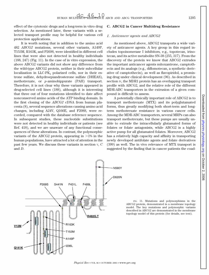
effect of the cytotoxic drugs and a long-term in vitro drugselection. As mentioned later, these variants with a se-lected transport profile may be helpful for various cellprotection applications.
It is worth noting that in addition to the amino acid482 ABCG2 mutations, several other variants, A149P,T153M, R163K, and P269S, were identified in different celllines that were also not detected in healthy individuals(188, 247) (Fig. 11). In the case of in vitro expression, theabove ABCG2 variants did not show any difference fromthe wild-type ABCG2 protein, neither in their subcellularlocalization in LLC-PK1 polarized cells, nor in their es-trone sulfate, dehydroepiandrosterone sulfate (DHEAS),methotrexate, or p-aminohippurate (PAH) transport.Therefore, it is not clear why these variants appeared indrug-selected cell lines (188), although it is interestingthat three out of four mutations identified to date affectnonconserved amino acids of the ATP binding domain. Inthe first cloning of the ABCG2 cDNA from human pla-centa (8), several sequence alterations causing amino acidchanges, including A24V, Q166E, and F208S, were re-corded, compared with the database reference sequence.In subsequent studies, these nucleotide substitutionswere not detected in healthy individuals or patients (seeRef. 418), and we are unaware of any functional conse-quences of these alterations. In contrast, the polymorphicvariants of the ABCG2 protein, appearing in �1% in thehuman populations, have attracted a lot of attention in thepast few years. We discuss these variants in section V, C
and D.
C. ABCG2 in Cancer Multidrug Resistance
1. Anticancer agents and ABCG2
As mentioned above, ABCG2 transports a wide vari-ety of anticancer agents. A key group in this regard in-cludes topoisomerase I inhibitors, e.g., topotecan, irino-tecan, and its active metabolite SN-38 (255, 317). From thediscovery of the protein we know that ABCG2 extrudesthe important anticancer agents mitoxantrone, camptoth-ecin and its analogs (e.g., diflomotecan, a synthetic deriv-ative of camptothecin), as well as flavopiridol, a promis-ing drug under clinical development (96). As described insection IV, the MDR1 protein has an overlapping transportprofile with ABCG2, and the relative role of the differentMDR-ABC transporters in the extrusion of a given com-pound is difficult to assess.
A potentially clinically important role of ABCG2 is totransport methotrexate (MTX) and its polyglutamatedforms, thus greatly modifying both short-term and long-term methotrexate resistance in various cancer cells.Among the MDR-ABC transporters, several MRPs can alsotransport methotrexate, but these pumps are usually un-able to extrude the intracellularly glutamated forms offolates or folate antagonists, while ABCG2 is a highlyactive pump for all glutamated folates. Moreover, ABCG2has a relatively high capacity and affinity in transportingnewly developed antifolate agents and folate derivatives(399) as well. The in vivo relevance of MTX transport issuggested by the finding that in cancer patients the coad-
FIG. 11. Mutations and polymorphisms in theABCG2 protein, demonstrated in a membrane topologymodel. The key mutations and polymorphic variantsdescribed in ABCG2 are demonstrated in the membranetopology model of this protein (for details, see text).
HUMAN MULTIDRUG RESISTANCE ABCB AND ABCG TRANSPORTERS 1205
Physiol Rev • VOL 86 • OCTOBER 2006 • www.prv.org
on October 2, 2006
physrev.physiology.orgD
ownloaded from
dc_46_10

ministration of benzimidazoles and MTX can result insevere toxicity, with an increase in the serum concentra-tions of MTX. Competition of MTX and the ABCG2 sub-strate benzimidazol on this transporter may explain thispharmacological interaction (37).
The chemically heterogeneous group of anticanceragents interacting with ABCG2 includes a variety of ty-rosine kinase inhibitors (TKIs). In current antitumor drugresearch, TKIs are highly promising agents for specificinhibition of malignant cell growth and metastasis forma-tion. However, their therapeutic potential also dependson access to intracellular targets, which may be signifi-cantly modulated by ABC membrane transporters. Sev-eral research groups have recently shown that the humanABCG2 multidrug transporter interacts with a number ofanticancer TKIs, including Imatinib (STI-571) and Iressa(ZD 1839).
According to our in vitro experiments (263), bothIressa and Imatinib inhibit the transport function and cellprotecting activity of ABCG2 in submicromolar concen-trations. Because low Iressa concentrations stimulatedthe ABCG2-ATPase activity, we suggested that Iressa maybe a transported substrate. Stewart et al. (355) examinedthe effect of Iressa in tumor xenografts and Saos2 cellsand found that this TKI reversed ABCG2-mediated resis-tance to irinotecan and SN-38. Still, their experimentsindicated that Iressa is not a transported substrate forABCG2.
To answer the question if Iressa is extruded byABCG2, two recent studies examined a pharmacologicallyrelevant model system, that is the growth of A431 tumorcells, depending on epidermal growth factor (EGF) recep-tor signaling. Yanase et al. (406) reported that in trans-fected A431 cells, ABCG2 provided a significant resis-tance against Iressa. In our experiments (93), the retrovi-ral expression of ABCG2 protected A431 cells from Iressa,while this protection was absent in the presence of theselective ABCG2 inhibitor Ko143 or in cells expressing aninactive ABCG2 mutant. ABCG2 function also preventedEGF receptor dephosphorylation and the extracellularappearance of phosphatidylserine, a marker for early ap-optosis (93).
When studying the action of Imatinib in Saos2 osteo-sarcoma cells expressing ABCG2, Houghton et al. (133)found that this TKI reversed SN-38 and topotecan resis-tance in submicromolar concentrations. However, thesensitivity to Imatinib, or the accumulation and efflux oflabeled Imatinib, was not modulated by ABCG2 expres-sion; therefore, these authors concluded that Imatinib is ahigh-affinity inhibitor, and not a transported substrate ofABCG2. In contrast, by using labeled Imatinib andABCG2-expressing MCF-7 or HEK cell lines, Burger et al.(40) showed that the accumulation of Imatinib was sig-nificantly lower in ABCG2-expressing cell lines than in theparental cells. Moreover, when the specific ABCG2 inhib-
itor Ko143 was added, Imatinib accumulation in ABCG2-expressing cell lines reached the same high level as inparental cells. Breedveld et al. (36) found an active, vec-torial transport of Imatinib in an epithelial cell monolayer,transfected with the mouse Abcg2. Moreover, accordingto their results, ABCG2 function limits the penetration ofImatinib through the blood-brain barrier (see below).
These experimental data suggest that both Iressa andImatinib interact with ABCG2 at low, pharmacologicallyrelevant concentrations and while they inhibit the trans-port of other substrates, these TKIs are also transportedby ABCG2. These data also indicate that the expressionand function of ABCG2 may significantly alter the absorp-tion, metabolism, and toxicity of TKIs (262).
Some of the above discussed results suggest thatthere may be a narrow concentration range in whichmultidrug resistance proteins can transport the TKIs,while above these concentrations TKIs inhibit the func-tion of these proteins (93, 262). Therefore, it is possiblethat ABCG2 may not influence the intestinal absorptionof, e.g., Iressa, present at this site in high concentrations,but may play a role in cellular extrusion of this compoundwhere its local concentration is lower.
2. ABCG2 expression in tumors
Although experimental data for cytotoxic drug trans-port by ABCG2 are convincing, a key question for the invivo relevance is the functional expression of ABCG2 invarious human tumors. Overexpression of ABCG2 wasfirst documented in drug-selected cell lines from ovary,lung, breast, colon, and gastric cancer, mediating in vitroresistance against various cytotoxic compounds (6). Ac-cording to the analysis of ABCG2 mRNA and proteinlevels in blast cells from acute leukemia patients, �30% ofthe patient samples were identified with relatively highexpression of ABCG2 (295, 304). In acute lymphoblasticleukemia (ALL), ABCG2 expression was more abundantin B-cell lineage ALL, with a function to produce signifi-cant drug resistance (275). More and more data indicatethat individuals with higher ABCG2 expression in theirleukemic blast cells have a higher probability of poorresponse to chemotherapy (28, 354, 358, 378).
Immunohistochemical studies in human tumors re-vealed frequent expression of ABCG2, especially in ade-nocarcinomas of the digestive tract, lung, and endome-trium (76). However, both intracellular and plasma mem-brane staining was observed; thus the drug extrusionrelevance of this protein expression may be questionable.In non-small cell lung cancer (NSCLC) tissue extracts, theexpression and function of ABCG2 were analyzed in par-allel experiments (174), and the authors concluded thatthese tumors expressed sufficient functional ABCG2 toconfer drug resistance. In a retrospective study in NSCLC,the chemotherapy response rate in patients was found to
1206 SARKADI, HOMOLYA, SZAKACS, AND VARADI
Physiol Rev • VOL 86 • OCTOBER 2006 • www.prv.org
on October 2, 2006
physrev.physiology.orgD
ownloaded from
dc_46_10

be correlated with ABCG2 expression (while not withMDR1, MRP1, or MRP3 expression), suggesting that theinhibition of ABCG2 function may help overcome drugresistance in such patients (408).
ABCG2 expression was greatly increased in irinote-can- and SN 38-resistant colon cancer cell lines, and the invivo overexpression of ABCG2 in the metastases of irino-tecan treated patients was also detected (44). Still, thecontroversy about the actual role of ABCG2 in cancermultidrug resistance is unresolved, and the major hope inthis regard is the introduction of proper, quantitativelaboratory diagnostic methods for the detection ofABCG2 in tumor samples.
3. Diagnostics of ABCG2 expression and function
On the basis of the above-described role of ABCG2 intumor drug resistance, the selective and sensitive detec-tion of the ABCG2 protein may have a major importancein cancer diagnostics and treatment. Determination ofRNA or total protein levels in the case of MDR-ABCtransporters may lead to seriously misleading results, asthe active form of these transporters is only the proteinproperly localized on the cell surface and in polarizedcells in the right membrane compartment. One possibleway is thus to determine the cell surface expression levelby using monoclonal antibodies, while the other approachis to directly measure the transport function of the proteinin intact tumor cells.
As to the determination of ABCG2 cell surface ex-pression, a group of monoclonal antibodies have beendeveloped that specifically recognize this protein on ex-tracellular epitopes in intact cells (430). Interestingly, theantibody, named 5D3, inhibits the transport and ATPasefunction of ABCG2 in intact cells (265, 430), and bothantibody binding and the inhibition of ABCG2 functiondepend on the actual conformation of the transporter(265). Thus the proper detection of quantitative ABCG2expression by this antibody requires fixation of the pro-tein in a high-affinity conformation, either by chemicalcross-linking or by stabilization of a specific step withinthe transport cycle. The increasing knowledge on theantibody-reactive conformations of ABCG2 should im-prove the use of this important diagnostic tool.
The other major way to characterize the relevantmultidrug transporter activity of ABCG2 is to measure theactive extrusion of a labeled or fluorescent compound. Inpractical laboratory diagnostics, the use of a nontoxicfluorescent dye, with a unique interaction with ABCG2,and providing high level fluorescence in a convenientwavelength area, would be most advantageous both inflow cytometry and fluorescence microscopy studies. Themost often applied method in this regard is the measure-ment of the fluorescent dye Hoechst 33342 by flow cytom-etry (430). Several other fluorescent substrates of ABCG2
(e.g., topotecan, flavopiridol, BODIPY-prazosin, or mitox-antrone) have also been applied in this regard. However,these dye transport assays are not specific for ABCG2,and the combined use of ABCG2-specific inhibitors isadvocated in these methods. A recent report suggests thatthe chlorophyll derivative pheophorbide a is a selectiveABCG2 substrate that can be used for flow cytometryanalysis of this protein (287).
As a summary, the laboratory detection of function-ally relevant ABCG2 expression in cancer cells has sev-eral encouraging options, but a highly sensitive and selec-tive morphological and/or functional recognition of thistransporter is still to be introduced.
D. Physiological and Pharmacological Functions
of ABCG2: Drugs, Sex, and Survival
Both the transport properties and the tissue distribu-tion of ABCG2 indicate a key role of this protein in theprotection of our body against xenobiotics (227, 316).Since ABCG2 transports a variety of amphipathic mole-cules, we can expect that both natural and artificial toxinscan be eliminated by its function. Moreover, ABCG2 isphysiologically expressed in relatively high levels in thecanalicular membrane of the liver, in the epithelia of smallintestine, colon, lung, kidney, adrenal, and sweat glands,as well as in the endothelia of veins and capillaries, in-cluding the capillary endothelial cells of the blood-brainbarrier, and in stem cells (66, 227, 430).
The apical membrane expression of ABCG2 in epi-thelial cells along the gastrointestinal track suggests amajor role of this protein in the first line of defenseagainst xenobiotics. This protection is further extendedby the function of ABCG2 in the biliary and renal luminalepithelia, responsible for toxin clearance. Interestingly,issues related to drugs, sex differences, hormonal regula-tion, and protection of the fetus and stem cells all seem tobe connected to ABCG2 function.
Again, it is important to note that ABCG2 has anoverlapping transported substrate profile and localizationwith other major multidrug transporters, especially thatof MDR1 and MRP2. Therefore, the appreciation of thephysiological and pharmacological role of ABCG2 re-quires special experimental approaches, that is, the use ofinhibitors or various transporter knock-out conditions.
1. First line defense and secretion: general
xenobiotic resistance
As to the role of Abcg2/ABCG2 in xenobiotic (toxinand drug) absorption and bioavailability, the first impor-tant information was provided by investigations carriedout in Mdr1a knock-out mice. When topotecan (anABCG2 substrate) and GF120918, an inhibitor of ABCG2,were coadministered orally to these animals, the bioavail-
HUMAN MULTIDRUG RESISTANCE ABCB AND ABCG TRANSPORTERS 1207
Physiol Rev • VOL 86 • OCTOBER 2006 • www.prv.org
on October 2, 2006
physrev.physiology.orgD
ownloaded from
dc_46_10

ability of topotecan in the blood plasma was drasticallyincreased. The hepatobiliary excretion of intravenouslyadministered topotecan was decreased by oral GF120918,consistent with an excretory role of canalicular Abcg2into the bile (163). Based on these experiments, similarstudies were carried out in human patients treated withtopotecan. The oral administration of the ABCG2/MDR1inhibitor GF120918 significantly increased the oral ab-sorption and systemic bioavailability of topotecan (192).These data indicate that ABCG2 function limits the uptakeof topotecan from the intestinal lumen, while it increasesthe hepatobiliary excretion of this compound.
To appreciate this physiological role, the develop-ment of the Abcg2 knock-out mice was initiated (161,429). The nonessential nature of Abcg2 in these knock-outanimals, that is, the lack of any phenotype in a controlledenvironment, was the first key observation. However, un-der various environmental or pharmacological challenges,several alterations have been observed connected to thelack of functional Abcg2.
The Dutch group first reported that in the Abcg2knock-out mice the combination of exposure to UV lightand an alfalfa diet, rich in chlorophyll, led to seriousphototoxic skin lesions (161). These lesions were causedby the accumulation of a chlorophyll degradation prod-uct, pheophorbide a, found in various plant-derived nutri-ents and food supplements. Normally, Abcg2 activelytransports pheophorbide a, efficiently limiting the uptakeof this compound from ingested food, while in the knock-outs there was an increased intestinal absorption anddecreased biliary secretion of this toxin. Based on thesestudies, it has been suggested that the malfunctioning ofABCG2 may cause an increased risk for developing pro-toporphyria and diet-dependent phototoxicity. This find-ing is a striking illustration of the importance of drugtransporters in protection from toxicity of normal foodconstituents (161). Indeed, van Herwaarden et al. (384)demonstrated that Abcg2 knock-out mice have elevatedplasma levels and decreased intestinal, fecal, and hepato-biliary excretion of the food carcinogen 2-amino-1-methyl-6-phenylimidazo[4,5-b]pyridine (PhIP). A recentstudy of Adachi et al. (1), using either MRP2-deficient orAbcg2 knock-out animals, demonstrated that ABCG2 hasa key role in extruding glucuronide and sulfate conjugatesof drugs and hormones, formed in the enterocytes, intothe intestinal lumen.
There are numerous ABCG2 substrate compoundsthat could compete with each other during absorption orsecretion, and this competition may involve various foodconstituents. Flavonoids are important modifiers of sev-eral physiological processes (see the “French paradox,”probably caused by flavonoids in red wine), and they alsointeract with ABCG2. As shown by Imai et al. (146),several flavonoids, including genistein and naringenin,increased the cellular accumulation of topotecan in
ABCG2-expressing cells. A direct transport of labeled,unmetabolized genistein has also been documented. Thusflavonoid consumption may significantly alter the phar-macokinetics, increasing the toxicity or the antitumoraction of ABCG2 substrate compounds. In summary, allthese data suggest that ABCG2 has a major role in themodulation of the absorption of toxic materials fromfood, as well as of their excretion mostly through thebiliary pathway (77).
2. Prevention of endogenous toxin accumulation
The Abcg2 knock-out mouse study, carried out in theUnited States, revealed a hypersensitivity of the tissues inthese animals to hypoxic challenges (191). Progenitorcells obtained from Abcg2 knock-out mice showed a re-duced ability to form colonies under hypoxia. Moreover,blocking of Abcg2 function in normal progenitor cellsreduced survival under hypoxic conditions. As it turnedout, the primary cause of this hypoxic susceptibility wasthe accumulation of cellular heme and/or porphyrins. Theblockade of heme/porphyrin synthesis reversed this con-dition, while cells overexpressing ABCG2 accumulatedlower amounts of porphyrin. Heme was shown to bespecifically bound by ABCG2, and drug transport byABCG2 was significantly modulated by heme.
These studies suggest that ABCG2 permits enhancedcell survival in oxygen-poor environments by reducing theaccumulation of toxic heme metabolites; thus ABCG2expression provides an important cell survival advantageunder hypoxic conditions. Of course, the chemical simi-larity of pheophorbide and porphyrin is evident andshould direct our interest to possible ABCG2-dependenttransport of various related compounds.
3. ABCG2 in the placenta
The wild-type human ABCG2 was first cloned from aplacenta library (8), and the protein is abundantly ex-pressed in chorion epithelial cells. The maternal-fetal bar-rier in the placenta, protecting the fetus from drugs andenvironmental toxins, has been indicated to involve thefunction of MDR1/Pgp. Further studies in Mdr1 knock-outmice indicated an important role of ABCG2 in this tissue.In pregnant, Mdr1-deficient mice, when topotecan wascoadministered with the inhibitor GF120918, the fetalpenetration of topotecan was significantly higher thanwithout this inhibitor (163). In the Abcg2 knock-out mice,the fetal exposure to topotecan and dietary toxins greatlyincreased, compared with normal mice (320). The selec-tive inhibition of Abcg2 by fumitremorgin C also pro-duced a large increase of the topotecan and mitoxantronelevels in the fetus (187). All these data indicate thatABCG2 plays a major role in protecting the fetus againsttoxic compounds ingested by the mother (Fig. 12).
1208 SARKADI, HOMOLYA, SZAKACS, AND VARADI
Physiol Rev • VOL 86 • OCTOBER 2006 • www.prv.org
on October 2, 2006
physrev.physiology.orgD
ownloaded from
dc_46_10

4. ABCG2 in the blood-brain barrier
The presence of ABCG2 in the brain endothelia wasindicated by RNA expression measurements (90, 423),and the expression of the ABCG2 protein was directlydemonstrated in the luminal surface of brain microves-sels, forming the blood-brain barrier (BBB), by immuno-fluorescence confocal microscopy (66). The vectorialdrug transport by ABCG2 overexpressed in immortalizedendothelial cells indicated a functional role of this pro-tein, limiting the brain penetration of hydrophobic com-pounds (115). By using in situ brain perfusion techniques,in Mdr1 knockout mice an efficient extrusion of prazosinand mitoxantrone by Abcg2 in the BBB was clearly dem-onstrated (62). Interestingly, flavonoid transport throughthe BBB was also modified by the function of ABCG2(409). In a rat BBB model, the expression and function ofrABCG2 was upregulated by astrocyte-derived solublefactors (132), and the application of an siRNA reducedboth the expression and the transport function of endog-enous rABCG2 in capillary endothelial cells (131). Re-cently, Breedveld et al. (36) have shown that the brainpenetration of intravenously administered Imatinib wassignificantly increased in Abcg2 knockout mice. All thesedata strongly indicate an important function of ABCG2 inthe BBB, protecting the brain from potentially toxicagents (Fig. 12).
5. ABCG2 in stem cells
A fascinating development in the field of stem cellresearch was the finding that a high-level expression ofthe ABCG2 protein and its fluorescent dye extrusion func-tion could identify hemopoietic stem cells (177, 314, 430).The so-called “side population (SP)” of progenitor cells,actively extruding the fluorescent Hoechst 33342 dye,seems to represent pluripotent stem cells in a variety oftissues (177, 201, 314, 357, 430) (Fig. 12). Although we donot know as yet why ABCG2 expression is high in these
cells, it seems likely that it is required for the metabolicprotection of stem cells. ABCG2 expression decreasesduring stem cell differentiation. Still, both the knock-outmouse studies and recent transplantation experimentssuggest that the lack of ABCG2 expression or ABCG2overexpression do not directly influence stem cell func-tion and differentiation (249, 429) (O. Ujhelly, unpub-lished data). As mentioned above, ABCG2 function mayprotect against local hypoxia; thus its presence may be asurvival factor for stem cells under unfavorable condi-tions. This role may have important implications for in-creasing medical activities connected to stem cell trans-plantation.
6. Sex differences in ABCG2 expression and function
Recent studies demonstrated a sex-dependent ex-pression and function of ABCG2 in various organs. Inmice a significant difference was observed in the pharma-cokinetic behavior of ABCG2 substrate drugs betweenmale and female animals, leading to much higher plasmalevels in female mice after a similar drug exposure (237).The main cause of this difference was the higher liverexpression and biliary clearance function of Abcg2 inmale mice, while this pharmacokinetic sex difference wasabsent in Abcg2 knockout animals. The authors alsofound a much higher hepatic expression of ABCG2 in menthan in women (237). Imai et al. (144) reported thatestrogens, like 17�-estradiol, downregulate ABCG2 ex-pression through an estrogen receptor �-interaction path-way in various cell lines (see below). Another recentstudy (371) has found a much higher ABCG2 expressionin the intestine and kidney of male rats compared withfemales, and a male-dominant expression was observed inthe mouse liver. Moreover, when following ABCG2 ex-pression, a suppressive effect of estradiol and an induc-tion by testosterone (371) were observed. These data mayhave a major importance in characterizing the often ob-
FIG. 12. Physiological role of the MDR1 andABCG2 multidrug/xenobiotic ABC transporters in theblood-brain barrier (A), in stem cells (B), and in pla-centa (C). The endothelial cells of the blood-brain bar-rier are connected with tight junctions and provide abarrier against the entrance of hydrophilic compoundsinto the brain area. Hydrophobic drugs or toxins pene-trate the cell membranes and may enter the brain com-partment. Multidrug transporters, especially MDR1/Pgpand ABCG2, are expressed in the apical membrane(facing the blood compartment) of the endothelial cellsand extrude a variety of hydrophobic drugs and toxins.ABCG2 is abundantly expressed in various stem cells,with as yet unknown function. Hemopoietic stem cellscan be separated by their low-level Hoechst dye stain-ing (side population, SP), due to active dye extrusion byABCG2. ABCG2 and MDR1/Pgp are highly expressed inthe maternal side of the chorion epithelial cells in theplacenta. These transporters prevent the entrance of avariety of toxins and drugs into the fetal compartment.
HUMAN MULTIDRUG RESISTANCE ABCB AND ABCG TRANSPORTERS 1209
Physiol Rev • VOL 86 • OCTOBER 2006 • www.prv.org
on October 2, 2006
physrev.physiology.orgD
ownloaded from
dc_46_10

served sex differences in drug effectiveness and toxicity.Importantly, the sex hormone-dependent regulation ofABCG2 expression may be connected to its role in thetransport of various hormone metabolites, e.g., estronesulfate.
In connection with sex-related differences, ABCG2was shown to have an important role in drug and toxinsecretion into milk. As reported recently by Jonker et al.(162), ABCG2 expression (as examined in mice, cows,and humans) is greatly increased in the mammary glandduring lactation, and the ABCG2 protein actively secretesvarious drug and toxin substrates into the milk. The se-cretion of the antibiotic nitrofurantoin into milk has beenreported to be 80 times higher in wild-type mice com-pared with the Abcg2 knock-out animals (236). Thus theregulation and function of ABCG2 may significantly mod-ulate contamination of milk with drugs and xenobiotics,leading to toxin exposures in infants or consumers ofdairy products.
E. Polymorphisms and Regulation of ABCG2
Both the pharmacological and physiological func-tions of the ABCG2 protein have been indicated to besignificantly influenced by interindividual variations andregulatory responses. In this section we discuss the twomain apparent sources of these variations: a widespreadgenetic polymorphism, probably affecting both proteinexpression and function, and the response to toxin expo-sure, stress, or hormonal changes by a variable expres-sion and processing of ABCG2.
1. SNPs in ABCG2
Analysis of the ABCG2 sequences in the human pop-ulation identified several single nucleotide polymor-phisms (SNPs), and their effects on protein expressionand function have been the subject of numerous recentstudies. Earlier reports indicated considerable interindi-vidual variations in the oral bioavailability and clearanceof drugs that are ABCG2 substrates, such as topotecan(163, 419), which may reflect such a polymorphism.
The ABCG2 gene is located on chromosome 4q22,spans over 66 kb, and consists of 16 exons, ranging from60 to 532 bp in length (20). In healthy individuals orpatients, altogether eight nonsynonymous (V12M, Q141K,I206L, F431L, S441N, F489L, N590Y, D620N), five synony-mous (silent) (c. 114T�C, c. 369C�T, c. 474C�T, c.1098G�A, c. 1425A�G) mutations, one nonsense (Q126X,c. 376C�T), and one frameshift (c. 1515delC) mutationwere identified in the coding region of ABCG2. The se-quence variant Q126X, leading to premature terminationof protein synthesis, was consistently observed in certainJapanese cohorts, while absent in different Caucasian andAfrican American groups (145, 148, 185). From these nu-
merous reported alterations, two protein variants, V12M,and Q141K, were found in relatively high frequencies,with significant differences in allele frequencies in differ-ent areas of the world (Fig. 11).
The V12M polymorphism affects the NH2-terminalintracellular region of the protein. Both the wild-type(valine) and the variant (methionine) amino acids haveuncharged, hydrophobic side chains. The V12M polymor-phism was found in all ethnic groups tested, with thehighest allele frequency in Mexican-Indians (90%), whileonly 2% in a Swedish population (18, 418), and also witha significant difference in allele frequencies in Caucasianand Japanese populations.
The Q141K polymorphism leads to the replacementof the uncharged glutamine residue with a positivelycharged lysine within the ATP-binding domain, betweenthe Walker A motif (amino acids 83–89) and the signatureregion (amino acids 186–189). The Q141K variant wasalso detected in all ethnic groups tested: the allele fre-quency ranged between 1 and 35% (the African and Afri-can-American subjects with low, while the Japanese andChinese populations with high allele frequencies; seeRefs. 45, 74, 185).
To clarify the possible physiological or pathologicalrelevance of the ABCG2 polymorphisms, several studiesattempting the functional characterization of the variantswere performed. Interestingly, the results of the differentresearch groups, regarding expression levels, localization,and functionality, are quite contradictory. Imai et al. (145)and Morisaki et al. (251), by using stable mammalianexpression systems, found that in PA317 or HEK-293 cellsthe expressed Q141K ABCG2 protein had a lower expres-sion level than the wild-type ABCG2, or the V12M variant.Morisaki et al. (251) demonstrated that both the V12M andQ141K ABCG2 could reach the plasma membrane in theHEK-293 cells, while a significant portion of Q141K re-mained intracellular. Other studies found a 30–40% reduc-tion in cell surface expression of the Q141K variant, de-spite a similar mRNA level than the wild-type ABCG2(145, 188).
Recent investigation of 99 Japanese placenta samplesrevealed that individuals homozygous for the Q141K vari-ant showed significantly lower expression levels of thistransporter protein, while the heterozygous samples dis-played an intermediate expression level (185). In contrast,another study, investigating the expression of natural al-lelic variants of ABCG2 in the human intestine, demon-strated no significant differences in mRNA and proteinlevels between subjects expressing the K141 allele inheterozygous form, or the Q141 (wild-type) allelic variant(418).
Mizuarai et al. (247) expressed ABCG2 in polarizedLLC-PK1 cells, and by using confocal microscopy, theauthors observed that the wild-type ABCG2 and Q141Kshowed mainly apical staining, while the V12M variant
1210 SARKADI, HOMOLYA, SZAKACS, AND VARADI
Physiol Rev • VOL 86 • OCTOBER 2006 • www.prv.org
on October 2, 2006
physrev.physiology.orgD
ownloaded from
dc_46_10

showed mostly intracellular localization. In a recentstudy, Kondo et al. (188) also used LLC-PK1 cells to ex-press the V12M and Q141K variants and found that allpolymorphisms, including V12M and Q141K, had an apicallocalization. These contradictory expression and localiza-tion data for the ABCG2 variants, even when similar celllines were applied, indicate that differences in cultureconditions or other cellular determinants may variablyaffect the cellular processing of these proteins.
When the functions of the ABCG2 variants were ex-amined in cytotoxicity assays, a 10-fold decrease in drugresistance, compared with the wild-type ABCG2, was re-ported by Mizuarai et al. (247), when the V12M or Q141K-transfected LLC-PK1 cells were challenged by mito-xantrone, topotecan, or an indolocarbazole topoisomer-ase I inhibitor. In contrast, Morisaki et al. (251) found thatonly the Q141K variant had a moderately lower levelresistance against mitoxantrone, topotecan, or SN-38,compared with the wild-type ABCG2-transfected cells.Moreover, in experiments comparing the transport activ-ities of the wild-type protein and its variants for estrone3-sulfate, dehydroepiandrosterone sulfate, methotrexate,or PAH, no significant differences were reported, whenthe transport activities were normalized for the expres-sion levels of ABCG2 proteins (188).
Q141K is mapped to a functionally important ABCregion of ABCG2; therefore, it is possible that the ATPaseactivity of this variant is altered. Two studies comparedthe vanadate-sensitive ATPase activity of ABCG2 V12Mand Q141K variants, using Sf9 (Spodoptera frugiperda)cell membranes (247, 251). A reduced basal ATPase ac-tivity was observed by both groups for the Q141K variant.On the other hand, the V12M ABCG2 showed a similarATPase activity as the wild-type protein.
All these transport, ATPase, and cell survival datasuggested that the investigated SNPs may not substan-tially alter the substrate specificity of ABCG2. On theother hand, if their expression level is indeed lower, ortheir membrane localization is impaired, these alterationsmay modify ABCG2-dependent drug transport. Clearly,more detailed studies are required to clarify the mecha-nism of a reduced protein expression for Q141K, and thealtered cellular localization found for the V12M andQ141K variants under certain conditions. It is importantto note that in heterozygous individuals, different ratios ofthe ABCG2 dimers may be present. The above in vitroexperiments investigated homodimers of the variants,which is a model applicable only for the rare cases ofhomozygotes.
In addition to the above-described in vitro investiga-tions, several pharmacokinetic studies are in progress toevaluate the potential functional differences of variousABCG2 genotypes. De Jong et al. (74) investigated pa-tients with solid malignant tumors receiving irinotecantreatment. In 84 European Caucasian patients, the fre-
quency of the K141 allele was 10.7%, with 14 patientscarrying heterozygous alleles and 2 patients homozygousfor the variant. According to this study, the pharmacoki-netic parameters of irinotecan and SN-38 were not signif-icantly different between patients carrying the wild-typeABCG2 or at least one polymorphic allele. However, oneof the two homozygous individuals showed increasedaccumulation of SN-38 and SN-38 glucuronide, indicatingthat the K141 homodimer may have an impaired function.
A recent investigation performed an exploratory, ret-rospective evaluation of the functional consequence ofthe ABCG2 Q141K variant in 20 adult patients, treatedwith diflomotecan, a synthetic derivative of camptothecin(350). In five patients carrying one K141 variant allele,after oral administration of diflomotecan, no differencewas found in serum levels compared with individuals withthe wild-type genotype. However, after intravenous ad-ministration of the drug, the plasma concentration in theheterozygous patients was markedly increased comparedwith the individuals with the wild-type genotype. Thisobservation is in harmony with studies indicating a re-duced protein expression and function for the Q141Kvariant, while it seems to contradict the results of De Jonget al. (74).
As mentioned above, the allele frequency of Q141Kvaries in diverse populations, and in Japan and China, thispolymorphism appears to be common, with an overallallele frequency of �30%. The resulting frequent occur-rence of the wild-type/K141 heterodimer and even theK141 homodimer ABCG2 transporters in this population,as well as the appearance of other mutations, leading to areduced ABCG2 protein level, may strongly modify theADME-Tox properties of many different drugs. Since therecently introduced anticancer tyrosine kinase inhibitorIressa (Gefitinib) is a transported substrate of ABCG2 (93,406), polymorphisms in this protein may be involved inthe different effectiveness and toxicity of this drug indistinct populations. Moreover, because ABCG2 was dem-onstrated to have a protective role against food toxins,especially plant metabolites, a lower expression or im-paired function of ABCG2 variants may affect the diet-related health conditions in a huge human population(161).
2. Regulation of ABCG2
A) TRANSCRIPTIONAL REGULATION. Relatively little isknown as yet about the transcriptional regulation andpromoter structure of ABCG2. In their pioneering work,Bailey-Dell et al. (20) have analyzed the 5�-upstream�2-kb region of the human ABCG2 by bioinformatic toolsand localized positive and negative regulatory elementsby a luciferase reporter assay. They showed that, similarto other ABC protein coding genes, the ABCG2 gene lacksthe canonical TATA box within the region 100 bp up-
HUMAN MULTIDRUG RESISTANCE ABCB AND ABCG TRANSPORTERS 1211
Physiol Rev • VOL 86 • OCTOBER 2006 • www.prv.org
on October 2, 2006
physrev.physiology.orgD
ownloaded from
dc_46_10

stream to the transcriptional start site. Several Sp1 siteswere found within the corresponding �222 to �49 bpregion (relative to the transcriptional start site). The pres-ence of Sp1 sites is a common feature of genes withoutTATA box, and ABCB1 and ABCC1, both lacking TATAboxes, possess several SP1 binding sites. A CCAAT-box ispresent in the �274 bp position, and CpG islands arefound in its downstream proximity. These elements pre-dict a putative promoter/enhancer in the �266 to �36 bpregion. The reporter assays identified potential positiveregulatory elements between �1285 and �62 bp, andpotential negative regulatory elements between �628 and�31 bp. Removal of the CCAAT box and part of thepredicted region reduced and further truncation of thisregion further decreased transcription activity.
A novel estrogen response element has been identi-fied in the �188 to �172 bp segment of the gene (89), andit has been demonstrated that the 17�-estradiol-ligandedestrogene receptor (E2/ER) binds directly to this elementand, by interacting with components of the RNA polymer-ase II transcriptional machinery, triggers an enhancedtranscription of human ABCG2 in ER-positive T47D: A18cells (89). The same element has been found by a system-atic genome-wide bioinformatic study by analyzing pro-moter elements for transcription factor binding sites withthe aim of identifying estrogen-responsive genes (169).However, the regulation of ABCG2 seems to be cell linespecific, because it is actually downregulated followingestrogen treatment in ER-positive MCF7 cells (169). More-over, as mentioned below, estrogen regulation of ABCG2may be mostly of posttranslational nature.
ABCG2 is probably also regulated by drug treatmentand cellular stress, but this phenomenon has not yet beenexplored as critically as in the case of MDR1 or MRPs.Benzopyrene conjugates were reported to induce ABCG2expression in the Caco-2 intestinal cells (87). Burger et al.(41) demonstrated that in the same cell type, the additionof Imatinib, an ABCG2-substrate TKI, upregulates thetransporter expression through an SXR-dependent mech-anism.
As mentioned above, ABCG2 may have a key role inprotecting the cells against porphyrin metabolites duringhypoxia, and this coincides with an upregulation of thistransport protein (191). ABCG2 expression was found tobe upregulated through an interaction of the hypoxia-inducible transcription factor complex HIF-1 with theABCG2 promoter region. Because MDR1/Pgp is also up-regulated during hypoxia by HIF-1 through the activationof a stress-activated kinase pathway (64, 65), this could bea general type of stress response also involving theABCG2 protein.
Interestingly, the lack of Mdr1 expression in knock-out mice greatly upregulates Abcg2 expression in a func-tionally important area, the BBB (62). This excessiveoverproduction of a related transporter in a given tissue
and its role in a defense network is discussed in detail insection VII.
B) POSTTRANSLATIONAL REGULATION. One possibility for aposttranslational regulation stems from the dimerizationproperties of this ABC half-transporter. In heterozygotes,dimerization of polymorphic variants in fact results infunctional heterodimer formation, and thus may alter thelocalization and/or function of the protein (see above).Clearly, a nonfunctional mutant (K86M) ABCG2 variantinduces a dominant negative effect, that is, a nonfunc-tional dimer formation suppresses the activity of the wild-type protein (93, 166). Such a dominant negative effectmay be a rare case in vivo, but the polymorphic het-erodimerization may be pharmacologically relevant.
Similarly to that found for MDR1/Pgp (see sect. IV),the transport function or localization of ABCG2 is notsignificantly modulated by protein glycosylation (78, 250,370). There are no published experimental data as yet onthe possible phosphorylation-dependent regulation ofABCG2. Based on earlier observations when ABCG2 wasexpressed in Sf9 cells (260, 261) and in Lactococcus (151),as well as on our recent experiments (Sarkadi, unpub-lished data), membrane lipid composition, especially thelocal concentration of cholesterol, has a key regulatoryeffect on ABCG2 transport activity. This may be mostrelevant in transport function, when ABCG2 is retained inthe cholesterol-poor endoplasmic reticulum membrane,or transferred to the cholesterol-rich plasma membrane.
As mentioned above, sex hormones have a regulatoryeffect on ABCG2, that is, estrogen in general downregu-lates ABCG2 protein expression and function (144). Thisregulation involves estrogen receptor alpha and is re-versed by the antiestrogen tamoxifen. Interestingly, ac-cording to this study, estrogen does not affect the level ofABCG2 mRNA, but the regulation is posttranscriptional,by decreasing ABCG2 protein biosynthesis and matura-tion. According to Tanaka et al. (371), based on gonadec-tomy and hypophysectomy experiments, the lower levelABCG2 expression in female rat kidney is probablycaused by estradiol suppression, while higher ABCG2 ex-pression in male mouse liver is induced by testosterone.
These data raise the possibility that ABCG2 is undera complex posttranslational regulation, which involvesthe interaction of this protein with cytoskeletal and otherintracellular elements that may significantly influence thecell surface exposure, that is, the drug extrusion functionof this protein.
F. ABCG2 in Medical Applications: Drug
Development and Gene Therapy
1. ABCG2 in drug screening and development
Based on the above-described drug interactions,ABCG2 plays a major role in tumor drug resistance, as
1212 SARKADI, HOMOLYA, SZAKACS, AND VARADI
Physiol Rev • VOL 86 • OCTOBER 2006 • www.prv.org
on October 2, 2006
physrev.physiology.orgD
ownloaded from
dc_46_10

well as in the absorption, metabolism, excretion, andtoxicity (ADME-Tox) of various pharmacological com-pounds. Therefore, an in vitro prescreening of new ther-apeutic compounds and their metabolites may provide agreat advantage; all these parameters could be predictedwithout tiresome and expensive in vivo screening, costinga lot of animal lives.
As described in section III, the major MDR-ABC trans-porter screening methods include direct enzymatic(ATPase, nucleotide trapping, fluorescence-based enzymeactivity measurements, etc.) and vesicular transport as-says, by using isolated membrane vesicles exclusively orpredominantly expressing the desired transport protein ina functional form. The overexpression of the ABCG2 pro-tein in the heterologous insect cell/baculovirus system isa well-established possibility for such screenings, andisolated membranes of mammalian cells could also beused for this purpose (see Ref. 104). Another type of basicdrug screening can be performed by using cultured mam-malian cell lines, specifically engineered to express thegiven transporter. In this case both the direct transportmeasurements for labeled compounds and indirect assaysby using fluorescent or otherwise labeled reporter com-pounds are available. Without detailing these assays here(see sect. III), we feel that these methods should be rapidlyoptimized and standardized by the research and develop-ment community, to provide the drug development indus-try with a well-established toolkit for prescreening newdrugs in their pipelines. Since, as demonstrated above,ABCG2 is a “transporter for all seasons” (306), performingnumerous tasks in a number of organs, screens forABCG2 should be in the first line of such developmentstrategies. The excessive polymorphisms and the sex-dependent distribution of this transporter add an extratask to design such a meaningful in vitro screening panel,but may also lead to a major increase in the cost-to-benefitratio of drug development.
2. ABCG2 in gene therapy
The physiological presence of ABCG2 in stem cellsprovides the basis for a gene therapy application of thistransport protein. The introduction of a selectable markerin ex vivo stem cell gene modification provides a selectiveadvantage of the modified cells after transplantation. Thecoexpression of a drug-resistance protein with a thera-peutic gene product should allow both an enrichment ofthe corrected cells and an in vivo drug selection duringclinical gene therapy. The use of the MDR1-Pgp as such aselectable marker has been widely investigated and advo-cated, while other studies reported major problems withthis approach (39, 113, 318). Recent studies in our labo-ratory suggest that a mutant ABCG2 protein is an idealcandidate for human stem cell protection and for use as aselectable marker in gene therapy. The cDNA encoding
this protein is relatively small (�2 kb), and the activedimer is spontaneously formed in the overexpressingcells (379). Because the substrate specificity of the R482Gvariant of ABCG2 differs from that of the wild-type pro-tein, this mutant has a special advantage in gene therapyapplications.
We have documented that when the mutant ABCG2was coexpressed with a therapeutic gene, the expressionof the therapeutic gene in hematopoietic progenitor cellscorrected the loss-of-function mutation responsible forhuman chronic granulomatous disease (379). At the sametime, the mutant ABCG2 protein selectively protected thetransduced cells against clinically applicable cytotoxicagents. Overexpression of ABCG2 did not affect in vitrohematopoietic cell maturation or the restoration of gran-ulocyte function by the therapeutic gene. The first in vivomouse studies (Ujhelly et al., unpublished data) havedemonstrated that the ABCG2 expressing bone marrowstem cells can be used for efficient transplantation, theproduction of all blood cells remains normal, and there isno change in cell repopulation or differentiation patterns.
VI. ADDITIONAL TRANSPORTERS IN CANCER
DRUG/XENOBIOTIC RESISTANCE
Up to now only three human ABC transporters,MDR1/Pgp (ABCB1), MRP1 (ABCC1), and ABCG2, haveunambiguously been shown to contribute to cancer mul-tidrug resistance. However, as mentioned earlier, thereare several other ABC proteins that were implicated incancer drug resistance and xenobiotic transport. In thissection we give an overview of selected ABC transportersthat are major candidates for participating in drug andxenobiotic resistance.
A. The MDR3 (ABCB4) Protein
P-glycoproteins (Pgps) form a small subfamily withinthe superfamily of ABC transporter proteins. In mice,there are three genes that encode for Pgps, Mdr1a,
Mdr1b, and Mdr2. In humans, in addition to MDR1, onlyone other Pgp gene has been identified: MDR3 (alsoknown as MDR2, PGY3, ABC21, or PFIC-3). According tothe classification of ABC transporters, MDR3 belongs tothe ABCB subfamily and is called ABCB4. Shortly afterthe identification of MDR1, the human MDR3 gene prod-uct was cloned from a liver cDNA library (382). Theprotein encoded by the MDR3 gene consists of 1,279amino acids and has a deduced Mr of 140,000, which isincreased by glycosylation. The MDR3 protein is a proto-typical ABC transporter, consisting of two cytosolic nu-cleotide-binding domains (ABC units) and two largetransmembrane domains, each composed of six trans-membrane helices. It shows a high, 77% amino acid sim-
HUMAN MULTIDRUG RESISTANCE ABCB AND ABCG TRANSPORTERS 1213
Physiol Rev • VOL 86 • OCTOBER 2006 • www.prv.org
on October 2, 2006
physrev.physiology.orgD
ownloaded from
dc_46_10

ilarity with the product of the MDR1 gene (52). The levelof sequence conservation is the highest in the ABC unitsand in the transmembrane domains and the lowest at theNH2 terminus and in the 60-amino acid linker regionconnecting the two halves of the protein.
The MDR3 gene covers 78.74 kb and maps to chro-mosome 7, at 7q21.1, in the proximity of the MDR1 gene(209). The two genes are separated by a 34-kb intergenicregion. It has been postulated that the MDR3 geneevolved from the MDR1 sequence by gene duplication.This hypothesis is supported by the observation that theexon/intron structure of these genes is almost identical,each containing 28 exons, of which 27 encode the func-tional protein (209).
1. Tissue distribution and cellular localization
of MDR3
The MDR1 and MDR3 gene products have differenttissue distributions. In contrast to the wide-range expres-sion of MDR1 in a number of organs (see sect. IV), theexpression of MDR3 protein is constrained predominantlyto the canalicular membrane of hepatocytes. Low levelsof MDR3 RNA have been found in some tissues other thanliver, namely, in the heart, skeletal muscle, and spleen,although the MDR3 protein was not detected by Westernanalysis (341). A high-affinity antibody, however, was ableto demonstrate MDR3 protein expression in the glomeruliof the kidney (315). The regulation of expression of MDR3is not clarified. It has recently been reported that theMDR3 protein, along with other major canalicular trans-porters, is expressed at midgestational stage during hu-man fetal development. However, the level of MDR3 ex-pression was found to be much lower in fetal liver than inthe adult tissue (53).
The sequences and protein-protein interactions thatare responsible for proper maturation and accurate tar-geting of MDR3 to the canalicular membrane are also yetto be discovered. Epitope insertion studies have revealedthat intracellular loops between transmembrane helices 3and 4 as well as between 4 and 5 are crucial for propertargeting (172). Mutation in the ABC unit of MDR3 alsoprevents correct trafficking and results in the loss offunction of the protein (79). The MDR3 protein colocal-izes with MDR1 and with the bile salt transporter (BSEP/ABCB11) in the apical compartment of hepatocytes. Bind-ing of these ABC transporters to regulatory proteins suchas HAX-1 and myosin II regulatory light chain (Mlc2) hasalso been demonstrated (47, 259). These observationssuggest that HAX-1, Mlc2, and possibly cortactin are in-volved in the trafficking of these ABC transporters. It hasbeen proposed that canalicular ABC transporters, likeseveral canalicular ectoenzymes and cell adhesion mole-cules, transcytose from the basolateral plasma membraneto the apical surface. However, evidence has been pro-
vided for the fact that newly synthesized MDR1 and MDR2are directly targeted from the Golgi apparatus to thecanalicular membrane without entering the basolateralcompartment (179).
2. Transport properties and the physiological function
of MDR3
Since the MDR3 protein was found to be sequentiallysimilar to MDR1, MDR3 was also predicted to be an effluxpump, possibly a drug transporter with broad substratespecificity. However, this Pgp, in contrast to MDR1, wasproven to be a highly specialized transporter which trans-locates solely phospholipids with a choline head group(297, 298, 345). The restricted tissue distribution, partic-ular cellular localization, and specialized transport prop-erties suggest a key physiological function for MDR3 inthe bile formation. The generation and phenotyping of theMdr2 �/� mouse unambiguously demonstrated that thistransporter is essential for biliary phosphatidylcholineextrusion (342), since the bile of Mdr2-deficient mice isdevoid of phosphatidylcholine. MDR3 is a selective trans-porter regarding the fatty acid moieties of the transportedlipid. Lipid analogs with C6 and C16 fatty acids as well aswith a ceramide backbone are transported by MDR3,whereas a phosphatidylcholine analog with two C8 fattyacids is not recognized (383). This substrate selectivity ofMDR3 is assumed to be responsible for the particular fattyacid composition of the phosphatidylcholine pool in thebile, which differs from that in the liver tissue.
A suggested model for MDR1 transport mechanismsis that this protein acts as a flippase, i.e., it translocatesamphipathic molecules from the inner leaflet of theplasma membrane to the outer leaflet (119). Based on thehomology between MDR1 and MDR3 proteins, the latterone was also predicted to function as a flippase, special-ized for phosphatidylcholine (PC) (see Fig. 13). This hy-pothesis was supported by detailed transport studiesdemonstrating an ATP-dependent translocation of fluo-rescent PC analogs from the outer to the inner leaflet ofyeast secretory vesicles containing Mdr2 (298). Thismodel system was used in a subsequent study to revealthat the flippase activity of Mdr2 was stimulated by thebile salt taurocholate but not by the nonmicelle-formingcounterpart taurodehydrocholate (297). Asymmetrictranslocation of PC analogs was also demonstrated byNies et al. (257) using rat canalicular membrane vesicles.This transport was also increased by taurocholate but notby taurodehydrocholate. The flippase model for MDR3transport mechanism was also supported by studies bySmith et al. (345) using fibroblasts from transgenic mice.In these experiments mouse fibroblasts were labeled with[14C]PC, and the efflux of the labeled substrate from thecells was monitored by adding purified PC transfer pro-tein and acceptor liposomes into the bath. Compared with
1214 SARKADI, HOMOLYA, SZAKACS, AND VARADI
Physiol Rev • VOL 86 • OCTOBER 2006 • www.prv.org
on October 2, 2006
physrev.physiology.orgD
ownloaded from
dc_46_10

normal mouse fibroblasts, which do not express Mdr2,cells expressing the human MDR3 exhibited a threefoldincrease in the rate of translocation of PC. All these datasupport the concept that MDR3 is an ATP-dependent PCflippase, which translocates its substrate from the inner tothe outer leaflet of the canalicular membrane of hepato-cytes.
Smith et al. (344) generated a transgenic mouse de-ficient in murine Mdr2, but expressing the human MDR3protein under the control of an albumin promoter. Theexpression of the transgene restored PC excretion in thetransgenic animals. The lack of other defects in thesemice raises doubt about the significance of low levelMdr2/MDR3 expression in extrahepatic tissues. More-over, these studies have clearly demonstrated that PCsecretion depends both on Mdr2/MDR3 expression andthe availability of bile salts. The current understanding ofbiliary PC excretion can be summarized as follows (35,92). The Mdr2/MDR3 protein, residing in the canalicularmembrane of hepatocytes, flips PC into the outerhemileaflet. Bile salts destabilize the PC-containing mi-crodomains, which grow into a vesicular structure by theongoing translocation of PC, driven by Mdr2/MDR3. Fi-nally, the biliary vesicle pinches off the canalicular mem-brane. This hypothesis was supported by the observationsthat vesiculation of the canalicular membrane is depen-dent both on bile salt secretion (69) and Mdr2 expression(68). It should be noted that, in addition to PC deficiency,Mdr2 �/� mice also exhibited reduced biliary cholesterol
excretion. However, the role of Mdr2 was proven to besecondary in this effect.
3. Clinical relevance of MDR3
Lack of PC in the bile of the Mdr2-deficient miceresults in a mild liver disease. Since bile salt secretion isnormal in these animals, the high bile salt concentrationcauses damage in the hepatocytes and the small bileducts, resulting in extensive proliferation of bile ductepithelium, portal inflammation, and fibrosis (92). Be-cause human bile salts are more hydrophobic than theirmurine counterparts, lack of MDR3 expression results ina more severe liver disease in humans. It was first shownby de Vree et al. (75) that mutations in the MDR3 gene arethe primary cause of type 3 progressive familial intrahe-patic cholestasis (PFIC). This autosomal recessive liverdisease is characterized by an early onset of cholestasisthat progresses to cirrhosis and liver failure before adult-hood. Patients with type 3 PFIC are distinguished fromthe other two types of PFIC by high �-glutamyltransferaseactivity and a bile that lacks phospholipids but has anormal bile acid concentration. These patients exhibitportal duct inflammation and ductular proliferation, rem-iniscent of the phenotype exhibited by the Mdr2 �/�mice. Several MDR3 mutations have been identified intype 3 PFIC patients (150). Most of these mutations resultin truncated proteins, which do not reach their destina-tion in the canalicular membrane. However, a recent
FIG. 13. Physiological localization and roleof ABC transporters in the bile formation of theliver. The ABC transporters, which play a crucialrole in the bile formation, include BSEP (bile saltexport pump, sister Pgp, ABCB11), MDR3(ABCB4), and the ABCG5/ABCG8 heterodimer.These transporters reside in the canalicularmembrane of hepatocytes and perform activetransport of various bile constituents. BSEP ex-trudes bile salts into the canaliculus, where bilesalt micelles are formed. MDR3, the phosphati-dylcholine (PC) flippase, translocates PC fromthe inner hemileaflet of the membrane into theouter hemileaflet. Bile salts promote destabiliza-tion of the PC-containing microdomains, whichultimately develop into phospholipid vesiclesrich in PC. These biliary vesicles and mixed mi-celles are loaded with cholesterol by the trans-port activity of the ABCG5/ABCG8 heterodimer,which makes cholesterol accessible for theacceptors.
HUMAN MULTIDRUG RESISTANCE ABCB AND ABCG TRANSPORTERS 1215
Physiol Rev • VOL 86 • OCTOBER 2006 • www.prv.org
on October 2, 2006
physrev.physiology.orgD
ownloaded from
dc_46_10

study reported that MDR3 protein expression was detect-able in the canalicular membrane of a few patients withtype 3 PFIC (175).
Two other diseases are associated with mutations inthe MDR3 gene, namely, intrahepatic cholestasis of preg-nancy (ICP) and low phospholipid-associated cholelithia-sis. ICP is a liver disorder characterized by cholestasisduring pregnancy in women with otherwise normal med-ical history and by the occurrence of generalized pruritusmostly in the third trimester. ICP is frequently associatedwith fetal distress, spontaneous immature delivery, andidiopathic intrauterine fetal death in the third trimester.Heterozygotes for either nonsense or missense mutationsin the MDR3 gene are susceptible to develop intrahepaticcholestasis of pregnancy (79, 149, 223). Recently, a de-tailed study has demonstrated the close correlation be-tween the genetic variations in MDR3 gene and the patho-physiology of pregnancy-associated cholestasis (269).
Mutations in the MDR3 gene lead to the absence orlow level of phospholipids in the bile. The elevated cho-lesterol-to-phospholipids ratio promotes lithogenicity ofthe bile with crystallization of cholesterol. Therefore, mu-tations in the MDR3 gene represent a risk factor forsymptomatic cholelithiasis (294). Recently, a novel phe-notype has been described in the Mdr2 �/� mice. By theend of the 15th week of age, half of the animals kept onchow diet develop gallstones with cholesterol crystals(200). Thus these mice can serve as an animal model forlow phospholipid-associated cholelithiasis.
4. MDR3 and drug resistance
Since the MDR3 gene most likely evolved from thegene encoding for the drug-transporting MDR1, it hasbeen postulated that MDR3 also possesses drug-pumpingactivity. However, initial studies using cells transfectedeither with the murine Mdr2 or the human MDR3 failed toconfirm this hypothesis. It has been shown that Mdr2exhibits reduced drug binding capacity compared withthat of Mdr1 (42). Surprisingly, Ruetz and Gros (298)found that the PC translocation in yeast secretory vesiclescontaining Mdr2 was inhibited by verapamil, a knowncompetitive inhibitor of the drug-pumping activity ofMDR1.
The first study, demonstrating MDR3-dependent drugresistance, was provided by Kino et al. (178). Yeast cells,transfected either with the human MDR1 or with thehuman MDR3, acquired resistance against AureobasidinA, an antifungal antibiotic. By using polarized cultures ofpig kidney epithelial cells transfected with MDR3, Smithet al. (346) have shown MDR3-dependent vectorial trans-port of digoxin, paclitaxel, and vinblastine. The transportof digoxin was inhibited by verapamil, cyclosporin A, andPSC833, compounds that are known MDR1 inhibitors.The interaction between MDR3 and these drugs was also
supported by the observation that nucleotide trapping inMDR3 is reduced by paclitaxel, vinblastine, verapamil,cyclosporin A, and PSC833 (346). However, no ATPaseactivity of MDR3 was detected in the same experimentalsetup. The most plausible explanation for these observa-tions is that MDR3 has a relatively high affinity for drugsbut shows a low transport rate, which is not sufficient forconferring drug resistance to the cells. In contrast, MDR1possesses high transport activity, which allows effectiveextrusion of hydrophobic drugs from the cells. This con-cept is supported by the recent finding that targeting ofMDR3 RNA with siRNA results in a minor decrease ofpaclitaxel resistance in human ovarian cell lines, whereasMDR1 siRNA strongly reduced drug resistance in thesecells (86).
These findings raise the question whether the ob-served affinity for drugs and the poor drug pumping ac-tivity of MDR3 is only an evolutionary relict or it has arelevant physiological role. It has been suggested thatMDR3 serves as a “dual-function protein,” which trans-ports PC and also some toxins that are especially threat-ening to the liver (35). However, these two types of trans-port activities of MDR3 can be combined in a singlefunction, if we consider PC translocation as a protectivemechanism against toxic compounds. MDR3 plays a keyrole in bile formation, which represents a central compo-nent of detoxification processes. The protective role ofMDR3 becomes evident from the phenotype of the Mdr2-deficient mice and the human diseases associated withMDR3 dysfunction. In these cases, the toxic bile acids arenot sequestered in mixed micelles by phospholipids, andthis causes extensive damage to hepatocytes and bile ductepithelial cells. The composition and cytotoxicity of bilesalts greatly varies from species to species. The lack ofphospholipids in the Mdr2 �/� mice results in a mild liverdisease, whereas in humans it leads to complete loss ofliver function. Interestingly, no biliary lipid excretion wasfound in the little skate Raja erinacea, implying that theseprimitive animals possess no functional ortholog of MDR3(91). The bile salt excreted by the little skate was stillfound to be as cytotoxic as taurocholate, and in experi-ments it stimulated phospholipid and cholesterol secre-tion in isolated mouse liver. It has been proposed that cellmembranes in little skate are protected against bile saltmainly by their high sphingomyelin content, and Mdr2/MDR3-like protective mechanism evolved later in evolu-tion.
In addition, it has been postulated that phospholipidvesicles and mixed micelles contribute to the biliary ex-cretion of organic anions and serve as a “micellar sink”(Fig. 13). It has been reported that the biliary excretion ofprotoporphyrin and indocyanine green is almost abol-ished in the Mdr2-deficient mouse, whereas estradiol-17�-D-glucuronide and glutathione excretion were reduced toa lesser extent (31, 136). The decrease in the number of
1216 SARKADI, HOMOLYA, SZAKACS, AND VARADI
Physiol Rev • VOL 86 • OCTOBER 2006 • www.prv.org
on October 2, 2006
physrev.physiology.orgD
ownloaded from
dc_46_10

phospholipid vesicles in the canalicular lumen of theseanimals was found to be similar to the reduction in biliaryexcretion of protoporphyrin and indocyanine green.These observations strongly support the hypothesis thatphospholipid vesicles participate in biliary excretion ofcertain organic anions.
Collectively, MDR3 either directly or indirectly con-tributes to the elimination of a number of toxic com-pounds from the body; therefore, this transporter shouldbe considered as an important component of xenobioticresistance and the proposed chemoimmunity network.
B. The ABCG5 and ABCG8 Proteins
The ABCG subfamily of ABC transporters consists offive members. All of them are half-transporters, i.e., theyare composed of only one NBD and a single TMD. Inaddition to ABCG2, discussed in section V, two othermembers of the ABCG subfamily, ABCG5 and ABCG8,may contribute to the chemoimmunity network by con-trolling the selective absorption and excretion of impor-tant dietary components.
The ABCG5 and ABCG8 transporters were identifiedby Berge et al. (29), based on a disease condition: muta-tions in either the ABCG5 or ABCG8 genes were found tocause sitosterolemia, an autosomal recessive disorder.This serious disease is characterized by the accumulationof plant-derived toxic sterols (phytosterols) and choles-terol in a large variety of tissues. As documented below,the active phytosterol and cholesterol extrusion by aheterodimer of ABCG5 and ABCG8 proteins is the protec-tive mechanism in our body to prevent such a diseasecondition.
The deduced 651- and 673-amino acid sequences forABCG5 and ABCG8 share 28% identity, and both show a“reverse order half-transporter” structure, like othermembers of the ABCG subfamily (see Fig. 2). Both genesmap to chromosome 2, at 2p21, and are tandemly ar-ranged in a head-to-head orientation, separated only by374 bp. The genomic organization of ABCG5 and ABCG8
genes are similar; they both contain 13 exons and span�28 kb each. The murine orthologs of ABCG5 and ABCG8exhibiting �80% identity with the corresponding humanproteins were identified by Lu et al. (221).
It has been reported that cholesterol feeding inducesa coordinate increase in ABCG5 and ABCG8 expression,suggesting the possible involvement of liver X receptor(LXR) in the regulation of these genes (29). Direct evi-dence for LXR regulation of ABCG5 and ABCG8 geneswas provided by several studies (193, 284). It has beendemonstrated that cholesterol, LXR agonists as well asretinoid X receptor (RXR) agonists upregulate ABCG5and ABCG8 expression in mice, whereas these changeswere not observed in LXR�-deficient mice (284). These
studies also pointed toward the possible involvement ofthe bile acid receptor (FXR) in the ABCG5 and ABCG8
gene regulation. This hypothesis was justified by a recentstudy demonstrating FXR-dependent increase in ABCG5and ABCG8 mRNA in response to cholate feeding (411).In contrast, diosgenin, a plant sterol that activates preg-nane X receptor (PXR), failed to increase ABCG5 andABCG8 expression levels (190, 411).
Although no obvious LXR response element wasidentified in the sequence (29, 284), a binding site fororphan nuclear hormone receptor homolog 1 (LHR-1) hasbeen identified in the intergenic region of ABCG5 andABCG8 genes (98). It has been shown that mutation inthis binding site greatly reduced the promoter activity,whereas LHR-1 overexpression increased the expressionof ABCG5 and ABCG8, unambiguously demonstratingthat LHR-1 is a positive transcription factor for thesegenes.
1. Tissue distribution and cellular localization
of ABCG5 and ABCG8
Initial studies demonstrated that ABCG5 and ABCG8exhibit a contiguous and restricted tissue distribution.These transporters are expressed congruently in the liverand the intestine, since the mRNA of both ABCG5 andABCG8 was found to be abundant in these tissues (29,204, 220). However, the tissue- and cell-specific distribu-tion of these proteins exhibits a more complex pattern. Adetailed analysis revealed that the mRNA expression pro-files are not uniform along the intestinal tract (253). Al-though the results of microarray and RT-PCR techniquesdid not correlate closely, the trend indicated low expres-sion levels in the colon and elevated levels in the jejunum,when compared with duodenal expression. Additionally,a physiological study demonstrated that the functionalpresence of ABCG5 and ABCG8 can be detected in thejejunum and the ileum rather than in the duodenum (85).A low-level expression for both ABCG5 and ABCG8 hasalso been detected in the choroid plexus (61).
For studying the expression pattern of ABCG5 andABCG8, in situ localization studies were performed withliver and intestinal sections from mice (284). In the liver,both proteins were uniformly distributed along the he-patic lobule, indicating expression in the hepatocytes,whereas in the intestine, ABCG5 and ABCG8 were foundin the enterocytes lining the villi. To circumvent the un-availability of good antibodies for these proteins, thesubcellular localization of ABCG5 and ABCG8 has beenstudied with epitope-tagged proteins in cultured hepato-cyte models (111). The coexpressed ABCG5 and ABCG8proteins were localized to the apical (canalicular) mem-brane of hepatocytes. This finding was further supportedby subsequent in vivo experiments, when antibodies forthese proteins became available. In these studies, Abcg5/
HUMAN MULTIDRUG RESISTANCE ABCB AND ABCG TRANSPORTERS 1217
Physiol Rev • VOL 86 • OCTOBER 2006 • www.prv.org
on October 2, 2006
physrev.physiology.orgD
ownloaded from
dc_46_10

Abcg8-deficient (G5G8 �/�) double-knockout mice wereinfected with adenovirus constructs for the humanABCG5 and ABCG8 (112). The human ABCG8 and anapical marker exhibited a similar distribution pattern inplasma membrane sheets isolated from the liver of thetransgenic mice.
A more straightforward and detailed biochemical andimmunolocalization analysis was performed by Klett et al.(180) by using human liver, gallbladder, and intestinesamples. They raised peptide antibodies against ABCG5and ABCG8 and demonstrated that these proteins aremostly cofractionated and colocalized to the apical sur-face in the liver and the intestine. Nevertheless, the dis-tribution of the two proteins was not completely contig-uous: ABCG5 was found primarily in the canalicular cells,whereas ABCG8 was more readily detectable in the bileduct cells. In addition, ABCG5 was seen in the canalicularmembrane, whereas ABCG8 in hepatocytes was ex-pressed in a more diffuse pattern. Conversely, in theintestine, ABCG8 was found in the apical membrane ofenterocytes, whereas the expression of ABCG5 was foundto be more diffuse. An interesting addition to the list ofdiverse expression pattern of ABCG5 and ABCG8 wasalso provided by Klett et al. (181), demonstrating thatboth hepatic and intestinal expression of Abcg5 remainedapical in the Abcg8-deficient mice.
2. Dimer formation
As discussed earlier, it is commonly accepted thatABC half-transporters must homo- or heterodimerize toyield a functioning unit. The genomic arrangement ofABCG5 and ABCG8 genes, which is typical for coordi-nately regulated genes that encode for subunits of func-tional complexes, suggests that these proteins may formheterodimers. The similar tissue distribution and cell-specific expression of these proteins is also indicative forheterodimerization. In addition, mutations in eitherABCG5 or ABCG8 genes cause a disease of a clinicallyidentical phenotype (29, 204, 220). By using epitope-tagged versions of ABCG5 or ABCG8 proteins, Graf et al.(111) have demonstrated that proper targeting of thesetransporters to the apical plasma membrane requires co-expression of both proteins, as single expression of eitherof the proteins resulted in retention in the endoplasmicreticulum. Further support for heterodimerization camefrom colocalization and coimmunoprecipitation of thesetagged proteins and from the observations that they un-dergo posttranslational modification only when coex-pressed (111).
Even more compelling confirmation of heterodimer-ization was provided by Graf et al. (112) in a subsequentstudy, showing the requirement of coexpression ofABCG5 and ABCG8 for function. They introduced eitherthe human ABCG5 and ABCG8 alone or both proteins into
the liver of G5G8 �/� mice. The phenotype of markedlyreduced biliary cholesterol secretion in the G5G8 �/�mice was restored only when both ABCG5 and ABCG8proteins were expressed. In contrast to coexpression ofABCG5 and ABCG8, the coexpression of ABCG2 eitherwith ABCG5 or ABCG8 in the G5G8 �/� mice failed topromote sterol excretion into the bile, indicating thatABCG5 and ABCG8 are obligate heterodimers.
3. Physiological function and clinical relevance
of ABCG5/ABCG8
The cholesterol homeostasis in our body is deter-mined by three processes: de novo synthesis, intestinalabsorption, and biliary excretion. Under normal circum-stances, our diet contains about equal amounts of animal-derived sterol, mostly cholesterol, and plant-derived ste-rols, that is, sitoserol, campesterol, or stigmasterol. Thesephytosterols are selectively eliminated from our body,and only regulated amounts of cholesterol are retained.
Mutations in either ABCG5 or ABCG8 genes are as-sociated with sitosterolemia (29), a rare autosomal reces-sive disorder characterized by accumulation of bothplant-derived sterols and cholesterol in the blood anddifferent tissues. Due to sterol deposition in various tis-sues, patients with sitosterolemia frequently develop ten-don and tuberous xanthomas, as well as premature cor-onary atherosclerosis. The accumulation of sterols in theblood is a consequence of both intestinal hyperabsorptionand reduced biliary excretion (239). Hitherto, six muta-tions in ABCG5 and nine mutations in ABCG8 genes havebeen identified in patents with sitosterolemia (29, 204,220, 398). Graf et al. (110) have demonstrated that mostdisease-causing missense mutations prevent the forma-tion of stable ABCG5/ABCG8 heterodimers and result inimpaired trafficking from the endoplasmic reticulum tothe cell surface.
Six missense polymorphisms, one in the ABCG5 geneand five in the ABCG8 gene, have been identified, four ofwhich were proven to be associated with changes inplasma sterol levels, either in sitosterol, campesterol, orcholesterol concentration (30, 137, 243). Two of thesepolymorphisms have been recently demonstrated to beresponsible for the gender-specific response of plasmacholesterol levels after changes in dietary composition(138). A third one, which was previously shown to belinked to altered plasma campesterol and sitosterol levels(137), has been found to be associated with a more pro-found response to statin therapy, i.e., atorvastatin treat-ment, in patients with hypercholesterolemia (167).
Direct evidence for the involvement of ABCG5/ABCG8 in fractional sterol absorption was provided by Yuet al. (412) by generating and phenotyping G5G8 �/�mice. These mice exhibited a 3-fold increase in intestinalabsorption of plant sterols and a 30-fold increase in the
1218 SARKADI, HOMOLYA, SZAKACS, AND VARADI
Physiol Rev • VOL 86 • OCTOBER 2006 • www.prv.org
on October 2, 2006
physrev.physiology.orgD
ownloaded from
dc_46_10

plasma sitosterol level. Interestingly, the accumulation ofplant sterols in these animals was associated with a com-pensatory decrease in plasma cholesterol levels (by�50%); thus the total sterol content of the blood was notsignificantly altered (415). However, the knockout micewere found to be extremely sensitive to changes in dietarysterol content. When they were challenged with a 2%cholesterol diet, the plasma cholesterol increased 2.4-foldin the G5G8 �/� animals but not in controls.
According to our current understanding, the intesti-nal uptake of sterols is primarily controlled by the Nie-mann-Pick C1 Like 1 protein (NPC1L1) (11, 72), a trans-porter residing in the apical membrane of the jejunalenterocytes, whereas the ABCG5/ABCG8 heterodimer isresponsible for selective extrusion of plant sterols andresecretion of cholesterol into the gut lumen. Exclusionof plant-derived sterols from the body seems to be crucial,since accumulation of these sterols profoundly perturbscholesterol homeostasis not only in the blood and theliver but also in the adrenal gland (407), a tissue/organ inwhich sterols have a very special role.
The net luminal absorption of cholesterol in entero-cytes is primarily determined by the steady state of fluxesdriven by the NPC1L1 protein and the ABCG5/ABCG8heterodimer, both transporters residing in the brush-bor-der membranes. This scenario is supported by severalexperimental observations. Overexpression of ABCG5and ABCG8 results in about a 40% reduction in fractionalcholesterol absorption (413). Conversely, impaired func-tion of ABCG5/ABCG8 leads to hypercholesterolemia andhyperabsorption of plant sterols in patients with sitoste-rolemia (239), as well as increased plasma levels of plantsterols in the G5G8 �/� mice (412). Despite the fact thatezetimibe, a known inhibitor of intestinal absorption ofboth animal- and plant-derived sterols, selectively blocksNPC1L1 (100), this drug also corrects the impaired frac-tional absorption and plasma levels of sterols in patientswith sitosterolemia (301) and in the G5G8 �/� mice(414), demonstrating the importance of balance betweenuptake and resecretion processes. It should be noted,however, that the total intestinal absorption of sterols inenterocytes is influenced by several other factors, i.e., theactivity of carriers from and to the brush border mem-brane, the esterification of cholesterol in the endoplasmicreticulum, as well as the serosal transport processes,probably involving another ABC transporter, ABCA1.
In addition to their role in the intestinal absorption,the involvement of ABCG5 and ABCG8 in biliary excre-tion of sterols is also supported by a growing body ofevidence. In the G5G8 �/� mice, secretion of sterols intothe bile was markedly reduced (by 91%) (412). Transgenicmice overexpressing human ABCG5 and ABCG8 showincreased biliary cholesterol levels (5-fold) and a reducedintestinal absorption of sterols (413). Selective hepaticoverexpression of human ABCG5 and ABCG8 in mice
results in elevated biliary excretion of cholesterol andplant-derived sterols without affecting their intestinal ab-sorption (402). In addition, overexpression of ABCG5 andABCG8 in the livers of G5G8 �/� mice restores biliarycholesterol levels (112). Moreover, hepatic mRNA levelsand biliary cholesterol concentrations were shown to bedirectly proportional in genetically modified mice con-taining different copy numbers of the ABCG5 and ABCG8
genes (411). All these observations imply that the ABCG5/ABCG8 heterodimer plays a crucial role in the biliaryexcretion of sterols.
It should be noted, however, that although Abcg8-deficient, single-knockout mice also exhibited impairedbiliary cholesterol excretion, sitosterol secretion into thebile was still maintained (181). Additionally, biliary cho-lesterol excretion in Abcg5-deficient mice remained in-ducible by LXR activation (276). These data can be ex-plained either by independent function of these proteinsor by the existence of an alternative sterol excretionpathway. The latter hypothesis is consistent with thefinding that at least two loci distinct from the ABCG5/ABCG8 locus in mice are associated with plant-derivedsterol levels (327).
4. Substrates and transport mechanism
of ABCG5/ABCG8
The key substrates of the ABCG5/ABCG8 het-erodimer are sterols and possibly sterol derivatives.These transporters are clearly responsible for the vitallyimportant exclusion of plant-derived sterols from ourbody; thus they must selectively recognize and removephytosterols even in the presence of high concentrationsof cholesterol. At the same time, ABCG5/ABCG8 alsoseem to participate in cholesterol transport at varioussites, including the gut and the bile canaliculi. How im-portant this latter function is in humans has not yet beenclearly established. Interestingly, our recent, unpublisheddata indicate that the ABCG5/ABCG8 heterodimer mayalso be involved in the transport of sterol derivative sexhormones.
Two distinct transport models for the operation ofABCG5/ABCG8 heterodimer have been suggested. In anal-ogy with the transport mechanism of MDR1 and MDR3,Wittenburg and Carey (400) proposed a flippase model forABCG5/ABCG8. Cholesterol is equally distributed in bothhemileaflets of the membrane. According to the flippasemodel, the ABCG5/ABCG8 heterodimer generates an un-even distribution of cholesterol by facilitating its translo-cation from the inner to the outer hemileaflet, from whichcholesterol is extracted by acceptors. Based on energeticconsiderations and experimental observations, Small(340) favored another, so-called “activation-collision”model for the operation of ABCG5/ABCG8. In this model,the energy of ATP hydrolysis is used by the transporters
HUMAN MULTIDRUG RESISTANCE ABCB AND ABCG TRANSPORTERS 1219
Physiol Rev • VOL 86 • OCTOBER 2006 • www.prv.org
on October 2, 2006
physrev.physiology.orgD
ownloaded from
dc_46_10

to partially push cholesterol from the inner leaflet to thelumen (activation). If this occurs without flipping themolecule, the isooctyl tail of cholesterol is presented forthe acceptors. Thus cholesterol can be readily transferredto small mixed micelles and phospholipid vesicles in thelumen.
In summary, ABCG5 and ABCG8 proteins form func-tional heterodimers in the brush-border membrane ofenterocytes, and in the canalicular membrane of hepato-cytes. They extrude sterols from these epithelia to thelumen (gut or bile canaliculus). These transporters areresponsible for the exclusion of plant-derived sterolsfrom the body. This physiological role seems to be vital,since the accumulation of these sterols is toxic and pro-foundly perturbs cholesterol homeostasis.
VII. ROLE OF ABC TRANSPORTERS IN
XENOBIOTIC METABOLISM: THE
CONCEPT OF A “CHEMOIMMUNITY”
DEFENSE SYSTEM
In this review we suggest that the ABC multidrugtransporters are essential parts of an immune-like defensesystem, and their network is a major contributor to “che-moimmunity” in living organisms. It has been documentedin detail in the preceding sections that substrate recogni-tion and ATP-dependent toxin extrusion by the MDR-ABCtransporters are directly relevant to xenobiotic resis-tance. How are these resistance proteins organized in ourbody to become part of a coordinated defense systemagainst toxic compounds? In the following passages wedescribe this concept, currently with a lot of uncertaintiesand without a quantitative model. Still, we feel that thismay stimulate further work in establishing a quantitativesystems biology approach and detailed modeling.
Classical immunology is concerned with the re-sponse of the organism to an environmental challenge bymostly water-soluble toxic compounds, microorganisms,or other living agents, e.g., cancer cells. The ultimatefunction of the immune system is to seek and destroyforeign agents and substances in our body. The classicalimmune system, however, is practically ineffective in thecase of hydrophobic toxic invader molecules, which rap-idly cross cell membrane-based tissue barriers. The lipidcore structure of the biological membranes makes thesebarriers essentially freely permeable for hydrophobiccompounds, and it is only the cellular toxin transport andmetabolism that can protect our body against such chem-icals.
The essential basis of the classical immune defense isthe discrimination of the self from the nonself, followedby an immediate response to the acute challenge of anypossible invaders by the apparatus of the innate immunesystem. If this response is weak or not fully successful, it
is followed by an adaptive, amplified response, more spe-cific for the intruders that could not be eliminated by thefirst line of defense. The adaptive form of the immunesystem shows memory for invader recognition and a com-plex regulation of the interacting elimination pathways.An overreactive response by the adaptive system causeshypersensitivity or allergy to certain immune challenges.We try to demonstrate below that the proposed chemo-immunity system has many similar features to these clas-sical pathways.
A. Chemoimmunity and Toxin Metabolism
Chemoimmunity, as appreciated here, is based on thecoordinated action of specific transport systems and cel-lular xenobiotic metabolism in a defense against hydro-phobic or amphipathic compounds. In textbooks, toxinmetabolism is usually divided into two major steps. PhaseI metabolism is characterized by the oxidation of the toxiccompounds, essentially based on the function of cyto-chrome P-450 (CYP) enzymes. This phase may also in-clude the reduction or chemical cleavage of certain toxiccompounds. Phase II metabolism involves conjugation,mostly of the already oxidized chemicals, with cellularglutathione, glucuronide, or other small hydrophilic mol-ecules. The abundance and variety of CYP superfamilyenzymes in our tissues makes xenobiotic oxidation aneffective means of detoxification. The large conjugationcapacity of phase II enzymes, especially in the liver cells,provides the following step for the elimination of thesetoxins.
In this review we do not detail these cellular toxinmetabolizing systems but concentrate on the role of MDR-ABC membrane transporters. These proteins are involvedin two additional, essential steps of the xenobiotic de-fense mechanisms that may be called phase zero andphase III.
1. Toxin extrusion at the gates
Phase zero has been described as the cellular uptakestep of the toxic compounds (see Refs. 356, 387). In thisphase, the above-described MDR-ABC proteins pump outseveral hydrophobic compounds before they would reachthe intracellular compartments and consequently the “mi-lieu interieur” of our body (“preemptive pumping”). Thesetransporters extrude xenobiotics directly from the plasmamembrane bilayer or from the vicinity of this lipid bilayer,in cells predominantly located in tissue barriers.
As discussed above, multidrug transporters preferen-tially interact with substrate molecules concentrating inthe hydrophobic membrane environment of the lipid bi-layer. The pumps remove the accumulated hydrophobiccompounds from the inner membrane leaflet and eitherpump these compounds into the external leaflet or di-
1220 SARKADI, HOMOLYA, SZAKACS, AND VARADI
Physiol Rev • VOL 86 • OCTOBER 2006 • www.prv.org
on October 2, 2006
physrev.physiology.orgD
ownloaded from
dc_46_10

rectly into the extracellular water-phase. We have nomeans as yet to distinguish these particular pathways, butthis is the mechanism after which the multidrug transport-ers have been named “hydrophobic vacuum cleaners”(see Refs. 107, 108, 119). It is clear though that theirfunction in phase zero is a most effective way to keepchemoinvaders out of our cells or entirely out of ourbody, without allowing toxin interaction with intracellularenzymes or compartments.
2. Extrusion of modified toxins
The need for a phase III of toxin metabolism, anadditional key step in the xenobiotic defense, was firstemphasized by Ishikawa (147). He predicted that duringtoxin metabolism the efficient elimination of the intracel-lular, already detoxified (oxidized and/or conjugated)molecules requires an active, ATP-dependent transportmechanism. This active extrusion has to help these me-tabolites to enter the extracellular fluids and eventuallythe bile or urine, and thus reduce their accumulation inthe key detoxifying cell types. Indeed, with the discoveryof the ABCC-type and the ABCG2 multidrug transporters,the identity of these efflux pumps was revealed, and it hasbecome clear that these proteins perform a crucial task ineliminating conjugated toxins.
In this review we do not detail the information re-lated to the transport function of the key conjugate exporttransporters, the MRPs, as this is extensively provided bythe review of Deeley et al. (73). We only emphasize herethat the transport features of MRPs are most adapted tothis type of phase III metabolic function. Although therelative transport capacity (turnover) of the MRP pumpsis much lower than that of MDR1/Pgp, their combinedaction allows the rapid transport of conjugated metabo-lites in the required direction with a flexible metaboliccontrol; that is, MRPs are variably located in the apical orbasolateral membranes of the cells, their transport isallosterically regulated by cellular metabolites throughmultiple binding and transport sites, and their expressionis also strongly modified depending on the actual meta-bolic conditions within the cell. As described in the rele-vant section, ABCG2 may be an important additionalplayer in phase III reactions.
As a conclusion, phase zero and phase III, and themultidrug transporters involved in these reactions, seemto be just as important in xenobiotic elimination as thegenerally acknowledged phase I and phase II metabolicreactions. In a composite toxicology approach, all thesepathways should be included and their interactions ana-lyzed. In addition, both MRPs and ABCG2 have a majorrole in the elimination of the endogenous toxic metabolicproducts, also using the phase III pathway (see Ref. 73).
Of course, in xenobiotic transport processes, the“passive” permeability of the respective cell membranes
for xenobiotics, based on physical solubility and diffusioncriteria, as well as the possible involvement of othertransporters should also be considered. Many pharmaceu-tical agents and toxins are transported by multispecificsolute carrier (SLC) transporters for organic anions (e.g.,OATP-SLCO/SLC21 and OAT-SLC22A1–3) and organiccations (OCT-SLC 22A4–5). These solute carriers, oftencalled “uptake transporters,” include a large variety ofrelated membrane proteins, and in many cases possessoverlapping substrate profiles with the MDR-ABC pro-teins (for recent reviews, see Refs. 196, 231, 246, 360).Some of these transporters perform obligatory exchangeof organic compounds (e.g., OAT3), while in others trans-port is modulated and/or driven by monovalent ions andthe membrane potential (e.g., OCTN). Moreover, there isa coordinated expression pattern for various SLC andABC transporters in the liver, kidney, and BBB, whichmay govern the overall vectorial transport of many com-pounds that are mutual substrates of MDR-ABC and SLCproteins (see Ref. 196).
At the time of writing this review there is an accel-erating accumulation of permeability and membrane-me-diated transport data that may elucidate the complexpattern of drug absorption and distribution. Moreover,expression systems with coordinated expression of MDR-ABC and SLC transporters are becoming available (70) forsuch detailed studies. We are aware of several consortialefforts in this regard; major international groups are set toperform this enormous task, by using a variety of screen-ing methods, establishing standard, comparative proto-cols and joint computer bases. A systems biology ap-proach, based on the above experimental work, shouldsoon allow a better understanding of this drug metabo-lism and transport network.
B. Innate Chemoimmunity
In the complex functioning of this xenobiotic defensemechanism, the analogy to the classical immune system israther compelling. Similarly to the immune system, thetransport and metabolic machineries have to react withan enormous number of compounds, previously not metby the organism. During this step, the immediate recog-nition of the nonself invader and an innate type of acutedefense reaction are both essential.
The first major question is how this selective, stillwide-range recognition of the toxic, nonself molecules bythe transporters and metabolic enzymes occurs. In thecase of the CYP system, the solution is basically similar tothat of any metabolic pathways. There are numerousforms of these enzymes; in seven subfamilies a largenumber of related proteins carry out somewhat differenttasks and recognize different substrate molecules (forreviews, see Refs. 60, 207, 376, 403). Still, the CYP pro-
HUMAN MULTIDRUG RESISTANCE ABCB AND ABCG TRANSPORTERS 1221
Physiol Rev • VOL 86 • OCTOBER 2006 • www.prv.org
on October 2, 2006
physrev.physiology.orgD
ownloaded from
dc_46_10

teins are relatively nonspecific enzymes and recognize avariety of substrate molecules. This is further emphasizedby the fact that, e.g., in humans, most of the oxidativedrug metabolism is carried out by only a limited numberof the members of the CYP1, CYP2, and CYP3 families,and a few key enzymes (like CYP1A2, CYP2A6, CYP2C9,and CYP3A4) perform most of the metabolic tasks. Thusselectivity and overlap in drug recognition are both im-portant features in this case.
As far as the functioning of the intracellular conjuga-tion systems is understood, these enzymes preferentiallyrecognize molecules already “decorated” by phase I me-tabolism, while they also conjugate a large variety ofmolecules that have available valences with little speci-ficity.
In the case of the multidrug transporters, especiallywhen extruding foreign chemicals (in the phase zero re-action) at the initial tissue barriers, e.g., in the intestineand the fetomaternal or blood-brain barrier, the recogni-tion of the foreign molecules is still a puzzling process.MDR1/Pgp, MRP1, and ABCG2 are the key transportersinvolved, and there is only one gene in the human genomefor each of these transporters. There are no known iso-forms produced, e.g., by alternative splicing or othermechanisms; thus one single protein, such as MDR1/Pgp,has to recognize and eliminate thousands of differenthydrophobic agents. In addition, a major task is the rec-ognition of the “self” molecules to prevent the removal ofessential elements.
As described in the sections dealing with the individ-ual transporters, the molecular mechanism of the discrim-ination between the self and nonself compounds by theMDR-ABC transporters is still far from being resolved,and we have no definite clue as yet how the “receptor”and “effector” functions, that is, the recognition and elim-ination of the foreign materials, respectively, are engi-neered into one single machinery within these proteins. Ina phenomenological approach, however, we can state thatthe function of MDR-ABC proteins in xenobiotic metabo-lism is based on an extremely wide-range recognition andextrusion of any possible foreign hydrophobic com-pounds.
From an evolutionary viewpoint, there are indica-tions that MDR-ABC transporters have evolved from earlybacterial transporters devoted to specific solute trans-port, initially using the energy of the protonmotive force(see Ref. 390). In the continuous fight for survival, bothattacking and protecting armors have developed, and mi-croorganisms possessing efficient toxins and antitoxindefense systems (at least protecting themselves fromthese toxins) had a selective advantage. A possible way ofproducing a promiscuous “defense” mechanism was todecrease the selectivity of a transporter by making thesubstrate recognition site larger, more complex, and ableto accommodate a variety of toxic compounds. This might
have led to a loss of efficiency in transport, but providedsurvival. The forthcoming acquisition of the ATP-bindingand hydrolytic domains as energy providers for this trans-port may have led to less dependence on the actual mem-brane potential and allowed the eukaryotic cells to usethis invention in their plasma membrane.
Regarding the physiological role of the mammalian/human ABC-MDR transporters, we have to deal with thefollowing two important issues.
1) Knockout animals lacking MDR1/Pgp, ABCC1, orABCG2 (including a variety of combined MDR1/Pgp andABCC1 knockouts) are practically healthy and have nopathological alterations in the controlled environment ofan animal house. These knockout animals are sensitive tovarious toxins that are normally harmless to the samespecies, but there are no major physiological alterationseven after long-term observations (see above). Anotherexample is the occurrence of homozygous MDR1/Pgp lossof function in the collie dogs, which are healthy, but verysensitive to certain drugs, e.g., ivermectin (298).
2) There are no data in the literature describinghuman diseases based on a nonfunctional MDR1/Pgp pro-tein, or disease conditions caused by any MDR1/Pgp mu-tation. An extensive search for null alleles of MDR1 hasalso remained negative. Thus the lack of MDR1/Pgp inhumans may be either early lethal (very unlikely) or harm-less. In the case of ABCG2, individuals with genes codingfor a nonfunctional ABCG2 (a truncated form of the pro-tein) have been found in a low percentage (141, 428), withno apparent disease condition. However, it has been sug-gested that drug hypersensitivity (188, 242, 251) and evenalfalfa hypersensitivity (see Refs. 271, 385) in some Japa-nese cohorts may be due to the absence or the polymor-phisms of ABCG2.
These data may suggest that, in a general sense,MDR-ABC transporters are not essential for the basicphysiological processes in mammalian life. Nevertheless,it is quite clear that they are key players in the “chemo-defense” mechanisms.
C. Adaptive Chemoimmunity Response, Memory,
and Hypersensitivity
Similarly to that seen in the classical immune system,there is a possibility that xenobiotic resistance based ontransporters may show an adaptive phase, with increasingspecificity and efficiency. In this review we have alreadydescribed the regulation of the individual MDR-ABC pro-teins, and in this context we only emphasize a few keypoints, concerning the adaptive nature of this network.
Several data indicate that the expression of the MDR-ABC transporters in tumors is characteristically inducedby cytotoxic agents. This finding is not directly relevant tophysiology, but the same may be true in normal tissues
1222 SARKADI, HOMOLYA, SZAKACS, AND VARADI
Physiol Rev • VOL 86 • OCTOBER 2006 • www.prv.org
on October 2, 2006
physrev.physiology.orgD
ownloaded from
dc_46_10

exposed to xenobiotics, and there are indications thatseveral tissues react to a toxin exposure by a rapid over-expression of the relevant multidrug transporter. In bac-teria, most multidrug transport systems are under thetight control of specific regulatory elements, composed oftoxin-recognition and DNA-binding domains and (usuallylocated within the same operon) that significantly modu-late the expression of the relevant transporter protein(s)(see Refs. 323, 388, 426). Toxin recognition by these reg-ulatory proteins is just as promiscuous and/or specific asthat by the regulated transporter, providing a very effi-cient way to increase or decrease the defense capabilitiesof the microorganism. In eukaryotes, exemplified, e.g., byyeast, these regulatory and multidrug transport systemsare combined into a complex network, containing a num-ber of overlapping elements.
With regard to the human MDR-ABC transporters,transcriptional and posttranscriptional regulatory mecha-nisms have been already detailed in the respective sec-tions. Here we just mention again that many stress fac-tors, including steroid hormones, heat shock, or hypoxia(for MDR1/Pgp, see Refs. 64, 65; for ABCG2, see Ref. 191),upregulate these transporters, which may thus be consid-ered as parts of a general cellular stress response. It isassumed that cellular toxin activated receptors, includingthe PXR-SXR (pregnane X receptor) and the CAR (con-stitutive androstane receptor), play a major role in thisresponse (see Refs. 195, 351, 403), which is an integratedphenomenon including the regulation of metabolic en-zymes as well (25, 55, 195). A general and coordinatedupregulation of the multidrug transporters and CYP en-zymes may significantly alter several drug effects. Thisseems to be the case, e.g., during the application of awidely used herbal extract from Saint John’s wort (SJW),a holistic medicine to accelerate wound healing, treatnerve pain, or depression. Chronic exposure by SJW re-duces the bioavailability for a number of drugs because ofthe induction of both CYP3 and MDR1/Pgp expressionand activity (274).
Interestingly, under conditions resulting in major celldamage and increased endotoxin exposure (55, 168), or invarious forms of inflammatory bowel disease (IBD) (267),the expression of several MDR-ABC transporters is down-regulated, which makes the affected tissues even more de-fenseless. The stress responsiveness of MDR1 expression isinterestingly exemplified by the observation that this proteinis measurably upregulated in nerve cells (normally hardlyexpressing MDR1) after limbic seizures (395).
In connection to the general stress responsiveness,the classical immune system also has a strong influenceon the chemoimmunity transporters, as several immunemediators directly influence MDR-ABC transporter ex-pression. As examples, transforming growth factor-� up-regulates MDR1/Pgp in the BBB (80), the cytokines inter-leukin (IL)-1�, IL-6, and tumor necrosis factor-� reduce
MDR1/Pgp expression in HepG2 cells (202) and in Caco2cells (26), while MDR1/Pgp expression is upregulatedagain by interferon-� (26). Inflammatory cytokines, in-cluding IL-1�, inhibit MRP2 expression in the liver (122),while IL-6 transcriptionally upregulates various MRPs innormal epidermal fibroblasts (84), through the activationof the STAT1, STAT3, and MAPK pathways. Lipopolysac-charide exposure regulates the expression of several he-patic MDR-ABC proteins, including MDR1/Pgp and MRP2(57).
It has been established that hormones and mediatorsalso have important regulatory effects on MDR-ABCtransporters. Thyroid hormones upregulate the expres-sion of MDR1/Pgp (241) in several tissues, while in theBBB MDR1 expression and related transport is reducedby endothelin through endothelin B receptors (116). In-terestingly, sex hormones have major regulatory effectson ABCG2 expression, that is, testosterone increaseswhile estradiol decreases ABCG2 expression in variousanimal models (144, 371). As mentioned above, patholog-ical activation of brain tissues increases MDR1/Pgp ex-pression, and this regulation may occur through gluta-mate release. This mediator was shown to upregulateMDR1/Pgp expression through an N-methyl-D-aspartatereceptor mechanism (431). Increasing glutamate releaseduring ischemic, anoxic brain injury may thus increasethe level of MDR-ABC transporters and provide a defensemechanism against accumulating toxins. Interestingly, se-rotonin may have an opposite effect: in the small intestineserotonin depletion induced an increased MDR1/Pgp ex-pression (121).
As a summary, the expression and function of MDR-ABC transporters are subject to complex regulation,which differently modifies their presence and efficiency indifferent tissues. Moreover, this regulation is in closecoupling with that of the metabolic enzymes and othermembrane transporters. It seems to be a general rule thatMDR-ABC transporters are regulated in a positive corre-lation with the key metabolic enzymes of antitoxin de-fense, while uptake (SLC) transporters, which in mostcases accelerate cellular toxin exposure, are coordinatelydownregulated (4).
Interestingly, the MDR-ABC transporters seem to “feel”the presence of their transporter network partners, and thereduction of one transporter modifies the expression of arelated other pump protein. This seems to be the case in theDubin-Johnson disease, or other alterations of conjugatetransport by MRP2, when conjugate export into the bile isinsufficient. In this case, several studies observed an upregu-lation of other MRP proteins, especially MRP3, transportingconjugates into the bloodstream. Although this does nothelp the biliary clearance of conjugated toxins, it at leastprotects the hepatocytes from a harmful accumulation ofthese metabolites. Another interesting example of this adap-tive cross-regulation may be the large increase of MDR1
HUMAN MULTIDRUG RESISTANCE ABCB AND ABCG TRANSPORTERS 1223
Physiol Rev • VOL 86 • OCTOBER 2006 • www.prv.org
on October 2, 2006
physrev.physiology.orgD
ownloaded from
dc_46_10

protein expression in ABCG2 knockout animals, especiallyobserved in the BBB (62).
The above-described, sometimes long-term modula-tion by hormones, mediators, immune-components oreven by the altered expression of corresponding trans-porters causes an adaptive memory in the chemoimmu-nity transporter network. We still have only very limitedinformation about the molecular details of this adapta-tion. At the cellular level, gene amplification, epigenetic,or other transcriptional modulation of gene expression,posttranscriptional, and even posttranslational modula-tion of various MDR-ABC transporters have already beendescribed (see above). On the basis of environmentaltoxicology studies and clinical experience, it is quite com-pelling that long-time exposure to toxic compounds re-sults in a new steady-state, with higher metabolic enzymeand MDR-ABC transporter expression levels all over ourbody. Although with little evidence as yet for humans, thismay be an important mechanism for survival in a pollutedenvironment, or for tolerating medical or nonmedicaldrug abuse.
At the same time, a hypersensitivity of our tissues tocertain chemicals may occur under conditions when ele-vated levels of the MDR-ABC transporters ensure theadaptation to a toxic environment. In the continuouspresence of various MDR-ABC transporter substrate tox-ins, the upregulation of the transporters provides a newsteady-state, in which an increased toxin and toxin me-tabolite extrusion allows proper adaptation. However,cells may become “addicted,” and the appearance of anew toxic compound, which inhibits the function of agiven ABC transporter, may disturb the balance in thisenvironment and cause an apparent hypersensitivity to alarge number of chemicals well tolerated before. Such aphenomenon has already been explored in several animalspecies (see Refs. 224, 339). Although with little evidenceas yet in humans, this chemosensitization effect may bean increasing danger in our over-polluted world. The com-plex, harmful effects of MDR-ABC transporter inhibitortoxins can only be investigated in the presence of other,unrelated toxic compounds (see Refs. 17, 224, 339, 343).The clinical consequences of this phenomenon, often de-noted as “collateral sensitivity,” have just begun to beappreciated. With as yet unknown mechanisms, a similarhypersensitivity was observed in several MDR1/Pgp-ex-pressing tumor cell lines against completely unrelatedchemicals, which are not MDR1/Pgp substrates but causecell destruction in close correlation with the MDR1/Pgpexpression (363).
D. Chemoimmunity Transporters: Why the
MDR-ABC Proteins?
As already mentioned above, defense against internaland external toxins is very old and efficient. Over 60% of
the current anticancer drugs are derived from naturalsources. These chemicals have evolved to provide anevolutionary preference for their hosts, which also had todevelop self-protecting mechanisms. Natural products are“privileged structures” selected by evolutionary pressureto interact with a wide array of biological targets (186).Perhaps not surprisingly, evolution produced equally ef-fective countermeasures against these noxious chemicals.MDR-ABC transporters are present from bacteria to hu-man; thus their structure and function survived all pres-sures during evolution. This requires a very efficient andadaptable mechanism of action. In fact, according to awhole-genome analysis, in Caenorhabditis elegans, the 60ABC transporters evolved with higher than expected ratesof duplication (338), suggesting that ABC transporters areparticularly adaptable evolutionary modules, used forvariable functions. How do we think MDR-ABC transport-ers could cope with all these requirements?
The trick of this efficiency could have been to coupleATP hydrolysis to substrate binding and export withinone protein complex, or later during the evolution withinone protein. In bacteria and lower eukaryotes, many ABCtransporters are formed from independent polypeptidesrepresenting the transmembrane domains or the ABCunits. In higher eukaryotes, these are combined into ma-jor units, containing at least one transmembrane and oneABC domain, but in most cases the two TM and two ABCdomains are within one polypeptide chain.
In section II we provided a description of our currentknowledge about the molecular mechanism of MDR-ABCtransporters. Although we are far from understanding alldetails, some principles of this mechanism of action canbe appreciated. These suggest that ATP hydrolysis is cou-pled to drug substrate transport in a relatively loose fash-ion, that is, the ATPase machinery has a low basic activityeven in the absence of transported drug substrates. Thismay be ensured by a relatively low rate of lipid flippaseactivity or by interactions with endogenous substrates. Inaccordance with this notion, the drug binding region inthe MDR-ABC transporters is a relatively large cavity withmultiple binding sites of variable polarity. According torecent models, this is the way substrate recognition maybe very wide and still efficient.
For the construction of efficient target/receptor in-teractions, nature has many different solutions. In theclassical immune system, foreign agent recognition is en-sured by a huge repertoire of antibody proteins, formedby millions of variants from relatively few genes throughgene rearrangement. Thus there is a good chance forevery single intruder “epitope” to find a recognition part-ner, which then can be selected and expressed in largequantities.
In the case of the chemoimmunity transporter net-work, the relatively few MDR-ABC proteins involvedshare a wide substrate recognition; thus only a few genes
1224 SARKADI, HOMOLYA, SZAKACS, AND VARADI
Physiol Rev • VOL 86 • OCTOBER 2006 • www.prv.org
on October 2, 2006
physrev.physiology.orgD
ownloaded from
dc_46_10

are required to handle the huge number of possible tox-ins. One trick for this “promiscuity” or multispecificitymight be the composition of the substrate binding site(see above), and also, that this drug binding site is buriedin the plasma membrane (see the “hydrophobic vacuumcleaner” model). In the case of the transport of amphi-pathic, and even more of hydrophobic toxic agents, thisresults in a relatively high local concentration of thesubstrates at the binding site. Thus even a relatively low-affinity binding allows the recognition of the transportedmolecules.
In the years to come we expect to learn a lot moreabout the structure and molecular mechanism of action ofthe human MDR-ABC transporters, which will solidify ordiscard some of the speculations presented here. Still, webelieve that the concepts described above may stimulatethe analysis of these systems as interacting members of ageneral physiological defense network.
ACKNOWLEDGMENTS
We appreciate the critical reading, useful suggestions andcorrections of this manuscript by Prof. G. Gardos.
Address for reprint requests and other correspondence: B.Sarkadi, National Medical Center, Institute of Haematology andImmunology, 1113 Budapest, Dioszegi u. 64, Hungary (e-mail:[email protected]).
GRANTS
We appreciate the support of research grants (OTKAAT48986, PF60435, AT48729, ETT, and NKFP, Hungary) thatallowed us to compile this review article. B. Sarkadi is a recip-ient of a Howard Hughes International Scholarship. L. Homolyais an EMBO-HHMI Young Investigator. G. Szakacs is the recip-ient of a Special Fellow Award from the Leukemia and Lym-phoma Society.
REFERENCES
1. Adachi Y, Suzuki H, Schinkel AH, and Sugiyama Y. Role ofbreast cancer resistance protein (Bcrp1/Abcg2) in the extrusion ofglucuronide and sulfate conjugates from enterocytes to intestinallumen. Mol Pharmacol 67: 923–928, 2005.
2. Ahmad S, Safa AR, and Glazer RI. Modulation of P-glycoproteinby protein kinase C alpha in a baculovirus expression system.Biochemistry 33: 10313–10318, 1994.
3. Akiyama S, Cornwell MM, Kuwano M, Pastan I, and Gottes-
man MM. Most drugs that reverse multidrug resistance also inhibitphotoaffinity labeling of P-glycoprotein by a vinblastine analog. Mol
Pharmacol 33: 144–147, 1988.4. Aleksunes LM, Slitt AM, Cherrington NJ, Thibodeau MS,
Klaassen CD, and Manautou JE. Differential expression ofmouse hepatic transporter genes in response to acetaminophenand carbon tetrachloride. Toxicol Sci 83: 44–52, 2005.
5. Allen JD, Jackson SC, and Schinkel AH. A mutation hot spot inthe Bcrp1 (Abcg2) multidrug transporter in mouse cell lines se-lected for Doxorubicin resistance. Cancer Res 62: 2294–2299, 2002.
6. Allen JD and Schinkel AH. Multidrug resistance and pharmaco-logical protection mediated by the breast cancer resistance protein(BCRP/ABCG2). Mol Cancer Ther 1: 427–434, 2002.
7. Allen JD, van Loevezijn A, Lakhai JM, van der Valk M, van
Tellingen O, Reid G, Schellens JH, Koomen GJ, and Schinkel
AH. Potent and specific inhibition of the breast cancer resistance
protein multidrug transporter in vitro and in mouse intestine by anovel analogue of fumitremorgin C. Mol Cancer Ther 1: 417–425,2002.
8. Allikmets R, Schriml LM, Hutchinson A, Romano-Spica V, and
Dean M. A human placenta-specific ATP-binding cassette gene(ABCP) on chromosome 4q22 that is involved in multidrug resis-tance. Cancer Res 58: 5337–5339, 1998.
9. Al-Shawi MK, Polar MK, Omote H, and Figler RA. Transitionstate analysis of the coupling of drug transport to ATP hydrolysisby P-glycoprotein. J Biol Chem 278: 52629–52640, 2003.
10. Al-Shawi MK and Senior AE. Characterization of the adenosinetriphosphatase activity of Chinese hamster P-glycoprotein. J Biol
Chem 268: 4197–4206, 1993.11. Altmann SW, Davis HR Jr, Zhu LJ, Yao X, Hoos LM, Tetzloff
G, Iyer SP, Maguire M, Golovko A, Zeng M, Wang L, Murgolo
N, and Graziano MP. Niemann-Pick C1 Like 1 protein is criticalfor intestinal cholesterol absorption. Science 303: 1201–1204, 2004.
12. Ambudkar SV, Dey S, Hrycyna CA, Ramachandra M, Pastan I,
and Gottesman MM. Biochemical, cellular, and pharmacologicalaspects of the multidrug transporter. Annu Rev Pharmacol Toxicol
39: 361–398, 1999.13. Ambudkar SV, Kimchi-Sarfaty C, Sauna ZE, and Gottesman
MM. P-glycoprotein: from genomics to mechanism. Oncogene 22:7468–7485, 2003.
14. Ambudkar SV, Lelong IH, Zhang J, Cardarelli CO, Gottesman
MM, and Pastan I. Partial purification and reconstitution of thehuman multidrug-resistance pump: characterization of the drug-stimulatable ATP hydrolysis. Proc Natl Acad Sci USA 89: 8472–8476, 1992.
15. Annereau JP, Szakacs G, Tucker CJ, Arciello A, Cardarelli C,
Collins J, Grissom S, Zeeberg BR, Reinhold W, Weinstein JN,
Pommier Y, Paules RS, and Gottesman MM. Analysis of ATP-binding cassette transporter expression in drug-selected cell linesby a microarray dedicated to multidrug resistance. Mol Pharmacol
66: 1397–1405, 2004.16. Armstrong S, Tabernero L, Zhang H, Hermodsen M, and
Stauffacher P. Powering the ABC transporters: the 2.5 A crystalstructure of the ABC domain of RBSA. Pediatr Pulmonol 17: 91–92,1998.
17. Backhaus T, Arrhenius A, and Blanck H. Toxicity of a mixtureof dissimilarly acting substances to natural algal communities:predictive power and limitations of independent action and con-centration addition. Environ Sci Technol 38: 6363–6370, 2004.
18. Backstrom G, Taipalensuu J, Melhus H, Brandstrom H,
Svensson AC, Artursson P, and Kindmark A. Genetic variationin the ATP-binding cassette transporter gene ABCG2 (BCRP) in aSwedish population. Eur J Pharm Sci 18: 359–364, 2003.
19. Bacso Z, Nagy H, Goda K, Bene L, Fenyvesi F, Matko J, and
Szabo G. Raft and cytoskeleton associations of an ABC trans-porter: P-glycoprotein. Cytometry 61: 105–116, 2004.
20. Bailey-Dell KJ, Hassel B, Doyle LA, and Ross DD. Promotercharacterization and genomic organization of the human breastcancer resistance protein (ATP-binding cassette transporter G2)gene. Biochim Biophys Acta 1520: 234–241, 2001.
21. Baker EK and El-Osta A. MDR1, chemotherapy and chromatinremodeling. Cancer Biol Ther 3: 819–824, 2004.
22. Bakos E, Evers R, Szakacs G, Tusnady GE, Welker E, Szabo K,
de Haas M, van Deemter L, Borst P, Varadi A, and Sarkadi B.
Functional multidrug resistance protein (MRP1) lacking the N-terminal transmembrane domain. J Biol Chem 273: 32167–32175,1998.
23. Bates SE, Lee JS, Dickstein B, Spolyar M, and Fojo AT.
Differential modulation of P-glycoprotein transport by protein ki-nase inhibition. Biochemistry 32: 9156–9164, 1993.
24. Bates SE, Medina-Perez WY, Kohlhagen G, Antony S, Nadjem
T, Robey RW, and Pommier Y. ABCG2 mediates differentialresistance to SN-38 (7-ethyl-10-hydroxycamptothecin) and homo-camptothecins. J Pharmacol Exp Ther 310: 836–842, 2004.
25. Bauer D, Wolfram N, Kahl GF, and Hirsch-Ernst KI. Transcrip-tional regulation of CYP2B1 induction in primary rat hepatocytecultures: repression by epidermal growth factor is mediated via adistal enhancer region. Mol Pharmacol 65: 172–180, 2004.
HUMAN MULTIDRUG RESISTANCE ABCB AND ABCG TRANSPORTERS 1225
Physiol Rev • VOL 86 • OCTOBER 2006 • www.prv.org
on October 2, 2006
physrev.physiology.orgD
ownloaded from
dc_46_10

26. Belliard AM, Lacour B, Farinotti R, and Leroy C. Effect oftumor necrosis factor-alpha and interferon-gamma on intestinalP-glycoprotein expression, activity, and localization in Caco-2 cells.J Pharm Sci 93: 1524–1536, 2004.
27. Belpomme D, Gauthier S, Pujade-Lauraine E, Facchini T,
Goudier MJ, Krakowski I, Netter-Pinon G, Frenay M, Gousset
C, Marie FN, Benmiloud M, and Sturtz F. Verapamil increasesthe survival of patients with anthracycline-resistant metastaticbreast carcinoma. Ann Oncol 11: 1471–1476, 2000.
28. Benderra Z, Faussat AM, Sayada L, Perrot JY, Chaoui D,
Marie JP, and Legrand O. Breast cancer resistance protein andP-glycoprotein in 149 adult acute myeloid leukemias. Clin Cancer
Res 10: 7896–7902, 2004.29. Berge KE, Tian H, Graf GA, Yu L, Grishin NV, Schultz J,
Kwiterovich P, Shan B, Barnes R, and Hobbs HH. Accumula-tion of dietary cholesterol in sitosterolemia caused by mutations inadjacent ABC transporters. Science 290: 1771–1775, 2000.
30. Berge KE, von Bergmann K, Lutjohann D, Guerra R, Grundy
SM, Hobbs HH, and Cohen JC. Heritability of plasma noncho-lesterol sterols and relationship to DNA sequence polymorphism inABCG5 and ABCG8. J Lipid Res 43: 486–494, 2002.
31. Beukeveld GJ, In ’t Veld G, Havinga R, Groen AK, Wolthers
BG, and Kuipers F. Relationship between biliary lipid and proto-porphyrin secretion: potential role of mdr2 P-glycoprotein in hepa-tobiliary organic anion transport. J Hepatol 24: 343–352, 1996.
32. Bolhuis H, van Veen HW, Molenaar D, Poolman B, Driessen
AJ, and Konings WN. Multidrug resistance in Lactococcus lactis:evidence for ATP-dependent drug extrusion from the inner leafletof the cytoplasmic membrane. EMBO J 15: 4239–4245, 1996.
33. Boraldi F, Quaglino D, Croce MA, Garcia Fernandez MI,
Tiozzo R, Gheduzzi D, Bacchelli B, and Pasquali Ronchetti I.
Multidrug resistance protein-6 (MRP6) in human dermal fibro-blasts. Comparison between cells from normal subjects and fromPseudoxanthoma elasticum patients. Matrix Biol 22: 491–500,2003.
34. Borges-Walmsley MI, McKeegan KS, and Walmsley AR. Struc-ture and function of efflux pumps that confer resistance to drugs.Biochem J 376: 313–338, 2003.
35. Borst P, Zelcer N, and van Helvoort A. ABC transporters in lipidtransport. Biochim Biophys Acta 1486: 128–144, 2000.
36. Breedveld P, Pluim D, Cipriani G, Wielinga P, van Tellingen
O, Schinkel AH, and Schellens JH. The effect of Bcrp1 (Abcg2)on the in vivo pharmacokinetics and brain penetration of imatinibmesylate (Gleevec): implications for the use of breast cancer re-sistance protein and P-glycoprotein inhibitors to enable the brainpenetration of imatinib in patients. Cancer Res 65: 2577–2582, 2005.
37. Breedveld P, Zelcer N, Pluim D, Sonmezer O, Tibben MM,
Beijnen JH, Schinkel AH, van Tellingen O, Borst P, and Schel-
lens JH. Mechanism of the pharmacokinetic interaction betweenmethotrexate and benzimidazoles: potential role for breast cancerresistance protein in clinical drug-drug interactions. Cancer Res 64:5804–5811, 2004.
38. Bryan J, Vila-Carriles WH, Zhao G, Babenko AP, and Aguilar-
Bryan L. Toward linking structure with function in ATP-sensitiveK� channels. Diabetes 53 Suppl 3: S104–S112, 2004.
39. Bunting KD, Lu T, Kelly PF, and Sorrentino BP. Self-selectionby genetically modified committed lymphocyte precursors reversesthe phenotype of JAK3-deficient mice without myeloablation. Hum
Gene Ther 11: 2353–2364, 2000.40. Burger H, van Tol H, Boersma AW, Brok M, Wiemer EA,
Stoter G, and Nooter K. Imatinib mesylate (STI571) is a substratefor the breast cancer resistance protein (BCRP)/ABCG2 drugpump. Blood 104: 2940–2942, 2004.
41. Burger H, van Tol H, Brok M, Wiemer EA, de Bruijn EA,
Guetens G, de Boeck G, Sparreboom A, Verweij J, and Nooter
K. Chronic imatinib mesylate exposure leads to reduced intracel-lular drug accumulation by induction of the ABCG2 (BCRP) andABCB1 (MDR1) drug transport pumps. Cancer Biol Ther 4: 742–752, 2005.
42. Buschman E and Gros P. The inability of the mouse mdr2 gene toconfer multidrug resistance is linked to reduced drug binding to theprotein. Cancer Res 54: 4892–4898, 1994.
43. Bush JA and Li G. Cancer chemoresistance: the relationshipbetween p53 and multidrug transporters. Int J Cancer 98: 323–330,2002.
44. Candeil L, Gourdier I, Peyron D, Vezzio N, Copois V, Bibeau
F, Orsetti B, Scheffer GL, Ychou M, Khan QA, Pommier Y, Pau
B, Martineau P, and Del Rio M. ABCG2 overexpression in coloncancer cells resistant to SN38 and in irinotecan-treated metastases.Int J Cancer 109: 848–854, 2004.
45. Cervenak J, Andrikovics H, Ozvegy-Laczka C, Tordai A,
Nemet K, Varadi A, and Sarkadi B. The role of the humanABCG2 multidrug transporter and its variants in cancer therapyand toxicology. Cancer Lett 234: 62–72, 2005.
46. Chambers TC, Germann UA, Gottesman MM, Pastan I, Kuo
JF, and Ambudkar SV. Bacterial expression of the linker regionof human MDR1 P-glycoprotein and mutational analysis of phos-phorylation sites. Biochemistry 34: 14156–14162, 1995.
47. Chan W, Calderon G, Swift AL, Moseley J, Li S, Hosoya H,
Arias IM, and Ortiz DF. Myosin II regulatory light chain is re-quired for trafficking of bile salt export protein (BSEP) to the apicalmembrane in MDCK cells. J Biol Chem: 280: 23741–23747, 2005.
48. Chang G. Multidrug resistance ABC transporters. FEBS Lett 555:102–105, 2003.
49. Chang G. Structure of MsbA from Vibrio cholera: a multidrugresistance ABC transporter homolog in a closed conformation. J
Mol Biol 330: 419–430, 2003.50. Chang G and Roth CB. Structure of MsbA from E. coli: a homolog
of the multidrug resistance ATP binding cassette (ABC) transport-ers. Science 293: 1793–1800, 2001.
51. Chaudhary PM and Roninson IB. Induction of multidrug resis-tance in human cells by transient exposure to different chemother-apeutic drugs. J Natl Cancer Inst 85: 632–639, 1993.
52. Chen CJ, Chin JE, Ueda K, Clark DP, Pastan I, Gottesman
MM, and Roninson IB. Internal duplication and homology withbacterial transport proteins in the mdr1 (P-glycoprotein) gene frommultidrug-resistant human cells. Cell 47: 381–389, 1986.
53. Chen HL, Liu YJ, Feng CH, Wu CY, Shyu MK, Yuan RH, and
Chang MH. Developmental expression of canalicular transportergenes in human liver. J Hepatol 43: 742–747, 2005.
54. Chen J, Lu G, Lin J, Davidson AL, and Quiocho FA. A tweezers-like motion of the ATP-binding cassette dimer in an ABC transportcycle. Mol Cell 12: 651–661, 2003.
55. Chen YH, Wang JP, Wang H, Sun MF, Wei LZ, Wei W, and Xu
DX. Lipopolysaccharide treatment downregulates the expressionof the pregnane X receptor, cyp3a11 and mdr1a genes in mouseplacenta. Toxicology 211: 242–252, 2005.
56. Chen ZS, Robey RW, Belinsky MG, Shchaveleva I, Ren XQ,
Sugimoto Y, Ross DD, Bates SE, and Kruh GD. Transport ofmethotrexate, methotrexate polyglutamates, and 17beta-estradiol17-(beta-D-glucuronide) by ABCG2: effects of acquired mutations atR482 on methotrexate transport. Cancer Res 63: 4048–4054, 2003.
57. Cherrington NJ, Slitt AL, Li N, and Klaassen CD. Lipopolysac-charide-mediated regulation of hepatic transporter mRNA levels inrats. Drug Metab Dispos 32: 734–741, 2004.
58. Chin KV, Tanaka S, Darlington G, Pastan I, and Gottesman
MM. Heat shock and arsenite increase expression of the multidrugresistance (MDR1) gene in human renal carcinoma cells. J Biol
Chem 265: 221–226, 1990.59. Chin KV, Ueda K, Pastan I, and Gottesman MM. Modulation of
activity of the promoter of the human MDR1 gene by Ras and p53.Science 255: 459–462, 1992.
60. Choudhary D, Jansson I, Sarfarazi M, and Schenkman JB.
Xenobiotic-metabolizing cytochromes P450 in ontogeny: evolvingperspective. Drug Metab Rev 36: 549–568, 2004.
61. Choudhuri S, Cherrington NJ, Li N, and Klaassen CD. Consti-tutive expression of various xenobiotic and endobiotic transportermRNAs in the choroid plexus of rats. Drug Metab Dispos 31:1337–1345, 2003.
62. Cisternino S, Mercier C, Bourasset F, Roux F, and Scherr-
mann JM. Expression, up-regulation, and transport activity of themultidrug-resistance protein Abcg2 at the mouse blood-brain bar-rier. Cancer Res 64: 3296–3301, 2004.
63. Combates NJ, Rzepka RW, Chen YN, and Cohen D. NF-IL6, amember of the C/EBP family of transcription factors, binds and
1226 SARKADI, HOMOLYA, SZAKACS, AND VARADI
Physiol Rev • VOL 86 • OCTOBER 2006 • www.prv.org
on October 2, 2006
physrev.physiology.orgD
ownloaded from
dc_46_10
trans-activates the human MDR1 gene promoter. J Biol Chem 269:29715–29719, 1994.
64. Comerford KM and Colgan SP. Assessing oxygen sensitivity ofthe multidrug resistance (MDR) gene. Methods Enzymol 381: 376–387, 2004.
65. Comerford KM, Cummins EP, and Taylor CT. c-Jun NH2-termi-nal kinase activation contributes to hypoxia-inducible factor 1alpha-dependent P-glycoprotein expression in hypoxia. Cancer Res
64: 9057–9061, 2004.66. Cooray HC, Blackmore CG, Maskell L, and Barrand MA. Lo-
calisation of breast cancer resistance protein in microvessel endo-thelium of human brain. Neuroreport 13: 2059–2063, 2002.
67. Cooray HC, Janvilisri T, van Veen HW, Hladky SB, and Bar-
rand MA. Interaction of the breast cancer resistance protein withplant polyphenols. Biochem Biophys Res Commun 317: 269–275,2004.
68. Crawford AR, Smith AJ, Hatch VC, Oude Elferink RP, Borst P,
and Crawford JM. Hepatic secretion of phospholipid vesicles inthe mouse critically depends on mdr2 or MDR3 P-glycoproteinexpression. Visualization by electron microscopy. J Clin Invest
100: 2562–2567, 1997.69. Crawford JM, Mockel GM, Crawford AR, Hagen SJ, Hatch VC,
Barnes S, Godleski JJ, and Carey MC. Imaging biliary lipidsecretion in the rat: ultrastructural evidence for vesiculation of thehepatocyte canalicular membrane. J Lipid Res 36: 2147–2163, 1995.
70. Cui Y, Konig J, and Keppler D. Vectorial transport by double-transfected cells expressing the human uptake transporterSLC21A8 and the apical export pump ABCC2. Mol Pharmacol 60:934–943, 2001.
71. Dano K. Active outward transport of daunomycin in resistantEhrlich ascites tumor cells. Biochim Biophys Acta 323: 466–483,1973.
72. Davis HR Jr, Zhu LJ, Hoos LM, Tetzloff G, Maguire M, Liu J,
Yao X, Iyer SP, Lam MH, Lund EG, Detmers PA, Graziano MP,
and Altmann SW. Niemann-Pick C1 Like 1 (NPC1L1) is the intes-tinal phytosterol and cholesterol transporter and a key modulatorof whole-body cholesterol homeostasis. J Biol Chem 279: 33586–33592, 2004.
73. Deeley RG, Westlake C, and Cole SPC. Transmembrane trans-port of endo- and xenobiotics by the ATP binding cassette multi-drug resistance proteins (MRPs). Physiol Rev 86: 849–899, 2006.
74. De Jong FA, Marsh S, Mathijssen RH, King C, Verweij J,
Sparreboom A, and McLeod HL. ABCG2 pharmacogenetics: eth-nic differences in allele frequency and assessment of influence onirinotecan disposition. Clin Cancer Res 10: 5889–5894, 2004.
75. De Vree JM, Jacquemin E, Sturm E, Cresteil D, Bosma PJ,
Aten J, Deleuze JF, Desrochers M, Burdelski M, Bernard O,
Oude Elferink RP, and Hadchouel M. Mutations in the MDR3gene cause progressive familial intrahepatic cholestasis. Proc Natl
Acad Sci USA 95: 282–287, 1998.76. Diestra JE, Scheffer GL, Catala I, Maliepaard M, Schellens
JH, Scheper RJ, Germa-Lluch JR, and Izquierdo MA. Frequentexpression of the multi-drug resistance-associated protein BCRP/MXR/ABCP/ABCG2 in human tumours detected by the BXP-21monoclonal antibody in paraffin-embedded material. J Pathol 198:213–219, 2002.
77. Dietrich CG, Geier A, and Oude Elferink RP. ABC of oralbioavailability: transporters as gatekeepers in the gut. Gut 52:1788–1795, 2003.
78. Diop NK and Hrycyna CA. N-linked glycosylation of the humanABC transporter ABCG2 on asparagine 596 is not essential forexpression, transport activity, or trafficking to the plasma mem-brane. Biochemistry 44: 5420–5429, 2005.
79. Dixon PH, Weerasekera N, Linton KJ, Donaldson O, Cham-
bers J, Egginton E, Weaver J, Nelson-Piercy C, de Swiet M,
Warnes G, Elias E, Higgins CF, Johnston DG, McCarthy MI,
and Williamson C. Heterozygous MDR3 missense mutation asso-ciated with intrahepatic cholestasis of pregnancy: evidence for adefect in protein trafficking. Hum Mol Genet 9: 1209–1217, 2000.
80. Dohgu S, Yamauchi A, Takata F, Naito M, Tsuruo T, Higuchi
S, Sawada Y, and Kataoka Y. Transforming growth factor-beta1upregulates the tight junction and P-glycoprotein of brain micro-vascular endothelial cells. Cell Mol Neurobiol 24: 491–497, 2004.
81. Doige CA, Yu X, and Sharom FJ. The effects of lipids anddetergents on ATPase-active P-glycoprotein. Biochim Biophys
Acta 1146: 65–72, 1993.82. Doyle LA and Ross DD. Multidrug resistance mediated by the
breast cancer resistance protein BCRP (ABCG2). Oncogene 22:7340–7358, 2003.
83. Doyle LA, Yang W, Abruzzo LV, Krogmann T, Gao Y, Rishi AK,
and Ross DD. A multidrug resistance transporter from humanMCF-7 breast cancer cells. Proc Natl Acad Sci USA 95: 15665–15670, 1998.
84. Dreuw A, Hermanns HM, Heise R, Joussen S, Rodriguez F,
Marquardt Y, Jugert F, Merk HF, Heinrich PC, and Baron JM.
Interleukin-6-type cytokines upregulate expression of multidrugresistance-associated proteins in NHEK and dermal fibroblasts.J Invest Dermatol 124: 28–37, 2005.
85. Duan LP, Wang HH, and Wang DQ. Cholesterol absorption ismainly regulated by the jejunal and ileal ATP-binding cassettesterol efflux transporters Abcg5 and Abcg8 in mice. J Lipid Res 45:1312–1323, 2004.
86. Duan Z, Brakora KA, and Seiden MV. Inhibition of ABCB1(MDR1) and ABCB4 (MDR3) expression by small interfering RNAand reversal of paclitaxel resistance in human ovarian cancer cells.Mol Cancer Ther 3: 833–838, 2004.
87. Ebert B, Seidel A, and Lampen A. Identification of BCRP astransporter of benzo[a]pyrene conjugates metabolically formed inCaco-2 cells and its induction by Ah-receptor agonists. Carcino-
genesis 26: 1754–1763, 2005.88. Eckford PD and Sharom FJ. The reconstituted P-glycoprotein
multidrug transporter is a flippase for glucosylceramide and othersimple glycosphingolipids. Biochem J 389: 517–526, 2005.
89. Ee PL, Kamalakaran S, Tonetti D, He X, Ross DD, and Beck
WT. Identification of a novel estrogen response element in thebreast cancer resistance protein (ABCG2) gene. Cancer Res 64:1247–1251, 2004.
90. Eisenblatter T, Huwel S, and Galla HJ. Characterisation of thebrain multidrug resistance protein (BMDP/ABCG2/BCRP) ex-pressed at the blood-brain barrier. Brain Res 971: 221–231, 2003.
91. Elferink RP, Ottenhoff R, Fricker G, Seward DJ, Ballatori N,
and Boyer J. Lack of biliary lipid excretion in the little skate, Raja
erinacea, indicates the absence of functional Mdr2, Abcg5, andAbcg8 transporters. Am J Physiol Gastrointest Liver Physiol 286:G762–G768, 2004.
92. Elferink RP, Tytgat GN, and Groen AK. Hepatic canalicularmembrane 1: the role of mdr2 P-glycoprotein in hepatobiliary lipidtransport. FASEB J 11: 19–28, 1997.
93. Elkind NB, Szentpetery Z, Apati A, Ozvegy-Laczka C, Varady
G, Ujhelly O, Szabo K, Homolya L, Varadi A, Buday L, Keri G,
Nemet K, and Sarkadi B. Multidrug transporter ABCG2 preventstumor cell death induced by the epidermal growth factor receptorinhibitor Iressa (ZD1839, Gefitinib). Cancer Res 65: 1770–1777,2005.
94. El-Osta A, Kantharidis P, Zalcberg JR, and Wolffe AP. Precip-itous release of methyl-CpG binding protein 2 and histone deacety-lase 1 from the methylated human multidrug resistance gene(MDR1) on activation. Mol Cell Biol 22: 1844–1857, 2002.
95. Feller N, Broxterman HJ, Wahrer DC, and Pinedo HM. ATP-dependent efflux of calcein by the multidrug resistance protein(MRP): no inhibition by intracellular glutathione depletion. FEBS
Lett 368: 385–388, 1995.96. Fischer PM and Gianella-Borradori A. Recent progress in the
discovery and development of cyclin-dependent kinase inhibitors.Exp Opin Invest Drugs 14: 457–477, 2005.
97. Fojo T, Farrell N, Ortuzar W, Tanimura H, Weinstein J, and
Myers TG. Identification of non-cross-resistant platinum com-pounds with novel cytotoxicity profiles using the NCI anticancerdrug screen and clustered image map visualizations. Crit Rev Oncol
Hematol 53: 25–34, 2005.98. Freeman LA, Kennedy A, Wu J, Bark S, Remaley AT, San-
tamarina-Fojo S, and Brewer HB Jr. The orphan nuclear recep-tor LRH-1 activates the ABCG5/ABCG8 intergenic promoter. J
Lipid Res 45: 1197–1206, 2004.
HUMAN MULTIDRUG RESISTANCE ABCB AND ABCG TRANSPORTERS 1227
Physiol Rev • VOL 86 • OCTOBER 2006 • www.prv.org
on October 2, 2006
physrev.physiology.orgD
ownloaded from
dc_46_10

99. Gadsby DC and Nairn AC. Control of CFTR channel gating byphosphorylation and nucleotide hydrolysis. Physiol Rev 79 Suppl:S77–S107, 1999.
100. Garcia-Calvo M, Lisnock J, Bull HG, Hawes BE, Burnett DA,
Braun MP, Crona JH, Davis HR Jr, Dean DC, Detmers PA,
Graziano MP, Hughes M, Macintyre DE, Ogawa A, O’Neill K A,
Iyer SP, Shevell DE, Smith MM, Tang YS, Makarewicz AM,
Ujjainwalla F, Altmann SW, Chapman KT, and Thornberry
NA. The target of ezetimibe is Niemann-Pick C1-Like 1 (NPC1L1).Proc Natl Acad Sci USA 102: 8132–8137, 2005.
101. Garrigues A, Escargueil AE, and Orlowski S. The multidrugtransporter, P-glycoprotein, actively mediates cholesterol redistri-bution in the cell membrane. Proc Natl Acad Sci USA 99: 10347–10352, 2002.
102. Geourjon C, Orelle C, Steinfels E, Blanchet C, Deleage G, Di
Pietro A, and Jault JM. A common mechanism for ATP hydro-lysis in ABC transporter and helicase superfamilies. Trends Bio-
chem Sci 26: 539–544, 2001.103. Germann UA, Chambers TC, Ambudkar SV, Licht T, Car-
darelli CO, Pastan I, and Gottesman MM. Characterization ofphosphorylation-defective mutants of human P-glycoprotein ex-pressed in mammalian cells. J Biol Chem 271: 1708–1716, 1996.
104. Glavinas H, Krajcsi P, Cserepes J, and Sarkadi B. The role ofABC transporters in drug resistance, metabolism, and toxicity.Curr Drug Delivery 1: 27–42, 2004.
105. Glavy JS, Horwitz SB, and Orr GA. Identification of the in vivophosphorylation sites for acidic-directed kinases in murine mdr1bP-glycoprotein. J Biol Chem 272: 5909–5914, 1997.
106. Goodfellow HR, Sardini A, Ruetz S, Callaghan R, Gros P,
McNaughton PA, and Higgins CF. Protein kinase C-mediatedphosphorylation does not regulate drug transport by the humanmultidrug resistance P-glycoprotein. J Biol Chem 271:13668–13674, 1996.
107. Gottesman MM and Pastan I. Biochemistry of multidrug resis-tance mediated by the multidrug transporter. Annu Rev Biochem
62: 385–427, 1993.108. Gottesman MM and Pastan I. The multidrug transporter, a dou-
ble-edged sword. J Biol Chem 263: 12163–12166, 1988.109. Gottesman MM, Pastan I, and Ambudkar SV. P-glycoprotein
and multidrug resistance. Curr Opin Genet Dev 6: 610–617, 1996.110. Graf GA, Cohen JC, and Hobbs HH. Missense mutations in
ABCG5 and ABCG8 disrupt heterodimerization and trafficking.J Biol Chem 279: 24881–24888, 2004.
111. Graf GA, Li WP, Gerard RD, Gelissen I, White A, Cohen JC,
and Hobbs HH. Coexpression of ATP-binding cassette proteinsABCG5 and ABCG8 permits their transport to the apical surface.J Clin Invest 110: 659–669, 2002.
112. Graf GA, Yu L, Li WP, Gerard R, Tuma PL, Cohen JC, and
Hobbs HH. ABCG5 and ABCG8 are obligate heterodimers forprotein trafficking and biliary cholesterol excretion. J Biol Chem
278: 48275–48282, 2003.113. Hafkemeyer P, Licht T, Pastan I, and Gottesman MM. Chemo-
protection of hematopoietic cells by a mutant P-glycoprotein resis-tant to a potent chemosensitizer of multidrug-resistant cancers.Hum Gene Ther 11: 555–565, 2000.
114. Hamada H and Tsuruo T. Purification of the 170- to 180-kilodal-ton membrane glycoprotein associated with multidrug resistance.170- to 180-kilodalton membrane glycoprotein is an ATPase. J Biol
Chem 263: 1454–1458, 1988.115. Han B and Zhang JT. Multidrug resistance in cancer chemother-
apy and xenobiotic protection mediated by the half ATP-bindingcassette transporter ABCG2. Curr Med Chem Anti-Canc Agents 4:31–42, 2004.
116. Hartz AM, Bauer B, Fricker G, and Miller DS. Rapid regulationof P-glycoprotein at the blood-brain barrier by endothelin-1. Mol
Pharmacol 66: 387–394, 2004.117. Henriksen U, Fog JU, Litman T, and Gether U. Identification of
intra- and intermolecular disulfide bridges in the multidrug resis-tance transporter ABCG2. J Biol Chem 280: 36926–36934, 2005.
118. Higgins CF. ABC transporters: physiology, structure and mecha-nism—an overview. Res Microbiol 152: 205–210, 2001.
119. Higgins CF and Gottesman MM. Is the multidrug transporter aflippase? Trends Biochem Sci 17: 18–21, 1992.
120. Higgins CF and Linton KJ. The ATP switch model for ABCtransporters. Nat Struct Mol Biol 11: 918–926, 2004.
121. Hiraoka H, Kimura N, Furukawa Y, Ogawara K, Kimura T, and
Higaki K. Up-regulation of P-glycoprotein expression in smallintestine under chronic serotonin-depleted conditions in rats.J Pharmacol Exp Ther 312: 248–255, 2005.
122. Hisaeda K, Inokuchi A, Nakamura T, Iwamoto Y, Kohno K,
Kuwano M, and Uchiumi T. Interleukin-1beta represses MRP2gene expression through inactivation of interferon regulatory fac-tor 3 in HepG2 cells. Hepatology 39: 1574–1582, 2004.
123. Holland IB and Blight MA. ABC-ATPases, adaptable energy gen-erators fuelling transmembrane movement of a variety of mole-cules in organisms from bacteria to humans. J Mol Biol 293: 381–399, 1999.
124. Hollo Z, Homolya L, Davis CW, and Sarkadi B. Calcein accu-mulation as a fluorometric functional assay of the multidrug trans-porter. Biochim Biophys Acta 1191: 384–388, 1994.
125. Homolya L, Hollo M, Muller M, Mechetner EB, and Sarkadi B.
A new method for a quantitative assessment of P-glycoprotein-related multidrug resistance in tumour cells. Br J Cancer 73: 849–855, 1996.
126. Homolya L, Hollo Z, Germann UA, Pastan I, Gottesman MM,
and Sarkadi B. Fluorescent cellular indicators are extruded by themultidrug resistance protein. J Biol Chem 268: 21493–21496, 1993.
127. Honjo Y, Hrycyna CA, Yan QW, Medina-Perez WY, Robey RW,
van de Laar A, Litman T, Dean M, and Bates SE. Acquiredmutations in the MXR/BCRP/ABCP gene alter substrate specificityin MXR/BCRP/ABCP-overexpressing cells. Cancer Res 61: 6635–6639, 2001.
128. Honjo Y, Morisaki K, Huff LM, Robey RW, Hung J, Dean M,
and Bates SE. Single-nucleotide polymorphism (SNP) analysis inthe ABC half-transporter ABCG2 (MXR/BCRP/ABCP1). Cancer
Biol Ther 1: 696–702, 2002.129. Hooiveld GJ, Heegsma J, van Montfoort JE, Jansen PL, Mei-
jer DK, and Muller M. Stereoselective transport of hydrophilicquaternary drugs by human MDR1 and rat Mdr1b P-glycoproteins.Br J Pharmacol 135: 1685–1694, 2002.
130. Hopfner KP, Karcher A, Shin DS, Craig L, Arthur LM, Carney
JP, and Tainer JA. Structural biology of Rad50 ATPase: ATP-driven conformational control in DNA double-strand break repairand the ABC-ATPase superfamily. Cell 101: 789–800, 2000.
131. Hori S, Ohtsuki S, Ichinowatari M, Yokota T, Kanda T, and
Terasaki T. Selective gene silencing of rat ATP-binding cassetteG2 transporter in an in vitro blood-brain barrier model by shortinterfering RNA. J Neurochem 93: 63–71, 2005.
132. Hori S, Ohtsuki S, Tachikawa M, Kimura N, Kondo T, Wa-
tanabe M, Nakashima E, and Terasaki T. Functional expressionof rat ABCG2 on the luminal side of brain capillaries and itsenhancement by astrocyte-derived soluble factor(s). J Neurochem
90: 526–536, 2004.133. Houghton PJ, Germain GS, Harwood FC, Schuetz JD, Stewart
CF, Buchdunger E, and Traxler P. Imatinib mesylate is a potentinhibitor of the ABCG2 (BCRP) transporter and reverses resistanceto topotecan and SN-38 in vitro. Cancer Res 64: 2333–2337, 2004.
134. Hrycyna CA, Ramachandra M, Ambudkar SV, Ko YH,
Pedersen PL, Pastan I, and Gottesman MM. Mechanism ofaction of human P-glycoprotein ATPase activity. Photochemicalcleavage during a catalytic transition state using orthovanadatereveals cross-talk between the two ATP sites. J Biol Chem 273:16631–16634, 1998.
135. Hu YP and Robert J. Inhibition of protein kinase C in multidrug-resistant cells by modulators of multidrug resistance. J Cancer Res
Clin Oncol 123: 201–210, 1997.136. Huang L and Vore M. Multidrug resistance P-glycoprotein 2 is
essential for the biliary excretion of indocyanine green. Drug
Metab Dispos 29: 634–637, 2001.137. Hubacek JA, Berge KE, Cohen JC, and Hobbs HH. Mutations in
ATP-cassette binding proteins G5 (ABCG5) and G8 (ABCG8) caus-ing sitosterolemia. Hum Mutat 18: 359–360, 2001.
138. Hubacek JA, Berge KE, Stefkova J, Pitha J, Skodova Z, Lan-
ska V, and Poledne R. Polymorphisms in ABCG5 and ABCG8transporters and plasma cholesterol levels. Physiol Res 53: 395–401, 2004.
1228 SARKADI, HOMOLYA, SZAKACS, AND VARADI
Physiol Rev • VOL 86 • OCTOBER 2006 • www.prv.org
on October 2, 2006
physrev.physiology.orgD
ownloaded from
dc_46_10

139. Hung LW, Wang IX, Nikaido K, Liu PQ, Ames GF, and Kim SH.
Crystal structure of the ATP-binding subunit of an ABC transporter.Nature 396: 703–707, 1998.
140. Hyde SC, Emsley P, Hartshorn MJ, Mimmack MM, Gileadi U,
Pearce SR, Gallagher MP, Gill DR, Hubbard RE, and Higgins
CF. Structural model of ATP-binding proteins associated withcystic fibrosis, multidrug resistance and bacterial transport. Nature
346: 362–365, 1990.141. Iida A, Saito S, Sekine A, Mishima C, Kitamura Y, Kondo K,
Harigae S, Osawa S, and Nakamura Y. Catalog of 605 single-nucleotide polymorphisms (SNPs) among 13 genes encoding hu-man ATP-binding cassette transporters: ABCA4, ABCA7, ABCA8,ABCD1, ABCD3, ABCD4, ABCE1, ABCF1, ABCG1, ABCG2, ABCG4,ABCG5, and ABCG8. J Hum Genet 47: 285–310, 2002.
142. Ikuma M and Welsh MJ. Regulation of CFTR Cl� channel gatingby ATP binding and hydrolysis. Proc Natl Acad Sci USA 97: 8675–8680, 2000.
143. Imai Y, Asada S, Tsukahara S, Ishikawa E, Tsuruo T, and
Sugimoto Y. Breast cancer resistance protein exports sulfatedestrogens but not free estrogens. Mol Pharmacol 64: 610–618, 2003.
144. Imai Y, Ishikawa E, Asada S, and Sugimoto Y. Estrogen-medi-ated post transcriptional down-regulation of breast cancer resis-tance protein/ABCG2. Cancer Res 65: 596–604, 2005.
145. Imai Y, Nakane M, Kage K, Tsukahara S, Ishikawa E, Tsuruo
T, Miki Y, and Sugimoto Y. C421A polymorphism in the humanbreast cancer resistance protein gene is associated with low ex-pression of Q141K protein and low-level drug resistance. Mol Can-
cer Ther 1: 611–616, 2002.146. Imai Y, Tsukahara S, Asada S, and Sugimoto Y. Phytoestrogens/
flavonoids reverse breast cancer resistance protein/ABCG2-medi-ated multidrug resistance. Cancer Res 64: 4346–4352, 2004.
147. Ishikawa T. Is the glutathione S-conjugate carrier an mdr1 geneproduct? Trends Biochem Sci 15: 219–220, 1990.
148. Itoda M, Saito Y, Shirao K, Minami H, Ohtsu A, Yoshida T,
Saijo N, Suzuki H, Sugiyama Y, Ozawa S, and Sawada J. Eightnovel single nucleotide polymorphisms in ABCG2/BCRP in Japa-nese cancer patients administered irinotacan. Drug Metab Phar-
macokinet 18: 212–217, 2003.149. Jacquemin E, Cresteil D, Manouvrier S, Boute O, and Had-
chouel M. Heterozygous non-sense mutation of the MDR3 gene infamilial intrahepatic cholestasis of pregnancy. Lancet 353: 210–211,1999.
150. Jacquemin E, De Vree JM, Cresteil D, Sokal EM, Sturm E,
Dumont M, Scheffer GL, Paul M, Burdelski M, Bosma PJ,
Bernard O, Hadchouel M, and Elferink RP. The wide spectrumof multidrug resistance 3 deficiency: from neonatal cholestasis tocirrhosis of adulthood. Gastroenterology 120: 1448–1458, 2001.
151. Janvilisri T, Venter H, Shahi S, Reuter G, Balakrishnan L, and
van Veen HW. Sterol transport by the human breast cancer resis-tance protein (ABCG2) expressed in Lactococcus lactis. J Biol
Chem 278: 20645–20651, 2003.152. Jin S and Scotto KW. Transcriptional regulation of the MDR1
gene by histone acetyltransferase and deacetylase is mediated byNF-Y. Mol Cell Biol 18: 4377–4384, 1998.
153. Johnson RA, Ince TA, and Scotto KW. Transcriptional repres-sion by p53 through direct binding to a novel DNA element. J Biol
Chem 276: 27716–27720, 2001.154. Johnson RA, Shepard EM, and Scotto KW. Differential regula-
tion of MDR1 transcription by the p53 family members. Role of theDNA binding domain. J Biol Chem 280: 13213–13219, 2005.
155. Johnstone RW, Ruefli AA, and Smyth MJ. Multiple physiologi-cal functions for multidrug transporter P-glycoprotein? Trends
Biochem Sci 25: 1–6, 2000.156. Jones PM and George AM. The ABC transporter structure and
mechanism: perspectives on recent research. Cell Mol Life Sci 61:682–699, 2004.
157. Jones PM and George AM. Mechanism of ABC transporters: amolecular dynamics simulation of a well characterized nucleotide-binding subunit. Proc Natl Acad Sci USA 99: 12639–12644, 2002.
158. Jones PM and George AM. Multidrug resistance in parasites:ABC transporters, P-glycoproteins and molecular modelling. Int J
Parasitol 35: 555–566, 2005.
159. Jones PM and George AM. A new structural model for P-glyco-protein. J Membr Biol 166: 133–147, 1998.
160. Jones PM and George AM. Subunit interactions in ABC trans-porters: towards a functional architecture. FEMS Microbiol Lett
179: 187–202, 1999.161. Jonker JW, Buitelaar M, Wagenaar E, Van Der Valk MA,
Scheffer GL, Scheper RJ, Plosch T, Kuipers F, Elferink RP,
Rosing H, Beijnen JH, and Schinkel AH. The breast cancerresistance protein protects against a major chlorophyll-deriveddietary phototoxin and protoporphyria. Proc Natl Acad Sci USA 99:15649–15654, 2002.
162. Jonker JW, Merino G, Musters S, van Herwaarden AE, Bol-
scher E, Wagenaar E, Mesman E, Dale TC, and Schinkel AH.
The breast cancer resistance protein BCRP (ABCG2) concentratesdrugs and carcinogenic xenotoxins into milk. Nat Med 11: 127–129,2005.
163. Jonker JW, Smit JW, Brinkhuis RF, Maliepaard M, Beijnen
JH, Schellens JH, and Schinkel AH. Role of breast cancer resis-tance protein in the bioavailability and fetal penetration of topote-can. J Natl Cancer Inst 92: 1651–1656, 2000.
164. Juliano RL and Ling V. A surface glycoprotein modulating drugpermeability in Chinese hamster ovary cell mutants. Biochim Bio-
phys Acta 455: 152–162, 1976.165. Julien M and Gros P. Nucleotide-induced conformational
changes in P-glycoprotein and in nucleotide binding site mutantsmonitored by trypsin sensitivity. Biochemistry 39: 4559–4568,2000.
166. Kage K, Tsukahara S, Sugiyama T, Asada S, Ishikawa E,
Tsuruo T, and Sugimoto Y. Dominant-negative inhibition ofbreast cancer resistance protein as drug efflux pump through theinhibition of S-S dependent homodimerization. Int J Cancer 97:626–630, 2002.
167. Kajinami K, Brousseau ME, Ordovas JM, and Schaefer EJ.
Interactions between common genetic polymorphisms inABCG5/G8 and CYP7A1 on LDL cholesterol-lowering response toatorvastatin. Atherosclerosis 175: 287–293, 2004.
168. Kalitsky-Szirtes J, Shayeganpour A, Brocks DR, and Pi-
quette-Miller M. Suppression of drug-metabolizing enzymes andefflux transporters in the intestine of endotoxin-treated rats. Drug
Metab Dispos 32: 20–27, 2004.169. Kamalakaran S, Radhakrishnan SK, and Beck WT. Identifica-
tion of estrogen-responsive genes using a genome-wide analysis ofpromoter elements for transcription factor binding sites. J Biol
Chem 280: 21491–21497, 2005.170. Karaszi E, Jakab K, Homolya L, Szakacs G, Hollo Z, Telek B,
Kiss A, Rejto L, Nahajevszky S, Sarkadi B, and Kappelmayer
J. Calcein assay for multidrug resistance reliably predicts therapyresponse and survival rate in acute myeloid leukaemia. Br J
Haematol 112: 308–314, 2001.171. Karpowich N, Martsinkevich O, Millen L, Yuan YR, Dai PL,
MacVey K, Thomas PJ, and Hunt JF. Crystal structures of theMJ1267 ATP binding cassette reveal an induced-fit effect at theATPase active site of an ABC transporter. Structure 9: 571–586,2001.
172. Kast C, Canfield V, Levenson R, and Gros P. Membrane topol-ogy of P-glycoprotein as determined by epitope insertion: trans-membrane organization of the N-terminal domain of mdr3. Bio-
chemistry 34: 4402–4411, 1995.173. Kast C, Canfield V, Levenson R, and Gros P. Transmembrane
organization of mouse P-glycoprotein determined by epitope inser-tion and immunofluorescence. J Biol Chem 271: 9240–9248, 1996.
174. Kawabata S, Oka M, Soda H, Shiozawa K, Nakatomi K, Tsu-
rutani J, Nakamura Y, Doi S, Kitazaki T, Sugahara K, Yamada
Y, Kamihira S, and Kohno S. Expression and functional analysesof breast cancer resistance protein in lung cancer. Clin Cancer Res
9: 3052–3057, 2003.175. Keitel V, Burdelski M, Warskulat U, Kuhlkamp T, Keppler D,
Haussinger D, and Kubitz R. Expression and localization ofhepatobiliary transport proteins in progressive familial intrahepaticcholestasis. Hepatology 41: 1160–1172, 2005.
176. Kerr KM, Sauna ZE, and Ambudkar SV. Correlation betweensteady-state ATP hydrolysis and vanadate-induced ADP trapping inhuman P-glycoprotein. Evidence for ADP release as the rate-limit-
HUMAN MULTIDRUG RESISTANCE ABCB AND ABCG TRANSPORTERS 1229
Physiol Rev • VOL 86 • OCTOBER 2006 • www.prv.org
on October 2, 2006
physrev.physiology.orgD
ownloaded from
dc_46_10

ing step in the catalytic cycle and its modulation by substrates.J Biol Chem 276: 8657–8664, 2001.
177. Kim M, Turnquist H, Jackson J, Sgagias M, Yan Y, Gong M,
Dean M, Sharp JG, and Cowan K. The multidrug resistancetransporter ABCG2 (breast cancer resistance protein 1) effluxesHoechst 33342 and is overexpressed in hematopoietic stem cells.Clin Cancer Res 8: 22–28, 2002.
178. Kino K, Taguchi Y, Yamada K, Komano T, and Ueda K. Aureo-basidin A, an antifungal cyclic depsipeptide antibiotic, is a sub-strate for both human MDR1 and MDR2/P-glycoproteins. FEBS Lett
399: 29–32, 1996.179. Kipp H and Arias IM. Newly synthesized canalicular ABC trans-
porters are directly targeted from the Golgi to the hepatocyte apicaldomain in rat liver. J Biol Chem 275: 15917–15925, 2000.
180. Klett EL, Lee MH, Adams DB, Chavin KD, and Patel SB.
Localization of ABCG5 and ABCG8 proteins in human liver, gallbladder and intestine. BMC Gastroenterol 4: 21, 2004.
181. Klett EL, Lu K, Kosters A, Vink E, Lee MH, Altenburg M,
Shefer S, Batta AK, Yu H, Chen J, Klein R, Looije N, Oude-
Elferink R, Groen AK, Maeda N, Salen G, and Patel SB. Amouse model of sitosterolemia: absence of Abcg8/sterolin-2 resultsin failure to secrete biliary cholesterol. BMC Med 2: 5, 2004.
182. Knutsen T, Mickley LA, Ried T, Green ED, du Manoir S,
Schrock E, Macville M, Ning Y, Robey R, Polymeropoulos M,
Torres R, and Fojo T. Cytogenetic and molecular characteriza-tion of random chromosomal rearrangements activating the drugresistance gene, MDR1/P-glycoprotein, in drug-selected cell linesand patients with drug refractory ALL. Genes Chromosomes Can-
cer 23: 44–54, 1998.183. Knutsen T, Rao VK, Ried T, Mickley L, Schneider E, Miyake K,
Ghadimi BM, Padilla-Nash H, Pack S, Greenberger L, Cowan
K, Dean M, Fojo T, and Bates S. Amplification of 4q21-q22 andthe MXR gene in independently derived mitoxantrone-resistant celllines. Genes Chromosomes Cancer 27: 110–116, 2000.
185. Kobayashi D, Ieiri I, Hirota T, Takane H, Maegawa S, Kigawa
J, Suzuki H, Nanba E, Oshimura M, Terakawa N, Otsubo K,
Mine K, and Sugiyama Y. Functional assessment of ABCG2(BCRP) gene polymorphisms to protein expression in human pla-centa. Drug Metab Dispos 33: 94–101, 2005.
186. Koehn FE and Carter GT. The evolving role of natural productsin drug discovery. Nat Rev Drug Discov 4: 206–220, 2005.
187. Kolwankar D, Glover DD, Ware JA, and Tracy TS. Expressionand function of ABCB1 and ABCG2 in human placental tissue.Drug Metab Dispos 33: 524–529, 2005.
188. Kondo C, Suzuki H, Itoda M, Ozawa S, Sawada J, Kobayashi
D, Ieiri I, Mine K, Ohtsubo K, and Sugiyama Y. Functionalanalysis of SNPs variants of BCRP/ABCG2. Pharm Res 21: 1895–1903, 2004.
189. Kopplow K, Letschert K, Konig J, Walter B, and Keppler D.
Human hepatobiliary transport of organic anions analyzed by qua-druple-transfected cells. Mol Pharmacol 68: 1031–1038, 2005.
190. Kosters A, Frijters RJ, Schaap FG, Vink E, Plosch T, Otten-
hoff R, Jirsa M, De Cuyper IM, Kuipers F, and Groen AK.
Relation between hepatic expression of ATP-binding cassettetransporters G5 and G8 and biliary cholesterol secretion in mice.J Hepatol 38: 710–716, 2003.
191. Krishnamurthy P, Ross DD, Nakanishi T, Bailey-Dell K, Zhou
S, Mercer KE, Sarkadi B, Sorrentino BP, and Schuetz JD. Thestem cell marker Bcrp/ABCG2 enhances hypoxic cell survivalthrough interactions with heme. J Biol Chem 279: 24218–24225,2004.
192. Kruijtzer CM, Beijnen JH, Rosing H, ten Bokkel Huinink WW,
Schot M, Jewell RC, Paul EM, and Schellens JH. Increased oralbioavailability of topotecan in combination with the breast cancerresistance protein and P-glycoprotein inhibitor GF120918. J Clin
Oncol 20: 2943–2950, 2002.193. Kruit JK, Plosch T, Havinga R, Boverhof R, Groot PH, Groen
AK, and Kuipers F. Increased fecal neutral sterol loss upon liverX receptor activation is independent of biliary sterol secretion inmice. Gastroenterology 128: 147–156, 2005.
194. Krupka RM. Uncoupled active transport mechanisms accountingfor low selectivity in multidrug carriers: P-glycoprotein and SMRantiporters. J Membr Biol 172: 129–143, 1999.
195. Kullak-Ublick GA and Becker MB. Regulation of drug and bilesalt transporters in liver and intestine. Drug Metab Rev 35: 305–317,2003.
196. Kusuhara H and Sugiyama Y. Active efflux across the blood-brainbarrier: role of the solute carrier family. NeuroRx 2: 73–85, 2005.
197. Kusuhara H and Sugiyama Y. Efflux transport systems for or-ganic anions and cations at the blood-CSF barrier. Adv Drug Deliv
Rev 56: 1741–1763, 2004.198. Kuzuya T, Kobayashi T, Moriyama N, Nagasaka T, Yokoyama
I, Uchida K, Nakao A, and Nabeshima T. Amlodipine, but notMDR1 polymorphisms, alters the pharmacokinetics of cyclosporineA in Japanese kidney transplant recipients. Transplantation 76:865–868, 2003.
199. Labialle S, Dayan G, Gayet L, Rigal D, Gambrelle J, and
Baggetto LG. New invMED1 element cis-activates human multi-drug-related MDR1 and MVP genes, involving the LRP130 protein.Nucleic Acids Res 32: 3864–3876, 2004.
200. Lammert F, Wang DQ, Hillebrandt S, Geier A, Fickert P,
Trauner M, Matern S, Paigen B, and Carey MC. Spontaneouscholecysto- and hepatolithiasis in Mdr2�/� mice: a model for lowphospholipid-associated cholelithiasis. Hepatology 39: 117–128,2004.
201. Lechner A, Leech CA, Abraham EJ, Nolan AL, and Habener
JF. Nestin-positive progenitor cells derived from adult humanpancreatic islets of Langerhans contain side population (SP) cellsdefined by expression of the ABCG2 (BCRP1) ATP-binding cassettetransporter. Biochem Biophys Res Commun 293: 670–674, 2002.
202. Lee G and Piquette-Miller M. Cytokines alter the expression andactivity of the multidrug resistance transporters in human hepa-toma cell lines; analysis using RT-PCR and cDNA microarrays.J Pharm Sci 92: 2152–2163, 2003.
203. Lee JY, Urbatsch IL, Senior AE, and Wilkens S. Projectionstructure of P-glycoprotein by electron microscopy. Evidence for aclosed conformation of the nucleotide binding domains. J Biol
Chem 277: 40125–40131, 2002.204. Lee MH, Lu K, Hazard S, Yu H, Shulenin S, Hidaka H, Kojima
H, Allikmets R, Sakuma N, Pegoraro R, Srivastava AK, Salen
G, Dean M, and Patel SB. Identification of a gene, ABCG5,important in the regulation of dietary cholesterol absorption. Nat
Genet 27: 79–83, 2001.205. Lerner-Marmarosh N, Gimi K, Urbatsch IL, Gros P, and Se-
nior AE. Large scale purification of detergent-soluble P-glycopro-tein from Pichia pastoris cells and characterization of nucleotidebinding properties of wild-type, Walker A, and Walker B mutantproteins. J Biol Chem 274: 34711–34718, 1999.
206. Letschert K, Komatsu M, Hummel-Eisenbeiss J, and Keppler
D. Vectorial transport of the peptide CCK-8 by double-transfectedMDCKII cells stably expressing the organic anion transporterOATP1B3 (OATP8) and the export pump ABCC2. J Pharmacol Exp
Ther 313: 549–556, 2005.207. Lewis DF. 57 varieties: the human cytochromes P450. Pharmaco-
genomics 5: 305–318, 2004.208. Lin JH and Yamazaki M. Clinical relevance of P-glycoprotein in
drug therapy. Drug Metab Rev 35: 417–454, 2003.209. Lincke CR, Smit JJ, van der Velde-Koerts T, and Borst P.
Structure of the human MDR3 gene and physical mapping of thehuman MDR locus. J Biol Chem 266: 5303–5310, 1991.
210. Litman T, Druley TE, Stein WD, and Bates SE. From MDR toMXR: new understanding of multidrug resistance systems, theirproperties and clinical significance. Cell Mol Life Sci 58: 931–959,2001.
211. Litman T, Skovsgaard T, and Stein WD. Pumping of drugs byP-glycoprotein: a two-step process? J Pharmacol Exp Ther 307:846–853, 2003.
212. Liu R, Siemiarczuk A, and Sharom FJ. Intrinsic fluorescence ofthe P-glycoprotein multidrug transporter: sensitivity of tryptophanresidues to binding of drugs and nucleotides. Biochemistry 39:14927–14938, 2000.
213. Locher KP. Structure and mechanism of ABC transporters. Curr
Opin Struct Biol 14: 426–431, 2004.214. Loo TW, Bartlett MC, and Clarke DM. Disulfide cross-linking
analysis shows that transmembrane segments 5 and 8 of human
1230 SARKADI, HOMOLYA, SZAKACS, AND VARADI
Physiol Rev • VOL 86 • OCTOBER 2006 • www.prv.org
on October 2, 2006
physrev.physiology.orgD
ownloaded from
dc_46_10

P-glycoprotein are close together on the cytoplasmic side of themembrane. J Biol Chem 279: 7692–7697, 2004.
215. Loo TW, Bartlett MC, and Clarke DM. The “LSGGQ” motif ineach nucleotide-binding domain of human P-glycoprotein is adja-cent to the opposing walker A sequence. J Biol Chem 277: 41303–41306, 2002.
216. Loo TW, Bartlett MC, and Clarke DM. Substrate-induced con-formational changes in the transmembrane segments of humanP-glycoprotein. Direct evidence for the substrate-induced fit mech-anism for drug binding. J Biol Chem 278: 13603–13606, 2003.
217. Loo TW and Clarke DM. Determining the structure and mecha-nism of the human multidrug resistance P-glycoprotein using cys-teine-scanning mutagenesis and thiol-modification techniques. Bio-
chim Biophys Acta 1461: 315–325, 1999.218. Loo TW and Clarke DM. Drug-stimulated ATPase activity of
human P-glycoprotein is blocked by disulfide cross-linking be-tween the nucleotide-binding sites. J Biol Chem 275: 19435–19438,2000.
219. Loo TW and Clarke DM. Reconstitution of drug-stimulatedATPase activity following co-expression of each half of humanP-glycoprotein as separate polypeptides. J Biol Chem 269: 7750–7755, 1994.
220. Lu K, Lee MH, Hazard S, Brooks-Wilson A, Hidaka H, Kojima
H, Ose L, Stalenhoef AF, Mietinnen T, Bjorkhem I, Bruckert
E, Pandya A, Brewer HB Jr, Salen G, Dean M, Srivastava A,
and Patel SB. Two genes that map to the STSL locus causesitosterolemia: genomic structure and spectrum of mutations in-volving sterolin-1 and sterolin-2, encoded by ABCG5 and ABCG8,respectively. Am J Hum Genet 69: 278–290, 2001.
221. Lu K, Lee MH, Yu H, Zhou Y, Sandell SA, Salen G, and Patel
SB. Molecular cloning, genomic organization, genetic variations,and characterization of murine sterolin genes Abcg5 and Abcg8. J
Lipid Res 43: 565–578, 2002.222. Lucci A, Han TY, Liu YY, Giuliano AE, and Cabot MC. Multi-
drug resistance modulators and doxorubicin synergize to elevateceramide levels and elicit apoptosis in drug-resistant cancer cells.Cancer 86: 300–311, 1999.
223. Lucena JF, Herrero JI, Quiroga J, Sangro B, Garcia-Foncillas
J, Zabalegui N, Sola J, Herraiz M, Medina JF, and Prieto J. Amultidrug resistance 3 gene mutation causing cholelithiasis, cho-lestasis of pregnancy, and adulthood biliary cirrhosis. Gastroenter-
ology 124: 1037–1042, 2003.224. Luckenbach T, Corsi I, and Epel D. Fatal attraction: synthetic
musk fragrances compromise multixenobiotic defense systems inmussels. Mar Environ Res 58: 215–219, 2004.
225. Lugo MR and Sharom FJ. Interaction of LDS-751 with P-glyco-protein and mapping of the location of the R drug binding site.Biochemistry 44: 643–655, 2005.
226. Luker GD, Pica CM, Kumar AS, Covey DF, and Piwnica-
Worms D. Effects of cholesterol and enantiomeric cholesterol onP-glycoprotein localization and function in low-density membranedomains. Biochemistry 39: 7651–7661, 2000.
227. Maliepaard M, Scheffer GL, Faneyte IF, van Gastelen MA,
Pijnenborg AC, Schinkel AH, van De Vijver MJ, Scheper RJ,
and Schellens JH. Subcellular localization and distribution of thebreast cancer resistance protein transporter in normal human tis-sues. Cancer Res 61: 3458–3464, 2001.
228. Marthinet E, Divita G, Bernaud J, Rigal D, and Baggetto LG.
Modulation of the typical multidrug resistance phenotype by tar-geting the MED-1 region of human MDR1 promoter. Gene Ther 7:1224–1233, 2000.
229. Martin C, Berridge G, Higgins CF, Mistry P, Charlton P, and
Callaghan R. Communication between multiple drug binding siteson P-glycoprotein. Mol Pharmacol 58: 624–632, 2000.
230. Martin C, Higgins CF, and Callaghan R. The vinblastine bindingsite adopts high- and low-affinity conformations during a transportcycle of P-glycoprotein. Biochemistry 40: 15733–15742, 2001.
231. Marzolini C, Tirona RG, and Kim RB. Pharmacogenomics of theOATP and OAT families. Pharmacogenomics 5: 273–282, 2004.
232. Maslak P, Hegewisch-Becker S, Godfrey L, and Andreeff M.
Flow cytometric determination of the multidrug-resistant pheno-type in acute leukemia. Cytometry 17: 84–93, 1994.
233. Matsuo M, Kimura Y, and Ueda K. KATP channel interaction withadenine nucleotides. J Mol Cell Cardiol 38: 907–916, 2005.
234. Mayer U, Wagenaar E, Beijnen JH, Smit JW, Meijer DK, van
Asperen J, Borst P, and Schinkel AH. Substantial excretion ofdigoxin via the intestinal mucosa and prevention of long-termdigoxin accumulation in the brain by the mdr 1a P-glycoprotein.Br J Pharmacol 119: 1038–1044, 1996.
235. Merino G, Jonker JW, Wagenaar E, Pulido MM, Molina AJ,
Alvarez AI, and Schinkel AH. Transport of anthelmintic benz-imidazole drugs by breast cancer resistance protein (BCRP/ABCG2). Drug Metab Dispos 33: 614–618, 2005.
236. Merino G, Jonker JW, Wagenaar E, van Herwaarden AE, and
Schinkel AH. The breast cancer resistance protein (BCRP/ABCG2) affects pharmacokinetics, hepatobiliary excretion, andmilk secretion of the antibiotic nitrofurantoin. Mol Pharmacol 67:1758–1764, 2005.
237. Merino G, van Herwaarden AE, Wagenaar E, Jonker JW, and
Schinkel AH. Sex-dependent expression and activity of the ATP-binding cassette transporter breast cancer resistance protein(BCRP/ABCG2) in liver. Mol Pharmacol 67: 1765–1771, 2005.
238. Mickley LA, Spengler BA, Knutsen TA, Biedler JL, and Fojo
T. Gene rearrangement: a novel mechanism for MDR-1 gene acti-vation. J Clin Invest 99: 1947–1957, 1997.
239. Miettinen TA. Phytosterolaemia, xanthomatosis and prematureatherosclerotic arterial disease: a case with high plant sterol ab-sorption, impaired sterol elimination and low cholesterol synthesis.Eur J Clin Invest 10: 27–35, 1980.
240. Minderman H, Vanhoefer U, Toth K, Yin MB, Minderman MD,
Wrzosek C, Slovak ML, and Rustum YM. DiOC2(3) is not asubstrate for multidrug resistance protein (MRP)-mediated drugefflux. Cytometry 25: 14–20, 1996.
241. Mitin T, Von Moltke LL, Court MH, and Greenblatt DJ. Levo-thyroxine up-regulates P-glycoprotein independent of the pregnaneX receptor. Drug Metab Dispos 32: 779–782, 2004.
242. Mitomo H, Kato R, Ito A, Kasamatsu S, Ikegami Y, Kii I, Kudo
A, Kobatake E, Sumino Y, and Ishikawa T. A functional studyon polymorphism of the ATP-binding cassette transporter ABCG2:critical role of arginine-482 in methotrexate transport. Biochem J
373: 767–774, 2003.243. Miwa K, Inazu A, Kobayashi J, Higashikata T, Nohara A,
Kawashiri M, Katsuda S, Takata M, Koizumi J, and Mabuchi
H. ATP-binding cassette transporter G8 M429V polymorphism as anovel genetic marker of higher cholesterol absorption in hypercho-lesterolemic Japanese subjects. Clin Sci 109: 183–188, 2005.
244. Miwa M, Tsukahara S, Ishikawa E, Asada S, Imai Y, and
Sugimoto Y. Single amino acid substitutions in the transmembranedomains of breast cancer resistance protein (BCRP) alter crossresistance patterns in transfectants. Int J Cancer 107: 757–763,2003.
245. Miyake K, Mickley L, Litman T, Zhan Z, Robey R, Cristensen
B, Brangi M, Greenberger L, Dean M, Fojo T, and Bates SE.
Molecular cloning of cDNAs which are highly overexpressed inmitoxantrone-resistant cells: demonstration of homology to ABCtransport genes. Cancer Res 59: 8–13, 1999.
246. Miyazaki H, Sekine T, and Endou H. The multispecific organicanion transporter family: properties and pharmacological signifi-cance. Trends Pharmacol Sci 25: 654–662, 2004.
247. Mizuarai S, Aozasa N, and Kotani H. Single nucleotide polymor-phisms result in impaired membrane localization and reducedATPase activity in multidrug transporter ABCG2. Int J Cancer 109:238–246, 2004.
248. Modok S, Heyward C, and Callaghan R. P-glycoprotein retainsfunction when reconstituted into a sphingolipid- and cholesterol-rich environment. J Lipid Res 45: 1910–1918, 2004.
249. Mogi M, Yang J, Lambert JF, Colvin GA, Shiojima I, Skurk C,
Summer R, Fine A, Quesenberry PJ, and Walsh K. Akt signalingregulates side population cell phenotype via Bcrp1 translocation.J Biol Chem 278: 39068–39075, 2003.
250. Mohrmann K, van Eijndhoven MA, Schinkel AH, and Schel-
lens JH. Absence of N-linked glycosylation does not affect plasmamembrane localization of breast cancer resistance protein (BCRP/ABCG2). Cancer Chemother Pharmacol 56: 344–350, 2005.
HUMAN MULTIDRUG RESISTANCE ABCB AND ABCG TRANSPORTERS 1231
Physiol Rev • VOL 86 • OCTOBER 2006 • www.prv.org
on October 2, 2006
physrev.physiology.orgD
ownloaded from
dc_46_10

251. Morisaki K, Robey RW, Ozvegy-Laczka C, Honjo Y, Polgar O,
Steadman K, Sarkadi B, and Bates SE. Single nucleotide poly-morphisms modify the transporter activity of ABCG2. Cancer Che-
mother Pharmacol 56: 161–172, 2005.252. Muller M, Bakos E, Welker E, Varadi A, Germann UA, Gottes-
man MM, Morse BS, Roninson IB, and Sarkadi B. Altereddrug-stimulated ATPase activity in mutants of the human multidrugresistance protein. J Biol Chem 271: 1877–1883, 1996.
253. Mutch DM, Anderle P, Fiaux M, Mansourian R, Vidal K, Wahli
W, Williamson G, and Roberts MA. Regional variations in ABCtransporter expression along the mouse intestinal tract. Physiol
Gen 17: 11–20, 2004.254. Nakagawa R, Hara Y, Arakawa H, Nishimura S, and Komatani
H. ABCG2 confers resistance to indolocarbazole compounds byATP-dependent transport. Biochem Biophys Res Commun 299:669–675, 2002.
255. Nakatomi K, Yoshikawa M, Oka M, Ikegami Y, Hayasaka S,
Sano K, Shiozawa K, Kawabata S, Soda H, Ishikawa T, Tanabe
S, and Kohno S. Transport of 7-ethyl-10-hydroxycamptothecin(SN-38) by breast cancer resistance protein ABCG2 in human lungcancer cells. Biochem Biophys Res Commun 288: 827–832, 2001.
256. Nakatsukasa H, Silverman JA, Gant TW, Evarts RP, and Thor-
geirsson SS. Expression of multidrug resistance genes in rat liverduring regeneration and after carbon tetrachloride intoxication.Hepatology 18: 1202–1207, 1993.
257. Nies AT, Gatmaitan Z, and Arias IM. ATP-dependent phosphati-dylcholine translocation in rat liver canalicular plasma membranevesicles. J Lipid Res 37: 1125–1136, 1996.
258. Ogretmen B and Safa AR. Identification and characterization ofthe MDR1 promoter-enhancing factor 1 (MEF1) in the multidrugresistant HL60/VCR human acute myeloid leukemia cell line. Bio-
chemistry 39: 194–204, 2000.259. Ortiz DF, Moseley J, Calderon G, Swift AL, Li S, and Arias IM.
Identification of HAX-1 as a protein that binds bile salt exportprotein and regulates its abundance in the apical membrane ofMadin-Darby canine kidney cells. J Biol Chem 279: 32761–32770,2004.
260. Ozvegy C, Litman T, Szakacs G, Nagy Z, Bates S, Varadi A, and
Sarkadi B. Functional characterization of the human multidrugtransporter, ABCG2, expressed in insect cells. Biochem Biophys
Res Commun 285: 111–117, 2001.261. Ozvegy C, Varadi A, and Sarkadi B. Characterization of drug
transport, ATP hydrolysis, and nucleotide trapping by the humanABCG2 multidrug transporter. Modulation of substrate specificityby a point mutation. J Biol Chem 277: 47980–47990, 2002.
262. Ozvegy-Laczka C, Cserepes J, Elkind NB, and Sarkadi B.
Tyrosine kinase inhibitor resistance in cancer: role of ABC multi-drug transporters. Drug Resist Updat 8: 15–26, 2005.
263. Ozvegy-Laczka C, Hegedus T, Varady G, Ujhelly O, Schuetz
JD, Varadi A, Keri G, Orfi L, Nemet K, and Sarkadi B. High-affinity interaction of tyrosine kinase inhibitors with the ABCG2multidrug transporter. Mol Pharmacol 65: 1485–1495, 2004.
264. Ozvegy-Laczka C, Koblos G, Sarkadi B, and Varadi A. Singleamino acid (482) variants of the ABCG2 multidrug transporter:major differences in transport capacity and substrate recognition.Biochim Biophys Acta 1668: 53–63, 2005.
265. Ozvegy-Laczka C, Varady G, Koblos G, Ujhelly O, Cervenak J,
Schuetz JD, Sorrentino BP, Koomen GJ, Varadi A, Nemet K,
and Sarkadi B. Function-dependent conformational changes ofthe ABCG2 multidrug transporter modify its interaction with amonoclonal antibody on the cell surface. J Biol Chem 280: 4219–4227, 2005.
266. Pajeva IK, Globisch C, and Wiese M. Structure-function relation-ships of multidrug resistance P-glycoprotein. J Med Chem 47: 2523–2533, 2004.
267. Panwala CM, Jones JC, and Viney JL. A novel model of inflam-matory bowel disease: mice deficient for the multiple drug resis-tance gene, mdr1a, spontaneously develop colitis. J Immunol 161:5733–5744, 1998.
267a.Park SW, Lomri N, Simeoni LA, Fruehauf JP, and Mechetner
F. Analysis of P-glycoprotein-mediated membrane transport in hu-man peripheral blood lymphocytes using the UIC2 shift. Cytometry
A 53: 67–78, 2003.
268. Pastan I and Gottesman MM. Multidrug resistance. Annu Rev
Med 42: 277–286, 1991.269. Pauli-Magnus C, Lang T, Meier Y, Zodan-Marin T, Jung D,
Breymann C, Zimmermann R, Kenngott S, Beuers U, Reichel
C, Kerb R, Penger A, Meier PJ, and Kullak-Ublick GA. Se-quence analysis of bile salt export pump (ABCB11) and multidrugresistance p-glycoprotein 3 (ABCB4, MDR3) in patients with intra-hepatic cholestasis of pregnancy. Pharmacogenetics 14: 91–102,2004.
270. Pauli-Magnus C, von Richter O, Burk O, Ziegler A, Mettang T,
Eichelbaum M, and Fromm MF. Characterization of the majormetabolites of verapamil as substrates and inhibitors of P-glyco-protein. J Pharmacol Exp Ther 293: 376–382, 2000.
271. Pavek P, Merino G, Wagenaar E, Bolscher E, Novotna M,
Jonker JW, and Schinkel AH. Human breast cancer resistanceprotein: interactions with steroid drugs, hormones, the dietarycarcinogen 2-amino-1-methyl-6-phenylimidazo(4,5-b)pyridine, andtransport of cimetidine. J Pharmacol Exp Ther 312: 144–152, 2005.
272. Pawagi AB, Wang J, Silverman M, Reithmeier RA, and Deber
CM. Transmembrane aromatic amino acid distribution in P-glyco-protein. A functional role in broad substrate specificity. J Mol Biol
235: 554–564, 1994.273. Pearce HL, Safa AR, Bach NJ, Winter MA, Cirtain MC, and
Beck WT. Essential features of the P-glycoprotein pharmacophoreas defined by a series of reserpine analogs that modulate multidrugresistance. Proc Natl Acad Sci USA 86: 5128–5132, 1989.
274. Perloff MD, von Moltke LL, Stormer E, Shader RI, and Green-
blatt DJ. Saint John’s wort: an in vitro analysis of P-glycoproteininduction due to extended exposure. Br J Pharmacol 134: 1601–1608, 2001.
275. Plasschaert SL, van der Kolk DM, de Bont ES, Kamps WA,
Morisaki K, Bates SE, Scheffer GL, Scheper RJ, Vellenga E,
and de Vries EG. The role of breast cancer resistance protein inacute lymphoblastic leukemia. Clin Cancer Res 9: 5171–5177, 2003.
276. Plosch T, Bloks VW, Terasawa Y, Berdy S, Siegler K, Van Der
Sluijs F, Kema IP, Groen AK, Shan B, Kuipers F, and Schwarz
M. Sitosterolemia in ABC-transporter G5-deficient mice is aggra-vated on activation of the liver-X receptor. Gastroenterology 126:290–300, 2004.
277. Polgar O, Robey RW, Morisaki K, Dean M, Michejda C, Sauna
ZE, Ambudkar SV, Tarasova N, and Bates SE. Mutational anal-ysis of ABCG2: role of the GXXXG motif. Biochemistry 43: 9448–9456, 2004.
278. Polli JW, Wring SA, Humphreys JE, Huang L, Morgan JB,
Webster LO, and Serabjit-Singh CS. Rational use of in vitroP-glycoprotein assays in drug discovery. J Pharmacol Exp Ther
299: 620–628, 2001.279. Qu Q, Chu JW, and Sharom FJ. Transition state P-glycoprotein
binds drugs and modulators with unchanged affinity, suggesting aconcerted transport mechanism. Biochemistry 42: 1345–1353,2003.
280. Rabindran SK, He H, Singh M, Brown E, Collins KI, Annable
T, and Greenberger LM. Reversal of a novel multidrug resistancemechanism in human colon carcinoma cells by fumitremorgin C.Cancer Res 58: 5850–5858, 1998.
281. Rabindran SK, Ross DD, Doyle LA, Yang W, and Greenberger
LM. Fumitremorgin C reverses multidrug resistance in cells trans-fected with the breast cancer resistance protein. Cancer Res 60:47–50, 2000.
282. Ramachandra M, Ambudkar SV, Chen D, Hrycyna CA, Dey S,
Gottesman MM, and Pastan I. Human P-glycoprotein exhibitsreduced affinity for substrates during a catalytic transition state.Biochemistry 37: 5010–5019, 1998.
283. Ramachandra M, Gottesman MM, and Pastan I. Recombinantvaccinia virus vectors for functional expression of P-glycoproteinin mammalian cells. Methods Enzymol 292: 441–455, 1998.
284. Repa JJ, Berge KE, Pomajzl C, Richardson JA, Hobbs H, and
Mangelsdorf DJ. Regulation of ATP-binding cassette sterol trans-porters ABCG5 and ABCG8 by the liver X receptors alpha and beta.J Biol Chem 277: 18793–18800, 2002.
285. Reyes CL and Chang G. Structure of the ABC transporter MsbA incomplex with ADP, vanadate and lipopolysaccharide. Science 308:1028–1031, 2005.
1232 SARKADI, HOMOLYA, SZAKACS, AND VARADI
Physiol Rev • VOL 86 • OCTOBER 2006 • www.prv.org
on October 2, 2006
physrev.physiology.orgD
ownloaded from
dc_46_10

286. Riordan JR. Assembly of functional CFTR chloride channels.Annu Rev Physiol 67: 701–718, 2005.
287. Robey RW, Steadman K, Polgar O, Morisaki K, Blayney M,
Mistry P, and Bates SE. Pheophorbide A is a specific probe forABCG2 function and inhibition. Cancer Res 64: 1242–1246, 2004.
288. Romiti N, Tramonti G, Donati A, and Chieli E. Effects ofgrapefruit juice on the multidrug transporter P-glycoprotein in thehuman proximal tubular cell line HK-2. Life Sci 76: 293–302, 2004.
289. Rosenberg MF, Callaghan R, Ford RC, and Higgins CF. Struc-ture of the multidrug resistance P-glycoprotein to 2.5 nm resolutiondetermined by electron microscopy and image analysis. J Biol
Chem 272: 10685–10694, 1997.290. Rosenberg MF, Callaghan R, Modok S, Higgins CF, and Ford
RC. Three-dimensional structure of P-glycoprotein: the transmem-brane regions adopt an asymmetric configuration in the nucleotide-bound state. J Biol Chem 280: 2857–2862, 2005.
291. Rosenberg MF, Kamis AB, Callaghan R, Higgins CF, and Ford
RC. Three-dimensional structures of the mammalian multidrugresistance P-glycoprotein demonstrate major conformationalchanges in the transmembrane domains upon nucleotide binding.J Biol Chem 278: 8294–8299, 2003.
292. Rosenberg MF, Mao Q, Holzenburg A, Ford RC, Deeley RG,
and Cole SP. The structure of the multidrug resistance protein 1(MRP1/ABCC1). Crystallization and single-particle analysis. J Biol
Chem 276: 16076–16082, 2001.293. Rosenberg MF, Velarde G, Ford RC, Martin C, Berridge G,
Kerr ID, Callaghan R, Schmidlin A, Wooding C, Linton KJ,
and Higgins CF. Repacking of the transmembrane domains ofP-glycoprotein during the transport ATPase cycle. EMBO J 20:5615–5625, 2001.
294. Rosmorduc O, Hermelin B, Boelle PY, Parc R, Taboury J, and
Poupon R. ABCB4 gene mutation-associated cholelithiasis inadults. Gastroenterology 125: 452–459, 2003.
295. Ross DD, Karp JE, Chen TT, and Doyle LA. Expression ofbreast cancer resistance protein in blast cells from patients withacute leukemia. Blood 96: 365–368, 2000.
296. Roulet A, Puel O, Gesta S, Lepage JF, Drag M, Soll M, Alvin-
erie M, and Pineau T. MDR1-deficient genotype in Collie dogshypersensitive to the P-glycoprotein substrate ivermectin. Eur
J Pharmacol 460: 85–91, 2003.297. Ruetz S and Gros P. Enhancement of Mdr2-mediated phosphati-
dylcholine translocation by the bile salt taurocholate. Implicationsfor hepatic bile formation. J Biol Chem 270: 25388–25395, 1995.
298. Ruetz S and Gros P. Phosphatidylcholine translocase: a physio-logical role for the mdr2 gene. Cell 77: 1071–1081, 1994.
299. Sadeque AJ, Wandel C, He H, Shah S, and Wood AJ. Increaseddrug delivery to the brain by P-glycoprotein inhibition. Clin Phar-
macol Ther 68: 231–237, 2000.300. Safa AR. Identification and characterization of the binding sites of
P-glycoprotein for multidrug resistance-related drugs and modula-tors. Curr Med Chem Anti-Canc Agents 4: 1–17, 2004.
301. Salen G, von Bergmann K, Lutjohann D, Kwiterovich P, Kane
J, Patel SB, Musliner T, Stein P, and Musser B. Ezetimibeeffectively reduces plasma plant sterols in patients with sitosterol-emia. Circulation 109: 966–971, 2004.
302. Sampath J, Sun D, Kidd VJ, Grenet J, Gandhi A, Shapiro LH,
Wang Q, Zambetti GP, and Schuetz JD. Mutant p53 cooperateswith ETS and selectively upregulates human MDR1 not MRP1.J Biol Chem 276: 39359–39367, 2001.
303. Sankaran B, Bhagat S, and Senior AE. Photoaffinity labelling ofP-glycoprotein catalytic sites. FEBS Lett 417: 119–122, 1997.
304. Sargent JM, Williamson CJ, Maliepaard M, Elgie AW, Scheper
RJ, and Taylor CG. Breast cancer resistance protein expressionand resistance to daunorubicin in blast cells from patients withacute myeloid leukaemia. Br J Haematol 115: 257–262, 2001.
305. Sarkadi B, Bauzon D, Huckle WR, Earp HS, Berry A, Suchin-
dran H, Price EM, Olson JC, Boucher RC, and Scarborough
GA. Biochemical characterization of the cystic fibrosis transmem-brane conductance regulator in normal and cystic fibrosis epithe-lial cells. J Biol Chem 267: 2087–2095, 1992.
306. Sarkadi B, Ozvegy-Laczka C, Nemet K, and Varadi A.
ABCG2—a transporter for all seasons. FEBS Lett 567: 116–120,2004.
307. Sarkadi B, Price EM, Boucher RC, Germann UA, and Scarbor-
ough GA. Expression of the human multidrug resistance cDNA ininsect cells generates a high activity drug-stimulated membraneATPase. J Biol Chem 267: 4854–4858, 1992.
308. Sasaki M, Suzuki H, Aoki J, Ito K, Meier PJ, and Sugiyama Y.
Prediction of in vivo biliary clearance from the in vitro transcellulartransport of organic anions across a double-transfected Madin-Darby canine kidney II monolayer expressing both rat organicanion transporting polypeptide 4 and multidrug resistance associ-ated protein 2. Mol Pharmacol 66: 450–459, 2004.
309. Sasaki M, Suzuki H, Ito K, Abe T, and Sugiyama Y. Transcel-lular transport of organic anions across a double-transfected Ma-din-Darby canine kidney II cell monolayer expressing both humanorganic anion-transporting polypeptide (OATP2/SLC21A6) andmultidrug resistance-associated protein 2 (MRP2/ABCC2). J Biol
Chem 277: 6497–6503, 2002.310. Sauna ZE and Ambudkar SV. Characterization of the catalytic
cycle of ATP hydrolysis by human P-glycoprotein. The two ATPhydrolysis events in a single catalytic cycle are kinetically similarbut affect different functional outcomes. J Biol Chem 276: 11653–11661, 2001.
311. Sauna ZE and Ambudkar SV. Evidence for a requirement for ATPhydrolysis at two distinct steps during a single turnover of thecatalytic cycle of human P-glycoprotein. Proc Natl Acad Sci USA
97: 2515–2520, 2000.312. Sauna ZE, Muller M, Peng XH, and Ambudkar SV. Importance
of the conserved Walker B glutamate residues, 556 and 1201, for thecompletion of the catalytic cycle of ATP hydrolysis by humanP-glycoprotein (ABCB1). Biochemistry 41: 13989–14000, 2002.
313. Sauna ZE, Smith MM, Muller M, Kerr KM, and Ambudkar SV.
The mechanism of action of multidrug-resistance-linked P-glyco-protein. J Bioenerg Biomembr 33: 481–491, 2001.
314. Scharenberg CW, Harkey MA, and Torok-Storb B. The ABCG2transporter is an efficient Hoechst 33342 efflux pump and is pref-erentially expressed by immature human hematopoietic progeni-tors. Blood 99: 507–512, 2002.
315. Scheffer GL, Kool M, Heijn M, de Haas M, Pijnenborg AC,
Wijnholds J, van Helvoort A, de Jong MC, Hooijberg JH, Mol
CA, van der Linden M, de Vree JM, van der Valk P, Elferink
RP, Borst P, and Scheper RJ. Specific detection of multidrugresistance proteins MRP1, MRP2, MRP3, MRP5, and MDR3 P-gly-coprotein with a panel of monoclonal antibodies. Cancer Res 60:5269–5277, 2000.
316. Scheffer GL, Maliepaard M, Pijnenborg AC, van Gastelen MA,
de Jong MC, Schroeijers AB, van der Kolk DM, Allen JD, Ross
DD, van der Valk P, Dalton WS, Schellens JH, and Scheper RJ.
Breast cancer resistance protein is localized at the plasma mem-brane in mitoxantrone- and topotecan-resistant cell lines. Cancer
Res 60: 2589–2593, 2000.317. Schellens JH, Maliepaard M, Scheper RJ, Scheffer GL,
Jonker JW, Smit JW, Beijnen JH, and Schinkel AH. Transportof topoisomerase I inhibitors by the breast cancer resistance pro-tein. Potential clinical implications. Ann NY Acad Sci 922: 188–194,2000.
318. Schiedlmeier B, Schilz AJ, Kuhlcke K, Laufs S, Baum C, Zeller
WJ, Eckert HG, and Fruehauf S. Multidrug resistance 1 genetransfer can confer chemoprotection to human peripheral bloodprogenitor cells engrafted in immunodeficient mice. Hum Gene
Ther 13: 233–242, 2002.319. Schinkel AH. The physiological function of drug-transporting P-
glycoproteins. Semin Cancer Biol 8: 161–170, 1997.320. Schinkel AH and Jonker JW. Mammalian drug efflux transport-
ers of the ATP binding cassette (ABC) family: an overview. Adv
Drug Deliv Rev 55: 3–29, 2003.321. Schinkel AH, Mayer U, Wagenaar E, Mol CA, van Deemter L,
Smit JJ, van der Valk MA, Voordouw AC, Spits H, van Tell-
ingen O, Zijlmans JM, Fibbe WE, and Borst P. Normal viabilityand altered pharmacokinetics in mice lacking mdr1-type (drug-transporting) P-glycoproteins. Proc Natl Acad Sci USA 94: 4028–4033, 1997.
322. Schmitt L and Tampe R. Structure and mechanism of ABC trans-porters. Curr Opin Struct Biol 12: 754–760, 2002.
HUMAN MULTIDRUG RESISTANCE ABCB AND ABCG TRANSPORTERS 1233
Physiol Rev • VOL 86 • OCTOBER 2006 • www.prv.org
on October 2, 2006
physrev.physiology.orgD
ownloaded from
dc_46_10

323. Schumacher MA and Brennan RG. Structural mechanisms ofmultidrug recognition and regulation by bacterial multidrug tran-scription factors. Mol Microbiol 45: 885–893, 2002.
324. Schwanstecher M, Sieverding C, Dorschner H, Gross I, Agui-
lar-Bryan L, Schwanstecher C, and Bryan J. Potassium channelopeners require ATP to bind to and act through sulfonylurea re-ceptors. EMBO J 17: 5529–5535, 1998.
325. Scotto KW. Transcriptional regulation of ABC drug transporters.Oncogene 22: 7496–7511, 2003.
326. Seelig A and Landwojtowicz E. Structure-activity relationship ofP-glycoprotein substrates and modifiers. Eur J Pharm Sci 12:31–40, 2000.
327. Sehayek E, Duncan EM, Lutjohann D, Von Bergmann K, Ono
JG, Batta AK, Salen G, and Breslow JL. Loci on chromosomes14 and 2, distinct from ABCG5/ABCG8, regulate plasma plant sterollevels in a C57BL/6J � CASA/Rk intercross. Proc Natl Acad Sci
USA 99: 16215–16219, 2002.328. Senior AE, al-Shawi MK, and Urbatsch IL. The catalytic cycle of
P-glycoprotein. FEBS Lett 377: 285–289, 1995.329. Shapiro AB and Ling V. ATPase activity of purified and reconsti-
tuted P-glycoprotein from Chinese hamster ovary cells. J Biol
Chem 269: 3745–3754, 1994.330. Shapiro AB and Ling V. Reconstitution of drug transport by
purified P-glycoprotein. J Biol Chem 270: 16167–16175, 1995.331. Sharma R, Awasthi YC, Yang Y, Sharma A, Singhal SS, and
Awasthi S. Energy dependent transport of xenobiotics and itsrelevance to multidrug resistance. Curr Cancer Drug Targets 3:89–107, 2003.
332. Sharom FJ. The P-glycoprotein multidrug transporter: interac-tions with membrane lipids, and their modulation of activity. Bio-
chem Soc Trans 25: 1088–1096, 1997.333. Sharom FJ, Liu R, Qu Q, and Romsicki Y. Exploring the struc-
ture and function of the P-glycoprotein multidrug transporter usingfluorescence spectroscopic tools. Semin Cell Dev Biol 12: 257–265,2001.
334. Sharom FJ, Liu R, and Romsicki Y. Spectroscopic and biophys-ical approaches for studying the structure and function of theP-glycoprotein multidrug transporter. Biochem Cell Biol 76: 695–708, 1998.
335. Sharom FJ, Liu R, Romsicki Y, and Lu P. Insights into thestructure and substrate interactions of the P-glycoprotein multi-drug transporter from spectroscopic studies. Biochim Biophys
Acta 1461: 327–345, 1999.336. Sharom FJ, Yu X, and Doige CA. Functional reconstitution of
drug transport and ATPase activity in proteoliposomes containingpartially purified P-glycoprotein. J Biol Chem 268: 24197–24202,1993.
337. Sharom FJ, Yu X, Lu P, Liu R, Chu JW, Szabo K, Muller M,
Hose CD, Monks A, Varadi A, Seprodi J, and Sarkadi B.
Interaction of the P-glycoprotein multidrug transporter (MDR1)with high affinity peptide chemosensitizers in isolated membranes,reconstituted systems, and intact cells. Biochem Pharmacol 58:571–586, 1999.
338. Sheps JA, Ralph S, Zhao Z, Baillie DL, and Ling V. The ABCtransporter gene family of Caenorhabditis elegans has implica-tions for the evolutionary dynamics of multidrug resistance ineukaryotes. Genome Biol 5: R15, 2004.
339. Shuilleabhain SN, Davoren M, Mothersill C, Sheehan D, Hartl
MG, Kilemade M, O’Brien NM, O’Halloran J, Van Pelt FN, and
Lyng FM. Identification of a multixenobiotic resistance mecha-nism in primary cultured epidermal cells from Oncorhynchus
mykiss and the effects of environmental complex mixtures on itsactivity. Aquat Toxicol 73: 115–127, 2005.
340. Small DM. Role of ABC transporters in secretion of cholesterolfrom liver into bile. Proc Natl Acad Sci USA 100: 4–6, 2003.
341. Smit JJ, Schinkel AH, Mol CA, Majoor D, Mooi WJ, Jongsma
AP, Lincke CR, and Borst P. Tissue distribution of the humanMDR3 P-glycoprotein. Lab Invest 71: 638–649, 1994.
342. Smit JJ, Schinkel AH, Oude Elferink RP, Groen AK, Wa-
genaar E, van Deemter L, Mol CA, Ottenhoff R, van der Lugt
NM, van Roon MA, van der Valk MA, Offerhaus GJA, Berns
AJM, and Borst P. Homozygous disruption of the murine mdr2
P-glycoprotein gene leads to a complete absence of phospholipidfrom bile and to liver disease Cell 75: 451–462, 1993.
343. Smital T, Luckenbach T, Sauerborn R, Hamdoun AM, Vega
RL, and Epel D. Emerging contaminants—pesticides, PPCPs, mi-crobial degradation products and natural substances as inhibitorsof multixenobiotic defense in aquatic organisms. Mutat Res 552:101–117, 2004.
344. Smith AJ, de Vree JM, Ottenhoff R, Oude Elferink RP,
Schinkel AH, and Borst P. Hepatocyte-specific expression of thehuman MDR3 P-glycoprotein gene restores the biliary phosphati-dylcholine excretion absent in Mdr2 (�/�) mice. Hepatology 28:530–536, 1998.
345. Smith AJ, Timmermans-Hereijgers JL, Roelofsen B, Wirtz
KW, van Blitterswijk WJ, Smit JJ, Schinkel AH, and Borst P.
The human MDR3 P-glycoprotein promotes translocation of phos-phatidylcholine through the plasma membrane of fibroblasts fromtransgenic mice. FEBS Lett 354: 263–266, 1994.
346. Smith AJ, van Helvoort A, van Meer G, Szabo K, Welker E,
Szakacs G, Varadi A, Sarkadi B, and Borst P. MDR3 P-glyco-protein, a phosphatidylcholine translocase, transports several cy-totoxic drugs and directly interacts with drugs as judged by inter-ference with nucleotide trapping. J Biol Chem 275: 23530–23539,2000.
347. Smith CD and Zilfou JT. Circumvention of P-glycoprotein-medi-ated multiple drug resistance by phosphorylation modulators isindependent of protein kinases. J Biol Chem 270: 28145–28152,1995.
348. Smith PC, Karpowich N, Millen L, Moody JE, Rosen J,
Thomas PJ, and Hunt JF. ATP binding to the motor domain froman ABC transporter drives formation of a nucleotide sandwichdimer. Mol Cell 10: 139–149, 2002.
349. Sonveaux N, Vigano C, Shapiro AB, Ling V, and Ruysschaert
JM. Ligand-mediated tertiary structure changes of reconstitutedP-glycoprotein. A tryptophan fluorescence quenching analysis.J Biol Chem 274: 17649–17654, 1999.
350. Sparreboom A, Gelderblom H, Marsh S, Ahluwalia R, Obach
R, Principe P, Twelves C, Verweij J, and McLeod HL. Diflomo-tecan pharmacokinetics in relation to ABCG2 421C � A genotype.Clin Pharmacol Ther 76: 38–44, 2004.
351. Staudinger JL, Madan A, Carol KM, and Parkinson A. Regu-lation of drug transporter gene expression by nuclear receptors.Drug Metab Dispos 31: 523–527, 2003.
352. Stein U, Walther W, and Wunderlich V. Point mutations in themdr1 promoter of human osteosarcomas are associated with invitro responsiveness to multidrug resistance relevant drugs. Eur J
Cancer 30A: 1541–1545, 1994.353. Stein WD. Kinetics of the multidrug transporter (P-glycoprotein)
and its reversal. Physiol Rev 77: 545–590, 1997.354. Steinbach D, Sell W, Voigt A, Hermann J, Zintl F, and Sauer-
brey A. BCRP gene expression is associated with a poor responseto remission induction therapy in childhood acute myeloid leuke-mia. Leukemia 16: 1443–1447, 2002.
355. Stewart CF, Leggas M, Schuetz JD, Panetta JC, Cheshire PJ,
Peterson J, Daw N, Jenkins JJ, 3rd Gilbertson R, Germain
GS, Harwood FC, and Houghton PJ. Gefitinib enhances theantitumor activity and oral bioavailability of irinotecan in mice.Cancer Res 64: 7491–7499, 2004.
356. Stieger B and Meier PJ. Bile acid and xenobiotic transporters inliver. Curr Opin Cell Biol 10: 462–467, 1998.
357. Summer R, Kotton DN, Sun X, Ma B, Fitzsimmons K, and Fine
A. Side population cells and Bcrp1 expression in lung. Am J
Physiol Lung Cell Mol Physiol 285: L97–L104, 2003.358. Suvannasankha A, Minderman H, O’Loughlin KL, Nakanishi
T, Ford LA, Greco WR, Wetzler M, Ross DD, and Baer MR.
Breast cancer resistance protein (BCRP/MXR/ABCG2) in adultacute lymphoblastic leukaemia: frequent expression and possiblecorrelation with shorter disease-free survival. Br J Haematol 127:392–398, 2004.
359. Suzuki M, Suzuki H, Sugimoto Y, and Sugiyama Y. ABCG2transports sulfated conjugates of steroids and xenobiotics. J Biol
Chem 278: 22644–22649, 2003.360. Sweet DH. Organic anion transporter (Slc22a) family members as
mediators of toxicity. Toxicol Appl Pharmacol 204: 198–215, 2005.
1234 SARKADI, HOMOLYA, SZAKACS, AND VARADI
Physiol Rev • VOL 86 • OCTOBER 2006 • www.prv.org
on October 2, 2006
physrev.physiology.orgD
ownloaded from
dc_46_10

361. Szabo K, Bakos E, Welker E, Muller M, Goodfellow HR, Hig-
gins CF, Varadi A, and Sarkadi B. Phosphorylation site muta-tions in the human multidrug transporter modulate its drug-stimu-lated ATPase activity. J Biol Chem 272: 23165–23171, 1997.
362. Szabo K, Welker E, Bakos Muller M, Roninson I, Varadi A,
and Sarkadi B. Drug-stimulated nucleotide trapping in the humanmultidrug transporter MDR1. Cooperation of the nucleotide bind-ing domains. J Biol Chem 273: 10132–10138, 1998.
363. Szakacs G, Annereau JP, Lababidi S, Shankavaram U, Ar-
ciello A, Bussey KJ, Reinhold W, Guo Y, Kruh GD, Reimers M,
Weinstein JN, and Gottesman MM. Predicting drug sensitivityand resistance: profiling ABC transporter genes in cancer cells.Cancer Cell 6: 129–137, 2004.
364. Szakacs G, Jakab K, Antal F, and Sarkadi B. Diagnostics ofmultidrug resistance in cancer. Pathol Oncol Res 4: 251–257, 1998.
365. Szakacs G, Ozvegy C, Bakos E, Sarkadi B, and Varadi A. Roleof glycine-534 and glycine-1179 of human multidrug resistanceprotein (MDR1) in drug-mediated control of ATP hydrolysis. Bio-
chem J 356: 71–75, 2001.366. Szakacs G, Ozvegy C, Bakos E, Sarkadi B, and Varadi A.
Transition-state formation in ATPase-negative mutants of humanMDR1 protein. Biochem Biophys Res Commun 276: 1314–1319,2000.
367. Szakacs G, Paterson J, Ludwig J, Booth-Genthe C, and
Gottesman MM. Targeting multidrug resistance in cancer. Nat
Rev Drug Discov 5: 219–234, 2006.368. Szentpetery Z, Kern A, Liliom K, Sarkadi B, Varadi A, and
Bakos E. The role of the conserved glycines of ATP-binding cas-sette signature motifs of MRP1 in the communication between thesubstrate-binding site and the catalytic centers. J Biol Chem 279:41670–41678, 2004.
369. Tainton KM, Smyth MJ, Jackson JT, Tanner JE, Cerruti L,
Jane SM, Darcy PK, and Johnstone RW. Mutational analysis ofP-glycoprotein: suppression of caspase activation in the absence ofATP-dependent drug efflux. Cell Death Differ 11: 1028–1037, 2004.
370. Takada T, Suzuki H, and Sugiyama Y. Characterization of po-larized expression of point- or deletion-mutated human BCRP/ABCG2 in LLC-PK1 cells. Pharm Res 22: 458–464, 2005.
371. Tanaka Y, Slitt AL, Leazer TM, Maher JM, and Klaassen CD.
Tissue distribution and hormonal regulation of the breast cancerresistance protein (Bcrp/Abcg2) in rats and mice. Biochem Bio-
phys Res Commun 326: 181–187, 2005.372. Tang-Wai DF, Brossi A, Arnold LD, and Gros P. The nitrogen of
the acetamido group of colchicine modulates P-glycoprotein-medi-ated multidrug resistance. Biochemistry 32: 6470–6476, 1993.
373. Toffoli G, Simone F, Corona G, Raschack M, Cappelletto B,
Gigante M, and Boiocchi M. Structure-activity relationship ofverapamil analogs and reversal of multidrug resistance. Biochem
Pharmacol 50: 1245–1255, 1995.374. Tombline G, Bartholomew LA, Tyndall GA, Gimi K, Urbatsch
IL, and Senior AE. Properties of P-glycoprotein with mutations inthe “catalytic carboxylate” glutamate residues. J Biol Chem 279:46518–46526, 2004.
375. Tombline G, Muharemagic A, White LB, and Senior AE. In-volvement of the “occluded nucleotide conformation” of p-glyco-protein in the catalytic pathway. Biochemistry 44: 12879–12886,2005.
376. Totah RA and Rettie AE. Cytochrome P450 2C8: substrates,inhibitors, pharmacogenetics, and clinical relevance. Clin Pharma-
col Ther 77: 341–352, 2005.377. Turzanski J, Grundy M, Shang S, Russell N, and Pallis M.
P-glycoprotein is implicated in the inhibition of ceramide-inducedapoptosis in TF-1 acute myeloid leukemia cells by modulation ofthe glucosylceramide synthase pathway. Exp Hematol 33: 62–72,2005.
378. Uggla B, Stahl E, Wagsater D, Paul C, Karlsson MG, Sirsjo A,
and Tidefelt U. BCRP mRNA expression v. clinical outcome in 40adult AML patients. Leuk Res 29: 141–146, 2005.
379. Ujhelly O, Ozvegy C, Varady G, Cervenak J, Homolya L, Grez
M, Scheffer G, Roos D, Bates SE, Varadi A, Sarkadi B, and
Nemet K. Application of a human multidrug transporter (ABCG2)variant as selectable marker in gene transfer to progenitor cells.Hum Gene Ther 14: 403–412, 2003.
380. Urbatsch IL, Beaudet L, Carrier I, and Gros P. Mutations ineither nucleotide-binding site of P-glycoprotein (Mdr3) preventvanadate trapping of nucleotide at both sites. Biochemistry 37:4592–4602, 1998.
381. Urbatsch IL, Gimi K, Wilke-Mounts S, and Senior AE. Inves-tigation of the role of glutamine-471 and glutamine-1114 in the twocatalytic sites of P-glycoprotein. Biochemistry 39: 11921–11927,2000.
382. Van der Bliek AM, Baas F, Ten Houte de Lange T, Kooiman
PM, Van der Velde-Koerts T, and Borst P. The human mdr3gene encodes a novel P-glycoprotein homologue and gives rise toalternatively spliced mRNAs in liver. EMBO J 6: 3325–3331, 1987.
383. Van Helvoort A, Smith AJ, Sprong H, Fritzsche I, Schinkel
AH, Borst P, and van Meer G. MDR1 P-glycoprotein is a lipidtranslocase of broad specificity, while MDR3 P-glycoprotein spe-cifically translocates phosphatidylcholine. Cell 87: 507–517, 1996.
384. Van Herwaarden AE, Jonker JW, Wagenaar E, Brinkhuis RF,
Schellens JH, Beijnen JH, and Schinkel AH. The breast cancerresistance protein (Bcrp1/Abcg2) restricts exposure to the dietarycarcinogen 2-amino-1-methyl-6-phenylimidazo[4,5-b]pyridine. Can-
cer Res 63: 6447–6452, 2003.385. Van Herwaarden AE and Schinkel AH. The function of breast
cancer resistance protein in epithelial barriers, stem cells and milksecretion of drugs and xenotoxins. Trends Pharmacol Sci 27: 10–16, 2006.
386. Van Loevezijn A, Allen JD, Schinkel AH, and Koomen GJ.
Inhibition of BCRP-mediated drug efflux by fumitremorgin-typeindolyl diketopiperazines. Bioorg Med Chem Lett 11: 29–32, 2001.
387. Van Montfoort JE, Hagenbuch B, Groothuis GM, Koepsell H,
Meier PJ, and Meijer DK. Drug uptake systems in liver andkidney. Curr Drug Metab 4: 185–211, 2003.
388. Van Veen HW and Konings WN. Drug efflux proteins in multidrugresistant bacteria. Biol Chem 378: 769–777, 1997.
389. Varadi A, Szakacs G, Bakos E, and Sarkadi B. P glycoproteinand the mechanism of multidrug resistance. Novartis Found Symp
243: 54–65, 2002.390. Venter H, Shilling RA, Velamakanni S, Balakrishnan L, and
Van Veen HW. An ABC transporter with a secondary-active mul-tidrug translocator domain. Nature 426: 866–870, 2003.
391. Vergani P, Nairn AC, and Gadsby DC. On the mechanism ofMgATP-dependent gating of CFTR Cl� channels. J Gen Physiol
121: 17–36, 2003.392. Vilaboa NE, Galan A, Troyano A, de Blas E, and Aller P.
Regulation of multidrug resistance 1 (MDR1)/P-glycoprotein geneexpression and activity by heat-shock transcription factor 1(HSF1). J Biol Chem 275: 24970–24976, 2000.
393. Volk EL, Farley KM, Wu Y, Li F, Robey RW, and Schneider E.
Overexpression of wild-type breast cancer resistance protein me-diates methotrexate resistance. Cancer Res 62: 5035–5040, 2002.
394. Volk EL and Schneider E. Wild-type breast cancer resistanceprotein (BCRP/ABCG2) is a methotrexate polyglutamate trans-porter. Cancer Res 63: 5538–5543, 2003.
395. Volk HA, Potschka H, and Loscher W. Increased expression ofthe multidrug transporter P-glycoprotein in limbic brain regionsafter amygdala-kindled seizures in rats. Epilepsy Res 58: 67–79,2004.
396. Walker JE, Saraste M, Runswick MJ, and Gay NJ. Distantlyrelated sequences in the alpha- and beta-subunits of ATP synthase,myosin, kinases and other ATP-requiring enzymes and a commonnucleotide binding fold. EMBO J 1: 945–951, 1982.
397. Wallstab A, Koester M, Bohme M, and Keppler D. Selectiveinhibition of MDR1 P-glycoprotein-mediated transport by the acri-done carboxamide derivative GG918. Br J Cancer 79: 1053–1060,1999.
398. Wang J, Joy T, Mymin D, Frohlich J, and Hegele RA. Pheno-typic heterogeneity of sitosterolemia. J Lipid Res 45: 2361–2367,2004.
399. Wielinga P, Hooijberg JH, Gunnarsdottir S, Kathmann I, Reid
G, Zelcer N, van der Born K, de Haas M, van der Heijden I,
Kaspers G, Wijnholds J, Jansen G, Peters G, and Borst P. Thehuman multidrug resistance protein MRP5 transports folates andcan mediate cellular resistance against antifolates. Cancer Res 65:4425–4430, 2005.
HUMAN MULTIDRUG RESISTANCE ABCB AND ABCG TRANSPORTERS 1235
Physiol Rev • VOL 86 • OCTOBER 2006 • www.prv.org
on October 2, 2006
physrev.physiology.orgD
ownloaded from
dc_46_10

400. Wittenburg H and Carey MC. Biliary cholesterol secretion by thetwinned sterol half-transporters ABCG5 and ABCG8. J Clin Invest
110: 605–609, 2002.401. Wu CP, Klokouzas A, Hladky SB, Ambudkar SV, and Barrand
MA. Interactions of mefloquine with ABC proteins, MRP1 (ABCC1)and MRP4 (ABCC4) that are present in human red cell membranes.Biochem Pharmacol 70: 500–510, 2005.
402. Wu JE, Basso F, Shamburek RD, Amar MJ, Vaisman B, Sza-
kacs G, Joyce C, Tansey T, Freeman L, Paigen BJ, Thomas F,
Brewer HB Jr, and Santamarina-Fojo S. Hepatic ABCG5 andABCG8 overexpression increases hepatobiliary sterol transport butdoes not alter aortic atherosclerosis in transgenic mice. J Biol
Chem 279: 22913–22925, 2004.403. Xu C, Li CY, and Kong AN. Induction of phase I, II and III drug
metabolism/transport by xenobiotics. Arch Pharm Res 28: 249–268,2005.
404. Xu J, Liu Y, Yang Y, Bates S, and Zhang JT. Characterization ofoligomeric human half-ABC transporter ATP-binding cassette G2.J Biol Chem 279: 19781–19789, 2004.
405. Yague E, Armesilla AL, Harrison G, Elliott J, Sardini A, Hig-
gins CF, and Raguz S. P-glycoprotein (MDR1) expression inleukemic cells is regulated at two distinct steps, mRNA stabiliza-tion and translational initiation. J Biol Chem 278: 10344–10352,2003.
406. Yanase K, Tsukahara S, Asada S, Ishikawa E, Imai Y, and
Sugimoto Y. Gefitinib reverses breast cancer resistance protein-mediated drug resistance. Mol Cancer Ther 3: 1119–1125, 2004.
407. Yang C, Yu L, Li W, Xu F, Cohen JC, and Hobbs HH. Disruptionof cholesterol homeostasis by plant sterols. J Clin Invest 114:813–822, 2004.
408. Yoh K, Ishii G, Yokose T, Minegishi Y, Tsuta K, Goto K,
Nishiwaki Y, Kodama T, Suga M, and Ochiai A. Breast cancerresistance protein impacts clinical outcome in platinum-based che-motherapy for advanced non-small cell lung cancer. Clin Cancer
Res 10: 1691–1697, 2004.409. Youdim KA, Qaiser MZ, Begley DJ, Rice-Evans CA, and Ab-
bott NJ. Flavonoid permeability across an in situ model of theblood-brain barrier. Free Radic Biol Med 36: 592–604, 2004.
410. Yu G, Ahmad S, Aquino A, Fairchild CR, Trepel JB, Ohno S,
Suzuki K, Tsuruo T, Cowan KH, and Glazer RI. Transfectionwith protein kinase C alpha confers increased multidrug resistanceto MCF-7 cells expressing P-glycoprotein. Cancer Commun 3: 181–189, 1991.
411. Yu L, Gupta S, Xu F, Liverman AD, Moschetta A, Mangelsdorf
DJ, Repa JJ, Hobbs HH, and Cohen JC. Expression of ABCG5and ABCG8 is required for regulation of biliary cholesterol secre-tion. J Biol Chem 280: 8742–8747, 2005.
412. Yu L, Hammer RE, Li-Hawkins J, Von Bergmann K, Lutjohann
D, Cohen JC, and Hobbs HH. Disruption of Abcg5 and Abcg8 inmice reveals their crucial role in biliary cholesterol secretion. Proc
Natl Acad Sci USA 99: 16237–16242, 2002.413. Yu L, Li-Hawkins J, Hammer RE, Berge KE, Horton JD, Cohen
JC, and Hobbs HH. Overexpression of ABCG5 and ABCG8 pro-motes biliary cholesterol secretion and reduces fractional absorp-tion of dietary cholesterol. J Clin Invest 110: 671–680, 2002.
414. Yu L, von Bergmann K, Lutjohann D, Hobbs HH, and Cohen
JC. Ezetimibe normalizes metabolic defects in mice lackingABCG5 and ABCG8. J Lipid Res 46: 1739–1744, 2005.
415. Yu L, von Bergmann K, Lutjohann D, Hobbs HH, and Cohen
JC. Selective sterol accumulation in ABCG5/ABCG8-deficientmice. J Lipid Res 45: 301–307, 2004.
416. Yuan YR, Blecker S, Martsinkevich O, Millen L, Thomas PJ,
and Hunt JF. The crystal structure of the MJ0796 ATP-bindingcassette. Implications for the structural consequences of ATP hy-drolysis in the active site of an ABC transporter. J Biol Chem 276:32313–32321, 2001.
417. Zaitseva J, Jenewein S, Jumpertz T, Holland IB, and Schmitt
L. H662 is the linchpin of ATP hydrolysis in the nucleotide-bindingdomain of the ABC transporter HlyB. EMBO J 24: 1901–1910, 2005.
418. Zamber CP, Lamba JK, Yasuda K, Farnum J, Thummel K,
Schuetz JD, and Schuetz EG. Natural allelic variants of breastcancer resistance protein (BCRP) and their relationship to BCRPexpression in human intestine. Pharmacogenetics 13: 19–28, 2003.
419. Zamboni WC, Bowman LC, Tan M, Santana VM, Houghton PJ,
Meyer WH, Pratt CB, Heideman RL, Gajjar AJ, Pappo AS, and
Stewart CF. Interpatient variability in bioavailability of the intra-venous formulation of topotecan given orally to children withrecurrent solid tumors. Cancer Chemother Pharmacol 43: 454–460,1999.
420. Zamora JM, Pearce HL, and Beck WT. Physical-chemical prop-erties shared by compounds that modulate multidrug resistance inhuman leukemic cells. Mol Pharmacol 33: 454–462, 1988.
421. Zelcer N, Saeki T, Bot I, Kuil A, and Borst P. Transport of bileacids in multidrug-resistance-protein 3-overexpressing cells co-transfected with the ileal Na�-dependent bile-acid transporter. Bio-
chem J 369: 23–30, 2003.422. Zeng H, Liu G, Rea PA, and Kruh GD. Transport of amphipathic
anions by human multidrug resistance protein 3. Cancer Res 60:4779–4784, 2000.
423. Zhang W, Mojsilovic-Petrovic J, Andrade MF, Zhang H, Ball
M, and Stanimirovic DB. The expression and functional charac-terization of ABCG2 in brain endothelial cells and vessels. FASEB
J 17: 2085–2087, 2003.424. Zhang Z, Wu JY, Hait WN, and Yang JM. Regulation of the
stability of P-glycoprotein by ubiquitination. Mol Pharmacol 66:395–403, 2004.
425. Zhao JY, Ikeguchi M, Eckersberg T, and Kuo MT. Modulationof multidrug resistance gene expression by dexamethasone in cul-tured hepatoma cells. Endocrinology 133: 521–528, 1993.
426. Zheleznova EE, Markham P, Edgar R, Bibi E, Neyfakh AA, and
Brennan RG. A structure-based mechanism for drug binding bymultidrug transporters. Trends Biochem Sci 25: 39–43, 2000.
427. Zhong X and Safa AR. RNA helicase A in the MEF1 transcriptionfactor complex up-regulates the MDR1 gene in multidrug-resistantcancer cells. J Biol Chem 279: 17134–17141, 2004.
428. Zhou Q, Sparreboom A, Tan EH, Cheung YB, Lee A, Poon D,
Lee EJ, and Chowbay B. Pharmacogenetic profiling across theirinotecan pathway in Asian patients with cancer. Br J Clin Phar-
macol 59: 415–424, 2005.429. Zhou S, Morris JJ, Barnes Y, Lan L, Schuetz JD, and Sor-
rentino BP. Bcrp1 gene expression is required for normal num-bers of side population stem cells in mice, and confers relativeprotection to mitoxantrone in hematopoietic cells in vivo. Proc
Natl Acad Sci USA 99: 12339–12344, 2002.430. Zhou S, Schuetz JD, Bunting KD, Colapietro AM, Sampath J,
Morris JJ, Lagutina I, Grosveld GC, Osawa M, Nakauchi H,
and Sorrentino BP. The ABC transporter Bcrp1/ABCG2 is ex-pressed in a wide variety of stem cells and is a molecular determi-nant of the side-population phenotype. Nat Med 7: 1028–1034, 2001.
431. Zhu HJ and Liu GQ. Glutamate up-regulates P-glycoprotein ex-pression in rat brain microvessel endothelial cells by an NMDAreceptor-mediated mechanism. Life Sci 75: 1313–1322, 2004.
1236 SARKADI, HOMOLYA, SZAKACS, AND VARADI
Physiol Rev • VOL 86 • OCTOBER 2006 • www.prv.org
on October 2, 2006
physrev.physiology.orgD
ownloaded from
dc_46_10

12. sz. közlemény
dc_46_10

dc_46_10

dc_46_10

dc_46_10

dc_46_10

dc_46_10

dc_46_10

dc_46_10

dc_46_10

dc_46_10

dc_46_10

dc_46_10

dc_46_10

dc_46_10

13. sz. közlemény
dc_46_10

dc_46_10
INVITED REVIEW
Portrait of multifaceted transporter, the multidrugresistance-associated protein 1 (MRP1/ABCC1)
Éva Bakos & László Homolya
Received: 2 January 2006 /Revised: 10 May 2006 /Accepted: 24 May 2006 / Published online: 23 December 2006# Springer-Verlag 2006
Abstract MRP1 (ABCC1) is a peculiar member of theABC transporter superfamily for several aspects. Thisprotein has an unusually broad substrate specificity and iscapable of transporting not only a wide variety of neutralhydrophobic compounds, like the MDR1/P-glycoprotein,but also facilitating the extrusion of numerous glutathione,glucuronate, and sulfate conjugates. The transport mecha-nism of MRP1 is also complex; a composite substrate-binding site permits both cooperativity and competitionbetween various substrates. This versatility and the ubiqui-tous tissue distribution make this transporter suitable forcontributing to various physiological functions, includingdefense against xenobiotics and endogenous toxic metabo-lites, leukotriene-mediated inflammatory responses, as wellas protection from the toxic effect of oxidative stress. In thispaper, we give an overview of the considerable amount ofknowledge which has accumulated since the discovery ofMRP1 in 1992. We place special emphasis on the structuralfeatures essential for function, our recent understanding ofthe transport mechanism, and the numerous assignments ofthis transporter.
Keywords ABC transporter . Xenobiotic transport .
Multispecific organic anion transporter .
Multidrug resistance . Cancer drug resistance .
Glutathione conjugates . Detoxification
AbbreviationsABC ATP-binding cassette transportersMDR multidrug resistanceMRP1 multidrug resistance-associated protein 1
(ABCC1), ABCC-type proteinABCC C subfamily of ATP-binding cassette
transportersMDR1 multidrug resistance protein 1 (P-glycoprotein,
ABCB1), ABCB-type proteinMDR3 multidrug resistance protein 3 (ABCB4),
ABCB-type proteinMRP2 multidrug resistance-associated protein 2
(cMOAT, ABCC2), ABCC-type proteinsPgp sister-P-glycoprotein (BSEP, ABCB11),
ABCB-type proteinCFTR cystic fibrosis transmembrane conductance
regulator (ABCC7), ABCC-type proteinSUR sulfonylurea receptor (ABCC8 and ABCC9),
ABCC-type proteinABCG2 multidrug transporter (breast cancer resistance
protein, MXR/BCRP/ABCP)NBD nucleotide-binding domain, ABC domainTMD transmembrane domainTMH transmembrane helixMOAT multispecific organic anion transporterLTA4-D4 leukotriene A4-D4
GSH reduced glutathioneGSSG glutathione disulfideGS-X glutathione conjugate
Pflugers Arch - Eur J Physiol (2007) 453:621–641DOI 10.1007/s00424-006-0160-8
É. BakosInstitute of Enzymology, Hungarian Academy of Sciences,Karolina út 29-31,1113 Budapest, Hungary
L. Homolya (*)Membrane Research Group for MembraneBiology and Immunopathology,Hungarian Academy of Sciences, National Medical Center,Diószegi u. 64,1113 Budapest, Hungarye-mail: [email protected]
dc_46_10

E217βG estradiol-17-β-D-glucuronideNNAL 4-(methylnitrosamino)-1-(3-pyridyl)-1-butanolALLN N-acetyl-leucinyl-leucinyl-norleucinalVP-16 etoposideAML acute myeloblastic leukemiaALL acute lymphoblastic leukemiaCLL chronic lymphoblastic leukemiaSCLC small-cell lung cancerNSCLC nonsmall-cell lung cancerLRP/MVP
lung resistance-related protein, major vaultprotein
CYP cytochrome P450AM acetoxy methyl esterMK571 inhibitor of MRP1, MRP2, MRP4, and MRP7;
LTD4 receptor antagonistNrf2 nuclear factor-E2 p45-related factorYcf1 yeast cadmium factor-1Kir6.2 inwardly rectifying K+ channelMJ0796 Methanococcus jannaschii hypothetical ABC
transporterHlyB Escherichia coli hemolysin transporterMalK ATP-hydrolyzing subunit of the Escherichia
coli maltose transporterRad50 Pyrococcus furiosus DNA repair ABC ATPaseBtuCD Escherichia coli vitamin B12 importerMsbA Salmonella typhimurium lipid A transporterPKC protein kinase CROS reactive oxygen speciesγ-GCSh γ-glutamylcysteine synthetase heavy subunit
Introduction
Multidrug resistance (MDR) is a major impediment tosuccessful chemotherapy of cancer. The MDR phenotype isfrequently associated with the overexpression of theMDR1/P-glycoprotein, which was identified as an activedrug-extruding transporter with a broad substrate specific-ity. This distinctive feature of this drug pump explainedthe cross-resistance to a series of structurally and function-ally unrelated anticancer agents. However, numerous un-explained cases of MDR phenotype implicated theexistence of other mechanisms for MDR. Identification ofthe multidrug resistance protein 1 (MRP1, also denoted asABCC1) in 1992 [24], and its initial characterization,elucidated that this protein indeed represents an alternativedrug pump. The MRP1 gene maps to chromosome 16p13.1and encodes for a protein of 1,531 amino acids. Sequenceanalysis identified MRP1 as a member of the ATP-bindingcassette (ABC) superfamily, to which the MDR1/P-glyco-protein also belongs. MRP1 is an integral membraneglycophosphoprotein with an apparent molecular weight
of 190 kDa [3, 41] and functions as a primary activetransporter utilizing the energy of ATP binding/hydrolysis[25, 181]. The transport activity of MRP1, similar to that ofother ABC transporters, is characterized by vanadatesensitivity and lack of a phosphorylated intermediate. LikeMDR1/P-glycoprotein, MRP1 also possesses a broadsubstrate specificity and can confer resistance to a widevariety of anticancer agents in tumor cell lines [25, 45,181].
Subsequent analysis of MRP1 revealed that, in contrastto MDR1/P-glycoprotein, MRP1 also transports organicanions such as compounds conjugated to glutathione,glucuronate, or sulfate [60, 80, 102]. Following thisperception, MRP1 has been identified as one of the elusivemultispecific organic anion transporters (MOATs) or gluta-thione-conjugate (GS-X) pumps, which were described wellbefore the discovery of MRP1. Since the cellular release ofthe inflammatory cytokine LTC4 was attributed to theactivity of MOATs, the MRP1 transporter has beenproposed to play a role in leukotriene-mediated inflam-matory responses [80]. Similarly, efflux of oxidizedglutathione (GSSG) from cells during oxidative stresswas reported years before the cloning of MRP1. Theability of MRP1 to transport GSSG [79] made thistransporter the number one candidate for the GSSGoverflow system.
Collectively, with the detailed investigation of MRP1,several previously discovered phenomena, such as non-P-glycoprotein-mediated drug resistance, transport of con-jugates, and cellular release of LTC4 and GSSG, convergedand became explainable by the function of a singletransporter. Due to its extraordinary transport properties,MRP1 contributes to several physiological functions andpathophysiological incidents.
Structure of MRP1 and the role of the amino-terminalregions
The cDNA encoding MRP1 was first cloned from adoxorubicin-selected, multidrug resistant human lungcancer cell line (H69R) by differential cDNA screening[24]. As mentioned above, on the basis of its primarysequence, MRP1 has been classified into the superfamily ofABC transporters. Thus, it contains characteristic shortATP-binding motifs (Walker A and Walker B) and in-between an additional conserved sequence (ABC signaturemotif). In addition to sequence homology, ABC proteinsshare a common molecular architecture: they are composedof the combinations of nucleotide-binding domains(NBDs), which harbor conserved peptide motifs, andtransmembrane domains (TMDs), which generally consistof six transmembrane helices (TMHs).
622 Pflugers Arch - Eur J Physiol (2007) 453:621–641 dc_46_10

Subsequent to the cloning of MRP1, several MRP-related proteins have been identified and classified into asubfamily. By the systematic classification of the humanABC transporters, the nine human MRPs, together with thecystic fibrosis transmembrane conductance regulator(CFTR) and two sulfonylurea receptors (SURs), form theABCC subgroup. Accordingly, a systematic name, ABCC1,has been assigned to MRP1. In contrast to other ABCtransporters, which exhibit a high similarity between thetwo NBDs, members of the ABCC subfamily share thepeculiarity of “NBD asymmetry”, that is, the two NBDs arefairly dissimilar in these proteins. The main differencecomes from a small 13-amino-acid-long sequence betweenthe Walker A and the ABC signature motifs, which ispresent only in the C-terminal NBDs (NBD2) of ABCCproteins. Other ABC proteins contain this “insert” in bothNBDs. Other dissimilarities between the two NBDs of theABCC subfamily members are found in sequences withinthe ABC signature regions and around the Walker B motifs.
The first sequence analysis of MRP1 indicated that thisprotein is not a canonical ABC transporter because thepredicted membrane topology of the TMDs was peculiar.Although a common TMD1–NBD1–TMD2–NBD2 domainarrangement has been proposed, the N- and C-terminalmembrane-bound regions have been described as domainsconsisting of eight and four TMHs, respectively [24]. Thistopology model was challenged by limited proteolysisexperiments and hydrophobicity analysis; consequently, arevised secondary structure for MRP1 has been proposedindependently by two research groups [7, 149]. Accordingto this new topology model, MRP1 has a domain ar-rangement of TMD0–L0–TMD1–NBD1–TMD2–NBD2,i.e., the ABC transporter “core region” is extended with anN-terminal transmembrane domain (TMD0), which isconnected to the core region with a characteristic intra-cellular linker region (L0). The three membrane-boundregions, TMD0, TMD1, and TMD2, consist of five, six,and another six TMHs, respectively (Fig. 1). Subsequent
investigations introducing mutations to the glycosylationsites [49] and inserting hemagglutinin epitopes [67, 68]fully supported the latter membrane topology model.Furthermore, studies using an antibody against the N-terminal 1–18 amino acids also confirmed the extracel-lular localization of the amino terminus of MRP1 [14].
The first but very limited set of structural data for MRP1was obtained at 22 Å resolution by electron microscopy ofsingle particles of the purified protein [137]. The MRP1monomer showed a pentagonal ring around a large pore.This ring exhibits a twofold pseudosymmetry, which maycorrespond to the core structure consisting of two TMDs andtwo NBDs. One of the two small dense regions, seen on theexternal side of the ring, might represent the TMD0 domain.
Although the TMD0 region is characteristic of severalmembers of ABCC family, this domain was found to bedispensable for the function of MRP1. A truncated mutant,which lacks this domain, was functional with respect totransport activity, and similarly to the wild-type protein,localized to the basolateral membrane in polarized cells [6].On the other hand, certain mutations in TMD0 resulted insignificant conformational changes and reduced transportactivity [59, 180]. Based on these observations, it has beensuggested that certain residues in TMD0 contribute to themaintenance of the correct structure of the protein.Recently, a detailed study investigating the influence ofTMD0 on the subcellular localization of MRP1 revealedthat, although TMD0 is not essential for basolateraltrafficking of MRP1, deletion of the TMD0 region affectsthe subcellular distribution of the protein [169]. About 50%of truncated protein was found in the recycling endosomes,whereas this was only 20% in the case of the intact MRP1.This finding suggests that TMD0 domain is important forthe retention or the recycling of MRP1 to the plasmamembrane. It has also been shown that both TMD0 and theremainder of the protein contain routing signals, and TMD0becomes essential in trafficking when the COOH-terminalregion of MRP1 is mutated or truncated [169].
Fig. 1 Membrane topologymodel of MRP1. The depictedmodel was constructed onthe basis of sequence analysisand the available experimentaldata (see text for details). TMDTransmembrane domain, NBDnucleotide-binding domain, L0linker region between TMD0and TMD1. Mutations affectingsubstrate specificity and catalyt-ic activity are indicated withred and blue marks, respectively
Pflugers Arch - Eur J Physiol (2007) 453:621–641 623 dc_46_10

It is important to note that the TMD0 domains in otherMRP1-related proteins, including the human MRP2 andyeast cadmium factor-1 (Ycf1), are required for correctapical targeting and for proper vacuolar localization,respectively [37, 98]. The TMD0 region of the humanSUR1 anchors the protein to Kir6.2 potassium channelsubunit. This interaction enhances their trafficking to thecell surface and supports the nucleotide-dependent gating ofthe channel [22].
In MRP1, the deletion of TMD0, together with the L0linker region, abrogates the activity of the pump and causesits accumulation in intracellular membranes, indicating thatthe L0 domain is essential for function and propertrafficking [6]. Coexpression of the L0 peptide with theinactive core region results in restored activity and baso-lateral targeting of the transporter, suggesting that L0 formsa distinct domain that works in specific interaction with thecore region of MRP1 [4]. The region in L0 responsible forthe proper basolateral trafficking was mapped betweenamino acids 208 and 270, whereas the region of L0required for transport activity falls between amino acids208 and 260 [170]. Photoaffinity labeling and mutationalstudies implicated contribution of the L0 region to theformation of substrate-binding site because the L0 regionwas required for the binding of photoreactive analogs ofcertain characteristic substrates (see below) [126, 130, 170].
Surprisingly, the isolated L0 peptide, which was pre-dicted to localize in the cytoplasm, was found to beattached to membranes [4]. Despite the relatively lowsequence similarity, the proposed secondary structure of theL0 regions seems to be conserved in all members of theABCC family. In accordance with the secondary structurepredictions, the L0 region contains two helices, one ofwhich is a characteristic amphipathic helix. This helix inMRP1 (amino acids 221–233) is responsible for theinteraction with cell membranes because elimination of thishelix abolishes membrane attachment of L0 [4]. In CFTR,an acidic cluster in the second helix of L0 was shown tointeract with the R (regulatory) domain, which controls thechannel activity [106]. In the case of MRP1, the nature ofthe interaction between the L0 linker region and the corestructure is yet to be clarified.
Handling of substrates
The substrates of MRP1, identified indirectly by cytotox-icity assays and substrate-stimulated ATPase measurements,as well as directly by cellular and vesicular transportstudies, comprise a vast variety of hydrophobic compounds,organic anion conjugates, and anionic nonconjugatedsubstances. Table 1 summarizes the most characteristicsubstrates of MRP1. The typical conjugate substrates
include glutathione, glucuronate, and sulfate conjugates,such as the cysteinyl leukotriene LTC4 [80, 102], estradiol-17-β-D-glucuronide (E217βG) [60, 85], estrone 3-sulfate[127], and sulfated bile acids [60]. The ability of MRP1 totransport glutathione conjugates and the expression of thisprotein in numerous tissues make MRP1 a ubiquitousglutathione-conjugate pump.
MRP1 can confer resistance not only to many commonlyused neutral natural product chemotherapeutic agents [25,45], like MDR1/P-glycoprotein, but also to various anioniccompounds, such as the folic acid antimetabolite, metho-trexate [52], certain heavy metal oxyanions (arsenical,
Table 1 Selected substrates of human MRP1
Substrates References
ConjugatesEndobiotic conjugatesGSH conjugatesLeukotriene C4, leukotriene D4, leukotriene E4 [60, 80, 102]Prostaglandin A2-SG [35]Hydroxynonenal-SG [132]Glucuronide conjugatesEstradiol-17-β-D-glucuronide [60, 85]Glucuronosylbilirubin [61]Sulfate conjugatesEstrone 3-sulfate, [127]Dehydroepiandrosterone 3-sulfate [60]Sulfatolithocholyl taurine [60]
Xenobiotic conjugatesGSH conjugatesAflatoxin B1-epoxide-SG [89]Chlorambucil-SG, melphalan-SG [9]Glucuronide conjugates4-(Methylnitrosamino)-1-(3-pyridyl)-1-butanol
(NNAL)-O-glucuronide[84]
Therapeutic agentsAnticancer drugsAnthracyclinesDaunorubicin, doxorubicin [25, 45, 133]Plant alkaloidsVinblastine, vincristine, SN-38, etoposide [25, 45, 87]OthersMethotrexate [52]Antiandrogens (flutamide) [46]
HIV protease inhibitorsSaquinavir, ritonavir [115]
Other compoundsMetalloidsSodium arsenite, potassium antimonite [25, 58]
PeptidesGSH, GSSG [79, 87]N-acetyl-Leu-Leu-norleucinal (ALLN) [31]
Fluorescent probesCalcein-AM, calcein [51]BCECF [33]
624 Pflugers Arch - Eur J Physiol (2007) 453:621–641 dc_46_10

antimonial) [25, 58], and antiandrogens (flutamide) [46].Contrary to the MDR1/P-glycoprotein, the MRP1-mediatedactive transport of unmodified chemotherapeutic drugs, aswell as that of certain anionic conjugates, requires thepresence of glutathione (GSH). The mechanism by whichGSH participates in MRP1-mediated efflux is rathercomplex. The transport of some hydrophobic agents, suchas vincristine, is stimulated by GSH, and conversely, thesecompounds stimulate GSH transport [87]. This cross-stimulation was originally explained by the cotransport ofthe two compounds, although a recent review by Borstet al. [15] suggests a mechanism of mutual heterotropiccooperativity of substrate-binding sites. On the other hand,glutathione stimulates the transport of certain substrates, butthese compounds have no effect on GSH transport. Theseare exemplified by estrone 3-sulfate, 4-(methylnitrosami-no)-1-(3-pyridyl)-1-butanol-O-glucuronide (NNAL-O-glucuronide), or daunorubicin [84, 127, 133]. In contrastto these substrates, certain compounds, such as verapamiland bioflavonoids, are not transported by MRP1 but theyenhance the MRP1-mediated transport of GSH [82, 88].
To identify the regions of MRP1, which are directlyinvolved in substrate binding, photoaffinity labeling studiesand mutational analyses have been performed. The GSH-dependent azidoagosterol A labeling of the protein wasdetected only in TMD2 [130], whereas the azido analogs ofGSH and unconjugated drugs, such as rhodamine123, orquinoline, labeled both halves of MRP1, reacting withTMHs 10–11 in TMD1 and TMHs 16–17 in TMD2 [29,65, 126]. The iodoarylazido analog of LTC4 labeled theabove two regions as well as TMH 12 [66], while thelabeling with unmodified LTC4 showed that the TMHs 6, 7,10, and 17 are parts of the LTC4 binding site [176].
As mentioned earlier, the L0 region has also been shownto participate in substrate binding. Removal of the L0region, together with TMD0, abolished the binding of LTC4
or a photoaffinity analog of glutathione (azidophenacyl-GSH). The GSH-dependent binding of azidoagosterol Awas also eliminated, although direct labeling of the L0peptide with these compounds could not be demonstrated[126, 130, 170]. In contrast, the binding of azidoarylderivative of glutathione to the L0 linker region has beendetected [65]. Typical substrates, which are transported asglutathione or glucuronide conjugates, e.g., LTC4 orE217βG, are not transported by an MRP1 variant mutatedin the L0 region (W222L, W223L, R231A, W261A,K267M). In contrast, the transport of an unconjugatedsubstrate (SN38) was not affected in this variant [108].These observations suggest that L0 participates in theformation of the site interacting with glutathione orglucuronide moieties. A photoaffinity analog of LTC4 alsolabeled the TMD0 domain; however, considering the factthat MRP1 lacking TMD0 is fully functional, it is more
likely that TMD0 region does not directly contribute to thebinding site of LTC4, and this region was labeled due to itsclose proximity to the photoreactive group [66].
A molecular model of MRP1 indicated that five residuesof TMHs 10, 11, 16, and 17 form an “aromatic basket”,lining the putative substrate translocation pathway [20].Mutational analyses demonstrated that amino acid changesaffecting substrate specificity are found in almost all TMHsof TMD1, TMD2, as well as in the L0 region (for a review,see the work of Haimeur et al. [47]). However, thesemutations are located predominantly in TMHs 11 and 17and in the cytoplasmic loop connecting TMH 15 and TMH16 (see Fig. 1).
Many substrates cross-inhibit the transport of oneanother, whereas several mutations inhibiting the transportof certain substrates have no effect on the transport of othersubstrates. These data indicate that these substrates bind toa common substrate-binding pocket and make contacts withoverlapping but nonidentical sets of residues. In addition tothe GSH-dependent transport, in some other cases, sub-strate-stimulated transports have also been observed. Theseinclude the cross-stimulation of the transport of LTC4 andestrone 3-sulfate at low concentrations [127], the acceler-ation of LTC4 transport by NNAL-O-glucuronide [84], andthe stimulation of N-ethylmaleimide glutathione transportby indomethacin at low concentrations [5]. These observa-tions suggest the existence of more than one allostericallycooperative, nonoverlapping substrate-binding sites withinthe large substrate-binding pocket. At low concentration,various substrates can bind to distinct substrate-bindingsites, facilitating the binding and transport of one another,whereas at higher concentrations, they saturate all theavailable binding sites and, thus, cross-inhibit the transportof one another.
Catalytic cycle of MRP1
As mentioned above, the transport of MRP1, similar to thatof most ABC transporters, is driven by ATP binding/hydrolysis. Unlike P-type ATPases, ABC transporters donot form phosphorylated intermediates during their trans-port cycle. To characterize the individual steps of thecatalytic cycle, such as ATP binding, formation of thetransition state, cleavage of the terminal phosphate bond(ATP hydrolysis), and release of ADP and phosphate,various experimental approaches are used. ATP binding isoften examined by photoaffinity labeling under nonhydro-lytic conditions by using azido-ATP. The transition-statecomplex formation can be followed by the so-called“nucleotide trapping” measurement. This also means photo-affinity labeling with azido-ATP but under hydrolyticconditions in the presence of a phosphate-mimicking anion,
Pflugers Arch - Eur J Physiol (2007) 453:621–641 625 dc_46_10

e.g., vanadate [43, 53, 105]. This anion stabilizes thetransition-state complex (MRP1·Mg·ADP·Pi) by replacingthe gamma phosphate and arrests the transporter in thisstate. Another approach to characterize the individual stepsof the ATPase cycle is the vanadate-catalyzed photooxida-tive cleavage reaction [69]. This method is based on theobservation that the cleavage of the polypeptide chainupon UV irradiation in the presence of vanadate dependson the conformation of the protein. Thus, the ATP bindingand the transition-state complex formation can be assessedby the accumulation of various cleavage products gener-ated under nonhydrolytic and hydrolytic conditions,respectively. The full catalytic cycle, which also includesATP hydrolysis and ADP/phosphate release, can befollowed by ATPase measurements, determining the rateof phosphate liberation [5].
The scheme of the catalytic cycle of the ABC trans-porters is further complicated by the fact that these proteinscontain two cooperating NBDs. In a number of bacterialABC proteins, the NBDs and TMDs are expressed asseparate proteins, which associate to form a functional unit.The analysis of the solved structures of dimeric NBDs ofMJ0796, HlyB, MalK, and Rad50, as well as the studies onthe structures of “complete” ABC transporters, which alsocontain the TMDs (BtuCD and MsbA), demonstrated thatthe two NDBs dimerize in a head-to-tail orientation andform two composite ATP-binding sites/catalytic sites [118].In these dimeric arrangements, the Walker A of one NBDand the ABC signature motif of the other NBD are involvedin the formation of an ATP-binding site. For simplicity, wesubsequently refer to an active site in NDB1 or NBD2,which includes Walker A and Walker B of the respectiveNBD.
Both vanadate-induced cleavage reactions and azido-ATP labeling experiments resulted in the conclusion thatboth NBDs bind ATP, although NBD1 has higher affinityfor ATP than NBD2 [43, 53, 69, 178]. Nucleotide trappinghas also been shown in both catalytic sites, but the rate ofthe transition-state complex formation was greater in NBD2than in NBD1 [43, 53, 105]. These observations imply thatboth sites are catalytically active, but the hydrolytic activityof NBD2 is higher than that of NBD1.
The two NBDs of MDR1/P-glycoprotein are functional-ly equivalent, and the integrity of both NBDs is needed fortransport. Inactivation of any one of the two ATP-bindingsites results in the complete loss of trapping activity of theprotein [101]. In contrast, chemical modifications ormutations of the consensus motifs in the two NBDs ofMRP1 have different effects on the transport [43, 53, 121,178]. Mutations of key residues in the Walker A or WalkerB of NBD1 decreased LTC4 transport activity to 30–50% ofthe wild-type protein, whereas the corresponding mutationsin NBD2 essentially inactivated the protein [43, 53, 121].
On the other hand, substitution of the conserved Gly in theABC signature sequence of NBD1 for Ala completelyinactivates the protein, while this mutation in NDB2 causesonly a partial inactivation [121].
Unambiguous evidence for allosteric interactions be-tween NBD1 and NBD2 has been provided by azido-ATPlabeling experiments, demonstrating that binding of ATP atNBD1 increases the affinity for ATP and ADP trapping atNBD2 [54]. Conversely, the trapping of ADP at NBD2 hasalso been shown to enhance the ATP binding at NBD1 inone report [53], while other studies indicated that mutationsaffecting nucleotide binding and trapping at NDB2 had noeffect on ATP binding at NBD1 [43, 121].
Collectively, the different properties of nucleotidebinding and transition-state complex formation of theindividual NBDs, the diverse consequences of mutationsin these domains, as well as their different allostericregulation implicate distinguishable activities and functionsfor the two NBDs of MRP1. The data detailed abovesuggest that ATP binding to both NBDs and the hydrolysisat the NBD2 are the key steps for MRP1-mediatedtransport. Although ATP is also hydrolyzed at NBD1, thesignificance of this hydrolysis in the transport is unknown.It is interesting to note that Yang et al. [179] and Zhao andChang [184] found that certain mutations in the NBD1decreased the affinity for ATP but increased the transportactivity. They interpreted their data in a way that thedissociation rate of nucleotide from the mutated variants ofNBD1 is higher than that of wild type, and they concludedthat the transport is promoted by the release of the boundnucleotide from NBD1 regardless of ATP hydrolysis.Similar functional asymmetry of NBDs has been shown inCFTR, where ATP binding to both NBDs is required forchannel opening, while hydrolysis at NBD2 is necessary forthe channel closing [12].
From among the several sequence differences betweenthe NBDs of MRP1, the only one which was established tocontribute to the observed functional differences is theputative catalytic base following the Walker B motifs [122].This residue is Asp in NBD1 and Glu in NBD2. An Asp toGlu mutation in NBD1 enhanced the hydrolytic activity ofNBD1, whereas the corresponding mutation of Glu to Aspdecreased the rate of hydrolysis at NBD2 and the rate ofADP release. These observations suggest that the presenceof Asp in this position is partially responsible for the lowhydrolytic activity of NBD1.
It has been documented that purified MRP1 reconstitutedin proteoliposomes as well as MRP1 in isolated insect cellmembranes shows ATPase activity, which is stimulated bycertain transported substrates of MRP1 [5, 97]. However,the mechanism by which transported substrates promoteATP hydrolysis is controversial. Gao et al. [43] found thatLTC4 increased the binding of azido-ATP to NBD1 several
626 Pflugers Arch - Eur J Physiol (2007) 453:621–641 dc_46_10

folds, whereas it had little effect on nucleotide binding toNBD2. In contrast, others have found that LTC4 enhancedATP binding to NBD2 but not to NBD1 [131]. These datadiffer from those obtained previously from nucleotidetrapping studies on MDR1/P-glycoprotein [154] and re-cently from vanadate cleavage experiments with MRP1[69]. These studies demonstrated that the transportedsubstrates have no effect on nucleotide binding butaccelerate the formation of the transition-state complex inboth catalytic sites, suggesting that substrates stimulate areaction step of the hydrolytic cycle after ATP binding butprecede the formation of the transition-state complex. Inaccordance with this observation, the trapping of azido-ADP was found to be enhanced by LTC4 at both NBDs[43]. The discrepancy considering the effect of substrateson ATP binding may be due to the fact that the experimentswere performed in two different systems: azido-ATPlabeling was done with coexpressed N-half and C-half ofMRP1, while vanadate cleavage was done with the full-length wild-type protein.
It has been commonly accepted that during the transportcycle of ABC transporters, the substrate binds to a high-affinity binding site, which is subsequently reoriented, andits substrate binding affinity becomes reduced, facilitatingthe release of the substrate. High- and low-affinity drugbinding states of MRP1 were also distinguished. EitherATP with or without vanadate, or the poorly hydrolyzableATP analog, ATPγS, can result in the transition from ahigh- to a low-affinity state [121]. Prolonged binding ofeither ATP or ADP in NBD2 effectively locked the proteinin a low-affinity substrate binding state, while increasedADP trapping in NBD1 prevented this transition [122].Based on these observations, it has been proposed thatoccupancy of both NBDs with ATP results in the formationof a low-affinity drug binding state, which persists untilNDB2 is occupied with ADP. Manciu et al. [96] havecharacterized the structural changes of MRP1 duringtransport by using protease accessibility and infraredspectroscopy. They have demonstrated that ATP bindinginduces conformational changes in TMDs and increases theaccessibility of the protein toward the aqueous medium,while no additional change in the conformational state ofTMHs takes place upon ATP hydrolysis. These findingsindicate that ATP binding rather than the hydrolysis drivesthe major structural reorganization in MRP1. However, inthe case of a bacterial ABC transporter, the MsbA, a recentelectron paramagnetic resonance spectroscopic study hasshown that the accessibility of the protein further increasesat the external side upon the formation of the high-energyADP-bound intermediate [32].
Taking together the structural and biochemical datadiscussed above, as well as the observed conformationalchanges during the catalytic cycle, the following transport
model can be proposed. First, the substrate binds to itshigh-affinity binding site and MgATP binds to NBD1.Binding of ATP at NBD1 promotes the ATP binding toNBD2. ATP binding results in the formation of a closeddimer NBD conformation, and the subsequent conforma-tional changes cause reconfiguration of the TMDs, resultingin lowering the affinity of the substrate binding site. Aclosed dimerization of NBDs allows the transition-statecomplex formation at NDB2. This causes a furtherconformational change, which results in getting the low-affinity substrate-binding site even more exposed to theextracellular medium, and the substrate becomes released.The step which follows the ATP binding and leads to thetransition-state complex formation is accelerated by thepresence of substrates. After ATP hydrolysis, the terminalphosphate and subsequently ADP are released from NBD2.There are three options at this point to restart the transportcycle: (1) the protein returns to the high-affinity substrate-binding state after ATP hydrolysis at NBD1 and ADPrelease, (2) the recovery takes place subsequent to therelease of nonhydrolyzed ATP from NBD1, or (3) theprotein is capable of binding of another substrate moleculewithout releasing the ATP from NBD1.
Substrate binding and ATP hydrolysis are promoted bydifferent segments of the protein. However, the substrate-stimulated ATP hydrolysis and the ATP-energized substratetranslocation require intramolecular interactions betweenthe two halves of the protein. Studies on the structure of theMsbA transporter [134] revealed that the intracellular loopbetween TMH 2 and TMH 3 of this protein (ICD1), whichconsists of three helices in a U-like configuration, estab-lishes contacts between the TMD and the NBD. Theposition of ICD1 makes feasible that this region is part ofthe pathway transmitting the conformational changesbetween the NBDs and the substrate-binding pocket. Thecorresponding first cytoplasmic loops in TMD1 and TMD2of MRP1 have the same predicted architecture as the ICD1in MsbA. Mutation of Asp 1084 in one of these loops inMRP1 drastically decreased nucleotide trapping at NBD2and prevented the transition from high- to low-affinity stateof protein, whereas ATP binding was unaffected [182].These findings suggest that this intracellular region con-tributes to the transduction of conformational changes inMRP1, triggered by nucleotide binding and hydrolysis.However, the structural changes introduced into this loopby replacement of two prolines with alanines had no effecton the overall transport activity of the protein [71].
A recent study from our laboratory suggests that theABC signature motif is a candidate for intramolecularcommunication [157]. The structures of ATP-bound NBDdimers show that the conserved second Gly residue in theABC signature motif interacts with the oxygen of thegamma-phosphate of the ATP, bound to the Walker motif of
Pflugers Arch - Eur J Physiol (2007) 453:621–641 627 dc_46_10

the opposite subunit [118]. Substitution of these glycinesfor aspartic acids in either NBDs of MRP1 eliminatestransport activity, but the mutants are still capable offorming the transition-state complex [157]. It is interestingto note that in these mutants, the formation of theintermediate complex is inhibited by substrates, whereas itis accelerated in the wild-type protein. Based on theseresults, we suggested that substrate binding facilitates thesignature region to turn toward the ATP molecule bound tothe opposing NDB. This movement allows the formation ofthe proper dimer interface for ATP hydrolysis. When acritical residue is mutated in this region, the characteristicsubstrate-induced conformational change is altered, impli-cating that the movement of the signature region is at theendpoint of the substrate-induced allosteric changes, andthis region is a key component of the coupling of substratebinding and ATP hydrolysis. Similar observations havebeen made with the MDR1/P-glycoprotein [156].
Regulation of MRP1 expression and function
MRP1 expression is a subject of multiple regulatorymechanisms. In some multidrug resistant cell lines thatoverexpress MRP1, the MRP1 gene is amplified, while inothers, an increased transcription occurs without geneamplification. The 5′-end region of MRP1 gene was cloned,and the analysis of this region showed that, similar to otherhuman multidrug transporters, MRP1 has a TATA-lesspromoter [186]. The basal promoter activity was localizedto nucleotides −91 to +103 in a GC-rich region of theMRP1 gene. There are putative Sp1 binding sites in the GCelements, and it was demonstrated that Sp1 in fact interactswith these binding sites, which were proven to be essentialfor optimal MRP1 transcriptional activity [187].
High-level MRP1 expression was observed even intumors originating from tissues, which originally exhibitlittle MRP1 expression. This elevation in MRP1 expressionis likely due to the upregulation by components that areinvolved in malignant transformation, such as oncogenesand tumor suppressor proteins. The wild-type p53 tumorsuppressor protein has been shown to repress the transcrip-tion of MRP1 partially by deactivation of promoter-boundSp1, whereas mutant p53 abrogates the repression of MRP1[168]. Therefore, a loss of p53 function and/or an increasein Sp1 activity in tumor cells could contribute to anupregulation of the MRP1 gene, as demonstrated in varioustumor types including prostate [153] and nonsmall-cell lungcancer (NSCLC) [117]. The MRP1 promoter contains aputative AP-1 site (−498 through −492), which interactswith a complex, containing c-jun and junD oncogenes [75].Accordingly, downregulation of MRP1 expression has beenfound in cells transfected with mutant c-jun [27].
Numerous chemotherapeutic agents, including doxoru-bicin [144] and vinblastine [141], as well as heavy metalssuch as arsenite, cadmium, and mercury [58, 70], have beenreported to induce MRP1 expression. In addition, reactiveoxygen species (ROS) have been found to regulate theexpression of MRP1. High intracellular ROS levels induceMRP1 expression, whereas the elevation in GSH concen-tration lowers the ROS levels and consequently results indownregulation of MRP1 [177]. Prooxidants, such asmenadione, tert-butylhydroquinone, 2,3-dimethoxy-1,4-naphthoquinone [177], and sulindac [161], which produceROS and generate oxidative stress, have been shown toinduce the expression of MRP1. It has been clearlydemonstrated that the nuclear factor-E2 p45-related factor2 (Nrf2), the key transcriptional factor of antioxidantresponsive element-driven genes [48], is involved in theregulation of MRP1 expression.
Similar to MRP1, another oxidative stress-sensitive geneproduct, the heavy subunit of γ-glutamylcysteine synthe-tase (γ-GCSh), which is the rate-limiting enzyme inglutathione biosynthesis, frequently shows elevated expres-sion in many drug-resistant cell lines. Expressions of MRP1and γ-GCSh are coinduced by many cytotoxic and redoxactive agents [58, 161]. Coordinated elevation in GSHproduction and in MRP1 pump activity ensures theeffective extrusion of these toxic compounds by anMRP1-mediated, GSH-dependent transport mechanism. Inaddition, the expression of MRP1 and γ-GCSh genes canalso be upregulated by interleukin-1β through generation ofnitric oxide, which is easily converted to reactive nitrogenspecies, reminiscent of ROS [56].
The mechanism underlying the concerted regulation ofthe MRP1 and γ-GCSh genes is unknown. Severaloxidative stress-responsive elements, located upstream fromthe promoter of the MRP1 and the γ-GCSh genes, havebeen identified [186]. One possibility is that these putativecis-acting elements mediate the oxidative stress-inducedexpression by interacting with redox-sensitive transcriptionfactors. On the other hand, it has recently been demonstrat-ed that doxorubicin upregulates the transcription of MRP1gene via doxorubicin-activated c-jun N-terminal kinase(JNK), which enhances the association of the activatedform of c-jun with the AP-1 site in the MRP1 promoter[144]. Moreover, stimulation of JNK activity has beenobserved upon depletion of intracellular GSH. Theseresults, together with the finding that γ-GCSh gene alsocontains an AP-1 binding site, implicate that c-jun/AP-1may play a role in the coregulated expression of MRP1 andγ-GCSh.
Very little is known about the posttranslational regu-lations of MRP1. This transporter is extensively glycosy-lated, but either the transport function or the localizationis not significantly modulated by protein glycosylation
628 Pflugers Arch - Eur J Physiol (2007) 453:621–641 dc_46_10

[7, 103]. MRP1 has been shown to be phosphorylatedprimarily on serine residues. Initially, it has been proposedthat phosphorylation by protein kinase C (PKC) plays a rolein the regulation of transport function of MRP1 becausePKC inhibitors blocked MRP1-mediated transport [93].However, this result might reflect a direct interaction ofPKC inhibitors with MRP1 instead of a regulation throughprotein phosphorylation.
Tissue distribution and subcellular localization
In contrast to MRP2 and MRP3, which exhibit a ratherconstrained tissue distribution, MRP1 is ubiquitouslyexpressed in the body. The tissues showing the highestlevel of MRP1 expression include the lung, testis, kidney,heart, and placenta, whereas a moderate MRP1 expressionwas found in the small intestine, colon, brain, andperipheral blood mononuclear cells [24, 42, 116, 123].Because most of these tissues belong to the defense lines ofthe body, it is somewhat unexpected that MRP1 isexpressed at a very low level in the liver, the major organof xenobiotic metabolism and detoxification. It is interest-ing to note that MRP1 expression is elevated in theregenerating regions of the liver after tissue damage [136].Because animal models are often used for pharmacokineticstudies, it is noteworthy that certain differences in tissuedistribution of MRP1 can be seen among various species,e.g., the canine liver shows a relatively high expression of
MRP1, whereas, as mentioned earlier, in the normal humanliver, MRP1 expression is hardly detectable [26, 42].Gender differences in MRP1 expression in the kidney andliver have also been reported, i.e., higher MRP1 expres-sions were found in female mice [94].
In tissues where MRP1 is expressed at a relatively highlevel, the transporter is not distributed uniformly butprimarily expressed in special cell types. Table 2 summa-rizes the cell-type-specific expression pattern of MRP1 invarious tissues. In general, cells with a specialized barrierfunction or cells at a high proliferative status exhibit higherexpression of MRP1 than the surrounding cell types. Theformer is exemplified by the choroid cells in the bloodcerebrospinal fluid barrier [129], or the syncytiotropho-blasts and fetal blood-vessel endothelial cells in theplacenta [147], whereas the latter notion is demonstratedby the reactive type II pneumocytes in hyperplastic alveoliof the lung [17], or the crypt Paneth cells in the smallintestine [123]. The increased level of MRP1 expression inthe reactive ductules of the damaged liver is explained bythe activation of hepatic progenitor cells [136], which alsorepresent a rapidly multiplying cell type. Similarly, theincrease in MRP1 expression during culturing of brainmicrovessel isolates can be a consequence of the presenceof a cell subpopulation with higher proliferative potential[143].
With respect to subcellular localization, MRP1 isprimarily expressed in the plasma membrane [41, 181].Immunocytochemical studies demonstrated basolateral lo-
Table 2 Cell type-specific distributions of MRP1
Tissues High MRP1 expression No/low MRP1expression
References
Lung Bronchial epithelial cells, bronchiolar epithelial cells,seromucinous glands, reactive type II pneumocytes,alveolar macrophages
Normal type I and IIalveolar pneumocytes
[17, 42, 140, 175]
Testis Sertoli cells in the seminiferous tubules, Leydig cells,testosterone-producing interstitial cells outsidethe seminiferous tubules
[42, 163, 173]
Kidney Glomeruli, ascending limb cells, epithelial cells of theloop of Henle, distal and collecting duct cells
Proximal tubule cells [123, 173]
Placenta Fetal blood vessels of the terminal and intermediate villi,syncytiotrophoblasts, epithelial cells of the endoplacentalyolk sac
[104, 146, 147]
Smallintestine
Crypt cells (Paneth cells) Enterocytes [123]
Colon Entire crypt–villous axis [123]Brain Brain capillary endothelial cell [107, 143, 151, 183]
Choroidal and ependymal cells, tanycytes in thechoroid plexus
[129, 171]
Glial cells, parenchyma astrocytes [28, 44]Blood Erythrocytes, T-cells, mast cells [10, 111, 116, 125, 172]
Pflugers Arch - Eur J Physiol (2007) 453:621–641 629 dc_46_10

calization for MRP1 in various polarized cell types,including drug-selected and transfected cells as well asepithelial and endothelial cells of normal tissues [17, 36,83, 123, 140, 175]. In contrast, other ABC transporterswhich are thought to be crucial in tissue defense, such asMDR1/P-glycoprotein, MRP2, and ABCG2, reside in theapical membrane of polarized cells [19, 95, 162]. Therelative position of MRP1 and these transporters inselected tissues is depicted in Fig. 2. Surprisingly, MRP1was found to localize to the apical surface of the braincapillary endothelial cells [107, 151, 183]. Thus, theipsilateral distribution of MRP1 with MDR1/P-glycopro-tein, MRP2, and ABCG2 in the brain vessels representsan exception. Contradicting data have been published onthe subcellular localization of MRP1 in the placentaltrophoblasts. Initial reports suggested apical localization[146]; however, more recent studies revealed that thistransporter localizes to the basolateral membrane ofsyncytiotrophoblasts [104, 147].
In addition to the plasma membrane localization,intracellular expression of MRP1 has been found in variouscell lines [3, 24, 44, 128, 167] and normal tissue samples[42, 174]. The subcellular organelles where MRP1 accu-mulation was seen include endocytic vesicles [3], perinu-clearly located lysosomes [128], and trans-Golgi vesicles[44, 167]. The role of intracellular MRP1 is yet to beclarified. Because MRP1-dependent accumulation of fluo-rescent drugs into these organelles was also demonstrated
[128, 167], it has been proposed that, in addition toextruding drugs through the plasma membrane, MRP1 canconfer drug-resistant phenotype to cells by sequestering thedrugs into the intracellular compartments. This suggestionwould resolve the contradiction how a drug-resistant cellline expressing MRP1 may exhibit the same level ofintracellular drug accumulation as its drug-sensitive coun-terpart [24]. Because these studies were performed withcells overexpressing the transporter and/or with MRP1tagged with fluorescent proteins, for the evaluation of thephysiological relevance of these observations, it should beconsidered that either overexpression or tagging maygreatly influence the subcellular localization pattern. How-ever, the predominant cytoplasmic expression of MRP1seen in several normal tissues [42, 174] indicates that theintracellular localization of the transporter might have aphysiological role. The interesting finding that MRP1rapidly translocates from the Golgi to the plasma membranein response to unconjugated bilirubin exposure [44]suggests that intracellularly expressed MRP1 serves as acellular reservoir, which can be easily mobilized andtargeted to the site of action. The particular traffickingsignals directing MRP1 to the plasma membrane, as well asthe retention and recycling mechanisms determining thefurther cellular routing of the transporter, are not knownyet. However, certain regions such as the L0 domain [4, 6,170], and the C-terminus in cooperation with the TMD0[169], were found to be crucial for proper targeting.
Fig. 2 Cell-type-specific ex-pression and subcellular locali-zation of MRP1 in variousphysiological barriers. The fig-ure illustrates the relative posi-tion of MRP1 and other selectedABC transporters (MDR1/P-glycoprotein, ABCG2, andMRP2) at the blood–brain bar-rier (a), the blood–cerebrospinalfluid barrier (b), the placenta (c),and the blood–testis barrier (d).MRP1 is expressed in the baso-lateral membrane of the ventric-ular epithelial cells in thechoroid plexus, the syncytiotro-phoblasts in the placenta, andthe Sertoli cells in the seminif-erous tubules. In contrast,MRP1 is localized to the apicalmembrane of the brain capillaryendothelial cells, ipsilaterally toMDR1/P-glycoprotein, ABCG2,and MRP2. MRP1 is alsoexpressed in the fetal bloodvessels in the placenta and in thetestosterone-producing Leydigcells in the testis
630 Pflugers Arch - Eur J Physiol (2007) 453:621–641 dc_46_10

Dual role of MRP1 in the physiological barriers
The substrate specificity of MRP1, together with itsdistribution in tissues which are considered as the majordefense lines of the body, implies a physiological role forMRP1 in protection against xenobiotics and endogenoustoxic metabolites (endobiotics). The cellular detoxificationprocess comprises four stages: the uptake of the toxiccompounds (phase 0), followed by oxidation (phase I) andconjugation with an anionic group (phase II), and finally,the conjugates are extruded from the cells in an ATP-dependent manner (phase III). MRP1 and related proteinswere suggested to play a role in this terminal step ofdetoxification [57].
Unambiguous verification for this function of MRP1 hasemerged from studies using knockout animals. Micedeficient in mrp1 (mrp1 (−/−)) were independently gener-ated by two groups and found to be viable, healthy, andfertile with normal histological and hematological param-eters [92, 172]. Challenging the knockout animals withetoposide (VP-16), however, revealed that the mrp1 (−/−)mice are hypersensitive to this cytotoxic drug. The affectedtissues include bone marrow, oropharyngeal mucosa,Sertoli cells in the seminiferous tubules of the testis, andurinary collecting tubules in the kidney [92, 173]. Similarly,developing spermatocytes and spermatids were damaged inthe testis of mrp1 (−/−) mice after a treatment with thepesticide methoxychlor [163].
Although numerous in vitro studies clearly demonstratedthe MRP1-mediated transport of various xeno- and endo-biotics, several attempts failed to prove this protective roleof MRP1 in vivo. Constitutive expression of MRP1 failedto protect mice from the toxic effect of arsenical andantimonial compounds [90]. The carcinogenicity of afla-toxin B1 in the mrp1 (−/−) mice was not different from thatseen in wild-type animals [91]. Similarly, the permeation ofseveral drugs and conjugates across the blood–brain barrierand the blood–cerebrospinal fluid barrier was unaffected inthe mrp1-deficient mice [23, 76]. These data wereexplained by the presence of other transporters which alsocontribute to these defense mechanisms and can compen-sate for the lack of MRP1. This explanation is ratherplausible because numerous transmembrane export pumpswith partially overlapping substrate specificities are presentand highly expressed in the physiological barriers. TheABC transporters which are thought to play a crucial role indetoxification and tissue defense include MDR1/P-glyco-protein, MRP1, MRP2, and ABCG2 (for recent review, seethe paper of Leslie et al. [83]).
More compelling results were obtained from studiescombining different single knockouts and/or using multipleknockout animals. In mice deficient in mdr1a, mdr1b, andmrp1, vincristine and etoposide were found to be toxic to
the bone marrow and the gastrointestinal mucosa [62],indicating their compensatory role in protection againstthese drugs. By using different knockout animals, the roleof mrp1 in the tissue distribution and excretion ofgrepafloxacin, a new quinolone antibiotic, has also beendemonstrated [139]. Contribution of MRP1 to blood–cerebrospinal fluid barrier was unambiguously shown bycomparing the etoposide levels in the blood–cerebrospinalfluid in mdr1a/mdr1b double and mdr1a/mdr1b/mrp1 tripleknockout mice after intravenous administration of thiscompound [171]. Similarly, the protective function ofmrp1 in the blood–brain barrier was demonstrated by thedecreased elimination rate of estradiol-17-β-D-glucuronidefrom the brain of the triple knockout mice [151].
With the exception of brain capillary endothelial cells, inmost physiological barriers, MRP1 is expressed in thebasolateral membrane of polarized cells, contralaterally toMDR1/P-glycoprotein, MRP2, and ABCG2 (see Fig. 2).This subcellular distribution confers a function for MRP1distinct from that of the other efflux pumps. In the first linedefense fronts of the body, in the lung and the gut, theprimary task of the protective efflux system is the removalof toxic compounds back to the lumen. In the airway andintestinal epithelial cells, however, the transport of MRP1 iscontrary to the luminal extrusion [123, 140]. Similarly, inthe kidney and the liver, which are the major sites ofelimination of xeno- and endobiotics, MRP1, whenexpressed, is facing the serosal side, opposite to thedirection of excretion [123, 136]. Thus, it has beenproposed that the role of MRP1 in these tissues isprotecting the special, sensitive cell types from toxic effectsof xeno- and endobiotics rather than the elimination ofthese compounds from the body [83]. This concept issupported by the note that a high level of MRP1 expressionwas observed in cells at a high proliferative status, such ashyperplastic reactive type II pneumocytes in the lung [17],Paneth cells in the small intestine [123], and hepaticprogenitor cells in the damaged liver [136] (see above).
On the other hand, the role of MRP1 in the blood–tissuebarriers seems to be different. As verified by the studiesusing mrp1-deficient mice, this transporter greatly contrib-utes to the barrier function, protecting the various sanctuarysites of the body. Both in the blood–brain barrier and thechoroid plexus, MRP1 is expressed at the serosal side, i.e.,the apical surface of brain capillary [107, 151, 183], and thebasolateral membrane of the ventricular epithelial (choroi-dal and ependymal) cells [129, 171], respectively. Thus,transport activity of MRP1 prevents the accumulation oftoxins in the brain and cerebrospinal fluid by extrudingthese compounds into the blood. In the placenta, MRP1 isprimarily expressed in the abluminal (basolateral) side offetal vessel endothelial cells [104, 146] and in the epithelialcells of the endoplacental yolk sac [147]. This localization
Pflugers Arch - Eur J Physiol (2007) 453:621–641 631 dc_46_10

brings about the protection of the fetus by limiting the entryand “quarantining” the toxic compounds. In contrast, thelow-level expression of MRP1 found in the basolateralmembrane of syncytiotrophoblasts rather serves as adefense system for the trophoblast itself or possiblymediates the transport of important metabolites across thematernal–fetal interface [83, 104]. In accordance with thisnotion, an increase in MRP1 expression can be observedwith placental maturation [120, 147]. In the testis, MRP1most probably plays a role in the local defense, protectingthe special cell types such as the Sertoli cells and thedeveloping spermatocytes in the seminiferous tubules. Thissuggestion has emerged from the observation that etoposideor methoxychlor exposure damaged primarily these cells inthe testes of mrp1 −/− mice [163, 173]. A special role forMRP1 has been proposed in the testosterone-producingLeydig cells, where the transporter is expressed at a highlevel [42, 173]. Because this cell type is the major site ofestrogen conjugation in the testis, and estrogen conjugatesare known as high-affinity MRP1 substrates, it is notimplausible to assume that MRP1 plays a role in protectingthe testis from the feminizing effect of estrogens [127].
The role of MRP1 in leukotriene metabolismand oxidative stress defense
In addition to exogenous and endogenous toxic com-pounds, the inflammatory cytokine LTC4 and its metabo-lites LTD4 and LTE4 were also identified as MRP1substrates [80, 86]. These cysteinyl leukotrienes are knownto increase vascular permeability and smooth-musclecontraction at the site of inflammation. LTC4 is synthesizedfrom LTA4 by conjugation with GSH in leukotriene-generating cells such as mast cells, macrophages, basophilicand eosinophilic granulocytes, dendritic cells, and platelets.LTC4 is released from these cells in an ATP-dependentmanner and rapidly converted extracellularly to LTD4 andLTE4 by γ-glutamyltranspeptidase and dipeptidase, respec-tively. Because numerous photoaffinity labeling and vesic-ular transport studies have demonstrated that LTC4 is one ofthe highest affinity MRP1 substrates [60, 80, 86, 102], ithas been postulated that this transporter is responsible forthe cellular release of LTC4 from the leukotriene-producingcells. This hypothesis attributes an additional physiologicalfunction to MRP1, which greatly differs from its role inprotecting special, sensitive cell types and sanctuary sites ofthe body.
Unambiguous evidence for the role of MRP1 in theleukotriene-mediated inflammatory responses was providedby studies with the mrp1 (−/−) mice. Bone marrow-derivedmast cells isolated from the knockout animals exhibitedonly a minor residual LTC4 release, which was accompa-
nied by an intracellular accumulation of this cytokine [172].This observation clearly demonstrated MRP1 as the majorefflux pump for LTC4 in mast cells. Even more compellingare the observations showing impaired immune reactions inthe mrp1 (−/−) mice. The response to arachidonic acid, aleukotriene-inducing inflammatory stimulus, was greatlyreduced in the mrp1-deficient mice as measured bydecreased ear edema and vascular permeability, whereasthe responses to topical application of phorbol ester(prostaglandin-induced inflammatory stimulus) or to intra-dermal injection of LTC4 were unaffected [172].
The mobilization of dendritic cells from the skin to thelymph nodes was also found to be greatly reduced in themrp1 knockout animals, whereas this defect was overcomeby the exogenous administration of cysteinyl leukotrienes(LTC4 and LTD4), demonstrating the crucial role of MRP1in the LTC4 release [135]. Surprisingly, mrp1 (−/−) micewere found to be more resistant to Streptococcus pneumo-niae-induced pneumonia than wild-type animals [142]. Thisobservation also connects MRP1 with leukotriene metabo-lism. As a consequence of the lack of mrp1, intracellularLTC4 is accumulated in the leukotriene-producing cells,which results in the accumulation of LTA4, a commonprecursor for LTC4 and LTB4. Thus, LTA4 becomes moreavailable for conversion to LTB4. This suggestion issupported by the observations that (1) LTB4 concentrationwas elevated in the bronchoalveolar lavage fluid of themrp1 (−/−) mice; (2) LTB4-dimethyl amide, an LTB4
antagonist, abolished the survival advantage of knockoutanimals; (3) peritoneal macrophages of mrp1 (−/−) miceproduce more LTB4 than macrophages from wild-type micein response to arachidonic acid or heat-killed S. pneumo-niae. Collectively, these studies with mrp1 (−/−) micefirmly established the intimate involvement of MRP1 in theleukotriene metabolism.
Several elements of the transcriptional regulation ofMRP1 gene, such as oxidative stress-responsive elements ofits promoter region [186] and regulation of expression byNrf2 [48] and by ROS [161, 177], implicate a connectionbetween this transporter and the redox state of the cell. At ahigh oxidative state, the cell abolishes the toxic effect ofperoxides by their reduction using glutathione (GSH) as anelectron donor, producing glutathione disulfide (GSSG).GSH is normally recycled by GSSG reductase enzyme, butduring oxidative stress, GSSG is also released from severalcell types via an overflow system. Implicating an involve-ment of MRP1 in the GSH/GSSG metabolism, in the mrp1(−/−) mice, increased accumulation of GSH was found intissues which normally express MRP1, whereas GSHconcentration was unchanged in tissues known to expresslittle MRP1 [92].
Reduced glutathione was proven to be a poor substrateof MRP1 [79, 87], but its transport is stimulated by
632 Pflugers Arch - Eur J Physiol (2007) 453:621–641 dc_46_10

bioflavonoids and verapamil by increasing the apparentaffinity of the transporter for GSH [82, 88]. In contrast,GSSG by itself was found to be a substrate with anapparent Km value of around 100 μM [79]. This affinitymakes MRP1 capable of extruding GSSG from cell duringoxidative stress when the activity of GSSG reductasebecomes rate limiting. Although overexpression of MRP1failed to increase resistance against oxidative stress intransfected cell lines [8], endogenously expressed MRP1was shown to mediate cellular GSSG release in response tooxidative stress in astrocytes and endothelial cells [50,100]. The specificity in these studies was demonstrated byblocking the GSSG efflux by the MRP1 inhibitor MK571or by MRP1 siRNA. Further support was provided by thefindings that the elevated endothelial GSSG efflux observedin the aorta of hypertensive mice was blocked by MK571and was absent in hypertensive mrp1 (−/−) mice [100]. Inaddition to its role in the GSSG efflux, MRP1 alsocontributes to defense against oxidative stress by removalof GS conjugates formed in the cells at a high oxidativestate. This is most well exemplified by 4-hydroxynonenal,the predominant toxic lipid peroxidation product, whichwas found to be bound to and transported by MRP1 [132].In summary, MRP1 seems to be a multifunctional effluxpump, which not only functions as a part of a defensesystem against xenobiotics and toxic metabolites but alsocontributes to immunological responses and to the elimi-nation of toxic effects caused by oxidative stress.
The pathophysiological aspect of MRP1
In addition to its diverse physiological functions, there is asignificant body of evidence that MRP1 also contributes tothe clinical MDR of several hematological and solidtumors. The MDR phenotype was described in drug-selected tumor cell lines, which exhibited cross-resistanceto a wide range of structurally and functionally unrelatedanticancer agents. With regard to the mechanism, it hasbeen generally accepted that MDR is a consequence of thepresence and activity of export pumps with broad substratespecificity. These transporters extrude cytotoxic agentsfrom the cells, maintaining the drug level below a cell-killing threshold. Several ABC transporters, includingMDR1/P-glycoprotein, MRP1-6, ABCG2, MRP7-8,MDR3 (ABCB4), sPgp (ABCB11), and ABCA2, havebeen shown to confer drug resistance for cell lines in vitro;however, their involvement in the clinical MDR has beenestablished only for MDR1/P-glycoprotein, MRP1, andABCG2.
As mentioned earlier, MRP1 was originally cloned froma doxorubicin-selected lung cancer cell line, whichexhibited resistance to a range of cytotoxic agents without
increased expression of MDR1/P-glycoprotein [24]. Thesubstrate specificity of MRP1, as assessed in initialtransport measurements with inside-out vesicles, did notcorrelate with the drug resistance profile found in cell lines.Most of these discrepancies were resolved by the findingsthat several drugs are transported by MRP1 as glutathioneor glucuronide conjugates or require GSH for theirtransport. The former is exemplified by chlorambucil-GS,melphalan-GS, or etoposide-glucuronide [9, 60], while thelatter is represented by vincristine and daunorubicin [86,87, 133]. Overexpression of MRP1 has been found innumerous drug-selected cell lines derived from varioustumors, including lung cancers, breast cancer, gastric andcolon carcinomas, prostate cancer, melanoma, neuroblasto-ma and glioma, fibrosarcoma and epidermoid carcinoma, aswell as diverse types of leukemias. Unlike MRP1-enrichedinside-out vesicles, these cellular model systems most likelypossess all the features, such as glutathione or the drug-conjugating apparatus, which are conceivably needed foreffective transport. However, the MRP1-conferred drugresistance profiles found in these cell lines were stilldissimilar. This variability was observed not only in celllines with different origin but also in cells derived from thesame parental cell line [30, 145], implicating that the MDRphenotype is multifactorial and possibly involves otherresistance factors such as DNA-topoisomerases, cyto-chrome P450 enzymes, and other ABC transporters capableof conferring drug resistance. Consistent with this idea,several reports indicated that overexpression of MDR1/P-glycoprotein followed MRP1 overexpression in sequentialselection procedures [18, 145, 185]. Thus, for a betterevaluation of drug resistance in cell lines, a more complexapproach is required. Recently, a novel pharmacogenomicmethod was introduced by Szakacs et al. [155], determiningexpression profiles for the entire human ABC transporterpanel in 60 various cancer cell lines (NCI-60) andcorrelating them with the growth inhibitory profile of about1,500 potential anticancer drugs.
To circumvent the problems deriving from multifactorialcellular responses to selective pressure of drug exposure,cell lines transfected with MRP1 cDNA have beengenerated to investigate MRP1-conferred MDR [3, 25, 45,181]. These studies revealed similar drug resistance profilesin transfected cells of various origins, although minordifferences still existed. In general, the transfectantsexhibited increased resistance to doxorubicin, daunorubi-cin, epirubicine, vincristine, and etoposide (VP-16) but notto cisplatin, taxol, and mitoxantrone. Colchicine andvinblastine resistance conferred by MRP1 slightly variedfrom cell line to cell line. In addition to these “classical”anticancer agents, resistance to methotrexate [52], etha-crynic acid [99], arsenical and antimonial oxyanions [25],as well as to cytotoxic peptides such as ALLN and 4A6
Pflugers Arch - Eur J Physiol (2007) 453:621–641 633 dc_46_10

[31] was observed in MRP1-transfected cells. Not only thedrug resistance of these cells but also reduced intracellularaccumulation and increased efflux of cytotoxic agents havebeen shown [25, 36, 52, 181]. After cloning the murine andrat orthologs of human MRP1, some remarkable speciesdifferences have been revealed. Cells transfected withmouse or rat mrp1 did not exhibit resistance to anthracy-clines; furthermore, rat mrp1-transfected cells alsoremained sensitive to vinca alkaloids [148, 159]. Thesespecies differences call our attention to a careful interpre-tation of pharmacological studies using these animalmodels.
Although the cellular model systems are powerful toolsto investigate the MDR phenotype conferred by MRP1, theclinical relevance of MRP1 in human malignancies wasestablished by studies investigating the expression and/orfunction of MRP1 in clinical samples. Increased level ofMRP1 expression has been found in a wide range ofhematological and solid tumors (for references, see Table 3).Some of them, such as nonsmall-cell lung cancer (NSCLC)[13, 109, 150, 175] or chronic lymphoblastic leukemia [63,111], generally exhibit high level of MRP1 expression andthese tumors are intrinsically multidrug resistant, whereasothers such as small-cell lung cancer (SCLC) [175], gastriccarcinoma [2, 34], neuroblastoma [114], and retinoblastoma[21] exhibit high MRP1 expression with a lower frequency.A correlation between the MRP1 expression and the stageof the tumor has been reported in certain tumor types, suchas acute myeloblastic leukemia (AML) [165], myelodys-plastic syndromes (MDS) [124], and prostate cancer [153].On the other hand, in lung adenocarcinomas, a reversecorrelation was seen with tumor grading [13]. High MRP1expression was also found to be associated with higher
grade of tumor differentiation in digestive tract carcinomas[160], endometrial carcinomas [72], and various subtypesof NSCLC, such as lung adenocarcinoma and squamouscell carcinoma [109, 150].
In numerous cases, elevated MRP1 expression wasfound to be associated with transcriptional regulationevents. Because p53 is a suppressor of MRP1 transcription[168], it is not surprising that a strong correlation betweenmutant p53 status and MRP1 expression was found inprostate cancer [153] and NSCLC [117]. Similarly, anassociation between the expression of MRP1 and the N-myc oncogen has been firmly established in neuroblastoma[114, 119]. Increased MRP1 expression was observed afterchemotherapy in relapsed SCLC [73], bladder carcinomas[158], and acute lymphoblastic leukemia (ALL) [11].Whether this is a consequence of repopulation of drug-resistant cells (selection) or upregulation of expression inresponse to drug exposure (induction) remains to beestablished.
Various clinical parameters, e.g., overall survival, event-free survival, responsiveness to chemotherapy, etc., werecorrelated with MRP1 expression to explore its contributionto clinical drug resistance. Many of these studies failed toestablish a causative role for MRP1 in the negative diseaseoutcome, whose failure is mainly attributable to theimpediments hindering this kind of investigations. One ofthese difficulties is the fact that MRP1 is expressed inalmost all normal tissues, and contamination of tumorsamples with variable amounts of normal tissues can lead toambiguous results. Another factor, which can also compli-cate the interpretation of the results of such studies, is thepossible presence of other drug resistance-associatedproteins, e.g., LRP/MVP or MDR1/P-glycoprotein. More-over, the drug extrusion activity of the transporter is notnecessarily proportional to the amount of mRNA or protein,especially at high levels of expression. Therefore, definitiveconclusions can be obtained only from carefully designedand performed multiparametric studies, which include theidentification of the tumor cells, detection of alternativeresistance-associated proteins, and use of a specific func-tional assay, whenever possible.
The significance of this issue is clearly demonstratedby the example of acute myeloblastic leukemia (AML).Several independent studies indicated that MRP1 expres-sion is not predictive of negative therapy outcome inAML [39, 81, 124]. However, when MDR1/P-glycoproteinand MRP1 were assessed together by an appropriatefunctional assay, the activity of the multidrug transporterswas proven to be a negative prognostic factor forachievement of complete remission [64, 77, 78, 166].Despite all these difficulties discussed above, MRP1expression has been found to be predictive of poor re-sponse to chemotherapy in NSCLC [13, 117] and SCLC
Table 3 Tumor types with elevated MRP1 expression
Tumor types References
Acute myeloblastic leukemia (AML) [77, 78, 124, 165]Acute lymphoblastic leukemia (ALL) [11]Chronic lymphoblastic leukemia (CLL) [63, 111]Nonsmall-cell lung cancer (NSCLC) [13, 73, 109, 150,
175]Small-cell lung cancers (SCLC) [13, 73, 175]Breast cancer [38, 110, 138]Prostate cancer [152, 164]Gastric carcinoma [2, 34]Esophageal squamous cell carcinoma (ESCC) [112, 160]Colorectal cancer [40, 160]Endometrial carcinoma [72]Glioma [1]Neuroblastoma [113, 114]Retinoblastoma [21]
634 Pflugers Arch - Eur J Physiol (2007) 453:621–641 dc_46_10

[55, 74]. Similarly, MRP1 positivity was strongly associ-ated with reduction in both overall survival and event-freesurvival in breast cancer [38, 110] and neuroblastoma[113, 114, 119]. In addition, a strong correlation betweenMRP1 expression and shorter times to relapse has beenfound in breast cancer [38, 110, 138].
Because MDR represents a major obstacle to cancertreatment, the observations discussed above emphasize thesignificance of MRP1 with respect to clinical practice.Enormous efforts have been made to develop chemo-therapeutics and modulators (reversing agents) to preventMDR (for recent review, see the work of Boumendjel et al.[16]). However, application of modulators is a double-edged sword because these agents may abolish thephysiological barriers or result in inhibition of otheressential physiological functions of these transporters.Consequently, a more complex approach for better therapyachievements should include a proper, multiparametricdiagnosis comprising the selective detection of all possibledrug-resistance proteins, as well as a personalized thera-peutic intervention that takes into consideration of theresults of such diagnosis.
Conclusions
Since the identification of MRP1, considerable amount ofexperimental data accumulated on the structure, transportfunction, substrates, cosubstrates, and inhibitors of thistransporter. Still, “we see in a mirror dimly” regarding thetransport mechanism due to the complexity and thepuzzling details of the transport cycle. Although MRP1has been originally identified as an alternative multidrugtransporter in drug-selected cell lines, it turned out to be aprotein with multiple physiological functions. Generationand detailed investigation of knockout animals brought abreakthrough in understanding of the diverse physiologicalroles of MRP1, although these studies were also hamperedby the compensatory effects of other ABC transporters witha substrate specificity partially overlapping that of MRP1.Collectively, according to our current understanding, thistransporter (1) protects special cell types from the toxiceffects of xeno- and endobiotics; (2) contributes to thebarrier function of the defense lines of certain sanctuarysites of the body; (3) participates in leukotriene-mediatedinflammatory responses through LTC4 transport; and (4)contributes to defense against oxidative stress by providingan overflow system for GSSG. In addition to thesenumerous beneficial functions, there is also a vicious aspectof this transporter. Namely, MRP1 has been proven to beresponsible for clinical MDR in several human malignan-cies such as lung, breast, prostate cancer, pediatric tumors,and various forms of leukemia.
Acknowledgments The authors apologize from those whose valu-able work was not cited in this review article for space limitations. Weare grateful to Dr. Balázs Sarkadi and Dr. András Váradi for theiradvice and comments. Dr. George Gárdos and Dr. Tamás Orbán areacknowledged for their critical reading and corrections of thismanuscript. We appreciate the support of research grants (OTKA AT048729, NF 61822), which allowed us to compile this review. LászlóHomolya is an EMBO-HHMI Young Scientist.
References
1. Abe T, Mori T, Wakabayashi Y, Nakagawa M, Cole SP, KoikeK, Kuwano M, Hori S (1998) Expression of multidrugresistance protein gene in patients with glioma after chemother-apy. J Neurooncol 40:11–18
2. Alexander D, Yamamoto T, Kato S, Kasai S (1999) Histopath-ological assessment of multidrug resistance in gastric cancer:expression of P-glycoprotein, multidrug resistance-associatedprotein, and lung-resistance protein. Surg Today 29:401–406
3. Almquist KC, Loe DW, Hipfner DR, Mackie JE, Cole SP,Deeley RG (1995) Characterization of the M(r) 190,000multidrug resistance protein (MRP) in drug-selected and trans-fected human tumor cell. Cancer Res 55:102–110
4. Bakos E, Hegedus T, Hollo Z, Welker E, Tusnady GE, ZamanGJ, Flens MJ, Varadi A, Sarkadi B (1996) Membrane topologyand glycosylation of the human multidrug resistance-associatedprotein. J Biol Chem 271:12322–12326
5. Bakos E, Evers R, Szakacs G, Tusnady GE, Welker E, Szabo K,de Haas M, van Deemter L, Borst P, Varadi A, Sarkadi B (1998)Functional multidrug resistance protein (MRP1) lacking the N-terminal transmembrane domain. J Biol Chem 273:32167–32175
6. Bakos E, Evers R, Calenda G, Tusnady GE, Szakacs G, VaradiA, Sarkadi B (2000) Characterization of the amino-terminalregions in the human multidrug resistance protein (MRP1). J CellSci 113(Pt 24):4451–4461
7. Bakos E, Evers R, Sinko E, Varadi A, Borst P, Sarkadi B (2000)Interactions of the human multidrug resistance proteins MRP1and MRP2 with organic anions. Mol Pharmacol 57:760–768
8. Balcerczyk A, Rychlik B, Kruszewski M, Burchell B, Bartosz G(2003) MRP1-transfected cells do not show increased resistanceagainst oxidative stress. Free Radic Res 37:189–195
9. Barnouin K, Leier I, Jedlitschky G, Pourtier-Manzanedo A,Konig J, Lehmann WD, Keppler D (1998) Multidrug resistanceprotein-mediated transport of chlorambucil and melphalanconjugated to glutathione. Br J Cancer 77:201–209
10. Bartosz G, Konig J, Keppler D, Hagmann W (1998) Humanmast cells secreting leukotriene C4 express the MRP1 gene-encoded conjugate export pump. Biol Chem 379:1121–1126
11. Beck JF, Brugger D, Brischwein K, Liu C, Bader P, NiethammerD, Gekeler V (2001) Anticancer drug-mediated induction ofmultidrug resistance-associated genes and protein kinase Cisozymes in the T-lymphoblastoid cell line CCRF-CEM and inblasts from patients with acute lymphoblastic leukemias. Jpn JCancer Res 92:896–903
12. Berger AL, Ikuma M, Welsh MJ (2005) Normal gating of CFTRrequires ATP binding to both nucleotide-binding domains andhydrolysis at the second nucleotide-binding domain. Proc NatlAcad Sci USA 102:455–460
13. Berger W, Setinek U, Hollaus P, Zidek T, Steiner E, Elbling L,Cantonati H, Attems J, Gsur A, Micksche M (2005) Multidrugresistance markers P-glycoprotein, multidrug resistance protein1, and lung resistance protein in non-small cell lung cancer:prognostic implications. J Cancer Res Clin Oncol 131:355–363
Pflugers Arch - Eur J Physiol (2007) 453:621–641 635 dc_46_10

14. Binyamin L, Assaraf YG, Haus-Cohen M, Stark M, Reiter Y(2004) Targeting an extracellular epitope of the humanmultidrug resistance protein 1 (MRP1) in malignant cells witha novel recombinant single chain Fv antibody. Int J Cancer110:882–890
15. Borst P, Zelcer N, van de Wetering K, Poolman B (2006) On theputative co-transport of drugs by multidrug resistance proteins.FEBS Lett 580:1085–1093
16. Boumendjel A, Baubichon-Cortay H, Trompier D, Perrotton T,Di Pietro A (2005) Anticancer multidrug resistance mediated byMRP1: recent advances in the discovery of reversal agents. MedRes Rev 25:453–472
17. Brechot JM, Hurbain I, Fajac A, Daty N, Bernaudin JF (1998)Different pattern of MRP localization in ciliated and basal cellsfrom human bronchial epithelium. J Histochem Cytochem46:513–517
18. Brock I, Hipfner DR, Nielsen BS, Jensen PB, Deeley RG, ColeSP, Sehested M (1995) Sequential coexpression of the multidrugresistance genes MRP and mdr1 and their products in VP-16(etoposide)-selected H69 small cell lung cancer cells. Cancer Res55:459–462
19. Buchler M, Konig J, Brom M, Kartenbeck J, Spring H, Horie T,Keppler D (1996) cDNA cloning of the hepatocyte canalicularisoform of the multidrug resistance protein, cMrp, reveals anovel conjugate export pump deficient in hyperbilirubinemicmutant rats. J Biol Chem 271:15091–15098
20. Campbell JD, Koike K, Moreau C, Sansom MS, Deeley RG,Cole SP (2004) Molecular modeling correctly predicts thefunctional importance of Phe594 in transmembrane helix 11 ofthe multidrug resistance protein, MRP1 (ABCC1). J Biol Chem279:463–468
21. Chan HS, Lu Y, Grogan TM, Haddad G, Hipfner DR, Cole SP,Deeley RG, Ling V, Gallie BL (1997) Multidrug resistanceprotein (MRP) expression in retinoblastoma correlates with therare failure of chemotherapy despite cyclosporine for reversal ofP-glycoprotein. Cancer Res 57:2325–2330
22. Chan KW, Zhang H, Logothetis DE (2003) N-terminal trans-membrane domain of the SUR controls trafficking and gating ofKir6 channel subunits. EMBO J 22:3833–3843
23. Cisternino S, Rousselle C, Lorico A, Rappa G, Scherrmann JM(2003) Apparent lack of Mrp1-mediated efflux at the luminalside of mouse blood-brain barrier endothelial cells. Pharm Res20:904–909
24. Cole SP, Bhardwaj G, Gerlach JH, Mackie JE, Grant CE,Almquist KC, Stewart AJ, Kurz EU, Duncan AM, DeeleyRG (1992) Overexpression of a transporter gene in a multi-drug-resistant human lung cancer cell line. Science 258:1650–1654
25. Cole SP, Sparks KE, Fraser K, Loe DW, Grant CE, Wilson GM,Deeley RG (1994) Pharmacological characterization of multi-drug resistant MRP-transfected human tumor cells. Cancer Res54:5902–5910
26. Conrad S, Viertelhaus A, Orzechowski A, Hoogstraate J, GjellanK, Schrenk D, Kauffmann HM (2001) Sequencing and tissuedistribution of the canine MRP2 gene compared with MRP1 andMDR1. Toxicology 156:81–91
27. Cripe LD, Gelfanov VM, Smith EA, Spigel DR, Phillips CA,Gabig TG, Jung SH, Fyffe J, Hartman AD, Kneebone P, MercolaD, Burgess GS, Boswell HS (2002) Role for c-jun N-terminalkinase in treatment-refractory acute myeloid leukemia (AML):signaling to multidrug-efflux and hyperproliferation. Leukemia16:799–812
28. Dallas S, Zhu X, Baruchel S, Schlichter L, Bendayan R (2003)Functional expression of the multidrug resistance protein 1 inmicroglia. J Pharmacol Exp Ther 307:282–290
29. Daoud R, Julien M, Gros P, Georges E (2001) Major photo-affinity drug binding sites in multidrug resistance protein 1(MRP1) are within transmembrane domains 10–11 and 16–17.J Biol Chem 276:12324–12330
30. David-Beabes GL, Overman MJ, Petrofski JA, Campbell PA, deMarzo AM, Nelson WG (2000) Doxorubicin-resistant variants ofhuman prostate cancer cell lines DU 145, PC-3, PPC-1, andTSU-PR1: characterization of biochemical determinants ofantineoplastic drug sensitivity. Int J Oncol 17:1077–1086
31. de Jong MC, Slootstra JW, Scheffer GL, Schroeijers AB, PuijkWC, Dinkelberg R, Kool M, Broxterman HJ, Meloen RH,Scheper RJ (2001) Peptide transport by the multidrug resistanceprotein MRP1. Cancer Res 61:2552–2557
32. Dong J, Yang G, McHaourab HS (2005) Structural basis ofenergy transduction in the transport cycle of MsbA. Science308:1023–1028
33. Draper MP, Martell RL, Levy SB (1997) Active efflux of the freeacid form of the fluorescent dye 2′,7′-bis(2-carboxyethyl)-5(6)-carboxyfluorescein in multidrug-resistance-protein-overexpressingmurine and human leukemia cells. Eur J Biochem 243:219–224
34. Endo K, Maehara Y, Ichiyoshi Y, Kusumoto T, Sakaguchi Y,Ohno S, Sugimachi K (1996) Multidrug resistance-associatedprotein expression in clinical gastric carcinoma. Cancer77:1681–1687
35. Evers R, Cnubben NH, Wijnholds J, van Deemter L, vanBladeren PJ, Borst P (1997) Transport of glutathione prosta-glandin A conjugates by the multidrug resistance protein 1.FEBS Lett 419:112–116
36. Evers R, Zaman GJ, van Deemter L, Jansen H, Calafat J, OomenLC, Oude Elferink RP, Borst P, Schinkel AH (1996) Basolaterallocalization and export activity of the human multidrugresistance-associated protein in polarized pig kidney cells. J ClinInvest 97:1211–1218
37. Fernandez SB, Hollo Z, Kern A, Bakos E, Fischer PA, BorstP, Evers R (2002) Role of the N-terminal transmembraneregion of the multidrug resistance protein MRP2 in routingto the apical membrane in MDCKII cells. J Biol Chem277:31048–31055
38. Filipits M, Pohl G, Rudas M, Dietze O, Lax S, Grill R, PirkerR, Zielinski CC, Hausmaninger H, Kubista E, Samonigg H,Jakesz R (2005) Clinical role of multidrug resistance protein 1expression in chemotherapy resistance in early-stage breastcancer: the Austrian Breast and Colorectal Cancer StudyGroup. J Clin Oncol 23:1161–1168
39. Filipits M, Suchomel RW, Zochbauer S, Brunner R, Lechner K,Pirker R (1997) Multidrug resistance-associated protein in acutemyeloid leukemia: no impact on treatment outcome. Clin CancerRes 3:1419–1425
40. Fillpits M, Suchomel RW, Dekan G, Stiglbauer W, Haider K,Depisch D, Pirker R (1997) Expression of the multidrugresistance-associated protein (MRP) gene in colorectal carcino-mas. Br J Cancer 75:208–212
41. Flens MJ, Izquierdo MA, Scheffer GL, Fritz JM, Meijer CJ,Scheper RJ, Zaman GJ (1994) Immunochemical detection of themultidrug resistance-associated protein MRP in human multidrug-resistant tumor cells by monoclonal antibodies. Cancer Res 54:4557–4563
42. Flens MJ, Zaman GJ, van der Valk P, Izquierdo MA, SchroeijersAB, Scheffer GL, van der Groep P, de Haas M, Meijer CJ,Scheper RJ (1996) Tissue distribution of the multidrug resistanceprotein. Am J Pathol 148:1237–1247
43. Gao M, Cui HR, Loe DW, Grant CE, Almquist KC, Cole SP,Deeley RG (2000) Comparison of the functional characteristicsof the nucleotide binding domains of multidrug resistanceprotein 1. J Biol Chem 275:13098–13108
636 Pflugers Arch - Eur J Physiol (2007) 453:621–641 dc_46_10

44. Gennuso F, Fernetti C, Tirolo C, Testa N, L’Episcopo F, CanigliaS, Morale MC, Ostrow JD, Pascolo L, Tiribelli C, Marchetti B(2004) Bilirubin protects astrocytes from its own toxicity byinducing up-regulation and translocation of multidrug resistance-associated protein 1 (Mrp1). Proc Natl Acad Sci USA 101:2470–2475
45. Grant CE, Valdimarsson G, Hipfner DR, Almquist KC, Cole SP,Deeley RG (1994) Overexpression of multidrug resistance-associated protein (MRP) increases resistance to natural productdrugs. Cancer Res 54:357–361
46. Grzywacz MJ, Yang JM, Hait WN (2003) Effect of the multidrugresistance protein on the transport of the antiandrogen flutamide.Cancer Res 63:2492–2498
47. Haimeur A, Conseil G, Deeley RG, Cole SP (2004) The MRP-related and BCRP/ABCG2 multidrug resistance proteins: biology,substrate specificity and regulation. Curr Drug Metab 5:21–53
48. Hayashi A, Suzuki H, Itoh K, Yamamoto M, Sugiyama Y (2003)Transcription factor Nrf2 is required for the constitutive andinducible expression of multidrug resistance-associated protein 1in mouse embryo fibroblasts. Biochem Biophys Res Commun310:824–829
49. Hipfner DR, Almquist KC, Leslie EM, Gerlach JH, Grant CE,Deeley RG, Cole SP (1997) Membrane topology of themultidrug resistance protein (MRP). A study of glycosylation-site mutants reveals an extracytosolic NH2 terminus. J BiolChem 272:23623–23630
50. Hirrlinger J, Konig J, Keppler D, Lindenau J, Schulz JB,Dringen R (2001) The multidrug resistance protein MRP1mediates the release of glutathione disulfide from rat astrocytesduring oxidative stress. J Neurochem 76:627–636
51. Hollo Z, Homolya L, Hegedus T, Sarkadi B (1996) Transportproperties of the multidrug resistance-associated protein (MRP)in human tumour cells. FEBS Lett 383:99–104
52. Hooijberg JH, Broxterman HJ, Kool M, Assaraf YG, Peters GJ,Noordhuis P, Scheper RJ, Borst P, Pinedo HM, Jansen G (1999)Antifolate resistance mediated by the multidrug resistanceproteins MRP1 and MRP2. Cancer Res 59:2532–2535
53. Hou Y, Cui L, Riordan JR, Chang X (2000) Allostericinteractions between the two non-equivalent nucleotide bindingdomains of multidrug resistance protein MRP1. J Biol Chem275:20280–20287
54. Hou YX, Riordan JR, Chang XB (2003) ATP binding, nothydrolysis, at the first nucleotide-binding domain of multidrugresistance-associated protein MRP1 enhances ADP.Vi trapping atthe second domain. J Biol Chem 278:3599–3605
55. Hsia TC, Lin CC, Wang JJ, Ho ST, Kao A (2002) Relationshipbetween chemotherapy response of small cell lung cancer and P-glycoprotein or multidrug resistance-related protein expression.Lung 180:173–179
56. Ikegami Y, Tatebe S, Lin-Lee YC, Xie QW, Ishikawa T, Kuo MT(2000) Induction of MRP1 and gamma-glutamylcysteine syn-thetase gene expression by interleukin 1beta is mediated by nitricoxide-related signalings in human colorectal cancer cells. J CellPhysiol 185:293–301
57. Ishikawa T (1992) The ATP-dependent glutathione S-conjugateexport pump. Trends Biochem Sci 17:463–468
58. Ishikawa T, Bao JJ, Yamane Y, Akimaru K, Frindrich K, WrightCD, Kuo MT (1996) Coordinated induction of MRP/GS-X pumpand gamma-glutamylcysteine synthetase by heavy metals inhuman leukemia cells. J Biol Chem 271: 14981–14988
59. Ito K, Weigl KE, Deeley RG, Cole SP (2003) Mutation ofproline residues in the NH(2)-terminal region of the multidrugresistance protein, MRP1 (ABCC1): effects on protein expres-sion, membrane localization, and transport function. BiochimBiophys Acta 1615:103–114
60. Jedlitschky G, Leier I, Buchholz U, Barnouin K, Kurz G,Keppler D (1996) Transport of glutathione, glucuronate, andsulfate conjugates by the MRP gene-encoded conjugate exportpump. Cancer Res 56:988–994
61. Jedlitschky G, Leier I, Buchholz U, Hummel-Eisenbeiss J,Burchell B, Keppler D (1997) ATP-dependent transport ofbilirubin glucuronides by the multidrug resistance proteinMRP1 and its hepatocyte canalicular isoform MRP2. BiochemJ 327(Pt 1):305–310
62. Johnson DR, Finch RA, Lin ZP, Zeiss CJ, Sartorelli AC (2001)The pharmacological phenotype of combined multidrug-resistance mdr1a/1b- and mrp1-deficient mice. Cancer Res 61:1469–1476
63. Juszczynski P, Niewiarowski W, Krykowski E, Robak T,Warzocha K (2002) Expression of the multidrug resistance-associated protein (mrp) gene in chronic lymphocytic leukemia.Leuk Lymphoma 43:153–158
64. Karaszi E, Jakab K, Homolya L, Szakacs G, Hollo Z, Telek B,Kiss A, Rejto L, Nahajevszky S, Sarkadi B, Kappelmayer J(2001) Calcein assay for multidrug resistance reliably predictstherapy response and survival rate in acute myeloid leukaemia.Br J Haematol 112:308–314
65. Karwatsky J, Daoud R, Cai J, Gros P, Georges E (2003) Bindingof a photoaffinity analogue of glutathione to MRP1 (ABCC1)within two cytoplasmic regions (L0 and L1) as well astransmembrane domains 10–11 and 16–17. Biochemistry 42:3286–3294
66. Karwatsky J, Leimanis M, Cai J, Gros P, Georges E (2005) Theleucotriene C4 binding sites in multidrug resistance protein 1(ABCC1) include the first membrane multiple spanning domain.Biochemistry 44:340–351
67. Kast C, Gros P (1998) Epitope insertion favors a six transmem-brane domain model for the carboxy-terminal portion of themultidrug resistance-associated protein. Biochemistry 37:2305–2313
68. Kast C, Gros P (1997) Topology mapping of the amino-terminalhalf of multidrug resistance-associated protein by epitope insertionand immunofluorescence. J Biol Chem 272:26479–26487
69. Kern A, Szentpetery Z, Liliom K, Bakos E, Sarkadi B, Varadi A(2004) Nucleotides and transported substrates modulate differentsteps of the ATPase catalytic cycle of MRP1 multidrugtransporter. Biochem J 380:549–560
70. Kim SH, Bark H, Choi CH (2005) Mercury induces multidrugresistance-associated protein gene through p38 mitogen-activatedprotein kinase. Toxicol Lett 155:143–150
71. Koike K, Conseil G, Leslie EM, Deeley RG, Cole SP (2004)Identification of proline residues in the core cytoplasmic andtransmembrane regions of multidrug resistance protein 1(MRP1/ABCC1) important for transport function, substratespecificity, and nucleotide interactions. J Biol Chem 279:12325–12336
72. Koshiyama M, Yoshida M, Fujii H, Konishi M, Nanno H,Hayashi M, Tauchi K (1999) Expression of multidrugresistance-associated protein in endometrial carcinomas: corre-lation with clinicopathology and prognosis. Ann Diagn Pathol3:81–87
73. Kreisholt J, Sorensen M, Jensen PB, Nielsen BS, Andersen CB,Sehested M (1998) Immunohistochemical detection of DNAtopoisomerase IIalpha, P-glycoprotein and multidrug resistanceprotein (MRP) in small-cell and non-small-cell lung cancer. Br JCancer 77:1469–1473
74. Kuo TH, Liu FY, Chuang CY, Wu HS, Wang JJ, Kao A (2003)To predict response chemotherapy using technetium-99m tetro-fosmin chest images in patients with untreated small cell lungcancer and compare with p-glycoprotein, multidrug resistance
Pflugers Arch - Eur J Physiol (2007) 453:621–641 637 dc_46_10

related protein-1, and lung resistance-related protein expression.Nucl Med Biol 30:627–632
75. Kurz EU, Cole SP, Deeley RG (2001) Identification of DNA-protein interactions in the 5′ flanking and 5′ untranslatedregions of the human multidrug resistance protein (MRP1)gene: evaluation of a putative antioxidant response element/AP-1 binding site. Biochem Biophys Res Commun 285:981–990
76. Lee YJ, Kusuhara H, Sugiyama Y (2004) Do multidrugresistance-associated protein-1 and -2 play any role in theelimination of estradiol-17 beta-glucuronide and 2,4-dinitro-phenyl-S-glutathione across the blood-cerebrospinal fluidbarrier? J Pharm Sci 93:99–107
77. Legrand O, Simonin G, Beauchamp-Nicoud A, Zittoun R,Marie JP (1999) Simultaneous activity of MRP1 and Pgp iscorrelated with in vitro resistance to daunorubicin and with invivo resistance in adult acute myeloid leukemia. Blood 94:1046–1056
78. Legrand O, Zompi S, Perrot JY, Faussat AM, Benderra Z,Chaoui D, Marie JP (2004) P-glycoprotein and multidrugresistance associated protein-1 activity in 132 acute myeloidleukemias according to FAB subtypes and cytogenetics riskgroups. Haematologica 89:34–41
79. Leier I, Jedlitschky G, Buchholz U, Center M, Cole SP, DeeleyRG, Keppler D (1996) ATP-dependent glutathione disulphidetransport mediated by the MRP gene-encoded conjugate exportpump. Biochem J 314(Pt 2):433–437
80. Leier I, Jedlitschky G, Buchholz U, Cole SP, Deeley RG,Keppler D (1994) The MRP gene encodes an ATP-dependentexport pump for leukotriene C4 and structurally related con-jugates. J Biol Chem 269:27807–27810
81. Leith CP, Kopecky KJ, Chen IM, Eijdems L, Slovak ML,McConnell TS, Head DR, Weick J, Grever MR, Appelbaum FR,Willman CL (1999) Frequency and clinical significance of theexpression of the multidrug resistance proteins MDR1/P-glyco-protein, MRP1, and LRP in acute myeloid leukemia: aSouthwest Oncology Group Study. Blood 94:1086–1099
82. Leslie EM, Deeley RG, Cole SP (2003) Bioflavonoid stimulationof glutathione transport by the 190-kDa multidrug resistanceprotein 1 (MRP1). Drug Metab Dispos 31:11–15
83. Leslie EM, Deeley RG, Cole SP (2005) Multidrug resistanceproteins: role of P-glycoprotein, MRP1, MRP2, and BCRP(ABCG2) in tissue defense. Toxicol Appl Pharmacol 204:216–237
84. Leslie EM, Ito K, Upadhyaya P, Hecht SS, Deeley RG, Cole SP(2001) Transport of the beta-O-glucuronide conjugate of thetobacco-specific carcinogen 4-(methylnitrosamino)-1-(3-pyridyl)-1-butanol (NNAL) by the multidrug resistance protein 1 (MRP1).Requirement for glutathione or a non-sulfur-containing analog.J Biol Chem 276:27846–27854
85. Loe DW, Almquist KC, Cole SP, Deeley RG (1996) ATP-dependent 17 beta-estradiol 17-(beta-D-glucuronide) transport bymultidrug resistance protein (MRP). Inhibition by cholestaticsteroids. J Biol Chem 271:9683–9689
86. Loe DW, Almquist KC, Deeley RG, Cole SP (1996) Multidrugresistance protein (MRP)-mediated transport of leukotriene C4and chemotherapeutic agents in membrane vesicles. Demonstra-tion of glutathione-dependent vincristine transport. J Biol Chem271:9675–9682
87. Loe DW, Deeley RG, Cole SP (1998) Characterization ofvincristine transport by the M(r) 190,000 multidrug resistanceprotein (MRP): evidence for cotransport with reduced glutathi-one. Cancer Res 58:5130–5136
88. Loe DW, Deeley RG, Cole SP (2000) Verapamil stimulatesglutathione transport by the 190-kDa multidrug resistanceprotein 1 (MRP1). J Pharmacol Exp Ther 293:530–538
89. Loe DW, Stewart RK, Massey TE, Deeley RG, Cole SP (1997)ATP-dependent transport of aflatoxin B1 and its glutathioneconjugates by the product of the multidrug resistance protein(MRP) gene. Mol Pharmacol 51:1034–1041
90. Lorico A, Bertola A, Baum C, Fodstad O, Rappa G (2002) Roleof the Multidrug Resistance Protein 1 in protection from heavymetal oxyanions: investigations in vitro and in MRP1-deficientmice. Biochem Biophys Res Commun 291:617–622
91. Lorico A, Nesland J, Emilsen E, Fodstad O, Rappa G (2002)Role of the multidrug resistance protein 1 gene in thecarcinogenicity of aflatoxin B1: investigations using mrp1-nullmice. Toxicology 171:201–205
92. Lorico A, Rappa G, Finch RA, Yang D, Flavell RA, Sartorelli AC(1997) Disruption of the murine MRP (multidrug resistanceprotein) gene leads to increased sensitivity to etoposide (VP-16)and increased levels of glutathione. Cancer Res 57:5238–5242
93. Ma L, Krishnamachary N, Center MS (1995) Phosphorylation ofthe multidrug resistance associated protein gene encoded proteinP190. Biochemistry 34:3338–3343
94. Maher JM, Slitt AL, Cherrington NJ, Cheng X, Klaassen CD(2005) Tissue distribution and hepatic and renal ontogeny of themultidrug resistance-associated protein (Mrp) family in mice.Drug Metab Dispos 33:947–955
95. Maliepaard M, Scheffer GL, Faneyte IF, van Gastelen MA,Pijnenborg AC, Schinkel AH, van De Vijver MJ, Scheper RJ,Schellens JH (2001) Subcellular localization and distribution ofthe breast cancer resistance protein transporter in normal humantissues. Cancer Res 61:3458–3464
96. Manciu L, Chang XB, Buyse F, Hou YX, Gustot A, Riordan JR,Ruysschaert JM (2003) Intermediate structural states involved inMRP1-mediated drug transport. Role of glutathione. J BiolChem 278:3347–3356
97. Mao Q, Leslie EM, Deeley RG, Cole SP (1999) ATPase activityof purified and reconstituted multidrug resistance protein MRP1from drug-selected H69AR cells. Biochim Biophys Acta1461:69–82
98. Mason DL, Michaelis S (2002) Requirement of the N-terminalextension for vacuolar trafficking and transport activity of yeastYcf1p, an ATP-binding cassette transporter. Mol Biol Cell13:4443–4455
99. Morrow CS, Smitherman PK, Townsend AJ (1998) Combinedexpression of multidrug resistance protein (MRP) and gluta-thione S-transferase P1-1 (GSTP1-1) in MCF7 cells and highlevel resistance to the cytotoxicities of ethacrynic acid butnot oxazaphosphorines or cisplatin. Biochem Pharmacol 56:1013–1021
100. Mueller CF, Widder JD, McNally JS, McCann L, Jones DP,Harrison DG (2005) The role of the multidrug resistance pro-tein-1 in modulation of endothelial cell oxidative stress. CircRes 97:637–644
101. Muller M, Bakos E, Welker E, Varadi A, Germann UA,Gottesman MM, Morse BS, Roninson IB, Sarkadi B (1996)Altered drug-stimulated ATPase activity in mutants of the humanmultidrug resistance protein. J Biol Chem 271:1877–1883
102. Muller M, Meijer C, Zaman GJ, Borst P, Scheper RJ, MulderNH, de Vries EG, Jansen PL (1994) Overexpression of the geneencoding the multidrug resistance-associated protein results inincreased ATP-dependent glutathione S-conjugate transport. ProcNatl Acad Sci USA 91:13033–13037
103. Muller M, Yong M, Peng XH, Petre B, Arora S, Ambudkar SV(2002) Evidence for the role of glycosylation in accessibility of theextracellular domains of human MRP1 (ABCC1). Biochemistry41:10123–10132
104. Nagashige M, Ushigome F, Koyabu N, Hirata K, Kawabuchi M,Hirakawa T, Satoh S, Tsukimori K, Nakano H, Uchiumi T,Kuwano M, Ohtani H, Sawada Y (2003) Basal membrane
638 Pflugers Arch - Eur J Physiol (2007) 453:621–641 dc_46_10

localization of MRP1 in human placental trophoblast. Placenta24:951–958
105. Nagata K, Nishitani M, Matsuo M, Kioka N, Amachi T, Ueda K(2000) Nonequivalent nucleotide trapping in the two nucleotidebinding folds of the human multidrug resistance protein MRP1.J Biol Chem 275:17626–17630
106. Naren AP, Cormet-Boyaka E, Fu J, Villain M, Blalock JE, QuickMW, Kirk KL (1999) CFTR chloride channel regulation by aninterdomain interaction. Science 286:544–548
107. Nies AT, Jedlitschky G, Konig J, Herold-Mende C, Steiner HH,Schmitt HP, Keppler D (2004) Expression and immunolocaliza-tion of the multidrug resistance proteins, MRP1-MRP6 (ABCC1-ABCC6), in human brain. Neuroscience 129:349–360
108. Noguchi T, Ren XQ, Aoki S, Igarashi Y, Che XF, Nakajima Y,Takahashi H, Mitsuo R, Tsujikawa K, Sumizawa T, HaraguchiM, Kobayashi M, Goto S, Kanehisa M, Aikou T, Akiyama S,Furukawa T (2005) MRP1 mutated in the L0 region transportsSN-38 but not leukotriene C4 or estradiol-17 (beta-D-glucuro-nate). Biochem Pharmacol 70:1056–1065
109. Nooter K, Bosman FT, Burger H, van Wingerden KE, Flens MJ,Scheper RJ, Oostrum RG, Boersma AW, van der Gaast A, StoterG (1996) Expression of the multidrug resistance-associatedprotein (MRP) gene in primary non-small-cell lung cancer. AnnOncol 7:75–81
110. Nooter K, Brutel de la Riviere G, Look MP, van Wingerden KE,Henzen-Logmans SC, Scheper RJ, Flens MJ, Klijn JG, Stoter G,Foekens JA (1997) The prognostic significance of expression ofthe multidrug resistance-associated protein (MRP) in primarybreast cancer. Br J Cancer 76:486–493
111. Nooter K, Burger H, Stoter G (1996) Multidrug resistance-associated protein (MRP) in haematological malignancies. LeukLymphoma 20:381–387
112. Nooter K, Kok T, Bosman FT, van Wingerden KE, Stoter G(1998) Expression of the multidrug resistance protein (MRP) insquamous cell carcinoma of the oesophagus and response to pre-operative chemotherapy. Eur J Cancer 34:81–86
113. Norris MD, Bordow SB, Haber PS, Marshall GM, Kavallaris M,Madafiglio J, Cohn SL, Salwen H, Schmidt ML, Hipfner DR,Cole SP, Deeley RG, Haber M (1997) Evidence that the MYCNoncogene regulates MRP gene expression in neuroblastoma. EurJ Cancer 33:1911–1916
114. Norris MD, Bordow SB, Marshall GM, Haber PS, Cohn SL,Haber M (1996) Expression of the gene for multidrug-resistance-associated protein and outcome in patients with neuroblastoma.N Engl J Med 334:231–238
115. Olson DP, Scadden DT, D’Aquila RT, De Pasquale MP (2002)The protease inhibitor ritonavir inhibits the functional activity ofthe multidrug resistance related-protein 1 (MRP-1). Aids 16:1743–1747
116. Oselin K, Mrozikiewicz PM, Pahkla R, Roots I (2003) Quantitativedetermination of the human MRP1 and MRP2 mRNA expression inFACS-sorted peripheral blood CD4+, CD8+, CD19+,and CD56+ cells. Eur J Haematol 71:119–123
117. Oshika Y, Nakamura M, Tokunaga T, Fukushima Y, Abe Y,Ozeki Y, Yamazaki H, Tamaoki N, Ueyama Y (1998)Multidrug resistance-associated protein and mutant p53 proteinexpression in non-small cell lung cancer. Mod Pathol 11:1059–1063
118. Oswald C, Holland IB, Schmitt L (2006) The motor domainsof ABC-transporters/What can structures tell us? NaunynSchmiedebergs Arch Pharmacol 372:385–399
119. Pajic M, Norris MD, Cohn SL, Haber M (2005) The role of themultidrug resistance-associated protein 1 gene in neuroblastomabiology and clinical outcome. Cancer Lett 228:241–246
120. Pascolo L, Fernetti C, Pirulli D, Crovella S, Amoroso A, TiribelliC (2003) Effects of maturation on RNA transcription and protein
expression of four MRP genes in human placenta and in BeWocells. Biochem Biophys Res Commun 303:259–265
121. Payen L, Gao M, Westlake C, Theis A, Cole SP, Deeley RG(2005) Functional interactions between nucleotide bindingdomains and leukotriene C4 binding sites of multidrug resistanceprotein 1 (ABCC1). Mol Pharmacol 67:1944–1953
122. Payen LF, Gao M, Westlake CJ, Cole SP, Deeley RG (2003)Role of carboxylate residues adjacent to the conserved coreWalker B motifs in the catalytic cycle of multidrug resistanceprotein 1 (ABCC1). J Biol Chem 278:38537–38547
123. Peng KC, Cluzeaud F, Bens M, Van Huyen JP, Wioland MA,Lacave R, Vandewalle A (1999) Tissue and cell distribution ofthe multidrug resistance-associated protein (MRP) in mouseintestine and kidney. J Histochem Cytochem 47:757–768
124. Poulain S, Lepelley P, Preudhomme C, Cambier N, Cornillon J,Wattel E, Cosson A, Fenaux P (2000) Expression of themultidrug resistance-associated protein in myelodysplasticsyndromes. Br J Haematol 110:591–598
125. Pulaski L, Jedlitschky G, Leier I, Buchholz U, Keppler D (1996)Identification of the multidrug-resistance protein (MRP) as theglutathione-S-conjugate export pump of erythrocytes. Eur JBiochem 241:644–648
126. Qian YM, Grant CE, Westlake CJ, Zhang DW, Lander PA,Shepard RL, Dantzig AH, Cole SP, Deeley RG (2002)Photolabeling of human and murine multidrug resistanceprotein 1 with the high affinity inhibitor [125I]LY475776and azidophenacyl-[35S]glutathione. J Biol Chem 277:35225–35231
127. Qian YM, Song WC, Cui H, Cole SP, Deeley RG (2001)Glutathione stimulates sulfated estrogen transport by multidrugresistance protein 1. J Biol Chem 276:6404–6411
128. Rajagopal A, Simon SM (2003) Subcellular localization andactivity of multidrug resistance proteins. Mol Biol Cell 14:3389–3399
129. Rao VV, Dahlheimer JL, Bardgett ME, Snyder AZ, Finch RA,Sartorelli AC, Piwnica-Worms D (1999) Choroid plexus epi-thelial expression of MDR1 P glycoprotein and multidrugresistance-associated protein contribute to the blood-cerebrospi-nal-fluid drug-permeability barrier. Proc Natl Acad Sci USA96:3900–3905
130. Ren XQ, Furukawa T, Aoki S, Nakajima T, Sumizawa T,Haraguchi M, Chen ZS, Kobayashi M, Akiyama S (2001)Glutathione-dependent binding of a photoaffinity analog ofagosterol A to the C-terminal half of human multidrug resistanceprotein. J Biol Chem 276:23197–23206
131. Ren XQ, Furukawa T, Haraguchi M, Sumizawa T, Aoki S,Kobayashi M, Akiyama S (2004) Function of the ABC signaturesequences in the human multidrug resistance protein 1. MolPharmacol 65:1536–1542
132. Renes J, de Vries EE, Hooiveld GJ, Krikken I, Jansen PL, MullerM (2000) Multidrug resistance protein MRP1 protects againstthe toxicity of the major lipid peroxidation product 4-hydrox-ynonenal. Biochem J 350(Pt 2):555–561
133. Renes J, de Vries EG, Nienhuis EF, Jansen PL, Muller M (1999)ATP- and glutathione-dependent transport of chemotherapeuticdrugs by the multidrug resistance protein MRP1. Br J Pharmacol126:681–688
134. Reyes CL, Chang G (2005) Structure of the ABC transporterMsbA in complex with ADP.vanadate and lipopolysaccharide.Science 308:1028–1031
135. Robbiani DF, Finch RA, Jager D, Muller WA, Sartorelli AC,Randolph GJ (2000) The leukotriene C(4) transporter MRP1regulates CCL19 (MIP-3beta, ELC)-dependent mobilization ofdendritic cells to lymph nodes. Cell 103:757–768
136. Ros JE, Libbrecht L, Geuken M, Jansen PL, Roskams TA (2003)High expression of MDR1, MRP1, and MRP3 in the hepatic
Pflugers Arch - Eur J Physiol (2007) 453:621–641 639 dc_46_10

progenitor cell compartment and hepatocytes in severe humanliver disease. J Pathol 200:553–560
137. Rosenberg MF, Mao Q, Holzenburg A, Ford RC, Deeley RG,Cole SP (2001) The structure of the multidrug resistance protein1 (MRP1/ABCC1). crystallization and single-particle analysis.J Biol Chem 276:16076–16082
138. Rudas M, Filipits M, Taucher S, Stranzl T, Steger GG, Jakesz R,Pirker R, Pohl G (2003) Expression of MRP1, LRP and Pgp inbreast carcinoma patients treated with preoperative chemothera-py. Breast Cancer Res Treat 81:149–157
139. Sasabe H, Kato Y, Suzuki T, Itose M, Miyamoto G, Sugiyama Y(2004) Differential involvement of multidrug resistance-associ-ated protein 1 and P-glycoprotein in tissue distribution andexcretion of grepafloxacin in mice. J Pharmacol Exp Ther310:648–655
140. Scheffer GL, Pijnenborg AC, Smit EF, Muller M, Postma DS,Timens W, van der Valk P, de Vries EG, Scheper RJ (2002)Multidrug resistance related molecules in human and murinelung. J Clin Pathol 55:332–339
141. Schrenk D, Baus PR, Ermel N, Klein C, Vorderstemann B,Kauffmann HM (2001) Up-regulation of transporters of the MRPfamily by drugs and toxins. Toxicol Lett 120:51–57
142. Schultz MJ, Wijnholds J, Peppelenbosch MP, VervoordeldonkMJ, Speelman P, van Deventer SJ, Borst P, van der Poll T (2001)Mice lacking the multidrug resistance protein 1 are resistant toStreptococcus pneumoniae-induced pneumonia. J Immunol166:4059–4064
143. Seetharaman S, Barrand MA, Maskell L, Scheper RJ (1998)Multidrug resistance-related transport proteins in isolatedhuman brain microvessels and in cells cultured from theseisolates. J Neurochem 70:1151–1159
144. Shinoda C, Maruyama M, Fujishita T, Dohkan J, Oda H,Shinoda K, Yamada T, Miyabayashi K, Hayashi R, KawagishiY, Fujita T, Matsui S, Sugiyama E, Muraguchi A, KobayashiM (2005) Doxorubicin induces expression of multidrugresistance-associated protein 1 in human small cell lungcancer cell lines by the c-jun N-terminal kinase pathway. IntJ Cancer 117:21–31
145. Slapak CA, Mizunuma N, Kufe DW (1994) Expression of themultidrug resistance associated protein and P-glycoprotein indoxorubicin-selected human myeloid leukemia cells. Blood84:3113–3121
146. St-Pierre MV, Serrano MA, Macias RI, Dubs U, Hoechli M,Lauper U, Meier PJ, Marin JJ (2000) Expression of members ofthe multidrug resistance protein family in human term placenta.Am J Physiol Regul Integr Comp Physiol 279:R1495–R1503
147. St-Pierre MV, Stallmach T, Freimoser Grundschober A, DufourJF, Serrano MA, Marin JJ, Sugiyama Y, Meier PJ (2004)Temporal expression profiles of organic anion transport proteinsin placenta and fetal liver of the rat. Am J Physiol Regul IntegrComp Physiol 287:R1505–R1516
148. Stride BD, Grant CE, Loe DW, Hipfner DR, Cole SP, Deeley RG(1997) Pharmacological characterization of the murine andhuman orthologs of multidrug-resistance protein in transfectedhuman embryonic kidney cells. Mol Pharmacol 52:344–353
149. Stride BD, Valdimarsson G, Gerlach JH, Wilson GM, Cole SP,Deeley RG (1996) Structure and expression of the messengerRNA encoding the murine multidrug resistance protein, an ATP-binding cassette transporter. Mol Pharmacol 49:962–971
150. Sugawara I, Yamada H, Nakamura H, Sumizawa T, Akiyama S,Masunaga A, Itoyama S (1995) Preferential expression of themultidrug-resistance-associated protein (MRP) in adenocarcino-ma of the lung. Int J Cancer 64:322–325
151. Sugiyama D, Kusuhara H, Lee YJ, Sugiyama Y (2003)Involvement of multidrug resistance associated protein 1(Mrp1) in the efflux transport of 17beta estradiol-D-17beta-
glucuronide (E217betaG) across the blood-brain barrier. PharmRes 20:1394–1400
152. Sullivan GF, Amenta PS, Villanueva JD, Alvarez CJ, Yang JM,Hait WN (1998) The expression of drug resistance gene productsduring the progression of human prostate cancer. Clin CancerRes 4:1393–1403
153. Sullivan GF, Yang JM, Vassil A, Yang J, Bash-Babula J, HaitWN (2000) Regulation of expression of the multidrug resistanceprotein MRP1 by p53 in human prostate cancer cells. J ClinInvest 105:1261–1267
154. Szabo K, Welker E, Bakos, Muller M, Roninson I, Varadi A,Sarkadi B (1998) Drug-stimulated nucleotide trapping in thehuman multidrug transporter MDR1. Cooperation of the nucle-otide binding domains. J Biol Chem 273:10132–10138
155. Szakacs G, Annereau JP, Lababidi S, Shankavaram U, ArcielloA, Bussey KJ, Reinhold W, Guo Y, Kruh GD, Reimers M,Weinstein JN, Gottesman MM (2004) Predicting drug sensitivityand resistance: profiling ABC transporter genes in cancer cells.Cancer Cell 6:129–137
156. Szakacs G, Ozvegy C, Bakos E, Sarkadi B, Varadi A (2001)Role of glycine-534 and glycine-1179 of human multidrugresistance protein (MDR1) in drug-mediated control of ATPhydrolysis. Biochem J 356:71–75
157. Szentpetery Z, Kern A, Liliom K, Sarkadi B, Varadi A, Bakos E(2004) The role of the conserved glycines of ATP-bindingcassette signature motifs of MRP1 in the communicationbetween the substrate-binding site and the catalytic centers.J Biol Chem 279:41670–41678
158. Tada Y, Wada M, Migita T, Nagayama J, Hinoshita E, MochidaY, Maehara Y, Tsuneyoshi M, Kuwano M, Naito S (2002)Increased expression of multidrug resistance-associated proteinsin bladder cancer during clinical course and drug resistance todoxorubicin. Int J Cancer 98:630–635
159. Takayanagi S, Ishikawa T (2003) Molecular identification andcharacterization of rat Abcc1 cDNA: existence of two splicingvariants and species difference in drug-resistance profile. J ExpTher Oncol 3:136–146
160. Takebayashi Y, Akiyama S, Natsugoe S, Hokita S, Niwa K,Kitazono M, Sumizawa T, Tani A, Furukawa T, Aikou T (1998)The expression of multidrug resistance protein in humangastrointestinal tract carcinomas. Cancer 82:661–666
161. Tatebe S, Sinicrope FA, Kuo MT (2002) Induction of multidrugresistance proteins MRP1 and MRP3 and gamma-glutamylcys-teine synthetase gene expression by nonsteroidal anti-inflamma-tory drugs in human colon cancer cells. Biochem Biophys ResCommun 290:1427–1433
162. Thiebaut F, Tsuruo T, Hamada H, Gottesman MM, Pastan I,Willingham MC (1987) Cellular localization of the multidrug-resistance gene product P-glycoprotein in normal human tissues.Proc Natl Acad Sci USA 84:7735–7738
163. Tribull TE, Bruner RH, Bain LJ (2003) The multidrugresistance-associated protein 1 transports methoxychlor andprotects the seminiferous epithelium from injury. Toxicol Lett142:61–70
164. van Brussel JP, Mickisch GH (2003) Multidrug resistance inprostate cancer. Onkologie 26:175–181
165. van der Kolk DM, de Vries EG, Noordhoek L, van den Berg E,van der Pol MA, Muller M, Vellenga E (2001) Activity andexpression of the multidrug resistance proteins P-glycoprotein,MRP1, MRP2, MRP3 and MRP5 in de novo and relapsed acutemyeloid leukemia. Leukemia 15:1544–1553
166. van der Kolk DM, de Vries EG, van Putten WJ, Verdonck LF,Ossenkoppele GJ, Verhoef GE, Vellenga E (2000) P-glycopro-tein and multidrug resistance protein activities in relation totreatment outcome in acute myeloid leukemia. Clin Cancer Res6:3205–3214
640 Pflugers Arch - Eur J Physiol (2007) 453:621–641 dc_46_10

167. Van Luyn MJ, Muller M, Renes J, Meijer C, Scheper RJ, NienhuisEF, Mulder NH, Jansen PL, De Vries EG (1998) Transport ofglutathione conjugates into secretory vesicles is mediated by themultidrug-resistance protein 1. Int J Cancer 76:55–62
168. Wang Q, Beck WT (1998) Transcriptional suppression ofmultidrug resistance-associated protein (MRP) gene expressionby wild-type p53. Cancer Res 58:5762–5769
169. Westlake CJ, Cole SP, Deeley RG (2005) Role of the NH2-terminal membrane spanning domain of multidrug resistanceprotein 1/ABCC1 in protein processing and trafficking. Mol BiolCell 16:2483–2492
170. Westlake CJ, Qian YM, Gao M, Vasa M, Cole SP, Deeley RG(2003) Identification of the structural and functional boundariesof the multidrug resistance protein 1 cytoplasmic loop 3.Biochemistry 42:14099–14113
171. Wijnholds J, deLange EC, Scheffer GL, van den Berg DJ, MolCA, van der Valk M, Schinkel AH, Scheper RJ, Breimer DD,Borst P (2000) Multidrug resistance protein 1 protects thechoroid plexus epithelium and contributes to the blood-cerebro-spinal fluid barrier. J Clin Invest 105:279–285
172. Wijnholds J, Evers R, van Leusden MR, Mol CA, Zaman GJ,Mayer U, Beijnen JH, van der Valk M, Krimpenfort P, Borst P(1997) Increased sensitivity to anticancer drugs and decreasedinflammatory response in mice lacking the multidrug resistance-associated protein. Nat Med 3:1275–1279
173. Wijnholds J, Scheffer GL, van der Valk M, van der Valk P,Beijnen JH, Scheper RJ, Borst P (1998) Multidrug resistanceprotein 1 protects the oropharyngeal mucosal layer and thetesticular tubules against drug-induced damage. J Exp Med188:797–808
174. Wioland MA, Fleury-Feith J, Corlieu P, Commo F, Monceaux G,Lacau-St-Guily J, Bernaudin JF (2000) CFTR, MDR1, andMRP1 immunolocalization in normal human nasal respiratorymucosa. J Histochem Cytochem 48:1215–1222
175. Wright SR, Boag AH, Valdimarsson G, Hipfner DR, CamplingBG, Cole SP, Deeley RG (1998) Immunohistochemical detectionof multidrug resistance protein in human lung cancer and normallung. Clin Cancer Res 4:2279–2289
176. Wu P, Oleschuk CJ, Mao Q, Keller BO, Deeley RG, Cole SP(2005) Analysis of human multidrug resistance protein 1(ABCC1) by MALDI-TOF mass spectrometry: toward identi-fication of leukotriene c4 binding sites. Mol Pharmacol 68:1455–1465
177. Yamane Y, Furuichi M, Song R, Van NT, Mulcahy RT, IshikawaT, Kuo MT (1998) Expression of multidrug resistance protein/
GS-X pump and gamma-glutamylcysteine synthetase genes isregulated by oxidative stress. J Biol Chem 273:31075–31085
178. Yang R, Cui L, Hou YX, Riordan JR, Chang XB (2003) ATPbinding to the first nucleotide binding domain of multidrugresistance-associated protein plays a regulatory role at lownucleotide concentration, whereas ATP hydrolysis at the secondplays a dominant role in ATP-dependent leukotriene C4transport. J Biol Chem 278:30764–30771
179. Yang R, McBride A, Hou YX, Goldberg A, Chang XB (2005)Nucleotide dissociation from NBD1 promotes solute transport byMRP1. Biochim Biophys Acta 1668:248–261
180. Yang Y, Chen Q, Zhang JT (2002) Structural and functionalconsequences of mutating cysteine residues in the aminoterminus of human multidrug resistance-associated protein 1.J Biol Chem 277:44268–44277
181. Zaman GJ, Flens MJ, van Leusden MR, de Haas M, Mulder HS,Lankelma J, Pinedo HM, Scheper RJ, Baas F, Broxterman HJet al (1994) The human multidrug resistance-associated proteinMRP is a plasma membrane drug-efflux pump. Proc Natl AcadSci USA 91:8822–8826
182. Zhang DW, Gu HM, Situ D, Haimeur A, Cole SP, Deeley RG(2003) Functional importance of polar and charged amino acidresidues in transmembrane helix 14 of multidrug resistanceprotein 1 (MRP1/ABCC1): identification of an aspartate residuecritical for conversion from a high to low affinity substratebinding state. J Biol Chem 278:46052–46063
183. Zhang Y, Schuetz JD, Elmquist WF, Miller DW (2004) Plasmamembrane localization of multidrug resistance-associated proteinhomologs in brain capillary endothelial cells. J Pharmacol ExpTher 311:449–455
184. Zhao Q, Chang XB (2004) Mutation of the aromatic amino acidinteracting with adenine moiety of ATP to a polar residue altersthe properties of multidrug resistance protein 1. J Biol Chem279:48505–48512
185. Zhou DC, Ramond S, Viguie F, Faussat AM, Zittoun R, MarieJP (1996) Sequential emergence of MRP- and MDR1-gene over-expression as well as MDR1-gene translocation in homoharring-tonine-selected K562 human leukemia cell lines. Int J Cancer65:365–371
186. Zhu Q, Center MS (1994) Cloning and sequence analysis of thepromoter region of the MRP gene of HL60 cells isolated forresistance to adriamycin. Cancer Res 54:4488–4492
187. Zhu Q, Center MS (1996) Evidence that SP1 modulatestranscriptional activity of the multidrug resistance-associatedprotein gene. DNA Cell Biol 15:105–111
Pflugers Arch - Eur J Physiol (2007) 453:621–641 641 dc_46_10