How similar or different nervous systems are? Answers from ...
19
1 How similar or different nervous systems are? Answers from the stem of the animal phylogenetic tree Essay by Simón Robledo-Cardona Student number: S4068955 Supervisor: Prof. Dr. Jean Cristophe-Billeter Behavioural and Cognitive Neuroscience Research Master Behavioural Neuroscience track (B-track) University of Groningen June 2021
-
Upload
khangminh22 -
Category
Documents
-
view
1 -
download
0
Transcript of How similar or different nervous systems are? Answers from ...

1
How similar or different nervous systems are? Answers from the stem of the animal phylogenetic tree
Essay by
Simón Robledo-Cardona Student number: S4068955
Supervisor: Prof. Dr. Jean Cristophe-Billeter
Behavioural and Cognitive Neuroscience Research Master Behavioural Neuroscience track (B-track)
University of Groningen
June 2021

2
Table of Contents
Page
0. Abstract
3
1. Introduction
3
2. Similarities and differences in the context of evolution
4
3. The cells and tissues that gave rise to nervous systems are homologous
5
4. The origins of neurons: two hypothesis
7
5. The electrical code is both convergent and divergent
9
6. The wiring code, in particular synapsis, are homologous
11
7. Discussion
12
8. References
16

3
0. Abstract A critical question that often emerges in neuroscience research is how similar nervous systems from different animals are. For instance, studies using model organisms need to address this question in order to extrapolate results. In evolutionary biology, similarity and difference are studied parting on how they came to be, which leads to the adjectives convergent (traits with different origin that came to serve the same purpose) and divergent (traits with the same origin that came to serve a different or similar purpose). This essay aims to answer how convergent or divergent nervous systems are while observing the lower section of the animal phylogenetic tree. In other words, the question becomes: how similar were the nervous systems of the first animals that roamed the Earth? To answer, the early evolution of nervous systems is split in 4 main components: the cells and tissues that preceded nervous systems, the origins of neurons, and the electrical and wiring code. Across these aspects, a total of 14 traits were analyzed, out of which 9 were divergent. This leads to the conclusion that many of the components of primitive nervous systems had the same origin, and despite the fact that the divergence-convergence ratio is subject to change the more cases are analyzed, it is still likely to favor convergence. Many molecular and cellular traits of nervous systems were already present in the last animal common ancestor (e.g. ionotropic glutamate receptors, opsins, etc.) and only when analyzing the more recent branches of the animal phylogenetic tree, cases of convergence start to emerge. This essay constitutes an initial approach to answer how similar the first nervous systems were, while proving that from a basal point of view within the evolutionary tree, a few study cases are sufficient to approach a conclusion. Key words: convergence, divergence, analogy, homology, exaptation, Metazoa. 1. Introduction The human nervous system is one of the most sophisticated and complex structures humanity has ever gazed upon. However, frequently forgotten, is the fact that this level of sophistication is not unique to humans, but spread among a wide variety of animal life. Humans only constitute one example, one solution if you may, to the environmental pressures that drove the evolution of nervous systems in the first place. Given the millions of existing nervous systems that natural selection and time have brought to this earth to provide, foremost, coordination to multicellular life forms, asking how similar or different nervous systems of different animals are is not trivial. Rather, the answer to this question changes dramatically when we move along the Tree of Life, which for our purpose, starts at the last common ancestor between prokaryotes an eukaryotes (Koumandou et al., 2013), and goes all the way up to the ~ 1 million animal species that have been described so far (Mora et al., 2011). To understand this concept, let’s start from the top end of the tree of life, gazing at the one million tips of branches that represent the species of animals that exist today. One small group of branches are the sharks. One tip within this group is the hammer shark, that moves in zig-zag, scanning the oceans’ floors, perceiving electric fields generated by prey buried in the sand using specialized sensing organs called ampullae of Lorenzini (Kajiura, 2001). Another much bigger group of branches are the insects, and among them are the honey bees,

4
that despite their tiny brain of 1 mm3, it packs a million neurons that enable amazing cognitive capabilities (far from an automata-like behavioural repertoire, often used to categorize insects) (Brebner & Chittka, 2021). When exploring their surroundings, honey bees memorize landscape cues that lead to a patch with a special abundance of flowers, and back in the hive they transmit this information to other honey bees with a “waggle dance” (Menzel & Giurfa 2001). Zooming out again we can appreciate the cluster of arthropods, that has a most interesting member: the mantis shrimp. Thought to have the most complex visual system among animals, with 16 classes of photoreceptors (compared to the two classes, cones and rods, humans have), which allow it to tune photoreception to better suit their current surroundings and behaviour (Cronin et al., 2014). You could repeat this process of zooming in and out of the tree of life a million times, and each time you would find an entry, a branch tip, that has a distinct nervous system, similar to others from their inner cluster of branches, but more and more different the further the tips are in the canopy. This is the reason why trying to categorize differences and similarities between nervous systems depends entirely on where one is sat on the tree. Interestingly, this analogy of sitting on a tree serves the point: close to the ground the branches are thicker and less likely to brake off. In other words, the complexity that has accumulated at the tips of the branches over millions of years, makes them an unstable context in which to search for fundamental differences and similarities. Rather, in order to identify the essential components that define nervous systems it is convenient to sit at the base of the tree, where distinctions and similarities are of greater consequence for the evolutionary history of animals. Thus, this essay sits on the thick lower section of the branch that leads to animal diversity, since animals (metazoans), are the only life forms with a nervous system; and from that perspective the goal is to answer how similar or different nervous systems are. However, we first need to define what accounts for similarity and difference in evolutionary biology. 2. Similarities and differences in the context of evolution In evolution, similarities and differences are studied parting from how they came to be, the two terms being replaced by four: convergence, analogy, divergence, and homology (Fig. 1). Convergence is the evolutionary process by which organisms independently evolve features that resemble each other, which were not present in the common ancestor of the organisms. These features are analogous, meaning that they have similar structure or function. Divergence, in the other hand, is the evolutionary process by which organisms develop differences in features that were present in their common ancestor. Divergent evolution gives rise to homologous structures, that share the same ancestral origin but might serve a different function. During the past two centuries evolutionary convergence and divergence had been studied by means of comparative anatomy or physiology (Nevo, 1979). However, due to advancements in DNA sequencing technology and bioinformatics, these questions can now be addressed in a much deeper, precise level. In this manner, the range of studied features expanded beyond wings, eyes and other external or internal morphological macro-traits to proteins, lipids, enzymes, genes and a vast array of molecular traits, whose evolution was not possible to study a few decades ago. Therefore, it is an exciting moment to explore convergence and divergence of nervous systems, and the analogy or the homology of the structures that

5
constitute them, from the channel receptors in synapses to the hemispheres of a brain. Through this essay I am going to summarize four key aspects in the evolution of nervous systems: 1. the cells and tissues that gave rise to nervous systems; 2. the origins of neurons; 3. the electrical code; and 4. the wiring code. I will review how similar or different the components that characterize each aspect are among different taxa, and in the process, close into the question: is this key property of nervous systems convergent (analogous) or divergent (homologous)? The instances of convergence or divergence are written in bold, numbered (1-14) (summarized in Table 1.), and will be used in the discussion section to reach an answer for each of the four steps and for a final conclusion. 3. The cells and tissues that gave rise to nervous systems are homologous As multicellular life arose 1,200 My ago from unicellular eukaryotic organisms (Sebé-Pedrós et al., 2017), and organisms got larger and more complex, a new challenge emerged: to synchronize the increasing number of cells towards a coordinated goal. Multicellularity provides a scaffold to solve this challenge through a process called cell-type diversification, involving the segregation and divergence of existing cellular modules or functions (Arendt et al., 2015). In particular, a set of cells within a group of eukaryotic multicellular life, underwent cell-type diversification steps leading to the development of a structure that could coordinate behaviour, movement and physiology in an efficient and fast manner: the nervous system (Arendt et al., 2016). However, before cell-type diversification could lead to the formation of a nervous system, the basal molecular machinery necessary to produce action potentials, synapsis and contractile responses had to be recruited within one type of cell that predated neurons (Liebeskind et al., 2017). This is the first step in the evolution of nervous systems, which will be reviewed in detail when addressing the electrical and the wiring code in later sections 5. and 6. respectively. Morphologically, the evolution of nervous systems is believed to have started within the first animal (or metazoan), the hypothetical choanoblastaea (Arendt et al., 2016) (Fig. 1.A.). Choanoblastaea was composed of cells that lacked spatial cell-type diversification, and performed several functions at once, including mechanosensation, ciliar movement, chemosensation, phagocytosis, and intracellular digestion. These multipurpose cells are thought to be similar to modern choanoflagellates, unicellular organisms that comprise the sister clade of metazoans (Nielsen, 2019). Comparative studies of choanoflagellates and metazoans have brought to light the fact that both groups share much of the molecular machinery that is at the basis of modern nervous systems. For instance, choanoflagellates and metazoans share synaptic adhesion/signaling molecules, postsynaptic density proteins, a neurosecretory apparatus, and several receptors important in neural signaling, including metabotropic glutamate receptors (1) (Burkhardt, 2015). Thus, choanoblastaea’s cells, being similar to modern choanoflagellates, probably possessed the necessary molecular machinery to support the morphological evolution of nervous systems. Having the basic molecular machinery within one type of cell, the second step was a process of cell-type diversification, along the infolding of the spherical body of the choanoblastaea into the cup-shaped gastraea, meaning “animal with primitive gut” (Arendt, et al., 2015). The infolding of choanoblastaea provided different compartments, each with different types of

6
Figure 1. Cell differentiation in the last common ancestor of metazoans. A. The hypothetical choanoblastaea was composed of choanoflagellate-like cells, or ancestral choanocytes. B. The choanoblastaea later in-folded and its cells differentiated to form C. the gastraea, the last common ancestor of eumetazoans. The colors of B. correspond to the colors in C. The influx of water into the Gastraea is indicated with arrows. Modified from Arendt, et al., 2015.
cells that served specific functions. Outer cells specialized in interacting with the environment (e.g. protection), and inner cells specialized in digestion. This cell-type diversification gave rise to three kinds of cells: the endochoanocytes in the inner cavity of the gastraea, specialized in feeding; ectochoanocytes forming the external epithelium, specialized in sensing the environment and with contractile ability; and the kopeocytes near the entrance to the primitive gut (gastropore), that sense and generate water flow (Arendt, et al., 2015) (Figure 1. B.). In turn, the newly evolved type of cells probably expressed mechano- and chemoreceptors, based on the fact that both mechano- and chemosensation are sensory modalities present in virtually all metazoans (Sebé-Pedrós et al., 2011). The hypothetical presence of mechano- and chemoreceptors in gastraea cells is regarded as proof of homology in extant metazoans rather than analogy. This notion is supported by the bHLH superfamily of transcription factors, which are exclusive to metazoans (Sebé-Pedrós et al., 2011). In turn, the BHLH superfamily contains three monophyletic gene families: the atonal family of genes, involved in the differentiation of specific mechano- and photoreceptor cells in a broad range of animals; the achaete-scute family, involved in the specification of chemosensory neurons; and E12/E47 superfamily, involved in the differentiation of smooth muscle (Simionato et al., 2007). The widespread presence of the bHLH superfamily of transcription factors among metazoans implies that the evolution of chemosensation and mechanosensation is divergent, and that such sensory modalities are homologous in animals (2) (Arendt, et al., 2015). The gastraea is regarded as the last common ancestor of eumetazoans (animal clade that excludes Porifera), an hypothesis supported by the prevalence of the structurally similar stage during animal development, the gastrula; often regarded as a recapitulation of the ancestral body plan (Arendt, et al., 2015). For instance, bilaterians (protostomes and deuterostomes) at the gastrula stage share the same type of cells and overall spatial organization with that of cnidarians and ctenophores (Arendt et al., 2016). Even in sponges, a transitory gastrula-like
A. B. C.
Ectochoanocyte
Endochoanocyte
Kopeocyte
Ancestral choanocyteChoanoblastaea Gastraea

7
stage is formed during metamorphosis, although the cells types present are not homologous to those of eumetazoan gastrulas (Nakanishi et al., 2014). The gastraea evolved into creatures similar to the Ediacarian fossils of the extinct genus Dickinsonia (Fig. 2), flat benthic animals characterized by the presence of a mucociliary sole: a digestive ventral surface used by early metazoans to graze on organic mats that covered the Edicarian sea floor (Sperling & Vinther, 2010). The emergence of Dickinsonia implies that the three cell types present in gastraea (endochoanocytes, ectochoanocytes and kopeocytes) further diversified as the gastraea became bottom feeding (Arendt, et al., 2015). Within these benthic feeding metazoans, the stage was set for the third step in the evolution of the nervous systems: the development of a nerve net, derived from ectochoanocytes. The nerve net responded to the challenge of body movement coordination, as body size increased to support external digestion by a mucociliary sole (Arendt, et al., 2015). Most probably, the nerve net coordinated ameboid locomotion, which required alternating and antagonistic contraction of muscle fibers, allowing the elongation of the body towards or away from stimuli. Nerve nets constitute a loose network of interconnected neurons that covered the body of early animals, still present in modern animals that branched off early from the metazoan evolutionary tree, like ctenophores and cnidarians (3) (Arendt et al., 2016). The evolution of nerve nets is closely linked to the evolution of musculature, since its presence in modern metazoans is correlated to the presence of muscle fibers (Arendt, et al., 2015). Inside the nerve nets of the early benthic gastraea, the first neurons evolved (Arendt et al., 2016). 4. The origins of neurons, two hypothesis The origin of neurons likely took place in the Ediacarian period (635-541 million years ago), within the nerve net of the hypothetical early metazoan, the benthic gastraea (Arendt, et al., 2015). As explained before, these first neurons were derived from ectochoanocytes that were mechanosensitive and had contractile capabilities. Neurons are present in three extant animal taxa: bilaterians (comprising vertebrates, insects, nematodes and other groups, with neurons that group into ganglia, nerve cords and brains); cnidarians (polyps and jellyfish with nerve nets that cover their entire body); and ctenophores, or comb jellies (Marlow & Arendt, 2014). However, the question remains of whether neurons evolved multiple times (a convergent process) or just one time during the metazoan evolutionary history (a divergent process) (Liebeskind et al., 2017). Thus, there are two theories for the origin of neurons: the single origin and the multiple origin hypotheses. The single origin hypothesis states that neurons evolved once in the common ancestor of metazoans, and porifera (sponges) and placozoans, which lack neurons, lost the trait (Fig. 2). The multiple origins hypothesis on the other hand, states that the ancestor of metazoans lacked neurons, which evolved independently in ctenophores and Planulozoa (cnidarians and bilaterians) (Liebeskind et al., 2017). The two conflicting hypotheses emerged in the light of a radical change to the animal evolutionary branch. It was previously thought that sponges were the first group to branch off from the metazoans, and that their sedentary lifestyle and lack of neurons was the ancestral condition of animals. However, phylogenetic analysis using modern molecular

8
Figure 2. Time tree of early animal divergence. The first divergences in the animal phylogenetic tree predate hundreds of millions of years the earliest unambiguous animal fossils. Depicted with bars on the tree are the hypothetical times when the choanoblastaea, and the gastraea lived, and the two hypothesis of the origin of neurons. The gastraea may be the last common ancestor of Metazoa (1) or the last common ancestor of Eumetazoa (2), depending on weather the gastrula-like stage of sponges is homologous to the eumetazoan gastrula (Nakanishi et al., 2014). Modified from Liebeskind et al., 2017.
techniques, placed ctenophores, free living marine organisms with nerve nets, as the earliest branching lineage of metazoans (Dunn et al., 2014) (Fig. 2). Since two different lineages that have neurons (Ctenophora and Planulozoa), are separated by clades that lack them (Porifera and Placozoa), neurons could have evolved once in metazoans and been lost in Porifera and Placozoa (single origin) (4), or neurons could have evolved two times independently, once in Ctenophora and once in Planulozoa (multiple origin) (5) (Fig. 2). Identical taxonomic distribution and molecular data can be used to support a single origin or multiple origins of neurons, depending on whether one assumes divergence from a single origin, or convergence from multiple origins (Liebeskind et al., 2017). However, despite the uncertainty of its origins in the Ediacarian, a neuron is consistently defined as an elongated excitable cell that synapses into another cell and encodes information. Neurons encode
Neuron multiple origins
Neuron single origin
Ordovician
Cambrian
EdicarianCryogenianTonian
Choanoblastaea
Gastraea Dickinsonia
1
2
Present
Bila
teria
Anim
als
Plan
uloz
oa
Lacks neurons

9
information in two ways: within themselves in an electrical code and among themselves in a wiring code (Liebeskind et al., 2017). 5. The electrical code is both convergent and divergent The electrical code relies on the generation of all-or-none electrical signals called action potentials. Action potentials are sent along the axon and arrive at the synapse with another cell, triggering a response. The types of synapses and their evolution will be covered when discussing the wiring code of neurons (Section 6.). The electrical code is made possible by three sets of proteins: 1. one that creates the potential energy for the action potential (ion homeostasis), 2. one that transduces sensory and intracellular signals into the electrical code (transduction), 3. and one that propagates the signal along neurons (propagation) (Liebeskind et al., 2017). First, in order to generate the potential energy necessary to create electrical signals, an electrochemical gradient has to be maintained along the plasma membrane of neurons. The proteins responsible for this are ion channels, broadly referred as ATPases, that actively pump ions in and out of neurons against their gradient (Laughlin et al. 1998). However, despite the crucial role that ATPases play in generating the electrical code of nervous systems, their evolution is not correlated with that of neurons. This is supported by the fact that P-type ATPases comprise a superfamily of channels that are present not only in animals, but in prokaryotes, plants and other life forms that don`t possess neurons (Fagan & Saier, 1994). For instance, humans have 24 isoforms of P-type ATPases, Drosophila flies have 12, Saccharomyces yeast have 14, and the plant Arabidopsis thaliana has 46 (Thever & Saier 2009). Therefore, an ancestral ATPase must have been present in the last common ancestor of eukaryotes and prokaryotes, millions of years before the emergence of the first neurons (Fagan & Saier, 1994). Consequently, the molecular modules that allow the ion homeostasis necessary to create action potentials pre-dated neurons, and later on were reappropriated by them to lay the basis of the electrical code; a case of exaptation (6) (Liebeskind et al., 2017). The next building block of the electrical code is the transduction of external signals from the environment or other cells, into action potentials. Transduction occurs at sensory neurons, like photoreceptors and olfactory neurons. Among the families of proteins that transduce stimuli, only two are novel in Metazoa (pannexins and acid-sensing channels), all others being present in unicellular taxa (Ryan et al., 2013). An important family of proteins that conform the first line of contact between an animal and its surroundings, is G protein-coupled receptors (GPCRs) (Liebeskind et al., 2017). For instance, opsins, which mediate photoreception, are a subclass of GPCRs. They form a diverse monophyletic subclass of proteins, which diverged early in animal evolution to give rise to a variety of homologous proteins, with different subclasses present in vertebrates, invertebrates and cnidarians (Liegertová et al., 2015). In mammals, GPCRs also mediate olfaction, a form of chemoreception, but in hexapods (insects, collembolans, proturans and diplurans), odorant receptors can also be ion channels (Liebeskind et al., 2017). The evolution of odorant receptors in animals as either GPCRs or ion channels makes a remarkable case of evolutionary flexibility and convergence of analogous structures that accomplish the same function (7). On the other hand, opsin receptors, being homologous and monophyletic in

10
Neuralia (Cnidaria, Ctenophora, and Bilateria) (Feuda et al., 2012), become an example of evolutionary divergence, given the plethora of different opsin receptors in metazoans (mantis shrimps alone, have 16 classes of opsins that detect different wavelengths) (8) (Cronin et al., 2014). The final building block of the electrical code of neurons is the process of electrical propagation. Once an external stimuli is transduced into an electrical signal, this signal is propagated into other neurons through synaptic connections. To propagate signals, neurons employ different kinds of ion channels sensitive to voltage or activated by a neurotransmitter ligand. In vertebrates, for instance, one important family of synaptic receptors is iGluRs (ionotropic glutamate receptors), which are activated by the neurotransmitter glutamate and generate a downstream excitatory response (Asztély & Gustafsson, 1996). On the other hand, iGluRs in protostomes like Drosophila and C. elegans elicit an inhibitory response (Lynagh et al., 2015). Despite the different neural functionality of iGluRs in vertebrates and protostomes, these receptors were already present in the last common ancestor of metazoans. Thus, iGluRs are homologous in metazoans making their variability in functionality a result of evolutionary divergence (9) (Ramos-Vicente, 2018). Ion channels that bind neurotransmitters, like ionotropic glutamate receptors, propagate electrical signals between neurons, but voltage-gated ion channels are necessary to propagate electrical signals along the axon so that it reaches the next neuron (Liebeskind et al., 2017). Voltage-gated ion channels and the action potentials they help generate are found across the tree of life. Some plants, like the Venus Flytrap, generate ionic action potentials when trapping their prey (Volkov et al., 2007). Protists produce action potentials associated with bioluminescence (Eckert, 1965), and bacterial biofilms generate them while coordinating colony growth (Prindle et al., 2015). However, in these organisms the action potential is a byproduct of the influx of ions, commonly calcium, that produces a cellular response. In animals, the action potential is the main task by its own right, and it is propagated through the axon without substantially altering cell biology, with the sole purpose of propagating information (Liebeskind et al., 2017). The key change that allowed the transition of action potentials from an intracellular and isolated process into the electrical code, which transmits information along vast numbers of neurons, is the evolution of voltage-gated sodium channels from preexisting voltage-gated calcium channels (Liebeskind et al., 2017). The influx of sodium does not trigger intracellular pathways as calcium does, which allowed neurons to generate action potentials in quick succession without substantially changing their internal biology (Hille, 2001). Sodium selective ion channels arose twice independently, once in cnidarians and once in bilaterians, which provides a case of convergent evolution for signal propagation (10). Likewise, voltage-gated potassium channels, which repolarize the cell after an action potential, have converged into just two distinct types of pore closure, despite having radiated independently in cnidarians and bilaterians (11) (Martinson et al., 2014). In general, the molecular machinery that supports the electrical code has both divergent and convergent components.

11
6. The wiring code, in particular synapsis, are homologous Information is not only encoded in the electrical signals per se, but also in the manner and the direction these signals propagate spatially within the connectome (Liebeskind et al., 2017): the wiring network formed by neurons. Neurons connect and transmit information with each other via synapses, a link occurring commonly between axons (presynaptic terminal) and dendrites (postsynaptic terminal). These links can be chemical, involving the release of neurotransmitters into a synaptic cleft, or electrical, a direct connection between cell membranes that doesn’t require neurotransmitters (Pereda, 2014). Both types of synapses are formed by different proteins and transmit information in a distinct fashion (Ovsepian & Vesselkin, 2014). Thus, the instances of analogy and homology vary depending on the synapse type. Chemical synapsis rely on complex presynaptic molecular machinery that regulates neurotransmitter release in a probabilistic manner (Pereda, 2014). If an action potential reaches the synaptic terminal of a neuron, it triggers the opening of voltage-gated calcium channels. Calcium ions flow into the cytoplasm, enabling the fusion of neurotransmitter-filled vesicles with the presynaptic membrane, and the subsequent release of neurotransmitters into the synaptic cleft (Dermietzel & Spray, 2016). The fusion process is mediated by the SNARE protein complex, which is conserved in all eukaryotes (12) (Kloepper et al., 2007). This is not surprising considering the essential role that the SNARE family of proteins plays in exocytosis and endocytosis, processes common to all kinds of eukaryotic cells, not only neurons (Khurana et al., 2018). Furthermore, SNARE proteins are not only conserved at the taxon level, but also at the subcellular level, since SNARE orthologs present in cell organelles are homologous and form well supported organelle-specific clades (Khurana et al., 2018). For the evolution of neurons and nervous systems, the remarkable conservation of the SNARE proteins implies that a crucial part of the constituents of chemical synapses were already present in the last eukaryotic common ancestor (LECA) ~2 billion years ago, 1.4 billion years before the first neurons evolved in the Edicarian sea (Koumandou et al., 2013). This suggests that ancestral modes of eukaryotic secretion were repurposed for neuronal function, an example of exaptation that explains the abrupt emergence of chemical synapses, rather than a gradual adaptive evolution (Ovsepian & Vesselkin, 2014). However, many other presynaptic and postsynaptic proteins, like the protein Homer, evolved only within Holozoa, a clade that includes animals, choanoflagellates, and their closest single-celled relatives (Burkhardt et al., 2014). Homer proteins play a role beyond vesicle fusion, being involved in synaptogenesis, receptor trafficking, signal transduction and motor neuron control (Xiao et al., 2000; Salanova et al., 2013). In fewer instances, some proteins evolved only within Vertebrata (e.g. Piccolo and Bassoon proteins) (Burkhardt et al., 2014). The function of Piccolo and Bassoon proteins is less clear (Mukherjee et al., 2010), but there is evidence that both induce the organization of super-molecular complexes, essential for various aspects of presynaptic function, including the assembly of active zone scaffolds in the presynaptic terminal, organization of neurotransmitter release machinery and the maintenance of synaptic integrity (Gundelfinger et al., 2016). The existence of synaptic proteins unique to holozoans and more derived metazoan clades suggests that chemical synapses require additional features absent in simpler vesicle fusion

12
events within eukaryotes. However, functional redundancy has been demonstrated for pairs of synaptic proteins unique to metazoans (e.g. Piccolo and Bassoon) (Acuna et al., 2016; Mukherjee et al., 2010), eroding the view that the complexity of chemical synapses is irreducible. Rather, the myriad of synaptic proteins present in Metazoa is explained by several events of gene duplication followed by the exaptation of the resulting novel proteins (Ovsepian & Vesselkin, 2014). While chemical synapses require the coordinated response of multiple protein complexes in order to successfully transmit information, electrical synapses are structurally simpler, made of gap junctions: clusters of intercellular channels that directly connect the interiors of two adjacent cells (Dermietzel & Spray, 2016). In this manner, the presynaptic and the postsynaptic membranes are connected, providing a passage through which electrical signals can spread faster than in chemical synapsis and also bidirectionally (Pereda, 2014). In chordates, gap junction channels are formed by protein subunits called connexins, while in non-chordates (e.g. arthropods), pannexins are the protein subunits responsible of forming the gap junction channels (Panchin, 2005). Connexins are unique to chordates, but pannexins are present in most metazoans (Abascal & Zardoya, 2013). Thus, it is possible to regard gap junction proteins as a case of both homology and analogy. Chordate connexins do not share any sequence homology with pannexins, yet, they perform the same role of forming junctional channels, providing a case for analogy and convergence (13) (Dermietzel & Spray, 2016). On the other hand, pannexins are present in both chordates and non-chordates, a case of homology and divergence (14) (Abascal & Zardoya, 2013). In contrast to connexins, pannexins in vertebrates form non-junctional channels (pannexons), involved in the activation of microglia by calcium waves (Dahl & Muller, 2014). Furthermore, since connexins and pannexins are not unique to nerve tissue, and the latter is present in all metazoan clades (except Porifera), it is possible to conclude that this fundamental component of the electrical synapse, as the SNARE proteins in chemical synapses, preceded the evolution of neurons (Ovsepian & Vesselkin, 2014). Electrical and chemical synapses coexist in most organisms and nervous structures, often interacting with one another, or merging in a mixed synapsis that has both types of transmission (Pereda, 2014). From an evolutionary perspective, it was thought that, given their apparent simplicity, electrical synapses preceded chemical synapses, the former being more abundant in invertebrates and cold blooded animals than in mammals (Eccles, 1964). However, this view was refuted by two main pieces of evidence: first, the existence of both electrical and chemical synaptic proteins homologs prior to the emergence of neurons (Abascal & Zardoya, 2013; Khurana et al., 2018); and second, the fact that electrical synapsis stablish the wiring of the developing brain, later being replaced by chemical synapsis (Elias & Kriegstein, 2008). Thus, it has been proposed that the proto-neurons of basal metazoans had synapses of mixed nature, with gap junction channels coexisting with chemical signaling (Ovsepian & Vesselkin, 2014). 7. Discussion Within this essay four important aspects of the early evolution of nervous systems were reviewed, highlighting cases of convergence and divergence within each aspect while observing the lower section of the animal phylogenetic tree. First, we described the cells and

13
Table 1. Summary of cases of convergence and divergence in the evolution of nervous systems. Aspect of nervous system
evolution Description Convergence (analogy) or
divergence (homology) The cells and the tissues that gave rise to nervous systems (mainly divergent)
1. Choanoblastaea cells contained the molecular machinery necessary to form neurons.
Divergence: the molecular machinery of neurons comes from a single origin.
2. The bHLH superfamily of transcription factors involved in the differentiation of sensory neurons are widespread in Metazoans.
Divergence: mechano-, chemo- and photosensory modalities in animals are likely homologous.
3. Presence of nerve nets in modern animals that branched off earlier form the metazoan evolutionary tree.
Divergence: nerve nets likely derived from ectochoanocytes in the gastraea, and further diverged into nervous systems.
The origin of neurons (divergent or convergent)
4. Neuron single origin hypothesis.
Divergence: neurons evolved once in the common ancestor of metazoans, and clades that lack neurons (Porifera and Placozoa) lost the trait.
5. Neuron multiple origin hypothesis.
Convergence: neurons evolved twice, once in Ctenophora and once in Planulozoa, and never evolved in Porifera and Placozoa.
The electrical code (divergent and convergent)
6. P-type ATPases, which allow the ion homeostasis to generate action potentials, were present in the last common ancestor of eukaryotes and prokaryotes.
Divergence: P-type ATPases diverged early the evolution of life and were subject of exaptation by neurons millions of years later. However it is not clear if there was one or several exaptation events.
7. GPCRs solely mediate olfaction in mammals, while in hexapods olfaction is mediated by ion channels as well.
Convergence: odorant receptors in animals can either be GPCRs or ion channels, that have different evolutionary origins but converged into the same function.
8. Opsins are GPCRs photoreceptors that form a monophyletic subclass of proteins, which is present in Neuralia.
Divergence: opsin receptors diverged from a single origin in Metazoa, providing a diverse and homologous group of proteins.
9. iGluRs, important synaptic receptors that allow the electrical propagation of signals, can elicit an inhibitory response or an excitatory response in different animal clades.
Divergence: despite this diversity in functionality, iGluRs are homologous in metazoans, deriving from evolutionary divergence.

14
10. Sodium selective ion channels that propagate electrical signals along the axons of neurons, evolved from calcium channels.
Convergence: sodium selective ion channels arose twice independently, once in cnidarians and once in bilaterians, and they both converged into similar molecular structure.
11. Voltage-gated potassium channels have converged into just two distinct types of pore closure.
Convergence: potassium channels share the same structure despite having radiated independently in cnidarians and bilaterians.
The wiring code (mainly divergent)
12. SNARE proteins are present in all eukaryotes.
Divergence: SNARE proteins present in the last eukaryotic common ancestor have diverged were and repurposed in chemical synapses.
13. Connexins form gap junction channels in vertebrates, and pannexins, a distinct set of proteins, perform the same function in non-vertebrates.
Convergence: since connexins and pannexins perform the same role in vertebrates and non-vertebrates, and these proteins are not homologous, they converged towards the same function.
14. Pannexins are present in all metazoans, although they form non-junctional channels in vertebrates.
Divergence: Vertebrate and non-vertebrate pannexins are homologous proteins that perform different roles.
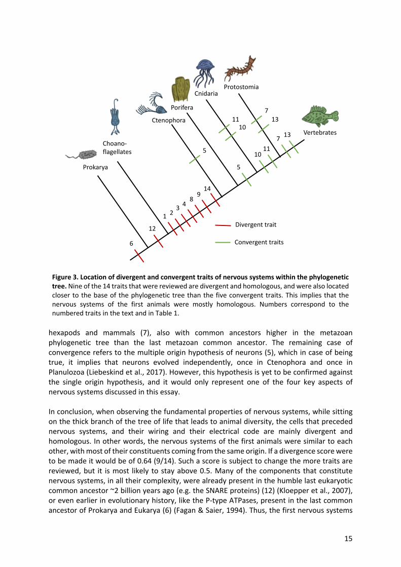
the tissues that preceded nervous systems. This section was supported by findings of comparative embryology, which suggest that the structures that gave rise to nervous systems are mainly divergent (3 divergent traits out of 3 discussed) (Arendt et al., 2015). Second, we reviewed the single origin and the multiple origin hypothesis of neurons, the former implying divergence and the later supporting convergence (Liebeskind et al., 2017). Third, we discussed how the components that gave rise to the electrical code of nervous systems have instances of both divergence and convergence (3 convergent and 3 divergent traits) (Liebeskind et al., 2017). Fourth, the wiring code and its molecular constituents were reviewed, leading to the conclusion that synapses are mainly divergent (3 divergent traits) (Ovsepian & Vesselkin, 2014). In total, 14 traits were discussed, out of which 5 were convergent (analogous) and 9 were divergent (homologous) (Table 1.). This finding suggest that with a basal perspective of the metazoan phylogeny, homology and divergence of the fundamental components of nervous systems is more likely. Moreover, the cases of convergence are more recent in evolutionary history than the cases of divergence. Two of the five cases of convergence that were discussed (10 & 11) arise when comparing cnidarians to bilaterians, whose common ancestor is more recent than the common ancestor of all metazoans, the latter being host of most of the divergent characteristics that were identified (1, 2, 3, 4, 8, 9 & 14) (Fig. 3). ). Other two cases of convergence were found while analyzing vertebrates and non-vertebrates (14), and
15
Figure 3. Location of divergent and convergent traits of nervous systems within the phylogenetic tree. Nine of the 14 traits that were reviewed are divergent and homologous, and were also located closer to the base of the phylogenetic tree than the five convergent traits. This implies that the nervous systems of the first animals were mostly homologous. Numbers correspond to the numbered traits in the text and in Table 1.
hexapods and mammals (7), also with common ancestors higher in the metazoan phylogenetic tree than the last metazoan common ancestor. The remaining case of convergence refers to the multiple origin hypothesis of neurons (5), which in case of being true, it implies that neurons evolved independently, once in Ctenophora and once in Planulozoa (Liebeskind et al., 2017). However, this hypothesis is yet to be confirmed against the single origin hypothesis, and it would only represent one of the four key aspects of nervous systems discussed in this essay. In conclusion, when observing the fundamental properties of nervous systems, while sitting on the thick branch of the tree of life that leads to animal diversity, the cells that preceded nervous systems, and their wiring and their electrical code are mainly divergent and homologous. In other words, the nervous systems of the first animals were similar to each other, with most of their constituents coming from the same origin. If a divergence score were to be made it would be of 0.64 (9/14). Such a score is subject to change the more traits are reviewed, but it is most likely to stay above 0.5. Many of the components that constitute nervous systems, in all their complexity, were already present in the humble last eukaryotic common ancestor ~2 billion years ago (e.g. the SNARE proteins) (12) (Kloepper et al., 2007), or even earlier in evolutionary history, like the P-type ATPases, present in the last common ancestor of Prokarya and Eukarya (6) (Fagan & Saier, 1994). Thus, the first nervous systems
Prokarya
Choano-flagellates
Ctenophora
Porifera
CnidariaProtostomia
Vertebrates
1 23 4
5
5
6
7
7
89
10
10
11
11
12
13
13
14
Divergent trait
Convergent traits

16
were a result of several cases of exaptation, the process of assigning a new function to a trait that is already present, rather than the evolution of completely new features (Ovsepian & Vesselkin, 2014, Liebeskind et al., 2017). Later in the evolutionary timescale, further away from the base of the animal phylogenetic tree, convergence starts to emerge ever more often: from the analogous odorant receptors in hexapods and mammals (Liebeskind et al., 2017), to more recent cases, like the convergence of cognitive tools between corvids and apes (Emery & Clayton, 2004). The cases of convergence and analogy become more and more numerous towards the thinner, taller branches of the Tree of Life, since the amount of traits increases (e.g. by gene duplications) while a limited set of selecting pressures persist. Furthermore, besides providing an answer to the main question, this essay confirms that a lower, closer to the ground position within the tree facilitates the study of convergence and divergence of nervous systems, since with 14 study cases it is possible to approach to a conclusion. Nonetheless, this privileged position has become available to us only recently through the advances in molecular biology, and it is a place still full of hypothesis, as evolutionary biology often is, that become more or less likely the more evidence emerges. A place where there is a considerable lack of fossil evidence and where definitive answers may never be reached. 8. References Abascal, F., & Zardoya, R. (2013). Evolutionary analyses of gap junction protein families. Biochimica et Biophysica Acta (BBA)-Biomembranes, 1828(1), 4-14. Acuna, C., Liu, X., & Südhof, T. C. (2016). How to make an active zone: unexpected universal functional redundancy between RIMs and RIM-BPs. Neuron, 91(4), 792-807. Arendt, D., Benito-Gutierrez, E., Brunet, T., & Marlow, H. (2015). Gastric pouches and the mucociliary sole: setting the stage for nervous system evolution. Philosophical Transactions of the Royal Society B: Biological Sciences, 370(1684), 20150286. Arendt, D., Tosches, M. A., & Marlow, H. (2016). From nerve net to nerve ring, nerve cord and brain—evolution of the nervous system. Nature Reviews Neuroscience, 17(1), 61. Asztély, F., & Gustafsson, B. (1996). Ionotropic glutamate receptors. Molecular neurobiology, 12(1), 1-11. Brebner, J., & Chittka, L. (2021). Animal Cognition: The Self-Image of a Bumblebee. Current Biology, 31(4), R207-R209. Burkhardt, P. (2015). The origin and evolution of synaptic proteins–choanoflagellates lead the way. Journal of Experimental Biology, 218(4), 506-514. Burkhardt, P., Grønborg, M., McDonald, K., Sulur, T., Wang, Q., & King, N. (2014). Evolutionary insights into premetazoan functions of the neuronal protein homer. Molecular biology and evolution, 31(9), 2342-2355.

17
Burkhardt, P., Stegmann, C. M., Cooper, B., Kloepper, T. H., Imig, C., Varoqueaux, F., ... & Fasshauer, D. (2011). Primordial neurosecretory apparatus identified in the choanoflagellate Monosiga brevicollis. Proceedings of the National Academy of Sciences, 108(37), 15264-15269. Cronin, T. W., Bok, M. J., Marshall, N. J., & Caldwell, R. L. (2014). Filtering and polychromatic vision in mantis shrimps: themes in visible and ultraviolet vision. Philosophical Transactions of the Royal Society B: Biological Sciences, 369(1636), 20130032. Dahl, G., & Muller, K. J. (2014). Innexin and pannexin channels and their signaling. FEBS letters, 588(8), 1396-1402. Dermietzel, R., & Spray, D. C. (2016). Gap junctions and electric synapses. In Neuroscience in the 21st Century: From Basic to Clinical, Second Edition (pp. 511-546). Springer New York. Dunn, C. W., Giribet, G., Edgecombe, G. D., & Hejnol, A. (2014). Animal phylogeny and its evolutionary implications. Annual review of ecology, evolution, and systematics, 45, 371-395. Eccles, J. C. (1964). The development of ideas on the synapse. In The Physiology of Synapses (pp. 1-10). Springer, Berlin, Heidelberg. Eckert, R. (1965). Bioelectric control of bioluminescence in the dinoflagellate Noctiluca: I. Specific nature of triggering events. Science, 1140-1142. Elias, L. A., & Kriegstein, A. R. (2008). Gap junctions: multifaceted regulators of embryonic cortical development. Trends in neurosciences, 31(5), 243-250. Emery, N. J., & Clayton, N. S. (2004). The mentality of crows: convergent evolution of intelligence in corvids and apes. science, 306(5703), 1903-1907. Fagan, M. J., & Saier, M. H. (1994). P-type ATPases of eukaryotes and bacteria: sequence analyses and construction of phylogenetic trees. Journal of molecular evolution, 38(1), 57-99. Feuda, R., Hamilton, S. C., McInerney, J. O., & Pisani, D. (2012). Metazoan opsin evolution reveals a simple route to animal vision. Proceedings of the National Academy of Sciences, 109(46), 18868-18872. Gundelfinger, E. D., Reissner, C., & Garner, C. C. (2016). Role of bassoon and piccolo in assembly and molecular organization of the active zone. Frontiers in synaptic neuroscience, 7, 19. Hille, B. (2001). Ion Channels of Excitable Membranes (Sinauer, Sunderland, MA), 3rd. Kajiura, S. M. (2001). Head morphology and electrosensory pore distribution of carcharhinid and sphyrnid sharks. Environmental Biology of Fishes, 61(2), 125-133. Khurana, G. K., Vishwakarma, P., Puri, N., & Lynn, A. M. (2018). Phylogenetic Analysis of the vesicular fusion SNARE machinery revealing its functional divergence across Eukaryotes. Bioinformation, 14(7), 361. Kloepper, T. H., Kienle, C. N., & Fasshauer, D. (2007). An elaborate classification of SNARE proteins sheds light on the conservation of the eukaryotic endomembrane system. Molecular biology of the cell, 18(9), 3463-3471.

18
Koumandou, V. L., Wickstead, B., Ginger, M. L., Van Der Giezen, M., Dacks, J. B., & Field, M. C. (2013). Molecular paleontology and complexity in the last eukaryotic common ancestor. Critical reviews in biochemistry and molecular biology, 48(4), 373-396. Laughlin, S. B., van Steveninck, R. R. D. R., & Anderson, J. C. (1998). The metabolic cost of neural information. Nature neuroscience, 1(1), 36-41. Liebeskind, B. J., Hofmann, H. A., Hillis, D. M., & Zakon, H. H. (2017). Evolution of animal neural systems. Annual review of ecology, evolution, and systematics, 48, 377-398. Liegertová, M., Pergner, J., Kozmiková, I., Fabian, P., Pombinho, A. R., Strnad, H., ... & Kozmik, Z. (2015). Cubozoan genome illuminates functional diversification of opsins and photoreceptor evolution. Scientific reports, 5, 11885. Lynagh, T., Beech, R. N., Lalande, M. J., Keller, K., Cromer, B. A., Wolstenholme, A. J., & Laube, B. (2015). Molecular basis for convergent evolution of glutamate recognition by pentameric ligand-gated ion channels. Scientific reports, 5(1), 1-8. Marlow, H., & Arendt, D. (2014). Evolution: ctenophore genomes and the origin of neurons. Current Biology, 24(16), R757-R761. Martinson, A. S., Van Rossum, D. B., Diatta, F. H., Layden, M. J., Rhodes, S. A., Martindale, M. Q., & Jegla, T. (2014). Functional evolution of Erg potassium channel gating reveals an ancient origin for IKr. Proceedings of the National Academy of Sciences, 111(15), 5712-5717. Menzel, R., & Giurfa, M. (2001). Cognitive architecture of a mini-brain: the honeybee. Trends in cognitive sciences, 5(2), 62-71. Mukherjee, K., Yang, X., Gerber, S. H., Kwon, H. B., Ho, A., Castillo, P. E., ... & Südhof, T. C. (2010). Piccolo and bassoon maintain synaptic vesicle clustering without directly participating in vesicle exocytosis. Proceedings of the National Academy of Sciences, 107(14), 6504-6509. Nakanishi, N., Sogabe, S., & Degnan, B. M. (2014). Evolutionary origin of gastrulation: insights from sponge development. BMC biology, 12(1), 1-9. Nevo, E. (1979). Adaptive convergence and divergence of subterranean mammals. Annual review of ecology and systematics, 10(1), 269-308. Nielsen, C. (2019). Early animal evolution: a morphologist's view. Royal Society open science, 6(7), 190638. Ovsepian, S. V., & Vesselkin, N. P. (2014). Wiring prior to firing: the evolutionary rise of electrical and chemical modes of synaptic transmission. Reviews in the Neurosciences, 25(6), 821-832. Panchin, Y. V. (2005). Evolution of gap junction proteins–the pannexin alternative. Journal of Experimental Biology, 208(8), 1415-1419. Pereda, A. E. (2014). Electrical synapses and their functional interactions with chemical synapses. Nature Reviews Neuroscience, 15(4), 250-263.

19
Prindle, A., Liu, J., Asally, M., Ly, S., Garcia-Ojalvo, J., & Süel, G. M. (2015). Ion channels enable electrical communication in bacterial communities. nature, 527(7576), 59-63. Ramos-Vicente, D., Ji, J., Gratacòs-Batlle, E., Gou, G., Reig-Viader, R., Luís, J., ... & Escriva, H. (2018). Metazoan evolution of glutamate receptors reveals unreported phylogenetic groups and divergent lineage-specific events. Elife, 7, e35774. Ryan, J. F., Pang, K., Schnitzler, C. E., Nguyen, A. D., Moreland, R. T., Simmons, D. K., ... & Putnam, N. H. (2013). The genome of the ctenophore Mnemiopsis leidyi and its implications for cell type evolution. Science, 342(6164). Salanova, M., Volpe, P., & Blottner, D. (2013). Homer protein family regulation in skeletal muscle and neuromuscular adaptation. IUBMB life, 65(9), 769-776. Sebe-Pedros, A., Degnan, B. M., & Ruiz-Trillo, I. (2017). The origin of Metazoa: a unicellular perspective. Nature Reviews Genetics, 18(8), 498. Sebé-Pedrós, A., de Mendoza, A., Lang, B. F., Degnan, B. M., & Ruiz-Trillo, I. (2011). Unexpected repertoire of metazoan transcription factors in the unicellular holozoan Capsaspora owczarzaki. Molecular biology and evolution, 28(3), 1241-1254. Simionato, E., Ledent, V., Richards, G., Thomas-Chollier, M., Kerner, P., Coornaert, D., ... & Vervoort, M. (2007). Origin and diversification of the basic helix-loop-helix gene family in metazoans: insights from comparative genomics. BMC evolutionary biology, 7(1), 1-18. Sperling, E. A., & Vinther, J. (2010). A placozoan affinity for Dickinsonia and the evolution of late Proterozoic metazoan feeding modes. Evolution & development, 12(2), 201-209. Thever, M. D., & Saier, M. H. (2009). Bioinformatic characterization of p-type ATPases encoded within the fully sequenced genomes of 26 eukaryotes. Journal of Membrane Biology, 229(3), 115-130. Volkov, Alexander G., Tejumade Adesina, and Emil Jovanov. "Closing of Venus flytrap by electrical stimulation of motor cells." Plant signaling & behavior 2.3 (2007): 139-145. Xiao, B., Tu, J. C., & Worley, P. F. (2000). Homer: a link between neural activity and glutamate receptor function. Current opinion in neurobiology, 10(3), 370-374.