Questions and Answers - CiteSeerX
250
TUBERCULOSIS CASE-FINDING AND CHEMOTHERAPY Questions and Answers K. TOMAN • WORLD HEALTH ORGANIZATION GENEVA 1979
-
Upload
khangminh22 -
Category
Documents
-
view
0 -
download
0
Transcript of Questions and Answers - CiteSeerX

TUBERCULOSIS CASE-FINDING
AND CHEMOTHERAPY
Questions and Answers
K. TOMAN
• WORLD HEALTH ORGANIZATION GENEVA
1979

ISBN 92 4 154136 9
<0 World Health Organization 1979
Publications of the World Health Organization enjoy copyright protection in accordance with the provisions of Protocol 2 of the Universal Copyright Convention. For rights of reproduction or translation of WHO publications, in part or in toto, application should be made to the Office of Publications, World Health Organization, Geneva, Swit:r..erland. The World Health Organization welcomes such applications.
The designations employed and the presentation of the material in this publication do not imply the expression of any opinion whatsoever on the part of the Secretariat ofthe World Health Organization concerning the legal status ofany country, territory, city or area or of its authorities, or concerning the delimitation of its frontiers or boundaries.
The mention of specific companies or of certain manufacturers' products does not imply that they are endorsed or recommended by the World Health Organization in preference to others of a similar nature that are not mentioned. Errors and omissions excepted. the names of proprietary products are distinguished by initial capital letters.
The author alone is responsible for the views expressed in this publication.
PRINTI!D IN SWITZERLAND

Contents
Preface by the Director-General of WHO
Acknowledgements
Introduction .
What is the role of case-finding in tuberculosis control 'I What is a case of tuberculosis 'I . . . . • . . . . . .
How many bacUlI are present in a sputum specimen found positive by smear
Page
ix
x
xi
3 4
microscopy 'I. . . . . . . . . . . . . . . . . . . . . . . . .. 6 What are the main causes of a false positive or false negative sputum smear 'I 9
How reliable is smear microscopy 'I . . . . . • . • . . . . • . . .. 14 What are the advantages and disadvantages of fluorescence microscopy'l . 23 How reliable is chest radiography 'I • • • • • • • • • • • • . • • •. 28 What is the probability of obtaining a negative culture from a sputum
specimen found positive by smear microscopy 'I. • . . . . . . . .. 38 What is the additional case yield from repeated sputum examinations by
smear microscopy and culture 'I. . . . . . . . . . . . . . . . .. 40 What are the relative merits of chest X-ray and sputum examination (smear
microscopy and culture) in case-finding among new outpatients with prolonged chest symptoms 'I . . . . . . . . . . . . . . . . • .. 44
What is the clinical and epidemiological significance of consistent negativity by smear microscopy in patients positive by culture only 'I • • . • .. SO
How does pulmonary tuberculosis develop and how can it be detected at an early stage 'I. . . . • . • . . . . . . . . . . • . . . . . .• 57
What is the role of case.finding by periodiC mass X-ray examination in tuberculosis control 'I • . . . . . . . . . • • . . • . • . . . .. 65
Chemotherapy
What were the main landmarks in the development of tuberculosis chemo-therapy 'I . • . . . . • . . . . . . . • • . . . . . . . . 75
What is the biological mechanism of chemotherapy 'I. . . . . . . 77 What is the role of the host factor in the treatment of tuberculosis 'I 81 How does drug resistance develop 'I . . . . . . . • 84 What is primary and what is initial drug resistance? . 87 What is natural drug resistance? . . . . 88 What is transient drug resistance '1. . . . . . . . . 89 What is the "fall and rise .. phenomenon? . . . . . 91 How many drug-resistant tubercle bacUli can be found in the sputum of
patients who have never received tuberculosis chemotherapy '1. . .. 93 Is primary drug resistance a new menace to the control of tuberculosis '1 97
v

What is the therapeutic effect and what is the toxicity of anti tuberculosis drugs? . • . • . . . • . • • . . • . . . . • • . • . • 101
What is the dosage of drugs in daily and intermittent regimens? . . .. 112
What are the standard regimens currently used ? • . . . . . . . . .. 113
What are the merits of thioacetazone as a companion drug to isoniazid, and what is the efficacy of the standard regimen isoniazid plus thio--acetazone 'I • . . . . . . . . . . . . . . • . . . . . . . . .. 116
What is the frequency of adverse reactions against thioacetazone, and what is the geographical distribution of natural resistance to the drug'l .. 119
What were the main findings of the Madras study comparing home and sanatorium treatment'1 • • • • • • • • • • • • • • • . . . . .. 122
What are two-phase chemotherapy and the so-called 100% regimens 'I. 130
What is intermittent chemotherapy and what is the scientific basis for intermittency 'I. . . . . . . . . . • • . . . . . . . • • . . .. 132
What was the efficacy of primary intermittent chemotherapy in controlled clinical studies 'I • . . . . . . . . • • • • . • • . . . • • . .. 137
What is the purpose of the initial intensive phase of two-phase chemo--therapy 'I • • . • • • • . • . . • • • • . • • • • . . . . . .. 145
What is rapid inactivation (acetylation) of isoniazid and what is its clinical significance 'I. . . . . • . . . • . . . . . . . . . . . . . . .. 148
When is a two-phase regimen indicated 'I . . . . . . . . . . . . . .. 151
What is the optimum duration of conventional long-term chemotherapy 'I 153
What is the optimum duration of the initial intensive phase of two--phase regimens '1. • . • • • • . • • • • • • • . . • • . . . . . . .. 156
What are the merits of supervised intermittent chemotherapy and self-administered daily chemotherapy? . . . • . . . . . . . . . . .. 161
What reserve regimens are available and what is their place in tuberculosis control programmes'1. . . • . . . . . . . . . . . . . . . . .. 163
How relevant are initial drug resistance and pretreatment susceptibility tests to the selection of chemotherapy regimens 'I . . . . . . . . .. 167
How reliable are drug susceptibility tests in routine practice 'I . . . . .. 173
What are the possible consequences of inaccurate drug-sensitivity testing 'I 175
When does chemotherapy fail 'I . . . . . . . . . . . . . . . . . .. 177
How can the progress of treatment be monitored and treatment results be assessed '1 . . . . . . . . . . . . . . . . . . . . . . . . . .. 179
How effective is short-course chemotherapy and what are its prospects? 183
What are the bactericidal and steri1izing mechanisms of short-course regimens '1. . . . . . . • . . . . . . . . . . . . . . . . . .. 200
What is bacterial persistence and how does it affect the chemotherapy of tuberculosis '1 . . . . • • . . . . . . . . • . . . . . • . . .. 204
What are the most usual signs of drug hypersensitivity and procedures of desensitization '1 . . . . 206
What are the keys to cure '1. . 208
- VI-

What is the significance of default in the chemotherapy of tuberculosis 'I 211
What is the place of sanatorium and hospital treatment today, and how infectious are tuberculosis patients under chemotherapy '! . . • • •. 217
How important is follow-up and what is the frequency of relapse after the completion of treatment 'I. . • . . . . . . . • • • • • • • • •• 223
What are the principles and requirements of a controlled chemotherapy trial '!. . . . . • . . . . . . . . . . . . . . . • • . • • • .. 226
- vii

1
1
1
1
1
1
1
1
1
1
1
1
1
1
1
1
1
1
1
1
1
1
1
1
1
1
1
1
1
1
1
1
1

PREFACE
One of the basic functions of the World Health Organization is the international transfer of scientific knowledge of direct practical value to countries in solving their health problems. A vast store of knowledge and experience has been accumulated in tuberculosis control. Through WHOassisted projects, simplified and largely standardized control methods have been developed for general use, even in the remotest rural areas of developing countries. The concept of the" national tuberculosis control programme" was formulated by WHO to enable the new technology to be applied effectively. The Organization's policy on tuberculosis control, contained mainly in concisely worded reports of the WHO Expert Committee on Tuberculosis, has given rise to a great many questions and requests for further information. It has long been thought, therefore, that a detailed commentary on the Scientific know ledge and practical experience underlying WHO's tuberculosis control policy would be a valuable element of WHO's technical cooperation with Member States. This book, presented in the form of questions and answers, is afirst step in that direction. I hope that it will reach all tuberculosis workers in key positions, the organizers and administrators responsible for tuberculosis control in national programmes, and the field staff concerned with the day-to-day problems of tuberculosis control in the community. The book is also directed towards those who teach about tuberculosis control in medical schools, schools of public health, nursing schools, and similar institutions.
H. MAHLER
Director-General
ix-

ACKNOWLEDGEMENTS
I am indebted to all those who, directly or indirectly, have made it possible for me to write this book.
I owe much to Or H. Mahler, who ten years ago conceived the idea of a technical reference manual on tuberculosis control, mainly for nonspecialized health personnel in the developing countries.
Grateful thanks are due to the International Union against Tuberculosis (IUAT). Its former director, Or J. Holm, and his successor, Or D. R. Thomson, took the first steps towards the realization of this book and helped in its technical editing; the present director of IUAT, Professor V. Farga, made helpful suggestions. Or Annik Rouillon, in her various areas of responsibility, gave whole-hearted cooperation. I had stimulating, candid, and fruitful discussions with the late Professor G. Canetti, Chairman of the IUAT Scientific Committees, his successor, Or J. R. Bignall, and the present Chairman of the committees and Director of the Tuberculosis Surveillance Research Unit, Or K. Stjblo. Thanks to the lively interest taken by Or J. Meijer and the initiative of Or H. A. van Geuns, the Sonnevanck Foundation, Netherlands, generously met part of the expenses. Or K. L. Hitze, Chief, Tuberculosis and Respiratory Infections, World Health Organization, lent his active support, counsel and encouragement.
Or Wallace Fox, Director, Tuberculosis and Chest Diseases Research Unit, Medical Research Council, and Professor D. A. Mitchison, Postgraduate Medical School, Hammersmith, London, who have contributed decisively to the fundamental changes in the treatment of tuberculosis, are to be thanked for their interest and criticism, and for allowing me to draw heavily on their pioneering studies.
Acknowledgements are due to my co-workers and students in developing countries-physicians, health officers, auxiliary workers, educators, and community leaders determined to free their fellow men from unnecessary suffering-who made me realize that tuberculosis and many other health problems can be eliminated only when their cultural, social, and economic interdependence has been understood.
I am grateful to my wife. Without her help and forbearance, this book could not have been written.
-x-

INTRODUCTION
Such has been the progress made in the epidemiology, the prevention, and especially the treatment of tuberculosis during the past three decades that this age-old killer of man has at last become a preventable and curable disease.
Through systematic research, it has been possible to develop an effective and simple technology that is inexpensive and amenable to standardization. As a result, the control of tuberculosis has ceased to be a primarily technical problem, requiring rare and highly specialized skills for its solution. Instead of only the privileged few who could be served in the past by the small number of specialists available, anyone suffering from tuberculosis or exposed to it may now enjoy the benefits of this technology, provided that it has been adapted to the requirements of a country's health programme. However, even within such a framework, the efficient operation of a tuberculosis programme may encounter many difficulties. There is no uniform solution to existing or future problems. Each country will have to find the one that will suit its own needs best. Yet the multitude of possible solutions will all have one feature in common: the participation of a rapidly growing number of nonspecialized people, including physicians, nurses, technicians, all categories of field staff and health administrators, members of non4
governmental agencies and international voluntary organizations, and interested groups of citizens. In view of the wide range of persons whom tuberculosis control now concerns, the author has attempted to direct this book to a broad spectrum of readers rather than to the specialized tuberculosis worker.
The dramatic turn taken by the fight against tuberculosis was reflected in the last two reports of the WHO Expert Committee on Tuberculosis. In 1964 the eighth report presented, for the first time, the concept of a national tuberculosis programme based on a simplified technology.l Ten years later, the ninth report re-emphasized the importance of this concept, stated the policies of the Expert Committee, and made recommendations for their implementation.lI This report, which was very widely distributed, serves as the principal background document at many international and national conferences, seminars, and training courses organized by
1 WHO Technical Report Series, No. 290, 1964 (Eighth report of the WHO Expert Committee on Tuberculosis).
11 WHO Technical Report Series, No. 552, 1974 (Ninth report of the WHO Expert Committee on Tuberculosis).
-xi-

INTRODUCTION
WHO and the International Union against Tuberculosis (lUAT), On such occasions, a rather sceptical attitude has sometimes been observed on the part of a few persons-mostly specialists-towards the new approach. This small but influential group have a deep-rooted if bonafide belief in the omnipotence of sophisticated technology. They distrust inexpensive and apparently less-than-perfect procedures that can be used by nonspecialized personnel, even though these procedures have a solid scientific basis and have proved their worth both in controlled trials and in regular practice.
Some of the recommendations of the WHO Expert Committee, especially those conflicting with time-honoured traditions and working methods, have prompted many questions. a large number of which recur with almost predictable regularity. Both WHO and IUAT receive letters demanding ready answers to these questions. For that reason, the two organizations requested the author to prepare the present volume in order to satisfy the demand for more detailed information on the scientific basis of the modern tuberculosis control policy that is recommended in the ninth report.
The information given on any particular subject is far from exhaustive. The aim was not completeness but deliberate selection. From among the numerous questions that are asked, those that recur the most frequently and that appear to be the most pertinent have been chosen. The question-and-answer formula seemed to be the most appropriate way of presenting the material. The numerous cross-references largely offset the inevitable overlap of some of the questions and answers, sparing the reader from sifting through unwanted information irrelevant to his problem. If this book or the ninth report of the WHO Expert Committee does not give the reader satisfaction on a particular question or problem he may have, he is welcome to consult WHO 1 or IUAT.2
This book deals only with pulmonary tuberculosis, because that is the most common form of the disease and practically the only one that is responsible for transmission from man to man. Special attention has been devoted to the needs of the developing countries, in which more than three-quarters of the world's tuberculosis is concentrated.
1 Tuberculosis and Respiratory Infections, World Health Organization, 1211 Geneva 27, Switzerland.
2 International Union against Tuberculosis, 3 rue Georges-ViIle, F-75116 Paris, France.
xii -

CASE-FINDING

1 1 1 1 1 1 1 1 1 1 1 1 1 1 1 1 1 1 1 1 1 1 1 1 1 1 1 1 1 1 1 1 1 1 1 1 1 1 1 1 1 1 1 1 1 1 1 1 1 1 1 1 1 1 1 1 1 1 1 1 1 1 1 I

CASE-FINDING
What is the role of case-finding in tuberculosis control?
Case-finding is an essential component of the control of tuberculosis and most other communicable diseases. Its object is to identify the sources of infection in a community-e.g., in the case of tuberculosis, to find persons who are discharging tubercle bacilli. By rendering them noninfectious through chemotherapy, the chain of transmission of tubercle bacilli from man to man is cut. Thus case-finding in itself has little purpose unless it is followed by chemotherapy. That is why these two activities should be regarded as a single functional entity (1).
However, to find cases is always easier than to treat them successfully. Thus case-finding activities sometimes outstrip the treatment capacity of a tuberculosis service, owing to the lack of personnel or drugs, or to organizational difficulties. But to search for cases without being able to treat them properly after they have been found is irrational-it is even harmful, because it increases patients' suffering and undermines confidence in the health care system.
In countries where newly detected cases are not yet treated satisfactorily, resources and efforts should be directed towards improving primary treatment, rather than expanding case-finding. Intensification of case-finding is justifiable only when it is certain that every new case found will be provided with adequate treatment (1).
REFERENCE
1. WHO Technical Report Series, No. 552, 1974 (Ninth report of the WHO Expert Committee on Tuberculosis), pp. 14 & 25

4 K. TOMAN
What is a case of tuberculosis ?
Much time has been spent in the past on defining the object of casefinding. National and international committees on diagnosis and classification have had lengthy arguments about what should be considered to be a case of tuberculosis, and numerous attempts have been made to formulate a generally valid definition. However, in spite of the efforts made, none of the proposed classifications and definitions has been universally agreed upon, and each has soon been replaced by a new one. The reasons for this lack of agreement are manifold.
In the past, chest physicians were split up into a number of groups representing different schools and occasionally contradictory pathogenetic concepts. The various diagnostic schemes and classifications were based sometimes on quantitative criteria, such as the extent oflung involvement. and sometimes on qualitative pathological features-e.g., exudative, caseous, and productive lesions. Some preferred the pathomorphological nomenclature used in radiological classifications; others introduced immunological concepts differentiating various stages of the disease; many clinicians tried an eclectic synthesis by combining various schemes, placing emphasis on one or another aspect according to their own judgement.
However, what seemed to be important to the pathologist-e.g., the presence of specific granulomatous tissue, giant cells, or caseation-was of little relevance to the clinician and even less to the epidemiologist or the bacteriologist, for whom the transmission of infection and the demonstration of tubercle bacilli were essential. And whereas the radiologist is compelled to depend on the morphology of X-ray shadows, a paediatrician may consider an unvaccinated baby (of a tuberculous mother) with a significant tuberculin reaction as a .. case" oftuberculosis, irrespective of other criteria. To the health officer, who has to plan and provide services, even a person who has been cured of tuberculosis but is suffering from some late effects of the disease may be a "case". In short. each medical branch has its specific working methods and objectives, and therefore also its own criteria.
To formulate a definition of a .. case" that would cover all the abovementioned aspects would not be impossible, since it would be a descriptive rather than an imaginative task. However, such a definition would be voluminous and academic, and thus probably of little practical use. The usefulness of a definition that is meant to serve as a basis for action -a working definition-is determined by its practical applicability, not by the degree of its completeness. A working definition must be judged in the light of its stated purpose. Therefore there is little sense in arguing

TUBERCULOSIS CASE-FINDING 5
to what extent such a definition is right: what is relevant is whether it is applicable-and whether it contributes to meeting the agreed purpose, which, in the given instance, is tuberculosis control.
Tuberculosis control, like the control of any communicable disease, aims to prevent the transmission of infection. The aim of case-finding in tuberculosis control is to identify the sources of infection in a community-Le., persons who transmit infection with tubercle bacilli. Consequently, for the purpose of tuberculosis control, a .. case" is an individual discharging tubercle bacilli.
Since the introduction of chemotherapy, the issue has become clearer and much of the argument about what is or is not a case is now mainly of academic interest. The chemotherapy of tuberculosis is antibacterial treatment acting chiefly against the tubercle bacillus. Hence,. ex~r.~t~rs of tubercle bacilli are a clear priority for chemotherapy and are thus the mget of case-ftnamg.

6 K. TOMAN
How many bacilli are present in a sputum specimen found positive by smear. microscopy ?
If a smear is properly prepared, it is likely that the number of bacilli contained in it will be related to the concentration of bacilli in the sputum. This numerical relationship, which has been investigated· by many authors (1-4), may be illustrated by the following example.
The amount of sputum on a slide for smear preparations is about 0.01 ml a-roughly the quantity of concentrated sputum delivered by a wire loop with an internal diameter of 3 mm (Fig. I). This sputum is spread over an area of 200 mm2 (10 X 20 mm). Since the area of an oilimmersion field seen in the microscope is about 0.02 mm2 , 10 000 such fields need to be screened in order to examine the whole smear./) If 100 oil-immersion fields are examined, only I % of the smear is screened.
Fig. 1
Wire loop and slide used for the preparation of sputum smears
Wire 100p (internal
diameter: 3 mm)
20mm
J
I ---f _ 10mm
-t WHO 78007
Thus, if a sputum specimen contains about 5000 bacilli per millilitre, the whole smear (if prepared as described) will contain about 50 bacilli. If these 50 bacilli were evenly distributed over the 10 000 fields of the
a Certain investigators estimate the amount to be only 0.003 ml. /) At a magnification of lOOOx - i.e., lOOx for the oil-immersion objective
lens and 10x for the eye-piece. (The size of a field in fluorescence microscopy is about IS times as large with an objective of 25x and an eye-piece of 10x.) By examining one length (20 mm) of a smear, about 100-120 microscopic fields are screened (the diameter of a field at the above-mentioned magnification being 0.16-0.2 mm).

TUBERCULOSIS CASE-FINDING 7
smear, there would be 1 bacillus in 200 fields. By examining 100 fields there is a 50% chance of finding this bacillus. In order to find at least 3 acid-fast bacilli (usually recommended as the minimum number for a smear to be reported as positive), about 600 fields would have to be screened. By examining 300 fields, there is an approximately 50% chance of finding 3 bacilli (5-7).
Under the same assumption-i.e., an even distribution of acid-fast bacilli in the specimen and smear-to find 1 acid-fast bacillus in every 10 fields (or 10 in 100 fields) would require 1000 such bacilli to be present in the smear (10 000 fields) or 100 000 (IOS) per millilitre of sputum (Table 1). To find 1 acid-fast bacillus per field on the average would require 106 bacilli per millilitre of sputum (Table 1). Thus a specimen
Table 1
Estimated numbers of acid-fast bacilli In sputum specimens and probable numbers of bacilli in smears (estimated minimum values)
No. of oil-immersion fields per bacillus
100 10 1
No. of bacilli per smear
100 1000
10000
No. of bacilli per ml of specimen
10000 100000
1000000
that is consistently found to be positive would have to contain at least 100 000 acid-fast bacilli per millilitre. These estimations have been made under the assumption that the bacilli are evenly dispersed throughout the specimen-Le., that each loop of material taken from the specimen will contain the same number of acid-fast bacilli spread evenly over the whole smear. However, from experience and from experiments it is known that bacilli are not evenly dispersed in a specimen, but are frequently found in clumps.a Thus, when several samples are taken from a sputum specimen, the number of bacilli will vary from one sample to another. Nevertheless, when special culture techniques were used to compare the number of bacilli in large numbers of samples taken from different sputum specimens, certain important observations could be made. In particular, the numbers of colonies cultured from samples taken from the same specimen differed from each other only within certain limits-i.e., not at random (see" How reliable is smear microscopy? "-page 14). Likewise, variations in colony counts among samples from different specimens did not occur randomly, but were due to differing concentrations of acid-fast bacilli in the specimens. In spite
a Bacilli in a smear tend to follow a Poisson distribution. like red blood corpuscles in a haematocytometer.

8 K. TOMAN
of the considerable sampling error, it may be concluded that the amount of bacilli in the smear corresponds fairly closely to the concentration of bacilli in the sputum (4). These experiments also showed that, below a certain concentration of bacilli in a sputum specimen, the probability that acid-fast bacilli will be transferred from the specimen to the smear and found by microscopy approaches zero.
REFERENCES
1. CARVALHO, A. Zeitschrlft flir Tuberkulose und Erkrankungen der ThortvCorgane, 63: 305 (1932)
2. CRUlKSHANK. P. B. Bacteriology of tuberculosis. In: Sellors, T. H. & Livingstone, J. L.. ed. Modern practice of tuberculosis. London, Butterworth, 1952
3. HOBBY, G. L. ET AL. Antimicrobial agents and chemotherapy, 4: 94 (1973)
4. DAVID, H. L. Bacteriology of the mycobacterioses. Atlanta, US Department of Health, Education, & Welfare, Communicable Disease Centre, 1976, p. 147
5. SMITHWlCK, R. W. Laboratory manual for aCid-fast microscopy. Atlanta, US Department of Health, Education, & Welfare, Public Health Service, 1976
6. INTERNATIONAL UNION AGAINST TUBERCULOSIS. Technical guide for examinations for tuberculosis by direct microscopy. Paris, 1977, p. 19
7. AMERICAN THORACIC SoCIETY. SCIENTIFIC AssEMBLY ON TUBERCULOSIS. Diagnostic standards and classification of tuberculosis and other mycobacterial diseases. New York, American Lung Association, 1974

TUBERCUWSIS CASE_FIlING 9
What are the main causes of a false itive or false negative sputum smear ?
False positive results
Acid-fast particles other than tubercle bacilli
Occasionally, a sputum specimen or smear may contain particles that are acid-fast-Le., when treated with the Ziehl-Neelsen method, they retain their red stain (carbolfuchsin) and resist decolorization with acidalcohol. These red particles may sometimes resemble tubercle bacilli. They include certain food particles (e.g., waxes, oils), precipitates, other micro-organisms, inorganic materials, and artifacts (1-6).
Food particles. To eliminate these, the patient should rinse his mouth with pure water and clean his teeth (without using tooth-paste or disinfectant) before producing the sputum specimen. It is even better if the patient produces the specimen before breakfast or on an empty stomach.
Precipitated stains. Though these are quite easy to differentiate from acid-fast bacilli, they may hamper reading or occasionally mislead an inexperienced microscopist. Precipitates can be removed by filtration of staining solutions. However, it is safer to use freshly prepared solutions, filled into carefully cleaned bottles, rather than stale staining solutions.
Saprophytic aCid-fast bacilli. These occur in soil and water, and may occasionally get into the specimen or smear during processing. This can be avoided by using distilled or boiled water from scrupulously clean containers.
Mycobacterium kansasii or Nocardia species. These occasionally 0c
cur in specimens. When they cause pulmonary disease, they are usually present in large numbers.
Spores of Bacillus subtilis. These are very rare, mostly of ovoid shape, and larger than tubercle bacillL
Fibres and pollens. Fibres, including those of wool, cotton, filter paper, and bamboo, usually occur singly, most often in only one microscopic field. The pollen of certain pine trees is seen as short, coccoid rods occurring very rarely in specimens.
Scratches on the slide. Scratches may sometimes retain the red stain and confuse beginners. They are usually seen in parallel rows, are generally longer than acid-fast bacilli, and are undulated. They can be identified easily, because they are found in a deeper layer on the slide, below the smear, disappearing when the cells (e.g., leucocytes) in the smear are focused on.

lO K. TOMAN
Contamination through the transfer of bacilli from one smear to another
It may happen that acid-fast bacilli are transferred accidentally from a positive slide to a negative one, when several slides are treated simultaneously in staining or decolorization tanks. This can be avoided by processing each slide separately-e.g., on a rack. Such racks are usually made of wire and can be decontaminated easily by flaming.
Acid-fast bacilli may also be transferred accidentally when the glass rod or dropper used for placing immersion oil on the slide touches the surface of a positive slide and rubs off some of the material. The same can happen when blotting paper is used for drying several stained smears consecutively. Therefore blotting paper should not be used at all, or for no more than one slide. The oil dropper should not touch the smear, and the oil should be allowed to drip freely on to the slide. For the same reason, the surface of the slide should not be rubbed with the oil immersion objective. Before a new smear is examined, the oil should be wiped off the lens with a piece of clean cotton tissue or, even better, with speciallens-cleaning paper.
When microscopy is used for the detection of acid-fast bacilli, slides should never be used more than once.
False negative results
False negative results (1-6) are commonly due to deficiencies in the preparation of the smear, in staining, and in scanning. Adequate collection of the specimen and subsequent selection of sputum particles are essential to the preparation of a smear and should receive special attention.
Deficiencies leading to false negative results include the following:
Inadequate sputum collection
The patient is sometimes not told clearly enough what constitutes a proper sputum specimen and how he should produce one. It must be made clear to him that saliva and nasopharyngeal discharge are unsuitable for examination. Patients should be encouraged and given time to produce bronchial sputum from the" depths of the chest". If repeated attempts have failed, tickling of the inner surface of the epiglottis or trachea with a swab, or intratracheal instillation of 5-lO m! of cool saline or sterile water may provoke a vigorous cough with sputum. Other techniques to stimulate the production of sputum, such as aerosol induction, gastric aspiration, and bronchoscopy, require more complex equipment or special skills.
If a patient discharges acid-fast bacilli in his sputum, these are more likely to be found in a specimen produced in the early morning than in

TUBERCULOSIS CASE-FINDING 11
one produced later in the day. If early-morning sputum is required. the patient should be given a container and instructed to place in it the very first sputum he produces in the morning. before breakfast and before taking any medicaments.
Inadequate storage of sputum specimens and stained smears Acid-fast bacilli may lose their acid-fastness as a result of exposure
of the specimen to direct sunlight, radiation (e.g .• ultraviolet light). excessive heat, or storage for more than a week in hot and dry conditions.
If Ziehl-Neelsen-stained smears have to be stored for re-examination, the immersion oil must be washed from the smears with xylol because it removes the stain from the acid-fast bacilli.
Fluorochrome-stained smears will lose their fluorescence with storage.
Failure to select suitable sputum particles for smear preparation Tubercle bacilli are most likely to be found in little blobs (0< lentils ")
of greenish-grey or yellowish matter of a thick, creamy consistency. (Such blobs usually consist of dead caseous tissue eliminated from a cavity in the lung.) If the sputum is not treated by a special concentration procedure involving centrifugation, these blobs have to be carefully separated from the rest of the sputum and transferred to a slide. They can be seen more easily in the sputum against a dark background.
Inadequate preparation of smears or staining of slides
False negative results may be obtained also when:
(a) too little material has been spread on the slide, so that the smear is too thin;
(b) the smear is too thick, so that sufficient light cannot pass through it;
(c) the slide has been over-heated when fixing the smear; (d) the smear has not been sufficiently fixed and parts of the material
have been washed off; (e) the staining with carbolfuchsin was too short or was overdone
by boiling; or (f) the counterstaining was too intensive, so that the acid-fast bacilli
have been obscured.
Inadequate examination of the smear
If the scanning is done erratically or too briefly, too few fields may be examined. (Occasionally the examiner is unable to distinguish the red-stained acid-fast bacilli because of colour blindness or other visual disturbances.)

12 K. TOMAN
Other reasom for false results
Administrative errors
Such errors may include:
(a) misidentification of patients, misspelling of names, or confusion of names or of code numbers of specimens and slides;
(b) mistakes in labelling containers; and (c) false recording or reporting.
Reading errors
Reader or observer error, which is due mainly to visual or psychological reasons, occurs in practically all diagnostic clinical and laboratory work. The nature of this phenomenon, sometimes called the "human factor", is to a large extent unknown. Nevertheless, under certain conditions, it is measurable. The degree and frequency of error-overreading as well as under-reading-varies from one person to another and also within the same individual at different times.
Inter-individual reader variation in smear microscopy has been repeatedly studied and its frequency has been found relatively low compared, for instance, with inter-individual error in chest radiography (see" How reliable is smear microscopy? "-page 14 and the section on Comparison of reader disagreement in chest radiography and smear microscopy under" How reliable is chest radiography? "-page 33). Several studies have been carried out to compare the results of different readers who independently examined smears prepared from the same specimens. On the question: "Is the smear positive for acid-fast bacilli-YesfNo?", the frequency of agreement was 93%. Such a high level of agreement has never been observed among readers of chest radiographs, even on such basic questions as : " Is the lung X-ray normal? -Yes/No?" and" Is there a cavity present ?-Yes/No?" (see" How reliable is chest radiography? "-page 28).
It seems likely that many reader errors would be avoided if each microscopist were properly trained and strongly advised to report what he actually saw, and never what he thought he was expected to see. Diagnostic bias in favour of sickness-or, in treated patients, in favour of cure--is a known reason for diagnostic error. However, discrepancies in the results of smear microscopy are far more often due to deficient sputum collection and smear preparation than to reader error.

TUBERCULOSIS CASE-FINDING 13
REFERENCES
I. KUBlCA, G. P. & DYE, W. E. Laboratory methods for clinical and public health mycobacteri%gy. Atlanta, US Department of Health, Education, and Welfare, Public HealtlI Service, 1967
2. SMITHWlCK, R. W. Laboratory manUClI for acid-fast microscopy. Atlanta, US Department of Health, Education, and Welfare, Public Health Service, 1976
3. DAVID, H. L. Bacteriology of the mycobacterioses. Atlanta, US Department of Health, Education, and Welfare, Public Health Service, 1976
4. TAKAHASHI, S. Handbook of direct smear examination of sputum for tubercle bacillus. Tokyo, South-East Asian Medical Information Center, 1975
5. AMERICAN LUNG AssOCIATION. Diagnostic standards and classification of tuberculosis and other mycobacterial diseases. New York, 1974
6. INTERNATIONAL UNION AGAINST TuBERCULOSIS. Technical guide for collection, storage, and transport of sputum specimens and for examinations for tuberculosis by direct microscopy. Paris, 1977

14 K. TOMAN
How reliable is smear microscopy?
To appraise the reliability of smear microscopy quantitatively, answers to the following questions are needed :
1. What is the probability of finding or not finding acid-fast bacilli in smears prepared from specimens containing bacilli in low, intermediate. and high concentrations? 2. What is the probability of reporting a (false) positive result for smears from specimens without tubercle bacilli? 3. What is the frequency of agreement between microscopists or laboratories reporting the results for smears prepared from the same specimens?
Part of the answer to question (1) is supplied by Table I under" How many bacilli are present in a sputum specimen ... ?-page 7. The figures in that table are derived partly from experimental findings and have been extrapolated on the assumption that bacilli are evenly distributed throughout specimens-i.e., that each sample or loopful of specimen contains the same amount of bacilli spread evenly over the smear. Since the bacillary content varies from one sample to another. however. such measurements must be performed on a large number of specimens, taking the results of culture as a yardstick. In several studies carried out in the past (2, 3), the bacillary counts of smears were compared with the number of colonies grown on cultures prepared from the same specimen.
In a cooperative study by eight laboratories I> it was confirmed that colony counts for samples taken from the very same specimen varied from one sample to the next. Nevertheless, these variations were, as a rule, within certain limits and thus not at random. The disparity of colony counts between samples from different specimens was due chiefly to the fact that the concentrations of bacilli in these specimens varied. It was concluded, therefore, that there is a positive correlation between the concentration of culturable bacilli in the specimens, the number of acid-fast bacilli in the corresponding smears, and the probability of their being identified by microscopy. The results (Table I) showed, as expected, that the chance of finding acid-fast bacilli in a smear increases with the concentration of bacilli in the specimen. By plotting the data,
a DAVlD, H. L. ET AL. Sensitivity and specificity of acid-fast microscopy. Atlanta, GA, U .S. Department of Health, Education, and Welfare, Center for Disease Control (unpublished working paper prepared for the WHO Expert Committee on Tuberculosis, Geneva, 1973).

TUBERCULOSIS CASE-FINDING 15
a smooth curve is obtained, which shows that the 50% probability of finding acid-fast bacilli in the smear occurs at a concentration of about 6000 bacilli per millilitre. Similar values had been reported earlier by Carvalho (2) and by Cruickshank (3).
Table 1
Numbers of acid-fast bacilli observed in smears, concentrations of culturable bacilli in sputum specimens,
and probability of positive results a
No. of bacilli observed
o in 100 or more fields' 1-4 in 300 fields 1-9 In 100 fields 1-9 in 10 fields 1-9 per field 10 or more per field
• After Davld (1).
Estimated concentration of bacilli per ml of specimen
less than 1 000 5000-10000
about 30000 about 50000 about 100 000 about 500 000
• The probabilities Indicated were determined empirically •
Probability of a positive result b
less than 10% 50% 80% 90"/0 96.2% 99.95%
• APfroXlmately 0.01 ml of homogenized sputum was placed on the slide and spread over an area 0 about 200 mm2• The area of a microscope field under 011 Immersion and at a magnification of 1000 is 0.02 mm'. Thus, a smear would contain about 10 000 such fields (see" How many bacilli are present In a sputum specimen ••• ? "-page 6),
In order to cross-check their findings, David et a1. tried to determine the frequency (probability) of not finding any acid-fast bacilli in the smear for various concentrations of bacilli estimated from viability counts. They examined 431 specimens in three independent experiments. The concentrations of bacilli ranged from 1500 to 300 000 per millilitre.
Each microscopist was to examine smears from all specimens obtained from a group of selected patients. Uniformity in the technical procedures of smear preparation and examination in the participating laboratories was ensured by a research protocol. The investigation was designed in such a way that no microscopist could know the results obtained by other microscopists or the origin of the specimens, or have access to any other information that might result in bias. The proportions of smears reported as negative are shown in Table 2.
This table clearly shows that the probability of not finding acid-fast bacilli in smears decreases steadily as the concentration of bacilli in the specimen increases. When the concentration exceeds 100 000 organisms per millilitre, the probability of a negative smear result approaches zero. This confirms earlier findings that smears found to be consistently positive, at any examination, had been prepared as a rule from specimens containing 105_106 acid-fast bacilli or more per millilitre.
However, in the opinion of the investigators, the use of culture colony counts for the calculation of the bacillary content of sputum has

16 K. TOMAN
Table 2 Percentage frequency (probability) of a negative result
for smears from specimens containing varying concentrations of bacilli estimated by culture (colony counts)"
Estimated concentration Experiment No.
Average 2 3 of bacilli per ml of speCimen
negative results (%) (%)
1500 85 92 88.5 3000 84 83 77 81.3
15000 25 28 6 19.6 30000 16 30 6 17.3
150000 0 0 5 1.6 300000 0 0 0 0.0
No. of smears studied 42 100 289
4 From H. L. David et al., unpublished (op. elt.).
limitations and it is technically difficult to obtain accurate results with this method. Rather large numbers of samples need to be examined and a special technique must be used in order to minimize the technical error occurring when specimens contain a large proportion of bacilli in aggregates. (It is impossible to tell whether a colony on a culture medium has grown out of a single bacillus or from a clump of bacilli.) On the other hand, acid-fast bacilli that can be seen under the microscope may not always be able to grow on culture-e.g., because they are dead or have a poor metabolism (see" What are the main causes of a false positive or false negative sputum smear? "-page 9). The investigators therefore chose a method that does not depend on culture results.
Since the aim was to measure the reliability (reproducibility of results) of the smear microscopy method, the reports of several proficient microscopists who examined smears from the same specimen were compared. Irrespective of whether a report was right or wrong, the frequency of agreement or disagreement between the microscopists was measured." The smears were read strictly independently, according to a protocol. The experiment was arranged as follows.
The 54 specimens under study were read by 4 microscopists. Four smears (1 per microscopist) were prepared from each specimen and examined independently. The four results obtained for each specimen were recorded according to the NTA scale (5), using the scores: O. (±), 1+. 2+. and 3+. The results for each specimen were compared separately, the result of one microscopist being compared with the results of the other three microscopists in all possible permutations.
" A similar. though more subtle, method of measuring the rate of agreement between readers of chest X-ray films had heen used earlier by Nyboe (4) (see .. How reliable is chest radiography? "-page 28).

TUBERCULOSIS CASE-FINDING 17
Example
For specimen X, the results of microscopists A, B, e, and D were AX, BX, ex, and DX, respectively. Comparisons were made of:
BX AX AX AX with ex BX with ex ex with BX
DX DX DX
AX DX with BX
ex Thus 12 results were obtained for each specimen. By this means, it was possible to construct a correlation table (Table 3) showing the frequency of agreement and disagreement between the four microscopists. The total number of comparisons was 648, of which 4 were not reported.
Table 3 Frequency of agreement or disagreement of one microscopist
with all other microscopists a .
Report of one Reports of all other microscopists • Total No. microscopist 0 (±) 1+ 2+ 3+ of observations
o· 233____.25 8 2 0 7,}309 (±)d 24 5 1 7 4
1+ 8 2 -11 18 4 43}
2+ 2 8 16 ----39---, 50 115 335
3+ 0 4 4 49 20 177
Total 267 4t 40 115 178 644 • .
311 333
.. See footnote to Table 2. b The figures in the boX. totalling 311 observations. are the readings reported by any
microscopist as positive-i.e., 1+. 2+. or 3+ • • 0 = negative result • .. (±J doubtful result: 1-2 acid-fast bacilli In the entire smear: the examination needs
to be repeated.
Table 3 shows that the highest frequency of agreement was on the extreme scores 0 and 3+ (all identical results are found on the diagonal). Furthermore, it may be seen from the table that, when one microscopist reported the result 0 or (±), in only 22 of 309 instances (7%) did other microscopists report a positive result (1 +, 2+, or 3 +) ; in other words, there was agreement between the microscopists in 287 of 309 cases (93 %). Likewise, if one microscopist reported a positive result, the probability of agreement with other microscopists was 311 out of 335 (92.8%),
The lowest frequency of agreement was on results reported as doubtful: when one microscopist reported (±), there was an 88% probability (36 out of 41 instances) that other microscopists would disagree. It is justifiable, therefore, to consider the finding of 1-2 acidfast bacilli in a smear not as positive but as inconclusive, and to repeat

18 K. TOMAN
the examination. As Table 3 shows, when one microscopist reported (±), the others reported a negative result in 24 out of 41 instances (59%). This is in accordance with the findings of another investigation, in which sputum specimens from patients with chest symptoms were negative on culture in 3 out of 4 cases when only 1-2 acid-fast bacilli had been seen on the smear. a
Regarding the grading of positive results, the data show that agreement decreased steeply below the score 3+ (Table 4). According to this table, agreement on the scores 1 + and 2+ was quite low: 25% and 34% (see data on the diagonal). Thus the differentiation between score 1 + and score 2+ appears to be rather illusory. Particularly under routine conditions in the field, a two-step grading of positivity might suffice for diagnostic purposes. Whether a multiple-step grading of positive results would be more informative for the monitoring of chemotherapy by smear microscopy needs to be demonstrated.
Table 4
Frequency of agreement or disagreement between one microscopist and all others on the score of positive results
(data from Table 3 presented in percentages)
All other microscopists
(±) 1+ Total (%)
0 2+ 3+
1+ 19 5 "25~ 42 9 100 One microscopist 2+ 2 7 14 34 ............... 43 100
3+ 0 2 2 28 68-... 100
The above-mentioned experiment has shown that the reliability (reproducibility) of results was high. By independent examination of smears prepared from the same specimens, the frequency of agreement between equally proficient microscopists may attain 93%. However, these results were achieved under experimental conditions and with experienced laboratory technicians. The question arises: "How does smear microscopy work under field conditions, particularly in peripheral health centres of developing countries? "
Smear microscopy under field conditions in developing countries
In peripheml health centres, sputum collection, the preparation and staining of smears, and their examination by microscopy are usually performed under suboptimal conditions--often by microscopists with limited experience. This applies to most of the peripheral health centres
a H. G. TEN DAM, unpublished observations, 1976.
TUBERCULOSIS CASE-FINDING 19
in rural areas, which are attended by the majority of patients complaining of chest symptoms. As a rule, such patients are offered a sputum examination for diagnosis. It follows that the standard of case-finding in developing countries depends, in addition to operational factors, largely on the technical performance of smear microscopy.
For the technical assessment of the qualitative performance of sputum examination in rural health institutions, several studies were carried out by the National Tuberculosis Institute, Bangalore (6, 7). In a South Indian district, where the " District Tuberculosis Programme" had been implemented about 6 months before the investigation, the performance of 9 randomly selected health centres was analysed. The microscopists at these centres were non-specialized health workers who had been trained for 2-4 weeks in the collection and examination of sputum according to a manual that had been given to them. Their training had been imparted on the job by an experienced laboratory technician-a member of the tuberculosis control team (8, 9) that was responsible for the implementation and supervision of the programme in the entire district (population: 1.5 million).
Method of assessment. In each of the 9 centres, one sputum specimen was collected from every patient complaining of prolonged chest symptoms, and a smear was prepared and examined on the spot. The slide was then sent, together with the specimen, to the laboratory at the National Tuberculosis Institute. Here, the slide was re-examined and a fresh (duplicate) smear, as well as a culture, were prepared from the specimen.
The results obtained at the peripheral health centre were then compared with those of the reference laboratory-i.e., the results of
(a) re-examination of the smear made at the peripheral centre, (b) examination of the duplicate smear, and (c) culture examination.
The results in respect of under- or over-reading were analysed and tabulated for each health centre separately. The result of culture was taken as the yardstick. Of 1681 specimens, 228 were found culturepositive and 1453 culture-negative.
Over-reading of culture-negative specimens. In order to estimate the extent of over-reading by the peripheral health centres, culture-negative specimens were taken as the standard and were compared with the results of the corresponding smears reported by the peripheral centre and by the reference laboratory (Table 5).
There were 1453 specimens negative by culture, of which 2.6% were reported by the health centre as positive. The same smears were reexamined at the reference laboratory and 1.3% were reported as positive. Thus over-reading was, on the average, higher at the peripheral health

20
A B C 0 E F G H I
K. TOMAN
Table 5
Over-reading of smears (prepared from culture-negative specimens) read at the peripheral health centre and at the reference laboratory (n
Total No. Read as smear-positive at: Centre of culture-negative peripheral reference specimens health centre laboratory
306 5 4 233 8 1 159 7 7 156 2 2 108 12 2 111 3 1 100 1 1 54 0 1
196 0 0
Total 1453 (100%) 3B (2.6%) 19 (1.3%)
centre than at the reference laboratory. However, a more detailed analysis shows that this difference was attributable mainly to one centre: E. Excluding this centre from the analysis, the proportion of overreading drops to 1.9%. The proportion of over-readings by duplicate smear was 1.2%, compared with 1.3 % by re-examination (not tabulated).
Under-reading of culture-positive specimens. In order to estimate the extent of under-reading at the peripheral centres, the culture-positive specimens were taken as the standard and compared with the results of the corresponding smears reported by the peripheral centres and the reference laboratory (Table 6).
A B C 0 E F G H I
Table 6
Under-reading of smears (prepared from culture-positive specimens) read at the peripheral health centre and at the reference laboratory (n
Total No. Read as smear-negative at: Centre of culture-positive peripheral reference specimens health centre laboratory
101 27 26 21 7 8 23 7 5 22 19 9 15 6 6 16 5 4 15 7 5 10 8 a 5 1 1
Total 228 (100%) 87 (3B.2%) 67 (29.4%)

TUBERCULOSIS CASE-FINDING 21
Under-reading at the health centre was 8.8% worse than at the reference laboratory. This difference was caused mainly by the poor performance of two centres: D and H. If these two centres are excluded from the analysis, the degree of under-reading at the peripheral centres and at the reference laboratory is practically the same: 23% and 26%, respectively.
The authors of the study concluded that over-diagnosis by the microscopists of the peripheral health centres was a problem in only 1 of 9 centres. The investigators expect that additional training or other corrective action would rectify the deficiency observed. This applies also to under-diagnosis in two of the centres, where corrective training and proper supervision are needed. Comparison of the results with those obtained in other tuberculosis laboratories in India (10, 11) revealed a similar range of over- and under-reading when the culture results were taken as the basis.
The authors concluded, furthermore, that non-specialized staff of general health institutions are capable of carrying out satisfactory smear microscopy. Taking into consideration the short period of training usually received, it may be expected that, under continuous supervision and by corrective retraining, the performance of such microscopists could be maintained at a satisfactory level (see" What are the main causes of a false positive of false negative sputum smear? "-page 9).
In another assessment study reported recently from Algeria (12), a similar method was used. The results of re-examination of smears prepared and read by non-specialized staff at the health centre and re-read at a central laboratory were comparable. Thus, double reading of 104 smears yielded 95% identical results. Of 86 smears classified as negative by the central laboratory, 2 were read as positive by the non-specialized microscopists, and of 18 smears read as positive at the central laboratory, 3 were judged to be negative at the peripheral centre. The authors commend the use of direct smear microscopy at peripheral health centres under the supervision of a central laboratory. Furthermore, they rightly point out that it makes little sense to strive for more refined diagnostic techniques or greater precision for as long as the health services are not able to provide adequate treatment for every case diagnosed-the principal purpose of case-finding.
Both field studies have indicated that smear microscopy done by non-specialized health workers may be fairly reliable. Training can be imparted even on the job by qualified technicians. However, to attain a satisfactory level of proficiency, retraining of those whose performance is below standard must be ensured. Re-examination of smears and examination of duplicate smears prepared from the same specimens are valuable techniques for the supervision and technical assessment of smear microscopy in peripheral health centres. At a later stage of development,
22 K. TOMAN
when culture facilities are introduced, they should be used primarily to assess diagnosis by direct smear examination and then, if possible, for clinical diagnosis and the evaluation of chemotherapy.
REFERENCES
1. DAVID, H. L. Bacteriology of mycobacterioses. Atlanta, US Department of of Health, Education, and Welfare, Public Health Service, Communicable Disease Center, 1976
2. CARVALHO, A. Zeitschrijt flir Tuberkulose und Erkrankungen der Thoraxorgane, 63: 305 (1932)
3. CRUICKSHANK, D. B. In: Sellors, T. H. & Livingston, J. L., ed. Modern practice of tuberculosis. London, Butterworth, 1952
4. NYBOE, J. Bulletin of the International Union against Tuberculosis, 41: 115 (1968)
5. NATIONAL 'TuBERCULOSIS AND RESPIRATORY DISEASE AssOCIATION. Diagnostic standards and classification of tuberculosis. New York, 1969, p. 41
6. RAo, K. P. ET AL. Indian journal of tuberculosis, 18: 10 (1971)
7. NAGPAUL, D. R. ET AL. Bulletin of the International Union against Tuberculosis, 51 : 148 (1968)
8. TOMAN, K. Bulletin of the International Union against Tuberculosis, 43: 165 (1970)
9. NAoPAUL, D. R. Indian journal of tuberculosis, 19: 3 (1972)
10. HOLST, E. ET AL. Indian journal of medical research, 47: 495 (1959)
11. SIKAND, B. K. & RAo, R. Indian journal of tuberculosis, 5 : 76 (1958)
12. BOULAHBAL, F. ET AL. Bulletin of the International Union against Tuberculosis, 51: 313 (1976)

TUBERCULOSIS CASE-FINDING 23
What are the advantages and disadvantages of 8uorescence microscopy ?
Fluorescence microscopy for the diagnosis of acid-fast bacilli was introduced about 40 years ago. At first, the equipment of these microscopes had many technical shortcomings. They were difficult to handle and had to be used in dark rooms. Therefore fluorescence microscopy was received with reservations and sometimes was criticized. The equipment has since been substantially improved and fluorescence microscopy has been acknowledged as a valuable method, particularly in general immunology (fluorescent antibody investigations). Furthermore, the examination of sputum smears by fluorescence microscopy has become a well-established method in a good many large laboratories.
The main advantage of the fluorescence microscope is that a lowpower objective is used. By that means, the field seen is many times larger than that seen through an oil-immersion objective: the size of the former, with a 25 x objective, is about 0.34 mm2 , whereas that of the latter is only about 0.02 mm2 • Thus, by fluorescence microscopy, the same area of a smear can be scanned in a much shorter time than it can by conventional Ziehl-Neelsen microscopy. While the maximum number of Ziehl-Neelsen-stained smears that a microscopist can examine properly in a working day is about 30-40, he could examine 200 or more smears by fluorescence microscopy in that time (1-3).
Since about 15 times as many fields can be scanned by fluorescence microscopy as by Ziehl-Neelsen microscopy in the same period, there should be a higher probability of finding acid-fast bacilli by the former than by the latter, particularly if a smear contains only a few bacilli. This was confirmed by a comparative study on a large amount of routine material. The study showed that fluorescence microscopy carried out for 1 minute gave more true positive, and no more false positive, findings than Ziehl-Neelsen microscopy for 4 minutes, judging by the culture results (1).
The two techniques have been compared in a number of studies. In a recent investigation, 175 sputum specimens were examined in parallel (H. L. David et aI., unpublished data, 1975).a From each specimen, duplicate smears were prepared and examined independently, one by conventional microscopy and the other by fluorescence microscopy. The results obtained with each technique were recorded for
a From a working paper for the WHO Expert Committee on Tuberculosis meeting, Geneva, 11-20 December 1973, prepared at the Center for Disease Control, Public Health Service, US Department of Health, Education, and Welfare, Atlanta GA, USA.

24 K. TOMAN
every pair of smears separately, and from these data a correlation table (Fig. 1) was constructed. Results that were identical are plotted on the diagonal. Scores higher by fluorescence microscopy are shown above the diagonal and those higher by Ziehl-Neelsen microscopy below it.
Fig. 1
Correlation between conventional bright-field microscopy (Ziehl-Neelsen) and fluorescence microscopy : results of examining
175 sets of duplicate smears independently by both methods
Fluorescence microscopv
+ ++ +++
0"77 11-! I 9 o o I I
.~ .. i : t !
~ (:t)a 11 5!! 2 1 0
t -::==:::~:::::~::::::=~~-:::---:::::=:::::-
, E Q)
N
+ 2 2: 1 5~4 2 : I • I , , , , , ,
++ 2 0 1 j 3 7~14 , . , , i t
+++ 0 0 1 ! 0 1 17 ---------0-.------.1 L •• ________ ® ____ ~_ .. .:.~
Total 92 18 19 13 33
a Doubtful result
Total
97
19
15
26
18
175
II'BO 78190
As the table shows, scores were definitely higher by fluorescence micro~ scopy. However, the total yield of positive results from the 175 specimens was only slightly higher by fluorescence microscopy : 65 (37%) positive as against 59 (34%) positive by the Ziehl-Neelsen method (see Fig. 2-a simplified version of Fig. 1 obtained by pooling the data under 0 and (±) a on one side and those under and on the other
a Only 1 or 2 acid-fast bacilli found.

TUBERCULOSIS CASE-FINDING 25
side). Disregarding the scores, 157 (104 + 53) of the 175 pairs of smears gave identical results-i.e., there was 90% agreement or 10% disagreement.
Fig. 2
Correlation between Ziehl-Neelsen and fluorescence microscopy (simplified version of Fig. 1)
Fluorescence microscopv
>- o or (±) Positive Total c. 0
~104 u Cl)
o or (±) 0 12 116 ... u
6~~ 'E c Cl) Cl)
Q) Cl)
z Positive "59 :c .. ~
Cl)
N Total 110 65 175
JVHO 78191
In a previous, more comprehensive, study comparing both techniques with the culture method (3), 1383 specimens were collected, a pair of smears and one culture being made from each. The smears were examined independently, one by conventional Ziehl-Neelsen microscopy and the other by fluorescence microscopy (Fig. 3). The main purpose of the study was to assess the efficacy of each technique, in comparison with culture. Another aim was to see whether fluorescence microscopy yielded false positive results and, if so, how many. This information was essential because it had been suggested that sputum might often contain naturally fluorescent particles that could be confused with acid-fast bacilli (4).
For convenience of comparison, the data from Fig. 3 have been presented in two separate tables (Fig. 4). Comparison of the positive yield of fluorescence and of Ziehl-Neelsen microscopy with that of culture showed perhaps a slight advantage in favour of fluorescence microscopy : of the 655 specimens positive by culture 441 (67.3%) were positive by that method and 433 (66.1%) by the Ziehl-Neelsen method.
There was practically no difference between the two methods as regards false positive results. Of the 456 positive by fluorescence microscopy, 15 (3.3%) were not confirmed by culture, compared with 14 (3.1 %) of 447 positive by Ziehl-Neelsen microscopy. In other words, 97% of the positive yield of either technique was unequivocally confirmed

26 K. roMAN
Fig. 3
Results of examining 1383 sputum specimens by fluorescence microscopy (FL) and Zlehl-Neelsen microscopy (ZN) and by culture:
summary table after Hoist et al. (S)
Smear results Specimens Category
FL ZN No. %
1) Smear + )~ + + 405 } + - ,36 33.9
Culture + + 28 -2) Smear + )~
+ + 11 } + - 4 1.2 Culture - 3 - +
3) Smear - )~ - - 186 13.4 Culture +
4) Smear - )~ - - 681
} Culture -51.5
Contaminated cultures - - 29
Total 1383 100.0 WHO 78J92
+
Culture
-
Total
Fig. 4
Comparison of fluorescence mlcroscopy with culture and Ziehl-Neelsen mlcroscopy with culture
Fluorescence Ziehl-Neelsen microscopy microscopy
+ - Total + -1"
\33 ~~'4 655 + 222
Culture ~ 15 713" 728 - 14 71,\
456 927 1383 Total' 447 936
Total
655
728
1383 WHO 71U93

TUBERCULOSIS CASE-FINDING 27
by culture. Thus the fears about a low specificity of the fluorescence technique a seemed to have been unwarranted (5). The examinations were carried out not by selected or senior members of the laboratory staff but by regular laboratory personnel with experience in fluorescence microscopy. The results may thus be regarded as a standard performance for reasonably competent technicians.
Disadvantages of fluorescence microscopy are the relatively high cost of a complete microscope unit and its maintenance. Nevertheless, in central or other large laboratories where the workload exceeds that of three technicians working with three conventional microscopes, it is cheaper to use one fluorescence microscope instead. That calculation applies also to places where the salaries of technicians are low-e.g., in developing countries (6). In countries where salaries are higher, fluorescence microscopy is generally less expensive than Ziehl-Neelsen microscopy because it saves costly manpower (7, 8).
Other disadvantages of the method are that the handling and maintenance of the optical equipment require advanced technical skill. The fluorescence microscope is also less robust. Spare parts-e.g., bulbshave to be replaced from time to time, and occasionally repairs are necessary; a continuous supply of standard electric power with almost no voltage fluctuations is needed. These requirements are often difficult to meet in developing countries.
a The high specificity of tluorescence microscopy in the diagnosis of tuberculosis has recently been confirmed by Laven (9).
REFERENCES
1. BENNEDSEN, J. & LARSEN, S. O. Scandinavian journal of respiratory diseases. if'!: 114 (1966)
2. SMITHWlCK, R. W. Laboratory manual for acid-fast microscopy, 2nd 00. Atlanta. GA, US Department of Health, Education. & Welfare. 1976
3. HOLST, E. ET AL. Indian journal of medical research, 47: 495 (1959)
4. RITTERHOFF, R. J. & BOWMAN, M. G. American journal of clinical pathology, 15 : 39 (1945)
5. MrrCHlsoN, D. A. In: Proceedings of the 6th Internati01lll1 Congress of Tropical Medicine and Malaria, Lisbon, 5-13 September 1958, vol. 4. Porto, Imprensa Portuguesa, 1959, p. 270
6. MrrcHISON, D. A. Bulletin of the I1IIer1llltio1lll1 Union against Tuberculosis. 41 : 139 (1968)
7. MrrCHJSON, D. A. British medical journal, 1: 424 (1972)
8. MrrCHISON, D. A. Tropical doctor, 4: 147 (1974)
9. LAVEN, G. T. American review of respiratory diseases, 115: 743 (1977)

28 K. TOMAN
Bow reliable is chest radiography?
The introduction of radiography as a diagnostic tool was an important landmark in our knowledge of the natural history and diagnosis of tuberculosis in man. No wonder that the enthusiasm with which it was received and applied sometimes caused the method to be overrated. Thus, it is still widely believed that tuberculosis of the lung can be diagnosed by chest radiography alone. However, practical experience and a number of studies have proved that no radiographic picture (or pattern) is absolutely typical of tuberculosis. Many diseases of the lung show a similar radiographic appearance and can easily imitate tuberculosis. On the other hand, the lesions of pulmonary tuberculosis can take almost any form on a radiographic picture (1).
Chest radiography can undoubtedly be very helpful in localizing abnormalities in the lung. But to establish the tuberculous etiology of an abnormality further examination is necessary. and only bacteriology can provide the final proof.
Observer error
The efficacy of chest radiography is determined largely by the reader's ability to detect abnormal opacities and to interpret them correctly. This implies not missing or under-reading them and, conversely, not over-reading normal opacities on the film. This ability varies from one reader to another (inter-individual variation). However, it also happens that one and the same reader may. at the first examination of a film, " see " certain abnormalities that he does not" see " after a week or so, when he examines the same film again. On the other hand, at the second reading, he may find abnormalities on a film that he regarded as normal at the previous examination. This inconsistency (intra-individual variation) is even more disquieting than disagreement among different readers is.
Observer error is not a phenomenon peculiar to chest radiography. Many clinical tests and laboratory procedures used in everyday practice and regarded as precise and objective are subject to observer error in varying degrees-e.g., blood pressure measurement, electrocardiography, blood cell counts, endoscopy, (visual) colorimetric methods, and quantitative skin tests. The extent of observer error was studied several decades ago, particularly after the Second World War, when antituberculosis campaigns were started in many industrialized countries. The first studies were actually carried out to explore the efficacy of various radiographic and photofluorographic techniques and equipment, such as small-size mirror camera films. Most of the early studies were designed

TUBERCULOSIS CASE-FINDING 29
or conducted by Yerushalmy-a biostatistician whose revealing findings soon aroused the interest of radiologists and chest physicians.
Over- and under-reading. In one of the trials, five experts investigated the effects of various sizes of film on the results of chest radiography (2). They found that the size of films was far less important than the degree of observer variation. Each expert had missed (under-read) approximately 25% of the" positive" films in each series (Table 1). When the same films were read again after about 3 months, each reader changed his mind in about one-fifth of the cases he had classified as " positive" at the first reading (an intra-individual inconsistency of 20%). Astonished by these findings. two groups of experienced radiologists investigated the question fnrther (7, 8). Their conclusion was that the findings were indeed correct.
Table 1
Observer error: under-reading and over-reading of chest radiographs" (mostly unselected survey material)
Under-reading Over-reading Study (reference)
% of positives % of negatives
1. 5 expert readers (2) 25 2. Readers with varying experience (a) 27 1.7 8. Mass radiography (4) S2 1.7 4. Danish Tuberculosis Index, mass
radiography (6) S2 1.6 5. Reader ganel (mass radiography of
15000 s udents, 10 readings per film (6) : (a) All 50 readers S9 1.2 (b) Selected readers •
Group A 21 0.5 Group B 26 0.8
.. After Garland (6). • The reader panel consisted of radiologists and chest specialists. From each group, the
five .. best .. readers were selected.
Further studies confirmed that 26-43% of films may be under-read (3). Thus, a Danish group (5, 9) of three experienced readers of photofiuorograms read 5000 unselected small films independently (Table 1). It was found that, on the average, there was 32% of under-reading and about 2% of over-reading." These observations were confirmed in the United Kingdom by Cochrane (10).
In an American study (4,6) on the utility of periodic X-ray screening, 15000 first-year university students (Table 1) each had a photofiuorogram taken. The films were read by a panel of 50 readers composed of equal
a. Under-reading was expressed as a percentage of .. positive" films; overreading. as a percentage of .. negative" films.

30 K. roMAN
numbers of radiologists and chest specialists. According to a randomization scheme, each reader provided 3000 readings, thus ensuring 10 independent readings of each film. Every student whose film was interpreted as positive by one or more readers was examined further by bacteriology, tuberculin test, and tomography, and was followed up for the duration of his or her stay at university. Ultimately, 249 films were classified as et definitely positive" by a small group of umpire readers who had all the necessary information to hand. The extent of interindividual variation was considerable. The level of under-reading of the whole panel of 50 readers was, on the average, 39% of the 249 films agreed as positive, and 1.2% (156 films) were over-read. Taking only the results of the 10" best" readers (5 radiologists and 5 chest specialists), the rates of under-reading and over-reading were appreciably lower, but still unsatisfactorily high (Table 1, studies Sa and 5b).
Influence of experience on X-ray reading resu/ts. A more recent study, conducted by the Research Institute of Tuberculosis, Tokyo, investigated the extent of over-reading and under-reading by 192 physicians participating in the Japanese national case-finding campaign (11). Special attention was paid to the effect of experience in X-ray reading on the degree of reader variation (Table 2).
Table 2
Observer error: under-reading and over-reading of chest radiophotographs (70-mm mirror reflex camera). Influence of experience
on the reading results of 192 phYSicians participating in the Japanese National Tuberculosis Case-finding Programme.
Study of the Japan Association against Tuberculosis, Research Institute of Tuberculosis (11)
Under-Experience No. of
readers reading
%
(iI) 1-4 years G 37 28.0 5-9 years 37 19.2 ;;;'10 years 88 17.6
or
(b) 1-{;000 films annually 43 22.4 5000-20 000 films annually 48 24.0 >20000 fllms annually 41 15.2
Average of all readers 192 21.8
Over-reading
%
18.0 19.0 17.0
17.5 18.0 15.5
19.5
G Results from physiCians who had practlsed reading for less than 1 year or had read fewer than 1000 films annually were excluded from the detailed analyses (a) and (b).
Fifty radiographs from persons whose health status was well known to the institute were selected for independent reading; 25 of these films were from persons with confirmed tuberculosis or other chest diseases,
TUBERCULOSIS CASE-FINDING 31
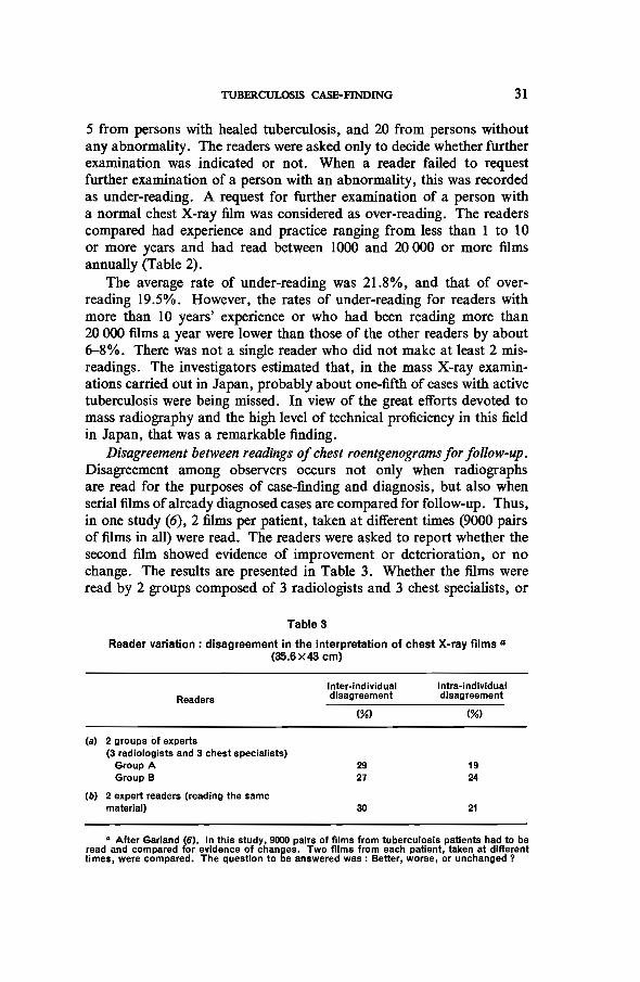
5 from persons with healed tuberculosis, and 20 from persons without any abnormality. The readers were asked only to decide whether further examination was indicated or not. When a reader failed to request further examination of a person with an abnormality, this was recorded as under-reading. A request for further examination of a person with a normal chest X-ray film was considered as over-reading. The readers compared had experience and practice ranging from less than 1 to 10 or more years and had read between 1000 and 20 000 or more films annually (Table 2).
The average rate of under-reading was 21.8%, and that of over· reading 19.5%. However, the rates of under-reading for readers with more than 10 years' experience or who had been reading more than 20 000 films a year were lower than those of the other readers by about 6-8%. There was not a single reader who did not make at least 2 misreadings. The investigators estimated that, in the mass X-ray examinations carried out in Japan, probably about one-fifth of cases with active tuberculosis were being missed. In view of the great efforts devoted to mass radiography and the high level of technical proficiency in this field in Japan, that was a remarkable finding.
Disagreement between readings of chest roentgenograms for follow-up. Disagreement among observers occurs not only when radiographs are read for the purposes of case-finding and diagnosis, but also when serial films of already diagnosed cases are compared for follow-up. Thus, in one study (6), 2 films per patient, taken at different times (9000 pairs of films in all) were read. The readers were asked to report whether the second film showed evidence of improvement or deterioration, or no change. The results are presented in Table 3. Whether the films were read by 2 groups composed of 3 radiologists and 3 chest specialists, or
(a)
(b)
Table 3
Reader variation: disagreement in the Interpretation of chest X-ray films a (35.6x43 cm)
Inter-Individual Intra-Individual Readers disagreement disagreement
(%) (%)
2 gro ups of experts (3 radiologists and 3 chest specialists)
Group A 29 19 Group B 27 24
2 expert readers (reading the same material) 30 21
" After Garland (6). In this study, 9000 pairs of films from tuberculosis patients had to be read and compared for evidence of changes. Two films from each patient, taken at different times, were compared. The question to be answered was: Better, worse, or unchanged?

32 K. TOMAN
by 2 expert readers only, the results did not differ much. The level of disagreement between readers was about 27-30% and a single reader was likely to disagree with himself in about 19-24% of cases.
[UAT international study on X-ray classification
One of the most important comparative studies of the reading and interpretation of chest X-ray films was organized by the International Union against Tuberculosis (IUAT). The main purpose of the study was to arrive at a uniform nomenclature and interpretation of X-ray findings that could serve as a basis for an international classification of chest radiographs (12).
A sample of 1100 films was chosen from among several hundred thousand films taken at one of the periodic mass radiography surveys of the adult population of Norway. The sample included 200 films from patients with infectious tuberculosis, 400 from patients with previously active tuberculosis, lOO from persons with minimal findings not requiring referral or follow-up, 300 from persons without abnormal findings, and lOO from patients with verified non-tuberculous lung disease. The films were mounted together in 7 film rolls and 10 copies of each roll were made.
The reading was done by 90 physicians (radiologists and chest physicians) highly competent for this task. Eighty of the readers were selected from 9 countries where mass X-ray examinations had been carried out for many years: Czechoslovakia, Denmark, Finland, France, Norway, Sweden, the United Kingdom, USA, and Yugoslavia. Ten readers were selected from various national and international WHO project staff.
The study had been designed (13) to measure primarily the extent of agreement or disagreement between readers and not the observer error resulting in under-reading or over-reading. A set of questions, prepared in advance, was answered by each reader independently. Most of the questions required the answer" yes" or "no "-e.g., "Is there an abnormality in the lung? YesfNo?"; "Is there a cavity present? YesfNo ? " ; " Does the patient need clinical attention? Yes/No?".
Since every question had to be answered by 90 readers for each of the 1100 films, there were 99000 answers to each question. The enormous amount of data collected was analysed and tabulated by means of electronic data processing machinery.
The material was evaluated according to a special statistical procedure (14) by which a series of values was obtained for each question. From these values a curve was then constructed that characterized the level of agreement (or rather disagreement) between readers for a given question (Fig. I). The advantage of the method is that a curve derived in this way can be described by a single figure-an index. This index can take any

TUBERCULOSIS CASE-FINDING 33
value from 0 to 100, reflectiug the extent of disagreement. Thus a low index indicates little disagreement and a high index, much disagreement. The nearer a curve is to the zero point of the two axes, the lower is the disagreement; the flatter the curve, or the farther removed from the axes, the higher is the disagreement. (The maximum of 100 would coincide with the intermittent line drawn from the left upper corner to the right lower corner.)
Fig. 1
IUAT study on X-ray classification (14) : curves of disagreement between readers In response to three questions (4: Is there any abnormality
in respiratory organs? 4c: Is there any abnormality in lymph nodes? and 8c: Is there any calcification in lymph nodes ?)
... 100
~ ::: 90
~ i::: 80 (I) <:! ~70 ~
~60 ,~ 0:..
~50 ~ 15 40
~ ~30 .... ~ 'I> 20 ~ ~ 10 ~
",I 1 'r" " \ " • I", • • • , , \ • , \ \ • . ~ • • \\ • • .. \ \ \,
\ '., \ \ Ltc "' I~ } -'.
"'" .......
INOE OF IOISA REEl ENT: uesli 0(4 : 32 uest n(4 ) ;4 uesti pn(3 ) : 6C
I", , "
"-
/ , , l? ,
" ""'''''
, , "'" ,
........ "" --. , ---:.:::. -, .... , ---~.:.~
, ~
00 10 20 30 40 50 60 70 80 90 100 / :..",. ~ .... - , Percentage of the YES-answers given '10 "NEGATIVE- films
For example, there was less disagreement on question 4 than on question 3c (Fig. 1). (The index is calculated by the sum of percentages of discordant answers at the point on the curve where they are equali.e., where the curve is intersected by the oblique line starting from the zero corner.)
Comparison of reader disagreement in chest radiography and smear microscopy
A similar study was undertaken by IUAT to measure the extent of disagreement between microscopists reading sputum smears for acid-fast bacilli.

34 K. TOMAN
The statistical design and evaluation of the study were made by the same author and with the same method of measurement (J. Nyboe, unpublished data, 1971). A series of 250 sputum smears from patients were examined independently in 10 laboratories by experienced laboratory technicians. Fig. 2 illustrates the curves of disagreement for three criteria of positivity.
Disagreement was the lowest (index: 10) when the criterion for a positive result was the demonstration of at least 8 acid-fast bacilli; it was only slightly higher (index: 12) when, as usually, 3 acid-fast bacilli were required as the minimum. The highest disagreement (index: 18) between readers was when 1 acid-fast bacillus was accepted as sufficient evidence of positivity. However, even the highest level of disagreement between microscopists was still substantially lower than the lowest level of disagreement between readers of chest X-rays.
Fig. 2
Examples of curves of disagreement in X-ray and sputum-smear examinations
35r-------------------------------------------~
1. X-RAY EXAMINATION: 30 Cavity present?
t! 2-4. SMEAR EXAMINATION
! 2. One or more bacilli on slide?
25 3. Three or more bacilli on slide? c: 4. Eight or more bacilli on slide? lIS
b c: ~ .... c: 20 '" "E 0 !t :c -0 15 *-c: 0
t 0 10 c. e
Il..
5
Proportion (%) of discordant "yes" answers

TUBERCULOSIS CASE-FINDING 35
Curve I (Fig. 2) characterizes the lowest level of disagreement achieved (index: 28) between X-ray readers-i.e., in reply to the question: " Cavity present '1 ". Disagreement on the question: " Smear positive for acid-fast bacilli '1" was substantially lower whatever the limit chosen. The investigators concluded that there was consistently better agreement among smear readers, no matter what the criterion for a positive smear was, than among X-ray readers (see also" How reliable is smear microscopy'1 "-page 14).
Levels of disagreement on the interpretation of chest radiographs and conclusions
A few indices of disagreement on other questions have been tabulated in Table 4. The questions were selected with a view to their use for a classification of X-ray findings. They include questions with the lowest and highest levels of disagreement.
Table 4
IUAT International study on X-ray classlflcation : Indices of disagreement on various questions (14)
Is the film abnormal? •.••••••••••••...•.••....•• Calcification In lung? • • . . . . . • • • • • • • . . • • • • • • • • . . • Non-calcifled abnormality. probably tuberculous? . Cavity present? . • . • • • • • . • . . . • . • . Abnormality in lung, probably not tuberculous? Abnormality in lymph nodes? • Need for medical action? • . • • • • • • • • •
34 42 37 28 45 60 31
What was quite unexpected was the disagreement on the question about abnormality in the respiratory organs. The poor agreement on the question about calcifications was also surprising. The discrepancies regarding abnormalities of the lymph nodes, including calcifications (one of the most frequently described X-ray findings), were particularly striking. The best agreement-or rather the least disagreement-was seen in response to the questions about cavities. This information has to be seen in the context of medical action. Thus 5% of smear-positive persons were reported as having a normal X-ray, 17% as having some probably non-tuberculous abnormality, and 24% as not being in ueed of clinical action for a tuberculous lesion. If treatment had been restricted to patients in whom 50% or more of the readers judged cavitation to be present, only one-third of those with positive sputum would have received treatment. On the other hand, among those who were regarded by 50% or more readers as probably tuberculous and in need of treatment, about four or five times as many bacteriologically negative persons as sputum-

36 K. TOMAN
positive patients would have received treatment-(a disproportion similar to that frequently observed in clinics where diagnosis is made merely on roentgenological grounds (15).
It was concluded from the study, therefore, that " purely radiologic criteria cannot give really satisfactory evidence of tuberculosis in the individual patient ... ". Even more disappointing was the conclusion that none of the categories or criteria of diagnostic importance could be included in a classification because disagreement was too wide. Consequently, the plan to produce an internationally accepted X-ray classification had to be dropped, and there is so far no indication that it will ever be revived.
In "Diagnostic standards and classification of tuberculosis ..... (a manual that has been published by the American Thoracic Society at 5-yearly intervals since 1917), roentgenographic status was the backbone of the basic classification for half a century. That is why the extent of disease, its pathogenetic type or characteristics, the presence or absence of parenchymal destruction, and the activity of the process were judged according to radiological criteria. This is no longer so. The American Thoracic Society recently developed a new classification based on our knowledge of the host-parasite relationship existing between man and the tubercle bacillus (16). Thus, in the latest edition (1), the basic classification of pulmonary tuberculosis rests primarily on bacteriology and on the chemotherapeutic status. Roentgenographic findings are regarded as necessary only in certain circumstances.a
Many of the terms used hitherto-e.g .• "minimal", "moderately advanced" • "far advanced", "active" • "inactive", "primary", " post-primary". "infiltration", .. caseation", and "fibrosis" -have been deleted from the current classification.
a Roentgenogram findings are subdivided as follows: 1. Nonnal 2. Abnonnal
(a) Cavitary or non-cavitary (b) Stable, worsening, or improving
REFERENCES
1. AMERICAN LUNG ASSOCIATION. Diagnostic standards and classification of tuberculosis and other mycobacterial diseases. New York, 1974, p. 22
2. BIRKELO, C. C. ET AL. Journal of the American Medical Association, 133: 359 (1947)
3. GARLAND, L. H. American journal ofroentgenology, radium therapy, and nuclear medicine, 64: 32 (1950)

TUBERCULOSIS CASE~FINDING 37
4. YERUSHALMY, J. ET AL. American review of tuberculosis, 61: 443 (1950)
5. GROTH-PETERSEN, E. ET AL. Acta tuberculosea Scandinavica, 26: 13 (1952)
6. GAlU.AND, L. H. Americanjournai ofroentgenology, radium therapy, and nuclear medicine, 82: 25 (1959)
7. GARLAND, L. H. Radi%gy, 133: 359 (1949)
8. NEWELL, R. R. ET AL. American review of tuberculosis, 69: 566 (1954)
9. GROTH-PETERSEN, E. & M0LLER, A. V. Acta tuberculosea Scandinavica, 30: 209 (1955)
10. CoCHRANE, A. L. & GARLAND, L. H. Lancet, 2: 205 (1952)
11. NAKAMURA, K. ET AL. Kekkaku, 45: 121 (1970)
12. SPRlNGETT, V. H. Bulletin of the International Union against Tuberculosis, 41 : 107 & 125 (1968)
13. WAALER, H. T. Bulletin of the International Union against Tuberculosis, 41 : 110 (1968)
14. NYBOE, J. Bulletin of the International Union against Tuberculosis, 41: 115 (1968)
15. WHO Technical Report Series, No. 552, 1974 (Ninth report of the WHO Expert Committee on Tuberculosis)
16. SBARBARO, J. A. Chest, 68: 436 (1975)

38 K. TOMAN
What is the probability of obtaining a negative culture from a sputum. specimen found positive by smear microscopy?
In a recent study on the efficacy of bacteriological measures under the conditions of the Singapore tuberculosis control programme (1), 1162 new patients with clinical and radiological signs suggesting tuberculosis were examined as follows.
Two sputum specimens were collected from each patient: one on each of two consecutive days. The specimens were collected in the presence of a trained supervisor. All specimens were examined independently by direct smear microscopy in one laboratory and by culture in another laboratory (one smear and one culture per specimen). Of the 1162 patients, 500 had a positive smear from one or both specimens, as shown below.
Yield from first specimen Additional yield from second specimen
Total
No. 011U!W patienU lormd positive 10' acid-fast badIIl
428 72
500
The results of two culture examinations of the sputum of these smear-positive patients are given in Table 1. As the table shows, in 17 of 500 patients-Le., less than 4%-the positive smear results were not confirmed by 2 culture examinations. Assuming that the contaminated cultures were all negative, the proportion of unconfirmed results would amount to 6% at the most. A further analysis (not tabulated) showed that, of the 115 patients found positive in only one of two smears, 101 (almost 90%) were confirmed by culture to be excreting tubercle bacilli.
Table 1
Results of 2 culture examinations on 2 consecutive sputum specimens from 500 new patients found smear-positive
on the basis of one or both specimens
No. of patients %
Total examined 500 100 Confirmed by first culture 399 80 Confirmed by second culture (additional) 73 14 Contaminated (both cultures) 11 2 Not confirmed by both cultures 17 4

TUBERCULOSIS CASE-FINDING 39
The authors concluded from these results that, when tubercle bacilli were identified by smear examination, culture examination of two specimens confirmed the smear result in all but a very small proportion of cases and, hence, that culture confirmation of a positive result based on two smear examinations did not appear to be necessary. This is particularly true in populations with a high prevalence of tuberculosis, where patients seek medical attention only because of haemoptysis or prolonged chest symptoms such as a productive cough.
A negative culture result with a specimen containing tubercle bacilli may be due to various causes. In patients receiving chemotherapy, the organisms may have lost their ability to grow on culture media and be practically dead. In patients who have not had chemotherapy, sputum specimens may have been exposed to sunlight or heat, stored too long, dried out, or contaminated. Excessive decontamination procedures before inoculation, inadequate culture media, and deficient incubation may also cause a negative culture result (see" What are the main causes of a false positive or false negative sputum smear? "-page 9).
The probability of obtaining a positive culture in patients with both sputum specimens negative by smear, being a quite different problem, is dealt with under the heading" What is the clinical and epidemiological significance of consistent negativity by smear microscopy in patients positive by culture only? "-page 50).
REFERENCE
CHAN, W. ET AL. Bulletin of the World Health Organization, 4!: SSI (1971)

40 K. TOMAN
What is the additional case yield from repeated sputum examinations by smear microscopy and culture ?
Several authors (1, 2, 8) have investigated the additional case yield from multiple examinations of successive sputum specimens. More recent studies carried out at the National Tuberculosis Institute of India (4, 5) sought to determine the additional case yield when 8 specimens (instead of one) from each eligible person were subjected to examinations by smear microscopy and culture (see also" What are the relative merits of chest X-ray and sputum examination ... ? "-page 44).
In 194 persons who had abnormal X-ray lung shadows and complained of prolonged chest symptoms suggestive of tuberculosis, 8 successive sputum specimens (4 collected on the spot and 4 produced overnight and collected by a home visitor) were examined concurrently by smear microscopy (Ziehl-Neelsen method) and culture. The number of specimens examined was thus 1552. Tubercle bacilli were found in the sputum of 75 patients (Table 1). Each specimen was examined independently, the laboratory technician having no knowledge of the persons examined or of previous results.
Table 1
Results of examining concurrently, by smear microscopy and culture, 8 sputum specimens (4 collected on the spot and 4 in the early morning)
obtained from each of 194 patients within 1 week
Specimens examined Patients examined Patients negative (all smears and cultures)
Patients positive: at least 1 smear and 1 culture at least 1 culture (all smears negative) at least 1 smear (all cultures negative)
• Two patients had smears with 3 or fewer acld·fast bacilli.
No. 1552
194 119
75
For the convenience of the reader, the data of Table 1 are presented in the form of a four-fold correlation table (Table 2).
Table 3 shows the yield of new cases from examination of the first and subsequent specimens, in chronological order.
It is obvious that, in successive examinations-whether by smear microscopy or by culture-most new positive results are obtained from the first and second specimens. The upper half of the table shows that

TUBERCULOSIS CASE-FINDING 41
45 (85%) of all smear-positive patients were already positive byexamination of two consecutive specimens. Considering only the smear-positive cases confirmed by culture, 41 (89%) were positive on the basis of two consecutive examinations.
Smear positive negative
Total
Table 2
Data from Table 1 presented in the form of a four-fold correlation table
positive
46 22
Culture
68
Table 3
negative
7 119
126
Total
53 141
194
Yield in cases from concurrent smear (5) and culture (C) examinations of 8 consecutive sputum specimens from each of 194 persons
with lung X-ray shadows and prolonged chest symptoms suggesting tuberculosis
Bacteriological category
Smear positive
Culture positive
S+ c+
S+ c-Total
C+ s+ c+ s-Total
No. of cases
46
7
53
46
22
68
Number of cases according to serial No. of specimen yielding 1st positive result
11 III IV V VI VII VIII
34 7 2 '-v-'
41 (89%)
2 2
36 9 2 3 '-v-'
45 (85%)
34 7 2
9 7 2
43 14 2 2 3 2
Another important finding of this investigation (Table 3) was that about the same number of new cases (45) were detected by the first two smears as by the first culture examination (43). From this result, it may be concluded that, in new, untreated patients with prolonged chest symptoms and abnormal lung X-ray shadows, two consecutive smear examinations (e.g., of on-the-spot and overnight sputum) were practically equivalent to one culture examination. This inference concords with the results of other studies, such as that undertaken in new patients attending

42 K. TOMAN
the Tuberculosis Chemotherapy Centre, Madras (6), and another recent study on bacteriological case-finding procedures in patients attending health services in Singapore (7). In the latter study, covering 1162 new patients with radiological signs and chest symptoms suggesting tuberculosis, culture of the first sputum specimen revealed 535 cases, whereas two consecutive smears detected 500 cases (see" What is the probability of obtaining a negative culture from a sputum specimen found positive by smear microscopy? "-page 38).
All the above-mentioned findings confirm Mitchison's earlier observations (8) that:
..... smear examination, especially of several specimens from each patient, is almost as efficient as culture examinations in clinics in developing countries."
This may apply also to other high-prevalence situations or to preselected groups of patients who have been prompted by symptoms (such as prolonged cough, purulent sputum, and haemoptysis) to seek relief at a health centre (3).
Another significant observation in the study was that the 46 patients who were found positive by smear and culture were discharging tubercle bacilli practically every day (of a total of 368 specimens from these 46 patients, 347-Le., 94%-were culture-positive). In contrast, out of 176 specimens from patients negative by smear microscopy and positive only by culture, only 62 (35.2%) were positive. This category of patients thus discharges bacilli only about every third day or in only every third specimen (see Table I in "What is the clinical and epidemiological significance of consistent negativity by smear microscopy ... ?"page 51). This confirms that patients positive only by culture and negative by smear microscopy are less significant, epidemiologically, than those positive by smear (and culture).
A similar study was made at the same institute in connexion with an epidemiological survey of a South Indian district (5). Eight consecutive sputum specimens were collected from each of 1652 persons with an abnormal chest X-ray and examined as in the study described before. The results were comparable: in those positive by smear microscopy and culture, the first smear was positive in 86.7% and the second smear added 10% of positive results. In those who were positive only by culture, the first specimen yielded only 32% of positive results, and the second, 18%. It was observed also in this study that patients discharging large numbers of bacilli were found positive in almost every specimen, whereas those positive by culture but smear-negative frequently produced specimens not containing any bacilli (see "What is the clinical and epidemiological significance of consistent negativity by smear microscopy in patients positive by culture only? "-page 50). The authors concluded that the examination of more than two specimens is not

TUBERCULOSIS CASE-FINDING 43
rewarding, and that two consecutive specimens (e.g., of on-the-spot and overnight sputum) would be the optimum number under the conditions of the national tuberculosis programme in India.
REFERENCES
1. ADLBR, H. & TOMAN, K. Acta tuberculosea Scandinavica, 25: 178 (1950)
2. TOMAN, K. Rozhledy v tuberkulose a v nemocech plicnich, 12: 79 (1952)
3. NAGPAUL, D. R. ET AL. Bulletin of the World Health Organization, 43: 17 (1970)
4. NAGPAUL, D. R. ET AL. In: Proceedings of the 9th Eastern Region Tuberculosis Conference and 29th National Conference on Tuberculosis and Chest Diseases, Delhi, November 1974. Delhi, Indian Tuberculosis Association, 1975
5. NAJR, S. S. ET AL. Indian journal of tuberculosis, 23: 152 (1976)
6. ANDREWS, R. H. & RADAKRlSHNA, S. Tubercle, 40: 155 (1959)
7. CRAN, W. ET AL. Bulletin of the World Health Organization, 45: 551 (1971)
8. MITcmsoN, D. A. Bulletin of the International Union against Tuberculosis, 41: 139 (1969)

44 K. TOMAN
What are the relative merits of chest X-ray and sputum examination (smear microscopy and culture) in case-finding among new outpatients with prolonged chest symptoms ?
This question, among others, was investigated in a comprehensive socioepidemiological study on outpatients, undertaken by the National Tuberculosis Institute, Bangalore, India (1, 2).
In this study, 2229 randomly selected new outpatients with chest symptoms (cough for 2 weeks or more, chest pain and fever for 4 weeks or more, or haemoptysis) were subjected to X-ray and bacteriological examinations. A sputum specimen was collected from each patient on the spot and examined by direct smear microscopy and culture. Smear examination was carried out with the Ziehl-Neelsen method while the patient waited. Culture was performed on two slopes of LOwensteinlensen medium. All positive cultures were tested in vitro for identification and drug sensitivity of the organisms. The bacteriological work was done by experienced technicians at the research laboratories of the National Tuberculosis Institute.
Table 1 shows that, of the 2229 patients, 227 were classified by X-ray as tuberculous (and thus in need of treatment), but that 81 of these were not confirmed as tuberculous by bacteriological examination. Among the remaining 2002 patients classified as normal or as having a disease other than tuberculosis, there were 31 in whom tubercle bacilli were found by sputum culture and/or smear microscopy.
Table 1
Results of X-ray examination compared with those of sputum smear mlcroscopy (S) and sputum culture (C) in out-patients
with clinical signs suggestive of tuberculosis (2)
Classlflcatlon No. of Result of sputum examination
by X-ray patients S+ s- s+ s-C+ C+ C- C-
Tuberculosis 227 122 20 4 81 Other abnormal shadows
(non-tuberculous) 304 8 4 291 Normal 1698 8 10 1 680
Total 2229 130 32 15 2052
Because sputum culture is regarded as the most reliable diagnostic method, a correlation was made between the results of X-ray and culture (Table 2). The data given in this table are identical with those in Table 1,

TUBERCULOSIS CASE-FINDING 45
except that the two groups of X-ray-normal and nontuberculous persons were pooled. Taking the results of culture as the criterion of correct diagnosis, 20 (12%) of 162 cUlture-positive patients would have been missed because they had been misclassified by X-ray as normal or nontuberculous. On the other hand, among the 227 patients classified radiologicallyas tuberculous, 85 (37%) were not confirmed as such by culture.
Table 2
Correlation of the yields of X-ray examination and sputum culture In patients with clinical signs suggestive of tuberculosis
Culture
positive negative total ., 0;
142 85 227 0 yes X-ray :;
e <J) no 20 1982 2002 .a .s
total 162 2067 2229
The results of direct smear microscopy and culture were correlated too (Table 3). As this table shows, 32 (20%) of the 162 culture-positive patients would have been missed by direct smear microscopy of a single spot specimen and 15 (10%) of the 145 smear-positive patients would have been negative by culture.
Table 3
Correlation of the yields of 2 cultures and of direct smear mlcroscopy of a single spot specimen in patients
Smear
with clinical signs suggestive of tuberculosis
positive negative
total
positive
130 32
162
Culture
negative
15 2052
2067
total
145 2084
2229
Taking the results of sputum culture as the criterion, the findings of the study may be summarized as follows: among 162 tuberculosis patients whose diagnosis was verified by culture, 32 (20%) would have been missed by smear microscopy and 20 (12%), by X-ray.
On the other hand, among 145 patients positive by smear, 130 (90%) were confirmed by culture. The remainder (10%) gave an apparently false positive result, owing to a reading error or the presence of artefacts,

46 K. TOMAN
or because the bacilli seen under the microscope had lost their ability to grow on culture. Possible reasons for the last-mentioned occurrence are many. Thus, in patients under treatment, these bacilli may have been killed or seriously harmed by effective chemotherapy. In untreated patients, the culturability of tubercle bacilli may have been impairede.g., by exposure of the specimen to heat or sunlight, long storage, excessive decontamination procedures, or an overheated incubator. However, patients whose first spot-specimen is indisputably positive by smear microscopy but negative by culture have a rather high chance of being positive by both examinations in subsequent specimens (see" What is the additional case yield from repeated sputum examinations ... ? "page 40). The authors concluded that, ifpersons with signs and symptoms suggestive of tuberculosis are treated on the basis of properly performed and clearly positive smear microscopy, not confirmed by culture, there is little likelihood of serious overtreatment.
On the other hand, of the 227 patients classified by X-ray as "tuberculous and in need of treatment". a sizeable proportion-37% a-were not confirmed by culture to be tuberculous. Several studies have shown that persons with X-ray lung shadows of unknown origin, who have no history of previous tuberculosis and in whom tubercle bacilli could not be demonstrated by smear microscopy and/or culture, particularly when repeated, are in fact rarely true cases of tuberculosis. Follow-up studies have demonstrated (3-7) that, though such persons have a definitely higher risk of becoming culture-positive than those with a normal X-ray, only a small proportion (0.4-4.8%) actually did so within the first year of observation. The risk declined in subsequent years."
Thus, to give chemotherapy-as a matter of routine-to persons with X-ray lung shadows of unknown origin would be to treat most of them unnecessarily or wrongly: these individuals might be exposed to the hazards of drug toxicity and irreversible harm might be caused in cases where the actual etiology is another serious disease. Therefore, such a practice is justifiable hardly anywhere. It is to be avoided especially in countries where tuberculosis patients and their families are branded with a permanent stigma causing mental suffering and economic hardship.
Unfortunately, in many countries it is common practice to rely on X-ray examination alone for the diagnosis of tuberculosis, neglecting bacteriological examination or ignoring its results. The usual conse-
a This proportion is likely to be much higher in populations examined by indiscriminate mass X-ray-i.e., irrespective of the presence of symptoms. The patients in the study referred to had been examined because they had symptoms.
" The rates varied with the population studied, age, the extent of X-ray lesions, additional risk factors, and bacteriological and other criteria. They commonly decreased with time, the highest rates being found at the beginning of observation.

TUBERCULOSIS CASE-FINDING 47
quence-overdiagnosis-is generally followed by overtreatment, which is a waste of resources and may become an unmanageable burden on the health services. This happens not infrequently in developing countries, in places where health centres are equipped with diagnostic X-ray apparatus.
The problem has been characterized very well by the WHO Expert Committee in its ninth report (page 16) :
..... when treatment for tuberculosis is initiated on the basis of radiographic findings alone, a substantial proportion of patients are treated unnecessarily. This wastes the resources that should be concentrated primarily on the treatment of infectious cases. It throws needless strain on understaffed and underfinanced treatment services and unnecessarily exposes many patients to loss of their job, loss of their home, and to serious social stigma. Hence, the importance of making a bacteriological diagnosis" (8).
Some technical and operational aspects of chest X-ray, sputum culture, and smear microscopy
By X-ray, as has been shown, 85 (37%) of 227 patients would have been wrongly classified as tuberculous (see" How reliable is chest radiography? "-page 28), yet 20 culture-positive patients in whom the disease was not diagnosed by direct smear microscopy were found by X-ray. Culture detected, in addition, 12 patients who were found neither by smear microscopy nor by X-ray. Thus the highest yield of cases discharging tubercle bacilli was achieved by culture.
The high rate of overdiagnosis by X-ray is a heavy penalty for the relatively small gain in patients that may be missed by smear microscopy. Moreover, the operational shortcomings of radiography in developing countries are considerable. For example, the District Tuberculosis Centres in India are equipped with X-ray units using mirror cameras and miniature films for fluorography. Such equipment and its installation are expensive. Its operation requires specially trained technicians. There are frequently prolonged interruptions owing to the breakdown of equipment, the lack of spare parts and repair facilities, the scarcity of films, or the shortage of electric power. Another operational disadvantage is that the results of X-ray examination are usually available only after 2-3 days, and sometimes later. As has been commonly observed, a sizeable proportion of patients do not return to the centre for their results. Efforts to retrieve them are often unsuccessful.
Sputum culture is known to be technically superior to smear microscopy as a diagnostic method. It is essential for the definitive identification of tubercle bacilli-Le., to differentiate them from other microorganisms. The technical superiority of culture over smear microscopy is largely due to quantitative factors. Whereas the amount of sputum on a smear is 0.01 m1 (see " How many bacilli are present in

48 K. roMAN
a sputum specimen ... 7 "-page 6), the size of an inoculum for culture is usually 0.1 ml-i.e., about 10 times as much." Moreover, usually only about 1-3% of the smear (100-300 oil-immersion fields) is examined by (bright.field) microscopy, whereas in the culture test tube the whole yield of colonies may be seen practically at a glance. Although a large proportion of organisms is destroyed by decontamination procedures, the quantitative differences are still so large that the probability of finding bacilli by culture is many times as great (perhaps ~ 30 times) as it is by direct smear microscopy. This is an obvioux advantage in cases where a specimen contains only small amounts of acid·fast bacilli (see" How reliable is smear microscopy 7 "-page 14).
Unfortunately, culture has a number of disadvantages, mainly of an operational nature. First of all, the method requires specially trained and skilled personnel, of whom there is a shortage in most developing countries. There is also a need for special facilities and equipment, a permanent supply of water and electric current, and reliable thermo· regulation of the hot room. Particularly in hot and humid climates, airconditioning facilities and special air filters are needed to prevent airborne contamination of cultures. Properly ventilated inoculation cabinets and other safety measures must also be provided. For all these reasons, in developing countries, culture methods are practicable in only a few laboratories.
One of the greatest shortcomings of sputum culture is the long interval before results become available: usually 4-6 weeks, and often more. In developing countries this delay leads to many patients being " lost". They cannot be retrieved and never return to the health centre. Thus the gains achieved by culture are often outweighed by the losses due to long waiting periods, and the technical superiority of culture is largely counterbalanced by its operational disadvantages.
Direct smear examination certainly has a number of technical short· comings, but its operational advantages are obvious. It is relatively easy to perform, much less expensive than X·ray or culture, and does not require highly specialized personnel. The fact that the diagnosis of tuberculosis (in persons discharging large amounts of bacilli) may be established and chemotherapy started on the same day is without doubt the greatest operational advantage of smear microscopy. It reduces to a minimum" losses" of patients owing to long waiting periods. Smear microscopy is the only diagnostic method practicable almost everywhere. That is why, in the developing countries, case-finding and diagnosis win have to rely on this method for some time to come.
" If culture media are contained in dishes (plates) instead of test tubes or bottles, the amount of sputum inoculated is about 30 times as great as that spread on a slide (9).

TUBERCULOSIS CASE-FINDING 49
The WHO Expert Committee on Tuberculosis, in its ninth report (page 25), emphasized that the selection of diagnostic procedures and the development of diagnostic services should be determined by priorities:
"In a logical and efficient sequence of priOrities, the first priority would be to provide facilities for direct smear examination of sputum from persons who, of their own volition, present with symptoms and to provide adequate treatment for those who are found to excrete tubercle bacilli. In such a programme, patients with persistent symptoms but whose sputum does not contain bacilli should be followed up; treatment with antituberculous drugs would be given only if the diagnosis can be confirmed bacteriologically" (8).a
It must be reiterated that, for the purpose of diagnosis, direct smear examination is important and should be performed under practically any circumstances-i.e., also in technically developed countries. This method makes it possible to identify patients who, if untreated, would have a most unfavourable prognosis and would be the most dangerous sources of infection in the community.
a A valuable, but often forgotten, rule of thumb is that persons having cough with purulent sputum for one month or longer, but who are eatedly negative by properly performed direct smear microscopy, are very likely s from a respi-ratory disease other than tuberculosis - the more severe their symptoms (and radiological signs) are, the higher is this probability.
REFERENCES
1. NAGPAUL, D. R. ET AL. Bulletin of the World Health Organization, 43: 17 (1970)
2. NAGPAUL, D. R. ET AL. In: Proceedings of the 9th Eastern Region Tuberculosis Conference and 29th National Conference on Tuberculosis and Chest Diseases, Delhi, November 1974. Delhi, Indian Tuberculosis Association/International Union against Tuberculosis, 1975
3. NAGPAUL, D. R. ET AL. Bulletin of the International Union against Tuberculosis, 41: 18 (1968)
4. HoaWITz, O. ET AL. Bulletin of the World Health Organization, 41 : 95 (1969)
5. DANKOVA, D. Bulletin of the International Union against Tuberculosis, 47: 145 (1972)
6. EoWARDS, L. B. ET AL. Bulletin of the International Union against Tubercuwsis, 47: 151 (1972)
7. GOTHI. G. D. ET AL. Indian journal of tuberculosis, 21: 90 (1974)
8. WHO Technical Report Series, No. 552. 1974 (Ninth report of the WHO Expert Committee on Tuberculosis)
9. KUBICA. G. P. & Dn. W. E. Laboratory methods for clinical and public health mycobacteriowgy. Atlanta. GA. US Department of Health, Education, and Welfare, 1967 (Public Health Service Publication. No. 1547)

50 K. TOMAN
What is the clinical and epidemiological significance of consistent negativity by smear microscopy in patients positive by culture only?
It has been proved that the probability of finding acid-fast bacilli by sputum smear microscopy is directly related to the concentration of bacilli in the sputum (see " How many bacilli are present in a sputum specimen found positive ... ? "-page 6 and" How reliable is smear microscopy? "-page 14). Thus, when 1 m1 of sputum contains fewer than 10 000 but more than 1000 acid-fast bacilli, the chance of a positive result by microscopy is about 40-50%. At concentrations below 1000 organisms per millilitre, the chance decreases rapidly, the result being negative in about 96% of cases.
When discussing the significance of smear-negative findings, one has to exclude all negative results that could have been caused by technical or operational deficiencies (see" What are the main causes of a false positive or false negative sputum smear? "-page 9).
All sources of error being eliminated, the concentration of bacilli in the sputum is determined largely by the type of tuberculous lesion from which the bacilli originate. Thus, a cavity about 2 cm in diameter (opening into a bronchus) may contain some 100 million organisms. A non-cavitated nodular lesion of the same size may contain 100-1000 (I)-i.e., one hundred thousand times fewer. The fact that the bacterial content of lesions differs considerably is of fundamental epidemiological, therapeutic, and diagnostic importance. Sputum from patients with tuberculous lung cavities, containing softened necrotic particles with enormous amounts of bacilli, will thus usually be found positive by direct smear microscopy. In contrast, sputum from patients with nodular, encapsulated lesions discharging only sma11 amounts of bacilli will almost invariably be negative by smear microscopy. Only by using refined laboratory techniques, such as culture or animal inoculation, can small numbers of bacilli be demonstrated-and then often only irregularly (2,3).
The difficulty of demonstrating small numbers of bacilli regularly has been shown in studies described elsewhere (4, 15, 21) (see" What is the additional case yield from repeated sputum examinations ... 1"-page 40). Eight sputum specimens (4 on the spot and 4 overnight) were collected from each of 194 patients with prolonged chest complaints. A smear was prepared from each specimen and 2 culture tubes of medium were inoculated. In 68 patients, the presence of tubercle bacilli could be ascertained by culture, and 46 of these patients were positive also by smear-i.e., in at least 1 of the 8 smears and 1 of the 8 cultures;
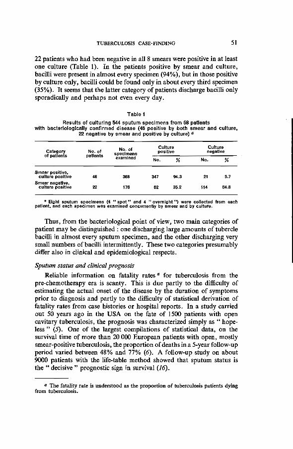
TUBERCULOSIS CASE-FINDING 51
22 patients who had been negative in all 8 smears were positive in at least one culture (Table 1). In the patients positive by smear and culture, bacilli were present in almost every specimen (94%), but in those positive by culture only, bacilli could be found only in about every third specimen (35%). It seems that the latter category of patients discharge bacilli only sporadically and perhaps not even every day.
Table 1
Results of culturing 544 sputum specimens from 68 patients with bacteriologically confirmed disease (46 positive by both smear and culture,
22 negative by smear and positive by culture) ..
No. of Culture Culture Category No. of specimens positive negative
of patients patients examined No. % No. %
Smear positive, culture positive 46 368 347 94.3 21 5.7
Smear negative, culture positive 22 176 62 35.2 114 64.8
• Eight sputum specimens (4 "spot" and 4 "overnight ") were collected from each patient, and each specimen was examined concurrently by smear and by culture.
Thus, from the bacteriological point of view, two main categories of patient may be distinguished: one discharging large amounts of tubercle bacilli in almost every sputum specimen, and the other discharging very small numbers of bacilli intermittently. These two categories presumably differ also in clinical and epidemiological respects.
Sputum status and clinical prognosis
Reliable information on fatality rates" for tuberculosis from the pre-chemotherapy era is scanty. This is due partly to the difficulty of estimating the actual onset of the disease by the duration of symptoms prior to diagnosis and partly to the difficulty of statistical derivation of fatality rates from case histories or hospital reports. In a study carried out 50 years ago in the USA on the fate of 1500 patients with open cavitary tuberculosis, the prognosis was characterized simply as " hopeless" (5). One of the largest compilations of statistical data, on the survival time of more than 20 000 European patients with open, mostly smear-positive tuberculosis, the proportion of deaths in a 5-year follow-up period varied between 48% and 77% (6). A follow-up study on about 9000 patients with the life-table method showed that sputum status is the" decisive" prognostic sign in survival (16) .
.. The fatality rate is understood as the proportion of tuberculosis patients dying from tuberculosis.

52 K. TOMAN
Anyway, there is little doubt that before the advent of chemotherapy the prognosis for patients with smear-positive pulmonary tuberculosis was gloomy. It is still serious for those who have been treated unsuccessfully and whose sputum remains consistently positive by direct smear microscopy. On the other hand, patients reported to have paucibacillary sputum sporadically were found to have---even before the advent of chemotherapy-a very good chance of normal survival. The same applied to patients who, having achieved quiescence after successful chemotherapy, yielded isolated positive cultures (I7). However, it would not be wise or appropriate to explain a multifactorial problem such as clinical prognosis by a single factor---e.g., the bacillary content of sputum. A number of complex endogenous and exogenous variables are of importance for the course of untreated tuberculosis. Nevertheless, the development of progressive lung tuberculosis, with caseation and the formation of cavities containing enormous amounts of bacilli that are discharged by coughing and in the sputum, is a chain of interdependent events of which the demonstration of acid-fast bacilli by smear microscopy is only the last link.
On the other hand, most patients with pulmonary lesions containing and discharging small numbers of bacilli, such as can be demonstrated only by culture, have a favourable prognosis compared with smearpositive patients. In South India, where an epidemiological survey had been repeated at intervals, the fate of newly discovered cases was studied (7, 8). Among the patients who, at the detection of their disease, had been smear-negative (two specimens) but positive by culture, more than one-half were classified as cured (i.e., negative by both smear and culture) within 18 months and about two-thirds within 3 years. Furthermore, the excess death rate was about one-third of that for the smear-positive cases. Thus, even under the living conditions of a very poor rural population, and without treatment, the prognosis for smear-negative, culture-positive patients was relatively favourable.
There is another piece of (negative) evidence on the relatively favourable prognosis for new patients who are definitely negative by smear and positive only by culture. It has been widely believed that, through repeated X-ray examinations, the occurrence of advanced, destructive, and smear-positive tuberculosis may be prevented, since all cases would be detected" early "-Le., at a stage where the disease is of minimal extent and the lesion(s) likely to contain a small number of bacilli demonstrable only by culture. It has also been assumed that these patients rarely have symptoms and thus may be detected best by indiscriminate mass radiography. But, to the general surprise, this hypothesis has not stood the test.
In a carefully conducted longitudinal study (see p. 58), the population of a district was examined by X-ray and bacteriology at 2-3-year
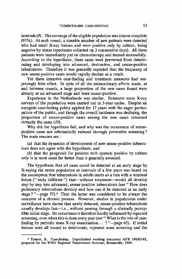
TUBERCULOSIS CASE-FINDING 53
intervals (9). The coverage of the eligible population was almost complete (95%). At each round, a sizeable number of new patients were detected who had small X-ray lesions and were positive only by culture, being negative by smear (specimens collected on 3 consecutive days). All these patients were immediately put on chemotherapy and treated successfully. According to the hypothesis, these cases were prevented from deteriorating and developing into advanced, destructive, and smear-positive tuberculosis. Therefore it was generally expected that the frequency of new smear-positive cases would rapidly decline as a result.
Yet these intensive case-finding and treatment measures had surprisingly little effect. In spite of all the extraordinary efforts made, at and between rounds, a large proportion of the new cases found were already at an advanced stage and were smear-positive.
Experience in the Netherlands was similar. Extensive mass X-ray surveys of the population were carried out in 3-year cycles. Despite an energetic case-finding policy applied for 17 years with the eager participation of the public, and though the overall incidence was declining, the proportion of smear-positive cases among the new cases remained virtually the same (10).
Why did the hypothesis fail, and why was the occurrence of smearpositive cases not substantially reduced through preventive screening? The main reasons are:
(a) that the dynamics of development of new smear-positive tuberculosis does not agree with the hypothesis, and
(b) that the prognosis for patients with sputum positive by culture only is in most cases far better than is generally assumed.
The hypothesis that all cases could be detected at an early stage by X-raying the entire population at intervals of a few years was based on the assumption that tuberculosis in adults starts as a rule with a minimal lesion (" early infiltrate ") that-without treatment-would all develop step by step into advanced, smear-positive tuberculosis (see" How does pulmonary tuberculosis develop and how can it be detected at an early stage? "-page 57).a Thus the latter was considered to be always the outcome of a chronic process. However, studies in populations under surveillance have shown that newly detected, smear-positive tuberculosis usually develops fast-i.e., without passing through a clinically perceptible initial stage. Its occurrence is therefore hardly influenced by repeated screening, even when this is done every year (see" What is the role of casefinding by periodic mass X-ray examination ... ? "-page 65). If initial lesions were all bound to deteriorate, repeated mass screening and the
a TOMAN. K. Case-finding. Unpublished working document AFR 190/OJ"()3. prepared for the WHO Regional Tuberculosis Seminar, Brazzaville. 1969.

54 K. TOMAN
treatment of all initial lesions detected would indeed greatly reduce the number of new, smear-positive cases found at later screening rounds. However, this is not the case, since the prognosis of initial smear-negative lesions positive only by culture has proved to be far better than was previously assumed, most of such lesions either healing or remaining unchanged. Only a few would deteriorate and thus only a few smearpositive cases would have been prevented.
Sputum status and infectivity
From the epidemiological point of view, also, the difference between these two categories of case is striking. Patients who are definitely negative by smear are substantially less infectious than the smear-positive are. This is not surprising, in view of the enormous difference in the numbers of bacilli discharged by these two categories of patient." For instance, for household contacts of smear-negative, culture-positive cases, the excess risk of becoming infectious is only a fraction (1/5-1/20) of that for household contacts of smear-positive sources.b The risk of infection for household contacts aged 0-14 years of smear-negative patients, whether culture-positive or not, is often almost the same as, or not much higher than, the risk for children of the same age group living in tuberculosis-free households under similar sociocultural and other ecological conditions (12,13). Furthermore, the risk of contracting the disease for household contacts of culture-positive, smear-negative patients is only about 1/5-1/10 as high as that for contacts of smearpositive patients (12,14) (see" What is the role of case-finding by periodic mass X-ray examination ... ? "-page 65).
It thus appears that the active search for patients with lesions harbouring and discharging small numbers of bacilli has a relatively low priority in tuberculosis control. If such a policy is adopted in a tuberculosis programme, it should never be at the expense of the first priority of case-finding-Le., the identification of smear-positive sources of infection.
In summary, the difference between cases discharging small quantities of tubercle bacilli demonstrable only by culture and cases with large amounts of bacilli in the sputum, positive by direct smear, is not only one of numbers or quantities. In their" pure" form, these cases behave as if they were two types of the same disease, differing in pathogenetic, prognostic, and epidemiological respects (19). That is why, in tuberculosis control, the first priority is given to patients positive by direct smear
.. The risk of exposure to smear-positive sources of infection is aggravated because such persons usually cough more frequently and violently (11).
/I Excess risk is the risk in addition to the basic risk due to exposure to the immediate (extradomiciliary) neighbourhood. The basic risk is naturally higher in crowded or slum conditions than the average risk for the total population that is often wrongly taken for comparison.

TUBERCULOSIS CASE-FINDING 55
microscopy, and why this category of patient is the principal target of case-finding efforts. This is reflected in the following statement by the WHO Expert Committee on Tuberculosis:
.. In a logical and efficient sequence of priorities, the first priority would be to provide facilities for direct smear examination of sputum from persons who, of their own volition, present with symptoms and to provide adequate treatment for those who are found to excrete tubercle bacilli. In such a programme, patients with persistent symptoms but whose sputum does not contain bacilli should be followed up; treatment with antituberculous drugs would be given only if the diagnosis can be confirmed bacteriologically (20). "
Regarding the choice of bacteriological procedures for case-finding, the Expert Committee provided clear guidelines:
.. From the point of view of their value in a tuberculosis control programme, bacteriological techniques may be ranked in the following order: examination of direct smears, culture, and, lastly, sensitivity testing.
If the prevalence of smear-positive patients is high in the patient population, the examination of smears from several specimens, especially an early morning one or an overnight collection, will detect a large proportion of the infectious cases in the community.
The examination of cultures in addition to smear examination will add to the number of patients with a confirmed diagnosis of tuberculosis, especially in those who are not excreting large numbers of bacilli. However, culture services should be provided only when a reasonably high proportion of the smear-positive cases in the country are being discovered and treated by chemotherapy and then they should be provided only in large laboratories (20). "
REFERENCES
I. CANETI'I, G. The tubercle bacillus in the pulmonary lesion. New York, Springer, 1955, p. 180
2. ADLER, H. & TOMAN, K. Acta tuberculosea Scandinavica, 2S: 178 (1950)
3. TOMAN, K. Rozhledy v tuberkulose a v nemocech plicnich, 12: 79 (1952)
4. NAGPAUL, D. R. ET AL. Bulletin of the World Health Organization, 43: 17 (1970)
5. BARNES, H. L. &BARNES, L. R. P. American review of tuberculosis andpulmonary diseases, 18: 412 (1928)
6. BERG, G. Acta tuberculosea Scandinavica, Suppl. IV (1939)
7. RAJ NARAIN ET AL. Bulletin of the World Health Organization, 39: 201 (1968)
8. NATIONAL TUBERCULOSIS INSTITUTE, BANGALORE. Bulletin of the World Health Organization, 51 : 473 (1974)
9. STYBLO, K. ET AL. Bulletin of the World Health Organization, 37 : 819 (1967)
10. M.ElmR, J. ET AL. Bulletin of the International Union against Tuberculosis, 44 : 5 (1971)

56 K. TOMAN
11. LoUDON, R. G. & SPOHN, S. K. American review of respiratory diseases, 99: 109 (1969)
12. SHAW, J. B. & WYNN-WILLIAMS, N. American review of tuberculosis and pulmonary diseases, 69: 724 (1954)
13. VAN GEUNS, H. A. Contact study, Rotterdam. The Hague, IUAT Tuberculosis Surveillance Research Unit, 1971
14. GRZYBOWSKI, S. & ALLEN, E. A. American review of respiratory diseases, 90 : 707 (1964)
15. NAIR, S. S. ET AL. Indian Journal of tuberculosis, 23: 152 (1976)
16. HILLEBOE, H. E. Public health reports, S6: 895 (1941)
17. EAST AFRICAN/BRITISH MEDICAL RESEARCH CoUNCILS. Lancet, 1: 1331 (1973)
18. IWASAKI, T. Bulletin of the International Union against Tuberculosis, 49: 28 (1974)
19. TOMAN, K. Bulletin of the International Union against Tuberculosis, 49: 192 (1974)
20. WHO Technical Report Series, No. 552, 1974 (Ninth report of the WHO Expert Committee on Tuberculosis)
21. NAGPAUL, D. R. In: Proceedings of the 9th Eastern Region Tuberculosis Conference and 29th National Conference on Tuberculosis and Chest Diseases, Delhi, November 1974. Delhi, Indian Tuberculosis Association, 1975

TUBERCULOSIS CASE-FINDING 57
How does pulmonary tuberculosis develop and how can it be detected at an early stage?
Much of our knowledge on incipient tuberculosis has come from mass X-ray surveys of apparently healthy population groups that had not been X-rayed before or were X-rayed many years previously. At these surveys, a sizeable number of individuals were found to have small X-ray lesions (opacities) in the lung. These persons, at the time of detection, were mostly symptom-free. Though the duration of the lesion was unknown, the association of the two findings seemed so obvious that it strongly supported the generalization that incipient tuberculosis is of minimal extent and usually asymptomatic. a But is that really and always so? And, if not, how often not?
In spite of a vast number of studies on incipient tuberculosis in adults, there is no certain knowledge of the actual macroscopic appearance of the first demonstrable lesions of incipient phthisis. Furthermore, it is not known what is the shortest period in which-starting with a normal lung-an incipient lesion can become radiologically or clinically manifest; nor-even more important-is it known what is the shortest time that it takes an incipient lesion to develop into advanced, smear-positive tuberculosis.
Most studies on incipient tuberculosis (e.g., 1-10) have concerned persons in whom the disease was detected by X-ray surveys, periodic follow-up examinations of certain high-risk groups (e.g., nurses, students, soldiers, prisoners, contacts, and immigrants), and routine clinical examinations. A brief summary of these studies might run as follows:
Incipient tuberculosis manifests itself radiologically as a small, indistinct shadow (infiltrate). The lesion is usually located in the apical or subapical area of the upper lobe of the lung. Symptoms are slight or absent. From that stage, phthisis develops in steps or phases. After months or years of stagnation or regression, the process may occasionally become arrested, but usually deterioration occurs. Each worsening, as a rule, has an acute character, while regressive changes are slow and follow a chronic pattern. Thus a deterioration phase is accompanied by pronounced symptoms, whereas in a regressive phase, symptoms are relatively mild. After several phases of deterioration, the outcome of the disease is usually fatal unless adequate chemotherapy is given. In general,
a So-called" primary tuberculosis "-i.e., tuberculosis occurring straight after the first infection and tuberculin conversion-is not included in this chapter, for the following reasons. It rarely develops directly into phthisis (chronic, destructive, and infectious tuberculosis) ; on the contrary, in the vast majority of cases, it heals spontaneously and remains undisclosed. It occurs mainly before adolescence is reached, particularly in conditions where the risk of infection is high.
58 K. TOMAN
a small, asymptomatic lesion in the upper part of the lung may be regarded as the beginning, or at least the potential beginnin~, of phthisis, after which step-by-step deterioration is almost inevitable.
Nevertheless, certain investigators have thrown doubt on the universality of this rule. Some have shown that almost all newly detected small lesions, particularly apical lesions, are old or healed and, if recent, have a favourable prognosis: "less than 10% develop within 5-10 years into phthisis" (11) and "90% of them are obviously not in need of treatment" (12). A rather large group of investigators, stressing the benignity of small apical lesions, insisted on the" early infraclavicular infiltrate " as the true beginning of phthisis (1 ,13). Some them stressed the exudative pneumonic character of these small lesions and their tendency to cavitation ; others stated that, at this stage, most of them are already cavitated (1, 2). Finally, some found that a rather large proportion of cases starts with a "massive pneumonic involvement without ever passing through a minimal stage" (14).
Before discussing these contradictory views and observations, it seems desirable to clarify certain fallacies that stil1linger on.
On clinical and epidemiological grounds, emphasis has rightly always been laid on early diagnosis. However, this has brought about a certain confusion. Early or incipient tuberculosis has frequently been identified with minimal disease. Likewise, advanced tuberculosis has usually been regarded as identical with old or chronic tuberculosis. Yet" early", " incipient", "chronic", and " old" are strictly and exclusively terms of time, while "minimal", "moderate", and "advanced" indicate merely the extent-Le., the volume of lung tissue involved. Terms such as " incipient" and " minimal" have by no means a constant relationship, nor need they be associated. It will be shown that a lesion of a few months' duration may be minimal or far advanced, and that the extent of a lesion gives little indication of its duration.
The age of a fresh lesion can be estimated only when X-ray evidence of a previously normal lung is available. Thus, thanks to a few longitudinal surveys and to periodic X-raying of large popuiations in certain countries, our knowledge of incipient tuberculosis has much improved.
In one of the longitudinal surveys (Kolin study), in a population of about 100 000, persons 14 years of age or more were screened repeatedly. The study (15,16) lasted 12 years, during which time the population was X-rayed 5 times at 2-year or 3-year intervals. Between X-ray rounds, the search for cases was continued by the local health services, where patients attended because of symptoms or for regular check-up. Since each X-ray film was carefully filed, it could always be compared with the most recent film. By that means, it was possible to determine within what period a new lesion had developed. The X-rays were read independently by two readers and one umpire reader (Table 1).

Interval) (months
.,;; 12
< 24 <36 ;;'37
Total
TUBERCULOSIS CASE-FINDING
Table 1 Interval between the last normal chest X-ray and the diagnosis
of pulmonary tuberculosis (15, 16)
No. Cumulative No. Cumulative No. first smear + smear - diagnosed
59
culture + % culture + % at necropsy
8 16 14 14 6 18 52 39 53 7 16 84 31 86 8 100 14 100 3
50 98 17
In 165 persons in whom tuberculosis had been newly detected and whose previous radiographs had been normal, the time between the last normal chest radiograph and the first abnormal one was measured. The patients were grouped into three categories: those sputum-smear-positive as well as culture-positive, those smear-negative but CUlture-positive, and those in whom the diagnosis had first been made at necropsy and verified by bacteriological examination (15, 16).
From Table 1, it is evident that, already within 12 months, 28 sputumpositive cases had developed. It was surprising that a significant proportion of these already had advanced tuberculosis with sputum positive by direct smear microscopy. Even more striking was the finding that 6 cases had developed so fast that they were found only at necropsyless than 12 months after the last normal chest radiograph. (In some of these cases, tuberculosis had been qualified by the pathologist as the leading cause of death.) a
The data presented in Table 1 might perhaps be biased, since the interval between X-ray rounds was never shorter than 2 years, though it was usually 3 years. In this connexion, very instructive data (see Fig. 2 in "What is the role of case-finding by periodic mass X-ray examination ... ? "-page 70) are available from Japan, where mass radiography of the population has been carried out at yearly intervals (17). Similar data are also available (18) from an epidemiological study in Niigata Prefecture, Japan (population: 2 350000).
Fig. 2 under "What is the role of case-finding by peri odic mass X-ray examination ... ?" (page 70) shows that more than one-half of
a During the second part of the study (1965-72), 10 previously unknown cases of new tuberculosis were first diagnosed at necropsy-some of them shortly after a normal X-ray. These 10 cases constituted one-quarter of all deaths from tuberculosis during that period-and that in spite of systematic and intensive screening of the population. The actual number might have been even higher, because only onequarter of all persons deceased in the study area were subjected to necropsy-i.e., mainly those who died in hospital (16).

60 K. roMAN
cases positive by direct smear microscopy developed within 12 months of the last normal radiograph. Since only one-fifth of the new cases had been detected by mass X-ray surveys, the majority (72%) were found by the health services that these patients attended-mainly because of symptoms. The same applied to new patients who were positive by culture only (18)/'
From the findings of the above-mentioned studies, it may be concluded that:
(a) a large proportion of new cases, starting in a normal lung, developed within months;
(b) even cases that were already so advanced that they were discharging large amounts of bacilli demonstrable by microscopy, and were probably cavitary, developed fast;
(c) a case of advanced smear-positive tuberculosis, when seen for the first time, is not necessarily old or chronic: it may well be as recent as a minimal lesion, positive by culture only; and
(d) both types of disease-advanced, smear-positive tuberculosis and minimal, only culture-positive tuberculosis---developed within the same time. Thus it is likely that the smear-positive cases developed so fast that they did not pass through a perceptible minimal phase.'"
These conclusions, however, are only facts that have been known for quite some time and have been rediscovered. They may have been forgotten during the enthusiastic era when mass radiography was regarded as the best-if not the only-means of early case-finding. Nevertheless, every clinician knows that, after a few days of sometimes vague or uncharacteristic complaints (and an initially normal chest X-ray), tuberculous pleuritis with an effusion of as much as one litre can appear suddenly; or a sometimes extensive pneumonic lesion may develop in an initially normal lung within a few days and cavitation may take place within one week (12).
In a study carried out in USA on 1000 patients with incipient tuberculosis (14, 19), the authors formulated their conclusions as follows.
(a) Sudden symptomatic onset is not less frequent in pulmonary tuberculosis than insidious onset is.
(b) The extent of the lesion does not bear a direct relation to the duration of the disease (of all patients reaching the far advanced stage, the majority do so within the first half year).
a Most so-called .. suspect" cases-i.e., X-ray lesions negative by smear and culture-also developed within 12 months.
" Rapid development should not be confused with so-called .. florid" or " galloping" phthisis. This form of fulminant tuberculosis, running an exceptionally rapid course, has been described in the older literature and more recently observed occasionally in individuals under extraordinary physical and mental strain-e.g., prisoners in concentration camps.

TUBERCULOSIS CASE-FINDING 61
(c) Cavitation is not a late occurrence; its frequency is nearly the same at all temporal stages of the disease.
In another investigation, a study was made of the intervals at which persons at special risk should be re-examined to discover all cases at the earliest possible stage (20). Such persons (e.g., contacts) were usually examined at yearly intervals; however, one series was examined every 6 months and another at shorter intervals. The results showed that in none of these series were all cases diagnosed with minimal lesions. Even with examinations at 4-monthly intervals, 21 % were found to be in the moderately advanced stage and a smaller proportion even at the far advanced stage of the disease. The conclusion was that a 6-monthly interval between examinations is too long to prevent the occurrence of advanced, severe cases.
Yet even in the most affluent countries, intervals shorter than 12 months in screening the adult population by indiscriminate radiography have not been found practicable. Moreover, as data from Japan have shown, even if they were, a large proportion of cases of bacteriologically verified disease would be found at an advanced, smear-positive stage. It is all the more evident that the mass radiography examinations repeated every 3 years in a number of countries come far too late, since more than 80% of cases develop in less than 3 years (Table 1). It is obvious that mass radiography will fail to detect the great majority of cases soon after their onset.
Incipient tuberculosis and symptoms It is still often asserted that mass radiography is necessary because
about one-half of new patients have no symptoms (16). The literature on this question is voluminous. Everyone knows that the taking of case histories is arbitrary and unreliable. The fact that many new patients are found by mass screening of apparently healthy persons does not necessarily mean that these have no symptoms. Likewise, when a new patient, asked whether he has symptoms, gives a negative answer, this should not be taken as objective evidence of their absence.
Few of the prospective studies reported so far have been designed in such a way as to eliminate bias, at least to a large extent. Some were designed by sociologists and carried out by specially trained personnel using a standardized interviewing technique according to a protocol. In one series of studies, persons were examined and interviewed in parallel without knowing the results of their examinations (22-27). In socioepidemiological studies carried out in a poor rural population in India, it was found that 95% of patients positive by direct smear microscopy were aware of one or more symptoms suggesting tuberculosis. About 70% complained of cough as the leading symptom, the rest attributing greater importance to other complaints (21, 22). About two-thirds had

62 K. TOMAN
symptoms of only 1-3 months' duration (23, 24). This was surprising in a population believed to be unaware of symptoms, or at least to be so apathetic that they would not heed symptoms such as cough or chest pain.
In another prospective study on case-finding in a population of about 6 million, some 1600 smear-positive patients were interviewed about symptoms. The study was carried out in parallel to the KoHn study (see Table 1) as one of the projects of the Tuberculosis Surveillance Research Unit (TSRU) (25). The population was of about the same composition as that in the KoHn study. The results were strikingly similar to those of the above-mentioned studies: 73% of patients complained of cough, ranking it first or second in importance as a symptom. The remaining 20% complained of fever or an influenza-like illness, and only 7% denied having any subjective symptoms. The duration of symptoms also was similar-i.e., 62% had had symptoms for less than 3 months and 83% for up to 6 months.
From the foregoing findings, it appears that symptoms are present in more than 90% of patients with sputum positive by direct smear microscopy, and that these symptoms are perceivable in the early phase of the disease.
The question may arise whether the development of less advanced or less serious disease-Le., with sputum negative by smear and positive by culture only-is asymptomatic. Data on this subject were obtained mainly from the above-mentioned studies in India, Czechoslovakia (KoHn), and Japan (Niigata).
In the socio-epidemiological study carried out in India, 54% (21) of the smear-negative, CUlture-positive patients had one or more symptoms suggesting tuberculosis. In the longitudinal survey carried out in KoHn during the period 1961-64, out of 180 new patients positive only by culture, 91 (51%) had symptoms (15).a In the Niigata study (18), 63 (57%) of 109 persons positive only by culture had symptoms that, in four-fifths of cases, had lasted less than 3 months.
Also in routine case-finding programmes studied by TSRU (25), most patients positive by culture only are found because of symptoms; thus, in the Netherlands, during the period 1951-67,40% more such patients were found on account of symptoms than by mass miniature radiography (MMR) carried out every 3 years. The figures from Canada, also studied by TSRU, are even higher. It is evident that, also in patients who are negative by smear and positive by culture only, symptoms are present in a large proportion of cases and develop early (see also" What is the clinical and epidemiological significance of consistent negativity by smear microscopy in patients positive by culture only? "-page 50).
a Including patients positive by culture only, detected during the mass radiography rounds (1961 and 1963), who complained of symptoms at the time when their disease was detected (15).

TUBERCULOSIS CASE-FINDING 63
Thus the generaliUltion about the insidious and asymptomatic onset of tuberculosis should be revised, or treated with great caution (30). It certainly does not apply to new patients with sputum positive by direct smear microscopy. Nowadays, under proper chemotherapy, the prognosis for such patients is good, but, as the group chiefly responsible for the spread of infection, it is of paramount epidemiological importance to detect these cases early. Since more than 90% of them develop perceptible symptoms within a few weeks of the onset of tuberculosis, early detection is possible-not by traditional mass radiography, but by sputum examination. Mass radiography could detect most of these cases only 1-3 years after the onset of the disease (31). Thus they would be found when they had probably already done most of the harm they could do to the community (see .. What is the role of case-finding by periodic mass X-ray examination ... ? "-page 65).
That is why the WHO Expert Committee on Tuberculosis laid so much emphasis on case-finding among patients with symptoms (27, 28). The Committee stressed the necessity for the community and for all categories of the medical profession to increase awareness of symptoms suggestive of tuberculosis. Especially patients having a cough for several weeks should have their sputum examined by microscopy as the first priority for case-finding (and, if found positive, the first priority for chemotherapy).
The search for patients positive only by culture is of secondary epidemiological importance. Patients without symptoms are not an urgent matter of public health concern. Their prognosis is likely to be favourable and their iufectiousness, if any, is slight. Indeed, it has been proved that without cough there is practically no transmission of iufection (29).
REFERENCES
1. ASSMANN, H. Klinische Wochenschrift, 6: 2128 (1927)
2. REoIlKER, F. Zentralblatt flir innere Medizin, 48: 818 (1927)
3. FlsHBIlRG, M. American review of tuberculosis and pulmonary diseases, 17: 1 (1928)
4. FiSHBIlRG, M. Pulmonary tuberculosis, 4th ed. Philadelphia, 1932
5. AMBERSON, J. B., Jr. American review of tuberculosis and pu[momuy diseases, 45: 660 (1942)
6. ISRAIlL, H. L. & LONG, E. R. American review of tuberculosis and pulmonary diseases, 43 : 42 (1941)
7. LOSCHKE, H. Beitriige zur Klinik der Tuberkulose und spezijischen TuberkuloseForschung, 68: 251 (1928)

64 K. TOMAN
8. MALMROS, H. & HEDWALL, E. American review of tuberculosis and pulmonary diseases, 41: 549 (1940)
9. RICH, A. R. The pathogenesis of tuberculosis, 2nd ed. Springfield, IL, Thomas, 1951
10. MEDLAR, E. M. American review of tuberculosis and pulmonary diseases, 71 : 1 (1955)
11. AMBERSON, J. B., Jr. Journal of the American Medical Association, 109: 1949 (1937)
12. PINNER, M. Pulmonary tuberculosis in the adult. Springfield, IL, Thomas, 1946
13. BRAEUNING, H. Beginn der Lungentuberkulose beim Erwachsenen. Leipzig. Thieme, 1938
14. DOUGLAS, B. H. ET AL. American review of tuberculosis and pulmonary diseases, 31: 162 (1935)
15. STYBLO, K. ET AL. Bulletin of the World Health Organization, 37: 819 (1967)
16. KiUvINKA, R. ET AL. Bulletin of the World Health Organization, 51 : 59 (1974)
17. IWASAKI, T. Bulletin of the International Union against Tuberculosis, 49 (1): 28 (1974)
18. SHIMAO, T. ET AL. Reports on medical research problems of the Japan AntiTuberculosis Association, No. 21, p. 17 (1974)
19. DoUGLAS, B. H. ET AL. American review of tuberculosis and pulmonary diseases, 21 : 305 (1930)
20. POLLAK, M. & COHEN, S. J. In: Pinner, M. Tuberculosis in the adult. Springfield, IL, Thomas, 1946
21. BANERJI, D. & ANDERSEN, S. Bulletin of the World Health Organization, 29 : 665 (1963)
22. ANDERSEN, S. & BANERJI, D. Bulletin of the World Health Organization, 29: 685 (1963)
23. NAGPAUL, D. R. ET AL. Bulletin of the World Health Organization, 43: 17 (1970)
24. BAILY, G. V. J. ET AL. Bulletin of the World Health Organization, 37 : 875 (1967)
25. MEIJER, J. ET AL. Bulletin of the Internatiolltll Union against Tuberculosis, 45; 5 (1971)
26. ALEXANDER, C. A. Bulletin of the International Union against Tuberculosis, 47: 111 (1972)
27. WHO Technical Report Series, No. 552, 1974 (Ninth report of the WHO Expert Committee on Tuberculosis)
28. WHO Technical Report Series, No. 290, 1964 (Eighth report of the WHO Expert Committee on Tuberculosis)
29. RILEy, R. L. & O'GRADY, F. Airborne infection: transmission and control. New York, Macmillan, 1961
30. TOMAN, K. WHO Chronic/e, 30: 51 (1976)
31. TOMAN, K. Bulletin of the International Union against Tuberculosis, 49: 192 (1974)

TUBERCULOSIS CASE-FINDING
What is the role of case-finding by periodic mass X-ray examination in tuberculosis control?
65
In tuberculosis control-as in the control of any communicable disease-case-finding means the continuous and early detection of hidden sources of infection. In other words, it aims at the identification of individuals discharging and transmitting tubercle bacilli in the community. But case-finding is not an end in itself: it is carried out in order to treat the bacilli-excretors detected, so as to alleviate their suffering and render them non-infectious.
Now the capacity of excretors of tubercle bacilli to infect others varies considerably. Bacteriological and epidemiological studies have revealed fundamental differences in the degree of infectiousness of certain categories of patient with pulmonary tuberculosis.
A comprehensive study (1) was made of the bacterial content of pulmonary lesions in patients who had never been given chemotherapy. The investigators found that the number of tubercle bacilli in the various types of lesion varied substantially. Thus, in an encapsulated, solid nodule 2 cm in diameter having no communication with the bronchi, the amount of bacilli ranged from about one hundred (l02) to a few thousand (l03). In contrast, a cavitated lesion of the same extent might contain about one hundred million bacilli (lOS)--Le., 100 000 times as many as in non-cavitated lesions. Such enormous quantities of tubercle bacilli discharged with the sputum can invariably be demonstrated by simple smear microscopy, while the small numbers coming from non-cavitary lesions are demonstrable only by refined bacteriological techniques such as culture and animal inocnlation.
This may explain why patients with non-cavitary tuberculosis and sputum positive only by culture have a comparatively favourable clinical clinical prognosis. They are more likely to undergo spontaneous healing than patients with cavitary lesions discharging large amounts of tubercle bacilli demonstrable by direct microscopy. Similarly, patients of differing bacteriological sputum status-Le., excreting small or large quantities of tubercle bacilli-are very likely also to have differing epidemiological relevance.
The relationship between the frequency of infection and the bacteriological status of the source of infection has been studied in a number of experimental (2) and epidemiological (3-5) studies.
Table I shows the results of a prospective study on the frequency of infection and disease in household contacts of newly detected tuberculosis cases. The study population consisted of 1532 non-tuberculous children followed up for 5 years. About 800 of these lived together with tubercu-

66 K. TOMAN
losis patients, the rest living in tuberculosis-free households. The latter children were termed" non-contacts" and served as a control group. All the children lived in the same area and were below 14 years of age. Table 1 shows the results of tuberculin testing of the children grouped according to the bacteriological status of the presumed source of infection with whom they lived in the same household. The results of tuberculin testing in child contacts were compared with those in non-contacts.
Table 1
Results of tuberculin testing of three contact groups (classified by bacteriological status of the source of infection)
and one non-contact group of children aged 0-14 years"
Groups tested No. of Reactors (by bacteriological status persons of the source of Infection) tested No. %
smear-positive, culture-positive 374 244 65.2 smear-negative, culture-positive 228 61 26.8 smear-negative, culture-negative 221 39 17.7 non-contact group 709 157 22.1
.. Attar Shaw &. Wynn-WllIlams (3).
Among the children under study, the contacts of patients positive by sputum smear microscopy ran the highest risk of infection-a risk significantly higher than that for children living in households where there were smear-negative but culture-positive patients. The risk for the latter group of children was only slightly higher than that for the contacts of patients whose sputum was negative by both smear microscopy and culture, and was not much different from the risk of infection for children living in tuberculosis-free households.
The results of the parallel study (3) on the frequency of tuberculosis disease observed in the various contact groups are given in Table 2. In addition to child contacts, the study covered adult contacts aged 15 years or above living in the same households.
The frequency of disease was the highest in contacts (children as well as adults) living in households where there were smear-positive patients. In contacts of smear-negative patients-irrespective of whether they were pcsitive or negative hy culture-the frequency of disease was considerably lower (by 90%).
These findings were later confirmed by a number of other investigators. Recently a study made in a country with one of the lowest known prevalences of tuberculous infection demonstrated that, even under such conditions, the risk of becoming infected is related to the bacteriological sputum status of the infecting patient rather than to the intimacy of contact (4). The results of this study are given in Table 3.

TUBERCULOSIS CASE· FINDING
Table 2
Frequency of respiratory tuberculosis among contacts, by age and by bacteriological status of the source of Infection"
67
Contacts aged Contacts aged All contacts Bacteriological status of source
of Infection
0-14 years 15 years and above
diseased diseased -----
diseased
smear-positive, culture-positive smear-negative, culture-positive smear-negative, culture-negative
Total No. %
374 42 11.2 228 4 1.8 221 1 0.5
• After Shaw & Wynn-Williams (3).
Table 3
Total Total No. % No. %
669 73 10.9 1043 115 11.0 408 2 0.5 636 6 1.0 354 5 1.4 575 6 1.0
Results of tuberculin testing in contacts of newly detected cases of pulmonary tuberculosis"
No.
150
103
85
Bacteriological status of sources of infection
smear-positive, culture.posltlve
smear-negative, culture.posltlve
smear-negative, culture.negatlve
• After van Geuns (4).
Household contacts
Total
391
252
216
Infected
No. %
79 20
3 1.2
2
Close relatives, friends
Total
2669
690
648
Infected
No. %
99 4
0 0
3 0.5
Other contacts (factory, office,
school)
Infected Totsl
No. %
3340 9 0.3
247 0 0
344 0 0
Once the category of sputum-smear·positive patients had been demonstrated to be of the highest epidemiological importance, the questions arose: .. How are these sources of infection currently being discovered ? " and .. What is the contribution of mass radiography to the detection of this first priority group?"
Detection of sputum·smear-positive tuberculosis cases: results of mass radiography
Table 4 reviews the results of WHO-assisted investigations, mainly carried out in cooperation with the Tuberculosis Surveillance Research Unit (TSRU) of the International Union against Tuberculosis (IUA1). The upper part of the table presents information on the routine case· finding programme by periodic mass X-ray; the lower p~ (below the dotted line) gives data from two longitudinal survey studies. In all the

68 K. TOMAN
countries participating in this investigation, mass radiography for tuberculosis case-finding had been a routine procedure for about 20 years (6).
Table 4
Mode of detection of sputum-positive tuberculosis cases
Mode of detection No. of mass Project 4 Study period smear-positive radlo- symptoms other cases graphy (%) (%)
(%)
Canada (TSRU) (6) Saskatchewan 1960-69 401 12 66 22 Ontario 1967-68 632 13 66 21
Czechoslovakia ~RIT/W':IO/TSRU (6) our regions 1967-68 1617 13 65 22
Netherlands (TSRU) (6) entire country 1951-67 2251 13-15 54-58 27-30 Rotterdam 1961-65 289 19 55 26
Longitudinal studies Czechoslovakia
(RIT/WHO/TSRU) (8) Kolln 1965-72 132 23 54 23
Japan \:A TA/WHO) Nliga (10) 1972-73 194 24 59 17
G .lATA = Japan Anti-Tuberculosis Association, Research Institute, Kiyose-shl, Tokyo: RIT Research Institute of Tuberculosis, Prague: TSRU Tuberculosis Surveillance Research Unit. The Hague.
As the upper part of Table 4 shows, mass radiography made a surprisingly small contribution to the detection of smear-positive cases. The great majority (about 85%) were discovered by means other than mass radiography, mostly (about 60%) through people seeking medical help on their own initiative because of symptoms. Even under stringent research conditions, such as those obtaining in the longitudinal surveys in Czechoslovakia and Japan (where there was a high concentration of skill and manpower, and a coverage of about 95% of the study population), only about one-fourth of the smear-positive cases were detected by mass radiography.
Similar observations (Fig. 1) were reported from a study on a 10% random sample of newly detected cases in Japan in 1972 (7).
Another important observation made in the study projects (Table 4), particularly in the Netherlands and in the Kolin study, was that, despite intensive mass radiography carried out at 2-3-year intervals, the proportion of smear-positive cases among those of newly detected tuberculosis had changed little. Thus, in the Netherlands, although the annual incidence rates of tuberculosis were steadily decreasing, the proportion

TUBERCULOSIS CASE-FINDING 69
of smear-positive cases remained unchanged duriug the observation period 1951-67. Of every 100 new patients discharging tubercle bacilli, 46 were smear-positive, despite 17 years of mass radiography at 3-year intervals (6).
Fig. 1
Mode of detection of new cases of tuberculosis (Japan, 1972)
Infectious cases
Total i I I I I I I J I t I
o w m ~ ~ ~ 00 ~ 00 00 m%
Passive case-finding
WHO '1$764
!:::l::::":'1Other
An equally puzzling observation was made in the KoHn study, carried out in an area with a population of about 100000 (fable 5), The annual incidence of new smear-positive cases was not substantially influenced by repeated surveys with a 95% coverage of the eligible population aged 14 years and above. Three-quarters of these cases developed in persons who had had a normal chest X-ray at the previous survey. The possibility that these advanced cases had developed from pre-existing, but overlooked, lesions could safely be ruled out. Each X-ray picture was assessed by two readers independently and one referee, and a random sample of the films was also examined by a WHO expert. In each new case with an abnormal finding, all the previous films were carefully scrutinized to ascertain whether any X-ray lung shadow had already existed before but had been missed or misinterpreted.
Table 5
New smear-positive cases detected in Kolfn district, Czechoslovakia, 1961-69 (8)
1960 1961 1962 1963 1964 1965 1966 1967 1963 1969
Year of X-ray survey No. of smear-positive
cases 21
X
27
X
26 29
X
22 24 9 14
X
22

70 K. TOMAN
All cases of tuberculosis and all individuals with abnormal chest X-rays discovered in any of the surveys were either treated or closely followed up. It was therefore expected that all new cases discovered in subsequent surveys would be mainly in persons with previously normal X-rays. Thus they would be found at a very early stage-at worst when the sputum was positive by culture only.
Yet year after year a considerable proportion of the newly diagnosed cases already had advanced smear-positive tuberculosis. Since, in the great majority (about 75%), a detectable lung lesion had not existed at the previous examination, the only conclusion to be drawn was that most of the new cases with smear-positive tuberculosis must have developed rapidly. This conclusion was tested and confirmed (8-10), as Fig. 2 demonstrates (see Table I in "How does pulmonary tuberculosis develop ... ? "-page 59).
Fig. 2
Interval between the last normal X-ray and the development of tuberculosis in persons of differing bacteriological status (Nligata, Japan).
(Each bar represents all cases with the bacteriological status indicated at the time of detection.)
Smear - positi ve
Smear- negative, culture- positive
Smear - negative, culture- negative
~UPt01 year
~:nl:l:i:n Over 1 year, -:.:.:.:.:-:.: up to 2 years
I I
o 10 I I
20 30 , I
40 50
Over 2 years, up to 3 years
.. Over 3 years
I I I I I 60 70 80 90 100%
1t'1ID 75766
~:jn:i:!;;ml Unknown
The figure shows that more than 50% of the newly discovered, smearpositive cases developed in less than one year. The cases positive by culture (usually with minimal lung lesions) developed within the same time. It looks as if new tuberculous lesions developed either slowly or fast right from the beginning. For reasons not yet fully known, tubercle bacilli grow at a very low speed in certain lesions and thus are present only in small numbers, whereas in other lesions they multiply rapidly, reaching enormous counts within a few weeks. Therefore it would be

TUBERCULOSIS CASE-FINDING 71
wrong to regard all patients with cavitary, smear-positive tuberculosis as old or chronic cases due to delayed diagnosis, be it because of the patient's or because of the doctor's negligence. As Fig. 2 shows, smearpositive tuberculosis of the lung can be as old or as recent as a small lung lesion positive only by culture, since at least half of them developed in less than one year in apparently healthy persons with a normal X-ray.
Data from the Kolin study (see Table 1 in " How does pulmonary tuberculosis develop ... ? "-page 59) showed also that about four-fifths of new patients who had had a normal chest X-ray at the previous mass radiography developed the disease within 3 years. Thus, it appears that mass radiography, carried out at intervals of 3 years, may to a large extent fail to detect cases sufficiently soon after their onset. Even intervals of 6 months may be too long, as an American study (11) has shown: although examinations were carried out every 4 months, 21 % of the cases detected already had moderately or far advanced disease. Thus, to find all cases at a " minimally advanced" stage by means of mass X-ray surveys does not appear to be feasible at present, even in highly industrialized countries.
Conclusions
It has been proved that the early detection of all cases with smearpositive pulmonary tuberculosis-the most dangerous sources of infection-by means of periodic mass radiography is impracticable, even when it is repeated at short intervals. The great majority of sputumsmear-positive cases develops in a shorter time than the shortest feasible interval between two mass radiography survey rounds. Moreover, 90% of patients with rapidly progressive pulmonary tuberculosis have objective symptoms, such as cough, fever, loss of weight, sputum, and haemoptysis (6, 12). These symptoms develop rather soon after the onset of the disease, prompting the patient to seek medical advice. That is why most smear-positive tuberculosis cases are detected outside (usually earlier than) the periodic case-finding campaigns (see Table 4) by the regular health services that the patient can consult whenever he feels ill.
For these reasons, mass radiography is not a recommendable casefinding method in developing countries either. Moreover, the lack of motorable roads, the high breakdown rate of vehicles and X-ray machinery, and the shortage of spare parts and repair facilities are additional obstacles to the effective operation of periodic mass radiography. An operational study in a rural district of South India has shown that sputum examination of patients with chest symptoms attending general health centres could detect, within one year, about 65% of all smear-positive cases existing in the district (with a population of about

72 K. TOMAN
1.5 million). To achieve the same yield, 900 000 adults would have needed to be screened by radiofluorography-the annual workload of about 25-30 radiophotographic units (J 3).
The WHO Expert Committee on Tuberculosis, in its ninth report,
" noted that mass miniature radiography is a very expensive screening procedure for tuberculosis, even when the prevalence is high. Other disadvantages of mass radiography are as follows: (1) it contributes only a small proportion of the total number of cases found; (2) it has no significant effect on the occurrence of subsequent smear-positive cases, as they usually develop so rapidly that they arise between the rounds of mass radiography examinations (thus it follows that case-finding and treatment facilities should be constantly available for an indefinite period to come) ; (3) it requires the services of highly qualified technicians and medical staff who could be better used in other health service activities; and (4) the apparatus or the vehicles used to transport it, are often out of service because of mechanical breakdown for months on end, especially where spare parts are in short supply. The Committee concluded that the policy of indiscriminate tuberculosis case-finding by mobile mass radiography should now be abandoned" (14).
REFERENCES
I. CANE'ITI, G. The tubercle bacillus in the pulmonary lesion of man. New York, Springer, 1955
2. RllBY, R. L. ET AL. American review of respiratory diseases, SS : III (1962)
3. SHAW, J. B. & WYNN-WILLIAMS, N. American review of tuberculosis and pulmonary diseases, 69: 5 (1954)
4. VAN GEUNS, H. A. ET AL. Bulletin of the International Union against Tuberculosis, SO: 107 (1975)
5. GRZYBOWSKI, S. & ALLEN, E. American review of respiratory diseases, 90: 5 (1967)
6. MEIJER, J. ET AL. Bulletin of the International Union against Tuberculosis, 45: 5 (1971)
7. IWASAKI, T. Bulletin of the International Union against Tuberculosis, 49 (1): 28 (1974)
8. KfuvrNKA, R. ET AL. Bulletin of the World Health Organization, 51: 59 (1974)
9. STYBLO, K. ET AL. Bulletin of the World Health Organization, 37: 819 (1967)
10. SHIMAO, T. ET AL. Reports on medical research problems of the Japan AntiTuberculosis Association, 21: 17 (1973)
11. PJNNER, M. Pulmonary tuberculosis in the adult. Springfleld, IL, Thomas, 1946
12. BANERJI, D. & ANDERSEN, S. Bulletin of the World Health Organization, 29 : 665 (1963)
13. BAlLY, G. V. J. ET AL. Bulletin of the World Health Organization, 37 : 875 (1967)
14. WHO Technical Report Series, No. 552,1974 (Ninth report of the WHO Expert Committee on Tuberculosis)

CHEMOTHERAPY


CHEMOTHERAPY
What were the main landmarks in the development of tuberculosis chemotherapy?
I. The discovery, in 1940, of the bacteriostatic effect of sulfonamides in guinea-pigs infected with tubercle bacilli. For the first time, it was demonstrated that a chemotherapeutic agent-a derivative of dapsone, known as Promin (glucosulfone sodium)-was capable of arresting the progress of otherwise fatal tuberculosis in guinea-pigs (1).
However, the effect of dapsone and other sulfone derivatives on tuberculosis in man was disappointing. On the other hand, these compounds were found to be effective in the treatment of leprosy, and dapsone is still the basic antileprosy drug (2).
2. In 1944, streptomycin-an antibiotic newly isolated by Waksman from the soil organism Streptomyces griseus-showed a striking therapeutic effect on experimental tuberculosis in guinea-pigs. Soon after, it was used for the first time in human patients (3, 4) (see "What is the therapeutic effect and what is the toxicity of antituberculosis drugs? "-page 101). /,,-, c' ,- '-, -.-,,~.,
3. In 1949, it was discovered th~ara-aminosaliCYlic acid (PAS) p~ents the emergence of drug resistance i~ in combiaation' with streptomycin. Since then, the administration of two or more drugs in combination has been considered essential for adequate tuberculosis chemotherapy.
/"" "'"
4. The discovery, in 1952, of the antituberculosis activit'L of isoniazi~~ a chemical compound synthesized 40 years before. Since ft~on, isoniazid has remained an important component of all primary drug regimens because it is highly effective, of relatively low toxicity, and inexpensive.
5. The startling results, in 1956, of trials in Madras demonstrating that ambulatory, domiciliary treatment was highly effective without increasing the risk of infection for family contacts (see" What were the main findings of the Madras study ... ?" -page 122). These findings prompted a radical departure from the traditional sanatorium treatment and opened new prospects for nation-wide treatment programmes in developing countries.
6. The demonstration, in 1964, th3t intermittent regimens ca.it be as effective as daily regimens, thereby offering the advantage offully supervisable medication (see" Wha.! is intermittent chemotherapy.:. ?"-
76 K. roMAN
page 132; "What was the efficacy of primary intermittent chemo~ therapy ... '! "-page 137; and" What are the merits of supervised intermittent chemotherapy ... '! "-page I6l).. ... .~ 7. The introduction, in 1972, ~~hort-course regimeD,.~ enabling the conventional duration of treatment to be approximlitely halved without lowering the therapeutic effect. The implications for the patient and for the health services are obvious (see "How effective is short-course chemotherapy ... '! "-page 183).
REFERENCES
1. BARRY. V. C. Development of the chemotherapeutic agent for tuberculosis. In: Barry, V. C., 00. Chemotherapy of tuberculosis. London. Butterworth, 1964, p. 46
2. WHO Technical Report Series, No. 607, 1977 (Fifth report of the WHO Expert Committee on Leprosy)
3. SCHATZ, A. ET AL. Proceedings of the Society for Experimental Biology and Medicine, 55 : 66 (1944)
4. FBLDMAN, W. H. & HINSHA W, H. C. Proceedings of the staff meetings of the Mayo CUnic, 19: 593 (1944)

TUBERCULOSIS CHEMOTHERAPY 77
What is tbe biological mechanism of chemotberapy ?
Before the advent of chemotherapy, the treatment of tuberculosis was aimed merely at strengthening the patient's resistance to the disease. This was attempted by altering local and general host factors through traditional measures such as the avoidance of physical and mental strain, prolonged bed-rest, a rich diet, artificial pneumothorax, and thoracoplasty.
Nowadays, host factors (see" What is the role of the host factor ... ? .. -page 81) are considered to be less relevant, and it is the action of the drug on the tubercle bacillus that has assumed overwhelming importance (1).
Chemotherapy is strictly antimicrobial treatment. Therefore its effect should be judged not by the anatomic healing of lesions but by their sterilization, or at least by the elimination of bacilli from the sputum. The effect of chemotherapy is determined mainly by bacteriological, environmental (anatomic and biochemical), and pharmacological factors.
Bacteriological factors
The numerical factor. The number of tubercle bacilli varies widely with the type oflesion (2). According to data on resected lung specimens from untreated patients (3), the number of bacilli in a middle-sized cavity communicating with the bronchi is about IOS (one hundred million), whereas, in an encapsulated nodular lesion of the same size with no bronchial communication, it can be as low as 1()2 (one hundred). (The numbers can be rather low also in extrapulmonary lesions of the skin, lymph glands, meninges, and bones.) Yet the larger the bacterial population, the higher the probability that resistant mutants are present even before treatment is started (see" How does drug resistance develop?" -page 84 and" How many drug-resistant tubercle bacilli can be found in the sputum ... ? "-page 93). That fact must be borne in mind when choosing the regimen.
The metabolic factor. Drugs are generally capable of killing only organisms that are fully viable-i.e., that metabolize actively and multiply continuously. But in each bacterial population there are bacilli that sometimes have a very low metabolism. Some are inhibited owing to a low pH ; others are dormant most of the time and grow-if at all-only during short periods. These organisms remain unaffected by most drugs (4,5) : only rifampicin or pyrazinamide may attack them effectively under certain conditions (p. 202). They survive even in the presence of such potent drugs as isoniazid and streptomycin in spite of being susceptible.

78 K. TOMAN
These organisms are also called .. persisters" (see "What is bacterial persistence ... ? "-page 204). This phenomenon explains to some extent why not all bacilli are killed off during treatment, and why drug-sensitive bacilli are coughed up for some time thereafter. That is also why a course of conventional chemotherapy lasts 12 months or more. Relapse with drug-sensitive organisms after the end of treatment or endogenous reactivation may be due to bacilli that have persisted in residual lesions for a long time in a dormant state (see " What are the bactericidal and sterilizing mechanisms ... ? "-page 200).
Environmental factors
The anatomic factor. The type of tissue harbouring tubercle bacilli may, under certain circumstances, modify drug action because not all chemotherapeutic agents are able to penetrate into all tissues and cells or permeate biological membranes, including the normal blood-brain barrier. In all animals, tubercle bacilli at certain developmental stages are to be found inside macrophages. Drug penetration through the cell walls in inhibitory concentrations may thus be crucial. Isoniazid, rifampicin, and pyrazinamide readily cross biological membranes, whereas streptomycin fails to enter many cells and is much less effective against intracellular bacilli than against extracellular bacilli (6, 7). In man, bacilli-particularly those in cavitated lesions-are mostly extracellular (8).
Biochemical factors. Important biochemical factors influencing the antimicrobial effect of the drug are the environmental pH and the oxygen pressure {pOtJ. At a neutral pH, as in cavity walls, all the bactericidal anti tuberculosis drugs are highly effective, but streptomycin is at its most active in a slightly alkaline (extracellular) environment, whereas pyrazinamide acts largely in an acid medium such as can be found inside cells (see" What are the bactericidal and sterilizing mechanisms ... ?"page 200). Little is known about the factors causing dormancy in bacilli, but it is suggested that dormant organisms survive within cells or in necrotic areas of old encapsulated lesions that do not communicate with a bronchus. There the pH is usually on the acid side and the p02 is decreased. That the p02 is an important factor is shown by the small numbers of bacilli found in closed extrapulmonary lesions.
Pharmacological factors
Dosage. It is a truism to say that drugs must be given in doses large enough to produce an inhibitory concentration in the sites of the bacilli. However, it appears not to be always necessary to keep this concentration at a constant level. On the contrary. studies on the role of dosage and serum levels of isoniazid (9) showed that it was the peak level that was important for the response to this drug. Thus, 400 mg of isoniazid

TUBERCULOSIS CHEMOTHERAPY 79
given once daily was therapeutically superior to the same dose divided into two parts and administered at 12-h intervals.
Combinations of drugs. As a rule, regimens should contain a combination of two or more drugs, particularly in the initial phase of treatment (see" What is the purpose of the initialintensive phase ... ?"-page 145). In patients whose lesions contain large numbers of bacilli, the regimen should include at least two drugs to which the bacilli are sensitive, otherwise failure due to the emergence of drug resistance is likely to result (see" How does drug resistance develop? "-page 84, and" When does chemotherapy fail? "-page 177). In the early days of chemotherapy, patients were treated with one drug; if that failed, further drugs were successively substituted or added, one at a time, with the result that these people eventually became chronic patients with organisms resistant to all the drugs they had received.
The" log-period" factor. In vitro experiments have shown that, when tubercle bacilli are exposed to a drug for a short time (6--24 h) and, after careful removal of the drug, are transferred to a drug-free medium, the surviving bacilli start to grow again after an interval of several days (10). This interval is called the" lag-period ", and varies with the type and concentration of the drug and with the length of exposure. (Regarding the lag-period after pulsed exposure to various drugs, see "What is intermittent chemotherapy ... ? "-page 132.) All drugs have been tested for their ability to produce a lag-period, in order to determine whether they are suitable for intermittent regimens. However, certain drugs are not capable of inducing this phenomenon, and the bacilli start to grow again immediately after removal of the drug. Such drugs seem to have only a bacteriostatic effect and are not suitable for intermittent use.
The biological mechanism of the antibacterial effect of chemotherapy needs to be investigated in greater depth. Many hypotheses still need to be confirmed before they can be interpreted uniformly (see "What are the bactericidal and sterilizing mechanisms ... ? "-page 2(0).
REFERENCES
1. MrrCHlSON, D. A. British medical journal, 1: 1333 (1965)
2. CANB1TI, O. Le baci/le de Koch dans la llsion tuberculeuse du poumon. Paris, Flammarion, 1946
3. CANB1TI, O. The tubercle bacillus In pulmonary lesions of mono New York, Springer, 1955
4. JENSEN, K. A. Bulletin of the International Union against Tuberculosis, 21: 17 (1952)
5. ScHABl'ER, W. B. American review of tuberculosis and pulmonary diseases, 69: 125 (1954)

80 K. TOMAN
6. MACKANESS, G. B. American review of tuberculosis and pulmonary diseases, 67: 322 (1956)
7. FuREsz, S. ET AL. Journal of drug research, 17: 534 (1967)
8. MrrCHISON. D. A. Bulletin of the International Union against Tuberculosis, 43: 322 (1970)
9. 'TuBERCULOSIS CHEMOTHERAPY CENTRE, MADRAS. Bulletin of the World Health Organization, 23: 535 (1966)
10. DICKlNSON, J. M. & MrrClUsoN, D. A. Tubercle, 47 : 370 (1966)

TUBERCULOSIS CHEMOTHERAPY
What is the role of the host factor in the treatment of tuberculosis ?
81
Host factors, such as age, sex, genetics, nutrition, emotional and physical strain, endocrinal changes, and intercurrent disease, are of great clinical and epidemiological importance for the emergence of tuberculosis. The same factors also largely determine the clinical course and outcome of the disease (including relapse) in individuals left untreated or inadequately treated by chemotherapy.
In the pre-chemotherapy era, the treatment of tuberculosis was directed merely towards strengthening the host's resistance. Special diets and rest were believed to improve the patient's immunoresponse ; by the avoidance of physical activity and by collapse therapy, such as artificial pneumothorax, pneumoperitoneum, thoracoplasty, and plombage, attempts were made to immobilize the involved lung tissue or to minimize the" respiratory trauma". By these measures, it was hoped to achieve a maximum of local rest, which would support the healing of destroyed lung tissue, especially cavitation. A more radical measure was the removal of affected parts of the lung by resectional surgery.
With the advent of chemotherapy, these methods have mostly become forgotten history. Now, it is fully realized that the influence of host factors decreases with the potency of drug regimens. Tubercle bacilli are killed primarily by drugs (see " What is the biological mechanism of chemotherapy? "-page 77 and" What were the main findings of the Madras study ... ? "-page 122). That fact has been proved by a number of studies.
Table 1 shows the factors influencing the success of chemotherapy. The results of the above-mentioned trials (1-4) have been confirmed by a large number of studies in many parts of the world, all of which demonstrated that time-honoured measures such as institutional treatment with strict bed-rest, dietary supplements, and collapse therapy have become irrelevant or obsolete.
One interesting-though not crucial-host factor is the inactivation a
of isoniazid in the human body. It has long been known that this drug, after ingestion, is transformed to a great extent into metabolites that are practically inactive against tubercle bacilli (5). The speed of inactivation of isoniazid varies widely in animals and in man, and is considered to be a genetic phenomenon found in families or ethnic groups (see" What is rapid inactivation of isoniazid ... ? "-page 148). The fear that the drug will not be fully effective in rapid inactivators has not been substantiated.
a Also referred to as " acetylation ".

82 K. TOMAN
Table 1
Factors influencing the response to treatment a
Unimportant
Relatively unimportant
Important
Rest Accommodation Diet Nursing Climate Sanatorium Psychological factors
Severity of disease
Adequate chemotherapy Regular drug Intake
4 After Fox, W. American review of respiratory diseases, 97: 767 (1968).
It has been shown that, in daily as well as in intermittent, twice-weekly regimens, rapid isoniazid inactivation did not influence the results. However, the response to once-weekly regimens in rapid inactivators is rather unsatisfactory. Increased doses of isoniazid-therapeutically very effective, particularly when given in one daily dose-are far better tolerated by rapid than by slow inactivators.
When it was found that thioacetazone had a remarkably low frequency oftoxicity in East African patients, compared, for example, with patients in Hong Kong, it was suggested that this difference was probably due to an ethnic host factor.
Host factors may also be responsible for certain side-effects of PAS and other antimicrobial drugs-e.g., drug hypersensitivity. Since drug toxicity can vary substantially in different populations, WHO has recommended that each new drug should be tested for side-effects in every country before being brought into large-scale use (6).
Chemotherapy has diminished the role of the host factor to an extent that is still not fully realized by all physicians. However, it is asking for the impossible to expect chemotherapy alone to produce complete healing of tuberculous lesions or cavity closure. Complete healing-i.e., restitutiQ ad integrum of lung parenchyma-practically never occurs, because once it has been destroyed by tuberculous caseification or necrosis the functional tissue of the lung cannot be regenerated. The nearest approach to perfect healing is seen when a fine scar forms after tuberculous tissue has been resorbed or rather sequestrated and eliminated through the bronchi, so that sometimes the radiological appearance of the lung may seem normal again. But that, if it occurs, can only be an indirect, secondary effect of chemotherapy.

TUBERCULOSIS CHEMOTHERAPY 83
As has been stated before, chemotherapy is merely antibacterial treatment. It kills tubercle bacilli or stops their multiplication. By that means, toxaemia is rapidly reduced and the mental and physical status of the patient is substantially strengthened, so that host factors can begin to take the initiative towards healing of the lesions.
REFERENCES
1. TY:tmLL, W. F. Lancet, 1 : 821 (1956)
2. WmR, 1. A. ET AL. (1957). Cited in: Fox, W. American review of respiratory diseases, 97: 767 (1968)
3. TUBERCULOSIS CHEMOTHERAPY CENTRE, MADRAS. Bulletin of the World Health Organization, 21 : 51 (1959)
4. EAsT Al'RICAN/BRlTISH MBDICAL REsEARCH CoUNCIL. Tubercle, 47: 315 (1966)
5. HUGHEs. H. B. Journal of pharmacology, 109 : 444 (1953)
6. WHO Technical Report Series, No. 290,1964 (Eighth report of the WHO Expert Committee on Tuberculosis)

84 K. TOMAN
How does drug resistance develop?
Thanks to clinical and laboratory observations and to comprehensive experimental studies, much is known of how resistance develops, of its clinical and epidemiological significance, and of how it can be controlled or prevented.
The phenomenon of resistance was detected soon after streptomycin had been introduced in the treatment of human tuberculosis. It was observed (1) that, when the drug was given alone, there was at first a striking improvement in the patient's symptoms, together with a rapid decrease in the number of bacilli in the sputum, but that the number of bacilli usually soon increased again and the patient's condition deteriorated. It was found that the bacilli isolated from the sputum of patients who had received streptomycin alone for a few months were drugresistant-Le., the bacilli, instead of being killed, continued to grow in vitro in the presence of high concentrations of the drug.
An explanation was soon found through a simple experiment (2). Sputum from patients who had never received any streptomycin was inoculated on media containing the drug in varying concentrations. In many of the cultures, a few colonies appeared in media containing an inhibitory concentration of streptomycin (5-1O Ilg/ml). It was obvious that some of the bacilli present in the bacterial population must have been resistant against streptomycin though they had never come in contact with the drug before. Another observation was that, the larger a bacterial population, the higher was the probability that resistant cells (mutants) were present.a
Furthermore, it was noticed that, during the treatment of these patients with streptomycin alone, the proportion of resistant bacilli rapidly increased. Mter 12 weeks of treatment, the number of colonies in media containing 100 or 1000 Ilg of streptomycin per ml approached the number of colonies in the control media without streptomycin.
It was concluded that large bacterial populations contain a minute proportion of cells that are barely or not susceptible to a particular drug already before its administration. While the susceptible bacterial cells (the majority) are killed by the drug, the non susceptible cells survive; they multiply and their nonsusceptible descendants, generation after generation, replace the susceptible cells of the population. Thus drug resistance
a It is a known phenomenon that, in certain species or populations, some individuals-e.g., bacterial cells-deviate from" normal". Such a deviation, when it is inborn and heritable, is called by geneticists " mutation ", and the deviant cells are known as .. mutants".
TUBERCULOSIS CHEMOTHERAPY 85
is probably the result of a selective process by which the nonsusceptible bacteria are singled out through the elimination of the susceptible majority.
Drug resistance due to incorrect chemotherapy is called " acquired " or .. secondary" resistance. .. Primary" drug resistance occurs in patients who have not received any tuberculosis chemotherapy before. Primary drug resistance is caused by infection-i.e., by the transmission of drug-resistant organisms from another patient with secondary resistance due to inadequate chemotherapy.
The emergence of secondary drug resistance in a patient receiving chemotherapy is a serious event and one of the frequent reasons for treatment failure. (For more detailed or quantified information, see .. What is. the • fall and rise' phenomenon? "-page 91 ; .. How many drug-resistant tubercle bacilli can be found in the sputum ... ? "page 93).
Occasionally it has been argued that bacterial drug resistance is due not to the selection of resistant mutants a but to a progressive adaptation of susceptible bacilli to the drug to which they are exposed. The hypothesis has been that, in an environment where a drug reaches only subinhibitory concentrations, tubercle bacilli gradually adjust themselves to the drug. In such an environment, a bacterial strain eventually becomes so well adapted that it can survive even in high concentrations, and so become resistant. It has been stated that the reason why drug resistance is the most frequently found in lung cavities is that, owing to the fibrotic, poorly vascularized outer wall of the cavity, only an inadequate fraction of the drug penetrates and thus reaches only a low concentration, to which the bacilli adapt themselves.
Some supporters of this theory have suggested so-called "zigzag" regimens that were assumed to prevent adaptation. Drugs were given singly, in fast alternating sequence-a practice that, in the light of present knowledge, seems harmful. Zigzag regimens have never gained wide recognition. Moreover, the adaptation theory of bacterial drug resistance has been largely dismissed, mainly on the basis of two facts:
1. It has been unequivocally proved that, after a therapeutic dose, the concentration of isoniazid inside cavities is of the same level as that in the blood and many times greater than the inhibitory value. The presence of drug-resistant bacilli may be regarded as evidence that the drug concentration was sufficiently high to eliminate the susceptible part of the bacterial population.
a Although there is overwhebning evidence in favour of the mutation and selection theory, other mechanisms of origin of drug resistance cannot be entirely excluded. However, if they exist at all, they are still unknown.

86 K. TOMAN
2. A truly homogeneously resistant strain has never become sensitive again. However, bacilli that are supposed to be resistant owing to adaptation should lose their resistance when they are grown on drug-free media, and revert to being sensitive. Yet numerous attempts to make a fully drug-resistant strain homogeneously drug-susceptible again have failed. A return to sensitivity in clinical practice was either an extremely rare event or a " delusion due to deceptive tests" (3).
REFERENCES
1. YOUMANS, G. P. ET AL. Proceedings of the staff meetings of the Ma)'o Clinic, 21 : 126 (1946)
2. l'Yw, M. M. Proceedings of the staff meetings of the Ma)'o Clinic, 22 : 465 (1947)
3. RlsT, N. In: Bany, V. C., ed. Chemotherap), of tuberculosis. London, Butterworth, 1964, p. 211

TUBERCULOSIS CHEMOTHERAPY 87
What is primary and what is initial drug resistance?
Primary resistance is due to infection with a strain originating from another patient who has acquired resistance owing to inadequate chemotherapy. Thus the patient with primary resistance to a drug has never taken this drug in the past, but his source of infection must have done so.
The frequency of primary resistance varies from place to place (see" Is primary drug resistance a new menace ... ? "-page 97). It is relatively low in most of the technically advanced countries: generally about 5% and not higher than 10%. In developing countries, also, it is on the average below 15%, except in Hong Kong, where it seems to be higher.
In surveys of the frequency of primary resistance, as well as in clinical practice, it is not easy to differentiate primary resistance and undisclosed acquired resistance, since the patient himself may not know, or may deny, that he has had previous tuberculosis chemotherapy. Occasionally, strains with primary resistance may include some with natural resistance (see" What is natural drug resistance? "-page 88). Therefore, when it is impossible to obtain from anew, drug-resistant patient a reliable history of previous chemotherapy, it is better to use the expression .. initial" drug resistance. That term would cover true primary as well as undisclosed acquired resistance; it does not include chronic patients who, after the failure oflong, usually irregular, courses of chemotherapy, have become resistant against one drug, and often against several.
There is evidence that resistance to a single drug, especially primary resistance, has little effect on the success of three-drug treatment (streptomycin + isoniazid + PAS), which is almost as good in such patients as in those with fully sensitive strains (J, 2) (see" How relevant are initial drug resistance and pretreatment susceptibility tests ... ? "-page 167). Even in the presence of primary resistance to two standard drugs, a favourable bacterial response is not infrequently obtained with three drugs (see" Is primary drug resistance a new menace ... ? "-page 97).
REFERENCES
1. INTERNATIONAL UNION AGAINST TuBERctJLOSIS. Bulletin of the International Union against Tuberculosis, 34: 83 (1964)
2. CANBTTI. G. ET AL. Bulletin of the World Health Organization. 41 : 21 (1969)

88 K. TOMAN
What is natural drug resistance ?
Strains that have never been exposed to any antibacterial drug are termed " wild strains". A naturally drug-resistant strain is a wild strain resistant to a particular drug without ever having been in contact with it. Thus neither the patient with naturally resistant bacilli nor his source of infection has had chemotherapy in the past. Therefore natural drug resistance may occur in areas where antituberculosis drugs have never come into use.
This type of drug resistance is of little practical importance. Only rarely may a wild strain possess a degree of (natural) resistance that would affect the response to standard chemotherapy. An exception is thioacetazone, to which natural resistance may occur in certain populations (1), Large-scale investigations of wild strains from various parts of the world have shown regional and genetic differences in the frequency of natural thioacetazone resistance, especially in certain indigenous population groups of Asia and Africa (2) (see " What is the frequency of adverse reactions against thioacetazone ... '1 "-page 119; and "What are the merits of thioacetazone ... '1 "-page 116).
REFERENCES
1. THOMAS, K. L. ET AL. Bulletin of the World Health Organization, 25: 747 (1%1)
2. MITCHISON, D. A. Tubercle, suppl. March 1%8

TUBERCULOSIS CHEMOTHERAPY 89
Wbat is transient drug resistance?
Occasionally, a positive culture of bacilli resistant to one or, more rarely, two drugs of a regimen may be observed in the course of successful chemotherapy. The resistant culture is composed of a few colonies -rarely more than five-usually obtained shortly before complete sputum conversion occurs, but sometimes later (Fig. 1). This finding could be due to resistant organisms that for unknown reasons outlived the sensitive
Fig. 1
Transient drug resistance in patients with favourable response to chemotherapy. The resistant mutants are killed more slowly than sensitive organisms during
treatment. Just before sputum conversion occurs, the few viable organisms resistant to isoniazid may appear for a short period (after Mitchison [5])
Susceptible Resistant Negative
lOS ! Start of treatment -~
Smear+++ \ 107
g 106 .Q .. "E
CD
105 .Q
~ CD
::c ..
Culture ex> \
\ I- \
\ Susceptible
\
Smear+ \ 0;; 104 -0
ci Z
103
102
Culture+++
\ Resistant " .............
............... Smear
101
Culture+ Minimum No. of bacilli detectable by culture ---.;...---' I --..
Smear-Culturj-
o 3 6 9 12 15
Weeks of treatment WHO 78357
90 K. roMAN
part of the bacterial population.a If transient resistance occurs, there is no justification for any change of treatment (1), since it carries a good prognosis. Transient resistance has been observed and comprehensively analysed in a number of studies (2-4).
Since transient drug resistance does not result in treatment failure, drug sensitivity tests are of little use during chemotherapy in patients with decreasing bacillary counts on consecutive smears or cultures.
a A simple explanation could be that susceptible organisms are always attacked by all the drugs of a given regimen, and therefore are eliminated first. On the other hand, if, for instance, isoniazid plus streptomycin are administered, the isoniazidresistant bacilli can be destroyed only by the streptomycin-i.e., by one drug alone. They may thus survive longer than the susceptible organisms.
REFERENCES
1. CANETI1, G. ET AL. Bulletin of the World Health Organization, 41 : 21 (1969)
2. MEDICAL REsEARCH COUNCIL. Tubercle, 43: 201 (1962)
3. INTERNATIONAL UNION AOAINST TUBERCULOSIS. Bulletin of the Interno.tiono.l Unioll against Tuberculosis, 34: 80 (1964)
4. TUBERCULOSIS CHIlMOI'HERAPV CENnul, MADRAS. Bulletin of the World Health Organization, 31 : 247 (1964)
5. MITCHlSQN, D. A. In: Hear, F. & Rusby, N., ed. Recent advances in tuberculosis. London, Churchill, 1968

TUBERCULOSIS CHEMOTHERAPY 91
What is the " fall and rise " phenomenon ?
Fig. 1 illustrates the" fall and rise" phenomenon frequently observed with inadequately treated patients (1,2).
The first pair of columns represents a bacterial population before the start of treatment. The patient's sputum is positive by direct smear and the total number of bacilli is 100 million (lOB) or more, as is commonly found in middle-sized cavities. A small proportion (perhaps several hundred bacilli) are mutants resistant to, say, isoniazid at concentrations usually found in cavities (see" How does drug resistance develop?"page 84 and" How many drug-resistant tubercle bacilli can be found in the sputum ... ? "-page 93).
107
.2l 10· E
" z
of treatment (isoniazid
alone)
Fig. 1
The" fall and rise" phenomenon
Weeks of treatment
After the start of treatment, the total number of bacilli decreases rapidly (second pair of columns). However, it is the drug-sensitive part of the population (light shading) that diminishes, whereas the resistant part (dark shading) remains practically unaffected. In the second month

92 K. TOMAN
(third pair of columns), the total number of bacilli has decreased further at the expense of the susceptible organisms.
In the subsequent period (fourth pair of columns), the total number of bacilli remains about the same; however, the structure of the population has changed fundamentally because the resistant mutants have gained the upper hand.
During the next period, the resistant bacilli, being now at a biological advantage, rapidly outgrow the rest of the drug-sensitive bacilli (fifth pair of columns). Mter about the fourth month (sixth pair of columns), the mutants have completely replaced the sensitive organisms: the strain has become fully resistant, the total amount of bacilli approaching the original number (seventh pair of columns).
Thus the sputum containing enormous numbers of bacilli was smearpositive at the beginning. Mter the start of treatment, the bacillary content of the sputum decreased until it was close to the borderline Q of demonstrability by direct microscopy (marked in the figure by a horizontal line between 1()4 and 1 ()I). Thereafter, the bacillary content dropped further: the sputum became negative by smear microscopy and positive only by culture-the" fall". After a certain time, the bacillary content increased again, the sputum again being positive by direct smear - the .. rise". What actually occurs, in fact, is the •• fall .. of the susceptible bacilli and the .. rise" of the resistant mutants of the strain.
11 In order to find about 10 acid-fast bacilli in about 100 oil-immersion fields, the number of bacilli per millilitre of sputum must be around 50000 (between 1()4 and 1()6) (see Table 1 under •• How reliable is smear microscopy? "-page 15).
REFERENCES
1. CROFfON. J. &: MrrCHISON. D. A. British mediCllI journal, 2: 1009 (1948)
2. MrrcHISON. D. A. In: Hear. F. &: Rusby, N. L.. ed. Recent advances ill tuberculosis. London, Churchill. 1968

TUBERCULOSIS CHEMOTHERAPY 93
How many drug-resistant tubercle bacilli can be found in the sputum of patients who have never received tuberculosis chemotherapy?
Resistant bacilli are present in wild strains-i.e., in normal bacterial populations that have never been exposed to antituberculosis drugs. This phenomenon was demonstrated soon after the discovery of streptomycin and was later found to occur with other antituberculosis drugs too (1-4) (see" How does drug resistance develop? "-page 84).
The demonstration of pre-existing resistant mutants is relatively easy. A wild strain of M. tuberculosis is inoculated on media containing concentrations of, say, isoniazid, ranging from 0 to 5 J.Lg per ml of medium. Abundant growth develops after about 14 days on the medium containing no isoniazid or as little as 0.05 J.Lg/ml. The tubes containing higher concentrations of the drug still remain clear, but after about 3 weeks some growth of colonies appears. Within the next few weeks their number increases and can amount to several hundred, depending on the drug concentration. Each colony, as a rule, originates from one resistant bacillus pre-existing in the original (wild) strain.
The frequency of drug-resistant mutants in a wild strain depends on the origin of the strain, the type of drug, its concentration, and, to a large extent, the total number of bacilli. As shown in Table 1, the probability that mutants are present decreases substantially as the bacterial population diminishes. Thus, for example, in a population of 1 million (106)
tubercle bacilli, the number of mutants resistant to 0.05 J.Lg/ml isoniazid ranges from 20000 to 40 000; yet in a population of 100 (102
), the number of resistant organisms is only 0-4 at the same drug concentration. This quantitative or numerical dependence is a factor of great practical importance.
Thus, drug-resistant mutants will be present especially in lesions harbouring large numbers of tubercle bacilli-e.g., in the pulmonary cavities of untreated patients. The number of bacilli commonly found inside cavities about 2.5 cm in diameter is of the order of 100 million (1os).a
Table 2 shows the estimated number of resistant mutants in two bacterial populations: one containing 100 million (1OS) and the other 100 thousand (lQ5) bacilli growing at drug concentrations such as are attained in cavities. The figures in the table acquire greater practical importance when applied to actual situations. For example, a patient with cavitary tuberculosis-heavily positive by smear microscopy-might
a Before the start of treatment.

94 K. TOMAN
Table 1
Average numbers of mutants resistant to various drugs, found In wild bacterial populatlons of varying size - observations made on more than 50 wild strains of human tubercle bacilli In L6wenstein-Jensen medium containing drugs (3, 4)
No. of bacilli in bacterial population exposed to the drug" Drug concentration (\.Ig/m!)
10· 10' 10'
Isoniazid 0.05 20 000-40 000 0-400 0-4
0.1 0-,200 0-2 0 0.2 0-40 0 0 1.0 0-10 0 0 5.0 0-10 0 0
Streptomycin 1.0 1 000-,200 000 10-2000 0-20 4.0 0-100 0-1 0
10.0 0-10 0 0 100.0 0-1 0 0
Pyrazinamide 10.0 0-1 million 0-10000 0-100 50.0 0-30000 0-300 0-3
Ethionamide 10.0 5 000-200 000 50-2000 0-20 20.0 0-20000 0-,200 0-2 SO.O 0-2000 0-20 0
~ Number of colonies after 28 days.
be treated with isoniazid alone. As the table shows, the number of isoniazid-resistant mutants present at the outset of treatment would be substantial. At an intracavitary isoniazid concentration as high as I I!g/ml, there might be about 300 resistant organisms; at a concentration ofO.2I!g/ml, the number of resistant mutants might be ofthe order of 500, and at the very low concentration of 0.1 I!g/ml, they might number 4000. Thus in large intracavitary populations there is an appreciable number of bacilli that are capable of multiplying and will not be affected by a single drug-e.g., isoniazid. This finding very likely accounts for the frequent failures observed with monotherapy (treatment with only one drug) of patients with large amounts of bacilli in their sputum (see "What is the' fall and rise' phenomenon? "-page 91; "When does chemotherapy fail? "-page 177).
However, when the patient is treated with two active drugs-e.g., isoniazid and streptomycin-the situation is quite different (see the lower part of Table 2). Mutants resistant to one drug are, as a rule, susceptible to the other, and vice versa. Only mutants resistant against both drugs simultaneously are a cause for concern. As can be seen in the lower

TUBERCULOSffi CHEMOTHERAPY 9S
Table 2
Estimated numbers of resistant mutants in populations of 10" and 10' bacilli (S)
Intracavitary drug No. of resistant bacilli Regimen concentration (Ilg/ml) in a population of:
isoniazid streptomycin 10" 10'
Isoniazid alone 1.0 330 0 0.2 500 0 0.1 4000 4
Streptomycin alone 20 40 0 4 4000 4 2 > 500000 >500
Isoniazid plus streptomycin 1.0 20 0 0 1.0 4 0 0 1.0 2 > 1.6 0 0.2 20 0 0 0.2 4 0 0 0.2 2 > 2.5 0 0.1 20 0 0 0.1 4 0 0 0.1 2 >20 0
part of the table, such doubly resistant mutants are present, if at all," only when the drug cOncentration is exceptionally low. Fortunately, such situations are rare. That is probably the main reason why chemotherapy with a combination of two or more active drugs is well known to be superior to treatment with one drug alone, particularly for patients with heavily positive sputum.
Another important finding was that, when the bacterial population diminishes from, say, lOS to 1()5, as usually happens after the start of effective treatment (see the last column of Table 2), there is little likelihood that any mutants resistant to only one drug are present and virtually no likelihood of the presence of doubly resistant mutants.
The conclusions drawn from those findings are obvious: chemotherapy with two or more drugs would most likely destroy any existing resistant mutants. By proper chemotherapy, particularly with an initial intensive phase, the total bacterial population could be reduced to such a low number that the risk of the emergence of new resistant mutants would become trivial. Thus, after an initial intensive phase, treatment could continue less aggressively-e.g., instead of three drugs daily from the outset, two drugs daily or intermittently, or even a single drug daily. could be given. This hypothesis was supported by experimental evidence
" Rule of thumb: average frequency of isoniazid-resistant mutants, ~ 1 in 100 000; streptomycin-resistant mutants, ~ 1 in 1000000; and doubly resistant mutants, ~ 1 in 100 000 000 or more bacilli.

96 K. TOMAN
in murine tuberculosis and became the concept of two-phase chemotherapy (see" What are two-phase chemotherapy and the so-called 100% regimens? "-page 130).
REFERENCES
1. PYLE, M. M. Proceedings of the staff meetings of the Mayo Clinic, ll: 465 (1947)
2. CANETl1, G. & SAENZ, A. Annales de rlnstitut Pasteur, 77: 611 (1949)
3. CANETn, G. & GROS8lIT, J. Annales de l'Instjtut Pasteur, 101: 28 (1961)
4. GROS8lIT, J. & CANETn, G. Annales de l'Institut Pasteur, 103: 163 (1962)

TUBERCULOSIS CHEMOTHERAPY 97
Is primary drug resistance a new menace to the control of tuberculosis?
In the early days of chemotherapy, regimens were often inadequate, irregularity of treatment was common, and the failure rate was high. As a result, the prevalence of chronic patients discharging drug-resistant organisms increased. It was generally feared that these patients would infect the community to such an extent that primary drug resistance (see" What is primary and what is initial drug resistance ?-page 87) might become an epidemiological and clinical problem similar to that of penicillin resistance in certain staphylococcal diseases. Alarming figures of drug resistance in 50% or more of newly attending patients were reported, mainly from developing countries, where, in fact, most of these patients were (concealed) treatment failures and thus had not primary but acquired (secondary) resistance.
Therefore, it was not possible to compare the data from different clinical reports or surveys, mainly on account of the considerable differences in laboratory techniques, criteria of drug resistance, and ways of selecting groups of patients for examination.
The safest way of obtaining comparable and reliable information on the prevalence and trend of primary drug resistance is to make periodic epidemiological surveys. For that purpose, stringent statistical methods have to be used for drawing adequate and representative samples of previously untreated patients to be examined; laboratory techniques, criteria of drug resistance, and interrogation procedures must be standardized and described in a carefully designed protocol and adhered to strictly. An important requirement is a bacteriological laboratory adequately equipped and staffed, and capable of performing susceptibility tests with consistently identical methods and criteria, so as to ensure comparability of the findings.
Only a few surveys have conformed to these requirements. The findings given in Table 1 were taken from surveys repeated at least once and covering observation periods of 5-10 years. The measurement of primary resistance to isoniazid is of the greatest epidemiological value, since this drug is widely used in treatment all over the world. Data on streptomycin resistance are also of value, but streptomycin is used less uniformly and less widely than isoniazid. The test of susceptibility to streptomycin is more difficult to perform reliably than that to isoniazid, and the occurrence of natural resistance may obscure the information sought. Tests of sensitivity to PAS and thioacetazone were not reported, since they do not provide important epidemiological information.

98 K. TOMAN
Table 1
Rates of primary resis1ance to isoniazid and streptomycin among tuberculosis patients with no history of previous chemotherapy
Resistance to : Country or area Year Study
(reference No.) Isoniazid streptomycin (%) (%)
United Kingdom 1955 (1) 0.6 2.2 1963 (l.') 1.3 2.8
j
1957 (3,4) 3.8 2.3 1958 5.9 3.3
Madras city, 1961 4.6 3.1 1962 6.4 9.0 India 1963 7.6 7.6 1964 4.9 6.6 1965 5.8 6.6
Madanapalle, { 1962 (5) 6.0 6.0 India 1967 (6) 7.6 6.7
I 1957 4.1 6.5 1959 4.3 8.3
Japan· 1961 6.8 9.4 1963 4.6 8.9 1966 3.7 9.4 1961 (7) 0.9 2.0 1962 1.8 3.0 1963 2.1 2.8
USA 1964 1.7 2.6 1965 2.3 1.3 1966 2.0 2.3 1967 1.4 1.4 1968 1.3 0.9
• T. Iwasakl, unpublished observations,1974.
Tables 2 and 3 give information on the frequency distribution of primary resistance to one, two, and three drugs, as well as on the frequency by age, derived from a report of the United States Public Health Service (7).
Table 2
Primary resistance to one or more drugs In 9880 mycobacterial strains from newly diagnosed, previously untreated patients examined
in a continuing sample survey from 1961 to 1968 in USA (n
Strains No. %
Total examined 9380 100 Total resistant 331 3.5
Resistant to one drug 244 2.6 Resistant to IsonIazid 88 0.9 Resistant to streptomycin 143 1.5 Resistant to PAS 13 0.1 Resistant to two drugs 61 0.7 Resistant to isoniazid + streptomycin 33 0.4 Resistant to three drugs 26 0.3

TUBERCULOSIS CHEMOTHERAPY
Table 3
Primary resistance to one or more drugs (isoniazid, streptomycin, and PAS) among 9380 newly diagnosed, previously untreated patients,
according to age and year of examination in a continuing sample survey of the US Public Health Service (after Doster et al. [7])
Year of Age at examination (years) examination <35 35-54 ;;. 55
" " " 1961-62 2.5 1.9 0.8 1963-85 2.4 1.9 1.9 1966-88 1.7 1.8 1.3
Total 1961-88 2.3 1.9 1.3
99
Resistance to one drug was found in about 3 out of 4 strains with primary resistance, whereas resistance to three drugs (isoniazid, streptomycin, and PAS) was found in only 1 of 12. Resistance was relatively more frequent among younger patients. This would suggest that it is due to infection with resistant organisms rather than to natural resistance, as is sometimes argued. However, there was no indication of a tendency towards increase in any age group.
Information from repeated surveys providing comparable data show no evidence of a progressive build-up of primary drug resistance during the past decade. Indeed, as far as trends can be observed, there is an indication that the levels are rather stable or that they even decrease as the standard of treatment improves (8).
The figures from Madras (Table 1) are especially instructive, since the patients were residents of a well-defined area of the city throughout the 10-year survey period. The patients were interrogated particularly carefully and were independently cross-checked by doctors, public health nurses, social workers, and health visitors. All specimens were examined in the same laboratory with the same methods throughout, and the data obtained were comparable. In that respect, the data are unique for a developing country.
The findings are particularly valuable since, before and during the period of investigation, the standard of treatment was deficient. There was a shortage of medicaments and staff, and the organization of treatment in the governmental and non-governmental health services was much below standard. Short, and often interrupted, courses of isoniazid alone, or other inadequate regimens, were common. There were no reserve regimens for the large number of drug-resistant chronic patients. The patients lived in overcrowded dwellings without any possibility of iso-

100 K. TOMAN
lation. There was neither BCG vaccination nor isoniazid chemoprophylaxis. In fact, all the conditions favouring the spread of drug resistance in the community had been operating for a long time. Nevertheless, the data provide no evidence of a significant build-up of primary drug resistance.
Moreover, there is now adequate evidence that, with standard chemotherapy. the prognosis for patients with primary resistance to one drug-the most common type-is almost as favourable as that for patients with susceptible organisms. It needs a very high level of primary or initial drug resistance in a population of patients to influence the overall success rate of standard two-phase chemotherapy including an initial phase of three drugs daily. Thus, even in a group of patients with newly diagnosed disease, every third of whom has drug-resistant organisms, the success rate would be only about 5% less favourable than in a group of patients with fully susceptible organisms, other circumstances being equal (see "How relevant are initial drug resistance and pretreatment susceptibility tests. . . ? "-page 167).
There is therefore no reason to assume that primary drug resistance is becoming a greater danger to the community than exposure to infection with drug-sensitive organisms is at present.
REFERENCES
1. Fox, W. liT AL. Tubercle, 38: 71 (1957)
2. MILLIIR, A. B. lIT AL. Tubercle, 47: 92 (1966)
3. TUBIIRCULOSIlI CHEMOTHERAPY Cl!NTRB, MADRAS. Bulletin of the World Health Organization, 23: 535 (1960)
4. MIINON, N. K.. Cited in Fox, W. Tubercle, 50: 55 (1969)
5. FRlMODT-M0LLIIR, J. Tubercle, 43: 88 (1962)
6. PARTHASARATHY, R. liT AL. In: Proceedings of the 22nd Tuberculosis and Chest Disease Workers' Conference, Hyderabad, February 1967. Delhi, Navchetan Press, 1967
7. DOSTIlR, B. liT AL. American re)flew of respiratory diseases, 113: 419 (1976)
8. CANIlTTI, O. liT AL. Bulletin of the World Health Organization, 41 : 21 (1969)

TUBERCULOS~ CHEMOTHERAPY 101
What is the therapeutic eifect and what is the toxicity of antitubercuIosis drugs ?
It is difficult to determine and measnre the efficacy or toxicity of a particular drug, since antituberculosis drugs are almost invariably administered in regimens containing several drugs in combination. However, if two or more drugs are taken simultaneously, synergistic as well as antagonistic interactions between the drugs and the host may occur, generally making it impossible to say what is due to what. Though valuable knowledge has been obtained from experimental work, there is still no suitable in vitro or animal model from which information can be transferred to man without reservations.
Streptomycin
The first effective drug against tuberculosis of man, discovered in 1943 by Waksman, was streptomycin-an antibiotic isolated from a soil organism. It is used in the form of streptomycin sulfate and is dispensed as a dry powder in I-g or 5-g vials. Streptomycin is administered by intramuscular injection. The daily dose is usually 1 g, given in a single injection. In older or weaker patients, 0.75 g has been shown to be equally effective and less toxic.
One hour after administration, streptomycin reaches its maximum serum concentration, which remains above inhibitory levels for many hours. Its bactericidal action depends to some extent on the pH of the site where the bacilli are located.
Streptomycin does not permeate cell walls or normal biological membranes, such as the meninges or the pleura, unless inflammatory tissue changes have taken place (see also" What is the biological mechanism of chemotherapy? "-page 77). The drug is excreted almost entirely through the kidneys. (When streptomycin was rare and expensive, it was recovered from the urine of patients receiving streptomycin injections, for repeated use.) In patients with impaired renal function, streptomycin may accumulate and reach undesirably high concentrations.
In wild strains of tubercle bacilli, mutants moderately resistant against streptomycin may be present in a proportion of 1 : 1 million.
Adverse reactions. Apart from hypersensitivity reactions, such as fever and rash (see also •• What are the most usual signs of drug hypersensitivity and procedures of desensitization? "-page 206), the main toxic effect of streptomycin is vestibular damage. The risk increases with the dose and age (over 40 years). Damage to the vestibular apparatus usually occurs in the first 2 months and is manifested by vertigo and

102 K. TOMAN
ataxia. The simplest way of demonstrating the latter is to ask the patient to walk along a straight line with his eyes closed. If he walks more unsteadily than when he has his eyes open, he has ataxia. If a patient complains of dizziness and the drug is stopped or the dosage reduced, the dizziness may disappear. If treatment continues, the vestibular damage may become worse and permanent. This risk is particularly high in patients with impaired excretory function of the kidneys. So far, no drug or additive to streptomycin has been found that would safely prevent or successfully treat vestibular damage due to this drug.
Transient and minor side-effects, such as circumoral numbness and tingling, may occur soon after injection.
It may be preferable to avoid giving streptomycin to pregnant women, because of a slight risk of impairing the eighth nerve of the fetus (1).
Para-aminosalicylic acid (PAS)
This drug, introduced in 1946, is generally used in its sodium salt form. The usual dose for adults is 10-12 g orally per day, in two or three doses. Though also lower doses, e.g., 6-8 g, have been shown in trials (2, 3) to be effective, they have never been used routinely on a wider scale.
PAS is particularly effective in preventing the emergence of isoniazidresistant organisms when the drug is used in combination with isoniazid. It is this property of PAS that has established its importance in the chemotherapy of tuberculosis.
PAS is rapidly excreted by the human patient. Therefore, in order to maintain the high blood level necessary, it must be administered in high doses and in combination with other drugs.
PAS is supplied in the form of tablets, powder, or granules. Some preparations do not keep well in tropical conditions. Other disadvantages are the large size of the cachets, the large number of tablets to be taken, and the unpleasant taste.
Adverse reactions. Apart from reactions due to hypersensitivity, such as fever, rashes, and itching (see" What are the most usual signs of drug hypersensitivity and procedures of desensitization? "-page 206), the main side-effects of PAS are gastrointestinal. The reported frequency varies with the country and the observer. However, patients can often be persuaded to put up with side-effects, and only in 1 % or 2% is it necessary to stop giving the drug.
Gastrointestinal disturbances can be diminished by taking PAS with or immediately after food. Nevertheless, there are some individuals who do not seem able to tolerate the drug, because of severe diarrhoea. Hepatitis and jaundice are rare complications in the case of which the drug must be stopped or replaced by ethambutol (see page 107).

TUBERCULOS~ CHEMOTHERAPY 103
Thioacelazone
The efficacy and toxicity of this drug are thoroughly discussed elsewhere (see .. What are the merits of thioacetazone ... ? "-page 116. and .. What is the frequency of adverse reactions against thioacetazone ... ? "-page 119).
Thioacetazone in doses of ISO mg daily given in a single dose has about the same rate of toxicity as PAS, its side-effects including rashes, jaundice, and bone-marrow depression. Gastrointestinal upsets seem to be somewhat more frequent with thioacetazone, especially in Asians. Moreover, cutaneous reactions appear to be more serious than with other drugs. Thus exfoliative dermatitis or Stevens-Johnson syndrome may occur if the drug is not stopped. Most of the serious adverse reactions have been observed within the first 4-6 weeks of chemotherapy.
Thioacetazone has been investigated in the largest controlled doubleblind toxicity trial conducted so far (see "What is the frequency of adverse reactions against thioacetazone ... ? "-page 119). Whereas the drug was poorly tolerated by the Chinese population of Singapore and Hong Kong, it was surprisingly well tolerated in East African countries.
Isoniazid
Isoniazid is isonicotinyl hydrazine-a chemical compound synthetized in Prague as long ago as 1912. However, its activity against the tubercle bacillus was discovered only in 1952. Since then, it has ranked among the most powerful antituberculosis chemotherapeutic agents. Isoniazid is known to be effective only against the tubercle bacillus, and not against other microbes. It penetrates rapidly into all tissues and lesions, and its activity is not influenced by the pH of the environment. Because of its potency, infrequent toxicity, small bulk, and cheapness, isoniazid is the drug most widely used in the treatment of tuberculosis.
Isoniazid is administered orally, the dosage for daily regimens being 4-5 mg per kg of body weight-Le., usually 200-300 mg. In intermittent regimens the dosage is 14-15 mg/kg-Le., about 700 mg, given in one single dose. for patients weighing 50 kg. The drug should not be given in divided doses because it has been proved that it is more important to attain a high peak concentration in the serum than to maintain a continuously inhibitory level (4).
The time during which an adequate isoniazid level is maintained in the tissues and body fluids depends also on the rate of iuactivation of the drug. Isoniazid is metabolized mainly by acetylation, at a rate that varies from one individual to another but is consistent in the same individual. The rate of inactivation is determined mainly by genetic factors. Patients can generally be divided into two groups: slow and rapid inactivators (acetylators) of isoniazid (see .. What is rapid inactivation (acetylation) of isoniazid ... ? "-page 148).

104 K. TOMAN
Adverse reactions. The most common toxic manifestation of isoniazid therapy is peripheral neuropathy. The earliest symptom is paraesthesia, followed by pricking pain and burning sensations in the feet and later in the hands. If left untreated, the symptoms become worse and may cause distress to the patient. The frequency of neuropathy increases with the size of the dose. It is generally more common in slow inactivators.
Isoniazid neurotoxicity can be prevented by pyridoxine (vitamin Bi )
in rather small doses (6 mg). Pyridoxine should always be given in a single dose together with isoniazid when the latter is administered in high doses, such as 14 mg per kg of body weight. Pyridoxine has also a therapeutic effect on isoniazid-induced neurotoxicity. However, high doses-e.g., 50 mg as a daily additive-though effective, may neutralize the bactericidal activity of isoniazid (5).
Another toxic effect of isoniazid, observed mainly in preventive chemotherapy, is drug-induced hepatitis (6). It occurs the most frequently (2%) in adults over 35 years of age in chemoprophylaxis programmes but rarely with standard chemotherapy of tuberculosis patients. Some cases have even been fatal (6, 34).
Infrequently, toxic psychosis and generalized epileptic convulsions may occur in both slow and rapid inactivators. Rarely, and only in patients with signs of malnutrition or malabsorption, a pellagroid syndrome has been observed (dermatitis, diarrhoea, and dementia). It seems that vitamin Bi (pyrodoxine) is both prophylactic and therapeutic in such cases.
In wild strains, mutants moderately resistant to isoniazid may be found in a proportion of 1 : 100 000 bacilli.
Ethionamide
Ethionamide and its slightly modified analogue, protionamide, are given mainly in regimens for the re-treatment of patients resistant to isoniazid and streptomycin (see "What reserve regimens are available ... ? "-page 163).
Ethionamide is an oral drug. Its efficacy is rated lower than that of the drugs mentioned previously. The dosage is 1 g daily, divided into two equal doses. This seems to be the maximum dose tolerated. However, a number of patients cannot tolerate this dose, and 0.75 g is the next lowest daily dose (0.25 g in the morning and 0.5 g in the evening, shortly before going to bed). A dose of 0.5 g probably causes the least unpleasant side-effects, but may give inferior results in average-sized and heavier individuals. Protionamide is claimed to be better tolerated (7, 8) than ethionamide, but has still not replaced it.
Though isoniazid and pyrazinamide are chemically related to ethionamide (all are derivatives of isonicotinic acid), there is no crossresistance between them. However, acquired thioacetazone resistance

TUBERCULOSIS CHEMOTHERAPY 105
often involves cross-resistance to ethionamide. Therefore re-treatment with ethionamide is not to be recommended for patients previously treated unsuccessfully with thioacetazone.
Adverse reactions. Ethionamide is regarded by many as one of the most unpleasant of all antituberculosis drugs for the patient to take. The principal side-effects are gastrointestinal-e.g., anorexia, salivation, nausea, abdominal pain, and diarrhoea. Gastric irritation can be reduced by entero-coating the pills (9). Some side-effects are due to the action of the drug on the central nervous system, and are difficult to control. An important factor that can influence the patient's tolerance of ethionamide is his determination not to give up treatment. However, that requires strong human support and persuasion by the doctor and nursing staff, as well as sound organization. The latter is important to provide. convenient therapeutic and social services to patients under re-treatment, who often have serious problems of social adjustment (see Management of re-treatment in .. What reserve regimens are available ... ? "-page 163).
Pyrazinamide
This drug was previously used mainly in re-treatment (reserve) regimens. It was found to be a relatively powerful antituberculosis drug, but early reports on hepatotoxic reactions were rather discouraging. However, since pyrazinamide was usually given in combination with other reserve drugs, such as ethionamide and cycloserine (both of which are quite toxic), it was difficult to ascertain to what extent pyrazinamide contributed to the adverse effects of the various reserve regimens.
Pyrazinamide has been found experimentally to be highly effective against murine tuberculosis. The drug has been shown to have a special sterilizing effect on organisms that grow very slowly owing to an acid pH of the environment-e.g., inside macrophages. Thus pyrazinamide is able to kill tubercle bacilli that could not otherwise be attacked by other current drugs. For this reason, the drug has recently been reintroduced into short-course chemotherapy (12) (see" What are the bactericidal and sterilizing mechanisms of short-course regimens? "-page 200). Pyrazinamide is given orally, and the usual daily dose is 1.5-2.0 g divided into two or three doses per day (10, 11) or 3 g when given twice weekly (see "What is the dosage of drugs in daily and intermittent regimens '1"page 112).
Adverse reactions. Toxicity reports have varied widely, partly owing to regional differences and partly because the drug has usually been administered together with ethionamide and cycloserine (see above). Pyrazinamide has been investigated in the re-treatment of chronic tuberculosis patients who for various reasons, such as concurrent disease

106 K. TOMAN
and liver function impairment, are difficult to treat and liable to suffer from more side-effects. Moreover. liver function tests are difficult to interpret, and reports on hepatic toxicity are often based merely on such tests. A recent evaluation stated that regimens with pyrazinamide in currently accepted doses, daily or intermittently, carry a low risk of hepatic toxicity (34).
In combination with isoniazid, streptomycin, or rifampicin, pyrazinamide has been found a rather convenient drug of low and manageable toxicity, whether administered daily (1.5-2.0 g) or intermittently (2.0-2.5 g thrice weekly or 3.0-3.5 g twice weekly}-the higher dose for patients over 50 kg (12). Other side-effects are pains in the joints and sometimes attacks of " gout" , probably due to the diminished excretion and accumulation of uric acid. The frequency of arthralgia in patients receiving pyrazinamide combined with isoniazid and streptomycin was 7% in daily regimens and 3% in twice-weekly regimens. These disturbances are successfully managed with acetylsalicylic acid or other analgesics, or with allopurinol.
Occasionally, hypersensitivity reactions, such as fever and rash, and other cutaneous reactions may be seen. Patients should be cautious in exposing their skin to sunshine, since photosensitivity may occasionally cause a reddish-brown colouration of the exposed areas, resembling sunburn.
Cycloserine
This is an antibiotic with a relatively weak effect against the tubercle bacillus. Cycloserine is used only in reserve regimens-preferably in a three-drug combination. It is given orally in doses of 0.5-1.0 g daily, divided into two doses. Cross-resistance to any of the other antituberculosis drugs has not been reported.
Adverse reactions. The main toxic effects concern the central nervous system. Cycloserine may cause confusion, depression, changes of behaviour, and sometimes sudden suicide. Outpatients treated with the drug, and their families, should be warned and advised to report any undue depression or personality changes immediately.
Viomycin, kanamycin, and capreomycin
These three drugs are antibiotics. Their action on the tubercle bacillus is rather weak and they are used only in three-drug regimens-usually in combination with other reserve drugs.
Viomycin, kanamycin, and capreomycin are given by intramuscular injection. The daily dose is I g in a single injection. CrosNesistance between the three drugs has often been reported and seems to be rather complex.

TUBERCULOSIS CHEMOTHERAPY 107
Adverse reactions. All three drugs may cause disturbances of the eighth nerve and renal impairment. The patient's urine should be tested for albumin. If there is any indication of giddiness or deafness the drug should be stopped. Capreomycin is at least as effective as viomycin and kanamycin, but is less toxic (13) and may ultimately replace them.
Ethambutol
Ethambutol is a synthetic compound, unrelated to previous antituberculosis drugs. It is effective against drug-resistant strains of M. tuberculosis and some other mycobacteria, e.g., M. kansasii, but it is not effective against other microbes or fungi. It is an oral drug, easily absorbed. The antituberculosis effect of ethambutol is mainly bacteriostatic, as experimental and clinical studies have demonstrated (14, 15), Though previously used mainly in the re-treatment of patients with acquired resistance to the standard drugs, ethambutol is increasingly being used in primary regimens as a substitute for PAS (15, 16).
It needed several controlled clinical trials to estabJish the safest effective dose of ethambutol, since ocular toxicity had been reported shortly after the drug had been introduced (17, 18).
The daily dose of 15-25 mg per kg of body weight has been demonstrated to be effective in reserve regimens and when ethambutol is given as a companion drug to isoniazid in primary regimens. The dose in intermittent regimens is usually 50 mg/kg twice weekly.
Adverse reactions. Ethambutol may produce retrobulbar neuritis, characterized by impairment of vision: a decrease in visual acuity, blurring, central scotoma, and red-green blindness. However, ocular toxicity seems to be clearly dose-dependent, and occurs only rarely when not more than 15 mg/kg are given daily (19). At this dose, periodic (monthly) visual testing is not necessary. However, every patient receiving ethambutol should be warned that, if visual symptoms become manifest, an ocular examination should be undertaken. Impaired vision usually returns to normal within a few weeks when the drug is stopped and may remain normal when a lower dose is given thereafter.
Ethambutol is now established as a bacteriostatic companion drug in primary and re-treatment regimens. It would have largely replaced PAS and thioacetazone by now, because it is better tolerated, had it not been far more expensive.
Rifampicin
Since the introduction of isoniazid, no antituberculosis drug has attracted more attention than rifampicin. This semisynthetic antibiotic is active against tubercle bacilli whether or not they are resistant to other

108 K. TOMAN
drugs. In vitro and in vivo studies (20,21) demonstrated the exceptional bactericidal effect of rifampicin and its suitability for intermittent use (22). Since nontoxic oral doses produced a serum concentration about one hundred times as high as levels inhibitory in vitro, rifampicin raised hopes, from the outset, that it would not only attack multiresistant organisms but also enable the duration of treatment to be shortened (see " How effective is short-course chemotherapy ... ? "-page 183) (23).
In wild strains, the proportion of mutants resistant to rifampicin was found to be substantially lower than that of isoniazid-resistant mutants.
The daily dose is usually 450-600 mg orally, in capsules of ISO mg. In intermittent regimens, the dose is usually 600-900 mg twice weekly, or occasionally up to 1200 mg once weekly.
There is evidence that rifampicin is a potent drug against human tuberculosis, whether it is used for primary treatment or for re-treatment, daily or intermittently. As recent studies have confirmed, it has opened up new prospects of short-course chemotherapy (12, 24). However, rifampicin has not yet become a universally used standard drug, mainly for two reasons: it is expensive and it may produce adverse reactions requiring special supervision and care.
Adverse reactions. Early reports praised rifampicin for its ease of administration and low toxicity compared with that of drugs used in reserve regimens (22). However, with growing experience, reports on rifampicin side-effects conflicted increasingly. While some authors did not register any serious adverse reactions, others (25-27) observed sometimes very serious toxicity, irrespective of whether rifampicin was administered with isoniazid and/or ethambutol, but mainly when it was given intermittently and in doses over 600 mg (32).
A clear account of rifampicin side-effects was presented in a report from Hong Kong, where a special investigation on the efficacy and toxicity of rifampicin was carried out in patients who had already failed on previous standard chemotherapy with primary drugs and had organisms resistant to isoniazid (28-29). Similar observations were reported from studies started earlier in Finland, in patients previously treated with various types of primary and reserve regimens (31). It was also observed that rifampicin can neutralize the effects of oral contraceptives (33).
As far as the frequency of side-effects is concerned, it was surprising that, unlike other drugs, rifampicin produced adverse reactions more frequently with intermittent than with daily regimens. Moreover, the risk of side-effects increased with the length of the interval of intermittency. Thus, once.weekly regimens carried a higher toxicity risk than twiceweekly regimens with the same dose, and a once-weekly regimen produced fewer side-effects when it was preceded by a 2-month initial daily phase. The following adverse reactions have been recorded (30,28).

TUBERCULOSIS CHEMOTHERAPY 109
With daily regimens, reactions were generally uncommon and trivial. Cutaneous and abdominal reactions (see 1 and 2 below), as well as liver dysfunction (see 6 below) in patients with a history of liver disturbances or alcoholism, were relatively frequent. Purpura occurred rarely. Reactions most frequently observed with intermittent regimens were as follows.
1. A cutaneous syndrome consisting offiushing and/or pruritus, with or without rash, involving particularly the face and scalp, often with redness and watering of the eyes. 2. An abdominal syndrome consisting of pain and nausea, sometimes accompanied by vomiting or, less commonly, diarrhoea. 3. A "fiu" syndrome consisting of attacks of fever, chills, malaise, headache, and bone pains, the last-mentioned sometimes being severe. 4. A respiratory syndrome (uncommon), consisting of shortness of breath rarely associated with collapse and shock. S. Purpura and other rare reactions, such as acute haemOlytic anaemia, shock, and renal failure. 6. Elevated transaminase serum levels (quite common but transient, even when treatment is continued), with a low risk of hepatitis (30, 34).
The first four syndromes typically begin 2-3 hours after the single, morning dose of rifampicin. Many patients had more than one syndrome simultaneously. The onset of the more common syndromes was as follows. Cutaneous episodes usually started during the first month. Gastrointestinal symptoms, which occur mostly with intermittent regimens, were spread over the first 6 months. The" fiu " syndrome, observed only with intermittent regimens, began predominantly in the 3rd to Sth months of treatment (32), The first three syndromes occurred most frequently on the once-weekly regimens. The frequency in regimens of 600 mg of rifampicin twice weekly was 4%, increasing with dose size and interval to 22% with once-weekly regimens (900 mg).
Management of adverse reactions to rifampicin (30). About one-half of patients with adverse reactions require no major modification of their regimens.
The cutaneous syndrome is often self-limiting and does not usually require more than symptomatic treatment. Rarely is it necessary to change the regimen, unless association with other side-effects, such as generalized hypersensitivity reactions, occurs. The abdominal syndrome requires only symptomatic treatment as long as it occurs alone. If the patient has been taking the drug on an empty stomach-as is recommended-reactions can usually be stopped by giving the drug during a meal.
The" fiu " syndrome, usually mild, requires no change of treatment. If it persists, reduction of the dose, a change to daily administration,

110 K. TOMAN
and the interruption or, rarely, termination of rifampicin, may be necessary. The" flu " syndrome is possibly of an immunological nature, and whether it is directly related to the appearance of drug-dependent antibodies in the serum of patients receiving rifampicin is still under discussion.
In patients with the respiratory syndrome caution is required, because shock may occasionally develop, with a sudden fall in the systolic blood pressure and anuria. Such cases require immediate, adequate hospital care. If shock is followed by renal failure (rare), rifampicin is to be stopped and never given again. This applies also to haemolytic anaemia.
In summary, adverse reactions to rifampicin-when not self~limitingcan usually be controlled by reducing either the dose size or the interval between doses, e.g., from once weekly to twice weekly or daily. That generally concludes the episodes or makes them so trivial or infrequent that they no longer give rise to concern (see also: "What are the most usual signs of drug hypersensitivity and procedures of desensitization? " -page 206). a;
If purpura occurs, rifampicin is stopped and not given again-not even in a small test dose. Thereafter the platelet count returns to normal within a few days (2, 3, 32).
For the other syndromes, the following steps are taken.
(a) If symptomatic treatment is not sufficient to control the episode, lowering of the dose, usually by one I50-mg capsule, is tried;
(b) If troublesome symptoms persist, tolerance may be induced by giving daily doses, starting with 150 mg and increasing them by 150 mg until a dose of 450 mg has been reached. This dose is then continued; and
(c) If procedures (a) and (b) fail entirely, rifampicin is discontinued.
a; A review of adverse reactions to rifampicin and their management is being prepared (Girling, D. J. & Hitze, K. L.) for publication in the Bulletin of the World Health Organization.
REFERENCES
1. CROFTON, J. & DOUGLAS, A. Respiratory diseases. London & Edinburgh, Blackwell, 1969, p. 242
2. TUBERCULOSIII CHEMOTHERAPY CENTRE, MADRAS. Bulletin of the World Health Organization, 34: 483 (1966)
3. REfSNER, D. In: Transactions of the 24th Research Conference in Pulmonary Diseases, Chicago, 25-28 January 1965. Washington, Veterans Administration, 1965
4. GANGADHARAM, P. R. J. Bulletin of the World Health Organization, 27: 765 (1961)

TUBERCULOSIS CHEMOTHERAPY 111
5. MCCUNE, R. ET AL. American review of tuberculosis and pulmonary diseases. 76: 1100 (1957)
6. BLACK. M. American review of respiratory diseases, 110: 1 (1974)
7. BROUET, G. ET AL. Presse medicale, 73: 201 (1963)
8. CHAM'BATIE, C. ET AL. Revue de la tubercu/ose et de pneumologie, 19: 33 (1965)
9. CITRON, K. M. In: Hear, F. & Rusby, N. L. Recent advances in respiratory tuberculosis. London, Churchill, 1968, p. 101
10. JANCIK, E. ET AL. Tubercle, 44 : 443 (1964)
11. Tou§EK, J. ET AL. Tuberc/e, 48: 27 (1967)
12. Fox, W. & MITCHISON, D. A. American review of respiratory diseases, 111 : 325 (1975)
13. DONOMAB, I. & YAMAMOTO, K. Annals of the New York Academy of Sciences, 135: 489 (1966)
14. CITRON, K. M. British medica/ journal, 1: 426 (1972)
15. TIuPATHY. S. P. In: Proceedings of the Symposium on Rifampicin in Tuberculosis Treatment, Helsinki, 22-23 February 1974. Helsinki, Uudenmaan Kirjapaino, 1974, p. 91
16. JOHNSTON, R. F. & WILDRICK. K. M. American review of respiratory diseases, 109 : 636 (1974)
17. COOPERATIVI! STUDY UNIT ON CHEMOTHERAPY OF TuBERCULOSIS OF THE NATIONAL
SANATORIA IN JAPAN. Tubercle, 46: 178 (1965)
18. UlBOLD. J. E. Anna/s of the New York Academy of Sciences, 135: 904 (1966)
19. DoSTBR, B. ET AL. American review of respiratory diseases, 107: 177 (1973)
20. GRUMBACH, F. & RIST, N. Revue de 10. tubercu/ose et de pneum%gle, 31: 749 (1967)
21. DICKINSON, J. M. & MITCHISON, D. A. Tubercle, SO: 327 (1969)
22. Tubercle. SO: 318 (1969)
23. CANETTI, G. ET AL. Tubercle, 48: 367 (1968)
24. EAST AFRICAN/BRITISH MEDICAL RESIIAllCH COUNCILS. Lancet, 1 : 1331 (1973)
25. LESOBRE, R. ET AL. Revue de 10. tuberculose et de pneuma/ogie, 33: 393 (1969)
26. ZmRsKI, M. In: Proceedings of the Symposium on Rifampicin, Prague, 5-9 October 1970. Prague, Cuchoslovak Medical Press, 1971, p. 104
27. Euu, H. In: [Proceedings of the 21st International Tuberculosis Conference, Moscow, 12-16 July 1971]. Moscow
28. AQUINAS, M. ET AL. British medical journal, 1: 765 (1972)
29. HONG KONG TUBERCULOSIS TREATMENT SERVICES/BRITISH MEDICAL RESEARCH
CoUNCIL INVESTIGATION. C/lnica/ allergy, 4: 1 (1974)
30. GIRLING, D. J. Journal of antimicrobial chemotherapy, 3: 115 (1977)
31. MATTSON, K. Scandinavian journal of respiratory diseases, Suppl. 82 (1973)
32. SINGAPORE TuBERCULOSIS SERVICE / BRITISH MEDICAL RESEARCH COUNCIL.
American review of respiratory diseases, 116: 807 (1977)
33. NOCKE-FINK, L. ET AL. In: Dukes, M. N. G., ed. Side-effects ofdrugs,AnnualI. Amsterdam & Oxford, Excerpta medica, 1977, p. 302
34. GIRLING, D. J. Tubercle, 59: 13 (1978)

112 K. TOMAN
What is the dosage of drugs in daily and intermittent regimens ?
The following table shows the usual dosages of drugs currently used in tuberculosis chemotherapy, as recommended by the International Union against Tuberculosis (JUAl) and by the American Thoracic Society (ATS).
IUATG ATS·
Drug Daily dose Twlce- Daily Twice-for body weight: weekly weekly
.:;; 50 kg > 50 kg dose dose dose
Isoniazid 300mg 450 mg 600-900 mg 5-10 mg/kg 15 mg/kg up to 300 mg StreptomycIn 0.75 9 1.0 9 1.0 9 15-20 mg/kg 25-30 mg/kg (i.m. Injection) up to 1.0 9
PAS 10 9 12 9 12-15 g 150 mg/kg up to 12.0 g
Thloacetazone 150 mg 150 mg
Rifampicin 450 mg 800mg 800 mg 10-20 mg/kg up to 800 mg
Ethambutol 1200 mg 1500 mg 2400 mg 15-25 mg/kg 50 mg/kg
Pyrazinamide 1500 mg 2000 mg 3000 mg 15-30 mg/kg up to 2.0 9
Ethionamide 500mg 750mg 15-30 mg/kg up to 1.0 9
Cycloserine 750mg 1000 mg 10-20 mg/kg upto1.0g
Kanamycin 750mg 1000mg 15-30 mg/kg (I.m. injection) upt01.0g Capreomycln 750mg 1000 mg 15-30 mg/kg (I.m. inJection) UP to 1.0 g ViomycIn 750 mg 1000 mg 15-30 mg/kg (I.m. inJection) up to 1.0 g
G International Union against Tuberculosis. Recommendations of the Committee for Chemotherapy, adopted 19 Saptember 1975 at the 23rd International Conference, Mexico (1).
• American Thoracic Society. Medical Section ofthe American Lung Association, Official Statement, 1977 (2).
REFERENCES
1. UNION INTERNATIONALB CONTRE LA TUBBRCULOSE, COMMISSION DU TltAITI!MENT.
Revue franf:aise des maladies respiratoires, 4: 157 (1976)
2. AMERICAN THORACIC SOCIBTY. A~rican review of respiratory diseases, 115: 185 (1977)

TUBERCULOSIS CHEMOTHERAPY 113
What are the standard regimens currently used ?
Since the term is often used in a rather conventional way, it might be useful to attempt to define, or at least describe, the requirements of a standard regimen of chemotherapy.
The main characteristics of a standard chemotherapeutic regimen are :
(1) a well-defined combination of drugs, regularly administered in a certain dosage and rhythm (daily or intermittently) for a sufficient time; (2) high efficacy and acceptability proved by well-conducted controlled trials; and (3) applicability on a community-wide scale.
The efficacy of current standard regimens has been established by controlled therapeutic studies undertaken during the last three decades. The rate of favourable bacteriological response (sputum conversion, bacteriological quiescence achieved at the end of treatment) was found to be about 90%, generally more, the relapse rate after the termination of treatment being 10% or less.
The WHO Expert Committee on Tuberculosis (1), and more recently the International Union against Tuberculosis (IUAT) (2), issued recommendations on chemotherapeutic regimens to help health authorities to formulate technical policies for tuberculosis treatment programmes. Since health resources are limited in most countries, the choice of regimens is largely determined by the cost of drugs and by the accessibility and organizational capacity of the health services.
The drugs usually contained in standard regimens are isoniazid with or without streptomycin, accompanied by PAS or thioacetazone (Table 1). With these drugs, highly effective and relatively inexpensive regimens can be formed. Where resources permit, rifampicin and ethambutol are included (Table 2). The minimum duration of treatment-which must not be interrupted-is 12 months, including an initial intensive phase of daily chemotherapy for 1-3 months, as recommended by IUAT (2) a
(see" What are two-phase chemotherapy and the so-called 100% regimens? "-page 130;" What is the purpose of the initial intensive phase of two-phase chemotherapy? "-page 145; and ,. When is a two-phase regimen indicated? "-page 151).
a All the regimens shown in Tables I and 2are included in the recommendations of the IUAT Committee on Treatment (2), adopted at the 23rd International Tuberculosis Conference, Mexico, 1975. For dosages, see .. What is the dosage of drugs .. .'rn-page 112).

114 K. TOMAN
Table 1 Regimens of 12 months' duration not containing rifampicin (2)
InHlal phase Continuation phase (1-3 months) (up to 12 months)
Drugs G Rhythm of Drugs G
Rhythm of administration administration
INH+SM+PAS daily INH + SM twice weekly INH + PAS twice weekly INH + PAS dally
INH +SM +TH daily INH + SM twice weeldy INH + TH daily
G INH - Isoniazid; SM - streptomycin; PAS - p-amlnosallcylic acid; TH = thioacetazone (for dosages, see" What Is the dosage of drugs ••• ? "-page 112).
In many areas where it is not possible to organize twice-weekly supervised treatment or daily injections, daily self-administered oral regimens (isoniazid plus PAS or isoniazid plus thioacetazone) are the second choice. However, in direct-smear-positive patients, that course should be taken only after every effort has been made to provide initial intensive treatment for at least 4 weeks. In countries where thioacetazone is not used or where PAS is less well tolerated, these drugs have been replaced by ethambutol, which, however, is far more expensive (see Table 2).
Table 2 Twelve-month regimens containing rifampicin (RIF)
and ethambutol (EMB) (2)
Initial phase (1-3 months)
Continuation phase (up to 12 months)
Rhythm of Drugs G administration
INH + RtF + EMS dally
INH + SM + RIF dally
INH +SM + EMS dally
Drugs G
INH + RIF INH + RIF INH+ EMS INH +RIF INH + RtF INH +SM INH + EMS INH + SM
G For dosages, see .. What is the dosage of drugs ••• ? "-page 112.
Regimens containing rifampicin and ethambutol (Table 2)
Rhythm of administration
daily twice weeldy
daily daily
twice weekly twice weekly
dally twice weekly
More recently, two-phase regimens containing rifampicin and ethambutol in addition to isoniazid or streptomycin have been found to be highly effective, even when given for less than 12 months. However, the high cost of rifampicin has so far impeded the widespread use of such regimens, particularly in the developing countries.

TUBERCULOSIS CHEMOTIIERAPY 115
Short-course regimens lasting 6-9 months These regimens have been studied in a series of controIJed trials (see
.. How effective is short-course chemotherapy ... ?"- page 183). Though the results of these trials have been highly promising, further studies are being carried out with the aim of establishing regimens of minimum duration that are as effective as the long-term standard regimens, easier to apply, more acceptable, and inexpensive.
Reserve regimens
Standard reserve regimens for the re-treatment of patients with drug resistant organisms are discussed under "What reserve regimens are available ... ? "-page 163.
REFERENCES
1. WHO Technical Report Series No. 552, 1974 (Ninth report of the WHO Expert Committee on Tuberculosis)
2. UNION INTERNATIONALB CONTRB LA TuBERCULOSE, CoMMISSION DU 'fRAITBMENT.
ReFue /ran~aise des maladies respiratoires, 4: 157 (1976)
3. Fox. W. & MITCHISON, D. A. American reFiew o/respiratory diseases, 111: 325 (1974)

116 K. TOMAN
What are the merits of thioacetazone as a companion drug to isoniazid, and what is the efficacy of the standard regimen isoniazid plus thioacetazone ?
Thioacetazone is one of the oldest known antituberculosis drugs. When it was introduced, in the late 1940s, there was, no doubt, good evidence of its efficacy. However, owing to the relatively high dosages used in those days, side-effects and toxicity were frequent. Thus, when isoniazid came on the scene, a few years later, thioacetazone was soon forgotten. Only in Germany, where the drug had been discovered, did it continue to be used for a while longer.
In the early 1960s, thioacetazone was re-investigated in a series of studies, as a companion drug to isoniazid. The intention was to find an alternative drug to PAS, that would equally well prevent the development of drug resistance to isoniazid and be less bulky as well as less expensive. It took a number of pilot studies to establish the optimum dosage of both drugs. The result was the introduction of a regimen containing 150 mg ofthioacetazone and 300 mg of isoniazid, given in one dose daily." This regimen proved to be as effective as the standard combination of PAS and isoniazid. However, the dosage was found to be critical: the results were no better for increasing the dose of either drug ; indeed, when the dose of thioacetazone was raised, the regimen increased in toxicity. When the dose of either drug was lowered, on the other hand, the therapeutic efficacy of the regimen dropped immediately (J, 2).
In a number of developing countries, the regimen has largely replaced the isoniazid-PAS regimen because it offers the following advantages:
I. It is more convenient to the patient because he needs to swallow only one tablet a day.
2. A month's supply consists of 30 tablets instead of 500 or more (with the isoniazid-PAS regimen), and is therefore easier to dispense and store.
3. It costs only about one-tenth as much as the isoniazid-PAS regimen.
4. The tablets have good keeping properties. Thioacetazone is stable in tropical climates, whereas PAS is liable to deteriorate.
Two standard regimens-I 50 mg of thioacetazone and 300 mg of isoniazid, given together in a single tablet daily, and 109 of sodium PAS with 200 mg ofisoniazid, in two doses daily-were compared in controlled clinical trials, one carried out in East Mrica and the other in South-East Asia (Madras) (2, 3). There was no initial intensive phase with either
" Tbis drug combination may be manufactured and dispensed in a single tablet.

TUBERCULOSIS CHEMOTHERAPY 117
regimen. In the East African trial, a favourable response was achieved in 83% of patients with the thioacetazone-isoniazid regimen and in 78% with Ute PAS-isoniazid regimen. In the Madras trial, both regimens prod~ced a favourable response in 82% of patients. The results of the latterftudY confirmed that thioacetazone with isoniazid and PAS with isoni 'd-both without an initial intensive phase-produced similar resul ,comparable to those of the East African study.
ThioJcetazone plus isoniazid with an initial supplement of streptomycin
Si~ce regimens of PAS with isoniazid have been shown to gain from an i~tial supplement of streptomycin (4), several trials were made to investigate the influence of a three-drug initial phase on the thioacetazoneisoniazid regimen (Table 1). The table shows that an initial supplement of streptomycin improved the results, the effectiveness of the regimen in~ing with the duration of the initial streptomycin treatment. However, 4 and 8 weeks of streptomycin gave almost the same results.
Table 1
Influetce of initial daily streptomycin supplements on the thioacetazone (150 mg) plus isoniazid (300 mg) regimen
I
Study
East A~rican/BritiSh Medical Rese~rch Council (5)
International Union against Tuberculosis (6)
East A'rican/Britlsh Medical Rese~rch Council (7)
Bacteriological status at 12 months
No. of patients Favourable status
No. of patients Favourable status
No. of Patients Favourable statUB
Duration of daily streptomycin supplement (weeks)
0 2 4 8
180 159 80% 92%
88 118 82% 92%
147 161 159 162 88% 90% 94% 96"A
Effec(iveness of thioacetazone with isoniazid in routine practice
IJ Kenya, a valuable comparison was made between the results obtained in a t~al (5) and in a group of patients treated by the routine tuberculosis servi4es. Both groups had received the same regimen-i.e., three drugs (3°OE' g of isoniazid, 150 mg ofthioacetazone, and 1 g of streptomycin) daily for 2 months and two drugs (150 mg of thioacetazone plus 300 mg of is, 'azid, in a single tablet) daily for 10 months. Whereas bacteriologi9tl quiescence had been achieved in 96% of patients in the East Africlan study at 12 months, the corresponding proportion in the "routine" group was only 76% (8). It seemed that the trial group had gained substantially from the initial supplement of streptomycin, while

118 K. TOMAN
the routine group did not show any benefit. From an analysis of the records it was clear that, in both groups, the results were associated with the regularity and duration of treatment after the initial intensive phase. Patients who showed gross irregularity and who stopped their treatment early did badly; those who were regular and continued for the full year did well. There was considerably more irregularity in the group of routinely treated patients than in the trial group. Irregularity in the continuation phase may nullify the benefits of an initial intensive phase. As long as a high level of regularity cannot be ensured, even first-rate regimens will produce inferior results.
REFERENCES
1. EAST AFRICANjBRInSH MEDICAL REsEARCH CoUNCIL INVESTIOATION. Tubercle, 41 : 399 (1960)
2. EAST AFRICAN/BlunsH MEDICAL REsEARCH CoUNCIL INVESTIOATION. Tubercle, 44: 301 (1963)
3. TtlBBllCULOSlS CHEMOTHEltApy CENTRE, MADRAS. Bulletin of the World Health Organization, 34 : 483 (1966)
4. INTERNATIONAL UNION AGAINST TuBERCULOSIS. Bulletin of the International Union against Tuberculosis, 34: 80 (1964)
S. EAsT AFRICANjBRInSH MEDICAL RE.sEAItCH COUNCIL INVESTIGATION. Tubercle. 47: 1 (1966)
6. INTERNATIONAL UNION AGAINST TtlBBllCULOSIS. Bulletin of the International Union against Tuberculosis, 43: 253 (1970)
7. EAST AFRICAN/BRInSH MEDICAL RE.sEAItCH CoUNCIL INVESTIGATION. Tubercle, 51 : 123 (1970)
8. 'KENT, P. W. ET AL. Tubercle,51: 24 (1970)

TUBERCULOSIS CHEMOTHERAPY 119
Wbat is the frequency of adverse reactions against tbio-aceWone, and what is the geograpbical distribution of natural resistance to the drug ?
There have been conflicting reports from various parts of the world of toxic manifestations due to thioacetazone. Since a dose of 150 mg proved to be safe in the East African trials (1) (see" What are the merits ofthioacetazone ... ? "-page 116), a large-scale trial was undertaken to ascertain the incidence and type of thioacetazone toxicity in other parts of the world. The investigation involved about 2000 patients from 13 countries of Africa, the Eastern Mediterranean, Europe, South-East Asia, and the Western Pacific.
The side-effects of two regimens-streptomycin with thioacetazone and isoniazid, and streptomycin with isoniazid (control group)-were compared (Table 1). For that purpose the isoniazid tablets with and
Table 1
International thioacetazone side-effect investigation (2). Incidence of various side-effects
(1002 thioacetazone patients and 987 control patients) a
Patients with side-effects
Side-effect thloacetazone control
% %
Gastric nausea/abdominal discomfort 4.0 1.6 vomiting 4.8 0.5 Jaundice/hepatitis 0.2 0.3
Cutsneous flushing/Itching 1.3 0.5 rash 3.9 1.0
Vestibular dlzzl ness/ giddiness 9.6 2.9 vertigo/ataxia 2.8 0.7 tinnitus/deafness 1.1 0.0
Other agranulocytosis 0.2 0.0 miscellaneous 3.9 2.1
.. After Miller et al. (5).
without thioacetazone had been specially manufactured so that they had the same appearance and taste. Thus the trial could be conducted as a double-blind controlled study (see" What are the principles and requirements of a controlled chemotherapy trial ? "-page 226).
120 K. TOMAN
There was some evidence that streptomycin partly contributed to some of the cutaneous, and particularly to the vestibular, side-effects, and that the thioacetazone only intensified them. The incidence of sideeffects as a result of which treatment had to be stopped was 3.4% for the regimen containing thioacetazone and 0.9% for the control regimen. It was concluded from these figures that the average incidence of sideeffects from thioacetazone is relatively low and acceptable if the drug is considered as an alternative to PAS.
The study also indicated regional differences in the frequency of toxicity. This seemed to be particularly high in the Chinese population of Hong Kong, as was later confirmed by further studies conducted in Hong Kong (3) and in Singapore (4). In the latter study, the investigator suggested that thioacetazone might be too toxic for large-scale use in the popUlation of Singapore, whether Chinese, Malay, or Indian. The regional differences in thioacetazone toxicity may be due to :
1. differences in the intensity of observation and in the interpretation of what should be regarded as side-effects ; 2. environmental factors. such as diet, physical activity, and exposure to sunlight; 3. the total exposure of the various study populations to other medicaments; and 4. genetic factors.
The epidemiology of thioacetazone side-effects is of great interest and is being investigated further.
Additives, such as vitamin B complex and antihistamines-widely used in some parts of Asia-do not prevent such side-effects, as a largescale investigation has demonstrated (5). They are therefore regarded as valueless, besides adding to the cost of chemotherapy and impairing the keeping properties of isoniazid-thioacetazone tablets.
Natural thioacetazone resistance
In some countries where neither thioacetazone nor ethionamide (which may cause cross-resistance to each other) has ever been used, varying proportions of strains have shown reduced sensitivity to thioacetazone. Thus, natural resistance was found to be high in Madras, moderately high in Hong Kong, and fairly low in East Mrica /% and the
/% In some areas of Africa, a naturally thioacetazone-resistant mycobacterial strain of a type termed M. africanum 'vas found (8). This strain occurred in the sputum of some 60-80% of patients in Cameroon, Ghana, and Rwanda, and 33-40% of those in Mali and Senegal. Regarding susceptibility to thioacetazone, M. africanum is not a homologous entity, and varying proportions of strains have been found to be thioacetazone-sensitive, responding favourably to treatment with isoniazid and thioacetazone (9).
TUBERCULOSIS CHEMOTHERAPY 121
United Kingdom. A slight association between pre-treatment sensitivity and the response to treatment with thioacetazone plus isoniazid was found in East Africa and in Madras, but none in Hong Kong. A favourable response was observed in 74% of patients in Hong Kong, 82% in Madras, and 86% in East Africa (6). In any case, there is evidence that thioacetazone toxicity and the efficacy of thioacetazone plus isoniazid vary in different parts of the world. For that reason, the WHO Expert Committee on Tuberculosis recommended in its eighth report (7) that
" ... whenever it is decided to use isoniazid plus thioacetazone on a wide scale in a community for the first time, it is essential first to undertake a careful investigation of the efficacy and toxicity of the regimen in an adequate sample of cases (7) ...
That recommendation is still valid.
REFERENCES
1. EAST AFRICAN/BRITISH MEDICAL REsEARCH CoUNCIL INVESTIGATION. Tubercle, 44 : 301 (1963)
2. MILLER, A. B. ET AL. Tubercle, 47 : 33 (1966)
3. HONG KONG/BRITISH MEDICAL RESEARCH COUNCIL INVESTIGATION. Tubercle, 49: 243 (1968)
4. SUPRAMANJAN, J. M. J. Cited in Fox, W. Practitioner, 205: 502 (1970)
5. MILLER, A. B. ET AL. Bulletin of the World Health Organization, 43: 107 (1970)
6. MITCHJSON, D. A. Tubercle, March suppl. (1968)
7. WHO Technical Report Series, No. 290, 1964 (Eighth report of the WHO Expert Committee on Tuberculosis)
8. WHO Technical Report Series, No. 552, 1974 (Ninth report of the WHO Expert Committee on Tuberculosis)
9. HUET, M. ET AL. In: Final report of the 8th Technical Conference of OCEAC, Yaounde, 28 February-3 March 1973. OCEAC, 1973

122 K. roMAN
What were the main findings of the Madras study comparing home and sanatorium treatment? IJ
Objectives of the trial
The study was designed to yield information on the relative merits of home and sanatorium treatment with chemotherapy for 12 months. In principle, it was concerned with the effect of physical activity, diet, and accommodation on the outcome of treatment in terms of radiographic and bacteriological response. The problem of infectivity of patients treated at home, i.e., the frequency of disease in close family contacts, was of particular interest.
Study popUlation
Persons living in Madras City, up to about 8 km from the Tuberculosis Chemotherapy Centre, 11 who were over 12 years of age, had had a sputum smear and/or culture positive for tubercle bacilli, and had received no previous tuberculosis chemotherapy (or for not longer than 2 weeks), were eligible. Most patients had far advanced cavitary disease. Those with tubercle bacilli resistant to isoniazid or PAS, or with serious concomitant disease such as leprosy or diabetes, or in need of emergency medical action, or known to be pregnant, were excluded.
Almost all the patients lived in the poorest section of Madras-a city with about 2 million inhabitants, situated in a humid, hot, tiring climate.
Drug regimen
Every patient admitted to the study received isoniazid and PAS (sodium salt). The drugs were supplied mixed, in powdered form, in cachets-each cachet containing 25 mg of isoniazid and 1.25 g of PAS. Each patient received 3-4 cachets twice a day, according to body weight, i.e., a total daily dose of 150-200 mg of isoniazid and 7.5-10 g of PAS. Most patients took 6-7 cachets a day.
Home treatment
Patients allocated to home treatment were supposed to take their drugs at home and were expected to attend the centre once a week to collect a weekly supply of medicine. In addition, they were visited by a
a In view of its crucial importance, this trial is described in some detail. 11 The Tuberculosis Chemotherapy Centre, Madras, was a joint research project
of the Indian Medical Research Council, the British Medical Research Council, the Government of Madras State (Tamil Nadu), and the World Health Organization.

TUBERCULOSIS CHEMOTHERAPY 123
health visitor and on certain occasions a .. surprise " specimen of urine was collected for a test check of whether the patient was taking the medicine as prescribed.
Patients' families received a free supply of milk powder monthly.
Sanatorium treatment
Patients allocated to treatment in a sanatorium were admitted with a minimum of delay (within a week of the allocation of treatment) to the main sanatorium for Madras State (Tamil Nadu), which was well staffed and had complete diagnostic and nursing facilities. In addition, every patient was seen weekly by the medical staff of the centre, by a health visitor, and by a social worker.
Physical activity
Patients admitted to the sanatorium remained in bed (with bed-pan facilities) for 3-4 months. After that period, they were allowed to be up for 2 and later for 4 hours daily. After 6 months, those considered to be sufficiently fit were permitted to go home once a month, but had to be back the same evening.
Patients admitted to home treatment were advised to take rest and to return gradually to their previous physical activity or work only when medically fit. However, most of them were ambulant much of the time. Particularly female patients were not able to restrict their usual work at home, and many male patients returned to work well before they could be considered as fit ; some refused to stop work at all. Those who had no regular jobs usually went for long walks.
At least once a week the home patients had to travel to the centre -a distance of up to 8 km each way, usually on foot because they could not afford fares.
The majority of the male patients were craftsmen, unskilled labourers, domestic servants, or vendors, and they usually had to work very long hours in tropical conditions.
Diet
The patients in the sanatorium received a rich diet in terms of calories, fats, and proteins (including animal proteins), minerals, and vitamins (1).
The diet of home patients was inferior. For example only 8% of them had a daily intake of 30 g or more animal protein, whereas all sanatorium patients had such an intake.
The difference in the diet is magnified by the fact that the home patients had much less rest and soon resumed their previous activities.

124 K. TOMAN
Accommodation Whereas sanatorium patients were treated in airy, well-ventilated,
clean wards, the great majority of home patients lived in overcrowded conditions with a floor space of less than 4.5 m2 per person.
Allocation of treatment The allocation was based on random sampling numbers. For every
patient eligible for the trial, a sealed envelope was opened, the random number on a slip inside it being decoded by the centre's statistical unit (see " What are the principles and requirements of a controlled chemotherapy trial? "-page 226).
Neither the centre's staff (medical and nonmedical) nor anyone else had prior knowledge of the treatment that any patient was to receive.
However, in spite of randomization, the patients treated at homeespecially females-were at a certain disadvantage with respect to the severity of the disease-Le., the radiological extent of cavitation, lung involvement, and the bacterial content of sputum.
Results and conclusions
Clinical response. There were 3 deaths from tuberculosis: 2 patients treated in the sanatorium and one treated at home. (One death not due to tuberculosis-the result of electrocution at work-occurred in a home patient.)
The sanatorium patients had gained more weight and shown a greater reduction in the erythrocyte sedimentation rate than those treated at home.
Radiological response. Radiological progress in terms of reduced cavity size or cavity closure was similar in both groups. When patients with corresponding pretreatment lesions were compared, progress in the two series showed an even closer similarity.
Bacteriological response. There was rapid bacteriological progress in both groups (Table 1, Fig. 1). The decline of sputum positivity occurred at almost the same speed in home and sanatorium patients. At 4 months
Table 1 Sputum conversion (all cultures negative) at 2-month intervals,
in home and sanatorium patients (2)
Home Sanatorium Months patients
(%) patients
(%)
2 45 49 4 89 93 (I 91 96 8 89 95
10 92 95 12 90 92
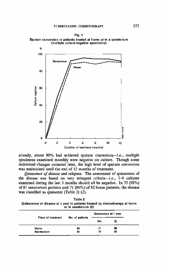
TUBERCULOSIS CHEMOTHERAPY
Fig. 1 Sputum conversion in patients treated at home or in a sanatorium
(multiple cUlture-negative specimens)
%
100
80
20
o
r·------~------·-~-----/ Home
I I I
I I
I I
I I
I I
I I I
I , , , I I ,
I , I
I I ,
t I I §
o L-__ ~ ____ ~~ ____ L-____ ~ ____ ~ __ ~~
o 2 4 6 a 10 12
Duration of treatment (months)
125
already, about 90% had achieved sputum conversion-i.e., multiple specimens examined monthly were negative on culture. Though some individual changes occurred later, the high level of sputum conversion was maintained until the end of 12 months of treatment.
Quiescence of disease and relapses. The assessment of quiescence of the disease was based on very stringent criteria-i.e., 7-9 cultures examined during the last 3 months should all be negative. In 75 (92%) of 81 sanatorium patients and 71 (86%) of 82 home patients, the disease was classified as quiescent (Table 2) (2).
Table 2 Quiescence of disease at 1 year in patients treated by chemotherapy at home
or in sanatorium (2)
Place of treatment
Home Sanatorium
No. of patients
82 81
Quiescence at 1 year
No. "
71 75
88 92

126 K. TOMAN
The frequency of bacteriological relapse was studied in 126 patients whose disease was quiescent after one year of chemotherapy (3); 69 sanatorium patients and 57 home patients could be followed up for up to 5 years (Table 3). During that observation period, II relapses
Table 3
Relapses In 126 patients with quiescent disease after 1 year of chemotherapy In a sanatorium or at home and followed up for a further 4 years (8)
Status Home Sanatorium Total
Quiescent 57 69 126
Relapsed In 2nd year 2 6 8 In 3-5th years 2 3
Total relapsed 4 7 11
" (7) (10)
occurred: 7 (10%) in the sanatorium patients and 4 (7%) in the home patients. Thus the small differences observed at one year (see preceding paragraph) were levelling out. Of the II patients who relapsed, 8 did so in the second year.
Risk to family contacts
The close family contacts of the patients admitted to the study were carefully followed up for 5 years. The main study of the attack-rate (4, 5) was undertaken in families whose only infectious member was the index case. In this way a straight comparison could be made between the " sanatorium" family contacts (whose infectious index case was isolated for a year in sanatorium) and the .. home" contacts (who remained exposed to their index case, living with him in the same household throughout the year of chemotherapy). In addition, both contact groups were equally exposed to the risks of the general urban environment of Madras.
All contacts who had radiographic lesions suggesting tuberculosis were excluded; the rest were subdivided into tuberculin nonreactors and reactors (0-4 mm and ~ 5 mm induration, respectively, to 5 TU of tuberculin given intradermally).
As Table 4 shows, the frequency of disease in the nonreactor group was almost equal in the home contacts and in the sanatorium contacts. (Among the reactors--a less homogeneous group than the nonreactorsthe frequency of disease was higher in the sanatorium contacts.)
TUBERCULOSIS CHIlMOTHERAPY 127
Table 4
Frequency of tuberculosis In family contacts of patients treated at home or In a sanatorium by chemotherapy (5)
Cases occurring Total cases Initial Contact No. of In observation year: In 5-year period
tuberculin group persons status at risk
1 2 3 4 5 No. %
Nonreactors home 86 7 0 1 1 0 9 10.5 sanatorium 87 7 1 2 2 0 10 11.5
Reactors home 159 5 4 4 1 1 15 9.4 sanatorium 177 13 4 7 7 2 28 15.8
Another important finding was that most of the contacts who developed disease during the first year of observation did so within the first 3 months t
irrespective of whether the index case was treated at home or in sanatorium. This was a strong indication that these contacts were probably in the incubation period of disease when first examined-Le., it is very likely that they had already been infected before the index case was discovered and treated.
Cooperation of the patients
In spite of a very active welfare service for the patients and their families, 12 of the sanatorium patients discharged themselves from treatment, 4 being readmitted later. Only one of the patients treated at home was lost through self-discharge.
With regard to the regularity of drug intake, it was found that sanatorium patients also, occasionally or during certain periods, did not ingest the prescribed medicaments. This may be because sanatorium supervision was not always sufficient to ensure that every patient actually took every dose.
Social problems
A careful social record was kept for each family. Major problems arose in 8 families of home patients, whereas they arose in 20 families of sanatorium patients. Furthermore, the difficulties were usually more serious in the latter and mostly resulted in disruption of the family.
Summary of findings
A controlled clinical trial was carried out to compare the effect of 12 months of chemotherapy in two groups of patients: one group treated under good conditions in a sanatorium, the other under poor conditions at home.
128 K. TOMAN
The results in the former, despite good accommodation, nuxsing, rich diet, and prolonged bed-rest, were not superior to those in patients treated in overcrowded homes, who had a poor diet, much less rest, and often very long working hours.
Radiographic changes, such as the reduction of cavity size and cavity closure, were very similar in both groups, particularly when patients with similar pretreatment lesions were compared.
Also bacteriological response, such as reduction in the bacterial content of sputum, or sputum conversion, occurred to the same extent and at almost the same speed in the two groups.
After about 4 months, around 90% of the home and sanatorium patients yielded multiple specimens all negative by culture, and this level was maintained throughout the rest of the treatment year.
Regarding quiescence of the disease at one year and relapses in the subsequent 4 years, the results showed little difference, if any, between home and sanatorium patients. Thus sanatorium treatment did not reduce the likelihood of relapse.
The risk to close family contacts was studied for 5 years, particularly in initial nonreactors to tuberculin. There was no difference in the incidence of disease between the contacts of patients treated at home and those of sanatorium patients, and exposure to the index case under effective chemotherapy appeared to present no major risk to contacts. Thus domiciliary treatment did not entail any special danger that might have been prevented by sanatorium treatment.
The study indicated that the major risk to contacts lay in exposure to the infectious index case before diagnosis was made and treatment initiated. At that point, all the harm the index case could do to his contacts had already been done, so that subsequent isolation in a sanatorium would have been of little benefit.
Sanatorium treatment has the disadvantage that it demands exceptional sacrifices from patients (it is difficult to keep a patient in the sanatorium, separated from his family for a whole year, and to make him observe the strict bed-rest regimen and the sanatorium discipline in general). Indeed, in this study, 12 patients discharged themselves from treatment (though 4 could be readmitted) against I self-discharge in home patients.
Furthermore, the study showed that treatment in a sanatorium is no safeguard against irregularity of drug-taking unless the patient is seen to swallow every dose.
A serious social disadvantage of sanatorium treatment was its disruptive effect on family life.
It was this study that determined the dramatic switch from institutional to ambulatory treatment as a general policy (see" What were the main landmarks in the development of tuberculosis chemotherapy ? "-page 75

TUBERCULOSIS CHEMOTHERAPY 129
and" What is the place of sanatorium and hospital treatment today ... ? to
-page 217).
REFERENCES
1. RAMAKRISHNAN, C. V. lIT AL. Bulletin of the World Health Organization, 25 : 339 (1961)
2. TuBERCULOSIS CHEMOTHERAPY CENrRB, MADRAS. Bulletin of the World Health Organization, 21 : 51 (1959)
3. DAWSON, Y. Y. ET AL. BuUetin of the World Health Organization, 34: 533 (1966)
4. ANDREWS, R. H.IlT AL. Bulletin of the World Health Organization, 23: 463 (1960)
5. KAMAT, S. R. ET AL. BuOetin of the World Health Organization, 34: 517 (1966)

130 K. TOMAN
What are two-phase chemotherapy and the so-called 100 % regimens ?
The concept of two-phase chemotherapy was arrived at on the basis of theoretical considerations, empirically and experimentally discovered facts, and technical and operational requirements for meeting patients' needs (1-3).
It was observed shortly after the introduction of chemotherapy for tuberculosis that daily treatment with three-drug regimens of isoniazid, streptomycin, and PAS had a success rate approaching 100% (2. 4). However, it was soon questioned. first. whether it was necessary to administer all three drugs for the whole duration of treatment, since bacterial conversion was usually achieved during the first few months ; and, secondly, whether-after that point had been reached-two drugs, or even one, would not suffice to maintain or accomplish the therapeutic effect. Thus, a course of chemotherapy would consist of two successive parts: an aggressive or intensive regimen would be given at the outset, followed by a less aggressive regimen during the continuation phase. In the former phase, a very potent regimen of two or, better, three drugs would be given daily. In the latter, a less potent regimen could be administered daily or intermittently. An important aspect was that the patient needed to take fewer drugs, and therefore the risk of toxicity would be lowered. Moreover, staff time and costs could be substantially reduced if an oral drug regimen could be prescribed for self-administration in the continuation phase.
Plenty of clinical and experimental information about the high efficacy of two-phase chemotherapy is now available. The two-phase regimens most frequently used at present are shown in Table I. Usually, in the intensive phase, three drugs are given daily; in the continuation phase, two drugs are administered daily or intermittently for the rest of the
Table 1
Two-phase chemotherapy: the «100% regimens» of primary chemotherapytJ (5)
InitIal Intensive phase
streptomycin + PAS + Isoniazid
streptomycin + thioacelazone + isoniazid <
Continuation phase
1. PAS + Isoniazid dally 2. streptomycin + Isoniazid } twice 3. PAS + Isoniazid weekly
1. thlocetazone + Isoniazid dally
2. streptomycin + Isoniazid { ~~~~y
"In these regimens, PAS or thloacetazone may be replaced by ethambutol.

TUBERCULOSIS CHEMOTHERAPY 131
planned period. All the regimens shown in the table are capable of achieving 100% success after 12 months in patients with newly diagnosed tuberculosis and a regular drug intake. PAS or thioacetazone may be replaced by ethambutol. Another, equally effective two-phase regimen consists of streptomycin plus isoniazid daily in the intensive phase, the same drugs being administered twice weekly in the continuation phase. Instead of streptomycin, rifampicin may be given with isoniazid daily in the intensive phase and twice weekly in the second phase. This regimen has the advantage of being an entirely oral regimen; however, the high cost of rifampicin and concern that it might be toxic when given intermittently have so far limited its use even in technically advanced countries.
Controlled trials of once-weekly continuation regimens of streptomycin and isoniazid or an oral regimen ofisoniazid and PAS twice weekly in the continuation phase have yielded promising results and are being investigated further (see .. What is intermittent chemotherapy ... ?"page 132 and" What was the efficacy of primary intermittent chemotherapy ... ? "-page 137).
Even a single drng-isoniazid-given daily in the continuation phase (after streptomycin plus isoniazid daily for 6 months) was highly effective in a controlled trial in Singapore (6). Among 114 patients so treated, only one was a bacteriological failure, and no relapse occurred within 3 years of follow-up. For further information, the reader is referred to : " What is the purpose of the initial intensive phase ... ? "-page 145 ; .. What is the optimum duration of the initial intensive phase ... ? "page 156; and" When is a two-phase regimen indicated? "-page 151.
REFERENCES
1. KAss, I. ET AL. Annals of internal mediCine, 47: 744 (1957)
2. RUSSEL, W. F. & MIDDLEBROOK, G. Chemotherapy of tuberculosis. Springfield, IL, Thomas, 1961
3. CANETIl, G. Tubercle, 43: 301 (1962)
4. Ross, 1. D. ET AL. British medical journal, 1 : 237 (1958)
5. Fox, W. Bulletin of the International Union against Tuberculosis, 47: 49 (1972)
6. SINGAPORE TuBERCULOSIS SERVICE/BROMPTON HOSPITAL/BRITISH MEDICAL RESEARCH COUNCIL INVESTIGATION. Tubercle, 55: 251 (1974)

132 K. TOMAN
What is intermittent chemotherapy and what is the scientific basis for intermittency?
Intermittent regimens are those in which the individual drug doses are given at intervals of more than one day-e.g., once or twice weekly.
Originally it was believed that antituberculosis drugs needed to be given every day in one or in several doses in order to maintain drug concentrations in the body continuously at inhibitory levels. However, in vitro studies and animal experiments have shown that certain drugs were effective also when the drug concentration dropped temporarily below that level, and indeed even after they had disappeared from the lesion (1) or the medium (2),
As has already been mentioned (see .. What is the biological mechanism of chemotherapy? "-page 77), in vitro experiments have demonstrated that, after a culture of M. tuberculosis had been exposed to certain drugs for some time, it took several days (the" lag period ") before new growth occurred.a Table 1 shows the lag in the growth of M. tuberculosis after exposure to certain drugs for varying times.
Table 1
Lag In growth of M. tuberculosis after temporary exposure to drugs (2, 3)
Drug Concentration Lag (days) after exposure for:
(mg/lltre) 6h 24 h
Isoniazid 0 &.-9 Ethionamide 5 0 10 Cycloserine 100 0 4-8 Ethambutol 10 0 4-5 Pyrazinamide 50 5-40- 40-Streptomycin 5 8-10 8-10 RHamplcln 0.2 2-3 2-3
Thloacetazone 10 0 0 Tiocarllde 10 0 0
- Depending on the pH of the medium (6.2-5.6).
Thioacetazone and tiocarlide did not produce any lag, even after exposure of 24 h or more." Immediately after these two drugs had been
a After exposure, the drug was always thoroughly washed out of the culture and the colonies were reinoculated into a drug-free medium.
" Up to 96 h. Within this exposure time, all drugs achieved the maximum lag.

TUBERCULOSIS CHEMOTHERAPY 133
removed from the culture medium, growth started again. This suggests that both drugs are unsuitable for intermittent chemotherapy, which was indeed confirmed by animal experiments.
The drugs listed in the upper part of the table have all been found to be bactericidal in vitro. However, for each drug there was a maximum lag (last column) that seems to indicate the practical limit beyond which the interval between two doses should not be extended. Animal studies (4, 5) showed conclusively that, the longer the chosen interval between doses, the higher the doses of most of the drugs needed to be. Thus, with high doses of isoniazid, a 3-day interval was shown to be the optimum, whereas an extension to 8 days gave significantly worse results.
Though experimental findings cannot be mechanically transferred to man, these results were promising enough to be explored in clinical studies.a A controlled clinical trial was therefore undertaken at the Tuberculosis Chemotherapy Centre, Madras (6).
A standard oral regimen of 100 mg of isoniazid plus 5 g of sodium PAS twice daily was compared with a twice-weekly regimen of I g of streptomycin given by intramuscular injection plus 14 mg of isoniazid per kg of body weight, given orally in a single dose. Both regimens were given to ambulatory patients at random. The oral regimen was dispensed for self-administration. For the intermittent regimens, the patients had to attend the clinic on two days a week at 3-4-day intervals. The treatment was fully supervised-Le., each patient had first to take his isoniazid tablets in the presence of the staff {who checked that the tablets actually had been swallowed), and then received the injection of streptomycin. The results at 12 months are shown in Table 2.
Table 2
Comparison of results at the end of 12 months' treatment with streptomycin (5) and isoniazid (H) twice weekly and with PAS (P) and H dally (6)
Status of disease
Bacteriologically quiescent Bacteriologically active (Death from tuberculosis)
Total patients
SH twice weekly G
No. %
68 94 2 2 2 3
72 100
G SH: streptomycin. 1 g I.m. + isoniazid. 14 mg/kg body weight.
PH dally·
No. %
58 85 9 14
2
88 100
• PH: sodium PAS, 10 g + isoniazid, 200 mg, dally. divided into two equal doses.
tI Several trials of intermittent regimens, carried out about 10 years earlier, had not been very successful owing to the use of inadequate dosages and drug combinations.

134 K. roMAN
The intermittent regimen was highly successful and perhaps slightly more effective than the daily regimen. The potency of intermittent chemotherapy is all the more striking as most of the patients admitted to the study had extensive, bilateral cavitated disease with sputum heavily positive by direct smear. That was a feature common to all the Madras studies, in which the patients had much more severe disease than is generally found nowadays in technically advanced countries. The relapse rates in a 2-year period were 8% for the twice-weekly and 12% for the daily regimen; after 4 years, they were 12% and 15%, respectively. In 4 out of 5 patients who relapsed on the intermittent regimen, the bacilli were susceptible to both isoniazid and streptomycin. This suggests that, had there been an intensive phase at the start of treatment , the susceptible bacilli would probably have been eliminated.
In another study, the possibility of increasing the interval between doses to one week was investigated. This study, also, was undertaken in outpatients in Madras. Four intermittent regimens were studied concurrently, but, for the sake of simplification, only the two regimens described below will be analysed here (for the results of the complete study, see "What was the efficacy of primary intermittent chemotherapy ... ? "-page 137).
The regimen consisting of streptomycin plus isoniazid twice weekly -SH(TW}-was compared with streptomycin plus isoniazid once weekly-SH (OW). The dosage was the same for both regimens: 1.0 or 0.75 g of streptomycin plus 15 mg of isoniazid per kg of body weight. All the patients received 6 mg of pyridoxine with every dose of isoniazid (incorporated in the isoniazid tablets) in order to prevent possible neurotoxicity owing to the high dosage of isoniazid. The effect of a lower dose (0.75 g) of streptomycin was studied because it seemed likely that the smaller dose would be sufficient and better tolerated, particularly by debilitated or elderly patients, than the usual I-g dose. The results of 12 months of treatment are summarized in Table 3.
Table 3 Comparison of results at the end of 12 months' treatment with streptomycin (S)
and isoniazid (H) twIce weekly and once weekly <n
Status of disease
Bacteriologically quiescent Bacteriologlcelly active (Death from tuberculosis)
Totel patients
SH twice weekly 4
No. %
107 91 9 8 1 1
117 100
4 SH: streptomycin. 1 g i.m. + Isoniazid. 15 mg/kg body weight.
SH once weekly 4
No. %
82 71 30 26 3 3
115 100

TUBERCULOSIS CHEMOTHERAPY 135
Again, the twice-weekly regimen proved to be highly successful and the once-weekly regimen considerably less effective. Nevertheless, it was rather impressive that, despite severe disease, 71 % of patients on the latter regimen still achieved bacteriological qmescence.a
The reasons for the inferiority of the once-weekly regimen were examined and the findings were both interesting and important. In the analysis, the patients were grouped according to the rate of inactivation of isoniazid (see "What is rapid inactivation (acetylation) of isoniazid ... ? "-page 148) and the dosage of streptomycin.
Table 4 shows that the twice-weekly regimen is influenced neither by the inactivation rate of isoniazid nor by a 25% reduction in the dosage of streptomycin. In contrast, the once-weekly regimen was clearly affected by the rate of isoniazid inactivation and, to a lesser extent, also by the reduction in the streptomycin dosage. The twice-weekly regimen can therefore be regarded as both robust and effective, even without an initial intensive phase.
Table 4
Streptomycin plus Isoniazid twice weekly - SH (TW) - compared with streptomycin plus Isoniazid once weekly - SH (OW) i according to the rate of inactivation of
isoniazid and the dosage of streptomycin (7)
Regimen
SH (TW) SH(OW)
Patients with quiescent disease at 1 year (%) Isoniazid Inactivation rate streptomycin dosage
slow
91 82
rapid
91 60
1 g
91 76
0.75 g
92
62
The isoniazid inactivation rate influenced also the response to other once-weekly regimens concurrently investigated (see .. What was the efficacy of primary intermittent chemotherapy ... ? "-page 137). This indicates that, when the interval between doses is extended to one week, intermittency reaches its practical limit of effectiveness. In order to achieve a sufficiently long lag, the length of exposure to isoniazid at a bactericidal concentration would have to be increased, necessitating an additional increase in the individual dosage of isoniazid. Unfortunately. the dosage cannot be increased further because of the risk of acute toxicity.
/J For the main analysis of the complete study, the results had to be statistically standardized. Only the patients who could be compared with regard to all requirements stated in the protocol were included. Consequently, the once-weekly regimen was found to be effective in only 68%.

136 K. TOMAN
Methods of compensating for the difference between slow and rapid inactivators are under investigation. The most promising approach so far seems to be the development of a slow-release preparation of isoniazid that would prolong absorption from the gastrointestinal tract and excretion of the drug. In any case, intermittent regimens, especially those administered once weekly, would benefit from an intensive phase for several weeks. However, the feasibility of extending the interval between doses to more than one week is at present viewed with scepticism (8) (see also" What was the efficacy of primary intermittent chemotherapy ... ? "-page 137).
Previously, a regimen of streptomycin twice weekly plus isoniazid daily was widely used, disregarding the findings of a controlled trial that this regimen frequently results in failure owing to the emergence of isoniazid resistance (9). This regimen should never be used.
REFERENCES
1. CANI!lTI, G. ET AL. Tubercle, 44: 236 (1963)
2. DICKINlION. J. M. & MrrCHISON. D. A. Tubercle, 47: 370 (1966)
3. MITCHISON, D. A. & DICKINSON, J. M. Postgraduate medical Journal, 47: 737 (1971)
4. GRUMBACH, F. ET AL. Tubercle, 48: 11 (1967)
5. DICKINSON, J. M. & MITCHISON, D. A. Tubercle, 47: 381 (1966)
6. TUBERCULOSIS CHEMOTHERAPY CBNI'RB, MADRAS. Bulletin of the World Health Organization, 31 : 247 (1964)
7. MENON, N. K. Cited in Fox, W. American rel1iew of respiratory disea:sea, 97 : 767 (1968)
8. MITCHlSON, D. A. Studia pneumologica et phtiseologica cechoslol1Qca, 31: 288 (1971)
9. GREAT BRITAIN, MEDICAL REsEARCH CoUNCIL. British medical Journal, 1 : 43S (1955)

TUBERCULOS~ CHEMOTHERAPY
What was the efficacy of primary" intermittent chemotherapy in controlled clinical studies?
Intermittent chemotherapy twice weekly
137
The results of twice-weekly chemotherapeutic regimens, compared with those of daily regimens, are shown in Table 1.
The study conducted at the Tuberculosis Chemotherapy Centre, Madras, in 1964 (1) is discussed in greater detail under "What is intermittent chemotherapy ... ? "-page 132).
Table 1
Comparison of the results of twice weekly and dally regimens for newly diagnosed pulmonary tuberculosis
Study
Tuberculosis Chemotherapy Centre, Madras, 1964 (1)
International Union against Tuberculosis, 1970 (2)
Lahlou, 1970 (3)
Decroix et al., 1971 (4)
Czechoslovak Tuberculosis Service/ WHO/British Medical Research CounCil, 1971 (5)
Czechoslovak Tuberculosis Service/ WHO/British Medical Research Council, 1974 (6)
Devl, 1972 (7)
Regimens·
PH daily SH twice weekly TH dally SH twice weekly EH daily SH weekly twice RH daily RH twice weekly PH dally SH twice weekly
SH twice weekly SH twice weekly
PH daily SH twice weekly
Tripathy, 1972 (8) } PH daily Tuberculosis Chemotherapy Centre, PH twice weekly
Madras, 1973 (9)
Initial Intensive No. of
phase patients (months) •
o 72 66
134 95
151 167 39 41 o
165 233 3
93 100
155 163 3
83 go
Quiescent disease
at 1 year (%)
85 94
95 92
88 c
go" 92 88 99
100
98 99
91 98
87 88
• P - PAS, H - isoniazid, S - streptomycin, T - thloacetazone, E - ethionamide. R - rifampicin.
• Three-drug combination given daily . • At 8 months.
In a cooperative trial organized by the International Union against Tuberculosis (2), the effect of 150 mg of thioacetazone plus 300 mg of isoniazid given together in a single dose daily was compared with that of a high dose (750 mg) of isoniazid plus streptomycin twice weekly in the continuation phase, after an initial daily phase of three drugs (isoniazid, thioacetazone, and streptomycin) combined, for one month. Both regimens were very effective and yielded similar results. In a subsequent study (10), the International Union against Tuberculosis investigated
t1. Primary chemotherapy is the administration of antituberculosis drugs for the first time.

138 K. TOMAN
whether the addition of PAS or thioacetazone to the twice-weekly isoniazid plus streptomycin regimen would improve the success rate. The proportions of bacteriologically unsatisfactory results were about the same as in the preceding study-i.e., no improvement could be demonstrated.
In a study (3) undertaken in North Mrica (Morocco, Tunisia, and Libya), a regimen of 750 mg of ethionamide plus 750 mg of isoniazid twice weekly was compared with 1 g of streptomycin plus 750 mg of isoniazid, also twice weekly. The results at 8 months were similar. This was the first report on a fully oral regimen. A further study by the same author indicated that the intermittent dose of ethionamide might be critical, since a 500-mg dose had been shown to be less effective. In the initial phase, streptomycin (1 g) plus ethionamide (500 mg) plus isoniazid (500 mg) were given daily.
Decroix et al. (4) found little difference between the daily regimen (600 mg of rifampicin and 750 mg of isoniazid) and the twice-weekly regimen (900 mg of rifampicin and 750 mg of isoniazid). There was no initial intensive phase. The study population was rather small. Other authors reported higher success rates.
The studies conducted jointly by the Czechoslovak Tuberculosis Service, the World Health Organization, and the British Medical Research Council (5, 6) will be discussed further below.
Devi (7) compared isoniazid plus PAS daily with streptomycin plus a high dose of isoniazid twice weekly-both after an initial three-drug regimen (isoniazid, PAS, and streptomycin) daily for 3 months. The regimens were randomly allocated to regular outpatients of a chest clinic in Singapore, 90% of whom had moderately or far advanced disease. Excluding from assessment patients who had major interruptions of treatment or in whom the regimen had to be changed, the results of the daily oral regimen came close to those of the intermittent regimen. It was concluded that the margin was too small to justify the introduction of intermittent regimens as a routine in Singapore.
Tripathy (8,9), on behalf of the Tuberculosis Chemotherapy Centre, Madras, reported on a controlled comparison of two oral regimens: isoniazid plus PAS daily and isoniazid plus PAS twice weekly. All patients had an initial phase (2 weeks) of 1 g of streptomycin plus 400 mg of isoniazid (in two divided doses) plus 6 g of PAS. The twice-weekly regimen consisted of 400-750 mg (15 mg per kg of body weight) of isoniazid and 7.5-10 g of PAS; pyridoxine was incorporated into the isoniazid tablets as usual. The results were similar for both regimens and, considering the severity of the disease,a were very encouraging for
a About four-fifths of the patients had extensive, bilateral cavitated disease; 90% had a positive smear from the first specimen of overnight sputum.

TUBERCULOSIS CHEMOTHERAPY 139
the intermittent regimen. However, there was a marked difference between the two groups in the toxicity of PAS: In the" intermittent" group, only 6% had toxic reactions to PAS, whereas, in the" daily" group, the frequency of toxicity was 21 %, including one death due to agranulocytosis and five patients who had to have their regimens changed. The investigators indicated that the effectiveness of the twiceweekly regimen was influenced particularly by the size of the initial bacterial population, and thought that an extension of the intensive phase from 2 to 4 weeks might improve the results. However, that could be difficult in areas where patients have to travel long distances.
The WHO Collaborating Centre for Tuberculosis Chemotherapy, Prague (5) reported excellent results from a study of which an important feature was the flexibility of the treatment organization. Each patient could choose to receive his treatment at the place most convenient to him-Le., a chest clinic, physician's surgery, factory dispensary, health centre, or hospital on the way to work. If necessary, an auxiliary could come to the patient's home. The area was partly rural and had satisfactory transport facilities.
Most of the patients admitted to the study were over 50 years of age. However, their disease was on the average less serious than it generally is in patients attending chest clinics in developing countries.
The success· of the study was due largely to the excellent cooperation of patients, which was achieved by adapting treatment services to their convenience. Though the treatment of tuberculosis in Czechoslovakia is almost entirely the responsibility of a rather dense network of specialized tuberculosis services (inpatients and outpatients), the participation of nonspecialized health services was of special importance. Moreover, the study clearly demonstrated how general health services can become increasingly involved in the management of tuberculosis patients, and that they are capable of taking over this responsibility from the specialized services if necessary. The same applies to all technically advanced countries, in which the tuberculosis specialist is leaving the scene. This also shows the developing countries that nowadays it is no longer necessary to build up a specialized tuberculosis service: the task of tuberculosis chemotherapy may be entrusted to the general health services as one of their routine duties.
More recently, in another study (6), two regimens-streptomycin plus isoniazid twice weekly with initial daily phases of two different durations (streptomycin plue isoniazid plus PAS for 6 weeks and for 13 weeks)were compared. No appreciable difference between the results obtained with these two regimens was demonstrated.
The use of twice-weekly regimens has become widespread in clinical practice. The potency of isoniazid plus streptomycin given twice a week has been demonstrated, for example in Algeria (11). In a pilot study

140 K. TOMAN
carried out in field conditions, this regimen was given intermittently, either right from the beginning or after a varying period of initial threedrug treatment (streptomycin, isoniazid, and PAS). The results were favourable and indicated that, when administration can be properly supervised, the success rate can be as high as that achieved under the conditions of controlled trials.
Intermittent chemotherapy once weekly
As already mentioned (see" What is intermittent chemotherapy ... ?" -page 132), a series of once-weekly regimens was investigated at the Tuberculosis Chemotherapy Centre, Madras (12). In outpatients with newly diagnosed, extensive, bilateral cavitated disease and with sputum positive by direct smear, four regimens were studied :
1. streptomycin plus 15 mg of isoniazid per kg of body weight, twice a week (SH (TW» ;
2. streptomycin plus 15 mg of isoniazid per kg of body weight, once a week (SH (OW» ;
3. streptomycin plus 15 mg of isoniazid per kg of body weight, plus a high dose (90 mgfkg) of pyrazinamide, once a week; and
4. streptomycin plus 400 mg of isoniazid daily for 4 weeks, followed by streptomycin plus 15 mg of isoniazid per kg of body weight, once a week.
The twice-weekly regimen was the most effective and the streptomycin plus isoniazid once-weekly regimen gave comparatively poor results, achieving quiescence in only 68% of patients (Table 2). The addition of pyrazinamide as a third drug to the once-weekly regimen SH (OW) made little difference. However, an initial daily phase of 4 weeks with streptomycin produced a substantial improvement.
Table 2
Status of patients after 1 year of treatment with twice-weekly (TW) and once-weekly (OW) chemotherapeutic regimens (12)
Proportion No. of of patients
Regimen" patients bacteriologically treated quiescent
(%)
SH (TW) 96 94
SH (OW) 101 68 SHZ (OW) 101 74
SHISH (OW) 77 68
.. S .. streptomycin. H -Isoniazid, Z - pyrazinamide.
Oeaths from
tuberculosis
(%)
2 o

TUBERCULOSIS CHEMOTHERAPY 141
The reasons for the inferiority of the three once-weekly regimens became apparent when the patients were grouped according to the rate of inactivation of isoniazid (Table 3). The twice-weekly regimen did not seem to be greatly impaired by the rapid inactivation of isoniazid: in a larger series of patients the figures were practically equal for rapid and for slow inactivators (91 % and 92%. respectively) (see" What is rapid inactivation (acetylation) of isoniazid ... ? "-page 148). However, in all the once-weekly regimens. rapid inactivators were at a considerable disadvantage. On the other hand, the introduction of an initial daily phase of 4 weeks led to a significant increase of efficacy, particularly in slow inactivators, reaching 95%.
Table 3
Quiescence of disease at 1 year in slow and rapid inactlvators of Isoniazid given twice-weekly (TW) and once-weekly (OW) chemotherapy
Regimen"
5H (TW) 5H (OW) 5HZ (OW) 5H/5H (OW)
Patients with quiescent disease whose Isoniazid Inactivation rate was;
slow (%) rapid (%)
97 76 87 95
91 5& 53 76
G 5 = streptomycin, H = Isoniazid, Z - pyrazinamide.
Attempts had been made to reduce rapid inactivation by adding PAS (13) as a third drug to the SH (OW) regimen.a However, neither the addition of PAS nor an increase in the isoniazid dose to 17 mgfkg improved the results in rapid inactivators.
The following conclusions may be drawn from the experience gained with intermittent chemotherapy without rifampicin.
1. Twice-weekly regimens containing isoniazid in high dosage (14-IS mgfkg) and streptomycin (0.75-1 g) are highly effective, whether given from the outset or after an initial intensive phase of treatment. Their efficacy in slow and rapid inactivators of isoniazid is very similar. The findings suggest that these regimens can be highly effective in patients with extensive disease and in populations with a high frequency of rapid inactivators. The twice-weekly regimens described in this chapter are no longer experimental, but have proved their value in the primary treatment of tuberculosis in routine practice.
a PAS competes with isoniazid for acetylation by the liver, and thus increases indirectly the serum level of isoniazid.

142 K. TOMAN
2. A once-weekly regimen of isoniazid (15 mg/kg) and streptomycin (1 g) after initial daily therapy with isoniazid (400 mg) and streptomycin (1 g) given for 4 weeks, approached closely the efficacy of the twiceweekly regimen; however, unlike the latter, it was substantially inferior in rapid inactivators. Once-weekly regimens containing rifampicin have been studied further in South-East Asia and in Europe.
In a recently reported controlled trial in Singapore, 481 patients were allocated to intermittent regimens administered once or twice weekly (J 4). All received streptomycin with isoniazid and rifampicin daily for the first 2 weeks. This initial phase was followed by a continuation phase with isoniazid (15 mg per kg of body weight) and rifampicin (600 mg or 900 mg, at random). The total duration of treatment was 12 or 18 months, at random. All patients were positive by direct smear microscopy at admission and almost three-quarters of them were rapid acetylators (inactivators) of isoniazid (see" What is rapid inactivation (acetylation) of isoniazid ... '/ "-page 148).
There was only one failure (1 %) during treatment in the twice-weekly group (on 600 mg of rifampicin), but 12 of 202 (6%) in the once-weekly group. All 13 (4%) belonged to the 310 rapid acetylators, whereas none of the 117 slow acetylators failed within the observation period of 30 months.
Relapses after chemotherapy occurred in only 2 patients (1 in the once-weekly and 1 in the twice-weekly group). Both belonged to the 12-month series. No relapse occurred in the I8-month series.
Adverse reactions to rifampicin (see " What is the therapeutic effect and what is the toxicity of antituberculosis drugs'/ "-page 101) were relatively frequent in the once-weekly group (14-25%), compared with 7-11 % in the twice-weekly group. Four of the five cases of thrombocytopenia occurred in the latter group. After rifampicin was terminated, all five returned to normal platelet counts within 48 hours. The most common, but mild, adverse reaction was the "flu syndrome" (in up to 22%), increasing with the interval and with the dose of rifampicin. No patient had jaundice.
The authors concluded that the twice-weekly regimen was strikingly effective in patients with newly-diagnosed drug-susceptible tuberculosis. The once-weekly regimen was less effective because of a high rate of rapid isoniazid acetylators. In countries where this rate is considerably lower (e.g., in the USA and certain European countries ~40%), the once-weekly regimen is likely to produce better results, particularly when the initial intensive phase is extended. Of 13 initially isoniazid-resistant patients, 9 (70%) responded favourably. The once-weekly regimen required only 64 doses (14 daily and 50 weekly)-a fact that might be of importance for short-course chemotherapy (see "How effective is short-course chemotherapy ... '/ "-page 183). The 18-month course of treatment offered no advantages over that lasting 12 months.

TUBERCULOSIS CHEMOTHERAPY 143
In another controlled trial reported from Europe (German Democratic Republic), which started in 1970, two once-weekly regimens of isoniazid plus rifampicin-one with an initial daily phase of both drugs for 4 weeks; the other without an initial phase-were compared (I5). The control regimen was the standard regimen of isoniazid with streptomycin and PAS daily for 4 months, followed by isoniazid and streptomycin twice weekly. The total duration of all regimens was 12 months. There were 2 failures during chemotherapy (one with each once-weekly regimen) and 1 bacteriological relapse after chemotherapy in the onceweekly regimen with an initial daily phase. The only disadvantage of the once-weekly regimen was a ":flu syndrome" rate of 45%. This was halved by lowering the dose of rifampicin to 15 mg/kg.
The authors considered both once-weekly regimens as equally good. They recommended that the use of rifampicin should be limited to a maximum of 12-20 weeks, within which period side-effects occur only occasionally or within acceptable limits, and to continue the treatment with isoniazid and streptomycin until 1 year. The practicability of this regimen is now being studied on a large scale.
REFERENCES
1. TuBERCULOSIS CHEMOTHERAPY CENTRE, MADRAS. Bulletin of the World Health Organization, 31 : 247 (1964)
2. INI'ERNATIONAL UNION AGAINST TUBERCULOSIS. Bulletin of the International Union against Tuberculosis. 44: 8 (1970)
3. LAHLOU. M. Bulletin of the International Union against Tuberculosis, 43: 289 (1970)
4. DECROIX. G. ET AL. Revue de la tuberculose et de pneumologle, 33: 751 (1971)
5. WHO CoLLABORATING CENTRE FOR TuBERCULOSIS CHEMOTHERAPY, PRAGUE.
Bulletin of the World Health Organization, 45 : 573 (1971)
6. WHO COLLABORATING CENTRE FOR TUBERCULOSIS CHEMOTHERAPY, PRAGUE.
Bulletin of the International Union against Tuberculosis, 49: 403 (1974)
7. DEVI, S. Bulletin of the International Union against Tuberculosis, 47: 15 (1972)
8. TRIPATHY, S. P. Bulletin of the International Union against Tuberculosis, 47: 30 (1972)
9. TUBERCULOSIS CHEMOTHERAPY CENTRE, MADRAS. British medical Journal, Z: 7 (1973)
10. INI'ERNATIONAL UNION AGAINST TuBERCULOSIS. Bulletin of the International Union against Tuberculosis, 49 : 7 (1974)
11. CHAULET, P. ET AL. Tubercle, 48: 128 (1967)
12. TuBERCULOSIS CHEMOTHERAPY CENTRE, MADRAS. Bulletin of tIw World Health Organization,43; 143 (1970)

144 K. TOMAN
13. NA.Z.ARETH, M. O. Y. Cited in Fox, W. PostgradUQte medical journal, 47: 729 (1971)
14. SINGAPORE TuBERCULOSIS SERVICE I BRITISH MEDICAL REsEARCH CoUNCIL. American review of respiratory diseases, 116: 807 (1977)
IS. EULE, H. ET AL. Bulletin of the International Union against Tuberculosis, 51 : 115 (1976)

TUBERCULOSIS CHEMOTHERAPY
What is the purpose of the initial intensive phase of two-phase chemotherapy?
145
There is ample experimental and clinical evidence that a two-drug and particularly a three-drug regimen, when administered daily in the initial period of treatment, may greatly improve the efficacy of chemotherapy. However, not any combination of two or three drugs will have this effect. At least two bactericidal drugs, such as isoniazid and streptomycin or isoniazid and rifampicin, are required daily in the initial phase. The addition of a third drug can contribute only little to such a potent regimen. However, a supplement of PAS, thioacetazone, or ethambutol is of benefit when initial drug-resistance is present. The efficacy of three or more bactericidal drugs~.g., isoniazid, rifampicin, and streptomycin, with or without pyrazinamide-has been explored especially in studies of short-course chemotherapy (see " How effective is short-coursechemotherapy ... ?"-page 183).
It has been consistently observed that the multiplication of susceptible organisms stops already during the first days of effective treatment (J, 2), and that the total number of bacilli in the sputum decreases rapidly, especially within the first 2 weeks of effective treatment (3). The experimental findings from laboratory and controlled clinical studies are summarized below.
1. It is of crucial importance for the outcome of treatment, especially for patients harbouring large bacterial populations, to put a rapid stop to bacterial mnltiplication and ensure that drug-sensitive bacilli are killed as soon as possible (" early kill "), for the following reasons.
(a) The serious deterioration that may occur in the initial period of treatment with regimens of low intensity and potency can be prevented and patients may be saved from an early death (4) - both early deterioration and death being caused by the multiplication of drug-sensitive organisms in the first weeks of treatment.
(b) If the bacterial population is rapidly reduced, say, from 108 (a number commonly found in lung cavities) to 108, there is little probability that new resistant mutants will appear, even after seven steps (generations) of uninhibited multiplication. Thus the emergence of new resistant mutants can be minimized or stopped by intensive chemotherapy at the beginning of treatment.
(c) There is good in vitro evidence that, the more rapid the antibacterial effect, the less likely are persisters to emerge (5). The risk of relapse is thus lessened (see" What is bacterial persistence ... ?"page 204).

146 K. TOMAN
2. In each three-drug combination, there are always two drugs capable of destroying single-drug-resistant mutants pre-existing in wild strains. Thus a three-drug regimen will safely prevent these organisms from multiplying. Such multiplication may be particularly dangerous in the early treatment phase because, at the start of treatment, an appreciable number of drug-resistant mutants may be present.a If these are allowed to multiply, resistance to two drugs can rapidly develop (6). 3. In patients with initial resistance to a single drug-i.e .• primary as well as undisclosed acquired resistance due to a short episode of inadequate treatment-the chances of responding favourably to chemotherapy are almost unimpaired if an initial period of daily treatment with three drugs is provided (see Hong Kong study in .. How relevant are initial drug resistance and pretreatment susceptibility tests ... ?"page 167).
The special importance of the intensive phase in short-course regimens is referred to elsewhere (see .. What are the bactericidal and sterilizing mechanisms in short-course chemotherapy ? "-page 200 and .. How effective is short-course chemotherapy and what are its prospects? "page 183).
The advantages of an initial intensive phase with three-drug regimens are obvious. However, it is important to realize that those who will gain from a third drug and an intensive phase are mainly patients who harbour large numbers of tubercle bacilli and therefore are usually positive by direct smear microscopy. Patients with lesions containing smaller numbers of bacilli respond very well to two-drug regimens with or without an initial intensive phase. The disadvantages of adding a third drug are higher toxicity and higher costs (see" When is a two-phase regimen indicated? "-page 151 and .. What is the optimum duration of the initial intensive phase ... ? "-page 156).
a In one million tubercle bacilli (of a wild strain). about 10-50 isoniazid-resistant mutants and about 1-5 streptomycin-resistant mutants may be found-i.e., in a population of 108 (a number commonly found in cavities), some SOOO isoniazidresistant and several hundred streptomycin-resistant mutants could be present at the outset (see .. How many drug-resistant tubercle bacilli can be found in the sputum of patients who have never received tuberculosis chemotherapy? tt-page 93).
REFERENCES
1. Y.AEGIlR, H. ET AL. American Teriew o/respiTatory diseases, 95: 998 (1967)
2. HOBBY. G. ET AL. Cited in Sbarbaro, 1. A. Chest. 68: 436 (1973)
3. MITCHJSON. D. A. British medicaljoUl'Ml, 1 : 1333 (1965)

TUBERCULOSIS CHEMOTHERAPY 147
4. TuBERCULOSIS CHEMOTHERAPY CBNnul, MADRAS. Bulletin of the World Health Organization, 43: 196 (1970)
5. CRoFTON. 1. & DOUGLAS, E. Respiratory diseases. Oxford. BlackweII, 1975, p. 98
6. MrrcmsoN. D. A. Bulletin of the International Union against Tuberculosis, 43: 322 (1970)

148 K. TOMAN
What is rapid inactivation (acetylation) of isoniazid and what is its clinical significance ?
A large proportion of isoniazid disintegrates in the human body and is converted into metabolites .. that are practically inactive against tubercle bacilli. The capacity to metabolize isoniazid varies greatly in mammals. In particular, it has been observed that the speed at which the drug is inactivated (acetylated) differs among human beings, though it is constant in each individual (1).
The degree of inactivation can be measured by examining the level of free isoniazid in the blood serum at various times after the ingestion of a certain dose. According to this level, it is possible to distinguish two broad groups of persons: slow and rapid inactivators (acetylators) of isoniazid. The rate of inactivation is related to genetic factors and is identical in monozygotic twins. There is evidence that, in certain ethnic groups, the frequency of rapid inactivation is rather high. Thus, the Japanese and Koreans (2), and particularly the Eskimos (3), have a prevalence of rapid inactivation ranging from 50% to 95% (5)-a higher rate than that in South India (Madras), where rapid inactivation was found in about 40% (4). In contrast, Europeans (Caucasians) are predominantly slow inactivators.
Clinical significance of rapid isoniazid inactivation
Several investigations have been undertaken to determine whether slow inactivators respond differently to regimens containing isoniazid than rapid inactivators because the latter cannot maintain high inhibitory levels of the drug for a long time.
A study carried out at the Tuberculosis Chemotherapy Centre, Madras, comparing isoniazid and PAS daily with three regimens of isoniazid alone daily in varying doses (4) showed slightly better results for slow inactivators. However, no such differences could be found in another Madras study in patients who received isoniazid daily in high doses (14 mg per kg of body weight) (6). Studies on intermittent regimens have shown that the rate of inactivation of isoniazid had no appreciable influence on the efficacy of twice-weekly regimens-e.g., isoniazid plus streptomycin (7), isoniazid plus PAS (8), or isoniazid plus ethambutol (9) administered twice a week after initial daily chemotherapy with three drugs for several weeks. However, when isoniazid was given once a week
.. Chiefly acetyl derivatives of isoniazid; hence the term .. acetylation". sometimes used instead of .. inactivation".

TUBERCULOSlli CHEMOTHERAPY 149
together with streptomycin (7) or ethambutol (9). the results were evidently inferior in rapid inactivators (see "What was the efficacy of primary intermittent chemotherapy ... ? "-page 137).
Thus, when intermittent chemotherapy once a week with regimens containing isoniazid is under consideration, it might be reasonable to identify and exclude the rapid inactivators. The currently used biochemical and bacteriochemical tests are too elaborate for routine use. However, there is reportedly a much simpler test-the sulfadimidine test-which requires a urine examination that can be undertaken quickly in large numbers of patients (10).
While the speed of inactivation of isoniazid has no practical effect on the response to daily or twice-weekly regimens containing isoniazid, it has a marked influence on the frequency of neurotoxicity due to the drug. The incidence of peripheral neuropathy is several times lower in rapid inactivators than in slow inactivators (7).
Several attempts to compensate for rapid inactivation of isoniazid by increasing the dose to 17 mgfkg or by adding a third drug-e.g., PAS (11) or pyrazinamide (7)-did not improve the results of once-weekly regimens. Promising attempts are being made to produce " slow-release " forms of isoniazid, the aim being to maintain in rapid inactivators effective levels of the drug for a period comparable to that in slow inactivators, while avoiding toxicity in the latter. The method is still under investigation.
REFERENCES
1. VIVIEN, J. N. & GROSSET, J. Advances in tuberculosis research, 11 : 45 (1961)
2. SUNAHARA, S. ET AL. Science, 134: 1530 (1961)
3. ARMsTRONO, A. R. & I'EAKT, H. E. American review of respiratory diseases, 81 : 588 (1960)
4. GANGADHARAM, P. R. J. Bulletin of the World Health Organization, 25: 793 (1961)
5. CHAN-YEUNo, M. ET AL. American review of respiratory diseases, 104: 861 (1971)
6. TuBERCULOSIS CHEMOTHI!llAPY CENTRE, MADRAS. Bulletin of the World Health Organization, 28: 455 (1963)
7. TuBERCULOSIS CHEMOTHI!llAPY CENTRE, MADRAS. Bulletin of the World Health Organization, 43: 143 (1970)
8. TuBERCULOSIS CHEMOTHERAPY CENTRE, MADRAS. British medical Journal, 1 : 7 (1973)

150 K. TOMAN
9. TRIPATHY, S. P. In: Proceedings of the symposium on rifampicin treatment of tuberculosis, Helsinki, 22-23 February 1974. Helsinki, Undenmaan Kirjapaino, 1974
10. RAo, K. V. N. BT AL. British medical journal, 3: 495 (1970)
11. NAZARETH, M. O. Y. (1970). Cited in Fox, W. Postgraduate medical journal, 47 : 729 (1971)

TUBERCULOSIS CHEMOTHERAPY 151
When is a two-phaSe regimen indicated ?
A two-phase regimen (see " What are two-phase chemotherapy and the so-called 100% regimens? "-page 130) is prescribed mainly:
(1) when intermittent chemotherapy is indicated and initial drug resistance is suspected, and
(2) for the treatment of patients with severe infectious disease.
Intermittent chemotherapy
When, right from the outset, drugs are given intermittently (see "What is intermittent chemotherapy ... ? "-page 132), one of which is bactericidal (e.g., isoniazid) and the other only bacteriostatic (e.g., thioacetazone, PAS, or ethambutol), it may happen that, during the intervals between doses, the drug concentrations will become so weak that it will not be possible to halt the growth of all organisms in a large bacterial population including resistant mutants. An intensive phase of a daily-preferably three-drug-regimen at the beginning of treatment is therefore required (see " What is the purpose of the initial intensive phase ... ? "-page 145). During such an intensive phase with three drugs, resistant mutants will be killed by the two drugs to which they are sensitive and the fully susceptible bacterial populations will be reduced to such low numbers that, in the continuation phase, two drugs given daily or intermittently will suffice to control the multiplication of the small remainder. This will prevent treatment failure due to the emergence of new resistant mutants, which otherwise would arise from the growing population of dividing bacilli (see •• How does drug resistance develop? "-page 84).
Treatment of severe cases
The early cessation of multiplication and the rapid killing of drugsensitive bacilli are particularly important for patients who are severely ill and whose lesions contain enormous quantities of viable organisms. A rapid reduction of the bacterial populations in the lesions averts deterioration of the disease and subsequent death (1), as well as the further transmission of infection. Thus a three-drug regimen would be of benefit for such cases. However, it is rarely practicable to administer a three-drug regimen, especially one that includes daily injections (of, e.g., streptomycin) daily for the whole duration of the course. On the other hand, it might be somewhat easier to give three drugs daily for a limited period-Le., in the initial phase-and to continue with two drugs daily or intermittently for the rest of the course (continuation phase).

152 K. TOMAN
A disadvantage of a daily regimen requiring injections is that it may sometimes necessitate hospitalization-e.g., of patients who live far from a health centre. It is therefore important to bear in mind that three-drug treatment has significant advantages, mainly for patients with severe disease. Thus the gains derived from two-phase chemotherapy appear to be much greater in developing countries, where patients attending for treatment usually have extensive disease, than in technically advanced countries, where patients generally have less severe disease. Unfortunately, in developing countries the cost of organizing two-phase chemotherapy involving daily injections (if only in the initial phase) may exceed the meagre budget of the health services and require travel that patients cannot afford. The question of the duration of the initial daily phase is therefore of considerable importance (see" What is the optimum duration of the initial intensive phase ... ? "-page 156).
REFERENCES
1. TuBERCULOSIS CHEMOTHERAPY CENTRB. MADRAS. Bulletin of the World Health Organization, 43: 196 (1970)

TUBERCULOSIS CHEMOTHERAPY 153
What is the optimum duration of conventional long-term chemotherapy? 11
The optimum duration of conventional chemotherapy with standard regimens containing isoniazid and streptomycin with or without PAS, thioacetazone, or ethambutol is not yet precisely known (1) (that of short·course chemotherapy is at present being studied intensively). Since the mid-1950s, standard chemotherapy of 18 or 24 months' duration has been recommended. Some have advocated chemotherapy for an indefinite period or even for life (2). These recommendations were partly based on findings from bacteriological examinations of resected lung specimens. It was found that the number of culturable organisms decreased proportionally with the duration of chemotherapy (3). After 12 months' treatment of patients, only small numbers of bacilli could be recovered, and between 15 and 18 months, none at all. Other information was derived from retrospective studies (4) analysing routine records of patients who had received treatment for various periods. In a group of 120 drugsensitive patients who had received 12-17 months of standard chemo· therapy, one relapsed, as well as one of 198 patients treated for more than 18 months (in the latter case, pretreatment resistance could not be safely excluded (11). The results were taken as an indication that chemo· therapy lasting 1 %-2 years was a prerequisite of success.
In the United Kingdom, 12 months was considered as the minimum duration of chemotherapy (1), but only 18 and 24 months were regarded as acceptable. Thus, in Scotland, the average duration was 22.8 months (5) and in the USA it was a popular practice to give chemotherapy for 2 years to most patients, though some found such a duration" excessive and unnecessary" (6).
A recent "official statement" of a committee of the American Thoracic Society (7) concerning the primary treatment of pulmonary tuberculosis, said that, in the USA, the most frequent regimen-isoniazid and ethambutol with or without initial streptomycin-is given for 18 months. However, the committee also stated that, when such a regimen has to be given under supervision, a duration of 18 months is almost always impracticable unless the patient is hospitalized or in prison. The committee therefore considered short-course chemotherapy more feasible, provided that the current controlled studies confirm the favourable reports from other countries.
a Chemotherapy with standard regimens (see .. What are the standard regimens currently used ?-page 113),

154 K. TOMAN
Few controlled tdals have been carried out to compare 12-month, 18-month, and 24-month durations of standard chemotherapy, such trials being difficult to organize. Statistically comparable and sufficiently large groups of patients would have to be randomly allocated to regimens of varying durations and thereafter followed up for an adequate period. Supervision would have to ensure drug ingestion, since the influence of irregularity on bacteriological response and subsequent relapse is even stronger than that of the duration of chemotherapy. It is particularly difficult to follow up large groups of patients for a long time, say, 5 years, after the cessation of treatment. In certain populations, losses due to migration or death from causes other than tuberculosis might be so high that proper evaluation would be impossible.
Practically all effective regimens reach the potential of bacteriological response within 6 months of the start of treatment, but 6 months is certainly too short for the standard chemotherapy of patients with initially sputum-smear-positive tuberculosis. Relapse occurs in about one-fourth of patients treated with streptomycin, isoniazid, and thioacetazone daily for a total period of 6 months (see" How effective is short-course chemotherapy ... ? "-page 183).
On the other hand, there is satisfactory evidence that more than 18 months of good chemotherapy produces little additional benefit, if any, in terms of relapse prevention (8). Precise, quantified information on the advantages of 18 or 24 months over 12 months of treatment is not available (as has been mentioned above), but the additional effect can only be slight. Taking into account that 50-100% more drugs need to be purchased, supplied, and administered, and that 50-100% more staff-time per patient is required, the additional benefit hardly justifies prescribing more than 12 months of chemotherapy as a general policy. Another objection is the fact that the vast majority of patients are definitely cured after 12 months of adequate chemotherapy, and only a few per cent may relapse (see" How important is follow-up ... ?"page 223). Thus about 90%, and usually more, of these patients are exposed longer than needed to the hazards of drug consumption, while, for the same resources, 50-100% more patients could be offered primary treatment. Thus it seems more rational to give good treatment for 12 months and to re-treat any patients that relapse. Moreover, since the large majority of these relapse with drug-sensitive organisms, they would not need to be given expensive and unpleasant reserve regimens, but could be re-treated with the original regimen (9).
In the light of the foregoing considerations on the duration of chemotherapy, the pragmatic approach expressed by the WHO Expert Committee on Tuberculosis (10) appears to be the most reasonable:
"It is still the practice in many countries to prescribe chemotherapy for a minimum of 18 months to 2 years for bacteriologically confirmed cases. There

TUBERCULOSIS CHEMOTHERAPY 155
is increasing evidence that such a duration of treatment is unnecessarily long. The Committee emphasized that the benefits of prolonging chemotherapy beyond a year are small. It is much more rewarding to concentrate efforts on ensuring that every patient continues on the regimen for one year without interruption."
This does not apply to the duration of short-course chemotherapy which, with certain regimens, offers prospects of reducing the length of treatment to 9 or 6 months, or even less (see " How effective is shortcourse chemotherapy ... ? "-page 183).
REFERENCES
1. BIONALL. I. R. In: Hear, F. & Rusby, N. L. Recent advances in respiratory tuberculosis. London. Churchill, 1968
2. Lancet, 2: 237 (1953)
3. STEWART, S. M. ET AL. Tubercle, 37: 388 (1956)
4. HORNE, N. W. Bulletin of the International Union against Tuberculosis, 37: 172 (1966)
5. HEFFERNAN, I. F. Tubercle, 57: 61 (1976)
6. BATES, I. H. Advances in internal medicine, 20: 1 (1975)
7. AMERICAN THORACIC SOCIETY. American review of respiratory diseases, 115 : 185 (1977)
8. HORNE, N. W. Tubercle, 45: 174 (1964)
9. SANTHA, T. ET AL. Tubercle, 57: 177 (1976)
10. WHO Technical Report Series, No. 552, 1974 (Ninth report of the WHO Expert Committee on Tuberculosis)
11. Roos, I. D. BuOetin of the International Union against Tuberculosis, 29: 710 (1959)

156 K. TOMAN
What is the optimum duration of the initial intensive pbase of two-pbase regimens ?
This question is of considerable scientific and practical importance. It is difficult to ensure the daily attendance of patients for a long period and to organize daily chemotherapy-e.g., involving injections. In developing countries, particularly, such treatment may be burdensome for the patient and strain the available resources for health. It would be highly desirable, therefore, to know the shortest possible duration of the daily intensive phase, below which no benefit would be obtained, and the degree of improvement to be expected from extending the intensive phase for various periods. In countries with sufficient resources, it would be important to know also the longest duration of the intensive phase beyond which no further gain would be obtained.
To provide a definitive answer to these questions is difficult, since the initial intensive phase of a two-phase regimen cannot be considered apart from other components of that regimen. In particular, the initial and continuation phases are interdependent. They are also related to the drug combination chosen, the rhythm of administration, the duration of treatment, the prevalence of initial drug resistance a and the initial bacteriological status (positive by smear microscopy or only by culture).
A further problem is the difficulty of ensuring the comparability of findings from different trials. Thus, the results of trials on the efficacy of regimens with different durations of the initial intensive phase, achieved in different popuiations, epidemiological situations, and experimental conditions (study design or protocol, for instance) are, strictly speaking, not comparable-at least they should not be compared without reservations.
Several studies have been designed especially to explore the effect of various lengths of the initial intensive phase. In a controlled trial in East Africa (1), the response to a two-phase regimen with varying durations of the initial phase was studied. Patients with smear-positive sputum, each receiving isoniazid (300 mg) and thioacetazone (1SO mg) daily, were randomly allocated to four groups. Three of the groups received an initial supplement of streptomycin (I g) daily for 8 weeks (Ss), 4 weeks (S,J, or 2 weeks (Sa), and the fourth group (TH) had no initial supplement.
a Initial drug resistance is either primary resistance or undisclosed secondary resistance acquired after a short course of inadequate or tentative chemotherapy (see •• What is primary and what is initial drug resistance ?-page 87).

TUBERCULOSIS CHEMOTIIERAPY 157
The results, in terms of favourable bacteriological response, are given in Table 1. The table shows an apparent trend of increasing effectiveness related to the duration of the intensive phase. The probability that the observed differences were due to chance was less than 5% (P = <0.05).
Table 1
Response to treatment with streptomycin plus isoniazid plus thioacetazone (5TH) daily for 2. 4, or 8 weeks, followed by isoniazid plus thioacetazone
in the continuation phase: assessment at 1 year (1)
Duration No. of Percentage of patients showing: of Initial tfhase patients favourable bacterio- isoniazid (ST ) treated logical response resistance
8 weeks 162 96 3 4 weeks 159 94 5 2 weeks 161 90 10 none" 147 88 10
.. Thloacetazone plus isoniazid, without Initial supplement.
The results suggested that an initial supplement of streptomycin to the basic thioacetazone plus isoniazid daily for 8 weeks had a success rate of 96% and a low failure rate associated with drug resistance. Two weeks of initial intensive treatment resulted in 10% of failures, all with organisms resistant to isoniazid. The patients who received an initial supplement of streptomycin for 4 weeks had only a slightly (2%) less favourable bacteriological response than those in the 8-week series.
The investigators concluded that it was desirable to supplement the daily thioacetazone-isoniazid regimen with streptomycin, preferably for the first 8 weeks of chemotherapy, but that, if this could not be organized, the aim should be to give streptomycin additionally for the first 4 weeks.
In Hong Kong, a trial was undertaken to investigate various treatment policies (2). The main purpose of the study was to evaluate (a) the role of pretreatment drug-sensitivity tests and (b) the effect of two different durations of initial intensive chemotherapy. Patients with sputum positive by direct smear microscopy were allocated at random to one of the three policies studied. In each policy group, they were further allocated at random to 3 or to 6 months of initial chemotherapy with streptomycin, isoniazid, and PAS, the total duration of treatment being 12 months. Thus a valid comparison could be made of 3 and 6 months of initial intensive treatment for each of the policies studied.a The
a .800 .. How relevant are initial drug resistance and pretreatment susceptibility tests to the selection of chemotherapy regimens "1 "-page 167).

158 K. TOMAN
findings (Table 2) showed that, with policy A. 89% of patients who had received 3 months of three-drug treatment showed a favourable response, compared with 90% of those in the 6-month group.
Table 2 Bacteriological response to 3 or 6 months' Initial chemotherapy with 3 drugs
(SHP) /l daily, followed by chemotherapy with 2 drugs (HP) /l dally, up to a total duration of 12 months: assessment based on 8 cultures at the 10th,
11th, and 12th months oftreatment b
Policy A' Policy B4 Policy C'
3 months 6 months 3 months 6 months 3 months 6 months
Totsl No. of patients treated 91 96 96 96 93 94 Percentsge of patients with favourable response 89 90 90 94 94 94
• Streptomycin (S) : 0.75 g: Isoniazid (H) : 300 mg In one dose: sodium PAS (P) : 10 g in one dose.
• After Hong Kong Tuberculosis Treatment Services/British Medical Research Council Investigation (2).
• Treatment ignoring the results of susceptibility tests throughout • .. Treatment stsrted and then, If necessary, adjusted after the results of susceptibility tests
became available. • Treatment adjusted if necessary, but not stsrted before the results of susceptibility tests
became available.
It was concluded that there was no evidence, in any of the groups, of any benefit from 6 months of initial three-drug treatment compared with 3 months of such treatment.
In Czechoslovakia, a study was undertaken on intermittent chemotherapy with two different periods of initial intensive treatment (3). Patients with sputum positive by smear and/or culture, allocated to the regimen streptomycin and isoniazid twice weekly /l were further allocated at random to 6 weeks or 13 weeks of initial streptomycin, isoniazid, and PAS daily b. The bacteriological results were assessed after 12 months of treatment. Of the 93 patients who had received initial three-drug treatment for 6 weeks. 98% showed a favourable bacteriological response, as against 99% of the 100 patients who had received such treatment for 13 weeks.
Thus both regimens produced excellent results, with success rates approaching 100%. There was no evidence that 13 weeks of daily threedrug treatment as an initial supplement to streptomycin plus isoniazid twice weekly were superior to 6 weeks. It should be mentioned that, in both the East African and Hong Kong studies, the patients had been
/l Streptomycin: 1 g; isoniazid: 14 mg per kg of body weight. b Streptomycin: 1 g or 0.75 g, depending on age, daily for the first 6 weeks,
then 0.5 g daily for 6-13 weeks, plus 300 mg of isoniazid and 10 g of sodium PAS daUy.

TUBERCULOSIS CHEMOTHERAPY 159
severely ill, with extensive cavitated disease and smear-positive sputum. Those of the Czechoslovak study had less severe and extensive disease and only one-half of them had sputum positive by smear microscopy. However, in this study the patients were older, two-thirds being over 50 years of age (see .. What was the efficacy of primary intermittent chemotherapy ... '/ "-page 137).
It seems likely, from the findings of the three studies, that an initial intensive phase of 2 weeks contributes little if anything to a daily standard regimen of isoniazid and thioacetazone. Regimens of isoniazid supplemented with thioacetazone or PAS usually achieve bacteriological quiescence in rather less than 90% of cases. In order to strengthen these regimens so that their success rate approaches that of the most effective regimens-i.e., well over 9O%-an initial supplement of streptomycin for at least 4 weeks appears to be necessary.
Two studies on intermittent regimens, both with an initial daily phase of 2 weeks, were undertaken in Madras (4) and in Singapore (5). The patients of the Madras study received isoniazid, streptomycin. and ethambutol daily for 2 weeks, followed by isoniazid and streptomycin twice weekly up to 12 months. The results were regarded as unsatisfactory. An extension of the initial intensive phase was not considered (see also "What was the efficacy of primary intermittent chemotherapy ... '/ "-page 137). In the Singapore study, isoniazid and rifampicin were given daily for 2 weeks, followed by the same drugs given twice weekly up to 12 or 18 months. The results were excellent. However, the contribution of the short initial daily phase remains questionable (see also "What was the efficacy of primary intermittent chemotherapy ... '/ "-page 137).
It seems also that, with standard regimens of conventional duration, an extension of the initial intensive phase beyond 8 weeks will not bring about any substantial gain. Moreover, it is evident that little or no advantage is to be gained from prolonging the intensive phase beyond the first 3 months.
If it is possible to use a more potent regimen that, in addition to isoniazid, contains another bactericidal drug, such as rifampicin, 4 weeks of initial intensive treatment may be sufficient, depending on the regimen in the continuation phase. However, that supposition needs to be confirmed in a situation where there is a high prevalence of patients with sputum-smear-positive tuberculosis.
Oearly, such conclusions are valid only if a high degree of regularity of drug intake can be ensured throughout the duration of treatment. As has been demonstrated, irregularity during the continuation phase can nullify the benefits from an intensive phase of any duration (6).
It would be of great interest and importance to know the minimum duration of the initial intensive phase in short-course regimens.

160 K. TOMAN
REFERENCES
1. EAST AFRICAN/BRITISH MEDICAL RllsEARCH COUNCIL INVESTIGATION. Tubercle. 51: 123 (1970)
2. HONG KONG TUBERCULOSIS TREATMENT SERVICES / BRITISH MEDICAL RllsEARCH
COUNCIL INVESTIGATION. American review of respiratory diseases, 106: 1 (1972)
3. POLANSKt, F. ET AL. Bulletin of the International Union against Tuberculosis, 49: 403 (1974)
4. TUBERCULOSIS Cm!:MOTHERAPY CENTRE, MADRAS. British medical journal, 1 : 7 (1973)
S. LEE. S. K. Bulletin of the International Union against Tuberculosis, 51: 109 (1975)
6. KENT, P. ET AL. Tubercle, 51: 24 (1970)

TUBERCULOSIS CHEMOTIlERAPYI
!
161
What are the merits of supervised intermittent chemotherapy and self-administered daily chemotherapy?
The main advantage of intermittent chemotherapy is that it is entirely supervisable. Thus no concealed irregularity can occur, as it can with self-administered regimens. It is essential to ensure that the patient actually ingests the oral drugs. To facilitate his swallowing them, he should be given a glass of water or tea. In certain circumstances, it is advisable to inspect the patient's mouth to check that the tablets have actually been swallowed. Injections should always be given after ingestion of oral drugs. Sometimes intermittent treatment with isoniazid plus streptomycin has failed because the injection was given first and the tablets were handed out to the patient afterwards. Supervised chemotherapy means that every dose is administered under direct supervision.
A further advantage of intermittent chemotherapy is its lower toxicity compared with daily regimens.a
The total amount of drugs is larger in daily regimens, with a corresponding increase in the cost. This factor is particularly important for countries that have to import drugs and pay for them in foreign currency.
Irregularity in supervised chemotherapy is immediately manifest. If a patient does not attend on the appointed day, he is bound to miss a dose unless prompt action is taken. In self-administered chemotherapy, it may be several weeks before even gross irregularity is detected.
Both supervised intermittent and self-administered daily regimens are highly effective, especially when given after an initial intensive phase. If both are given with equal regularity, the results also are comparable.
A disadvantage of supervised intermittent chemotherapy is the rather high workload for the treatment services. A patient collecting his drugs for daily self-administration (without an intensive phase) attends the health centre about 15-25 times a year. A patient on twice-weekly treatment (without an intensive phase) has to attend 8-9 times a month -i.e., more than lOO times a year.
Intermittent supervised chemotherapy should always be considered for sputum-positive patients who are apparently at risk of being irregular in self-administered chemotherapy and who live within easy reach of the health centre in charge of treatment. Convenience to the patient is
a Except in the case of rifampicin, which generally produces more side-effects when given intermittently (see .. What is the therapeutic effect and what is the toxicity of antituberculosis drugs? "-page 101).

162 K. TOMAN
essential for the success of intermittent chemotherapy. It is important to organize for every patient an appointment schedule with convenient points and hours of treatment delivery (see page 139). However, that may be difficult in situations where health services are scarce and understaffed.

TUBERCULOSIS CHEMOTHERAPY 163
What rese"e regimens are available and what is their place in tuberculosis control programmes?
Reserve regimens are used when treatment with standard regimens has failed owing mainly to acquired drug resistance (see" How does drug resistance develop? "-page 84). Since such resistance is the result of inadequate primary chemotherapy, the need for re-treatment with reserve regimens is avoidable. Before the various reserve regimens are reviewed, some principles of the management of re-treatment will be discussed. Without an organizational framework and without knowledge of the operational requirements of treatment with reserve regimens, the chances of success will be small.
Management of re-treatment
The treatment of patients whose organisms are resistant to the standard drugs or who do not tolerate them presents many difficulties. These difficulties are caused by the drugs themselves and, to a great extent, by patients' behaviour and the attitudes of the health staff.
With few exceptions, the reserve drugs available before rifampicin was introduced are not very effective. They often produce toxic reactions, which are not only unpleasant but sometimes dangerous. This may necessitate restricting the dosage, with the result that efficacy is reduced.
A large proportion of patients with drug-resistant disease belong to groups whose cooperation is not easy to achieve. These often include individuals suffering from personality disorders or who are psychopaths, alcoholics, drug addicts, or vagrants. Such patients are unwilling or unable to tolerate the discomfort caused by side-effects. They tend to give up therapy, and special efforts are needed if they are to be persuaded to resume treatment.
For these reasons many physicians insist that reserve-drug therapy should be given in hospital, because close observation for toxic effects and the supervision of regularity are necessary. Only after tolerance of the drug regimen has been ascertained and the patient's cooperation has been secured should ambulatory treatment be given. However, these patients· usually dislike hospital discipline and often behave badly towards physicians or nurses. They not infrequently discharge themselves from hospital. Thus, extraordinary measures are required to persuade and encourage the patient not to stop the treatment, which, with all its discomforts, is usually the last that stands between him and death. If the doctor and his staff are convinced of this, they can induce the patient to cooperate. Even then, every dose of pills must be swallowed under

164 K. roMAN
the direct supervision of a devoted nursing statIo Without going into the details of the highly specialized biochemical laboratory follow-up examinations required, it is evident that the organization of re-treatment with reserve drugs demands special measures. These are a heavy drain on skilled-manpower time, hospital-bed days, and financial resources, including foreign currency for drugs that have to be imported.
Re-treatment regimens for patients with organisms resistant to the standard drugs
This chapter refers to the treatment of patients who have failed once or repeatedly to respond to long-term chemotherapy, mostly with standard regimens, in contrast to patients with initial drug resistance who have never had long-term chemotherapy before and nearly all of whom can be effectively treated with primary standard regimens (see .. How relevant are initial drug resistance and pretreatment susceptibility tests to the selection of chemotherapy regimens ?"-page 167),
Failure or reserve regimens that have proved to be potentially effective (Table 1) are: rifampicin with ethambutol; rifampicin with ethambutol and ethionamide; ethionamide with pyrazinamide and cycloserine; and streptomycin with PAS and pyrazinamide (1). All these are usually daily regimens. Drugs that occasionally may be added to the above-mentioned regimens or may replace one or another reserve drug are kanamycin and capreomycin (viomycin is being less used, and tiocarlide, though effective experimentally, is of doubtful activity in man (2,3), The efficacy and
Table 1
Summary of reserve regimens (15)
Initial phase Continuation phase
Drugs· Rhythm &. rr:rlod of admlnls ration Drugs Rhythm &. period
of administration
RIF+ EMB dally RIF + EMB dally (1-3 months) RIF +EMB twice weekly
(up to 12 months)
RIF + EMB + ETH • dally RIF +EMB dally (1-3 months) RIF +EMB twice weekllt
(up to 12 mon hs) SM + PZA+ PAS dally PZA+ PAS dally
(6 months) (up to 18 months) ETH + CS'+ PZA dally ETH + CS dally
(8-4 months) ETH + PZA dally (up to 12 months)
ETH + CS + KAN" dally ETH + CS dally (8-4 months)
ETH + PZA (up to 12 .. onths)
dally
.. RIF - rifampicin; EMB = ethambutol; ETH - ethionamide; SM = streptomycin: PZA - pyrazinamld!'.i PAS - p-amlnosalicylic acid i CS = cycloserine; KAN - kanamycin, For dosages, see" what Is the dosage of drugs. , , 1 "--page 112).
TUBERCULOSIS CHEMOTHERAPY 165
side-effects of these drugs are discussed elsewhere (see" What is the therapeutic effect and what is the toxicity of antituberculosis drugs ? "page 101).
In the treatment of chronic patients with drug resistance, three-drug reserve regimens have been found superior to two-drug reserve regimens (4). Some authors (5, 6) reported about 90% sputum conversion in 55 patients observed for 5 years. However, these results were accomplished largely through the concentrated efforts of a team of research workers and their dedicated nursing staff.
In certain countries, where the standard primary regimen is thioacetazone plus isoniazid, with or without an initial streptomycin supplement, a successful reserve regimen has been streptomycin with PAS and pyrazinamide (because PAS has not been used in primary regimens in these countries and because the strains are almost invariably sensitive to streptomycin (7).
Some authors include thioacetazone in reserve regimens where PAS is being used in primary regimens (3).
Ethambutol plus rifampicin, with or without a third drug, has been shown to be a highly effective regimen for re-treating patients whose organisms are resistant against isoniazid and streptomycin (8) or who .. even have failed on reserve regimens containing ethionamide, pyrazinami~e, and cycloserine" (9, 14). Rifampicin plus ethambutol is progressively administered intermittently (10), thus reducing the cost of this very expensive regimen (11). However, the frequency of serious side-effects from rifampicin usually increases with the dose and length of the interval between doses (12) (see " What is the therapeutic effect and what is the toxicity of antituberculosis drugs? "-page 101).
The use of reserve regimens is limited because of their difficult management and high cost. Regimens containing rifampicin and ethambutol are particularly costly (US$ 200--700 per patient, at 1973 rates), and are thus prohibitively expensive for most countries.
Particularly in countries with seriously limited financial resources, health facilities, and staff, in which the total expenditure on health is only about a dollar a head, or even less, the provision of reserve regimens would be an unbearable drain on resources. It would be irrational for any country to divert resources to re-treatment with reserve regimens for as long as a large proportion of new infectious cases remains untreated and primary treatment has not reached its full therapeutic potential (13). The demand for reserve drugs reflects the quality of primary chemotherapy. The vicious circle shown overleaf can occur only too easily.

166
r Fewer resources
'-
K. TOMAN
Primary treatment
Inadequate organization'
Re-treatment
Reserve regimens
More staff and more money needed
REFERENCES
Treatment failures
1. Fox, W. Bulletin of the International Union against Tuberculosis, 47 : 49 (1912)
2. LEFFORD, M. 1. British medical abstracts, March 1970, p. 78
3. CROFTON, J. &: DOUGLAS, A. Respiratory diseases. Oxford &: Edinburgh,
Blackwell, 1969
4. SoMNI!R, A. R. &: BRACE, A. A. Tubercle, 43 : 345 (1962)
5. 1ANCIIt, E. ET AL. Tubercle, 44: 443 (1963)
6. TOUSEK, J. ET AL. Tubercle, 48: 27 (1967)
7. EAST AFRICAN I BRITISH MEDICAL REsEARCH CoUNCIL RETREATMENT INVESTIGA
TION. Tubercle, 50: 55 (1971)
8. GOMI, 1. Kekkaku, 45 : 27 (1970)
9. LARBAOUI, D. ET AL. Revue de la tuberculose et de pneumologie, 34: 559 (1970)
10. COOPEIlATIVE TUBERCULOSIS CHEMOTHl!RAPY STUDY, POLAND. Tubercle, 56: 1 (1975)
11. ANASTASATU, C. ET AL. In: Proceedings of the Symposium on rifampiCin in tuberculosis treatment, Helsinki, 2-23 February 1974. Helsinki, Uudenmaan Kirjapaino, 1974, p. 61
12. AQUINAS, M. ET AL. British medical journal. 1 : 765 (1912)
13. WHO Technical Report Series, No. 552, 1974 (Ninth report of the WHO Expert Committee on Tuberculosis)
14. ZIERSKI, M. ET AL. Bulletin of the International Union against TuberculosiS, 51 : 121 (1976)
15. UNION INTERNATlONALE CONTRE LA TUBIlRCULOSE, COMMISSION DU TRAITBMIlNT.
Revue franfiaise des maladies respiratoires, 4: 157 (1976)

TUBERCULOSIS CHEMOTHERAPY 167
How relevant are initial drug resistance a and pretreatment susceptibility tests to the selection of chemotherapy regimens ?
Many physicians regard it as essential to select a regimen for patients with newly diagnosed tuberculosis on the basis of pretreatment drug sensitivity tests. It is usual practice, particularly in technically advanced countries, to administer a standard daily three-drug regimen until the results of drug sensitivity tests become available. These tests are made on sputum obtained before treatment starts, and when the tests show that the patient's organisms are susceptible to the standard drugs treatment continues as planned. But if the results show that the patient had drug-resistant bacilli (before the start of treatment) the regimen is changed and reserve drugs are introduced. That usually requires hospitalization in order to manage the rather frequent side-effects, toxicity, and other problems raised by reserve drugs.
However, the practice of adjusting primary chemotherapy to the results of pretreatment susceptibility tests has been seriously questioned, mainly for two reasons:
1. The technique of drug susceptibility testing is elaborate, requiring the skill of a specialist and laboratories of a high standard, and even in technically advanced countries it is difficult to find laboratories that perform sensitivity tests accurately. Thus, if such tests were the only basis for the selection of regimens, mistakes would occur frequently (see" How reliable are drug susceptibility tests in routine practice?" -page 173).
2. There is good evidence that patients with primary drug resistance respond well to standard chemotherapy (J, 2), given daily as well as intermittently (3). It was concluded that, in countries where primary resistance is infrequent, the risk of failure is low if standard regimens are used and that, in such epidemiological conditions, pretreatment sensitivity tests are not a prerequisite for success. However, it was not known whether standard chemotherapy would also be effective in the presence of a high prevalence of initial drug resistance-i .e., both primary and undisclosed acquired resistance. It was also important to know
a Initial resistance is resistance occurring in patients who give no history of previous chemotherapy. It includes primary resistance and acquired (secondary) resistance in patients who have either concealed a history of previous chemotherapy, usually of short duration (2-3 months), or have received chemotherapy for only a. short period-e.g., isoniazid for about 4 weeks-without knowing it (see .. What is primary and what is initial drug resistance? "-page 87).

168 K. TOMAN
whether, in such situations, special laboratory services for routine drug sensitivity testing were needed and would have to be planned.
To investigate these questions, a study was undertaken in Hong Kong, in a population known to have a high frequency of initial drug resistance. The study was specially designed to determine whether and to what extent the use of pretreatment susceptibility tests would improve the management of chemotherapy and its effectiveness (4).
Three policies (A, B, and C) were investigated in the Hong Kong study:
Policy A. The patients received streptomycin plus PAS plus isoniazid (SPH) daily for 3 or 6 months, followed by PAS plus isoniazid (PH) daily. The results of pretreatment sensitivity tests were ignored.
Policy B. Again, treatment was started with SPH daily for 3 or 6 months, as in policy A. About ~ weeks later, when the results of the pretreatment tests became available and showed resistance to one, two, or all three drugs, reserve drugs (ethionamide, or ethionamide plus pyrazinamide, or ethionamide plus pyrazinamide and cycloserine, respectively) were substituted. For patients with no drug-resistant organisms, the regimen was the same as under policy A.
Policy C. A rapid slide sensitivity test was done before treatment started. When the results became available (after one week), treatment was begun for 3 or 6 months with three drugs to which the patient's bacilli were fully susceptible, followed by two of these drugs. Thus patients with no resistance to any of the tested drugs received the same standard regimen as under policies A and B.
Patients aged 15 years or more, with pulmonary tuberculosis, whose sputum was positive on direct smear and who had received no previous chemotherapy, or chemotherapy for no longer than 4 months, were eligible for admission to the study. All had serious disease, and twothirds had cavitated lung lesions. In four-fifths, the very first sputum specimen examined was positive by direct smear microscopy. Almost one in three had drug-resistant organisms.a The status of these patients on admission is summarized in Table 1.
All three policies were highly effective and produced similar results in terms of bacteriological quiescence at 12 months (Table 2). Policy A (SPH/PH, ignoring the results of sensitivity tests) achieved quiescence in 89% of 187 patients-Le., only slightly less than policies Band C. Policy B (adjusting the regimen according to the results of pretreatment susceptibility tests ~ weeks after the start of treatment) brought about
a Susceptibility tests were performed in two selected local laboratories, and the reference laboratory of the study was the Medical Research Council Unit for Laboratory Studies of Tuberculosis, London, England.

TUBERCULOSIS CHEMOnIERAPY 169
quiescence in 92% of 192 patients. Policy C (selecting the regimen from the outset of treatment on the basis of a rapid slide culture susceptibility test) resulted in quiescence in 94% of 187 patients."
Table 1 Hong Kong study of pretreatment susceptibility tests (4-7): status of 566 patients
on admission to treatment
Characteristic
Previous chemotherapy: none possible deflnlte
Direct smear positive first specimen
Cavitation present (on posteroanterlor radiography)
Resistance to one or more standard drugs 1 drug 2 drugs 3 drugs
Table 2
No. %
397 70 29 5
140 25
566 100 465 82
352 62
172 30 100 18 36 6 36 6
Hong Kong study of pretreatment susceptibility tests: bacteriological quiescence at 12 months, according to policy and pretreatment drug sensitivity (5)
Policies Pretreatment status of patients A B C All
(%) (%) (%) Wo)
Strains sensitive to all drugs 98 92 95 95
Strains resistant to: 1 drug 82 90 92 89 2 drugs 72 89 89 81 3 drugs 40 90 88 85
All patients with sensitive and resistant strains 89 92 94 92
Comments and conclusions b
The benefits gained from acting on the results of accurate pretreatment sensitivity tests were surprisingly small, though a large proportion (30%) of patients had drug-resistant organisms. Considering the disadvantages -toxicity and high cost of reserve drugs, the need for hospitalization, and the difficulties and expense of drug sensitivity testing-policy A
" The study also showed that (; months of daily chemotherapy with a standard three-drug regimen at the beginning of treatment had no advantage over 3 months.
b This study did not include chronic patients, who often develop multiple resistance after the failure of long courses of chemotherapy. Thus the conclusions refer ouly to the group investigated-i.e., patients with initial drug resistance.

170 K. TOMAN
showed a clear advantage over the other two policies. This is encouraging for countries with few or no facilities for drug susceptibility testing and limited financial and/or manpower resources.
The results of the study also confirmed another important finding: although drug administration was well organized and supervised. and the conditions of the trial were stringent, the rate of quiescence achieved in patients with fully sensitive organisms was only 95%. Thus the classical standard regimen, with a potential success rate of 100%. failed in 5% of cases. There was evidence that this failure was due to inadequate drug intake: some patients failed to take their oral drugs regularly in the continuation phase. Drug resistance as a cause of failure could be safely excluded in these patients.
On the basis of the failure rates due to irregular drug intake and drug resistance, observed in the study, failure rates to be expected in patient populations with varying levels of initial drug resistance were estimated (Table 3). From this table, it appears that, in countries where the proportion of patients with organisms resistant to one or more drugs is below 5%, the gain from pretreatment sensitivity tests (policy B) is trivial. If initial drug resistance is moderate-Le., occurs in about 10% of patients-as it is in many developing countries, the benefit from pretreatment susceptibility tests (policy B) would be less than a 2% reduction in the failure rate. Indeed, the proportion of patients with initial resistance to one or more drugs needs to be 30% for standard chemotherapy to fail in 5% of cases. Thus such a high level of drug resistance would be necessary to result in the same level of failure as that caused by inadequate drug intake in patients with fully sensitive organisms, as was found in the Hong Kong study.
Table 3
Estimated failure rate according to the level of pretreatment drug resistance if treated under policy A or policy B "
Total expected failure rate· with: Difference in Proportion of failure rate with
patients with initial PolicY Bd
poliCies A and B drug reSistance Policy A' owing to initial
(%) (%) (%) drug resistance
(%)
0 5.1 5.1 0.0 5 6.1 5.2 0.9
10 7.3 5.6 1.7 20 9.4 6.0 3.4 30 11.8 6.7 5.1
4 Based on the report of the Hong Kong Tuberculosis Treatment Services/British Medical Research Council Investigation (4).
• Calculated from the levels of success of the three poliCies for patients with susceptible organisms and of the individual policies for patients with resistant organisms •
• Standard regimen, disregarding susceptibility tests. d Adjusting the regimen according to the results of susceptibility tests.

TUBERCULOSIS CHEMOTHERAPY 171
It is now clear that the importance of initial drug resistance as a cause of the failure of standard chemotherapy has been exaggerated. Regularity of drug intake is far more important, because, as was confirmed by the Hong Kong study, it is mainly that factor which determines the level of success or failure. In situations where no facilities for drug sensitivity tests exist, and information on drug resistance is needed, patients should be thoroughly interrogated about any previous chemotherapy, its duration, and the drugs used. This information, which indicates quite well whether pretreatment drug resistance is likely to be present, is on hand at the time when the regimen has to be decided upon, whereas it may take 6-10 weeks before laboratory results become available.
The findings of the Hong Kong study can be widely applied in both developing and technically advanced countries. They are particularly encouraging for the many countries in which susceptibility testing facilities are generally not available. Even for the developed countries, the conclusions are obvious. In most of these countries, the level of initial resistance is so low that only about I in 200 patients would benefit from having his chemotherapy adjusted in accordance with the results of pretreatment susceptibility tests. In any case, such adjustments could be made only with a substantial delay. They are bound to produce more toxicity and to require hospitalization and complex laboratory monitoring of patients' liver and kidney function.
Thus adhering to policy A-i.e., dispensing with susceptibility testing-would spare patients a number of unpleasant and serious sideeffects and, moreover, avoid waste in terms of manpower, laboratory resources, drug costs, and hospital beds.
In a recent study on the influence of drug resistance in newly detected cases without a history of previous chemotherapy, in Algiers (8), the findings of the above-mentioned study were confirmed. It was found that adjusting the standard regimen according to pretreatment sensitivity tests would have had little influence on the overall success rate. This was because the rate of" organizational failure" (there was gross irregularity and 18% of the patients dropped out of the study) was many times higher than the rate of failure due to drug resistance.
REFERENCES
1. CROFTON, 1. British medical jOUl7/i1I, 2: 449 (1960)
2. INTERNATIONAL UNION AGAINST TUBERCULOSIS. Tubercle, 45: 174 (1964)
3. TUBBRCULOSIS CHEMOTHI!RAPY CilNrRB, MADRAS. Bulletin of the World Health Organizaton. 43: 143 (1970)

172 K. TOMAN
4. HONG KONG TuBERCULOSIS TREATMENT SBllVICIlS/BRlTlSH MEDICAL REsEARCH
COUNCD.. INVESTIGATION. American review of respiratory diseases, 106: 1 (1972)
S. ALLAN, W. G. L. Bulletin of the International Union against Tuberculosis, 47: 3 (1972)
6. MrrcBISON, D. A. Bulletin of the International Union against Tubercuwsis, '" : 9 (1972)
7. Fox, W. Bulletin of the International Union against Tuberculosis, 47 : 49 (1972)
8. ABDBRllABIM, K. ET AL. Bulletin of the International Union against Tuberculosis, 51 : 859 (1976)

TUBERCULOSIS CHEMOTHERAPY 173
How reliable are drug susceptibility tests in routine practice ?
Two international meetings of leading specialists in the bacteriology of tuberculosis were organized by WHO (1, 2) to define the criteria of drug resistance and specify techniques for reliable drug sensitivity tests. The current situation with regard to reliability was summed up as follows: "The wide scale on which unreliable sensitivity tests are reported, even in technically advanced countries, gives cause for concern" (2).
Owing to the complexity of the technique, it is difficult to perform sensitivity tests accurately even when skilled personnel is available and laboratory facilities are of a high standard. In countries where skilled manpower and adequate facilities for such tests are scarce, accuracy is even more difficult to attain.
The standard of reliability of susceptibility tests was investigated by two reference laboratories. One study was carried out for the Public Health Laboratory Service of England and Wales by a reference laboratory for tuberculosis bacteriology, currently responsible for confirmatory susceptibility tests (3). The laboratory re-tested 234 strains reported by local laboratories as being resistant, and the findings were compared (Table 1). As can be seen from the table, there was substantial disagreement between the findings of the local laboratories and those of the reference laboratory. A large proportion (50-70%) of the strains reported by the former to be resistant to streptomycin, PAS, or isoniazid was found, on re-testing, to be drug-sensitive, and only in a minority of strains were the results confirmed. The position was summed up by the investigator as follows: " In most parts of the country, the diagnosis of resistance is more often wrong than right ... ".
Table 1
Standards of sensitivity testing in England and Wales (4)
Local laboratory Reference laboratory
Strains Strains Anonymous Strain resistant to : No. of resistant sensitive mycobacteria
strains on retesting on retestlng on reteallng No. % No. % No. %
Streptomycin 82 30 37 41 50 11 13 PAS 66 18 27 28 42 20 30 Isoniazid 86 42 49 23 27 21 24

174 K. roMAN
In the other study, the performance of sensitivity testing in the laboratories of 43 hospitals of the US Veterans' Administration was assessed (5). The hospitals were selected on the ground of having above-average laboratory standards. Strains were sent to the 43 laboratories by a reference laboratory for susceptibility testing, and the results were classified as follows:
Consistently good Adequate ... . Poor ..... .
23 9
11
Thus a substantial proportion of laboratories of above-average standard did not show consistently reliable results.
In a recent review of the current status of chemotherapy, it was stated that .. there are probably very few. if any, laboratories in the world that can perform reliable sensitivity tests" (6). Clinical action on the basis of unreliable susceptibility tests can be harmful to the patient (see "What are the possible consequences of inaccurate drug-sensitivity testing? "-page 175).
REFERENCES
1. CANETI1, G. ET AL. Bulletin of the World Health Organization, 29: 565 (1963)
2. CANETI1, G. ET AL. Bulletin of the World Health Organization, 41 : 21 (1969)
3. MARICS, 1. Monthly bulletin of the Ministry of Health and the Public Health SerJlice (United Kingdom), 14: 2 (1965)
4. MARICS.l. Cited in Fox, W. Bulletin of the International Union against Tuberculosis, tf7 : 49 (1972)
5. WICHELHAUSI!N, R. H. & ROBINSON, L. B. American review of respiratory diseases, 99: 1 (1969)
6. Fox, W. Proceedings of the Royal SOCiety of Medicine, 70: 4 (1977)

TUBERCULOSIS CHEMOTHERAPY
What are the possible consequences of inaccurate drugsensitivity testing ?
115
The consequences of inaccurate sensitivity testing may be as follows:
Misclassification of strains Unnecessary changes of chemotherapy Use of reserve drugs, leading to :
more toxicity less chance of cure more difficult management the need for hospitalization more laboratory work
More staff needed Higher costs
Resistant strains may be misclassified as sensitive, and vice versa. If sensitive strains are reported as resistant, regimens may be changed unnecessarily and reserve drugs, if available, may be introduced. However, such drugs are usually more toxic, less effective, and more costly than the drugs used for primary chemotherapy (1).
The management of patients receiving reserve drugs while ambulatory may become too difficult. Such patients often have to be hospitalized for a long time, which costs about twelve times as much as domiciliary treatment. More staff will be needed, in particular for the additional laboratory work required (repeated tests of kidney and liver function, blood examinations, and close bacteriological follow-up), and this will add to the cost of hospital treatment. Thus there may be a heavy drain on resources allocated to therapeutic services, merely as a consequence of inaccurate sensitivity tests.
At a meeting of leading experts in the bacteriology of tuberculosis, it was stated that" Indeed, a position may be reached where, paradoxically, sensitivity testing might even result in actual harm by leading to unnecessary changes of chemotherapy . .. " (2).
It cannot be emphasized too often that, whatever the stage of develop~ ment of a country's laboratory services, no laboratory should embark on drug-sensitivity testing and re-treatment with reserve drugs for as long as there are deficiencies in case-finding and primary chemotherapy. In such cases, resources should be utilized rather to improve the treatment with standard chemotherapy of persons in whom tuberculosis has been newly diagnosed. That is still the most effective way of avoiding the development of drug resistance-a man-made problem.

176 K. TOMAN
REFERENCES
1. Fox. W. Bulletin of th8 Inter1lllti01lll1 Union agaiMt Tuberculosis. 47: 49 (1972)
2. CANETrI. O. lIT AL. Bulletin ofth8 World Health Organlzation. 41: 21 (1969)
TUBERCULOSIS CHEMOTHERAPY 177
When does chemotherapy fall ?
Nowadays every tuberculosis patient has an excellent chance of being cured, especially if he has not received a long course of chemotherapy before. With such long-known routine regimens as PAS with isoniazid or thioacetazone with isoniazid, a level of success of the order of 90% can be achieved. If an initial intensive phase of chemotherapy can be added, the potential for success can be raised to 100%. Indeed, today, there is already a choice of so-called" 100% regimens" that can be used routinely (see" What are two-phase chemotherapy and the so-called 100% regimens? "-page 130).
However, in practice, the failure rate is still rather high, mainly for the following reasons: prescription of inadequate regimens, irregularity in taking drugs, premature cessation of drug-taking, drug toxicity, and initial drug resistance (1).
Prescription of inadequate regimens
The only regimens that should be prescribed are those that have been tried out successfully in controlled clinical studies-i.e., as regards not only the combination of drugs, but also their dosage, rhythm, and length of application. Any arbitrary alteration of such regimens, as well as any improvisation, must be strongly discouraged.
Irregularity in taking drugs
This is the most common cause of treatment failure. Particularly when drugs are self-administered, irregularity is unfortunately quite frequent, for reasons that are complex and difficult to control (see .. What is the significance of default ... ? "-page 211).
Premature cessation of drug-taking
Another common cause of failure is premature discontinuation of treatment. which may be due to operational shortcomings (such as bad planning of drug supplies), migration, or self-discharge from treatment. The reasons for self-discharge are similar to those for irregularity.
Drug toxicity
Drug toxicity can become a source of failure if adequate care is not taken in time. Especially in older patients, an episode of hepatitis or hypersensitivity can complicate the management of treatment. In such

178 K. TOMAN
cases, if, e.g., for the purpose of desensitization, only one drug is given for too long a period, drug resistance may develop (see" What are the most usual signs of drug hypersensitivity and procedures of desensitization ? "-page 206).
Initial drug resistance
Among the reasons for failure, this is the least important. A study of the importance of pretreatment sensitivity tests has shown clearly that initial drug resistance caused only a small proportion of failures (2). Not only patients with primary drug resistance responded well to the standard treatment, but also those who had secondary drug resistance due to a course of incorrect treatment for a month or two. However, this does not necessarily apply to chronic patients with drug resistance resulting from previous long courses of inadequate chemotherapy, since such patients were not included in the study (see .. How relevant are initial drug resistance and pretreatment susceptibility tests ... ? "page 167).
REFERENCES
1. Fox, W. Bulletin of the Internati01wl Union agaiMt Tuberculosis, 47: 49 (1972)
2. HONG KONG TtmERCULOSIs TREATMENT SERVICES/BRITISH MEDICAL REsEARCH CoUNCIL INVESTIGATION. American review of respiratory diseases, 106: 1 (1972)

TUBERCULOSIS CHEMOTHERAPY
How can the progress of treatment be monitored and treatment results be assessed ?
179
The main method of monitoring the progress of treatment and assessing the final result is bacteriological examination. Radiological examination also is often used.
Assessment by bacteriology Bacteriological assessment is usually made by examining sputum by
smear and by culture. The relative merits of both methods were investigated with regard to their efficiency in providing the information needed.
Sputum specimens were obtained from initially sputum-smear-positive patients during treatment and each was examined by direct smear and by culture every month. On comparing the results, four main patterns could be observed (Table 1).
Table 1 Results of examining sputum specimens by smear microscopy and by culture·
Month (1)
Smear Culture (2)
Smear Culture (3)
Smear Culture (4)
Smear Culture
+++ +++ +++ +++ +++ +++ +++ +++ 2 ++ +++ ++ ++ +++ +++ ++ ++ 3 + ++ + + ++ +++ + + 4 0 + 0 + +++ ++ 0 + 5 0 0 + + ++ +++ 0 0 6 0 0 + ++ +++ ++ 0 0 7 0 0 ++ ++ +++ +++ 0 0 11 0 0 ++ +++ ++ +++ 0 0 9 0 0 +++ +++ +++ ++ 0 +
10 0 0 +++ +++ ++ +++ + ++ 11 0 0 +++ +++ +++ +++ ++ +++ 12 0 0 +++ +++ +++ +++ +++ +++
favourable temporary Response: response; .. fall and rise" no response response
lasting negativity Bacteriological bacteriological bacteriological bacteriological bacteriological assessment at Quiescence actlvl~ activity relapse 12 months: attained persls s persists
Initial drug Initially initially resistance: susceptible Initially already susceptible
unknown and fully resistant, Irrelevant or the patient
Eventual drug very probably did not take the reSistant If resistance: resistant prescribed drugs continued to
If patient take drugs, or continued possibly sensitive
taking drugs If treatment was stopped
• After Fox (4).

180 K. TOMAN
Pattern (1) is typical of progress in patients who eventually attain bacteriological quiescence. The culture remains positive for a month or two after the smears have become negative, but ultimately both smears and cultures are consistently negative.
Pattern (2) is met with in patients who, after an initial decrease in the bacterial content of their sputum, again become heavily positive and remain so for the rest of their treatment with the same regimen.
Pattern (3) shows consistently strong positivity throughout. Pattern (4) may be found in patients who show a favourable response,
with several months of negativity. Then they become positive again, the culture becoming positive one or two months earlier 11 than the smear. Both subsequently remain positive.
From Table 1, it is evident that, if culture results were not available, serial smears alone would clearly show all the different courses. Thus culture examinations in monitoring treatment are merely confirmatory. It is exceptional for patients receiving chemotherapy to be consistently negative on smear yet positive on culture: the patient who was smearpositive initially either attains culture negativity and quiescence of the disease or reverts to smear positivity.
However, a laboratory report stating" sputum not obtainable" or .. patient does not expectorate" by no means implies sputum-smear negativity. It is important not to be satisfied with such a laboratory report. In patients who, despite thorough instructions, do not produce sputum, tickling the throat with a laryngeal swab can provoke a clearing cough and sputum suitable for smear examination.
From serial smears, it is also possible to predict the results of drug sensitivity tests with fairly high accuracy. Thus it may be said that the patient who has become consistently negative-i.e., has attained bacteriological qUiescence-is clearly a therapeutic success. It is irrelevant whether his bacilli were initially resistant or what their sensitivity pattern was throughout the period of treatment.
A patient whose serial smears fol1ow pattern (2), which corresponds to the " fall and rise " phenomenon (see "What is the 'fall and rise' phenomenon'/ "-page 91), may be assumed to have organisms resistant to at least one drug of the regimen he is receiving. That is almost certain if he has been taking the drugs and is still positive after 6 months of treatment.
The patient who is consistently positive (pattern (3» does not respond to treatment because he is probably resistant to one or more of the drugs of the regimen that he has been receiving. Consistent positivity right
11 However, it takes 4-8 weeks before macroscopic growth can be seen on the culture medium. and by that time the smears also have become positive.

TUBERCULOSIS CHEMOTHERAPY 181
from the beginning could also mean that the patient has been taking little or none of the drugs prescribed.
The patient who, after having become negative (smear and culture) for a time, has reverted to positivity (pattern (4» is likely to be drugresistant ifhe was taking the prescribed drugs when the relapse occurred. However, if the relapse occurred after adequate chemotherapy was completed, there is a good chance that the organisms are still sensitive to the drugs he took (1).
There is also evidence that the results of chemotherapy at 12 months can be predicted with confidence 6 months earlier. For example, when sputum smears are still positive after 6 months of treatment, it is unlikely that, with the same regimen, quiescence will occur at 12 months. On the other hand, when sputum smears are negative at 6 months, it is very likely that, if the same regimen is continued (and taken), quiescence will be achieved at 12 months. Such a prediction was correct in 90% of cases when based on a single smear result and in 92% on the basis of a single culture result (2).
Assessment by radiography
Serial radiophotography is still preferred by many physicians. However, as several studies have shown, it can be very misleading for assessing the progress and eventual results of treatment.
Patients may show radiographic improvement and still discharge tubercle bacilli. On the other hand, those with bacteriologically quiescent disease-this being considered a therapeutic success-may be misc1assified as failures because radiography still shows residual lesions including cavitation. Finally, a patient with persisting bacteriologically quiescent disease may show radiographic changes that are interpreted by expert assessors as evidence of deterioration. Thus, in one study, 112 patients with bacteriologically quiescent disease were followed up by bacteriology and radiography for 4 years. In 35 patients (31 %), radiographic changes were classified as a deterioration. In 12 patients (11 %), an increase in cavitation or the appearance of cavitation was recorded. Thus, assessment of treatment results based on radiographic changes alone can be utterly misleading (3).
Assessment on the basis of the erythrocyte sedimentation rate (ESR) and weight changes
The ESR is not a satisfactory method of assessing the progress or activity of disease in patients under chemotherapy. Too often, it has been found to be misleading because high values may be common in patients with quiescent disease. Also weight increases or decreases have little prognostic value.

182 K. TOMAN
Summary and conclusions
A reliable and inexpensive method of assessing the results of chemotherapy in initially sputum-smear-positive patients is examination by sputum-smear microscopy. In practice, this is a valuable guide in assessing progress or the attainment of bacteriological quiescence. Sputnm smears have proved almost as informative as culture in morutoring and assessing the effectiveness of treatment. However, it is not necessary to have the sputnm examined every month. Two pretreatment specimens and, thereafter, one specimen every 3 months, or at least at the 6th and at the 12th month, should be examined in any large-scale treatment programme.
Radiological and other criteria, such as the ESR and weight changes, have been found unsatisfactory for the follow-up of progress. The bacteriological method is the most reliable way to supervise and evaluate chemotherapy. This is quite logical, since chemotherapy is strictly antimicrobial treatment (see " What is the biological mechanism of chemotherapy? ., -page 77).
Serial examination of smears and, if available, of cultures is also an efficient way of assessing the performance of treatment services. However, the patient benefits from bacteriological assessment only when, in the event of treatment failure, another course of treatment with an effective regimen can be provided to him.
REFERENCES
1. VELU, S. ET AL. Bulletin of the World Health Organization, 2S: 409 (1961)
2. DEVADATTA, S. ET AL. Bulletin of the World Health Organization, 34: 573 (1966)
3. DAWSON, J. J. Y. ET AL. Bulletin of the World Health Organization, 34: 373 (1966)
4. Fox, W. In: Heaf, F. & Rusby, N. L., ed. Recent advances in respiratory tuberculosis. London, Churchill, 1968

TUBERCULOSIS CHEMOTHERAPY 183
How effective is short-course chemotherapy and what are its prospects?
After it became known that prolonged chemotherapy with two or more drugs can prevent treatment failure due to drug resistance, and that it takes 15-18 months of treatment to eliminate all viable organisms from the lungs of patients with cavitary tuberculosis (1), the general consensus was that chemotherapy must be of long duration. Most physicians agreed on a period of 18-24 months, but some advocated indefinitely prolonged chemotherapy or even chemotherapy for life (2). Only WHO suggested a minimum duration of one year as a priority of national tuberculosis programmes (3, 4). This recommendation was based mainly on cooperative studies of the Tuberculosis Chemotherapy Centre, Madras, showing that one year of adequate chemotherapy is highly effective and that gains from longer courses of treatment can only be small (see" What were the main findings of the Madras study comparing home and sanatorium treatment? "-page 122).
However, it soon became evident that the organization of regular chemotherapy for one year was beyond the capacity of the human and financial resources of the health services in most of the developing countries. Even the so-called 100% drug regimens (see" What are twophase chemotherapy and the so-called 100% regimens? "-page 130) were successful in fewer than half of the cases under field conditions. The main reasons for these poor results were, as shown by analyses, the irregularity of drug-taking and the high drop-out rate (see" What is the significance of default ... ? "-page 211)-both increasing with the duration of treatment. The need for shorter regimens was obvious.
There was already an indication that the high effectiveness of twophase regimens (see" What are two-phase chemotherapy and the socalled 100% regimens ?-page 130) was due largely to the intensive treatment given in the initial phase. Relevant information was obtained in a controlled trial carried out in Singapore (5). The regimen studied comprised isoniazid plus streptomycin daily for 6 months (intensive phase), followed by isoniazid alone for a further 12 or 18 months. Among 114 patients so treated, only one was a bacteriological failure at the end of treatment and there was no relapse during a 3-year follow-up period. The investigators suggested that the efficacy of this regimen may have been due mainly to the intensive administration of streptomycin with isoniazid, the isoniazid in the continuation phase contributing little.

184 K. TOMAN
Eventually. when rifampicin proved to be a unique drug with special sterilizing properties. it seemed justified to resume research into short~ course chemothrapy. which in the past had been attempted without success.a:
A large cooperative research project was therefore set up in East Africa for the purpose of exploring short~ourse chemotherapy in depth.
The first East African study (7-9)
The regimens studied are listed in Table 1. The standard regimen for East Africa, consisting of streptomycin with thioacetazone and isoniazid daily for 2 months. followed by thioacetazone and isoniazid daily for 16 months. was chosen as a control regimen. This regimen has proved to be of high efficacy and low toxicity in Africans (10). Pyrazinamide was included in the trial because of its demonstrated efficacy in man (11.12) and in mice (13). Another consideration was that pyrazinamide might perhaps serve as an alternative drug to the more expensive rifampicin. The streptomycin-isoniazid-thioacetazone (SH1) regimen was included mainly because thioacetazone is widely used in Africa and is inexpensive. and the streptomycin-isoniazid (SH) regimen was chosen because it had been effective in Singapore (see above) and to find out to what extent the addition of a third drug contributed to the efficacy of the other three experimental regimens. All the patients admitted to the study had radiologically extensive disease and were positive by direct sputum smear microscopy. Nearly all of them had a positive smear from the first specimen examined.
Table 1
First East African study on short~course chemotherapy:
Regimen-
6SHR 6SHZ 6SHT 6SH 18STH(TH (control regimen)
Regimens
Drug combinations
streptomycin + Isoniazid + rifampicin streptomycin + Isoniazid + pyrazinamide streptomycin + leonlazld + thloacetazone streptomycin + Isoniazid streptomycin + IsoniaZid + thloacetszone followed by Isoniazid + thloacetazonll
Duration (months)
6 6 6 6 2
16
S Dosage: S (streptomycin), 1 g daily; H (Isoniazid), 300 mg dally; R (rifampIcin), 450-600 mg dally (depending on body weight) ; Z (pyrazinamide), 2 g dally; T (thloacetazone), 150 mg dally.
a: In the mid-1950s, the British Medical Research Council studied a short-course regimen of isoniazid and PAS, which produced an unacceptably high relapse rate. At that time, it seemed that short-course regimens had little prospect of realization (6).

TUBERCULOSIS CHEMOTHERAPY 185
The results of the study are shown in Table 2. Failures during treatment. At the completion of 6 months' treatment,
all four experimental regimens showed a very high sputum conversion rate. In fact, among the 690 patients assessed, there were only 2 failures.
However, in contrast to the uniform results at the termination of treatment, relapse rates after: treatment showed striking differences between the four short-course regimens (see Table 2).
Table 2
First East African stud~ on short-course chemotherapy: Bacteriological failures during c~emotherapy and bacteriological relapses
after the cesslation of chemotherapy (in patients with organi~ms sensitive before treatment) Cl
Bacteriological failures I Bacteriological relapses after the cessation during treatment : of chemotherapy
Regimen -----------------------------No. of Failures No. of Relapses Month of relapses patients patients
assessed No. % assessed No. % 7-18 19-30 31-60
68HR 176 0 0 145 5 3 4 0 68HZ 180 0 0 153 13 8 13 0 6SHT 154 0 0 104 23 22 20 3 6SH 180 2 112 33 29 31 2 18 STH/TH 146 5 3 125 3 2 2 (control regi men)
• After East African/British Medical i;tesearch Council reports (7-9, 14).
Relapses after treatment. A crucial criterion of success is the frequency of relapse after short-course ch~motherapy has been stopped. Relapse was defined as the reappearance! of culturable bacilli in the sputum after 3 consecutive months of bacteriplogical quiescence.
Among the four experimenUlI regimens, those containing rifampicin or pyrazinamide were significantly superior (Table 2). The streptomycinisoniazid-rifampicin (SHR) regimen produced relapses in only 3% of patients and the streptomycin-isoniazid-pyrazinamide (SHZ) regimen, in 8%. In contrast, the SHT re&imen produced relapses in 22% of cases and the SH regimen, in 29%. 'jI'he control (I8-month) regimen carried a relapse rate of 3%. Patients who received the short-course regimens containing rifampicin and those on the control regimen were followed up for 5 years; the other series, for 30 months.
Other important observation~ were as follows. (a) The great majority of relapses occurred within 6 months of the
cessation of chemotherapy. The risk of late relapse was small (see Table 2).

186 K. TOMAN
(b) More than 90% of patients who relapsed had organisms sensitive to isoniazid and streptomycin. All 5 patients who relapsed after treatment with regimen SHR had fnlly sensitive organisms (14).
Thus relapse was not due to the emergence of drug resistance. Either the viable drug-sensitive organisms of the strain had not been fully eliminated or persistent (dormant) bacilli had revived (15) (see" What is bacterial persistence ... ? tt_page 204).
The influence of pretreatment drug resistance on the efficacy of shortcourse chemotherapy is analysed later (see Table 5, page 188).
The second East African study on short-course chemotherapy (16, 17, 30)
It was clear, from the outset, that the regimens explored in the first East Mrican study would be of little practical importance because they required daily injections of streptomycin for 6 months. Therefore further studies had to be undertaken to investigate regimens appropriate for application in the developing countries. The second East Mrican study was designed to examine short-course regimens that could be administered orally or intermittently (see" What is intermittent chemotherapy ... ? tt-page 132).
The four 6-month regimens studied are listed in Table 3.
Table 3
Second East African study on short-course chemotherapy: Regimens 11
Regimen Drug combination Rhythm
1. SHR streptomycin + Isoniazid + rifampiCin daily 2. HR isoniazid + rifampicin dally 3. SHRZ/TH streptomycin + Isoniazid + rifampicin +
pyrazinamide dally then thiOllcetazone + Isoniazid daily
4. SHRZ/S.H.Z. streptomYcin + Isoniazid + rifampicin + pyrazinamide daily
then streptomycin + isoniazid + pyrazinamide twice weekly
Duration (months)
6 6
2 4
2
4
.. Dosage: S (streptomycin), 1 g dally i.m.; S., 1 g twice weekly I.m.; H (isoniazid), 300 mg daily: H2 • 600-900 mg twice weekly·: R (rifampicin). 450-600 mg dally b ; Z (pyrazinamide). 1.5-2.0 g daily·: Z., 3-4 g twice weekly·; T (thioacetazone) , 150 mg dally.
b The higher dose was given to patients weighing more than 50 kg.
Regimens. The SHR regimen was identical with the most effective regimen of the first African study and served as a control. Regimen HR (isoniazid-rifampicin) was chosen because it was entirely oral and because it might indicate whether streptomycin appreciably contributed to the efficacy of the other regimens containing rifampicin and isoniazid. The remaining two regimens were included to find out whether an initial daily

TUBERCULOSIS CHEMOTHERAPY 187
phase of four drugs for 2 montlhs would be sufficient if followed by two oral and inexpensive drugs-tqioacetazone and isoniazid (TH)-daily or by three drugs (SHZ) twice weekly. Both regimens were expected to show whether the period of daily administration of rifampicin might be reduced from 6 months to 2 months. In'view of the high cost of rifampicin, such reduction was considered to be of great importance.
In this study, also, all patients had advanced disease, sputum from the very first specimen being positive by direct smear in nearly all cases.
Results
At 6 months, when chemotherapy was stopped, only 3 out of 700 initially drug-sensitive patients showed an unfavourable bacteriological response (16). The relapse rate after treatment in patients with drug-sensitive organisms before treatment was low with all four regimens (Table 4). Comparison of the HR and SHR regimens showed that the additional effect of streptomycin was small but statistically significant.
I Table 4
Second East African stL\dy on short-course chemotherapy: Bacteriological relapse durlnQ the 2 years following a 6-month course
of chemotherapy in patients who were drug-sensitive before treatment a
No. of patients Bacteriological relapses Regimen assessed No. %
SHR 1171 4 2 HR 1:64 12 6 7 SHRZ 179 13 7
SHRZIS2H2Zt 159 7 4
.. After Second East African/Brltlshl Medical Research Council Study (16). • One relapse with organisms resl~tant to Isoniazid. All the other relapses were with
organisms fully susceptible to isonlazld.!streptomycln. and rifampicin.
Table 5 shows the results in patients with pretreatment drug resistance. In order to assess the influence: of initial drug resistance on the outcome of short-course chemotherapy, ,the findings of both East Mrican studies have been amalgamated (17). It is evident from Table 5 that, in patients with pretreatment resistance t~ isoniazid alone (the most frequent type of drug resistance), the overall isuccess rate was about 75%. In patients resistant to both isoniazid and streptomycin-fortunately an uncommon event-the success rate would be only about 50%. Initial resistance to streptomycin alone seemed to ijave no detectable influence on the results of the regimens assessed. It wals concluded that, in East African patients with pretreatment drug resistance, about 70% did well on 6 months' chemotherapy. These findings are similar to those for standard chemotherapy of 12 months' duratiQn. It was further estimated that, if all patients under study had had! susceptible organisms before treatment,

188 K. roMAN
the total success rate would have been better by only 2% (see " How relevant are initial drug resistance and pretreatment susceptibility tests ... ? "-page 167).
Table 5 First and second East African studies on short-course chemotherapy:
Results at 24 months in patients with pretreatment drug resistance, treated with regimens containing rifampicin and/or pyrazinamide (HR, SHR, SHZ, SHRZ) (17)
Failures during Relapses after Estimated Drug chemotherapy chemotherapy total failure resistance rate to: Patients Failures Patients Relapses (%) assessed assessed
Isoniazid alone 53 2 48 10 24 Streptomycin alone 17 0 14 0 0 Isoniazid +
streptomycin 15 5 10 2 53
Hong Kong short-course chemotherapy study (18)
The purpose of the study was to try out regimens containing pyrazinamide, such as SHZ, which was shown to be effective in the first East Mrican study, Patients were randomly allocated to three regimens (SHZ daily, twice weekly, and thrice weekly), each of either 6 or 9 months' duration (Table 6), The patients admitted to the study had extensive pulmonary tuberculosis. All were positive by direct smear microscopy, and in three-quarters of these cases the very first sputum specimen examined gave a positive result.
Table 6 Hong Kong study on short-course chemotherapy with streptomycin (S),
Isoniazid (H), and pyrazinamide (Z) : Regimens
Regimen Dosage
SHZ S: 0.75-1.0 g: H: 300 mg; Z: 1.5-2.0 g a
S.H.Z. S: 0.75-1.0 g; H : 15 mg/kg; Z: 2.0-2.5 g" S.H.Z. S: 0.75-1.0 g j H : 15 mg/kg ; Z: 3.0-3.5 g "
.. The higher dose was given to patients weighing over 50 kg.
Results
Rhythm
dally 3xweekly 2 x weekly
Duration (months)
6&9 6 &9 6 &9
Failure during treatment. In order to demonstrate the variations in the individual groups, the results were tabulated for each regimen separately, according to pretreatment drug susceptibility and the duration of treatment. All patients who had had treatment for 9 months were assessed also at 6 months (Table 7).

TUBERCULOSIS CHEMOTHERAPY 189
Table 7
Hong Kong study on short-course chemotherapy: Bacteriological failures during 6-month or 9-month chemotherapy,
according to pretreatment drug susceptlbllitya
Pretreatment susceptibility
Sensitive
Resistant"
Regimen
SHZ S.HaZ" S.H.Z.
SHZ S.HaZ" StH•Z•
Patients treated for 6 months •
Failures No.
No. %
137 0 0 141 2 1 126 5 4 33 10 30 41 15 37 36 14 39
Patients who continued
treatment up to 9 months
Failures • No.
No. %
70 0 0 69 0 0 78 18 0 0 21 5 21 2 10
4 After Hong Kong Chest Service/British Medical Research Council (18).
Estimated total failure rate during
chemotherapy
%
0
5 30 42 48
• Patients allocated to 9 months of chemotherapy were assessed also at 6 months • • Failures occurring in months 7, 8, and 9. " Drug resistance to Isoniazid or streptomycin, or both.
Among the initially drug-sensitive patients, there were no bacteriological failures during treatment in the daily SHZ series, I % of failures in the thrice-weekly SaHaZa series, and 5% in the twice-weekly S2H2Z:J series (Table 7).
The failure rate was high for all regimens among the 110 initially drug-resistant patients.
Relapse after treatment. In contrast to the failure rates during treatment, rates of relapse after treatment were not influenced by the rhythm of the regimen or by initial drug resistance. Therefore the results could be amalgamated for easier tabulation (Table 8).
Table 8
Hong Kong study on short-course chemotherapy: Bacteriological relapse within 2 years of the cessation of chemotherapy
of 6 or 9 months' duratlon a
6 months' chemotherapy 9 months' chemotherapy Sus&efctibllity
Relapses Regimen be ore Total Relapses Total treatment assessed No. assessed % No. %
SHZ } sensitive 167 35 21 179 10 6 S.H.Za resistant 29 7 24 26 1 4 StHtZt
4 After Hong Kong Chest Service/British Medical Research Council (18).
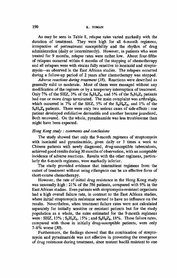
190 K. TOMAN
As may be seen in Table 8, relapse rates varied markedly with the duration of treatment. They were high for all 6-month regimens, irrespective of pretreatment susceptibility and the rhythm of drug administration (daily or intermittently). However, in patients who were treated for 9 months, relapse rates were rather low. About four-fifths of relapses occurred within 6 months of the stopping of chemotherapy and all relapses were with strains fully sensitive to isoniazid and streptomycin-as observed in the East African studies. The relapses occurred during a follow-up period of 2 years after chemotherapy was stopped.
Adverse reactions during treatment (18). Reactions were described as generally mild to moderate. Most of them were managed without any modification of the regimen or by a temporary interruption of treatment. Only 7% of the SHZ, 3% of the SaHaZs, and 5% of the StH:aZ1l patients had one or more drugs terminated. The main complaint was arthralgia, which occurred in 7% of the SHZ, 5% of the S3H~, and 1 % of the S2HaZs patients. There were only two serious cases of side-effects: one patient developed exfoliative dermatitis and another became jaundiced. Both recovered. On the whole, pyrazinamide was less trOUblesome than might have been expected.
Hong Kong study: comments and conclusions
The study showed that only the 9-month regimens of streptomycin with isoniazid and pyrazinamide, given daily or 3 times a week to Chinese patients with newly diagnosed, drug-susceptible tuberculosis, achieved good results during 30 months of observation, with an acceptable incidence of adverse reactions. Results with the other regimens, particularly the 6-month regimens, were markedly inferior.
The study provided evidence that intermittent regimens from the outset of treatment without using rifampicin can be an effective form of short-course chemotherapy.
However, the rate of initial drug resistance in the Hong Kong study was unusually high: 21 % of the 586 patients, compared with 9% in the East African studies. Even patients with streptomycin-resistant organisms had a high overall failure rate, in contrast to the East African studies, where initial streptomycin resistance seemed to have no influence on the results. Nevertheless, when treatment failure rates were not calculated separately for initially sensitive or resistant patients but for the study population as a whole, the rates estimated for the 9-month regimens were: SHZ, 13% ; SsHsZs, 15% ; and S2H~, 18%. These failure rates, compared with those in initially drug-susceptible patients, were only 7-8% worse (30).
Furthermore, the findings showed that the combination of streptomycin and pyrazinamide was not effective in preventing the emergence of drug resistance during treatment, since mutant bacilli resistant to one

TUBERCULOSIS CHEMOTHERAPY 191
of these drugs may not be inhibited by the other. This is probably because streptomycin (but not pyrazinamide) is active against tubercle bacilli in an alkaline or neutral environment, whereas pyrazinamide is active in an acid environment and streptomycin is not (see" What are the bactericidal and sterilizing mechanisms of short-course regimens ? "page 2(0).
However, the study confirmed that pyrazinamide effectively eliminates fully susceptible strains. Relapse rates were low with all three 9-month regimens. This was taken as evidence that pyrazinamide was the drug that contributed most to the sterilizing activity of these regimens.
New studies (see page 196) are under way to find out whether the addition of a limited quantity of rifampicin will improve the results of short-course regimens containing pyrazinamide also in patients with levels of pretreatment drug resistance as high as those observed in the Hong Kong study.
Other short-course chemotherapy trials
Investigations on short-course regimens, all containing rifampicin, have been reported from a number of countries. Some of the larger studies with results after 2 years of follow-up will be mentioned.
Argentina. This study (19) compared two 6-month regimens of isoniazid plus rifampicin. Patients were randomly allocated to isoniazid (500 mg) plus rifampicin (600 mg) daily throughout, or to isoniazid (500 mg) plus rifampicin (600 mg) daily for 2 months followed by isoniazid (1 g) plus rifampicin (600 mg) twice weekly for 4 months. All patients, at admission, had sputum positive by direct smear microscopy and organisms sensitive to isoniazid.
The results were classified as equally good in both groups. The proportion of relapses with the daily regimen was 1 %, and with the intermittent regimen, 3%. The toxicity rate with the latter was very low and the author ascribes this finding to the relatively low dose of rifampicin. Compared with the daily regimen, which required 180 doses of rifampicin, the intermittent regimen needed only 96 doses. In that way, the quantity of rifampicin could be reduced to almost one-half.
Brazil. In this study (20), all patients received rifampicin (600 mg) plus isoniazid (500 mg) and ethambutol (1.2 g) daily for 6 months. One-half received fully supervised treatment in hospital throughout; the other half were admitted for only about 2.5 months and continued for the rest of the period on ambulatory, self-administered chemotherapy. All patients admitted to the study had advanced pulmonary tuberculosis with sputum positive by direct smear microscopy and by culture.
The results were similar in both groups and were therefore pooled. The authors expressed the opinion that entirely self-administered chemotherapy would have been equally successful. The overall success rate
192 K. TOMAN
was 96%. Among 30 patients of whom one-half had resistant organisms before treatment and the other half had no sensitivity tests performed, there was only one relapse. It remains doubtful whether the addition of ethambutol was of appreciable value.
France. A cooperative study in France (21, 22)-see Table 9-investigated regimens of 6, 9, and 12 months with rifampicin (600 mg) plus isoniazid (450 mg), supplemented during the first 3 months with streptomycin (1 g) or ethambutol (1 g), all drugs being given daily. Streptomycin and ethambutol were not allocated at random but according to the preference of the physicians in charge. All patients admitted to the study had bacteriologically confirmed tuberculosis positive by direct smear microscopy and by culture, with organisms sensitive to isoniazid and rifampicin.
Table 9
Short-course chemotherapy studies in Argentina, Brazil, France, and the United Kingdom
Country Duration No. of Relapse Follow-up of study Regimen (months) patients rate period
assessed (%) (months)
Argentina (19) HR<HR 6 93 1 24 HaR• 6 95 3 24
Brazil (20) EHR 6 114 4 24
SHR> HR 6 66 4 30
France (21, 22) 9 74 0 27 EHR 12 69 0 24
6 160 5 27 United SHR>HR 9 135 0 24
Kingdom (23, 24) EHR 12 155 1 21 18 125 0 15
The results were described as excellent. There were 4% of failures due to relapse in the 6-month series and none in the 9-month and 12-month series. The relapses occurred late (one 18 months and two 27 months after the end of chemotherapy). Whether giving the third drug additionally for 3 months contributed to the high level of success remains doubtful.
United Kingdom. A large cooperative investigation organized by the British Thoracic and Tuberculosis Association (23, 24) studied daily regimens of various durations. All regimens included isoniazid (300 mg) and rifampicin (450-600 a mg), supplemented in the first 2 months by ethambutol (25 mg per kg of body weight) or streptomycin (0.75 g Lm.). Patients over 60 years of age received ethambutol instead of streptomycin as a third drug.
a The higher dose was given to patients weighing more than 50 kg.

TUBERCULOSIS CHEMOTHERAPY 193
Depending on the radiographic findings at admission to the study, patients were allocated to regimens of differing durations. Those with cavities larger than 2 cm in diameter were treated at random for 9 or 18 months. Patients with smaller or no cavities were treated for 6 or 12 months. All patients, at admission to the study, had cUlture-positive sputum and had not received more than 2 weeks of antituberculosis therapy previously.
There were only 10 relapses among 575 patients: 8 (5%) in the 6-month series and 2 (1 %) in the 12-month series. No relapses were observed in the 9-month and I8-month groups. In contrast to the East African and Hong Kong studies, the relapses were distributed evenly throughout the 18 months following the cessation of chemotherapy. Of the 8 relapsed patients of the 6-month group, 4 were known to have taken their drugs irregularly. Thus the relapse rate in this group of patients might have been substantially lower.
All relapses occurred with organisms fully sensitive to all four drugs used in the study. The investigators found that factors such as the presence of cavitation or whether streptomycin or ethambutol was given as a third drug influenced neither the speed of conversion nor the relapse rate. They concluded that 9 months of daily chemotherapy with rifampicin plus isoniazid, supplemented by ethambutol (25 mg/kg) in the first 2 months should become a standard regimen for the United Kingdom.
Minimum duration of short-course chemotherapy and minimum number of doses
The benefits resulting from curtailing the conventional duration of chemotherapy from the original 18 or 24 months to 9 or 6 months are obvious. Yet in rural communities, particularly in developing countries, it is difficult to ensure that patients take their drugs regularly even for only 6 months. Also in urban conditions, where a large proportion of tuberculosis patients belong to the underprivileged and fluctuating part of the population suffering from alcoholism and problems of social maladjustment, only supervised chemotherapy of very short duration has a chance of success. Another important problem is the high cost of rifampicin, which also makes it desirable either to shorten the duration of treatment to less than 6 months or, at least, to reduce the number of doses. Thus there is an obvious justification for seeking shorter and less expensive regimens.
On the basis of such considerations, a French research group (25) studied a 3-month regimen of rifampicin 0.2 g) plus isoniazid (900 mg), given either daily or 3 times weekly. Both regimens were supplemented with streptomycin (1 g) daily. All patients had bacteriologically confirmed disease of moderate or large extent. There were 7 (15%) relapses among

194 K. TOMAN
47 patients of the daily series and 5 (11 %) of 44 patients in the intermittent series. There was no treatment failure during the 3 months of chemotherapy.
The authors did not find any statistically significant difference between the results of the two regimens (the number of patients admitted to the study was rather small). Yet, apart from the 12 relapses, cure was achieved in at least 85% of patients with advanced disease followed up for 2 years or more. Perhaps the regimen could be strengthened by adding pyrazinamide or by extending its duration by a few weeks.
An indication that the total number of doses could be further reduced was found in two studies on intermittent regimens containing rifampicin and isoniazid (see" What was the efficacy of primary intermittent chemotherapy ... ? "-page 137). Though both studies were concerned with regimens of long duration (1 year or more), certain findings were of relevance to short-course chemotherapy.
A study in the German Democratic Republic (26) investigated onceweekly isoniazid plus rifampicin without and with a daily initial phase of both drugs for 4 weeks (see also" What was the efficacy of primary intermittent chemotherapy". '1 "-page 137), The total duration of treatment was 52 weeks. All patients had culture-positive cavitary tuberculosis with drug-sensitive organisms.
The overall success rate was high: about 95%. The investigators found that the initial daily phase offered no detectable advantage. The dose of rifampicin of 15 mg per kg of body weight once weekly was considered to be sufficient, producing a manageable proportion of" flu " reactions. The latter increased substantially after 16 weeks of chemotherapy. The authors therefore recommend that rifampicin should not be given for longer than 4 months, after which it could be replaced by streptomycin. At this juncture, bacteriological conversion would have been achieved in about 95% and the side-effects from rifampicin would have occurred in only 4-8% of patients. The total number of doses with an initial daily phase of 4 weeks was 75; without a daily phase, only 52 doses were needed. This important finding was supported by the foJlowing investigation.
A study, undertaken in Singapore (27-29), compared once-weekly with twice-weekly rifampicin (600 or 900 mg per kg of body weight) plus 15 mg of isoniazid per kg of body weight (see also" What was the efficacy of primary intermittent chemotherapy, .. '1 "-page 137). Both regimens included an initial phase of isoniazid with streptomycin and rifampicin given daily for only 2 weeks. At admission, all 481 patients had sputum positive for tubercle bacilli on direct smear examination, confirmed by culture.
At 12 months, the bacteriological conversion rate of the once-weekly regimens was 94% on the average. Only one failure (I %) during treat-

TUBERCULOSIS CHEMOTHERAPY 195
ment occurred in the twice-weekly series. All 13 patients who failed during chemotherapy were rapid acetylators of isoniazid." The relapse rate so far-i .e., within 2 years-has been very low: only 2 bacteriological relapses have occurred in over 400 patients followed up.
An important finding was that a high level of success could be achieved by the once-weekly regimens-i.e., with as few as 64 doses. On the basis of this finding, the investigators concluded that the development of a 50-dose regimen of 3-4 months' duration has excellent prospects (30).
Concluding remarks
To reduce the duration of chemotherapy and the number of doses to the minimum is necessary for developed as well as for developing countries. The advantages of short-course chemotherapy are obvious: the smaller the quantity of drugs required and the shorter the duration of treatment, the greater the convenience to the patient and the better the chance that he will cooperate. Further benefits are: a smaller risk of chronic drug toxicity and reduced inputs in terms of money, treatment facilities, and health personnel. This is of special importance for countries where skilled manpower is scarce and where the import of drugs is limited owing to the restricted reserves of foreign currency.
However, apart from curtailing the number of drug doses and the duration of treatment, there are yet other problems to be solved. These include the need for short-course regimens that can be applied successfully in populations with a high frequency of initial drug resistance without preliminary screening by pretreatment susceptibility tests. This is all the more important as these tests are known to be unreliable (see" How relevant are initial drug resistance and pretreatment susceptibility tests ... ? "-page 167). Reliable routine sensitivity testing is at present performed only in a few laboratories (30), all of which are located in places where initial resistance is negligible.
There is still more to be learned about the need for and length of an initial intensive phase, and about intermittency from the very start or in the continuation phase of short-course chemotherapy. It also remains to be explored how to reduce the side-effects of rifampicin given intermittently and to find regimens that do not include this drug. The role of pyrazinamide in short-course chemotherapy also needs to be elucidated by further study.
To that end, two large trials, each covering about 1000 patients, have been launched: the third East Mrican trial and the second Hong Kong study. Preliminary results (at 6 months after the completion of chemotherapy) were reported recently.
11 The total number of rapid acetylators of isoniazid was 310. The remaining 111 patients were slow acetylators ; none of these failed during treatment (see "What is rapid inactivation (acetylation) of isoniazid ..... ?-page 148).

196 K. TOMAN
Both trials are studying the effect of pyrazinamide as a companion drug to isoniazid, rifampicin, and streptomycin in the initial daily or thrice-weekly phase and without rifampicin in the continuation phase. Rifampicin is given only in the initial phase (30 or 60 doses), in order to reduce adverse reactions, particularly the "flu syndrome", which occurs more frequently when the drug is given intermittently and for longer than 4 months.
The third East African trial (31). The following regimens were allocated at random.
2SHRZjTH streptomycin, isoniazid, rifampicin, and pyrazinamide daily for 2 months, followed by thioacetazone and isoniazid daily
ISHRZjTH the same regimen, but with a I-month intensive phase only
ISHRZ/S2H2Z2 the same I-month initial phase, followed by streptomycin, isoniazid, and pyrazinamide twice weekly
2SHRjTH the same regimen as the first without pyrazinamide, in order to measure its additional effect.
All regimens were given for 6 or 8 months, at random. With the 8-month regimens, isoniazid and thioacetazone were given daily during the seventh and eighth months.
The results during chemotherapy in patients with initially susceptible organisms were excellent with all four regimens. The regimens containing pyrazinamide produced culture negativity faster than those without this drug. Relapse rates during the 6 months following the end of treatment were 7-15% with the 6-month regimen and 0-5% with the 8-month regimen. Initial resistance to isoniazid caused treatment failure in about 20% of cases. Adverse reactions were very infrequent: only I % of patients had to have their regimens modified or stopped for 7 days or more.
The second Hong Kong/British Medical Research Council study (32). The following regimens were allocated at random.
SHR streptomycin, isoniazid, and rifampicin daily for 6 months
streptomycin, isoniazid, and pyrazinamide, with rifampicin, daily for 2 months; then without rifampicin, twice weekly
the same regimen as the second, except that the pyrazinamide was replaced by ethambutol

TUBERCULOSIS CHEMOTHERAPY 197
S3H3R3Za/Sl!H~ a regimen similar to the second, but in the initial phase the fonr drugs are given thrice weekly for 4 months.
The duration of the last three regimens was 6 or 8 months, at random. There was only one failure during chemotherapy. Relapse rates during
the 6 months after the cessation of treatment were low with regimens not containing ethambutol (3-5% with 6-month regimens and 1-2% with 8-month regimens). Regimens with ethambutol, however, showed a high relapse rate (18% and 9%, respectively). Drug toxicity was not a special problem.
The findings confirm that pyrazinamide is an important drug in shortcourse chemotherapy and that it cannot be adequately replaced by ethambutol. The 8-month regimens were more effective than those lasting 6 months.
Isolated positive cultures after the termination of chemotherapy. As already observed with long-term chemotherapy, 14-16% of patients who had achieved bacteriological quiescence later yielded isolated positive cultures. Nevertheless, these patients almost invariably remained cured. So far, there is no regimen able to kill all tubercle bacilli in all patients. However, after good chemotherapy, isolated positive cultures-usually consisting of only ::> few colonies--do not mean that a relapse has occurred, nor do they carry a bad prognosis (8, 24).
In any case, there is no longer a rational basis for the administration of chemotherapy for a period longer than that which has been proved by controlled studies to be adequate. There is, however, a tendency to prolong treatment as a precautionary measure in an attempt to further reduce the probability of relapse. For example, by treating 100 patients for, say, 9 or 12 months instead of 6 months, perhaps 2-5 relapses may be prevented. This benefit cannot be taken at its face value, but must be balanced against the penalty imposed on the 95-98% of patients who are treated unnecessarily and are thus needlessly exposed to the risks of toxicity-quite apart from the waste of resources. Moreover, these relapses are nearly always with fully drug-sensitive organisms, and can be readily and successfully re-treated with a standard drug regimen (33).
Practical considerations. Even when all technological difficulties have been overcome, many questions concerning the feasibility of short-course chemotherapy on a national scale in developing countries remain to be answered. These questions, however, are mainly of a nonmedical nature. Yet they are crucial because they are closely related to the social and cultural structures of the communities concerned, their economies, and their health care delivery systems. Thus a series of operational studies may be needed to obtain solutions that could be adapted to the varying situations in these countries.

198 K. TOMAN
It is beyond doubt that, once short-course chemotherapy is universally practised, it will be of great benefit to tuberculosis patients and substantially improve the global trend of the disease.
REFERENCES
1. STEWART, S. M. ET AL. Tubercle, 37: 388 (1956)
2. Lancet, 1: 237 (1953)
3. WHO Technical Report Series, No. 290, 1964 (Eighth report of the WHO Expert Committee on Tuberculosis)
4. WHO Technical Report Series, No. 552, 1974 (Ninth report of the WHO Expert Committee on Tuberculosis)
5. SINGAPORE TUBERCULOSIS SERVICES I BROMPTON HOSPITAL I BRITISH MEDICAL RESEARCH COUNCIL INVESTIGATION. Tubercle, SS: 251 (1974)
6. GREAT BRITAIN, MEDICAL RESEARCH CoUNCIL TUBERCULOSIS CHEMOTHERAPY TRIALS COMMITTEE. Tubercle, 43: 201 (1962)
7. EAsT AFRICAN I BRITISH MEDICAL REsEARCH CoUNCILS. Lancet, 1: 1079 (1972)
8. EAST AFRICAN I BRITISH MEDICAL RESEARCH COUNCILS. Lancet, 1: 1331 (1973)
9. EAST AFRICAN I BRrTISH MEDICAL REsEARCH COUNCILS. Lancet, 1: 237 (1974)
10. KENT, P. W. ET AL. Tubercle, SI: 24 (1970)
11. McDERMOTT, W. ET AL. American review of tuberculosis and pulmonary diseases, 69: 319 (1954)
12. SUNAHARA, S. Bulletin of the International Union against Tuberculosis, 43: 316 (1970)
13. GRUMBACH, F. Anna/es de I'lnstitut Pasteur de Li/le, 94: 694 (1958)
14. EAST AFRICAN I BRITISH MEDICAL RESEARCH CoUNCIL STUDY. American review of respiratory diseases, 116 : 3 (1977)
15. Fox, W. & MITCHISON, A. D. American review ofrespiratory diseases, 111 : 325 (1975)
16. SECOND EAST AFRICAN I BRITISH MEDICAL REsEARCH CoUNCIL STUDY. American review of respiratory diseases, 114: 471 (1976)
17. JINDANI, A. BuI/etin of the International Union against Tubercu/osis, 51: 53 (1976)
18. HONG KONG CHEST SERVICE I BRITISH MEDICAL RESEARCH COUNCIL. American review of respiratory diseases, lIS: 727 (1977)
19. DUBRA, F. A. Bulletin of the International Union against Tuberculosis, 51: 61 (1976)
20. POPPE DE FIGUEIR1!DO, F. ET AL. Bulletin of the International Union against Tuberculosis, 49: 382 (1974)
21. BROUET, G. ET AL. Revue franr-aise des maladies respiratoires, vol. 5, suppJ. 1 (1977)
22. BROUET, G. Bulletin of the International Union against Tuberculosis, 49: 369 (1974)

TUBERCULOSIS CHEMOTHERAPY 199
23. ANGEL, J. A. ET AL. Bulletin of the International Union against Tuberculosis, 51 : 57 (1976)
24. BRITISH THORACIC AND TUBERCULOSIS ASSOCIATION. Lancet, 2: 1102 (1976)
25. KREIS, B. & PRETET, S. Bulletin of the International Union against Tuberculosis, 51 : 71 (1976)
26. EULE, H. ET AL. Bulletin of the International Union against Tuberculosis, 51 : 115 (1976)
27. LEE, S. K. Bulletin of the International Union against Tuberculosis, 51: 109 (1976)
28. SINGAPORE TUBERCULOSIS SERVICE I BRmSH MEDICAL RESEARCH COUNCIL.
Lancet, 2: 1105 (1975)
29. SINGAPORE TUBERCULOSIS SERVICE I BRITISH MEDICAL RESEARCH COUNCIL.
American review of respiratory diseases, 116: 807 (1977)
30. Fox, W. Proceedings of the Royal Society of Medicine, 70: 4 (1977)
31. GIRLING, D. J. Tubercle, 59: 65 (1978)
32. PANG, S. C. Tubercle, 59: 64 (1978)
33. HONG KONG TuBERCULOSIS TREATMENT SERVICES AND EAST AFRICAN AND BRlTISH
MEDICAL RESEARCH COUNCILS. Lancet, 1: 162 (1976)

200 K. -TOMAN
What are the bactericidal and sterilizing mechanisms of short-course regimens?
The development of short-course chemotherapy has helped to explain the antimicrobial action of certain drugs. Much information has been derived from in vitro and animal experiments and from well-conducted clinical trials. However, the basic immune process and the biochemical dynamics are far too complex to be simulated in the available experimental models. Thus some of the present hypotheses require substantiation by further evidence. Nevertheless, it has been possible, on the basis of existing knowledge, to develop a working hypothesis for selecting effective drug combinations and short-course regimens (see also" How effective is short-course chemotherapy and what are its prospects? "-page 183). A comprehensive review of the current status of short-course chemotherapy has been made recently (1).
The drugs that have been investigated with a view to short-course chemotherapy are isoniazid, rifampicin, pyrazinamide, streptomycin, and ethambutol. Their antibacterial activity should be considered from two angles:
(a) the killing of actively growing bacilli (in the phase of rapid multiplication and uninhibited metabolic activity) and
(b) the sterilization of persisting bacilli-i.e., metabolically inhibited organisms in a quasi dormant state (see also "What is bacterial persistence and how does it affect the chemotherapy of tuberculosis? " -page 204).
A bacterial population may contain four kinds of organism that vary in their metabolism and react differently to chemotherapy. Group A consists of metabolically active, continuously growing bacilli, which are to be found, for instance, in cavity walls where the pH is neutral. These are easily killed by isoniazid, rifampicin, and streptomycin. Group B contains organisms that are dormant most of the time, occasionally growing for very short periods during which only rifampicin can kill them. Group C contains bacilli found mainly inside cells; their growth is largely inhibited by intracellular acidity, which makes pyrazinamide most effective. Group D contains fully dormant bacilli not affected by any drug. These usually die off and rarely cause relapse. It is mainly B and C organisms persisting in a temporarily dormant state that cause relapse. Thus it is essential to destroy them.
Sterilizing activity of various drug combinations: a brief review of experimental findings
Evidence of the bactericidal and sterilizing effect of drugs on M. tuberculosis has been obtained mainly from systematic studies by research

TUBERCULOSIS CHEMOTHERAPY 201
groups at the Pasteur Institute, Paris (2), and at Comell University (3). The animal model used was the experimentally infected mouse. Though certain important features were at variance with those found in man, the murine model was considered to be useful. Reference will be made chiefly to findings that appear to be relevant to the chemotherapy of human tuberculosis."
An important experimental finding was that effective chemotherapy is a biphasic antibacterial process, early killing occurring in the first phase and sterilization in the second. While these two phases are clearly demarcated in the mouse, such a temporal distinction is hardly demonstrable in man. There was also evidence that, the more rapid the initial bactericidal effect on susceptible organisms, the less likely are persisters to emerge. Regimens containing rifampicin and pyrazinamide eliminate susceptible bacilli sooner and more lastingly than other regimens. This was confirmed clinically in the first East African and subsequent shortcourse chemotherapy trials (see " How effective is short-course chemotherapy ... ? "-page 183).
A number of tentative conclusions may be drawn from the murine model as regards the sterilizing potency of certain drug regimens :
1. Isoniazid 11 with ethambutol was the regimen least capable of sterilizing, because the effect of ethambutol was essentially only bacteriostatic (4,5). That finding seemed to be consistent with the results of a controlled clinical study on various regimens containing these two drugs. The relapse rate with this regimen given for 12 months daily was 9% ; given twice weekly, 16%, and once weekly. 25%. The initial phase for all regimens was streptomycin with isoniazid and ethambutol for 2 weeks (6).
2. Isoniazid with rifampicin emerged as the most effective sterilizing regimen, rifampicin appearing to be mainly responsible for the sterilizing activity. When mice were treated for an initial period with isoniazid and rifampicin, and subsequently with isoniazid, streptomycin, or rifampicin alone, rifampicin was the only drug capable of sterilizing the lungs and preventing ultimate relapse. Nevertheless, the rifampicin serum level needed to be three times as high as that achieved in man.
3. Pyrazinamide in a high dosage also had a sterilizing effect in miceperhaps even superior to that of rifampicin.
4. Streptomycin, if added to rifampicin with isoniazid, produced no appreciable extra benefit (in the mouse).
Certain findings in the mouse contrast with those in the guinea-pig, in vitro, and in man-largely because, in the mouse, most bacilli grow
" The sterilizing effect of rifampicin and pyrazinamide in experimental shortcourse chemotherapy has recently been evaluated by Grosset (7).
11 Isoniazid is inactivated very rapidly in the mouse (7).

202 K. TOMAN
virtually inside macrophages (Le., in an acid environment), whereas in the guinea-pig they are mostly to be found in an alkaline, extracellular environment. Therefore streptomycin appears to be more effective in the guinea-pig than in the mouse while pyrazinamide in high dosage is highly active in the mouse and does not act in the guinea-pig. However, in order to achieve the maximum efficacy, serum levels of pyrazinamide in the mouse may have to reach values 5-10 times as high as those tolerated in man (7). While the immune system of the mouse is more dependent on a low p02a and acidity, in the guinea-pig other mechanisms are more important and less pH-dependent. In man, the range of the environmental pH is much wider than it is in either the mouse or the guinea-pig, and the immune system is intermediate between those of the mouse and guinea-pig, but closer to that of the mouse./)
In summary, the key concept of short-course chemotherapy is based on the experimental finding that effective chemotherapy is an antibacterial process involving two phases: one of early killing and another of sterilization. Studies of experimental tuberculosis indicate that, in the early bactericidal phase of treatment, most antituberculosis drugs are effective. In the sterilizing phase, only rifampicin and pyrazinamide have been shown to have the desired effect, particularly when given in combination with isoniazid (4).
The sterilizing activity of pyrazinamide in the second phase of treatment is explained as follows. After the first bactericidal phase, the initial bacillary population has been reduced to a small number of survivors. These usually persist in acid intracellular sites. Increasing acidity slows down the growth rate and at the same time increases the activity of pyrazinamide. This drug is now able to kill intracellular persisting bacilli, which would remain unaffected by other drugs.
The sterilizing activity of rifampicin seems to have a more complicated mechanism. Like isoniazid, rifampicin is experimentally less active against slowly growing organisms in vitro. The most probable explanation for the sterilizing action of rifampicin is the unique speed at which it attacks dormant bacilli that start growing. Dormant bacilli, after having survived the bactericidal phase of treatment, sometimes start growing again in sudden bursts of multiplication lasting perhaps only a few hours. Within this short period, rifampicin is capable of killing such organisms,
a Oxygen pressure. It is likely that dormant bacilli survive in macrophages or in areas of necrotic tissue without communication with an open bronchus. In such conditions, the p02 is lowered and thus limits bacterial growth.
/) For a number of reasons, briefly mentioned before, the mouse is at a disadvantage compared with man. Experimental tuberculosis in mice is generally more difficult to treat than tuberculosis in man. Therefore many experimental workers find the murine model to be valuable, hoping that what can be achieved in the mouse may also be achieved in man (7).

TUBERCULOSIS CHEMOTHERAPY 203
but this cannot be done by other drugs because they need much more time to act effectively. Thus, any persisting dormant organisms that start growing for short periods are killed only by rifampicin (4).
In short, pyrazinamide and rifampicin may act by different mechanisms that seem to have a synergistic effect experimentally. After the bactericidal phase of treatment, these drugs are capable of killing surviving persisters-Le.. of sterilizing tuberculous lesions and thus preventing relapse. Though these findings are of considerable value for the development of short-course regimens, caution in extrapolating the findings to man is recommended.
Final remarks
The success of chemotherapy is endangered mainly by two events: in conventional, long-term chemotherapy, the main concern is that drug resistance may develop during treatment; in short-course chemotherapy, relapse may occur after the cessation of treatment.
Drug resistance is due to the survival and multiplication of nonsusceptible mutants, whereas relapse results from the survival and multiplication of susceptible organisms including persisters. Failure owing to drug resistance may be prevented by drug combinations that kill resistant mutants.
Relapse may be prevented either by continuing chemotherapy for a long time or by short-course chemotherapy with sterilizing regimensi.e., those that kill also persisters. Drugs that prevent drug resistance do not necessarily prevent relapse. The more quickly persisters can be killed, the shorter the duration of chemotherapy may be.
REFERENCES
1. Fox, W. & MITCHlSON, D. A. American review o/respiratory diseoses, 111: 325 (1975)
2. GRUMBACH, F. Antibiotics and chemotherapy, 16: 392 (1970)
3. MCCUNE, R. M. ET AL. Journal 0/ experimental medicine, 123: 445 (1966)
4. DICKINSON, J. M. & MITCHISON, D. A. Bulletin o/the International Union against Tuberculosis, 51 : 79 (1976)
5. GRUMBACH, F. Annales de /'Institut Posteur, 110: 69 (1969)
6. TRlPATHY, S. P. In: Proceedings 0/ the Symposium on Rifampicin in Tuberculosis, Helsinki, 22-23 February 1974. Helsinki, Uudenmaan Kirjapaino, 1974, p. 41
7. GROSSET, J. Bulletin 0/ the International Union against Tuberculosis, 53: 5 (1978)

204 K. TOMAN
What is bacterial persistence and how does it affect the chemotherapy of tuberculosis ?
Bacterial persistence is a phenomenon of special pathogenetic and therapeutic importance. In vitro and in vivo laboratory experiments have shown that bactericidal drugs such as isoniazid and streptomycin kill tubercle bacilli only when they are in the multiplication phase. When the bacilli are at a stage of low metabolic activity-i.e., when bacterial growth has almost come to a standstill a and the organisms are " dormant "-they are not killed by otherwise bactericidal drugs. Such organisms are referred to as " persisters ". Though they may survive in the presence of drugs, behaving as if they were drug-resistant, they are in fact susceptible to the drugs. Thus, if for some reason these organisms regained their ability to multiply freely, they would be killed by the very drugs that had not harmed them before.
Little is known of how tubercle bacilli in human lesions become dormant, but it has been found that they may persist in such form for months or even years. These bacteria may die ofinanition, but sometimes they may suddenly start to multiply. When dormant bacilli again become metabolically active and start multiplying during effective chemotherapy, they are soon killed. But once chemotherapy has been completed the revived bacilli may continue to multiply and thus cause relapse. This explains why conventional chemotherapy needs to be of long duration. It may be concluded that, in order to avoid relapse, persisters must be eliminated, or treatment would have to last several years. Persisters may also play a role in the endogenous reactivation of tuberculosis.
Persistence during chemotherapy can best be explained by the presence of a proportion of the bacterial population in a non-multiplying -Le., dormant-state. Sometimes it is asserted that persistence of susceptible mycobacteria during chemotherapy occurs when bacilli are present in sites that are inaccessible to drugs. This seems unlikely. Isoniazid, rifampicin, and pyrazinamide, as has been mentioned (see " What is the therapeutic effect and what is the toxicity of anti tuberculosis drugs? "-page 101), easily pervade an kinds oflesion in concentrations approximating those in the blood. They permeate all tissue membranes including the normal blood-brain barrier, and attack even bacilli located in the cells {l)./)
a Persistence has been observed during the stationary phase of bacterial cultures or when growth has been stopped-e.g., by low temperatures, anaerobic conditions, or nutritional deficiencies.
/) Streptomycin does not penetrate all normal tissue membranes and is much less active against intracellular than against extracellular bacilli. This seems to be related to the environmental pH, which inside the cell is on the acid side and thus unfavour·

TUBERCULOSIS CHEMOTHERAPY 205
It is more likely that persistence occurs in necrotic areas with no bronchial communication, a shortage of oxygen, and a shift of the environmental pH to the acid side, or inside macrophages-i.e., under conditions in which the metabolism of mycobacteria is largely suppressed and the organisms are in a state akin to dormancy. Bacterial persistence has been observed also iu other diseases-e.g., staphylococcal infections, influenza (2), and leprosy (3).
able to the action of streptomycin. Pyrazinamide, however, favours an acid environment and is active against intracellular bacilli (see "What are the bactericidal and sterilizing mechanisms of short-course regimens 1"- page 200) (4). The environmental pH has apparently little or no influence on the action of isoniazid and rifampicin.
REFERENCES
1. Fox, W. & MrrCHIsoN, D. A. American review o/respuatory diseases, Ill: 325 (1975)
2. CROFfON, J. & DOUGLAS, A. Respiratory diseases. Oxford, Blackwell. 1975, p.98
3. WHO Technical Report Series, No. 607,1977 (Fifth report of the WHO Expert Committee on Leprosy), p. 24
4. GROSSET.1. Bulletin o/the International Union against Tuberculosis, 53: 1 (1978)
206 K. TOMAN
What are the most usual signs of drug hypersensitivity and procedures of desensitization?
Allergic reactions to any drug may occur, but their frequency differs considerably according to the individual, the country, and the drug. The mechanism of the emergence of drug hypersensitivity is not yet fully understood, and regional differences in its occurrence are explained by genetic factors, nutrition (food habits), environmental factors such as housing and climate, possibly by other currently available medicines, and perhaps by certain cosmetics and stains used by particular ethnic groups (tribes).
The most common manifestations are fever and rash, often occurring together with pruritus. Less common are enlargement of the lymph nodes, splenomegaly, and hepatomegaly with or without jaundice. If the warning signs are neglected or unrecognized, encephalopathy or severe bone-marrow depression may occur. Since it is sometimes difficult to distinguish between pure drug-toxicity and hypersensitivity, some investigators include among the drug allergies the rather uncommon exfoliative dermatitis or severe mucocutaneous conditions such as the Stevenslohnson syndrome.
Most hypersensitivity reactions appear during the first month of treatment, but others, particularly rashes, may occasionally be seen after several months.
It is a sensible rule that any new fever or sudden increase of fever occurring within 4 weeks of starting chemotherapy should be suspected of being due to an allergic reaction, unless there is some other obvious reason.
Hypersensitivity to only one of the drugs used may occur, but it may sometimes develop successively or simultaneously also to other drugs.
Desensitization (I, 2)
First, the drug or drugs responsible for an allergic reaction must be identified. This cannot be done merely on the basis of the clinical picture, which is often similar for all drugs. The first step is to stop giving the drugs. The fever will usually decrease within 24 hours. When the temperature has become normal and the skin reaction has cleared, drug tests should be carried out. Small test-doses should be given and the patient watched in order to determine whether the observed condition was due to hypersensitivity, and to what drug or drugs he has become hypersensitive.
It seems advisable to establish as early as possible the drugs to which the patient is not allergic, so that they can be administered again without delay. Therefore it is best to start testing with the drug that is known

TUBERCULOSIS CHEMOTHERAPY 207
to be less likely to sensitize than the other drugs of the regimen given. For example, if the regimen consists of streptomycin, PAS (or thioacetazone), and isoniazid, the first test-dose given will be of isoniazid.
The size of the test dose is chosen according to the severity of the reaction: the more severe the reaction, the smaller the test dose. If the reaction was mild, the first test-dose may be half of the standard dosee.g., 0.5 g ofstreptomycin, 2.5 g of PAS, and 150 mg of isoniazid. With moderate reactions, the routine procedure would be to give one-quarter of the standard dose. If the reaction was severe, the first test-dose should be about one-tenth of the standard dose.
If a patient is hypersensitive to the drug tested, a rise in temperature, pruritus, or rash will develop within 2-3 hours. If there is no reaction to the test dose, the next highest test dose may be tried. In less severe cases, the second dose may be twice the first dose; in more severe cases, an intermediate dose may be tried first. However, if a reaction to a test dose occurs, the same dose should be repeated or a lower dose should be given, once the reaction has subsided.
If the patient is hypersensitive to only one of three drugs, the other two should be administered in the usual combination and dosage while desensitization proceeds. For example, if the patient is hypersensitive only to PAS, then isoniazid and streptomycin may be given throughout the procedure. It is practical and time-saving to give test doses every 12 hours.
If the patient is hypersensitive to two drugs-say, PAS and streptomycin-it is dangerous to continue isoniazid alone while the other drugs are given in small test-doses: resistance to isoniazid might develop, especially jf the desensitization procedure is prolonged. In that case, it would be better to combine isoniazid with one or two reserve drugs.
If desensitization to rifampicin is needed, the first dose should be 75 mg. If no reaction occurs (usually within a few hours), the same dose may be given twice the next day and three times the following day. Subsequently the dose may be increased until the full daily dosage is reached.
Desensitization is usually successful, and often the full therapeutic dose is reached within 3-7 days. Only in severe and unmanageable cases should corticosteroids be used so that the regimen may be continued unrestricted. However, the corticosteroids should be withdrawn gradually, as early as possible, and within 3 months.
REFERENCES
1. BIGNALL, J. R. In: Heaf, F. & Rusby, N. L., ed. Recent advances in tuberculosis. London, Churchill, 1968, p. 76
2. CaoFToN, J. & DOUGLAS, A. Respiratory diseases. Oxford, Blackwell, 1975, p.256

208 K. TOMAN
What are the keys to cure?
How is it that cure rates, on the average, are still mediocre, if not unsatisfactory, despite the extraordinary potency of present-day chemotherapy?
It is widely believed that high success rates can be achieved only in certain outstandingly good treatment centres and that, in the great majority of centres, the rate of success will remain below the optimum unless more effective drugs, such as rifampicin, are supplied to them without restriction for routine treatment. This is a rather superficial view, ignoring certain facts. For more than two decades, there have been drugs from which 100%-effective regimens can be composed (see " What are two-phase chemotherapy and the so-called 100% regimens ? "-page 130). Thus the key to cure does not lie in the introduction of new and better drugs or regimens, but elsewhere (see "When does chemotherapy fail? "-page 177).
An important technical requirement for successful chemotherapy is the prescription of adequate regimens-i.e., only those whose effectiveness has been established by controlled trials. A regimen should contain at least two drugs to which the patient's bacilli are susceptible. The chosen drugs should be given in the same dosage, at the same rhythm (daily or intermittently), and for the same period as was done in controlled trials. Deviations from this rule that have no scientific basis and cannot be clearly justified should be regarded as malpractice that may result in inexcusable harm.
Another technical, almost axiomatic, prerequisite is the regularity of drug intake. Since the advent of chemotherapy, many changes have taken place. Drug combinations and dosages have been varied and the rhythm of administration and duration of treatment have changed, but the need for regularity of drug intake persists. No new regimen or drug has been able to do away with the necessity for regularity. Any interruption of the regular rhythm of treatment increases the risk of failure. It must be borne in mind that the main reason for treatment failure is not initial drug resistance but irregularity of drug ingestion (see .. What is primary and what is initial drug resistance? "-page 87 and" How relevant are initial drug resistance ... ? "-page 167).
It is illusory to expect that new drugs will solve the main problem of chemotherapy, unless a regimen can be found that needs to be administered in one injection or only for a few days. The success of chemotherapy is determined as much by operational as by technical factors. Even the most effective regimens currently available, irrespective of the drug combination or duration of treatment, may fail if not administered

TUBERCULOSIS CHEMOTHERAPY 209
regularly. Thus, today it is not the lack of knowledge about adequate chemotherapy but its adequate administration (1) that is the crux of the matter (see" When does chemotherapy fail? "-page 177). This is one of the medical problems that, so far, cannot be solved by technical or medical means, but mainly by organizational measures. Indeed, to ensure regular drug intake has become a managerial task par excellence. Nearly all attempts in this direction, through health education-e.g., by thoroughly instructing the patients about the importance of regularity and the bad prognosis in case of irregularity-have been insufficient to motivate patients to take their drugs regularly as prescribed (see" What is the significance of default ... ? "-page 211). Verbal motivation of patients is rarely successful unless applied in an adequate organizational framework satisfying certain operational requirements.
Operational requirements
Treatment services must be easily accessible. Patients who feel very ill may be willing to travel long distances in order to be seen by a reputed physician. However, they can rarely repeat such travel or stay at the place of treatment for a long time. Treatment services should therefore be within easy reach and should be free of charge (2,3).
Treatment services should be accepted and utilized by the community. The staff should be able to communicate with patients in their own language and should be sympathetic to their complaints and needs. Patients should be helped to handle problems causing default. Services must be compatible with local beliefs, traditions, and habits, as well as efficient. In short, they should inspire confidence (see "What is the significance of default ... ? "-page 211).
Drugs should always be available in suffiCient supply. When patients have to be turned away because drugs are out of stock, the effect on regularity is bound to be detrimental.
Whenever possible, treatment should be supervised. What is meant by " supervised chemotherapy" is that each dose should be administered under direct supervision. However, it is not always easy to organize such treatment for every patient. In many instances, individual arrangements will have to be made. Sometimes the supervision of treatment will have to be delegated to other institutions-e.g., to a hospital or health post located close to the patient's work or home.
In summary, treatment must be organized with a view to the convenience of the patient, rather than to that of the treatment service (see .. What are the merits of supervised intermittent chemotherapy ... ? " -page 161),

210 K. TOMAN
Thus at present the key to cure is to be found in the organization of the delivery of chemotherapy. Even the best available regimen will have a low success rate for as long as treatment services are not focused on the cooperation of patients. On the other hand, a policy using secondbest regimens may be highly successful when delivered through an adequate treatment organization.
REFERENCES
1. Fox, W. Bulletin of the International Union against Tuberculosis, 47: 49 (1972)
2. WHO Technical Report Series, No. 290, 1964 (Eighth report of the WHO Expert Committee on Tuberculosis)
3. WHO Technical Report Series, No. 552, 1974 (Ninth report of the WHO Expert Committee on Tuberculosis)
TUBERCULOSIS CHEMOTHERAPY 211
What is the significance of default in the chemotherapy of tuberculosis ?
Treatment default is regarded as the crux of chemotherapy. Though nowadays there are treatment regimens capable of curing almost every patient-even those with severe disease-the actual results, on the average, are much inferior. The most common reason for this unsatisfactory situation is irregularity of drug-taking-e.g., gross interruptions and premature termination of treatment, mainly by self-discharge. Careful analysis of individual treatment records shows that, frequently, of 100 patients who have started treatment, not even one-half, and sometimes no more than one-third, take the prescribed drugs for the minimum period of 12 months.
A case in point is illustrated by data from two important field studies: one from South India (1) and the other from East Africa (2).
In the South Indian study, 10% of patients had died or left the district by the end of the 12-month treatment period; 27% refused to attend the treatment services; and of the remainder only 47% collected four-fifths of their medicaments (Table 1). Thus a regimen with a potential of about 90% success (isoniazid plus PAS) achieved quiescence of the disease in fewer than 50% of patients. In a further study, in which the same investigators used a three-drug regimen, the same low proportion of patients completed their treatment (3).
Table 1
Treatment default: chemotherapy field study in 3 towns of South India (1)
Month
o 3 6 9
12
Alive. remaining in the district
No.
231 225 221 216 207
Patients
Refusing Collecting treatment 80% of the drugs
(cumulative) prescribed
% %
6 15 62 19 54
25 49 27 47
Another study illustrating the problem of default was an analysis of treatment results in a random sample of patients who received chemotherapy in the routine treatment services in Kenya (and were not included in a controlled clinical trial). As Table 2 shows, only two-thirds of the

212 K. TOMAN
initially bacteriologically positive patients remained under treatment for more than 6 months and only 37% of the patients attended for treatment for 12 months (though the policy in Kenya was to treat all patients for 18 months). Of the patients "lost". the majority were lost during the first 6 months.
Table 2
Treatment default: sample survey of patients routinely treated In Kenya (J.I)
Duration of treatment Patients attending
for treatment (months)
No. %
More than 3 528 79 More than 8 433 85 More than 9 312 47 12 248 37
Patients ad mltted 660 100
Needless to say, the patients who remained under treatment for only a short time did very badly. Of those who received isoniazid plus thioacetazone for up to 6 months, only 12% became culture-negative, as against 88% for patients treated for 12 months-Le., a result comparable to those achieved in controlled clinical trials.
However. it would be wrong to believe that these problems apply merely to the developing countries. If routine treatment results obtained in technically advanced countries are studied, the loss of patients from observation and failure to remain under treatment for an adequate period are seen to be major problems, just as they are in the developing countries (4).
Reasons for default
How is it that so many patients do not take, or refuse to take, the drugs that can save them from dying of tuberculosis and in all probability will cure them of the disease? The reasons have often been examined and were analysed in detail by Rouillon (5). Some typical reasons given by health service staff are discussed below.
(a) Even when he has been thoroughly informed, the patient is too ignorant to understand the fundamental need to ingest drugs for a long time.
(b) The patient firmly believes that, when the symptoms have subsided and he is feeling well again, he does not need any further treatment. He will not accept that he is in danger when he stops taking the drugs, even when he has been told so.

TUBERCULOSIS CHEMOTHERAPY 213
(c) When side-effects appear or symptoms do not subside as fast as he had expected, the patient suspects that he is getting the wrong treatment. He stops taking the prescribed drugs and may seek advice elsewhere.
These three reasons for default are put down to ignorance, lack of intelligence, and indifference-all on the part of the patient. Such patients are classified as uncooperative and are blamed for the default.
Other reasons for default include the following.
(d) The patient has not been properly instructed about the essentials of his disease and its treatment or, on the contrary, has been given too much information: he has been shown his bacilli under the microscope, or his chest X-ray film, and has been informed about diet, infectiousness, cough discipline, handling of sputum, schedules of drug-taking, and possible side-effects-aIl at the first session. When patients were interrogated after such instruction, it was found that many had not understood what they had been told. A sizeable number had been so shocked to learn that they had tuberculosis that they had not been able to grasp other important information. As a result, only a few patients were actually induced or motivated to take the prescribed drugs for the full course of treatment-e.g., for one year.
(e) Some patients give up treatment because they resent the shabby behaviour of certain health workers or when they are asked for extra payments or gratifications. Patients are particularly disappointed when, often after having waited long hours, they are told that there are no more drugs in stock and that no-one knows when the next supply will arrive. This rather frequent event is a major reason for interruption or cessation of treatment.
(I) Patients may give up treatment when other problems become more pressing than their suffering due to tuberculosis-e.g., hunger, debts, or the loss of their homes. Some change their domicile often without informing the health centre where they have been receiving treatment. This happens particularly often when the patient loses his job or when he is driven to conceal the disease because of the social stigma adhering to tuberculosis, with all its ill consequences for him and his family.
(g) There is a category of patients who are given chemotherapy though the diagnosis of tuberculosis has never been confirmed by bacteriological examination. The treatment is often started on account of pulmonary lesions of unknown or dubious origin observed on the patient's first X-ray film. In many countries this is the case with the large majority of patients put on chemotherapy. When these patients stop taking their drugs many do not suffer any adverse consequences. Therefore they do not feel any necessity to go to the health service. In spite of all efforts, they refuse to continue or to resume chemotherapy.

214 K. TOMAN
They may be right in doing so and should not be called defaulters (6). Unfortunately, they may induce other patients who definitely need treatment to stop taking it.
(h) Distance is a common reason for irregularity. The patient may not be able to walk long distances, and transport-if available-may be too expensive for him.
(i) Inconvenient consulting hours may be also be a reason for default. This can be remedied by organizing treatment near the patient's home or adjusting consulting hours to his convenience.
(j) Seasonal factors also deserve mention as a cause of treatment default.
From the foregoing, it is obvious that it would be wrong to put the blame for default on the patient rather than on the services. Is it the patient's fault that he does not understand why it is essential for him to take his drugs regularly and for a long time, or that he does not believe he still needs the drugs after the symptoms have gone? It requires some sophistication to understand that one must continue treatment because one is ill though one feels fine, but that one should live and work as if one were healthy though one has to take drugs.
Today it is perfectly well known that, usually after one or two months of effective chemotherapy, the patient will feel symptom-free. From that moment it seems pointless to him to take medicaments that he may find unpleasant or that produce minor side-effects causing more discomfort than the disease itself. It is only natural to enjoy the euphoria of recovery and forget all about drug-taking.
The same has been observed also in a number of other conditions requiring prolonged drug ingestion, such as cardiovascular diseases, rheumatic fever, leprosy, epilepsy, malaria (prophylaxis), and diabetes. This is true also with the self-administration of oral contraceptives.
Default, according to Webster's dictionary, is failure to do something required by duty or law. When default might cause harm to the individual or community, corrective or preventive action should be taken. In the case of tuberculosis patients, irregularity or premature cessation of chemotherapy usually has serious consequences not only for the patient but for the community as a whole. It is the moral, if not the legal, duty of the health services to take the necessary precautions. However, since the interruption or self-termination of treatment are a common feature of human behaviour, these precautions must be an essential part of the strategy of chemotherapy-a built-in element of treatment organization.
The prevention and management of default are integral components of treatment and are thus, above all and undoubtedly, the responsibility of the doctor or person in charge of treatment. Therefore, if treatment failure is due to default, it is unjust to hold only the patient responsible.

TUBERCULOSIS CHEMOTHERAPY 215
For as long as the organizers of treatment services do not shoulder this responsibility, even the most excellent drug regimens will fail to produce the high level of therapeutic and epidemiological success of which they are capable.
It is only fair to say that it is easier to identify the causes of default than to remedy them. This is mainly because treatment default has many of its roots outside the health system, in the various activities of daily life and deep in the social structure and cultural traditions.
Many health professionals believe that health education of the sick and of the public is all that is needed to ensure that patients comply with medical instructions. However, experience has shown that such efforts, or even detailed instructions by a doctor, are generally not sufficient to motivate patients to take the prescribed regimen (see under (d), page 213).
There is far more to motivation than informing and instructing people: it is a matter of mutual human relations, requiring an understanding of the patient's non-medical problems, his way of life, work, religion, wants, fears, and attitudes towards traditional and modern medicine. Motivation requires a person who speaks the patient's " language" and is capable of bridging intellectual and social distances, removing cultural barriers, and, if necessary, changing attitudes and habits. Positive motivating factors are efficient professional performance and success, good working morale, the compassion of the staff, and their identification with the community they serve.
In summary, motivation is a problem of human communication, differing from one patient to another and one community to another. That is why no uniform and generally applicable recipe can be given. Failure to communicate with the patient will embarrass him; a patronizing approach or bad behaviour will alienate him and instead of confidence will create distrust, resulting in the rejection of treatment.
Lastly, the problem of treatment default due to the lack of communication is by no means restricted to developing countries. In technically advanced countries also, as has been reported, treatment default is not uncommon. It is a problem frequently encountered in communities with minority groups of various ethnic, religious, and social backgrounds, where, in addition to tuberculosis, patients are suffering from economic insecurity and other social stresses.
REFERENCES
1. F'RiMODT-M0LLER, J. & PARTHABARATHY, R. (1964). Cited in Fox, W. Bulletin of the International Union against Tuberculosis, 47: 49 (1972)
2. EAST AFRICAN I BRITISH MEDICAL REsEARCH COUNCIL INVESTIGATION. Cited in Fox, W. Bulletin of the International Union against Tuberculosis, 47: 49 (1972)
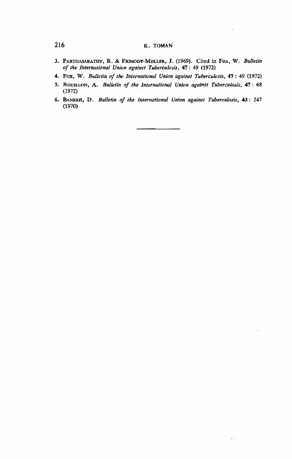
216 K. TOMAN
3. PARTHASARATHY, R. & FRIMODT-M0LLER, J. (1969). Cited in Fox, W. Bulletin of the International Union against Tuberculosis, 47: 49 (1972)
4. Fox, W. Bulletin of the International Union against Tuberculosis, 47: 49 (1972)
S. ROUlLLON, A. Bulletin of the International Union against Tuberculosis, 47: 68 (1972)
6. BANElUI, D. Bulletin of the International Union against Tuberculosis, 43: 247 (1970)

TUBERCULOSIS CHEMOTHERAPY 217
What is the place of sanatorium and hospital treatment today, and how infectious are tuberculosis patients under chemotherapy?
The accumulated evidence from controlled trials proving the high efficacy of ambulatory chemotherapy is not appreciated everywhere. In many countries, every newly registered patient is routinely admitted to a sanatorium or hospital for many months on the ground that, with such a policy:
(1) treatment gives the best results; (2) patients cooperate better ; (3) it is easier to educate and to reassure patients about the disease; (4) it can be ensured that the patients take their drugs; (5) drug toxicity can be treated more easily ; and (6) the patients are isolated and thus cease to be a danger to their
families and other contacts.
These assertions, however, have not been substantiated, as will be shown point by point.
(1) There is a solid body of evidence that outpatient treatment is highly effective and by no means inferior to sanatorium treatment, even in the case of patients suffering from extensive disease and living in poor conditions. This has been proved not only in the classical Madras study (see "What were the main findings of the Madras study ... ?"page 122), but also in other controlled trials undertaken in various developed and developing countries. The summary of these studies (Table 1) is based mainly on a review of the world literature by Fox (1).
Table 1
Controlled studies on the effects of exercise, work, rest, and sanatorium or home treatment on the results of chemotherapy
Comparison Investigation (rei. No.) Country Year
1. Rest and exercise (2) USA 1957 (8) England 1966
2. Rest and normal work (4) Scotland 1966
3. Various duratlons of (5) Czechoslovakia 1971 hospital treatment (6) USSR 1972
4. Home and sanatorium (7) Scotland 1956 (8) India 1959 (9) Ghana 1960
(10) Kenya, Uganda, and Tanzania 1966

218 K. TOMAN
None of the studies demonstrated any evidence of the superiority of rest over exercise or work, or any particular benefit from sanatorium treatment compared with outpatient treatment, even when the former was continued for a whole year.
Only the study of the Tuberculosis Society of Scotland (4) was restricted to patients with limited disease. In all the other investigations, at least some of the patients had advanced disease. In particular, outpatients in the studies in Ghana, Madras, and East Africa mostly had extensive, direct-smear-positive tuberculosis and furthermore lived in unfavourable conditions, had to continue working, and evidently were not as well nourished as the inpatients.
The relapse rate studies in Scotland (4) and in Madras (see" What were the main findings of the Madras study ... ? "-page 122) showed no difference between the two groups of patients. Even pregnancy in outpatients did not influence the attainment of bacteriological quiescence adversely, or lead to more relapses (11).
(2) The cooperation of sanatorium patients was no better than that of patients treated at home. In both technically advanced and developing countries, many patients are unwilling or unable to tolerate a long separation from their families and normal environment. Though patients in the three studies (8-10) in developing countries had good sanatorium accommodation, a nutritious diet, and ample rest-none of which they had in their homes-self-discharge from treatment was not a rare event: 8 % of the sanatorium patients discharged themselves from treatment, compared with only 1 % of those treated at home (8).
(3) It is doubtful whether patients in sanatoria can be better educated and reassured about their disease than home-treated patients. It seemed rather an advantage when the personnel in charge of treatment throughout the course was also responsible for educating and reassuring the patient. The same staff could also educate and reassure the patient's family, thanks to a direct knowledge of the patient's home and social condition. Major personal problems, including the disruption of family life (8), were much more frequent in the sanatorium series (21 %) than in the home treatment series (8%).
(4) Sanatorium or hospital treatment itself does not guarantee regular ingestion of tlle prescribed medicaments, as has been found by urine tests or the counting of tablets. Even in sanatoria, regular drug intake can be ensured only by strict supervision-Le., if the patient is actually seen to swallow every dose, which is by no means always certain.
(5) Severe toxic reactions to the drugs used in standard regimens are uncommon. If, occasionally, a drug produces toxicity that cannot be managed under domiciliary conditions, then of course the patient has to be admitted

TUBERCULOSIS CHEMOTHERAPY 219
to hospital. However, in primary chemotherapy, such an event is rather the exception and does not justify the hospitalization of every patient as a general policy.
(6) One of the strongest and most serious arguments against home treatment is that the patient with infectious tuberculosis must be isolated, since he is a danger to his household and other close contacts. This problem has been carefully studied in a number of investigations. In the classical Madras study (8), in which patients treated in a sanatorium were isolated from their families for 12 months, there was no difference in the occurrence of tuberculin conversion in the tuberculin-negative contacts of patients treated at home or in the sanatorium during a 5-year observation period (J 2). Thus no additional risk of infection was found for close family contacts of patients under chemotherapy (see also" What were the main findings of the Madras study ... ? "-page 122).
Furthermore, even in the tuberculin-negative contacts, the disease did not develop more frequently in the home-treated series (10.5%) than in the sanatorium-treated series (11.5%). Most cases of tuberculosis developed within the first year, chiefly within the first 3 months, indicating that the infection took place before the start of treatment (12). From that it was concluded that for contacts the greatest risk of infection was before treatment began-Le., before the case was discovered.
How infectious are tuberculosis patients under chemotherapy?
In a subsequent study at the Tuberculosis Chemotherapy Centre, Madras, it was found that even contacts of patients treated with isoniazid alone ran no extra risk compared with contacts of patients treated with isoniazid and PAS. The attack rate in originally tuberculin-negative contacts was 7% in both groups (13).
More recently, American investigators have reported on the risk of infection to the contacts of patients discharged from hospital after one month of treatment. Some of the patients still had direct-smear-positive sputum, while others were negative by culture (14, 15). There was no difference in the incidence oftuberculin conversion among the tuberculinnegative contacts of the two groups. Following the start of treatment, there was no higher risk for contacts associated with the sputum status of the patient. Once effective treatment has been started, the presence of tubercle bacilli in the patient's sputum does not imply infectiousness.
These important findings have been in agreement with the results of other studies (16, 17). Patients with sputum-positive tuberculosis were randomly allocated to hospitalization for 2 weeks or for long periods. Infection did not occur more frequently in the tuberculin-negative contacts of patients hospitalized for only 2 weeks than in those of patients kept in hospital longer.

220 K. TOMAN
Though it has been demonstrated that, in untreated patients, infectiousness is closely related to their sputum status (see "What is the role of case-finding by periodic mass X-ray examination ... ? "-page 65), no such relationship exists once the patient is on adequate chemotherapy. The fast decline in infectiousness is largely determined by the rapid fall in the number of viable organisms in the sputum (18) and probably also by the reduced frequency of coughing that promptly follows the start of effective treatment.
In summary, during adequate chemotherapy an index case" is not a major source of risk, if any, to contacts.
Conclusion
There is no scientific justification, or any objective reason, for the general use of institutional treatment of newly diagnosed tuberculosis as a policy.
The WHO Expert Committee on Tuberculosis, in its eighth report, challenged all those advocating the superiority of institutional treatment " ... to conduct studies to determine whether their contention is substantiated by objective evidence" (19). More than 10 years later, such evidence has still not been produced. None of the controlled clinical trials that studied the effect of treatment at home compared with treatment in a sanatorium, or that of rest versus ambulation, has demonstrated any particular benefit from institutional treatment and bed-rest in comparison with ambulatory treatment and physical activity.
The American Thoracic Society issued a clear policy statement:
.. The major emphasis in tuberculosis control must now be placed on the provision of adequate ambulatory care facilities. The lack of such facilities is no longer an acceptable justification on the part of any community or governmental body for extended hospitalization; if adequate hospital facilities can be maintained. adequate outpatient services can be created and maintained" (20).
The Society subsequently stated that:
.. Chemotherapy is so effective that patients can be encouraged to return to their usual activities and occupation early in the course of treatment .•. To forbid physical activity or to encourage a tuberculous patient to withdraw from social and community life is rarely necessary and is usually a disservice .. (21).
More recently (1976), the Society declared that:
.. In summary. in this era of chemotherapy, tuberculosis should be treated in whatever setting most appropriately meets the needs of the patient and the community. Some patients can be entirely treated at home. Others may require
.. An index case is a person known to be discharging tubercle bacilli and thus registered as a possible source of infection.
TUBERCULOSIS CHEMOTHERAPY 221
some short period of hospitalization in a general hospital followed by ambulatory care. Still others may require longer-term care in an institution mainly because of their other medical and social problems. The fact of tuberculosis should not be the primary determinant of the local care, nor should it act all a constraint. Continuity and completion or chemotherapy are the keys to recovery wherever the care is provided. Tuberculosis patients should be managed as other patients within the general care system. A separate categorical system for tuberculosis is obsolete" (22).
With regard to certain tendencies to retain sanatorium services, or even to establish new ones, the WHO Expert Committee, in its ninth report,
..... noted with concern the adherence to outmoded long-term sanatorium treatment in some technically advanced countries and recommended that the national authorities should review the reasons for the retention of traditional sanatorium service.s despite the dramatic progress of chemotherapy in the last 20 years. In this connexion the Committee emphasized that it is the responsibility of the national authorities to make alternative careers available to sanatorium physicians" (21).
REFERENCES
l. Fox, W. American review of respiratory diseases, 97: 767 (1968)
2. WIER, J. A. ET AL. In: Transactions of the 16th Conference on the Chemotherapy of Tuberculosis, St. Louis, MO, 11-14 February 1957. Washington, DC, Veterans Administration, 1957
3. WYNN-WILLIAMS, N. & SHAW, J. B. Tubercle, 41: 352 (1960)
4. TuBERCULOSIS SOCIETY OF ScoTLAND. Tubercle, 41: 161 (1960)
5. CZECHOSLOVAK: TUBERCUL01lIS SERVICE / WHO I BRITISH MEDICAL RE.sEA:RCH
COUNCIL. Bulletin of the World Health Organization, 4S: 573 (1971)
6. SHMELEV, N. A. & ALYABINA, M. G. Bulletin of the International Union against Tuberculosis, 47 : 22 (1972)
7. TYRREL, N. F. Lancet, 1: 1171 (1956)
8. TUBERCULOSIS CIlEMOTHERAPY CENTRE, MADRAS. Bulletin of the World Health Organization, ll: 51 (1959)
9. BELL, J. W. British journal of diseases of the chest, 54: 247 (1960)
10. EAST AFRICAN I BRITISH MEDICAL REsEARCH COUNCIL FOURTH THIACETAZONE
INVESTIGATION. Tubercle, 47: 315 (1966)
11. RAMAICRISHNAN, C. V. ET AL. Bulktin of the World Health Organization, 34: 553 (1966)
12. KAMAT, S. R. ET AL. Balletin of the World Health Organization, 34: 517 (1966)
13. DEVADATIA, S. liT AL. Bulletin of the World Health Organization, 41: 337 (1970)
14. GUNNELS, J. J. liT AL. American review of respiratory diseases, 105: 989 (1972)
15. GUNNELS, J. J. liT AL. American review of respiratory diseases, 109: 323 (1974)
16. BROOKS, S. M. liT AL. American review of respiratory diseases, 108: 799 (1973)

222 K. TOMAN
11. EDDISON, G. G. & FARMER, B. M. American review of respiratory diseases, 103: 886 (1911)
18. YEAOER, H. JR ET AL. American review of respiratory diseases, 95 : 998 (1961)
19. WHO Technical Report Series No. 290, 1964 (Eighth report of the WHO Expert Committee OD Tuberculosis)
20. AMERICAN THORACIC SoCIE'IY. American review of respiratory diseases, 102: 410 (1910)
21. AMERICAN THORACIC SoClE'IY. American review of respiratory diseases, 102: 992 (1910)
22. AMEruCAN THORACIC SOCIE'IY. American review of respiratory diseases, 113: 253 (1916)
23. WHO Technical Report Series, No. 552, 1914 (Ninth report of the WHO Expert Committee on Tuberculosis)

TUBERCULOSIS CHEMOTHERAPY 223
How important is follow-up and what is the frequency of relapse a after the completion of treatment?
Before the advent of chemotherapy, the complete cure of pulmonary tuberculosis-i.e., of the post-primary or adult type-was observed only rarely. Pathologists and clinicians maintained that the disease practically never healed in the strict sense of the term, but could only be arrested, become stabilized, or be rendered inactive; since bacilli almost always persisted in the residua oftuberculous lesions, relapse could occur at any time, even many years after treatment. Indeed, relapse was common, and that was the reason for the adoption of a policy of life-long follow-up of patients who had completed treatment. These patients were kept on a register and examined regularly at intervals of several months, or at least once a year.
That routine, however, placed a steadily increasing burden on the health services, absorbing a substantial proportion of staff-time and financial resources. No wonder the dramatic success of chemotherapy called into question the usefulness of indefinite follow-up and prompted demands for the reassessment of this policy. For that purpose, two questions needed to be answered:
I. What is the frequency of relapse ? 2. How is relapse detected?
In a longitudinal survey and recent analytic studies, it was found that relapse still accounted for about 15-20% of the annual incidence of newly registered sources of infection (1-4).
Furthermore, it was found that the individual risk of relapse among persons with a history of bacteriologically confirmed tuberculosis varied substantially and was determined mainly by three factors:
(a) whether chemotherapy had been received or not; (b) whether or not the regimen given was adequate and regularly
taken; and (c) the time that had elapsed since bacteriological quiescence had
been achieved.
In all the reports recently published on that question (5-1), there was agreement that the highest relapse rate was found in patients who had
a .. Relapse" may be defined as the reappearance of tubercle bacilli in the sputum after a long period of bacteriological quiescence (negativity by smear microscopy and culture), usually accompanied by clinical deterioration. The occurrence of isolated positive cultures commonly consisting of a few colonies is not considered as a relapse (13) (see under "How effective is short-course chemotherapy ... ?"-page 197).

224 K. TOMAN
never received any chemotherapy (about 5% per annum) and the next highest rate (about 2%) in patients with deficient treatment (5). There was, however, a definite time trend, the risk in both groups diminishing appreciably after 3-5 years to about 1 % (5). Other factors, in addition to lacking or deficient chemotherapy, that contributed to the risk of relapse were found to be alcoholism and other diseases, as well as social maladjustment or deprivation (7).
However, the most important finding was the striking effect of adequate chemotherapy on relapse, which falls to a few per mille per annum (5-8). Though this risk is still considerably higher than the risk of disease in persons with no history of previous tuberculosis, it does not warrant life8 10ng follow-up.
Moreover, reports on the mode of detection of newly registered relapses point out that these were mostly not discovered at the regular check-up, as might have been expected. About two-thirds of the relapses were discovered on other occasions (9), mainly on account of chest or other symptoms. The proportion of relapses detected through follow-up seemed to vary with the frequency and regularity of examination. Nevertheless, in a longitudinal survey lasting 12 years (a WHO-assisted research project), each person with a history of previous tuberculosis was examined bacteriologically every 6 months and by X-ray once a year, according to the research protocol. Yet less than one-half of the relapses were discovered through follow-up examinations, despite a stringent research discipline. One-half of the relapses were detected on account of symptoms-Le., between regular check-ups (8). (This does not imply that all the other relapsed patients detected through follow-up examination were symptom-free at the time of check-up.)
In summary, it appears that individuals with a high risk of relapse belong to one of two we1l8 defined and easily identifiable groups-patients who have not had chemotherapy at all and those who have had inadequate chemotherapy. These groups will become progressively smaller with the improvement of chemotherapy. Persons of these groups may deserve active attention for 3 years after the termination of treatment. (Whether preventive treatment-that is, chemoprophylaxis-is useful in previously inadequately treated patients needs to be proved. In controlled trials, patients with inactive tuberculosis who originally had received isoniazid did not benefit from chemoprophylaxis with isoniazid (10).)
In those who have been adequately treated with chemotherapy, the risk of relapse is too small to justify prolonged follow-up (14). Thus routine follow-up examinations may be drastically curtailed or, to a large extent, abandoned. This conclusion was reached by the Center for Disease Control of the Public Health Service in the USA (11), as well as by investigators who followed up patients treated in Scotland (12). The former stated :

TUBERCULOSIS CHEMOTHERAPY 225
"Tuberculosis patients who complete adequate chemotherapy should be considered cured. They have no need for routine life-time periodic recall for X-ray examination. Indeed, perpetuating life-time follow-up of such treated patients diverts clinic personnel and resources from the crucial task of providing services for those who really need them" (11).
However, ex-patients should be strongly advised to come without delay for examination if they develop symptoms suggestive of tuberculosis (14). General practitioners and physicians who are likely to come across patients with a history of previous tuberculosis should be informed about the possibilities of relapse and the significance of respiratory symptoms such as a prolonged cough.
REFERENCES
1. CHAN-YEUNO, M. ET AL. American review of respiratory diseases, 104: 861 (1971)
2. DANKOVA., D. Bulletin of the InterTUltioTUlI Union against Tuberculosis, 47: 145 (1972)
3. STElNBRUCK, P. Bulletin of the InterTUltional Union against Tuberculosis, 47: 135 (1972)
4. NAKIELNA, E. M. ET AL. American review of respiratory diseases, 112: 765 (1974)
5. GRZYBOWSKI, S. ET AL. American review of respiratory diseases, 93: 352 (1966)
6. STEAD, W. W. & JUROENS, G. H. American review of respiratory diseases, 108: 314 (1973)
7. HORWITZ, O. American review of respiratory diseases, 99: 183 (1969)
8. KJ.MNKA, R. ET AL. Bulletin of the World Health Organization, 51: 59 (1974)
9. EDSALL, J. & COLLINS, G. American review of respiratory diseases, 107: 851 (1973)
10. FEREBEE, S. H. Advances in tuberculosis research, 17: 28 (1969)
11. CENTER FOR DISEASE CONTROL. Morbidity and mortality weekly report, 23: 75 (1974) .
12. PEARCE, S. J. & HORNll, N. W. Lancet, 2: 641 (1974)
13. BRITISH THORACIC AND TUJlERCULOSIS AssOCIATION. LAncet, 2: 1102 (1976)
14. WHO Technical Report Series, No. 552, 1974 (Ninth report of the WHO Expert Committee on Tuberculosis)

226 K. roMAN
What are the principles and requirements of a controUed chemotherapy trial?
Each conscientious doctor treats his patients only with methods and medicaments in which he has confidence. However, different doctors often treat one and the same disease in different ways. If the patient recovers, the physician, understandably, ascribes the success to his way of treatment. How subjective and changeable these judgements are can be seen from the large number of medicaments and treatment methods that are brought into the limelight, praised by bona fide advocates, and eventually consigned to oblivion. Sometimes it may take a long time for the value of a certain treatment method to be determined. It took centuries before therapeutic blood-letting and drastic purging were abandoned. Gold salts had been in use for almost 20 years as a specific treatment of tuberculosis, as recommended by prominent medical personalities in about 200 published papers, when it was recognized that they were useless, if not harmful (I).
In the first half of this century, innumerable therapeutic methods, diets, and compounds were used in the treatment of tuberculosis. Starting with the therapeutic fiasco of tuberculin, other biological agents, such as bacterial extracts and fractions, attenuated mycobacteria, antisera, and antitoxins; cod-liver oil ; vitamin C ; calcium injections; creosote; saltfree diets; radiation therapy; and various climates (high altitudes, ocean beaches and hot, dry climates)-all had their passionate advocates (2). And then there was a host of therapeutic interventions: pneumothorax, diaphragmatic paralysis, pneumoperitoneum, oleothorax, pneumonolysis, plombage, cavity drainage, thoracoplasty, and finally resectional surgery. This is far from being a complete account, but it serves to recall the confused situation in the middle of this century.
It is mainly in the last 25 years that determined efforts have been made to use scientific techniques in evaluating the treatment of tuberculosis on a larger scale. An important advance in this field has been the development of an assessment method known as the controlled trial, which has been found particularly suitable for studying the effect of chemotherapeutic substances. Since their introduction, many controlled trials have been carried out, and have made it possible to establish the efficacy, toxicity, and applicability of practically all the chemotherapeutic regimens currently used. However, there are still physicians who do not appreciate the value and scope of the method. And there are some authors who call their investigations controlled trials without observing the essential requirements. Therefore it might be worth recalling the main features

TUBERCULOSIS CHEMOTHERAPY 227
of the controned trial method and its use for assessing the effects of tuberculosis chemotherapy.
The method
In each controned trial, two or more equivalent groups of patients are formed, of which one-the control group-remains untreated or receives a treatment of known, previously established, effects while the other groups-the experimental groups-receive the treatments to be studied.
Example: In 1946, a group receiving 1 g of streptomycin was compared with an
untreated control group. There must be an ethical justification for having an untreated control or
. placebo a group. Thus, at the time when streptomycin bad just been discovered, there was no other effective drug that could be given to tbe control group.
Nowadays, the control group usually receives a standard regimen.
Example: Control retrtmen
1. thioacetazone (150 mg) + isoniazid (200 mg) 2. thioacetazone (150 mg) + isoniazid (300 mg) > PAS (10 g) + isoniazid (200 mg) 3. thioacetazone (100 mg) + isoniazid (300 mg)
The purpose of the study was to determine the optimum dosage of a regimen of isoniazid and thioacetazone combined. The example shows that it is possible, in one trial, to test several experimental regimens against one control regimen.
By the use of certain study schemes known as factorial designs, it is possible not only to measure the effects of the tested regimens but sometimes also to identify the contribution of each drug separately, provided that the drugs do not interact.b
Example: The new drugs of which the experimental regimens are composed are X,
Y, and Z, the symbol for the control regimen being ADC. By combining the drugs X, Y, and Z, four regimens have been constructed :
Coni,ot ,eglmen
l.XYZ JJ1~ 2. XYJ ] ADC 3. XZ] - ____ 4. YZ ___
a A placebo is a dummy substituted for the experimental drug. It consists of a neutral substance without any direct therapeutic effect, and is harmless-e.g., coloured starch, saline. Ideally, a placebo should be of the same appearance, taste, and smell as the experimental drug.
b It would exceed the scope of this chapter to describe other designs, and the interested reader is referred to the papers by Fox (3) and Truelove (4).

228 K. TOMAN
By means of such a design, 10 different comparisons can be made. Each experimental regimen can be compared with the control regimen. By comparing the experimental regimens with one another, the individual contributory effect a of each drug or factor as well as the relationships b
between them can be studied in terms of bacteriological and radiographic response, side-effects, the emergence of drug resistance, and the relapse rate. By these comparisons, the occurrence of synergistic or antagonistic interactions between the drugs or factors under study may be revealed.
Ethical considerations
There are still critics who reject controlled trials with generalizations such as : " Conducting controlled therapeutic trials is experimenting on people and is thus unethical". However, such statements disregard the fact that prescribing a treatment-any treatment-that is not supported by quantified evidence of the benefits and risks to the patient is, whether one likes it or not, experimenting on people. Moreover, it is experimentation with a treatment the effects of which remain uncertain. Unless the disease treated is known to be fatal, unavoidable bias may easily result in errors, and it is now widely accepted that it is neither ethical for the doctor nor safe for the patient to use a new treatment that has not been tested in a controlled trial.
There must always be an important reason for a trial-e.g., the need for a treatment of higher efficacy or acceptability, or for reducing the duration of treatment, the level of toxicity, the relapse rate, or the cost of treatment (11). Furthermore, there must be good justification for taking risks. The possible risks from the experimental treatment should be balanced against the risks to the patient and to the community if the disease were left untreated or were treated in the conventional (previous) way. A doctor participating in the trial should be given the assurance, laid down in the protocol, that he may withdraw a patient from the trial or break the code whenever continuation of the treatment might, in his view, cause serious harm. This must be ensured, even at the risk of nullifying the whole trial. Thus the administration of a new treatment with strict observance of the principles of the controlled trial safeguards medical ethics and ensures scientific research of a high standard. t:
a Thus by comparing regimen XYZ with regimen XY, the contribution of Z and, similarly, by comparing XYZ with XZ or YZ, the complementary effect of Y and X can be measured.
/) By comparing XY with XZ, the relationship of Y to Z (or XY with YZ, the relationship of X to Z, or XZ with YZ, the relationship of X to Y) can be demonstrated.
t: A frequent question is whether it is ethical to withhold treatment from those in the control group and to give them only a placebo. Such a situation may arise only where there is no treatment of established value for a disease with a bad prog-

TUBERCULOSffi CHEMOTHERAPY 229
The protocol of a controlled trial
An essential requirement of a controlled trial is that it must be prepared and conducted according to a meticulous plan and working programme: the protocol. After it has been decided to embark on a controlled trial, a team of experts drafts the protocol. This team should consist not only of physicians, but also of representatives of other disciplines involved in the trial-e.g., a bacteriologist, a statistician, a nurse, a sociologist, and an administrator. In the protocol, the objectives, methods, working procedures, and schednles, as well as the responsibilities of the parties involved in the trial, are defined. It is a document to which every cooperating staff member is strictly bound to adhere, and should be consulted for instruction and guidance throughout the trial. Any, even the slightest, deviation from the protocol must have the consent of the coordinating centre, otherwise the whole trial may be seriously impaired or invalidated. Thus, a protocol needs to be prepared with great care, expertise, and responsibility. An unplanned trial-Le., a trial without a protocol-is not a controlled trial, and the resnlts of a trial with a deficient protocol are unconvincing, if not void.
Preliminary test runs
Sometimes, before a protocol is completed, preliminary (pilot) studies have to be made in order to obtain information rapidly-e.g., on the feasibility or operational efficiency of certain procedures, on unknown effects, or on the acceptability of certain policies. Sometimes it is useful to have a short test run of the protocol to see whether it contains any flaws. Each investigator has the right to ask for amendments before participating in the trial, so that they can be made in time. He should feel that it is his own trial, for which he will be sharing responsibility and recognition. It is important that the final version of the protocol should be agreed upon by all authorized participants before the trial starts.
In the protocol, the instructions and definitions can usually be found under the following headings:
1. Aim of the trial. 2. Treatments to be studied. 3. Study population and requirements for admission. 4. Allocation to treatment groups. 5. Management of treatment. 6. Monitoring of progress. 7. Recording and reporting.
nosis. In tuberculosis, withholding a new treatment from one of the trial groups does not mean that no treatment is given, since there is already a choice of standard regimens that can serve as control regimens instead of a placebo.

230 K. roMAN
8. Analysis of data, assessment, and interpretation of results. 9. Presentation of the report on the trial.
Aim of the trial
First the problem must be clearly defined and the objective of the study stated-Le., how it is intended to solve the problem or what is to be proved.
Example:
The problem. Self-administered regimens, such as daily isoniazid + PAS or isoniazid + thioacetazone, have a high failure rate owing to irregular drug intake.
The objective. To reduce the failure rate by supervised administration of isoniazid + streptomycin twice weekly. It is to be proved (or disproved) that the additional resources required are commensurate with the marginal benefits. The study must be conducted in such a way as to show the advantages of one regimen over the other in clinical, epidemiological, and economic terms.
Though it is theoretically possible to investigate many problems by means of one trial, it is wise to limit the number of problems to be studied to a few.
Most of the controlled trials in the field of tuberculosis are designed to explore the clinical aspects of chemotherapy, such as the efficacy and toxicity of various dosages of drugs, or the efficacy, side-effects, and relapse rate of various drug combinations used in regimens administered daily or intermittently (see" What was the efficacy of primary intermittent chemotherapy ... ? "-page 137). Present knowledge of the chemotherapy of tuberculosis is based almost entirely on controlled trials. Some trials investigated only the side-effects of certain drugs-e.g., the remarkable cooperative study on the frequency and geographical distribution of thioacetazone side-effects (see "What is the frequency of adverse reactions against thioacetazone . . . ? "-page 119) and other trials studying the management of the neuropathic side-effects of isoniazid and their prevention by pyridoxine.
However, the controlled trial is not only a device for measuring the effects of drugs. It has also been successfully employed to establish the value of certain policies of therapy and general management of tuberculosis patients. The best-known example is the classical Madras study (5) comparing home and sanatorium treatment (see .. What were the main findings of the Madras study ... ? "-page 122). This trial not only compared the therapeutic efficacy and relapse rate of a regimen administered in a sanatorium or at home, but also established the influence of rest, exercise, accommodation, diet, and nursing on the effect of chemotherapy and the influence of hospitalization on family cohesion. A further achievement of the trial was a 5-year follow-up of the patients'

TUBERCULOSIS CHEMOTHERAPY 231
families, so that the risk of infection and disease in contacts could be studied (see Risk to family contacts, under" What were the main findings of the Madras study ... ? "-page 126).
Another policy trial was the study on the influence of home-visiting of varying frequency on the regularity of drug ingestion (6).
An important policy trial was carried out on the role of pretreatment sensitivity testing in the choice of chemotherapeutic regimens (7). The study was carried out in a population with a high level of initial resistance to one or more drugs. The study was to determine to what extent pretreatment susceptibility tests improve the results of standard chemotherapy and whether these tests need to be performed as a routine, thus necessitating special laboratory services. Three policies were tested. one of which was to pay no attention to the results of susceptibility tests and to treat one group of patients with a standard regimen as if no tests had been carried out. In the other groups, the regimens were adjusted according to the test results (for details and conclusions, see "How relevant are initial drug resistance and pretreatment susceptibility tests ... ? "-page 167).
Recently a series of controlled trials (8) investigated regimens lasting 6 and 9 months (see" How effective is short-course chemotherapy ... ? ., -page 183). So far, the results of these controlled trials have been promising with regard to efficacy and the relapse rate. In particular, regimens containing rifampicin and isoniazid were highly effective when given daily. but very expensive. When given intermittently, they were just as effective and less expensive, but more toxic. Controlled trials of short-course chemotherapy will certainly be continued intensively. If these studies yield regimens lasting 6 months or less that are as effective as, and not more toxic and costly than, standard regimens lasting 12, 18, or 24 months, a radical change in present treatment policies will occur.
Thus, controlled trials have a wide spectrum of objectives. Besides therapeutic efficacy and toxicity, the acceptability and efficiency of treatment policies can be ascertained.
Treatments to be studied
The drugs, dosage, and method of administration used in the trial should be described precisely, so that the treatment can be repeated elsewhere and the results verified. Thus, it should be made clear in the protocol, as well as in the report, what compound is to be used (e.g., PAS = para-aminosalicylic acid sodium salt; streptomycin streptomycin sulfate base powder diluted with sterile distilled water); the form of preparation (e.g., powder, granules, tablets, enteric coated dragees); the exact quantity per dose; and the mode of administration (e.g., in one single dose or divided into smaller doses; at what time of the day and at what intervals). The control regimen, whether it is a standard regimen

232 K. TOMAN
or not, must be as well described as the others. No doubt or ambiguity should be left on any important point, since that might lead to confusion and cause harmful errors.
Study population
The criteria for admission to a trial should be laid down clearly. Thus, not only the types of patient eligible, but also those to be excluded, should be closely defined.
Example: Eligible for admission: patients of both sexes, 15 years of age and above,
living within 5 km of the treatment centre, with sputum positive for tubercle bacilli by microscopy and culture, and organisms sensitive to isoniazid and streptomycin.
Not eligible: patients who have been treated for tuberculosis before, weigh less than 40 kg, have diabetes or jaundice, are pregnant, or are migrants likely to move out of the area within tbe next 2 years.
It is useful, for assessment, to keep the characteristics (age, sex, severity of disease, etc.) of patients in the various treatment groups as uniform as possible. Sometimes it is necessary, though time-consuming. to have every patient approved by a panel (e.g., the coordinating research committee) for admission to or exclusion from the trial.
The number of patients to be admitted to the trial is an important question. It will depend largely on the nature and objective of the trial, the number of treatment groups, the estimated magnitude of differences that is expected to be observed, and the precision required for valid comparison of the results. A correct decision on the number of patients to be admitted and on the number and size of groups cannot be made without consulting a competent statistician.
This does not necessarily mean that a controlled trial requires vast numbers of patients. On the contrary, if strictly comparable groups can be formed, the statistician may find groups of 100 patients or fewer sufficiently large. Large numbers in themselves are often worse than useless if the groups to be assessed are not comparable. Morever, large numbers may create false confidence in results that may be utterly fallacious.
However, if the number of patients required needs to be large, so that the period of intake to the trial would be very long, or if the number is larger than one treatment centre can cope with, the trial should be decentralized. It is one of the advantages of a controlled trial that it can be conducted simultaneously in a series of centres in one or more countries or even continents. In that way, the intake period can be substantially shortened and all the patients, though treated in different places, can be handled uniformly according to the protocol.

TUBERCULOSIS CHEMOTHERAPY 233
Allocation to treatment groups
The allocation of patients to the various treatment groups is of crucial importance for the correct conduct of a controlled trial, the aim being to ensure statistical comparability of the groups. These must therefore be similar in every respect except the treatment. Only then can the differences between results be measured and the effects attributable to the various treatments identified.
Allocation to the various treatment groups must be made strictly at random. The procedures of randomization should be designed by well-qualified statisticians, laid down in the protocol, and rigidly followed. Proper randomization will ensure that group differences in the results obtained will only, or most likely, be due to differences in the regimens studied and not to differences (variations) in the groups of patients. If the randomization is deficient, the whole trial may be null and void. The medical literature is full of errors due to neglect of the principles of randomization.
Some randomization procedures still in use leave much to be desired. For instance, randomization by alternation-the allocation of every second or third patient admitted to a particular regimen, or allocation according to whether the year of birth is an even or an odd number-is unsatisfactory because the allocated treatment can be easily identified and the investigator or assessor cannot help being biased, consciously or unconsciously. Allocation by alternation is, moreover, inviting manipulation. If, for instance, several patients happen to be admitted to the trial at one time, the order of admissions can be arranged so that certain patients can be allocated to the treatment that is thought preferable by the person in charge.
In many trials the so-called envelope system is used-i.e., the investigator is given a number of serially numbered sealed envelopes, each containing an indication of the treatment to be given to a patient admitted to the trial. However, at admission, a serial number must be assigned to each patient before the corresponding envelope is opened, otherwise, when several patients are to be allocated at the same time, the envelopes may be opened first, and then the treatments could be allocated according to the investigator's prejudice. The envelope system works satisfactorily also in blind trials-Le., where the medicaments cannot be identified by their appearance, taste, and smell, their actual nature being kept confidential. In such a case, the envelope contains a code number a coinciding with the number of the receptacle containing the medicaments for the corresponding patient.
a The code remains confidential, and can be broken only in case of emergency or for assessment purposes.

234 K. TOMAN
A frequently used and satisfactory randomization method is for a secret list of serial numbers to be accessible only to a neutral unit or person without vested interests in the trial. Each serial number in the list corresponds to a certain treatment, the sequential order of the treatments being arranged according to a table of random sampling numbers, commonly used by statisticians. When an investigator has had a patient admitted to the trial, he communicates the patient's particulars to the neutral unit, whereupon he is informed of the treatment to be given. Practically no prejudice can enter into such an arrangement.
In summary. randomization is essential to avoid biased selection and to obtain equivalent groups. Correct random allocation ensures that each person admitted to a study has an equal chance of being allocated to any of the trial groups. Thus like can be compared with like.
Management of treatment
After the requested pretreatment examinations have been carried out as prescribed by the protocol and the necessary forms (e.g., the treatment card) have been filled in, treatment is started and administered precisely as laid down in the protocol. The dates when the patient has to attend are recorded in advance on the treatment card according to an individual appointment schedule. If a patient does not attend on the appropriate date, it will be noticed immediately and the action required by the protocol can be taken without delay. If side-effects occur, they must be recorded, together with any other observations, and must be handled and reported as the protocol prescribes.
If a patient has to change, interrupt, or stop treatment, this should be done, whenever possible, with the consent of the coordinating centre. The centre also decides whether such patients should be excluded from or kept in the trial for follow-up and assessment. Every exclusion for any reason-including "lost sight of the patient" and the refusal of treatment or of important examinations-should be considered carefully, since the results of the trial may be substantially biased through exclusions from assessment.
Monitoring ofprogress
A special section of the protocol should be allotted to the various monitoring measures and their timing. All routine examinations, as well as the special examinations requested only on certain occasions (e.g., in the case of side-effects), should be described in detail.
Example:
Bacteriological examinations before the start of treatment and thereafter at 3, 6, 11, and 12 months.

TUBERCULOSIS CHEMOTHERAPY 235
Two sputum specimens (on the spot and early morning) are collected and examined by microscopy with the Ziehl-Neelsen method (see laboratory instructions). Three culture test tubes of LlSwenstein-lensen medium should be inoculated from the second (early morning) specimen. Drug susceptibility tests should be carried out from positive cultures at 0, 6, and 12 months according to the laboratory instructions given in the protocol.
The uniformity of all monitoring procedures should be ensured. It may be useful to have examinations requiring a specialist's skill and accuracy to be performed in a central (reference) laboratory. The same applies to function tests (e.g., of the liver, kidneys, and eyes) and neurological examinations (e.g., of vestibular function) to be carried out at specialized institutes.
The tests to be performed, and the dates on which they are to take place, should be recorded in advance on an analysis card issued for each patient at the beginning of treatment. Test results and observations are entered on the same card. By means of this card, it can be seen immediately what follow-up examinations are due or have been missed, so that departures from the appointment schedule may be minimized. Too many departures from the time-table may wreck a trial. Another advantage of the analysis card is that data entered on it can be extracted easily for the periodic reports to the coordination centre.
Recording and reporting
The importance of the design of forms and of an efficient system for the routing of information is too often under-rated. A form (record or report) should not require lengthy instructions, but be self-explanatory whenever possible. Only questions demanding clear-cut answers should be posed. If answers cannot be formulated unequivocally and uniformly, how can they be analysed and tabulated ?
Before the design of a form is completed, it is sometimes necessary to test whether it is well understood and easy to fill in by the staff concerned. That applies also to forms with preformulated answers that need only crossing or ringing. Sometimes it is advisable to include .. trap" questions for cross-checking the correctness of certain recorded data. However, a record should not be used for the collection of information irrelevant to the operation and assessment of the trial.
If a record form or card is well designed, it should be easy to transfer all the requested data to the periodic (e.g., weekly, monthly) progress reports regularly sent to the coordinating centre. Here, these reports are checked, the data needed for the preliminary and final analyses are transferred to " master" analysis cards, and the reports are stored.
As can be seen, tuberculosis trials involving long periods of observation require the collection and processing of a huge amount of datai.e., a well-organized system of administrative and clerical procedures.

236 K. TOMAN
A continuous check on the completeness and accuracy of data and reports has to be maintained and reminders have to be sent out promptly to the reporting centres if necessary. Therefore, research centres running large-scale multicentre trials may have to use electronic data processing machinery (9), which can save many hours of time-consuming up-dating and checking and sometimes monotonous clerical work; moreover, it enables speedy analysis to be made at any stage of the trial.
Analysis of data and assessment Before each analysis, whether interim or final, the data on the master
analysis cards should be rechecked for completeness and correctness. An important interim analysis is the periodic tabulation of the bacteriological results and side-effects according to regimen, duration (months) of treatment, and regularity of drug intake. This provides up-to-date information on the merits of experimental regimens, and occasionally early warning of the risks involved. If interim analyses are repeated periodically, the final analysis can usually be produced soon after the last data have been entered, thus speeding up considerably the completion of the final report.
Analyses, tabulations, and the interpretation of results should always be made in close collaboration with the statistician(s). There is generally no disagreement on the factors to be analysed in order to determine the efficacy of drugs or regimens. However, the classification, and thus the assessment, of response to chemotherapy in bacteriological, radiological, or clinical terms may easily vary from one centre to another unless clearcut criteria have been established in the protocol and rigidly applied. Definitions of terms such as "quiescence", "favourable response", " cavity closure", " improvement", .. failure". and .. relapse" should therefore be foolproof.
If radiological assessment is required (though this is of minor importance) and the extent of lung involvement (size and number of cavities) at various times has to be compared, the reader(s) should use an agreed-i.e., uniform-nomenclature. Because the interpretation of radiographic findings is unavoidably intluenced by the individual reading error, the assessment of chest X-rays should be undertaken by a panel of independent readers, if possible. However, it is difficult to organize multiple readings in large-scale trials. Therefore generally all films are read by a single reader, who is not otherwise involved in the trial. Such a solution is usually satisfactory, since the aim is mainly to compare the radiological status at the outset and subsequently. In any case, radiographic assessment must be undertaken without knowledge of the patients' particulars and of the treatment that they have been receiving. Whenever possible, bacteriological and other findings should also .be assessed " blindly " .

TUBERCULOSIS CHEMOTHERAPY 237
The analysis of failures, relapses, and deaths occurring during the entire observation period is just as important as the study of efficacy and success. Furthermore, all patients who have had to have their treatment changed because of side-effects, or who have had major interruptionseven if these seem to have been entirely unrelated to the therapy-should be studied in detail irrespective of the outcome. The premature cessation of treatment, or self-discharge owing to drug toxicity, may be attributed to a shortcoming of the therapy. Often a relatively high frequency of " drop-outs " or irregularity in taking a particular regimen may indicate an acceptability problem requiring special investigation. Thus, e.g., patients of a certain ethnic group defaulted because, after the start of chemotherapy, they observed that they were passing masses of worms with their stools, which conflicted with their belief that worms should not be expelled from the body or be killed. In another place, it was found that female patients discontinued treatment because a certain drug caused stains on their linen.
Presentation of the report on the trial
In reporting the results of a trial, it is important to lay the whole plan and conduct of the study before the reader. The report should therefore contain the essentials of the protocol, in particular the criteria for admission, regimens studied, method of randomization, management of patients, and methods of assessing patients' response to treatment. The total number of patients admitted to the study and allocated to the various treatment groups and the reasons for exclusion from the main analysis must be specified. All the measures taken to eliminate bias should be described so that the reader can judge the correctness of the individual decisions.
In order to show the comparability of the various treatment groups, the report should include tabulated data on the initial status (such as age, sex, weight, bacteriological sputum status, drug susceptibility, isoniazid inactivation rates, radiographic extent of the disease, and cavities) of the patients al10tted to the various treatments.
In the evaluation of treatment results, due consideration should also be given to the analysis of variables, other than the treatment, that might have influenced the response to treatment or the relapse rate-e.g., migration, famine. When interpreting the results, authors should give good reasons for ascribing certain effects to the regimens applied and others merely to chance variations.
The report should be presented in such a way that the reader can understand what was done and how it was done, so that he can assess the merits of the trial by himself. He should be enabled to draw his own conclusions based on scientifically established facts and findings. That is

238 K. TOMAN
why the results of well-conducted controlled trials are so convincing and why they are so often readily and widely accepted.
Concluding remarks
But this does not mean that the controlled trial method has met with universal approval. It is often argued that it is incorrect to generalize results, because the groups studied are too small or because people are not alike and individual differences may be so great that generalization becomes misleading, or because each individual's response to a drug is variable and therefore unpredictable.
It is true that age, sex, metabolism, living conditions, physical and mental stress, and a host of other external factors determining the course and outcome of a disease may differ considerably from one individual to another. From that the opponents of the controlled trial conclude that it does not compare like with like, and hence that such a comparison is not valid. However, this conclusion disregards the very principles of the method.
From biostatistics we have learned (10) that variability is an essential characteristic ofliving matter and, as such, natural or normal. However, this variability is within a certain range that can be defined by statistical techniques. When, for instance, a series of observations is made on a certain variable in a randomized group (sample), one may find that the values obtained are grouped with increasing frequency around a certain value. The characteristics of this distribution may be expressed in measurable terms enabling comparisons to be made between one series of observations and another. By that means, we obtain information that is fully valid for the samples studied. In controlled trials, the results obtained are group results-Le., results valid for the group as a whole. It cannot be predicted precisely from those results how a particular individual will respond to a treatment previously tested in a certain group, but it can be stated with reasonable certainty how a group similar to the trial group will respond. Only the controlled trial method can neutralize the effects of individual differences between human beings in their illnesses and responses to treatment. Therefore these differences do not invalidate the method but justify it.
On the other hand, it is known that judgements based on personal impressions may often be deceptive. Clinical experience based on personal impressions can undoubtedly be valuable, but an assessmente.g., of a therapeutic regimen-based merely on intuitive impressions cannot be accepted without reservations or scepticism.
Many physicians are guided in their daily work by previous clinical impressions of their own or by school doctrines originating from the impressions of others. Such doctrines, particularly when they are perpetuated in text-books and repeatedly quoted by reputed teachers,

TUBERCULOSIS CHEMOTHERAPY 239
can easily become fixed formulas in the minds of some people-just as if they were proved facts. Owing to traditional ways of learning and teaching, authoritarian judgements and statements have come to be respected and adopted without criticism. Graduates or postgraduates frequently accept them without finding it necessary to ascertain whether they have been subjected to a scientific test.
However, the treatment of the sick must be based on the best scientific knowledge available. The last three decades have shown clearly that the controlled trial is by far the quickest way of obtaining conclusive and reliable information on the efficacy and risks of a new therapy. The dramatic progress made in the treatment of tuberculosis has been largely due to the fact that all the regimens currently being used have been tested beforehand by means of controlled clinical trials. These trials have laid the foundation for the standardization of tuberculosis chemotherapy and hence for its world-wide application.
REFERENCES
1. MARTINI, P. & ROSENDAHL, A. Zeitschrift FUr Tuberkulose und Erkrank.ungen der Thoraxorgane, 84: 330 (1940)
2. P!NNER, M. Pulmonary tuberculosis in the adult. Springfield, IL, Thomas, 1946, p.424
3. Fox, W. Bulletin of the World Health Organization, 4S: 559 (1971)
4. TRUELOVE, S. C. In: Witts, L. J., ed. Medical surveys and clinical trials, 2nd ed. London, Oxford Unity Press, 1964, p. 161
5. TuBERCULOSIS CHEMOTHERAPY CENTRE, MADRAS. Bulletin of the World Health Organization, 21: 51 (1959)
6. EAsT AFRICAN / BRITISH MEDICAL REsi!ARCH COUNCIL THIRD THIACETAZONE INVESTIGATION. Tubercle, 47: I (1966)
7. HONG KONG TuBERCULOSIS TRIlATMENT SERVICES / BRITISH MllDICAL RESEARCH CoUNCIL INVESTIGATION. American review of respiratory diseases, 106: I (1972)
8. Fox, W. & MITCHISON, D. A. American review of respiratory diseases, 111 : 325 (1975)
9. TALL, R. & DEVINB, C. M. Bulletin of the World Health Organization, 43: 27 (1970)
10. HILL, A. B. Principles of medical statistics, 8th ed. London. The Lancet Ltd, 1967, p. 12
11. BIGNALL, J. R. In: Heaf, F. & Rushy, N. L. ed. Recent advances in respiratory tuberculosis. London, Churchill, 1968, p. 68

WHO publications may be obtained, direct or through booksellers, from:
ALGElUA I SocJeI6 Natkmale d'1!dltlon et de Dl1fWdon. 3 bel ZIrout LEBANON I The Levant DlstrlbutMl Co. S.A.RL., Box 1181, Yow:cf, ALoD!U Makdassi Street. Hanna Blda. BmlUT
AltGENTINA: Carlos Hirsch SRL, FlorIda 165, GaIerIaa Gtlcmes. LUXEMBOURG: Llbtalrle du Cenll'e. 49 bel Ro781. L'IlXDiIBOUIlo Eacrftorio 453/465, BUIINOII AnU!8 MALAYSIA: The WHO Programme CoordInator. Rnom 1004.
AUSTBAUA: MaU Order Salea: Australian Government Pub- Fltzpatric:k Bulldlng. Jalan Raja ChuJan. KUAU LlIMPtIll 0s-02 Ushlng Service BookshoPS. P.O. Box 84, CANBllllllA A.C.T. - Jubilee (Book) Slore Lld, 97 Jalan TuanItu AbduJ Rabman. 2600: or o.er the COUliter from Austra.llan Governmenl Publlca- P.O. Box 629, KUAU LtlMPUIl 014}8 Parry" Book Center. tions and inquiry Centml o.t: 113-115 London Clrcult. CAN- K. L. Hilton Hotel, JIn. Treacher. P.O. Box 960. KuAU.LtlMPmt _ CITY A.C.T. 2600 ; Shop 42. The Valley Cenll'e, BIUSBANE, MEXICO: La Prensa Medica Mexlcana.. Edldones Cientlllcaa. QuecnsInnd 4000; 347 swantlton Street. MilLBOURNE VlC 3000; Paseo de !as FaeuJtades 26, Apl. Postal 20-413, MExlCO CITY 20. 309 Pllt Street, SYDNEY N.S.W. 2000; MI Newman House, D.F. 200 SI. Geon!e's Te:rraee. PERm WA 6000; Industr:v House. MONGOUA: see India. WHO Rellional 0fIk1e 12 PIrie Streel, A.1:lJlLAIDB SA SOOO; 156-162 Macquarie Stteel. MOROCCO: Editions La Porte, 281 avenue Mohammed V. RAlIAT HOBART TAS 7000 - Hunter Publications, 58A GIpps StteeI. MOZAMBIQUE: INLD. caixa Postal 4030. MAPtrro COI:J:.INowOOD VlC 3066 NEPAL: see India. WHO Regional 0fIk1e
AUSTRIA: Gerold &. Co .. Graben 31.1011 VIENNA I NETHERLANDS: N. y. Martinus NljhuJf's Boekhandel en BANGLADESH: The WHO Proammme CoordInalo GPO Uitgevers Maatsehappij, Lanse Voorhoul 9, Tlm HAGUI! 2000
Box 250, DACCA 5 The Association of VOluntaryr~. NEW ZEALAND: Government Prlntlnll OfIk1e, Mulgrave Streel. P.O. Box 5045, DACCA 5 ' Private Bag, WELLINGTON 1. Go.ernmenJ Bookshops at:
BELGIUM 0fIk1e in ._.. Rutland Street. P.O. Box 5344, AUCKLAND: 130 Oxford Terrace. : ternau.,..... de Ubralrle. 30 avenue Mamix. P.O. Box 1721, CHRlnCHtTRCH; Alma Stteet. P.O. Box 857.
1050 BItU8SELS Subscrlptlmu to World Health 0IIly: Jean de HAMn.TON; Prina:s Street. P.O. Box 1104. DUNEDIN _ R. HIlI Lanno:v. 202 avenue dn. RoI. 1060 BItUS!l!lS &. Son, Ltd. Ideal House. Cor G:iIIies Avenue &. Eden St •• New-
BRAZIL: Blblinteca Re,!!onal de MedlcIna OMS/OPS, UoIdade market. AVCKLAND I de Vends de Pu~s. Caixa Postal 20.381. Vlla C1emen- NIGERIA: . University Bookshop Ni.llerla Ltd. UoIversltY of tlno. 04023 SAo PAULo. S.P. lbadan. lBADAN G. O. Odatuwa PubUshers &. Booksellers Co.,
BURMA: see India. WHO Regional omce 9 Hausa Road. &:PELl!. BSNDIlL STATE CANADA: Single and blllk copies of Indh>ltbtal I1Ubllcallmu (1101 NORWAY: Johan Grundt Tanum Bokhandel. Karl Johansgl. 43,
subscrlptfollS): Canadian Publlc Health Association, 133S 1010 OSLO I Carlinll Avenue. SuJrc 210. OrrAWA, Onto KIZ 8N8. Sub- PAKISTAN: Mina Book Agency. 65 Shahrah-E-Quald.B-Azam. scrlptlollS: Subscrlpt/oIt orders. /JCcomI1QJl/ed by cheque made out P.O. Box 729. LAHoItll 3 to the Royal Banlt of Canada. OrrAWA. Account World Health PAPUA NEW GUINEA.: WHO Programme Coordinator. P.O. Organization, should be se1U to the World Health Organization. Box 5896. BollOKO P.O. Box 1800, Postal Station B, OrrAWA. Ont. KIP SR5. Cor- PHILIPPINES: World Health Organization. Regional OffIce for resIJOlldence cOllCl!l'1flllg subscrlptlO1lS should be addressed to the the Western PacifIc. P.D.Box2932. MANILA-The MocIemBook World Health Organization. Distribution and Sales. 1211 GENEVA ComPanY Inc .. P.O. Box 632. 926 R.IzaI Avenue, MANILA 27, Switzerland POLAND: SItIadnicaKslegarslta, ulMazowiecka 9. 00052 WA1IJJAW
CHINA: China Natlonal PubHcations ImPort Corporation, P.O, (except periodicals) BKWZ Ruch. ul Wrooia 23. 00840 Box 88, I'EKlNG WA1IJJAW (periodicals only)
COLOMBIA: Distrllibros Ltd. Plo Alfonso GarcIa Carrera Ala PORTUGAL: Uvraria Rodrliues. 186 Rua do Oum. LIsBoN 2 Nos 36-119, CAJl.TAGI!.NA •• SIERRA LEONE: NJala UoIverslty CoUClIC BOOkshop (UniversitY
CZECHOSLOVAKIA: ArtIa. Ve Smeckach 30 11127 PllAGUI! I of Sierra Leone). Private Mail Bag. FIumroWN DENM:ARK: EInar Munlalgaard. Ltd. N",rre~ 6. 1164 CoPI!N- SINGAPORE: The WHO Progremme Coordinator, 144 Moulmein
HAGI!N K Road. G.P.O. Box 3457, S1NGAPOItIlI Select Books (PIe) Ltd. ECUADOR: Ubn:rIa ClentIftca S.A., P.O. Box 362. LnQue 223. 215 Tanglin Shopping Cenll'e. 2/F. 19 Tangijn Road, S1NOA-
GVAYAQtJIL POIUl 10 EGYPT: Nabaa El FIItt Bookshop 55 Saad ZalJhlouJ StteeI SOUTH AFRICA: Van SChalk's Bookstore (Pt:v) Ltd. P.O. Box
ALBx.t.NDJUA • • 724. 268 Church Street. PItEroJUA 0001 EL SALVADOR: Llbrerln Estudlantll Edl1Iclo ComercIal D No 3 SPAIN: ComercIal Athenenm S.A •• COnseJo de Ciento 130-136,
Avenlda Llbertad. SAN SALVADOIt • , BAItCllLONA 15: General Moseard6 29. MADllID 20 - Llbrerln FIJI: The WHO Progremme Coordinator. P.O. Box 113 SUVA Dlaz de Santos. I.agasca 95, MADllID 6: BaImes 417 y 419. FINLAND' Altateemincn Klrj ka K.......... 2' 0010 BAItCllLONA 6 Hm:.stNKI·IO a UPP8, .. ~ ....... tu. I SRI LANKA: see indJa, WHO RegIonal 0fIk1e FRANCE : Llbrairie Arnette. 2 rue Casimir-Delavlsne, 75006 PAllD SWEDEN: Akt!ebo!aget C. E. Fritzea KungI. Hovbokhandel, GERMAN DEMOCRATIC REPUBLIC: Buchhaus Leipzig Post,. RegerinItssatan 12. 10327 8TOcxHoLM
fach 140. 701 LEIPZIG • SWITZERLAND: Medlz!t!lscher VerIq Bans Hubet, J..in.rgsas GERMANY. FEDERAL REPUBLIC OF: Go~Verlag GmbH, Strasse 76. 3012 BlmNE 9
GlnnheimerstraSSe 20. Postfach 5360, 6236 EscHooI\N _ W. E. SYRIAN ARAB REPUBLIC: M. Farras Kekhia. P.O. Box Saarbach, POStfach 101610. FollerstraSSe 2.5 CoLOGNE I _ Alex. No. 5221. ALI!PPG Horn. Sp\egelgasse 9. Postfach 3340. 6200 WIESlIADEN THAILAND! ,,!e Indla. WHO Regional Om.:e
GREECE: G. C. E1eftheroudakls S.A .. Llbrairie Internatkmale. TUNISIA: SoeieI6 TuoIsIenne de DllfusInn. 5 avenue de CarthallC. rue Nlkis 4. A11U!NS <T. 126) TuNIs
HAITl : Max Bouchereau. Ubralric"A la CaraveUe", Bolte POSIaIe TURKEY: Haset Kitapevi. 469 Istlk:Ial caddesl. Be:v0fllu. IsrANBlJL Ill-B. l'oItT-AV-I'iuNCE UNITED KINGDOM: H. M. Stationery 0fIk1e : 49 Hlgb. HoIbom,
HONG KONG: Hong Kong Government Information ServIces LoNDON WCIV 6HB: 138 CaaIIn Street. EDINlIUItGH EH2 3AR; BeaconsfieI.d House. 6th Floor. Queen's Road. Central, VICTOn:. 41 The Hues. CAl!.DIFF CFI IJW: 80 ChIchester Street. BELFAST
HUNGAltY: Kultura. P.O.B. 149. BUDAPIIST 62 _ Akad6mlal. BTI 4JY: Br:azennose Street, MANCHESTlUl MOO 8AS: 258 Broad Kon:vvesbolt. Vac! utca 22. BUDAPIIST V Street. BnwrNGHAM DI 2HE; Soutlte:v House. Wine Stteet.
ICELAND: Snaebll'Jfl1 Jonsson &. Co .. P.O, Box 1131. Hafn.ar. BrusroL BSI 2BQ. A.ll mall ordersshould be senllo P.O. Box 569. straeti 9, REYKJAVIK LoNDON SEI 9NH
INDIA: WHO Reglonalomce for South-East A.8ia, World Health UNITED STATES OF AMERICA: SflUle and blllk coDlu of House, Indraprastha Estate, Ring Road, NEW DELHl 110002 _ /ndMdual pU/)lJcatlons (1101 sllbscrlPlfons) : WHO PubUcations OrlON BooIt &. Stationery Co •• Selndla House. NEW DELHl Centre USA. 49 Sherldan Avenue, ALIIANY, NY 12210. Sub-110000; 17 Park Street. CALCVTTA 700016 (Sub-Agelll) scriptlOllS: Sul»;crlptfon ortkrs. /JCcomI1QJl/ed by check made out
INDONESIA, MI5 KaIman Book ServIce Ltd, JIn. CikInI Raya to the Chemleal BanIt. New York, Account World Health No. 63, P.O. Box 3105/Jkt., JAKARTA Organization. should be selll to the World Health Organization.
IRAN: iranian Amalsamated Distribution Agency. 151 Khlaban P.D. Box 5284. Church Street Station, NEW YOIIK. NY 10249. Soraya, TmmIlAN Corrtl9pondmce coIICerntng subscriptions should be addressed to
IRAQ: MinIstr:v of information, National House for Publishing, the World Health Organlzatlnn. Distribution and Sales. 1211 DlsttIbutlnll and Advertising. BAOHDAD Gl!NEvA 27, Switzerland. Publlco.tlollS are also ",aJlable from the
IRELAND: The Stationery OfIk1e, DtJBUN 4 UoIted Nations Bookshop. NEW YOIIK, NY 10017 (retail only) ISRAEL: Helllaer &. Co .. 3 Nalhan Strauss Street, JUus.u.t!M USSR: For readers In the USSR requtr/ng Russian edltlollS: Kom-ITALY: EdIzinnl Minerva Medica, Como Bramante 83-85. 10126 snmolskij prospekl 18. Medlclnska.la Knisa. Moscow - For
TUItIN; VIll Lamarmora 3. 20100 MILAN readers outside the USSR requiring RusstaII edltlmu: Kuzneckij JAPAN: Maruzen Co. Ltd. P.O. Box 5050, Tono International most 18. Me!dunarodnaja Knlg8. Moscow 0-200
100-31 VENEZUELA: Edltonal Interamericana de Venezuela C.A •• Apar-KOREA, REPUBLIC OF: The WHO Proammme Coordinator, tado 50785. CAllACAS 105 - Llbrerln del Bate, AParUtdo 60337.
Central P.O. Box 540. SxolJL CAllACAS 106 KUW An': The Kuwait Bookshops Co. Ltd. Thuna:van Al-Ghanom YUGOSLAVIA: J1lIIosIovenska Knilsa. TerazIle 27/n, 11000
BIdg, P.O. Box 2942. KUWAIT BIlLGItADI LAO PEOPLE'S DEMOCRATIC REPUBLIC: The WHO ZAIRE: Ubrairie uoIvemltaire. avenue de la PaD< N° 167. B.P.
Programme CoordInator, P.O. Box 343. VlENTIANE 1682, KINsHASA I
Special terms for developing countries are obtainable on application to the WHO Programme Coordinators or WHO Regional Offices listed above or to the World Health Organization. Distribution and Sales Service. 1211 Geneva 27, Switzerland, Orders from countries where sales agents have not yet been appOinted may also be sent to the Geneva address, but must be paid for In pounds sterling. US dollars, or Swiss francs.
Price; Sw. fr. 32.- Prices are subject to change without notice. C/2/78