Host cell traversal is important for progression of the malaria parasite through the dermis to the...
9
Cell Host & Microbe Article Host Cell Traversal Is Important for Progression of the Malaria Parasite through the Dermis to the Liver Rogerio Amino, 1,2,6 Donatella Giovannini, 1,6 Sabine Thiberge, 1 Pascale Gueirard, 1 Bertrand Boisson, 1 Jean-Franc ¸ ois Dubremetz, 3 Marie-Christine Pre ´ vost, 4 Tomoko Ishino, 1,5 Masao Yuda, 5 and Robert Me ´ nard 1, * 1 Unite ´ de Biologie et Ge ´ ne ´ tique du Paludisme, Institut Pasteur, 28 Rue du Dr Roux, 75724 Paris cedex 15, France 2 Departamento de Bioquimica, Universidade Federal de Sao Paulo, Rua Tre ˆ s de Maio 100, 04044-020, Sao Paulo, Brazil 3 UMR 5539 CNRS, Universite ´ de Montpellier 2, Place Euge ` ne Bataillon, 34095 Montpellier cedex 05, France 4 Plate-forme de microscopie e ´ lectronique, Institut Pasteur, 25-28 rue du Dr Roux, 75724 Paris cedex 15, France 5 Mie University, School of Medicine, 2–174 Edobashi, Tsu, Mie 514-0001, Japan 6 These authors contributed equally to this work. *Correspondence: [email protected] DOI 10.1016/j.chom.2007.12.007 SUMMARY The malaria sporozoite, the parasite stage transmit- ted by the mosquito, is delivered into the dermis and differentiates in the liver. Motile sporozoites can invade host cells by disrupting their plasma membrane and migrating through them (termed cell traversal), or by forming a parasite-cell junction and settling inside an intracellular vacuole (termed cell in- fection). Traversal of liver cells, observed for sporo- zoites in vivo, is thought to activate the sporozoite for infection of a final hepatocyte. Here, using Plas- modium berghei, we show that cell traversal is im- portant in the host dermis for preventing sporozoite destruction by phagocytes and arrest by nonphago- cytic cells. We also show that cell infection is a path- way that is masked, rather than activated, by cell tra- versal. We propose that the cell traversal activity of the sporozoite must be turned on for progression to the liver parenchyma, where it must be switched off for infection of a final hepatocyte. INTRODUCTION Malaria infection is initiated when an Anopheles mosquito injects Plasmodium sporozoites into the dermis of the host (Sidjanski and Vanderberg, 1997; Matsuoka et al., 2002; Vanderberg and Frevert, 2004; Amino et al., 2006; Yamauchi et al., 2007). Sporo- zoites travel from the site of mosquito bite to the liver, where they enter and settle inside hepatocytes. They then multiply and dif- ferentiate into the parasite form called merozoite, which infects red blood cells and causes the symptoms of the disease. The journey and fate of the sporozoite in the mammalian host is still a poorly documented part of the parasite life cycle. A re- cent quantitative in vivo imaging study has revealed that sporo- zoites inoculated by a mosquito reach not only the liver, via the bloodstream, but also the lymph node draining the site of the mosquito bite, where most are internalized inside dendritic cells and some can initiate development (Amino et al., 2006). In liver sinusoids, sporozoites interact with resident macrophages, the Kupffer cells, which are thought to act as necessary gates to the underlying parenchyma (Pradel and Frevert, 2001; Frevert et al., 2005; Baer et al., 2007). The elongated sporozoite cell displays an active gliding loco- motion on solid substrates in vitro and in host tissues, reaching speeds up to 4 mm/s, which is powered by a submembranous actin-myosin motor (Me ´ nard, 2001; Kappe et al., 2004). Using this motor, the sporozoite can invade host cells in two distinct ways. Like other invasive stages of Apicomplexa protozoa, it can penetrate the cell inside a so-called parasitophorous vacu- ole (PV) formed by invagination of the host cell plasma mem- brane. Typically, the apicomplexan zoite forms an intimate junc- tion between its anterior pole and the contacting host cell surface, the so-called moving junction (MJ), on which it must ex- ert force to pull itself inside the nascent vacuole (Hollingdale et al., 1981; Sibley, 2004). This process, termed here cell infec- tion, is a prerequisite for complete sporozoite differentiation into merozoites and occurs in vivo inside hepatocytes. The sporozoite can also disrupt host membranes and migrate through and out of the cell. The cell traversal behavior of the spo- rozoite was first described with macrophages (Vanderberg et al., 1990) and later shown to also occur with epithelial cells and fibro- blasts (Mota et al., 2001). In the P. berghei species that infect ro- dents, this activity was documented in vivo only in the liver (Fre- vert et al., 2005; Mota et al., 2001). Based on work performed with P. berghei-hepatocyte in vitro systems, the current view is that, in vivo, sporozoites traverse several hepatocytes before infecting a final hepatocyte, and that the former step has a dual activating role on the latter. First, it appeared to render the spo- rozoite competent for infecting a final cell inside a PV, by induc- ing progressive exocytosis of the parasite proteins specifically involved in this process (Mota et al., 2002). Second, it was found to cause the release of hepatocyte growth factor (HGF) from traversed cells, and HGF to promote parasite development in infected cells via cMET-dependent signaling pathways (Carrolo et al., 2003). More recent work, however, has challenged these conclu- sions. Inactivation in P. berghei of the genes named spect (Ishino 88 Cell Host & Microbe 3, 88–96, February 2008 ª2008 Elsevier Inc.
-
Upload
independent -
Category
Documents
-
view
0 -
download
0
Transcript of Host cell traversal is important for progression of the malaria parasite through the dermis to the...

Cell Host & Microbe
Article
Host Cell Traversal Is Importantfor Progression of the Malaria Parasitethrough the Dermis to the LiverRogerio Amino,1,2,6 Donatella Giovannini,1,6 Sabine Thiberge,1 Pascale Gueirard,1 Bertrand Boisson,1
Jean-Francois Dubremetz,3 Marie-Christine Prevost,4 Tomoko Ishino,1,5 Masao Yuda,5 and Robert Menard1,*1Unite de Biologie et Genetique du Paludisme, Institut Pasteur, 28 Rue du Dr Roux, 75724 Paris cedex 15, France2Departamento de Bioquimica, Universidade Federal de Sao Paulo, Rua Tres de Maio 100, 04044-020, Sao Paulo, Brazil3UMR 5539 CNRS, Universite de Montpellier 2, Place Eugene Bataillon, 34095 Montpellier cedex 05, France4Plate-forme de microscopie electronique, Institut Pasteur, 25-28 rue du Dr Roux, 75724 Paris cedex 15, France5Mie University, School of Medicine, 2–174 Edobashi, Tsu, Mie 514-0001, Japan6These authors contributed equally to this work.*Correspondence: [email protected]
DOI 10.1016/j.chom.2007.12.007
SUMMARY
The malaria sporozoite, the parasite stage transmit-ted by the mosquito, is delivered into the dermisand differentiates in the liver. Motile sporozoitescan invade host cells by disrupting their plasmamembrane and migrating through them (termed celltraversal), or by forming a parasite-cell junction andsettling inside an intracellular vacuole (termed cell in-fection). Traversal of liver cells, observed for sporo-zoites in vivo, is thought to activate the sporozoitefor infection of a final hepatocyte. Here, using Plas-modium berghei, we show that cell traversal is im-portant in the host dermis for preventing sporozoitedestruction by phagocytes and arrest by nonphago-cytic cells. We also show that cell infection is a path-way that is masked, rather than activated, by cell tra-versal. We propose that the cell traversal activity ofthe sporozoite must be turned on for progression tothe liver parenchyma, where it must be switched offfor infection of a final hepatocyte.
INTRODUCTION
Malaria infection is initiated when an Anopheles mosquito injects
Plasmodium sporozoites into the dermis of the host (Sidjanski
and Vanderberg, 1997; Matsuoka et al., 2002; Vanderberg and
Frevert, 2004; Amino et al., 2006; Yamauchi et al., 2007). Sporo-
zoites travel from the site of mosquito bite to the liver, where they
enter and settle inside hepatocytes. They then multiply and dif-
ferentiate into the parasite form called merozoite, which infects
red blood cells and causes the symptoms of the disease.
The journey and fate of the sporozoite in the mammalian host
is still a poorly documented part of the parasite life cycle. A re-
cent quantitative in vivo imaging study has revealed that sporo-
zoites inoculated by a mosquito reach not only the liver, via the
bloodstream, but also the lymph node draining the site of the
mosquito bite, where most are internalized inside dendritic cells
88 Cell Host & Microbe 3, 88–96, February 2008 ª2008 Elsevier Inc
and some can initiate development (Amino et al., 2006). In liver
sinusoids, sporozoites interact with resident macrophages, the
Kupffer cells, which are thought to act as necessary gates to
the underlying parenchyma (Pradel and Frevert, 2001; Frevert
et al., 2005; Baer et al., 2007).
The elongated sporozoite cell displays an active gliding loco-
motion on solid substrates in vitro and in host tissues, reaching
speeds up to 4 mm/s, which is powered by a submembranous
actin-myosin motor (Menard, 2001; Kappe et al., 2004). Using
this motor, the sporozoite can invade host cells in two distinct
ways. Like other invasive stages of Apicomplexa protozoa, it
can penetrate the cell inside a so-called parasitophorous vacu-
ole (PV) formed by invagination of the host cell plasma mem-
brane. Typically, the apicomplexan zoite forms an intimate junc-
tion between its anterior pole and the contacting host cell
surface, the so-called moving junction (MJ), on which it must ex-
ert force to pull itself inside the nascent vacuole (Hollingdale
et al., 1981; Sibley, 2004). This process, termed here cell infec-
tion, is a prerequisite for complete sporozoite differentiation
into merozoites and occurs in vivo inside hepatocytes.
The sporozoite can also disrupt host membranes and migrate
through and out of the cell. The cell traversal behavior of the spo-
rozoite was first described with macrophages (Vanderberg et al.,
1990) and later shown to also occur with epithelial cells and fibro-
blasts (Mota et al., 2001). In the P. berghei species that infect ro-
dents, this activity was documented in vivo only in the liver (Fre-
vert et al., 2005; Mota et al., 2001). Based on work performed
with P. berghei-hepatocyte in vitro systems, the current view is
that, in vivo, sporozoites traverse several hepatocytes before
infecting a final hepatocyte, and that the former step has a dual
activating role on the latter. First, it appeared to render the spo-
rozoite competent for infecting a final cell inside a PV, by induc-
ing progressive exocytosis of the parasite proteins specifically
involved in this process (Mota et al., 2002). Second, it was found
to cause the release of hepatocyte growth factor (HGF) from
traversed cells, and HGF to promote parasite development in
infected cells via cMET-dependent signaling pathways (Carrolo
et al., 2003).
More recent work, however, has challenged these conclu-
sions. Inactivation in P. berghei of the genes named spect (Ishino
.

Cell Host & Microbe
Role of Cell Traversal in Plasmodium Migration
et al., 2004) or spect2 (Ishino et al., 2005a) impaired the sporozo-
ite capacity to traverse hepatoma cells lines without affecting its
ability to develop inside these cells. Host cell traversal was found
to be important for sporozoite crossing the liver sinusoid barrier,
possibly by migrating through Kupffer cells (Ishino et al., 2004,
2005a). SPECT and SPECT2 are structurally unrelated secretory
proteins, and SPECT2 possesses a typical membrane-attack/
perforin (MACPF)-like domain, found in pore-forming proteins
such as components of the mammalian complement system
and perforin.
Here, to further study the role of host cell traversal in vivo, we
rendered spect(�) and spect2(�) sporozoites fluorescent and
characterized their behavior by real-time imaging both in vivo
and in vitro.
RESULTS
Generation and Cell Traversal Activity of the ConF,SpectF, and Spect2F SporozoitesWe first generated a fluorescent P. berghei ANKA clone, named
ConF, by integrating at the DHFR-TS locus of the wild-type the
GFP gene fused to HSP70 regulatory sequences (see Figure S1
available online). Erythrocytic stages of the P. berghei spect(�)
and spect2(�) clones (Ishino et al., 2004, 2005a) were separately
mixed with erythrocytic stages of the ConF clone and transmitted
to Anopheles stephensi mosquitoes, where meiosis and random
chromosome segregation occur. The double transgenic para-
sites spect�/gfp+ and spect2�/gfp+ emerging from cross-fertil-
ized zygotes, named SpectF and Spect2F, respectively, were
cloned as erythrocytic stages after parasite cycling (Figure S1).
The ConF, SpectF, and Spect2F clones were then transmitted
to Anopheles stephensi mosquitoes, and 18 days after parasite
transmission, similar numbers of sporozoites in the three clones
were present in the mosquito salivary glands. In matrigel, �80%
of the sporozoites in the three clones glided with an average
velocity of �1.4 mm/s for up to 30 min at 37�C (Figure 1A and
Figure S2), typically following a corkscrew path while occasion-
ally moving randomly. To examine sporozoite capacity to wound
host cell plasma membranes, sporozoites were recorded for
30 min inside matrigels containing host cells in the presence of
SYTOX Orange, a nucleic acid stain that penetrates cells with
compromised plasma membranes and fluoresces in the nucleus.
Cells fluorescent after 30 min were individually examined and
scored as wounding events when cell fluorescence started
less than 2 min after sporozoite contact. ConF sporozoites
wounded all cell types tested, i.e., mast cells and dermal fibro-
blasts (data not shown), primary hepatocytes (Figure 1B), and
HepG2 hepatoma cells (Figure 1C and Movie S1). In primary
hepatocytes, ConF sporozoites (multiplicity of infection = 1) pro-
voked an average of 1.8 wounding events/sporozoite/hr. In con-
trast, SpectF and Spect2F sporozoites did not induce host cell
fluorescence in any of the cell types tested. The spect and
spect2 genes are thus both dispensable for sporozoite gliding
in three-dimensional (3D) matrices but individually critical for
the membrane-damaging capacity of the sporozoite.
Host Cell Traversal Is Important in the Dermis of the HostTo test whether the traversal activity was important during the
skin phase of the sporozoite’s journey, we compared infectivity
C
of intravenously or subcutaneously injected wild-type or
spect(�) sporozoites in animals treated with liposome-encapsu-
lated clodronate. Clodronate destroys macrophages in the liver
and, to a lesser extent, in the spleen, but not in other tissues
(van Rooijen et al., 1997). In these animals, intravenously injected
wild-type and mutant sporozoites had similar infectivity, whereas
subcutaneously injected mutant sporozoites were�5- to 10-fold
less infective than the wild-type (Figure 2A). This suggested that
the traversal activity was important during the sporozoite transit
in the skin.
We then examined by intravital imaging the fate of sporozo-
ites in the dermis of mice after natural transmission. A single
mosquito was allowed to probe the ear of an anesthetized
Hairless mouse for 1 min, and the probed site was observed
by spinning disk confocal microscopy (Amino et al., 2007) at
various times postinfection (p.i.). Like P. berghei NK65 sporo-
zoites (Amino et al., 2006), most (�80%) ANKA ConF sporozo-
ites glided at an average speed of �1–2 mm/s, following a tor-
tuous path (Figures 2B and 2C; Figure S3). In contrast, most
mutant sporozoites were immotile, with only �10% of the spo-
rozoites in the two mutant clones gliding between 15 and
30 min p.i. (Figures 2B and 2C; Figure S3). In agreement
with their normal gliding in 3D matrices, the path and speed
of the few mutant sporozoites that were motile were similar
to those of ConF sporozoites. Therefore, most of the mutants,
despite normally gliding in 3D matrices, were rapidly immobi-
lized in the dermis, presumably by host cells they cannot
traverse.
Host Cell Traversal Is Important for ResistingClearance by Phagocytes in the DermisWe next tested whether host leukocytes were involved in the
arrest of cell traversal-deficient mutants in the dermis. Spect2F
sporozoites were transmitted to Hairless mice by mosquito
bite, and the site of bite was extracted, stained with antibodies
to CD11b, a leukocyte-specific antigen, and examined by confo-
cal microscopy. At 5 and 30 min p.i., when �75% of the control
sporozoites are motile, about 10% and 50% of the mutant spo-
rozoites, respectively, were associated with CD11b+ cells
(Figure 3A), and �25% of the mutants could be detected inside
these cells after 30 min (Figure 3B). Similar results were obtained
using SpectF sporozoites (data not shown).
Interactions between SpectF sporozoites and host phago-
cytes were then imaged in real-time in lys-gfp mice (Faust
et al., 2000), in which myelomonocytic cells (macrophages, neu-
trophil granulocytes, and dendritic leuykocytes) express GFP
(Figures 3C and 3D). At the mosquito bite sites, weakly fluores-
cent, resident phagocytes were present, while brightly fluores-
cent cells were recruited starting at�25 min p.i. (Figure 3C). Mu-
tant sporozoites were rapidly immobilized, as early as 3 min p.i.,
and were frequently seen in contact with dermal cells, labeled by
red fluorescent BSA, or green fluorescent phagocytic cells. The
fluorescence of many mutant sporozoites gradually faded during
1 hr observation periods (Figure 3D, blue inset). Importantly,
however, although cell traversal-deficient sporozoites could be
destroyed by phagocytic cells, the fluorescence of some sporo-
zoites remained unchanged with time (Figure 3D, red inset),
suggesting that immobilized mutants could also escape degra-
dation.
ell Host & Microbe 3, 88–96, February 2008 ª2008 Elsevier Inc. 89

Cell Host & Microbe
Role of Cell Traversal in Plasmodium Migration
Figure 1. Sporozoite Gliding Motility and
Cell Traversal Ability
(A) Sporozoite motility in matrigel. Sporozoites
were mixed with matrigel and were imaged in mul-
tiple z layers for 5 min. The path of sporozoites is
represented by the maximum intensity projection
of the fluorescent signal.
(B and C) Time-lapse microscopy of a ConF spo-
rozoite traversing a primary hepatocyte (B) or
a HepG2 hepatoma cell (C) in the presence of
SYTOX Orange. Sequential images are shown as
maximum intensity projections of the fluorescence
signal recorded during the time intervals (B) or time points (C) indicated. Each Z plane, acquired using the 488 nm (GFP) and the 568 nm (SYTOX) channels, was
projected over time and pseudocolored: Z4 (depth of 40 mm) in red, Z5 (depth of 50 mm) in green, and Z6 (depth of 60 mm) in blue.
Cell Traversal-Deficient Sporozoites Are Arrestedby and Readily Invade Dermal FibroblastsThe fact that a proportion of mutant sporozoites immobilized in
the dermis were not associated with CD11b+ cells and were not
destroyed by phagocytes suggested that mutants might also
invade nonprofessional phagocytes. To test this, we examined
sporozoite interactions with fibroblasts, a major nonphagocytic
cell type in the dermis. ConF, SpectF, or Spect2F sporozoites
were mixed with either human foreskin fibroblasts (HFF) or
mouse dermal fibroblasts (ATCC CRL-2017) in matrigel, the tra-
jectories of gliding sporozoites were visualized as maximal in-
tensity projections, and the proportion of immotile sporozoites
was counted at various times (Figures 4A–4D). For up to 30
min, �80% of the ConF sporozoites moved in a pattern indistin-
guishable from that in cell-free matrigel, showing that the pres-
ence of host cells did not affect motility of normal sporozoites.
In contrast, less than 20% of the sporozoites in both mutant
clones were still motile after 30 min (Figures 4C and 4D),
most mutant sporozoites being immobilized in association
with a cell (Figure 4B). Time-lapse imaging showed that mutant
sporozoites were suddenly arrested upon the first contact with
a fibroblast, frequently remaining bound to the host cell surface
for extended periods of time (Figure 4E and Movie S2). To test
whether mutant sporozoites could penetrate fibroblasts,
SpectF sporozoites were incubated with HFF cells, and after
30 min thin sections were examined by transmission electron
microscopy (TEM). SpectF sporozoites were detected inside
cells and surrounded by a membrane (Figure 4F), showing
that mutant sporozoites could also be arrested by and invade
dermal fibroblasts.
Figure 2. Host Cell Traversal Is Important in
the Skin
(A) Infectivity of wild-type and spect(�) sporozo-
ites injected intravenously (iv; 30,000) or subcuta-
neously (sc; 50,000) in clodronate treated rats
(n > 5 animals). The graph shows the average
+ SD of the parasitemia in logarithmic scale.
(B and C) Motility of sporozoites injected by a mos-
quito into the ear of a Hairless mouse. (B) Percent-
age of motile parasites during the first 15 min and
between 15 and 30 min after the bite. The bar rep-
resents the average + SD in 10 min of analysis
(n = 3–6 bites). Numbers of sporozoites analyzed:
ConF = 174, SpectF = 157, Spect2F = 195. (C)
Maximum intensity projections of the fluorescent
signal (in red) of sporozoites gliding during the
indicated period of time. Scale bar, 50 mm.
(D) Proportion of cell traversal-deficient mutants
that reach the politeal lymph node (LN) 2 hr after
infection compared to the wild-type (n = 5 ani-
mals). The bars represent the average + SD of
five experiments for each mutant.
90 Cell Host & Microbe 3, 88–96, February 2008 ª2008 Elsevier Inc.

Cell Host & Microbe
Role of Cell Traversal in Plasmodium Migration
Host Cell Traversal Is Not Essential for CrossingEndothelial Barriers in the DermisNext, we tested whether host cell traversal played a role during
sporozoite crossing endothelial barriers in the dermis. We have
shown that sporozoites can actively cross the walls of both blood
and lymphatic vessels in the dermis, and that�1% of the P. ber-
ghei NK65 sporozoites inoculated in the mouse footpad by sub-
cutaneous injection terminate their journey and accumulate in
the first draining (popliteal) lymph node (Amino et al., 2006). Sim-
ilarly, after injection of 104 ConF sporozoites in the footpad of
mice, an average of �1%–2% was counted in the popliteal
node after 2 hr. To compare the capacity of mutant and control
sporozoites to reach the popliteal node, we injected the same
number of SpectF or Spect2F sporozoites in the footpad of
a mouse and of ConF sporozoites contralaterally and counted
the number of sporozoites in the popliteal nodes 2 hr p.i.
(Figure 2D). The numbers of SpectF and Spect2F sporozoites
were only 25% and 32% of that of ConF sporozoites, respec-
tively. The similar decrease in the proportion of mutant sporozo-
ites in the lymph node at 2 hr p.i. (Figure 2D) and in the proportion
that initially glided in the dermis (Figure 2B) thus suggests that
mutant sporozoites have no specific defect in crossing the wall
of lymph vessels. The residual infectivity of mutant sporozoites
delivered to normal rats by mosquito bite (data not shown) or in-
jected subcutaneously in normal (data not shown) or clodronate-
treated animals (Figure 2A) also indicates that cell traversal is not
essential for crossing the wall of dermal blood vessels. There-
fore, immobilization of mutant sporozoites inside leukocytes
or other cell types in the dermis seems to constitute a primary
defect, rather than a consequence of an inability to cross endo-
thelial barriers in the dermis.
Cell Traversal Retards, Rather Than Activates,Hepatocyte InfectionWe next investigated sporozoite traversal of hepatocytes. Previ-
ous studies have suggested that sporozoites traverse several he-
patocytes in vivo before final infection (Frevert et al., 2005; Mota
et al., 2001). In agreement with this, ConF sporozoites were found
to glide extensively in the liver parenchyma before finally arrest-
ing, as exemplified in Figure S4 (Movie S4). To study cell traversal
in the liver parenchyma, we first compared the differentiation of
ConF, SpectF, and Spect2F sporozoites in rodent primary hepa-
tocytes. P. berghei sporozoites develop into exoerythrocytic
forms (EEF) that yield after �60 hr thousands of mature merozo-
ites, the erythrocyte-infecting stage (Sturm et al., 2006). No differ-
ence was noticed between the three clones in the number, size,
and fluorescence intensity of EEF at 4, 12, 24, or 48 hr in rat
(Figure 5A) or mouse (data not shown) primary hepatocytes.
The three clones generated merozoites with similar infectivity to
rats, as measured by prepatent periods of infection (data not
shown). Therefore parasite development inside hepatocytes
does not appear to depend on prior traversal of these cells.
To examine sporozoite entry into primary hepatocytes, infec-
tion events (parasites internalized inside a vacuole) were mea-
sured after 1 hr incubation, when sporozoites are no longer motile
and invasive. For this, samples fixed at 1 hr were labeled with an-
tibodies to the sporozoite CS surface protein to discriminate ex-
tracellular (red) from intracellular (green) parasites, and infection
Figure 3. Mutant Sporozoite Interactions with Host Leukocytes
(A and B) Spect2F and ConF interaction with CD11b+ cells in Hairless
mice. A mouse was bitten by Spect2F- or ConF- infected mosquitoes in
the abdomen, and the bite site was extracted, fixed with 4% paraformal-
dehyde/PBS, stained with rat anti-mouse CD11b-Alexa 647 mAb, and an-
alyzed by confocal microscopy. Scale bars, 15 mm. (A) Percentage of
Spect2F or ConfF sporozoites associated or not with CD11b+ cells 5
and 30 min after the bite (86 Spect2F and 49 ConF sporozoites analyzed).
Similar results were obtained using SpectF sporozoites (n = 51). (B) A
SpectF sporozoite inside a CD11b+ cell in the dermis. The panel shows
6 z stacks (z step, 1 mm) recorded sequentially with a double wavelength
excitation of 488 nm (GFP) and 647 nm (CD11b). The centrality of the spo-
rozoite within the panel of z stacks confirms its intracellular localization.
(C and D) Fate of SpectF sporozoites in the dermis of the ear of lys-gfp
mice.
(C) Time-lapse microscopy of leukocyte recruitment at the sporozoite-
containing site in the dermis. Dermal cells, which take up red-fluorescent
BSA injected intravenously into the animal prior to imaging, are labeled in
red, and the blood vessel is represented in blue. A massive recruitment of
brightly fluorescent cells is seen starting at�25 min p.i. that progressively
infiltrate the sporozoite-containing site. Scale bar, 30 mm.
(D) Time-lapse microscopy of the fate of six dermal SpectF sporozoites.
The upper panel (three images) follows the six sporozoites over time: three
(indicated by blue arrowheads) no longer fluoresce after 35 min, while
three others (indicated by red arrowheads) still fluoresce after that time.
The lower left time-lapse view (five images) shows the progressive degra-
dation of the leftmost sporozoite of the upper panel (blue-squared). The
lower right image shows the red-squared sporozoite of the upper panel
that still fluoresces after 80 min. Scale bars, 15 mm.
Cell Host & Microbe 3, 88–96, February 2008 ª2008 Elsevier Inc. 91

Cell Host & Microbe
Role of Cell Traversal in Plasmodium Migration
events counted as green parasites (see Experimental Proce-
dures). After 1 hr incubation with primary hepatocytes, �20% of
the initial sporozoites in the three clones were scored as infection
events (Figure 5B). Similar results were obtained using CRL-2017
dermal fibroblasts (Figure 5B). The kinetics of cell invasion by mu-
tant and control sporozoites were then compared by TEM analy-
sis of primary hepatocytes fixed after 10 or 30 min incubation. Us-
ing Spect2F sporozoites, at both 10 and 30 min a high proportion
of cells (37%) contained an intracellular parasite, and as ex-
pected, 100% of the intracellular Spect2F sporozoites were sur-
rounded by a membrane (Figure 5C and Figure S5). Instead, using
ConF sporozoites, a lower proportion of cells (�15%) contained
an intracellular parasite, and the proportion of intracellular para-
sites surrounded by a membrane increased from 35% to 60%
after 10 and 30 min, respectively (Figure 5C and Figure S5).
The rapid kinetics of hepatocyte invasion by mutant sporozoites
was confirmed by CS staining assays (Figures 6A and 6B). Using
Spect2F, the first intracellular sporozoites were detected after
only 2 min incubation, >2% of the sporozoites appeared bicolor
(half red/CS-half green/GFP) during the first 4 min, presumably
fixed duringcell penetration, and the final levels of 20% of internal-
ized sporozoites were reached after only 10 min. Similar results
were obtained during entry of Spect2F sporozoites into dermal fi-
broblasts (Figure S6) and with SpectF sporozoites in both cell
types (data not shown). Time-lapse imaging of Spect2F sporozo-
ites incubated with primary hepatocytes readily showed sporozo-
ites displaying and moving through a constriction, suggestive of
a MJ, at the site of host cell contact (Figure 6C and Movie S3). Fi-
nally, TEM analysis of samples fixed at 3 min detected Spect2F
sporozoites entering primary hepatocytes while forming a MJ
with the host cell surface (Figure 6D),which is seen here for the first
time between a Plasmodium sporozoite and a mammalian cell.
Constitutive Infection by Mutant SporozoitesIs Due to Lack of Cell TraversalFinally, we addressed the possibility that the ‘‘rapid invader’’
phenotype of the mutants might be due to secondary changes
on their surfaces, conferring gain-of-function infective capacities
that would normally be activated by traversal of host cells. We
first compared adhesion of control and mutant sporozoites to
confluent CRL-2017 monolayers in the presence of 1 mg/ml cyto-
chalasin (Figures 7A and 7B), which prevents parasite internali-
zation into but not attachment to host cells, or using sporozoites
metabolically inhibited by 0.03% azide (data not shown). No sig-
nificant difference between adhesion of control and mutant spo-
rozoites to cells was noticed in any of these conditions. Also, us-
ing real-time qPCR, we compared in salivary gland sporozoites
the levels of transcripts encoding the 13 parasite products cur-
rently known or suspected to be involved in sporozoite adhesion
and/or invasion of host cells, listed in Figure 7C. No significant
difference was observed in expression of any of these genes in
ConF, SpectF, and Spect2F sporozoites (Figure 7C). These
data confirm the view that the ‘‘rapid invader’’ phenotype of
the mutants is a direct consequence of their lack of cell traversal.
DISCUSSION
The primary function of the host cell traversal capacity of the Plas-
modium sporozoite, first described by Vanderberg and collabora-
tors in 1990, remains controversial. Because the traversal activity
in vivo was documented first in hepatocytes in rodents, it was
presumed that traversing hepatocytes was an important step
that would directly favor the final infection step in a PV (Mota
et al., 2001). In vitro data have suggested that traversal of hepato-
cytes was essential in two distinct ways: by rendering the sporozo-
ite competent for entering a cell inside a PV (Mota et al., 2002) and
by modifying the infected hepatocyte for optimal development
of he parasite in the PV (Carrolo et al., 2003). The results presented
here, along with previous studies (Ishino et al., 2004, 2005a),
suggest a different contribution of the cell traversal activity.
We show here that this activity is first important in the dermis of
the host, where it primarily prevents sporozoite destruction by
phagocytic cells. A secondary effect of cell traversal might be
to avoid infection of cells that sporozoites can penetrate inside
Figure 4. Mutant Sporozoite Interaction
with and Invasion of Dermal Fibroblasts
In Vitro
(A–D) Sporozoites were mixed with matrigel con-
taining fibroblasts and motility recorded by video-
microscopy. (A and B) Maximum intensity projec-
tions of ConF (A) and SpectF (B) sporozoites in
HFF-containing matrigel recorded between 15
and 25 min incubation. (C and D) Percentage of
motile sporozoites after 10 min incubation in
HFF- or CRL-2017-containing matrigel. The bar
represents the average ± SD in 10 min of analysis
(n = 3–6 samples). Numbers of sporozoites ana-
lyzed using HFF cells: ConF = 110, SpectF = 40,
Spect2F = 99; using CRL-2017 cells: ConF = 171,
SpectF = 86, Spect2F = 175.
(E) Immobilization of a representative SpectF spo-
rozoite upon contact with a HFF cell. Upper panel:
maximum intensity projection of the parasite (in
red before the initial contact, green after). The tim-
ing of the color code is indicated in seconds.
Lower panel: velocity profile of the sporozoite.
(F) TEM pictures of SpectF sporozoites internal-
ized in HFF cells after 30 min incubation.
92 Cell Host & Microbe 3, 88–96, February 2008 ª2008 Elsevier Inc.

Cell Host & Microbe
Role of Cell Traversal in Plasmodium Migration
Figure 5. The Cell Traversal-Deficient Mutants Infect Primary Hepatocytes at Normal Levels and Normally Develop inside These Cells
(A) Percentage of parasites developing inside rat primary hepatocytes. Salivary gland sporozoites (15 3 104) were added to nonconfluent cells (multiplicity of infection
= 1), and maturing parasites were counted in the entire well by fluorescence microscopy after various incubation times at 37�C. (B) Percentage of sporozoites inter-
nalized inCRL-2017dermalfibroblastsand rat primaryhepatocytes after 1hr incubation. Infections were initiatedas in (A), samples were labeled withanti-CSantibody
after 1 hr, and the proportion of intracellular parasites counted by fluorescence microscopy as green parasites. The bars in (A) and (B) represent the average + SD of
three independent experiments each performed in triplicate. (C) Percentage of intracellular parasites surrounded by a vacuolar membrane in rat primary hepatocytes.
Infections were processed for TEM after 10 or 30 min incubation, and 159 ConF (in 620 cells) and 111 Spect2F (in 269 cells) intracellular parasites were counted.
a vacuole but are not their final destination, such as dermal fibro-
blasts. These two roles might serve the sporozoite at other steps
of its journey to the liver parenchyma, including, as previously
suggested, for resistance to clearance by Kupffer cells in liver si-
nusoids (Ishino et al., 2004, 2005a). We found no evidence, how-
ever, that cell traversal is important for crossing endothelial bar-
riers in the dermis, and whether it is required for crossing the
endothelium of liver sinusoids is still debated. Clodronate, which
kills Kupffer cells, restores infectivity of the intravenously injected
mutant sporozoites. This would suggest a role of cell traversal in
resisting killing by Kupffer cells, but clodronate treatment might
also create gaps in the liver sinusoidal barrier and thus artificial
gates to the parenchyma (Baer et al., 2007). Therefore, cell tra-
versal appears to be important in at least two steps of the sporo-
zoite’s journey: in the dermis, for freely moving until endothelial
barriers are reached and for resisting attacks by phagocytic cells
in the process, and in the liver sinusoids, presumably for resisting
destruction by Kupffer cells.
The interactions between the cell traversal-deficient mutants
and primary hepatocytes described here are irreconcilable with
the currently accepted model of the role played by hepatocyte
traversal in the sporozoite’s life (Mota and Rodriguez, 2004; Leir-
iao et al., 2004; Waters et al., 2005; Prudencio et al., 2006). First,
the normal differentiation of cell traversal-deficient parasites in-
side hepatocytes, as well as in vivo in the absence of Kupffer
cells (Ishino et al., 2004, 2005a), demonstrates that parasite de-
velopment does not depend on any factor released by wounded
cells acting on infected cells. Second, the kinetics of cell infec-
tion by sporozoites, observed using both primary hepatocytes
and dermal fibroblasts, clearly suggests that prior cell traversal
retards, rather than activates, the final cell infection step. We
found that mutant and control sporozoites only differed by the
lack of cell traversal and a rapid/synchronized cell infection in
the former, suggesting that lack of cell traversal directly causes
(and might be sufficient for) the rapid invader phenotype. There-
fore, the cell infection pathway unmasked by the lack of cell tra-
versal in the mutants should also be constitutively available in the
normal sporozoite. Successful cell infection by the normal spo-
rozoite might only necessitate turning off the antagonistic cell
traversal activity.
In this view, sporozoite entry inside a PV would require forma-
tion of the MJ and the PV as well as silencing of the cell traversal
activity. The MJ and PV biogenesis, which clearly do not depend
on prior cell traversal, might be triggered by host cell contact.
Figure 6. The Cell Traversal-Deficient Mutants Rapidly Invade Host Cells by Forming a Moving Junction
(A) Percentage of Spect2F sporozoites entering or fully internalized in rat primary hepatocytes during the first 10 min incubation. Infections were initiated as in
Figure 5A, samples were labeled with anti-CS antibody after the various incubation times, and entering and intracellular sporozoites were scored as bicolor and
green parasites, respectively. The bars represent the average + SD of three independent experiments each performed in triplicate.
(B) Representative merged pictures of entering, bicolor Spect2F sporozoites. The arrowheads indicate the junction between the extracellular (yellow) and intra-
cellular (green) portions of the parasites.
(C) Time-lapse microscopy of a Spect2F sporozoite interacting with a primary hepatocyte in matrigel. Arrowheads point to the parasite constriction at the site of
host cell contact. The time (in seconds) is indicated.
(D) TEM pictures of a Spect2F sporozoite entering a rat primary hepatocyte and zoom-in (right panel) on the host-parasite moving junction.
Cell Host & Microbe 3, 88–96, February 2008 ª2008 Elsevier Inc. 93
Cell Host & Microbe
Role of Cell Traversal in Plasmodium Migration
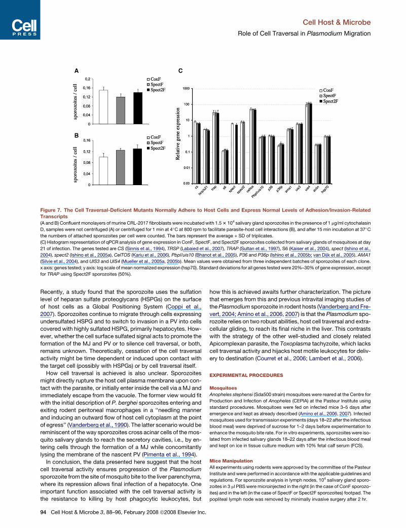
Figure 7. The Cell Traversal-Deficient Mutants Normally Adhere to Host Cells and Express Normal Levels of Adhesion/Invasion-Related
Transcripts
(A and B) Confluent monolayers of murine CRL-2017 fibroblasts were incubated with 1.5 3 104 salivary gland sporozoites in the presence of 1 mg/ml cytochalasin
D, samples were not centrifuged (A) or centrifuged for 1 min at 4�C at 800 rpm to facilitate parasite-host cell interactions (B), and after 15 min incubation at 37�C
the numbers of attached sporozoites per cell were counted. The bars represent the average + SD of triplicates.
(C) Histogram representation of qPCR analysis of gene expression in ConF, SpectF, and Spect2F sporozoites collected from salivary glands of mosquitoes at day
21 of infection. The genes tested are CS (Sinnis et al., 1994), TRSP (Labaied et al., 2007), TRAP (Sultan et al., 1997), S6 (Kaiser et al., 2004), spect (Ishino et al.,
2004), spect2 (Ishino et al., 2005a), CelTOS (Kariu et al., 2006), Pbpl/uis10 (Bhanot et al., 2005), P36 and P36p (Ishino et al., 2005b; van Dijk et al., 2005), AMA1
(Silvie et al., 2004), and UIS3 and UIS4 (Mueller et al., 2005a, 2005b). Mean values were obtained from three independent batches of sporozoites of each clone.
x axis: genes tested; y axis: log scale of mean normalized expression (hsp70). Standard deviations for all genes tested were 20%–30% of gene expression, except
for TRAP using Spect2F sporozoites (50%).
Recently, a study found that the sporozoite uses the sulfation
level of heparan sulfate proteoglycans (HSPGs) on the surface
of host cells as a Global Positioning System (Coppi et al.,
2007). Sporozoites continue to migrate through cells expressing
undersulfated HSPG and to switch to invasion in a PV into cells
covered with highly sulfated HSPG, primarily hepatocytes. How-
ever, whether the cell surface sulfated signal acts to promote the
formation of the MJ and PV or to silence cell traversal, or both,
remains unknown. Theoretically, cessation of the cell traversal
activity might be time dependent or induced upon contact with
the target cell (possibly with HSPGs) or by cell traversal itself.
How cell traversal is achieved is also unclear. Sporozoites
might directly rupture the host cell plasma membrane upon con-
tact with the parasite, or initially enter inside the cell via a MJ and
immediately escape from the vacuole. The former view would fit
with the initial description of P. berghei sporozoites entering and
exiting rodent peritoneal macrophages in a ‘‘needling manner
and inducing an outward flow of host cell cytoplasm at the point
of egress’’ (Vanderberg et al., 1990). The latter scenario would be
reminiscent of the way sporozoites cross acinar cells of the mos-
quito salivary glands to reach the secretory cavities, i.e., by en-
tering cells through the formation of a MJ while concomitantly
lysing the membrane of the nascent PV (Pimenta et al., 1994).
In conclusion, the data presented here suggest that the host
cell traversal activity ensures progression of the Plasmodium
sporozoite from the site of mosquito bite to the liver parenchyma,
where its repression allows final infection of a hepatocyte. One
important function associated with the cell traversal activity is
the resistance to killing by host phagocytic leukocytes, but
94 Cell Host & Microbe 3, 88–96, February 2008 ª2008 Elsevier Inc
how this is achieved awaits further characterization. The picture
that emerges from this and previous intravital imaging studies of
the Plasmodium sporozoite in rodent hosts (Vanderberg and Fre-
vert, 2004; Amino et al., 2006, 2007) is that the Plasmodium spo-
rozoite relies on two robust abilities, host cell traversal and extra-
cellular gliding, to reach its final niche in the liver. This contrasts
with the strategy of the other well-studied and closely related
Apicomplexan parasite, the Toxoplasma tachyzoite, which lacks
cell traversal activity and hijacks host motile leukocytes for deliv-
ery to destination (Courret et al., 2006; Lambert et al., 2006).
EXPERIMENTAL PROCEDURES
Mosquitoes
Anopheles stephensi (Sda500 strain) mosquitoes were reared at the Centre for
Production and Infection of Anopheles (CEPIA) at the Pasteur Institute using
standard procedures. Mosquitoes were fed on infected mice 3–5 days after
emergence and kept as already described (Amino et al., 2006, 2007). Infected
mosquitoes used for transmission experiments (days 18–22 after the infectious
blood meal) were deprived of sucrose for 1–2 days before experimentation to
enhance the mosquito bite rate. For in vitro experiments, sporozoites were iso-
lated from infected salivary glands 18–22 days after the infectious blood meal
and kept on ice in tissue culture medium with 10% fetal calf serum (FCS).
Mice Manipulation
All experiments using rodents were approved by the committee of the Pasteur
Institute and were performed in accordance with the applicable guidelines and
regulations. For sporozoite analysis in lymph nodes, 104 salivary gland sporo-
zoites in 3 ml PBS were microinjected in the right (in the case of ConF sporozo-
ites) and in the left (in the case of SpectF or Spect2F sporozoites) footpad. The
popliteal lymph node was removed by minimally invasive surgery after 2 hr.
.

Cell Host & Microbe
Role of Cell Traversal in Plasmodium Migration
ell Host & Microbe 3, 88–96, February 2008 ª2008 Elsevier Inc. 95
Microscopy and Image Analysis
Microscopic analysis was carried out using a high-speed spinning disk confo-
cal system (Ultraview ERS, Perkin Elmer) mounted on an inverted Axiovert 200
microscope (Carl Zeiss) with Optovar option linked to a Orca II ER camera
(Hamamatsu, Japan). Image files were processed using ImageJ. Sporozoites
in the dermis of mice were imaged as already described (Amino et al., 2007).
Primary Hepatocytes
Rat (4-week-old female Wistar) and mouse (6- to 8-week-old female C57BL/6)
hepatocytes (Janvier, France) were isolated from animals treated with sodium
pentobarbital by a modified two-step collagenase perfusion method. Briefly,
the liver was perfused via the portal vein for 10 min with liver perfusion medium
13 (GIBCO 17701) followed by a 10 min perfusion with liver digest medium 13
(GIBCO 17703) at a flow rate of 6–10 ml/min. Isolated cells were washed and
centrifuged through a Percoll gradient to remove damaged cells. Cell viability
(>90%) was determined by trypan blue exclusion. Hepatocytes were plated at
a density of 6–15 3 104 in 8-well permanox Lab-Tek chamber slides in Williams
medium E containing 10% FCS. Cells were maintained at 37�C in a humidified
atmosphere of 95% air and 5% CO2.
Cell Traversal Assays
Cell traversal events by sporozoites were visualized by spinning disk confocal
microscopy. Sporozoites and freshly trypsinized cells in a 1:1 ratio were mixed
to 10 ml Matrigel (BD Biosciences) in the presence of 5 mM SYTOX Orange Nu-
cleid Acid Stain (S-11368, Molecular Probes), a high-affinity nucleic acid stain
that penetrates cells with a compromised plasma membrane. Samples were
recorded for 30 min in multiple z layers with a double wavelength excitation
of 488 nm and 568 nm for GFP-expressing parasites and SYTOX Orange,
respectively.
Cell Infection Assays and CS Staining
Infection assays were performed at a multiplicity of infection of 1. Eight-well
permanox Lab-Tek chamber slides and 15 3 104 sporozoites freshly dissected
out from mosquito salivary glands were used in experiments with rat primary
hepatocytes; MatTek glass bottom culture dishes and 2 3 104 sporozoites
were used for CRL-2017 dermal fibroblasts infection. First, sporozoites were
added onto cells on ice, and samples were centrifuged for 1 min at 4�C at
800 rpm to facilitate parasite-host cell interactions. Samples were then placed
at 37�C, and at various incubation times samples were fixed with 4% parafor-
maldehyde (PFA), which does not permeabilize the host cell plasma membrane,
and stained with the 3D11 anti-CS mAb coupled to Alexa Fluor 568 (A-20184,
Molecular Probes). This allowed for discriminating between internalized sporo-
zoites (green) and external sporozoites (red). Note that, in the case of the wild-
type sporozoites, a proportion of the green parasites after 1 hr correspond to
sporozoites that are located inside a cell, but not inside a vacuole (sporozoites
arrested during cell traversal), a proportion that does not exceed 5%–10%.
Sporozoite Attachment Assay
Infection experiments were performed with 1.75 3 104 salivary gland sporozo-
ites and a confluent monolayer of CRL-2017 dermal fibroblasts plated on Lab-
Tek chamber slides (8-well permanox). Sporozoites were plated on cells in the
presence of cytD 1 mg/ml, and after 15 min at 37�C, samples were washed
three times in PBS and then stained with an Ab-CS to differentiate intracellular
from extracellular parasites. As expected, no intracellular parasites were
found. In experiments with metabolic inhibited parasites, sporozoites were
preincubated 1 hr at room temperature with 0.03% sodium azide and then
plated on cells in the continued presence of this compound.
Transmission Electron Microscopy
Samples were fixed by addition of 2.5% glutaraldehyde/0.15 M cacodylate
buffer followed by 1% osmium tetroxyde, dehydrated in a series of ethanol
concentrations, and embedded in EPON resin mixture. Ultrathin sections
(50 to 60 nm) were observed with a Jeol 1200EXII (Tokio, Japon) transmission
electron microscope. Images were recorded using an Eloise Keenview camera
and the Analysis Pro software version 3.1 (Eloise SARL, Roissy, France).
C
Real-Time Quantitative RT-PCR
Real-time qPCR was performed on cDNA preparations using the SYBR green
detection system and the ABI Prism 7900 sequence detector (Applied Biosys-
tems) according to the manufacturer’s instructions. Salivary gland sporozoites
from the ConF, SpectF, and Spect2F clones were isolated at day 21 after the
blood meal. RNA was extracted with TriZol and DNase treated, and cDNAs
synthesized with Superscript II reverse transcriptase (Invitrogen) using random
primers. Three independent RNA preparations were made for each sample.
PCR conditions were 1 cycle of 95�C for 10 min followed by 40 cycles of
95�C for 15 s, 55�C for 15 s, and 60�C for 45 min). qPCR was performed in trip-
licate with three serial dilutions. The standard curve was analyzed for all
primers and gave amplification efficiencies of 90%–100%. Data were analyzed
with SDS 2.1 software. Analysis was performed using the 2�DDCt method (User
Bulletin 2, ABI). The following primers were used for qPCR analysis: Ama1, for-
ward, ATTTGGGTTGATGGTTATTG; Ama1, reverse, TCCTTGTCGAAATTTGG
TAG; CS, forward, ACAGAGGAATGGTCTCAATG; CS, reverse, TTATCCATTT
TACAAATTTCAG; TRAP, forward, AACATTCACTCCATTCTTCC; TRAP, re-
verse, CATGTTATTCCAATGCTCAC; P36p, forward, CTAATACGACCTTAG-
GACACTTTGAA; P36p, reverse, GATGTTCCATTTGGGTTTACATGATC; P36,
forward, GCCTAATGCAAAATATTATCCCGATTTAG; P36, reverse, GCTAGT
CCTTTGTTCCCATTATATG; Pbpl/UIS10, forward, GTTACACTGATAGAGAA
GATGT; Pbpl/UIS10, reverse, GTGGTACTATACAAACAATATGTTGG; Spect2,
forward, AAGGAGTTTCAGCTATGCAC; Spect2, reverse, CAGTTCATTTATG
CCTGACC; Spect1, forward, TAGCCTAATTCAAATAAACGAAC; Spect1, re-
verse, GAAGTTAATTAATTCTGATACCCT; UIS4, forward, CCAAACCAAGCG
ATCATACATACAG; UIS4, reverse, CTTCACCCACTAAATCGCTTAATTC; S6,
forward, GCTAGTGAAATCGAAGAAGC; S6, reverse, GCCTTGCAAATAATG
AGAAC; Hsp70, forward, TGCAGCAGATAATCAAACTC; Hsp70, reverse,
ACTTCAATTTGTGGAACACC; PbActin, forward, GGAAACATTATAACAGTA
GGTAATGAAAG; PbActin, reverse, GTTGTACCTCCAGATAAAACAATGTTT
CC; Tsrp, forward, TAAAGATAAGAGCACGAGTTAGTAGT; Tsrp, reverse,
AATTGTTACATATTTACTTAGCATTCT; Celtos, forward, GTTCTATGTTTGAG
AGGCAAAAATGG; Celtos, reverse, TGATGACGAGTCTTGTTGAAATGCAC;
UIS3, forward, AGTTGCAATTGCTTTGTTATCATCAGGA; UIS3, reverse,
GTTCTTTATATTTTGTACTAGTGCTTGGC.
Supplemental Data
The Supplemental Data include six supplemental figures and four supple-
mental movies and can be found with this article online at http://www.
cellhostandmicrobe.com/cgi/content/full/3/2/88/DC1/.
ACKNOWLEDGMENTS
We thank S. Shorte, P. Roux, and the other members of the ‘‘Platforme d’image-
rie dynamique’’ (Institut Pasteur) for help with confocal microscopy;
C. Bourgouin, I. Thiery, and the other members of the ‘‘Centre de Production
et d’Infection des Anopheles’’ (Institut Pasteur) for mosquito rearing;
V. Richard from the Electron Microscopy Facility (University of Montpellier 2);
G. Milon, G. Lauvau, and T. Graf for providing the lys-gfp mice; and Patricia Bal-
dacci and Samantha Blazquez for comments on the manuscript. This work was
supported by funds from the Institut Pasteur (‘‘Grand Programme Horizontal
Anopheles’’), the Howard Hughes Medical Institute, the European Commission
(FP6 BioMalPar Network of Excellence), and the Core Research for Evolutional
Science and Technology (CREST). R.A. was supported by the Pasteur Institute
GPH fellowship, D.G. is a BioMalPar Ph.D. student, T.I. was supported by a
fellowship from the Association Pasteur-Japon, M.Y. is a CREST Scholar,
and R.M. is a Howard Hughes Medical Institute International Scholar.
Received: August 2, 2007
Revised: October 18, 2007
Accepted: December 26, 2007
Published: February 13, 2008
REFERENCES
Amino, R., Martin, B., Thiberge, S., Celli, S., Shorte, S., Frischknecht, F., and
Menard, R. (2006). Quantitative imaging of Plasmodium transmission from
mosquito to mammal. Nat. Med. 12, 220–224.

Cell Host & Microbe
Role of Cell Traversal in Plasmodium Migration
Amino, R., Thiberge, S., Blazquez, S., Baldacci, P., Renaud, O., Shorte, S., and
Menard, R. (2007). Imaging malaria sporozoites in the dermis of the mamma-
lian host. Nat. Protocols 2, 1705–1712.
Baer, K., Roosevelt, M., Clarkson, A.B., Jr., van Rooijen, N., Schnieder, T., and
Frevert, U. (2007). Kupffer cells are obligatory for Plasmodium yoelii sporozoite
infection of the liver. Cell. Microbiol. 9, 397–412.
Bhanot, P., Schauer, K., Coppens, I., and Nussenzweig, V. (2005). A surface
phospholipase is involved in the migration of Plasmodium sporozoites through
cells. J. Biol. Chem. 280, 6752–6760.
Carrolo, M., Giordano, S., Cabrita-Santos, L., Corso, S., Vigario, A.M., Silva,
S., Leiriao, P., Carapau, D., Armas-Portela, R., Comoglio, P.M., et al. (2003).
Hepatocyte growth factor and its receptor are required for malaria infection.
Nat. Med. 9, 1363–1369.
Coppi, A., Tewari, R., Bishop, J.R., Bennett, B.L., Lawrence, R., Esko, J.D.,
Billker, O., and Sinnis, P. (2007). Heparan sulfate proteoglycans provide a sig-
nal to Plasmodium sporozoites to stop migrating and productively invade host
cells. Cell Host Microbe 2, 316–327.
Courret, N., Darche, S., Sonigo, P., Milon, G., Buzoni-Gatel, D., and Tardieux, I.
(2006). CD11c- and CD11b-expressing mouse leukocytes transport single
Toxoplasma gondii tachyzoites to the brain. Blood 107, 309–316.
Faust, N., Varas, F., Kelly, L.M., Heck, S., and Graf, T. (2000). Insertion of en-
hanced green fluorescent protein into the lysozyme gene creates mice with
green fluorescent granulocytes and macrophages. Blood 96, 719–726.
Frevert, U., Engelmann, S., Zougbede, S., Stange, J., Ng, B., Matuschewski,
K., Liebes, L., and Yee, H. (2005). Intravital observation of Plasmodium berghei
sporozoite infection of the liver. PLoS Biol. 3, e192. 10.1371/journal.pbio.
0030192.
Hollingdale, M.R., Leef, J.L., McCullough, M., and Beaudouin, R.L. (1981).
In vitro cultivation of the exoerythrocytic stage of Plasmodium berghei from
sporozoites. Science 213, 1021–1022.
Ishino, T., Yano, K., Chinzei, Y., and Yuda, M. (2004). Cell-passage activity is
required for the malarial parasite to cross the liver sinusoidal cell layer. PLoS
Biol. 2, 77–84.
Ishino, T., Chinzei, Y., and Yuda, M. (2005a). A Plasmodium sporozoite protein
with a membrane attack complex domain is required for breaching the liver
sinusoidal cell layer prior to hepatocyte infection. Cell. Microbiol. 7, 199–208.
Ishino, T., Chinzei, Y., and Yuda, M. (2005b). Two proteins with 6-cys motifs
are required for malarial parasites to commit to infection of the hepatocyte.
Mol. Microbiol. 58, 1264–1275.
Kaiser, K., Camargo, N., Coppens, I., Morrisey, J.M., Vaidya, A.B., and Kappe,
S.H. (2004). A member of a conserved Plasmodium protein family with mem-
brane-attack complex/perforin (MACPF)-like domains localizes to the micro-
nemes of sporozoites. Mol. Biochem. Parasitol. 133, 15–26.
Kappe, S.H., Buscaglia, C.A., and Nussenzweig, V. (2004). Plasmodium spo-
rozoite molecular cell biology. Annu. Rev. Cell Dev. Biol. 20, 29–59.
Kariu, T., Ishino, T., Yano, K., Chinzei, Y., and Yuda, M. (2006). CelTOS, a novel
malarial protein that mediates transmission to mosquito and vertebrate hosts.
Mol. Microbiol. 59, 1369–1379.
Labaied, M., Camargo, N., and Kappe, S.H. (2007). Depletion of the Plasmo-
dium berghei thrombospondin-related sporozoite protein reveals a role in
host cell entry by sporozoites. Mol. Biochem. Parasitol. 153, 158–166.
Lambert, H., Hitziger, N., Dellacasa, I., Svensson, M., and Barragan, A. (2006).
Induction of dendritic cell migration upon Toxoplasma gondii infection poten-
tiates parasite dissemination. Cell. Microbiol. 8, 1611–1623.
Leiriao, P., Rodrigues, C.D., Albuquerque, S.S., and Mota, M.M. (2004). Sur-
vival of protozoan intracellular parasites in host cells. EMBO Rep. 5, 1142–
1147.
Matsuoka, H., Yoshida, S., Hirai, M., and Ishii, A. (2002). A rodent malaria, Plas-
modium berghei, is experimentally transmitted to mice by merely probing of
infective mosquito, Anopheles stephensi. Parasitol. Int. 51, 17–23.
Menard, R. (2001). Gliding motility and cell invasion by Apicomplexa: Insights
from the Plasmodium sporozoite. Cell. Microbiol. 3, 63–73.
96 Cell Host & Microbe 3, 88–96, February 2008 ª2008 Elsevier Inc
Mota, M.M., Pradel, G., Vanderberg, J.P., Hafalla, J.C., Frevert, U., Nussenz-
weig, R.S., Nussenzweig, V., and Rodriguez, A. (2001). Migration of Plasmo-
dium sporozoites through cells before infection. Science 291, 141–144.
Mota, M.M., Hafalla, J.C.R., and Rodriguez, A. (2002). Migration through host
cells activates Plasmodium sporozoites for infection. Nat. Med. 8, 1318–1322.
Mota, M.M., and Rodriguez, A. (2004). Migration through host cells: The first
steps of Plasmodium sporozoites in the mammalian host. Cell. Microbiol. 6,
1113–1118.
Mueller, A.K., Camargo, N., Kaiser, K., Andorfer, C., Frevert, U., Matuschew-
ski, K., and Kappe, S.H. (2005a). Plasmodium liver stage developmental arrest
by depletion of a protein at the parasite-host interface. Proc. Natl. Acad. Sci.
USA 102, 3022–3027.
Mueller, A.K., Labaied, M., Kappe, S.H., and Matuschewski, K. (2005b). Ge-
netically modified Plasmodium parasites as a protective experimental malaria
vaccine. Nature 433, 164–167.
Pimenta, P.F., Touray, M., and Miller, L. (1994). The journey of malaria sporo-
zoites in the mosquito salivary gland. J. Eukaryot. Microbiol. 41, 608–624.
Pradel, G., and Frevert, U. (2001). Malaria sporozoites actively enter and pass
through rat Kupffer cells prior to hepatocyte invasion. Hepatology 33, 1154–
1165.
Prudencio, M., Rodriguez, A., and Mota, M.M. (2006). The silent path to thou-
sands of merozoites: The Plasmodium liver stage. Nat. Rev. Microbiol. 11,
849–856.
Sibley, L.D. (2004). Intracellular parasite invasion strategies. Science 304, 248–
253.
Sidjanski, S., and Vanderberg, J.P. (1997). Delayed migration of Plasmodium
sporozoites from the mosquito bite site to the blood. Am. J. Trop. Med. Hyg.
57, 426–429.
Silvie, O., Franetich, J.F., Charrin, S., Mueller, M.S., Siau, A., Bodescot, M.,
Rubinstein, E., Hannoun, L., Charoenvit, Y., Kocken, C.H., et al. (2004). A
role for apical membrane antigen 1 during invasion of hepatocytes by Plasmo-
dium falciparum sporozoites. J. Biol. Chem. 279, 9490–9496.
Sinnis, P., Clavijo, P., Fenyo, D., Chait, B.T., Cerami, C., and Nussenzweig, V.
(1994). Structural and functional properties of region II-plus of the malaria cir-
cumsporozoite protein. J. Exp. Med. 180, 297–306.
Sturm, A., Amino, R., van de Sand, C., Regen, T., Retzlaff, S., Rennenberg, A.,
Krueger, A., Pollok, J.M., Menard, R., and Heussler, V.T. (2006). Manipulation
of host hepatocytes by the malaria parasite for delivery into liver sinusoids. Sci-
ence 313, 1287–1290.
Sultan, A.A., Thathy, V., Frevert, U., Robson, K.J., Crisanti, A., Nussenzweig,
V., Nussenzweig, R., and Menard, R. (1997). TRAP is necessary for gliding mo-
tility and infectivity of Plasmodium sporozoites. Cell 90, 511–522.
Vanderberg, J.P., Chew, S., and Stewart, M.J. (1990). Plasmodium sporozoite
interactions with macrophages in vitro: A videomicroscopic analysis. J. Proto-
zool. 37, 528–536.
Vanderberg, J.P., and Frevert, U. (2004). Intravital microscopy demonstrating
antibody-mediated immobilisation of Plasmodium berghei sporozoites in-
jected into skin by mosquitoes. Int. J. Parasitol. 34, 991–996.
van Dijk, M.R., Douradinha, B., Franke-Fayard, B., Heussler, V., van Dooren,
M.W., van Schaijk, B., van Gemert, G.J., Sauerwein, R.W., Mota, M.M., Wa-
ters, A.P., and Janse, C.J. (2005). Genetically attenuated, P36p-deficient ma-
larial sporozoites induce protective immunity and apoptosis of infected liver
cells. Proc. Natl. Acad. Sci. USA 102, 12194–12199.
van Rooijen, N., Bakker, J., and Sanders, A. (1997). Transient suppression of
macrophage functions by liposome-encapsulated drugs. Trends Biotechnol.
15, 178–185.
Waters, A.P., Mota, M.M., van Dijk, M.R., and Janse, C.J. (2005). Parasitology.
Malaria vaccines: Back to the future? Science 307, 528–530.
Yamauchi, L.M., Coppi, A., Snounou, G., and Sinnis, P. (2007). Plasmodium
sporozoites trickle out of the injection site. Cell. Microbiol. 9, 1215–1222.
.