Host carbon sources modulate cell wall architecture, drug resistance and virulence in a fungal...
17
Host carbon sources modulate cell wall architecture, drug resistance and virulence in a fungal pathogen Iuliana V. Ene, 1 Ashok K. Adya, 2 Silvia Wehmeier, 1 Alexandra C. Brand, 1 Donna M. MacCallum, 1 Neil A. R. Gow 1 and Alistair J. P. Brown 1 * 1 Aberdeen Fungal Group, School of Medical Sciences, Institute of Medical Sciences, University of Aberdeen, Foresterhill, Aberdeen AB25 2ZD, UK. 2 Division of Biotechnology and Forensic Sciences, School of Contemporary Sciences, University of Abertay Dundee, Dundee DD1 1HG, UK. Summary The survival of all microbes depends upon their ability to respond to environmental challenges. To establish infection, pathogens such as Candida albicans must mount effective stress responses to counter host defences while adapting to dynamic changes in nutrient status within host niches. Studies of C. albicans stress adaptation have gen- erally been performed on glucose-grown cells, leaving the effects of alternative carbon sources upon stress resistance largely unexplored. We have shown that growth on alternative carbon sources, such as lactate, strongly influence the resistance of C. albicans to antifungal drugs, osmotic and cell wall stresses. Similar trends were observed in clinical isolates and other pathogenic Candida species. The increased stress resistance of C. albicans was not dependent on key stress (Hog1) and cell integrity (Mkc1) signalling path- ways. Instead, increased stress resistance was promoted by major changes in the architecture and biophysical properties of the cell wall. Glucose- and lactate-grown cells displayed significant dif- ferences in cell wall mass, ultrastructure, elasticity and adhesion. Changes in carbon source also altered the virulence of C. albicans in models of systemic candidiasis and vaginitis, confirming the importance of alternative carbon sources within host niches during C. albicans infections. Introduction Most microbes inhabit microenvironments that are in a constant state of flux. This is particularly true for clinically important microbial pathogens which must counter the biochemical and immunological insults imposed by their host. Pathogens must activate appropriate responses to these local environmental stresses while adapting to and assimilating the available nutrients within that niche. Candida albicans is a major fungal pathogen of humans that can cause a variety of infections (Calderone and Clancy, 2011). C. albicans is carried as a relatively harm- less commensal in the oral, gastrointestinal and urogeni- tal microflora of 40–80% of the population. Mucosal infections (thrush) can arise when immunological defects or pharmacological interventions affect host–fungus inter- actions, and a significant proportion of these infections can be recurrent (Calderone, 2002). In severely immuno- compromised patients C. albicans can thrive in the blood- stream, ultimately colonizing and forming lesions on internal organs (Perlroth et al., 2007; Pfaller and Diekema, 2010). Up to half of these systemic infections are fatal (Perlroth et al., 2007). Therefore, C. albicans can thrive within diverse niches in its human host. Clearly to cause infection, C. albicans must grow and divide in these diverse niches. Cells must assimilate locally available carbon sources which can include fermentable sugars and non-fermentable carbon sources (Lorenz and Fink, 2001; Lorenz et al., 2004; Piekarska et al., 2006; Vieira et al., 2010; Ueno et al., 2011). Physiologically rel- evant sugars include glucose, fructose and galactose. However, these sugars are only present at low levels and are even absent in many host niches. Consequently, other non-fermentable carbon sources become essential to support yeast growth and metabolism in vivo (Piekarska et al., 2006; Vieira et al., 2010; Ueno et al., 2011). These alternative carbon sources include amino acids and organic acids. For example, lactic acid is present in ingested foods, produced via host metabolic activity, gen- erated by endogenous lactic acid bacteria in the urogenital and gastrointestinal tracts, and is an important carbon source for Candida in the intestine (Ueno et al., 2011). Thinking about carbon source utilization in yeasts is strongly influenced by the Saccharomyces cerevisiae paradigm. When this relatively benign model yeast is faced with a mixture of carbon sources it preferentially utilizes sugars such as glucose before assimilating Received 7 March, 2012; revised 19 April, 2012; accepted 9 May, 2012. *For correspondence. E-mail [email protected]; Tel. (+44) 1224 437482; Fax (+44) 1224 437465. Re-use of this article is permitted in accordance with the Terms and Conditions set out at http://wileyonlinelibrary.com/onlineopen# OnlineOpen_Terms. Cellular Microbiology (2012) 14(9), 1319–1335 doi:10.1111/j.1462-5822.2012.01813.x First published online 5 June 2012 © 2012 Blackwell Publishing Ltd cellular microbiology
-
Upload
independent -
Category
Documents
-
view
2 -
download
0
Transcript of Host carbon sources modulate cell wall architecture, drug resistance and virulence in a fungal...

Host carbon sources modulate cell wall architecture,drug resistance and virulence in a fungal pathogen
Iuliana V. Ene,1 Ashok K. Adya,2 Silvia Wehmeier,1
Alexandra C. Brand,1 Donna M. MacCallum,1
Neil A. R. Gow1 and Alistair J. P. Brown1*1Aberdeen Fungal Group, School of Medical Sciences,Institute of Medical Sciences, University of Aberdeen,Foresterhill, Aberdeen AB25 2ZD, UK.2Division of Biotechnology and Forensic Sciences,School of Contemporary Sciences, University of AbertayDundee, Dundee DD1 1HG, UK.
Summary
The survival of all microbes depends upon theirability to respond to environmental challenges. Toestablish infection, pathogens such as Candidaalbicans must mount effective stress responses tocounter host defences while adapting to dynamicchanges in nutrient status within host niches.Studies of C. albicans stress adaptation have gen-erally been performed on glucose-grown cells,leaving the effects of alternative carbon sourcesupon stress resistance largely unexplored. Wehave shown that growth on alternative carbonsources, such as lactate, strongly influence theresistance of C. albicans to antifungal drugs,osmotic and cell wall stresses. Similar trends wereobserved in clinical isolates and other pathogenicCandida species. The increased stress resistanceof C. albicans was not dependent on key stress(Hog1) and cell integrity (Mkc1) signalling path-ways. Instead, increased stress resistance waspromoted by major changes in the architecture andbiophysical properties of the cell wall. Glucose-and lactate-grown cells displayed significant dif-ferences in cell wall mass, ultrastructure, elasticityand adhesion. Changes in carbon source alsoaltered the virulence of C. albicans in models ofsystemic candidiasis and vaginitis, confirming theimportance of alternative carbon sources withinhost niches during C. albicans infections.
Introduction
Most microbes inhabit microenvironments that are in aconstant state of flux. This is particularly true for clinicallyimportant microbial pathogens which must counter thebiochemical and immunological insults imposed by theirhost. Pathogens must activate appropriate responses tothese local environmental stresses while adapting to andassimilating the available nutrients within that niche.
Candida albicans is a major fungal pathogen of humansthat can cause a variety of infections (Calderone andClancy, 2011). C. albicans is carried as a relatively harm-less commensal in the oral, gastrointestinal and urogeni-tal microflora of 40–80% of the population. Mucosalinfections (thrush) can arise when immunological defectsor pharmacological interventions affect host–fungus inter-actions, and a significant proportion of these infectionscan be recurrent (Calderone, 2002). In severely immuno-compromised patients C. albicans can thrive in the blood-stream, ultimately colonizing and forming lesions oninternal organs (Perlroth et al., 2007; Pfaller andDiekema, 2010). Up to half of these systemic infectionsare fatal (Perlroth et al., 2007). Therefore, C. albicans canthrive within diverse niches in its human host.
Clearly to cause infection, C. albicans must grow anddivide in these diverse niches. Cells must assimilate locallyavailable carbon sources which can include fermentablesugars and non-fermentable carbon sources (Lorenz andFink, 2001; Lorenz et al., 2004; Piekarska et al., 2006;Vieira et al., 2010; Ueno et al., 2011). Physiologically rel-evant sugars include glucose, fructose and galactose.However, these sugars are only present at low levels andare even absent in many host niches. Consequently, othernon-fermentable carbon sources become essential tosupport yeast growth and metabolism in vivo (Piekarskaet al., 2006; Vieira et al., 2010; Ueno et al., 2011). Thesealternative carbon sources include amino acids andorganic acids. For example, lactic acid is present iningested foods, produced via host metabolic activity, gen-erated by endogenous lactic acid bacteria in the urogenitaland gastrointestinal tracts, and is an important carbonsource for Candida in the intestine (Ueno et al., 2011).
Thinking about carbon source utilization in yeasts isstrongly influenced by the Saccharomyces cerevisiaeparadigm. When this relatively benign model yeast isfaced with a mixture of carbon sources it preferentiallyutilizes sugars such as glucose before assimilating
Received 7 March, 2012; revised 19 April, 2012; accepted 9 May,2012. *For correspondence. E-mail [email protected]; Tel.(+44) 1224 437482; Fax (+44) 1224 437465.Re-use of this article is permitted in accordance with the Termsand Conditions set out at http://wileyonlinelibrary.com/onlineopen#OnlineOpen_Terms.
Cellular Microbiology (2012) 14(9), 1319–1335 doi:10.1111/j.1462-5822.2012.01813.xFirst published online 5 June 2012
© 2012 Blackwell Publishing Ltd
cellular microbiology

alternative carbon sources (Johnston, 1999). This hierar-chical metabolic activity is regulated by a complexglucose signalling network involving the co-ordinatedaction of AMP kinase, protein kinase A and sugar receptorrepressor signalling pathways (Kim and Johnston, 2006).This signalling network downregulates metabolic path-ways required for the assimilation of alternative carbonsources at both transcriptional and post-transcriptionallevels (Yin et al., 1996; Sexton et al., 2007; Gancedo,2008; Hedbacker and Carlson, 2008; Zaman et al., 2008;Askew et al., 2009). To some extent this signallingnetwork and the downstream regulatory mechanismshave been conserved in C. albicans (Sabina and Brown,2009). However, recent work has shown that there hasbeen major transcriptional rewiring as well as significantdivergence in the components that regulate carbohydrateand lipid metabolism in C. albicans (Martchenko et al.,2007; Lavoie et al., 2009). Indeed, unlike S. cerevisiae,C. albicans is classified as a glucose-negative yeastbecause it continues to respire in the presence of glucose(Niimi et al., 1988). Clearly, benign and pathogenic yeastshave evolved to respond differentially to glucose, reflect-ing their contrasting niches. S. cerevisiae is thought tohave evolved under conditions of ‘feast and famine’,rapidly exploiting fermentable sugars before switching toalternative carbon sources (Johnston, 1999). In contrast,C. albicans often inhabits niches that are glucose-limitedbut rich in alternative carbon sources. The physiologicalrobustness of this pathogen in vivo is probably enhancedby its ability to assimilate multiple carbon sources simul-taneously, rather than the sequential use of ‘preferred’and then ‘non-preferred’ carbon sources (Brown et al.,2007).
Genome-wide expression profiling studies have con-firmed the ability of C. albicans to undergo rapid metabolictransitions (Lorenz et al., 2004; Rodaki et al., 2009). Forseveral fungi, as well as some bacteria (e.g. Mycobacte-rium tuberculosis), the glyoxylate cycle plays an importantrole in pathogenesis and is required for full virulence in thehost (Lorenz and Fink, 2001). Recent work has reinforcedthe essentiality of carbon metabolism for fungal patho-genicity (Price et al., 2011; Vylkova et al., 2011). In par-ticular, the induction of lactate transporters and metabolicenzymes upon macrophage internalization (Lorenz et al.,2004) suggests that high concentrations of lactate arepresent in the phagosome. As peroxisomal fatty acidb-oxidation is not essential for virulence (Piekarska et al.,2006), it is conceivable that lactate assimilation and utili-zation might support flux through the glyoxylate cyclethereby contributing to the survival of the pathogen underglucose-limiting conditions. The dependence of Candidaglabrata upon lactate assimilation in certain host nichesfurther strengthens this view (Ueno et al., 2011). However,most virulence studies have been performed using cells
grown in rich glucose-containing media, and thus theimpact of carbon source on infection development has yetto be addressed.
How does growth on different carbon sources affect theability of yeasts to adapt to dynamic changes in theirenvironment? In S. cerevisiae, glucose downregulatesthe core stress response via cAMP signalling, renderingcells more sensitive to environmental insults. Followingthe diauxic shift to alternative carbon sources, this yeastbecomes more stress-resistant (Gounalaki and Thireos,1994; Stanhill et al., 1999; Görner et al., 2002). The situ-ation is less clear in C. albicans as most studies of stressadaptation have been performed on glucose-grown cells.The core stress response has diverged significantly inC. albicans (Enjalbert et al., 2003; 2006; Ramsdale et al.,2008), and while glucose exposure and cAMP signallingdo influence stress resistance in C. albicans (Wilsonet al., 2007; Rodaki et al., 2009), this is by unknownmechanisms. The effects of alternative carbon sourcesupon stress resistance of C. albicans remain unexploreddespite the fact that this yeast depends upon such nutri-ents in many host niches (Lorenz and Fink, 2001; Barelleet al., 2006).
Similarly, the impact of growth on alternative carbonsources on the C. albicans cell wall remains largelyunexplored, although the cell wall is a key effector of cellmorphology and robustness, the major point of contactwith the host, an active modulator of host immunedefences and a prime target for antifungal drugs. Fur-thermore, a large proportion of assimilated carbon isinvested in cell wall biogenesis as the cell wall comprises~ 30% of the yeast cell dry weight. The C. albicans yeastcell wall contains three main components: mannopro-teins (~ 39%), b-glucans (~ 59%) and chitin (~ 2%)(Aguilar-Uscanga and François, 2003). At a gross level,the composition of the S. cerevisiae cell wall has beenshown to vary considerably in response to changes incarbon source, temperature, pH and aeration (Aguilar-Uscanga and François, 2003). Several observationsimply mutual interactions between stress adaptation andcell wall architecture in C. albicans. Mannan chain lengthand complexity is altered when cells are grown in bloodor serum rather than standard laboratory growth media(Kruppa et al., 2011). The stress-activated protein kinaseHog1 (Smith et al., 2004) influences cell wall biosynthe-sis (Eisman et al., 2006), partly by regulating chitin syn-thesis (Munro et al., 2007; Walker et al., 2008). Also, inresponse to certain stresses, the cell integrity pathwayactivates cell wall biogenesis (Navarro-Garcia et al.,1998; Eisman et al., 2006; Munro et al., 2007; Blanken-ship et al., 2010). In addition, stressors have recentlybeen shown to influence mannan structures in C. albi-cans (Koyama et al., 2009). However, few studies haveexamined the influence of growth conditions on the
1320 I. V. Ene et al.
© 2012 Blackwell Publishing Ltd, Cellular Microbiology, 14, 1319–1335

cell wall (Kulkarni et al., 1980; Aguilar-Uscanga andFrançois, 2003; Kawahata et al., 2006; Kruppa et al.,2011), and none has examined this in any detail despiteits relevance to disease progression.
In this study we first characterized the impact of carbonsource on the C. albicans cell wall, because of the criticalrole of the fungal cell wall during infection. We comparedcells grown on glucose versus the physiologically relevantcarboxylic acid, lactate, with a view to comparing thisgluconeogenic carbon source with the carbon sourceclassically used for experimental dissection of stressadaptation in C. albicans. We chose sodium lactate forthese detailed cell wall analyses because lactate hasbeen used previously to study metabolic flexibility (Vieiraet al., 2010) and stress resistance (Rodaki et al., 2009).Lactate is an abundant carbon source in the intestine andvaginal mucosa and is produced at high rates by redblood cells, brain and muscle (Buchalter et al., 1989;Ueno et al., 2011). It is also found clinically in isotonicsolutions commonly used after trauma, surgery or burninjury (lactated Ringer’s solutions, Hartmann’ solution), allof which increase the risk of systemic candidiasis (Pfallerand Diekema, 2010). Having demonstrated the impact oflactate, we then confirmed that C. albicans is affected byother carbon sources that are commonly found in thehost, including galactose, fructose, oleic acid, pyruvate,sorbitol and amino acids.
Our analyses revealed the major effects of carbonsource on the architecture as well as the biochemical andbiophysical properties of the cell wall. Alternative carbonsources strongly influenced adhesion, stress adaptationand drug resistance in C. albicans in vitro, as well as thevirulence of this major pathogen in in vivo models ofsystemic candidiasis and vaginitis.
Results
Carbon source affects cell wall architecture
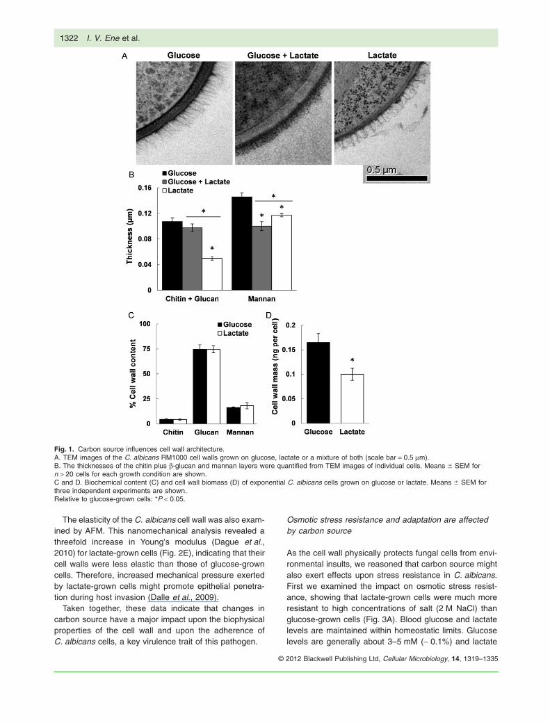
The cell wall architecture of yeast cells grown on glucose,lactate or a combination of these two carbon sources wasexamined by high-resolution freeze substitution transmis-sion electron microscopy (FS-TEM). This revealed majordifferences in cell wall structure, thickness and density(Fig. 1A), which were confirmed by measuring the thick-ness of b-glucan and mannan layers (Fig. 1B). The cellwalls of lactate-grown cells were thinner, with the b-glucanand chitin layer dramatically reduced. Furthermore, theouter mannan fibrils displayed less ordered structurescompared with those of glucose-grown cells.
We also examined the effects of carbon source uponC. albicans cell wall composition. Lactate- and glucose-grown cells were similar with regard to the overall propor-tions of chitin, b-glucan and mannan in their cell walls
(Fig. 1C). However, there were dramatic differences in cellwall biomass. The cell wall dry mass of lactate-grown cellswas 50% less than that of glucose-grown cells (Fig. 1D).Clearly, carbon source strongly influences the cell wallarchitecture of C. albicans.
Carbon source affects biophysical properties of thecell wall
Given the major impact on cell wall architecture, we rea-soned that changes in carbon source might also affectC. albicans cell wall properties. Cell wall porosity wasinvestigated using an assay based on the polycation-induced leakage of UV-absorbing compounds from cells(De Nobel et al., 1990). This assay compares leakageinduced by small polycations (e.g. poly-L-lysine, whichinduces cell leakage independent of cell wall porosity)with the release caused by large polycations (such asDEAE-dextrans, which cause limited leakage dependingupon the degree of porosity of the cell wall). The cell wallporosity of lactate-grown cells was twofold higher than ofglucose-grown cells (Fig. 2A), providing the evidence thatcarbon source has a major impact upon biophysical prop-erties of the C. albicans cell wall. The increased porosityof lactate-grown cells was consistent with their reducedmannan fibrils (Fig. 1B), as increased porosity has beenpreviously associated with shorter mannan side-chains(De Nobel et al., 1990).
Cell surface hydrophobicity was examined using a bio-physical assay that defines contact angles between cellsurfaces and aqueous solutions (Amaral et al., 2006;Henriques et al., 2007). Significant differences wereobserved in these contact angles (Fig. 2B) suggestingthat the surface of lactate-grown cells is more hydropho-bic than that of glucose-grown cells.
Hydrophobicity has been recently linked with adhesionto abiotic surfaces in C. albicans clinical isolates (Yoshi-jima et al., 2010; Sardi et al., 2011). Adhesion is a keyvirulence trait, playing major roles in tissue colonizationand biofilm formation on indwelling devices in hospitalpatients (Calderone, 2002). Adhesion of C. albicans cellsis influenced by growth on different sugars (Willis et al.,2000; Bain et al., 2001) and therefore we predicted thatgrowth on non-fermentable carbon sources might alsoinfluence C. albicans adherence. As expected, lactate-grown C. albicans cells adhered more strongly toplastic surfaces than cells grown on glucose (Fig. 2C).Furthermore, we confirmed that the adhesive force oflactate-grown cells was significantly greater than forglucose-grown cells using atomic force microscopy inforce-mapping mode (AFM-FM) (Müller and Dufrêne,2011) (Figs 2D and S1). Moreover, the adhesion energydisplayed by lactate-grown cells was also significantlyelevated (> 50%) (Fig. 2D).
Carbon source, fungal virulence and stress responses 1321
© 2012 Blackwell Publishing Ltd, Cellular Microbiology, 14, 1319–1335
The elasticity of the C. albicans cell wall was also exam-ined by AFM. This nanomechanical analysis revealed athreefold increase in Young’s modulus (Dague et al.,2010) for lactate-grown cells (Fig. 2E), indicating that theircell walls were less elastic than those of glucose-growncells. Therefore, increased mechanical pressure exertedby lactate-grown cells might promote epithelial penetra-tion during host invasion (Dalle et al., 2009).
Taken together, these data indicate that changes incarbon source have a major impact upon the biophysicalproperties of the cell wall and upon the adherence ofC. albicans cells, a key virulence trait of this pathogen.
Osmotic stress resistance and adaptation are affectedby carbon source
As the cell wall physically protects fungal cells from envi-ronmental insults, we reasoned that carbon source mightalso exert effects upon stress resistance in C. albicans.First we examined the impact on osmotic stress resist-ance, showing that lactate-grown cells were much moreresistant to high concentrations of salt (2 M NaCl) thanglucose-grown cells (Fig. 3A). Blood glucose and lactatelevels are maintained within homeostatic limits. Glucoselevels are generally about 3–5 mM (~ 0.1%) and lactate
Fig. 1. Carbon source influences cell wall architecture.A. TEM images of the C. albicans RM1000 cell walls grown on glucose, lactate or a mixture of both (scale bar = 0.5 mm).B. The thicknesses of the chitin plus b-glucan and mannan layers were quantified from TEM images of individual cells. Means � SEM forn > 20 cells for each growth condition are shown.C and D. Biochemical content (C) and cell wall biomass (D) of exponential C. albicans cells grown on glucose or lactate. Means � SEM forthree independent experiments are shown.Relative to glucose-grown cells: *P < 0.05.
1322 I. V. Ene et al.
© 2012 Blackwell Publishing Ltd, Cellular Microbiology, 14, 1319–1335

concentrations are about 1–20 mM (up to 0.2%). Lactatelevels vary in mucosal niches with concentrations in thevaginal fluid reaching 110 mM (~ 1.2%) (O’Hanlon et al.,2011). Therefore, we tested a wide range of lactate andglucose concentrations (0.1% to 4%). This revealed thatdifferential osmotic stress sensitivities are observed forcells grown at more physiological levels of these carbonsources (Fig. S2).
This increased resistance might reflect increased sig-nalling via the Hog1 stress-activated protein kinase,because this MAP kinase pathway is critical for osmoa-daptation in C. albicans (San José et al., 1996; Smithet al., 2004). This was tested by examining the dynamicsof Hog1 phosphorylation during hyperosmotic stress(1 M NaCl; Fig. 3B). Hog1 was activated to a lesserextent in lactate-grown cells, although they were moreresistant to 2 M NaCl. Furthermore, this increased resist-ance was retained in a hog1D mutant (Fig. 3A). Theseobservations indicate that these effects of carbon sourceupon osmotic stress resistance were not dependentupon Hog1 signalling.
The cell integrity (Mkc1) signalling pathway also playsa role in cell wall biogenesis and osmoadaptation inC. albicans (Navarro-Garcia et al., 1998; 2005). There-fore, we examined the dynamics of Mkc1 phosphorylation(Fig. 3C). Higher basal levels of Mkc1 phosphorylationwere observed in lactate-grown cells, suggesting activecell wall remodelling even in the absence of stress. WhileMkc1 phosphorylation was delayed in glucose-growncells, Mkc1 activation was observed immediately afterNaCl addition to lactate-grown cells (Fig. 3C). However,MKC1 deletion did not block the effects of lactate onosmotic stress resistance (Fig. 3A). We conclude that theeffects of lactate are not dependent on signalling via thecell integrity pathway.
As signalling through these key pathways was notessential for the elevated osmotic stress resistance oflactate-grown cells (Fig. 3A) and given the dramaticchanges observed in cell wall architecture (Fig. 1), wereasoned that the increased osmotic stress resistance oflactate-grown cells might be mediated through alterationsin the biophysical properties of the cell wall. To test this we
Fig. 2. Carbon source affects biophysicalproperties of the cell wall. C. albicansRM1000 cells were grown to mid-exponentialphase (OD600 0.4–0.6) prior to the analyses.A. The relative cell wall porosity of glucose orlactate-grown cells assayed via polycation-induced leakage of UV-absorbing compounds(De Nobel et al., 1990).B. Cell surface hydrophobicity of glucose- andlactate-grown C. albicans RM1000 cellsassayed via the water contact angles betweena liquid surface and these cells (n > 20drops).C. Adhesion of C. albicans cells to anon-treated polystyrene plastic surface.Adhered cells were scraped from the surface,serially diluted, plated on agar and theirnumbers determined as CFUs (shown aspercentage CFUs compared to glucose-growncontrols).D. Adhesion force and adhesion energy ofglucose- and lactate-grown cells examined byAFM (n = 6).E. Cell surface flexibility (indicated by Young’smodulus) measured by AFM (n = 6). Resultsare presented as means � SEM for at leastthree independent experiments.Relative to glucose-grown cells: *P < 0.05.
Carbon source, fungal virulence and stress responses 1323
© 2012 Blackwell Publishing Ltd, Cellular Microbiology, 14, 1319–1335

examined the dynamic changes in cell volume propa-gated by hyperosmotic stress (Fig. 3C). Relative toglucose-grown cells, lactate-grown cells displayed dra-matic reductions in cell volume (> 50% versus 20%). Fur-thermore, we observed significant decreases in the ratesof volume reduction and recovery in lactate-grown cells(-30.65 mm3 min-1 and 13.75 mm3 min-1 respectively)compared to glucose-grown cells (-87.21 mm3 min-1
and 38.56 mm3 min-1 respectively). Thus, although thedecrease in cell volume was more dramatic for lactate-grown cells, their volume changes occurred at a slowerrate, consistent with the AFM observation that the wallsof lactate-grown cells are less elastic (Fig. 2E). Takentogether, these observations suggest that changes incarbon source affect the architecture and flexibility of thecell wall, and that this in turn affects the dynamics ofcellular adaptation and resistance to hyperosmotic stress.
Carbon source affects resistance to other stresses andantifungal drugs
Clearly, the influence of cell wall remodelling might extendto other types of stress, and cell wall stresses in particular.Calcofluor White and Congo Red interfere with the syn-
thesis and cross-linking of chitin and glucan respectively.Lactate-grown cells displayed increased resistance toboth cell wall agents (Fig. 4A). This increased resistancewas also observed for hog1D and mkc1D cells grown onlactate (Fig. S3A–C).
Next, we compared the sensitivity of glucose- andlactate-grown cells to antifungal drugs. We tested caspo-fungin (an echinocandin that inhibits 1,3-b-glucan syn-thase), tunicamycin (a nucleoside antibiotic that blocksN-linked mannosylation), amphotericin B (a polyene thatinterferes with plasma membrane function by binding toergosterol) and miconazole (an azole that inhibits ergos-terol synthesis). Compared with glucose-grown cells,lactate-grown cells were resistant to caspofungin, tuni-camycin and amphotericin B, but were more sensitive tomiconazole (Fig. 4B). To address the contrasting effectsof carbon source on resistance to membrane-targetingantifungals, we tested the sensitivity of an ergosterol bio-synthetic mutant (erg11D) to amphotericin B and micona-zole. Ergosterol levels in erg11D cells are below detectionand consequently this strain displays increased resist-ance to amphotericin B and azoles (Sanglard et al.,2003). Although glucose-grown erg11D cells displayedincreased resistance to miconazole compared to the
Fig. 3. Osmotic stress resistance andadaptation are affected by carbon source.A. Resistance of wild-type (RM1000), hog1Dand mkc1D C. albicans cells (Table S1) toosmotic stress (2 M NaCl) during exponentialgrowth on glucose, lactate or glucose pluslactate. Means � SEM for four independentexperiments are presented. Relative toglucose-grown cells: *P < 0.05.B. Phosphorylation and activation of Hog1and Mkc1 in glucose- and lactate-grown cellsfollowing exposure to 1 M NaCl as revealedby Western blotting with phospho-specificantibodies. Similar results were obtained infour independent experiments.C. Dynamic changes in cell volume followinghyperosmotic shock (1 M NaCl; n > 25 cells).
1324 I. V. Ene et al.
© 2012 Blackwell Publishing Ltd, Cellular Microbiology, 14, 1319–1335

control strain (CAF4-2) (Fig. S3E), the differential mico-nazole resistance of glucose- and lactate-grown cells wasmaintained in both mutant and control strains (Fig. S3Dand E). Although sterol levels may differ between glucose-and lactate-grown cells, this does not account for theirdifferential sensitivities to ergosterol-targeting antifungals.This suggests that differential carbon source availability invivo, and the resultant cell wall remodelling, is likely toalter the susceptibility of C. albicans cells to therapeuticintervention.
The observed phenotypes extend to other pathogenicCandida species
We tested whether the phenotypic impact of carbon sourceis observed in other pathogenic Candida species. Wecompared the resistance of lactate- and glucose-growncells of C. glabrata, Candida dubliniensis, Candida tropi-calis, Candida krusei, Candida lusitaniae and Candidaguillermondii clinical isolates (Table S1) to hyperosmoticstress and amphotericin B. In addition, we included twoC. albicans clinical isolates (ATCC90028, a blood isolate,and ATCC10231, an oropharynx isolate) for comparisonwith the laboratory strains examined in the above experi-ments (MacCallum et al., 2009). In all cases, when theseCandida species were grown on lactate they displayedsignificantly increased resistance to hyperosmotic stressand amphotericin B compared with cells grown on glucose
(Fig. 5). This confirmed that carbon source has a pro-nounced effect upon stress and drug resistance for all ofthe pathogenic Candida species tested.
Observed phenotypes extend to other physiologicallyrelevant carbon sources
Host niches contain complex mixtures of fermentableand non-fermentable carbon sources. Therefore, weextended our analyses to test the impact of mixedcarbon sources. Growing C. albicans cells on a mixedmedium containing both glucose and lactate (each at1%) resulted in intermediate levels of resistance com-pared to cells grown on glucose or lactate alone (Figs 3Aand 4). This correlated with the intermediate architectureof the cell wall in cells grown on lactate plus glucose(Fig. 1A and B).
Next, we examined how stress and drug resistance areaffected by other carbon sources, including sugars (galac-tose, fructose and sorbitol), a fatty acid (oleic acid),another short-chain organic acid (pyruvate) and mixedamino acids. Cells grown on these carbon sources dis-played significant differences in their sensitivities tohyperosmotic stress, cell wall stress and antifungal drugswhen compared to the glucose-grown cells (Fig. 6). Ingeneral, cells grown on sugars exhibited lower resist-ances to osmotic stress (Fig. 6A) and higher sensitivitiesto amphotericin B (Fig. 6B).
Fig. 4. Carbon source impacts upon resistance to other stresses and antifungal drugs.A. Sensitivity of C. albicans RM1000 to Calcofluor White (200 mg ml-1) and Congo Red (300 mg ml-1) when grown on glucose, lactate or amixture of both.B. Sensitivity of C. albicans RM1000 to the antifungal drugs amphotericin B (Ambisome; 10 mg ml-1), caspofungin (6.4 ng ml-1), miconazole(25 mg ml-1) and tunicamycin (4 mg ml-1).Relative to glucose-grown cells: *P < 0.05.
Carbon source, fungal virulence and stress responses 1325
© 2012 Blackwell Publishing Ltd, Cellular Microbiology, 14, 1319–1335

It was conceivable that stress resistance simply corre-lated with a decreased growth rate. Hence, we monitoredthe growth of C. albicans cells on the different carbonsources during 48 h (Fig. 6D) and calculated the doub-ling times of these cultures during exponential phase(Fig. S4A). The doubling time was then plotted againstthe resistance level to hyperosmotic stress (Fig. S4B),amphotericin B (Fig. S4C) and Congo Red (Fig. S4D).This showed that stress resistance did not correlatedirectly with the relative doubling time on different carbonsources (Spearman correlation coefficients: sensitivitiesto osmotic stress P = 0.46; amphotericin B P = 0.062;Congo Red P = 0.65; Fig. S3B–D). This was most clearlyillustrated by the sensitivities of cells grown in medium
containing glucose plus lactate. These cells displayedsimilar growth rates to those grown on glucose alone, butsignificant differences were observed in their resistance toosmotic stress, cell wall stresses and antifungal drugs(Figs 3A and 4). Therefore, stress resistance did not cor-relate inversely with growth rate.
Growth on serum affects C. albicans cellwall architecture
We analysed mixed carbon sources because host micro-environments contain mixtures of nutrients (above). Toreinforce the physiological relevance of our observations,we examined the impact of serum upon cell wall architec-
Fig. 5. Carbon source impacts upon resistance of other pathogenic Candida species. Candida cells (Table S1) were grown to exponentialphase on glucose or lactate, and their resistance to (A) hyperosmotic stress (2 M NaCl) and (B) amphotericin B (Ambisome; 10 mg ml-1) wasdetermined. Means � SEM for at least three independent experiments are presented.Relative to glucose-grown cells: *P < 0.05.
1326 I. V. Ene et al.
© 2012 Blackwell Publishing Ltd, Cellular Microbiology, 14, 1319–1335

ture. As expected, the cell walls of C. albicans grown inserum (< 0.1% glucose) showed marked differences com-pared with those of glucose-grown cells (Fig. 7A). In par-ticular, a dramatic decrease in the length of mannanchains and a decrease in the thickness of the chitin andb-glucan layer were observed (Fig. 7B). These differ-ences in the mannan length and structure were consistentwith a recent biochemical report describing alterations inmannan complexity in cells grown on blood or serum agar(Kruppa et al., 2011). We observed that cells grown onserum supplemented with 1% glucose displayed interme-diate phenotypes, marked by a significant lengthening ofthe outer mannan fibrils (Fig. 7A). C. albicans cells grownon serum displayed high levels of filamentation and self-adhesion, thereby precluding colony-forming units (CFUs)measurements. Therefore, we were unable to compare
the stress or drug resistance of these cells with lactate-and glucose-grown cells (Figs 3 and 4). Nevertheless, itwas clear that serum-grown cells displayed analogouschanges in cell wall architecture to those grown on amixed carbon source (Figs 1A and 7A).
Carbon source affects the virulence of C. albicans
The above observations strongly suggested that changesin carbon source might affect the physiological fitness ofC. albicans cells in vivo. To test this, we assayed thevirulence of cells grown on galactose, glucose, oleic acid,lactate, glucose plus lactate or a mix of amino acids inmouse models of systemic and vaginal infection.
For systemic infection, virulence was assayed by deter-mining infection outcome scores, which are based on
Fig. 6. Impact of various carbon sources upon stress and drug resistance.A–C. Sensitivity of C. albicans RM1000 to (A) 2 M NaCl, (B) 10 mg ml-1 amphotericin B and (C) 300 mg ml-1 Congo Red following growth ondifferent carbon sources. Data represent results from at least three independent experiments (means � SEM).Relative to glucose-grown cells: *P < 0.05.D. Growth curves of C. albicans RM1000 on different carbon sources. Cells were grown in either 2% or 0.2% (for oleic acid) carbon source for4 h at 30°C and the relative levels of growth were determined by monitoring the OD600. Each curve represents the average of three biologicalreplicates, with a maximum SEM � 0.4.
Carbon source, fungal virulence and stress responses 1327
© 2012 Blackwell Publishing Ltd, Cellular Microbiology, 14, 1319–1335

measurements of weight changes and kidney burdens 3days post challenge (MacCallum et al., 2010). In thisassay a higher ‘outcome score’ reflects greater weightloss and higher fungal burdens and thus increased viru-lence (MacCallum et al., 2010). Carbon source was found
to have a significant impact upon the virulence of C. albi-cans during systemic infection (Fig. 8). Indeed, significantdifferences were found for all parameters (kidney burden,weight change and outcome score). Mice infected withC. albicans cells grown on lactate, glucose plus lactate,
Fig. 7. Growth on serum affects cell wallarchitecture.A. TEM of the C. albicans RM1000 yeast cellwalls growing on 2% glucose, serum or serumplus 1% glucose (scale bar = 0.5 mm).B. The thicknesses of the chitin plus b-glucanand mannan layers were quantified from TEMimages of individual cells. Means � SEM forn > 20 cells for each growth condition areshown.Relative to glucose-grown cells: *P < 0.05.
Fig. 8. Growth on alternative carbon sourcesinfluences C. albicans virulence. C. albicans(RM1000 containing Clp20, Table S1) cellsgrown on different carbon sources were usedto infect mice intravenously.A and B. Kidney burdens (A) and weightchange (B) were measured 72 h post infection(means � SEM; n = 6).C. Infection outcome scores were calculatedafter 72 h (means � SEM; n = 6), higheroutcome scores reflecting more severeinfection (MacCallum et al., 2010).D. Mice were infected intravaginally withC. albicans (RM1000 containing Clp20) cellsgrown on different carbon sources. Fungalburdens in vaginal swabs were measured72 h post infection (means � SEM; n = 6).In all panels, the asterisk (*) indicatesP < 0.05 relative to glucose-grown C. albicansinfection.
1328 I. V. Ene et al.
© 2012 Blackwell Publishing Ltd, Cellular Microbiology, 14, 1319–1335

amino acids all displayed increased fungal burdens andweight loss compared with glucose-grown cells (Fig. 8Aand B). In contrast, mice infected with C. albicans cellsgrown on oleic acid displayed reduced fungal burdens inthe kidney and these mice actually gained weight (Fig. 8Aand B). Therefore, growth on oleic acid reduced the viru-lence of C. albicans in systemic infections (Fig. 8C).These observations closely paralleled the stress pheno-types induced by growth on alternative carbon sources(Fig. 6).
For vaginal infection, virulence was assayed by meas-uring fungal burdens in mice 3 days post infection.Significantly increased virulence was observed for C. al-bicans cells grown on lactate or oleic acid compared tothe control glucose-grown cells (Fig. 8D). Differences forthe other carbon sources were not statistically significant.Interestingly, growth on oleic acid led to reduced virulencein the intravenous model but increased virulence in thevaginal model (Fig. 8C and D).
Discussion
Candida albicans occupies diverse niches in its humanhost and many of these microenvironments offer complexmixtures of nutrients. Some niches continuously supplylow concentrations of glucose (e.g. ~ 0.1% in the blood,during haematogenously disseminated candidiasis), whileothers provide transient exposure to fermentable sugars(e.g. the oral cavity and gastrointestinal tract) or probablylack glucose (e.g. on skin or nails). Nevertheless, it isoften assumed that in vitro observations made with cellsgrown on relatively high concentrations of glucose arerelevant to growth in vivo. Almost without exception, theexperimental dissection of fitness attributes, such asstress adaptation, in C. albicans has been performed onglucose-grown cells (Alonso-Monge et al., 2003; Enjalbertet al., 2003; Smith et al., 2004; Navarro-Garcia et al.,2005; Eisman et al., 2006; Cheetham et al., 2007). Fur-thermore, antifungal drug susceptibilities are generallyassayed on glucose-grown cells, the glucose concentra-tion and the medium composition significantly influencingthe minimum inhibitory concentration of drug (Bartizal andOdds, 2003).
We reveal that growth on alternative, physiologicallyrelevant carbon sources has a major impact upon thestress, drug resistance and virulence of C. albicans. Cellsgrown on some non-fermentable carbon sources weremore resistant to amphotericin B and caspofungin as wellas to osmotic and cell wall stresses (Figs 3, 4 and 6).These observations were replicated in other pathogenicCandida species (Fig. 5). Consistent with these in vitroobservations, growth of C. albicans cells on alternativecarbon sources significantly altered their virulence(Fig. 8). Moreover, the impact on virulence was highly
dependent on the site of infection. While growth on somecarbon sources (e.g. oleic acid) promoted virulenceduring mucosal infection, presumably conferring a fitnessadvantage during disease onset in this environment, thesame carbon source reduced virulence in a systemicinfection (Fig. 8). Interestingly, growth on sugars such asglucose or galactose resulted in low fungal burdens inboth infection models, whereas growth on amino acids orlactate promoted both systemic and vaginal infection. Arecent study has also suggested that lactate utilization isrequired for Candida proliferation in the gastrointestinaltract (Ueno et al., 2011).
It has been reported previously that the growth mediumused to prepare C. albicans inoculums affects the abilityof strains to gain an initial invasive advantage immediatelyafter injection into host, therefore influencing virulence(Odds et al., 2000). Our data now indicate that the carbonsource strongly influences cell wall architecture and envi-ronmental adaptation during infection. Although C. albi-cans cells will adapt to the available nutrients in their localniche (Lorenz et al., 2004; Rodaki et al., 2009), our datashow clearly that the initial fitness advantage conferred bygrowth on different carbon sources is ultimately reflectedin the infection outcome in both systemic and vaginalinfections (Fig. 8). Obviously differential carbon sourceavailability within diverse host niches will also influencethe virulence of C. albicans during disease progression.
It was conceivable that carbon source might influencestress sensitivity in C. albicans via key stress signallingmechanisms such as the Hog1 and the Mkc1 pathways.However, elevated stress resistance did not correlate withincreased activation of the Hog1 and Mkc1 MAP kinases(Fig. 3B). Moreover, the increased resistance of lactate-grown cells was not blocked by inactivation of eitherkinase (Fig. 3A). Therefore, while the impact of carbonupon signalling almost certainly contributes to stressadaptation, this does not account for the major effects ofcarbon source upon stress resistance.
Instead, carbon source exerts its effects on stressresistance largely through alterations in the C. albicanscell wall. Growth on a non-fermentable carbon source, ormixtures of fermentable plus non-fermentable carbonsources, led to major changes in the C. albicans cell wallarchitecture (Fig. 1). These changes were manifested insignificant alterations in the biophysical and mechanicalproperties of the cell wall, some of which affect stressadaptation (Fig. 2). Such properties have been predictedby mathematical modelling to play an important role in theadaptive processes of yeast cells (Schaber et al., 2010).Therefore, we suggest that the observed changes in cellwall elasticity led to decreased rates of cell volumechange in C. albicans cells (Fig. 3C) thereby promotingtheir increased osmotic stress resistance. Presumably,the major remodelling of the cell wall in response to
Carbon source, fungal virulence and stress responses 1329
© 2012 Blackwell Publishing Ltd, Cellular Microbiology, 14, 1319–1335

carbon source also underlies the altered cellular resist-ance to cell wall stresses and antifungal drugs.
How does the carbon source influence the cell wall?Given that remarkably little is known about how the cellwall polysaccharides are interlinked, the detailed mecha-nisms by which carbon source affects these linkages maytake some time to define. However, the carbon source islikely to affect cell wall biogenesis by impacting directlyupon metabolic fluxes as well as indirectly through regu-latory networks (Ihmels et al., 2005; Martchenko et al.,2007; Askew et al., 2009). We reason that when glucoseis present at high concentrations, excess carbon mightflow via hexose phosphates into the biosynthesis ofb-glucan and mannan, generating an elaborate cell wallthat is relatively thick. In contrast, during growth on non-fermentable carbon sources the hexose phosphatesrequired for cell wall biosynthesis must be generated bygluconeogenesis, an energetically demanding process.Less carbon is probably committed to cell wall biosynthe-sis under these more challenging conditions, leading tothe construction of a leaner but stiffer cell wall.
In conclusion, our findings show clearly that carbonsource significantly influences the resistance of C. albi-cans and other pathogenic Candida species to environ-mental stresses and antifungal drugs. This infers thatcarbon sources such as lactate and amino acids, whichare available in many human niches, modulate the fitnessof C. albicans cells in vivo, and furthermore that differen-tial nutrient availability within diverse host niches has astrong influence upon the ability of Candida cells to coun-teract local stresses and resist pharmacological interven-tion. Indeed, growth on different carbon sources stronglyinfluenced the virulence of C. albicans in murine modelsof systemic and vaginal candidiasis. The next challenge isto define which local nutrients are assimilated by C. albi-cans cells during disease establishment and progression,and how this influences the behaviour of this pathogen inthe host.
Experimental procedures
Strains and growth conditions
The Candida strains and clinical isolates used in this study aredescribed in Table S1. Strains were grown at 30°C in minimalmedium containing 2% carbon source (glucose, fructose, galac-tose, sodium lactate, sodium pyruvate, sorbitol or a mix of aminoacids), 0.67% yeast nitrogen base without amino acids (YNB)and supplemented with 10 mg ml-1 of the appropriate auxotrophicrequirements. Similar to previous analyses (Piekarska et al.,2006), a lower concentration of carbon source was used for cellsgrown on oleic acid (0.2% instead of 2%). For serum experi-ments, cells were grown in 20% fetal bovine serum in PBS(Invitrogen, Paisley, UK) with or without 1% glucose. Unlessotherwise stated, RM1000 (Negredo et al., 1997) cells weregrown overnight at 30°C, diluted to an OD600 of 0.1 in fresh
medium, and harvested in mid-exponential phase (OD600 = 0.5)for analyses and sensitivity assays. With the exception of serumcultures (pH 6.5–7.0), all experiments were performed with yeastcells grown at a pH 5.2–5.6.
Growth was monitored on different carbon sources by meas-uring the biomass of cultures (OD600) during 48 h of growth. Eachcurve represents the average of three biological replicates, with amaximum SEM � 0.4. The linear equation during exponentialphase was calculated for each curve, and the doubling time wascalculated using this equation.
Freeze substitution transmission electron microscopy
Freeze substitution transmission electron microscopy was per-formed as described previously (Netea et al., 2006), except thatC. albicans cells were harvested by filtration rather than by cen-trifugation to maximize cell wall integrity and ultrathin sectionswere cut at a thickness of 100 nm. Samples were imaged with aPhilips CM10 transmission microscope (FEI UK) equipped with aGatan 600 W camera and images were recorded using DigitalMicrograph (Gatan, Abingdon Oxon, UK). The thicknesses of thechitin plus b-glucan and mannan cell wall layers were measuredusing Image J and by averaging > 20 measurements for each cell(n > 20 cells).
Cell wall biochemistry
The chitin, b-glucan and mannan content of cell walls were meas-ured using previously described procedures (Plaine et al., 2008;Lee et al., 2011). Briefly, cell walls were prepared from mid-exponential C. albicans grown on glucose or lactate. Cellswere washed five times with dH2O, disrupted with glass beads(Sigma, G9268) using a Fastprep cell breakage machine(Thermo Savant, Middlesex, UK). Extracts were centrifuged at13 000 r.p.m. for 3 min and washed five times in 1 M NaCl. Cellwalls were washed by heating at 100°C for 10 min in 2% SDS,0.3 M b-mercaptoethanol, 1 mM EDTA, 50 mM Tris-HCl, pH 6.8.Cell wall pellets were then washed and resuspended in dH2O,freeze-dried and the dry weight recorded to calculate the cell wallbiomass per cell. The cell wall pellets were hydrolysed in 100%trifluoroacetic acid at 100°C for 3 h, the acid evaporated at 90°Cfor 1 h and samples resuspended in sterile dH2O at 10 mg ml-1.Glucosamine, glucose and mannose contents were then deter-mined by high-performance anion-exchange chromatographywith pulsed amperometric detection (HPAEC-PAD) in a carbohy-drate analyser system from Dionex (Surrey, UK) (Lee et al.,2011), using hydrolysed samples from three independent biologi-cal replicates, each with technical duplicates.
Cell wall porosity
Cell wall porosity was assayed using methods adapted from anexisting protocol (De Nobel et al., 1990). Briefly, mid-exponentialcells were washed twice with dH2O and aliquots of 108 cells wereincubated with shaking (200 r.p.m.) for 30 min at 30°C in 1 ml ofeither 10 mM Tris-HCl pH 7.4 (control), the same buffer contain-ing 5 mg ml-1 DEAE-dextran (500 kDa) or buffer containing15 mg ml-1 poly-L-lysine (50 kDa). Cells were then pelleted bycentrifugation and the A260 of supernatants measured. Relativeporosity was calculated using the following formula: relative
1330 I. V. Ene et al.
© 2012 Blackwell Publishing Ltd, Cellular Microbiology, 14, 1319–1335

porosity = 100 ¥ (ADEAE - Abuffer) / (Apoly-L-lysine - Abuffer). Results aremeans � SEM of five independent experiments, each with twotechnical replicates.
Surface hydrophobicity
Microscope slides were prepared with a uniform layer of 2% agarin 10% glycerol in water. C. albicans cells were harvested inmid-exponential phase, resuspended in sterile H2O to an OD600 of2 and applied to the surface of these agar slides. This procedurewas repeated 2–3 times and the slides left to dry until a uniformlawn was obtained. Water contact angles were then determinedat room temperature using the sessile drop technique (Soumyaet al., 2011) with a goniometer (FTA 1000 Analyzes System, FirstTen Angstroms, Cambridge, UK) according to manufacturer’sinstructions. At least seven independent measurements weretaken for each slide and three independent replicates were ana-lysed for each growth condition.
Cell adhesion
Mid-exponential C. albicans cells were washed twice with dH2Oand resuspended in PBS. About 107 cells in 2 ml of PBS wereadded to 12-well plates (non-treated polystyrene; Costar,Corning, Ewloe, UK) and cells allowed to adhere for 1 h at 37°Cwithout shaking. After washing three times with PBS, adheredcells were scraped off the plastic surface into 1 ml of PBS andquantified by OD600 and counting CFUs. Results represent theaverage CFUs � SEM for five independent replicate experimentsfor each growth condition, each with technical duplicates.
Atomic force microscopy measurements
The nanomechanical properties (i.e. adhesive force, adhesiveenergy and Young’s modulus) of mid-exponential C. albicanscells grown in glucose or lactate were investigated by AFM forcemapping (Adya and Canetta, 2011). These experiments wereconducted in contact mode and a Si3N4 cantilever (Bruker UK) ofnominal spring constant 0.01 N m-1 and a resonant frequency of~ 7 kHz was used. The tip (radius of curvature < 10 nm) micro-fabricated on the AFM cantilever was brought into contact withthe surface of the cell. AFM experiments were performed over agrid of 10 ¥ 10 points on an individual cell and the force spec-troscopy curves recorded on each point. Measurements wereagain repeated on three randomly chosen cells per duplicateslide, thus giving a total of six measurements growth condition.During the force mapping experiments an average normal forceof 2 nN was applied by the AFM cantilever onto the cell. The tipwas withdrawn vertically away from the cell surface at a speed of0.5 mm s-1. In each experiment, the maximum adhesive force innanonewtons (nN) of the cell surface to the tip (Fmax), themaximum distance in micrometres (mm) of deformation of the cell(dmax) and the adhesion energy in joules (J) (Wadh) were meas-ured. The force spectroscopy curves obtained during an AFMforce mapping experiment were force–height curves. To trans-form these curves into force–distance (F–d) curves, the realdistance, d, between the sample and the AFM tip was calculatedby subtracting the deflection of the cantilever, z, from the heightvalues that corresponded to the measured piezodisplacement,zpiezo: d = zpiezo - z (Binning et al., 1986). The Young’s modulus of
elasticity (E) was determined by fitting the slope of the tracecurve to the Hertz model (Hertz, 1986).
Cell volume
Mid-exponential C. albicans cells were adhered to a poly-L-lysine-coated surface for 30–60 min and stained with 2 mg ml-1
Calcofluor White to visualize cell wall chitin. Dynamic changes inindividual cell volumes were monitored microscopically using aZeiss Observer Z.1 microscope (Zeiss, Welwyn Garden City, UK)and their initial volume was recorded using Volocity software(Cambridge, UK). Cells were exposed to 1 M NaCl and changesin cell volume monitored by taking Z-stacked images every 10 sfor the first minute and then every minute for the next 30 min(n > 25 cells for each condition).
Western blotting
Protein extracts were prepared from mid-exponential C. albicanscells and subjected to Western blotting as described before(Smith et al., 2004). Hog1 activation was detected using aphospho-specific phospho-p38 MAPK (Thr180/Tyr182) antibody9211 (New England Biolabs, Hitchin, UK). Mkc1 activation wasdetected using a phospho-specific phospho-p44/42 MAPK(Erk1/2; Thr202/Tyr204) antibody 4370 (New England Biolabs).In both cases the secondary antibody was HRP-labelled anti-rabbit IgG 7074 (New England Biolabs) which was detected usingPierce ECL PlusTM Western blotting reagents (Thermo Scien-tific, Cramlington, UK).
Stress resistance
To examine hyperosmotic stress, mid-exponential C. albicanscells were exposed to 2 M NaCl for 1 h, at 30°C and CFUsmeasured relative to untreated control cells. Means � SEM for atleast five independent experiments are presented. To examinecell wall stresses, serial dilutions of mid-exponential C. albicanscells were plated onto agar starting from a dilution of 5 ¥ 107 cellsper spot and diluted 1/10 thereafter. The YNB agar contained thespecified carbon source and was supplemented with Congo Red(300 mg ml-1) or Calcofluor White (200 mg ml-1). Plates were incu-bated for 2–4 days at 30°C and then photographed. Results werecompared against no stress plates. Results shown are repre-sentative of data accumulated from at least three independentexperiments.
Antifungal susceptibility
Antifungal drug susceptibility was analysed by treating mid-exponential C. albicans cells grown on the specified carbonsource with tunicamycin (4 mg ml-1), caspofungin (0.08 mg ml-1),amphotericin B (Ambisome 10 mg ml-1 and 20 mg ml-1 for twoCandida pathogenic isolates that showed decreased sensitivity)or miconazole (25 mg ml-1) for 1 h at 30°C. Cells were then seri-ally diluted and plated onto agar. CFUs were quantified andantifungal drug sensitivities calculated relative to those observedfor untreated cells. Means � SEM for at least three independentexperiments are presented.
Carbon source, fungal virulence and stress responses 1331
© 2012 Blackwell Publishing Ltd, Cellular Microbiology, 14, 1319–1335

Candida albicans virulence assays
All animal experimentation conformed to the requirements of theUK Home Office legislation and of the Ethical Review Committeeof the University of Aberdeen.
The 3 day murine intravenous challenge model of C. albicansinfection (MacCallum et al., 2010) was used to determine theimpact of carbon source on systemic virulence. Female BALB/cmice [6–8 weeks; 18.9 � 0.1 g (mean � SEM); Harlan, UK] wererandomly assigned to and housed in groups of six with food andwater provided ad libitum. RM1000 + Clp20 (JC21) (Smith et al.,2004) cells were grown overnight at 30°C in YNB medium with 2%carbon source with constant agitation. The cultures were diluted toan OD600 of 0.1 in fresh medium, and harvested at mid-exponentialphase (OD600 = 0.5). Cells were harvested, washed with sterilesaline and the cell counts adjusted by OD600 to provide a 100 mlcell suspension estimated to deliver a challenge dose of5 ¥ 104 CFU g-1 mouse body weight.Actual challenge doses weredetermined from viable counts read after 24 h and were approxi-mately 4.8 ¥ 104 CFU g-1 (range: 4.2–6.4 ¥ 104 CFU g-1). Eachinoculum was used to infect one group of mice, which wasrandomly allocated. Mice were infected intravenously via a lateraltail vein, and were monitored and weighed daily. At 72 h postchallenge, mice were weighed, humanely terminated by cervicaldislocation, the kidneys removed aseptically and renal fungalburdens determined. For each animal, fungal burdens were meas-ured for both kidneys by viable count. Virulence was assessed byfungal kidney burdens and by percentage weight change at 72 hand an outcome score calculated (MacCallum et al., 2010). Theoutcome score is devised to allow approximately equal contribu-tions of the two parameters and was calculated using the followingformula: outcome score = log CFU g-1 kidney - (0.5 ¥ percentageweight change) (MacCallum et al., 2010). Statistical differencesbetween body weight changes, kidney burdens and outcomescores were determined by the Mann–Whitney and Kruskall–Wallis tests using IBM SPSS (version 19).
The murine vaginal infection model (Fidel and Sobel, 1999)was used to determine the effect of carbon source on the abilityof C. albicans to colonize/infect vaginal tissue. Female BALB/cmice [6–8 weeks; 20.2 � 0.1 g (mean � SEM); Harlan, UK] wererandomly allocated into groups of six mice. On day 3, relative toinfection, mice were treated subcutaneously with 20 mg of oestra-diol valerate (Sigma) in sesame oil (Sigma). Oestradiol wasadministered every 3 days during the infection. C. albicansinocula were prepared from the cultures used for the intravenouschallenge model, aiming for 1 ¥ 106 CFU per 20 ml. Actual inocu-lum levels were determined by viable counts and were approxi-mately 1 ¥ 106 CFU per mouse (range: 0.9–1.3 ¥ 106 CFU permouse). Inocula were administered intravaginally using a pipette.Mice were sampled using swabs on day 3 and day 7 post infec-tion. Swabs were dispersed in 100 ml of 3% Tween80 in salineand dilutions plated out on Sabouraud dextrose plus chloram-phenicol and gentamicin agar. On day 7 mice were humanelyterminated by cervical dislocation.
Statistical analyses
Results from independent replicate experiments are expressedas means � SEM. With the exception of animal experiments,results were compared using two-sample Student’s t-test, with asignificance cut-off of 0.05. A significance cut-off of 0.1 was used
for adhesion force and adhesion energy measured by AFM.Correlation was determined using the Spearman correlation coef-ficient (r) with P-values smaller than 0.05 denoting a significantcorrelation.
Acknowledgements
We thank members of the Aberdeen Fungal Group and the Euro-pean FINSysB Network, and particularly Dr Despoina Kaloriti, DrLouise Walker and Ms Keunsook Lee, for insightful discussionsand assistance with some experiments. We thank Susan Budgefor excellent technical support and Dr Elisabeta Canetta for helpwith AFM experiments. This work was supported by the Euro-pean Commission (FINSysB, PITN-GA-2008-214004; STRIFE,ERC-2009-AdG-249793) and by the UK Biotechnology and Bio-logical Research Council (BB/F00513X/1) and the WellcomeTrust (080088).
Author contributions
Conceived and designed the experiments: I. V. E., D. M.,N. A. R. G., A. J. P. B. Performed the experiments:I. V. E., A. K. A., D. M. Analysed the data: I. V. E., D. M.,A. K. A., A. J. P. B. Contributed analysis tools: S. W., A. B.Wrote the manuscript: I. V. E., A. J. P. B.
References
Adya, A.K., and Canetta, E. (2011) Atomic force microscopicinvestigation of commercial pressure sensitive adhesivesfor forensic analysis. Forensic Sci Int 210: 16–25.
Aguilar-Uscanga, B., and François, J.M. (2003) A study of theyeast cell wall composition and structure in response togrowth conditions and mode of cultivation. Lett Appl Micro-biol 37: 268–274.
Alonso-Monge, R., Navarro-García, F., Román, E., Negredo,A.I., Eisman, B., Nombela, C., and Pla, J. (2003) The Hog1mitogen-activated protein kinase is essential in the oxida-tive stress response and chlamydospore formation inCandida albicans. Eukaryot Cell 2: 351–361.
Amaral, P.F., Lehocky, M., Barros-Timmons, A.M., Rocha-Leão, M.H., Coelho, M.A., and Coutinho, J.A. (2006) Cellsurface characterization of Yarrowia lipolytica IMUFRJ50682. Yeast 23: 867–877.
Askew, C., Sellam, A., Epp, E., Hogues, H., Mullick, A.,Nantel, A., and Whiteway, M. (2009) Transcriptional regu-lation of carbohydrate metabolism in the human pathogenCandida albicans. PLoS Pathog 5: e1000612.
Bain, J.M., Stubberfield, C., and Gow, N.A. (2001) Ura-status-dependent adhesion of Candida albicans mutants.FEMS Microbiol Lett 204: 323–328.
Barelle, C.J., Priest, C.L., MacCallum, D.M., Gow, N.A.,Odds, F.C., and Brown, A.J. (2006) Niche-specific regula-tion of central metabolic pathways in a fungal pathogen.Cell Microbiol 8: 961–971.
Bartizal, C., and Odds, F.C. (2003) Influences of methodo-logical variables on susceptibility testing of caspofunginagainst Candida species and Aspergillus fumigatus. Anti-microb Agents Chemother 47: 2100–2107.
1332 I. V. Ene et al.
© 2012 Blackwell Publishing Ltd, Cellular Microbiology, 14, 1319–1335

Binning, G., Quate, C.F., and Gerber, C. (1986) Atomic forcemicroscopy. Phys Rev Lett 56: 930–933.
Blankenship, J.R., Fanning, S., Hamaker, J.J., and Mitchell,A.P. (2010) An extensive circuitry for cell wall regulation inCandida albicans. PLoS Pathog 6: e1000752.
Brown, A.J.P., Odds, F.C., and Gow, N.A. (2007) Infection-related gene expression in Candida albicans. Curr OpinMicrobiol 10: 307–313.
Buchalter, S.E., Crain, M.R., and Kreisberg, R. (1989) Regu-lation of lactate metabolism in vivo. Diabetes Metab Rev 5:379–391.
Calderone, R.A. (2002) Candida and Candidiasis, 1st edn.Washington DC: ASM Press.
Calderone, R.A., and Clancy, C.J. (2011) Candida and Can-didiasis, 2nd edn. Washington DC: ASM Press.
Cheetham, J., Smith, D.A., Dantas, A., Doris, K.S., Patterson,M.J., Bruce, C.R., and Quinn, J. (2007) A single MAPKKKregulates the Hog1 MAPK pathway in the pathogenicfungus Candida albicans. Mol Biol Cell 18: 4603–4614.
Dague, E., Bitar, R., Ranchon, H., Durand, F., Yken, H.M.,and François, J.M. (2010) An atomic force microscopyanalysis of yeast mutants defective in cell wall architecture.Yeast 27: 673–684.
Dalle, F., Wächtler, B., L’Ollivier, C., Holland, G., Bannert, N.,Wilson, D., et al. (2009) Cellular interactions of Candidaalbicans with human oral epithelial cells and enterocytes.Cell Microbiol 12: 248–271.
De Nobel, J.G., Klis, F.M., Munnik, T., Priem, J., and Van denEnde, H. (1990) An assay of relative cell wall porosity inSaccharomyces cerevisiae, Kluyveromyces lactis andSchizosaccharomyces pombe. Yeast 6: 483–490.
Eisman, B., Alonso-Monge, R., Román, E., Arana, D.,Nombela, C., and Pla, J. (2006) The Cek1 and Hog1mitogen-activated protein kinases play complementaryroles in cell wall biogenesis and chlamydospore formationin the fungal pathogen Candida albicans. Eukaryot Cell 5:347–358.
Enjalbert, B., Nantel, A., and Whiteway, M. (2003)Stress-induced gene expression in Candida albicans:absence of a general stress response. Mol Biol Cell 14:1460–1467.
Enjalbert, B., Smith, D.A., Cornell, M.J., Alam, I., Nicholls, S.,Brown, A.J., and Quinn, J. (2006) Role of the Hog1 stress-activated protein kinase in the global transcriptionalresponse to stress in the fungal pathogen Candida albi-cans. Mol Biol Cell 17: 1018–1032.
Fidel, P.L., and Sobel, J.D. (1999) Murine models of Candidavaginal infections. In Handbook of Animal Models of Infec-tion. Zak, O. (ed.). New York, NY, USA: Academic Press,pp. 741–748.
Gancedo, J.M. (2008) The early steps of glucose signalling inyeast. FEMS Microbiol Rev 32: 673–704.
Görner, W., Durchschlag, E., Wolf, J., Brown, E.L., Ammerer,G., Ruis, H., and Schüller, C. (2002) Acute glucosestarvation activates the nuclear localization signal of astress-specific yeast transcription factor. EMBO J 21: 135–144.
Gounalaki, N., and Thireos, G. (1994) Yap1p, a yeast tran-scriptional activator that mediates multidrug resistance,regulates the metabolic stress response. EMBO J 13:4036–4041.
Hedbacker, K., and Carlson, M. (2008) SNF1/AMPK path-ways in yeast. Front Biosci 13: 2408–2420.
Henriques, M., Azeredo, J., and Oliveira, R. (2007) Theinvolvement of physico-chemical interactions in the adhe-sion of Candida albicans and Candida dubliniensis toepithelial cells. Mycoses 50: 391–396.
Hertz, H. (1986) In Miscellaneous Papers. London:MacMillian.
Ihmels, J., Bergmann, S., Gerami-Nejad, M., Yanai, I.,McClellan, M., Berman, J., and Barkai, N. (2005) Rewiringof the yeast transcriptional network through the evolution ofmotif usage. Science 309: 938–940.
Johnston, M. (1999) Feasting, fasting and fermenting.Glucose sensing in yeast and other cells. Trends Genet 15:29–33.
Kawahata, M., Masaki, K., Fujii, T., and Iefuji, H. (2006) Yeastgenes involved in response to lactic acid and acetic acid:acidic conditions caused by the organic acids in Saccha-romyces cerevisiae cultures induce expression of intracel-lular metal metabolism genes regulated by Aft1p. FEMSYeast Res 6: 924–936.
Kim, J.H., and Johnston, M. (2006) Two glucose-sensingpathways converge on Rgt1 to regulate expression ofglucose transporter genes in Saccharomyces cerevisiae.J Biol Chem 281: 26144–26149.
Koyama, T., Makita, M., Shibata, N., and Okawa, Y. (2009)Influence of oxidative and osmotic stresseson the structureof the cell wall mannan of Candida albicans serotype A.Carbohydr Res 44: 2195–2200.
Kruppa, M., Greene, R.R., Noss, I., Lowman, D.W., and Wil-liams, D.L. (2011) C. albicans increases cell wall manno-protein, but not mannan, in response to blood, serum andcultivation at physiological temperature. Glycobiology 21:1173–1180.
Kulkarni, R.K., Hollingsworth, P.J., and Volz, P.A. (1980)Variation in cell surface features of Candida albicanswith respect to carbon sources. Sabouraudia 18: 255–260.
Lavoie, H., Hogues, H., and Whiteway, M. (2009) Rearrange-ments of the transcriptional regulatory networks ofmetabolic pathways in fungi. Curr Opin Microbiol 12: 655–663.
Lee, K.K., MacCallum, D.M., Jacobsen, M.D., Walker, L.A.,Odds, F.C., Gow, N.A., and Munro, C.A. (2011) Elevatedcell wall chitin in Candida albicans confers echinocandinresistance in vivo. Antimicrob Agents Chemother 56: 208–217.
Lorenz, M.C., and Fink, G.R. (2001) The glyoxylate cycle isrequired for fungal virulence. Nature 412: 83–86.
Lorenz, M.C., Bender, J.A., and Fink, G.R. (2004) Transcrip-tional response of Candida albicans upon internalization bymacrophages. Eukaryot Cell 3: 1076–1087.
MacCallum, D.M., Castillo, L., Nather, K., Munro, C.A.,Brown, A.J., Gow, N.A., and Odds, F.C. (2009) Propertydifferences among the four major Candida albicans strainclades. Eukaryot Cell 8: 373–387.
MacCallum, D.M., Coste, A., Ischer, F., Jacobsen, M.D.,Odds, F.C., and Sanglard, D. (2010) Genetic dissection ofazole resistance mechanisms in Candida albicans andtheir validation in a mouse model of disseminated infection.Antimicrob Agents Chemother 54: 1476–1483.
Carbon source, fungal virulence and stress responses 1333
© 2012 Blackwell Publishing Ltd, Cellular Microbiology, 14, 1319–1335

Martchenko, M., Levitin, A., Hogues, H., Nantel, A., andWhiteway, M. (2007) Transcriptional rewiring of fungalgalactose-metabolism circuitry. Curr Biol 17: 1007–1013.
Müller, D.J., and Dufrêne, Y.F. (2011) Force nanoscopy ofliving cells. Curr Biol 21: R212–R216.
Munro, C.A., Selvaggini, S., de Bruijn, I., Walker, L., Lenar-don, M.D., Gerssen, B., et al. (2007) The PKC, HOG andCa2+ signalling pathways co-ordinately regulate chitinsynthesis in Candida albicans. Mol Microbiol 63: 1399–1413.
Navarro-Garcia, F., Alonso-Monge, R., Rico, H., Pla, J.,Sentandreu, R., and Nombela, C. (1998) A role for the MAPkinase gene MKC1 in cell wall construction and morpho-logical transitions in Candida albicans. Microbiology 144:411–424.
Navarro-Garcia, F., Eisman, B., Fiuza, S.M., Nombela, C.,and Pla, J. (2005) The MAP kinase Mkc1p is activatedunder different stress conditions in Candida albicans.Microbiology 151: 2737–2749.
Negredo, A., Monteoliva, L., Gil, C., Pla, J., and Nombela, C.(1997) Cloning, analysis and one-step disruption of theARG5,6 gene of Candida albicans. Microbiology 143: 297–302.
Netea, M.G., Gow, N.A., Munro, C.A., Bates, S., Collins, C.,Ferwerda, G., et al. (2006) Immune sensing of Candidaalbicans requires cooperative recognition of mannans andglucans by lectin and Toll-like receptors. J Clin Invest 166:1642–1650.
Niimi, M., Kamiyama, A., and Tokunaga, M. (1988) Respira-tion of medically important Candida species and Saccha-romyces cerevisiae in relation to glucose effect. J Med VetMycol 26: 195–197.
O’Hanlon, D.E., Moench, T.R., and Cone, R.A. (2011) Invaginal fluid, bacteria associated with bacterial vaginosiscan be suppressed with lactic acid but not hydrogen per-oxide. BMC Infect Dis 11: 200.
Odds, F.C., Van Nuffel, L., and Gow, N.A. (2000) Survival inexperimental Candida albicans infections depends oninoculum growth conditions as well as animal host. Micro-biology 146: 1881–1889.
Perlroth, J., Choi, B., and Spellberg, B. (2007) Nosocomialfungal infections: epidemiology, diagnosis, and treatment.Med Mycol 45: 321–346.
Pfaller, M.A., and Diekema, D.J. (2010) Epidemiology of inva-sive mycoses in North America. Crit Rev Microbiol 36:1–53.
Piekarska, K., Mol, E., van den Berg, M., Hardy, G., van denBurg, J., van Roermund, C., et al. (2006) Peroxisomal fattyacid beta-oxidation is not essential for virulence of Candidaalbicans. Eukaryot Cell 5: 1847–1856.
Plaine, A., Walker, L., Da Costa, G., Mora-Montes, H.M.,McKinnon, A., Gow, N.A., et al. (2008) Functional analysisof Candida albicans GPI-anchored proteins: roles in cellwall integrity and caspofungin sensitivity. Fungal GenetBiol 45: 1404–1414.
Price, M.S., Betancourt-Quiroz, M., Price, J.L., Toffaletti, D.L.,Vora, H., Hu, G., et al. (2011) Cryptococcus neoformansrequires a functional glycolytic pathway for disease but notpersistence in the host. mBio 2: e00103–e00111.
Ramsdale, M., Selway, L., Stead, D., Walker, J., Yin, Z.,Nicholls, S.M., et al. (2008) MNL1 regulates weak acid
induced stress responses of the fungal pathogen Candidaalbicans. Mol Biol Cell 19: 4393–4403.
Rodaki, A., Bohovych, I.M., Enjalbert, B., Young, T., Odds,F.C., Gow, N.A., and Brown, A.J.P. (2009) Glucose pro-motes stress resistance in the fungal pathogen Candidaalbicans. Mol Biol Cell 20: 4845–4855.
Sabina, J., and Brown, V. (2009) Glucose sensing network inCandida albicans: a sweet spot for fungal morphogenesis.Eukaryot Cell 8: 1314–1320.
San José, C., Monge, R.A., Pérez-Díaz, R., Pla, J., andNombela, C. (1996) The mitogen-activated protein kinasehomolog HOG1 gene controls glycerol accumulation in thepathogenic fungus Candida albicans. J Bacteriol 178:5850–5852.
Sanglard, D., Ischer, F., Parkinson, T., Falconer, D., and Bille,J. (2003) Candida albicans mutations in the ergosterolbiosynthetic pathway and resistance to several antifungalagents. Antimicrob Agents Chemother 47: 2404–2412.
Sardi, J.C., Duque, C., Mariano, F.S., Marques, M.R., Höfling,J.F., and Gonçalves, R.B. (2011) Adhesion and invasion ofCandida albicans from periodontal pockets of patients withchronic periodontitis and diabetes to gingival human fibrob-lasts. Med Mycol 50: 43–49.
Schaber, J., Adrover, M.A., Eriksson, E., Pelet, S., Petelenz-Kurdziel, E., Klein, D., et al. (2010) Biophysical propertiesof Saccharomyces cerevisiae and their relationshipwith HOG pathway activation. Eur Biophys J 39: 1547–1546.
Sexton, J.A., Brown, V., and Johnston, M. (2007) Regulationof sugar transport and metabolism by the Candida albicansRgt1 transcriptional repressor. Yeast 24: 847–860.
Smith, D.A., Nicholls, S., Morgan, B.A., Brown, A.J., andQuinn, J. (2004) A conserved stress-activated proteinkinase regulates a core stress response in the humanpathogen Candida albicans. Mol Biol Cell 15: 4179–4190.
Soumya, A., Mohamed, M., Fatimazahra, B., Hassan, L.,Abdellah, H., Fatima, H., and Saad, I.K. (2011) Study ofmicrobial adhesion on some wood species: theoretical pre-diction. Mikrobiologiia 80: 47–52.
Stanhill, A., Schick, N., and Engelberg, D. (1999) The yeastras/cyclic AMP pathway induces invasive growth by sup-pressing the cellular stress response. Mol Biol Cell 19:7529–7538.
Ueno, K., Matsumoto, Y., Uno, J., Sasamoto, K., Sekimizu,K., Kinjo, Y., and Chibaba, H. (2011) Intestinal residentyeast Candida glabrata requires Cyb2p-mediated lactateassimilation to adapt in mouse intestine. PLoS ONE 6:e24759.
Vieira, N., Casal, M., Johansson, B., MacCallum, D.M.,Brown, A.J., and Paiva, S. (2010) Functional specializationand differential regulation of short-chain carboxylic acidtransporters in the pathogen Candida albicans. Mol Micro-biol 75: 1337–1354.
Vylkova, S., Carman, A.J., Danhof, H.A., Collette, J.R., Zhou,H., and Lorenz, M.C. (2011) The fungal pathogen Candidaalbicans autoinduces hyphal morphogenesis by raisingextracellular pH. mBio 2: e00055–e00011.
Walker, L.A., Munro, C.A., de Bruijn, I., Lenardon, M.D.,McKinnon, A., and Gow, N.A. (2008) Stimulation of chitinsynthesis rescues Candida albicans from echinocandins.PLoS Pathog 4: e1000040.
1334 I. V. Ene et al.
© 2012 Blackwell Publishing Ltd, Cellular Microbiology, 14, 1319–1335

Willis, A.M., Coulter, W.A., Hayes, J.R., Bell, P., and Lamey,P.J. (2000) Factors affecting the adhesion of Candida albi-cans to epithelial cells of insulin-using diabetes mellituspatients. J Med Microbiol 49: 291–293.
Wilson, D., Tutulan-Cunita, A., Jung, W., Hauser, N.C., Her-nandez, R., Williamson, T., et al. (2007) Deletion of thehigh-affinity cAMP phosphodiesterase encoded by PDE2affects stress responses and virulence in Candida albi-cans. Mol Microbiol 65: 841–856.
Yin, Z., Smith, R.J., and Brown, A.J.P. (1996) Multiple signal-ling pathways trigger the exquisite sensitivity of yeastgluconeogenic mRNAs to glucose. Mol Microbiol 20:751–761.
Yoshijima, Y., Murakami, K., Kayama, S., Liu, D., Hirota, K.,Ichikawa, T., and Miyake, Y. (2010) Effect of substratesurface hydrophobicity on the adherence of yeast andhyphal Candida. Mycoses 53: 221–226.
Zaman, S., Lippman, S.I., Zhao, X., and Broach, J.R. (2008)How Saccharomyces responds to nutrients. Annu RevGenet 42: 27–81.
Supporting information
Additional Supporting Information may be found in the onlineversion of this article:
Fig. S1. Illustration of the atomic force microscopy experiment(adapted from Adya and Canetta, 2011).A. Contact between the AFM probe and the sample surface isfollowed by cantilever bending with AFM probe indentation duringthe trace (approach) cycle of the AFM experiment. On the retrace(retract) cycle, the sample surface is then stretched until the pointof detachment.B. An example of a typical experimental F–d curve for a trace andretrace cycle is presented; the stages are identified by thearrows. The meanings of Fmax, dmax and Wadh are also illustrated.Fig. S2. The impact of carbon source on stress resistance is notdependent on the concentration of the carbon source. Resist-ance of wild-type (RM1000) C. albicans cells to osmotic stress(2 M NaCl) during exponential growth on different concentrationsof glucose or lactate (0.1–4%). Means � SEM for four independ-
ent experiments are presented. Relative to glucose-grown cells:*P < 0.05. Each concentration was also compared against itscorresponding glucose or lactate 2% standard concentration:#P < 0.05.Fig. S3. The impact of carbon source upon cell wall stress resist-ance is not dependent on Hog1, Mkc1 signalling or cellular ergos-terol levels. Increased resistance to (B) Calcoflouor White(200 mg ml-1) and (C) Congo Red (300 mg ml-1) was observed inwild-type (RM1000), hog1D and mkc1D cells (Table S1) grown onlactate when compared to glucose-grown cells and relative tono-stress plates (A). Plates were incubated for 2–4 days at 30°Cand then photographed. Pictures shown are representative ofthree independent experiments. (D) Resistance to amphotericinB (10 mg ml-1) and (E) resistance to miconazole (25 mg ml-1) ofergosterol depleted erg11 cells and corresponding wild-typestrain. Relative to glucose-grown cells: *P < 0.05. The resistanceof the erg11D strain was compared to the corresponding wild-typecontrol, #P < 0.05.Fig. S4. Stress resistance does not correlate inversely withgrowth rate.A. Doubling times were calculated as the interval of time requiredby the cells to double from an OD600 = 0.5 to 1.0. The linearequation of the exponential phase slope was used for thesecalculations.B–D. (B) Resistance to osmotic stress, (C) resistance to ampho-tericin B and (D) resistance to Congo Red of cells grown ondifferent carbon sources were plotted against their relative dou-bling times under each growth condition. Resistance to CongoRed was assessed based on the number of dilutions (out of sixplated) growing on Congo Red supplemented plates (Fig. 6C).No correlation was found between the rate of growth and level ofresistance against the different stressors – Spearman correlationcoefficient for (B) NaCl (2 M; P = 0.46), (C) amphotericin B(Ambisome; 10 mg ml-1; P = 0.062) and (D) Congo Red(300 mg ml-1; P = 0.65).Table S1. List of fungal strains and clinical isolates used in thisstudy.
Please note: Wiley-Blackwell are not responsible for the contentor functionality of any supporting materials supplied by theauthors. Any queries (other than missing material) should bedirected to the corresponding author for the article.
Carbon source, fungal virulence and stress responses 1335
© 2012 Blackwell Publishing Ltd, Cellular Microbiology, 14, 1319–1335