Stress Response to Long Distance Transportation of Common Carp (Cyprinus carpio L.)

Horseradish Peroxidase Binding to Intestinal Brush-Border Membranes Of Cyprinus carpio. Identificationof a Putative Receptor
Rodolfo Amthauer,* Luis Tobar, Hector Molina, Margarita Concha, and Julieta Villanueva
Instituto de Bioquımica, Facultad de Ciencias, Universidad Austral de Chile, Valdivia, Chile
Abstract Morphologic studies have shown that the classic endocytosis tracer horseradish peroxidase (HRP) isactively internalized by vesicular transport in the carp intestine, suggesting the existence of specific binding sites in theapical membrane of enterocytes. The aim of the present study was to develop an in vitro binding assay using isolatedcarp intestinal brush-border membranes (BBM) to demonstrate and characterize these specific HRP binding sites. Theresults obtained show that HRP binding to BBM exhibits a saturable mode and high affinity (Kd 5 22 nM). In addition,HRP binding sites are highly enriched in BBM compared to basolateral membranes. On the other hand, HRP interactionwith these sites is apparently of an ionic character because binding increased concomitantly with decreasing NaClconcentrations in the assay, reaching a maximum in the absence of NaCl. Other proteins that are also internalized incarp intestine did not significantly inhibit HRP binding to BBM. A lectin-type of interaction was discarded becauseneither manan nor ovoalbumin inhibited HRP binding. Proteinase K treatment of BBM reduced HRP binding by 70%,suggesting a proteic nature for this binding site. Finally, ligand blotting assays showed that HRP binds specifically to a15.3-kDa protein. Taken together, these results are consistent with the existence of a functional receptor for HRP in carpintestinal mucosa that could mediate its internalization. J. Cell. Biochem. 80:274–284, 2000. © 2000 Wiley-Liss, Inc.
Key words: horseradish peroxidase; brush-border membranes; binding site; protein absorption; endocytosis; intestine;carp; fish
Intestinal internalization of small amounts
of intact proteins that escape gastrointestinal
digestion is considered a normal physiologic
process [Stern and Walker, 1984; Gardner,
1988]. In fact, the ability to absorb intact mac-
romolecules has been demonstrated in differ-
ent vertebrate organisms, both in newborn and
adult animals [Gardner, 1988; Heyman and
Desjeux, 1992; Sire and Vernier, 1992].
The internalization across the intestinal ep-
ithelium of a wide variety of intact proteins
(e.g., peroxidase [Rombout et al., 1985; McLean
and Ash, 1986; McLean and Ash, 1987]; hu-
man, bovine, and salmon growth hormone [Le
Bail et al., 1989; Moriyama et al., 1990; Hertz
et al., 1991]; insulin [Hertz et al., 1992; Vera et
al., 1993]; IgG [Nakamura et al., 1990; Naka-
mura et al., 1998]; apolipoprotein A-I [Vera et
al., 1992]; and lactoferrin [Sakai et al., 1995])
has been demonstrated in several teleost fish
species. Most of these studies were designed to
demonstrate that after oral or anal administra-
tion of several proteins of biologic interest, they
could reach the systemic circulation retaining
their biologic activities. The purpose of several
other studies performed in adult fish, has been
the elucidation of the mechanism(s) of transep-
ithelial transport of proteins. Using morpho-
logic approaches, it has been demonstrated
that this process occurs primarily via a trans-
cellular route and it is initiated by endocytosis
of the protein in the columnar epithelial cells
[Noaillac-Depeyre and Gas, 1973; Rombout et
al., 1985]. On the other hand, in mammals, the
endocytosis of proteins takes place mainly in
specialized cells overlying Peyer’s patches,
called the M-cells [Kimm et al., 1994; Neutra et
al., 1996]. In polarized epithelia of higher ver-
tebrates, this pathway of internalization is me-
diated by the binding of the macromolecules to
Grant sponsor: FONDECYT; Grant number: 1980993;
Grant sponsor: DID-UACH; Grant number: S-95-23.
*Correspondence to: Rodolfo Amthauer, Instituto de Bio-
quımica, Facultad de Ciencias, Universidad Austral de
Chile, Casilla 567, Valdivia, Chile. E-mail: ramthaue@
uach.cl
Received 28 April 2000; Accepted 7 July 2000
Journal of Cellular Biochemistry 80:274–284 (2000)
© 2000 Wiley-Liss, Inc.

specific sites in the apical membrane [Lamaze
and Schmid, 1995]. However, in fish intestinal
mucosa, the presence of such binding sites has
not been demonstrated yet.
In cellular morphologic studies, horseradish
peroxidase (HRP) is commonly used as an en-
docytosis tracer because it can be easily de-
tected by histochemical methods [Yamaguchi
et al., 1993; Stoorvogel, 1998]. HRP is a
mannose-terminated glycoprotein and there-
fore its uptake in macrophages and hepatic
nonparechymal cells is mediated by a mannose
receptor. On the other hand, in other cell types,
HRP is mainly taken up by fluid-phase endo-
cytosis [Stoorvogel, 1998]. For example, this is
the case for rat hepatocytes, when high concen-
trations of HRP are used. However, at lower
concentrations of the enzyme, the uptake is
mediated by a saturable mechanism, probably
involving a low specificity mannose-binding
site [Yamaguchi et al., 1993].
Intestinal HRP internalization in fish (e.g.,
carp, goldfish, and rainbow trout) occurs by en-
docytosis and vesicular transport [Noaillac-
Depeyre and Gas, 1973; Rombout et al., 1985;
Georgopoulou et al., 1988; Abaurrea et al., 1993].
In carp, the rapid appearance of this enzyme in
the circulation after oral administration suggests
an efficient absorption mechanism [Hertz et al.,
1991;McLean andAsh, 1986]. In fact, after 5min
of oral intubation, HRP was readily detected
bound to the apical membranes and in apical
vesicles of the enterocytes [Rombout et al, 1985].
From electron-microscopic observations, HRP
appears to be transported through a selective
pathway resembling the transfer of IgG in neo-
natal rat [Abrahamson and Rodewald, 1981;
Rombout et al., 1985]. Taken together, the above
data suggests the existence of specific binding
sites for HRP in the apical membranes of the
carp intestinal epithelia. In the present study we
have developed an in vitro binding assay using
isolated carp intestinal brush-border membranes
that allowed us to demonstrate for the first time
the existence of HRP-specific binding sites in ab-
sorptive intestinal cells. It also allowed us to
identify and characterize a putative receptor pro-
tein.
MATERIALS AND METHODS
Animals
Common carp (Cyprinus carpio) were caught
in the Cayumapu river and maintained in an
outdoor tank with running river water. Fish
weighing 800–1,200 g were acclimated at 20 6
2°C with a photoperiod of 14L:10D, for at least
three weeks before they were killed. Fish were
fed to satiation twice every day.
Membrane Fractionation Procedure
Brush-border membranes (BBM) and baso-
lateral membranes (BLM) were isolated from
intestinal mucosa by the divalent cation pre-
cipitation procedure described for carp intes-
tine [Lee and Cossins, 1990]. The isolated
membrane fractions were stored frozen in ali-
quots at 280°C. The purity of the isolated
membranes relative to the crude mucosa ho-
mogenate was assessed by estimating the spe-
cific activity of intestinal alkaline phosphatase
(IAP) and ouabain-sensitive Na1/K1 ATPase
for BBM and BLM, respectively [Villanueva et
al., 1997]. The protein content was determined
by the bicinchoninic acid method [Smith et al.,
1985] using bovine serum albumin as stan-
dard. Typically, a 10-fold enrichment for alka-
line phosphatase and a 5.5-fold enrichment of
ouabain-sensitive Na1/K1 ATPase were ob-
tained. IAP specific activity of the BBM prep-
arations used in this study was in the range of
1.7–2.1 U/mg protein.
Binding of HRP to Isolated BBM
Standard binding assays were carried out at
25°C for 15 min in a final volume of 25 ml,
containing 25 mM Tris-HCl, pH 7.5; 100 nM
HRP (Type VI, Sigma, St. Louis, MO); and
0.6 mg/ml of membrane protein. After incuba-
tion, the reaction mixture was cooled on ice and
layered onto 400 ml of a sucrose cushion con-
taining 25 mM Tris-HCl, pH 7.5 and 250 mM
sucrose. Bound and unbound HRP were sepa-
rated by centrifugation at 14,000g for 30 min at
4°C. The supernatant was discarded and the
pellet containing HRP bound to BBM was re-
suspended in 100 ml of 100 mM sodium citrate,
pH 4.5. The amount of bound HRP was deter-
mined by measuring its catalytic activity, us-
ing a standard kinetic assay at 30°C, with
o-phenylenediamine as substrate. The HRP ac-
tivity was expressed as DA450/min.
Nonspecific binding of HRP to BBM was de-
termined by addition of a 100-fold excess of
inactivated HRP (HRPi) to the assay. Inacti-
vated HRP was prepared according to Ortiz de
Montellano et al. [1988]. Specific modifications
275Peroxidase Binding to Brush-Border Membranes
and/or additions to the standard binding assay
are indicated in the legends to the figures. All
the assays were done in triplicate.
The dissociation constant (Kd) and the bind-
ing capacity (Bmax) were estimated by Scat-
chard plot analysis and saturation binding
curve adjustments using the built-in equation
from the software SigmaPlot 3.0.
Enzymatic Treatments of BBM
Purified brush-border membranes were inde-
pendently treated with three different en-
zymes: phosphatidyl inositol phospholipase C
(PIPL-C), generously provided by Dr. S. Uden-
friend (Roche Research Center, Nutley, NJ,
USA); hialuronidase; and proteinase K (both
from Merck, Darmstadt, Germany). PIPL-C
treatment was carried out for 30 min at 25°C in
a final volume of 100 ml containing 25 mM
Tris-HCl, pH 7.5; 5 mg/ml of BBM protein; and
15 U of the enzyme. Hialuronidase and protein-
ase K treatments were performed essentially
as described for PIPL-C, except for the enzyme
concentrations used were 0.4 mg/ml and
0.075 mg/ml, respectively. In addition, the in-
cubation with proteinase K was shorter
(10 min). For each treatment a control tube
was incubated in parallel under the conditions
already detailed, but omitting the enzyme. Af-
terwards, all the reaction mixtures were cooled
on ice and layered onto 400 ml of a sucrose
cushion containing 25 mM Tris-HCl, pH 7.5,
and 250 mM sucrose. Brush-border mem-
branes were recovered by centrifugation at
14,000g for 30 min at 4°C. The pellets were
resuspended in 500 ml of 25 mM Tris-HCl, pH
7.5, and centrifuged as described above. This
washing step was repeated once more and the
final pellets were resuspended in 75 ml of
25 mM Tris-HCl, pH 7.5. Finally, protein con-
centration and IAP activity were determined
as previously described.
Western Blot Analyses
The pellet recovered from the binding assay,
containing HRP bound to BBM, was resus-
pended in reducing sodium dodecyl sulfate
(SDS) sample buffer and loaded on 12%
polyacrylamide-SDS slab gels. Electrophoresis
was conducted according to Laemmli [1970].
The proteins were transferred electrophoreti-
cally to nitrocellulose membranes using a semi-
dry blotter unit. Membranes were blocked with
5% (w/v) nonfat dry milk in phosphate-buffered
saline/Tween-20 (0.1% v/v). Horseradish perox-
idase was detected by incubation with a poly-
clonal anti-HRP antibody (Sigma, St. Louis,
MO) diluted 1:3,000 followed by incubation
with alkaline phosphatase-conjugated anti-
body (Gibco BRL) diluted 1:3,000. Finally, al-
kaline phosphatase activity was developed in-
cubating the membrane at room temperature
for 20 min in 0.1 M Tris-HCl, pH 9.5; 0.1 M
NaCl; 5 mM MgCl2 containing 0.16 mg/ml
5-bromo-4-chloro-3-indolyl phosphate; and
0.33 mg/ml nitroblue tetrazolium.
Ligand Blot Analyses
Brush-border membrane proteins were sepa-
rated by electrophoresis in 15% polyacrylamide-
SDS gels under nonreducing conditions using the
discontinuous buffer system described by Lae-
mmli [1970]. The proteins were then electroblot-
ted to Immobilon-P membranes (Millipore, Inc.,
Bedford, MA) and blocked with 2% (v/v)
Tween-20 in 25 mM Tris-HCl, pH 7.5 [Bolte et
al., 1997]. After extensive washing with 25 mM
Tris-HCl, pH 7.5, the membranes were incu-
bated overnight at 4°C with 100 mM HRP in
25 mM Tris-HCl, pH 7.5. After final washings,
the peroxidase activity was detected by incuba-
tion with 50 mM Tris-HCl, pH 7.5, containing 0.5
mg/ml 3,3’-diamino benzidine and 0.03% (v/v)
H2O2.
RESULTS
During the optimization of the in vitro bind-
ing assay, we established that HRP binding to
BBM follows a very fast kinetics, reaching
equilibrium in less than 5 min and remaining
constant for at least 30 min (data not shown).
Therefore, all the binding assays were per-
formed with an incubation time of 15 min. Be-
cause the quantification of HRP binding to
BBM is based on measuring the enzymatic ac-
tivity, it was important to verify the integrity
of peroxidase during the incubation period
with BBM. No degradation of HRP was ob-
served by Western blot analysis. As shown in
Figure 1, a single band corresponding to HRP
was detected both in the binding assay media
and in the pellet recovered after the incubation
period, indicating the absence of proteolytic
degradation (Fig. 1, lanes 2 and 3, respective-
ly). Also, HRP activity remained stable during
a 2-h incubation period performed at 25°C with
276 Amthauer et al.

BBM, prior to the enzyme kinetic assay. In
addition, a linear correlation between bound
HRP detected by Western blot analysis and
activity measurements was observed when dif-
ferent HRP concentrations were used in the
binding assay (data not shown). Taken to-
gether, these results validate the binding assay
used.
As shown in Figure 2, when a fixed amount
of BBM was incubated with increasing concen-
trations of HRP, the specific binding exhibited
a saturable mode, whereas nonspecific binding
increased in a linear way at a significantly
lower level. Binding analysis by Scatchard plot
(Fig. 2, panel B) revealed the existence of one
population of binding sites with a Kd of 22 nM
and a Bmax of 0.68 nM.
The internalization of proteins from the in-
testinal lumen into the enterocyte is a vectorial
process and therefore one should expect more
binding sites in the apical than in the basolat-
eral membrane. Thus, it was important to eval-
uate the binding of HRP to both BBM and BLM
Fig. 1. Western blot analysis of the integrity of horseradishperoxidase (HRP) in the binding assay. Ten nanograms of con-trol HRP (lane 1), 10 ml aliquot of a binding assay (lane 2), andpellet recovered from the binding assay containing HRP boundto brush-border membranes (BBM) (lane 3). Samples were frac-tionated by sodium dodecyl sulfate-polyacrylamide gel electro-phoresis, transferred to nitrocellulose membranes, incubatedwith anti-HRP antibody followed by an alkaline phosphatase-labeled antibody as described in Materials and Methods.
Fig. 2. Concentration-dependent binding of horseradish per-oxidase (HRP) to brush-border membrane (BBM). A: Brush-border membranes were incubated with varying concentrationsof HRP. The activity of bound HRP was determined as de-scribed in Materials and Methods. Total HRP binding (●) andnon-specific binding was determined by adding 100 times themolar ratio of inactivated HRP to the binding assay (Œ). Specificbinding was calculated by subtracting the nonspecific bindingfrom the total binding (■). Values represent means 6 standarderror measurements of triplicate determinations. B: Scatchardplot for the specific binding. Estimated Kd 5 22 nM and Bmax 5
0.68 nM.
277Peroxidase Binding to Brush-Border Membranes
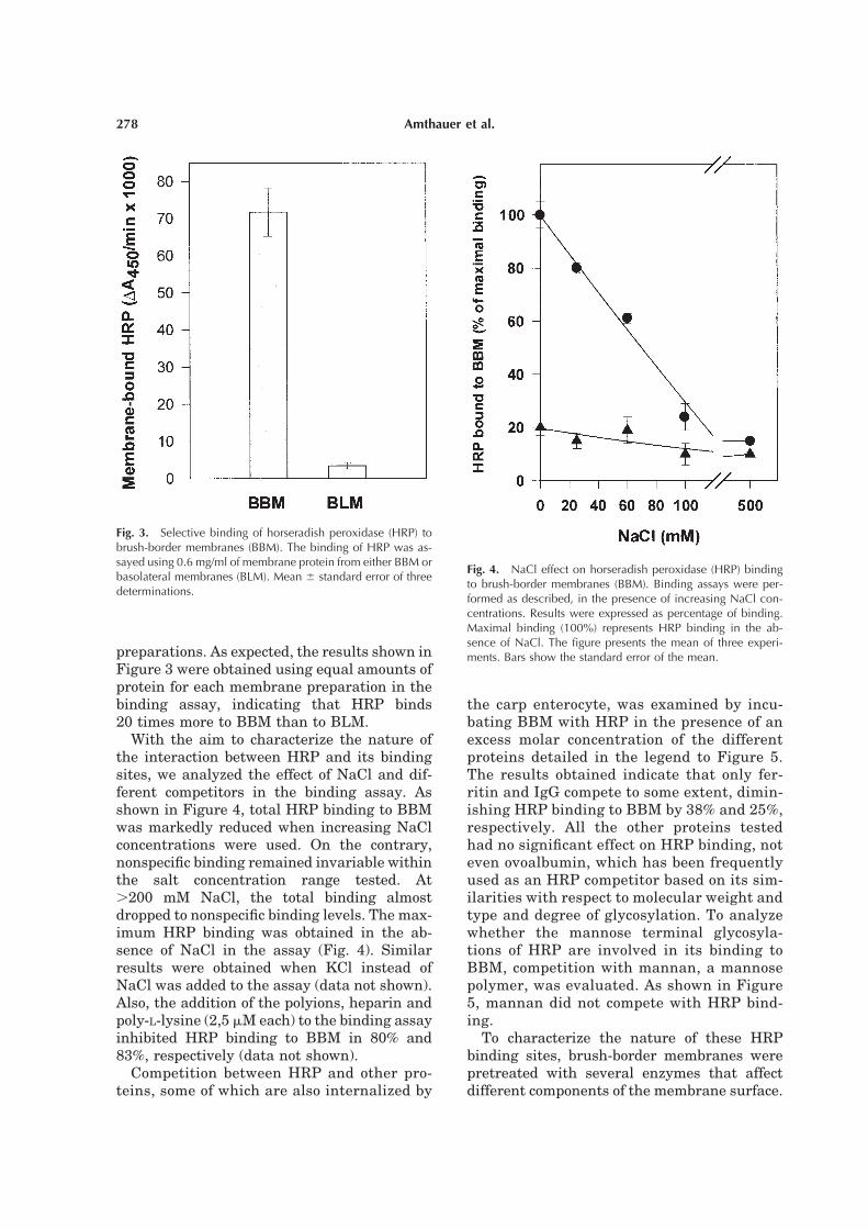
preparations. As expected, the results shown in
Figure 3 were obtained using equal amounts of
protein for each membrane preparation in the
binding assay, indicating that HRP binds
20 times more to BBM than to BLM.
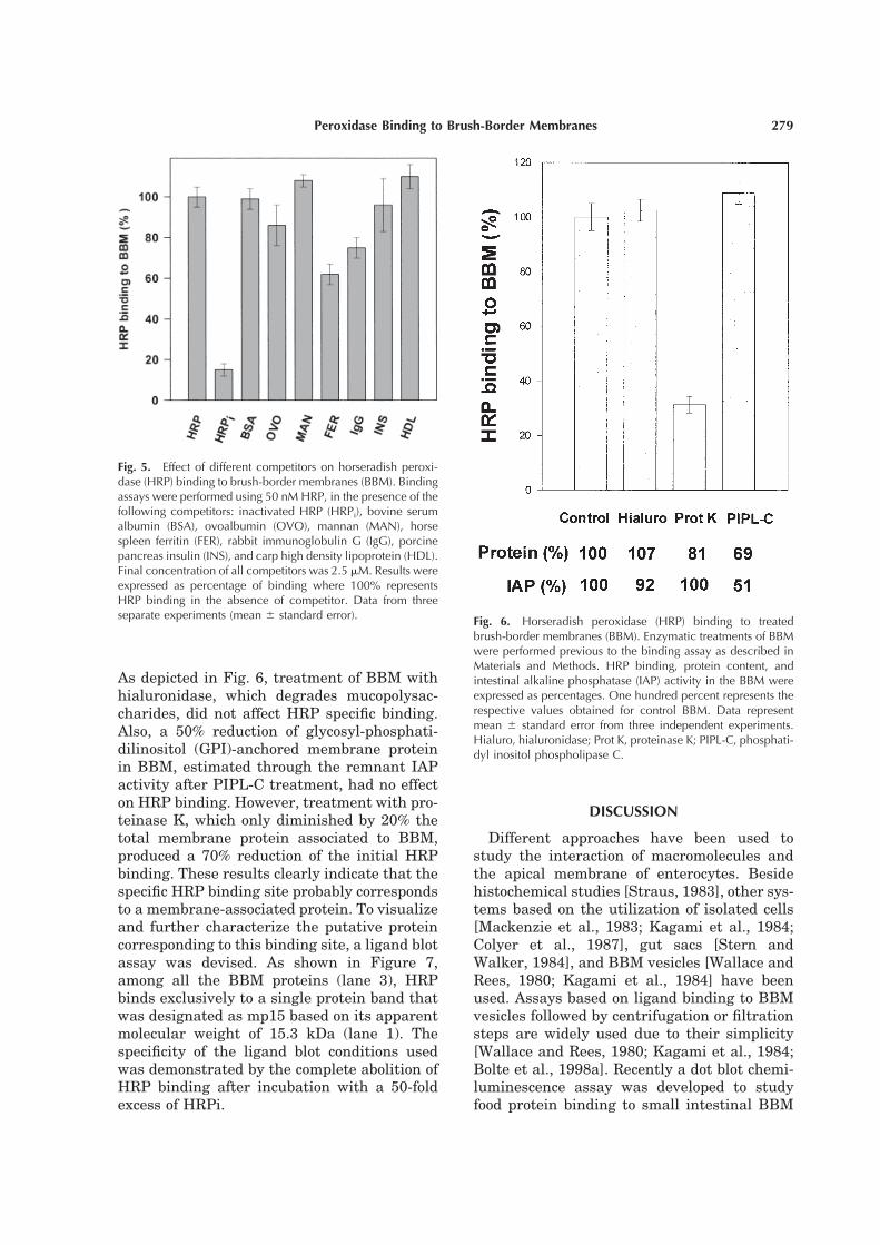
With the aim to characterize the nature of
the interaction between HRP and its binding
sites, we analyzed the effect of NaCl and dif-
ferent competitors in the binding assay. As
shown in Figure 4, total HRP binding to BBM
was markedly reduced when increasing NaCl
concentrations were used. On the contrary,
nonspecific binding remained invariable within
the salt concentration range tested. At
.200 mM NaCl, the total binding almost
dropped to nonspecific binding levels. The max-
imum HRP binding was obtained in the ab-
sence of NaCl in the assay (Fig. 4). Similar
results were obtained when KCl instead of
NaCl was added to the assay (data not shown).
Also, the addition of the polyions, heparin and
poly-L-lysine (2,5 mM each) to the binding assay
inhibited HRP binding to BBM in 80% and
83%, respectively (data not shown).
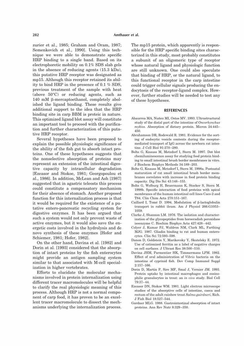
Competition between HRP and other pro-
teins, some of which are also internalized by
the carp enterocyte, was examined by incu-
bating BBM with HRP in the presence of an
excess molar concentration of the different
proteins detailed in the legend to Figure 5.
The results obtained indicate that only fer-
ritin and IgG compete to some extent, dimin-
ishing HRP binding to BBM by 38% and 25%,
respectively. All the other proteins tested
had no significant effect on HRP binding, not
even ovoalbumin, which has been frequently
used as an HRP competitor based on its sim-
ilarities with respect to molecular weight and
type and degree of glycosylation. To analyze
whether the mannose terminal glycosyla-
tions of HRP are involved in its binding to
BBM, competition with mannan, a mannose
polymer, was evaluated. As shown in Figure
5, mannan did not compete with HRP bind-
ing.
To characterize the nature of these HRP
binding sites, brush-border membranes were
pretreated with several enzymes that affect
different components of the membrane surface.
Fig. 3. Selective binding of horseradish peroxidase (HRP) tobrush-border membranes (BBM). The binding of HRP was as-sayed using 0.6 mg/ml of membrane protein from either BBM orbasolateral membranes (BLM). Mean 6 standard error of threedeterminations.
Fig. 4. NaCl effect on horseradish peroxidase (HRP) bindingto brush-border membranes (BBM). Binding assays were per-formed as described, in the presence of increasing NaCl con-centrations. Results were expressed as percentage of binding.Maximal binding (100%) represents HRP binding in the ab-sence of NaCl. The figure presents the mean of three experi-ments. Bars show the standard error of the mean.
278 Amthauer et al.
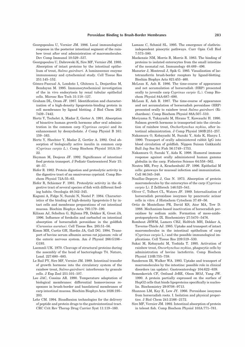
As depicted in Fig. 6, treatment of BBM with
hialuronidase, which degrades mucopolysac-
charides, did not affect HRP specific binding.
Also, a 50% reduction of glycosyl-phosphati-
dilinositol (GPI)-anchored membrane protein
in BBM, estimated through the remnant IAP
activity after PIPL-C treatment, had no effect
on HRP binding. However, treatment with pro-
teinase K, which only diminished by 20% the
total membrane protein associated to BBM,
produced a 70% reduction of the initial HRP
binding. These results clearly indicate that the
specific HRP binding site probably corresponds
to a membrane-associated protein. To visualize
and further characterize the putative protein
corresponding to this binding site, a ligand blot
assay was devised. As shown in Figure 7,
among all the BBM proteins (lane 3), HRP
binds exclusively to a single protein band that
was designated as mp15 based on its apparent
molecular weight of 15.3 kDa (lane 1). The
specificity of the ligand blot conditions used
was demonstrated by the complete abolition of
HRP binding after incubation with a 50-fold
excess of HRPi.
DISCUSSION
Different approaches have been used to
study the interaction of macromolecules and
the apical membrane of enterocytes. Beside
histochemical studies [Straus, 1983], other sys-
tems based on the utilization of isolated cells
[Mackenzie et al., 1983; Kagami et al., 1984;
Colyer et al., 1987], gut sacs [Stern and
Walker, 1984], and BBM vesicles [Wallace and
Rees, 1980; Kagami et al., 1984] have been
used. Assays based on ligand binding to BBM
vesicles followed by centrifugation or filtration
steps are widely used due to their simplicity
[Wallace and Rees, 1980; Kagami et al., 1984;
Bolte et al., 1998a]. Recently a dot blot chemi-
luminescence assay was developed to study
food protein binding to small intestinal BBM
Fig. 5. Effect of different competitors on horseradish peroxi-dase (HRP) binding to brush-border membranes (BBM). Bindingassays were performed using 50 nM HRP, in the presence of thefollowing competitors: inactivated HRP (HRPi), bovine serumalbumin (BSA), ovoalbumin (OVO), mannan (MAN), horsespleen ferritin (FER), rabbit immunoglobulin G (IgG), porcinepancreas insulin (INS), and carp high density lipoprotein (HDL).Final concentration of all competitors was 2.5 mM. Results wereexpressed as percentage of binding where 100% representsHRP binding in the absence of competitor. Data from threeseparate experiments (mean 6 standard error).
Fig. 6. Horseradish peroxidase (HRP) binding to treatedbrush-border membranes (BBM). Enzymatic treatments of BBMwere performed previous to the binding assay as described inMaterials and Methods. HRP binding, protein content, andintestinal alkaline phosphatase (IAP) activity in the BBM wereexpressed as percentages. One hundred percent represents therespective values obtained for control BBM. Data representmean 6 standard error from three independent experiments.Hialuro, hialuronidase; Prot K, proteinase K; PIPL-C, phosphati-dyl inositol phospholipase C.
279Peroxidase Binding to Brush-Border Membranes

[Bolte et al., 1997]. Although this assay was
more sensitive for binding studies than the
centrifugation assay, it presents an important
disadvantage that should be considered. Dur-
ing immobilization of the BBM, the membrane
structures become disrupted [Bolte et al., 1997;
1998b] and as a consequence, intravesicular
proteins (such as villin and actin) might be-
come accessible to interact with the ligand
tested. Therefore, we preferred to use BBM
vesicles in a soluble binding assay. On the
other hand, the utilization of a ligand of high
specific activity such as HRP fulfilled the re-
quirements for sensitivity in our binding assay.
Lee and Cossins [1990] established an isola-
tion procedure to obtain crude preparations of
BBM and BLM from the same carp intestinal
homogenate, with reasonable purity and lim-
ited cross-contamination. These authors also
determined that marker enzymes for mito-
chondria, lysosomes, and endoplasmic reticu-
lum showed no enrichment either in BBM or in
BLM fractions. However, these carp BBM
preparations have never been evaluated for the
presence of possible protease activities. This
could be a problem for the proper validation of
binding assays utilizing these preparations. In
fact, in rabbit, the degradation of
b-lactoglobulin by proteases in ileum BBM
preparations has been demonstrated [Caillard
and Tome, 1994]. In spite of this possible com-
plication, our results (Fig. 1) indicate that the
addition of protease inhibitors such as benza-
midine and phenylmethylsulfonyl fluoride to
BBM preparations prevented any detectable
degradation of the ligand (HRP) in our binding
assays. Moreover, the activity of the enzyme
also remained unaffected after incubation with
BBM preparations.
Horseradish peroxidase binding was mea-
sured using a protein concentration range from
2 to 150 nM. The results indicate that satura-
tion of BBM binding sites occurs with an HRP
concentration higher than 100 nM. The corre-
sponding dissociation constant and maximal
binding was estimated expressing these data
in the form of a Scatchard plot. The Kd (22 nM)
obtained indicates that this HRP binding site
has comparable or higher affinity than other
BBM protein binding sites from similar sys-
tems, e.g., the binding of IgG to BBM
(Kd '1028 M) [Wallace and Rees, 1980] or lac-
totransferrin to rabbit BBM (Kd 1.8 mM) [Ma-
zurier et al., 1985]. In a recent study, the sat-
urable binding of proteins to BBM was also
demonstrated, but unfortunately no dissocia-
tion constants were noted [Bolte et al., 1998a].
In fish as well as in mammals, the columnar
cells of the intestinal epithelium are held to-
gether at their apical boundary by a continuous
network of tight junctions acting as a barrier to
the passive diffusion of ions and macromole-
cules through a paracellular route [Weinberg,
1976; Kiliaan et al., 1996]. Thus, the protein to
be internalized must interact with the apical
membrane (BBM) of the enterocyte. Therefore,
our results showing an enrichment of specific
HRP binding sites in BBM compared with
BLM is consistent with this proposed mecha-
nism for HRP internalization and also with the
polarized nature of the enterocytes. On the
other hand, these results are also in agreement
with the morphologic evidence that demon-
Fig. 7. Identification of horseradish peroxidase (HRP) bindingsites in brush-border membranes (BBM) by ligand blot analyses.BBM proteins were separated by SDS-PAGE and electrotrans-ferred to Immobilon-P membranes. After the blocking step, themembranes were incubated with 100 nM HRP (lane 1) and100 nM HRP plus 5 mM inactivated HRP (lane 2). The HRPactivity was developed using 3,3’-diamino benzidine/H2O2.Lane 3 shows BBM proteins stained with Coomasie blue. Ar-rows indicate protein molecular weight standards (Mr) in kDa.
280 Amthauer et al.

strate the early interaction of HRP with the
apical membrane of the intestinal epithelium
[Noaillac-Depeyre and Gas, 1973; Rombout et
al., 1985]. All the above data suggest that the
specific HRP binding to the carp intestinal
brush-border membranes is the necessary first
step that will lead to its endocytosis and trans-
epithelial transport during the internalization
process.
Seven horseradish peroxidase isoenzymes
have been isolated and characterized [Shannon
et al., 1966]. The isoenzyme used in this study
is basic [Oliver et al., 1989] and therefore is
positively charged at the pH used in the bind-
ing assay. The existence of anionic sites in dif-
ferent cell membranes, including the apical
membrane of mammalian enterocytes, has
been demonstrated using cationized ferritin
and other chemically modified proteins as li-
gand [Danon et al., 1972; Triguereo et al., 1989;
Sanderson and Walker, 1993; Lehr, 1994]. The
ionic interaction of a ligand with these anionic
sites is usually associated with a nonreceptor
mediated endocytosis that segregates the li-
gand to a degradative pathway within the cell
[Gomez-Pascual et al., 1995]. Our results show
that increasing NaCl concentrations in the
binding assay only affected the specific bind-
ing, suggesting that HRP is not interacting
with the broad family of anionic sites. In fact,
the Scatchard analysis gave a linear plot,
which is consistent with the presence of a ho-
mogeneous population of binding sites. Never-
theless, the specific interaction of HRP with its
binding sites, under the in vitro conditions
used, presents an ionic character. It must be
considered that these conditions do not neces-
sarily represent the in vivo interaction of HRP
with the apical membrane of enterocytes be-
cause isolated BBM vesicles lack important
parts of the mucosal barrier [Sanderson and
Walker, 1993].
It is clear from the results presented here
that the binding of HRP to carp BBM satisfies
all the criteria required for a receptor molecule.
The interaction is saturable, has a high bind-
ing affinity, and is specific for HRP. In fact,
only two of six proteins tested, IgG and ferritin,
partially compete for HRP binding to BBM.
Although most of the competitor proteins used
are structurally unrelated to HRP, all of them
meet the criteria of being internalized when
administered orally to the carp. Bolte et al.
[1998a], studying the binding of food proteins
to rat small intestinal BBM, also tested the
specificity using different unrelated dietary
proteins. In this study they demonstrated that
all the proteins tested were able to compete
with each other, supporting the idea of common
or at least adjoining binding sites for different
food proteins in rat BBM. On the contrary, our
results are consistent with a specific and inde-
pendent binding site for HRP.
Horseradish peroxidase is a mannose-
terminated glycoprotein [Clarke and Shannon,
1976] and therefore its uptake by fluid phase
endocytosis is considered to be receptor-
mediated via a mannose receptor in macro-
phages and hepatic nonparenchymal cells
[Yamaguchi et al., 1993]. In addition,
mannose-specific binding sites for HRP have
been demonstrated in several other cell types
[Straus, 1983]. Taking in consideration that
the HRP binding to carp BBM was inhibited by
neither mannan nor ovoalbumin, a glycopro-
tein that also exhibits mannose terminal
groups, the involvement of a lectin-type of in-
teraction is very unlikely.
To analyze the nature of the HRP binding
sites, brush-border membranes were pre-
treated with several enzymes to affect different
components of the membrane. Hialuronidase
treatment did not affect HRP specific binding
to carp BBM. This treatment had been previ-
ously used to remove components of the glyco-
calyx in rat BBM where it also effectively re-
duced nonspecific binding, thereby enhancing
the IgG-specific binding [Wallace and Rees,
1980]. A second treatment that also did not
affect HRP binding was performed with
PIPL-C, an enzyme that partially removes
GPI-anchored proteins from the membranes.
This result suggests that most probably the
putative receptor does not correspond to this
class of membrane protein. Nevertheless, it is
important to note that some GPI-anchored pro-
teins are resistant to PIPL-C [Wong and Low,
1992]. Protease treatment is a standard proce-
dure used to demonstrate the proteic nature of
a macromolecule with biologic activity. Treat-
ments of BBM with proteinase K, which pro-
duces a limited proteolysis of the outer leaflet
of the vesicle, reduced the HRP binding by
70%. Taken together, these results strongly
suggest that the putative receptor for HRP is a
protein.
With the ligand blotting technique, different
membrane receptors have been identified [Ma-
281Peroxidase Binding to Brush-Border Membranes
zurier et al., 1985; Graham and Oram, 1987;
Semenkovich et al., 1990]. Using this tech-
nique we were able to demonstrate specific
HRP binding to a single band. Based on its
electrophoretic mobility on 0.1% SDS slab gels
in the absence of reducing agents (15.3 kDa),
this putative HRP receptor was designated as
mp15. Although this receptor retained its abil-
ity to bind HRP in the presence of 0.1 % SDS,
previous treatment of the sample with heat
(above 50°C) or reducing agents, such as
140 mM b-mercaptoethanol, completely abol-
ished the ligand binding. These results give
additional support to the idea that the HRP
binding site in carp BBM is proteic in nature.
This optimized ligand blot assay will constitute
an important tool to proceed with the purifica-
tion and further characterization of this puta-
tive HRP receptor.
Several hypotheses have been proposed to
explain the possible physiologic significance of
the ability of the fish gut to absorb intact pro-
teins. One of these hypotheses suggests that
the nonselective absorption of proteins may
represent an extension of the intestinal diges-
tive capacity by intracellular degradation
[Ezeasor and Stokoe, 1981; Georgopoulou et
al., 1986]. In addition, McLean and Ash [1987]
suggested that in agastric teleosts this process
could constitute a compensatory mechanism
for their absence of stomach. Another proposed
function for this internalization process is that
it would be required for the existence of a pu-
tative entero-pancreatic recycling system for
digestive enzymes. It has been argued that
such a system would not only prevent waste of
active enzymes, but it would also save the en-
ergetic costs involved in the hydrolysis and de
novo synthesis of these enzymes [Hofer and
Schiemer, 1981; Hofer, 1982].
On the other hand, Davina et al. [1982] and
Dorin at al. [1993] considered that the absorp-
tion of intact proteins by the fish enterocytes
might provide an antigen sampling system
similar to that associated with M-cell special-
ization in higher vertebrates.
Efforts to elucidate the molecular mecha-
nisms involved in protein internalization using
different tracer macromolecules will be helpful
to clarify the real physiologic meaning of this
process. Although HRP is not a normal compo-
nent of carp food, it has proven to be an excel-
lent tracer macromolecule to dissect the mech-
anisms underlying the internalization process.
The mp15 protein, which apparently is respon-
sible for the HRP specific binding sites charac-
terized in this study, most probably constitutes
a subunit of an oligomeric type of receptor
whose natural ligand and physiologic function
are still unknown. One could also speculate
that binding of HRP, or the natural ligand, to
this functional receptor in the carp intestine
could trigger cellular signals producing the en-
docytosis of the receptor-ligand complex. How-
ever, further studies will be needed to test any
of these hypotheses.
REFERENCES
Abaurrea MA, Nunez MI, Ostos MV. 1993. Ultrastructural
study of the distal part of the intestine of Oncorhynchus
mykiss. Absorption of dietary protein. Micron 24:445–
450.
Abrahamson DR, Rodewald R. 1981. Evidence for the sort-
ing of endocytic vesicle contents during the receptor-
mediated transport of IgG across the newborn rat intes-
tine. J Cell Biol 91:270–280.
Bolte G, Knauss M, Metzdorf I, Stern M. 1997. Dot blot
chemiluminescence assay for studying food protein bind-
ing to small intestinal brush border membranes in vitro.
J Biochem Biophys Methods 34:189–203.
Bolte G, Knauss M, Metzdorf I, Stern M. 1998a. Postnatal
maturation of rat small intestinal brush border mem-
branes correlates with increase in food protein binding
capacity. Dig Dis Sci 43:148–155.
Bolte G, Wolburg H, Beuermann K, Stocker S, Stern M.
1998b. Specific interaction of food proteins with apical
membranes of the human intestinal cell lines Caco-2 and
T84. Clin Chim Acta 270:151–167.
Caillard I, Tome D. 1994. Modulation of b-lactoglobulin
transport in rabbit ileum. Am J Physiol 266:G1053–
1059.
Clarke J, Shannon LM. 1976. The isolation and character-
ization of the glycopeptides from horseradish peroxidase
isoenzyme C. Biochim Biophys Acta 427:428–442.
Colyer J, Kumar PJ, Waldron NM, Clark ML, Farthing
MJG. 1987. Gliadin binding to rat and human entero-
cytes. Clin Sci 72:593–598.
Danon D, Goldstein Y, Marikovsky Y, Skutelsky E. 1972.
Use of cationized ferritin as a label of negative charges
on cell surfaces. J Ultrast Res 38:500–510.
Davina JHM, Parmentier HK, Timmermans LPM. 1982.
Effect of oral administration of Vibrio bacteria on the
intestine of cyprinid fish. Dev Comp Immunol Suppl
2:157–166.
Dorin D, Martin P, Sire MF, Smal J, Vernier JM. 1993.
Protein uptake by intestinal macrophages and eosino-
philic granulocytes in trout: an in vivo study. Biol Cell
79:37–44.
Ezeasor DN, Stokoe WM. 1981. Light electron microscope
studies of the absorptive cells of intestine, caeca and
rectum of the adult rainbow trout Salmo gairdneri, Rich.
J Fish Biol 18:527–544.
Gardner MLG. 1988. Gastrointestinal absorption of intact
proteins. Ann Rev Nutr 8:329–350.
282 Amthauer et al.
Georgopoulou U, Vernier JM. 1986. Local immunological
response in the posterior intestinal segment of the rain-
bow trout after oral administration of macromolecules.
Dev Comp Immunol 10:529–537.
Georgopoulou U, Dabrowski K, Sire MF, Vernier JM. 1988.
Absorption of intact proteins by the intestinal epithe-
lium of trout, Salmo gairdneri. A luminescence enzyme
immunoassay and cytochemical study. Cell Tissue Res
251:145–152.
Gomez-Pascual A, Londono I, Ghitescu L, Desjardins M,
Bendayan M. 1995. Immunocytochemical investigation
of the in vivo endocytosis by renal tubular epithelial
cells. Microsc Res Tech 31:118–127.
Graham DL, Oram JF. 1987. Identification and character-
ization of a high-density lipoprotein-binding protein in
cell membranes by ligand blotting. J Biol Chem 262:
7439–7442.
Hertz Y, Techelet A, Madar Z, Gerter A. 1991. Absorption
of bioactive human growth hormone after oral adminis-
tration in the common carp (Cyprinus carpio) and its
enhancement by deoxycholate. J Comp Physiol B 161:
159–163.
Hertz Y, Shechter Y, Madar Z, Gertler A. 1992. Oral ab-
sorption of biologically active insulin in common carp
(Cyprinus carpio L.). Comp Biochem Physiol 101A:19–
22.
Heyman M, Desjeux JF. 1992. Significance of intestinal
food protein transport. J Pediatr Gastroenterol Nutr 15:
48–57.
Hofer R. 1982. Protein digestion and proteolytic activity in
the digestive tract of an omnivorous cyprinid. Comp Bio-
chem Physiol 72A:55–63.
Hofer R, Schiemer F. 1981. Proteolytic activity in the di-
gestive tract of several species of fish with different feed-
ing habits. Oecologia 48:342–345.
Kagami A, Fidge N, Suzuki N, Nestel P. 1984. Character-
istics of the binding of high-density lipoprotein-3 by in-
tact cells and membrane preparations of rat intestinal
mucosa. Biochim Biophys Acta 795:179–190.
Kiliaan AJ, Scholten G, Bijlsma PB, Dekker K, Groot JA.
1996. Influence of forskolin and carbachol on intestinal
absorption of horseradish peroxidase in the goldfish
(Carassius auratus). Cell Tissue Res. 285:51–56.
Kimm MH, Curtis GH, Hardin JA, Gall DG. 1994. Trans-
port of bovine serum albumin across rat jejunum: role of
the enteric nervous system. Am J Physiol 266:G186–
G193.
Laemmli UK. 1970. Cleavage of structural proteins during
the assembly of the head of bacteriophage T4. Nature,
Lond. 227:680–685.
Le Bail PY, Sire MF, Vernier JM. 1989. Intestinal transfer
of growth hormone into the circulatory system of the
rainbow trout, Salmo-gairdneri: interference by granule
cells. J Exp Zool 251:101–107.
Lee JAC, Cossins AR. 1990. Temperature adaptation of
biological membranes: differential homeoviscous re-
sponses in brush-border and basolateral membranes of
carp intestinal mucosa. Biochim Biophys Acta 1026:195–
203.
Lehr CM. 1994. Bioadhesion technologies for the delivery
of peptide and protein drugs to the gastrointestinal tract.
CRC Crit Rev Therap Drug Carrier Syst 11:119–160.
Lamaze C, Schmid SL. 1995. The emergence of clathrin-
independent pinocytic pathways. Curr Opin Cell Biol
7:573–580.
Mackenzie NM, Morris B, Morris R. 1983. The binding of
proteins to isolated enterocytes from the small intestine
of the neonatal rat. Immunology 48:489–496.
Mazurier J, Montreuil J, Spik G. 1985. Visualization of lac-
totransferrin brush-border receptors by ligand-blotting.
Biochim Biophys Acta 821:453–460.
McLean E, Ash R. 1986. The time-course of appearance
and net accumulation of horseradish (HRP) presented
orally to juvenile carp Cyprinus carpio (L.). Comp Bio-
chem Physiol 84A:687–690.
McLean E, Ash R. 1987. The time-course of appearance
and net accumulation of horseradish peroxidase (HRP)
presented orally to rainbow trout Salmo gairdneri (Ri-
chardson). Comp Biochem Physiol 88A:507–510.
Moriyama S, Takayashi M, Hirano T, Kawauchi H. 1990.
Salmon growth hormone is transported into the circula-
tion of rainbow trout, Onchorhynchus mykiss, after in-
testinal administration. J Comp Physiol 160B:251–257.
Nakamura O, Kobayashi M, Suzuki Y, Aida K, Hanyu I.
1990. Transport of orally administered rabbit IgG into
blood circulation of goldfish. Nippon Suisan Gakkaishi
Bull Jap Soc Sci Fish 56:1749–1753.
Nakamura O, Suzuki Y, Aida K. 1998. Humoral immune
response against orally administered human gamma
globulin in the carp. Fisheries Science 64:558–562.
Neutra MR, Frey A, Kraehenbuhl JP. 1996. Epithelial M
cells: gateways for mucosal infection and immunization.
Cell 86:345–348.
Noaillac-Depeyre J, Gas N. 1973. Absorption of protein
macromolecules by the enterocytes of the carp (Cyprinus
carpio L). Z Zellforsch 146:525–541.
Oliver C, Tolbert CL, Waters JF. 1989. Internalization of
horseradish peroxidase isozymes by pancreatic acinar
cells in vitro. J Histochem Cytochem 37:49–56.
Ortiz de Montellano PR, David KS, Ator MA, Tew D.
1988. Mechanism-based inactivation of horseradish per-
oxidase by sodium azide. Formation of meso-azido-
protoporphyrin IX. Biochemistry 27:5470–5476.
Rombout JHWM, Lamers CHJ, Helfrich MH, Dekker A,
Taverne-Thiele AJ. 1985. Uptake and transport of intact
macromolecules in the intestinal epithelium of carp
(Cyprinus carpio L.) and the possible immunological im-
plications. Cell Tissue Res 239:519–530.
Sakai M, Kobayashi M, Yoshida T. 1995. Activation of
rainbow trout, Oncorhynchus mykiss, phagocytic cells by
administration of bovine lactoferrin. Comp Biochem
Physiol 110B:755–759.
Sanderson IR, Walker WA. 1993. Uptake and transport of
macromolecules by the intestine: possible role in clinical
disorders (an update). Gastroenterology 104:622–639.
Semenkovich CF, Ostlund JrRE, Olson MOJ, Yang JW.
1990. A protein partially expressed on the surface of
HepG2 cells that binds lipoproteins specifically is nucleo-
lin. Biochemistry 29:9708–9713.
Shannon LM, Kay E, Lew JY. 1966. Peroxidase isozymes
from horseradish roots. I. Isolation and physical proper-
ties. J Biol Chem 241:2166–2172.
Sire MF, Vernier JM. 1992. Intestinal absorption of protein
in teleost fish. Comp Biochem Physiol 103A:771–781.
283Peroxidase Binding to Brush-Border Membranes

Smith PK, Krohn RI, Hermanson GT, Mallia AK, Gartner
FH, Provenzano MD, Fujimoto EK, Goeke NM, Olson
BJ, Klenk DC. 1985. Measurement of protein using
bicinchoninic acid. Anal Biochem 150:76–85.
Stern M, Walker WA. 1984. Food proteins and gut mucosal
barrier I. Binding and uptake of cow’s milk proteins by
adult rat jejunum in vitro. Am J Physiol 246:G556–562.
Stoorvogel W. 1998. Analysis of the endocytic system by
using horseradish peroxidase. Trends Cell Biol 8:503–
505.
Straus W. 1983. Mannose-specific binding sites for horse-
radish peroxidase in various cells of the rat. J Histochem
Cytochem 31:78–84.
Triguereo D, Buciak J, Yang J, Padridge W. 1989. Blood-
brain barrier transport of cationized immunoglobulin G.
Enhanced delivery compared to native protein. Proc Natl
Acad Sci 86:4761–4766.
Vera MI, Romero F, Amthauer R, Figueroa J, Goicoechea
O, Leon G, Krauskopf M. 1992. Carp apolipoprotein A-I
intestinal absorption and transfer into the systemic cir-
culation during the acclimatization of the carp (Cyprinus
carpio). Comp Biochem Physiol 101A:573–581.
Vera MI, Romero F, Figueroa J, Amthauer R, Leon G,
Villanueva J, Krauskopf M. 1993. Oral administration of
insulin in winter-acclimatized carp (Cyprinus carpio) in-
duces hepatic ultrastructural changes. Comp Biochem
Physiol 106A:677–682.
Villanueva J, Vanacore R, Goicoechea O, Amthauer R.
1997. Intestinal alkaline phosphatase of the fish Cypri-
nus carpio: regional distribution and membrane associ-
ation. J Exp Zool 279:347–355.
Wallace KH, Rees AR. 1980. Studies on the immuno-
globulin-G Fc-Fragment receptor from neonatal rat
small intestine. Biochem J 188:9–16.
Weinberg S. 1976. Morphology of the intestine of goldfish
(Carassius auratus). Bijdragen Tot de Dierkunde 46:35–
46.
Wong YW, Low MG. 1992. Phospholipase resistance of the
glycosyl-phosphatidylinositol membrane anchor on hu-
man alkaline phosphatase. Clin Chem 38:2517–2525.
Yamaguchi Y, Dallemolle E, Hardison WGM. 1993. Hepa-
tocyte horseradish peroxidase uptake is saturable and
inhibited by mannose-terminal glycoproteins. Am J
Physiol 264:G880–G885.
284 Amthauer et al.
Copyright © 2022 FDOKUMEN