Perioperative fasting in adults and children (full) - Great ...

Domestic Animal Endocrinology 31 (2006) 105–122
Hormonal and metabolic adaptation to fasting:Effects on the hypothalamic–pituitary–ovarian axis
and reproductive performance of rabbit does
Gabriele Brecchia a, Adriana Bonanno b, Giovanna Galeati c,Claudia Federici a, Margherita Maranesi a, Anna Gobbetti d,
Massimo Zerani d, Cristiano Boiti a,∗a Dipartimento di Scienze Biopatologiche Veterinarie ed Igiene delle Produzioni Animali e Alimentari,
Sezione di Fisiologia, Laboratorio di Biotecnologie, Universita degli Studi di Perugia,via S. Costanzo 4, 06126 Perugia, Italy
b Dipartimento S.EN.FI.MI.ZO., Sezione di Produzioni Animali, Universita degli Studi di Palermo,Viale delle Scienze, I-90128 Palermo, Italy
c Dipartimento di Morfofisiologia Veterinaria e Produzioni Animali, Universita degli Studi di Bologna,via Tolara di Sopra 50, I-40064 Ozzano Emilia, Bologna, Italy
d Dipartimento di Biologia Molecolare, Cellulare e Animale, Universita degli Studi di Camerino,via F. Camerini 1, I-62032 Camerino, Italy
Received 20 April 2005; received in revised form 7 September 2005; accepted 15 September 2005
Abstract
To assess the impact of acute caloric shortage on reproduction, rabbit does were either fed ad libitum(control, AL), or fasted for 24 (STF) or 48 h (LTF) before induction of ovulation with GnRH injection.Blood samples were collected during the last 3 h of fasting, and the following 4 h after GnRH injection,when feed was provided again, to measure plasma concentrations of LH, estradiol-17�, leptin, insulin,T3, corticosterone, glucose, and NEFA.
Before re-feeding, plasma leptin, insulin, and T3 concentrations were lower (P ≤ 0.01) in bothfasted groups than in controls, but then gradually increased following realimentation to match thoseof controls. During fasting, corticosterone levels were higher (P ≤ 0.01) in LTF than in STF andAL does, but decreased to control values soon after realimentation. During fasting, plasma glucoseconcentrations did not differ among groups, but upon re-feeding they markedly increased (P ≤ 0.01)
∗ Corresponding author. Tel.: +39 075 5857639; fax: +39 075 5857654.E-mail address: [email protected] (C. Boiti).
0739-7240/$ – see front matter © 2005 Elsevier Inc. All rights reserved.doi:10.1016/j.domaniend.2005.09.006
106 G. Brecchia et al. / Domestic Animal Endocrinology 31 (2006) 105–122
both in STF and LTF does. NEFA levels were also more elevated (P ≤ 0.01) in fasted rabbits thanin controls, and rapidly decreased (P ≤ 0.01) after re-feeding. Following GnRH injection, LH peakwas lower (P ≤ 0.01) in LTF than in AL and STF does. Estradiol-17� showed higher pulse frequencyand amplitude in AL than in STF and LTF does. Compared to controls, receptivity rate of STFand LTF artificially inseminated does declined respectively by −20.5% (P ≤ 0.05) and −22.7%, andfertility rate by −23.9% (P ≤ 0.05) and 21.4%, but no difference was found in ovulation rate. Insummary, nutritional status of does, as modified by fasting, greatly influenced fertility, metabolic andreproductive hormones.© 2005 Elsevier Inc. All rights reserved.
Keywords: Fasting; LH; Estradiol-17�; Leptin; Insulin; T3; Corticosterone; Rabbit
1. Introduction
Nutrition is one of the major factors affecting the reproductive efficiency particularly infemale mammals. Negative energy balance in young rabbit does can result in infertility andhigh culling rate because of the high energy demands for pregnancy and lactation [1].
Prolonged caloric restriction can inhibit pulsatile LH secretion by depressing the GnRHpulse generator within the hypothalamus in primates [2] and induce a condition of anoestrousin cattle [3]. Interestingly, however, even mild reduction in the level of nutrition can markedlyaffect reproduction [4]. Using different animal models, it was demonstrated that caloriesare important in the regulation of LH secretion. In primates and rats, for example, a singleday of fasting is sufficient to reduce the pulsatile LH secretion pattern [5,6].
The effect of chronic fed restriction on the regulatory mechanisms of metabolism has beeninvestigated in several animal species, but only recently in rabbits. In this species, long-termnutrient deficiency during development has major neuroendocrine consequences evokingprominent homeostatic reactions of the corticotropic, leptinergic, and thyrotropic pathways[7], but its involvement with the gonadal axis is still unclear [8]. Several metabolites, includ-ing glucose and non-esterified fatty acids (NEFA), and hormones, such as insulin and IGF-I,regulate ovulation rate, follicle development and embryo survival [4,9,10]. Recently, alsoleptin has been implicated in several key points of the mammalian reproductive functions[11,12]. The recent findings of leptin receptors in different ovarian structures [13] and theoviduct [14] of rabbits suggest that leptin may have a role in the steroidogenesis of pre- andpost-ovulatory follicles as well as in early developmental stages of blastocyst. Leptin mayact as a critical link between adipose tissue and the neuroendocrine axis, indicating whetheradequate energy reserves are present for normal reproductive function [15].
Although the causal link between nutrition and fertility has been known for a long time,the underlying mechanisms by which caloric shortage affects reproductive function remainunclear and difficult to disclose [16]. Thus, complete deprivation of food for a short periodof time could be a useful model for analyzing the adaptive responses of metabolic processesand their regulatory systems as well as the interrelationships between nutritional deficiencyand reproductive function. The objective of the present study was to examine the effectstriggered by 1- or 2-days of fasting on (1) plasma concentrations of leptin, insulin, T3,corticosterone, glucose, and NEFA as indicators of hormonal and metabolic adaptations to

G. Brecchia et al. / Domestic Animal Endocrinology 31 (2006) 105–122 107
food deprivation, (2) hormonal profiles of LH and estradiol-17�, as markers of the responsesof the gonadal axis, and (3) reproductive performance of rabbit does.
2. Materials and methods
2.1. Animals and experimental design
Animals were caged individually in indoor brick shed facilities and kept under a tem-perature of 20–28 ◦C by forced heating systems and a constant photoperiod of 16 h lightand 8 h darkness. All animals were fed a standard commercial pellet diet ad libitum onceper day, containing 11.0 MJ DE/kg dry matter and 18.3% crude protein, with free access towater. Free suckling of litters equalized to 8–9 young within 2 days after parturition andweaning at 31 days were standard procedure.
Sexual receptivity was checked on the day of insemination by evaluating vulva color.Accordingly, does were scored either receptive when their vulva was red (or purple) andturgid, or non-receptive in all the other cases. Ovulation was induced by 0.8 �g GnRHanalogue (Receptal, Intervet, Milan, Italy) intramuscular injection. Artificial insemination(AI) was performed with heterospermic pool of fresh semen, assessed for mass motilityunder a microscope (200×) and diluted 1:5 in a commercial extender (Cortalap, IMVTechnologies Italia, Piacenza, Italy), to ensure a minimum concentration of about 50 millionspermatozoa per ml. The does were inseminated in the lordosis position with 0.6 ml ofdiluted semen through a single-use plastic pipette, following GnRH injection.
2.2. Experimental design
To verify the effect of fasting on hormonal and metabolic adaptation, as well as onoverall reproductive performance and the hypothalamus–pituitary–ovarian (HPO) axis, twoexperimental protocols were designed.
2.2.1. Experiment 1To avoid potentially confounding effects of lactations and doe–litter interactions, only
young unmated rabbit females were employed. New Zealand White (NZW, HY/CR strain)rabbits (Charles River Italia, Lecco, Italy) of 5 months age, weighing 3.5–3.8 kg wererandomly assigned to one of the following three groups (12 animals/group): control (AL),short-term fasting (STF), and long-term fasting (LTF). Control does were fed ad libitum, STFand LTF does were deprived of food for 24 and 48 h, respectively, before GnRH injection.Immediately after GnRH administration (time 0), fasted does were re-fed ad libitum thesame standard diet as the controls. The daily ration of feed was given to the rabbits at8:00 a.m.
On the day of the experiment, a catheter was inserted into the central ear vein of eachrabbit. Blood samples (1 ml) were collected from unrestrained catheterized animals, freeto move in their cages without interfering with their normal activity, every 15 min duringthe last 3 h of fasting and the following 4 h after re-feeding. The samples, drawn into tubescontaining EDTA, were immediately centrifuged at 3000 × g for 15 min and plasma was

108 G. Brecchia et al. / Domestic Animal Endocrinology 31 (2006) 105–122
stored frozen until assayed for hormones and metabolites. Packed cell volume was analyzedevery 2 h to exclude rabbits with anemia from the experiment.
2.2.2. Experiment 2In three successive trials New Zealand White rabbit does with same parity and similar
body weight were homogeneously assigned to control or treated groups. In the first trial,lactating does were either fed ad libitum (AL, n = 38) or fasted for 24 h (STF, n = 43) 1day before AI at day 11 postpartum. In the second, non-lactating does were fed ad libitum(AL, n = 19) or deprived of food for 48 h (LTF, n = 16) before AI. The latter approach wasadopted in order to avoid undue stress to young rabbits as a consequence of the drop inmilk production by their foster mothers deprived of food for two consecutive days. BothSTF and LTF does were re-fed ad libitum 2 h prior to AI. For each doe, the following datawere recorded: body weight and feed intake the day prior to and after fasting, body weight10 days after re-feeding, sexual receptivity at AI, fertility rate, number of total born andborn-alive at parturition.
In the third trial, non-lactating rabbit does, randomly allocated (n = 6/group) either tocontrol or to STF and LTF groups, were sacrificed 48 h after AI to determine the ovu-lation rate by counting the number of newly formed corpora lutea (CL) present in eachovary.
2.3. Hormone assays
Plasma concentrations of LH were determined by a homologous double antibody RIAmethod. The antibody used was an anti-Rb LH (AFP-3120489GP) supplied by A.F. Parlow(Harbour UCLA Medical Centre, CA) at a working dilution of 1:75,000. The tracer wasobtained by iodination of rabbit LH (AFP-7818C) by the iodogen method [17]. The sensi-tivity of the assay was 0.5 ng/ml plasma. The intra-assay (10 determinations) and inter-assay(10 assays) coefficients of variation were 3.9 and 4.5%, respectively.
Estradiol-17� concentrations in plasma samples were assayed using a commercial 125IRIA kit (ICN Pharmaceuticals Inc., Diagnostic Division, Costa Mesa, CA, USA). The limitof detection was 0.8 pg/ml and the intra- and inter-assay coefficients of variation were <6and <10%, respectively.
Insulin, leptin, total triiodothyronine (T3), and corticosterone concentrations were deter-mined by RIA, as reported elsewhere [7]. Plasma insulin was determined by the doubleantibody/PEG technique using a porcine insulin RIA kit (Linco Research Inc., St. Charles,MO, USA). The antiserum was guinea pig anti-porcine insulin, while both labeled antigenand standards used purified recombinant human insulin. The limit of sensitivity was 2 �U/mland intra- and inter-assay coefficients of variations were 6.8 and 9.2%, respectively. Leptinconcentrations were measured by double antibody RIA using the multi-species leptin kit(Linco Research Inc., St. Charles, MO, USA). The intra-assay coefficient of variation was3.9%. Total triiodothyronine (T3) was assayed by RIA according to the procedure providedby the manufacturer (Immunotech, Marseille, France). The sensitivity of the assay was0.13 ng/ml, and the major analogs of T3 did not interfere with the assay. Intra- and inter-assay coefficients of variations were 4.9 and 6.1%, respectively. Plasma corticosteroneconcentrations were evaluated by RIA, using the CORT kit (ICN Biomedicals Inc., Costa

G. Brecchia et al. / Domestic Animal Endocrinology 31 (2006) 105–122 109
Mesa, CA, USA), with a sensitivity of 0.15 ng/ml and coefficient of variations of 7.6 and9.2% for intra- and inter-assay, respectively.
2.4. Metabolite assays
Glucose was analyzed by the glucose oxidase method using the Glucose Infinity kitobtained from Sigma (Sigma Diagnostic Inc., St. Louis, MO, USA). The non-esterifiedfatty acids (NEFA) concentrations were analyzed using a two-reaction, enzymatic-basedcolorimetric assay from Wako (NEFA-C, Wako Chemicals GmbH, Neuss, Germany), basedon the ability of NEFA to acylate coenzyme A in the presence of CoA synthetase.
2.5. Statistical analysis
The estradiol-17� pulses were identified in sequential blood samples collected for 7 hfrom both treated and control does. The criteria for identifying a single-point pulse was avalue 3 S.D. above the baseline whereas those for two- and three-points pulses were 2 and1.5 S.D., respectively [18]. The number of peaks of estradiol-17� and mean plasma valueswere evaluated by ANOVA followed by Student’s t-test.
Data relative to overall treatment effects on other hormones and metabolites during thetime-course of short- and long-term fasting period were analyzed by ANOVA for repeatedmeasurements according to a model which included treatment group, doe within group,time period, and interaction between group and time period: doe within treatment was usedas the error term. Comparison between effects was performed by Student’s t-test. A valueof P < 0.05 was considered significant. The distribution of the does in two groups accordingto the magnitudes of their peak LH responses to GnRH challenge was compared usinga Fisher’s exact test. The differences between means relative to productive performanceobtained from experiment 2 were tested by Student’s t-test, whereas proportional data wereevaluated by Chi square test. All statistical analyses were performed using Prism (GraphPadSoftware, Inc., San Diego, CA, USA).
3. Results
3.1. Metabolic hormonal profiles
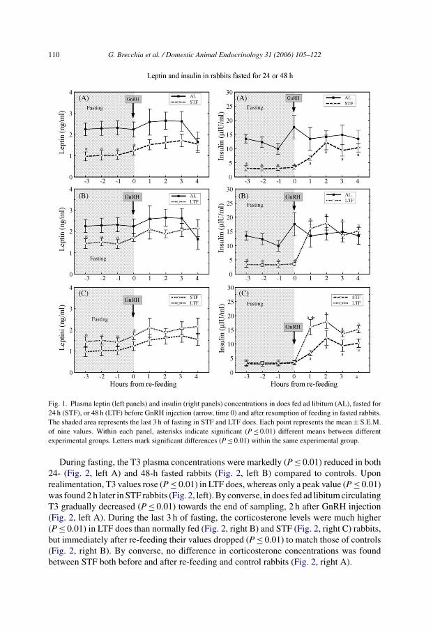
During the last 3 h of fasting, plasma leptin concentrations were lower (P ≤ 0.01) in bothSTF (Fig. 1, left A) and LTF (Fig. 1, left B) does than in control rabbits, but then graduallyincreased matching those of AL does within the next 1–4 h after re-feeding (Fig. 1, left Aand B). The length of fasting affected leptin levels, being constantly higher (P ≤ 0.01) inLTF than in STF rabbits, before re-feeding (Fig. 1, left C). Insulin levels were five-fold lower(P ≤ 0.01) in both STF (Fig. 1, right A) and LTF (Fig. 1, right B) does than in controls duringthe last 3 h of fasting, but 1 h after feed was provided again, plasma insulin concentrationsrose (P ≤ 0.01) to reach the same levels as controls in both treated groups. Upon feeding,insulin concentrations rebounded more quickly and higher (P ≤ 0.01) in LTF than in STFdoes (Fig. 1, right C).
110 G. Brecchia et al. / Domestic Animal Endocrinology 31 (2006) 105–122
Fig. 1. Plasma leptin (left panels) and insulin (right panels) concentrations in does fed ad libitum (AL), fasted for24 h (STF), or 48 h (LTF) before GnRH injection (arrow, time 0) and after resumption of feeding in fasted rabbits.The shaded area represents the last 3 h of fasting in STF and LTF does. Each point represents the mean ± S.E.M.of nine values. Within each panel, asterisks indicate significant (P ≤ 0.01) different means between differentexperimental groups. Letters mark significant differences (P ≤ 0.01) within the same experimental group.
During fasting, the T3 plasma concentrations were markedly (P ≤ 0.01) reduced in both24- (Fig. 2, left A) and 48-h fasted rabbits (Fig. 2, left B) compared to controls. Uponrealimentation, T3 values rose (P ≤ 0.01) in LTF does, whereas only a peak value (P ≤ 0.01)was found 2 h later in STF rabbits (Fig. 2, left). By converse, in does fed ad libitum circulatingT3 gradually decreased (P ≤ 0.01) towards the end of sampling, 2 h after GnRH injection(Fig. 2, left A). During the last 3 h of fasting, the corticosterone levels were much higher(P ≤ 0.01) in LTF does than normally fed (Fig. 2, right B) and STF (Fig. 2, right C) rabbits,but immediately after re-feeding their values dropped (P ≤ 0.01) to match those of controls(Fig. 2, right B). By converse, no difference in corticosterone concentrations was foundbetween STF both before and after re-feeding and control rabbits (Fig. 2, right A).

G. Brecchia et al. / Domestic Animal Endocrinology 31 (2006) 105–122 111
Fig. 2. Plasma T3 (left panels) and corticosterone (right panels) concentrations in does fed ad libitum (AL),fasted for 24 h (STF), or fasted for 48 h (LTF) before GnRH injection (arrow, time 0) and after realimentation infasted animals. The shaded zone represents the last 3 h of fasting in STF and LTF does. Each point representsthe mean ± S.E.M. of nine values. Within each panel, asterisks indicate significant (P ≤ 0.01) different meansbetween different experimental groups. Different letters mark significant differences (P ≤ 0.01) within the sameexperimental group.
3.2. Metabolic parameters
Glucose in plasma of fully fed and 24- or 48-h fasted rabbits did not change during thelast 3 h of fasting (Fig. 3, left panels). Soon after feed was provided again, glycemia rose(P ≤ 0.01) to higher levels in STF and LTF does compared to controls during the next 4 h(Fig. 3, panels A and B). Within 1 h after feeding, glucose levels were higher (P ≤ 0.01) inSTF than in LTF does (Fig. 3, left C).

112 G. Brecchia et al. / Domestic Animal Endocrinology 31 (2006) 105–122
Fig. 3. Plasma glucose (left panels) and NEFA (right panels) concentrations in does fed ad libitum (AL), fastedfor 24 h (STF), or fasted for 48 h (LTF) before GnRH injection (arrow, time 0) and after realimentation in fastedanimals. The shaded area represents the last 3 h of fasting in STF and LTF does. Each point represents themean ± S.E.M. of nine values. Within each panel, asterisks indicate significant (P ≤ 0.01) different means betweendifferent experimental groups. Letters mark significant differences (P ≤ 0.01) within the same experimentalgroup.
During the last 3 h of complete food deprivation, both fasted groups had higher (P ≤ 0.01)plasma NEFA concentrations than ad libitum fed rabbits (Fig. 3, right A and B). Soonafter re-feeding, NEFA values rapidly declined (P ≤ 0.01), although at different rates, inboth STF (Fig. 3, right A) and LTF does (Fig. 3, right B) to values higher (P ≤ 0.01)than controls in the first and the next 2 h, respectively. Circulating NEFA levels were
G. Brecchia et al. / Domestic Animal Endocrinology 31 (2006) 105–122 113
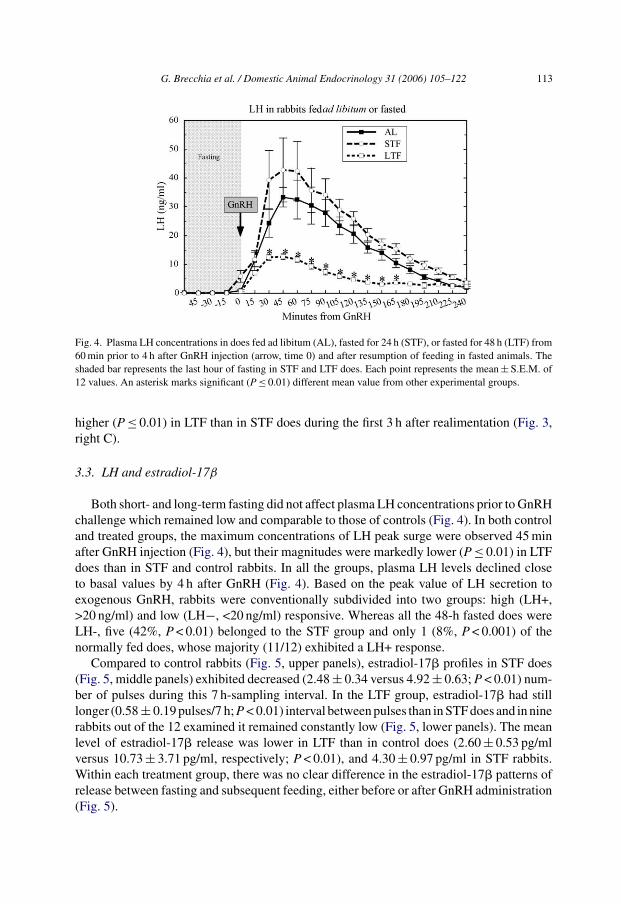
Fig. 4. Plasma LH concentrations in does fed ad libitum (AL), fasted for 24 h (STF), or fasted for 48 h (LTF) from60 min prior to 4 h after GnRH injection (arrow, time 0) and after resumption of feeding in fasted animals. Theshaded bar represents the last hour of fasting in STF and LTF does. Each point represents the mean ± S.E.M. of12 values. An asterisk marks significant (P ≤ 0.01) different mean value from other experimental groups.
higher (P ≤ 0.01) in LTF than in STF does during the first 3 h after realimentation (Fig. 3,right C).
3.3. LH and estradiol-17β
Both short- and long-term fasting did not affect plasma LH concentrations prior to GnRHchallenge which remained low and comparable to those of controls (Fig. 4). In both controland treated groups, the maximum concentrations of LH peak surge were observed 45 minafter GnRH injection (Fig. 4), but their magnitudes were markedly lower (P ≤ 0.01) in LTFdoes than in STF and control rabbits. In all the groups, plasma LH levels declined closeto basal values by 4 h after GnRH (Fig. 4). Based on the peak value of LH secretion toexogenous GnRH, rabbits were conventionally subdivided into two groups: high (LH+,>20 ng/ml) and low (LH−, <20 ng/ml) responsive. Whereas all the 48-h fasted does wereLH-, five (42%, P < 0.01) belonged to the STF group and only 1 (8%, P < 0.001) of thenormally fed does, whose majority (11/12) exhibited a LH+ response.
Compared to control rabbits (Fig. 5, upper panels), estradiol-17� profiles in STF does(Fig. 5, middle panels) exhibited decreased (2.48 ± 0.34 versus 4.92 ± 0.63; P < 0.01) num-ber of pulses during this 7 h-sampling interval. In the LTF group, estradiol-17� had stilllonger (0.58 ± 0.19 pulses/7 h; P < 0.01) interval between pulses than in STF does and in ninerabbits out of the 12 examined it remained constantly low (Fig. 5, lower panels). The meanlevel of estradiol-17� release was lower in LTF than in control does (2.60 ± 0.53 pg/mlversus 10.73 ± 3.71 pg/ml, respectively; P < 0.01), and 4.30 ± 0.97 pg/ml in STF rabbits.Within each treatment group, there was no clear difference in the estradiol-17� patterns ofrelease between fasting and subsequent feeding, either before or after GnRH administration(Fig. 5).

114 G. Brecchia et al. / Domestic Animal Endocrinology 31 (2006) 105–122
Fig. 5. Plasma levels of estradiol-17� in sequential, 15-min samples collected from four different, representative,control rabbits fed ad libitum (AL, upper panels), or 24-h (STF, middle panels) and 48-h (LTF, lower panels)fasted does during the last 3 h of fasting (shaded bars) and the following 4 h after GnRH injection (arrows, time0). Immediately after GnRH injection, feed was provided ad libitum to previously fasted animals.
3.4. Reproductive performance
The overall reproductive performances of does deprived of food for 1 or 2 days beforeAI are summarized in Tables 1 and 2, respectively. In lactating does (Table 1), STF reduced
Table 1Effect of 24 h of fasting (STF) before insemination at day 11 postpartum on reproductive performance of lactatingrabbit does (mean ± S.D.)
STF Control
Does/litters (no.) 43 38Receptivity (%) 55.8 a 76.3 bFertility (%) 41.9 a 65.8 bTotal born (no.) 7.8 ± 4.8 7.5 ± 3.5Born alive (no.) 6.9 ± 4.7 6.6 ± 3.6Weight variation during fasting (%) −3.84 ± 3.0 A 0.07 ± 3.4 BFeed intake before fasting (g DM d−1) 376 ± 102 374 ± 102Feed intake after fasting (g DM d−1) 351 ± 142 313 ± 123
In the same row, different letters indicate significant differences between means at a, b: P ≤ 0.05; A, B; P ≤ 0.001.

G. Brecchia et al. / Domestic Animal Endocrinology 31 (2006) 105–122 115
Table 2Effect of 48 h of fasting (LTF) before insemination on reproductive performance of non-lactating rabbit does(mean ± S.D.)
LTF Control
Does/litters (no.) 16 19Receptivity (%) 56.2 78.9Fertility (%) 31.2 52.6Total born (no.) 10.2 ± 3.6 10.7 ± 2.7Born alive (no.) 8.6 ± 4.1 10.7 ± 2.7Weight variation during fasting (%) −7.0 ± 3.5 A −1.7 ± 3.9 BFeed intake before fasting (g DM d−1) 302 ± 21 301 ± 42Feed intake after fasting (g DM d−1) 297 ± 55 259 ± 55
In the same row, different letters indicate significant differences between means at A, B: P ≤ 0.001.
(P ≤ 0.05) both receptivity (−20.5%) and fertility rate (−23.9%) in comparison with con-trols, but did not influence litter size and fetal viability at birth. The STF affected bodyweight of treated does which was almost 4% lower (P ≤ 0.001) than that of fully fed con-trol does in the same time interval. Interestingly, however, soon after realimentation, feedintake of treated does increased together with a gain in body weight between days 12 and21 (3.38 ± 5.1% versus −1.40 ± 5.3%; P ≤ 0.001) of lactation, so that they matched theweight of controls (4439 ± 532 g versus 4469 ± 493 g).
Also LTF appeared to negatively influenced sexual receptivity (56.2% versus 78.9%;P = 0.16), fertility rate (31.2% versus 52.6%; P = 0.35), and litter size at birth (8.6 versus10.7; P = 0.27), but the differences were not significant due to the low number (16 versus 19)of observations (Table 2). The LTF greatly influenced body weight of treated does which,at the end of fasting, was 5.3% lower (P ≤ 0.01) than that of control does. Within 10 dfollowing realimentation, this loss was regained, so that both treated and control does hada similar body weight (4608 ± 332 g versus 4558 ± 234 g).
All control and fasted does ovulated following GnRH injection. Ovulation rates didnot differ among controls (9.7 ± 2.1 CL), STF and LTF does (9.3 ± 2.4 and 8.6 ± 2.3,respectively).
4. Discussion
The effect of acute feed restriction on fertility has been investigated in several animalspecies [19–22], but only marginally in rabbits [1,8]. Although the basic underlying phys-iological responses to fasting might be common in mammals, to our knowledge this isthe first report to describe the hormonal and metabolic homeostatic adaptations that occurin response to acute food deprivation and their repercussion on the neuroendocrine axisand fertility of rabbits. Moreover, complete feed restriction spanned the 2 days of fastingjust before ovulation to exacerbate the impact of maternal nutritional status on reproductivefunction. In the present study, however, we were mainly interested in verifying the hormonaland metabolic dynamic changes during the transition from fasting to fed status, just beforeand after GnRH-induced ovulation, rather than throughout the whole period of fasting.
116 G. Brecchia et al. / Domestic Animal Endocrinology 31 (2006) 105–122
Compared to rabbits fed ad libitum, fasting altered metabolic hormones and metabolitesdifferently. Decreased plasma concentrations of leptin, insulin, and T3 and increased NEFAconcentrations were all important events for the metabolic adaptation to fasting together withthe activation of the adrenal axis in more severely feed deprived rabbits. During fasting,similar modifications have been described in all the species so far studied [4,19,23–25]demonstrating that substantial homology exists in mammalian species in the regulation ofenergy homeostasis in coping with nutritional stress.
In the present study, fasting caused a clear reduction in plasma concentrations of bothinsulin and leptin, probably in response to reduced availability of carbohydrates. Similarfindings were observed in several other species [23,26–30] and in young growing rabbitsas a consequence of the restricted feed intake [7]. Insulin is a key player in the controlof intermediary metabolism, and exerts an important role in ovarian function in manyspecies. Since there is evidence of active transfer of both insulin and leptin into the brain,these hormones could have a role in signaling the metabolic state of the animal and inthe regulation of appetite [31]. However, the mechanisms through which fasting decreasescirculating leptin levels are still poorly understood. An increasing body of evidence suggeststhat leptin secretion is regulated by other factors not directly dependent on adipose bodymass content [32,33], but rather on the availability of oxidizable nutrients [34]. Notably,there was also a differential leptin response to the severity of fasting. Unexpectedly, in fact,in LTF does leptin concentrations, although lower than in controls, were higher than those of24-h fasted rabbits. This differential response may be partially explained by the stimulatoryaction of corticosterone, whose peripheral plasma concentrations were indeed much higherin LTF than in STF rabbits. In fact, dexamethasone administration was found to potentlystimulate leptin mRNA expression and release in cows [35].
Changes in the energetic balance clearly affected systemic thyroid hormone concentra-tions in does. In fact, during fasting, T3 concentrations were lower than in control rabbits.Thus, food deprivation reduced thyroid function so that animals could spare energy bydecreasing adaptive thermogenesis [36]. Similar down-regulation of thyroidal function wasalso found in growing rabbits during long-term feed restriction [7] and also in severalother species, when undernourished [37,38]. According to recent views, the drop of thyroidhormone during starvation is mediated by decreased expression of thyrotropin-releasinghormone in the hypothalamus upon falling leptin levels [39].
Corticosterone plasma concentrations were higher in 48-h fasted rabbits compared bothto ad libitum fed and 24-h fasted animals. These high corticosterone circulating concentra-tions may represent an adaptation to the prolonged metabolic stress induced by completefood deprivation. However, the activation of the adrenal axis may have both beneficialeffects, by stimulating gluconeogenesis, and several other detrimental side effects resultingin disturbances at both hypothalamic–pituitary and ovarian levels, similarly to what wasdescribed by other authors [40]. Conversely, corticosterone was found to decrease underprolonged feed restriction in rabbits, counteracting the excessive catabolic rate of basalmetabolism [7].
In both 24- and 48-h food deprived rabbits, glucose was maintained at a steady statelevel comparable to that of ad libitum fed rabbits. Conversely, long-term underfeeding wasfound to reduce glucose concentration in rabbits [7]. In our experimental model, NEFAconcentrations were higher in feed restricted rabbits than in control toward the end of

G. Brecchia et al. / Domestic Animal Endocrinology 31 (2006) 105–122 117
fasting. Moreover, the longer the fasting the higher the NEFA concentrations. Free fattyacids are released by the action of hormone sensitive lipase on triglyceride stores in adiposetissue and increased NEFA concentrations are indicative of negative energy balance [41].Hence, augmented circulating NEFA during fasting may reflect redirection of fat metabolismtowards increased lipolysis due to decreased plasma insulin concentrations.
Interestingly, leptin, insulin, T3, and corticosterone adapted very quickly from fasting tore-feeding conditions, as their plasma concentrations were comparable to controls withina few hours after realimentation. During the immediate realimentation period, leptin levelsgradually rose to match those of rabbits fed ad libitum in both fasted groups. This findingsuggests, indirectly, that leptin may signal the current availability of nutrient oxidizable fuel[34] rather than the overall metabolic energy stored in fat depots in accordance with whatwas reported in dairy cattle [30]. Within 1 h after realimentation, insulin quickly reboundedto normal values, probably in response to increasing glucose concentrations derived fromabsorption of carbohydrate with food intake. Thus, when does start to feed again a suddenshift from fat to carbohydrate metabolism occurs and secretion of insulin increases. Similarfindings were reported in a variety of species [3,25,30]. These acute changes in bloodglucose and insulin might have mediated the leptin response of rabbits to realimentation[42]. Whereas low insulin during the fasting state facilitates lipolysis, high insulin followingpost-fast realimentation stimulates de novo lipogenesis [43]. In our study, T3 concentrationsrose to values similar to those in ad libitum fed rabbits 1 h after re-feeding. The mechanismsby which food intake and availability of different carbon sources activate the thyroidal axisare not entirely understood, but recent studies provided experimental evidence about therole of the leptin pathway [39].
Immediately upon re-feeding, glucose concentrations rose by 30–40% over those of con-trols in both fasted groups of does, and remained sustained the next 4 h. The accompanyinghyperinsulinemia suggests that acute fasting may induce insulin resistance. However, thepossibility that this hyperglycemia depends on increased voluntary food intake triggered inpreviously fasted does cannot be ruled out. The increased food intake aimed at recoveringthe body weight lost during fasting is probably favored by low leptin and insulin concentra-tions, which increase anabolic central neuronal pathways [44]. Within 2 h after re-feeding,NEFA concentrations in STF does declined to values comparable to those of ad libitum fedanimals in coincidence with increased insulin and glycemia. Conversely, in LTF does, thedecrease of NEFA after re-feeding was more gradual, even though insulin and glucose weremuch higher than in STF animals.
In the present study, fasting influenced the neuroendocrine axis by modifying the responseof the anterior pituitary gland to GnRH and ovarian steroidogenic activity. Acute fooddeprivation negatively affected the temporal release patterns of estradiol-17�, whose pulsefrequency and amplitude were lower in 48-h fasted than in both control and 24-h fastedanimals. The rapid decline of estradiol-17� concentrations from peak values observed insome STF does in the early post fasting period could be due to an increased estradiol-17� metabolic clearance rate by the liver, as reported in unfed cows whose liver bloodflow increased three-fold, immediately after feeding, for the following 4.5 h [45]. How-ever, the underlying factors responsible for the decreased estrogen levels are still unclearas they may affect the ovarian steroidogenic function either directly or indirectly throughgonadotropin-related mechanisms and impaired folliculogenesis. The nutritionally derived

118 G. Brecchia et al. / Domestic Animal Endocrinology 31 (2006) 105–122
factors, in fact, may target both hypothalamus and pituitary, as well as the steroid com-petent growing follicles within the ovary; this may occur either directly or in associationwith systemic hormones [46], such as insulin [47,48], IGF-I [49], thyroid hormones [50],and local growth factors, including TGF-� and VEGF [51], acting in a paracrine/autocrinefashion to regulate steroidogenesis. The recent findings of leptin receptors in different ovar-ian structures of rabbits, including follicles at different stages of development [13], suggestthat also leptin may regulate steroidogenesis of pre- and post-ovulatory follicles. More-over, leptin was found to inhibit the production of estradiol-17� by granulosa cells underdifferent in vitro conditions [52]. Nevertheless, there is compelling evidence supportingthe view that shortage of metabolizable fuels and the down regulation of many nutritionalmediators, due to reduced feed intake, may directly influence the steroidogenic capabil-ity of ovarian follicles through gonadotropin-independent mechanisms similarly to whatwas found in gilts [51,53]. Thus, the fasting-induced fall in circulating estradiol-17� waslikely the combined results of a reduced synthesis by the ovary and increased clearance bythe liver.
In the present study, the neuronal limb of the ovulatory reflex was bypassed by theexogenous administration of GnRH; this acted on the anterior pituitary to trigger LH releasewithin a few minutes following binding to GnRH receptors. However, the LH peak surgewas consistently low in every 2-day fasted doe and also in 42% of rabbits fasted for 24 h. It isnoteworthy that the GnRH-dependent LH surge showed considerable doe-to-doe variationparticularly within the STF group, but the reasons for this nutritionally dependent variabilitystill remain unclear. In fact, it can be ascribed to individual genetic susceptibility to metabolicstress and/or to their different pre-fasting metabolic condition. It remains to be established,however, which factors were actually responsible for the fasting-induced reduction of LHsecretion, as this might be due to down regulation of pituitary GnRH receptors and/or toreduced synthesis of LH by pituitary cells.
In the present study, the role of estradiol-17� in the maintenance of a functionalhypothalamic–pituitary axis capable of responding appropriately to GnRH appears to beessential; in fact, STF and LTF rabbits showed a lower hormonal pulse frequency and mostof the LTF does had very low plasma estrogen concentrations. However, the sites of actionwithin this system and the mechanisms whereby decreasing estradiol-17� concentrationsinhibit GnRH-induced LH surge are not yet clear. In rabbits, moreover, the reflex LH surge-generator neuronal apparatus is usually unresponsive to the positive feedback by estrogen,which is in striking contrast with spontaneous ovulatory species [54].
A direct role of leptin in the neuroendocrine regulation of energy homeostasis [55,56]and gonadotropin secretion by the pituitary [23] cannot be ruled out. However, from our datathere is little evidence that leptin may act in the short term to regulate pituitary LH secretionand ovarian estradiol-17� release. In fact, in does fasted for 24 h, the pituitary responses toGnRH, as well as estradiol-17� patterns, were normal in 58% of animals, despite their havingseverely reduced peripheral plasma leptin concentrations. These results, rather than beingthe failure of leptin to directly signal caloric shortage to the neuroendocrine axis and theovary, may reflect the maintenance of an adequate or close-to-adequate level of oxidizablemetabolites in 1-day fasted rabbits. Taken together, these findings indicate that leptin regu-lation of the GnRH/LH secretory system may depend upon the concurrently steroid milieuand overall nutritional status. Thus, it is more plausible that leptin exerts a permissive role,

G. Brecchia et al. / Domestic Animal Endocrinology 31 (2006) 105–122 119
together with other metabolic hormones and different nutrients, in signaling body conditionand energy reserves to the hypothalamus–pituitary–ovarian axis for controlling reproductivefunction.
Insulin was found to directly influence LH release by the anterior pituitary in rats [57];at the same time, it may exert a direct effect on the ovary by enhancing steroidogenesis[48]. Consequently, the low plasma insulin levels might be responsible for the impairedestradiol-17� synthesis and release by the ovaries of fasted rabbits. However, the low insulinhypothesis per se does not fully explain the present results, given the differential gonadaland pituitary response observed between does fasted for 24 or 48 h.
In adult female rats, hypothyroidism inhibits follicular development and estrogen secre-tions by the ovary and lowers LH pituitary response to GnRH [50]. These findings suggestthat a similar functional relationship between thyroid and both gonadotropic and gonadalhormones may exist also in the rabbit during fasting.
In the rat, there is evidence that activation of the hypothalamic-pituitary-adrenal axis isresponsible for the fasting-induced suppression of LH secretion, because administration ofa corticotropin-releasing hormone antagonist can reverse the hypogonadotropism arisingfrom food deprivation [6]. Glucocorticoid receptors have been localized in rat granulosa cellsand cortisol was found to inhibit FSH-stimulated aromatase. [58]. These results, togetherwith our findings on the up regulation of the adrenal axis in 2-day fasted does, suggest thatthe adrenal axis may be deeply involved in the down regulation of the reproductive axis,acting either centrally or peripherally.
Independently of the duration of fasting, the present data show that caloric shortagenegatively affected sexual receptivity and fertility rate, but not ovulation rate, litter size andmortality rate at birth, which is considered a good indicator of offspring health. Althoughboth STF and LTF resulted in a temporary body weight loss in does, they subsequentlyrecovered within a few days due to increased feed intake and compensatory growth. Atpresent, however, it is not clear what metabolic signals are specifically involved in all thesenutritionally related effects induced by acute food deprivation on reproduction, nor whetherthey act directly upon the hypothalamic–pituitary–ovarian axis or indirectly to regulate thesecretion of other hormones, such as GH [55] or prolactin; these in turn may influence thegonads. Changes of the metabolic status due to short-term feed restriction have been foundto cause a large array of negative effects on reproduction in a variety of species: delayingpuberty, altering estrous cycles, inhibiting estrous behavior, and influencing both oocytematuration and embryonic survival [4,20,59–63].
In summary, the present data evidenced changes in circulating reproductive, as wellas metabolic, hormones and in key metabolites evoked by acute caloric deprivation justbefore ovulation. Pre-conception nutritional status of the does, as modified by fasting,greatly influenced fertility and sexual receptivity rather than the ovulatory index. Beforeovulation, a constellation of hormones and oxidizable metabolites are likely involved insignaling the energy status of the rabbits to the central reproductive axis and ovary, thusinfluencing follicular development and oocyte quality. Taken together, these results maycontribute to attain more insight into possible hormonal mechanisms mediating nutritionaleffects on ovulation and reproductive function. In this respect, the rabbit can represent aninteresting animal model, given that, following GnRH exogenous induction, its ovulation isprecisely timed.

120 G. Brecchia et al. / Domestic Animal Endocrinology 31 (2006) 105–122
Acknowledgments
We thank Dr. A.F. Parlow (Harbour UCLA, Medical Centre, CA, USA) for kindlingproviding us with rabbit LH (Lot #AFP7818C) and anti-RbLH (Lot #AFP3120489GP)reagents through NHPP program.
This work was partially supported by a grant from the Italian “Ministero IstruzioneUniversita e Ricerca” (Progetto Ricerca Interesse Nazionale—Prot. 2001072484 and2003074002).
The authors gratefully acknowledge the revision of the English text by Dr. James Burge.
References
[1] Fortun-Lamothe L. Effects of pre-mating energy intake on reproductive performance of rabbit does. AnimSci 1998;66:263–9.
[2] Cameron JL. Regulation of reproductive hormone secretion in primates by short-term changes in nutrition.Rev Reprod 1996;1:117–26.
[3] Diskin MG, Mackey DR, Roche JF, Sreenan JM. Effects of nutrition and metabolic status on circulatinghormones and ovarian follicle development in cattle. Anim Reprod Sci 2003;78:345–70.
[4] Ferguson EM, Ashworth CJ, Edwards SA, Hawkins N, Hepburn N, Hunter MG. Effect of different nutritionalregimens before ovulation on plasma concentrations of metabolic and reproductive hormones and oocytematuration in gilts. Reproduction 2003;126:61–71.
[5] Schreihofer DA, Amico JA, Cameron JL. Reversal of fasting-induced suppression of luteinizing hormone(LH) secretion in male rhesus monkeys by intragastric nutrient infusion: evidence for rapid stimulation ofLH by nutritional signals. Endocrinology 1993;132:1890–7.
[6] Maeda K, Cagampang FR, Coen CW, Tsukamura H. Involvement of the catecholaminergic input to the par-aventricular nucleus and of corticotropin-releasing hormone in the fasting-induced suppression of luteinizinghormone release in female rats. Endocrinology 1994;134:1718–22.
[7] Rommers JM, Boiti C, Brecchia G, Mejerhof R, Noordhuizen JPTM, Decuypere E, et al. Metabolic adap-tation and hormonal regulation in young rabbit does during long-term caloric restriction and subsequentcompensatory growth. Anim Sci 2004;79:255–64.
[8] Rommers JM, Mejerhof R, Noordhuizen JPTM, Kemp B. Effect of feeding program during rearing and ageat fist insemination on performances during subsequent reproduction in young rabbit does. Reprod Nutr Dev2004;44:321–32.
[9] Ashworth CJ, Beattie L, Antipatis C, Vallet J. Effects of pre- and post-mating feed intake on blastocyst size,secretory function and glucose metabolism in Meishan gilts. Reprod Fertil Dev 1999;11:323–7.
[10] Comin A, Gerin D, Cappa A, Marchi V, Renaville R, Motta M, et al. The effect of an acute energy deficit onthe hormone profile of dominant follicles in dairy cows. Theriogenology 2002;58:899–910.
[11] Clarke IJ, Henry BA. Leptin and reproduction. Rev Reprod 1999;4:48–55.[12] Cunningham MJ, Clifton DK, Steiner RA. Leptin’s actions on the reproductive axis: perspectives and mech-
anisms. Biol Reprod 1999;60:216–22.[13] Zerani M, Boiti C, Zampini D, Brecchia G, Dell’Aglio C, Ceccarelli P, et al. Ob receptor in rabbit ovary and
leptin in vitro regulation of corpora lutea. J Endocrinol 2004;183:279–88.[14] Zerani M, Boiti C, Dall’Aglio C, Pascucci L, Maranesi M, Brecchia G, et al. Leptin receptor expression
and in vitro leptin actions on prostaglandin release and nitric oxide synthase activity in the rabbit oviduct. JEndocrinol 2005;185:319–25.
[15] Moschos S, Chan JL, Mantzoros CS. Leptin and reproduction: a review. Fertil Steril 2002;77:433–44.[16] Wade GN, Jones JE. Neuroendocrinology of nutritional infertility. Am J Physiol Regul Integr Com Physiol
2004;25:R1277–96.[17] Mattioli M, Conte F, Galeati G, Seren E. Effect of naloxone on plasma concentrations of prolactin and LH
in lactating sows. J Reprod Fertil 1986;76:167–73.
G. Brecchia et al. / Domestic Animal Endocrinology 31 (2006) 105–122 121
[18] Orstead KM, Hess DL, Spies HG. Pulsatile patterns of gonadotropins and ovarian steroids during estrus andpseudopregnancy in the rabbit. Biol Reprod 1988;38:733–43.
[19] Booth PJ, Cosgrove JR, Foxcroft GR. Endocrine and metabolic responses to realimentation in feed-restrictedprepubertal gilts: associations among gonadotropins, metabolic hormones, glucose and uteroovarian devel-opment. J Anim Sci 1996;74:840–8.
[20] Prunier A, Quesnel H. Nutritional influences on the hormonal control of reproduction in female pigs. LivProd Sci 2000;63:1–16.
[21] Rhind SM, McMillen SR, Duff E, Kyle CE, Wright S. Effect of long term feed restriction on seasonalendocrine changes in Soay sheep. Physiol Behav 2000;71:343–51.
[22] Boland MP, Lonergan P, O’Callaghan D. Effect of nutrition on endocrine parameters, ovarian physiology,and oocyte and embryo development. Theriogenology 2001;55:1323–40.
[23] Nagatani S, Zenk Y, Keisler DH, Foster DL, Jaffe CA. Leptin regulates pulsatile luteinizing and growthhormone secretion in the sheep. Endocrinology 2000;141:3965–75.
[24] Prunier A, Martin C, Mounier AM, Bonneau M. Metabolic and endocrine changes associated with undernu-trition in the peripubertal gilt. J Anim Sci 1993;71:1887–94.
[25] Henry BA, Goding JW, Tilbrook AJ, Dunshea FR, Blache D, Clarke IJ. Leptin-mediated effects of undernu-trition or fasting on luteinizing hormone and growth hormone secretion in ovariectomized ewes depends onthe duration of metabolic perturbation. J Neuroendocrinol 2004;16:244–55.
[26] Amstalden M, Garcia MR, Williams SW, Stanko RL, Nizielski SE, Morrison CD, et al. Leptin gene expres-sion, circulating leptin, and luteinizing hormone pulsatility are acutely responsive to short-term fasting inprepubertal heifers: relationships to circulating insulin and insulin-like growth factor I (1). Biol Reprod2000;63:127–33.
[27] MacManus CJ, Fitzgerald BP. Effects of feed restriction on changes in serum leptin, gonadotropins, prolactinand metabolites in aged and young mares. Domest Anim Endocrinol 2000;19:1–13.
[28] Whisnant CS, Harrell RJ. Effect of short-term feed restriction on serum concentrations of leptin, luteinizinghormone and insulin in ovariectomized gilts. Domest Anim Endocrinol 2002;22:73–80.
[29] Chelikani PK, Ambrose JD, Keisler DH, Kennelly JJ. Effect of short term fasting on plasma concentrationsof leptin and other hormones and metabolites in dairy cattle. Domest Anim Endocrinol 2004;26:33–48.
[30] Govoni N, Galeati G, Castellani G, Tamanini C. Leptin concentrations in plasma and follicular fluid fromprepubertal gilts as influenced by fasting, refeeding and insulin. Horm Metab Res 2005;37:152–8.
[31] Woods SC, Seeley RJ, Baskin DG, Schwarts MW. Insulin and the blood–brain barrier. Curr Pharm Des2003;10:795–800.
[32] Barb CR. The brain–pituitary–adipocyte axis: role of leptin in modulating neuroendocrine function. J AnimSci 1999;77:1249–57.
[33] Chilliard Y, Bonnet M, Delavaud C, Faulconnier Y, Leroux C, Djiane J, et al. Leptin in ruminants. Geneexpression in adipose tissue and mammary gland, and regulation of plasma concentrations. Domest AnimEndocrinol 2001;21:271–95.
[34] Obici S, Rossetti L. Nutrient sensing and the regulation of insulin action and energy balance. Endocrinology2003;144:5172–8.
[35] Houseknecht KL, Portocarrero CP, Ji S, Lemenager R, Spurlock ME. Growth hormone regulates leptin geneexpression in bovine adipose tissue: correlation with adipose IGF-I expression. J Endocrinol 2000;164:51–7.
[36] Hornick JL, Van Eenaeme C, Gerard O, Dufrasne I, Istasse L. Mechanisms of reduced and compensatorygrowth. Domest Anim Endocrinol 2000;19:121–32.
[37] Cassar-Malek I, Kahl S, Jurie C, Picard B. Influence of feeding level during postweaning growth on circulatingconcentrations of thyroid hormones and exrtathyroidal 5′-deiodination in steers. Anim Sci 2001;79:2679–87.
[38] Buyse J, Janssens K, Van der Geyten S, Van der As P, Decuypere E, Darras VM. Pre- and postprandial changein plasma hormone and metabolite levels and hepatic deiodenase activities in meal-fed broiler chickens. BrJ Nutr 2002;88:641–53.
[39] Harris M, Aschkenasi C, Elias CF, Chandrankunnel A, Nillni EA, Bjoorbaek C, et al. Transcriptionalregulation of the thyrotropin-releasing hormone gene by leptin and melanocortin signalling. J Clin Invest2001;107:111–20.
[40] Barb CR, Barrett JB, Kraeling RR. Role of leptin in modulating the hypothalamic-pituitary axis and luteinizinghormone secretion in prepubertal gilt. Domest Anim Endocrinol 2004;26:201–14.

122 G. Brecchia et al. / Domestic Animal Endocrinology 31 (2006) 105–122
[41] Emery RS, Liesman JS, Herdt TH. Metabolism of long chain fatty acids in ruminant liver. Anim Nutr Phys1992;122:832–7.
[42] Walker CG, Bryson JM, Bel-Anderson KS, Hancock DP, Denyer GS, Caterson ID. Insulin determines leptinresponses during a glucose challenge in fed and fasted rats. Int J Obes 2005;29:398–405.
[43] Bruckert E, Dejager S. Lipoprotein lipase: a key enzyme of lipid metabolism. Rev Pract 1994;44:1487–93.[44] Schwartz MW, Woods SC, Porte D, Seeley RJ, Baskin DG. Central nervous system control of food intake.
Nature 2000;404:661–71.[45] Sangsritavong S, Combs DK, Sartori R, Armentano LE, Wiltbank MC. High fees intake increases liver blood
flow and metabolism of progesterone and estradiol-17� in dairy cattle. J Dairy Sci 2002;85:2831–42.[46] Webb R, Garnsworthy PC, Gong JG, Armstrong DG. Control of follicular growth and nutritional influences.
J Anim Sci 2004;82:E63–74.[47] Armstrong DG, Gong JC, Gardner JO, Baxter G, Hogg CO, Webb R. Steroidogenesis in bovine granulosa
cells: the effect of short-term changes in dietary intake. Reproduction 2002;123:371–8.[48] Butler ST, Pelton SH, Butler WR. Insulin increases 17�-estradiol production by the dominant follicles of the
first postpartum follicle wave in dairy cows. Reproduction 2004;127:537–45.[49] Chandrashekar V, Zaczek D, Bartke A. The consequences of altered somatotropic system on reproduction.
Biol Reprod 2004;71:17–27.[50] Tobei A. Studies on the functional relationship between thyroid, adrenal and gonadal hormones. J Reprod
Dev 2004;50:9–20.[51] Galeati G, Spinaci M, Govoni N, Zannoni A, Fantinati P, Seren E, et al. Stimulatory effects of fasting
on vascular endothelial growth factor (VEGF) production by growing pig ovarian follicles. Reproduction2003;126:647–52.
[52] Zachow RJ, Magoffin DA. Direct intraovarian effects of leptin: impairment of the synergistic action of insulin-like growth factor-I on follicle-stimulating hormone-dependent estradiol-17 beta production by rat ovariangranulosa cells. Endocrinology 1997;138:847–50.
[53] Cosgrove JR, Tilto JE, Hunter MG, Foxcroft GR. Gonadotropin-independent mechanisms participate inovarian responses to realimentation in feed-restricted prepubertal gilts. Biol Reprod 1992;47:736–45.
[54] Dufy-Barbe L, Dufy B, Vincent JD. Serum gonadotropin levels in the ovariectomized rabbit: effect of acuteand chronic administration of estradiol. Biol Reprod 1978;18:118–24.
[55] Baratta M, Saleri R, Mainardi GL, Valle D, Giustina A, Tamanini C. Leptin regulates GH expression andsecretion and nitric oxide production in pig pituitary cells. Endocrinology 2002;143:551–7.
[56] Wiesner G, Morash RA, Ur E, Wilkinson M. Food restriction regulates adipose-specific cytokines in pituitarygland but not in hypothalamus. J Endocrinol 2004;180:R1–6.
[57] Weiss JM, Polack S, Diedrich K, Ortmann O. Effects of insulin on luteinizing hormone and prolactin secretionand calcium signaling in female rat pituitary cells. Arch Gynecol Obstetr 2003;269:45–50.
[58] Schoonmaker JN, Erickson GF. Glucocorticoid modulation of follicle-stimulating hormone-mediated gran-ulosa cell differentiation. Endocrinology 1983;113:1356–63.
[59] Bishop DK, Wettemann RP, Spicer LJ. Body energy reserves influence the onset of luteal activity after earlyweaning of beef cows. J Anim Sci 1994;72:2703–8.
[60] Burns PD, Spitzer JC, Henricks DM. Effect of dietary energy restriction on follicular development and lutealfunction in nonlactating beef cows. J Anim Sci 1997;754:1078–86.
[61] Foxcroft GR. Mechanisms mediating nutritional effects on embryonic survival in pigs. J Reprod Fertil Suppl1998;52:31–46.
[62] Mburu JN, Einarsson S, Kindahl H, Madej A, Rodriguez-Martinez H. Effects of post-ovulatory food depriva-tion on oviductal sperm concentration, embryo development and hormonal profiles in the pig. Anim ReprodSci 1998;52:221–34.
[63] Almeida FRCL, Kirkwood RN, Aherne FX, Foxcroft GR. Consequences of different patterns of feed intakeduring the estrous cycle in gilts on subsequent fertility. J Anim Sci 2000;78:1556–63.
Copyright © 2022 FDOKUMEN